EP2168985B1 - Antikörper zur Bestimmung des Prothrombin-Fragments F2/F1+2 in einem homogenen Immunoassay - Google Patents
Antikörper zur Bestimmung des Prothrombin-Fragments F2/F1+2 in einem homogenen Immunoassay Download PDFInfo
- Publication number
- EP2168985B1 EP2168985B1 EP09011791.2A EP09011791A EP2168985B1 EP 2168985 B1 EP2168985 B1 EP 2168985B1 EP 09011791 A EP09011791 A EP 09011791A EP 2168985 B1 EP2168985 B1 EP 2168985B1
- Authority
- EP
- European Patent Office
- Prior art keywords
- fragment
- antibody
- prothrombin
- carboxy
- immune complex
- Prior art date
- Legal status (The legal status is an assumption and is not a legal conclusion. Google has not performed a legal analysis and makes no representation as to the accuracy of the status listed.)
- Active
Links
Images
Classifications
-
- C—CHEMISTRY; METALLURGY
- C07—ORGANIC CHEMISTRY
- C07K—PEPTIDES
- C07K16/00—Immunoglobulins [IG], e.g. monoclonal or polyclonal antibodies
- C07K16/18—Immunoglobulins [IG], e.g. monoclonal or polyclonal antibodies against material from animals or humans
- C07K16/36—Immunoglobulins [IG], e.g. monoclonal or polyclonal antibodies against material from animals or humans against blood coagulation factors
-
- C—CHEMISTRY; METALLURGY
- C07—ORGANIC CHEMISTRY
- C07K—PEPTIDES
- C07K16/00—Immunoglobulins [IG], e.g. monoclonal or polyclonal antibodies
- C07K16/42—Immunoglobulins [IG], e.g. monoclonal or polyclonal antibodies against immunoglobulins
-
- C—CHEMISTRY; METALLURGY
- C12—BIOCHEMISTRY; BEER; SPIRITS; WINE; VINEGAR; MICROBIOLOGY; ENZYMOLOGY; MUTATION OR GENETIC ENGINEERING
- C12N—MICROORGANISMS OR ENZYMES; COMPOSITIONS THEREOF; PROPAGATING, PRESERVING, OR MAINTAINING MICROORGANISMS; MUTATION OR GENETIC ENGINEERING; CULTURE MEDIA
- C12N9/00—Enzymes; Proenzymes; Compositions thereof; Processes for preparing, activating, inhibiting, separating or purifying enzymes
- C12N9/14—Hydrolases (3)
- C12N9/48—Hydrolases (3) acting on peptide bonds (3.4)
- C12N9/50—Proteinases, e.g. Endopeptidases (3.4.21-3.4.25)
- C12N9/64—Proteinases, e.g. Endopeptidases (3.4.21-3.4.25) derived from animal tissue
- C12N9/6421—Proteinases, e.g. Endopeptidases (3.4.21-3.4.25) derived from animal tissue from mammals
- C12N9/6424—Serine endopeptidases (3.4.21)
- C12N9/6429—Thrombin (3.4.21.5)
-
- C—CHEMISTRY; METALLURGY
- C12—BIOCHEMISTRY; BEER; SPIRITS; WINE; VINEGAR; MICROBIOLOGY; ENZYMOLOGY; MUTATION OR GENETIC ENGINEERING
- C12Y—ENZYMES
- C12Y304/00—Hydrolases acting on peptide bonds, i.e. peptidases (3.4)
- C12Y304/21—Serine endopeptidases (3.4.21)
- C12Y304/21005—Thrombin (3.4.21.5)
-
- G—PHYSICS
- G01—MEASURING; TESTING
- G01N—INVESTIGATING OR ANALYSING MATERIALS BY DETERMINING THEIR CHEMICAL OR PHYSICAL PROPERTIES
- G01N33/00—Investigating or analysing materials by specific methods not covered by groups G01N1/00 - G01N31/00
- G01N33/48—Biological material, e.g. blood, urine; Haemocytometers
- G01N33/50—Chemical analysis of biological material, e.g. blood, urine; Testing involving biospecific ligand binding methods; Immunological testing
- G01N33/53—Immunoassay; Biospecific binding assay; Materials therefor
- G01N33/563—Immunoassay; Biospecific binding assay; Materials therefor involving antibody fragments
-
- G—PHYSICS
- G01—MEASURING; TESTING
- G01N—INVESTIGATING OR ANALYSING MATERIALS BY DETERMINING THEIR CHEMICAL OR PHYSICAL PROPERTIES
- G01N33/00—Investigating or analysing materials by specific methods not covered by groups G01N1/00 - G01N31/00
- G01N33/48—Biological material, e.g. blood, urine; Haemocytometers
- G01N33/50—Chemical analysis of biological material, e.g. blood, urine; Testing involving biospecific ligand binding methods; Immunological testing
- G01N33/53—Immunoassay; Biospecific binding assay; Materials therefor
- G01N33/573—Immunoassay; Biospecific binding assay; Materials therefor for enzymes or isoenzymes
-
- G—PHYSICS
- G01—MEASURING; TESTING
- G01N—INVESTIGATING OR ANALYSING MATERIALS BY DETERMINING THEIR CHEMICAL OR PHYSICAL PROPERTIES
- G01N33/00—Investigating or analysing materials by specific methods not covered by groups G01N1/00 - G01N31/00
- G01N33/48—Biological material, e.g. blood, urine; Haemocytometers
- G01N33/50—Chemical analysis of biological material, e.g. blood, urine; Testing involving biospecific ligand binding methods; Immunological testing
- G01N33/86—Chemical analysis of biological material, e.g. blood, urine; Testing involving biospecific ligand binding methods; Immunological testing involving blood coagulating time or factors, or their receptors
-
- C—CHEMISTRY; METALLURGY
- C07—ORGANIC CHEMISTRY
- C07K—PEPTIDES
- C07K2317/00—Immunoglobulins specific features
- C07K2317/30—Immunoglobulins specific features characterized by aspects of specificity or valency
- C07K2317/32—Immunoglobulins specific features characterized by aspects of specificity or valency specific for a neo-epitope on a complex, e.g. antibody-antigen or ligand-receptor
-
- C—CHEMISTRY; METALLURGY
- C07—ORGANIC CHEMISTRY
- C07K—PEPTIDES
- C07K2319/00—Fusion polypeptide
-
- G—PHYSICS
- G01—MEASURING; TESTING
- G01N—INVESTIGATING OR ANALYSING MATERIALS BY DETERMINING THEIR CHEMICAL OR PHYSICAL PROPERTIES
- G01N2333/00—Assays involving biological materials from specific organisms or of a specific nature
- G01N2333/90—Enzymes; Proenzymes
- G01N2333/914—Hydrolases (3)
- G01N2333/948—Hydrolases (3) acting on peptide bonds (3.4)
- G01N2333/95—Proteinases, i.e. endopeptidases (3.4.21-3.4.99)
- G01N2333/964—Proteinases, i.e. endopeptidases (3.4.21-3.4.99) derived from animal tissue
- G01N2333/96425—Proteinases, i.e. endopeptidases (3.4.21-3.4.99) derived from animal tissue from mammals
- G01N2333/96427—Proteinases, i.e. endopeptidases (3.4.21-3.4.99) derived from animal tissue from mammals in general
- G01N2333/9643—Proteinases, i.e. endopeptidases (3.4.21-3.4.99) derived from animal tissue from mammals in general with EC number
- G01N2333/96433—Serine endopeptidases (3.4.21)
- G01N2333/96441—Serine endopeptidases (3.4.21) with definite EC number
- G01N2333/96463—Blood coagulation factors not provided for in a preceding group or according to more than one of the proceeding groups
Definitions
- the present invention is in the field of coagulation diagnostics and relates to antibodies which specifically bind to a complex of F2 / F1 + 2 and an F2 / F1 + 2-specific binding partner, as well as their preparation and their use in methods for the determination of F2 / F1 +. 2
- a variety of diagnostically relevant parameters are available for the clinical assessment of the status of the blood coagulation system of a patient.
- the quantitative detection of so-called activation markers of coagulation allows the assessment of the coagulation or fibrinolysis process.
- the determination of the prothrombin fragment F2 / F1 + 2 in a patient sample allows the detection or exclusion of increased thrombin formation in vivo and is used, for example, for detecting intravascular thrombin formation in consumption coagulopathy, in acute venous thromboembolism or in arterial vascular occlusions (myocardial infarction, cerebral infarction). or used for monitoring anticoagulant therapies.
- Prothrombin is the proenzyme of thrombin, the central enzyme in the blood clotting cascade.
- the prothrombin protein has a modular structure and consists of an N-terminal F1 + 2 portion and a C-terminal thrombin portion.
- Prothrombin is cleaved by the proteolytic activity of factor Xa, so that a thrombin molecule (30 kD) is formed per prothrombin molecule (70 kD) with the release of a prothrombin fragment F1 + 2 (35-37 kD).
- the autocatalytic cleavage of prothrombin by thrombin can further lead to cleavage of the fragments F1 (23 kDa) and F2 (14 kDa).
- thrombin does not occur in free form in the blood, but is bound to inhibitors and fibrin immediately after its formation, the extent must thrombin formation are detected indirectly, z. B. by the determination of the activation marker F1 + 2 and / or F2.
- the determination of the plasma F2 / F1 + 2 concentration is therefore the determination of the plasma F2 / F1 + 2 concentration.
- prothrombin in the sample is in a 1,000 to 10,000 fold molar excess compared to F2 / F1 + 2.
- the immunological detection of F2 / F1 + 2 in the presence of prothrombin is hampered by the fact that the released prothrombin fragments F2 / F1 + 2 against intact, uncleaved prothrombin have only a single F2 / F1 + 2 -specific, antigenic epitope namely the resulting after the cleavage of thrombin carboxy terminus of F2 / F1 + 2 peptide (neoepitope).
- the known antibodies are suitable for determining the F2 / F1 + 2 concentration in plasma samples in heterogeneous immunoassays, preferably in the form of a Sandwich ELISA method.
- the F2 / F1 + 2 neoepitope-specific antibodies (“primary antibodies”) are coupled to a solid phase and incubated with the sample so that the F2 / F1 + 2 peptides can bind to the immobilized antibodies.
- primary antibodies By means of a washing step, unbound proteins, in particular prothrombin, are removed before the second, F2 / F1 + 2 and prothrombin-binding antibody (“secondary antibody”) is administered by addition of an antibody solution.
- this second antibody is associated with a signal-forming component that allows for quantification of the F2 / F1 + 2 concentration.
- the object is achieved by providing a monoclonal antibody which binds specifically to an immune complex, which prothrombin fragment F2 / F1 + 2, to which an antibody or an antibody fragment having specificity for the carboxy-terminal neoepitope of the prothrombin fragment F2 / But the antibody does not bind to the prothrombin fragment F1 + 2 or F2 alone and not to the antibody or antibody fragment having specificity for the carboxy-terminal neoepitope of the prothrombin fragment F2 / F1 + 2 alone.
- Antibodies with specificity for immunocomplexes which consist of an analyte, to the an analyte-specific binding partner, eg. B. a primary antibody or a primary antibody fragment is bound exist, are known in principle, for. B. off EP 264 219 A2 . EP 475 784 A1 .
- an immunocomplex in order to obtain an antibody of the invention having specificity for a prothrombin fragment, F2 / F1 + 2 binding partner immune complex, an immunocomplex must be used as the immunization antigen containing a peptide containing at least the complete prothrombin fragment F2, to which an antibody fragment with specificity for the carboxy-terminal neoepitope of the prothrombin fragment F2 / F1 + 2 is bound.
- the present invention is an isolated, purified immune complex containing a peptide comprising at least the complete prothrombin fragment F2, at the carboxy terminus of an antibody fragment with specificity for the carboxy-terminal neoepitope of the prothrombin fragment F2 / F1 + 2, which at least the four carboxy-terminal Amino acids Ile-Glu-Gly-Arg-OH, is bound and wherein the immune complex is coupled to an immunostimulatory carrier protein.
- the prothrombin fragment F2 can be obtained synthetically by chemical synthesis or recombinantly by expression in a prokaryotic or eukaryotic expression system.
- Suitable antibody fragments with specificity for the carboxy-terminal neoepitope of the prothrombin fragment F2 / F1 + 2, such as. B. Fab, Fab ', F (ab') 2 or Fv fragments, for example, can be obtained recombinantly or by enzymatic cleavage of antibodies, preferably monoclonal antibodies, wherein the antibodies are specific for the carboxy-terminal neoepitope of the prothrombin fragment F2 / F1 + 2 and do not bind to prothrombin.
- Such F2 / F1 + 2-specific antibodies that do not bind to prothrombin are, for.
- EP 303 983 A2 (Pelzer et al.
- the enzymatic cleavage of monoclonal antibodies can be carried out, for example, by means of papain, pepsin or ficin. Particularly preferred antibody fragments are Fab fragments, the z. B. can be obtained by enzymatic cleavage by papain.
- Very particularly preferred antibody fragments are fragments, in particular Fab fragments, of the monoclonal, F2 / F1 + 2 neoepitope-specific antibody 96-163 / 04, which according to the teaching in US Pat US 5,830,681 (Hursting et al.
- the peptide comprising at least the complete prothrombin fragment F2 and the antibody fragment with specificity for the carboxy-terminal neoepitope of the prothrombin fragment F2 / F1 + 2 are combined to form the immune complex and incubated under suitable conditions. Subsequently the immune complex is separated from the other reactants, ie isolated and purified, preferably chromatographically.
- the complex is chemically cross-linked, preferably by treatment of the complex with an aldehyde, such as. As glutaraldehyde or formaldehyde.
- the immune complex is coupled to a carrier protein, such as keyhole limpet hemocyanin or ovalbumin.
- a carrier protein such as keyhole limpet hemocyanin or ovalbumin.
- Another object of the present invention relates to the use of an immunocomplex according to the invention as an immunizing antigen in a process for obtaining monoclonal antibodies that specifically bind the complex, but not the individual components of the complex.
- Methods for obtaining monoclonal antibodies are well known, such as. B. the establishment of hybridoma cells with subsequent purification of the secreted antibody.
- the invention furthermore relates to monoclonal antibodies which are distinguished by having an immune complex comprising a peptide comprising at least the complete prothrombin fragment F2 to which an antibody fragment with specificity for the carboxy-terminal neoepitope of the prothrombin fragment F2 / F1 + 2 Is, bind but not to the prothrombin fragment F2 alone and not to the antibody fragment with specificity for the carboxy-terminal neoepitope of the prothrombin fragment F2 / F1 +2 alone.
- antibodies for the purposes of the present invention are antibodies that bind specifically to an immune complex containing the complete F2 peptide, to which a Fab fragment of a monoclonal antibody, which is formed by the hybridoma cell line DSM ACC 2911.
- Such antibodies are z. B. produced by the hybridoma cell lines 2006-175 / 024, 2006-188 / 044 or 2006-188 / 069. These hybridoma cell lines were deposited with the DSMZ - Deutsche Sammlung von Mikroorganismen und Zellkulturen GmbH, Inhoffenstrasse 7B, 38124 Braunschweig, Germany, on June 3, 2008 under the accession numbers DSM ACC2912, DSM ACC2913 and DSM ACC2914.
- the subject of this invention is also an antibody according to the invention which is associated with a solid phase and / or a component of a signal-forming system.
- solid phase in the context of this invention includes an article that consists of porous and / or non-porous, usually water-insoluble material and may have a variety of forms, such as. Vascular, tube, microtiter plate, ball, microparticle, rod, strip, filter or chromatography paper, etc.
- the surface of the solid phase is hydrophilic or can be rendered hydrophilic.
- the solid phase may consist of a variety of materials such. As inorganic and / or organic materials, synthetic, naturally occurring and / or modified naturally occurring materials. Examples of solid phase materials are polymers such as.
- Cellulose nitrocellulose, cellulose acetate, polyvinyl chloride, polyacrylamide, crosslinked dextran molecules, agarose, polystyrene, polyethylene, polypropylene, polymethacrylate or nylon; Ceramics, glass, metals, in particular precious metals, including gold and silver; Magnetite, mixtures or combinations thereof, etc.
- the solid phase may comprise a coating of one or more layers, e.g. From proteins, carbohydrates, lipophilic substances, biopolymers, organic polymers or mixtures thereof, for example in order to suppress or prevent the unspecific binding of sample constituents to the solid phase or, for example, to achieve improvements with regard to the suspension stability of particulate solid phases, storage stability, the shaping stability or the Resistance to UV light, microbes or other destructive agents.
- layers e.g. From proteins, carbohydrates, lipophilic substances, biopolymers, organic polymers or mixtures thereof, for example in order to suppress or prevent the unspecific binding of sample constituents to the solid phase or, for example, to achieve improvements with regard to the suspension stability of particulate solid phases, storage stability, the shaping stability or the Resistance to UV light, microbes or other destructive agents.
- a “signal producing system” may be one or more components, at least one component being a detectable label.
- a label is any molecule that can itself produce a signal or induce the production of a signal such as.
- a fluorescent substance a radioactive substance, an enzyme or a chemiluminescent substance.
- the signal may be detected or measured by enzyme activity, luminescence, light absorption, light scattering, radiated electromagnetic or radioactive radiation, or a chemical reaction.
- a label can itself generate a detectable signal, so that no further components are necessary. Many organic molecules absorb ultraviolet and visible light, and energy transmitted by light absorption can place these molecules in an excited energy state and deliver the absorbed energy in the form of light of a wavelength different from that of the incident light. Still other labels can directly generate a detectable signal such.
- the signal-producing system in such a case includes all the components required for the signal formation such.
- substrates coenzymes, quenchers, accelerators, additional enzymes, substances that react with enzyme products, catalysts, activators, cofactors, inhibitors, ions, etc.
- Suitable labels include, for example, enzymes including horseradish peroxidase, alkaline phosphatase, glucose-6-phosphate dehydrogenase, alcohol dehydrogenase, glucose oxidase, ⁇ -galactosidase, luciferase, urease and acetylcholinesterase; dyes; fluorescent substances including fluorescein isothiocyanate, rhodamine, Phycoerythrin, phycocyanin, ethidium bromide, 5-dimethylaminonaphthalene-1-sulfonyl chloride and rare earth fluorescent chelates; chemiluminescent substances including luminol, isoluminol, acridinium compounds, olefin, enol ether, enamine, aryl vinyl ether, dioxene, arylimidazole, lucigenin, luciferin and aequorin; Sensitizers including eo
- a signal-forming system may also include components which, when in close proximity to each other, may undergo a detectable interaction with each other, e.g. In the form of energy donors and energy receivers such as photosensitizers and chemiluminescent substances (see e.g. EP 515 194 A2 ), Photosensitizers and fluorophores ( WO 95/06877 ), radioactive iodine-125 and fluorophores ( Udenfriend, S. et al. (1985) Proc. Natl. Acad. Sci. 82: 8672-8676 ), Fluorophores and fluorophores ( Mathis, G. (1993) Clin. Chem.
- energy donors and energy receivers such as photosensitizers and chemiluminescent substances (see e.g. EP 515 194 A2 ), Photosensitizers and fluorophores ( WO 95/06877 ), radioactive iodine-125 and fluorophores ( Udenfriend, S.
- the direct transfer of energy between the components e.g. As by light or electron radiation and short-lived reactive chemical molecules, mitein reminder.
- processes in which the activity of one component is inhibited or enhanced by one or more others for example the inhibition or enhancement of enzyme activity or the inhibition, enhancement or alteration (eg wavelength shift, polarization) of the one affected Component emitted electromagnetic radiation.
- the interaction between the components also includes enzyme cascades.
- the components are enzymes, at least one of which provides the substrate for another such that a maximum or minimum reaction rate of the coupled substrate reaction results.
- An effective interaction between the components usually takes place when they are spatially adjacent, so z. B. within a distance range of a few micrometers, in particular within a distance range of less than 600 nm, preferably below 400 nm, most preferably of less than 200 nm.
- Microparticles are often used as a solid phase and / or as a label.
- the term "microparticles" is to be understood as meaning particles having an approximate diameter of at least 20 nm and not more than 20 .mu.m, usually between 40 nm and 10 .mu.m, preferably between 0.1 and 10 .mu.m, in particular preferably between 0.1 and 5 microns, most preferably between 0.15 and 2 microns.
- the microparticles may be regular or irregular in shape. They can represent spheres, spheroids, spheres with more or less large cavities or pores.
- the microparticles may consist of organic, inorganic material or a mixture or combination of both.
- the microparticles may consist of a porous or non-porous, a swellable or non-swellable material.
- the microparticles may have any density, but preferred are particles having a density close to the density of the water, such as from about 0.7 to about 1.5 g / ml.
- the preferred microparticles are suspendible in aqueous solutions and as long as possible suspension stable. They may be transparent, partially transparent or opaque.
- the microparticles may consist of several layers, such as the so-called core-and-shell particles having a core and one or more enveloping layers.
- microparticles includes, for example, dye crystals, metal sols, silica particles, glass particles, magnetic particles, polymer particles, oil drops, lipid particles, dextran, and protein aggregates.
- Preferred microparticles are particles which can be suspended in aqueous solutions and consist of water-insoluble polymer material, in particular substituted polyethylenes. Very particularly preferred are latex particles z.
- polystyrene acrylic acid polymers, methacrylic acid polymers, acrylonitrile polymers, acrylonitrile-butadiene-styrene, polyvinyl acetate acrylate, polyvinylpyridine, vinyl chloride acrylate.
- latex particles with reactive groups on their surface such as carboxyl, amino or aldehyde groups, which has a covalent bond z.
- B. of specific binding partners to the latex particles allow.
- the production of latex particles is for example in EP 80 614 A2 .
- association is to be understood broadly and includes, for example, a covalent and a non-covalent bond, a direct and an indirect bond, the adsorption to a surface and the inclusion in a depression or a cavity, etc.
- a covalent bond Antibody bound via a chemical bond to the solid phase or to a component of a signaling system.
- non-covalent bonding are surface adsorption, inclusion in cavities, or binding of two specific binding partners.
- the antibodies may also be bound to the solid phase or the label indirectly via specific interaction with other specific binding partners (see, for example, US Pat. EP 411 945 A2 ).
- the biotinylated antibody may be bound to the label via label-bound avidin, or a fluorescein-antibody conjugate may be bound to the solid phase via solid phase-bound anti-fluorescein antibodies, or the antibody may be immunoglobulin-binding Proteins are bound to the solid phase or the label.
- the antibodies according to the invention are suitable for use in methods for the quantitative or qualitative determination of the prothrombin fragment F2 / F1 + 2 in a biological sample of a subject, preferably in a blood or plasma sample of a patient.
- Another object of the present invention is therefore a method for the determination of the prothrombin fragment F2 / F1 + 2 in a sample, wherein the sample with an antibody fragment with specificity for the carboxyterminal neoepitope of the prothrombin fragment F2 / F1 + 2 to form an immune complex is brought into contact with the prothrombin fragment F2 / F1 + 2 contained in the sample.
- the sample with an antibody according to the invention or a fragment thereof the binds the immune complex but does not contact the prothrombin fragment F2 / F1 + 2 alone and not the antibody fragment having specificity for the carboxy-terminal neoepitope of the prothrombin fragment F2 / F1 + 2 alone, and the amount of the bound immune complex becomes certainly.
- the sample is incubated with an F (ab ') 2 or a Fab fragment of the antibody, which is formed by the deposited hybridoma cell line DSM ACC2911, to form an immune complex with the prothrombin fragment F2 / contained in the sample.
- F1 + 2 is contacted, and the amount of the immune complex is determined by means of an antibody having specificity for this immune complex, preferably with the aid of an antibody which is formed by one of the deposited hybridoma cell lines DSM ACC2912, DSM ACC2913 or DSM ACC2914 or a fragment thereof ,
- the amount or concentration of F2 / F1 + 2 in the sample is measured.
- quantitative detection also includes semiquantitative methods which can only capture the approximate amount or concentration of F2 / F1 + 2 in the sample or can only serve for a relative quantity or concentration indication.
- qualitative detection is meant the detection of the presence of F2 / F1 + 2 in the sample at all or the indication that the concentration of F2 / F1 + 2 in the sample is below or above a certain threshold (s).
- the invention thus also relates to methods for the quantitative or qualitative detection of F2 / F1 + 2 in a sample and suitable reagents therefor. These methods can be so-called heterogeneous or homogeneous binding tests, in which the presence, absence or amount of F2 / F1 + 2 in a sample can be deduced by specific binding of F2 / F1 + 2 to a binding partner.
- Immunoassays are examples of binding assays.
- the so-called “heterogeneous binding tests” are characterized by one or more separation steps and / or washing steps.
- the separation can, for example, by immunoprecipitation, precipitation with substances such as polyethylene glycol or ammonium sulfate, filtration, magnetic separation, attachment to a solid phase.
- Such a "solid phase” consists of porous and / or non-porous, usually water-insoluble material. It can have many different forms such.
- B vessel, tube, microtitration plate, sphere, microparticles, rods, strips, filter or chromatography paper, etc.
- one of the F2 / F1 + 2-specific binding partner is usually bound to a solid phase and serves for separation the binding complex "F2 / F1 + 2 - F2 / F1 + 2-specific binding partner" from the liquid phase, while the other analyte-specific binding partner for detecting the binding complex, a detectable label (eg, an enzyme, a fluorescent or chemiluminescent label, etc.) wearing.
- a detectable label eg, an enzyme, a fluorescent or chemiluminescent label, etc.
- test methods are further subdivided into so-called one-step sandwich tests in which the two specific binding partners are incubated simultaneously with the sample and in two-step sandwich tests in which the sample is first incubated with the solid phase reagent and after a separation and washing step the solid-phase-bound binding complex of F2 / F1 + 2 and F2 / F1 + 2-specific binding partner is incubated with the detection reagent.
- homogeneous immunoassays are many turbidimetric or nephelometric methods wherein the analyte-specific binding partner used for detection may be associated with latex particles; EMIT ® tests; CEDIA ® tests; Fluorescence polarization immunoassays; Luminescent Oxygen Channeling Immunoassays ("LOCI”, see EP 515 194 A2 ; Ullman, EF et al. (1994) Proc. Natl. Acad. Sci., 91: 5426-5430 ; Ullman, EF et al. (1996) Clinical Chemistry, 42: 1518-1526 ) etc. In a homogeneous sandwich immunoassay, such.
- a nephelometric latex test incubate the antibody reagents together with the sample and measure the signal during and / or after incubation without performing a separation or washing step prior to the measurement. In other words, there is no separation of the antibody-bound analyte from the free analyte or antibodies that did not bind an analyte.
- Homogeneous and heterogeneous binding tests can also be carried out in the form of a so-called "sandwich assay".
- the analyte z For example, in a heterogeneous binding assay, a solid phase-associated analyte-specific binding partner and an analyte-specific binding partner associated with a component of a signal-generating system are bound.
- sample analyte and reagent analyte eg, a "modified analyte,” such as a labeled or tagged F2 / F1 + 2 or F2 / F1 + 2 fragment
- reagent analyte eg, a "modified analyte,” such as a labeled or tagged F2 / F1 + 2 or F2 / F1 + 2 fragment
- the detection of F2 / F1 + 2 with the antibodies according to the invention can also be carried out by methods such as: Western blot, dot blot, immunoelectrophoresis, immunofixation electrophoresis, electroimmuno-diffusion, immunoprecipitation, radial immunodiffusion, immuno-fixation, immunochromatography, latex agglutination, turbidimetric or Nephelometric test, homogeneous or heterogeneous binding test, one or two step test, sandwich test, indirect test, competitive test, point-of-care tests, etc.
- Western blot dot blot, immunoelectrophoresis, immunofixation electrophoresis, electroimmuno-diffusion, immunoprecipitation, radial immunodiffusion, immuno-fixation, immunochromatography, latex agglutination, turbidimetric or Nephelometric test, homogeneous or heterogeneous binding test, one or two step test, sandwich test, indirect test
- POC tests includes tests that do not require a separate analyzer or tester for test performance or evaluation.
- POC tests are based on immunochromatographic procedures, immune complex separations by filtration and / or immunofixation techniques.
- the POC tests are particularly useful for on-site measurements, e.g. B. at the bedside or at home, for the ambulance and / or the general practitioner and less intended for the large laboratory.
- POC tests may also be performed by persons who have no in-depth medical education and experience in the field of laboratory medicine.
- the term "POC tests” also means so-called home tests or OTC tests which may be carried out by medical laymen. B. the various pregnancy tests that are sold for home use.
- POC tests include, for example, the detection of heart attack markers, drugs, drugs, markers of infection and inflammation.
- specific binding partners are or are associated on or on filter or chromatographic strips or discs during the course of the assay.
- a positive or negative detection response may be associated with the appearance or non-appearance of a color band in a particular test field and / or the appearance or non-appearance of a particular symbol, e.g. a "+", a "-" and / or the intensity of the respective measuring signal.
- a POC assay for F2 / F1 + 2 may be constructed as follows: Sample and labeled antibody fragments having specificity for the carboxy-terminal neoepitope of the prothrombin fragment F2 / F1 + 2 are applied to a test strip. Suitable labels are z. Colored latex particles, colloidal gold, enzymes, etc. If F2 / F1 + 2 is included in the sample, F2 / F1 + 2 antibody fragment complexes will form. These complexes move z. B.
- test kit containing at least one reagent containing an antibody of the invention bound to an immune complex containing prothrombin fragment F2 to which an antibody fragment having specificity for the carboxy-terminal neoepitope of the prothrombin fragment F2 / F1 + 2 , binds but not to the prothrombin fragment F2 alone and not to the antibody fragment with specificity for the carboxy-terminal neoepitope of the prothrombin fragment F2 / F1 + 2 alone.
- a preferred test kit contains an antibody which is formed by one of the deposited hybridoma cell lines DSM ACC2912, DSM ACC2913 or DSM ACC2914.
- test kit all or only some components of a test are usually contained in packaged form.
- the antibodies of the invention may be associated with one or more solid phases and / or one or more components of a signal-producing system.
- the test kit may include, for example, standards, controls, and other reagents, such as. B. Buffers, wash solutions, measurement signal-triggering solutions and / or enzyme substrate; cuvettes; Contain pipettes and / or test instructions.
- a test kit according to the invention preferably also contains an antibody or an antibody fragment with specificity for the carboxyterminal neoepitope of the prothrombin fragment F2 / F1 + 2, preferably an F (ab ') 2 or a Fab fragment of the monoclonal antibody 96-163 / 04, which is formed by the deposited hybridoma cell line DSM ACC2911.
- FIG. 1 A first figure.
- immune complex prothrombin fragment F2 / F1 + 2 antibody 96-163 / 04 crosslinking carrier protein a) F2 / F1 + 2-peptide 15mer complete --- --- b) F2 / F1 + 2-peptide 15mer complete glutaraldehyde --- c) complete F2 fragment complete --- --- d) complete F2 fragment complete glutaraldehyde --- e) complete F2 fragment Fab fragment glutaraldehyde KLH f) complete F2 fragment Fab fragment --- ovalbumin
- Synthetic peptide consisting of the 14 C-terminal amino acid residues of the human prothrombin fragment F2 / F1 + 2 and an additional amino-terminal cysteine residue (sequence: H-Cys-Leu-Asp-Glu-Asp-Ser-Asp-Glu-Glu-Arg). Ala-Ile-Glu-Gly-Arg-OH).
- the monoclonal antibody F1 + 2 96-163 / 04 with specificity for the carboxy-terminal neoepitope of the prothrombin fragment F2 / F1 + 2 was prepared according to the teaching in US Pat US 5,830,681 (Hursting et al. ) produced.
- the hybridoma cell line producing the antibody was deposited under the accession number DSM ACC2911.
- the antibody F1 + 2 96-163 / 04 was cleaved with papain by known methods and the Fab fragment was enriched.
- the synthetic F2 / F1 + 2 peptide (15mer) or the purified complete F2 fragment in 10-30 molar excess to the monoclonal antibody F1 + 2 96-163 / 04 or to its Fab fragment was incubated in PBS buffer (pH 7.2) for 3 to 3.5 hours at room temperature and then the immunocomplex was purified by chromatography on Sephacryl TM S-100 (GE Healthcare Europe GmbH, Germany).
- Glutaraldehyde was added at a concentration of 0.2% and the reaction was incubated at 2-8 ° C for 2 hours. The reaction was stopped by addition of NaBH 4 , and the crosslinked immune complex was purified on a HiPrep TM desalting column (GE Healthcare Europe GmbH, Germany) (running buffer PBS pH 7.2).
- the coupling of the immune complexes or the crosslinked immune complexes to keyhole limpet hemocyanin (KLH) or ovalbumin was carried out according to known routine methods.
- BALB / c mice were each immunized intraperitoneally with 20 ⁇ g immunization antigen (see immune complexes from Example 1). Spleen cells from the immunized animals were fused with myeloma cells (Sp2 / 0), and hypergymal cell lines were established.
- Suitable antibodies were identified as follows:
- ELISA No. 1 Reactivity with the immune complex consisting of F2 fragment and the F (ab ') 2 fragment of the monoclonal antibody F1 + 2-96-163 / 04
- Microtiter plates coated with the F (ab ') 2 fragment of the monoclonal antibody F1 + 2 96-163 / 04 were incubated with a solution containing the F2 fragment (see Example 1) to obtain an immune complex from the F (ab') 2- Fragment of the F2 / F1 + 2 specific antibody and the F2 fragment.
- Microtiter plates were coated with purified F2 fragment (see Example 1).
- Microtiter plates were coated with the F (ab ') 2 fragment of F2 / F1 + 2 specific antibody 96-163 / 04.
- Chromogen TMB solution tetramethylbenzidine solution, Siemens Healthcare Diagnostics Products GmbH, Marburg, Germany
- stop solution POD Siemens Healthcare Diagnostics Products GmbH, Marburg, Germany
- ELISA No. 1 ELISA No. 2 ELISA No. 3 2006-140 / 0651 1 3,645 0,025 0.135 2006-175 / 024 1 1,490 0.034 0,107 2006-188 / 044 1 3,808 0.034 0,099 2006-188 / 069 1 3,779 0.031 0.121 2006-188 / 086 1 3.835 0.024 0,107 2006-188 / 0143 1 3,795 0,025 0.132 2006-209 / 09 1 3,171 0.027 0,110 2006-216 / 04 1 1,548 0,020 0,102 Prothr.
- the antibodies according to the invention of group 1 show the desired properties of a monoclonal antibody directed against the immunocomplex, namely in ELISA No. 1 the best possible reactivity with the complex, whereas in ELISA no. 2 and 3 only minimal to no reaction.
- the LOCI® technology used here is based on a latex particulate-coupled chemiluminescent compound (Chemibeads, CB) and a latex particle-coupled photosensitizer (Sensibeads, SB) being brought into spatial proximity by binding to an analyte such that singlet oxygen, produced by the photosensitizer which can stimulate chemiluminescent compound.
- the amount of chemiluminescence generated correlates with the amount of analyte.
- LOCI® technology a signal dependence of the F2 / F1 + 2 concentration was demonstrated in a homogeneous immunological assay setup.
- the immune complex-specific antibodies 2006-175 / 024 (from DSM ACC 2912), 2006-188 / 044 (from DSM ACC 2913) or 2006-188 / 069 (from DSM ACC 2914) were coupled to Chemibeads.
- the second component used was streptavidin-coated Sensibeads.
- the third component used was the biotinylated F2 / F1 + 2 neoepitope-specific antibody 96-163 / 04 (from DSM ACC2911).
- the F2 / F1 + 2 neoepitope-specific antibody 96-163 / 04 was coupled to chemibeads.
- the second component used was streptavidin-coated Sensibeads.
- the third component used was the biotinylated immune complex-specific antibodies 2006-175 / 024 (from DSM ACC 2912), 2006-188 / 044 (from DSM ACC 2913) or 2006-188 / 069 (from DSM ACC 2914).
- a plasma sample was mixed with the two reagents (chemibeads and biotinylated antibody) and incubated for 220 sec at 37 ° C. Thereafter, the Sensibeads were added as a further signal component and mixed. This was followed by another incubation for 360 seconds at 37 ° C. This was followed by measuring the chemiluminescence in the LOCI reader.
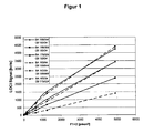
- F1 + 2 was adjusted in a suitable buffer to a defined concentration of 5000 pmol / l. From this stock solution, a dilution series was prepared and analyzed as described above. Plotting the F1 + 2 concentration (X-axis) against the measured signal levels gives a calibration curve. How out FIG. 1 As can be seen, the generated signal in the two different test setups is proportional to the F1 + 2 concentration in the sample, which allows the quantification of the F2 / F1 + 2 concentration.
Landscapes
- Health & Medical Sciences (AREA)
- Chemical & Material Sciences (AREA)
- Life Sciences & Earth Sciences (AREA)
- Engineering & Computer Science (AREA)
- Immunology (AREA)
- Organic Chemistry (AREA)
- Molecular Biology (AREA)
- Hematology (AREA)
- Biomedical Technology (AREA)
- General Health & Medical Sciences (AREA)
- Biochemistry (AREA)
- Medicinal Chemistry (AREA)
- Genetics & Genomics (AREA)
- Urology & Nephrology (AREA)
- Biotechnology (AREA)
- Microbiology (AREA)
- Zoology (AREA)
- Wood Science & Technology (AREA)
- Bioinformatics & Cheminformatics (AREA)
- Food Science & Technology (AREA)
- Cell Biology (AREA)
- Physics & Mathematics (AREA)
- Analytical Chemistry (AREA)
- General Physics & Mathematics (AREA)
- Pathology (AREA)
- General Engineering & Computer Science (AREA)
- Biophysics (AREA)
- Proteomics, Peptides & Aminoacids (AREA)
- Peptides Or Proteins (AREA)
- Preparation Of Compounds By Using Micro-Organisms (AREA)
Description
- Die vorliegende Erfindung liegt auf- dem Gebiet der Gerinnungsdiagnostik und betrifft Antikörper, die spezifisch an einen Komplex aus F2/F1+2 und einem F2/F1+2-spezifischen Bindungspartner binden, sowie deren Herstellung und deren Verwendung in Verfahren zur Bestimmung von F2/F1+2.
- Für die klinische Beurteilung des Status des Blutgerinnungssystems eines Patienten stehen eine Vielzahl diagnostisch relevanter Parameter zur Verfügung. Insbesondere der quantitative Nachweis sogenannter Aktivierungsmarker der Gerinnung ermöglicht die Beurteilung des Gerinnungs- oder Fibrinolyseprozesses. Die Bestimmung des Prothrombin-Fragments F2/F1+2 in einer Patientenprobe ermöglicht den Nachweis oder Ausschluss einer gesteigerten Thrombinbildung in vivo und wird zum Beispiel zur Erfassung der intravasalen Thrombinbildung bei einer Verbrauchskoagulopathie, bei akuten venösen Thromboembolien oder bei arteriellen Gefäßverschlüssen (Myokardinfarkt, Hirninfarkt) oder zur Überwachung von Antikoagulanzientherapien eingesetzt.
- Prothrombin ist das Proenzym von Thrombin, dem zentralen Enzym der Blutgerinnungskaskade. Das Prothrombin-Protein ist modular aufgebaut und besteht aus einem N-terminalen F1+2-Anteil und einem C-terminalen Thrombin-Anteil. Durch die proteolytische Aktivität des Faktors Xa wird Prothrombin gespalten, so dass je Prothrombin-Molekül (70 kD) ein Thrombin-Molekül (30 kD) unter Freisetzung eines Prothrombin-Fragments F1+2 (35-37 kD) entsteht. Durch die autokatalytische Spaltung von Prothrombin durch Thrombin kann es des weiteren zur Spaltung der Fragmente F1 (23 kDa) und F2 (14 kDa) kommen. Da Thrombin in freier Form im Blut nicht vorkommt, sondern unmittelbar im Anschluss an seine Bildung an Inhibitoren und an Fibrin gebunden wird, muss das Ausmaß der Thrombinbildung indirekt erfasst werden, z. B. durch die Bestimmung der Aktivierungsmarker F1+2 und/oder F2. Eine Möglichkeit zum Nachweis oder zum Ausschluss einer gesteigerten Thrombinbildung in vivo ist demnach die Bestimmung der F2/F1+2-Konzentration im Plasma.
- Generell tritt bei F2/F1+2-Testen die Schwierigkeit auf, dass Prothrombin in der Probe im Vergleich zu F2/F1+2 in 1 000 bis 10 000fachem molarem Überschuss vorliegt. Der immunologische Nachweis von F2/F1+2 in Gegenwart von Prothrombin wird dadurch erschwert, dass die freigesetzten Prothrombin-Fragmente F2/F1+2 gegenüber intaktem, ungespaltenem Prothrombin nur über ein einziges F2/F1+2-spezifisches, antigenes Epitop verfügen und zwar den nach der Abspaltung von Thrombin entstehenden Carboxyterminus des F2/F1+2-Peptids (Neoepitop). Ausschließlich Antikörper, die gegen den carboxyterminalen Bereich des F2/F1+2-Peptids gerichtet sind, sind in der Lage spezifisch an die Prothrombin-Fragmente F2/F1+2 zu binden, ohne gleichzeitig eine Spezifität für intaktes Prothrombin aufzuweisen. Alle anderen antigenen Determinanten des F2/F1+2-Fragmentes sind ebenso spezifisch für intaktes Prothrombin.
- Die Herstellung spezifischer F2/F1+2-Antikörper, die nicht an Prothrombin binden, ist z. B. in
EP 303 983 A2 (Pelzer et al. US 5,830,681 (Hursting et al. ) oder inUS 2003/0219845 A1 (Ruiz et al. ) beschrieben. Für die Spezifität der anti-F2/F1+2-Antikörper ist wichtig, dass diese an ein Epitop binden, welches zumindest die vier carboxyterminalen Aminosäuren der F2/F1+2-Fragmente (Ile-Glu-Gly-Arg-OH) enthält. Da in der Regel zur Bestimmung der F2/F1+2-Konzentration Sandwich-Immunoassays eingesetzt werden, werden zwei anti-F2/F1+2-Antikörper benötigt. InEP 1 541 676 A1 (Teigelkamp et al. ) sind Antikörper beschrieben, die an ein Epitop auf der N-terminalen Hälfte des F2-Fragments und damit auch an intaktes Prothrombin binden, sich jedoch zur Verwendung als Sekundärantikörper in Kombination mit den F2/F1+2-Neoepitop-spezifischen Primärantikörpern in einem Sandwich-Immunoassay besonders eignen. - Die bekannten Antikörper eignen sich zur Bestimmung der F2/F1+2-Konzentration in Plasmaproben in heterogenen Immunoassays, bevorzugt in Form eines Sandwich-ELISA-Verfahrens. Dazu werden die F2/F1+2-Neoepitop-spezifischen Antikörper ("Primärantikörper") an eine Festphase gekoppelt und mit der Probe inkubiert, so dass die F2/F1+2-Peptide an die immobilisierten Antikörper binden können. Durch einen Waschschritt werden ungebundene Proteine, insbesondere Prothrombin, entfernt, bevor durch Zugabe einer Antikörperlösung, der zweite, F2/F1+2- und Prothrombin-bindende Antikörper ("Sekundärantikörper") appliziert wird. In der Regel ist dieser Zweitantikörper mit einer signalbildenden Komponente assoziiert, die eine Quantifizierung der-F2/F1+2-Konzentration erlaubt.
- Der Aufbau eines direkten, homogenen Sandwich-Immunoassays ohne Wasch-oder Trennschritte ist mit den aus dem Stand der Technik bekannten monoklonalen Antikörpern jedoch nur eingeschränkt möglich, da der F2/F1+2- und Prothrombin-bindende Sekundärantikörper im homogenen Testaufbau zum Großteil durch das in der Probe enthaltene Prothrombin gebunden wird und somit nicht mehr für die Sandwich-Bildung mit dem Analyten F2/F1+2, welche für die Signalgenerierung unerlässlich ist, zur Verfügung steht.
- Der vorliegenden Erfindung lag also die Aufgabe zugrunde, Mittel bereitzustellen, die es ermöglichen, F2/F1+2 in Proben, die Prothrombin im Überschuss enthalten, spezifisch in einem homogenen Bindungstest zu bestimmen.
- Die Lösung der Aufgabe besteht in der Bereitstellung der in den Ansprüchen beschriebenen erfindungsgemäßen Mittel und Verfahren.
- Insbesondere wird die Aufgabe dadurch gelöst, dass ein monoklonaler Antikörper bereitgestellt wird, der spezifisch an einen Immunkomplex bindet, welcher Prothrombin-Fragment F2/F1+2, an welches ein Antikörper oder ein Antikörperfragment mit Spezifität für das carboxyterminale Neoepitop des Prothrombin-Fragmentes F2/F1+2 gebunden ist, enthält und wobei der Antikörper aber nicht an das Prothrombin-Fragment F1+2 oder F2 alleine und nicht an den Antikörper oder das Antikörperfragment mit Spezifität für das carboxyterminale Neoepitop des Prothrombin-Fragmentes F2/F1+2 alleine bindet.
- Antikörper mit Spezifität für lmmunkomplexe, welche aus einem Analyten, an den ein analytspezifischer Bindungspartner, z. B. ein Primärantikörper oder ein Primärantikörperfragment gebunden ist, bestehen, sind prinzipiell bekannt, z. B. aus
EP 264 219 A2 EP 475 784 A1 WO 85/04422 A1 WO 87/07147 - Gegenstand der vorliegenden Erfindung ist ein isolierter, gereinigter Immunkomplex enthaltend ein Peptid, das zumindest das vollständige Prothrombin-Fragment F2 umfasst, an dessen Carboxyterminus ein Antikörperfragment mit Spezifität für das carboxyterminale Neoepitop des Prothrombin-Fragments F2/F1+2, welches zumindest die vier carboxyterminalen Aminosäuren Ile-Glu-Gly-Arg-OH enthält, gebunden ist und wobei der Immunkomplex an ein immunstimulatorisches Trägerprotein gekoppelt ist.
- Ein Peptid, das zumindest das vollständige Prothrombin-Fragment F2 umfasst, d. h. die Aminosäurereste 156-273 von humanem Prothrombin (siehe z. B.
Fig. 1 in Walz, D.A. (1977) Amino acid sequence of human prothrombin fragments 1 and 2. Proc. Natl. Acad. Sci. USA Vol. 74, No. 5, 1969-1972), und das das carboxyterminale Neoepitop des Prothrombin-Fragments F2/F1+2 aufweist, kann z. B. durch Inkubation einer Prothrombin-haltigen Lösung mit Faktor Xa bzw. durch Aktivierung der Blutgerinnungskaskade in einer Blutgerinnungsfaktor-haltigen Lösung, beispielsweise mit dem Schlangengift Russell's Viper Venom (RW), und anschließender chromatographischer Reinigung des F2-Fragments gewonnen werden. Alternativ kann das Prothrombin-Fragment F2 synthetisch durch chemische Synthese oder rekombinant durch Expression in einem prokaryotischen oder eukaryotischen Expressionssystem gewonnen werden. - Geeignet sind alle Peptide, die zumindest das vollständige Prothrombin-Fragment F2 inklusive des carboxyterminalen Neoepitops umfassen, also auch das vollständige- Prothrombin-Fragment F1+2 sowie die verschiedenen aminoterminal verkürzten F1+2-Fragmente.
- Geeignete Antikörperfragmente mit Spezifität für das carboxyterminale Neoepitop des Prothrombin-Fragments F2/F1+2, wie z. B. Fab, Fab', F(ab')2 oder Fv-Fragmente, können beispielsweise rekombinant oder mittels enzymatischer Spaltung von Antikörpern, vorzugsweise von monoklonalen Antikörpern, gewonnen werden, wobei die Antikörper spezifisch sind für das carboxyterminale Neoepitop des Prothrombin-Fragments F2/F1+2 und nicht an Prothrombin binden. Derartige F2/F1+2-spezifische Antikörper, die nicht an Prothrombin binden, sind z. B. in
EP 303 983 A2 (Pelzer et al. US 5,830,681 (Hursting et al. ) oder inUS 2003/0219845 A1 (Ruiz et al. ) beschrieben. Die enzymatische Spaltung von monoklonalen Antikörpern kann beispielweise mittels Papain, Pepsin oder Ficin durchgeführt werden. Besonders bevorzugte Antikörperfragmente sind Fab-Fragmente, die z. B. mittels enzymatischer Spaltung durch Papain gewonnen werden können. - Ganz besonders bevorzugte Antikörperfragmente sind Fragmente, insbesondere Fab-Fragmente, des monoklonalen, F2/F1+2-Neoepitop-spezifischen Antikörpers 96-163/04, der gemäß der Lehre in
US 5,830,681 (Hursting et al. , insbesondere Examples I-VI) unter Verwendung eines synthetischen Peptids der Sequenz Cys-Gly-Ser-Asp-Arg-Ala-Ile-Glu-Gly-Arg-OH ("PF2") als Immunisierungsantigen hergestellt wurde und der von einer Hybridomazelllinie produziert wird, die am 03. Juni 2008 bei der DSMZ - Deutsche Sammlung von Mikroorganismen und Zellkulturen GmbH, Inhoffenstraße 7B, 38124 Braunschweig, Deutschland, unter der Eingangsnummer DSM ACC2911 hinterlegt wurde. - Zur Herstellung des erfindungsgemäßen Immunkomplexes werden das Peptid, das zumindest das vollständige Prothrombin-Fragment F2 umfasst und das Antikörperfragment mit Spezifität für das carboxyterminale Neoepitop des Prothrombin-Fragments F2/F1+2 zur Bildung des Immunkomplexes zusammengegeben und unter geeigneten Bedingungen inkubiert. Anschließend wird der Immunkomplex von den übrigen Reaktanden abgetrennt, d. h. isoliert und gereinigt, bevorzugt chromatographisch.
- In einer bevorzugten Ausführungform des erfindungsgemäßen Immunkomplexes ist der Komplex chemisch quervernetzt, vorzugsweise durch Behandlung des Komplexes mit einem Aldehyd, wie z. B. Glutaraldehyd oder Formaldehyd.
- Weiterhin ist der Immunkomplex an ein Trägerprotein, wie beispielsweise Keyhole Limpet Hämocyanin oder Ovalbumin gekoppelt. Die immunstimulatorische Wirkung derartiger Trägerproteine und Verfahren zu deren Kopplung sind im Stand der Technik bekannt.
- Ein weiterer Gegenstand der vorliegenden Erfindung betrifft die Verwendung eines erfindungsgemäßen Immunkomplexes als Immunisierungsantigen in einem Verfahren zur Gewinnung monoklonaler Antikörper, die spezifisch den Komplex binden, aber nicht die einzelnen Komponenten des Komplexes. Verfahren zur Gewinnung monoklonaler Antikörper sind hinreichend bekannt, wie z. B. die Etablierung von Hybridomazellen mit anschließender Reinigung der sekretierten Antikörper.
- Die Erfindung betrifft weiterhin monoklonale Antikörper, die sich dadurch auszeichnen, dass sie an einen Immunkomplex enthaltend ein Peptid, das zumindest das vollständige Prothrombin-Fragment F2 umfasst, an welches ein Antikörperfragment mit Spezifität für das carboxyterminale Neoepitop des Prothrombin-Fragments F2/F1+2 gebunden Ist, binden, aber nicht an das Prothrombin-Fragment F2 alleine und nicht an das Antikörperfragment mit Spezifität für das carboxyterminale Neoepitop des Prothrombin-Fragmentes F2/F1 +2 alleine.
- Besonders bevorzugte Antikörper im Sinne der vorliegenden Erfindung sind Antikörper, die spezifisch an einen Immunkomplex binden, der das vollständige F2-Peptid enthält, an welches ein Fab-Fragment eines monoklonalen Antikörpers gebunden ist, der von der Hybridomazelllinie DSM ACC 2911 gebildet wird.
- Derartige Antikörper werden z. B. von den Hybridomazelllinien 2006-175/024, 2006-188/044 oder 2006-188/069 produziert. Diese Hybridomazelllinien wurden am 03. Juni 2008 bei der DSMZ - Deutsche Sammlung von Mikroorganismen und Zellkulturen GmbH, Inhoffenstraße 7B, 38124 Braunschweig, Deutschland, unter den Eingangsnummern DSM ACC2912, DSM ACC2913 und DSM ACC2914 hinterlegt.
- Gegenstand dieser Erfindung ist auch ein erfindungsgemäßer Antikörper, der mit einer Festphase und/oder einer Komponente eines signalbildenden Systems assoziiert ist.
- Der Begriff "Festphase" im Sinne dieser Erfindung beinhaltet einen Gegenstand, der aus porösem und/oder nicht porösem, in der Regel wasserunlöslichem Material besteht und die unterschiedlichsten Formen aufweisen kann, wie z. B. Gefäß, Röhrchen, Mikrotitrationsplatte, Kugel, Mikropartikel, Stäbchen, Streifen, Filter- oder Chromatographiepapier etc. In der Regel ist die Oberfläche der Festphase hydrophil oder kann hydrophil gemacht werden. Die Festphase kann aus den unterschiedlichsten Materialien bestehen wie z. B. aus anorganischen und/oder aus organischen Materialien, aus synthetischen, aus natürlich vorkommenden und/oder aus modifizierten natürlich vorkommenden Materialien. Beispiele für Festphasenmaterialien sind Polymere wie z. B. Zellulose, Nitrozellulose, Zelluloseacetat, Polyvinylchlorid, Polyacrylamid, vernetzte Dextranmoleküle, Agarose, Polystyrol, Polyethylen, Polypropylen, Polymethacrylat oder Nylon; Keramik, Glas, Metalle, insbesondere Edelmetalle wie Gold und Silber; Magnetit, Mischungen oder Kombinationen derselben etc.
- Die Festphase kann einen Überzug aus einer oder mehreren Schichten aufweisen, z. B. aus Proteinen, Kohlehydraten, lipophilen Substanzen, Biopolymeren, organischen Polymeren oder Mischungen hiervon, um beispielsweise die unspezifische Bindung von Probenbestandteilen an die Festphase zu unterdrücken oder zu verhindern, oder um beispielsweise Verbesserungen zu erreichen hinsichtlich der Suspensionsstabilität von partikulären Festphasen, der Lagerstabilität, der formgebenden Stabilität oder der Resistenz gegen UV-Licht, Mikroben oder sonstige zerstörend wirkender Agenzien.
- Bei einem "signalbildenden System" kann es sich um eine oder mehrere Komponenten handeln, wobei es sich wenigstens bei einer Komponente um ein nachweisbares Label handelt. Als Label ist jedes Molekül zu verstehen, das selbst ein Signal produziert oder die Produktion eines Signals induzieren kann wie z. B. eine fluoreszierende Substanz, eine radioaktive Substanz, ein Enzym oder eine chemilumineszierende Substanz. Das Signal kann beispielsweise anhand der Enzymaktivität, der Lumineszenz, der Lichtabsorption, der Lichtstreuung, der ausgestrahlten elektromagnetischen oder radioaktiven Strahlung oder einer chemischen Reaktion nachgewiesen oder gemessen werden.
- Ein Label vermag selbst ein nachweisbares Signal zu erzeugen, so dass keine weiteren Komponenten notwendig sind. Viele organische Moleküle absorbieren ultraviolettes und sichtbares Licht, wobei durch die Lichtabsorption übertragene Energie diese Moleküle in einen angeregten Energiezustand kommen können und die absorbierte Energie in Form von Licht einer anderen Wellenlänge als der des eingestrahlten Lichts abgeben. Wieder andere Label können direkt ein nachweisbares Signal erzeugen wie z. B. radioaktive Isotope oder Farbstoffe.
- Wieder andere Label benötigen zur Signalerzeugung weitere Komponenten, d. h. das signalproduzierende System schließt in einem solchen Fall all die für die Signalbildung benötigten Komponenten mit ein wie z. B. Substrate, Coenzyme, Quencher, Beschleuniger, zusätzliche Enzyme, Substanzen die mit Enzymprodukten reagieren, Katalysatoren, Aktivatoren, Cofaktoren, Inhibitoren, Ionen etc.
- Geeignete Label sind beispielsweise Enzyme einschließlich Meerrettichperoxidase, alkalische Phosphatase, Glukose-6-Phosphatdehydrogenase, Alkoholdehydrogenase, Glucoseoxidase, β -Galactosidase, Luciferase, Urease und Acetylcholinesterase; Farbstoffe; fluoreszierende Substanzen einschließlich Fluoresceinisothiocyanat, Rhodamin, Phycoerythrin, Phycocyanin, Ethidiumbromid, 5-Dimethylaminonaphthalen-1-sulfonylchlorid und fluoreszierende Chelate von seltenen Erden; chemilumineszierende Substanzen einschließlich Luminol, Isoluminol, Acridiniumverbindungen, Olefin, Enolether, Enamin, Arylvinylether, Dioxen, Arylimidazol, Lucigenin, Luciferin und Aequorin; Sensitizer einschließlich Eosin, 9,10-Dibromoanthracen, Methylen Blau, Porphyrin, Phthalocyanin, Chlorophyll, Rose Bengal; Coenzyme; Enzymsubstrate; radioaktive Isotope einschließlich 125I, 131I, 14C, 3H, 32P, 35S, 14C, 51Cr, 59Fe, 57Co und 75Se; Partikel einschließlich magnetische Partikel oder Partikel, bevorzugt Latexpartikel, die selbst beispielsweise mit Farbstoffen, Sensitizern, fluoreszierenden Substanzen, chemilumineszierenden Substanzen, Isotopen oder anderen nachweisbaren Labeln markiert sein können; Solpartikel einschließlich Gold- oder Silbersolen etc.
- Ein signalbildendes System kann auch Komponenten umfassen, die bei räumlicher Nähe miteinander in eine nachweisbare Wechselwirkung treten können, z. B. in Form von Energiespendern und Energieempfängern wie beispielsweise Photosensitizer und chemolumineszierende Substanzen (siehe z. B.
EP 515 194 A2 WO 95/06877 US 3,996,345 ). Unter einer Wechselwirkung zwischen den Komponenten ist die direkte Übertragung von Energie zwischen den Komponenten, z. B. durch Licht-oder Elektronenstrahlung sowie über kurzlebige reaktive chemische Moleküle, miteingeschlossen. Ferner umfasst sind hiervon auch Vorgänge, bei denen die Aktivität einer Komponente durch eine oder mehrere andere inhibiert oder verstärkt wird, beispielsweise die Hemmung oder Steigerung der Enzymaktivität oder die Hemmung, Steigerung oder Veränderung (z. B. Wellenlängenverschiebung, Polarisation) des von der beeinflußten Komponente ausgesendeten elektromagnetischen Strahlung. Die Wechselwirkung zwischen den Komponenten umfasst auch Enzymkaskaden. In diesem Fall sind die Komponenten Enzyme, von denen mindestens eines das Substrat für ein anderes liefert, so dass eine maximale oder minimale Reaktionsgeschwindigkeit der gekoppelten Substratumsetzung resultiert. - Eine effektive Wechselwirkung zwischen den Komponenten findet in der Regel statt, wenn diese räumlich benachbart vorliegen, also z. B. innerhalb eines Abstandbereiches von wenigen Mikrometern, insbesondere innerhalb eines Abstandsbereiches von unter 600 nm, bevorzugt unter 400 nm, ganz besonders bevorzugt von unter 200 nm.
- Mikropartikel werden häufig als Festphase und/oder als Label genutzt. Unter dem Begriff "Mikropartikel" sind im Sinne dieser Erfindung Teilchen zu verstehen, die einen ungefähren Durchmesser von wenigstens 20 nm und nicht-mehr als 20 µm aufweisen, üblicherweise zwischen 40 nm und 10 µm, bevorzugt zwischen 0,1 und 10 µm, besonders bevorzugt zwischen 0,1 und 5 µm, ganz besonders bevorzugt zwischen 0,15 und 2 µm. Die Mikropartikel können regelmäßig oder unregelmäßig geformt sein. Sie können Kugeln, Spheroide, Kugeln mit mehr oder weniger großen Kavitäten oder Poren darstellen. Die Mikropartikel können aus organischem, aus anorganischem Material oder aus einer Mischung oder Kombination von beiden bestehen. Sie können aus einem porösen oder nicht porösen, einem schwellfähigen oder nicht schwellfähigen Material bestehen. Prinzipiell können die Mikropartikel jegliche Dichte haben, bevorzugt sind jedoch Partikel mit einer Dichte, die der Dichte des Wassers nahe kommt wie etwa 0,7 bis etwa 1,5 g/ml. Die bevorzugten Mikropartikel sind in wässrigen Lösungen suspendierbar und möglichst lange suspensionsstabil. Sie mögen durchsichtig, teilweise durchsichtig oder undurchsichtig sein. Die Mikropartikel können aus mehreren Schichten bestehen wie beispielsweise die sogenannten "core-andshell" Partikel mit einem Kern und einer oder mehreren umhüllenden Schichten. Der Begriff Mikropartikel umfasst beispielsweise Farbstoffkristalle, Metallsolen, Silica-Partikel, Glaspartikel, Magnetpartikel, Polymerteilchen, Öltropfen, Lipidpartikel, Dextran, und Proteinaggregate. Bevorzugte Mikropartikel sind in wässrigen Lösungen suspendierbare und aus wasserunlöslichem Polymermaterial bestehende Partikel, insbesondere aus substituierten Polyethylenen. Ganz besonders bevorzugt sind Latexpartikel z. B. aus Polystyrol, Acrylsäurepolymeren, Methacrylsäurepolymeren, Acrylnitril-Polymeren, Acrylnitril-Butadien-Styrol, Polyvinylacetat-Acrylat, Polyvinylpyridin, Vinylchlorid-Acrylat. Von besonderem Interesse sind Latexpartikel mit reaktiven Gruppen an ihrer Oberfläche wie beispielsweise Carboxyl-, Amino- oder Aldehydgruppen, die eine kovalente Bindung z. B. von spezifischen Bindungspartnern an die Latexpartikel erlauben. Die Herstellung von Latexpartikeln ist beispielsweise in
EP 80 614 A2 EP 227 054 A2 EP 246 446 A2 - Der Begriff "assoziiert" ist breit zu verstehen und umfasst beispielsweise eine kovalente und eine nicht-kovalente Bindung, eine direkte und eine indirekte Bindung, die Adsorption an eine Oberfläche und den Einschluss in eine Vertiefung oder einen Hohlraum etc. Bei einer kovalenten Bindung sind die Antikörper über eine chemische Bindung an die Festphase oder an eine Komponente eines signalgebenden Systems gebunden. Beispiele für eine nicht-kovalente Bindung sind die Oberflächenadsorption, der Einschluss in Hohlräume oder die Bindung von zwei spezifischen Bindungspartnern. Neben einer direkten Bindung an die Festphase oder die Komponente eines signalgebenden Systems können die Antikörper auch indirekt über spezifische Wechselwirkung mit anderen spezifischen Bindungspartnern an die Festphase oder das Label gebunden sein (siehe z. B.
EP 411 945 A2 - Die erfindungsgemäßen Antikörper eignen sich zur Verwendung in Verfahren zur quantitativen oder qualitativen Bestimmung des Prothrombin-Fragments F2/F1+2 in einer biologischen Probe eines Probanden, bevorzugt in einer Blut- oder Plasmaprobe eines Patienten. Ein weiterer Gegenstand der vorliegenden Erfindung ist daher ein Verfahren zur Bestimmung des Prothrombin-Fragments F2/F1+2 in einer Probe, wobei die Probe mit einem Antikörperfragment mit Spezifität für das carboxyterminale Neoepitop des Prothrombin-Fragmentes F2/F1+2 zur Bildung eines Immunkomplexes mit dem in der Probe enthaltenen Prothrombin-Fragment F2/F1+2 in Kontakt gebracht wird. Weiterhin wird die Probe mit einem erfindungsgemäßen Antikörper oder einem Fragment desselben, der an den Immunkomplex bindet, aber nicht an das Prothrombin-Fragment F2/F1+2 alleine und nicht an das Antikörperfragment mit Spezifität für das carboxyterminale Neoepitop des Prothrombin-Fragmentes F2/F1+2 alleine, in Kontakt gebracht, und die Menge des gebundenen Immunkomplexes wird bestimmt. In einer bevorzugten Ausführungsform wird die Probe mit einem F(ab')2- oder mit einem Fab-Fragment des Antikörpers, der von der hinterlegten Hybridomazelllinie DSM ACC2911 gebildet wird, zur Bildung eines Immunkomplexes mit dem in der Probe enthaltenen Prothrombin-Fragment F2/F1+2 in Kontakt gebracht, und die Menge des Immunkomplexes wird mit Hilfe eines Antikörpers mit Spezifität für diesen Immunkomplex bestimmt, bevorzugt mit Hilfe eines Antikörpers, der von einer der hinterlegten Hybridomazelllinien DSM ACC2912, DSM ACC2913 oder DSM ACC2914 gebildet wird oder eines Fragments derselben.
- Bei einem Verfahren zur quantitativen Bestimmung von F2/F1+2 wird die Menge oder die Konzentration von F2/F1+2 in der Probe gemessen. Von dem Begriff des "quantitativen Nachweises" sind auch semiquantitative Methoden umfasst, die nur die ungefähre Menge oder Konzentration von F2/F1+2 in der Probe erfassen oder nur zu einer relativen Mengen- oder Konzentrationsangabe dienen können. Unter einem qualitativen Nachweis ist der Nachweis des Vorhandenseins von F2/F1+2 in der Probe überhaupt oder das Anzeigen, dass die Konzentration von F2/F1+2 in der Probe unterhalb oder oberhalb eines bestimmten oder mehrerer bestimmter Schwellenwerte liegt, zu verstehen.
- Die Erfindung betrifft somit auch Verfahren zum quantitativen oder qualitativen Nachweis von F2/F1+2 in einer Probe und geeignete Reagenzien hierfür. Bei diesen Verfahren kann es sich um sogenannte heterogene oder homogene Bindungsteste handeln, bei denen durch eine spezifische Bindung von F2/F1+2 an einen Bindungspartner auf die Anwesenheit, Abwesenheit oder Menge von F2/F1+2 in einer Probe geschlossen werden kann. Immunoassays sind Beispiele für Bindungsteste.
- Die sogenannten "heterogenen Bindungsteste" sind durch einen oder mehrere Trennungsschritte und/oder Waschschritte gekennzeichnet. Die Trennung kann beispielsweise durch Immunfällung, Fällung mit Substanzen wie Polyethylenglykol oder Ammoniumsulfat, Filtration, magnetische Abtrennung, Anbindung an eine Festphase erfolgen. Ein solche "Festphase" besteht aus porösem und/oder nicht porösem, in der Regel wasserunlöslichem Material. Sie kann die unterschiedlichsten Formen aufweisen wie z. B.: Gefäß, Röhrchen, Mikrotitrationsplatte, Kugel, Mikropartikel, Stäbchen, Streifen, Filter- oder Chromatographiepapier etc. Bei heterogenen Bindungstesten im Sandwichformat ist in der Regel einer der F2/F1+2-spezifischen Bindungspartner an eine Festphase gebunden und dient zur Abtrennung des Bindungskomplexes "F2/F1+2 - F2/F1+2-spezifischer Bindungspartner" von der flüssigen Phase, während der andere analytspezifische Bindungspartnerzum Nachweis des Bindungskomplexes ein nachweisbares Label (z. B. ein Enzym, ein Fluoreszenz- oder Chemilumineszenzlabel etc.) trägt. Man unterteilt diese Testverfahren weiter in sogenannte Einschritt-Sandwich-Teste, bei denen die beiden spezifischen Bindungspartner simultan mit der Probe inkubiert werden und in Zweischritt-Sandwich-Teste, bei denen die Probe zunächst mit dem Festphasenreagenz inkubiert wird und nach einem Trenn- und Waschschritt der festphasengebundene Bindungskomplex aus F2/F1+2 und F2/F1+2-spezifischem Bindungspartner mit dem Nachweisreagenz inkubiert wird.
- Bei "homogenen Bindungstesten" erfolgt keine Trennung zwischen freien und an den "F2/F1+2 - F2/F1+2-spezifischer Bindungspartner"-Komplex gebundenen Komponenten des signalbildenden Systems. Der Testansatz, welcher die F2/F1+2-spezifischen Bindungspartner, die signalbildenden Komponenten und die Probe enthält, wird nach oder sogar während der Bindungsreaktion ohne einen weiteren Trenn- und/oder Waschschritt gemessen und das entsprechende Messsignal bestimmt. Beispiele für homogene Immunoassays sind viele turbidimetrische oder nephelometrische Methoden, wobei die zum Nachweis verwendeten analytspezifischen Bindungspartner mit Latexpartikel assoziiert sein können; EMIT®-Teste; CEDIA®-Teste; Fluoreszenz-Polarisations-Immunoassays; Luminescent Oxygen Channeling Immunoassays ("LOCI", siehe
EP 515 194 A2 - Homogene und heterogene Bindungsteste können auch in Form eines sogenannten "Sandwichassays" durchgeführt werden. Hierbei wird der Analyt z. B. bei einem heterogenen Bindungstest von einem Festphasen-assoziierten analytspezifschen Bindungspartner und einem analytspezifischen Bindungspartner, der mit einer Komponente eines signalbildenden System assoziiert ist, gebunden.
- Bei einem homogenen oder heterogenen "kompetitiven Bindungstest" konkurrieren Proben-Analyt und Reagenz-Analyt (beispielsweise ein "modifizierter Analyt" wie z. B. ein gelabeltes oder markiertes F2/F1+2 oder F2/F1+2-Fragment), um die Bindung an eine limitierte Anzahl von analytspezifischen Bindungspartnern. Beispiele zur Illustration des Prinzips: (i) Proben-Analyt konkurriert mit Reagenz-Analyt, der mit einer Komponente eines signalbildenden System assoziiert ist, um die Bindung an Festphasen-assoziierte analytspezifische Bindungspartner oder (ii) Proben-Analyt konkurriert mit Festphasen-assoziiertem Analyt (= Reagenz-Analyt) um die Bindung an analytspezifische Bindungspartner, die mit einer Komponente eines signalbildenden Systems assoziiert sind.
- Der Nachweis von F2/F1+2 mit den erfindungsgemäßen Antikörpern kann beispielsweise auch mit Verfahren erfolgen wie beispielsweise: Western Blot, Dot Blot, Immunelektrophorese, Immunfixations-Elektrophorese, Elektroimmundiffusion, Immunpräzipitation, radiale Immundiffusion, Immunfixation, Immunchromatographie, Latex-Agglutination, turbidimetrischer oder nephelometrischer Test, homogener oder heterogener Bindungstest, Ein- oder Zweischritt-Test, Sandwich-Test, indirekter Test, kompetitiver Test, "point-of-care"-Teste etc. Diese und andere Nachweisverfahren sind beispielsweise in "Labor und Diagnose", ed. L. Thomas, TH-Books Verlagsgesellschaft mbH, Frankfurt, 1998, Kapitel 60 beschrieben.
- Der Begriff "point-of-care-Teste" oder "POC-Teste" schließt Teste ein, bei denen kein separates Analyse- oder Messgerät zur Testdurchführung oder Testauswertung benötigt wird. POC-Teste basieren in vielen Fällen auf immunchromatographischen Verfahren, Immunkomplexabtrennungen- per Filtration und/oder Immunfixationstechniken. Die POC-Teste sind insbesondere für Messungen vor Ort, z. B. am Krankenbett oder daheim, für den Notarzt und/oder beim niedergelassenen Arzt und weniger für das Großlabor gedacht. POC-Teste können insbesondere auch von Personen, die keine eingehende medizinischtechnische Ausbildung und Erfahrung auf dem Gebiet der Laboratoriumsmedizin haben, durchgeführt werden. Unter der Begriff "POC-Teste" sind im Sinne dieser Erfindung auch sogenannte Heimteste oder OTC-Teste zu verstehen, die von medizinischen Laien durchgeführt werden dürfen, so z. B. die diversen Schwangerschaftsteste die für den Heimgebrauch vertrieben werden. Andere POC-Teste betreffen beispielsweise den Nachweis von Herzinfarktmarkern, Drogen, Arzneimitteln, Infektions- und Entzündungsmarkern. Bei vielen POC-Testen sind oder werden im Laufe der Testdurchführung spezifische Bindungspartner an oder auf Filter- oder Chromatographiestreifen oder -scheiben assoziiert. Eine positive oder negative Nachweisreaktion kann zum Beispiel mit dem Erscheinen oder Nichterscheinen einer Farbbande in einem bestimmten Testfeld verknüpft sein und/oder dem Erscheinen oder Nichterscheinen eines bestimmten Symbols, z.B. einem "+",einem "-" und/oder der Intensität des jeweiligen Meßsignals.
- Ein POC-Test für F2/F1+2 kann beispielsweise so aufgebaut sein: Probe und gelabelte Antikörperfragmente mit Spezifität für das carboxyterminale Neoepitop des Prothrombin-Fragmentes F2/F1+2 werden auf einen Teststreifen aufgetragen. Geeignete Label sind z. B. gefärbte Latexpartikel, kolloidales Gold, Enzyme etc. Sofern F2/F1+2 in der Probe enthalten ist, werden sich F2/F1+2-Antikörperfragment-Komplexe ausbilden. Diese Komplexe bewegen sich z. B. mittels Kapillarkraft in Richtung auf einen Bereich, in dem erfindungsgemäße Antikörper, die spezifisch an den Immunkomplex enthaltend F2/F1+2 und das Antikörperfragment mit Spezifität für das carboxyterminale Neoepitop des Prothrombin-Fragmentes F2/F1+2 zu binden vermögen, z. B. in Form einer Bande fixiert sind oder im Laufe des Testverfahrens fixiert werden (z. B. über eine Biotin-Avidin-Brücke). Die gelabelten F2/F1+2-Antikörper-Immunkomplexe werden in diesem Bereich gebunden und bilden mit den fixierten Antikörpern einen Sandwichkomplex aus. Die Intensität des Labelsignals ist hier proportional zur F2/F1+2-Probenkonzentration.
- Ein anderer erfindungsgemäßer Gegenstand ist ein Testkit, der mindestens ein Reagenz enthält, das einen erfindungsgemäßen Antikörper enthält, der an einen Immunkomplex enthaltend Prothrombin-Fragment F2, an welches ein Antikörperfragment mit Spezifität für das carboxyterminale Neoepitop des Prothrombin-Fragmentes F2/F1+2 gebunden ist, bindet, aber nicht an das Prothrombin-Fragment F2 alleine und nicht an das Antikörperfragment mit Spezifität für das carboxyterminale Neoepitop des Prothrombin-Fragmentes F2/F1+2 alleine. Ein bevorzugtes Testkit enthält einen Antikörper, der von einer der hinterlegten Hybridomazelllinien DSM ACC2912, DSM ACC2913 oder DSM ACC2914 gebildet wird. In einem solchen Kit sind üblicherweise alle oder nur einige Komponenten eines Testes in abgepackter Form enthalten. Die erfindungsgemäßen Antikörper können beispielsweise mit einer oder mehreren Festphasen und/oder einer oder mehreren Komponenten eines signalbildenden Systems assoziiert sein. Das Testkit kann beispielsweise Standards, Kontrollen, sowie andere Reagenzien, wie z. B. Puffer, Waschlösungen, Messsignalauslösende Lösungen und/oder Enzymsubstrat; Küvetten; Pipetten und/oder Testanweisungen enthalten.
- Bevorzugterweise enthält ein erfindungsgemäßer Testkit weiterhin einen Antikörper oder ein Antikörperfragment mit Spezifität für das carboxyterminale Neoepitop des Prothrombin-Fragmentes F2/F1+2, bevorzugterweise ein F(ab')2-oder ein Fab-Fragment des monoklonalen Antikörpers 96-163/04, der von der hinterlegten Hybridomazelllinie DSM ACC2911 gebildet wird.
- Kalibrationskurven für die Quantifizierung von F2/F1+2 in einem homogenen LOCI®-Assay (siehe Ausführungsbeispiel 3). Die gemessenen Signale verhalten sich sowohl in einem ersten Testaufbau, bei dem biotinylierte Immunkomplexspezifische Antikörper (BA) in Kombination mit Chemibead-gekoppeltem F27F1+2-Neoepitop-spezifischem Antikörper (CB) eingesetzt wurden (durchgezogene Linien) als auch in einem zweiten Testaufbau, bei dem ein biotinylierter F2/F1+2-Neoepitop-spezifischer Antikörper (BA) in Kombination mit Chemibeadgekoppelten Immunkomplex-spezifischen Antikörpern (CB) eingesetzt wurde (gestrichelte Linien), proportional zur F1+2-Konzentration.
- Die folgenden Ausführungsbeispiele dienen der Veranschaulichung der vorliegenden Erfindung und sind nicht als Einschränkung zu verstehen.
- Folgende Immunkomplexe wurden als Immunisierungsantigene verwendet:
Tabelle 1 Immunkomplex Prothrombinfragment F2/F1+2-Antikörper 96-163/04 Quervernetzung Trägerprotein a) F2/F1+2-Peptid 15mer komplett --- --- b) F2/F1+2-Peptid 15mer komplett Glutaraldehyd --- c) vollständiges F2-Fragment komplett --- --- d) vollständiges F2-Fragment komplett Glutaraldehyd --- e) vollständiges F2-Fragment Fab-Fragment Glutaraldehyd KLH f) vollständiges F2-Fragment Fab-Fragment --- Ovalbumin - Synthetisches Peptid bestehend aus den 14 C-terminalen Aminosäureresten des humanen Prothrombinfragments F2/F1+2 und einem zusätzlichen aminoterminalen Cystein-Rest (Sequenz: H-Cys-Leu-Asp-Glu-Asp-Ser-Asp-Glu-Glu-Arg-Ala-Ile-Glu-Gly-Arg-OH).
- Zur Gewinnung eines vollständigen F2-Fragments (14 kDa) wurde ein kommerziell erhältliches, Gerinnungsfaktor-Konzentrat (humaner Prothrombinkomplex) als Prothrombinquelle verwendet (Beriplex P/N 500, CSL Behring GmbH, Marburg, Deutschland). Die Herstellung der F2-Präparation wurde folgendermaßen durchgeführt:
- 1. Lösen von Beriplex in Aktivierungspuffer (10 mmol Tris, 10 mmol CaCl2, pH 7,5),
- 2. Gerinnungsaktivierung mittels FX-spezifischer RVV-Sepharose (Schlangengift gekoppelt auf Trägermaterial) und Inkubation unter Rühren bei 37 °C,
- 3. Abtrennung des Trägermaterials mittels Nutsche oder Zentrifugation und anschließendes Abstoppen der Gerinnungsaktivität mittels PMSF (Phenylmethylsulfonylfluorid) sowie Zugabe von Natrium-Citrat,
- 4. Zweimalige Adsorption/Ausfällung der Spaltprodukte mit Gla-Domänen mittels Bariumchlorid und Abtrennung mittels Zentrifugation,
- 5. Stabilisierung, Filtration und Ankonzentrierung des Überstandes,
- 6. Gelfiltration des Überstandes mittels Superdex™ 75 Gelmaterial (GE Healthcare Europe GmbH, Deutschland), Laufpuffer 50 mmol NaHPO4, pH 8,5,
- Verwendung der F2-haltigen Fraktionen für die weitere Aufreinigung,
- 7. Anionenaustauscher-Chromatographie unter Verwendung von Mono Q
- Trägermaterial (GE Healthcare Europe GmbH, Deutschland):
- Puffer A: 50 mmol NaHPO4, pH 8,5
- Puffer B: 50 mmol NaHPO4 + 1 mol NaCl, pH 8,5
- Auftrag der F2-haltigen Fraktionen in Puffer A, Elution mittels linearem
- Salzgradienten (0-40 % Puffer B, 20 Säulenvolumen), Sammeln und Poolen der F2-haltigen Fraktionen,
- Trägermaterial (GE Healthcare Europe GmbH, Deutschland):
- 8. Überprüfung der Reinheit der Präparation mittels SDS-PAGE und Western-Blot.
- Der monoklonale Antikörper F1+2 96-163/04 mit Spezifität für das carboxyterminale Neoepitop des Prothrombin-Fragmentes F2/F1+2 wurde gemäß der Lehre in
US 5,830,681 (Hursting et al. ) hergestellt. Die den Antikörper produzierende Hybridomazelllinie wurde unter der Eingangsnummer DSM ACC2911 hinterlegt. Für die Herstellung des Fab-Fragments wurde der Antikörper F1+2 96-163/04 mittels bekannter Verfahren mit Papain gespalten und das Fab-Fragment wurde angereichert. - Zur Herstellung der Immunkomplexe wurde das synthetische F2/F1+2-Peptid (15mer) bzw. das gereinigte vollständige F2-Fragment in 10-30 molarem Überschuss zum dem monoklonalen Antikörper F1+2 96-163/04 bzw. zu dessen Fab-Fragment gegeben, in PBS-Puffer (pH 7,2) für 3 bis 3,5 Stunden bei Raumtemperatur inkubiert und anschließend wurde der Immunkomplex über Sephacryl™ S-100 (GE Healthcare Europe GmbH, Deutschland) chromatographisch aufgereinigt.
- Glutaraldehyd wurde in einer Konzentration von 0,2 % zum Ansatz gegeben, und der Ansatz wurde 2 Stunden bei 2-8 °C inkubiert. Die Reaktion wurde durch Zugabe von NaBH4 abgestoppt, und der vernetzte Immunkomplex wurde über eine HiPrep™-Desalting Säule (GE Healthcare Europe GmbH, Deutschland) aufgereinigt (Laufpuffer PBS pH 7,2).
- Die Kopplung der Immunkomplexe bzw. der quervernetzten Immunkomplexe an Keyhole Limpet Hämocyanin (KLH) oder Ovalbumin erfolgte gemäß bekannter Routineverfahren.
- Gemäß dem Stand der Technik wurden BALB/c Mäuse jeweils mit 20 µg Immunisierungsantigen (siehe Immunkomplexe aus Beispiel 1) intraperitoneal immunisiert. Milzzellen der immunisierten Tiere wurden mit Myelomzellen (Sp2/0) fusioniert, und es wurden Hybrodimazelllinien etabliert.
- Mit dem F(ab')2-Fragment des monoklonalen Antikörpers F1+2 96-163/04 beschichtete Mikrotiterplatten wurden mit einer Lösung enthaltend F2-Fragment (siehe Beispiel 1) inkubiert, um einen Immunkomplex aus dem F(ab')2-Fragment des F2/F1+2-spezifischen Antikörpers und dem F2-Fragment zu bilden.
- Mikrotiterplatten wurden mit gereinigtem F2-Fragment (siehe Beispiel 1) beschichtet.
- Mikrotiterplatten wurden mit dem F(ab')2-Fragment des F2/F1+2-spezifischen Antikörpers 96-163/04 beschichtet.
- In jede Vertiefung der beschichteten Mikrotiterplatten wurden 100 µl Zellkulturüberstand (Verdünnung 1:2) pipettiert und 1 Stunde bei +15 bis +25 °C inkubiert. Nach zweimaligen Waschen der Platte mit Waschlösung-POD (OSEW; Siemens Healthcare Diagnostics Products GmbH, Marburg, Deutschland) wurden in jede Vertiefung 100 µl anti-Maus lgG/Fc-POD-Konjugat (Peroxidase-Konjugat, Sigma-Aldrich GmbH, Deutschland, Prod. Nr. A0168) eingefüllt und 1 Stunde bei +15 bis +25 °C inkubiert. Nach weiterem zweimaligen Waschen der Platte wurde in jede Vertiefung 100 µl Chromogen TMB-Lösung (Tetramethylbenzidin-Lösung, Siemens Healthcare Diagnostics Products GmbH, Marburg, Deutschland) eingefüllt und weitere 30 Minuten bei +15 bis +25 °C inkubiert. Nach der Inkubation wurde in jede Vertiefung 100 µl Stopplösung POD (Siemens Healthcare Diagnostics Products GmbH, Marburg, Deutschland) eingefüllt und die Mikrotiterplatte am Tecan Sunrise™ Absorbance Reader (Tecan Deutschland, GmbH, Crailsheim, Deutschland) bei 450 nm ausgewertet.
- Die Zelllinien, deren Zellkulturüberstände eine gute Reaktivität mit dem Immunkomplex (ELISA Nr. 1) und eine minimale bis keine Reaktivität mit den einzelnen Komponenten zeigten (ELISA Nr. 2 und 3), wurden kloniert, und aus diesen Klonen wurden größere Mengen Antikörper gereinigt. Die Konzentration der gereinigten Antikörper wurde auf 1 bzw 0,1 µg/mL eingestellt und in den ELISA's Nr. 1 bis 3 gestestet.
- Die Ergebnisse sind in den Tabellen 2 (Antikörperkonzentration 1 µg/mL) und 3 (Antikörperkonzentration 0,1 µg/mL ) dargestellt. Als Messdaten sind die Extinktionswerte bei 450 nm angegeben.
Tabelle 2 MAK Gruppe Nr. ELISA Nr. 1 ELISA Nr. 2 ELISA Nr. 3 2006-140/0651 1 3,961 0,038 0,511 2006-175/024 1 2,761 0,028 0,108 2006-188/044 1 3,906 0,027 0,186 2006-188/069 1 3,848 0,029 0,233 2006-188/086 1 3,918 0,036 0,174 2006-188/0143 1 3,970 0,031 0,387 2006-209/09 1 3,819 0,030 0,166 2006-216/04 1 1,940 0,024 0,230 Prothr.195/097 2 4,103 3,570 0,135 96-163/04 3 0,923 1,921 0,112 95-332/03 3 0,290 3,552 0,097 CRP 268/094 4 0,062 0,027 0,099 Background - 0,082 0,034 0,097 Tabelle 3 MAK Gruppe Nr. ELISA Nr. 1 ELISA Nr. 2 ELISA Nr. 3 2006-140/0651 1 3,645 0,025 0,135 2006-175/024 1 1,490 0,034 0,107 2006-188/044 1 3,808 0,034 0,099 2006-188/069 1 3,779 0,031 0,121 2006-188/086 1 3,835 0,024 0,107 2006-188/0143 1 3,795 0,025 0,132 2006-209/09 1 3,171 0,027 0,110 2006-216/04 1 1,548 0,020 0,102 Prothr. 195/097 2 3,534 1,850 0,102 96-163/04 3 0,174 0,615 0,096 95-332/03 3 0,096 1,203 0,096 CRP 268/094 4 0,081 0,025 0,101 Background - 0,082 0,034 0,097 - Die getesteten monoklonalen Antikörper wurden für eine bessere Übersichtlichkeit in Gruppen aufgeteilt:
- Gruppe 1: erfindungsgemäße Antikörper mit Spezifität für den Immunkomplex bestehend aus F2-Fragment und dem F(ab')2-Fragment des F2/F1+2-Neoepitop-spezifischen Antikörpers F1+2 96-163/04;
- Gruppe 2: Prothrombin-spezifischer Antikörper, der an ein Epitop auf der N-terminalen Hälfte des F2-Fragments bindet (siehe
EP 1 541 676 A1 , Beispiel 2 d) und e), Hybridomazelllinie unter Eingangsnummer DSM ACC2607 hinterlegt), mitgeführt als Kontrolle; - Gruppe 3: Monoklonale Antikörper mit Spezifität für das carboxyterminale Neoepitop des Prothrombin-Fragmentes F2/F1+2, hergestellt gemäß der Lehre in
US 5,830,681 (Hursting et al. ), mitgeführt als Kontrolle; - Gruppe 4: systemfremder monoklonaler Antikörper, mitgeführt als Negativkontrolle.
- Die erfindungsgemäßen Antikörper der Gruppe 1 zeigen die gewünschten Eigenschaften eines gegen den lmmunkomplex gerichteten monoklonalen Antikörpers, nämlich im ELISA Nr. 1 eine möglichst gute Reaktivität mit dem Komplex, im ELISA Nr. 2 und 3 hingegen nur minimale bis keine Reaktion.
- Die hier verwendete LOCI® Technologie beruht darauf, dass eine auf Latexpartikein gekoppelte Chemilumineszenzverbindung (Chemibeads, CB) und ein auf Latexpartikeln gekoppelter Photosensibilisator (Sensibeads, SB) durch Bindung an einen Analyten in eine räumliche Nähe zueinander gebracht werden, so dass Singulett-Sauerstoff, der vom Photosensibilisator erzeugt wird, die Chemilumineszenzverbindung anregen kann. Die Menge der erzeugten Chemilumineszenz korreliert mit der Analytmenge. Unter Verwendung der LOCI® Technologie wurde eine Signalabhängigkeit von der F2/F1+2-Konzentration in einem homogenen immunologischen Testaufbau gezeigt.
- Für einen ersten Testaufbau wurden die Immunkomplex-spezifischen Antikörper 2006-175/024 (aus DSM ACC 2912), 2006-188/044 (aus DSM ACC 2913) oder 2006-188/069 (aus DSM ACC 2914) auf Chemibeads gekoppelt. Als zweite Komponente wurden Streptavidin-beschichtete Sensibeads eingesetzt. Als dritte Komponente wurde der biotinylierte F2/F1+2-Neoepitop-spezifische Antikörper 96-163/04 (aus DSM ACC2911) eingesetzt.
- Für einen zweiten Testaufbau wurde der F2/F1+2-Neoepitop-spezifische Antikörper 96-163/04 (aus DSM ACC2911) auf Chemibeads gekoppelt. Als zweite Komponente wurden Streptavidin-beschichtete Sensibeads eingesetzt. Als dritte Komponente wurden die biotinylierten Immunkomplex-spezifischen Antikörper 2006-175/024 (aus DSM ACC 2912), 2006-188/044 (aus DSM ACC 2913) oder 2006-188/069 (aus DSM ACC 2914) eingesetzt.
- Zur Quantifizierung der F2/F1+2-Konzentration wurde eine Plasmaprobe mit den beiden Reagenzien (Chemibeads und biotinyliertem Antikörper) gemischt und für 220 Sekunden bei 37 °C inkubiert. Danach wurden die Sensibeads als weitere Signalkomponente zugegeben und gemischt. Hiernach erfolgte eine erneute Inkubation über 360 Sekunden bei 37 °C. Im Anschluss erfolgte die Messung der Chemilumineszenz im LOCI-Reader.
- Zur Quantifizierung der F2/F1+2-Konzentration in Humanplasmaproben wurde eine Standardkurve erstellt. Hierfür wurde F1+2 in einem geeigneten Puffer auf eine definierte Konzentration von 5000 pmol/l eingestellt. Von dieser Stammlösung wurde eine Verdünnungsreihe angelegt und wie oben beschrieben analysiert. Die Auftragung der F1+2-Konzentration (X-Achse) gegen die gemessenen Signalhöhen ergibt eine Kalibrationskurve. Wie aus
Figur 1 ersichtlich, ist das generierte Signal in den beiden verschiedenen Testaufbauten proportional der F1+2 Konzentration in der Probe, welches die Quantifizierung der F2/F1-+2 Konzentration ermöglicht. - Weitere Untersuchungen haben gezeigt, dass mit den hier gewählten Testaufbauten kein High-Dose-Hook-Effekt bis zu einer Konzentration von 1600000 pmol/l F1+2 festgestellt wurde. Eine falsch niedrige Bestimmung des Analyten ist somit nahezu ausgeschlossen.
Claims (16)
- Isolierter Immunkomplex enthaltend ein Peptid, das zumindest das vollständige Prothrombin-Fragment F2 umfasst, an dessen Carboxyterminus ein Antikörperfragment mit Spezifität für das carboxyterminale Neoepitop des Prothrombin-Fragmentes F2/F1+2, welches zumindest die vier carboxyterminalen Aminosäuren Ile-Glu-Gly-Arg-OH enthält, gebunden ist, wobei der Immunkomplex an ein immunstimulatorisches Trägerprotein gekoppelt ist.
- Immunkomplex gemäß Anspruch 1, wobei das gebundene Antikörperfragment ein Fab, Fab', F(ab')2 oder Fv-Fragment ist.
- Immunkomplex gemäß einem der vorhergehenden Ansprüche, wobei das gebundene Antikörperfragment ein Fragment eines monoklonalen Antikörpers ist, der von der Hybridomazelllinie DSM ACC 2911 gebildet wird.
- Immunkomplex gemäß einem der vorhergehenden Ansprüche, der an das immunstimulatorische Trägerprotein Keyhole Limpet Hämocyanin gekoppelt ist.
- Immunkomplex gemäß einem der vorhergehenden Ansprüche, der mittels eines Aldehyds quervernetzt ist.
- Verwendung eines Immunkomplexes gemäß einem der Ansprüche 1 bis 5 als Immunisierungsantigen in einem Verfahren zur Herstellung eines monoklonalen Antikörpers.
- Monoklonaler Antikörper dadurch gekennzeichnet, dass er an einen Immunkomplex bindet, wobei der Immunkomplex ein Peptid enthält, das zumindest das vollständige Prothrombin-Fragment F2 umfasst und an dessen Carboxyterminus ein Antikörperfragment mit Spezifität für das carboxyterminale Neoepitop des Prothrombin-Fragmentes F2/F1+2, welches zumindest die vier carboxyterminalen Aminosäuren Ile-Glu-Gly-Arg-OH enthält, gebunden ist, wobei der monoklonale Antikörper aber nicht an das Prothrombin-Fragment F2 alleine und nicht an das Antikörperfragment mit Spezifität für das carboxyterminale Neoepitop des Prothrombin-Fragmentes F2/F1+2 alleine bindet.
- Monoklonaler Antikörper gemäß Anspruch 7 dadurch gekennzeichnet, dass dieser von einer der Hybridomazelllinien DSM ACC2912, DSM ACC2913 oder DSM ACC2914 produziert wird.
- Monoklonaler Antikörper gemäß einem der Ansprüche 7 und 8 dadurch gekennzeichnet, dass er mit einer Komponente eines signalgebenden Systems assoziiert ist.
- Monoklonaler Antikörper gemäß einem der Ansprüche 7 bis 9 dadurch gekennzeichnet, dass er mit einer Festphase assoziiert ist.
- Verwendung eines monoklonalen Antikörpers gemäß einem der Ansprüche 7 bis 10 oder eines Fragments desselben, welches an einen Immunkomplex bindet, wobei der Immunkomplex ein Peptid enthält, das zumindest das vollständige Prothrombin-Fragment F2 umfasst und an dessen Carboxyterminus ein Antikörperfragment mit Spezifität für das carboxyterminale Neoepitop des Prothrombin-Fragmentes F2/F1+2, welches zumindest die vier carboxyterminalen Aminosäuren Ile-Glu-Gly-Arg-OH enthält, gebunden ist, wobei der monoklonale Antikörper aber nicht an das Prothrombin-Fragment F2 alleine und nicht an das Antikörperfragment mit Spezifität für das carboxyterminale Neoepitop des Prothrombin-Fragmentes F2/F1+2 alleine bindet, in einem in vitro Verfahren zum quantitativen oder qualitativen Nachweis von F2/F1+2.
- Hybridomazelllinie, die einen monoklonalen Antikörper gemäß Anspruch 7 produziert.
- Hybridomazelllinie gemäß Anspruch 12, die bei der DSMZ unter der Eingangsnummer DSM ACC2912, DSM ACC2913 oder DSM ACC2914 hinterlegt ist.
- Reagenz enthaltend einen monoklonalen Antikörper gemäß einem der Ansprüche 7 bis 10 oder ein Fragment desselben, welches an einen Immunkomplex bindet, wobei der Immunkomplex ein Peptid enthält, das zumindest das vollständige Prothrombin-Fragment F2 umfasst und an dessen Carboxyterminus ein Antikörperfragment mit Spezifität für das carboxyterminale Neoepitop des Prothrombin-Fragmentes F2/F1+2, welches zumindest die vier carboxyterminalen Aminosäuren Ile-Glu-Gly-Arg-OH enthält, gebunden ist, wobei der monoklonale Antikörper aber nicht an das Prothrombin-Fragment F2 alleine und nicht an das Antikörperfragment mit Spezifität für das carboxyterminale Neoepitop des Prothrombin-Fragmentes F2/F1+2 alleine bindet.
- Testkit enthaltend ein Reagenz gemäß Anspruch 14.
- Verfahren zur Bestimmung des Prothrombin-Fragments F2/F1+2 in einer Probe, wobei die Probe mit einem Antikörper oder einem Antikörperfragment mit Spezifität für das carboxyterminale Neoepitop des Prothrombin-Fragmentes F2/F1+2 zur Bildung eines Immunkomplexes mit dem in der Probe enthaltenen Prothrombin-Fragment F2/F1+2 in Kontakt gebracht wird dadurch gekennzeichnet, dass die Probe zusätzlich mit einem Antikörper gemäß Anspruch 7 in Kontakt gebracht wird.
Applications Claiming Priority (1)
| Application Number | Priority Date | Filing Date | Title |
|---|---|---|---|
| DE102008049601A DE102008049601A1 (de) | 2008-09-30 | 2008-09-30 | Antikörper zur Bestimmung des Prothrombin-Fragments F2/F1+2 in einem homogenen Immunoassay |
Publications (2)
| Publication Number | Publication Date |
|---|---|
| EP2168985A1 EP2168985A1 (de) | 2010-03-31 |
| EP2168985B1 true EP2168985B1 (de) | 2014-08-27 |
Family
ID=41557538
Family Applications (1)
| Application Number | Title | Priority Date | Filing Date |
|---|---|---|---|
| EP09011791.2A Active EP2168985B1 (de) | 2008-09-30 | 2009-09-16 | Antikörper zur Bestimmung des Prothrombin-Fragments F2/F1+2 in einem homogenen Immunoassay |
Country Status (6)
| Country | Link |
|---|---|
| US (1) | US9096674B2 (de) |
| EP (1) | EP2168985B1 (de) |
| JP (1) | JP5498110B2 (de) |
| CA (1) | CA2681287C (de) |
| DE (1) | DE102008049601A1 (de) |
| ES (1) | ES2507083T3 (de) |
Families Citing this family (9)
| Publication number | Priority date | Publication date | Assignee | Title |
|---|---|---|---|---|
| EP2634577A1 (de) | 2012-02-29 | 2013-09-04 | Siemens Healthcare Diagnostics Products GmbH | Verfahren zur Erkennung von Interferenzen in in vitro-diagnostischen Testen |
| JP6032470B2 (ja) * | 2012-08-09 | 2016-11-30 | 富士レビオ株式会社 | Pivka−ii測定方法、測定試薬及び測定キット |
| WO2015017535A2 (en) * | 2013-08-02 | 2015-02-05 | Siemens Healthcare Diagnostics Inc. | Cell-based control and method |
| CN114910636B (zh) * | 2017-10-26 | 2025-02-18 | 科美诊断技术股份有限公司 | 一种血清总IgE抗体检测试剂盒及其检测方法 |
| CN109991411B (zh) * | 2017-12-29 | 2023-01-20 | 上海索昕生物科技有限公司 | 一种检测待测样本中目标抗体的免疫测定方法及其应用 |
| CN109988241A (zh) * | 2017-12-29 | 2019-07-09 | 博阳生物科技(上海)有限公司 | 一种特异性识别人免疫复合物的抗体及其制备方法和用途 |
| CN109991405B (zh) * | 2017-12-29 | 2023-01-24 | 上海索昕生物科技有限公司 | 一种免疫检测试剂盒及其应用 |
| EP4239338A1 (de) | 2022-03-03 | 2023-09-06 | Siemens Healthcare Diagnostics Products GmbH | Globaltest zur feststellung des status des blutgerinnungssystems |
| EP4325224A1 (de) | 2022-08-18 | 2024-02-21 | Siemens Healthcare Diagnostics Products GmbH | Thrombozytenaktivierungstest zur diagnose einer heparin-induzierten thrombozytopenie |
Family Cites Families (19)
| Publication number | Priority date | Publication date | Assignee | Title |
|---|---|---|---|---|
| US3996345A (en) | 1974-08-12 | 1976-12-07 | Syva Company | Fluorescence quenching with immunological pairs in immunoassays |
| DE3145082A1 (de) | 1981-11-13 | 1983-05-19 | Behringwerke Ag, 3550 Marburg | "ein latex, biologisch aktive latexkonjugate und verfahren zu ihrer herstellung" |
| DE3364347D1 (en) * | 1982-04-09 | 1986-08-07 | Fujirebio Kk | Anti immune complex antibody and preparation thereof |
| GB8408193D0 (en) * | 1984-03-30 | 1984-05-10 | Cambridge Patent Dev | Antibodies |
| DE3433652A1 (de) * | 1984-09-13 | 1986-03-20 | Boehringer Mannheim Gmbh, 6800 Mannheim | Immunchemisches messverfahren fuer haptene und proteine |
| DE3545595A1 (de) | 1985-12-21 | 1987-06-25 | Behringwerke Ag | Dispersionspolymere, verfahren zu ihrer herstellung und ihre verwendung |
| DE3613111A1 (de) | 1986-04-18 | 1987-10-22 | Behringwerke Ag | Dispersionspolymere, verfahren zu ihrer herstellung und ihre verwendung |
| EP0247730A3 (de) * | 1986-04-28 | 1989-04-12 | Antibody Technology Limited | Antikörper, ihre Herstellung sowie Verwendung und dieselben enthaltende Produkte |
| WO1987007147A1 (en) | 1986-05-28 | 1987-12-03 | Murex Corporation | Method for producing monoclonal antibodies to an immunogenic antibody-antigen conjunction |
| US5223441A (en) | 1986-10-09 | 1993-06-29 | Syntex (U.S.A.) Inc. | Receptors for immune complexes |
| DE3727610A1 (de) | 1987-08-19 | 1989-03-02 | Behringwerke Ag | Synthetische peptide, gegen diese gerichtete antikoerper und deren verwendung |
| US6541275B1 (en) | 1988-02-03 | 2003-04-01 | Dade Behring Inc. | Immunoassay for F1.2 prothrombin fragment |
| US5512659A (en) | 1989-08-04 | 1996-04-30 | Syntex (U.S.A.) Inc. | Compositions useful in heterogeneous immunoassays |
| IE903930A1 (en) * | 1989-11-06 | 1991-05-08 | Akzo Nv | Immunoassays for and monoclonal antibodies to prothrombin¹activation peptides and their degradation products |
| CA2072758A1 (en) | 1990-09-14 | 1992-03-15 | Kenneth Francis Buechler | Antibodies to complexes of ligand receptors and ligands and their utility in ligand-receptor assays |
| EP0984281B1 (de) | 1991-05-22 | 2007-01-03 | Dade Behring Marburg GmbH | Homogenes Testsystem zur Bestimmung eines Analytes |
| WO1995006877A1 (en) | 1993-09-03 | 1995-03-09 | Behringwerke Ag | Fluorescent oxygen channeling immunoassays |
| FI20022048A0 (fi) * | 2002-11-18 | 2002-11-18 | Valtion Teknillinen | Ei-kompetetiivinen immunomääritys pienille analyyteille |
| DE10354403A1 (de) | 2003-11-20 | 2005-06-23 | Dade Behring Marburg Gmbh | Gegen das Prothrombin-Fragment F 1+2 gerichtete Antikörper, ihre Herstellung und Verwendung |
-
2008
- 2008-09-30 DE DE102008049601A patent/DE102008049601A1/de not_active Withdrawn
-
2009
- 2009-09-16 ES ES09011791.2T patent/ES2507083T3/es active Active
- 2009-09-16 EP EP09011791.2A patent/EP2168985B1/de active Active
- 2009-09-24 US US12/566,464 patent/US9096674B2/en active Active
- 2009-09-29 JP JP2009223732A patent/JP5498110B2/ja active Active
- 2009-09-30 CA CA2681287A patent/CA2681287C/en active Active
Also Published As
| Publication number | Publication date |
|---|---|
| US20100081212A1 (en) | 2010-04-01 |
| JP5498110B2 (ja) | 2014-05-21 |
| CA2681287A1 (en) | 2010-03-30 |
| CA2681287C (en) | 2017-09-12 |
| EP2168985A1 (de) | 2010-03-31 |
| ES2507083T3 (es) | 2014-10-14 |
| US9096674B2 (en) | 2015-08-04 |
| DE102008049601A1 (de) | 2010-04-01 |
| JP2010085407A (ja) | 2010-04-15 |
Similar Documents
| Publication | Publication Date | Title |
|---|---|---|
| EP2168985B1 (de) | Antikörper zur Bestimmung des Prothrombin-Fragments F2/F1+2 in einem homogenen Immunoassay | |
| DE69434286T2 (de) | Immunologische bestimmungen von prostata-spezifischem antigen | |
| DE3486262T2 (de) | Verfahren zur Herstellung eines monoklonalen Antikörpers mit Spezifizität für vernetzte Fibrinderivate und ein Testverfahren mittels dieses Antikörpers. | |
| EP1110970B1 (de) | Gegen Procalcitonin gerichtete Antikörper, ihre Herstellung und Verwendung | |
| EP2333554B1 (de) | Heterogener Gerinnungstest | |
| EP1888642B2 (de) | Bindungspartner des plazentalen wachstumsfaktors insbesondere gegen den plazentalen wachstumsfaktor gerichtete antikörper, ihre herstellung und verwendung | |
| US8063180B2 (en) | Antibodies directed against prothrombin fragment F1+2, the preparation and use thereof | |
| EP1630175B1 (de) | Gegen den Marburg I Polymorphismus der Faktor VII aktivierenden Protease (FSAP) gerichtete Antikörper, ihre Herstellung und Verwendung | |
| DE68911574T2 (de) | Reagenzsystem zur Bestimmung des Komplexes des menschlichen Plasminogenaktivator-Inhibitors und des menschlichen Gewebeplasminogenaktivators und Testsatz dafür. | |
| DE69505365T2 (de) | Immunologisches verfahren zum nachweis von glykosaminoglykan-aktiviertem antithrombin iii; die entsprechenden monoklonalen antikörper und deren herstellung | |
| EP0513565B1 (de) | Monoklonale Antikörper gegen den Plasmin-Antiplasmin Komplex, Verfahren zu ihrer Herstellung sowie ihre Verwendung | |
| DE602004007112T2 (de) | Verfahren zum immunologischen testen der tartrat-resistenten säuren phosphatase 5b sowie dabei zu verwendender kit | |
| DE60034660T2 (de) | Monoklonaler antikörper gegen protein c inhibitor | |
| EP1692515A1 (de) | Konjugate und deren verwendung in nachweisverfahren | |
| DE3855007T2 (de) | METHODE ZUR REINIGUNG REKOMBINANTER t-PA VON DEN IHREN ENTSPRECHENDEN WIRTSZELLPROTEINEN | |
| WO2005062048A1 (de) | Homogenes nachweisverfahren | |
| DE69628878T2 (de) | Methode zur Bestimmung von Cholinesterase und Methode zur Unterscheidung zwischen Leberzirrhose und Hepatitis | |
| EP2919013B1 (de) | Verfahren zur Detektion von Modulatoren der GPIb-Thrombin-Interaktion | |
| DE10041215A1 (de) | Gegen Procalcitonin gerichtete Antikörper, ihre Herstellung und Verwendung | |
| DE10036641A1 (de) | Monoklonale Antikörper für die den Blutgerinnungsfaktor VII aktivierende Protease (FSAP)und ihre Verwendung | |
| EP0306813A2 (de) | Verfahren zur Bestimmung von "löslichem" Fibrin | |
| DE3342807C2 (de) | Verfahren zum Nachweis von Krebs in Menschen sowie Analysekoffer zur Verwendung bei dem Verfahren | |
| DD259045A1 (de) | Verfahren zur quantitativen bestimmung von c-reaktivem protein (crp) |
Legal Events
| Date | Code | Title | Description |
|---|---|---|---|
| PUAI | Public reference made under article 153(3) epc to a published international application that has entered the european phase |
Free format text: ORIGINAL CODE: 0009012 |
|
| AK | Designated contracting states |
Kind code of ref document: A1 Designated state(s): AT BE BG CH CY CZ DE DK EE ES FI FR GB GR HR HU IE IS IT LI LT LU LV MC MK MT NL NO PL PT RO SE SI SK SM TR |
|
| AX | Request for extension of the european patent |
Extension state: AL BA RS |
|
| 17P | Request for examination filed |
Effective date: 20100809 |
|
| 17Q | First examination report despatched |
Effective date: 20100830 |
|
| REG | Reference to a national code |
Ref country code: DE Ref legal event code: R079 Ref document number: 502009009849 Country of ref document: DE Free format text: PREVIOUS MAIN CLASS: C07K0016400000 Ipc: C07K0016360000 |
|
| GRAP | Despatch of communication of intention to grant a patent |
Free format text: ORIGINAL CODE: EPIDOSNIGR1 |
|
| RIC1 | Information provided on ipc code assigned before grant |
Ipc: C07K 16/36 20060101AFI20140311BHEP Ipc: C07K 16/42 20060101ALI20140311BHEP Ipc: G01N 33/86 20060101ALI20140311BHEP Ipc: C12N 9/74 20060101ALI20140311BHEP |
|
| INTG | Intention to grant announced |
Effective date: 20140326 |
|
| GRAS | Grant fee paid |
Free format text: ORIGINAL CODE: EPIDOSNIGR3 |
|
| GRAA | (expected) grant |
Free format text: ORIGINAL CODE: 0009210 |
|
| AK | Designated contracting states |
Kind code of ref document: B1 Designated state(s): AT BE BG CH CY CZ DE DK EE ES FI FR GB GR HR HU IE IS IT LI LT LU LV MC MK MT NL NO PL PT RO SE SI SK SM TR |
|
| REG | Reference to a national code |
Ref country code: GB Ref legal event code: FG4D Free format text: NOT ENGLISH |
|
| REG | Reference to a national code |
Ref country code: CH Ref legal event code: EP Ref country code: CH Ref legal event code: NV Representative=s name: SIEMENS SCHWEIZ AG, CH |
|
| REG | Reference to a national code |
Ref country code: AT Ref legal event code: REF Ref document number: 684501 Country of ref document: AT Kind code of ref document: T Effective date: 20140915 |
|
| REG | Reference to a national code |
Ref country code: IE Ref legal event code: FG4D Free format text: LANGUAGE OF EP DOCUMENT: GERMAN |
|
| REG | Reference to a national code |
Ref country code: DE Ref legal event code: R096 Ref document number: 502009009849 Country of ref document: DE Effective date: 20141009 |
|
| REG | Reference to a national code |
Ref country code: ES Ref legal event code: FG2A Ref document number: 2507083 Country of ref document: ES Kind code of ref document: T3 Effective date: 20141014 |
|
| REG | Reference to a national code |
Ref country code: NL Ref legal event code: T3 |
|
| REG | Reference to a national code |
Ref country code: LT Ref legal event code: MG4D |
|
| PG25 | Lapsed in a contracting state [announced via postgrant information from national office to epo] |
Ref country code: GR Free format text: LAPSE BECAUSE OF FAILURE TO SUBMIT A TRANSLATION OF THE DESCRIPTION OR TO PAY THE FEE WITHIN THE PRESCRIBED TIME-LIMIT Effective date: 20141128 Ref country code: NO Free format text: LAPSE BECAUSE OF FAILURE TO SUBMIT A TRANSLATION OF THE DESCRIPTION OR TO PAY THE FEE WITHIN THE PRESCRIBED TIME-LIMIT Effective date: 20141127 Ref country code: LT Free format text: LAPSE BECAUSE OF FAILURE TO SUBMIT A TRANSLATION OF THE DESCRIPTION OR TO PAY THE FEE WITHIN THE PRESCRIBED TIME-LIMIT Effective date: 20140827 Ref country code: PT Free format text: LAPSE BECAUSE OF FAILURE TO SUBMIT A TRANSLATION OF THE DESCRIPTION OR TO PAY THE FEE WITHIN THE PRESCRIBED TIME-LIMIT Effective date: 20141229 Ref country code: SE Free format text: LAPSE BECAUSE OF FAILURE TO SUBMIT A TRANSLATION OF THE DESCRIPTION OR TO PAY THE FEE WITHIN THE PRESCRIBED TIME-LIMIT Effective date: 20140827 Ref country code: BG Free format text: LAPSE BECAUSE OF FAILURE TO SUBMIT A TRANSLATION OF THE DESCRIPTION OR TO PAY THE FEE WITHIN THE PRESCRIBED TIME-LIMIT Effective date: 20141127 Ref country code: FI Free format text: LAPSE BECAUSE OF FAILURE TO SUBMIT A TRANSLATION OF THE DESCRIPTION OR TO PAY THE FEE WITHIN THE PRESCRIBED TIME-LIMIT Effective date: 20140827 |
|
| PG25 | Lapsed in a contracting state [announced via postgrant information from national office to epo] |
Ref country code: CY Free format text: LAPSE BECAUSE OF FAILURE TO SUBMIT A TRANSLATION OF THE DESCRIPTION OR TO PAY THE FEE WITHIN THE PRESCRIBED TIME-LIMIT Effective date: 20140827 Ref country code: LV Free format text: LAPSE BECAUSE OF FAILURE TO SUBMIT A TRANSLATION OF THE DESCRIPTION OR TO PAY THE FEE WITHIN THE PRESCRIBED TIME-LIMIT Effective date: 20140827 Ref country code: HR Free format text: LAPSE BECAUSE OF FAILURE TO SUBMIT A TRANSLATION OF THE DESCRIPTION OR TO PAY THE FEE WITHIN THE PRESCRIBED TIME-LIMIT Effective date: 20140827 Ref country code: IS Free format text: LAPSE BECAUSE OF FAILURE TO SUBMIT A TRANSLATION OF THE DESCRIPTION OR TO PAY THE FEE WITHIN THE PRESCRIBED TIME-LIMIT Effective date: 20141227 |
|
| PG25 | Lapsed in a contracting state [announced via postgrant information from national office to epo] |
Ref country code: RO Free format text: LAPSE BECAUSE OF FAILURE TO SUBMIT A TRANSLATION OF THE DESCRIPTION OR TO PAY THE FEE WITHIN THE PRESCRIBED TIME-LIMIT Effective date: 20140827 Ref country code: DK Free format text: LAPSE BECAUSE OF FAILURE TO SUBMIT A TRANSLATION OF THE DESCRIPTION OR TO PAY THE FEE WITHIN THE PRESCRIBED TIME-LIMIT Effective date: 20140827 Ref country code: EE Free format text: LAPSE BECAUSE OF FAILURE TO SUBMIT A TRANSLATION OF THE DESCRIPTION OR TO PAY THE FEE WITHIN THE PRESCRIBED TIME-LIMIT Effective date: 20140827 Ref country code: CZ Free format text: LAPSE BECAUSE OF FAILURE TO SUBMIT A TRANSLATION OF THE DESCRIPTION OR TO PAY THE FEE WITHIN THE PRESCRIBED TIME-LIMIT Effective date: 20140827 Ref country code: SK Free format text: LAPSE BECAUSE OF FAILURE TO SUBMIT A TRANSLATION OF THE DESCRIPTION OR TO PAY THE FEE WITHIN THE PRESCRIBED TIME-LIMIT Effective date: 20140827 |
|
| REG | Reference to a national code |
Ref country code: DE Ref legal event code: R097 Ref document number: 502009009849 Country of ref document: DE |
|
| PG25 | Lapsed in a contracting state [announced via postgrant information from national office to epo] |
Ref country code: PL Free format text: LAPSE BECAUSE OF FAILURE TO SUBMIT A TRANSLATION OF THE DESCRIPTION OR TO PAY THE FEE WITHIN THE PRESCRIBED TIME-LIMIT Effective date: 20140827 Ref country code: MC Free format text: LAPSE BECAUSE OF FAILURE TO SUBMIT A TRANSLATION OF THE DESCRIPTION OR TO PAY THE FEE WITHIN THE PRESCRIBED TIME-LIMIT Effective date: 20140827 |
|
| PG25 | Lapsed in a contracting state [announced via postgrant information from national office to epo] |
Ref country code: BE Free format text: LAPSE BECAUSE OF NON-PAYMENT OF DUE FEES Effective date: 20140930 |
|
| PLBE | No opposition filed within time limit |
Free format text: ORIGINAL CODE: 0009261 |
|
| STAA | Information on the status of an ep patent application or granted ep patent |
Free format text: STATUS: NO OPPOSITION FILED WITHIN TIME LIMIT |
|
| 26N | No opposition filed |
Effective date: 20150528 |
|
| PG25 | Lapsed in a contracting state [announced via postgrant information from national office to epo] |
Ref country code: SI Free format text: LAPSE BECAUSE OF FAILURE TO SUBMIT A TRANSLATION OF THE DESCRIPTION OR TO PAY THE FEE WITHIN THE PRESCRIBED TIME-LIMIT Effective date: 20140827 |
|
| PG25 | Lapsed in a contracting state [announced via postgrant information from national office to epo] |
Ref country code: SM Free format text: LAPSE BECAUSE OF FAILURE TO SUBMIT A TRANSLATION OF THE DESCRIPTION OR TO PAY THE FEE WITHIN THE PRESCRIBED TIME-LIMIT Effective date: 20140827 |
|
| PG25 | Lapsed in a contracting state [announced via postgrant information from national office to epo] |
Ref country code: MT Free format text: LAPSE BECAUSE OF FAILURE TO SUBMIT A TRANSLATION OF THE DESCRIPTION OR TO PAY THE FEE WITHIN THE PRESCRIBED TIME-LIMIT Effective date: 20140827 |
|
| PG25 | Lapsed in a contracting state [announced via postgrant information from national office to epo] |
Ref country code: LU Free format text: LAPSE BECAUSE OF NON-PAYMENT OF DUE FEES Effective date: 20140916 Ref country code: HU Free format text: LAPSE BECAUSE OF FAILURE TO SUBMIT A TRANSLATION OF THE DESCRIPTION OR TO PAY THE FEE WITHIN THE PRESCRIBED TIME-LIMIT; INVALID AB INITIO Effective date: 20090916 Ref country code: TR Free format text: LAPSE BECAUSE OF FAILURE TO SUBMIT A TRANSLATION OF THE DESCRIPTION OR TO PAY THE FEE WITHIN THE PRESCRIBED TIME-LIMIT Effective date: 20140827 |
|
| REG | Reference to a national code |
Ref country code: FR Ref legal event code: PLFP Year of fee payment: 8 |
|
| REG | Reference to a national code |
Ref country code: FR Ref legal event code: PLFP Year of fee payment: 9 |
|
| PG25 | Lapsed in a contracting state [announced via postgrant information from national office to epo] |
Ref country code: MK Free format text: LAPSE BECAUSE OF FAILURE TO SUBMIT A TRANSLATION OF THE DESCRIPTION OR TO PAY THE FEE WITHIN THE PRESCRIBED TIME-LIMIT Effective date: 20140827 |
|
| REG | Reference to a national code |
Ref country code: FR Ref legal event code: PLFP Year of fee payment: 10 |
|
| PGFP | Annual fee paid to national office [announced via postgrant information from national office to epo] |
Ref country code: NL Payment date: 20200915 Year of fee payment: 12 |
|
| REG | Reference to a national code |
Ref country code: NL Ref legal event code: MM Effective date: 20211001 |
|
| PG25 | Lapsed in a contracting state [announced via postgrant information from national office to epo] |
Ref country code: NL Free format text: LAPSE BECAUSE OF NON-PAYMENT OF DUE FEES Effective date: 20211001 |
|
| PGFP | Annual fee paid to national office [announced via postgrant information from national office to epo] |
Ref country code: IT Payment date: 20230920 Year of fee payment: 15 Ref country code: IE Payment date: 20230920 Year of fee payment: 15 Ref country code: AT Payment date: 20230814 Year of fee payment: 15 |
|
| PGFP | Annual fee paid to national office [announced via postgrant information from national office to epo] |
Ref country code: GB Payment date: 20231009 Year of fee payment: 15 |
|
| PGFP | Annual fee paid to national office [announced via postgrant information from national office to epo] |
Ref country code: ES Payment date: 20231218 Year of fee payment: 15 |
|
| REG | Reference to a national code |
Ref country code: AT Ref legal event code: MM01 Ref document number: 684501 Country of ref document: AT Kind code of ref document: T Effective date: 20240916 |
|
| GBPC | Gb: european patent ceased through non-payment of renewal fee |
Effective date: 20240916 |
|
| PG25 | Lapsed in a contracting state [announced via postgrant information from national office to epo] |
Ref country code: GB Free format text: LAPSE BECAUSE OF NON-PAYMENT OF DUE FEES Effective date: 20240916 |
|
| PG25 | Lapsed in a contracting state [announced via postgrant information from national office to epo] |
Ref country code: IT Free format text: LAPSE BECAUSE OF NON-PAYMENT OF DUE FEES Effective date: 20240916 |
|
| PG25 | Lapsed in a contracting state [announced via postgrant information from national office to epo] |
Ref country code: AT Free format text: LAPSE BECAUSE OF NON-PAYMENT OF DUE FEES Effective date: 20240916 |
|
| PG25 | Lapsed in a contracting state [announced via postgrant information from national office to epo] |
Ref country code: IE Free format text: LAPSE BECAUSE OF NON-PAYMENT OF DUE FEES Effective date: 20240916 |
|
| PGFP | Annual fee paid to national office [announced via postgrant information from national office to epo] |
Ref country code: FR Payment date: 20250915 Year of fee payment: 17 |
|
| REG | Reference to a national code |
Ref country code: ES Ref legal event code: FD2A Effective date: 20251030 |
|
| REG | Reference to a national code |
Ref country code: CH Ref legal event code: U11 Free format text: ST27 STATUS EVENT CODE: U-0-0-U10-U11 (AS PROVIDED BY THE NATIONAL OFFICE) Effective date: 20251210 |
|
| PGFP | Annual fee paid to national office [announced via postgrant information from national office to epo] |
Ref country code: DE Payment date: 20251120 Year of fee payment: 17 |
|
| PGFP | Annual fee paid to national office [announced via postgrant information from national office to epo] |
Ref country code: CH Payment date: 20251210 Year of fee payment: 17 |
|
| PG25 | Lapsed in a contracting state [announced via postgrant information from national office to epo] |
Ref country code: ES Free format text: LAPSE BECAUSE OF NON-PAYMENT OF DUE FEES Effective date: 20240917 |