EP1535083B1 - Microvascular blood volume magnetic resonance imaging - Google Patents
Microvascular blood volume magnetic resonance imaging Download PDFInfo
- Publication number
- EP1535083B1 EP1535083B1 EP03755747A EP03755747A EP1535083B1 EP 1535083 B1 EP1535083 B1 EP 1535083B1 EP 03755747 A EP03755747 A EP 03755747A EP 03755747 A EP03755747 A EP 03755747A EP 1535083 B1 EP1535083 B1 EP 1535083B1
- Authority
- EP
- European Patent Office
- Prior art keywords
- magnetic resonance
- blood
- sequence
- set forth
- inversion
- Prior art date
- Legal status (The legal status is an assumption and is not a legal conclusion. Google has not performed a legal analysis and makes no representation as to the accuracy of the status listed.)
- Expired - Lifetime
Links
Images


Classifications
-
- G—PHYSICS
- G01—MEASURING; TESTING
- G01R—MEASURING ELECTRIC VARIABLES; MEASURING MAGNETIC VARIABLES
- G01R33/00—Arrangements or instruments for measuring magnetic variables
- G01R33/20—Arrangements or instruments for measuring magnetic variables involving magnetic resonance
- G01R33/44—Arrangements or instruments for measuring magnetic variables involving magnetic resonance using nuclear magnetic resonance [NMR]
- G01R33/48—NMR imaging systems
- G01R33/50—NMR imaging systems based on the determination of relaxation times, e.g. T1 measurement by IR sequences; T2 measurement by multiple-echo sequences
-
- A—HUMAN NECESSITIES
- A61—MEDICAL OR VETERINARY SCIENCE; HYGIENE
- A61B—DIAGNOSIS; SURGERY; IDENTIFICATION
- A61B5/00—Measuring for diagnostic purposes; Identification of persons
- A61B5/05—Detecting, measuring or recording for diagnosis by means of electric currents or magnetic fields; Measuring using microwaves or radio waves
- A61B5/055—Detecting, measuring or recording for diagnosis by means of electric currents or magnetic fields; Measuring using microwaves or radio waves involving electronic [EMR] or nuclear [NMR] magnetic resonance, e.g. magnetic resonance imaging
-
- G—PHYSICS
- G01—MEASURING; TESTING
- G01R—MEASURING ELECTRIC VARIABLES; MEASURING MAGNETIC VARIABLES
- G01R33/00—Arrangements or instruments for measuring magnetic variables
- G01R33/20—Arrangements or instruments for measuring magnetic variables involving magnetic resonance
- G01R33/44—Arrangements or instruments for measuring magnetic variables involving magnetic resonance using nuclear magnetic resonance [NMR]
- G01R33/48—NMR imaging systems
- G01R33/483—NMR imaging systems with selection of signals or spectra from particular regions of the volume, e.g. in vivo spectroscopy
-
- G—PHYSICS
- G01—MEASURING; TESTING
- G01R—MEASURING ELECTRIC VARIABLES; MEASURING MAGNETIC VARIABLES
- G01R33/00—Arrangements or instruments for measuring magnetic variables
- G01R33/20—Arrangements or instruments for measuring magnetic variables involving magnetic resonance
- G01R33/44—Arrangements or instruments for measuring magnetic variables involving magnetic resonance using nuclear magnetic resonance [NMR]
- G01R33/48—NMR imaging systems
- G01R33/54—Signal processing systems, e.g. using pulse sequences ; Generation or control of pulse sequences; Operator console
- G01R33/56—Image enhancement or correction, e.g. subtraction or averaging techniques, e.g. improvement of signal-to-noise ratio and resolution
- G01R33/563—Image enhancement or correction, e.g. subtraction or averaging techniques, e.g. improvement of signal-to-noise ratio and resolution of moving material, e.g. flow contrast angiography
Definitions
- the following relates to the diagnostic imaging arts. It finds particular application in non-invasive measurement by magnetic resonance imaging of cerebral blood volumes, and will be described with particular reference thereto. However, it also finds application in measurement by magnetic resonance imaging of blood volumes in other tissues.
- In-situ measurement of blood volume is useful in various clinical, diagnostic, and research applications.
- Local cerebral blood volume changes for example, correlate with local neuronal activity in the brain.
- Cerebral blood volume measurements during physiological stimulation thus provides a tool for functional studies of brain activity.
- Cerebral blood volume measurements can also provide information about impaired and/or damaged tissue in stroke victims, as well as about lesions in many disorders, including, but not limited to cancer, vascular disorders, and the like.
- Blood volume imaging of other organs besides the brain can similarly provide functional and diagnostic data that is useful in clinical studies, diagnoses, and tests (e.g. stress tests or tests of vascular compliance).
- positron emission tomography single photon emission computed tomography
- magnetic resonance imaging various paramagnetic contrast gents are commonly used for this purpose.
- the requirement of an administered contrast agent is a substantial disadvantage of these techniques.
- Magnetic resonance imaging of blood oxygenation level dependence is a non-invasive technique for indirectly measuring blood volume.
- blood hemoglobin is used as an endogenous contrast agent.
- magnetic resonance imaging is performed as a function of physiological stimulation that causes changes in blood oxygenation level.
- Blood volume is estimated from BOLD measurements by making assumptions pertaining to other parameters that affect blood oxygenation level, such as blood flow. Hence, BOLD does not provide a direct measure of the blood volume.
- a disadvantage of both the invasive techniques and the BOLD techniques as applied to blood volume measurement is that these existing techniques generally do not differentiate between blood in large blood vessels, on the one hand, and perfused blood in small capillaries or other microvessels, on the other hand.
- the blood volume in larger blood vessels is principally controlled by sympathetic regulation.
- blood volume in microvessels having typical diameters of less than about 200 microns tends to vary to maintain local homeostasis or in response to chemicals such as vasodilators or vasorestrictive compounds. Consequently, the blood volume of microvessels responds to physiological perturbations such as local neuronal activity.
- the volume of blood in the microvessels is typically of principle interest, while the blood signal from larger blood vessels is interfering and thus undesirable.
- total microvascular plus macrovascular blood volume may change in some diseases, including but not limited to for instance arteriovenous malformations.
- the present invention contemplates an improved apparatus and method that overcomes the aforementioned limitations and others.
- a magnetic resonance imaging method is provided.
- a blood-nulling magnetic resonance excitation sequence is performed that substantially nulls a magnetic resonance signal from blood.
- a readout magnetic resonance sequence is performed to acquire a magnetic resonance signal from tissue other than the nulled blood.
- a magnetic resonance system is disclosed.
- a blood nulling means is provided for performing a blood nulling magnetic resonance excitation sequence that substantially nulls a magnetic resonance signal from blood.
- a readout means is provided for performing a readout magnetic resonance sequence to acquire a magnetic resonance signal from tissue other than the nulled blood, the readout means operating subsequent to operation of the blood nulling means.
- One advantage resides in measuring the parenchymal vascular space occupancy, which is more sensitive to physiological perturbations than is the total vascular volume which includes the large blood vessels.
- Another advantage resides in providing measurements of the absolute blood volume.
- Yet another advantage resides in providing images with substantially nulled blood magnetic resonance signal, i.e. a blood signal reduction sufficient to have MRI signal remaining that is predominantly from other tissues.
- the invention may take form in various components and arrangements of components, and in various process operations and arrangements of process operations.
- the drawings are only for the purpose of illustrating preferred embodiments and are not to be construed as limiting the invention.
- a magnetic resonance imaging scanner 10 includes main magnet coils 12, which are preferably superconducting coils, although resistive main magnet coils or a permanent magnet can also be employed.
- the main magnet coils 12 are energized to generate a substantially uniform main magnetic field in an examination region 14.
- Magnetic field gradient coils 16 produce gradients in selected spatial directions to spatially encode magnetic resonances that are generated by energizing a radio frequency coil 18.
- a whole-body radio frequency coil 18 is shown; however, local coils such as head coils, phased radio frequency coil arrays, SENSE coils, and the like can be used instead of or in conjunction with the whole-body radio frequency coil 18 to excite magnetic resonances and/or to detect magnetic resonance echoes.
- a magnetic resonance sequence controller 30 coordinates and controls a radio frequency transmitter 34 that is coupled to the whole-body radio frequency coil 18 or another radio frequency coil to excite magnetic resonance echoes, and controls magnetic field gradient controllers 32 coupled to the gradient coils 16 to spatially encode the excited magnetic resonance echoes.
- One or more radio frequency receivers 36 coupled to the whole-body radio frequency coil 18 or another radio frequency coil detects, demodulates, and digitizes the magnetic resonance echoes and stores digital magnetic resonance samples in a k-space memory 40.
- a reconstruction processor 44 performs a Fourier transform-based image reconstruction or other type of image reconstruction to generate one or more reconstructed images from the stored k-space magnetic resonance samples.
- the reconstructed images are stored in an image memory 46, processed by a video processor 50 and displayed on a user interface 52, transmitted over a local computer network or the Internet, or otherwise processed.
- the user interface 52 includes a display, printer, or other output device that allows a radiologist or other operator to view, render, or otherwise manipulate the reconstructed images.
- the user interface 52 preferably enables the radiologist or other operator to communicate with the magnetic resonance sequence controller 30 to create magnetic resonance imaging sequences, modify imaging sequences, execute imaging sequences, or otherwise control the magnetic resonance imaging scanner 10.
- VASO vascular space occupancy
- a suitable value for the inversion time (TI) 60 for nulling blood can be obtained in a number of ways.
- a blood T1 measuring sequence 62 is applied by the sequence controller 30 to measure a T1 value of a representative blood sample using a blood perfusion apparatus, and the inversion time (TI) 60 is computed therefrom.
- a major blood vessel can be identified in a reconstructed image, and a direct measurement of the T1 value of blood inside the identified blood vessel is obtained using conventional magnetic resonance imaging sequences.
- suitable inversion times can be computed from measurements of a representative blood sample of substantially normal human blood or of animal blood of suitable mammalian origin and collected in a table 64, such as Table I and Table II contained herein. These values are generally suitable for substantially normal human blood, and include inversion time dependence upon the main magnetic field strength (Table I providing values for a 1.5T field, and Table II providing values for a 3.0T field) and on the sequence repeat time (TR). Rather than employing tabulated data, the inversion time 60 can be related to magnetic field strength, repeat time TR, and optionally other parameters by an empirical functional relationship or other suitable relationship.
- the tabulated TI values are preferably used as a guideline for determining an inversion time TI that substantially nulls blood so that signals are predominantly from tissue.
- the tabulated values do not exclude other TI values that can also accomplish blood nulling.
- An inversion time adjustment is optionally performed to compensate for deviations in blood T1 value resulting from abnormal hematocrit values, sickle-cell pathologies, or another blood abnormality of a specific imaging subject.
- the blood-nulling inversion recovery imaging sequence is performed for several inversion times around the blood nulling inversion time selected from Tables I and II, and the blood nulling inversion time 60 is selected as the inversion time providing substantially negligible image signal from a large blood vessel.
- a data flow line 66 in FIGURE 1 corresponds to selecting the blood nulling inversion time 60 based on magnetic resonance measurements performed using the scanner 10.
- FIGURE 2 diagrammatically shows a suitable magnetic resonance imaging pulse sequence employing a blood-nulling inversion recovery magnetic resonance excitation sequence 70 to null the blood signal, and an exemplary single shot echo planar imaging readout 72.
- the echo planar imaging readout is exemplary only, and does not exclude employing additional or other magnetic resonance imaging, magnetic resonance spectroscopy or localized spectroscopy detection schemes.
- the readout sequence 72 can be a single-shot imaging sequence, a single-shot echo planar sequence, a multi-shot imaging sequence, a spectroscopy sequence, a multiple slice image, a one-dimensional, two-dimensional, or three dimensional spatial encoding sequence, a fractional k-space acquisition sequence, a spin echo readout sequence, a gradient echo readout sequence, or the like.
- an inversion pulse 74 is applied to invert the spins.
- the inversion pulse 74 is a 180° pulse implementing a 180° flip angle for the spins.
- the inversion time TI 60 is substantially representative to induce proper blood signal reduction based on the T1 value of blood for the particular experimental conditions (field, hematocrit, oxygenation, etc). It is also contemplated, however, to use an inversion pulse having a flip angle greater than 90° but other than 180°, in which case the appropriate inversion time TI 60 is readily computed from the proper T1 and acquisition parameters such as the repetition time TR.
- the inversion time TI 60 is selected as a time during which the longitudinal component of the flipped spins of blood decay from the flip angle to the crossover or null position.
- the null condition corresponds to a zero-crossing point of the longitudinal spin component; as the longitudinal spin component decays from the flipped or inverted alignment back toward the normal, non-inverted alignment it passes through a point where the longitudinal component passes substantially through zero, that is, the longitudinal spin component through a substantially zero crossing point.
- Substantially zero is understood to correspond to a substantially negligible blood signal such that the acquired magnetic resonance signal predominantly contains signals from tissues other than blood.
- the inversion pulse 74 is not accompanied by a spatial encoding magnetic field gradient pulse or is accompanied by a relatively small spatial encoding magnetic field gradient pulse. This ensures that the spins of blood throughout the subject region of interest reach the null condition after the inversion time delay TI 60.
- the blood nulling is independent of blood flow since the blood-nulling inversion pulse 74 is spatially non-selective or selects a relatively large region. Thus, flowing blood that flows into the slice of interest at the time of excitation or at the time of readout is nulled appropriately.
- an excitation pulse 80 is applied in conjunction with a slice-selective magnetic field gradient pulse 82 to excite spins in a selected slice of the subject region of interest.
- the excitation pulse 80 is preferably a 90° excitation pulse having a flip angle of 90°; however, an excitation pulse with other than a 90° flip angle is also contemplated. Because the blood is in a null condition at the time the excitation pulse 80 is applied, negligible magnetic resonance signal is excited in the nulled blood by the excitation pulse 80.
- Tissue such as fat, gray and white brain tissues, and the like generally have a different T1 value from that of blood, and so these tissues are not at a null condition at the time the excitation pulse 80 is applied. Hence, the excitation pulse 80 excites magnetic resonance predominantly in tissue.
- the exemplary single shot echo planar imaging readout 72 samples the magnetic resonance excited in the tissue by the excitation pulse 80.
- the illustrated single shot echo planar imaging readout 72 is a conventional readout including a 180° spin refocusing radio frequency pulse 84 and slice-selective gradient pulse 86 that create a spin echo in a selected slice at a time-to-echo interval TE after the excitation pulse 80.
- a series of phase-encoding magnetic field gradient pulses 88 and a generally sinusoidal read magnetic field gradient waveform 90 step through a grid of k-space values in the selected slice while the radio frequency receiver 36 of FIGURE 1 performs sampling 92 of the spin echo.
- the magnetic resonance signal k-space samples are stored in the k-space memory 40 of FIGURE 1 and are processed to produce a reconstructed image representation as described previously.
- the single shot echo planar imaging readout 72 shown in FIGURE 2 is exemplary only. Substantially any type of magnetic resonance imaging or magnetic resonance spectroscopy readout or acquisition sequence can be employed.
- the readout sequence may be a fast, single-shot sequence that acquires at least one slice per excitation.
- the exemplary single shot echo planar imaging readout 72 can be replaced by a short gradient echo readout sequence.
- An echo time (TE) of the readout shown in FIGURE 2 is preferably kept short so that the readout is performed while the blood remains substantially in the nulled condition and to minimize contributions such as those from the blood-oxygen-level-dependent (BOLD) effect.
- one or more additional inversion pulses are applied to keep the blood close to the null condition during longer readout sequences.
- the inversion recovery blood-nulling sequence 70 is preferred, other pulse sequences can be employed to substantially null the blood signal so that the magnetic resonance is predominantly due to tissues other than blood.
- TE long echo time
- BOLD extravascular blood oxygenation level dependence
- CBV cerebral blood volumes
- the CBV contribution is quantified from interpolation of the TE dependence from a series of TE values at sufficiently long TE to avoid intravascular contributions that would occur at shorter TE and/or from differences between such interpolations under different physiological conditions or between normal and diseased tissue.
- the blood-nulling magnetic resonance sequence of FIGURE 2 is optionally repeated with a repetition time (TR) to acquire reconstructed images for multiple slices or for multiple three-dimensional (3D), two-dimensional (2D), or one-dimensional (1D) spatial encodings, for example to obtain a three-dimensional reconstructed image volume.
- TR repetition time
- an exemplary succeeding spatially non-selective inversion recovery pulse 74' corresponds to the beginning of the next repetition of the blood-nulling imaging sequence.
- the blood nulling establishes a substantially negligible blood signal, it is possible to acquire multiple slices or multiple 1D, 2D, or 3D spatial encodings per single nulling condition. This applies to both imaging and spectroscopy applications.
- Reconstructed images acquired using inversion-recovery blood-nulling magnetic resonance sequences such as the exemplary sequence of FIGURE 2 can be used in various ways.
- the determined weighting values are typically composite values having contributions from both blood and tissue.
- ⁇ , T1, and T2 weightings for tissue can be obtained without interference from the blood signal.
- other tissues are nulled in addition to blood, to minimize contributions from those other tissues or components.
- such combined blood and tissue nulling can be used to largely isolate a magnetic resonance signal from cerebral spinal fluid (CSF) in the brain.
- CSF cerebral spinal fluid
- Reconstructed images acquired using inversion-recovery blood-nulling magnetic resonance sequences contain information pertaining to vascular space occupancy insofar as the images includes contributions from tissue but substantially exclude contributions from the blood volume.
- the blood volume can be changed. For example, cerebral blood volume undergoes vasodilation responsive to visual stimulation and breath-hold. Similarly, cerebral blood volume undergoes vasoconstriction responsive to hyperventilation. Blood volume changes can also be induced by administration of a selected drug, for example, but not limited to, for stress testing or the assessment of vascular compliance.
- certain diseases such as cardiac ischemia, stroke, cancer, vascular deformations, and the like, represent a chronic or transient physiological perturbation that can case a change in blood volume detectable with the methodology described herein.
- the change in blood volume occurs principally in the microvessels rather than in the large vessels and parenchyma.
- the blood volume changes measured using the blood-nulled reconstructed images reflect the parenchymal blood volume (denoted BV herein) which substantially corresponds to the volume of the microvessels without contributions from the larger vessels.
- vascular volume effects measured by BOLD and many other existing techniques include the large vessels and parenchyma and other tissues close to these vessels (for example, cerebral spinal fluid).
- the measured parenchymal vascular space occupancy ⁇ advantageously is more sensitive to physiological perturbation, including permanent disease-induced perturbation, than is the total vascular volume. Changes in large-vessel volume (that is, outside parenchymal regions) are also accessible by the blood nulling approach. At appropriate resolution, changes in large-vessel volume do not interfere with the indicated parenchymal blood volume changes due to the applied spatial encoding.
- the magnetic resonance signal (denoted by S herein) is proportional to a sum of the magnetization contributions of the microvessels and of the pure tissue.
- Equation (2) Inserting the vascular space occupancy ⁇ and water density factors C par and C blood for parenchymal tissue and blood, respectively, into Equation (2) yields: S par ⁇ C blood ⁇ ⁇ ⁇ M blood ⁇ e - TE / T ⁇ 2 blood + C par - ⁇ ⁇ C blood ⁇ M tissue ⁇ e - TE / T ⁇ 2 tissue
- T2 blood and T2 tissue are suitable time constants for the exemplary spin echo readout 72 of FIGURE 2 due to the spin refocusing produced by the radio frequency pulse 84.
- the T2 values should be replaced by T2* values in Equation (3).
- M blood and M tissue correspond to the initial transverse magnetization of the blood and the tissue, respectively, produced by the excitation radio frequency pulse 80.
- M blood corresponding to the blood is substantially zero, and so Equation (3) reduces to: S par ⁇ C par - ⁇ ⁇ C blood ⁇ M tissue ⁇ e - TE / T ⁇ 2 tissue
- ⁇ S S 1 - ⁇ act ⁇ C blood C par ⁇ e - TE / T ⁇ 2 tissue act - 1 / T ⁇ 2 tissue rest - ( 1 - ⁇ rest ⁇ C blood C par ) 1 - ⁇ rest ⁇ C blood C par
- FIGURE 3 shows a block diagram of an exemplary processor 100 that computes a rest blood volume BV rest and a blood volume change ⁇ BV based on Equation (10).
- the processor 100 receives imaging data input from the image memory 46.
- the imaging data includes images taken at a plurality of echo times TE 102 with the imaging subject at rest, and corresponding images taken at the plurality of (that is, two or more) echo times TE with the imaging subject in a perturbed state.
- a processor 104 computes the fractional signal difference ⁇ S/S between the image of the subject in the rest state and the image of the subject in the perturbed state for each echo time TE to form a table 106 of fractional signal difference ⁇ S/S versus echo time TE.
- Equation (10) is a linear equation in echo time TE
- a linear regression processor 110 computes a slope 112 and an ordinate-intercept 114 of the ⁇ S/S v. TE relationship.
- Equation (11) is the slope component of the linear relationship between AS/S and TE of Equation (10).
- the rightmost side of Equation (11) is written with the rest parenchymal vascular space occupancy ⁇ rest replaced by blood volume BV rest divided by the volume V par of the parenchymal tissue under consideration in accordance with Equation (1).
- the BV rest calculator 116 uses literature values for the water density factors C par and C blood , the known volume V par of the parenchymal tissue under consideration, and a value for ⁇ R2 calculated from T2 tissue,act and T2 tissue,rest values obtained by fitting the signal value S at several echo times TE for each of the perturbed and rest states, respectively. If V par is unknown, the method can be used to determine the absolute blood volume fraction or vascular space occupancy ( ⁇ rest ).
- the ⁇ BV calculator 122 uses literature values for the water density factors C par and C blood , the known volume V par of the parenchymal tissue under consideration, and the rest blood volume BV rest 118 computed by the BV rest calculator 116. Alternatively, the absolute blood volume fraction or vascular space occupancy ( ⁇ rest ) can be used if V par is unknown.
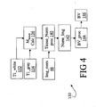
- FIGURE 4 shows a block diagram of another processor 130 that computes an absolute blood volume BV.
- Computation of BV by the processor 130 is not based on a transient response to a physiological perturbation. Rather, data from the image memory 46, which may be a single image, is processed using Equation (4).
- the image is preferably acquired using a short echo time TE, so that the term exp(-TE/T2 tissue ) in Equation (4) is suitably approximated as unity so that the signal S is given by: S par ⁇ C par - BV V par ⁇ C blood ⁇ M tissue where the parenchymal vascular space occupancy ⁇ is replaced by the expression in Equation (1).
- TR is the repetition time of the pulse sequence (indicated, for example, in FIGURE 2 ).
- a suitable value for T1 tissue can be measured using a known magnetic resonance technique such as saturation recovery pulse sequence and inversion recovery pulse sequence or, preferably, by a series of inversion pulses so that blood remains substantially nulled, while tissue signal decays with the representative T1 tissue .
- a known magnetic resonance technique such as saturation recovery pulse sequence and inversion recovery pulse sequence or, preferably, by a series of inversion pulses so that blood remains substantially nulled, while tissue signal decays with the representative T1 tissue .
- brain imaging it is known that the T1 tissue values for white and gray brain matter differ.
- a T1 white value 132 and a T1 gray value 134 are preferably determined.
- S par also contains instrumental factors, for instance related to coil sensitivity and excitation homogeneity, the combined contribution of which we will call IF.
- This constant can also be determined during the T1 measurement from the voxels in ventricles, which only contains CSF and has a proton density very close to pure water.
- a blood volume processor 144 estimates the absolute blood volume BV 146 or the absolute blood volume fraction or vascular space occupancy ( ⁇ rest ) by applying Equation (13) to the normalized image 142.
- FIGURE 5 shows a processor 150 that computes a clinical image from blood-nulled magnetic resonance images.
- the processor 150 is suitable in diagnostic applications for detecting an abnormality such as vasodilation or vasoconstriction caused by certain diseases such as cardiac ischemia, stroke, cancer, and the like.
- the image memory 46 stores an image of the imaging subject to be diagnosed.
- the image is compared with a reference image 152 by a difference processor 154 to generate a clinical image 156.
- the difference processor 154 computes an absolute difference between the images, so that normal areas appear dark in the clinical image 156 whereas abnormal areas appear brighter in the clinical image 156 (or vice versa) due to differences between the subject image and the reference image in the vicinity of the abnormality.
- the difference processor 154 computes a signed difference with a constant intensity level offset.
- regions of vasodilation and regions of vasocontraction have opposite intensity polarities respective to the constant intensity level.
- the reference image 152 can be obtained from various sources.
- an image of a normal subject can be employed for the comparison.
- a contralateral image can be used.
- a suspect right-side of the brain can be compared with a presumed normal left-side of the brain, preferably after a suitable left-right transposition of the contralateral comparison image.
- a suspect portion of the organ can be compared with presumed normal portion of the same organ.
- the normalized image 142 of FIGURE 4 is optionally employed in the processor 150.
- the normalized image 142 advantageously has suppressed contrast between white matter regions and gray matter regions of the brain image, which makes the vascular space occupancy contrast more visible.
- large vessels changes may occur in addition to microvascular changes. These can also be detected, and, at sufficient spatial resolution, can be separated from the microvascular changes.
- TE table 110 Linear regression processor 112 Slope 114 Ordinate-intercept 116 Rest blood volume calculator 118 Rest blood volume 122 Blood volume change rate calculator 124 Blood volume change rate 130 Processor for computing absolute blood volume 132 T1 value for white brain tissue 134 T1 value for gray brain tissue 136 M tissue calculator 140 Tissue normalizing processor 142 Normalized image 144 Blood volume processor 146 Blood volume 150 Processor for generating a clinical image 152 Reference image 154 Difference processor 156 Clinical image
Landscapes
- Physics & Mathematics (AREA)
- Health & Medical Sciences (AREA)
- High Energy & Nuclear Physics (AREA)
- Nuclear Medicine, Radiotherapy & Molecular Imaging (AREA)
- Life Sciences & Earth Sciences (AREA)
- Condensed Matter Physics & Semiconductors (AREA)
- General Physics & Mathematics (AREA)
- Radiology & Medical Imaging (AREA)
- Engineering & Computer Science (AREA)
- General Health & Medical Sciences (AREA)
- Signal Processing (AREA)
- Molecular Biology (AREA)
- Spectroscopy & Molecular Physics (AREA)
- Optics & Photonics (AREA)
- Biophysics (AREA)
- Pathology (AREA)
- Biomedical Technology (AREA)
- Heart & Thoracic Surgery (AREA)
- Medical Informatics (AREA)
- Vascular Medicine (AREA)
- Surgery (AREA)
- Animal Behavior & Ethology (AREA)
- Public Health (AREA)
- Veterinary Medicine (AREA)
- Magnetic Resonance Imaging Apparatus (AREA)
- Investigating Or Analysing Biological Materials (AREA)
- Medicines Containing Antibodies Or Antigens For Use As Internal Diagnostic Agents (AREA)
Applications Claiming Priority (3)
| Application Number | Priority Date | Filing Date | Title |
|---|---|---|---|
| US40604002P | 2002-08-27 | 2002-08-27 | |
| US406040P | 2002-08-27 | ||
| PCT/US2003/026580 WO2004021028A1 (en) | 2002-08-27 | 2003-08-26 | Microvascular blood volume magnetic resonance imaging |
Publications (2)
| Publication Number | Publication Date |
|---|---|
| EP1535083A1 EP1535083A1 (en) | 2005-06-01 |
| EP1535083B1 true EP1535083B1 (en) | 2008-08-13 |
Family
ID=31978260
Family Applications (1)
| Application Number | Title | Priority Date | Filing Date |
|---|---|---|---|
| EP03755747A Expired - Lifetime EP1535083B1 (en) | 2002-08-27 | 2003-08-26 | Microvascular blood volume magnetic resonance imaging |
Country Status (8)
| Country | Link |
|---|---|
| US (1) | US8527030B2 (https=) |
| EP (1) | EP1535083B1 (https=) |
| JP (1) | JP4613065B2 (https=) |
| CN (1) | CN100501439C (https=) |
| AT (1) | ATE404877T1 (https=) |
| AU (1) | AU2003273242A1 (https=) |
| DE (1) | DE60322916D1 (https=) |
| WO (1) | WO2004021028A1 (https=) |
Families Citing this family (26)
| Publication number | Priority date | Publication date | Assignee | Title |
|---|---|---|---|---|
| EP1773194A1 (en) * | 2004-06-01 | 2007-04-18 | VAN ZIJL, Peter C.M. | Quantifying blood volume using magnetization transfer magnetic resonance imaging |
| WO2008136274A1 (ja) * | 2007-04-27 | 2008-11-13 | Hitachi Medical Corporation | 磁気共鳴イメージング装置及び方法 |
| US20130200900A1 (en) * | 2010-10-13 | 2013-08-08 | Koninklijke Philips Electronics N.V. | Mri phantom with a plurality of compartments for t1 calibration |
| DE102011007835B4 (de) | 2011-04-21 | 2022-05-12 | Siemens Healthcare Gmbh | Verfahren zur Erstellung von MR-Angiographiebildern |
| WO2012166673A1 (en) * | 2011-05-27 | 2012-12-06 | O2 Insights, Inc. | Systems and methods for assessment of oxygenation |
| US20130144153A1 (en) * | 2011-12-01 | 2013-06-06 | The Regents Of The University Of California | Functional magnetic resonance imaging apparatus and methods |
| KR101282124B1 (ko) * | 2012-03-29 | 2013-07-04 | 고려대학교 산학협력단 | 자기공명영상 장치 및 이를 이용하여 영상을 생성하는 방법 |
| KR101310706B1 (ko) * | 2012-04-05 | 2013-09-24 | 고려대학교 산학협력단 | 선택적 회질 영상을 획득할 수 있는 자기공명영상 장치 및 이를 이용한 자기공명영상 획득방법 |
| US10653394B2 (en) * | 2012-08-08 | 2020-05-19 | Circle Cardiovascular Imaging Inc. | Measuring oxygenation changes in tissue as a marker for vascular function |
| DE102012217724B4 (de) * | 2012-09-28 | 2015-03-19 | Siemens Aktiengesellschaft | Vorrichtung zur Bestimmung eines Schädigungskennwertes einer Niere |
| DE102012222413B4 (de) * | 2012-12-06 | 2023-10-26 | Siemens Healthcare Gmbh | Verfahren zum Erzeugen eines HF-Anregungspulses zur Anregung eines beliebig geformten Volumens, Verfahren zum gezielten Anregen von Spins innerhalb eines Gefäßes und Verfahren zur Erstellung von MR-Angiographiebildern sowie entsprechende Magnetresonanzanlage |
| US20150088002A1 (en) * | 2013-09-21 | 2015-03-26 | Leo Technologies, Inc. | Hydration monitoring |
| DE102014201134B4 (de) * | 2014-01-22 | 2017-04-06 | Siemens Healthcare Gmbh | Verfahren und Vorrichtung zur Erzeugung eines 2-D-Projektionsbildes eines Gefäßsystems nebst korrespondierenden Gegenständen |
| US10551460B1 (en) * | 2014-04-08 | 2020-02-04 | Arizona Board Of Regents On Behalf Of The University Of Arizona | Method of generating reproducible quantitative magnetic resonance data |
| WO2016124491A1 (en) * | 2015-02-03 | 2016-08-11 | Koninklijke Philips N.V. | Method and system for susceptibility weighted magnetic resonance imaging |
| EP3325992B1 (en) * | 2015-07-24 | 2026-01-28 | Northeastern University | Quantitative magnetic resonance imaging of the vasculature |
| US10054653B2 (en) * | 2016-05-03 | 2018-08-21 | Siemens Healthcare Gmbh | Magnetic resonance method and apparatus for quantitative simultaneous multi-slice assessment of tissue displacement, deformation, and related biomarker parameters |
| CN106037804A (zh) * | 2016-06-27 | 2016-10-26 | 中国科学院苏州生物医学工程技术研究所 | 一种脑部病变区域的定位系统 |
| KR102008499B1 (ko) * | 2016-11-09 | 2019-08-07 | 삼성전자주식회사 | 자기 공명 영상 장치 및 자기 공명 영상 획득 방법 |
| EP3321708A3 (en) | 2016-11-09 | 2018-06-13 | Samsung Electronics Co., Ltd. | Magnetic resonance imaging (mri) apparatus and method of obtaining magnetic resonance image |
| US12099106B2 (en) | 2018-05-04 | 2024-09-24 | Fraunhofer-Gesellschaft zur Foerderung der angewandten Forschung eingetragener Verein | Arterial spin labeling with evaluation of inversion state of magnetization |
| US12557999B2 (en) | 2019-01-03 | 2026-02-24 | Northeastern University | Quantitative measurement of disruptions in the blood brain barrier |
| CN109960848B (zh) * | 2019-01-17 | 2022-05-10 | 哈尔滨工程大学 | 避免产生共振的模态避让方法 |
| WO2022214865A1 (en) * | 2021-04-09 | 2022-10-13 | Victoria Link Limited | Perfusion measurement with low field nmr |
| CN117310585B (zh) * | 2023-11-28 | 2024-02-23 | 首都医科大学宣武医院 | 一种在线测量组织反转恢复零点的方法 |
| CN118604701B (zh) * | 2024-05-21 | 2025-03-18 | 首都医科大学附属北京天坛医院 | 低场磁共振脑卒中鉴别序列及具有该鉴别序列的设备 |
Family Cites Families (35)
| Publication number | Priority date | Publication date | Assignee | Title |
|---|---|---|---|---|
| GB8715302D0 (en) | 1987-06-30 | 1987-08-05 | Ordidge R J | Nmr spectroscopy |
| US4945478A (en) * | 1987-11-06 | 1990-07-31 | Center For Innovative Technology | Noninvasive medical imaging system and method for the identification and 3-D display of atherosclerosis and the like |
| JP2595006B2 (ja) | 1988-01-29 | 1997-03-26 | 株式会社日立製作所 | Mrイメージング方法 |
| US5070876A (en) * | 1989-08-07 | 1991-12-10 | The Board Of Trustees Of The Leland Stanford Junior University | Flow-independent magnetic resonance projection angiography |
| US5221899A (en) | 1991-04-29 | 1993-06-22 | The Trustees Of Columbia University In The City Of New York | Signal acquisition in magnetic resonance analysis |
| US5681812A (en) * | 1991-12-10 | 1997-10-28 | Rush Presbyterian-St. Luke's Medical Center | Methods and compositions for reducing multidrug resistance |
| US5677626A (en) * | 1993-04-27 | 1997-10-14 | Kabushiki Kaisha Toshiba | System for magnetic resonance imaging |
| JP3386509B2 (ja) * | 1993-04-27 | 2003-03-17 | 株式会社東芝 | Mr撮像方法および磁気共鳴イメージング装置 |
| JP3396507B2 (ja) * | 1993-05-12 | 2003-04-14 | 株式会社東芝 | Mr撮像方法および磁気共鳴イメージング装置 |
| US5429134A (en) * | 1994-06-27 | 1995-07-04 | General Electric Company | Multi-phase fat suppressed MRI cardiac imaging |
| JPH0815439A (ja) * | 1994-06-30 | 1996-01-19 | Shimadzu Corp | 血流量測定装置 |
| JP3512482B2 (ja) * | 1994-09-06 | 2004-03-29 | 株式会社東芝 | 磁気共鳴映像装置 |
| US5786693A (en) * | 1996-04-26 | 1998-07-28 | Picker International, Inc. | Batch multi-volume angiography using magnetic resonance imaging |
| US20020188190A1 (en) * | 1998-03-05 | 2002-12-12 | Yoshimori Kassai | Mr imaging providing tissue/blood contrast image |
| US7254437B2 (en) * | 1998-04-17 | 2007-08-07 | Kabushiki Kaisha Toshiba | MR imaging providing tissue/blood contrast image |
| US6198958B1 (en) * | 1998-06-11 | 2001-03-06 | Beth Israel Deaconess Medical Center, Inc. | Method and apparatus for monitoring a magnetic resonance image during transcranial magnetic stimulation |
| US6370416B1 (en) * | 1998-11-25 | 2002-04-09 | Ge Medical Systems Global Technology Company Llc | fMRI signal processing |
| EP1171028B1 (en) * | 1999-03-26 | 2005-11-16 | Leif Ostergaard | System for determining haemodynamic indices by use of tomographic data |
| US6265875B1 (en) * | 1999-05-17 | 2001-07-24 | General Electric Company | Method and apparatus for efficient MRI tissue differentiation |
| JP2001204708A (ja) * | 2000-01-27 | 2001-07-31 | Toshiba Corp | 磁気共鳴映像装置 |
| US6603989B1 (en) * | 2000-03-21 | 2003-08-05 | Dmitriy A. Yablonskiy | T2 contrast in magnetic resonance imaging with gradient echoes |
| EP1139109A1 (en) * | 2000-03-28 | 2001-10-04 | Bracco International B.V. | A method for magnetic resonance imaging of the lung |
| US6462542B1 (en) * | 2000-07-21 | 2002-10-08 | Schlumberger Technology Corporation | Nuclear magnetic resonance measurements and methods of analyzing nuclear magnetic resonance data |
| WO2002012284A2 (en) * | 2000-08-04 | 2002-02-14 | Loma Linda University Medical Center | Iron regulating protein-2 (irp-2) as a diagnostic for neurodegenerative disease |
| JP4693227B2 (ja) * | 2000-11-14 | 2011-06-01 | 株式会社東芝 | 磁気共鳴イメージング装置及び磁気共鳴イメージングのスキャン同期方法 |
| US7308298B2 (en) * | 2000-12-22 | 2007-12-11 | Kabushiki Kaisha Toshiba | Magnetic resonance imaging using MT pulse of which duration is shorter |
| US20030120151A1 (en) * | 2001-01-10 | 2003-06-26 | Johns Hopkins University | Magnetic resonance imaging methods and compositions |
| DE60129472T2 (de) * | 2001-04-03 | 2008-04-17 | U-Cytech B.V. | Behandlung von hypoxya/ischaemia blutflusswiderstand mit beta-interferon |
| TWI221406B (en) * | 2001-07-30 | 2004-10-01 | Epix Medical Inc | Systems and methods for targeted magnetic resonance imaging of the vascular system |
| US6498946B1 (en) * | 2001-10-05 | 2002-12-24 | Ge Medical Systems Global Technology Co., Llc | Efficient multi-slice acquisition with black blood contrast |
| US7546155B2 (en) * | 2001-10-05 | 2009-06-09 | General Electric Company | Efficient multi-slice acquisition with black blood contrast in fast spin echo imaging |
| JP3995542B2 (ja) * | 2002-06-28 | 2007-10-24 | 東芝医用システムエンジニアリング株式会社 | 磁気共鳴イメージング装置及び磁気共鳴イメージングのデータ収集方法 |
| US7561909B1 (en) * | 2002-09-16 | 2009-07-14 | The United States Of America As Represented By The Department Of Health And Human Services | MRI navigator methods and systems |
| AR047692A1 (es) * | 2003-07-10 | 2006-02-08 | Epix Medical Inc | Imagenes de blancos estacionarios |
| EP1773194A1 (en) * | 2004-06-01 | 2007-04-18 | VAN ZIJL, Peter C.M. | Quantifying blood volume using magnetization transfer magnetic resonance imaging |
-
2003
- 2003-08-26 AU AU2003273242A patent/AU2003273242A1/en not_active Abandoned
- 2003-08-26 WO PCT/US2003/026580 patent/WO2004021028A1/en not_active Ceased
- 2003-08-26 DE DE60322916T patent/DE60322916D1/de not_active Expired - Lifetime
- 2003-08-26 CN CNB038204886A patent/CN100501439C/zh not_active Expired - Fee Related
- 2003-08-26 AT AT03755747T patent/ATE404877T1/de not_active IP Right Cessation
- 2003-08-26 EP EP03755747A patent/EP1535083B1/en not_active Expired - Lifetime
- 2003-08-26 US US10/525,699 patent/US8527030B2/en active Active
- 2003-08-26 JP JP2004532984A patent/JP4613065B2/ja not_active Expired - Fee Related
Also Published As
| Publication number | Publication date |
|---|---|
| ATE404877T1 (de) | 2008-08-15 |
| WO2004021028A1 (en) | 2004-03-11 |
| EP1535083A1 (en) | 2005-06-01 |
| US20050215881A1 (en) | 2005-09-29 |
| CN1685243A (zh) | 2005-10-19 |
| DE60322916D1 (de) | 2008-09-25 |
| US8527030B2 (en) | 2013-09-03 |
| AU2003273242A1 (en) | 2004-03-19 |
| JP4613065B2 (ja) | 2011-01-12 |
| JP2005537096A (ja) | 2005-12-08 |
| CN100501439C (zh) | 2009-06-17 |
Similar Documents
| Publication | Publication Date | Title |
|---|---|---|
| EP1535083B1 (en) | Microvascular blood volume magnetic resonance imaging | |
| Conturo et al. | Diffusion MRI: precision, accuracy and flow effects | |
| Buonincontri et al. | Multi-site repeatability and reproducibility of MR fingerprinting of the healthy brain at 1.5 and 3.0 T | |
| EP1136836B1 (en) | T2 contrast in magnetic resonance imaging with gradient echoes | |
| Van Gelderen et al. | Pittfalls of MRI measurement of white matter perfusion based on arterial spin labeling | |
| Petersen et al. | Model‐free arterial spin labeling quantification approach for perfusion MRI | |
| US8086003B2 (en) | Method and magnetic resonance apparatus to acquire temporally successive image data sets | |
| US6271665B1 (en) | Extra slice spin tagging (EST) magnetic resonance imaging for determining perfusion | |
| US7904135B2 (en) | Magnetic resonance spatial risk map for tissue outcome prediction | |
| US6064203A (en) | Method and apparatus for determining or imaging longitudinal spin relaxation time or producing images which substantially reflect longitudinal spin relaxation time contrast | |
| Tofts et al. | Towards quantitative measurements of relaxation times and other parameters in the brain | |
| US7235971B2 (en) | Shimming of MRI scanner involving fat suppression and/or black blood preparation | |
| US8099149B2 (en) | MRI method for quantification of cerebral perfusion | |
| US10330762B2 (en) | Measurement of blood volume using velocity-selective pulse trains on MRI | |
| US7863895B2 (en) | System, program product, and method of acquiring and processing MRI data for simultaneous determination of water, fat, and transverse relaxation time constants | |
| US6400978B1 (en) | Method and apparatus for detecting mental disorders | |
| US8928317B2 (en) | System and method for controlling apparent timing dependencies for T2-weighted MRI imaging | |
| US10617343B2 (en) | Methods and systems for quantitative brain assessment | |
| US11061094B2 (en) | Simultaneous pH and oxygen weighted MRI contrast using multi-echo chemical exchange saturation transfer imaging (ME-CEST) | |
| Ragot et al. | Characterizing contrast origins and noise contribution in spin-echo EPI BOLD at 3 T | |
| Lai et al. | FAIR exempting separate T 1 measurement (FAIREST): a novel technique for online quantitative perfusion imaging and multi‐contrast fMRI | |
| EP1877818B1 (en) | Determination of relaxation rate changes for mr molecular imaging | |
| Robison et al. | Evaluation of axial gradient Echo spiral MRI of the spine at 1.5 T | |
| Ragot | Characterizing Contrast Origins and Noise Contribution in Spin-echo BOLD at 3 T | |
| Jones | Diffusion tensor magnetic resonance imaging in the central nervous system |
Legal Events
| Date | Code | Title | Description |
|---|---|---|---|
| PUAI | Public reference made under article 153(3) epc to a published international application that has entered the european phase |
Free format text: ORIGINAL CODE: 0009012 |
|
| 17P | Request for examination filed |
Effective date: 20050319 |
|
| AK | Designated contracting states |
Kind code of ref document: A1 Designated state(s): AT BE BG CH CY CZ DE DK EE ES FI FR GB GR HU IE IT LI LU MC NL PT RO SE SI SK TR |
|
| AX | Request for extension of the european patent |
Extension state: AL LT LV MK |
|
| DAX | Request for extension of the european patent (deleted) | ||
| RIN1 | Information on inventor provided before grant (corrected) |
Inventor name: GOLAY, XAVIER, PH.D. BIOMED SCIENCES INSTITUTES Inventor name: LU, HANZHANG, (NMI) Inventor name: VAN ZIJL, PETER, C., M. |
|
| RIN1 | Information on inventor provided before grant (corrected) |
Inventor name: LU, HANZHANG Inventor name: GOLAY, XAVIER, PH.D. BIOMED SCIENCES INSTITUTES Inventor name: VAN ZIJL, PETER, C., M. |
|
| GRAP | Despatch of communication of intention to grant a patent |
Free format text: ORIGINAL CODE: EPIDOSNIGR1 |
|
| GRAS | Grant fee paid |
Free format text: ORIGINAL CODE: EPIDOSNIGR3 |
|
| GRAA | (expected) grant |
Free format text: ORIGINAL CODE: 0009210 |
|
| AK | Designated contracting states |
Kind code of ref document: B1 Designated state(s): AT BE BG CH CY CZ DE DK EE ES FI FR GB GR HU IE IT LI LU MC NL PT RO SE SI SK TR |
|
| REG | Reference to a national code |
Ref country code: GB Ref legal event code: FG4D |
|
| REG | Reference to a national code |
Ref country code: CH Ref legal event code: EP |
|
| REG | Reference to a national code |
Ref country code: IE Ref legal event code: FG4D |
|
| REF | Corresponds to: |
Ref document number: 60322916 Country of ref document: DE Date of ref document: 20080925 Kind code of ref document: P |
|
| PG25 | Lapsed in a contracting state [announced via postgrant information from national office to epo] |
Ref country code: ES Free format text: LAPSE BECAUSE OF FAILURE TO SUBMIT A TRANSLATION OF THE DESCRIPTION OR TO PAY THE FEE WITHIN THE PRESCRIBED TIME-LIMIT Effective date: 20081124 Ref country code: NL Free format text: LAPSE BECAUSE OF FAILURE TO SUBMIT A TRANSLATION OF THE DESCRIPTION OR TO PAY THE FEE WITHIN THE PRESCRIBED TIME-LIMIT Effective date: 20080813 |
|
| PG25 | Lapsed in a contracting state [announced via postgrant information from national office to epo] |
Ref country code: FI Free format text: LAPSE BECAUSE OF FAILURE TO SUBMIT A TRANSLATION OF THE DESCRIPTION OR TO PAY THE FEE WITHIN THE PRESCRIBED TIME-LIMIT Effective date: 20080813 Ref country code: AT Free format text: LAPSE BECAUSE OF FAILURE TO SUBMIT A TRANSLATION OF THE DESCRIPTION OR TO PAY THE FEE WITHIN THE PRESCRIBED TIME-LIMIT Effective date: 20080813 Ref country code: SI Free format text: LAPSE BECAUSE OF FAILURE TO SUBMIT A TRANSLATION OF THE DESCRIPTION OR TO PAY THE FEE WITHIN THE PRESCRIBED TIME-LIMIT Effective date: 20080813 |
|
| PG25 | Lapsed in a contracting state [announced via postgrant information from national office to epo] |
Ref country code: BE Free format text: LAPSE BECAUSE OF FAILURE TO SUBMIT A TRANSLATION OF THE DESCRIPTION OR TO PAY THE FEE WITHIN THE PRESCRIBED TIME-LIMIT Effective date: 20080813 Ref country code: MC Free format text: LAPSE BECAUSE OF NON-PAYMENT OF DUE FEES Effective date: 20080831 |
|
| REG | Reference to a national code |
Ref country code: CH Ref legal event code: PL |
|
| PG25 | Lapsed in a contracting state [announced via postgrant information from national office to epo] |
Ref country code: DK Free format text: LAPSE BECAUSE OF FAILURE TO SUBMIT A TRANSLATION OF THE DESCRIPTION OR TO PAY THE FEE WITHIN THE PRESCRIBED TIME-LIMIT Effective date: 20080813 Ref country code: BG Free format text: LAPSE BECAUSE OF FAILURE TO SUBMIT A TRANSLATION OF THE DESCRIPTION OR TO PAY THE FEE WITHIN THE PRESCRIBED TIME-LIMIT Effective date: 20081113 |
|
| REG | Reference to a national code |
Ref country code: IE Ref legal event code: MM4A |
|
| PG25 | Lapsed in a contracting state [announced via postgrant information from national office to epo] |
Ref country code: CZ Free format text: LAPSE BECAUSE OF FAILURE TO SUBMIT A TRANSLATION OF THE DESCRIPTION OR TO PAY THE FEE WITHIN THE PRESCRIBED TIME-LIMIT Effective date: 20080813 Ref country code: SK Free format text: LAPSE BECAUSE OF FAILURE TO SUBMIT A TRANSLATION OF THE DESCRIPTION OR TO PAY THE FEE WITHIN THE PRESCRIBED TIME-LIMIT Effective date: 20080813 Ref country code: RO Free format text: LAPSE BECAUSE OF FAILURE TO SUBMIT A TRANSLATION OF THE DESCRIPTION OR TO PAY THE FEE WITHIN THE PRESCRIBED TIME-LIMIT Effective date: 20080813 Ref country code: PT Free format text: LAPSE BECAUSE OF FAILURE TO SUBMIT A TRANSLATION OF THE DESCRIPTION OR TO PAY THE FEE WITHIN THE PRESCRIBED TIME-LIMIT Effective date: 20090113 |
|
| PLBE | No opposition filed within time limit |
Free format text: ORIGINAL CODE: 0009261 |
|
| STAA | Information on the status of an ep patent application or granted ep patent |
Free format text: STATUS: NO OPPOSITION FILED WITHIN TIME LIMIT |
|
| PG25 | Lapsed in a contracting state [announced via postgrant information from national office to epo] |
Ref country code: LI Free format text: LAPSE BECAUSE OF NON-PAYMENT OF DUE FEES Effective date: 20080831 Ref country code: CH Free format text: LAPSE BECAUSE OF NON-PAYMENT OF DUE FEES Effective date: 20080831 |
|
| 26N | No opposition filed |
Effective date: 20090514 |
|
| REG | Reference to a national code |
Ref country code: FR Ref legal event code: ST Effective date: 20090630 |
|
| GBPC | Gb: european patent ceased through non-payment of renewal fee |
Effective date: 20081113 |
|
| PG25 | Lapsed in a contracting state [announced via postgrant information from national office to epo] |
Ref country code: EE Free format text: LAPSE BECAUSE OF FAILURE TO SUBMIT A TRANSLATION OF THE DESCRIPTION OR TO PAY THE FEE WITHIN THE PRESCRIBED TIME-LIMIT Effective date: 20080813 Ref country code: IE Free format text: LAPSE BECAUSE OF NON-PAYMENT OF DUE FEES Effective date: 20080826 |
|
| PG25 | Lapsed in a contracting state [announced via postgrant information from national office to epo] |
Ref country code: IT Free format text: LAPSE BECAUSE OF FAILURE TO SUBMIT A TRANSLATION OF THE DESCRIPTION OR TO PAY THE FEE WITHIN THE PRESCRIBED TIME-LIMIT Effective date: 20080813 |
|
| PG25 | Lapsed in a contracting state [announced via postgrant information from national office to epo] |
Ref country code: FR Free format text: LAPSE BECAUSE OF NON-PAYMENT OF DUE FEES Effective date: 20080901 |
|
| PG25 | Lapsed in a contracting state [announced via postgrant information from national office to epo] |
Ref country code: GB Free format text: LAPSE BECAUSE OF NON-PAYMENT OF DUE FEES Effective date: 20081113 |
|
| PG25 | Lapsed in a contracting state [announced via postgrant information from national office to epo] |
Ref country code: SE Free format text: LAPSE BECAUSE OF FAILURE TO SUBMIT A TRANSLATION OF THE DESCRIPTION OR TO PAY THE FEE WITHIN THE PRESCRIBED TIME-LIMIT Effective date: 20081113 |
|
| PG25 | Lapsed in a contracting state [announced via postgrant information from national office to epo] |
Ref country code: LU Free format text: LAPSE BECAUSE OF NON-PAYMENT OF DUE FEES Effective date: 20080826 Ref country code: HU Free format text: LAPSE BECAUSE OF FAILURE TO SUBMIT A TRANSLATION OF THE DESCRIPTION OR TO PAY THE FEE WITHIN THE PRESCRIBED TIME-LIMIT Effective date: 20090214 Ref country code: CY Free format text: LAPSE BECAUSE OF FAILURE TO SUBMIT A TRANSLATION OF THE DESCRIPTION OR TO PAY THE FEE WITHIN THE PRESCRIBED TIME-LIMIT Effective date: 20080813 |
|
| PG25 | Lapsed in a contracting state [announced via postgrant information from national office to epo] |
Ref country code: TR Free format text: LAPSE BECAUSE OF FAILURE TO SUBMIT A TRANSLATION OF THE DESCRIPTION OR TO PAY THE FEE WITHIN THE PRESCRIBED TIME-LIMIT Effective date: 20080813 |
|
| PG25 | Lapsed in a contracting state [announced via postgrant information from national office to epo] |
Ref country code: GR Free format text: LAPSE BECAUSE OF FAILURE TO SUBMIT A TRANSLATION OF THE DESCRIPTION OR TO PAY THE FEE WITHIN THE PRESCRIBED TIME-LIMIT Effective date: 20081114 |
|
| REG | Reference to a national code |
Ref country code: DE Ref legal event code: R082 Ref document number: 60322916 Country of ref document: DE Representative=s name: VON KREISLER SELTING WERNER, DE Ref country code: DE Ref legal event code: R082 Ref document number: 60322916 Country of ref document: DE Representative=s name: VON KREISLER SELTING WERNER - PARTNERSCHAFT VO, DE Ref country code: DE Ref legal event code: R082 Ref document number: 60322916 Country of ref document: DE Representative=s name: DOMPATENT VON KREISLER SELTING WERNER - PARTNE, DE |
|
| PGFP | Annual fee paid to national office [announced via postgrant information from national office to epo] |
Ref country code: DE Payment date: 20180716 Year of fee payment: 16 |
|
| REG | Reference to a national code |
Ref country code: DE Ref legal event code: R119 Ref document number: 60322916 Country of ref document: DE |
|
| PG25 | Lapsed in a contracting state [announced via postgrant information from national office to epo] |
Ref country code: DE Free format text: LAPSE BECAUSE OF NON-PAYMENT OF DUE FEES Effective date: 20200303 |