EP1534146B1 - Dispositifs cardiaques de reparation percutanee des valvules auriculoventriculaires - Google Patents
Dispositifs cardiaques de reparation percutanee des valvules auriculoventriculaires Download PDFInfo
- Publication number
- EP1534146B1 EP1534146B1 EP03785293A EP03785293A EP1534146B1 EP 1534146 B1 EP1534146 B1 EP 1534146B1 EP 03785293 A EP03785293 A EP 03785293A EP 03785293 A EP03785293 A EP 03785293A EP 1534146 B1 EP1534146 B1 EP 1534146B1
- Authority
- EP
- European Patent Office
- Prior art keywords
- catheter
- catheter assembly
- leaflets
- grasping elements
- valve
- Prior art date
- Legal status (The legal status is an assumption and is not a legal conclusion. Google has not performed a legal analysis and makes no representation as to the accuracy of the status listed.)
- Expired - Lifetime
Links
Images


Classifications
-
- A—HUMAN NECESSITIES
- A61—MEDICAL OR VETERINARY SCIENCE; HYGIENE
- A61B—DIAGNOSIS; SURGERY; IDENTIFICATION
- A61B17/00—Surgical instruments, devices or methods, e.g. tourniquets
- A61B17/00234—Surgical instruments, devices or methods, e.g. tourniquets for minimally invasive surgery
-
- A—HUMAN NECESSITIES
- A61—MEDICAL OR VETERINARY SCIENCE; HYGIENE
- A61B—DIAGNOSIS; SURGERY; IDENTIFICATION
- A61B17/00—Surgical instruments, devices or methods, e.g. tourniquets
- A61B17/064—Surgical staples, i.e. penetrating the tissue
-
- A—HUMAN NECESSITIES
- A61—MEDICAL OR VETERINARY SCIENCE; HYGIENE
- A61B—DIAGNOSIS; SURGERY; IDENTIFICATION
- A61B17/00—Surgical instruments, devices or methods, e.g. tourniquets
- A61B17/068—Surgical staplers, e.g. containing multiple staples or clamps
- A61B17/0682—Surgical staplers, e.g. containing multiple staples or clamps for applying U-shaped staples or clamps, e.g. without a forming anvil
-
- A—HUMAN NECESSITIES
- A61—MEDICAL OR VETERINARY SCIENCE; HYGIENE
- A61B—DIAGNOSIS; SURGERY; IDENTIFICATION
- A61B17/00—Surgical instruments, devices or methods, e.g. tourniquets
- A61B17/064—Surgical staples, i.e. penetrating the tissue
- A61B17/0644—Surgical staples, i.e. penetrating the tissue penetrating the tissue, deformable to closed position
-
- A—HUMAN NECESSITIES
- A61—MEDICAL OR VETERINARY SCIENCE; HYGIENE
- A61B—DIAGNOSIS; SURGERY; IDENTIFICATION
- A61B17/00—Surgical instruments, devices or methods, e.g. tourniquets
- A61B17/00234—Surgical instruments, devices or methods, e.g. tourniquets for minimally invasive surgery
- A61B2017/00238—Type of minimally invasive operation
- A61B2017/00243—Type of minimally invasive operation cardiac
-
- A—HUMAN NECESSITIES
- A61—MEDICAL OR VETERINARY SCIENCE; HYGIENE
- A61B—DIAGNOSIS; SURGERY; IDENTIFICATION
- A61B17/00—Surgical instruments, devices or methods, e.g. tourniquets
- A61B2017/00743—Type of operation; Specification of treatment sites
- A61B2017/00778—Operations on blood vessels
- A61B2017/00783—Valvuloplasty
-
- A—HUMAN NECESSITIES
- A61—MEDICAL OR VETERINARY SCIENCE; HYGIENE
- A61B—DIAGNOSIS; SURGERY; IDENTIFICATION
- A61B17/00—Surgical instruments, devices or methods, e.g. tourniquets
- A61B2017/00831—Material properties
- A61B2017/00867—Material properties shape memory effect
-
- A—HUMAN NECESSITIES
- A61—MEDICAL OR VETERINARY SCIENCE; HYGIENE
- A61B—DIAGNOSIS; SURGERY; IDENTIFICATION
- A61B17/00—Surgical instruments, devices or methods, e.g. tourniquets
- A61B17/30—Surgical pincettes without pivotal connections
- A61B2017/306—Surgical pincettes without pivotal connections holding by means of suction
Definitions
- the present invention relates generally to devices (i.e., articles of manufacture, apparatus, systems, instruments) for treating heart disease and, in particular, to devices for minimally invasive repair of atrioventricular valve regurgitation (MR) occurring in the context of mitral valve prolapse (MVP), flail mitral valve, and/or ischemic MR.
- devices i.e., articles of manufacture, apparatus, systems, instruments
- MVP mitral valve prolapse
- flail mitral valve flail mitral valve
- ischemic MR ischemic MR
- a mitral valve such as shown in Figure 1A, including an anterior leaflet 1 and a posterior leaflet 2, is the inlet valve to the main heart pumping chamber (left ventricle 4).
- the mitral valve is forced to close when the ventricle 4 contracts, preventing backward flow of blood.
- leaflets 1,2 are restrained by a network of tendinous chords 5 anchored to the posterior wall of the heart 6.
- the leaflets normally meet or coapt so that their tips are juxtaposed, along with up to one-third of their lengths, forming an effective seal 8 to prevent MR.
- leaflets fail to meet properly due to leaflet elongation or elongation or rupture of the chords 5.
- one or more leaflet portions 10 may then prolapse into the left atrium 7, reducing coaptation and creating a gap between the leaflets 1,2 that allows MR to occur (blood flow illustrated by arrows 9.).
- Such regurgitation can produce heart failure, rhythm disorders, sudden death, and a predisposition to lethal heart valve infections. Therapy has until now required open-heart surgery to replace the valve or repair it by approximating leaflet tips.
- a suture 12 may be placed between the tips of the leaflets 1,2 to prevent prolapse of one leaflet relative to the other. As shown in Figure 1D, this produces a competent valve with two openings 14, one on each side of the suture 12 position. Such an orifice does not materially obstruct inflow of blood into the left ventricle.
- WO 00/60995 discloses a device for repairing a cardiac valve comprising a catheter assembly configured to pass from a remote vasculature of a patient to a position within the heart adjacent the cardiac valve, at least two maneuverable grasping elements independently translatable through the catheter assembly, the grasping elements being reversibly from the catheter assembly to capture and reposition portions of at least two valve leaflets and a fastening mechanism.
- US patent application publication US 2002/0013571 discloses methods and devices for grasping and optional repositioning and fixation of the valve leaflets to treat cardiac valve regurgitation, particularly mitral valve regurgitation.
- US patent 6,165,183 discloses an approach to mitral or tricuspid valve repair involving the performance of an edge-to-edge fastening/securing of opposing heart valve leaflets through a catheter entering the heart.
- a rivet or staple 15 emerging from the catheter 20 may simply displace the leaflets 1,2 from the catheter tip 22 (as shown in Figure 2C) rather than successfully penetrate the leaflets.
- approaches that employ grasping the leaflets generally do so in a plane perpendicular to the catheter, and do not provide a sufficiently large and stable leaflet surface area through which a staple or a suture can be inserted.
- the present invention provides novel devices for minimally invasive methods to treat heart disease related to mitral valve regurgitation occurring in the context of cardiac valve prolapse as defined in claim 1.
- the present invention allows for independent capture and repositioning of mitral leaflets regardless of whether they are aligned, and provides a stabilized large leaflet surface area through which sutures, staples, rivets and the like may be inserted.
- Use of a novel percutaneous catheter and other novel devices disclosed herein enables therapeutic maneuvers that do not require opening the chest or heart, however the present invention is not limited to percutaneous approaches.
- the several specific preferred embodiments of the present invention referring to the invention's applicability to the mitral valve are meant in no way to be limiting. It will be readily appreciated that the present invention may additionally be effectively applied to leaflets of the tricuspid valve.
- the present invention provides a device as defined in claim 1 for use in a method to repair a mitral valve, comprised of a catheter assembly for delivering in a minimally invasive manner a pair of maneuverable grasping elements that are independently translatable through the catheter assembly to a position near the mitral valve.
- the grasping elements are reversibly and radially extendable from the catheter assembly so as to capture and stabilize portions of two valve leaflets in an apposed position so that a fastening mechanism (e.g. , a staple, rivet or the like) which is also translated through the catheter assembly may be used to affix portions of the anterior and posterior leaflets to one another.
- a fastening mechanism e.g. , a staple, rivet or the like
- the catheter assembly comprises a single catheter, that may be substantially straight for an antegrade approach, or alternatively may have a curved distal end so as to enable valve access retrograde via the aorta.
- Shape memory materials such as nickel titanium (e.g ., NITINOLTM www.nitinol.com) may be used to manufacture several components of the present invention, including the curved distal end of the catheter assembly.
- the fastening mechanism and grasping elements are optionally composed of shape memory material as well.
- the catheter assembly comprises a pair of slidably linked catheters, each of which is responsible for delivering a grasping element to the mitral valve region.
- the fastening mechanism is radially extendable and disengagable from the catheter assembly through at least one side port of the catheter assembly. Described below is a mechanism by which the fastening mechanism may be disengaged from the catheter assembly after insertion through the leaflets, which occurs, significantly, without displacing the captured leaflets in a direction away from the catheter assembly and each other.
- the grasping elements extend from the distal end of the catheter assembly with reverse deformation so as to clip the ventricular side of the valve leaflets.
- the grasping elements extend from side ports in the catheter assembly and are steered so as to be able to grasp the atrial sides of the leaflets.
- two pairs of grasping elements are employed to grasp both sides of both the posterior and anterior leaflets.
- Various embodiments of the grasping elements are described below, including clip-like configurations and embodiments having operable jaws disposed at the ends of each grasping element.
- each grasping element is triggered by sensors for detecting contact and/or proximity of the leaflet to be grasped to the grasping element1.
- External or internal cardiac imaging of the region of the mitral valve may be employed to facilitate proper positioning of the grasping elements and/or monitoring of effectiveness of the procedure.
- This imaging can alternatively be achieved through the use of ultrasound, magnetic resonance or fiber optics.
- Devices in accordance with the present invention are characterized in that they permit the anterior and posterior leaflets to be separately grasped and independently translated along an axis of a catheter assembly until leaflet apposition is achieved for the purpose of fastening the leaflets to one another.
- Use of the devices is not, however, limited to the mitral valve, as use on the tricuspid or any other cardiac valve will be readily appreciated by artisans.
- a percutaneous catheter 20 is inserted through the interatrial septum into the left atrium 7 and then between the mitral leaflets (only anterior leaflet 1 shown) to the left ventricle 4 to a position adjacent leaflet 1 that is to be repositioned.
- the mitral leaflets only anterior leaflet 1 shown
- the grasping element 24 is composed of a shape memory material that has been pre-formed to have a shape and dimensions appropriate for clipping or compressing leaflet 1 against the external surface 23 of the catheter 20, such as shown in Figure 3B.
- the grasping element 24 When the grasping element 24 is extruded from the tip 22, the element takes its pre-formed shape, which as depicted can be a reverse curve 25 that results in a clip-like structure on the ventricular surface 3 of leaflet 1.
- the manufacturing of biocompatible shape memory components is well described in the art, thus will not be recited here.
- Figure 3C depicts a view of the mitral valve looking down onto the upper surface of the anterior mitral leaflet. Note that the device operates in this case in the gap between the chordal connections 5 to each side of the leaflet.
- the grasping element 24 has preferably two side arms 26 that extend to either side of the catheter axis in a V-shaped or similar configuration having a separation there between sufficient to allow a staple or rivet 30 to be inserted through the leaflet unopposed by the grasping element .
- This design provides greater stability than a single point of leaflet contact, and allows a staple or rivet 30 to be inserted between the side arms 26 of the grasping element without the leaflet being displaced in the process.
- the design also provides a relatively large, stabilized leaflet surface area against the external surface 23 of the catheter 20 and minimizes leaflet displacement during the process of staple or rivet fastening.
- An alternative embodiments includes the use of auxiliary suctioning ports on the catheter 20 to assist in the grasping process; these may be independently translatable.
- alternative embodiments of the grasping elements are each comprised of a control rod 34 having a steerable distal end 33, upon which is disposed a grabbing tool 32.
- a variety of mechanisms for steering percutaneous surgical instruments are known to artisans and may be employed in steering the distal end 33 so as to position the grabbing tool 32 adjacent a portion of the atrial surface 35 to be grasped.
- Each distal end 33 is extended and retracted through a side port 37 or groove in catheter 20.
- the grabbing tool 32 in one embodiment, has two pincer-like jaws 36 (two versions of which are shown in Figures 4B, 4C) that grasp the atrial surface 35 of the leaflet.
- One experienced in the art will recognize that a variety of other mechanisms currently available can be used as grabbing tools as an alternative to the jaws 36.
- One such alternative includes the use of suction tips combined with pincers, a design serving to improve leaflet-catheter contact but not requiring as high a suction rate such as needed to grasp both leaflets simultaneously, the suction herein being used only for fine positioning and alignment.
- more than one grasping element is employed to grasp the atrial surface of the leaflet.
- Multiple grasping elements extend in a similar direction away from the catheter to allow grasping of a selected axial length of the atrial surface. For example, there could be two grasping elements pointing to one side of the valve, and two to the other, thus stabilizing a square or rectangular portion of the leaflet surface.
- a single catheter 20 has been used to deliver and position the grasping elements to positions adjacent the ventricular or atrial surfaces of the leaflets.
- four independent grasping elements are used to grasp both surfaces of both leaflets, two elements per leaflet on opposite sides. This configuration provides the greatest flexibility in repositioning the leaflets into apposition and stabilizing them through the fastening step.
- the grasping elements 24 may be delivered independently through separate but linked catheters 21,21'.
- the catheters 21,21' are linked, for example, by a tongue and groove configuration, that allows them to move in a common plane. Translating the catheters achieves the desired leaflet 1,2 approximation after the grasping step.
- Each of the catheters 21,21' in the catheter assembly has opening that together form a channel 28 through which the staple or rivet 30 is extended when the leaflets are repositioned in apposition.
- the staples or rivets 30 in one embodiment are composed of shape memory material and dimensioned to allow translation through the catheter 20 lumen and rapid expansion, effecting insertion and leaflet fastening, when the staples or rivets 30 reach an orifice 31 in the side wall of the catheter.
- the staples or rivets 30 are translated down the catheter 20 lumen by a plunger 29 until the orifice 31 is encountered.
- the staples or rivets 30 then emerge and pierce both leaflets, which have been previously stabilized by the grasping elements 24 described above.
- the shape memory staple or rivet emerges on the side of the leaflet opposite that of the catheter, and curves back to form a staple. Two or more such staples or rivets may be inserted at the same site, each facing in a different direction.
- retractable door 40 in the side wall of the catheter can then be opened to disengage the catheter from the staples and move it to another site, so the leaflets can also be fastened together at additional locations to stabilize the repair, if necessary.
- the shape memory device continues to compress the leaflets together until an effective seal is formed.
- FIGs 5B through 5E illustrate the sequence of maneuvers that achieve leaflet fastening at more than one site.
- the cardiac valve is viewed from side to side as if looking down on its anterior leaflet 1 (as in the orientation shown in Figure 3C.)
- Catheter 20 is depicted as entering from an atrial route, although the same process can be practiced with the catheter entering retrograde from the aorta.
- staples or rivets 30 have been inserted through the orifice 31 in the catheter that allows their extrusion into the leaflets.
- a portion 40 of the side wall of the catheter is retracted into the catheter lumen, activated by a wire or other connection to the catheter controls outside the patient.
- This step opens a hole 41 in the side of the catheter.
- the catheter is then disengaged from the staple or riveting device, and translated parallel to the catheter axis 37.
- Figure 5D shows the catheter translated to a position closer to one side of the leaflet.
- Figure 5E shows the retractable catheter wall portion 40 replaced to re-form the piercing orifice 31 and allow fastening of the leaflets at another site. This can increase the efficacy of repair, as necessary, depending upon the precise geometry of the separated and regurgitant leaflets.
- This approach can be practiced with the linked sub-catheters shown in Figures 3D and 3E as well, using retractable wall portions for each sub-catheter. Retractable portions of the catheter side walls may be placed on both sides of the catheter to allow translation in either direction.
- the disengaged catheter 20 may also be translated parallel to its axis 37 to insert additional staples at more than one site along the central axis of the left ventricle, which is parallel to the initial catheter axis.
- an alternative embodiment inserts the catheter 20 retrograde into the left ventricle 4 through the aorta.
- the catheter is shaped or steered to curve back toward the mitral leaflet tips using shape memory materials or other steering mechanisms known in the art.
- An advantage of the present invention is the need for only a single catheter assembly due to the novel leaflet grasping elements, as opposed to catheters coming from both aortic and artial routes.
- Leaflet-grasping elements, after positioning, are then applied to the distal leaflet tips.
- the grasping elements 24 are extended forward from the catheter onto the ventricular sides of the leaflet to surround the leaflet between catheter and clip. This forward grasping may be achieved through a number of mechanisms.
- the grasping elements 24 may be comprised of preformed shape memory material that compresses the leaflets upon extension from side ports in the catheter.
- the grasping elements may be comprised of more rigid members that are hinged upon control rods within a sock-like component that, when extended beyond the hinge point, urges the members back towards the catheter, encompassing a portion of the leaflet therebetween.
- the entire process is preferably performed under the guidance of an imaging technique, such as echocardiography.
- an imaging technique such as echocardiography.
- Such guidance may be achieved using near infrared visualization (such as described in U.S. patent 6,178,346 ), with an intracardiac magnetic resonance imaging coil, or by transthoracic real-time three-dimensional echocardiographic imaging.
- Ultrasound imaging may be performed from the chest surface, esophagus, or within the heart, using an independent intracardiac echo (ICE) catheter (commercially available) within the adjacent right ventricular outflow tract, or an ultrasound transducer within the left ventricle itself, built into the catheter 20.
- ICE intracardiac echo
- This arrangement allows an imaging element 50 to be maneuvered via the catheter to provide high-resolution images of the cardiac valve during the procedure, while also displaying the mitral leaflets and regurgitant jet.
- a two-dimensional matrix array of piezoelectric crystals may be used, or a linear phased array may be rapidly rotated to produce a three-dimensional image.
- the imaging may also be employed to confirm catheter contact with the leaflets and trigger the grasping step.
- the imaging element can be comprised of a simple piezoelectric crystal or optical coherence device that senses the motion of the leaflets. When the leaflet approaches the catheter in systole, the device outputs a triggering signal confirmable manually on an output display device. Once confirmed, the following triggering signal is output to a mechanism to translate the grasping element rapidly down the lumen to assume the retroflexing shape thereby effecting capture.
- leaflet capture is effected by rapidly advancing a magnetic rod through the bore of the catheter to a position wherein the distal end of the extruded or extended grasping element, which in this embodiment is composed at least in part of a ferromagnetic material, is rapidly attracted toward the magnetic rod and the catheter.
- a jointed rod is rapidly advanced so as to retroflex the grasping element against the leaflet.
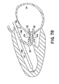
- Both catheter introduction routes may also be practiced using a stabilizing catheter extension 60 advanced to the left ventricle apex 62, as shown in Figures 7A and 7B.
- a stabilizing catheter extension 60 advanced to the left ventricle apex 62, as shown in Figures 7A and 7B.
- the contact element 64 may be made of shape memory elastic material which, when extruded from the catheter, forms a curving or tined contacting structure.
- Catheter 20 may then be translated along this wire to position the grasping elements 24,32 optimally in the region of the leaflet.
- the stabilizing catheter extension 60 is in continuity with the fastening catheter 20, while for retrograde aortic routes ( Figure 7B), the fastening catheter 20 emerges from an introducer catheter 64, and is steered toward the leaflets.
- the fastening catheter 20 is passed through a port in the introducer catheter 64, either directly or through a directing arm that may have an occlusive rubber seal.
- the introducer catheter 64 is stabilized by the curving contact element 64 extending beyond the catheter tip and shaped to maintain contact with the ventricular apex.
- the contact element 64 may alternatively be comprised of several elements composed of shape memory elastic material which, when extended from the catheter itself, form curving or tined contacting elements that may contact other portions of the interior of the cavity.
- the stabilizing mechanism described here is merely illustrative.
- the stabilizing mechanism is not limited to a wire or shape memory materials, nor is it limited to a single extendable contact element.
- some embodiments may employ a plurality of stabilizing legs or telescoping extensions to contact inner left ventricle at numerous points around its circumference.
- the device of this invention may also be practiced effectively in conjunction with procedures that assist in approximating the leaflets by inserting a mitral annular ring percutaneously.
- a mitral annular ring percutaneously.
- Such a device is inserted into the coronary sinus, a vein that encircles the mitral valve at the level of its annulus (the insertion of the leaflets onto the rest of the heart). Reducing annular size also helps in approximating the leaflets to achieve optimal repair.
- This combination of techniques utilizing the device of the present invention would provide a completely percutaneous approach to comprehensive repair of mitral valve prolapse and degenerative mitral regurgitation.
- the present invention can also be used to treat ischemic mitral regurgitation in dilated hearts to reduce volume overload and prevent heart failure. Many of the more severe regurgitant lesions in such patients also involve malcoaption of non-opposed leaftet portions that can be repaired by this device.
- the present invention may be used with methods for reducing the tension on the leaflets to be so repaired, including percutaneous annular ring reductions and methods for reducing ventricular size. It can also be used to appose and link any two scallops of the posterior leaflet that may be malaligned or prolapse relative to each other, causing regurgitation.
Claims (17)
- Dispositif pour réparer une valvule cardiaque, comportant :un ensemble formant cathéter (20, 21, 21') configuré pour passer à partir d'une vascularisation éloignée d'un patient vers une position dans le coeur adjacente à la valvule cardiaque,au moins deux éléments de préhension pouvant être manoeuvrés (24, 32) pouvant être déplacés en translation indépendamment à travers l'ensemble formant cathéter, les éléments de préhension pouvant être étendus de manière réversible et radialement à partir de l'ensemble formant cathéter pour capturer et repositionner des parties d'au moins deux valves de valvule (1, 2) dans une position apposée, etun mécanisme de fixation (30) pouvant être déplacé en translation à travers l'ensemble formant cathéter pour fixer les parties stabilisées des deux valves de valvule,dans lequel le mécanisme de fixation peut être étendu radialement et peut être libéré de l'ensemble formant cathéter à travers au moins un orifice latéral (31, 41) de l'ensemble formant cathéter.
- Dispositif selon la revendication 1, dans lequel l'ensemble formant cathéter (20, 21, 21') comporte un cathéter unique ayant une extrémité distale incurvée de manière à permettre un accès rétrograde à une valvule via l'aorte.
- Dispositif selon la revendication 2, dans lequel l'extrémité distale est constituée au moins en partie d'un matériau à mémoire de forme.
- Dispositif selon la revendication 1, dans lequel l'ensemble formant cathéter (20, 21, 21') est constitué d'une paire de cathéters liés de manière coulissante, un premier des éléments de préhension (24, 32) étant disposé dans chaque cathéter, et dans lequel chaque cathéter comporte au moins un orifice latéral (31, 41) à travers lequel le mécanisme de fixation (30) peut être étendu et peut être libéré de l'ensemble formant cathéter.
- Dispositif selon la revendication 1, dans lequel le mécanisme de fixation (30) comporte une agrafe ou un rivet pouvant être inséré(e) à travers chacune des parties de valve capturées sans déplacer les valvules (1, 2).
- Dispositif selon la revendication 1, dans lequel le mécanisme de fixation (30) est constitué au moins en partie d'un matériau à mémoire de forme.
- Dispositif selon la revendication 1, comportant en outre une partie rétractable de l'ensemble formant cathéter (20, 21, 21') à proximité du au moins un orifice latéral (31, 41), dans son état rétracté, créant une encoche dans l'ensemble formant cathéter à travers laquelle le mécanisme de fixation peut être libéré de l'ensemble formant cathéter.
- Dispositif selon l'une quelconque des revendications 1 à 7, dans lequel chacun des éléments de préhension (24, 32) s'étend à partir de l'extrémité distale de l'ensemble formant cathéter (20, 21, 21') avec une déformation inverse, de manière à serrer une surface ventriculaire (13) d'une des valves de valvule (1, 2).
- Dispositif selon la revendication 8, comportant en outre :des éléments de préhension (24, 32, 33) pouvant être déplacés en translation indépendamment à travers l'ensemble formant cathéter (20, 21, 21'), chaque élément de préhension pouvant s'étendre de manière réversible et radialement à partir d'un parmi au moins deux orifices latéraux opposés (31, 41) dans l'ensemble formant cathéter avec une déformation ajustable, de manière à saisir une surface auriculaire (35) d'une des valves de valvule (1, 2).
- Dispositif selon la revendication 1, dans lequel chacun des éléments de préhension (24, 32) s'étend à partir d'un parmi au moins deux orifices latéraux opposés (31, 41) dans l'ensemble formant cathéter (20, 21, 21'), avec au moins une parmi(a) une déformation de manière à serrer une surface ventriculaire (3) d'une des valves de valvules (1, 2), ou(b) une déformation ajustable de manière à saisir une surface auriculaire (35) des valves de valvule (1, 2).
- Dispositif selon la revendication 1 ou 10, comportant en outre une paire de mâchoires opérationnelles (36) disposées sur chaque extrémité distale des éléments de préhension (24, 32).
- Dispositif selon la revendication 1 ou 10, comportant en outre un orifice d'aspiration disposé sur chaque extrémité distale des éléments de préhension (24, 32).
- Dispositif selon l'une quelconque des revendications 1 à 12, comportant en outre un cathéter d'introduction pour faire avancer l'ensemble formant cathéter (20, 21, 21') vers la valvule cardiaque.
- Dispositif selon la revendication 13, dans lequel le cathéter d'introduction comporte en outre au moins un élément parmi :a. un bras de direction à travers lequel l'ensemble formant cathéter (20, 21, 21') peut être manoeuvré vers une position à proximité de la valvule cardiaque,b. un premier agencement configuré pour stabiliser temporairement une position du cathéter d'introduction dans le ventricule gauche (4), ouc. un second agencement comportant un ou plusieurs éléments de contact pouvant s'étendre de manière réversible à partir du cathéter d'introduction de manière à venir en contact avec une surface interne de la cavité cardiaque au niveau d'un ou plusieurs points.
- Dispositif selon la revendication 14, dans lequel les un ou plusieurs éléments de contacts sont constitués d'un matériau élastique à mémoire de forme.
- Dispositif selon l'une quelconque des revendications 1 à 15, comportant en outre un dispositif d'imagerie (50) orienté de manière à réaliser l'imagerie d'une zone proche de la valvule cardiaque.
- Dispositif selon la revendication 1, comportant en outre :un mécanisme de déclenchement de capture comportant un ou plusieurs capteurs pour détecter la position des valves de valvule (1, 2) par rapport aux éléments de préhension (24, 32), dans lequel les éléments de préhension s'étendent en réponse à une indication que les valves sont à une distance prédéterminée des éléments de préhension.
Priority Applications (2)
| Application Number | Priority Date | Filing Date | Title |
|---|---|---|---|
| EP07122916.5A EP1917919B1 (fr) | 2002-08-13 | 2003-08-13 | Dispositifs cardiaques de réparation percutanée des valvules auriculoventriculaires |
| EP10185283A EP2319427A2 (fr) | 2002-08-13 | 2003-08-13 | Dispositifs cardiaques et procédés de réparation percutanée des valvules auriculoventriculaires |
Applications Claiming Priority (3)
| Application Number | Priority Date | Filing Date | Title |
|---|---|---|---|
| US40307302P | 2002-08-13 | 2002-08-13 | |
| US403073P | 2002-08-13 | ||
| PCT/US2003/025521 WO2004014282A2 (fr) | 2002-08-13 | 2003-08-13 | Dispositifs cardiaques et procedes de reparation percutanee des valvules auriculoventriculaires |
Related Child Applications (1)
| Application Number | Title | Priority Date | Filing Date |
|---|---|---|---|
| EP07122916.5A Division EP1917919B1 (fr) | 2002-08-13 | 2003-08-13 | Dispositifs cardiaques de réparation percutanée des valvules auriculoventriculaires |
Publications (3)
| Publication Number | Publication Date |
|---|---|
| EP1534146A2 EP1534146A2 (fr) | 2005-06-01 |
| EP1534146A4 EP1534146A4 (fr) | 2005-11-09 |
| EP1534146B1 true EP1534146B1 (fr) | 2008-01-23 |
Family
ID=31715930
Family Applications (2)
| Application Number | Title | Priority Date | Filing Date |
|---|---|---|---|
| EP03785293A Expired - Lifetime EP1534146B1 (fr) | 2002-08-13 | 2003-08-13 | Dispositifs cardiaques de reparation percutanee des valvules auriculoventriculaires |
| EP10185283A Withdrawn EP2319427A2 (fr) | 2002-08-13 | 2003-08-13 | Dispositifs cardiaques et procédés de réparation percutanée des valvules auriculoventriculaires |
Family Applications After (1)
| Application Number | Title | Priority Date | Filing Date |
|---|---|---|---|
| EP10185283A Withdrawn EP2319427A2 (fr) | 2002-08-13 | 2003-08-13 | Dispositifs cardiaques et procédés de réparation percutanée des valvules auriculoventriculaires |
Country Status (8)
| Country | Link |
|---|---|
| US (1) | US7559936B2 (fr) |
| EP (2) | EP1534146B1 (fr) |
| JP (1) | JP4929428B2 (fr) |
| AT (1) | ATE384479T1 (fr) |
| AU (1) | AU2003262683A1 (fr) |
| CA (1) | CA2496007C (fr) |
| DE (1) | DE60318861T2 (fr) |
| WO (1) | WO2004014282A2 (fr) |
Cited By (1)
| Publication number | Priority date | Publication date | Assignee | Title |
|---|---|---|---|---|
| DE102008013858A1 (de) * | 2008-03-12 | 2009-09-24 | Siemens Aktiengesellschaft | Kathetervorrichtung und zugehörige medizinische Untersuchungs- und Behandlungseinrichtung |
Families Citing this family (213)
| Publication number | Priority date | Publication date | Assignee | Title |
|---|---|---|---|---|
| US6050936A (en) | 1997-01-02 | 2000-04-18 | Myocor, Inc. | Heart wall tension reduction apparatus |
| CA2264561C (fr) * | 1997-06-27 | 2013-04-09 | The Trustees Of Columbia University In The City Of New York | Procede et appareil de reparation de valvules cardiaques |
| FR2768324B1 (fr) | 1997-09-12 | 1999-12-10 | Jacques Seguin | Instrument chirurgical permettant, par voie percutanee, de fixer l'une a l'autre deux zones de tissu mou, normalement mutuellement distantes |
| US6332893B1 (en) * | 1997-12-17 | 2001-12-25 | Myocor, Inc. | Valve to myocardium tension members device and method |
| US6260552B1 (en) | 1998-07-29 | 2001-07-17 | Myocor, Inc. | Transventricular implant tools and devices |
| US8216256B2 (en) | 1999-04-09 | 2012-07-10 | Evalve, Inc. | Detachment mechanism for implantable fixation devices |
| ATE492219T1 (de) | 1999-04-09 | 2011-01-15 | Evalve Inc | Vorrichtung zur herzklappenoperation |
| US6752813B2 (en) | 1999-04-09 | 2004-06-22 | Evalve, Inc. | Methods and devices for capturing and fixing leaflets in valve repair |
| US10327743B2 (en) * | 1999-04-09 | 2019-06-25 | Evalve, Inc. | Device and methods for endoscopic annuloplasty |
| US7811296B2 (en) | 1999-04-09 | 2010-10-12 | Evalve, Inc. | Fixation devices for variation in engagement of tissue |
| US7666204B2 (en) | 1999-04-09 | 2010-02-23 | Evalve, Inc. | Multi-catheter steerable guiding system and methods of use |
| US20040044350A1 (en) | 1999-04-09 | 2004-03-04 | Evalve, Inc. | Steerable access sheath and methods of use |
| EP1113497A3 (fr) * | 1999-12-29 | 2006-01-25 | Texas Instruments Incorporated | Boîtier à semi-conducteur, avec l'impédance du conducteur sélectionnée au cour de l'assemblage |
| US6723038B1 (en) | 2000-10-06 | 2004-04-20 | Myocor, Inc. | Methods and devices for improving mitral valve function |
| US6602286B1 (en) | 2000-10-26 | 2003-08-05 | Ernst Peter Strecker | Implantable valve system |
| US6575971B2 (en) * | 2001-11-15 | 2003-06-10 | Quantum Cor, Inc. | Cardiac valve leaflet stapler device and methods thereof |
| US20030120341A1 (en) * | 2001-12-21 | 2003-06-26 | Hani Shennib | Devices and methods of repairing cardiac valves |
| US6764510B2 (en) | 2002-01-09 | 2004-07-20 | Myocor, Inc. | Devices and methods for heart valve treatment |
| US7048754B2 (en) | 2002-03-01 | 2006-05-23 | Evalve, Inc. | Suture fasteners and methods of use |
| US6752828B2 (en) | 2002-04-03 | 2004-06-22 | Scimed Life Systems, Inc. | Artificial valve |
| AU2003248750A1 (en) | 2002-06-27 | 2004-01-19 | J. Luis Guerrero | Ventricular remodeling for artioventricular valve regurgitation |
| AU2003265354A1 (en) | 2002-08-01 | 2004-02-23 | The General Hospital Corporation | Cardiac devices and methods for minimally invasive repair of ischemic mitral regurgitation |
| US7087064B1 (en) | 2002-10-15 | 2006-08-08 | Advanced Cardiovascular Systems, Inc. | Apparatuses and methods for heart valve repair |
| US7112219B2 (en) | 2002-11-12 | 2006-09-26 | Myocor, Inc. | Devices and methods for heart valve treatment |
| US7981152B1 (en) | 2004-12-10 | 2011-07-19 | Advanced Cardiovascular Systems, Inc. | Vascular delivery system for accessing and delivering devices into coronary sinus and other vascular sites |
| US7335213B1 (en) | 2002-11-15 | 2008-02-26 | Abbott Cardiovascular Systems Inc. | Apparatus and methods for heart valve repair |
| US8187324B2 (en) | 2002-11-15 | 2012-05-29 | Advanced Cardiovascular Systems, Inc. | Telescoping apparatus for delivering and adjusting a medical device in a vessel |
| US7485143B2 (en) | 2002-11-15 | 2009-02-03 | Abbott Cardiovascular Systems Inc. | Apparatuses and methods for heart valve repair |
| US9149602B2 (en) | 2005-04-22 | 2015-10-06 | Advanced Cardiovascular Systems, Inc. | Dual needle delivery system |
| US6945957B2 (en) * | 2002-12-30 | 2005-09-20 | Scimed Life Systems, Inc. | Valve treatment catheter and methods |
| US7257450B2 (en) * | 2003-02-13 | 2007-08-14 | Coaptus Medical Corporation | Systems and methods for securing cardiovascular tissue |
| US10631871B2 (en) | 2003-05-19 | 2020-04-28 | Evalve, Inc. | Fixation devices, systems and methods for engaging tissue |
| US7998112B2 (en) | 2003-09-30 | 2011-08-16 | Abbott Cardiovascular Systems Inc. | Deflectable catheter assembly and method of making same |
| US8128681B2 (en) | 2003-12-19 | 2012-03-06 | Boston Scientific Scimed, Inc. | Venous valve apparatus, system, and method |
| US7854761B2 (en) | 2003-12-19 | 2010-12-21 | Boston Scientific Scimed, Inc. | Methods for venous valve replacement with a catheter |
| EP2308425B2 (fr) | 2004-03-11 | 2023-10-18 | Percutaneous Cardiovascular Solutions Pty Limited | Prothese de valvule cardiaque percutanee |
| US20050240202A1 (en) * | 2004-04-21 | 2005-10-27 | Hani Shennib | Devices and methods of repairing cardiac valves |
| EP3143944B1 (fr) | 2004-05-14 | 2018-08-01 | Evalve, Inc. | Mécanismes de verrouillage pour dispositifs de fixation |
| US7566343B2 (en) | 2004-09-02 | 2009-07-28 | Boston Scientific Scimed, Inc. | Cardiac valve, system, and method |
| EP1796597B1 (fr) * | 2004-09-14 | 2013-01-09 | Edwards Lifesciences AG | Dispositif de traitement de la régurgitation valvulaire |
| US8052592B2 (en) | 2005-09-27 | 2011-11-08 | Evalve, Inc. | Methods and devices for tissue grasping and assessment |
| CA2581852C (fr) | 2004-09-27 | 2012-11-13 | Evalve, Inc. | Procedes et dispositifs de saisie et d'evaluation de tissus |
| ATE513516T1 (de) | 2005-01-21 | 2011-07-15 | Mayo Foundation | Thoraskopisches herzklappenreparaturvorrichtung |
| US7854755B2 (en) | 2005-02-01 | 2010-12-21 | Boston Scientific Scimed, Inc. | Vascular catheter, system, and method |
| US20060173490A1 (en) | 2005-02-01 | 2006-08-03 | Boston Scientific Scimed, Inc. | Filter system and method |
| US7878966B2 (en) | 2005-02-04 | 2011-02-01 | Boston Scientific Scimed, Inc. | Ventricular assist and support device |
| US7670368B2 (en) | 2005-02-07 | 2010-03-02 | Boston Scientific Scimed, Inc. | Venous valve apparatus, system, and method |
| US8470028B2 (en) | 2005-02-07 | 2013-06-25 | Evalve, Inc. | Methods, systems and devices for cardiac valve repair |
| US7780722B2 (en) | 2005-02-07 | 2010-08-24 | Boston Scientific Scimed, Inc. | Venous valve apparatus, system, and method |
| WO2006086434A1 (fr) | 2005-02-07 | 2006-08-17 | Evalve, Inc. | Procedes, systemes et dispositifs pour reparer une valvule cardiaque |
| US7867274B2 (en) | 2005-02-23 | 2011-01-11 | Boston Scientific Scimed, Inc. | Valve apparatus, system and method |
| US8608797B2 (en) | 2005-03-17 | 2013-12-17 | Valtech Cardio Ltd. | Mitral valve treatment techniques |
| US7722666B2 (en) | 2005-04-15 | 2010-05-25 | Boston Scientific Scimed, Inc. | Valve apparatus, system and method |
| SE531468C2 (sv) * | 2005-04-21 | 2009-04-14 | Edwards Lifesciences Ag | En anordning för styrning av blodflöde |
| US8012198B2 (en) | 2005-06-10 | 2011-09-06 | Boston Scientific Scimed, Inc. | Venous valve, system, and method |
| US8502681B2 (en) | 2005-06-20 | 2013-08-06 | Biovigil, Llc | Hand cleanliness |
| US7616122B2 (en) | 2005-06-20 | 2009-11-10 | Biovigil, Llc | Hand cleanliness |
| US8303510B2 (en) * | 2005-07-01 | 2012-11-06 | Scimed Life Systems, Inc. | Medical imaging device having a forward looking flow detector |
| US8951285B2 (en) | 2005-07-05 | 2015-02-10 | Mitralign, Inc. | Tissue anchor, anchoring system and methods of using the same |
| CN100445488C (zh) | 2005-08-01 | 2008-12-24 | 邱则有 | 一种现浇砼成型用空腔构件 |
| US7569071B2 (en) | 2005-09-21 | 2009-08-04 | Boston Scientific Scimed, Inc. | Venous valve, system, and method with sinus pocket |
| US7799038B2 (en) | 2006-01-20 | 2010-09-21 | Boston Scientific Scimed, Inc. | Translumenal apparatus, system, and method |
| US7648527B2 (en) * | 2006-03-01 | 2010-01-19 | Cook Incorporated | Methods of reducing retrograde flow |
| GB2437921B (en) * | 2006-05-10 | 2011-08-03 | Francis Wells | Heart valve repair |
| US8932348B2 (en) | 2006-05-18 | 2015-01-13 | Edwards Lifesciences Corporation | Device and method for improving heart valve function |
| US20070282429A1 (en) | 2006-06-01 | 2007-12-06 | Hauser David L | Prosthetic insert for improving heart valve function |
| US9883943B2 (en) | 2006-12-05 | 2018-02-06 | Valtech Cardio, Ltd. | Implantation of repair devices in the heart |
| WO2010004546A1 (fr) | 2008-06-16 | 2010-01-14 | Valtech Cardio, Ltd. | Dispositifs d’annuloplastie et procédés de mise en place de ceux-ci |
| US11259924B2 (en) | 2006-12-05 | 2022-03-01 | Valtech Cardio Ltd. | Implantation of repair devices in the heart |
| US9192471B2 (en) | 2007-01-08 | 2015-11-24 | Millipede, Inc. | Device for translumenal reshaping of a mitral valve annulus |
| WO2008091493A1 (fr) | 2007-01-08 | 2008-07-31 | California Institute Of Technology | Formation in situ d'une valvule |
| US7967853B2 (en) | 2007-02-05 | 2011-06-28 | Boston Scientific Scimed, Inc. | Percutaneous valve, system and method |
| US11660190B2 (en) | 2007-03-13 | 2023-05-30 | Edwards Lifesciences Corporation | Tissue anchors, systems and methods, and devices |
| US8828079B2 (en) | 2007-07-26 | 2014-09-09 | Boston Scientific Scimed, Inc. | Circulatory valve, system and method |
| CN101902975B (zh) | 2007-10-18 | 2014-06-04 | 尼奥绰德有限公司 | 搏动心脏中瓣膜小叶的微创修复 |
| US8512362B2 (en) | 2007-11-05 | 2013-08-20 | Usgi Medical Inc. | Endoscopic ligation |
| US20100298930A1 (en) * | 2007-12-02 | 2010-11-25 | Boris Orlov | Access to the left atrium and reduction of mitral valve leaflet mobility |
| US7892276B2 (en) | 2007-12-21 | 2011-02-22 | Boston Scientific Scimed, Inc. | Valve with delayed leaflet deployment |
| US8382829B1 (en) | 2008-03-10 | 2013-02-26 | Mitralign, Inc. | Method to reduce mitral regurgitation by cinching the commissure of the mitral valve |
| US20090276040A1 (en) | 2008-05-01 | 2009-11-05 | Edwards Lifesciences Corporation | Device and method for replacing mitral valve |
| EP3613383B1 (fr) | 2008-11-21 | 2023-08-30 | Percutaneous Cardiovascular Solutions Pty Limited | Prothèse de valvule cardiaque |
| EP2379008B1 (fr) | 2008-12-22 | 2021-02-17 | Valtech Cardio, Ltd. | Dispositif d'annuloplastie réglable |
| US8715342B2 (en) | 2009-05-07 | 2014-05-06 | Valtech Cardio, Ltd. | Annuloplasty ring with intra-ring anchoring |
| US9011530B2 (en) | 2008-12-22 | 2015-04-21 | Valtech Cardio, Ltd. | Partially-adjustable annuloplasty structure |
| US10517719B2 (en) | 2008-12-22 | 2019-12-31 | Valtech Cardio, Ltd. | Implantation of repair devices in the heart |
| US8911494B2 (en) | 2009-05-04 | 2014-12-16 | Valtech Cardio, Ltd. | Deployment techniques for annuloplasty ring |
| US8241351B2 (en) | 2008-12-22 | 2012-08-14 | Valtech Cardio, Ltd. | Adjustable partial annuloplasty ring and mechanism therefor |
| US8147542B2 (en) | 2008-12-22 | 2012-04-03 | Valtech Cardio, Ltd. | Adjustable repair chords and spool mechanism therefor |
| US8353956B2 (en) | 2009-02-17 | 2013-01-15 | Valtech Cardio, Ltd. | Actively-engageable movement-restriction mechanism for use with an annuloplasty structure |
| US9968452B2 (en) | 2009-05-04 | 2018-05-15 | Valtech Cardio, Ltd. | Annuloplasty ring delivery cathethers |
| WO2013069019A2 (fr) | 2011-11-08 | 2013-05-16 | Valtech Cardio, Ltd. | Fonction d'orientation commandée d'un outil de pose d'implant |
| EP3042615A1 (fr) | 2009-09-15 | 2016-07-13 | Evalve, Inc. | Procédés, systèmes et dispositifs de réparation de valvule cardiaque |
| US20110077733A1 (en) * | 2009-09-25 | 2011-03-31 | Edwards Lifesciences Corporation | Leaflet contacting apparatus and method |
| US10098737B2 (en) | 2009-10-29 | 2018-10-16 | Valtech Cardio, Ltd. | Tissue anchor for annuloplasty device |
| US9011520B2 (en) | 2009-10-29 | 2015-04-21 | Valtech Cardio, Ltd. | Tissue anchor for annuloplasty device |
| US9180007B2 (en) | 2009-10-29 | 2015-11-10 | Valtech Cardio, Ltd. | Apparatus and method for guide-wire based advancement of an adjustable implant |
| US8201828B2 (en) * | 2009-11-12 | 2012-06-19 | Scott Curry | Wagering game based on Bayes' theorem |
| WO2011067770A1 (fr) | 2009-12-02 | 2011-06-09 | Valtech Cardio, Ltd. | Outil distributeur pour l'implantation d'un ensemble à bobine accouplé à un ancrage hélicoïdal |
| US8449599B2 (en) | 2009-12-04 | 2013-05-28 | Edwards Lifesciences Corporation | Prosthetic valve for replacing mitral valve |
| US8870950B2 (en) | 2009-12-08 | 2014-10-28 | Mitral Tech Ltd. | Rotation-based anchoring of an implant |
| DK2590595T3 (en) | 2010-07-09 | 2015-12-07 | Highlife Sas | Transcatheter atrioventricular heart valve prosthesis |
| US11653910B2 (en) | 2010-07-21 | 2023-05-23 | Cardiovalve Ltd. | Helical anchor implantation |
| US20120053680A1 (en) | 2010-08-24 | 2012-03-01 | Bolling Steven F | Reconfiguring Heart Features |
| WO2012054608A1 (fr) * | 2010-10-19 | 2012-04-26 | Sonavation, Inc. | Cathéter d'échocardiographie intracardiaque (ice) à imagerie tridimensionnelle |
| US10080659B1 (en) | 2010-12-29 | 2018-09-25 | Neochord, Inc. | Devices and methods for minimally invasive repair of heart valves |
| US8845717B2 (en) | 2011-01-28 | 2014-09-30 | Middle Park Medical, Inc. | Coaptation enhancement implant, system, and method |
| US8888843B2 (en) | 2011-01-28 | 2014-11-18 | Middle Peak Medical, Inc. | Device, system, and method for transcatheter treatment of valve regurgitation |
| EP2713894B1 (fr) | 2011-06-01 | 2021-01-20 | NeoChord, Inc. | Système de réparation à effraction minimale des valvules cardiaques |
| US9918840B2 (en) | 2011-06-23 | 2018-03-20 | Valtech Cardio, Ltd. | Closed band for percutaneous annuloplasty |
| US10792152B2 (en) | 2011-06-23 | 2020-10-06 | Valtech Cardio, Ltd. | Closed band for percutaneous annuloplasty |
| US9668859B2 (en) | 2011-08-05 | 2017-06-06 | California Institute Of Technology | Percutaneous heart valve delivery systems |
| US8945177B2 (en) | 2011-09-13 | 2015-02-03 | Abbott Cardiovascular Systems Inc. | Gripper pusher mechanism for tissue apposition systems |
| US8858623B2 (en) | 2011-11-04 | 2014-10-14 | Valtech Cardio, Ltd. | Implant having multiple rotational assemblies |
| EP3281608B1 (fr) | 2012-02-10 | 2020-09-16 | CVDevices, LLC | Mediyinprodukt mit rahmen und viszeralen pleura |
| US10849755B2 (en) | 2012-09-14 | 2020-12-01 | Boston Scientific Scimed, Inc. | Mitral valve inversion prostheses |
| US10543088B2 (en) | 2012-09-14 | 2020-01-28 | Boston Scientific Scimed, Inc. | Mitral valve inversion prostheses |
| WO2014052818A1 (fr) | 2012-09-29 | 2014-04-03 | Mitralign, Inc. | Système de distribution de verrous de plicature et procédé d'utilisation de celui-ci |
| EP3517052A1 (fr) | 2012-10-23 | 2019-07-31 | Valtech Cardio, Ltd. | Fonction de direction contrôlée pour outil de pose d'implant |
| WO2014064695A2 (fr) | 2012-10-23 | 2014-05-01 | Valtech Cardio, Ltd. | Techniques d'ancrage de tissu percutané |
| US9730793B2 (en) | 2012-12-06 | 2017-08-15 | Valtech Cardio, Ltd. | Techniques for guide-wire based advancement of a tool |
| US9681952B2 (en) | 2013-01-24 | 2017-06-20 | Mitraltech Ltd. | Anchoring of prosthetic valve supports |
| CA2900862C (fr) | 2013-02-11 | 2017-10-03 | Cook Medical Technologies Llc | Cadre de support extensible et dispositif medical |
| WO2014134183A1 (fr) | 2013-02-26 | 2014-09-04 | Mitralign, Inc. | Dispositif et procédés pour réparation percutanée de valve tricuspide |
| US10449333B2 (en) | 2013-03-14 | 2019-10-22 | Valtech Cardio, Ltd. | Guidewire feeder |
| EP2968847B1 (fr) | 2013-03-15 | 2023-03-08 | Edwards Lifesciences Corporation | Systèmes de cathéters de translation |
| US9744037B2 (en) | 2013-03-15 | 2017-08-29 | California Institute Of Technology | Handle mechanism and functionality for repositioning and retrieval of transcatheter heart valves |
| US10070857B2 (en) | 2013-08-31 | 2018-09-11 | Mitralign, Inc. | Devices and methods for locating and implanting tissue anchors at mitral valve commissure |
| US10299793B2 (en) | 2013-10-23 | 2019-05-28 | Valtech Cardio, Ltd. | Anchor magazine |
| US10166098B2 (en) | 2013-10-25 | 2019-01-01 | Middle Peak Medical, Inc. | Systems and methods for transcatheter treatment of valve regurgitation |
| US9610162B2 (en) | 2013-12-26 | 2017-04-04 | Valtech Cardio, Ltd. | Implantation of flexible implant |
| US10390943B2 (en) | 2014-03-17 | 2019-08-27 | Evalve, Inc. | Double orifice device for transcatheter mitral valve replacement |
| US9572666B2 (en) | 2014-03-17 | 2017-02-21 | Evalve, Inc. | Mitral valve fixation device removal devices and methods |
| CA2958061A1 (fr) | 2014-06-18 | 2015-12-23 | Middle Peak Medical, Inc. | Implants de valvule mitrale pour le traitement de la regurgitation valvulaire |
| ES2914153T3 (es) | 2014-06-24 | 2022-06-07 | Polares Medical Inc | Sistemas para anclar un implante |
| US9180005B1 (en) | 2014-07-17 | 2015-11-10 | Millipede, Inc. | Adjustable endolumenal mitral valve ring |
| EP4331503A2 (fr) | 2014-10-14 | 2024-03-06 | Edwards Lifesciences Innovation (Israel) Ltd. | Techniques de restriction de feuillet |
| CN111437068B (zh) | 2014-12-04 | 2023-01-17 | 爱德华兹生命科学公司 | 用于修复心脏瓣膜的经皮夹具 |
| US10188392B2 (en) | 2014-12-19 | 2019-01-29 | Abbott Cardiovascular Systems, Inc. | Grasping for tissue repair |
| WO2016110760A1 (fr) | 2015-01-05 | 2016-07-14 | Strait Access Technologies Holdings (Pty) Ltd | Dispositif de capture de feuillet de valvule cardiaque |
| RU2708222C2 (ru) * | 2015-01-05 | 2019-12-04 | Стрейт Эксесс Текнолоджиз Холдингз (Пти) Лтд | Устройство захвата створки клапана сердца |
| CA3162308A1 (fr) | 2015-02-05 | 2016-08-11 | Cardiovalve Ltd. | Valvule prosthetique a chassis coulissants sur le plan axial |
| WO2016130991A1 (fr) | 2015-02-13 | 2016-08-18 | Millipede, Inc. | Remplacement de valvule à l'aide d'ancrages rotatifs |
| US20160256269A1 (en) | 2015-03-05 | 2016-09-08 | Mitralign, Inc. | Devices for treating paravalvular leakage and methods use thereof |
| US20160287383A1 (en) * | 2015-04-01 | 2016-10-06 | Edwards Lifesciences Corporation | Heart valve repair devices |
| US10524912B2 (en) | 2015-04-02 | 2020-01-07 | Abbott Cardiovascular Systems, Inc. | Tissue fixation devices and methods |
| CN114515173A (zh) | 2015-04-30 | 2022-05-20 | 瓦尔泰克卡迪欧有限公司 | 瓣膜成形术技术 |
| CN115836929A (zh) | 2015-05-14 | 2023-03-24 | 爱德华兹生命科学公司 | 心脏瓣膜密封装置及其递送装置 |
| EP3539509B1 (fr) | 2015-06-01 | 2021-07-07 | Edwards Lifesciences Corporation | Dispositifs de réparation de valvule cardiaque configurés pour administration percutanée |
| US10376673B2 (en) | 2015-06-19 | 2019-08-13 | Evalve, Inc. | Catheter guiding system and methods |
| US10238494B2 (en) | 2015-06-29 | 2019-03-26 | Evalve, Inc. | Self-aligning radiopaque ring |
| US10667815B2 (en) | 2015-07-21 | 2020-06-02 | Evalve, Inc. | Tissue grasping devices and related methods |
| US10413408B2 (en) | 2015-08-06 | 2019-09-17 | Evalve, Inc. | Delivery catheter systems, methods, and devices |
| US10335275B2 (en) | 2015-09-29 | 2019-07-02 | Millipede, Inc. | Methods for delivery of heart valve devices using intravascular ultrasound imaging |
| WO2017059406A1 (fr) | 2015-10-01 | 2017-04-06 | Neochord, Inc. | Bande sans anneau pour la réparation de valvules cardiaques |
| US10238495B2 (en) | 2015-10-09 | 2019-03-26 | Evalve, Inc. | Delivery catheter handle and methods of use |
| US9592121B1 (en) | 2015-11-06 | 2017-03-14 | Middle Peak Medical, Inc. | Device, system, and method for transcatheter treatment of valvular regurgitation |
| CN111329541B (zh) | 2015-11-17 | 2023-09-19 | 波士顿科学国际有限公司 | 用于使心脏瓣环重新定形的可植入装置和输送系统 |
| US10751182B2 (en) | 2015-12-30 | 2020-08-25 | Edwards Lifesciences Corporation | System and method for reshaping right heart |
| US10828160B2 (en) | 2015-12-30 | 2020-11-10 | Edwards Lifesciences Corporation | System and method for reducing tricuspid regurgitation |
| US10531866B2 (en) | 2016-02-16 | 2020-01-14 | Cardiovalve Ltd. | Techniques for providing a replacement valve and transseptal communication |
| US10799675B2 (en) | 2016-03-21 | 2020-10-13 | Edwards Lifesciences Corporation | Cam controlled multi-direction steerable handles |
| US10702274B2 (en) | 2016-05-26 | 2020-07-07 | Edwards Lifesciences Corporation | Method and system for closing left atrial appendage |
| US10736632B2 (en) | 2016-07-06 | 2020-08-11 | Evalve, Inc. | Methods and devices for valve clip excision |
| GB201611910D0 (en) | 2016-07-08 | 2016-08-24 | Valtech Cardio Ltd | Adjustable annuloplasty device with alternating peaks and troughs |
| US10856975B2 (en) | 2016-08-10 | 2020-12-08 | Cardiovalve Ltd. | Prosthetic valve with concentric frames |
| WO2018058157A1 (fr) * | 2016-09-26 | 2018-03-29 | Williams Mervyn Alexander | Accessoire pour aider à la réparation d'une valve mitrale |
| US11071564B2 (en) | 2016-10-05 | 2021-07-27 | Evalve, Inc. | Cardiac valve cutting device |
| US10653862B2 (en) | 2016-11-07 | 2020-05-19 | Edwards Lifesciences Corporation | Apparatus for the introduction and manipulation of multiple telescoping catheters |
| US10363138B2 (en) | 2016-11-09 | 2019-07-30 | Evalve, Inc. | Devices for adjusting the curvature of cardiac valve structures |
| US10398553B2 (en) | 2016-11-11 | 2019-09-03 | Evalve, Inc. | Opposing disk device for grasping cardiac valve tissue |
| US10426616B2 (en) | 2016-11-17 | 2019-10-01 | Evalve, Inc. | Cardiac implant delivery system |
| US10779837B2 (en) | 2016-12-08 | 2020-09-22 | Evalve, Inc. | Adjustable arm device for grasping tissues |
| US10314586B2 (en) | 2016-12-13 | 2019-06-11 | Evalve, Inc. | Rotatable device and method for fixing tricuspid valve tissue |
| US10905554B2 (en) | 2017-01-05 | 2021-02-02 | Edwards Lifesciences Corporation | Heart valve coaptation device |
| CN110381887B (zh) | 2017-02-10 | 2022-03-29 | 波士顿科学国际有限公司 | 用于重塑心脏瓣膜环的可植入装置和输送系统 |
| JP7159230B2 (ja) | 2017-03-13 | 2022-10-24 | ポラレス・メディカル・インコーポレイテッド | 弁逆流の経カテーテル治療のためのデバイス、システム、および方法 |
| US10653524B2 (en) | 2017-03-13 | 2020-05-19 | Polares Medical Inc. | Device, system, and method for transcatheter treatment of valvular regurgitation |
| US10478303B2 (en) | 2017-03-13 | 2019-11-19 | Polares Medical Inc. | Device, system, and method for transcatheter treatment of valvular regurgitation |
| US10213306B2 (en) | 2017-03-31 | 2019-02-26 | Neochord, Inc. | Minimally invasive heart valve repair in a beating heart |
| US11045627B2 (en) | 2017-04-18 | 2021-06-29 | Edwards Lifesciences Corporation | Catheter system with linear actuation control mechanism |
| US11224511B2 (en) | 2017-04-18 | 2022-01-18 | Edwards Lifesciences Corporation | Heart valve sealing devices and delivery devices therefor |
| DK3682854T3 (da) | 2017-04-18 | 2022-02-14 | Edwards Lifesciences Corp | Hjerteklaptætningsindretninger og tilførselsindretninger dertil |
| US10799312B2 (en) | 2017-04-28 | 2020-10-13 | Edwards Lifesciences Corporation | Medical device stabilizing apparatus and method of use |
| US10842619B2 (en) | 2017-05-12 | 2020-11-24 | Edwards Lifesciences Corporation | Prosthetic heart valve docking assembly |
| WO2018209313A1 (fr) | 2017-05-12 | 2018-11-15 | Evalve, Inc. | Pince de réparation de valvule à bras long |
| US11069220B2 (en) | 2017-07-10 | 2021-07-20 | Biovigil Hygiene Technologies, Llc | Hand cleanliness monitoring |
| US11051940B2 (en) | 2017-09-07 | 2021-07-06 | Edwards Lifesciences Corporation | Prosthetic spacer device for heart valve |
| US11110251B2 (en) | 2017-09-19 | 2021-09-07 | Edwards Lifesciences Corporation | Multi-direction steerable handles for steering catheters |
| US10835221B2 (en) | 2017-11-02 | 2020-11-17 | Valtech Cardio, Ltd. | Implant-cinching devices and systems |
| US11135062B2 (en) | 2017-11-20 | 2021-10-05 | Valtech Cardio Ltd. | Cinching of dilated heart muscle |
| CN109953778B (zh) * | 2017-12-26 | 2022-02-22 | 先健科技(深圳)有限公司 | 夹合装置及固定组织的系统 |
| US10111751B1 (en) | 2018-01-09 | 2018-10-30 | Edwards Lifesciences Corporation | Native valve repair devices and procedures |
| US10231837B1 (en) | 2018-01-09 | 2019-03-19 | Edwards Lifesciences Corporation | Native valve repair devices and procedures |
| MX2020005397A (es) | 2018-01-09 | 2020-08-17 | Edwards Lifesciences Corp | Dispositivos y procedimientos de reparacion de valvulas nativas. |
| US10245144B1 (en) | 2018-01-09 | 2019-04-02 | Edwards Lifesciences Corporation | Native valve repair devices and procedures |
| WO2019144121A1 (fr) | 2018-01-22 | 2019-07-25 | Edwards Lifesciences Corporation | Ancre de préservation de la forme du cœur |
| WO2019145947A1 (fr) | 2018-01-24 | 2019-08-01 | Valtech Cardio, Ltd. | Contraction d'une structure d'annuloplastie |
| WO2019145941A1 (fr) | 2018-01-26 | 2019-08-01 | Valtech Cardio, Ltd. | Techniques pour faciliter la fixation de valve cardiaque et le remplacement de cordon |
| US11026791B2 (en) | 2018-03-20 | 2021-06-08 | Medtronic Vascular, Inc. | Flexible canopy valve repair systems and methods of use |
| US11285003B2 (en) | 2018-03-20 | 2022-03-29 | Medtronic Vascular, Inc. | Prolapse prevention device and methods of use thereof |
| US10588620B2 (en) | 2018-03-23 | 2020-03-17 | Neochord, Inc. | Device for suture attachment for minimally invasive heart valve repair |
| US11389297B2 (en) | 2018-04-12 | 2022-07-19 | Edwards Lifesciences Corporation | Mitral valve spacer device |
| US11207181B2 (en) | 2018-04-18 | 2021-12-28 | Edwards Lifesciences Corporation | Heart valve sealing devices and delivery devices therefor |
| US11253360B2 (en) | 2018-05-09 | 2022-02-22 | Neochord, Inc. | Low profile tissue anchor for minimally invasive heart valve repair |
| US11173030B2 (en) | 2018-05-09 | 2021-11-16 | Neochord, Inc. | Suture length adjustment for minimally invasive heart valve repair |
| EP3820406B1 (fr) | 2018-07-12 | 2023-12-20 | Edwards Lifesciences Innovation (Israel) Ltd. | Systèmes d'annuloplastie et outils de verrouillage associés |
| CN113194854A (zh) | 2018-09-07 | 2021-07-30 | 尼奥绰德有限公司 | 用于微创心脏瓣膜修复的缝线附接装置 |
| US10945844B2 (en) | 2018-10-10 | 2021-03-16 | Edwards Lifesciences Corporation | Heart valve sealing devices and delivery devices therefor |
| FI3923867T3 (fi) | 2019-02-14 | 2023-12-01 | Edwards Lifesciences Corp | Sydänläpän sulkulaitteita ja niiden sisäänvientilaitteita |
| US11376126B2 (en) | 2019-04-16 | 2022-07-05 | Neochord, Inc. | Transverse helical cardiac anchor for minimally invasive heart valve repair |
| KR20220122966A (ko) | 2019-10-29 | 2022-09-05 | 에드워즈 라이프사이언시스 이노베이션 (이스라엘) 리미티드 | 고리 성형술 및 조직 앵커 기술 |
| US11464634B2 (en) | 2020-12-16 | 2022-10-11 | Polares Medical Inc. | Device, system, and method for transcatheter treatment of valvular regurgitation with secondary anchors |
| US11759321B2 (en) | 2021-06-25 | 2023-09-19 | Polares Medical Inc. | Device, system, and method for transcatheter treatment of valvular regurgitation |
Family Cites Families (25)
| Publication number | Priority date | Publication date | Assignee | Title |
|---|---|---|---|---|
| DE3504292C1 (de) * | 1985-02-08 | 1986-07-24 | Richard Wolf Gmbh, 7134 Knittlingen | Instrument fuer endoskopische Eingriffe,insbesondere zur perkutanen Gallensteinentfernung oder Gallenblasenveroedung |
| US5395367A (en) * | 1992-07-29 | 1995-03-07 | Wilk; Peter J. | Laparoscopic instrument with bendable shaft and removable actuator |
| JP2001527429A (ja) | 1995-05-10 | 2001-12-25 | イクリプス サージカル テクノロジーズ インコーポレイテッド | 心臓組織の治療診断装置及び方法 |
| US5716399A (en) | 1995-10-06 | 1998-02-10 | Cardiomend Llc | Methods of heart valve repair |
| US6183411B1 (en) | 1998-09-21 | 2001-02-06 | Myocor, Inc. | External stress reduction device and method |
| US5876373A (en) * | 1997-04-04 | 1999-03-02 | Eclipse Surgical Technologies, Inc. | Steerable catheter |
| CA2264561C (fr) * | 1997-06-27 | 2013-04-09 | The Trustees Of Columbia University In The City Of New York | Procede et appareil de reparation de valvules cardiaques |
| FR2768324B1 (fr) * | 1997-09-12 | 1999-12-10 | Jacques Seguin | Instrument chirurgical permettant, par voie percutanee, de fixer l'une a l'autre deux zones de tissu mou, normalement mutuellement distantes |
| US6165183A (en) * | 1998-07-15 | 2000-12-26 | St. Jude Medical, Inc. | Mitral and tricuspid valve repair |
| US7569062B1 (en) * | 1998-07-15 | 2009-08-04 | St. Jude Medical, Inc. | Mitral and tricuspid valve repair |
| US6352503B1 (en) * | 1998-07-17 | 2002-03-05 | Olympus Optical Co., Ltd. | Endoscopic surgery apparatus |
| US6080175A (en) | 1998-07-29 | 2000-06-27 | Corvascular, Inc. | Surgical cutting instrument and method of use |
| US6355030B1 (en) | 1998-09-25 | 2002-03-12 | Cardiothoracic Systems, Inc. | Instruments and methods employing thermal energy for the repair and replacement of cardiac valves |
| US6178346B1 (en) | 1998-10-23 | 2001-01-23 | David C. Amundson | Infrared endoscopic imaging in a liquid with suspended particles: method and apparatus |
| US6432039B1 (en) | 1998-12-21 | 2002-08-13 | Corset, Inc. | Methods and apparatus for reinforcement of the heart ventricles |
| US6752813B2 (en) * | 1999-04-09 | 2004-06-22 | Evalve, Inc. | Methods and devices for capturing and fixing leaflets in valve repair |
| ATE492219T1 (de) | 1999-04-09 | 2011-01-15 | Evalve Inc | Vorrichtung zur herzklappenoperation |
| US6312447B1 (en) * | 1999-10-13 | 2001-11-06 | The General Hospital Corporation | Devices and methods for percutaneous mitral valve repair |
| US6626930B1 (en) * | 1999-10-21 | 2003-09-30 | Edwards Lifesciences Corporation | Minimally invasive mitral valve repair method and apparatus |
| US6840246B2 (en) | 2000-06-20 | 2005-01-11 | University Of Maryland, Baltimore | Apparatuses and methods for performing minimally invasive diagnostic and surgical procedures inside of a beating heart |
| US6575971B2 (en) | 2001-11-15 | 2003-06-10 | Quantum Cor, Inc. | Cardiac valve leaflet stapler device and methods thereof |
| US20030120341A1 (en) | 2001-12-21 | 2003-06-26 | Hani Shennib | Devices and methods of repairing cardiac valves |
| US6540666B1 (en) | 2002-01-08 | 2003-04-01 | Heart Care Associates, Llc | Adaptive device for supporting cardiac function during diastolic dysfunction and method therefor |
| AU2003248750A1 (en) | 2002-06-27 | 2004-01-19 | J. Luis Guerrero | Ventricular remodeling for artioventricular valve regurgitation |
| AU2003265354A1 (en) | 2002-08-01 | 2004-02-23 | The General Hospital Corporation | Cardiac devices and methods for minimally invasive repair of ischemic mitral regurgitation |
-
2003
- 2003-08-13 JP JP2004528141A patent/JP4929428B2/ja not_active Expired - Fee Related
- 2003-08-13 DE DE60318861T patent/DE60318861T2/de not_active Expired - Lifetime
- 2003-08-13 CA CA2496007A patent/CA2496007C/fr not_active Expired - Lifetime
- 2003-08-13 EP EP03785293A patent/EP1534146B1/fr not_active Expired - Lifetime
- 2003-08-13 US US10/640,974 patent/US7559936B2/en active Active
- 2003-08-13 WO PCT/US2003/025521 patent/WO2004014282A2/fr active IP Right Grant
- 2003-08-13 AT AT03785293T patent/ATE384479T1/de not_active IP Right Cessation
- 2003-08-13 EP EP10185283A patent/EP2319427A2/fr not_active Withdrawn
- 2003-08-13 AU AU2003262683A patent/AU2003262683A1/en not_active Abandoned
Cited By (1)
| Publication number | Priority date | Publication date | Assignee | Title |
|---|---|---|---|---|
| DE102008013858A1 (de) * | 2008-03-12 | 2009-09-24 | Siemens Aktiengesellschaft | Kathetervorrichtung und zugehörige medizinische Untersuchungs- und Behandlungseinrichtung |
Also Published As
| Publication number | Publication date |
|---|---|
| EP1534146A2 (fr) | 2005-06-01 |
| AU2003262683A8 (en) | 2004-02-25 |
| US20040122448A1 (en) | 2004-06-24 |
| WO2004014282A3 (fr) | 2004-07-08 |
| US7559936B2 (en) | 2009-07-14 |
| CA2496007A1 (fr) | 2005-02-11 |
| ATE384479T1 (de) | 2008-02-15 |
| EP2319427A2 (fr) | 2011-05-11 |
| JP4929428B2 (ja) | 2012-05-09 |
| JP2006500977A (ja) | 2006-01-12 |
| WO2004014282A9 (fr) | 2004-06-03 |
| DE60318861T2 (de) | 2009-01-08 |
| CA2496007C (fr) | 2013-02-05 |
| DE60318861D1 (de) | 2008-03-13 |
| WO2004014282A2 (fr) | 2004-02-19 |
| AU2003262683A1 (en) | 2004-02-25 |
| EP1534146A4 (fr) | 2005-11-09 |
Similar Documents
| Publication | Publication Date | Title |
|---|---|---|
| EP1534146B1 (fr) | Dispositifs cardiaques de reparation percutanee des valvules auriculoventriculaires | |
| US10499905B2 (en) | Methods and apparatus for atrioventricular valve repair | |
| US6875224B2 (en) | Devices and methods for percutaneous mitral valve repair | |
| EP0930845B1 (fr) | Appareil de réparation de valvules cardiaques | |
| US10327743B2 (en) | Device and methods for endoscopic annuloplasty | |
| US10499941B2 (en) | Mitral valve repair devices | |
| JP4156922B2 (ja) | 僧帽弁および三尖弁の修復 | |
| US8323334B2 (en) | Methods and apparatus for cardiac valve repair | |
| WO2006116558A2 (fr) | Dispositif et procedes d'annuloplastie endoscopique | |
| AU2006241065A1 (en) | Device and methods for endoscopic annuloplasty | |
| EP1917919B1 (fr) | Dispositifs cardiaques de réparation percutanée des valvules auriculoventriculaires | |
| CN113288516A (zh) | 一种锚定夹及使用其的瓣膜闭合器械 |
Legal Events
| Date | Code | Title | Description |
|---|---|---|---|
| PUAI | Public reference made under article 153(3) epc to a published international application that has entered the european phase |
Free format text: ORIGINAL CODE: 0009012 |
|
| 17P | Request for examination filed |
Effective date: 20050315 |
|
| AK | Designated contracting states |
Kind code of ref document: A2 Designated state(s): AT BE BG CH CY CZ DE DK EE ES FI FR GB GR HU IE IT LI LU MC NL PT RO SE SI SK TR |
|
| AX | Request for extension of the european patent |
Extension state: AL LT LV MK |
|
| A4 | Supplementary search report drawn up and despatched |
Effective date: 20050926 |
|
| RIC1 | Information provided on ipc code assigned before grant |
Ipc: 7A 61B 17/064 B Ipc: 7A 61B 17/08 A |
|
| DAX | Request for extension of the european patent (deleted) | ||
| 17Q | First examination report despatched |
Effective date: 20060125 |
|
| GRAP | Despatch of communication of intention to grant a patent |
Free format text: ORIGINAL CODE: EPIDOSNIGR1 |
|
| RTI1 | Title (correction) |
Free format text: CARDIAC DEVICES FOR PERCUTANEOUS REPAIR OF ATRIOVENTRICULAR VALVES |
|
| GRAS | Grant fee paid |
Free format text: ORIGINAL CODE: EPIDOSNIGR3 |
|
| GRAA | (expected) grant |
Free format text: ORIGINAL CODE: 0009210 |
|
| AK | Designated contracting states |
Kind code of ref document: B1 Designated state(s): AT BE BG CH CY CZ DE DK EE ES FI FR GB GR HU IE IT LI LU MC NL PT RO SE SI SK TR |
|
| REG | Reference to a national code |
Ref country code: GB Ref legal event code: FG4D |
|
| REG | Reference to a national code |
Ref country code: CH Ref legal event code: EP |
|
| REG | Reference to a national code |
Ref country code: IE Ref legal event code: FG4D |
|
| REF | Corresponds to: |
Ref document number: 60318861 Country of ref document: DE Date of ref document: 20080313 Kind code of ref document: P |
|
| NLV1 | Nl: lapsed or annulled due to failure to fulfill the requirements of art. 29p and 29m of the patents act | ||
| PG25 | Lapsed in a contracting state [announced via postgrant information from national office to epo] |
Ref country code: LI Free format text: LAPSE BECAUSE OF FAILURE TO SUBMIT A TRANSLATION OF THE DESCRIPTION OR TO PAY THE FEE WITHIN THE PRESCRIBED TIME-LIMIT Effective date: 20080123 Ref country code: FI Free format text: LAPSE BECAUSE OF FAILURE TO SUBMIT A TRANSLATION OF THE DESCRIPTION OR TO PAY THE FEE WITHIN THE PRESCRIBED TIME-LIMIT Effective date: 20080123 Ref country code: ES Free format text: LAPSE BECAUSE OF FAILURE TO SUBMIT A TRANSLATION OF THE DESCRIPTION OR TO PAY THE FEE WITHIN THE PRESCRIBED TIME-LIMIT Effective date: 20080504 Ref country code: CH Free format text: LAPSE BECAUSE OF FAILURE TO SUBMIT A TRANSLATION OF THE DESCRIPTION OR TO PAY THE FEE WITHIN THE PRESCRIBED TIME-LIMIT Effective date: 20080123 |
|
| REG | Reference to a national code |
Ref country code: CH Ref legal event code: PL |
|
| PG25 | Lapsed in a contracting state [announced via postgrant information from national office to epo] |
Ref country code: AT Free format text: LAPSE BECAUSE OF FAILURE TO SUBMIT A TRANSLATION OF THE DESCRIPTION OR TO PAY THE FEE WITHIN THE PRESCRIBED TIME-LIMIT Effective date: 20080123 Ref country code: BG Free format text: LAPSE BECAUSE OF FAILURE TO SUBMIT A TRANSLATION OF THE DESCRIPTION OR TO PAY THE FEE WITHIN THE PRESCRIBED TIME-LIMIT Effective date: 20080423 |
|
| PG25 | Lapsed in a contracting state [announced via postgrant information from national office to epo] |
Ref country code: SI Free format text: LAPSE BECAUSE OF FAILURE TO SUBMIT A TRANSLATION OF THE DESCRIPTION OR TO PAY THE FEE WITHIN THE PRESCRIBED TIME-LIMIT Effective date: 20080123 Ref country code: PT Free format text: LAPSE BECAUSE OF FAILURE TO SUBMIT A TRANSLATION OF THE DESCRIPTION OR TO PAY THE FEE WITHIN THE PRESCRIBED TIME-LIMIT Effective date: 20080623 Ref country code: BE Free format text: LAPSE BECAUSE OF FAILURE TO SUBMIT A TRANSLATION OF THE DESCRIPTION OR TO PAY THE FEE WITHIN THE PRESCRIBED TIME-LIMIT Effective date: 20080123 |
|
| ET | Fr: translation filed | ||
| PG25 | Lapsed in a contracting state [announced via postgrant information from national office to epo] |
Ref country code: SK Free format text: LAPSE BECAUSE OF FAILURE TO SUBMIT A TRANSLATION OF THE DESCRIPTION OR TO PAY THE FEE WITHIN THE PRESCRIBED TIME-LIMIT Effective date: 20080123 Ref country code: CZ Free format text: LAPSE BECAUSE OF FAILURE TO SUBMIT A TRANSLATION OF THE DESCRIPTION OR TO PAY THE FEE WITHIN THE PRESCRIBED TIME-LIMIT Effective date: 20080123 Ref country code: SE Free format text: LAPSE BECAUSE OF FAILURE TO SUBMIT A TRANSLATION OF THE DESCRIPTION OR TO PAY THE FEE WITHIN THE PRESCRIBED TIME-LIMIT Effective date: 20080423 Ref country code: DK Free format text: LAPSE BECAUSE OF FAILURE TO SUBMIT A TRANSLATION OF THE DESCRIPTION OR TO PAY THE FEE WITHIN THE PRESCRIBED TIME-LIMIT Effective date: 20080123 Ref country code: NL Free format text: LAPSE BECAUSE OF FAILURE TO SUBMIT A TRANSLATION OF THE DESCRIPTION OR TO PAY THE FEE WITHIN THE PRESCRIBED TIME-LIMIT Effective date: 20080123 |
|
| PG25 | Lapsed in a contracting state [announced via postgrant information from national office to epo] |
Ref country code: RO Free format text: LAPSE BECAUSE OF FAILURE TO SUBMIT A TRANSLATION OF THE DESCRIPTION OR TO PAY THE FEE WITHIN THE PRESCRIBED TIME-LIMIT Effective date: 20080123 |
|
| PLBE | No opposition filed within time limit |
Free format text: ORIGINAL CODE: 0009261 |
|
| STAA | Information on the status of an ep patent application or granted ep patent |
Free format text: STATUS: NO OPPOSITION FILED WITHIN TIME LIMIT |
|
| 26N | No opposition filed |
Effective date: 20081024 |
|
| PG25 | Lapsed in a contracting state [announced via postgrant information from national office to epo] |
Ref country code: MC Free format text: LAPSE BECAUSE OF NON-PAYMENT OF DUE FEES Effective date: 20080831 |
|
| PG25 | Lapsed in a contracting state [announced via postgrant information from national office to epo] |
Ref country code: EE Free format text: LAPSE BECAUSE OF FAILURE TO SUBMIT A TRANSLATION OF THE DESCRIPTION OR TO PAY THE FEE WITHIN THE PRESCRIBED TIME-LIMIT Effective date: 20080123 |
|
| PG25 | Lapsed in a contracting state [announced via postgrant information from national office to epo] |
Ref country code: IE Free format text: LAPSE BECAUSE OF NON-PAYMENT OF DUE FEES Effective date: 20080813 Ref country code: CY Free format text: LAPSE BECAUSE OF FAILURE TO SUBMIT A TRANSLATION OF THE DESCRIPTION OR TO PAY THE FEE WITHIN THE PRESCRIBED TIME-LIMIT Effective date: 20080123 |
|
| PG25 | Lapsed in a contracting state [announced via postgrant information from national office to epo] |
Ref country code: IT Free format text: LAPSE BECAUSE OF FAILURE TO SUBMIT A TRANSLATION OF THE DESCRIPTION OR TO PAY THE FEE WITHIN THE PRESCRIBED TIME-LIMIT Effective date: 20080123 |
|
| PG25 | Lapsed in a contracting state [announced via postgrant information from national office to epo] |
Ref country code: LU Free format text: LAPSE BECAUSE OF NON-PAYMENT OF DUE FEES Effective date: 20080813 Ref country code: HU Free format text: LAPSE BECAUSE OF FAILURE TO SUBMIT A TRANSLATION OF THE DESCRIPTION OR TO PAY THE FEE WITHIN THE PRESCRIBED TIME-LIMIT Effective date: 20080724 |
|
| PG25 | Lapsed in a contracting state [announced via postgrant information from national office to epo] |
Ref country code: TR Free format text: LAPSE BECAUSE OF FAILURE TO SUBMIT A TRANSLATION OF THE DESCRIPTION OR TO PAY THE FEE WITHIN THE PRESCRIBED TIME-LIMIT Effective date: 20080123 |
|
| PG25 | Lapsed in a contracting state [announced via postgrant information from national office to epo] |
Ref country code: GR Free format text: LAPSE BECAUSE OF FAILURE TO SUBMIT A TRANSLATION OF THE DESCRIPTION OR TO PAY THE FEE WITHIN THE PRESCRIBED TIME-LIMIT Effective date: 20080424 |
|
| REG | Reference to a national code |
Ref country code: DE Ref legal event code: R082 Ref document number: 60318861 Country of ref document: DE Representative=s name: MAIWALD PATENTANWALTSGESELLSCHAFT MBH, DE |
|
| REG | Reference to a national code |
Ref country code: DE Ref legal event code: R081 Ref document number: 60318861 Country of ref document: DE Owner name: LEVINE, ROBERT A., US Free format text: FORMER OWNER: THE GENERAL HOSPITAL CORP., BOSTON, US Effective date: 20130705 Ref country code: DE Ref legal event code: R082 Ref document number: 60318861 Country of ref document: DE Representative=s name: MAIWALD PATENTANWALTSGESELLSCHAFT MBH, DE Effective date: 20130705 Ref country code: DE Ref legal event code: R081 Ref document number: 60318861 Country of ref document: DE Owner name: LEVINE, ROBERT A., BROOKLINE, US Free format text: FORMER OWNER: THE GENERAL HOSPITAL CORP., BOSTON, MASS., US Effective date: 20130705 Ref country code: DE Ref legal event code: R082 Ref document number: 60318861 Country of ref document: DE Representative=s name: MAIWALD PATENTANWALTS- UND RECHTSANWALTSGESELL, DE Effective date: 20130705 |
|
| REG | Reference to a national code |
Ref country code: GB Ref legal event code: 732E Free format text: REGISTERED BETWEEN 20130919 AND 20130925 |
|
| REG | Reference to a national code |
Ref country code: FR Ref legal event code: PLFP Year of fee payment: 13 |
|
| REG | Reference to a national code |
Ref country code: FR Ref legal event code: PLFP Year of fee payment: 14 |
|
| REG | Reference to a national code |
Ref country code: FR Ref legal event code: PLFP Year of fee payment: 15 |
|
| REG | Reference to a national code |
Ref country code: FR Ref legal event code: PLFP Year of fee payment: 16 |
|
| PGFP | Annual fee paid to national office [announced via postgrant information from national office to epo] |
Ref country code: GB Payment date: 20220623 Year of fee payment: 20 |
|
| PGFP | Annual fee paid to national office [announced via postgrant information from national office to epo] |
Ref country code: FR Payment date: 20220609 Year of fee payment: 20 |
|
| PGFP | Annual fee paid to national office [announced via postgrant information from national office to epo] |
Ref country code: DE Payment date: 20220608 Year of fee payment: 20 |
|
| REG | Reference to a national code |
Ref country code: DE Ref legal event code: R071 Ref document number: 60318861 Country of ref document: DE |
|
| REG | Reference to a national code |
Ref country code: GB Ref legal event code: PE20 Expiry date: 20230812 |
|
| PG25 | Lapsed in a contracting state [announced via postgrant information from national office to epo] |
Ref country code: GB Free format text: LAPSE BECAUSE OF EXPIRATION OF PROTECTION Effective date: 20230812 |