EP1476750B1 - Verfahren zur herstellung einer nukleosomen-präparation und deren verwendung zur in vitro-diagnose von systemischem lupus erythemastosus (sle) - Google Patents
Verfahren zur herstellung einer nukleosomen-präparation und deren verwendung zur in vitro-diagnose von systemischem lupus erythemastosus (sle) Download PDFInfo
- Publication number
- EP1476750B1 EP1476750B1 EP02795090A EP02795090A EP1476750B1 EP 1476750 B1 EP1476750 B1 EP 1476750B1 EP 02795090 A EP02795090 A EP 02795090A EP 02795090 A EP02795090 A EP 02795090A EP 1476750 B1 EP1476750 B1 EP 1476750B1
- Authority
- EP
- European Patent Office
- Prior art keywords
- nucleosomes
- saccharose
- isolated
- sle
- nucleosome
- Prior art date
- Legal status (The legal status is an assumption and is not a legal conclusion. Google has not performed a legal analysis and makes no representation as to the accuracy of the status listed.)
- Expired - Lifetime
Links
- 108010047956 Nucleosomes Proteins 0.000 title claims abstract description 114
- 210000001623 nucleosome Anatomy 0.000 title claims abstract description 114
- 201000000596 systemic lupus erythematosus Diseases 0.000 title claims abstract description 43
- 238000002360 preparation method Methods 0.000 title claims abstract description 36
- 238000003745 diagnosis Methods 0.000 title claims abstract description 12
- 238000000338 in vitro Methods 0.000 title claims description 11
- 238000004519 manufacturing process Methods 0.000 title claims description 5
- 238000000034 method Methods 0.000 claims abstract description 26
- 238000003556 assay Methods 0.000 claims abstract description 3
- 229930006000 Sucrose Natural products 0.000 claims description 29
- CZMRCDWAGMRECN-UGDNZRGBSA-N Sucrose Chemical compound O[C@H]1[C@H](O)[C@@H](CO)O[C@@]1(CO)O[C@@H]1[C@H](O)[C@@H](O)[C@H](O)[C@@H](CO)O1 CZMRCDWAGMRECN-UGDNZRGBSA-N 0.000 claims description 29
- 238000001514 detection method Methods 0.000 claims description 26
- 108010033040 Histones Proteins 0.000 claims description 22
- FAPWRFPIFSIZLT-UHFFFAOYSA-M Sodium chloride Chemical compound [Na+].[Cl-] FAPWRFPIFSIZLT-UHFFFAOYSA-M 0.000 claims description 19
- 210000002966 serum Anatomy 0.000 claims description 11
- 210000003855 cell nucleus Anatomy 0.000 claims description 10
- 101710163270 Nuclease Proteins 0.000 claims description 8
- 239000011780 sodium chloride Substances 0.000 claims description 8
- 238000000746 purification Methods 0.000 claims description 7
- 239000000020 Nitrocellulose Substances 0.000 claims description 6
- 229920001220 nitrocellulos Polymers 0.000 claims description 6
- 239000007787 solid Substances 0.000 claims description 5
- 239000002033 PVDF binder Substances 0.000 claims description 4
- 239000012620 biological material Substances 0.000 claims description 4
- 239000007788 liquid Substances 0.000 claims description 4
- 229920002981 polyvinylidene fluoride Polymers 0.000 claims description 3
- 239000003153 chemical reaction reagent Substances 0.000 claims description 2
- 235000013681 dietary sucrose Nutrition 0.000 claims 9
- 229960004793 sucrose Drugs 0.000 claims 9
- 238000002965 ELISA Methods 0.000 claims 2
- 239000012752 auxiliary agent Substances 0.000 claims 1
- 238000010166 immunofluorescence Methods 0.000 abstract description 2
- 102000004169 proteins and genes Human genes 0.000 description 20
- 239000005720 sucrose Substances 0.000 description 20
- 108090000623 proteins and genes Proteins 0.000 description 19
- 239000000427 antigen Substances 0.000 description 18
- 102000036639 antigens Human genes 0.000 description 18
- 108091007433 antigens Proteins 0.000 description 18
- 239000000243 solution Substances 0.000 description 16
- 108020004414 DNA Proteins 0.000 description 10
- 239000008188 pellet Substances 0.000 description 9
- 239000000126 substance Substances 0.000 description 8
- 238000003018 immunoassay Methods 0.000 description 7
- 102000006947 Histones Human genes 0.000 description 6
- 238000012360 testing method Methods 0.000 description 6
- 108010077544 Chromatin Proteins 0.000 description 5
- 102100039869 Histone H2B type F-S Human genes 0.000 description 5
- 101001035372 Homo sapiens Histone H2B type F-S Proteins 0.000 description 5
- 210000003483 chromatin Anatomy 0.000 description 5
- 239000000499 gel Substances 0.000 description 5
- 239000000725 suspension Substances 0.000 description 5
- YBJHBAHKTGYVGT-ZKWXMUAHSA-N (+)-Biotin Chemical compound N1C(=O)N[C@@H]2[C@H](CCCCC(=O)O)SC[C@@H]21 YBJHBAHKTGYVGT-ZKWXMUAHSA-N 0.000 description 4
- 201000009594 Systemic Scleroderma Diseases 0.000 description 4
- 206010042953 Systemic sclerosis Diseases 0.000 description 4
- 206010003246 arthritis Diseases 0.000 description 4
- 201000010099 disease Diseases 0.000 description 4
- 208000037265 diseases, disorders, signs and symptoms Diseases 0.000 description 4
- 210000001541 thymus gland Anatomy 0.000 description 4
- 208000002109 Argyria Diseases 0.000 description 3
- 102000053602 DNA Human genes 0.000 description 3
- 201000003066 Diffuse Scleroderma Diseases 0.000 description 3
- 102000004190 Enzymes Human genes 0.000 description 3
- 108090000790 Enzymes Proteins 0.000 description 3
- 239000004793 Polystyrene Substances 0.000 description 3
- 241000219061 Rheum Species 0.000 description 3
- 239000007984 Tris EDTA buffer Substances 0.000 description 3
- 239000007983 Tris buffer Substances 0.000 description 3
- 239000011543 agarose gel Substances 0.000 description 3
- 239000000872 buffer Substances 0.000 description 3
- 244000309466 calf Species 0.000 description 3
- 238000005119 centrifugation Methods 0.000 description 3
- 238000006243 chemical reaction Methods 0.000 description 3
- 238000000432 density-gradient centrifugation Methods 0.000 description 3
- 229940088598 enzyme Drugs 0.000 description 3
- 239000012634 fragment Substances 0.000 description 3
- 238000002523 gelfiltration Methods 0.000 description 3
- 238000011534 incubation Methods 0.000 description 3
- 206010025135 lupus erythematosus Diseases 0.000 description 3
- 210000004940 nucleus Anatomy 0.000 description 3
- 229920002223 polystyrene Polymers 0.000 description 3
- 238000003127 radioimmunoassay Methods 0.000 description 3
- 239000000523 sample Substances 0.000 description 3
- 230000000405 serological effect Effects 0.000 description 3
- 238000002415 sodium dodecyl sulfate polyacrylamide gel electrophoresis Methods 0.000 description 3
- LENZDBCJOHFCAS-UHFFFAOYSA-N tris Chemical compound OCC(N)(CO)CO LENZDBCJOHFCAS-UHFFFAOYSA-N 0.000 description 3
- 238000001262 western blot Methods 0.000 description 3
- 108090001008 Avidin Proteins 0.000 description 2
- 102000003915 DNA Topoisomerases Human genes 0.000 description 2
- 108090000323 DNA Topoisomerases Proteins 0.000 description 2
- KCXVZYZYPLLWCC-UHFFFAOYSA-N EDTA Chemical compound OC(=O)CN(CC(O)=O)CCN(CC(O)=O)CC(O)=O KCXVZYZYPLLWCC-UHFFFAOYSA-N 0.000 description 2
- 102000006491 HMGN Proteins Human genes 0.000 description 2
- 108010044429 HMGN Proteins Proteins 0.000 description 2
- 108091036060 Linker DNA Proteins 0.000 description 2
- 241000699670 Mus sp. Species 0.000 description 2
- 238000000246 agarose gel electrophoresis Methods 0.000 description 2
- 230000004520 agglutination Effects 0.000 description 2
- 229960002685 biotin Drugs 0.000 description 2
- 235000020958 biotin Nutrition 0.000 description 2
- 239000011616 biotin Substances 0.000 description 2
- 210000004027 cell Anatomy 0.000 description 2
- 210000000349 chromosome Anatomy 0.000 description 2
- 238000011109 contamination Methods 0.000 description 2
- 239000013256 coordination polymer Substances 0.000 description 2
- 230000009260 cross reactivity Effects 0.000 description 2
- 238000002405 diagnostic procedure Methods 0.000 description 2
- 230000029087 digestion Effects 0.000 description 2
- 238000001962 electrophoresis Methods 0.000 description 2
- 238000000605 extraction Methods 0.000 description 2
- 238000003119 immunoblot Methods 0.000 description 2
- 238000002955 isolation Methods 0.000 description 2
- 239000003446 ligand Substances 0.000 description 2
- 239000003550 marker Substances 0.000 description 2
- 239000011049 pearl Substances 0.000 description 2
- 229920000136 polysorbate Polymers 0.000 description 2
- 239000007790 solid phase Substances 0.000 description 2
- 239000006228 supernatant Substances 0.000 description 2
- 230000009885 systemic effect Effects 0.000 description 2
- XLYOFNOQVPJJNP-UHFFFAOYSA-N water Substances O XLYOFNOQVPJJNP-UHFFFAOYSA-N 0.000 description 2
- 101710191936 70 kDa protein Proteins 0.000 description 1
- 229920001817 Agar Polymers 0.000 description 1
- 102000002260 Alkaline Phosphatase Human genes 0.000 description 1
- 108020004774 Alkaline Phosphatase Proteins 0.000 description 1
- 208000023275 Autoimmune disease Diseases 0.000 description 1
- 108091003079 Bovine Serum Albumin Proteins 0.000 description 1
- 206010015150 Erythema Diseases 0.000 description 1
- 208000031637 Erythroblastic Acute Leukemia Diseases 0.000 description 1
- 208000036566 Erythroleukaemia Diseases 0.000 description 1
- -1 Flourescein isothiocyanates Chemical class 0.000 description 1
- 241000287828 Gallus gallus Species 0.000 description 1
- 108010015776 Glucose oxidase Proteins 0.000 description 1
- 239000004366 Glucose oxidase Substances 0.000 description 1
- 102000055206 HMGN2 Human genes 0.000 description 1
- 108700010011 HMGN2 Proteins 0.000 description 1
- 208000000185 Localized scleroderma Diseases 0.000 description 1
- 241001465754 Metazoa Species 0.000 description 1
- 241000699666 Mus <mouse, genus> Species 0.000 description 1
- 108091005461 Nucleic proteins Proteins 0.000 description 1
- 241000283973 Oryctolagus cuniculus Species 0.000 description 1
- 102000003992 Peroxidases Human genes 0.000 description 1
- 244000007853 Sarothamnus scoparius Species 0.000 description 1
- 206010039710 Scleroderma Diseases 0.000 description 1
- 208000034189 Sclerosis Diseases 0.000 description 1
- 239000012506 Sephacryl® Substances 0.000 description 1
- BQCADISMDOOEFD-UHFFFAOYSA-N Silver Chemical compound [Ag] BQCADISMDOOEFD-UHFFFAOYSA-N 0.000 description 1
- 241000473945 Theria <moth genus> Species 0.000 description 1
- 229920004890 Triton X-100 Polymers 0.000 description 1
- 239000013504 Triton X-100 Substances 0.000 description 1
- 108010046334 Urease Proteins 0.000 description 1
- 208000021841 acute erythroid leukemia Diseases 0.000 description 1
- 239000008272 agar Substances 0.000 description 1
- 210000004102 animal cell Anatomy 0.000 description 1
- 230000002583 anti-histone Effects 0.000 description 1
- 230000003460 anti-nuclear Effects 0.000 description 1
- 230000001363 autoimmune Effects 0.000 description 1
- 230000005784 autoimmunity Effects 0.000 description 1
- 239000011324 bead Substances 0.000 description 1
- 102000005936 beta-Galactosidase Human genes 0.000 description 1
- 108010005774 beta-Galactosidase Proteins 0.000 description 1
- OWMVSZAMULFTJU-UHFFFAOYSA-N bis-tris Chemical compound OCCN(CCO)C(CO)(CO)CO OWMVSZAMULFTJU-UHFFFAOYSA-N 0.000 description 1
- 239000000969 carrier Substances 0.000 description 1
- 230000015556 catabolic process Effects 0.000 description 1
- 238000004113 cell culture Methods 0.000 description 1
- 238000012512 characterization method Methods 0.000 description 1
- 238000004587 chromatography analysis Methods 0.000 description 1
- 239000003593 chromogenic compound Substances 0.000 description 1
- 238000004140 cleaning Methods 0.000 description 1
- 239000011248 coating agent Substances 0.000 description 1
- 238000000576 coating method Methods 0.000 description 1
- 208000018631 connective tissue disease Diseases 0.000 description 1
- 239000007771 core particle Substances 0.000 description 1
- 230000008878 coupling Effects 0.000 description 1
- 238000010168 coupling process Methods 0.000 description 1
- 238000005859 coupling reaction Methods 0.000 description 1
- 230000037029 cross reaction Effects 0.000 description 1
- 239000013078 crystal Substances 0.000 description 1
- 239000007857 degradation product Substances 0.000 description 1
- 238000006731 degradation reaction Methods 0.000 description 1
- 238000004925 denaturation Methods 0.000 description 1
- 230000036425 denaturation Effects 0.000 description 1
- 238000004043 dyeing Methods 0.000 description 1
- 230000000694 effects Effects 0.000 description 1
- 238000010828 elution Methods 0.000 description 1
- 230000007515 enzymatic degradation Effects 0.000 description 1
- 231100000321 erythema Toxicity 0.000 description 1
- 210000003743 erythrocyte Anatomy 0.000 description 1
- ZMMJGEGLRURXTF-UHFFFAOYSA-N ethidium bromide Chemical compound [Br-].C12=CC(N)=CC=C2C2=CC=C(N)C=C2[N+](CC)=C1C1=CC=CC=C1 ZMMJGEGLRURXTF-UHFFFAOYSA-N 0.000 description 1
- 229960005542 ethidium bromide Drugs 0.000 description 1
- 210000003527 eukaryotic cell Anatomy 0.000 description 1
- 239000012091 fetal bovine serum Substances 0.000 description 1
- 238000001914 filtration Methods 0.000 description 1
- 239000007850 fluorescent dye Substances 0.000 description 1
- 238000001502 gel electrophoresis Methods 0.000 description 1
- 238000010353 genetic engineering Methods 0.000 description 1
- 229940116332 glucose oxidase Drugs 0.000 description 1
- 235000019420 glucose oxidase Nutrition 0.000 description 1
- 230000001900 immune effect Effects 0.000 description 1
- 239000004615 ingredient Substances 0.000 description 1
- 210000003734 kidney Anatomy 0.000 description 1
- 238000002372 labelling Methods 0.000 description 1
- 229920000126 latex Polymers 0.000 description 1
- 239000004816 latex Substances 0.000 description 1
- 239000007791 liquid phase Substances 0.000 description 1
- 210000004185 liver Anatomy 0.000 description 1
- 239000000463 material Substances 0.000 description 1
- 238000013508 migration Methods 0.000 description 1
- 230000005012 migration Effects 0.000 description 1
- 108020004707 nucleic acids Proteins 0.000 description 1
- 102000039446 nucleic acids Human genes 0.000 description 1
- 150000007523 nucleic acids Chemical class 0.000 description 1
- 239000002245 particle Substances 0.000 description 1
- 230000007918 pathogenicity Effects 0.000 description 1
- 108040007629 peroxidase activity proteins Proteins 0.000 description 1
- 230000000704 physical effect Effects 0.000 description 1
- 210000004224 pleura Anatomy 0.000 description 1
- 208000008423 pleurisy Diseases 0.000 description 1
- 238000002264 polyacrylamide gel electrophoresis Methods 0.000 description 1
- 208000005987 polymyositis Diseases 0.000 description 1
- 239000002244 precipitate Substances 0.000 description 1
- 230000000750 progressive effect Effects 0.000 description 1
- 201000001474 proteinuria Diseases 0.000 description 1
- 230000002285 radioactive effect Effects 0.000 description 1
- 238000011160 research Methods 0.000 description 1
- 206010039073 rheumatoid arthritis Diseases 0.000 description 1
- 239000012146 running buffer Substances 0.000 description 1
- 239000012723 sample buffer Substances 0.000 description 1
- 230000035945 sensitivity Effects 0.000 description 1
- 229910052709 silver Inorganic materials 0.000 description 1
- 239000004332 silver Substances 0.000 description 1
- 238000010186 staining Methods 0.000 description 1
- 238000000856 sucrose gradient centrifugation Methods 0.000 description 1
- 208000011580 syndromic disease Diseases 0.000 description 1
- 210000001519 tissue Anatomy 0.000 description 1
- 238000005406 washing Methods 0.000 description 1
- 238000010626 work up procedure Methods 0.000 description 1
Images

Classifications
-
- G—PHYSICS
- G01—MEASURING; TESTING
- G01N—INVESTIGATING OR ANALYSING MATERIALS BY DETERMINING THEIR CHEMICAL OR PHYSICAL PROPERTIES
- G01N33/00—Investigating or analysing materials by specific methods not covered by groups G01N1/00 - G01N31/00
- G01N33/48—Biological material, e.g. blood, urine; Haemocytometers
- G01N33/50—Chemical analysis of biological material, e.g. blood, urine; Testing involving biospecific ligand binding methods; Immunological testing
- G01N33/53—Immunoassay; Biospecific binding assay; Materials therefor
- G01N33/564—Immunoassay; Biospecific binding assay; Materials therefor for pre-existing immune complex or autoimmune disease, i.e. systemic lupus erythematosus, rheumatoid arthritis, multiple sclerosis, rheumatoid factors or complement components C1-C9
-
- C—CHEMISTRY; METALLURGY
- C07—ORGANIC CHEMISTRY
- C07K—PEPTIDES
- C07K14/00—Peptides having more than 20 amino acids; Gastrins; Somatostatins; Melanotropins; Derivatives thereof
- C07K14/435—Peptides having more than 20 amino acids; Gastrins; Somatostatins; Melanotropins; Derivatives thereof from animals; from humans
- C07K14/46—Peptides having more than 20 amino acids; Gastrins; Somatostatins; Melanotropins; Derivatives thereof from animals; from humans from vertebrates
- C07K14/47—Peptides having more than 20 amino acids; Gastrins; Somatostatins; Melanotropins; Derivatives thereof from animals; from humans from vertebrates from mammals
- C07K14/4701—Peptides having more than 20 amino acids; Gastrins; Somatostatins; Melanotropins; Derivatives thereof from animals; from humans from vertebrates from mammals not used
- C07K14/4713—Autoimmune diseases, e.g. Insulin-dependent diabetes mellitus, multiple sclerosis, rheumathoid arthritis, systemic lupus erythematosus; Autoantigens
-
- C—CHEMISTRY; METALLURGY
- C12—BIOCHEMISTRY; BEER; SPIRITS; WINE; VINEGAR; MICROBIOLOGY; ENZYMOLOGY; MUTATION OR GENETIC ENGINEERING
- C12N—MICROORGANISMS OR ENZYMES; COMPOSITIONS THEREOF; PROPAGATING, PRESERVING, OR MAINTAINING MICROORGANISMS; MUTATION OR GENETIC ENGINEERING; CULTURE MEDIA
- C12N15/00—Mutation or genetic engineering; DNA or RNA concerning genetic engineering, vectors, e.g. plasmids, or their isolation, preparation or purification; Use of hosts therefor
- C12N15/09—Recombinant DNA-technology
- C12N15/10—Processes for the isolation, preparation or purification of DNA or RNA
- C12N15/1003—Extracting or separating nucleic acids from biological samples, e.g. pure separation or isolation methods; Conditions, buffers or apparatuses therefor
-
- C—CHEMISTRY; METALLURGY
- C12—BIOCHEMISTRY; BEER; SPIRITS; WINE; VINEGAR; MICROBIOLOGY; ENZYMOLOGY; MUTATION OR GENETIC ENGINEERING
- C12P—FERMENTATION OR ENZYME-USING PROCESSES TO SYNTHESISE A DESIRED CHEMICAL COMPOUND OR COMPOSITION OR TO SEPARATE OPTICAL ISOMERS FROM A RACEMIC MIXTURE
- C12P21/00—Preparation of peptides or proteins
-
- G—PHYSICS
- G01—MEASURING; TESTING
- G01N—INVESTIGATING OR ANALYSING MATERIALS BY DETERMINING THEIR CHEMICAL OR PHYSICAL PROPERTIES
- G01N33/00—Investigating or analysing materials by specific methods not covered by groups G01N1/00 - G01N31/00
- G01N33/48—Biological material, e.g. blood, urine; Haemocytometers
- G01N33/50—Chemical analysis of biological material, e.g. blood, urine; Testing involving biospecific ligand binding methods; Immunological testing
- G01N33/68—Chemical analysis of biological material, e.g. blood, urine; Testing involving biospecific ligand binding methods; Immunological testing involving proteins, peptides or amino acids
- G01N33/6875—Nucleoproteins
-
- G—PHYSICS
- G01—MEASURING; TESTING
- G01N—INVESTIGATING OR ANALYSING MATERIALS BY DETERMINING THEIR CHEMICAL OR PHYSICAL PROPERTIES
- G01N2800/00—Detection or diagnosis of diseases
- G01N2800/10—Musculoskeletal or connective tissue disorders
- G01N2800/101—Diffuse connective tissue disease, e.g. Sjögren, Wegener's granulomatosis
- G01N2800/104—Lupus erythematosus [SLE]
Definitions
- the present invention relates to a process for the preparation of a nucleosome preparation, which is characterized in that it is used in the immunofluorescence detection of specific antibodies directed against the nucleosomes in human samples and thus compared to the prior art more specific diagnosis of systemic lupus erythematosus (SLE), also referred to as lupus erythematosus disseminatus.
- SLE systemic lupus erythematosus
- SLE Systemic lupua erythematosus
- SLE is a systemic disease. The most commonly affected are the skin (butterfly erythema), the vessels (Raynoud's syndrome), the pleura (commonly occurring pleurisy) and the joints (arthritis). Before all the serological examination plays an essential role in the diagnosis of a SLE.
- Characteristic of a SLE is the occurrence of autoreactive antibodies directed against one or more antigens.
- An example is antibodies against double-stranded DNA (ds-DNA), Sm, SS-A and nucleosomes.
- nucleosomes In eukaryotic cells, DNA is organized into chromosomes. When the chromosomes are partially unfolded, the structure of the resulting chromatin resembles a string of pearls.
- the individual pearls are called nucleosomes. They consist of nucleic acids and proteins, with one protein core containing 8 histone molecules: 2 copies each of the histones H2A, H2B, H3 and H4. Around this histone center is the DNS with 146 Wound base pairs.
- the nucleosomes are linked via linker DNA (about 30-40 bp), which results in the pearl-chain-like structure of chromatin.
- Histone H1 is located on the outside of the nucleosomes near the linker DNA [ Luger, K, Gurder, AW, Richmond, RK, Sargent, DF, Richmond, TJ. Crystal structure of the nucleosome core particle at 2.8 ⁇ resolution. Nature, 1997, 389, 251-260 ].
- nucleosomes In the prior art, antibodies to nucleosomes (ANuA) have been described as a sensitive marker for SLE [Chabre et al., 1995, supra].
- ANuA antibodies to nucleosomes
- a method for producing a nucleosome preparation has been described [Amoura et al., 1994, supra] in which, after release of the chromatin components from prepared nuclei, the nucleosomes are represented by enzymatic degradation and purified by sucrose gradient chromatography. Were subjected to centrifugation. The applied gradient was 5 to 20%. So far, however, it is not a working group succeeded in purifying the nucleosomes so far that only SLE patients react.
- Anti-nucleosome antibody (ANuA) studies have shown that antibodies to nucleosomes are detectable in 20% to 68% of patients with progressive sclerosis (PSS) [ Dick, T, Vondegracht, MC, Franz, K, Mierau, R, Genth, E. Detection of anti-nucleosome antibodies in patients with SLE, systemic sclerosis, and control populations.
- PSS progressive sclerosis
- antinucleosome antibodies of the IgG3 subclass are markers of renal pathogenicity in systemic lupus erythematosus. Arthritis Rheum. 2000; 43, 76-84 ]. Even with polymyositis, a small percentage of ANuA can be detected with the conventional test system. So far, it has not been possible to distinguish between SLE and other diseases by determining the ANuA, thus the value of the ANuA as a diagnostic marker is very limited.
- the object of the present invention is therefore to provide an in vitro diagnostic method with which it is possible to specifically detect systemic lupus erythematosus and to clearly distinguish it from progressive systemic sclerosis.
- the object is achieved according to the invention by a new nucleosome preparation and an assay using these nucleosomes as antigen.
- nucleosomes are very well suited as specific markers when freed from histone 1, non-histone proteins and the protein Scl-70 .
- Scl-70 free nucleosomes are obtained by a modified work-up procedure in which, after purification of the cell nuclei and subsequent nuclease digestion, purified nucleosomes are isolated by density centrifugation with a sucrose gradient.
- a fraction which is significantly purer than the prior art can be obtained if a sucrose gradient having at least 30 (preferably at least 35)% by weight of sucrose, preferably 30 to 50% by weight, is particularly preferred 35 (or 40) to 50 wt%, most preferably about 50 wt% sucrose.
- a nucleosome preparation is isolated which consists of mononucleosomes free of histone 1 and Scl-70 protein and preferably also free of other non-histone proteins, e.g. HMGN, are.
- free of histone 1 and Scl-70 protein or “free of other non-histone proteins” means that the Nucleosome preparation does not contain the proteins mentioned or only in such a small proportion, which is not disturbing for the specific SLE detection and the distinction of PSS.
- other non-histone proteins is meant, for example, also HMG (high mobility group) proteins, which have also been described as autoantigens of PSS [ Rosenberg AM, Uziel Y, Krafchik BR, Hauta SA, Prokopchuk PA, Silverman ED, Laxer RM. Antinuclear antibodies in children with localized scleroderma.
- Mononucleosomes can be pure or nearly pure for the present purposes, if one purifies nucleosomes, for example by sucrose gradient centrifugation using a sucrose gradient of over 30% and in this way of histone H1 and other proteins, in particular of a 70 kDa Protein fraction, separated.
- nucleosomes are obtained which have an increased purity compared with the preparations known in the prior art and which do not result in cross-reactivity with PSS-associated antibodies in the ELISA.
- in vitro diagnostics e.g. As an antigen in the ELISA, both a sensitive and highly specific test system for the serological determination of the LED is obtained.
- the present invention thus provides a nucleosome preparation which is free of histone 1 (H1) and Scl-70, obtainable by a process in which cell nuclei are isolated from biological material, and the cell nuclei are enzymatically digested with a nuclease and purified by density gradient centrifugation with a sucrose gradient in the presence of 0.55 mol / l NaCl, isolating the nucleosomes from a fraction containing at least 30% by weight sucrose.
- H1 histone 1
- Scl-70 a nucleosome preparation which is free of histone 1 (H1) and Scl-70
- the invention further relates to a method for producing a histone 1- (Hl) and Scl-70-free (and preferably a non-histone-protein-free) nucleosome preparation, in which cell nuclei are isolated from biological material, the cell nuclei digested enzymatically with a nuclease and purified by density gradient centrifugation with a sucrose gradient of up to 50 wt .-% in the presence of 0.55 mol / l NaCl, wherein the nucleosomes from a fraction having at least 30 (preferably at least 35) wt.
- Hl histone 1-
- Scl-70-free and preferably a non-histone-protein-free
- sucrose preferably 30 to 50 wt .-% sucrose isolated, with a range of 35 (or 40) to 50 wt .-% is particularly preferred.
- Most preferred is to isolate the nucleosomes from a fraction containing about 50% by weight sucrose.
- gel filtration can also be carried out. The fractions containing the nucleosomes, as described below, are thereupon to be checked to see whether, in particular, Scl-70 has been separated off or is still present in the preparation.
- the invention further relates to the use of an aforementioned nucleosome preparation for the detection of systemic lupus erythematosus (SLE) in vitro.
- SLE systemic lupus erythematosus
- the invention thus provides an in vitro method for the diagnosis of systemic lupus erythematosus (SLE), in wherein a liquid sample of a patient, such as serum, is assayed by an antibody detection method using the nucleosomes of the present invention and diagnosing SLE by detecting antibodies to the nucleosomes.
- SLE systemic lupus erythematosus
- the binding of the antibodies to the antigen is determined by a change in the chemical or physical properties of the direct detection, so that subsequent detection steps with labeled binding partners omitted.
- the detection of the autoantibodies against the nucleosomes preferably takes place in an immunoassay, preferably in a solid phase immunoassay, with direct or indirect coupling of a reaction partner with a readily detectable labeling substance.
- the detection can be carried out in an ELISA, an RIA or a fluorescence immunoassay. The performance of these detection methods is well known to those skilled in the art.
- the nucleosomes are bound directly or indirectly to a carrier substance (eg polystyrene).
- a carrier substance eg polystyrene
- antigen-bound antibodies detected by substances may be antibodies, fragments of antibodies or high affinity ligands such as avidin, which binds a biotin label.
- suitable enzymes are peroxidase, alkaline phosphatase, ⁇ -galactosidase, urease or glucose oxidase.
- the antigen of the invention is bound directly or indirectly to a carrier, such as polystyrene.
- a carrier such as polystyrene.
- the antibodies bound to the antigen are detected by means of substances carrying a radioactive label, eg 125 I.
- radioactive label eg 125 I.
- these substances can, similar to the ELISA antibody, fragments of antibodies or high affinity ligands such as avidin, which binds a biotin label.
- the quantitative detection of the antigen bound to the antibody via the bound radioactivity by means of a suitable instrument.
- the detection of antibodies against the nucleosomes is similar to that in the RIA.
- the label in this test system consists of a fluorescence label, e.g. Flourescein isothiocyanates (FITC).
- FITC Flourescein isothiocyanates
- agglutination test e.g. agglutination test
- agglutination test antigen-bearing particles become eg latex or polystyrene beads cross-linked by antibodies, eg from the serum. The aggregates formed thereby can be detected by turbodimetry.
- an in vitro method for SLE diagnosis comprising administering a liquid sample of a patient, e.g. Patient serum, examined by means of an ELISA in which the microtiter plates are coated with nucleosomes according to the invention and SLE is diagnosed by detecting antibodies against the nucleosomes in the sample.
- a liquid sample of a patient e.g. Patient serum
- an ELISA in which the microtiter plates are coated with nucleosomes according to the invention and SLE is diagnosed by detecting antibodies against the nucleosomes in the sample.
- the microtiter plate is a microtiter plate in which at least a part of the cavities (alternatively: all cavities) are (are) coated with the nucleosomes of the present invention.
- the subject matter of the present invention also includes other solid carriers which are coated with the nucleosomes according to the invention or on the surface of which the nucleosomes are fixed (see above), for example nitrocellulose or PVDF (polyvinylidene fluoride).
- solid carriers which are coated with the nucleosomes according to the invention or on the surface of which the nucleosomes are fixed (see above), for example nitrocellulose or PVDF (polyvinylidene fluoride).
- the invention relates to an in vitro method for diagnosis of SLE, in which a strip test (eg EURO LINE ®; see Price, CP and Newman, DJ., Supra) is carried out using nucleosome-coated nitrocellulose or PVDF.
- a strip test eg EURO LINE ®; see Price, CP and Newman, DJ., Supra
- the invention further relates to a kit for carrying out a said in vitro method for the diagnosis of systemic lupus erythematosus which contains a nucleosome preparation according to the invention, a solid carrier mentioned above or a microtiter plate, nitrocellulose or PVDF mentioned above.
- the kit may contain other reagents and auxiliaries necessary or useful for carrying out the antibody detection method.
- 5g calf thymus [alternatively, cells from another source may also be used, usually from animal tissues (human rather less), such as rat liver, rabbit thymus, etc., or animal cells, such as chicken erythrocytes, and cell cultures (eg mouse erythroleukemia L1210 cells)]. are taken up in 150 ml of 0.9% NaCl solution and comminuted. Subsequently, the suspension is homogenized. After filtering off the coarse ingredients, the suspension is centrifuged (15 min at 1100 xg, 4 ° C). The pellet is washed twice in 30 ml of 0.9% NaCl solution twice as above centrifuged. Subsequently, the pellet is further processed for core purification
- the core purification is carried out according to Subirana [ Subirana, YES. Studies on the thermal denaturation of nucleohistones. J Mol Biol. 1973, 74, 363-86 ].
- the pellet is resuspended in 40 mL sucrose-1 solution (250 mM sucrose, 3 mM CaCl 2 .2H 2 O) and then centrifuged (12 min, 5,000 rpm and 4 ° C) . After uptake of the pellet in 45 ml of sucrose-2 solution (2 M sucrose, 3 mM CaCl 2 ⁇ 2 H 2 O, 0.5% Triton-X-100) is again centrifuged (22 min, 13,200 ⁇ g and 4 ° C ).
- the pellet is resuspended in 45 ml of Buffer 1 (0.1 M Tris / HCl pH 8.0) and again centrifuged (15 min, 1100 xg, 4 ° C). The last washing step is repeated. The pellet is taken up in 45 mL of 0.9% NaCl solution and the solution is centrifuged (15 min, 1100 xg, 4 ° C). The supernatant is discarded and the cell nucleus-containing pellet processed.
- Buffer 1 0.1 M Tris / HCl pH 8.0
- the pellet is dissolved in 40 ml of buffer 2 (15 mM Tris / HCl, pH 7, 5, 15 mM NaCl, 60 mM KNO 3 , 0.25 M sucrose, 5 mM MgCl 2 .6H 2 O, 1 mM CaCl 2) . 2 H 2 O, 1 mM DTT) except and homogenized suspension. Then 750 units of the nuclease S7 solution are added to the suspension, the solution incubated for 30 min in a water bath at 37 ° C and then centrifuged (15 min, 4,000 rpm, 4 ° C). The resulting pellet is taken up in 10 mL of 2 mM EDTA solution. The suspension is incubated for 45 min with shaking at room temperature and then centrifuged (15 min, 4,000 rpm, 4 ° C). Finally, the supernatant is carefully removed and conventional. Nucleosome preparation used.
- buffer 2 15 mM Tris / HCl, pH 7, 5, 15 mM
- the nucleosomes are protein-depleted, the result being essentially nucleus nuclei, which find particular application as antigens in the in vitro diagnosis of SLE.
- the production is described by an example.
- the conventional nucleosome preparation is dialyzed against buffer 2 overnight. The solution is then cooled (0 ° C.) and, while shaking, an NaCl solution is added dropwise to the nucleosome preparation until a final concentration of 0.55 M NaCl is reached. Finally, 150 units of nuclease S7 per mL of nucleosome solution are added, the solution is incubated for 30 min in a water bath at 37 ° C and then cooled again to 0 ° C.
- the resulting nucleosomes are purified by density gradient centrifugation: 25 mL each of 50%, 30% and 10% sucrose solutions in TE buffer (10 mM Tris, 1 mM EDTA, 0.55 M NaCl) are layered in an ultracentrifuge tube and 5 mL of nucleosome solution applied. The gradient is centrifuged overnight at 4 ° C and 140,000 x g.
- grav-dients may be used, such as e.g. 50/40/30/10% or 50/35/10% etc.
- the protein-derived nucleosomes are in at least 30% sucrose, preferably at least 35% (or 40%) or in the fraction containing about 50% sucrose.
- Isolation of the nucleosomes may also be carried out by gel filtration (HiPrep 26/60 Sephacryl S-300 HR, Amersham-Bioscience) instead of sucrose density centrifugation. 10 mL of the resulting nucleosomes are applied to the column equilibrated with TE buffer and eluted at an elution rate of 1 ml / min (TE buffer). The fractions containing the nucleosomes are pooled.
- ELISA has been used to study sera from patients with SLE and progressive systemic sclerosis who support the hypothesis that the clinical specificity of using the nucleosome as an autoantigen for SLE may increase to 100%.
- the prerequisite for this is that the nucleosomes are isolated according to the invention until no further proteins can be detected by silver staining after SDS-PAGE of the nucleosomes.
- Two ELISA were performed using different nucleosome preparations to coat the microtiter plate.
- microtiter plates are coated overnight with PBS-diluted nucleosomes at room temperature. Subsequently, the antigen-coated plates are washed with PSS / 0.1% Tween and blocked for 2 hours at room temperature with PBS-10% fetal bovine serum. Thereafter, the microtiter plates are washed again with PBS-0.1% Tween. The incubation of the plates takes place as described in the EUROIMMUN manuals.
- the two preparations used in the ELISA differ in that on the one hand the conventional nucleosome preparation was prepared, on the other hand the nucieosomal preparation, which was subjected to further purification to remove further proteins and thus to obtain the nucleosomes according to the invention:
- the nucleosomes according to the invention were analyzed by means of SPS-PAGE with subsequent silver staining [all materials for carrying out the electrophoreses come from Invitrogen: NuPAGE 4-12% Bis-Tris gel, NuPage sample buffer, NuPage MES running buffer, Mark 12th Subsequently, a silver dyeing was carried out according to Heukeshofen].
- the electrophoretic behavior of the nucleosomes is in Fig. 1 shown.
- the SDS-PA gel of the nucleosome preparation according to the invention shows no protein band above 21 kDa, ie that in addition to the nucleons in the nucleus core histones H2A, H2B, H3 and H4, no further proteins are detectable in this preparation.
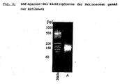
- the size of the DNA fragments in the nucleosome preparation was determined by native 4% Tris-acetate-EDTA agarose gel electrophoresis. The samples were subjected to extraction of the proteins of the Wizard DNA Clean up. The agarose gel of the nucleosomes ( Fig. 2 .) shows that only mononucleosome DNA with 150 broom pairs is present in the nucleosome preparation. Hover-chain chromatin residues are undetectable.
- the protein histone 1 in contrast to the histones 2A, 2B, 3 and 4, is located on the outside of the nucleosomes.
- the molecular weight of histone 1 is about 21 kDa, but this protein shows a changed migration behavior in SDS gel electrophoresis due to its strong positive landing: it appears as a band at about 33 kDa.
- Histone 1 plays an important role as an autoantigen in rheumatoid arthritis. To exclude cross-reactions and to preserve the specificity of the nucleosomes for SLE, histone 1 must be separated from the nucleosome preparation. This is possible by the cleaning methods described above.
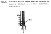
- nucleosomes according to the invention were investigated for their contamination with histone 1 using Western blots.
- the nucleosomes according to the invention and purified histone 1 were applied to the SDS-PA gel.
- the gel was blotted onto nitrocellulose and incubated after blotting with ⁇ -histone-1 antibody-positive serum.
- This Westernblots ( Fig. 3 ) it can be shown that the nucleosomes according to the invention are free of histone 1.
- Scl-70 is a specific antigen for the diagnosis of progressive systemic sclerosis (prevalence: 25-70%).
- This antigen is the protein DNA topoisomerase I.
- This enzyme is involved in the reaction that accompanies a change in DNA topology.
- the molecular weight is 100 kDa, however, the antibodies of the PSS sera also react with the 70 kDa degradation product of the DNA topoisomerase I, therefore also the name Scl-70.
- Table 1 the ELISA coated with the conventional nucleosomes has a PSS sensitivity of 41%, whereas the ELISA coated with the nucleosomes according to the invention shows no reaction with the PSS sera. This effect can be attributed to the fact that the Scl-70 was almost quantitatively separated by the above-described preparation of the nucleosomes according to the invention.
- the nucleosomes according to the invention were examined for their contamination with Scl-70.
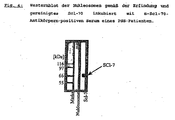
- the nucleosomes and isolated Scl-70 were applied, after electrophoresis, the proteins were blotted on nitrocellulose and incubated with an ⁇ -Scl-70 antibody-positive serum.
- the in Fig. 4 Immunoblot shown clearly shows that the nucleosomes according to the invention are free of Scl-70.
- the preparation of the nucleosomes according to the invention consists only of mononucleosomes, höberkettige chromatin residues are not detectable.
- the examination of the nucleosomes according to the invention by means of SDS-PAGE with subsequent silver staining has only proteins with a molecular mass below 21 kDa. Immunoblots showed that the nucleosomes are free of H1 and Scl-70. Preferably, they also contain no other non-histone proteins, e.g. HMGN.
Landscapes
- Health & Medical Sciences (AREA)
- Life Sciences & Earth Sciences (AREA)
- Chemical & Material Sciences (AREA)
- Engineering & Computer Science (AREA)
- Organic Chemistry (AREA)
- Molecular Biology (AREA)
- Biomedical Technology (AREA)
- Immunology (AREA)
- Hematology (AREA)
- General Health & Medical Sciences (AREA)
- Zoology (AREA)
- Biochemistry (AREA)
- Genetics & Genomics (AREA)
- Biotechnology (AREA)
- Wood Science & Technology (AREA)
- Urology & Nephrology (AREA)
- Microbiology (AREA)
- Bioinformatics & Cheminformatics (AREA)
- General Engineering & Computer Science (AREA)
- Physics & Mathematics (AREA)
- Medicinal Chemistry (AREA)
- Analytical Chemistry (AREA)
- Rheumatology (AREA)
- Cell Biology (AREA)
- Food Science & Technology (AREA)
- Rehabilitation Therapy (AREA)
- Proteomics, Peptides & Aminoacids (AREA)
- Biophysics (AREA)
- General Physics & Mathematics (AREA)
- Pathology (AREA)
- Toxicology (AREA)
- Diabetes (AREA)
- Gastroenterology & Hepatology (AREA)
- Chemical Kinetics & Catalysis (AREA)
- General Chemical & Material Sciences (AREA)
- Crystallography & Structural Chemistry (AREA)
- Plant Pathology (AREA)
- Peptides Or Proteins (AREA)
- Medicines Containing Antibodies Or Antigens For Use As Internal Diagnostic Agents (AREA)
- Measuring Or Testing Involving Enzymes Or Micro-Organisms (AREA)
Description
- Die vorliegende Erfindung betrifft ein Verfahren zur Herstellung einer Nukleosomen-Präparation, die sich dadurch auszeichnet, daß sie sich zum Einsatz beim Immunfluoreszenznachweis spezifischer, gegen die Nukleosomen gerichteter Antikörper in humanen Proben und damit zu einer gegenüber dem Stand der Technik spezifischeren Diagnose von Systemischem lupus erythematodes (SLE), auch bezeichnet als Lupus erythematodes disseminatus, eignet.
- Systemischer lupua erythematodes (SLE) ist eine vermutlich genetisch prädisponierte Autoimmunerkrankung aus der Gruppe der Kollagenosen [Yim, YS, Wakeland, EK. The genetics of lupus. Curr. Opi. Nephrol Hypertens, 2001, 10, 437-443]. Die Erkrankung beginnt meistens zwischen dem 25. und 35. Lebensjahr. Frauen erkranken wesentlich häufiger als Männer (Frauen:Männer 9:1, jährlich 50 Neuerkrankungen pro 100.000 Einwohner) [W. Olliver und D.P.M. Symmons; Autoimmunität, 1995, Spektrum Akademischer Verlag Heidelberg].
- SLE ist eine systemische Erkrankung. Am häufigsten betroffen sind die Haut (Schmetterlingserythem), die Gefäße (Raynoudsyndrom), das Rippenfell (gehäuft auftretende Pleuritis) und die Gelenke (Arthritis). Vor alle die serologische Untersuchung spielt bei der Diagnose eines SLE eine wesentliche Rolle.
- Charakteristisch für einen SLE ist das Auftreten autoreaktiver Antikörper, die gegen eines oder mehrere Antigene gerichtet sind. Ein Beispiel sind Antikörper gegen Doppelstrang DNS (ds-DNS), Sm, SS-A und Nukleosomen.
- In eukaryontischen Zellen ist die DNS in Chromosomen organisiert. Werden die Chromosomen partiell aufgefaltet, so ähnelt die Struktur des resultierenden Chromatins einer Perlenkette. Die einzelnen Perlen nennt man Nukleosomen. Sie bestehen aus Nukleinsäuren und Proteinen, dabei enthält ein Proteinkern 8 Histonmoleküle: je 2 Exemplare der Histone H2A, H2B, H3 und H4. Um dieses Histonzentrum ist die DNS mit 146 Basenpaaren gewickelt. Verbunden sind die Nukleosomen über Linker-DNS (ca. 30-40 Bp,) wodurch die perleenkettenartige Struktur des Chromatins entsteht. Das Histon H1 befindet sich außen an den Nukleosomen in der Nähe der Linker-DNS [Luger, K, Mäder, AW, Richmond, RK, Sargent, DF, Richmond, TJ. Crystal structure of the nucleosome core particle at 2.8 Å resolution. Nature, 1997, 389, 251-260].
- Chabre et al. konnten zeigen, daß nicht nur die "nackte" DNS selbst, sondern Nukleosomen ein charakteristisches Autoantigen für SLE darstellen [Chabre, H, Amoura, Z, Piette, JC, Godeau, P, Bach, JF, Koutouzov, S. Presence of nucleosome-retricted antibodies in patients with sytemic lupus erythematosus. Arthritis Rheum. 1995, 38, 1485-1491]. Es gibt Hinweise darauf, daß sich in einem frühen Stadium des SLE zunächst Antikörper gegen Nukleosomen bilden und erst zu einem späteren Zeitpunkt der Erkrankung Antikörper gegen die Bestandteile der Nukleosomen (ds-DNS, Histone) im Serum nachweisbar sind [Amoura, Z, Chabre, H, Koutouzov, S, Lotton, C, Chabrespines, A, Bach, JF, Jacob, L. Nucleosome-restricted antibodies are detected before anti-ds-DNA and/or Antihistone antibodies in serum of MRL-MP lpr/lpr and +/+ Mice, and are present in kidney eluates of lupus mice with proteinuria. Arthritis Rheum, 1994, 37, 1684-1688].
- Im Stand der Technik werden Antikörper gegen Nukleosomen (ANuA) als sensitiver Marker für SLE beschrieben [Chabre et al., 1995, siehe oben]. In diesem Zusammenhang wurde ein Verfahren zur Erzeugung einer Nukleosomen-Präparation beschrieben [Amoura et al., 1994, siehe oben], bei dem nach Freisetzung der Chromatin-Bestandteile aus präparierten Zellkernen die Nukleosomen durch enzymatische Degradation dargestellt und einer Reinigung durch Saccharose-Gradienten-Zentrifugation unterzogen wurden. Der angewandte Gradient betrug 5 bis 20 %. Bislang ist es jedoch keiner Arbeitsgruppe gelungen, die Nukleosomen soweit zu reinigen, daß nur noch SLE-Patienten reagieren.
- Studien zum Thema Anti-Nukleosomen-Antikörper (ANuA) zeigten, daß bei 20% bis 68% der Patienten mit progressiver stemsklerose (PSS) Antikörper gegen Nukleosomen nachweisbar sind [Dick, T, Vondegracht, MC, Franz, K, Mierau, R, Genth, E. Nachweis von anti-Nukleosomen-Antikörpern bei Patienten mit SLE, systemische Sklerose und Kontrollkollektiven. Vergleich von 4 ELISA Systemen, Rheumaklinik und Rheumaforschungsinstitut Aachen; Amoura Z, Koutouzov S, Chabre H, Cacoub P, Amoura I, Musset L, Bach JP, Piette JC.Presence of antinucleosome autoantibodies in a restricted set of connective tissue diseases: antinucleosome antibodies of the IgG3 subclass are markers of renal pathogenicity in systemic lupus erythematosus. Arthritis Rheum. 2000; 43, 76-84]. Auch bei Polymyositis lassen sich zu einem geringen Prozentsatz ANuA mit dem konventionellen Testsystem nachweisen. Bisher ist es also nicht gelungen, durch die Bestimmung der ANuA zwischen SLE und anderen Erkrankungen zu unterscheiden, dadurch ist der Wert der ANuA als diagnostischer Marker sehr stark eingeschränkt.
- Das im Stand der Technik bekannte Verfahren zur SLE-Diagnose ist daher unspezifisch und erfordert die Anwendung weitergehender diagnostischer Methoden, um eine Unterscheidung zwischen SLE und PSS zu ermöglichten.
- Aufgabe der vorliegenden Erfindung ist es daher, ein in vitro-Diagnose-Verfahren bereitzustellen, mit dem es möglich ist, systemischen lupus erythematosus spezifisch nachzuweisen und eindeutig von progressiver Systemsklerose zu unterscheiden. Die Aufgabe wird erfindungsgemäß durch eine neue Nukleosomen-Präparation und einen Assay unter Verwendung dieser Nukleosomen als Antigen gelöst.
- Im Rahmen der vorliegenden Erfindung wurde überraschenderweise festgestellt, daß sich Nukleosomen - entgegen den im Stand der Technik vorliegenden Daten und Informationen - sehr wohl als spezifischer Marker eignen, wenn man sie von Histon 1, nicht-Histon-Proteinen und dem Protein Scl-70 befreit. Diese Scl-70 freien Nukleosomen werden durch ein modifiziertes Aufarbeitungsverfahren erhalten, bei dem man nach Aufreinigung der Zellkerne und anschließendem Nuklease-Verdau aufgereinigte Nukleosomen durch Dichtezentrifugation mit einem Saccharose-Gradienten isoliert. Im Rahmen der vorliegenden Erfindung kann unerwarteterweise eine gegenüber dem Stand der Technik deutlich reinere Fraktion erhalten werden, wenn man einen Saccharose-Gradienten mit mindestens 30 (vorzugsweise mindestens 35) Gew.-% Saccharose, vorzugsweise 30 bis 50 Gew.-%, besonders bevorzugt 35 (oder 40) bis 50 Gew.-%, am meisten bevorzugt etwa 50 Gew.-% Saccharose, verwendet. Auf diese Weise wird eine Nukleosomen-Präparation isoliert, die aus Mononukleosomen besteht, die frei von Histon 1 und Scl-70-Protein und vorzugsweise auch frei von anderen nicht-Histon-Proteinen, wie z.B. HMGN, sind.
- Überraschenderweise kommt es erfindungsgemäß zu keiner Kreuzreaktivität mit PSS-assoziierten Antikörpern, wenn man die Nukleosomen dieser modifizierten Aufreinigungsprozedur unterzieht, so daß die gewonnene Präparation frei von Scl-70-Protein ist.
- Vorliegend bedeutet "frei von Histon 1 und Scl-70-Protein" bzw. "frei von anderen nicht-Histon-Proteinen", daß die Nukleosomen-Präparation die genannten Proteine nicht oder nur in einem so geringen Anteil enthält, der für den spezifischen SLE-Nachweis und die Unterscheidung von PSS nicht störend ist. Unter "anderen nicht-Histon-Proteinen" werden beispielsweise auch HMG (high mobility group) Proteine verstanden, die ebenfalls als Autoantigene der PSS beschrieben wurden [Rosenberg AM, Uziel Y, Krafchik BR, Hauta SA, Prokopchuk PA, Silverman ED, Laxer RM. Antinuclear antibodies in children with localized scleroderma. J Rheumatol 1995, 22, 2337-2343; Vlachoyiannopoulos PG, Boumba VA, Tzioufas AG, Seferiadis C, Tsolas O, Moutsopoulos HM. Autoantibodies to HMG-17 nucleosomal protein in patients with scleroderma. J Autoimmun 1994, 7, 193-201].
- Mononukleosomen lassen sich für die vorliegenden Zwecke rein oder nahezu rein erhalten, wenn man Nukleosomen beispielsweise durch Saccharose-Gradienten-Zentrifugation unter Verwendung eines Saccharose-Gradienten von über 30 % aufreinigt und auf diese Weise von Histon H1 und weiteren Proteinen, insbesondere von einer 70 kDa Proteinfraktion, abtrennt. Dadurch werden erfindungsgemäß Nukleosomen erhalten, die gegenüber den im Stand der Technik bekannten Präparationen eine erhöhte Reinheit aufweisen und im ELISA zu keiner Kreuzreaktivität mit PSS-assoziierten Antikörpern führen. Durch die Verwendung dieser Mononukleosomen in der in vitro-Diagnostik, z.B. als Antigen im ELISA, wird sowohl ein sensitives als auch hoch spezifisches Testsystem für die serologische Bestimmung des LED erhalten.
- Gegenstand der vorliegenden Erfindung ist somit eine Nukleosomen-Präparation, die frei von Histon 1 (H1) und Scl-70 ist, erhältlich durch ein Verfahren, bei dem man Zellkerne aus biologischem Material isoliert, man die Zellkerne enzymatisch mit einer Nuklease verdaut und durch Dichtegradientenzentrifugation mit einem Saccharose-Gradienten in Gegenwart von 0,55 mol/l NaCl aufreinigt, wobei man die Nukleosomen aus einer Fraktion mit mindestens 30 Gew.-% Saccharose isoliert.
- Die Erfindung betrifft ferner ein Verfahren zur Herstellung einer Histon 1- (Hl-) und Scl-70-freien (und vorzugsweise einer nicht-Histon-Protein-freien) Nukleosomen-Präparation, bei dem man Zellkerne aus biologischem Material isoliert, man die Zellkerne enzymatisch mit einer Nuklease verdaut und durch Dichtegradientenzentrifugation mit einem Saccharose-Gradienten von bis zu 50 Gew.-% in Gegenwart von 0,55 mol/l NaCl aufreinigt, wobei man die Nukleosomen aus einer Fraktion mit mindestens 30 (vorzugsweise mindestens 35) Gew.-% Saccharose, vorzugsweise 30 bis 50 Gew.-% Saccharose isoliert, wobei ein Bereich von 35 (oder 40) bis 50 Gew.-% besonders bevorzugt ist. Am meisten bevorzugt ist, die Nukleosomen aus einer Fraktion mit etwa 50 Gew.-% Saccharose zu isolieren. Alternativ kann auch eine Gelfiltration durchgeführt werden. Dabei sind die Nukleosomen enthaltenden Fraktionen, wie unten beschrieben, daraufhin zu prüfen, ob insbesondere Scl-70 abgetrennt wurde oder noch in der Präparation vorhanden ist.
- Die Erfindung betrifft ferner die Verwendung einer vorgenannten Nukleosomen-Präparation zum Nachweis von Systemischen lupus erythematodes (SLE) in vitro.
- Erfindungsgemäß wird somit ein in vitro-Verfahren zur Diagnose von Systemischen lupus erythematodes (SLE) bereitgestellt, bei dem man eine flüssige Probe eines Patienten, wie z.B. Serum, mit Hilfe eines Antikörper-Nachweisverfahrens untersucht, bei dem man die erfindungsgemäßen Nukleosomen verwendet und man SLE dadurch diagnostiziert, daß man Antikörper gegen die Nukleosomen nachweist.
- Dieser immunologische Nachweis wird mittels bekannter Methoden durchgeführt, wobei zur Detektion der Autoantikörper gegen Nukleosomen gemäß der Erfindung jedes beliebige direkte oder indirekte Verfahren zur Anwendung kommen kann. Dabei sind sowohl Flüssigphasenimmunassays als auch Festphasenimmunoassays denkbar [vgl. auch Price, C.P. und Newman, D.J., a.a.O. Principles and Practice of Immunoassay, 2nd Ed., 1997, Stockton Press, New York].
- Im Gegensatz zu den indirekten Verfahren wird bei den direkten Nachweisen die Bindung der Antikörper an das Antigen über eine Änderung der chemischen oder physikalischen Eigenschaften bestimmt, so daß nachfolgende Detektionsschritte mit markierten Bindungspartnern entfallen.
- Erfindungsgemäß bevorzugt erfolgt der Nachweis der Autoantikörper gegen die Nukleosomen in einem Immunoassay, bevorzugt in einem Festphasenimmunoassay, unter direkter oder indirekter Kopplung eines Reaktionspartners mit einer gut nachweisbaren Markierungssubstanz. Besonders bevorzugt kann der Nachweis in einem ELISA, einem RIA oder einem Fluoreszensimmunoassay erfolgen. Die Durchführung dieser Nachweisverfahren ist dem Fachmann gut bekannt.
- In einem ELISA (enzyme linked immunosorbent assay) der vorliegenden Erfindung werden die Nukleosomen direkt oder indirekt an eine Trägersubstanz (z.B. Polystyrol) gebunden. Nach Inkubation mit den nachzuweisenden Antikörpern, z. B. aus dem Serum des Patienten, werden Antigen-gebundene Antikörper mittels Substanzen nachgewiesen. Diese Substanzen können Antikörper, Fragmente von Antikörpern oder hochaffine Liganden wie z.B. Avidin, das eine Biotin-Markierung bindet, sein. Als Enzyme kommen beispielsweise die Peroxidase, alkalische Phosphatase, β-Galaktosidase, Urease oder Glucoseoxidase in Betracht. Durch Zugabe eines chromogenen Substrats können die gebundenen Enzyme und damit beispielsweise die gebundenen Nukleosomen-Antikörper quantifiziert werden.
- Auch in einem Radio-Immunoassay (RIA) ist das Antigen der Erfindung, die Nukleosomen, direkt oder indirekt an eine Trägersubstanz, wie etwa Polystyrol, gebunden. Nach Inkubation mit den nachzuweisenden Antikörpern, z.B. aus dem Serum von Patienten, werden die an das Antigen gebundenen Antikörper mit Hilfe von Substanzen nachgewiesen, die eine radioaktive Markierung tragen, z.B. 125I. Diese Substanzen können ähnlich wie bei dem ELISA Antikörper, Fragmente von Antikörpern oder hochaffine Liganden wie z.B. Avidin, das eine Biotin-Markierung bindet, sein. Der quantitative Nachweis der an das Antigen gebundene Antikörper erfolgt über die gebundene Radioaktivität mittels eines geeigneten Meßgerätes.
- Im erfindungsgemäßen Fluoreszenzimmunoassay (FIA) erfolgt der Nachweis von Antikörpern gegen die Nukleosomen ähnlich wie im RIA. Die Markierung in diesem Testsystem besteht aus einer Floureszenz-Markierung, wie z.B. Flourescein-Isothiocyanate (FITC). Auch hier kann ein quantitativer Nachweis der an das Antigen gebundenen Antikörper über den Nachweis des gebundenen Fluoreszenzfarbstoffes mittels eines geeigneten Meßgerätes erfolgen.
- Denkbar ist im Rahmen der vorliegenden Erfindung auch der Nachweis von Antikörpern gegen die Nukleosomen über einen Agglutinationstest oder Geldiffusionstest. Beim Agglutinationstest werden Antigen-tragende Partikel z.B. Latex oder Polystyrol-Kügelchen durch Antikörper, z.B. aus dem Serum, quervernetzt. Die dadurch gebildeten Aggregate lassen sich turbodimetrisch nachweisen.
- Beim Geldiffusionstest werden in einer Agar- oder Agarose-Gel-Platte benachbarte Vertiefungen mit dem Antigen z. B. Nukleosomen und dem Antikörper, z.B. Patientenserum gefüllt. Diffundieren die Substanzen aus den Vertiefungen, bilden sich Konzentrationsgradienten. In den überlappenden Bereichen bilden sich gut sichtbare Präzipitate, sobald die Konzentrationsverhältnisse zwischen Antigen und Antikörper ein Optimum erreicht und vorausgesetzt, daß Antikörper gegen das Antigen in der Antikörperlösung vorhanden sind.
- Gemäß einer bevorzugten Ausführungsform wird ein in vitro-Verfahren zur SLE-Diagnose bereitgestellt, bei dem man eine flüssige Probe eines Patienten, wie z.B. Patientenserum, mit Hilfe eines ELISA untersucht, bei dem die Mikrotiterplatten mit erfindungsgemäßen Nukleosomen beschichtet sind und man SLE dadurch diagnostiziert, daß man in der Probe Antikörper gegen die Nukleosomen nachweist.
- Gemäß einer besonderen Ausführungsform der Erfindung handelt es sich bei der Mikrotiterplatte um eine Mikrotiterplatte, bei der mindestens ein Teil der Kavitäten (alternativ: alle Kavitäten) mit den Nukleosomen der vorliegenden Erfindung beschichtet ist (sind).
- Gegenstand der vorliegenden Erfindung sind neben der erwähnten Mikrotiterplatte auch andere feste Träger, die mit den erfindungsgemäßen Nukleosomen beschichtet sind bzw. auf deren Oberfläche die Nukleosomen fixiert sind (s.o.), wie z.B. Nitrocellulose oder PVDF (Polyvinylidenfluorid).
- Gemäß einer weiteren Ausführungsform betrifft die Erfindung ein in vitro-Verfahren zu SLE-Diagnose, bei dem man einen Streifentest (z.B. EUROLINE®; vgl. Price, C.P. und Newman, D.J., a.a.O.) unter Verwendung Nukleosomen-beschichteter Nitrocellulose oder PVDF durchführt.
- Die Erfindung betrifft ferner ein Kit zur Durchführung eines genannten in vitro-Verfahrens zur Diagnose von Systemischen lupus erythematodes, das eine erfindungsgemäße Nukleosomen-Präparation, einen oben genannten festen Träger oder eine oben genannte Mikrotiterplatte, Nitrocellulose oder PVDF enthält. Gegebenenfalls können in dem Kit weitere zur Durchführung des Antikörper-Nachweisverfahrens erforderliche oder nützliche Reagenzien und Hilfsmittel enthalten sein.
- Die Erfindung wird nachfolgend anhand von Beispielen und Figuren beschrieben:
- 5g Kalbsthymus [alternativ können auch Zellen einer anderen Quelle verwendet werden, üblicherweise von tierischen Geweben (humane eher weniger), wie z.B. Rattenleber, Kaninchenthymus etc., oder tierische Zellen, wie Hühner-Erythrocyten, und Zellkulturen (z.B. mouse erythroleukemia L1210 cells)] werden in 150 mL 0,9%iger NaCl-Lösung aufgenommen und zerkleinert. Anschließend wird die Suspension homogenisiert. Nach Abfiltrieren der groben Bestandteile wird die Suspension zentrifugiert (15 Min bei 1.100 x g, 4°C). Das Pellet wird in jeweils 30 mL 0,9%ige NaCl-Lösung zwei mal gewaschen wie oben zentrifugiert. Anschließend wird das Pellet zur Kern-Aufreinigung weiterverarbeitet
- Die Kern-Aufreinigung erfolgt nach Subirana [Subirana, JA. Studies on the thermal denaturation of nucleohistones. J Mol Biol. 1973, 74, 363-86]. Das Pellet wird in 40 mL Saccharose-1-Lösung (250 mM Saccharose, 3 mM CaCl2·2 H2O) resuspendiert und anschließend zentrifugiert (12 Min, 5.000 Upm und 4°C). Nach Aufnahme des Pellets in 45 mL Saccharose-2-Lösung (2 M Saccharose, 3 mM CaCl2·2 H2O, 0,5 % Triton-X-100) wird wieder zentrifugiert (22 Min, 13.200 x g und 4°C). Das Pellet wird in 45 mL Puffer 1 (0,1 M Tris/HCl pH 8, 0) resuspendiert und wiederum zentrifugiert (15 Min, 1.100 x g, 4°C). Der letze Waschschritt wird wiederholt. Das Pellet wird in 45 mL 0,9%ige NaCl-Lösung aufgenommen und die Lösung zentrifugiert (15 Min, 1.100 x g, 4°C). Der Überstand wird verworfen und das Zellkern-enthaltene Pellet weiterverarbeitet.
- Das Pellet wird in 40 mL Puffer 2 (15 mM Tris/HCl, pH 7, 5, 15 mM NaCl, 60 mM KNO3, 0,25 M saccharose, 5 mM MgCl2·6 H2O, 1 mM CaCl2-2 H2O, 1 mM DTT) ausgenommen und Suspension homogenisiert. Dann werden 750 Units der Nuklease S7-Lösung zur Suspension hinzu gefügt, die Lösung 30 Min im Wasserbad bei 37°C inkubiert und danach zentrifugiert (15 Min, 4.000 Upm, 4°C). Das entstandene Pellet wird in 10 mL 2 mM-EDTA-Lösung aufgenommen. Die Suspension wird 45 Min unter Schütteln bei Raumtemperatur inkubiert und anschließend zentrifugiert (15 Min, 4.000 Upm, 4°C). Schließlich wird der Überstand vorsichtig abgenommen und als konventionelle. Nukleosom-Präparation eingesetzt.
- Gemäß der Erfindung werden die Nukleosomen proteinabgereichert, das Resultat sind im wesentlichen Nukleoaomenkerne, die besonders als Antigene in der in-vitro-Diagnostik des SLE ihre Anwendung finden. Hier wird die Herstellung an einem Beispiel beschrieben.
- Die konventionelle Nukleosom-Präparation wird über Nacht gegen Puffer 2 dialysiert. Anschließend wird die Lösung gekühlt (0°C) und unter Schütteln eine NaCl-Lösung in die Nukleosomen-Präparation getropft, bis eine Endkonzentration von 0,55 M NaCl erreicht wird. Schließlich werden 150 Units Nuklease S7 pro mL Nukleosom-Lösung hinzugefügt, die Lösung 30 Min im Wasserbad bei 37°C inkubiert und danach wieder auf 0°C abkühlt.
- Die erhaltenen Nukleosomen werden durch einen Dichtegradienten-Zentrifugation aufgereinigt: Jeweils 25 mL 50%, 30% und 10% Saccharose-Lösungen in TE-Puffer (10 mM Tris, 1 mM EDTA, 0,55 M NaCl) werden in ein Ultrazentrifugenröhrchen geschichtet und 5 mL Nukleosom-Lösung aufgetragen. Der Gradient wird über Nacht bei 4°C und 140.000 x g zentrifugiert.
- Alternativ können auch andere Gravdienten verwendet werden, wie z.B. 50/40/30/10% oder 50/35/10% usw.
- Die proteinabgereichten Nukleosomen befinden sich in der mindestens 30% Saccharose, vorzugsweise in der mindestens 35% (oder 40%) bzw. in der etwa 50% Saccharose enthaltenden Fraktion.
- Die Isolierung der Nukleosomen kann ebenso anstelle der Saccharose-Dichte-zentrifugation über eine Gelfiltration (HiPrep 26/60 Sephacryl S-300 HR, Amersham-Bioscience) erfolgen. 10 mL der erhaltenen Nukleosomen werden auf die mit TE-Puffer äquilibrierte Säule aufgetragen und bei einer Elutionsgeschwindigkeit von 1 ml/min eluiert (TE-Puffer). Die Nukleosomen enthaltenden Fraktionen werden vereinigt.
- Mittels ELISA wurden Studien mit Seren von Patienten mit SLE und progressiver Systemsklerose durchgeführt, welche die These unterstützen, daß die klinische Spezifität bei Verwendung der Nukleosomen als Autoantigen für SLE auf 100% steigen kann. Voraussetzung dafür ist, daß die Nukleosomen erfindungsgemäß soweit isoliert werden, bis mittels Silberfärbung nach SDS-PAGE der Nukleosomen keine weiteren Proteine mehr nachweisbar sind.
- Es wurden zwei ELISA durchgeführt, bei denen verschiedene Nukleosomen-Präparationen zur Beschichtung der Mikrotiterplatte verwendet wurden.
- Die Mikrotiterplatten werden mit in PBS verdünnten Nukleosomen über Nacht bei Raumtemperatur beschichtet. Anschließend werden die mit Antigen beschichteten Platten mit PSS/0,1% Tween gewaschen und für 2 Stunden bei Raumtemperatur mit PBS-10% Fetalem Rinderserum geblockt. Danach werden die Mikrotiterplatten erneut mit PBS-0.1% Tween gewaschen. Die Inkubation der Platten erfolgt wie in den EUROIMMUN-Arbeitsanleitungen beschrieben.
- Die beiden, im ELISA eingesetzten Präparationen unterscheiden sich dadurch, daß einerseits die konventionelle Nukleosomen-Präparation angesetzt wurde, andererseits die Nukieosomen-Präparation, die einer weiteren Aufreinigung unterzogen wurde, um weitere Proteine zu entfernen und damit die Nukleosomen gemäß der Erfindung zu erhalten:

- Die Nukleosomen gemäß der Erfindung wurden mittels SPS-PAGE mit anschließender Silberfärbung untersucht [alle Materalien für die Durchführung der Elektrophoresen stammen von Invitrogen: NuPAGE 4-12% Bis-Tris-Gel, NuPage Sample-Buffer, NuPage-MES-Laufpuffer, Mark-12. Anschließend wurde eine Silberfärbung nach Heukeshofen durchgeführt]. Das elektrophoretische Verhalten der Nukleosomen ist in
Abb. 1 dargestellt. Das SDS-PA-Gel der Nukleosomen-Präparation gemäß der Erfindung zeigt keine Proteinbande oberhalb von 21 kDa, d.h., daß neben den im Nukleosomen-Kern befindlichen Histonen H2A, H2B, H3 und H4 keine weiteren Proteine in dieser Präparation nachweisbar sind. - Die Größe der DNS-Fragmente in der Nukleosomen-Präparation wurde anhand von nativen 4%igen Tris-Acetat-EDTA-Agarose-Gel-Elektrophoresen bestimmt. Die Proben wurden zur Extraktion der Proteine der Wizard-DNA-Clean up unterzogen. Das Agarose-Gel der Nukleosomen (
Fig. 2 .) zeigt, daß in der Nukleosomenpräparation nur Mononukleosomen-DNS mit 150 Besenpaaren vorliegt. Höberkettige Chromatinreste sind nicht nachweisbar. [Hinsichtlich der Durchführung dieser Elektrophoresemethode wird auf folgende Publikation verwiesen: Hans Günter Gassen, Gangolf Schrimpf (Hrsg.): Gentechnische Methoden, 2. Auflage, 1999, Spektrum Akademischer Verlag, Heidelberg, Berlin, die Färbung des Agarose-Gels erfolgt Mittels Ethidiumbromid.] - Das Protein Histon 1 befindet sich im Gegensatz zu den Histonen 2A, 2B, 3 und 4 außen an den Nukleosomen. Das Molekulargewicht von Histon 1 beträgt etwa 21 kDa, jedoch zeigt dieses Protein aufgrund seiner stark positiven Landung ein verändertes Migrationsverhalten in der SDS-Gelelektrophorese: es erscheint als Bande bei ca. 33 kDa.
- Histon 1 spielt als ein Autoantigen bei der rheumatoiden Arthritis eine große Rolle. Um Kreuzreaktionen auszuschließen und die Spezifität der Nukleosomen für SLE zu erhalten, muß Histon 1 von der Nukleosomen-Präparation abgetrennt werden. Dies ist durch die oben beschriebenen Reinigungsmethoden möglich.
- Nukleosomen gemäß der Erfindung wurden hinsichtlich ihrer Kontamination mit Histon 1 mit Hilfe von Westernblots untersucht. Auf das SDS-PA-Gel wurden die Nukleosomen gemäß der Erfindung und gereinigtes Histon 1 aufgetragen. Das Gel wurde auf Nitrocellulose geblottet und nach dem Blotten mit α-Histon-1-Antikörper-positivem Serum inkubiert. Anhand dieses Westernblots (
Fig. 3 ) kann gezeigt werden, daß die Nukleosomen gemäß der Erfindung frei von Histon 1 sind. - Scl-70 ist ein spezifisches Antigen für die Diagnose von progressiver Systemsklerose (Prävalenz: 25-70%). Es handelt bei diesem Antigen um das Protein DNS-Topoisomerase I. Dieses Enzym ist an der Reaktion, die mit einer Änderung der DNS-Topologie einhergeht, beteiligt. Das Molekulargewicht beträgt 100 kDa, jedoch reagieren die Antikörper der PSS-Seren auch mit dem 70 kDa-Abbauprodukt der DNS-Topoisomerase I, daher auch der Name Scl-70. Wie in Tabelle 1 ersichtlich ist, weist der ELISA beschichtet mit den konventionellen Nukleosomen eine Sensitivität für PSS von 41% auf, dagegen zeigt der mit den Nukleosomen gemäß der Erfindung beschichtete ELISA keine Reaktion mehr mit den PSS-Seren. Dieser Effekt läßt sich darauf zurückführen, daß durch die oben beschriebene Präparation der Nukleosomen gemäß der Erfindung das Scl-70 nahezu quantitativ abgetrennt wurde.
- Mittels Westernblots wurden die Nukleosomen gemäß der Erfindung hinsichtlich ihrer Kontamination mit Scl-70 untersucht. Auf das SDS-PA-Gel wurden die Nukleosomen und isoliertes Scl-70 aufgetragen, nach der Elektrophorese wurden die Proteine auf Nitrocellulose geblottet und mit einem α-Scl-70-Antikörpern-positivem Serum inkubiert. Der in
Fig. 4 gezeigte Immunoblot zeigt eindeutig, daß die Nukleosomen gemäß der Erfindung frei von Scl-70 sind. - Anhand von ELISA Studien zeigt sich, daß die Nukleosomen gemäß der Erfindung spezifisch für SLE sind.
- Die Präparation der Nukleosomen gemäß der Erfindung besteht nur aus Mononukleosomen, höberkettige Chromatinreste sind nicht nachweisbar. Die Untersuchung der Nukleosomen gemäß der Erfindung mittels SDS-PAGE mit anschließender Silberfärbung weist nur Proteine mit einer Molekularmasse unterhalb 21 kDa auf. Anhand von Immunoblots konnte gezeigt werden, daß die Nukleosomen frei von H1 und Scl-70 sind. Vorzugsweise enthalten sie ferner keine anderen nicht-Histon-Proteine, wie z.B. HMGN.
Claims (16)
- Nukleosomen-Präparation, dadurch gekennzeichnet, daß sie frei von Histon 1 und Scl-70 ist erhältlich durch ein Verfahren, bei dem man Zellkerne aus biologischem Material isoliert, man die Zellkerne enzymatisch mit einer Nuklease verdaut und durch Dichtegradientenzentrifugation mit einem Saccharose-Gradienten in Gegenwart von 0,55 mol/l NaCl aufreinigt, wobei man die Nukleosomen aus einer Fraktion mit mindestens 30 Gew.-% Saccharose isoliert.
- Nukleosomen-Präparation nach Anspruch 1, dadurch gekennzeichnet, daß sie Nukleosomen enthält, die aus einer Fraktion mit mindestens 40 Gew.-% Saccharose isoliert sind.
- Nukleosomen-Präparation nach Anspruch 2, dadurch gekennzeichnet, daß sie Nukleosomen enthält, die aus einer Fraktion mit etwa 50 Gew.-% Saccharose isoliert sind.
- Verfahren zur Herstellung einer Histon 1- und Scl-70-freier Nukleosomen-Präparation, bei dem man Zellkerne aus biologischem Material isoliert, man die Zellkerne enzymatisch mit einer Nuklease verdaut und durch Saccharose-Dichtegradientenzentrifugation aufreinigt, dadurch gekennzeichnet, daß man mit einem Saccharose-Gradienten in Gegenwart von 0,55 mol/l NaCl aufreinigt, wobei man die Nukleosomen aus einer Fraktion mit mindestens 30 Gew.-% Saccharose isoliert.
- Verfahren nach Anspruch 4, dadurch gekennzeichnet, daß man die Nukleosomen aus einer Fraktion mit mindestens 40 Gew.-% Saccharose isoliert.
- Verfahren nach Anspruch 5, dadurch gekennzeichnet, daß man die Nukleosomen aus einer Fraktion mit etwa 50 Gew.-% Saccharose isoliert.
- Verwendung einer Nukleosomen-Präparation nach den Ansprüchen 1 bis 3 zum Nachweis von Systemischem lupus erythematodes (SLE) in vitro.
- In vitro-Verfahren zur Diagnose von Systemischem lupus erythematodes (SLE), bei dem man eine flüssige Probe eines Patienten mit Hilfe eines Antikörper-Nachweisverfahrens untersucht, bei dem man Nukleosomen nach den Ansprüchen 1 bis 3 verwendet und man SLE dadurch diagnostiziert, daß man Antikörper gegen die Nukleosomen nachweist.
- Verfahren nach Anspruch 8, dadurch gekennzeichnet, daß das Antikörper-Nachweisverfahren ein ELISA ist, bei dem die Mikrotiterplatte des ELISA mit Nukleosomen nach den Ansprüchen 1 bis 3 beschichtet ist.
- Verfahren nach Anspruch 8, dadurch gekennzeichnet, daß das Antikörper-Nachweisverfahren ein Streifentest ist, bei dem ein fester Träger mit Nukleosomen nach den Ansprüchen 1 bis 3 beschichtet ist.
- Verfahren nach den Ansprüchen 8 bis 10, dadurch gekennzeichnet, daß die flüssige Probe Serum ist.
- Fester Träger, dadurch gekennzeichnet, daß er mit Nukleosomen nach den Ansprüchen 1 bis 3 beschichtet ist.
- Träger nach Anspruch 12, dadurch gekennzeichnet, daß er Nitrocellulose oder Polyvinylidenfluorid ist.
- Mikrotiterplatte, dadurch gekennzeichnet, daß mindestens ein Teil der Kavitäten mit Nukleosomen nach den Ansprüchen 1 bis 3 beschichtet ist.
- Kit zur Durchführung des Verfahrens nach einem der Ansprüchen 8 bis 11, dadurch gekennzeichnet, dass es eine Nukleosomen-Präparation nach den Ansprüchen 1 bis 3, einen festen Träger nach Anspruch 12 oder 13 oder eine Mikrotiterplatte nach Anspruch 14 umfasst.
- Kit nach Anspruch 15, dadurch gekennzeichnet, daß er weitere zur Durchführung des Antikörper-Nachweisverfahrens erforderliche oder nützliche Reagenzien und Hilfsmittel enthält.
Applications Claiming Priority (3)
| Application Number | Priority Date | Filing Date | Title |
|---|---|---|---|
| DE10207735 | 2002-02-15 | ||
| DE10207735A DE10207735A1 (de) | 2002-02-15 | 2002-02-15 | Verfahren zur Herstellung einer Nukleosomen-Präparation und deren Verwendung zur in vitro-Diagnose von Lupus erythematodes disseminatus (LED) |
| PCT/EP2002/013629 WO2003069337A1 (de) | 2002-02-15 | 2002-12-02 | Verfahren zur herstellung einer nukleosomen-präparation und deren verwendung zur in vitro-diagnose von lupus erythematodes disseminatus (led) |
Publications (2)
| Publication Number | Publication Date |
|---|---|
| EP1476750A1 EP1476750A1 (de) | 2004-11-17 |
| EP1476750B1 true EP1476750B1 (de) | 2009-02-04 |
Family
ID=27635266
Family Applications (1)
| Application Number | Title | Priority Date | Filing Date |
|---|---|---|---|
| EP02795090A Expired - Lifetime EP1476750B1 (de) | 2002-02-15 | 2002-12-02 | Verfahren zur herstellung einer nukleosomen-präparation und deren verwendung zur in vitro-diagnose von systemischem lupus erythemastosus (sle) |
Country Status (6)
| Country | Link |
|---|---|
| US (1) | US7566545B2 (de) |
| EP (1) | EP1476750B1 (de) |
| AT (1) | ATE422244T1 (de) |
| AU (1) | AU2002360955A1 (de) |
| DE (2) | DE10207735A1 (de) |
| WO (1) | WO2003069337A1 (de) |
Families Citing this family (2)
| Publication number | Priority date | Publication date | Assignee | Title |
|---|---|---|---|---|
| US20080206855A1 (en) * | 2005-05-06 | 2008-08-28 | Kalypsys, Inc. | Automated Compound Screening Using Gel-Permeation Matrix and Pin-Based Sample Transfer |
| GB201115098D0 (en) * | 2011-09-01 | 2011-10-19 | Belgian Volition Sa | Method for detecting nucleosomes containing histone variants |
Family Cites Families (2)
| Publication number | Priority date | Publication date | Assignee | Title |
|---|---|---|---|---|
| US4444879A (en) * | 1981-01-29 | 1984-04-24 | Science Research Center, Inc. | Immunoassay with article having support film and immunological counterpart of analyte |
| EP0182896A1 (de) * | 1984-06-06 | 1986-06-04 | Scripps Clinic And Research Foundation | Diagnosetestverfahren zum nachweis von mit medikamenten induziertem lupus erythemotosus verbundenen antikörpern |
-
2002
- 2002-02-15 DE DE10207735A patent/DE10207735A1/de not_active Withdrawn
- 2002-12-02 DE DE50213263T patent/DE50213263D1/de not_active Expired - Lifetime
- 2002-12-02 AU AU2002360955A patent/AU2002360955A1/en not_active Abandoned
- 2002-12-02 WO PCT/EP2002/013629 patent/WO2003069337A1/de not_active Ceased
- 2002-12-02 US US10/504,569 patent/US7566545B2/en not_active Expired - Lifetime
- 2002-12-02 AT AT02795090T patent/ATE422244T1/de not_active IP Right Cessation
- 2002-12-02 EP EP02795090A patent/EP1476750B1/de not_active Expired - Lifetime
Also Published As
| Publication number | Publication date |
|---|---|
| AU2002360955A1 (en) | 2003-09-04 |
| US20050233322A1 (en) | 2005-10-20 |
| WO2003069337A1 (de) | 2003-08-21 |
| EP1476750A1 (de) | 2004-11-17 |
| ATE422244T1 (de) | 2009-02-15 |
| DE50213263D1 (de) | 2009-03-19 |
| US7566545B2 (en) | 2009-07-28 |
| DE10207735A1 (de) | 2003-08-28 |
Similar Documents
| Publication | Publication Date | Title |
|---|---|---|
| DE68918103T3 (de) | Verfahren zur überwachung des kollagenabbaus. | |
| DE69626026T2 (de) | Differentialtest für ulzerative kolitis, primäre sklerosierende cholangitis und autoimmun-hepatitis vom type 1. | |
| DE3874530T2 (de) | Untersuchung des abbaus von kollagen typ iii. | |
| DE69607176T2 (de) | Verfahren zur quantitativen bestimmung menschlichen akutphasen-serum-amyloid-a-proteins; rekombinantes protein; spezifischer antikörper | |
| EP0944832B1 (de) | ANTIGENSPEZIFISCHER IgM-NACHWEIS | |
| DE69026615T2 (de) | Igg3-antikörper mit verkürzter hinge-region und einem komplementaktivierungstest | |
| EP0544869B1 (de) | Immunologisches bestimmungsverfahren zur bestimmung von antikörpern in biologischen flüssigkeiten sowie kit zur durchführung des verfahrens | |
| EP0697021B1 (de) | Photoaktivierbare biotinderivate und deren einsatz zum entstören von immunoassays | |
| DE60317315T2 (de) | Nachweis von dna-bindungsproteinen | |
| DE10225853A1 (de) | Werkzeuge zur Diagnostik, molekularen Definition und Therapieentwicklung chronischer entzündlicher Gelenkerkrankungen | |
| DE69031662T2 (de) | Test für die alkalische Phosphatase aus Knochen | |
| DE69204718T2 (de) | CA-195 ähnliches immunreaktives Antigen von menschlicher Amnionflüssigkeit. | |
| EP1015885B1 (de) | Aufreinigung von substanzen aus einer biologischen probe | |
| DE3650497T2 (de) | Verfahren und Zusammensetzung zum Nachweis von Analyt-Anteilen | |
| DE60030701T2 (de) | VERFAHREN ZUM BEWERTEN VON AUTOIMMUNKRANKHEITEN, VERFAHREN ZUM NACHWEIS VON GEGEN Reg GERICHTETEN AUTOANTIKÖRPERN | |
| DE69626860T2 (de) | Elisa testsystem zur bestimmung von substanzen, die die bindung zwischen ig e und dessen receptor inhibiert | |
| EP0532979B1 (de) | Petide zur Herstellung von Mitteln zur Diagnose und Therapie von systemischem Lupus | |
| DE69927668T2 (de) | Massenfingerabdrucken und seiner verwendung zur identifizierung von protein-affinitätsliganden | |
| EP0776906A2 (de) | Peptide des Antigens Sm-D und ihre Verwendung, insbesondere für die Diagnostik des SLE | |
| EP1476750A1 (de) | Verfahren zur herstellung einer nukleosomen-präparation und deren verwendung zur in vitro-diagnose von lupus erythematodes disseminatus (led) | |
| DE3854834T2 (de) | Immuntest unter Verwendung von Antigenen, produziert in heterologen Organismen | |
| EP0363449B1 (de) | Verfahren zur untersuchung von antikörpern spezifisch für rheumakrankheiten, insbesondere chronische polyarthritis sowie reagens hierfür | |
| DE20212524U1 (de) | In vitro-Diagnosekit zum Nachweis von Lupus erythematodes disseminatus (LED) | |
| EP1334208A2 (de) | Verfahren zur markierung chemischer substanzen | |
| EP1692515A1 (de) | Konjugate und deren verwendung in nachweisverfahren |
Legal Events
| Date | Code | Title | Description |
|---|---|---|---|
| PUAI | Public reference made under article 153(3) epc to a published international application that has entered the european phase |
Free format text: ORIGINAL CODE: 0009012 |
|
| 17P | Request for examination filed |
Effective date: 20040913 |
|
| AK | Designated contracting states |
Kind code of ref document: A1 Designated state(s): AT BE BG CH CY CZ DE DK EE ES FI FR GB GR IE IT LI LU MC NL PT SE SI SK TR |
|
| AX | Request for extension of the european patent |
Extension state: AL LT LV MK RO |
|
| 17Q | First examination report despatched |
Effective date: 20050520 |
|
| 17Q | First examination report despatched |
Effective date: 20050520 |
|
| GRAP | Despatch of communication of intention to grant a patent |
Free format text: ORIGINAL CODE: EPIDOSNIGR1 |
|
| RTI1 | Title (correction) |
Free format text: METHOD FOR PRODUCING A NUCLEOSOME PREPARATION AND USE THEREOF IN IN VITRO DIAGNOSIS OF SYSTEMIC LUPUS ERYTHEMATOSUS (SLE) |
|
| GRAS | Grant fee paid |
Free format text: ORIGINAL CODE: EPIDOSNIGR3 |
|
| GRAA | (expected) grant |
Free format text: ORIGINAL CODE: 0009210 |
|
| AK | Designated contracting states |
Kind code of ref document: B1 Designated state(s): AT BE BG CH CY CZ DE DK EE ES FI FR GB GR IE IT LI LU MC NL PT SE SI SK TR |
|
| REG | Reference to a national code |
Ref country code: GB Ref legal event code: FG4D Free format text: NOT ENGLISH |
|
| REG | Reference to a national code |
Ref country code: CH Ref legal event code: EP |
|
| REG | Reference to a national code |
Ref country code: IE Ref legal event code: FG4D Free format text: LANGUAGE OF EP DOCUMENT: GERMAN |
|
| REF | Corresponds to: |
Ref document number: 50213263 Country of ref document: DE Date of ref document: 20090319 Kind code of ref document: P |
|
| NLV1 | Nl: lapsed or annulled due to failure to fulfill the requirements of art. 29p and 29m of the patents act | ||
| PG25 | Lapsed in a contracting state [announced via postgrant information from national office to epo] |
Ref country code: SI Free format text: LAPSE BECAUSE OF FAILURE TO SUBMIT A TRANSLATION OF THE DESCRIPTION OR TO PAY THE FEE WITHIN THE PRESCRIBED TIME-LIMIT Effective date: 20090204 Ref country code: NL Free format text: LAPSE BECAUSE OF FAILURE TO SUBMIT A TRANSLATION OF THE DESCRIPTION OR TO PAY THE FEE WITHIN THE PRESCRIBED TIME-LIMIT Effective date: 20090204 Ref country code: ES Free format text: LAPSE BECAUSE OF FAILURE TO SUBMIT A TRANSLATION OF THE DESCRIPTION OR TO PAY THE FEE WITHIN THE PRESCRIBED TIME-LIMIT Effective date: 20090515 Ref country code: FI Free format text: LAPSE BECAUSE OF FAILURE TO SUBMIT A TRANSLATION OF THE DESCRIPTION OR TO PAY THE FEE WITHIN THE PRESCRIBED TIME-LIMIT Effective date: 20090204 |
|
| PG25 | Lapsed in a contracting state [announced via postgrant information from national office to epo] |
Ref country code: SE Free format text: LAPSE BECAUSE OF FAILURE TO SUBMIT A TRANSLATION OF THE DESCRIPTION OR TO PAY THE FEE WITHIN THE PRESCRIBED TIME-LIMIT Effective date: 20090504 |
|
| REG | Reference to a national code |
Ref country code: IE Ref legal event code: FD4D |
|
| PG25 | Lapsed in a contracting state [announced via postgrant information from national office to epo] |
Ref country code: DK Free format text: LAPSE BECAUSE OF FAILURE TO SUBMIT A TRANSLATION OF THE DESCRIPTION OR TO PAY THE FEE WITHIN THE PRESCRIBED TIME-LIMIT Effective date: 20090204 Ref country code: PT Free format text: LAPSE BECAUSE OF FAILURE TO SUBMIT A TRANSLATION OF THE DESCRIPTION OR TO PAY THE FEE WITHIN THE PRESCRIBED TIME-LIMIT Effective date: 20090706 Ref country code: IE Free format text: LAPSE BECAUSE OF FAILURE TO SUBMIT A TRANSLATION OF THE DESCRIPTION OR TO PAY THE FEE WITHIN THE PRESCRIBED TIME-LIMIT Effective date: 20090204 Ref country code: EE Free format text: LAPSE BECAUSE OF FAILURE TO SUBMIT A TRANSLATION OF THE DESCRIPTION OR TO PAY THE FEE WITHIN THE PRESCRIBED TIME-LIMIT Effective date: 20090204 Ref country code: CZ Free format text: LAPSE BECAUSE OF FAILURE TO SUBMIT A TRANSLATION OF THE DESCRIPTION OR TO PAY THE FEE WITHIN THE PRESCRIBED TIME-LIMIT Effective date: 20090204 |
|
| PG25 | Lapsed in a contracting state [announced via postgrant information from national office to epo] |
Ref country code: SK Free format text: LAPSE BECAUSE OF FAILURE TO SUBMIT A TRANSLATION OF THE DESCRIPTION OR TO PAY THE FEE WITHIN THE PRESCRIBED TIME-LIMIT Effective date: 20090204 |
|
| PLBE | No opposition filed within time limit |
Free format text: ORIGINAL CODE: 0009261 |
|
| STAA | Information on the status of an ep patent application or granted ep patent |
Free format text: STATUS: NO OPPOSITION FILED WITHIN TIME LIMIT |
|
| 26N | No opposition filed |
Effective date: 20091105 |
|
| PG25 | Lapsed in a contracting state [announced via postgrant information from national office to epo] |
Ref country code: BG Free format text: LAPSE BECAUSE OF FAILURE TO SUBMIT A TRANSLATION OF THE DESCRIPTION OR TO PAY THE FEE WITHIN THE PRESCRIBED TIME-LIMIT Effective date: 20090504 |
|
| BERE | Be: lapsed |
Owner name: EUROIMMUN MEDIZINISCHE LABORDIAGNOSTIKA AG Effective date: 20091231 |
|
| PG25 | Lapsed in a contracting state [announced via postgrant information from national office to epo] |
Ref country code: MC Free format text: LAPSE BECAUSE OF NON-PAYMENT OF DUE FEES Effective date: 20100701 |
|
| REG | Reference to a national code |
Ref country code: CH Ref legal event code: PL |
|
| PG25 | Lapsed in a contracting state [announced via postgrant information from national office to epo] |
Ref country code: GR Free format text: LAPSE BECAUSE OF FAILURE TO SUBMIT A TRANSLATION OF THE DESCRIPTION OR TO PAY THE FEE WITHIN THE PRESCRIBED TIME-LIMIT Effective date: 20090505 Ref country code: LI Free format text: LAPSE BECAUSE OF NON-PAYMENT OF DUE FEES Effective date: 20091231 Ref country code: BE Free format text: LAPSE BECAUSE OF NON-PAYMENT OF DUE FEES Effective date: 20091231 Ref country code: CH Free format text: LAPSE BECAUSE OF NON-PAYMENT OF DUE FEES Effective date: 20091231 |
|
| PG25 | Lapsed in a contracting state [announced via postgrant information from national office to epo] |
Ref country code: LU Free format text: LAPSE BECAUSE OF NON-PAYMENT OF DUE FEES Effective date: 20091202 |
|
| PG25 | Lapsed in a contracting state [announced via postgrant information from national office to epo] |
Ref country code: AT Free format text: LAPSE BECAUSE OF NON-PAYMENT OF DUE FEES Effective date: 20091202 |
|
| PG25 | Lapsed in a contracting state [announced via postgrant information from national office to epo] |
Ref country code: TR Free format text: LAPSE BECAUSE OF FAILURE TO SUBMIT A TRANSLATION OF THE DESCRIPTION OR TO PAY THE FEE WITHIN THE PRESCRIBED TIME-LIMIT Effective date: 20090204 |
|
| PG25 | Lapsed in a contracting state [announced via postgrant information from national office to epo] |
Ref country code: CY Free format text: LAPSE BECAUSE OF FAILURE TO SUBMIT A TRANSLATION OF THE DESCRIPTION OR TO PAY THE FEE WITHIN THE PRESCRIBED TIME-LIMIT Effective date: 20090204 |
|
| REG | Reference to a national code |
Ref country code: DE Ref legal event code: R082 Ref document number: 50213263 Country of ref document: DE |
|
| REG | Reference to a national code |
Ref country code: FR Ref legal event code: PLFP Year of fee payment: 14 |
|
| REG | Reference to a national code |
Ref country code: FR Ref legal event code: PLFP Year of fee payment: 15 |
|
| REG | Reference to a national code |
Ref country code: FR Ref legal event code: PLFP Year of fee payment: 16 |
|
| PGFP | Annual fee paid to national office [announced via postgrant information from national office to epo] |
Ref country code: GB Payment date: 20211222 Year of fee payment: 20 Ref country code: FR Payment date: 20211220 Year of fee payment: 20 Ref country code: DE Payment date: 20211231 Year of fee payment: 20 |
|
| PGFP | Annual fee paid to national office [announced via postgrant information from national office to epo] |
Ref country code: IT Payment date: 20211230 Year of fee payment: 20 |
|
| REG | Reference to a national code |
Ref country code: DE Ref legal event code: R071 Ref document number: 50213263 Country of ref document: DE |
|
| REG | Reference to a national code |
Ref country code: GB Ref legal event code: PE20 Expiry date: 20221201 |
|
| PG25 | Lapsed in a contracting state [announced via postgrant information from national office to epo] |
Ref country code: GB Free format text: LAPSE BECAUSE OF EXPIRATION OF PROTECTION Effective date: 20221201 |