EP1451646B1 - Apparatus and method for digital holographic imaging - Google Patents
Apparatus and method for digital holographic imaging Download PDFInfo
- Publication number
- EP1451646B1 EP1451646B1 EP02804141.6A EP02804141A EP1451646B1 EP 1451646 B1 EP1451646 B1 EP 1451646B1 EP 02804141 A EP02804141 A EP 02804141A EP 1451646 B1 EP1451646 B1 EP 1451646B1
- Authority
- EP
- European Patent Office
- Prior art keywords
- sample
- medium
- refraction
- index
- quantitative phase
- Prior art date
- Legal status (The legal status is an assumption and is not a legal conclusion. Google has not performed a legal analysis and makes no representation as to the accuracy of the status listed.)
- Expired - Lifetime
Links
Images


Classifications
-
- G—PHYSICS
- G03—PHOTOGRAPHY; CINEMATOGRAPHY; ANALOGOUS TECHNIQUES USING WAVES OTHER THAN OPTICAL WAVES; ELECTROGRAPHY; HOLOGRAPHY
- G03H—HOLOGRAPHIC PROCESSES OR APPARATUS
- G03H1/00—Holographic processes or apparatus using light, infrared or ultraviolet waves for obtaining holograms or for obtaining an image from them; Details peculiar thereto
- G03H1/04—Processes or apparatus for producing holograms
- G03H1/0443—Digital holography, i.e. recording holograms with digital recording means
-
- G—PHYSICS
- G03—PHOTOGRAPHY; CINEMATOGRAPHY; ANALOGOUS TECHNIQUES USING WAVES OTHER THAN OPTICAL WAVES; ELECTROGRAPHY; HOLOGRAPHY
- G03H—HOLOGRAPHIC PROCESSES OR APPARATUS
- G03H1/00—Holographic processes or apparatus using light, infrared or ultraviolet waves for obtaining holograms or for obtaining an image from them; Details peculiar thereto
- G03H1/0005—Adaptation of holography to specific applications
-
- G—PHYSICS
- G03—PHOTOGRAPHY; CINEMATOGRAPHY; ANALOGOUS TECHNIQUES USING WAVES OTHER THAN OPTICAL WAVES; ELECTROGRAPHY; HOLOGRAPHY
- G03H—HOLOGRAPHIC PROCESSES OR APPARATUS
- G03H1/00—Holographic processes or apparatus using light, infrared or ultraviolet waves for obtaining holograms or for obtaining an image from them; Details peculiar thereto
- G03H1/04—Processes or apparatus for producing holograms
- G03H1/08—Synthesising holograms, i.e. holograms synthesized from objects or objects from holograms
- G03H1/0866—Digital holographic imaging, i.e. synthesizing holobjects from holograms
-
- G—PHYSICS
- G03—PHOTOGRAPHY; CINEMATOGRAPHY; ANALOGOUS TECHNIQUES USING WAVES OTHER THAN OPTICAL WAVES; ELECTROGRAPHY; HOLOGRAPHY
- G03H—HOLOGRAPHIC PROCESSES OR APPARATUS
- G03H1/00—Holographic processes or apparatus using light, infrared or ultraviolet waves for obtaining holograms or for obtaining an image from them; Details peculiar thereto
- G03H1/0005—Adaptation of holography to specific applications
- G03H2001/0033—Adaptation of holography to specific applications in hologrammetry for measuring or analysing
-
- G—PHYSICS
- G03—PHOTOGRAPHY; CINEMATOGRAPHY; ANALOGOUS TECHNIQUES USING WAVES OTHER THAN OPTICAL WAVES; ELECTROGRAPHY; HOLOGRAPHY
- G03H—HOLOGRAPHIC PROCESSES OR APPARATUS
- G03H1/00—Holographic processes or apparatus using light, infrared or ultraviolet waves for obtaining holograms or for obtaining an image from them; Details peculiar thereto
- G03H1/04—Processes or apparatus for producing holograms
- G03H1/0443—Digital holography, i.e. recording holograms with digital recording means
- G03H2001/0454—Arrangement for recovering hologram complex amplitude
- G03H2001/0456—Spatial heterodyne, i.e. filtering a Fourier transform of the off-axis record
-
- G—PHYSICS
- G03—PHOTOGRAPHY; CINEMATOGRAPHY; ANALOGOUS TECHNIQUES USING WAVES OTHER THAN OPTICAL WAVES; ELECTROGRAPHY; HOLOGRAPHY
- G03H—HOLOGRAPHIC PROCESSES OR APPARATUS
- G03H1/00—Holographic processes or apparatus using light, infrared or ultraviolet waves for obtaining holograms or for obtaining an image from them; Details peculiar thereto
- G03H1/04—Processes or apparatus for producing holograms
- G03H1/0465—Particular recording light; Beam shape or geometry
- G03H2001/0473—Particular illumination angle between object or reference beams and hologram
-
- G—PHYSICS
- G03—PHOTOGRAPHY; CINEMATOGRAPHY; ANALOGOUS TECHNIQUES USING WAVES OTHER THAN OPTICAL WAVES; ELECTROGRAPHY; HOLOGRAPHY
- G03H—HOLOGRAPHIC PROCESSES OR APPARATUS
- G03H2210/00—Object characteristics
- G03H2210/30—3D object
-
- G—PHYSICS
- G03—PHOTOGRAPHY; CINEMATOGRAPHY; ANALOGOUS TECHNIQUES USING WAVES OTHER THAN OPTICAL WAVES; ELECTROGRAPHY; HOLOGRAPHY
- G03H—HOLOGRAPHIC PROCESSES OR APPARATUS
- G03H2210/00—Object characteristics
- G03H2210/62—Moving object
-
- G—PHYSICS
- G03—PHOTOGRAPHY; CINEMATOGRAPHY; ANALOGOUS TECHNIQUES USING WAVES OTHER THAN OPTICAL WAVES; ELECTROGRAPHY; HOLOGRAPHY
- G03H—HOLOGRAPHIC PROCESSES OR APPARATUS
- G03H2210/00—Object characteristics
- G03H2210/63—Environment affecting the recording, e.g. underwater
-
- G—PHYSICS
- G03—PHOTOGRAPHY; CINEMATOGRAPHY; ANALOGOUS TECHNIQUES USING WAVES OTHER THAN OPTICAL WAVES; ELECTROGRAPHY; HOLOGRAPHY
- G03H—HOLOGRAPHIC PROCESSES OR APPARATUS
- G03H2227/00—Mechanical components or mechanical aspects not otherwise provided for
- G03H2227/03—Means for moving one component
-
- G—PHYSICS
- G03—PHOTOGRAPHY; CINEMATOGRAPHY; ANALOGOUS TECHNIQUES USING WAVES OTHER THAN OPTICAL WAVES; ELECTROGRAPHY; HOLOGRAPHY
- G03H—HOLOGRAPHIC PROCESSES OR APPARATUS
- G03H2240/00—Hologram nature or properties
- G03H2240/50—Parameters or numerical values associated with holography, e.g. peel strength
- G03H2240/54—Refractive index
Definitions
- the invention relates to digital holographic imaging (DHI) apparatuses and methods which provide an holographic representation of a sample.
- DHI digital holographic imaging
- Holography is a three-dimensional (3D) imaging technique that makes use of the interference between a reference wave and a wave emanating from the sample called object wave.
- the purpose of this interference is to record the phase of the object wave, which is related to the 3D character of the sample.
- DHI digital holographic imaging
- CCD charged coupled device
- This idea has been proposed for the first time over 30 years ago by J. W. Goodman, R. W. Lawrence, in "Digital image formation from electronically detected holograms," Appl. Phys. Lett, Vol. 11, 1967 .
- this numerical or digital approach of holography has considerably extended the fields of its potential applications and different types of DHI-inspired imaging systems have been developed during the last years.
- DHI techniques can be classified in two main categories: in-line techniques characterized by the fact that the reference and object waves have similar propagation directions, and off-axis techniques for which the two interfering waves propagates along different direction.
- the procedure for hologram formation in in-line digital holography is similar to the procedure used for phase measurements with so-called phase-shifting interferometric techniques.
- Hologram formation with in-line techniques requires the acquisition of several images, at least three, that must be recorded during a modulation of the reference phase.
- Off-axis techniques are more simple from the experimental point of view since they require a single hologram acquisition without modulation of the phase of the reference wave.
- In-line techniques however present the advantage that the reconstructed images are free of twin images and zero order of diffraction.
- a key element of a DHI method is the numerical method used for hologram reconstruction.
- An original reconstruction procedure which allows for reconstructing simultaneously the amplitude and the phase of the object wave, on the basis of a single off-axis hologram acquisition, has been developed by Cuche et al and is presented in US patent 6,262,218 , and in WO 00/20929 .
- Different applications and implementations of this technique are presented in " Digital holography for quantitative phase-contrast imaging", Optics Letters, Vol. 24, 1999, pp. 291-293 , in “ Simultaneous amplitude-contrast and quantitative phase-contrast microscopy by numerical reconstruction of Fresnel off-axis holograms", Applied Optics, Vol.
- DHI method presents interesting possibilities of applications in cell biology. Indeed a living cell behaves optically as a phase object, i.e. a transparent sample whose constituents can be optically probed on the basis of the phase shift they induce on the light crossing them.
- phase-shifting behavior of transparent sample is well known, and for a long time as it constitutes the mechanism of image formation in phase-contrast (PhC) and Nomarski (DIC) microscopy.
- PhC phase-contrast
- DIC Nomarski
- the DHI method instead, is reminiscent of classical interferometry, which is the most commonly used technique for phase measurements.
- DHI methods offer a novel alternative to classical interferometry with similar performances but simplified experimental procedures.
- the main advantage originates from the fact that complex and costly experimental optical devices can be handled by digital processing methods.
- digital processing methods For example, as described by E. Cuche et al. in "Simultaneous amplitude-contrast and quantitative phase-contrast microscopy by numerical reconstruction of Fresnel off-axis holograms", Applied Optics, Vol. 38, 1999, pp. 6994-7001 , the wave front deformations appearing when a microscope objective is introduced along the path of the object wave can be compensated using a digital procedure. This particular feature opens attractive possibilities in the fields of microscopy.
- DHI techniques performs faster than interferomeric techniques, and provides more information about the sample, in particular, the amplitude and the phase of the object wave can be obtained simultaneously on the basis of a single hologram acquisition.
- DHI methods have been applied to static imaging of biological cells, without phase reconstruction by K. Boyer et al. in "Biomedical three-dimensional holographic microimaging at visible, ultraviolet and X-ray wavelength", Nature Medecine, Vol. 2, 1996, pp. 939-941 , and by F. Dubois et al. in "Improved three-dimensional imaging with a digital holography microscope with a source of partial spatial coherence," Applied Optics, Vol. 38, 1999, pp. 7085-7094 .
- DHI of cells using a phase measurement modality requiring several image acquisitions has been reported by G. Indebetouw and P. Klysubun in "Saptiotemporal digital microholography,” Journal of the Optical Society of America A, Vol. 18, 2001, pp. 319-325 .
- DHI digital to analog converter
- image acquisition can be performed at video-rate, and even faster using appropriate image acquisition systems, for experimental periods of up to several hours. Due to its interferometric nature, DHI has a high axial resolution (nanometer scale), which allows for observing subtle and minute modifications of sample shape, opening a wide field of applications in both life and material sciences.
- the present invention aims at providing an apparatus and a method for performing digital holographic imaging (DHI) which consider the medium incorporating the sample as an active parameter of the image formation process. It is also an objective that such an apparatus and method improve the performances of DHI by providing more accurate measurements. It is another objective to study dynamic effects resulting from interactions between the sample and the incorporating medium. Finally, another objective is to provide more information about the sample, in particular concerning its geometrical and dielectric properties.
- DHI digital holographic imaging
- the present invention concerns an apparatus and a method as defined in the claims.
- the invention is particularly useful with a method or a device as disclosed in PCT patent application WO 00/20929 and in particular with a device comprising an hologram creation device which includes a source of radiation, means to illuminate the sample unit with said radiation, means to create an object wave by collecting the radiation after interaction with the sample unit, means to create a reference wave from said radiation, means to produce a hologram by interference between said reference wave and said object wave, means to produce a digital hologram by acquiring and digitizing said hologram, and means to transmit said digital hologram to said hologram reconstruction unit, and wherein said hologram reconstruction unit provides an amplitude-contrast image and/or a quantitative phase contrast image by numerical calculations applied to said digital hologram, and wherein a processing unit determine the dielectric properties of the sample and/or the shape of the sample by processing a plurality of said amplitude contrast images and/or a plurality of said quantitative phase-contrast images,
- the medium which includes the sample.
- Fig. 1 this means that the sample 1 is located inside a given volume, defined for example by a container 2, filled with a medium 3.
- the sample unit 4 is composed of the sample 1, the container 2 and the medium 3.
- the medium 3 can be a gas, a liquid, a gel, a solid, a powder or a mixture of solid particles in suspension in a liquid. If the medium 3 is a gas, the container 2 is closed. If medium 3 is a liquid, the container 2 can also be closed, but the top face of the container 2 can be opened.
- the sample 1 can be a living sample embedded in a physiological solution.
- medium 3 may be composed of, or may comprise, elements that change the index of refraction of medium 3.
- medium 3 can be a liquid element comprising different concentrations of molecules such as metrizamide (C 18 H 22 I 3 N 3 O 8 ), and/or different concentrations of manitol, and/or iohexol, and/or iodixanol, and/or ficoll, and/or percoll.
- medium 3 may be composed of, or may comprise, elements that have dispersion properties, meaning that the dielectric properties of medium 3 are different for different wavelength of the electromagnetic spectrum.
- medium 3 may comprise concentrations of an organic molecule, and/or of an inorganic molecule, and/or of a dye, and/or of a stain, and/or of a chromophore, and/or of pigments, and/or of a colloid, and/or of a neutral stain, and/or of a diazotic compound, and/or of a triphenylmethan compound, and/or of a xanthen compound, and/or of a aminoxanthen compound, and/or of a hydroxyxanthen compound, and/or of pyronyn, and/or of rhodamin, and/or of fluorescein, and/or of eosin, and/or of carbocyanin, and/or of oxadicarbocyanin, and/or of methyl
- medium 3 may be a mixture of particles, micro- and/or nano-particles in particular, in suspension in a liquid.
- these particles may be metallic particles, and/or dielectric particles, and/or coated particles, and/or gold particles, and/or silver particles, and/or copper particles, and/or silicon particles, and/or crystalline particles, and/or semi-conductors particles, and/or CdS particles, and/or CdSe particles, and/or latex beds, and/or charged particles, and/or colloidal gold particles, and/or silver particles.
- a medium controller device 5 is used, more particularly if medium 3 is a gas or a liquid.
- the role of the medium controller device 5 is to permit exchanges of medium 3.
- the medium controller device 5 can be used to change the concentration of one of these constituents, or of a plurality of these constituents.
- the medium controller device 5 can also comprise means to control the temperature, and/or the pressure of medium 3. It may also include means to control gas partial pressures such as oxygen or carbon dioxide.
- the medium controller device 5 can also comprise means, to induce electrical or magnetic stimulations of the sample 1 or of the entire sample unit 4. In particular, electrodes or electrode arrays may be included in the medium control device 5.
- the medium controller device 5 can also comprise means to induce a mechanical stimulation of the sample, for example a tip and a cantilever as performed in atomic force microscopy (AFM).
- AFM atomic force microscopy
- medium 3 may be a mixture of particles, micro- and/or nano-particles in particular, in suspension in a liquid, and the medium controller device 5 may be used to control the type of these particles, and/or the concentration of these particles, and/or the sizes of these particles.
- the sample unit 4 When introduced in a set-up for digital holographic imaging (DHI), the sample unit 4 is illuminated by an incident radiation called illumination wave 6. After interaction with the sample unit, the incident radiation becomes the object wave 7.
- the incident radiation is provided by a radiation source 8, which emits preferentially radiations of the electromagnetic type. It is clear that to enable interaction between the sample 1 and the illumination wave 6, at least one face of container 2 comprises a zone that is transparent for the incident radiation.
- the medium controller device 5 may comprise means, such as piezo-electric or acoustic transducers, to induce mechanical vibrations of the sample unit 4 or a vibration of medium 3. Such vibrations are expected to have a positive impact on the signal to noise ratio (SNR) of the method by inducing a random variation of the propagation directions of parasitic radiations emitted by elements of the container 2 that interact with the illumination wave 6.
- SNR signal to noise ratio
- the standard procedure for digital holographic imaging (DHI) is described for example in patent WO 00/20929 .
- DHI digital holographic imaging
- the object wave 7 is collected using appropriate means 9 (e.g. a lens, an assembly of lenses or a microscope objective), and appropriate means (e.g. an assembly beam splitters, mirrors, lenses and apertures) are used to produce a reference wave 10 from the radiation source 8.
- Appropriate means 11 e.g. an assembly beam splitters, mirrors, lenses and apertures
- a hologram 12 by making the reference wave 10, and the object wave 7, to interfere with each other.
- the hologram 12 is acquired by an image acquisition system 13, which transmit a digital hologram 14 to a hologram reconstruction unit 15.
- the reconstruction unit 15 applies numerical calculations to reconstruct an amplitude contrast image 16 and/or a quantitative phase contrast image 17.
- An additional processing unit 18 is used to collect and to process a plurality of said amplitude contrast images and/or a plurality of said quantitative phase contrast images.
- the main function of the processing unit 18 is to provide results that are specific to the method of the present invention.
- the processing unit 18 can be used to describe the evolution of the sample 1 during time, and/or to provide measurements and a representation of its dielectric properties, and/or to provide measurements and a representation of its thickness, and/or to provide measurements and a representation of its topography.
- the processing unit 18 can also be used to improve the accuracy of the measurements by applying statistical procedure to a plurality of said amplitude contrast images 16 and/or to a plurality of said quantitative phase contrast images 17.
- the processing unit 18 can be used to process temporal signals extracted from the plurality of amplitude contrast images 16 and/or the plurality of quantitative phase contrast images.
- the processing unit can be used to Fourier-transform said temporal signals, and/or can be used to apply band-pass, and/or low-pass, and/or high pass filtering techniques to said temporal signal.
- a plurality of amplitude contrast images 16 and/or the plurality of quantitative phase contrast images 17 can be collected after successive modifications of the irradiation wavelength ⁇ . If the holograms are recorded in the transmission geometry with a semi transparent sample, and provided that the dispersion law of the refractive index is known, a tomographic image of the sample can be obtained
- a plurality of amplitude contrast images 16 and/or the plurality of quantitative phase contrast images 17 can be collected at variable incidence angles of the irradiating beam. This achievement can be obtained by rotating the sample.
- the sample can be kept fixed and the irradiating beam can be moved in order to vary the incidence angle of the irradiating beam over the sample.
- the processing unit 18 can be used to operate a Radon transform of the collected data. Furthermore, such a mean can be operated to compute the local dielectric properties of the sample by applying an algorithm of back-projection. Fourier slice theorem in particular can be applied to the calculation of the dielectric properties of the sample.
- a plurality of amplitude contrast images 16 and/or the plurality of quantitative phase contrast images 17 can be collected after successive modifications of the irradiation wavelength ⁇ and after successive modifications of the incidence angles of the irradiating beam.
- the processing unit 18 can be used to compute the local dielectric properties of the sample by applying an algorithm. Fourier slice theorem as commonly applied in diffraction tomography.
- the processing unit 18 can also be used to generate representations of the obtained results, for example by producing three-dimensional perspective representations of the obtained images, and/or by generating so-called false colors representations of the obtained images, and/or by generating movies or animations from a plurality of obtained images.
- the processing control unit can also be used to compare them which each other, for example in order to reveal the spatial distribution of signal changes.
- the quantity measured in one pixel of said quantitative phase-contrast image depends on the surface topography of the sample and on the index of refraction of the medium incorporating the sample n m .
- Expressions (1) and (2) gives only examples of expressions describing the behavior of OPL as a function of the specimen shape and dielectric properties.
- the present invention can also be applied to determine the specimen shape and the dielectric properties of the specimen on the bases of more complex or more general models describing the interaction of an electromagnetic radiation with a specimen.
- such models can be obtained within the framework of the Maxwell equations, and/or within the framework of a vectorial or tensorial theory of electromagnetic interaction, and/or within the framework of the scattering potential equation, and/or within the framework of theoretical model describing the diffraction of an electromagnetic radiation by a specimen.
- models may involve other dielectric properties than the refractive index, in particular the absorption, and/or the complex index of refraction. In particular such models may give more accurate results for small samples, and/or when the dielectric properties of the sample and the dielectric properties of medium are significantly different.
- the proposed method and apparatus can be used to study the effect of a medium on the sample.
- the medium controller device 5 can be used to control the quantity of a given medium in the medium incorporating the sample. If this medium interact with the sample and induces OPL variations, theses OPL variations can be measured and monitored during time with DHI.
- the medium incorporating the sample can be used to modulate the OPL signal measured by DHI. More particularly, and as clearly stated by expressions (1) and (2), changing n m has a direct influence on the measured OPL signal.
- an OPL signal amplification can be achieved by increasing n m in reflection, or by increasing n s - n m in transmission.
- the medium controller device 5 can be used to control the composition of medium 3, said composition comprising elements that changes the index of refraction of the medium.
- medium 3 can be a liquid element comprising different concentrations of molecules or ions.
- medium 3 can comprise elements such as metrizamide (C 18 H 22 I 3 N 3 O 8 ), and/or manitol, and/or iohexol, and/or iodixanol, and/or ficoll, and/or percoll.
- medium 3 can be a mixture of particles in suspension in a liquid, and the index of refraction of the medium 3 can be changed by changing the distribution of the sizes of these particles, and/or by changing the statistical distribution of the size of these particles, and/or by changing the concentration of these particles, and/or by changing the material of these particles.
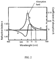
- the medium 3 enveloping the sample 1 may be composed of, or may comprise, an element having dispersion properties, meaning that the index of refraction of the medium incorporating the sample n m is different for different wavelength of the radiation. Therefore, in an unclaimed embodiment n m can be changed by changing the wavelength ⁇ of the radiation illuminating the sample unit 4.
- the medium having dispersion properties can be characterized by an absorption spectrum showing absorption lines or absorption bands.
- the dispersion behavior of such a medium in a wavelength interval near an absorption line or near an absorption band, is characterized by the fact that important refractive index changes can occur even in small wavelength intervals.
- two wavelengths ⁇ 1 and ⁇ 2 can be chosen so that they are located along the decreasing side of an absorption band, where dispersion effects are more pronounced. This enables fast and important modulations of n m .
- the thickness of the sample ( d ), and the index of refraction of the sample n s can be obtained separately or simultaneously, assuming that the different values of n m are known or have been measured previously.
- OPL1 and OPL2 two different OPL measurements performed for two different values of n m , respectively n m1 and n m2 .
- OPL 1 n s ⁇ n m 1 d
- OPL 2 n s ⁇ n m 2 d
- n m Even if two image acquisitions are sufficient, more acquisitions can be performed for different values of n m , or for the same value of n m , for example in order to increase the accuracy of the measurements.
- Different values of n m can be obtained by changing the medium.
- different values of n m can also be obtained by using a medium having dispersion properties that allows to change its index of refraction by changing the wavelength of the radiation illuminating the specimen unit.
- n m n s -OPL/d. More generally, the present document can be used as a technique for refractometry when the geometry of the specimen is known.
- the invention can also be used to obtain a tomography of the sample.
- n(x,y) is the local refractive index which is equal to n m outside the sample and approximately n s with local variations over the volume of the sample.
- OPL( ⁇ ,R) data can be obtained by collecting a plurality of amplitude contrast images 16 and/or a plurality of quantitative phase contrast images 17 can be collected at variable incidence angles ⁇ of the irradiating beam. From the collected data the refractive index n(x,y) of the sample can be computed by applying an algorithm of back-projection.
- the invention can be used to measure a property or a behavior of the medium 3.
- the present invention can be used to study, or to observe, dynamics fluid phenomenon introducing local variations of OPL, flow analysis in particular, for example with flows tracers.
- the present invention can also be used to analyze internal stresses inside the material.
- DHI can be used to image and to measure the polarization sate of the object wave.
- DHI can be used to image and to measure the polarization sate of the object wave.
- the present document can also be used to observe and to analyses processes that influence the polarization of the radiation, and in particular dynamic processes inducing transient modifications of the polarization.
- the medium 3 may have birefringent properties.
- the corresponding plurality of quantitative phase contrast images can be processed, for example by the processing unit 18, in order to compensate temporal variation of the OPL or phase signal, said temporal variations having other origins than the behavior of the sample, for example such temporal variation may be caused by mechanical vibrations of elements composing the apparatus used to generate holograms, and/or may be caused by air turbulences and/or may be caused by thermal effects.
- the present document uses a computer-based method that measures the phase signal in regions of the quantitative phase-contrast images where the sample has no influence on the OPL signal, or in other words in regions where d, or h, or n m have no influence on the measured signal.
- OPL measurements provide a reference signal that monitor the influence of undesirable sources of signal variations.
- the compensation of the influence of these undesirable sources of signal variations can be performed by subtracting the reference signal from the signal measured in other area of the quantitative phase-contrast images, in particular in area where the sample influences the measured signal.
- DHI methods in general, and the present invention appear as a powerful mean for real-time imaging of processes inducing dynamic changes of a sample shape and/or dynamic changes of physical properties of a sample. These dynamic changes may appear in relation to a spontaneous process and/or in relation to an induced process.
- DHI methods can be applied to monitor the response of a sample to external perturbations of different forms, for example chemical perturbations, and/or biological perturbations, and/or electrical perturbations, and/or mechanical perturbations.
- the present invention can be combined with fluorescence microscopy, or with laser scanning confocal microscopy, or with two-photon microscopy, or with second or third harmonic generation microscopy, or with atomic force microscopy.
- the present invention can also be applied in parallel with experimental techniques used in electrophysiology, and/or in fluid mechanics.
- the DHI techniques in general, and the present invention in particular are particularly well adapted for non-invasive analysis, because no contrast agents are required, and because low radiation intensities are used.
- the measured irradiance at the sample plane may be several orders of magnitude lower than the irradiance used for example by other microscopy or imaging techniques. It is therefore an interesting potential application of the present invention that the method can be applied to image delicate sample, and biological sample in particular.
- the sample can be a biological cell, a living biological cell, a culture of biological cell, a mono-layer of cultured biological cells, a tissue composed of biological cell, a biochip, a preparations of proteins.
- a particular aspect of the method according to the invention is characterized by the fact that the medium incorporating the sample can be changed during time and/or that properties of the medium incorporating the sample can be changed during time.
- Such changes of the medium are expected to induce modifications of the sample shape and/or modifications of the dielectric properties of the sample, and said modifications of the sample are monitored by DHI.
- the method of the present invention is particularly well adapted to study the dynamics of fast sample-medium reactions.
- DHI is also a non-invasive imaging technique working without contrast agents and with low radiation intensities
- the method of the present invention is particularly well adapted to study the dynamic of reactions with minimized external perturbations, and during long time periods of hours or even days. This is of particular interest for applications in life sciences.
- Fig. 3 presents an example of application of the present invention to image living cells in culture enclosed in a perfusion chamber 30 filled with a physiological solution 37.
- the cell culture 31 is illuminated by an incident radiation 32 and a microscope objective 33 collects the transmitted radiation to form an object wave 34.
- a CCD camera 35 records the hologram.
- An example of hologram is presented in Fig. 3b .
- the hologram is produced by the interference with a reference wave 36.
- a numerical procedure is then applied to reconstruct the hologram.
- An example of quantitative phase-contrast image is presented in Fig. 3c .
- the obtained phase-contrast image can be presented in 3D perspective, as presented for example in Fig. 3d .
- the present invention has been applied to study the effects of an excitatory neurotransmitter (glutamate) on the morphology of cultured mouse cortical neurons.
- glutamate excitatory neurotransmitter
- the influence of glutamate on neuron morphology has been already described as resulting from excitotoxicity, for high concentrations (20-500 ⁇ M) and long exposures of several minutes.
- NMDA N-methyl-D-aspartate
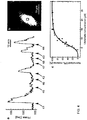
- FIG. 4a presents the temporal evolution of the phase signal measured, in real-time by DHI, in the domain delimited by a black rectangle in Fig. 4b , which presents a typical example of a quantitative phase contrast image of a living mouse cortical neuron immersed in a physiological solution.
- Cells were observed in a control solution (HEPES-buffered physiological solution) and pharmacological agents were applied as short pulses, of 20 seonds duration. These pulses of pharmacological agents are indicated by black triangles in Fig. 4a .
- Marked phase increases occur when glutamate (GLUT) is applied alone, at instants indicated by black triangles 41, 42, 43 and 44.
- the concentrations of GLUT are respectively 15 ⁇ M, 10 ⁇ M, 15 ⁇ M and 20 ⁇ M.
- 400 ⁇ M of kynurenate (KYN) a broad spectrum ionotropic glutamate receptor antagonist, has been added to GLUT. No increase of the phase signal occurs in this case, indicating that the observed effect of GLUT is inhibited by KYN.
- GLUT The effect of GLUT is also inhibited when 5 ⁇ M of MK-801, an NMDA specific receptor antagonist, has been added to GLUT in the physiological solution, at instants indicated by black triangles 47 and 48.
- MK-801 an NMDA specific receptor antagonist
- FIG. 4a the application of glutamate pulses induced reversible OPL or phase increases lasting for several minutes. These OPL increases could be reproduced several times over periods of more than three hours.
- Figure 4a shows also that these glutamate-mediated responses were inhibited by kynurenate, a broad-spectrum ionotropic glutamate receptor antagonist, and by MK-801, a specific NMDA receptor antagonist, thus stressing the receptor specific nature of the effect.
- a concentration-response curve ( Fig.
- glycine is an NMDA receptor co-agonist
- this behavior further emphasizes the role these receptors in the glutamate-evoked responses.
- Figure 4a shows an example of the evolution of the OPL averaged over a specific area of the sample, and gives only a partial idea of the real potential of the method of the present invention.
- DHI the spatial distribution of OPL can be defined, with a transverse resolution of for example 0.3 ⁇ m, and OPL time courses, such as presented in Fig. 4a , can be obtained for different micro-domains of the cell, or for each pixel of the quantitative phase-contrast image.
- the recorded signal can therefore be followed over time and space, for example by constructing animated sequences, for example by using the processing unit 18.
- the high sensitivity of the present invention has revealed that glutamate applied during a few seconds at physiological concentrations induces reversible morphological changes mediated by NMDA receptors, in microdomains of mouse cortical neurons.
- Morphological neuronal changes (following pharmacological medium applications: Glutamate, MK801..) have been monitored during a few hours.
- the whole holographic microscope may be enclosed in a container.
- different noise sources remain (such as coherent noise), extremely sensitive to opto-mechanical instabilities, which may perturb the reconstructed images.
- These residual noise sources render the quantitative analysis of neuronal morphology changes very difficult.
- a device that generates and delivers a controlled energy in the area of the biological sample. This controlled energy may be mechanical vibrations and may be transmitted by perfusion line to the chamber.
- the method of the present invention uses in priority a DHI technique to measure the phase of a radiation, and in particular the method described in patent WO 00/20929 .
- a DHI technique to measure the phase of a radiation
- the present invention is also related to methods using phase measuring techniques such as in-line digital holography, or Fourier-transform digital holography, or digital heterodyne holography, or phase-shifting interferometric techniques.
Landscapes
- Physics & Mathematics (AREA)
- General Physics & Mathematics (AREA)
- Engineering & Computer Science (AREA)
- Computing Systems (AREA)
- Theoretical Computer Science (AREA)
- Investigating Or Analysing Materials By Optical Means (AREA)
- Holo Graphy (AREA)
- Image Processing (AREA)
Priority Applications (1)
| Application Number | Priority Date | Filing Date | Title |
|---|---|---|---|
| EP17173722.4A EP3252538B1 (en) | 2001-12-04 | 2002-12-04 | Apparatus and method for digital holographic imaging |
Applications Claiming Priority (3)
| Application Number | Priority Date | Filing Date | Title |
|---|---|---|---|
| WOPCT/CH01/00701 | 2001-12-04 | ||
| CH0100701 | 2001-12-04 | ||
| PCT/CH2002/000660 WO2003048868A1 (en) | 2001-12-04 | 2002-12-04 | Apparatus and method for digital holographic imaging |
Related Child Applications (3)
| Application Number | Title | Priority Date | Filing Date |
|---|---|---|---|
| EP17173722.4A Division-Into EP3252538B1 (en) | 2001-12-04 | 2002-12-04 | Apparatus and method for digital holographic imaging |
| EP17173722.4A Division EP3252538B1 (en) | 2001-12-04 | 2002-12-04 | Apparatus and method for digital holographic imaging |
| EP17166423.8 Division-Into | 2017-04-13 |
Publications (2)
| Publication Number | Publication Date |
|---|---|
| EP1451646A1 EP1451646A1 (en) | 2004-09-01 |
| EP1451646B1 true EP1451646B1 (en) | 2018-09-19 |
Family
ID=4358268
Family Applications (2)
| Application Number | Title | Priority Date | Filing Date |
|---|---|---|---|
| EP02804141.6A Expired - Lifetime EP1451646B1 (en) | 2001-12-04 | 2002-12-04 | Apparatus and method for digital holographic imaging |
| EP17173722.4A Expired - Lifetime EP3252538B1 (en) | 2001-12-04 | 2002-12-04 | Apparatus and method for digital holographic imaging |
Family Applications After (1)
| Application Number | Title | Priority Date | Filing Date |
|---|---|---|---|
| EP17173722.4A Expired - Lifetime EP3252538B1 (en) | 2001-12-04 | 2002-12-04 | Apparatus and method for digital holographic imaging |
Country Status (5)
| Country | Link |
|---|---|
| US (1) | US6943924B2 (cg-RX-API-DMAC7.html) |
| EP (2) | EP1451646B1 (cg-RX-API-DMAC7.html) |
| JP (1) | JP4358631B2 (cg-RX-API-DMAC7.html) |
| AU (1) | AU2002365676A1 (cg-RX-API-DMAC7.html) |
| WO (1) | WO2003048868A1 (cg-RX-API-DMAC7.html) |
Families Citing this family (48)
| Publication number | Priority date | Publication date | Assignee | Title |
|---|---|---|---|---|
| US8438927B2 (en) * | 2003-08-12 | 2013-05-14 | Northwestern University | Scanning near field thermoelastic acoustic holography (SNFTAH) |
| US7448269B2 (en) * | 2003-08-12 | 2008-11-11 | Northwestern University | Scanning near field ultrasound holography |
| US7119905B2 (en) * | 2003-08-26 | 2006-10-10 | Ut-Battelle Llc | Spatial-heterodyne interferometry for transmission (SHIFT) measurements |
| US6999178B2 (en) | 2003-08-26 | 2006-02-14 | Ut-Battelle Llc | Spatial-heterodyne interferometry for reflection and transmission (SHIRT) measurements |
| SE0302676D0 (sv) * | 2003-10-09 | 2003-10-09 | Neural Ab | Method and apparatus for holographic refractometry |
| JP4698992B2 (ja) * | 2004-09-14 | 2011-06-08 | 浜松ホトニクス株式会社 | 試料計測装置及び計測方法 |
| JP2007076842A (ja) * | 2005-09-15 | 2007-03-29 | Brother Ind Ltd | 原稿保持トレイ、原稿搬送装置、および画像読取装置 |
| CN101317138B (zh) * | 2005-10-06 | 2011-05-04 | 西北大学 | 扫描近场超声波全息方法及系统 |
| US7948632B2 (en) * | 2005-12-22 | 2011-05-24 | Phase Holographic Imaging Phi Ab | Method and apparatus for analysis of a sample of cells |
| CN102866494A (zh) * | 2007-01-26 | 2013-01-09 | 纽约大学 | 被全息捕获三维结构的全息显微术 |
| US8194124B2 (en) * | 2007-10-09 | 2012-06-05 | Nanyang Technological University | In-line digital holographic microscope and a method of in-line digital holographic microscopy |
| EP2206021B1 (en) | 2007-10-30 | 2015-01-28 | New York University | Tracking and characterizing particles with holographic video microscopy |
| JP5022274B2 (ja) * | 2008-03-06 | 2012-09-12 | 浜松ホトニクス株式会社 | 観察装置 |
| US7889413B1 (en) * | 2008-06-12 | 2011-02-15 | The Unites States Of America As Represented By The Secretary Of The Air Force | Field generating nanoparticles doped in light valves |
| US20110157601A1 (en) * | 2008-06-19 | 2011-06-30 | Mikael Carl Sebesta | Analysis of transparent biological objects |
| JP5168693B2 (ja) * | 2008-09-30 | 2013-03-21 | 株式会社 エフケー光学研究所 | 試料の高さの計測方法 |
| JP5269207B2 (ja) * | 2009-01-16 | 2013-08-21 | ニューヨーク ユニバーシティー | ホログラフィビデオ顕微鏡観察を伴う自動的リアルタイム粒子特徴付け及び三次元速度測定 |
| EP2401601B1 (en) | 2009-02-24 | 2021-09-22 | Lyncee Tec S.A. | Monitoring energy and matter fluxes by use of electromagnetic radiations |
| US8264694B2 (en) * | 2009-03-16 | 2012-09-11 | Ut-Battelle, Llc | Quantitative phase-contrast and excitation-emission systems |
| US8248614B2 (en) * | 2009-03-16 | 2012-08-21 | Ut-Battelle, Llc | Quantitative phase-imaging systems |
| US7978336B2 (en) * | 2009-03-16 | 2011-07-12 | Ut-Battelle, Llc | Three wavelength quantitative imaging systems |
| EP2446251B1 (en) | 2009-06-25 | 2018-12-05 | Phase Holographic Imaging Phi AB | Analysis of ova or embryos with digital holographic imaging |
| WO2011099925A1 (en) * | 2010-02-09 | 2011-08-18 | Phase Holographic Imaging Phi Ab | Method for and use of digital holographic microscopy and imaging on labelled cell samples |
| EP2576759A4 (en) | 2010-05-25 | 2014-01-22 | Arryx Inc | METHODS AND APPARATUSES FOR DETECTING FREEDOM OF POSITION OF PARTICLES IN BIOLOGICAL AND CHEMICAL ANALYZES AND APPLICATION IN IMMUNODIAGNOSTICS |
| US8354651B2 (en) | 2010-06-30 | 2013-01-15 | Ut-Battelle, Llc | Positron emission imaging device and method of using the same |
| US9360468B2 (en) * | 2010-08-20 | 2016-06-07 | Lyncee Tec S.A. | Method for monitoring cell viability |
| CN103238120B (zh) * | 2010-11-12 | 2016-08-10 | 布鲁塞尔大学 | 用于表征透明颗粒的光学方法 |
| WO2013010595A1 (en) * | 2011-07-19 | 2013-01-24 | Ovizio Imaging Systems N.V. | An object database and object database improving method |
| EP2594334A1 (en) | 2011-11-21 | 2013-05-22 | Drive O2 | Sample vial for digital holographic analysis of a liquid cell sample |
| KR20160103175A (ko) | 2011-07-19 | 2016-08-31 | 오비지오 이미징 시스템스 엔.브이. | 세포 샘플에서의 암 세포들의 검출 및/또는 분류를 위한 방법 및 시스템 |
| WO2013019640A1 (en) * | 2011-07-29 | 2013-02-07 | The Regents Of The University Of California | Lensfree holographic microscopy using wetting films |
| EP2626686A1 (en) | 2012-02-13 | 2013-08-14 | Ovizio Imaging Systems NV/SA | Flow cytometer with digital holographic microscope |
| EP2898310B1 (en) | 2012-09-20 | 2019-05-01 | Ovizio Imaging Systems NV/SA | Digital holographic microscope with fluid systems |
| ES2534960B1 (es) | 2013-10-30 | 2016-02-09 | Universitat De València | Microscopio, método y programa de ordenador para la obtención de imágenes cuantitativas de fase por medio de microscopía holográfica digital, y kit para adaptar un microscopio óptico |
| KR102425768B1 (ko) | 2014-02-12 | 2022-07-26 | 뉴욕 유니버시티 | 콜로이드 입자의 홀로그래픽 추적 및 특징화를 위한 고속 특징부 식별 |
| DK3218690T3 (da) | 2014-11-12 | 2022-05-02 | Univ New York | Kolloidt fingeraftryk til bløde materialer med brug af total holografisk karakterisation |
| JP6929560B2 (ja) | 2015-09-18 | 2021-09-01 | ニュー・ヨーク・ユニヴァーシティー | 精密スラリ内の大きい不純物粒子のホログラフィック検出及び特徴付け |
| EP3196631A1 (en) | 2016-01-19 | 2017-07-26 | Ovizio Imaging Systems NV/SA | Digital holographic microscope with electro-fluidic system, said electro-fluidic system and methods of use |
| ES2901608T3 (es) | 2016-02-08 | 2022-03-23 | Univ New York | Caracterización holográfica de agregados proteicos |
| US10670677B2 (en) | 2016-04-22 | 2020-06-02 | New York University | Multi-slice acceleration for magnetic resonance fingerprinting |
| JP6894620B2 (ja) * | 2017-02-09 | 2021-06-30 | 国立大学法人千葉大学 | 物理厚さ推定プログラム及び屈折率データ推定プログラム、並びに、物理厚さデータ推定システム及び屈折率データ推定システム |
| KR102412253B1 (ko) * | 2018-11-30 | 2022-06-24 | 한국전자통신연구원 | 미생물 분석 장치 및 방법 |
| CA3127700A1 (en) | 2019-02-01 | 2020-08-06 | Universite Laval | System and method for determining a refractive index of a medium |
| US11543338B2 (en) | 2019-10-25 | 2023-01-03 | New York University | Holographic characterization of irregular particles |
| US11948302B2 (en) | 2020-03-09 | 2024-04-02 | New York University | Automated holographic video microscopy assay |
| US12625358B2 (en) * | 2020-07-10 | 2026-05-12 | Sony Group Corporation | Method comprising determining a quantitative dispersion image of an object and digital in-line hologram microscope scanner |
| TR202019536A2 (tr) * | 2020-12-02 | 2022-06-21 | Yildiz Teknik Ueniversitesi Doener Sermaye Isletme Mued | Hücre ve mikro organizmaların ışık geçirgenliğine göre ayırabilen elektro-holografik mikroskop sistemi. |
| CN120490015B (zh) * | 2025-07-16 | 2025-09-12 | 中北大学 | 一种基于类全息条纹的气体折射率测量装置 |
Citations (1)
| Publication number | Priority date | Publication date | Assignee | Title |
|---|---|---|---|---|
| US4195131A (en) * | 1977-03-09 | 1980-03-25 | Papas Gary R | Environmentally controlled unit |
Family Cites Families (6)
| Publication number | Priority date | Publication date | Assignee | Title |
|---|---|---|---|---|
| US6249349B1 (en) * | 1996-09-27 | 2001-06-19 | Vincent Lauer | Microscope generating a three-dimensional representation of an object |
| US6262818B1 (en) | 1998-10-07 | 2001-07-17 | Institute Of Applied Optics, Swiss Federal Institute Of Technology | Method for simultaneous amplitude and quantitative phase contrast imaging by numerical reconstruction of digital holograms |
| FR2792082B1 (fr) * | 1999-04-06 | 2003-05-30 | Thomson Csf | Dispositif d'holographie numerique |
| US6022943A (en) | 1999-04-07 | 2000-02-08 | General Electric Company | Optical quality polycarbonates with reduced static charge and method for making same |
| CA2724743C (fr) * | 2001-06-29 | 2014-11-25 | Universite Libre De Bruxelles | Procede et dispositif destines a l'obtention par microscopie d'images en trois dimensions d'un echantillon |
| CN1310045C (zh) * | 2002-10-01 | 2007-04-11 | 古河电气工业株式会社 | 光纤、光传送线路以及光纤的制造方法 |
-
2002
- 2002-12-04 AU AU2002365676A patent/AU2002365676A1/en not_active Abandoned
- 2002-12-04 WO PCT/CH2002/000660 patent/WO2003048868A1/en not_active Ceased
- 2002-12-04 JP JP2003549995A patent/JP4358631B2/ja not_active Expired - Lifetime
- 2002-12-04 EP EP02804141.6A patent/EP1451646B1/en not_active Expired - Lifetime
- 2002-12-04 US US10/497,312 patent/US6943924B2/en not_active Expired - Lifetime
- 2002-12-04 EP EP17173722.4A patent/EP3252538B1/en not_active Expired - Lifetime
Patent Citations (1)
| Publication number | Priority date | Publication date | Assignee | Title |
|---|---|---|---|---|
| US4195131A (en) * | 1977-03-09 | 1980-03-25 | Papas Gary R | Environmentally controlled unit |
Non-Patent Citations (3)
| Title |
|---|
| ETIENNE CUCHE ET AL: "Quantitative phase contrast microscopy of living cells by numerical reconstruction of digital holograms", PROCEEDINGS VOLUME 9025 IS&T/SPIE ELECTRONIC IMAGING | 2-6 FEBRUARY 2014 INTELLIGENT ROBOTS AND COMPUTER VISION XXXI: ALGORITHMS AND TECHNIQUES, vol. 3604, 1 June 1999 (1999-06-01), US, pages 84 - 89, XP055414275, ISSN: 0277-786X, ISBN: 978-1-5106-1354-6, DOI: 10.1117/12.349218 * |
| LIEBLING M ET AL: "A novel non-diffractive reconstruction method for digital holographic microscopy", BIOMEDICAL IMAGING, 2002. PROCEEDINGS. 2002 IEEE INTERNATIONAL SYMPOSI UM ON JULY 7-10, 2002, PISCATAWAY, NJ, USA,IEEE, 7 July 2002 (2002-07-07), pages 625 - 628, XP010600666, ISBN: 978-0-7803-7584-0 * |
| NEWMAN J W: "OPTICAL LEAK TESTING OF HERMETIC PACKAGES", PROCEEDINGS OF SPIE, S P I E - INTERNATIONAL SOCIETY FOR OPTICAL ENGINEERING, US, vol. 4587, 9 January 2001 (2001-01-09), pages 645 - 650, XP009054273, ISSN: 0277-786X, ISBN: 0-8194-0820-4 * |
Also Published As
| Publication number | Publication date |
|---|---|
| US20050036181A1 (en) | 2005-02-17 |
| AU2002365676A1 (en) | 2003-06-17 |
| EP3252538B1 (en) | 2019-02-06 |
| US6943924B2 (en) | 2005-09-13 |
| WO2003048868A1 (en) | 2003-06-12 |
| EP1451646A1 (en) | 2004-09-01 |
| EP3252538A1 (en) | 2017-12-06 |
| JP4358631B2 (ja) | 2009-11-04 |
| JP2005512127A (ja) | 2005-04-28 |
Similar Documents
| Publication | Publication Date | Title |
|---|---|---|
| EP3252538B1 (en) | Apparatus and method for digital holographic imaging | |
| TWI797377B (zh) | 表面形狀量測裝置以及表面形狀量測方法 | |
| Kim | Principles and techniques of digital holographic microscopy | |
| US6262818B1 (en) | Method for simultaneous amplitude and quantitative phase contrast imaging by numerical reconstruction of digital holograms | |
| US10113961B2 (en) | Apparatus and method for quantitive phase tomography through linear scanning with coherent and non-coherent detection | |
| Kim | Applications of digital holography in biomedical microscopy | |
| US7839551B2 (en) | Holographic microscopy of holographically trapped three-dimensional structures | |
| US7880891B1 (en) | Total internal reflection holographic microscope | |
| US7127109B1 (en) | Digital interference holographic microscope and methods | |
| EP1683074A1 (en) | Digital imaging assembly and methods thereof | |
| Kemper et al. | Holographic interferometric microscopy systems for the application on biological samples | |
| Rybnikov et al. | Using digital off-axis holograms to investigate changes of state of living neuronal cultures | |
| US20230324275A1 (en) | A simple in-line digital holography system for measuring 3d cell shape | |
| Creath | Dynamic quantitative phase images of pond life, insect wings, and in vitro cell cultures | |
| Coupland et al. | Holographic particle image velocimetry and its application in engine development | |
| Peruhov et al. | Applications of holographic microscopy in life sciences | |
| Vertu et al. | Diffraction microtomography with sample rotation: Primary result on the influence of a missing apple core in the recorded frequency space | |
| Bergoënd et al. | Diffraction tomography for biological cells imaging using digital holographic microscopy | |
| Lokberg | Speckles and speckle techniques for biomedical applications | |
| Xiong et al. | Optical diffraction tomography for 3D refractive index imaging: Techniques and applications | |
| Kim et al. | Optical diffraction tomography for simultaneous 3D visualization and tracking of optically trapped particles | |
| Shaked et al. | Quantitative phase microscopy of biological cell dynamics by wide-field digital interferometry | |
| Pavillon et al. | Optical tomography by digital holographic microscopy | |
| Charrière et al. | Use of digital holographic microscopy in tomography | |
| Ash III et al. | Total internal reflection holographic microscopy for quantitative phase characterization of cellular adhesion |
Legal Events
| Date | Code | Title | Description |
|---|---|---|---|
| PUAI | Public reference made under article 153(3) epc to a published international application that has entered the european phase |
Free format text: ORIGINAL CODE: 0009012 |
|
| 17P | Request for examination filed |
Effective date: 20040601 |
|
| AK | Designated contracting states |
Kind code of ref document: A1 Designated state(s): AT BE BG CH CY CZ DE DK EE ES FI FR GB GR IE IT LI LU MC NL PT SE SI SK TR |
|
| AX | Request for extension of the european patent |
Extension state: AL LT LV MK RO |
|
| 17Q | First examination report despatched |
Effective date: 20040901 |
|
| RIN1 | Information on inventor provided before grant (corrected) |
Inventor name: MARQUET, PIERRE Inventor name: CUCHE, ETIENNE Inventor name: DEPEURSINGE, CHRISTIAN Inventor name: MAGISTRETTI, PIERRE |
|
| GRAP | Despatch of communication of intention to grant a patent |
Free format text: ORIGINAL CODE: EPIDOSNIGR1 |
|
| RIC1 | Information provided on ipc code assigned before grant |
Ipc: G03H 1/04 19740701ALI20180328BHEP Ipc: G03H 1/08 19740701AFI20180328BHEP |
|
| INTG | Intention to grant announced |
Effective date: 20180412 |
|
| GRAS | Grant fee paid |
Free format text: ORIGINAL CODE: EPIDOSNIGR3 |
|
| GRAA | (expected) grant |
Free format text: ORIGINAL CODE: 0009210 |
|
| AK | Designated contracting states |
Kind code of ref document: B1 Designated state(s): AT BE BG CH CY CZ DE DK EE ES FI FR GB GR IE IT LI LU MC NL PT SE SI SK TR |
|
| REG | Reference to a national code |
Ref country code: GB Ref legal event code: FG4D |
|
| REG | Reference to a national code |
Ref country code: CH Ref legal event code: EP |
|
| REG | Reference to a national code |
Ref country code: AT Ref legal event code: REF Ref document number: 1043952 Country of ref document: AT Kind code of ref document: T Effective date: 20181015 |
|
| REG | Reference to a national code |
Ref country code: IE Ref legal event code: FG4D |
|
| REG | Reference to a national code |
Ref country code: DE Ref legal event code: R096 Ref document number: 60249613 Country of ref document: DE |
|
| REG | Reference to a national code |
Ref country code: CH Ref legal event code: NV Representative=s name: ANDRE ROLAND S.A., CH |
|
| REG | Reference to a national code |
Ref country code: NL Ref legal event code: FP |
|
| REG | Reference to a national code |
Ref country code: SE Ref legal event code: TRGR |
|
| PG25 | Lapsed in a contracting state [announced via postgrant information from national office to epo] |
Ref country code: FI Free format text: LAPSE BECAUSE OF FAILURE TO SUBMIT A TRANSLATION OF THE DESCRIPTION OR TO PAY THE FEE WITHIN THE PRESCRIBED TIME-LIMIT Effective date: 20180919 Ref country code: BG Free format text: LAPSE BECAUSE OF FAILURE TO SUBMIT A TRANSLATION OF THE DESCRIPTION OR TO PAY THE FEE WITHIN THE PRESCRIBED TIME-LIMIT Effective date: 20181219 Ref country code: GR Free format text: LAPSE BECAUSE OF FAILURE TO SUBMIT A TRANSLATION OF THE DESCRIPTION OR TO PAY THE FEE WITHIN THE PRESCRIBED TIME-LIMIT Effective date: 20181220 |
|
| REG | Reference to a national code |
Ref country code: CH Ref legal event code: PK Free format text: BERICHTIGUNGEN |
|
| RIC2 | Information provided on ipc code assigned after grant |
Ipc: G03H 1/08 20060101AFI20180328BHEP Ipc: G03H 1/04 20060101ALI20180328BHEP |
|
| REG | Reference to a national code |
Ref country code: AT Ref legal event code: MK05 Ref document number: 1043952 Country of ref document: AT Kind code of ref document: T Effective date: 20180919 |
|
| PG25 | Lapsed in a contracting state [announced via postgrant information from national office to epo] |
Ref country code: ES Free format text: LAPSE BECAUSE OF FAILURE TO SUBMIT A TRANSLATION OF THE DESCRIPTION OR TO PAY THE FEE WITHIN THE PRESCRIBED TIME-LIMIT Effective date: 20180919 Ref country code: CZ Free format text: LAPSE BECAUSE OF FAILURE TO SUBMIT A TRANSLATION OF THE DESCRIPTION OR TO PAY THE FEE WITHIN THE PRESCRIBED TIME-LIMIT Effective date: 20180919 Ref country code: AT Free format text: LAPSE BECAUSE OF FAILURE TO SUBMIT A TRANSLATION OF THE DESCRIPTION OR TO PAY THE FEE WITHIN THE PRESCRIBED TIME-LIMIT Effective date: 20180919 Ref country code: EE Free format text: LAPSE BECAUSE OF FAILURE TO SUBMIT A TRANSLATION OF THE DESCRIPTION OR TO PAY THE FEE WITHIN THE PRESCRIBED TIME-LIMIT Effective date: 20180919 Ref country code: IT Free format text: LAPSE BECAUSE OF FAILURE TO SUBMIT A TRANSLATION OF THE DESCRIPTION OR TO PAY THE FEE WITHIN THE PRESCRIBED TIME-LIMIT Effective date: 20180919 |
|
| PG25 | Lapsed in a contracting state [announced via postgrant information from national office to epo] |
Ref country code: SK Free format text: LAPSE BECAUSE OF FAILURE TO SUBMIT A TRANSLATION OF THE DESCRIPTION OR TO PAY THE FEE WITHIN THE PRESCRIBED TIME-LIMIT Effective date: 20180919 Ref country code: PT Free format text: LAPSE BECAUSE OF FAILURE TO SUBMIT A TRANSLATION OF THE DESCRIPTION OR TO PAY THE FEE WITHIN THE PRESCRIBED TIME-LIMIT Effective date: 20190119 |
|
| REG | Reference to a national code |
Ref country code: DE Ref legal event code: R097 Ref document number: 60249613 Country of ref document: DE |
|
| PLBE | No opposition filed within time limit |
Free format text: ORIGINAL CODE: 0009261 |
|
| STAA | Information on the status of an ep patent application or granted ep patent |
Free format text: STATUS: NO OPPOSITION FILED WITHIN TIME LIMIT |
|
| PG25 | Lapsed in a contracting state [announced via postgrant information from national office to epo] |
Ref country code: DK Free format text: LAPSE BECAUSE OF FAILURE TO SUBMIT A TRANSLATION OF THE DESCRIPTION OR TO PAY THE FEE WITHIN THE PRESCRIBED TIME-LIMIT Effective date: 20180919 |
|
| 26N | No opposition filed |
Effective date: 20190620 |
|
| PG25 | Lapsed in a contracting state [announced via postgrant information from national office to epo] |
Ref country code: LU Free format text: LAPSE BECAUSE OF NON-PAYMENT OF DUE FEES Effective date: 20181204 Ref country code: MC Free format text: LAPSE BECAUSE OF FAILURE TO SUBMIT A TRANSLATION OF THE DESCRIPTION OR TO PAY THE FEE WITHIN THE PRESCRIBED TIME-LIMIT Effective date: 20180919 |
|
| REG | Reference to a national code |
Ref country code: IE Ref legal event code: MM4A |
|
| PG25 | Lapsed in a contracting state [announced via postgrant information from national office to epo] |
Ref country code: SI Free format text: LAPSE BECAUSE OF FAILURE TO SUBMIT A TRANSLATION OF THE DESCRIPTION OR TO PAY THE FEE WITHIN THE PRESCRIBED TIME-LIMIT Effective date: 20180919 Ref country code: IE Free format text: LAPSE BECAUSE OF NON-PAYMENT OF DUE FEES Effective date: 20181204 |
|
| PG25 | Lapsed in a contracting state [announced via postgrant information from national office to epo] |
Ref country code: TR Free format text: LAPSE BECAUSE OF FAILURE TO SUBMIT A TRANSLATION OF THE DESCRIPTION OR TO PAY THE FEE WITHIN THE PRESCRIBED TIME-LIMIT Effective date: 20180919 |
|
| PG25 | Lapsed in a contracting state [announced via postgrant information from national office to epo] |
Ref country code: CY Free format text: LAPSE BECAUSE OF FAILURE TO SUBMIT A TRANSLATION OF THE DESCRIPTION OR TO PAY THE FEE WITHIN THE PRESCRIBED TIME-LIMIT Effective date: 20180919 |
|
| PGFP | Annual fee paid to national office [announced via postgrant information from national office to epo] |
Ref country code: SE Payment date: 20211221 Year of fee payment: 20 Ref country code: DE Payment date: 20211210 Year of fee payment: 20 Ref country code: FR Payment date: 20211224 Year of fee payment: 20 Ref country code: GB Payment date: 20211221 Year of fee payment: 20 |
|
| PGFP | Annual fee paid to national office [announced via postgrant information from national office to epo] |
Ref country code: NL Payment date: 20211221 Year of fee payment: 20 Ref country code: CH Payment date: 20211221 Year of fee payment: 20 Ref country code: BE Payment date: 20211221 Year of fee payment: 20 |
|
| REG | Reference to a national code |
Ref country code: DE Ref legal event code: R071 Ref document number: 60249613 Country of ref document: DE |
|
| REG | Reference to a national code |
Ref country code: NL Ref legal event code: MK Effective date: 20221203 |
|
| REG | Reference to a national code |
Ref country code: CH Ref legal event code: PL |
|
| REG | Reference to a national code |
Ref country code: GB Ref legal event code: PE20 Expiry date: 20221203 |
|
| REG | Reference to a national code |
Ref country code: BE Ref legal event code: MK Effective date: 20221204 |
|
| PG25 | Lapsed in a contracting state [announced via postgrant information from national office to epo] |
Ref country code: GB Free format text: LAPSE BECAUSE OF EXPIRATION OF PROTECTION Effective date: 20221203 |
|
| REG | Reference to a national code |
Ref country code: SE Ref legal event code: EUG |