EP1435833B1 - Appareil de diagnostic endobronchique - Google Patents
Appareil de diagnostic endobronchique Download PDFInfo
- Publication number
- EP1435833B1 EP1435833B1 EP02768834.0A EP02768834A EP1435833B1 EP 1435833 B1 EP1435833 B1 EP 1435833B1 EP 02768834 A EP02768834 A EP 02768834A EP 1435833 B1 EP1435833 B1 EP 1435833B1
- Authority
- EP
- European Patent Office
- Prior art keywords
- lung
- compartment
- catheter
- measurement data
- sensor
- Prior art date
- Legal status (The legal status is an assumption and is not a legal conclusion. Google has not performed a legal analysis and makes no representation as to the accuracy of the status listed.)
- Expired - Lifetime
Links
- 238000003745 diagnosis Methods 0.000 title description 6
- 210000004072 lung Anatomy 0.000 claims description 241
- 230000002685 pulmonary effect Effects 0.000 claims description 106
- 238000005259 measurement Methods 0.000 claims description 72
- 230000007246 mechanism Effects 0.000 claims description 72
- 239000007789 gas Substances 0.000 claims description 68
- 238000003384 imaging method Methods 0.000 claims description 51
- 238000012360 testing method Methods 0.000 claims description 44
- 201000010099 disease Diseases 0.000 claims description 38
- 208000037265 diseases, disorders, signs and symptoms Diseases 0.000 claims description 38
- 230000000007 visual effect Effects 0.000 claims description 30
- 238000000034 method Methods 0.000 claims description 28
- 239000012530 fluid Substances 0.000 claims description 21
- 238000012545 processing Methods 0.000 claims description 17
- 238000013507 mapping Methods 0.000 claims description 14
- 238000010790 dilution Methods 0.000 claims description 12
- 239000012895 dilution Substances 0.000 claims description 12
- QVGXLLKOCUKJST-UHFFFAOYSA-N atomic oxygen Chemical compound [O] QVGXLLKOCUKJST-UHFFFAOYSA-N 0.000 claims description 11
- 239000001301 oxygen Substances 0.000 claims description 11
- 229910052760 oxygen Inorganic materials 0.000 claims description 11
- 229910052756 noble gas Inorganic materials 0.000 claims description 10
- 230000033001 locomotion Effects 0.000 claims description 6
- 230000003287 optical effect Effects 0.000 claims description 6
- 238000012546 transfer Methods 0.000 claims description 6
- 230000003434 inspiratory effect Effects 0.000 claims description 5
- 230000000241 respiratory effect Effects 0.000 claims description 4
- 238000004891 communication Methods 0.000 claims description 3
- 230000008569 process Effects 0.000 claims description 3
- 239000000126 substance Substances 0.000 claims description 3
- 239000012528 membrane Substances 0.000 claims description 2
- 238000006552 photochemical reaction Methods 0.000 claims description 2
- 238000002834 transmittance Methods 0.000 claims description 2
- 238000011282 treatment Methods 0.000 description 55
- 210000001519 tissue Anatomy 0.000 description 20
- 230000000694 effects Effects 0.000 description 16
- CURLTUGMZLYLDI-UHFFFAOYSA-N Carbon dioxide Chemical compound O=C=O CURLTUGMZLYLDI-UHFFFAOYSA-N 0.000 description 12
- 230000006870 function Effects 0.000 description 12
- 210000003484 anatomy Anatomy 0.000 description 10
- 238000002405 diagnostic procedure Methods 0.000 description 10
- 239000000835 fiber Substances 0.000 description 9
- 210000000621 bronchi Anatomy 0.000 description 8
- 238000011038 discontinuous diafiltration by volume reduction Methods 0.000 description 8
- 229910002092 carbon dioxide Inorganic materials 0.000 description 7
- 239000007788 liquid Substances 0.000 description 7
- 238000002663 nebulization Methods 0.000 description 7
- 238000002591 computed tomography Methods 0.000 description 6
- 238000002955 isolation Methods 0.000 description 6
- 230000009467 reduction Effects 0.000 description 6
- 230000029058 respiratory gaseous exchange Effects 0.000 description 6
- 239000001569 carbon dioxide Substances 0.000 description 5
- 230000007423 decrease Effects 0.000 description 5
- 239000003814 drug Substances 0.000 description 4
- 238000002001 electrophysiology Methods 0.000 description 4
- 230000007831 electrophysiology Effects 0.000 description 4
- 238000011156 evaluation Methods 0.000 description 4
- 239000000463 material Substances 0.000 description 4
- 238000002156 mixing Methods 0.000 description 4
- 230000010412 perfusion Effects 0.000 description 4
- 210000002345 respiratory system Anatomy 0.000 description 4
- 210000003437 trachea Anatomy 0.000 description 4
- 238000009423 ventilation Methods 0.000 description 4
- 239000011165 3D composite Substances 0.000 description 3
- 208000002352 blister Diseases 0.000 description 3
- 238000001125 extrusion Methods 0.000 description 3
- 238000002594 fluoroscopy Methods 0.000 description 3
- 230000036541 health Effects 0.000 description 3
- 239000001307 helium Substances 0.000 description 3
- 229910052734 helium Inorganic materials 0.000 description 3
- SWQJXJOGLNCZEY-UHFFFAOYSA-N helium atom Chemical compound [He] SWQJXJOGLNCZEY-UHFFFAOYSA-N 0.000 description 3
- 238000012634 optical imaging Methods 0.000 description 3
- 230000009325 pulmonary function Effects 0.000 description 3
- 238000013125 spirometry Methods 0.000 description 3
- 238000002604 ultrasonography Methods 0.000 description 3
- 238000012800 visualization Methods 0.000 description 3
- 208000006545 Chronic Obstructive Pulmonary Disease Diseases 0.000 description 2
- 206010014561 Emphysema Diseases 0.000 description 2
- 208000019693 Lung disease Diseases 0.000 description 2
- 239000008280 blood Substances 0.000 description 2
- 210000004369 blood Anatomy 0.000 description 2
- 206010006451 bronchitis Diseases 0.000 description 2
- 230000015556 catabolic process Effects 0.000 description 2
- 230000008859 change Effects 0.000 description 2
- 238000006243 chemical reaction Methods 0.000 description 2
- 238000013170 computed tomography imaging Methods 0.000 description 2
- 238000010276 construction Methods 0.000 description 2
- 238000002059 diagnostic imaging Methods 0.000 description 2
- 230000026058 directional locomotion Effects 0.000 description 2
- 238000005286 illumination Methods 0.000 description 2
- 229920001684 low density polyethylene Polymers 0.000 description 2
- 238000012544 monitoring process Methods 0.000 description 2
- 238000004806 packaging method and process Methods 0.000 description 2
- 230000035479 physiological effects, processes and functions Effects 0.000 description 2
- 238000002600 positron emission tomography Methods 0.000 description 2
- 230000004044 response Effects 0.000 description 2
- 230000000717 retained effect Effects 0.000 description 2
- 238000007789 sealing Methods 0.000 description 2
- 238000001356 surgical procedure Methods 0.000 description 2
- 238000002560 therapeutic procedure Methods 0.000 description 2
- 206010001052 Acute respiratory distress syndrome Diseases 0.000 description 1
- 206010003598 Atelectasis Diseases 0.000 description 1
- 206010006458 Bronchitis chronic Diseases 0.000 description 1
- 102000008186 Collagen Human genes 0.000 description 1
- 108010035532 Collagen Proteins 0.000 description 1
- 206010061218 Inflammation Diseases 0.000 description 1
- 206010030113 Oedema Diseases 0.000 description 1
- 239000004696 Poly ether ether ketone Substances 0.000 description 1
- 239000004952 Polyamide Substances 0.000 description 1
- 208000007123 Pulmonary Atelectasis Diseases 0.000 description 1
- 208000013616 Respiratory Distress Syndrome Diseases 0.000 description 1
- 238000013473 artificial intelligence Methods 0.000 description 1
- 208000006673 asthma Diseases 0.000 description 1
- 230000009286 beneficial effect Effects 0.000 description 1
- 230000008901 benefit Effects 0.000 description 1
- JUPQTSLXMOCDHR-UHFFFAOYSA-N benzene-1,4-diol;bis(4-fluorophenyl)methanone Chemical compound OC1=CC=C(O)C=C1.C1=CC(F)=CC=C1C(=O)C1=CC=C(F)C=C1 JUPQTSLXMOCDHR-UHFFFAOYSA-N 0.000 description 1
- 238000004364 calculation method Methods 0.000 description 1
- 208000007451 chronic bronchitis Diseases 0.000 description 1
- 229920001436 collagen Polymers 0.000 description 1
- 239000002131 composite material Substances 0.000 description 1
- 239000002872 contrast media Substances 0.000 description 1
- 229940039231 contrast media Drugs 0.000 description 1
- 230000001419 dependent effect Effects 0.000 description 1
- 238000013461 design Methods 0.000 description 1
- 238000007435 diagnostic evaluation Methods 0.000 description 1
- 239000003172 expectorant agent Substances 0.000 description 1
- 238000011990 functional testing Methods 0.000 description 1
- 230000004927 fusion Effects 0.000 description 1
- 238000010438 heat treatment Methods 0.000 description 1
- 229920001903 high density polyethylene Polymers 0.000 description 1
- 208000015181 infectious disease Diseases 0.000 description 1
- 230000004054 inflammatory process Effects 0.000 description 1
- 230000002262 irrigation Effects 0.000 description 1
- 238000003973 irrigation Methods 0.000 description 1
- 229920000126 latex Polymers 0.000 description 1
- 239000004816 latex Substances 0.000 description 1
- 239000004702 low-density polyethylene Substances 0.000 description 1
- 230000004199 lung function Effects 0.000 description 1
- 238000000691 measurement method Methods 0.000 description 1
- 238000012986 modification Methods 0.000 description 1
- 230000004048 modification Effects 0.000 description 1
- 229940066491 mucolytics Drugs 0.000 description 1
- 230000004118 muscle contraction Effects 0.000 description 1
- 229920001778 nylon Polymers 0.000 description 1
- 239000013307 optical fiber Substances 0.000 description 1
- 230000003534 oscillatory effect Effects 0.000 description 1
- 230000036284 oxygen consumption Effects 0.000 description 1
- 238000006213 oxygenation reaction Methods 0.000 description 1
- 229920002647 polyamide Polymers 0.000 description 1
- 229920002530 polyetherether ketone Polymers 0.000 description 1
- 229920001343 polytetrafluoroethylene Polymers 0.000 description 1
- 239000004810 polytetrafluoroethylene Substances 0.000 description 1
- 229920002635 polyurethane Polymers 0.000 description 1
- 239000004814 polyurethane Substances 0.000 description 1
- 229920000915 polyvinyl chloride Polymers 0.000 description 1
- 239000004800 polyvinyl chloride Substances 0.000 description 1
- 238000002271 resection Methods 0.000 description 1
- 229920002379 silicone rubber Polymers 0.000 description 1
- 239000004945 silicone rubber Substances 0.000 description 1
- 238000002603 single-photon emission computed tomography Methods 0.000 description 1
- 238000009662 stress testing Methods 0.000 description 1
- 239000004094 surface-active agent Substances 0.000 description 1
- 230000008685 targeting Effects 0.000 description 1
- 239000003106 tissue adhesive Substances 0.000 description 1
- 229940075469 tissue adhesives Drugs 0.000 description 1
- 230000000472 traumatic effect Effects 0.000 description 1
Images


Classifications
-
- A—HUMAN NECESSITIES
- A61—MEDICAL OR VETERINARY SCIENCE; HYGIENE
- A61B—DIAGNOSIS; SURGERY; IDENTIFICATION
- A61B5/00—Measuring for diagnostic purposes; Identification of persons
- A61B5/05—Detecting, measuring or recording for diagnosis by means of electric currents or magnetic fields; Measuring using microwaves or radio waves
- A61B5/055—Detecting, measuring or recording for diagnosis by means of electric currents or magnetic fields; Measuring using microwaves or radio waves involving electronic [EMR] or nuclear [NMR] magnetic resonance, e.g. magnetic resonance imaging
-
- A—HUMAN NECESSITIES
- A61—MEDICAL OR VETERINARY SCIENCE; HYGIENE
- A61B—DIAGNOSIS; SURGERY; IDENTIFICATION
- A61B5/00—Measuring for diagnostic purposes; Identification of persons
- A61B5/68—Arrangements of detecting, measuring or recording means, e.g. sensors, in relation to patient
- A61B5/6846—Arrangements of detecting, measuring or recording means, e.g. sensors, in relation to patient specially adapted to be brought in contact with an internal body part, i.e. invasive
- A61B5/6847—Arrangements of detecting, measuring or recording means, e.g. sensors, in relation to patient specially adapted to be brought in contact with an internal body part, i.e. invasive mounted on an invasive device
- A61B5/6852—Catheters
- A61B5/6853—Catheters with a balloon
-
- A—HUMAN NECESSITIES
- A61—MEDICAL OR VETERINARY SCIENCE; HYGIENE
- A61B—DIAGNOSIS; SURGERY; IDENTIFICATION
- A61B5/00—Measuring for diagnostic purposes; Identification of persons
- A61B5/08—Detecting, measuring or recording devices for evaluating the respiratory organs
-
- A—HUMAN NECESSITIES
- A61—MEDICAL OR VETERINARY SCIENCE; HYGIENE
- A61B—DIAGNOSIS; SURGERY; IDENTIFICATION
- A61B5/00—Measuring for diagnostic purposes; Identification of persons
- A61B5/08—Detecting, measuring or recording devices for evaluating the respiratory organs
- A61B5/0813—Measurement of pulmonary parameters by tracers, e.g. radioactive tracers
-
- A—HUMAN NECESSITIES
- A61—MEDICAL OR VETERINARY SCIENCE; HYGIENE
- A61B—DIAGNOSIS; SURGERY; IDENTIFICATION
- A61B5/00—Measuring for diagnostic purposes; Identification of persons
- A61B5/08—Detecting, measuring or recording devices for evaluating the respiratory organs
- A61B5/082—Evaluation by breath analysis, e.g. determination of the chemical composition of exhaled breath
-
- A—HUMAN NECESSITIES
- A61—MEDICAL OR VETERINARY SCIENCE; HYGIENE
- A61B—DIAGNOSIS; SURGERY; IDENTIFICATION
- A61B5/00—Measuring for diagnostic purposes; Identification of persons
- A61B5/08—Detecting, measuring or recording devices for evaluating the respiratory organs
- A61B5/083—Measuring rate of metabolism by using breath test, e.g. measuring rate of oxygen consumption
-
- A—HUMAN NECESSITIES
- A61—MEDICAL OR VETERINARY SCIENCE; HYGIENE
- A61B—DIAGNOSIS; SURGERY; IDENTIFICATION
- A61B5/00—Measuring for diagnostic purposes; Identification of persons
- A61B5/08—Detecting, measuring or recording devices for evaluating the respiratory organs
- A61B5/085—Measuring impedance of respiratory organs or lung elasticity
-
- A—HUMAN NECESSITIES
- A61—MEDICAL OR VETERINARY SCIENCE; HYGIENE
- A61B—DIAGNOSIS; SURGERY; IDENTIFICATION
- A61B5/00—Measuring for diagnostic purposes; Identification of persons
- A61B5/08—Detecting, measuring or recording devices for evaluating the respiratory organs
- A61B5/087—Measuring breath flow
-
- A—HUMAN NECESSITIES
- A61—MEDICAL OR VETERINARY SCIENCE; HYGIENE
- A61B—DIAGNOSIS; SURGERY; IDENTIFICATION
- A61B5/00—Measuring for diagnostic purposes; Identification of persons
- A61B5/72—Signal processing specially adapted for physiological signals or for diagnostic purposes
- A61B5/7271—Specific aspects of physiological measurement analysis
- A61B5/7278—Artificial waveform generation or derivation, e.g. synthesising signals from measured signals
-
- A—HUMAN NECESSITIES
- A61—MEDICAL OR VETERINARY SCIENCE; HYGIENE
- A61B—DIAGNOSIS; SURGERY; IDENTIFICATION
- A61B5/00—Measuring for diagnostic purposes; Identification of persons
- A61B5/74—Details of notification to user or communication with user or patient ; user input means
- A61B5/742—Details of notification to user or communication with user or patient ; user input means using visual displays
-
- A—HUMAN NECESSITIES
- A61—MEDICAL OR VETERINARY SCIENCE; HYGIENE
- A61B—DIAGNOSIS; SURGERY; IDENTIFICATION
- A61B6/00—Apparatus or devices for radiation diagnosis; Apparatus or devices for radiation diagnosis combined with radiation therapy equipment
- A61B6/48—Diagnostic techniques
- A61B6/481—Diagnostic techniques involving the use of contrast agents
-
- A—HUMAN NECESSITIES
- A61—MEDICAL OR VETERINARY SCIENCE; HYGIENE
- A61B—DIAGNOSIS; SURGERY; IDENTIFICATION
- A61B6/00—Apparatus or devices for radiation diagnosis; Apparatus or devices for radiation diagnosis combined with radiation therapy equipment
- A61B6/48—Diagnostic techniques
- A61B6/485—Diagnostic techniques involving fluorescence X-ray imaging
-
- A—HUMAN NECESSITIES
- A61—MEDICAL OR VETERINARY SCIENCE; HYGIENE
- A61B—DIAGNOSIS; SURGERY; IDENTIFICATION
- A61B8/00—Diagnosis using ultrasonic, sonic or infrasonic waves
- A61B8/12—Diagnosis using ultrasonic, sonic or infrasonic waves in body cavities or body tracts, e.g. by using catheters
-
- A—HUMAN NECESSITIES
- A61—MEDICAL OR VETERINARY SCIENCE; HYGIENE
- A61M—DEVICES FOR INTRODUCING MEDIA INTO, OR ONTO, THE BODY; DEVICES FOR TRANSDUCING BODY MEDIA OR FOR TAKING MEDIA FROM THE BODY; DEVICES FOR PRODUCING OR ENDING SLEEP OR STUPOR
- A61M16/00—Devices for influencing the respiratory system of patients by gas treatment, e.g. mouth-to-mouth respiration; Tracheal tubes
- A61M16/04—Tracheal tubes
- A61M16/0402—Special features for tracheal tubes not otherwise provided for
- A61M16/0404—Special features for tracheal tubes not otherwise provided for with means for selective or partial lung respiration
-
- A—HUMAN NECESSITIES
- A61—MEDICAL OR VETERINARY SCIENCE; HYGIENE
- A61M—DEVICES FOR INTRODUCING MEDIA INTO, OR ONTO, THE BODY; DEVICES FOR TRANSDUCING BODY MEDIA OR FOR TAKING MEDIA FROM THE BODY; DEVICES FOR PRODUCING OR ENDING SLEEP OR STUPOR
- A61M16/00—Devices for influencing the respiratory system of patients by gas treatment, e.g. mouth-to-mouth respiration; Tracheal tubes
- A61M16/04—Tracheal tubes
- A61M16/0434—Cuffs
- A61M16/0454—Redundant cuffs
- A61M16/0459—Redundant cuffs one cuff behind another
-
- A—HUMAN NECESSITIES
- A61—MEDICAL OR VETERINARY SCIENCE; HYGIENE
- A61M—DEVICES FOR INTRODUCING MEDIA INTO, OR ONTO, THE BODY; DEVICES FOR TRANSDUCING BODY MEDIA OR FOR TAKING MEDIA FROM THE BODY; DEVICES FOR PRODUCING OR ENDING SLEEP OR STUPOR
- A61M16/00—Devices for influencing the respiratory system of patients by gas treatment, e.g. mouth-to-mouth respiration; Tracheal tubes
- A61M16/04—Tracheal tubes
- A61M16/0486—Multi-lumen tracheal tubes
-
- A—HUMAN NECESSITIES
- A61—MEDICAL OR VETERINARY SCIENCE; HYGIENE
- A61M—DEVICES FOR INTRODUCING MEDIA INTO, OR ONTO, THE BODY; DEVICES FOR TRANSDUCING BODY MEDIA OR FOR TAKING MEDIA FROM THE BODY; DEVICES FOR PRODUCING OR ENDING SLEEP OR STUPOR
- A61M25/00—Catheters; Hollow probes
- A61M25/0021—Catheters; Hollow probes characterised by the form of the tubing
- A61M25/0023—Catheters; Hollow probes characterised by the form of the tubing by the form of the lumen, e.g. cross-section, variable diameter
- A61M25/0026—Multi-lumen catheters with stationary elements
-
- A—HUMAN NECESSITIES
- A61—MEDICAL OR VETERINARY SCIENCE; HYGIENE
- A61M—DEVICES FOR INTRODUCING MEDIA INTO, OR ONTO, THE BODY; DEVICES FOR TRANSDUCING BODY MEDIA OR FOR TAKING MEDIA FROM THE BODY; DEVICES FOR PRODUCING OR ENDING SLEEP OR STUPOR
- A61M25/00—Catheters; Hollow probes
- A61M25/10—Balloon catheters
-
- A—HUMAN NECESSITIES
- A61—MEDICAL OR VETERINARY SCIENCE; HYGIENE
- A61B—DIAGNOSIS; SURGERY; IDENTIFICATION
- A61B2562/00—Details of sensors; Constructional details of sensor housings or probes; Accessories for sensors
- A61B2562/02—Details of sensors specially adapted for in-vivo measurements
- A61B2562/0247—Pressure sensors
-
- A—HUMAN NECESSITIES
- A61—MEDICAL OR VETERINARY SCIENCE; HYGIENE
- A61M—DEVICES FOR INTRODUCING MEDIA INTO, OR ONTO, THE BODY; DEVICES FOR TRANSDUCING BODY MEDIA OR FOR TAKING MEDIA FROM THE BODY; DEVICES FOR PRODUCING OR ENDING SLEEP OR STUPOR
- A61M16/00—Devices for influencing the respiratory system of patients by gas treatment, e.g. mouth-to-mouth respiration; Tracheal tubes
- A61M16/04—Tracheal tubes
- A61M16/0434—Cuffs
-
- A—HUMAN NECESSITIES
- A61—MEDICAL OR VETERINARY SCIENCE; HYGIENE
- A61M—DEVICES FOR INTRODUCING MEDIA INTO, OR ONTO, THE BODY; DEVICES FOR TRANSDUCING BODY MEDIA OR FOR TAKING MEDIA FROM THE BODY; DEVICES FOR PRODUCING OR ENDING SLEEP OR STUPOR
- A61M16/00—Devices for influencing the respiratory system of patients by gas treatment, e.g. mouth-to-mouth respiration; Tracheal tubes
- A61M16/04—Tracheal tubes
- A61M16/0402—Special features for tracheal tubes not otherwise provided for
- A61M16/0411—Special features for tracheal tubes not otherwise provided for with means for differentiating between oesophageal and tracheal intubation
- A61M2016/0413—Special features for tracheal tubes not otherwise provided for with means for differentiating between oesophageal and tracheal intubation with detectors of CO2 in exhaled gases
-
- A—HUMAN NECESSITIES
- A61—MEDICAL OR VETERINARY SCIENCE; HYGIENE
- A61M—DEVICES FOR INTRODUCING MEDIA INTO, OR ONTO, THE BODY; DEVICES FOR TRANSDUCING BODY MEDIA OR FOR TAKING MEDIA FROM THE BODY; DEVICES FOR PRODUCING OR ENDING SLEEP OR STUPOR
- A61M25/00—Catheters; Hollow probes
- A61M25/10—Balloon catheters
- A61M2025/1043—Balloon catheters with special features or adapted for special applications
- A61M2025/1052—Balloon catheters with special features or adapted for special applications for temporarily occluding a vessel for isolating a sector
Definitions
- the present invention relates generally to medical, systems. Particularly, the present invention relates to systems for performing diagnostic testing on individual subsections or segments of a lung. Further, the present invention provides systems for more accurate evaluation of the extent and severity of pulmonary disease in the subsections and segments and the effectiveness of various treatment options.
- COPD chronic obstructive pulmonary disease
- imaging tests such as chest x-rays, CT scans, MRI, perfusion scans, and bronchograms, provide a good indicator of the location, homogeneity and progression of the diseased tissue.
- functional testing such as spirometry, plethysmography, oxygen saturation, and oxygen consumption stress testing, to name a few.
- Treatment for emphysema may include a variety of options, one such option is Lung Volume Reduction which typically involves resecting diseased portions of the lung. Resection of diseased portions of the lungs both promotes expansion of the non-diseased regions of the lung and decreases the portion of inhaled air which goes into the lungs but is unable to transfer oxygen to the blood. Lung reduction is conventionally performed in open chest or thoracoscopic procedures where the lung is resected, typically using stapling devices having integral cutting blades. While effective in many cases, conventional lung reduction surgery is significantly traumatic to the patient, even when thoracoscopic procedures are employed.
- diagnostic tests are limited in the amount and type of information that may be generated.
- diagnostic imaging may provide information to the physician regarding which lung segments "appear" more diseased, but in fact a segment that appears more diseased may actually function better than one that appears less diseased.
- Functional testing is performed on the lungs as a whole.
- the information provided to the physician is generalized to the whole lung and does not provide information about functionality of individual lung segments.
- physicians may find difficulty targeting interventional treatments to the segments most in need and to avoid unnecessarily treating segments that are not in need of treatment or less in need.
- the diseased segments cannot be differentiated, prioritized for treatment or assessed after treatment for level of response to therapy.
- Patents and applications relating to lung access, diagnosis, and/or treatment include U.S. Patent Nos. 6,174,323 , 6,083,255 , 5,972,026 , 5,752,921 ; 5,707,352 ; 5,682,880 ; 5,660,175 ; 5,653,231 ; 5,645,519 ; 5,642,730 ; 5,598,840 ; 5,499,625 ; 5,477,851 ; 5,361,753 ; 5,331,947 ; 5,309,903 ; 5,285,778 ; 5,146,916 ; 5,143,062 ; 5,056,529 ; 4,976,710 ; 4,955,375 ; 4,961,738 ; 4,958,932 ; 4,949,716 ; 4,896,941 ; 4,862,874 ; 4,850,371 ; 4,846,153 ; 4,819,664 ; 4,784,133 ; 4,742,819 ; 4,716,89
- WO 99/01076 describes devices and methods for reducing the size of lung tissue by applying heat energy to shrink collagen in the tissue.
- air may be removed from a bleb in the lung to reduce its size. Air passages to the bleb may then be sealed, e.g., by heating, to fix the size of the bleb.
- WO 98/49191 describes a plug-like device for placement in a lung air passage to isolate a region of lung tissue, where air is not removed from the tissue prior to plugging.
- WO 98/48706 describes the use of surfactants in lung lavage for treating respiratory distress syndrome.
- mucolytic agents for clearing lung obstructions is described in Sclafani (1999) AARC Times, January, 69-97 .
- Use of a balloon-cuffed bronchofiberscope to reinflate a lung segment suffering from refractory atelectasis is described in Harada et al. (1983) Chest 84:725-728 .
- EP 0692273 describes a nebulization catheter system for delivering a medicine to a patient via the patient's respiratory system.
- the nebulization catheter is positioned in the patient's respiratory system so that a distal end of the nebulization catheter is in the respiratory system and a proximal end is outside the body.
- the nebulization catheter conveys medicine in liquid form to the distal end at which location the medicine is nebulized by a pressurized gas or other nebulizing mechanism.
- the nebulized medicine is conveyed to the patient's lungs by the patient's respiration which may be assisted by a ventilator.
- a first sensor may be located on the distal end of the endotrachial tube for use in timing the generation of nebulization pulses.
- a further sensor may be positioned on the nebulization catheter and another sensor may be positioned on a separate device such as a separate catheter which is located further distally in the respiratory system.
- a further sensor may be positioned in the ventilator circuit or elsewhere on the patient. These sensors may measure pressure, flow or a physiological parameter of the patient such as muscle contraction, electrophysiological activity etc..
- the sensor outputs are supplied to a controller that operates the flow control portion of the nebulization catheter system.
- WO 01/02042 A discloses a system for reducing lung volume by isolating a lung segment and aspirating the isolated segment.
- the system comprises sensors including a pressure sensor and a flow sensor; a pulmonary catheter configured for accessing the lung compartment through a lung passageway, the catheter having an occlusion member near its distal end to seal off the lung passageway around the catheter leading to the lung compartment to provide direct communication with the lung compartment isolated from the remainder of the lung; and an endobronchial pulmonary diagnostic device connectable with the catheter, the device comprising: transferring means for transferring fluid or gas to or from the lung compartment through the pulmonary catheter, and receiving means for receiving the pressure and flow measurement data from the sensors.
- the system for reducing lung volume may be combined with diagnostic methods which permit to determine whether the isolated segment is in fact diseased and should be collapsed. Parameters which may be measured include pressure, pressure-volume curves, and segment compliance curves.
- a lung compartment comprises a subportion of a lung, such as a lobe or a segment, for example.
- the level of disease of the pulmonary system may be more precisely defined.
- compartments may be separately imaged to provide further diagnostic information.
- individual compartments Once individual compartments are characterized, they may be compared and ranked based on a number of variables reflecting, for example, level of disease or need for treatment. Such comparison may be aided by simultaneous display of such variables or images on a visual display.
- the same diagnostic tests may be performed on the lung as a whole or on both lungs and to determine the effect of the diseased lung compartments on the overall lung performance.
- the diseased lung compartments may be temporarily isolated and the diagnostic tests performed on the remainder of the lung to determine the affect of the isolation on lung performance. As a result, the most beneficial treatment options may be selected.
- a pulmonary diagnostic system comprising an Endobronchial Pulmonary Diagnostic (EPD) device.
- the EPD device is connectable with a pulmonary catheter configured for introduction into a compartment of a lung.
- the pulmonary catheter may take a variety of forms, each suitable for acquiring measurement data to characterize the lung compartment or to perform a treatment on the lung compartment In some cases, such measurement is aided by one or more sensors positioned on the catheter, often near the catheter tip.
- the pulmonary catheter comprises an access catheter.
- Typical access catheters comprise a catheter body having a relatively large inner diameter to allow sufficient flow of gas or air through the catheter to and/or from the lung compartment.
- access catheters often include an occlusion member, such as an inflatable occlusion balloon, near its distal end to seal off the lung passageway around the access catheter leading to the compartment. This provides direct communication with the lung compartment, isolated from the remainder of the lung.
- the access catheter may have a number of additional features, such as a guidewire lumen, optical imaging capability and steering capability, to name a few. Additional embodiments of the pulmonary catheter will be described later in conjunction with their use.
- a sensor may be disposed on the catheter for generating measurement data reflecting a respiratory feature of the lung compartment.
- the EPD device typically comprises mechanisms for transferring fluid or gas to or from the lung compartment through the pulmonary catheter. This may be performed to pressurize the lung compartment, a state desired during many testing or measurement procedures.
- this mechanisms for transferring may comprise a pump or other driving mechanism and appropriate tubing or conduits for passage of the fluid or gas.
- a pump or other driving mechanism may be disposed outside of the EPD device. In this case, the mechanism for transferring the fluid or gas of the EPD device may simply comprise a conduit between the driving mechanisms and the pulmonary catheter.
- the sensors gather measurement data or information which is transmitted to the EPD device.
- the EPD device has a mechanism for receiving the measurement data.
- the EPD device also comprises mechanisms for processing the measurement data. Processing may comprise converting the measurement data into a form which may be visually displayed, such as in graphs, charts, tables, numbers, images or figures. Or, processing may comprise analyzing the data wherein the data is used to determine or calculate secondary information or data such as an average pressure value, a volume value, a compliance value, an average tidal volume value and/or a resistance value, to name a few. Alternatively, processing may comprise converting the measurement data into a computer readable format. Such conversion may be of the measurement data itself or of secondary data derived from the measurement data.
- the processed data is then received by a data receiving component.
- the receiving component often comprises a visual display.
- the component may alternatively take the form of a computer readable medium, a printer, or a chart recorder, to name a few.
- the computer readable medium may comprise, for example, disks, diskettes, CD ROMs and tapes.
- a variety of measuring components may be used in connection with or disposed within the EPD device.
- the components include mechanical, electrical, chemical or other means to generate measurement data which characterizes the compartment of the lung which is being measured.
- a component may include a gas source and a pump which are used to fill the compartment with the gas for pressure or volume measurement.
- a component works in conjunction with one or more sensors which are located at any location within the pulmonary diagnostic system. The component may collect data from the sensor and utilize the data in further calculations and measurement functions. Or, the component may simply display the data on a visual display or readout.
- the EPD device serves as a central feature of the measurement system, providing user input to control the measurement procedures, coordinating the activities of the measuring components, and transmitting the measurement data between the sensors, for example.
- the measuring component comprises a pulmonary mechanics unit.
- the pulmonary mechanics unit is used for measuring a number of variables related to the pulmonary mechanics of the lung compartment.
- the pulmonary mechanics unit includes mechanisms for generating pressure and volume data of the lung compartment. Pressure is measured by a pressure sensor and volume is derived from measurement by a flow sensor.
- the sensors may be located near the distal end of the catheter or at any other locations throughout the pulmonary diagnostic system.
- the pressure and volume data may be plotted on a graph, the pressure data plotted along an x-axis and the volume plotted along a y-axis.
- the resulting pressure-volume (PV) curve provides information regarding physical characteristics and corresponding level of disease of the lung compartment which is being measured.
- the pulmonary mechanics unit or the EPD device may be used to calculate a variety of data values related to the physical characteristics of the lung compartment.
- the unit or device may include mechanisms for calculating a compliance value for the lung compartment, mechanisms for calculating an average tidal volume value, and mechanisms for calculating a resistance value corresponding to the lung compartment.
- the measuring component comprises a physiological testing unit.
- the physiological testing unit is used for measuring a number of variables related to the physiology of the lung compartment.
- the physiological testing unit may include mechanisms for measuring ventilation or air flow movement in and out of the lung compartment.
- the pulmonary catheter may comprise a microcatheter having a velocity sensor mounted on its distal end. After the microcatheter is positioned such that the sensor is located in the passageway entering the compartment to be measured, the velocity sensor measures the movement of airflow into and out of the compartment. Comparison of these values to standard values or values from other compartments in the lung gives an indication of the degree of air trapping or bulk gas exchange in the compartment.
- the physiological testing unit may include mechanisms to measure CO 2 and/or O 2 concentration in the compartment in real time during a breathing cycle to provide an indication of gas exchange.
- the physiological testing unit may include mechanisms for measuring electrophysiology characteristics of the lung compartment.
- the mechanisms includes mechanisms for measuring the electrical resistance of the tissue in the compartment and in another embodiment mechanisms includes mechanisms for measuring the electrical activity of the musculature of the tissue in the compartment. Graphical or numerical representation of these values generated by the physiological testing unit or the EPD device may be stored for later use or displayed on the visual display.
- the measuring component comprises a gas dilution unit.
- the gas dilution unit includes mechanisms for performing Functional Residual Capacity (FRC) testing.
- FRC testing typically involves introducing a known volume of a noble gas, such as helium, to the lung compartment through, for example, an access catheter.
- the known volume of noble gas is allowed to mix with the unknown volume of air in the compartment.
- a sensor measures the concentration of one of the gases in the system and the volume of air that was initially in the compartment is then calculated. Determining the volume of air initially in the compartment may be useful information used during later treatment.
- the measuring component comprises an imaging unit.
- the imaging unit may include mechanisms for generating at least one image of a lung compartment.
- the image includes an X-ray image, a fluoroscopic image, a computed tomography (CT) image, a positron emission tomography (PET) image, a single-photon emission computed tomography (SPECT) image, magnetic resonance image (MRI), or an ultrasonic image.
- CT computed tomography
- PET positron emission tomography
- SPECT single-photon emission computed tomography
- MRI magnetic resonance image
- ultrasonic image Often traditional external imaging equipment is used while the imaging unit provides, for example, mechanisms for transferring various gases to the lung compartment, including a gas having radiopaque properties, a polarized gas as in the case of MRI, or a liquid as in the case of ultrasonic imaging.
- Such transfer of gas or liquid may be accomplished with the use of any pulmonary catheter.
- the resulting images may be individual views of the lung compartment or the views may be combined to generate a composite three-dimensional image of the lung compartment.
- the views may be of the entire lung minus an isolated compartment or compartments.
- the measuring component comprises a mapping unit.
- the mapping unit is used for determining the position of the pulmonary catheter as it is introduced to the lung and advanced through the bronchial passageways. Due to the multiple branchings of the bronchial anatomy, the position of the catheter within the passageways may be difficult to determine. Thus, the mapping unit can be used to locate the catheter at any time.
- a sensor is mounted on the catheter tip and the unit may include mechanisms for locating the sensor and imaging the position of the sensor within the passageways, reflecting the real time position of the catheter in the lung passageways
- the sensor may track directional movements. The positioning images may be shown on the visual display for user ease.
- the EPD device may be connected with a data receiving component comprising a visual display that is suitable for displaying various acquired data and graphical outputs.
- a data receiving component comprising a visual display that is suitable for displaying various acquired data and graphical outputs.
- the information provided by the visual display may be presented in a number of formats and may include a limitless number and type of measurement information. For example, information collected and generated from one or many measuring components may be compiled and displayed on the visual display. Such combination of data may allow the operator or physician to more readily compare information related to various compartments in the lung anatomy, compare data related to an individual patient's lung compartments to other patient's data, compare current measurement data to baseline or previous values, and compare individual compartments to whole lung data.
- Such display may be graphical, numerical or any other type.
- the multiple sets of information are displayed simultaneously, wherein viewing is controlled by the user.
- Such display may more easily allow the user to rank the compartments in order of level of disease or in order of need for treatment.
- images generated from the imaging unit may also be displayed on the visual display.
- lung volume reduction may be prescribed as the desired treatment protocol.
- the lung passageway which leads to the lung compartment to be reduced may be temporarily occluded with a blockage catheter.
- the blockage catheter comprises a catheter body having an occlusion member mounted near its distal end: The blockage catheter is advanced through the lung passageways to the compartment that is to be reduced. At this point the lung passageway is occluded by the occlusion member and the lung compartment is effectively isolated from the remainder of the lung. Testing, imaging and evaluation of the overall lung performance may be undertaken to measure the effects of such isolation.
- This technique of temporary occlusion with a blockage catheter may also be employed as a stand alone diagnostic tool wherein a compartment or compartments are isolated and the remainder of the lung is functionally measured or imaged to assess level of disease.
- the measuring component may comprise a treatment unit.
- the treatment unit is used to perform a lung volume reduction procedure on a lung compartment or any other treatment option.
- Minimally invasive lung volume reduction typically involves aspirating the contents of the compartment after isolating the compartment from the remainder of the anatomy. This is typically achieved with the use of the an access catheter introduced endotracheally to the target compartment.
- the compartment Once in position, the compartment is isolated by occluding the air passageway, typically by inflating an occlusion balloon mounted on the access catheter.
- the target compartment is then collapsed by aspirating air and any other gases or liquids that may have been introduced, from the compartment, typically through a lumen in the access catheter.
- the passageway may then be sealed, for example by deploying a plug within the air passageway.
- a pulmonary catheter is connected to the EPD device for introduction into the lung anatomy of the patient.
- the distal end of the catheter is introduced through the bronchial passageways of the lung to the compartment of the lung to be measured.
- Measurement data is generated characterizing the compartment of the lung with the use of the pulmonary diagnostic system. Any of the above described measuring components and/or pulmonary catheters may be used to generate such measurement data. As previously described in relation to each of the components, the generated information and images may be displayed on the visual display.
- the pulmonary catheter may then be repositioned to another compartment of the lung and measurement data characterizing the other compartment of the lung may then be generated using the pulmonary diagnostic system.
- the data and/or images may be displayed on the visual display unit.
- data characterizing the compartment and the other compartments are simultaneously displayed on the visual display. These steps may be repeated for any number of compartments in the patient's lung and the results may be simultaneously displayed for comparison purposes. Methods may further include ranking the compartments based on level of disease or need for treatment.
- a blockage catheter may be introduced to the compartment or compartments targeted for possible treatment.
- the compartment is then isolated from the remainder of the lung by occluding the lung passageway leading to the compartment with an occlusion member on the blockage catheter.
- a pulmonary catheter may then be positioned or repositioned in a lung passageway leading to the whole lung or a portion of the lung having the isolated compartment within.
- the pulmonary catheter may then be used to generate measurement data characterizing the whole lung (or portion having the isolated compartment therein) with the use of the pulmonary diagnostic system.
- kits may include a pulmonary diagnostic system comprising an EPD device and optionally at least one measuring component connectable with the device.
- the kit shall include instructions for use, setting forth methods according to the present invention. For example, such methods may include connecting a pulmonary catheter to the EPD device, introducing the distal end of the catheter to a compartment of a lung and generating measurement data characterizing the compartment of the lung with the use of the pulmonary diagnostic system.
- kits may further include any of the other system components described in relation to the present invention, any of the other materials or items relevant to the present invention.
- the present invention provides for a pulmonary diagnostic system for measuring one or more of a number of parameters related to pulmonary function and/or appearance which may be used in diagnosis, treatment and monitoring or occasional assessment of a patient's disease level.
- a pulmonary diagnostic system for measuring one or more of a number of parameters related to pulmonary function and/or appearance which may be used in diagnosis, treatment and monitoring or occasional assessment of a patient's disease level.
- EPD Endobronchial Pulmonary Diagnostic
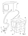
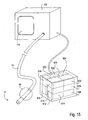
- the EPD device 102 may include a variety of mechanisms and features, which will be described hereinafter, depending on its intended use.
- the device is connectable with a pulmonary catheter 120, as shown, which is configured for accessing a lung compartment through one or more lung passageways.
- the catheter 120 is shown as having a proximal end 122, distal end 124, an optional lumen 126 therethrough and an optional occlusion member 128, the lumen 126 and occlusion member 128 shown in dashed-line.
- pulmonary catheters 120 may be used, a few embodiments of which will be discussed in later sections.
- the sensors may include pressure sensors, temperature sensors, air flow sensors, CO 2 sensors, O 2 sensors, infrared Doppler devices, current or resistivity sensors, laser diode sensors, pulse emitting diode sensors, and/or frequency emitting diodes, to name a few.
- the sensors 140 may be located near the distal end 124 of the catheter 120. Alternatively, the sensors 140 may be located at any point along the catheter 120 or within the EPD device 102 or one or more measuring components 104.
- Measuring components 104 may take many forms and may perform a variety of functions.
- the components 104 may include a pulmonary mechanics unit 107, a physiological testing unit 109, a gas dilution unit 106, an imaging unit 108, a mapping unit 112 or a treatment unit 113, to name a few. Embodiments of such components 104 will be discussed in detail in later sections.
- the components 104 may be integral with or disposed within the EPD device 102.
- some or all of the components 104 may be external to and/or removably connectable with the EPD device 102.
- a data receiving component 115 may be integral with, disposed within or removably connectable with the EPD device 102.
- the data receiving component 115 is shown as a visual display 110.
- the component 115 may alternatively take the form of a computer readable medium, a printer, or a chart recorder, to name a few.
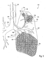
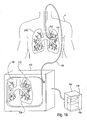
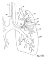
- the catheter 120 is configured for introduction into the pulmonary anatomy 150, particularly into a bronchial passageway.
- the catheter 120 may be introduced into the bronchial passageways of a lung LNG to various depths.
- the catheter 120 may be introduced so that it's distal end 124 is positioned within a distant lung segment 152 of the branching passageways.
- the catheter 120 can optionally isolate and measure an individual compartment 154 of the lung LNG, illustrated by a shaded dashed-lined circle.
- the distal end 124 may be positioned in a larger lung segment 156 of the branching passageways.
- the catheter 120 can measure another individual compartment 154 of the lung LNG, illustrated by a larger shaded dashed-lined circle.
- the distal end 124 may be positioned in an even larger lung segment 158 to measure an even larger compartment 154, such as one or more lobes.
- the lung LNG itself can be measured for comparison to the individual compartments.
- the distal end 124 in the trachea T, above the takeoff branches 160 the both of the lungs can be measured.
- any testing, imaging or other functions described in relation to a compartment 154 may also be performed on an entire lung LNG or both lungs.
- the isolated compartment 154 can be assessed. Fluid or gas is transferred to or from the lung compartment through the pulmonary catheter. This may be performed to pressurize the lung compartment, a state desired during many testing or measurement procedures.
- the EPD device 102 comprises mechanisms for transferring such fluid or gas. In some instances, this mechanisms for transferring may comprise a pump or other driving mechanisms and appropriate tubing or conduits for passage of the fluid or gas. In other instances, a pump or other driving mechanisms may be disposed outside of the EPD device 102. In this case, the mechanisms for transferring the fluid or gas of the EPD device 102 may simply comprise a conduit between the driving mechanisms and the pulmonary catheter.
- the sensors 140 gather measurement data or information which is transmitted to the EPD device 102.
- the EPD device 102 has mechanisms for receiving the measurement data.
- the EPD device 102 also comprises mechanisms for processing the measurement data. Processing may comprise converting the measurement data into a form which may be visually displayed, such as in graphs, charts, tables, numbers, images or figures. Or, processing may comprise analyzing the data wherein the data is used to determine or calculate secondary information or data such as an average pressure value, a volume value, a compliance value, an average tidal volume value and/or a resistance value, to name a few. Alternatively, processing may comprise converting the measurement data into a computer readable format. Such conversion may be of the measurement data itself or of secondary data derived from the measurement data.
- the processed data is then received by a data receiving component 115.
- the receiving component 115 comprises a visual display 110.
- one or more measuring components 104 receive the processed data.
- the processed data may then be used in conjunction other mechanisms within the components. For example, a component may perform a testing function while maintaining the lung compartment at a specific level of pressurization. Thus, the component may utilize measurement data from a pressure sensor while performing testing functions.
- the EPD device 102 comprises mechanisms for coordinating the functioning of the measuring components, such as the transfer of gas or fluid between the components and the lung compartment, the passage of information or measurement data between the measuring components, between the sensors and the measuring components or between the measuring components and the data receiving components, to name a few. Such control of activities may result from pre-programming, user input or both.
- measurement simply involves one or more measuring components without the use of a sensor.
- a component 104 infuses an isolated compartment 154 with an imaging fluid or gas.
- the lung compartment 154 may be visualized externally, with the use of a fluoroscopy, nuclear, MRI or CT imaging system, or may be visualized with the use of another component 104 within or attached to the EPD device 102. This may also be the case in measuring perfusion parameters.
- the measurement information is then processed by the EPD device 102 and received by a receiving component 115.
- Measurement information for a given lung compartment 154 may be compared with measurement information from one or more other lung compartments 154.
- information from a distant lung segment may be compared to information from another distant lung segment.
- the segments can be ranked in terms of level of disease, for example.
- information from a lobe can be compared with information from a distant lung segment within the lobe. In this way, the affect of the lung segment on overall performance of the lobe can be compared.
- a lung compartment 154 may be treated, such as by reduction and/or isolation, and remaining areas of the lung or lungs can be measured to determine the effect of the treatment.
- a blockage catheter may be used which is introduced to a target compartment, the compartment which has been targeted for treatment. With the blockage catheter in place, such treatment is simulated and the effect of the treatment may be determined by measuring the untreated areas, such as a larger compartment which encompasses or contains the target compartment, using, for example, CT imaging or plethysmography. Thus, more effective treatments may be achieved by pinpointing the most efficient compartments to treat.
- the above described measurement data or information may be provided to the user in various formats. Typically, such information will be displayed on the visual display 110 in visual form. This may include graphs, charts, tables, number images or figures, to name a few. Measurement data or information from a number of compartments may be directly compared by simultaneous display of the information from each compartment. Multiple imaging views of a compartment may be obtained to establish a three-dimensional composite view of the compartment. In addition, other types of displays may be provided.
- the EPD device 102 performs a variety of functions which depend on the elements included in the pulmonary diagnostic system 100 and the functions in which the system 100 is designed to perform. Descriptive embodiments of possible elements comprising the pulmonary diagnostic system 100 are presented below.
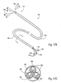
- the pulmonary catheter 120 comprises an access catheter 10.
- An exemplary access catheter 10 is illustrated in Fig. 4 and comprises a catheter body 12 having a distal end 14, a proximal end 16, an inflatable occlusion balloon 18 near its distal end, and at least one lumen therethrough.
- catheter 10 will have at least two lumens, and catheter 10 includes both a central lumen 20 and an annular lumen 22 defined by inner body member 24 and outer body member 26 which is coaxially disposed about the inner body member.
- the annular lumen 22 opens to port 30 on a proximal hub 31 and provides for inflation of balloon 18.
- the central lumen 20 opens to port 36 on hub 31 and provides for multiple functions, including optional introduction over a guidewire, aspiration, introduction of secondary catheters, such as sealing catheters, measurement catheters and the like.
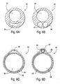
- the access catheter 10 may be modified in a number of ways, some of which are illustrated in Figs. 6A-6F .
- the catheter can be a single extrusion having a catheter body 30 with a circular main lumen 32 and a crescent-shaped inflation lumen 34, as illustrated in Fig. 6A .
- the catheter body 40 may be formed as a single extrusion having three lumens, i.e., a primary lumen 42 for receiving a guidewire, applying aspiration, delivering secondary catheters, and/or other functions.
- a second lumen 44 can be provided for inflating the occlusion balloon, and a third lumen 46 can be provided as an alternative guidewire or functional lumen.
- Catheter body 50 comprising a main tubular body 52 having an outer layer 54 fused thereover to define a lumen 56 suitable for balloon inflation as shown in Fig. 6C .
- a primary lumen 58 is formed within the main tubular member 52.
- catheter body 60 can be formed from a primary tubular member 62, and a secondary tubular member 64, where the tubular members are held together by an outer member 66, such as a layer which is applied by heat shrinking.
- the primary tubular member 62 provides the main lumen 68 while secondary tube 64 provides a secondary lumen 70.
- the secondary lumen 70 will typically be used for balloon inflation, while the primary lumen 68 can be used for all other functions of the access catheter.
- the dimensions and materials of access catheter 10 are selected to permit endotracheal introduction and intraluminal advancement through the lung bronchus, optionally over a guidewire, and/or through a primary tracheal tube structure and/or inside the working channel of a bronchoscope.
- Suitable materials include low and high density polyethylenes, polyamides, nylons, PTFE, PEEK, and the like, particularly for the inner tubular member 24.
- the outer member, including the occlusion balloon can be made from elastomeric materials, such as polyurethane, low density polyethylene, polyvinylchloride, silicone rubber, latex, and the like.
- portions of the outer tubular member 26 proximal to the inflatable balloon can be made thicker and/or reinforced so that they do not dilate upon pressurization of the balloon.
- Exemplary dimensions for the access catheter 10 are dependent on its use.
- a multi-purpose access catheter 10 should have a working lumen, such as a central lumen 20, main lumen 32, primary lumen 42 or similar such lumen, adequately sized for a number of procedures. If the catheter 10 is to be used in procedures such as functional residual capacity testing or the generation of pressure vs. volume curves, the working lumen should be approximately 1.5-3.5 mm ID, assuming a catheter 10 length of approximately 45-80 cm. In other situations, however, the working lumen may be smaller.
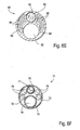
- the access catheter in the present invention can be provided with optical imaging capability.
- catheter body 80 can be formed to include four lumens, typically by conventional extrusion processes.
- Lumen 82 is suitable for passage over a guidewire.
- Lumens 84 and 86 both contain light fibers 88 for illumination.
- Lumen 90 carries an optical wave guide or image fiber 92.
- Lumen 82 can be used for irrigation, aspiration or other functions, typically after the guidewire is withdrawn.
- Balloon inflation can be effected through the space remaining and lumens 84 and 86 surrounding the light fibers 88.
- an alternative embodiment of the catheter body 71 is formed as a coaxial arrangement of a number separate tubes.
- Outer tube 72 contains a separate guidewire tube 74 defining lumen 76 which permits introduction over a guidewire as well as perfusion and aspiration after the guidewire is removed.
- Second inner tubular member 75 will carry an optical image fiber 77 and a plurality of light fibers 78 are passed within the remaining space 79 within the outer tubular member.
- forward imaging can be effected by illuminating through the light fibers and detecting an image through a lens at the distal end of the catheter. The image can be displayed on conventional cathode-ray or other types of imaging screens.
- forward imaging permits a user to selectively place the guidewire for advancing the catheters through a desired route through the branching bronchus.
- an alternative cross-sectional design will be implemented to provide the necessary dimensions.
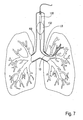
- the catheter 10 can be advanced to a compartment within a lung through a patient's trachea. Advancement through the trachea T is relatively simple and will optionally employ a guidewire to select the advancement route through the branching bronchus. As described above, steering can be effected under real time imaging using the imaging access catheters illustrated in Figs. 6E-6F .
- the catheter may be inserted through the working channel of a bronchoscope, using the bronchoscope vision for navigation.
- the access catheter 10 may be introduced through a visualizing tracheal tube, such as that described in U.S. Patent No. 5,285,778 . As shown in Fig.
- the visualizing endotracheal tube 130 includes an occlusion cuff 132 which may be inflated within the trachea just above the branch of the left bronchus and right bronchus LB and RB, respectively.
- the visualizing endotracheal tube 130 includes a forward-viewing optical system, typically including both illumination fibers and an image fiber to permit direct viewing of the main branch between the left bronchus LB and right bronchus RB.
- initial placement of access catheter can be made under visualization of the visualizing endotracheal tube 130 and optionally the access catheter 10 itself.
- the access catheter 10 is advanced until its distal end 14 reaches a region in the bronchus which leads directly into the lung compartment.
- the access catheter 10 may have elements or accessories for steering and sufficient torque response and pushability to make advancement and navigation through the bronchial tree possible.
- the catheter 10 may include positioning sensors so as to determine the location of the catheter with respect to the complete lung anatomy. This will be described in detail in a later section.
- the access catheter 10 can be a modular system or a multi-component system.
- the access catheter 10 may comprises a viewing scope and a sheath for use with the viewing scope as described in US 658 56 39 .
- the viewing scope includes or consists essentially of a flexible elongated body, an optical viewing fiber or video chip, and a light transmitting bundle.
- the viewing scope may be in the form of conventional bronchoscope or a conventional articulated flexible scope having dimensions suitable for introduction in and through the lung passageways.
- the sheath comprises a flexible tubular body having a proximal end, a distal end, and at least a first lumen therethrough.
- the sheath will further comprise an inflatable cuff disposed near its distal end, where the inflatable cuff may be inflated through a lumen which is present in the tubular body itself or formed in a separate inflation tube.
- the viewing scope is introduced into the lumen of the flexible tubular body of the sheath to form an assembly where a viewing end of the viewing scope is located at the distal end of the sheath.
- the assembly of the viewing scope and sheath may then be introduced to a lung passageway so that the inflatable cuff lies adjacent to a target location in the passageway.
- the cuff may then be inflated to temporarily occlude the target location.
- the sheath may also have additional working channels in order to perform aspects of the diagnostic testing, such as carbon dioxide sensing or polarized gas delivery.
- the access catheter 10 may comprise one or more sensors to measure a variety of variables related to pulmonary function. Such sensors will typically be located near the distal end 14 of the catheter 10, however they may be located at any location along the length of the catheter body 12. Individual sensor types will be described in relation to each type of measurement described below.
- a measuring component 104 of the pulmonary diagnostic system 100 comprises a pulmonary mechanics unit 200, as shown in Fig. 8 .
- the pulmonary mechanics unit 200 is illustrated as a separate attachable unit, however it may be appreciated that the unit 200 may be internal to the EPD device 102.
- the pulmonary mechanics unit 200 is used for measuring a number of variables related to the pulmonary mechanics of a compartment 154 of a lung LNG.
- the pulmonary mechanics unit 200 may include mechanisms 202 for generating pressure and volume data of the lung compartment 154. Generation of such data is achieved by slowly inflating the lung compartment 154 and measuring volume delivered and real-time pressure. The inflation process is performed slowly to minimize the affect of any system resistance on the pressure readings.
- the inflation medium is delivered to the compartment 154 through an access catheter 10 which is removably attached to the EPD device 102. In this case, the distal end 14 of the catheter 10 is inserted into the lung passageway leading to the compartment 154 to be measured and the balloon 18 is inflated to occlude the passageway. In this way, all inflation medium is delivered to the compartment 154 and cannot escape to other areas of the lung.
- Pressure is measured by a pressure sensor 204 and volume is derived from measurement by a flow sensor 206.
- the sensors 204, 206 may be disposed near the distal end 14 of the catheter 10 or at other locations, including within the EPD device 102 and/or the pulmonary mechanics unit 200.
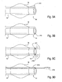
- a number of different types of pressure sensors 204 are shown in Figs. 9A-9D .
- the pressure sensor 204 comprises a secondary cuff 210 disposed at the distal end 14 of the access catheter 10, distal to the occlusion balloon 18.
- Another embodiment, shown in Fig. 9B comprises a Wheatstone bridge, microbellows pressure transducer, or optical fiber 212 imbedded in the wall of the distal end 14.
- a pressure sensor 204 in Fig. 9C comprises ultrasonic or fiberoptic pressure transducers with a send element 214 and a receive element 216.
- the embodiment shown in Fig. 9D comprises a bellows or Wheatstone bridge 218 protruding from a channel 219 in the catheter 10.
- Pressurization of the compartment 154 can be performed while the rest of the lung is at an expiratory hold to truly isolate the target compartment and eliminate extraneous events.
- pressurization can be performed during an inspiratory hold, during regular ventilation or during a pressure hold that is in between end-expiratory pressure and peak inspiratory pressure.
- the pressure and volume data is plotted on a graph wherein the pressure data is plotted along an x-axis X and the volume is plotted along a y-axis Y, as illustrated in Fig. 10A .
- the resulting PV curve 220 provides information regarding the health and level of disease of the compartment 154.
- Fig. 10A shows three such PV curves 220, the difference in the curves are due to various disease states as stated.
- Compliance refers to the distensibility of an elastic structure (such as a lung compartment 154) and is defined as the change in volume of that structure produced by a change in pressure across the structure. In other words, compliance can be defined as the slope of a PV curve 220 at a given point along the curve. As shown in Fig. 10B , in a normal healthy lung compartment at low volume, relatively little positive pressure needs to be applied to increase the volume of the lung quite a bit, as shown by the high compliance area 222. Lung compliance decreases with increasing volume so as the lung compartment is further inflated, more pressure must be applied to get the same increase in volume. This corresponds to the low-compliance area 224.
- the compliance will be calculated at an upper inflection point or peak inspiratory pressure PIP, identified in Fig. 10A .
- Mechanisms 226 for calculating a compliance value from the pressure and volume data is depicted within the pulmonary mechanics unit 200 in Fig. 8 , however such mechanisms 226 may alternatively be disposed within the EPD device 102..
- the PV curves 220 and compliance values will typically be displayed on the visual display 110.
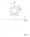
- volume and flow data may be plotted on a graph as in Fig. 11 .
- flow data is plotted along an x-axis X and volume is plotted along a y-axis Y.
- volume increases as flow increases and then declines, as indicated by arrow 221.
- volume decreases as flow increases and then declines, as indicated by arrow 223.
- the resulting trace is a loop 225 indicative of the flow volume characteristics of the compartment 154 accessed.
- the loop 225 provides information regarding the health and level of disease of the compartment 154.
- Fig. 11 shows three such loops 225, each corresponding to a different compartment 154 or to the same compartment 154 over time as disease progresses.
- Additional respiratory parameters may also be derived from pressure and volume data.
- the average tidal volume can be measured for a given lung compartment 154. Tidal volume may be described as the volume of air inhaled and exhaled with each breath.
- the pulmonary mechanics unit 200 or the EPD device 102 may comprise mechanisms 228 for calculating an average tidal volume value.
- pressure is set to the PIP and the compartment is ventilated at that pressure. This may be performed while the rest of the lung is in an expiratory hold. Volume is typically measured for three to five breaths over approximately 30 seconds and an average is taken of these values to determine the average tidal volume.
- the resistance of a compartment can be derived from pressure and volume data.
- Resistance may be described as the pressure divided by the volumetric flow rate.
- the EPD device 102 or the pulmonary mechanics unit 200 may comprise mechanisms 230 for calculating a resistance value.
- work of breathing of a compartment can be derived from pressure and volume data. This is done by converting pressure and volume into Joules/liter.
- the EPD device 102 or the pulmonary mechanics unit 200 may also comprise mechanisms 234 for calculating an average work of breathing value. Graphical or numerical representation of these values may be received by a data receiving component 115 for visual display.
- a measuring component 104 comprises a physiological testing unit 300, as shown in Figs. 12-15 .
- the physiological testing unit 300 is illustrated as a separate attachable unit, however it may be appreciated that the unit 300 may be integral or internal to the EPD device 102.
- the physiological testing unit 300 is used for measuring a number of variables related to the physiology of a compartment 154 of a lung LNG.
- the physiological testing unit 300 may include mechanisms 400 for measuring ventilation or velocity of air movement in and out of a compartment 154.
- the pulmonary catheter 120 comprises a microcatheter 402 having a proximal end 404, a distal end 406, a lumen 408 therethrough and at least one sensor 410 mounted on its distal end 406.
- the sensor 410 may be a velocity sensor.
- the microcatheter 402 is positioned such that its distal end 406 is entering a compartment 154 to be measured.
- the microcatheter 402 is sized so that the compartment 154 is not isolated and air movement is not retarded.
- the velocity sensor 410 measures the velocity of airflow into and out of the compartment 154.
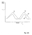
- Fig. 12A velocity versus time data is plotted wherein time is plotted along an x-axis X and velocity is plotted along a y-axis Y.
- time is plotted along an x-axis X and velocity is plotted along a y-axis Y.
- inspiration velocity increases to an inspiratory peak 421 and then decreases over time.
- expiration velocity increases to an expiratory peak 423 and then decreases over time.
- the resulting trace 425 is indicative of the characteristics of the compartment 154 accessed.
- the trace 425 provides information regarding the health and level of disease of the compartment 154.
- Fig. 12A shows two such traces 425, each corresponding to a different compartment 154 or to the same compartment 154 over time as disease progresses.
- the senor 410 may be an oxygen and/or carbon dioxide sensor.
- the sensor 410 can measure the amount of, for example, carbon dioxide retained in the compartment. Carbon dioxide is indicative of trapped air. Therefore, data derived from such a sensor may provide information as to the level of disease in the compartment.
- a sensor that measures the amount of oxygen retained in the compartment may indicate the level of disease affecting gas transfer through the alveolar sacs.
- Oxygen sensors can also be used in the performance of oxygen wash-out tests. Here, the air in a lung compartment is replaced as much as possible with 100% oxygen. Then, the decay of oxygen concentration is measured over time using sensor 410. Such decay indicates how well a compartment contributes to ventilation. Further, the ratio of carbon dioxide to oxygen can be determined which is also indicative of disease state.
- the physiological testing unit 300 may also include mechanisms 450 for measuring electrophysiology characteristics of a lung compartment 154.
- the mechanisms 450 includes measuring the resistance of the tissue in the compartment 154.
- a pulse emitting sensor 452 is mounted on the distal end 124 of the pulmonary catheter 120.
- the proximal end 122 of the catheter 120 is removably attached to the EPD device 102 and the distal end 124 is inserted into a lung compartment 154.
- a receiver 454 is positioned at a second location, for example on the outside of the patient P, and is connected to the EPD device 102.
- a pulse is emitted from the sensor 452 and a signal is measured by the receiver 454.
- the signal determines the resistance of the tissue and therefore the state of the disease. For example, diseased tissue will have a different conductivity because of the breakdown of elasticity and/or because of edema/inflammation of the tissue.
- the mechanisms 450 for measuring electrophysiology characteristics of a lung compartment 154 includes measuring the electrical activity of the musculature of the tissue in the compartment 154.
- two or more leads are mounted on the pulmonary catheter.
- the leads measure a characteristic voltage signal of the tissue which determines the state of the disease. For example, diseased tissue will have weaker signals due to the breakdown of elasticity.
- the senor 410 is an infrared sensor which is positioned against the bronchial tissue and a venous oxygen saturation measurement is made. Because blood perfusing diseased lung compartments will have lower oxygenation, disease level can be determined.
- Graphical or numerical representation of these values generated by the EPD device 102 or physiological testing unit 300 may be displayed on the visual display 110.
- a measuring component 104 of the pulmonary diagnostic system 100 comprises a gas dilution unit 500, as shown in Fig. 14 .
- the gas dilution unit 500 is illustrated as a separate attachable unit, however it may be appreciated that the unit 500 may be internal to the EPD device 102.
- the gas dilution unit 500 is used primarily for Functional Residual Capacity (FRC) testing and/or residual volume (RV) testing of a compartment 154 of a lung LNG, since these parameters reflect level of disease.
- FRC Functional Residual Capacity
- RV residual volume
- the access catheter 10 is used as the pulmonary catheter 120 attached to the EPD device 102, as shown.
- the compartment 154 is inflated to the PIP, as previously determined by the mechanisms 202 for generating pressure and volume data. This can be achieved by the pulmonary mechanics unit 200, if available, or it may be achieved by mechanisms 504 for generating pressure and volume data within the gas dilution unit 500 or the EPD device 102.
- a known volume of a noble gas such as helium, is introduced from a source of noble gas 506 to the compartment 154 through the access catheter 10.
- the known volume of noble gas is allowed to mix with the unknown volume of air in the compartment 154 (at PIP). Thorough mixing is accomplished by using a pump 508 that moves gas back and forth through the access catheter 10 in an oscillatory motion. Due to the low volume of the access catheter 10 compared to that of the compartment 154, complete mixing should be accomplished in approximately 1-5 minutes, depending on the mixing efficiency of the incoming noble gas.
- a sensor 502 measures the concentration of one of the gases in the system.
- the sensor 502 is mounted on the distal end 14 of the catheter 10 as shown.
- the sensor 502 may be any of the following: a membrane chemical transfer sensor, a photochemical reaction sensor, an electropotential sensor, a microchip, a laser diode, an optical transmittance sensor, or a piezoelectric sensor.
- a membrane chemical transfer sensor a photochemical reaction sensor
- an electropotential sensor equilibrates
- a microchip e.g., a laser diode
- an optical transmittance sensor e.g., a piezoelectric sensor.
- the gas dilution unit 500 or EPD device 102 may include mechanisms 510 for determining the concentration of a gas, such as helium, in the system and mechanisms 512 for calculating the initial volume of air in a lung compartment.
- Determining the volume of air initially in the compartment may be useful information used during later treatment.
- the compartment may be treated by aspirating trapped air in the compartment.
- the effectiveness of the treatment may be determined.
- a measuring component 104 of the pulmonary diagnostic system comprises an imaging unit 600, as shown in Fig. 15 .
- the imaging unit 600 is illustrated as a separate attachable unit, however it may be appreciated that the unit 600 may be integral or internal to the EPD device 102.
- the imaging unit 600 is used for generating or assisting in the generation of two-dimensional or three-dimensional images of a compartment 154 of a lung LNG. Often traditional external imaging equipment is used to generate the images while the imaging unit 600 assists in the visualization of individual compartments, as will be described below. In this case, the imaging unit 600 may also serve to transmit and optionally manipulate the images for display in the visual display 110.
- the access catheter 10 is typically used as the pulmonary catheter 120 attached to the EPD device 102, as shown.
- the compartment may be visualized by imaging. Such imaging may be enhanced by occluding the lung passageway with the balloon 18 prior to visualization.
- the imaging unit 600 may include mechanisms 602 for transferring a fluid or gas having radiopaque properties to the lung compartment 154. Such a fluid or gas may be radiopaque or be labeled with radiopaque markers, for example.
- the compartment 154 is inflated with the fluid or gas to the PIP, as previously determined by the mechanisms 202 for generating pressure and volume data.
- the imaging unit 600 may further include mechanisms 606 for generating an ultrasound or MRI image of the lung compartment 154. Or, an image may be taken with equipment external to the pulmonary diagnostic system 100 using CT, MRI, PET, x-ray, ultrasound, fluoroscopy or a perfusion scan. While the compartment is filled with an imaging gas or fluid as described above, the remainder of the lung may be filled with a gas or fluid having a stronger or weaker imaging capability. For example, the lung compartment may be filled with a radiopaque gas and the remainder of the lung filled with a weaker concentration of radiopaque gas.
- the complete lung will be visible under fluoroscopy with the lung compartment "highlighted" by the stronger concentration of radiopaque gas. It may be appreciated that the concentrations may be reversed wherein the lung is filled with a gas or fluid having a higher concentration than the lung compartment and the lung compartment is highlighted by the weaker concentration of radiopaque gas. Similarly, an individual lung compartment may be blocked or isolated and the remainder of the lung imaged by the methods described above. In any case, the individual compartments may be evaluated which may provide information as to its level of disease.
- Multiple images of the lung compartment 154 may be generated, each image having a different view. This may be achieved externally or by mechanisms 608 for generating multiple images. Mechanisms 610 for generating a three-dimensional composite image of the compartment 154 from the individual views may also be included in the imaging unit 600 or the EPD device 102. Alternatively, multiple images of the lung compartment 154 may be generated so that a three-dimensional image is obtained by combining image "slices" of the compartment. This may provide even more diagnostic information regarding the status of the compartment and its level of disease.
- the imaging unit 600 may include mechanisms 612 for transferring a polarized gas to the lung compartment 154. Again, the compartment 154 is inflated with the gas to the PIP, either with the mechanisms 202 or the mechanisms 604 for generating pressure and volume data.
- the imaging unit 600 may further include mechanisms 614 for generating at least one magnetic resonance image (MRI) of the lung compartment 154. Or, an image may be taken with external MRI equipment. In either case, the anatomy of the compartment may be visualized which may provide information as to its level of disease.
- the imaging unit 600 may further include mechanisms 616 for generating multiple magnetic resonance images of the lung compartment 154 and mechanisms 618 for generating a three-dimensional composite image of the compartment 154. It may be appreciated that some of these mechanisms may be included in the EPD device 102. These images may provide even more diagnostic information regarding the status the compartment and its level of disease.
- the imaging unit 600 may include mechanisms 620 for transferring a liquid such as perfluroban to the lung compartment 154.
- the imaging unit 600 may further include mechanisms 622 for generating an ultrasonic image of the lung compartment 154.
- an image may be taken with external ultrasound equipment. In either case, the anatomy of the compartment may be visualized which may provide information as to its level of disease.
- imaging may be undertaken with the use of any contrast media appropriate for the imaging technique.
- imaging may be performed at different points in the breathing cycle, such as at the end of normal inspiration and exhalation and/or at the end of forced inspiration and exhalation.
- These images can then be used to calculate lung volumes relevant to disease, such as residual volume (RV), total lung capacity (TLC) and RV/TLC.
- imaging may be performed on any number of lung compartments and imaging results may be compared for diagnostic or other purposes.
- a measuring component 104 of the pulmonary diagnostic system 100 comprises an mapping unit 700, as shown in Fig. 16 .
- the mapping unit 700 is illustrated as a separate attachable unit, however it may be appreciated that the unit 700 may be integral or internal to the EPD device 102.
- the mapping unit 700 is used for determining the position of the pulmonary catheter 120 as it is introduced and advanced through the bronchial passageways. Due to the multiple branchings of the bronchial anatomy, the position of the catheter 120 within the passageways may be difficult to determine and thus the lung compartment 154 to be measured may also be difficult to determine: With the use of the mapping unit 700, the position of the catheter 120 may be more readily visualized.
- a sensor 702 is used to track the position of the catheter 120 in the bronchial passageways.
- the sensor 702 is mounted on the catheter 120, typically near its distal end 124.
- the sensor 702 can be a simple magnetic type device or a frequency emitting device, to name a few.
- the sensor 702 could be an optical imaging element and coupled with artificial intelligence that tracks successive directional movements of the catheter tip, hence knowing its position at any given time.
- the mapping unit 700 may include mechanisms 704 for receiving the signal from the sensor and mechanisms 706 for processing the signal.
- Mechanisms for processing the signal include mechanisms for generating positioning data of the sensor 702 within the passageways and mechanisms for generating an image of the sensor positioned within the passageways reflecting the positioning data of the sensor 702. This is illustrated in Fig.
- the catheter 120 is attached to the EPD device 102 and its distal end 124 is introduced to a lung passageway of a patient P.
- a computer generated pulmonary anatomy image 708 is shown on the visual display 110.
- a catheter positioning image 710 is shown within the anatomy image 708 reflecting the real-time position of the catheter 120 within the actual patient's anatomy. In this way, the target lung compartment may be more easily located and identified for later access if desired.
- the data receiving component 115 receives processed data from the EPD device 102 or any of the components 104 for output to the user.
- the component 115 is the visual display 110 and the processed data is presented in visual form. It may be appreciated that the possibilities of displaying measurement information in visual form are limitless. A few embodiments are presented as examples.
- the pulmonary diagnostic system is used to measure compartments of a lung, wherein a compartment could be an entire lobe, a segment or a subsegment and beyond.
- information generated from a compartment may be used in determining the level of disease of the compartment itself, comparison of the generated information to other information is also useful in diagnosis and assessment of disease.
- information generated from a compartment may be compared with baseline information from the patient or to information from a healthy patient.
- both or multiple sets of information may be displayed in the visual display 110 simultaneously. Such display may be graphical, numerical or any other type.
- information generated from one compartment may be compared with information from another similar compartment within the same patient. This concept may be extended to numerous compartments.
- multiple sets of information are displayed in the visual display 110 simultaneously in accordance with the invention. This in turn may allow the physician to rank the compartments in order of level of disease or in order of need for treatment. Similarly, information generated from one compartment may be compared with information from other types or sizes of compartments to determine the affect of each compartment on the others. Again, multiple sets of information may be displayed in the visual display 110 simultaneously for this purpose.
- visual images of the lung anatomy can be displayed on the visual display 110 as noted above. This may be useful in both diagnosis of disease, positioning of the pulmonary catheter 120 and determining the affect of treatment.
- a treatment protocol may be determined.
- lung volume reduction may be prescribed.
- the lung passageway which leads to the lung compartment to be reduced may be temporarily occluded with a blockage catheter.
- this temporary occlusion with a blockage catheter may itself be the diagnostic test.
- FIG. 17A illustrates the blockage catheter 750 as comprising a catheter body 752 having a proximal end 756, a distal end 754, and an inflatable occlusion balloon 758 near its distal end 754.
- the blockage catheter 750 may have a smaller outer diameter than the access catheter 10 or other pulmonary catheters 120 which have lumens of significant diameter for various testing purposes.
- lumens may simply be sized for inflation of the occlusion balloon 758 or passage of a guidewire.
- FIGs. 17B-D illustrates the blockage catheter 750 as comprising a catheter body 770 having a proximal end 772 and a multiplicity of distal ends 774, each distal end 774 having an inflatable occlusion balloon 776 mounted thereon.
- a separate guidewire lumen 780 and balloon inflation lumen 782 within the catheter body 770 are present from the proximal end 772 to each distal end 774.
- End connectors 784 may be present near the proximal end 772 for access to each lumen 780, 782.
- an outer sleeve 786 encasing the catheter body 770 unites the distal ends 774 for introduction purposes.
- Fig. 17C shows a cross-section of the distal ends 774 of the catheter body 770.
- the catheter body 770 may comprise separate catheters, each having a guidewire lumen 780, inflation lumen 782 and occlusion balloon 776, which are held together by the outer sleeve 786 or other means.
- placement of the blockage catheter 750 involves positioning each distal end 774 into a different lung passageway.
- Each distal end 774 may be independently positioned with the use of a guidewire.
- a number of lung compartments 154 may be simultaneously isolated by the blockage catheter 750.
- the affected lung compartment 154 will be isolated from the remainder of the lung.
- testing, imaging and evaluation of the overall lung performance may be undertaken to measure the effects of the isolation.
- Such techniques would include, for example, CT scanning, spirometry, or plethysmography to obtain images, spirometry data or plethysmography data, respectively. This in turn reflects the effect of reduction of that isolated compartment.
- testing and evaluation may be performed on specific segments of the lung for assessing particular regions of the lung. Referring back to Fig. 17A , such testing with a pulmonary catheter 120 while the blockage catheter 750 is in place is illustrated.
- the blockage catheter 750 may be introduced through a lumen in the pulmonary catheter 120 or the blockage catheter 750 may simply lie in parallel with the pulmonary catheter 120. If the pulmonary catheter 120 utilizes an occlusion member 128, the member 128 may seal against the lumen and the catheter body 752 of the blockage catheter 750. Both the blockage catheter 750 and pulmonary catheter 120, or any additional catheters, may be simultaneously connected to the EPD device 100 if desired. The results of such assessment may determine the most effective course of treatment.
- a measuring component 104 of the pulmonary diagnostic system 100 may comprise a treatment unit 800, as shown in Fig. 18 .
- the treatment unit 800 is illustrated as a separate attachable unit, however it may be appreciated that the unit 800 may be integral or internal to the EPD device 102.
- the treatment unit 800 may be used to perform a lung volume reduction procedure on a lung compartment 154.
- the unit 800 would include mechanisms 802 for performing lung volume reduction. This typically involves aspirating the contents of the compartment after isolating the compartment from the remainder of the anatomy. This is typically achieved by introducing the distal end 14 of the access catheter 10 endotracheally to the target compartment 154.
- the compartment may be isolated by occluding the air passageway, such as by inflating the occlusion balloon 18 within the passageway.
- the target compartment may then be collapsed by aspirating air, and any other gases or liquids that may have been introduced, from the compartment, such as through a lumen in the access catheter 10.
- the passageway may then be sealed, for example by deploying a plug within the air passageway.
- Some sealing methods include the use of tissue adhesives, occlusive balloons, expanding occlusive structures, the use of energy-induced tissue fusion and the like.
- Preferred embodiments of various types of treatments are described in U.S. Patent Application Nos. 09/425,272 , 09/347,032 , 09/606,320 , 09/523,016 , and 09/699,302 . After treatment, the affect of treatment may be evaluated by some of the measurement methods described above.
- Kits 900 may comprise any number of items related to the pulmonary diagnostic system described and instructions for use IFU. As shown in Fig. 19 , such kits 900 typically include the EPD device 102 and instructions for use IFU setting forth methods according to the present invention. The EPD device 102 may have one or more measuring components disposed internally, however the kit 900 may also include one or more measuring components 104 as shown. Optionally, the kits 900 may further include any of the other system components described above, such as one or more pulmonary catheters 120, guidewires 902, or a variety of accessories, such as a receiver 454. Some or all kit components will usually be packaged together in a pouch 905 or other conventional medical device packaging.
- kit components such as a pulmonary catheter 120
- pulmonary catheter 120 which will be used in performing the procedure on the patient will be sterilized and maintained within the kit.
- separate pouches, bags, trays or other packaging may be provided within a larger package, where the smaller packs may be opened separately to separately maintain the components in a sterile fashion.
Landscapes
- Health & Medical Sciences (AREA)
- Life Sciences & Earth Sciences (AREA)
- Pulmonology (AREA)
- Engineering & Computer Science (AREA)
- Heart & Thoracic Surgery (AREA)
- Biomedical Technology (AREA)
- Animal Behavior & Ethology (AREA)
- General Health & Medical Sciences (AREA)
- Public Health (AREA)
- Veterinary Medicine (AREA)
- Biophysics (AREA)
- Medical Informatics (AREA)
- Physics & Mathematics (AREA)
- Pathology (AREA)
- Molecular Biology (AREA)
- Surgery (AREA)
- Physiology (AREA)
- Anesthesiology (AREA)
- Hematology (AREA)
- Emergency Medicine (AREA)
- Nuclear Medicine, Radiotherapy & Molecular Imaging (AREA)
- Radiology & Medical Imaging (AREA)
- High Energy & Nuclear Physics (AREA)
- Optics & Photonics (AREA)
- Obesity (AREA)
- Psychiatry (AREA)
- Signal Processing (AREA)
- Child & Adolescent Psychology (AREA)
- Computer Vision & Pattern Recognition (AREA)
- Artificial Intelligence (AREA)
- Apparatus For Radiation Diagnosis (AREA)
- Measuring And Recording Apparatus For Diagnosis (AREA)
- Measurement Of The Respiration, Hearing Ability, Form, And Blood Characteristics Of Living Organisms (AREA)
- Magnetic Resonance Imaging Apparatus (AREA)
- Ultra Sonic Daignosis Equipment (AREA)
- Endoscopes (AREA)
Claims (17)
- Système de diagnostic pulmonaire (100) comprenant :des capteurs pour générer des données de mesure reflétant une caractéristique respiratoire d'un compartiment pulmonaire, les capteurs comprenant un capteur de pression (204) pour générer des données de pression et un capteur de débit (206) pour générer des données de mesure de débit ;une sonde pulmonaire (120) configurée pour accéder au compartiment pulmonaire par l'intermédiaire d'une voie pulmonaire, la sonde comportant un élément d'obturation (18) près de son extrémité distale pour bloquer la voie pulmonaire autour de la sonde aboutissant au compartiment pulmonaire afin de permettre une communication directe avec le compartiment pulmonaire isolé du reste du poumon ;un dispositif de diagnostic pulmonaire endobronchique (102) raccordable à la sonde pulmonaire qui est configurée pour accéder au compartiment pulmonaire, le dispositif comprenant :un moyen de transfert pour transférer un fluide ou un gaz en direction ou en provenance du compartiment pulmonaire par l'intermédiaire de la sonde pulmonaire,un moyen de réception pour recevoir les données de pression et de mesure de débit provenant des capteurs, etun moyen de traitement pour traiter les données de pression et de mesure de débit, le moyen de traitement comprenant un moyen pour calculer des données de mesure de volume à partir des données de mesure de débit et un moyen d'analyse configuré pour déterminer des caractéristiques physiques et un niveau pathologique correspondant d'un compartiment pulmonaire à partir des données de pression et de mesure de volume ; etau moins un élément récepteur de données (115) configuré pour recevoir les données traitées, l'élément récepteur de données comprenant un affichage visuel (110) et étant configuré pour afficher simultanément de multiples ensembles d'informations correspondant à de multiples compartiments pulmonaires pour permettre la comparaison d'informations concernant divers compartiments pulmonaires dans l'anatomie pulmonaire.
- Système selon la revendication 1, comprenant en outre au moins un capteur additionnel pour générer des données de mesure reflétant une caractéristique respiratoire comprenant la vitesse de déplacement de l'air pénétrant dans le compartiment pulmonaire et sortant du compartiment pulmonaire, la concentration d'oxygène, ou la concentration de gaz nobles.
- Système selon l'une quelconque des revendications précédentes, dans lequel le moyen d'analyse est configuré pour calculer une valeur de pression moyenne et une valeur de volume.
- Système selon l'une quelconque des revendications précédentes, dans lequel les capteurs (140) sont disposés sur ou dans la sonde pulmonaire (120).
- Système selon l'une quelconque des revendications 1 à 3, dans lequel les capteurs (140) sont disposés à l'intérieur du dispositif ou disposés à l'intérieur d'au moins un élément de mesure du système.
- Système selon l'une quelconque des revendications précédentes, le système comprenant plus d'un élément de mesure, et le dispositif de diagnostic pulmonaire endobronchique (102) comprenant des mécanismes pour coordonner le passage d'informations ou de données de mesure entre les éléments de mesure.
- Système de diagnostic pulmonaire selon la revendication 1, comprenant : un élément de mesure et une unité de tests physiologiques (300) comprenant :une source de fluide ou de gaz,un moyen pour produire un écoulement du fluide ou du gaz,un moyen pour recevoir les données de pression et de mesure de débit provenant des capteurs ;l'élément récepteur de données (115) étant agencé pour recevoir les données de pression et de mesure de débit qui ont été traitées ; etle dispositif de diagnostic pulmonaire endobronchique (102) étant raccordable à l'unité de tests physiologiques et à l'élément de mesure, et comprenant un moyen pour contrôler la production de l'écoulement, la réception des données de pression et de mesure de débit par l'unité de tests physiologiques, et la réception des données de pression et de mesure de débit qui ont été traitées par l'élément de mesure.
- Système selon la revendication 7, dans lequel le moyen de traitement comprend un moyen pour générer une courbe pression-volume à partir des données de pression et de mesure de volume.
- Système selon la revendication 8, dans lequel le moyen d'analyse comprend en outre un moyen pour calculer une valeur de conformité à un point d'inflexion supérieur ou une pression inspiratoire maximum le long de la courbe pression-volume, une valeur moyenne de volume courant ou une valeur de résistance du compartiment pulmonaire en utilisant les données de pression et de mesure de volume.
- Système selon la revendication 7, comprenant en outre :un capteur (140) qui mesure la concentration d'un gaz, une unité de dilution de gaz raccordable au dispositif, comprenant une source de gaz noble, etun moyen pour produire un écoulement du gaz noble, le moyen de réception étant configuré pour recevoir les données de mesure de la concentration de gaz provenant du capteur et le moyen de traitement étant configuré pour traiter les données de mesure de la concentration de gaz.
- Système selon la revendication 10, dans lequel l'élément de mesure est agencé pour recevoir les données de mesure de la concentration de gaz qui ont été traitées, et le dispositif comprend en outre un moyen pour contrôler la production d'un écoulement du gaz noble, la réception des données de mesure de la concentration de gaz par l'unité de dilution de gaz, et la réception des données de mesure de la concentration de gaz qui ont été traitées par l'élément de mesure.
- Système selon la revendication 10 ou 11, dans lequel le capteur qui mesure la concentration de gaz comprend un capteur à membrane à transfert chimique, un capteur à réaction photochimique, un capteur mesurant le potentiel électrique, une puce, une diode laser, un capteur à transmittance optique ou un capteur piézo-électrique.
- Système selon la revendication 10, 11 ou 12, dans lequel le moyen pour traiter les données de mesure de la concentration de gaz comprend un moyen d'analyse configuré pour calculer le volume initial d'air dans le compartiment en utilisant les données de mesure de la concentration de gaz.
- Système selon l'une quelconque des revendications précédentes, comprenant en outre une unité d'imagerie comprenant :une source de fluide ou de gaz d'imagerie, etun moyen pour produire un écoulement du fluide ou du gaz d'imagerie.
- Système selon la revendication 1, dans lequel le moyen d'analyse est configuré pour calculer la résistance du compartiment pulmonaire.
- Système selon l'une quelconque des revendications précédentes, comprenant en outre :au moins un capteur émettant un signal de localisation (702) disposé sur la sonde pulmonaire (120) ;une unité de mappage (700) raccordable au dispositif (102) comprenant :un moyen (704) pour recevoir le signal de localisation provenant du capteur, etun moyen (706) pour traiter le signal de localisation ; l'élément récepteur de données étant configuré pour recevoir le signal de localisation qui a été traité et l'unité de mappage et l'élément récepteur de données comprenant un moyen pour contrôler la réception du signal qui a été traité par l'élément récepteur de données.
- Système selon la revendication 16, dans lequel le moyen d'analyse est configuré pour analyser le signal de localisation pour déterminer l'emplacement du capteur quand celui-ci se trouve à l'intérieur de la voie pulmonaire.
Applications Claiming Priority (3)
| Application Number | Priority Date | Filing Date | Title |
|---|---|---|---|
| US31853901P | 2001-09-10 | 2001-09-10 | |
| US318539P | 2001-09-10 | ||
| PCT/US2002/028863 WO2003022221A2 (fr) | 2001-09-10 | 2002-09-10 | Procede et appareil de diagnostic endobronchique |
Publications (3)
| Publication Number | Publication Date |
|---|---|
| EP1435833A2 EP1435833A2 (fr) | 2004-07-14 |
| EP1435833A4 EP1435833A4 (fr) | 2008-06-18 |
| EP1435833B1 true EP1435833B1 (fr) | 2014-05-21 |
Family
ID=23238603
Family Applications (1)
| Application Number | Title | Priority Date | Filing Date |
|---|---|---|---|
| EP02768834.0A Expired - Lifetime EP1435833B1 (fr) | 2001-09-10 | 2002-09-10 | Appareil de diagnostic endobronchique |
Country Status (5)
| Country | Link |
|---|---|
| US (5) | US20030051733A1 (fr) |
| EP (1) | EP1435833B1 (fr) |
| JP (1) | JP4301945B2 (fr) |
| AU (1) | AU2002331842A1 (fr) |
| WO (1) | WO2003022221A2 (fr) |
Families Citing this family (161)
| Publication number | Priority date | Publication date | Assignee | Title |
|---|---|---|---|---|
| US8251070B2 (en) | 2000-03-27 | 2012-08-28 | Asthmatx, Inc. | Methods for treating airways |
| US20020112729A1 (en) * | 2001-02-21 | 2002-08-22 | Spiration, Inc. | Intra-bronchial obstructing device that controls biological interaction with the patient |
| AU2002331842A1 (en) * | 2001-09-10 | 2003-03-24 | Pulmonx | Method and apparatus for endobronchial diagnosis |
| US7883471B2 (en) * | 2001-09-10 | 2011-02-08 | Pulmonx Corporation | Minimally invasive determination of collateral ventilation in lungs |
| US20030050648A1 (en) * | 2001-09-11 | 2003-03-13 | Spiration, Inc. | Removable lung reduction devices, systems, and methods |
| WO2003030975A2 (fr) * | 2001-10-11 | 2003-04-17 | Emphasys Medical, Inc. | Dispositifs de commande d'ecoulement bronchique et procedes d'utilisation |
| US6592594B2 (en) | 2001-10-25 | 2003-07-15 | Spiration, Inc. | Bronchial obstruction device deployment system and method |
| DE60228322D1 (de) * | 2001-10-30 | 2008-09-25 | Hamilton Medical Ag | Vorrichtung zur Überwachung von Druck-Volumen Kurven |
| US6929637B2 (en) * | 2002-02-21 | 2005-08-16 | Spiration, Inc. | Device and method for intra-bronchial provision of a therapeutic agent |
| US20060235432A1 (en) * | 2002-02-21 | 2006-10-19 | Devore Lauri J | Intra-bronchial obstructing device that controls biological interaction with the patient |
| US20030154988A1 (en) * | 2002-02-21 | 2003-08-21 | Spiration, Inc. | Intra-bronchial device that provides a medicant intra-bronchially to the patient |
| US20030216769A1 (en) | 2002-05-17 | 2003-11-20 | Dillard David H. | Removable anchored lung volume reduction devices and methods |
| US20030181922A1 (en) * | 2002-03-20 | 2003-09-25 | Spiration, Inc. | Removable anchored lung volume reduction devices and methods |
| US20030195385A1 (en) * | 2002-04-16 | 2003-10-16 | Spiration, Inc. | Removable anchored lung volume reduction devices and methods |
| US20040059263A1 (en) * | 2002-09-24 | 2004-03-25 | Spiration, Inc. | Device and method for measuring the diameter of an air passageway |
| US20100158795A1 (en) * | 2008-06-12 | 2010-06-24 | Pulmonx | Methods and systems for assessing lung function and delivering therapeutic agents |
| DE60315488T2 (de) * | 2002-10-03 | 2008-05-08 | Etview Ltd. | Endotrachealtubus mit einem bildaufnahmesensor |
| US7447536B2 (en) * | 2002-11-12 | 2008-11-04 | G.E. Medical Systems Global Technology Company, Llc | System and method for measurement of local lung function using electron beam CT |
| US7100616B2 (en) * | 2003-04-08 | 2006-09-05 | Spiration, Inc. | Bronchoscopic lung volume reduction method |
| EP1620154A1 (fr) * | 2003-04-22 | 2006-02-01 | Medi-Physics, Inc. | Systeme de mesure et de commande d'un volume tidal compatible avec mri/nmr, procedes et dispositifs d'administration de gaz respiratoire et hyperpolarise |
| US7811274B2 (en) * | 2003-05-07 | 2010-10-12 | Portaero, Inc. | Method for treating chronic obstructive pulmonary disease |
| US7426929B2 (en) | 2003-05-20 | 2008-09-23 | Portaero, Inc. | Intra/extra-thoracic collateral ventilation bypass system and method |
| US7533667B2 (en) * | 2003-05-29 | 2009-05-19 | Portaero, Inc. | Methods and devices to assist pulmonary decompression |
| US7252086B2 (en) * | 2003-06-03 | 2007-08-07 | Cordis Corporation | Lung reduction system |
| US7377278B2 (en) * | 2003-06-05 | 2008-05-27 | Portaero, Inc. | Intra-thoracic collateral ventilation bypass system and method |
| WO2005007023A2 (fr) * | 2003-07-09 | 2005-01-27 | Emphasys Medical, Inc. | Plan de traitement comprenant des dispositifs d'isolement bronchique implantables |
| US7682332B2 (en) * | 2003-07-15 | 2010-03-23 | Portaero, Inc. | Methods to accelerate wound healing in thoracic anastomosis applications |
| US7533671B2 (en) * | 2003-08-08 | 2009-05-19 | Spiration, Inc. | Bronchoscopic repair of air leaks in a lung |
| US20050178389A1 (en) * | 2004-01-27 | 2005-08-18 | Shaw David P. | Disease indications for selective endobronchial lung region isolation |
| US20060063973A1 (en) | 2004-04-21 | 2006-03-23 | Acclarent, Inc. | Methods and apparatus for treating disorders of the ear, nose and throat |
| US20190314620A1 (en) | 2004-04-21 | 2019-10-17 | Acclarent, Inc. | Apparatus and methods for dilating and modifying ostia of paranasal sinuses and other intranasal or paranasal structures |
| US8702626B1 (en) | 2004-04-21 | 2014-04-22 | Acclarent, Inc. | Guidewires for performing image guided procedures |
| US7803150B2 (en) | 2004-04-21 | 2010-09-28 | Acclarent, Inc. | Devices, systems and methods useable for treating sinusitis |
| US8747389B2 (en) | 2004-04-21 | 2014-06-10 | Acclarent, Inc. | Systems for treating disorders of the ear, nose and throat |
| US10188413B1 (en) | 2004-04-21 | 2019-01-29 | Acclarent, Inc. | Deflectable guide catheters and related methods |
| US7654997B2 (en) | 2004-04-21 | 2010-02-02 | Acclarent, Inc. | Devices, systems and methods for diagnosing and treating sinusitus and other disorders of the ears, nose and/or throat |
| US20070167682A1 (en) | 2004-04-21 | 2007-07-19 | Acclarent, Inc. | Endoscopic methods and devices for transnasal procedures |
| US9399121B2 (en) | 2004-04-21 | 2016-07-26 | Acclarent, Inc. | Systems and methods for transnasal dilation of passageways in the ear, nose or throat |
| JP4767252B2 (ja) * | 2004-06-14 | 2011-09-07 | ヌームアールエックス・インコーポレーテッド | 肺のアクセス装置 |
| US20050288684A1 (en) * | 2004-06-16 | 2005-12-29 | Aronson Nathan A | Method of reducing collateral flow in a portion of a lung |
| US7766891B2 (en) * | 2004-07-08 | 2010-08-03 | Pneumrx, Inc. | Lung device with sealing features |
| US7766938B2 (en) * | 2004-07-08 | 2010-08-03 | Pneumrx, Inc. | Pleural effusion treatment device, method and material |
| WO2006050388A2 (fr) * | 2004-11-02 | 2006-05-11 | The Children's Hospital Of Philadelphia | Systeme de spirometrie, de controle du debit/volume respiratoire pour un environnement irm |
| US7949407B2 (en) | 2004-11-05 | 2011-05-24 | Asthmatx, Inc. | Energy delivery devices and methods |
| US20060118126A1 (en) * | 2004-11-19 | 2006-06-08 | Don Tanaka | Methods and devices for controlling collateral ventilation |
| US7398782B2 (en) * | 2004-11-19 | 2008-07-15 | Portaero, Inc. | Method for pulmonary drug delivery |
| US8220460B2 (en) * | 2004-11-19 | 2012-07-17 | Portaero, Inc. | Evacuation device and method for creating a localized pleurodesis |
| CA2587857C (fr) * | 2004-11-23 | 2017-10-10 | Pneumrx, Inc. | Dispositif orientable destine a acceder a un site cible et procedes correspondants |
| US7824366B2 (en) * | 2004-12-10 | 2010-11-02 | Portaero, Inc. | Collateral ventilation device with chest tube/evacuation features and method |
| WO2006078678A2 (fr) * | 2005-01-18 | 2006-07-27 | Traxtal Inc. | Methode et appareil de guidage d'un instrument jusqu'a une region cible d'un poumon |
| US11883029B2 (en) * | 2005-01-20 | 2024-01-30 | Pulmonx Corporation | Methods and devices for passive residual lung volume reduction and functional lung volume expansion |
| EP1838217B1 (fr) * | 2005-01-20 | 2014-06-25 | Pulmonx | Determination de ventilation collaterale dans les poumons a effraction minimale |
| US20080228137A1 (en) * | 2007-03-12 | 2008-09-18 | Pulmonx | Methods and devices for passive residual lung volume reduction and functional lung volume expansion |
| US8496006B2 (en) * | 2005-01-20 | 2013-07-30 | Pulmonx Corporation | Methods and devices for passive residual lung volume reduction and functional lung volume expansion |
| US20070186932A1 (en) * | 2006-01-06 | 2007-08-16 | Pulmonx | Collateral pathway treatment using agent entrained by aspiration flow current |
| US8876791B2 (en) | 2005-02-25 | 2014-11-04 | Pulmonx Corporation | Collateral pathway treatment using agent entrained by aspiration flow current |
| US8951225B2 (en) | 2005-06-10 | 2015-02-10 | Acclarent, Inc. | Catheters with non-removable guide members useable for treatment of sinusitis |
| JP2009501568A (ja) * | 2005-07-13 | 2009-01-22 | プルモンクス | 肺の区域診断のための方法及びシステム |
| US20070142742A1 (en) * | 2005-07-13 | 2007-06-21 | Pulmonx | Methods and systems for segmental lung diagnostics |
| US7628789B2 (en) | 2005-08-17 | 2009-12-08 | Pulmonx Corporation | Selective lung tissue ablation |
| US8104474B2 (en) * | 2005-08-23 | 2012-01-31 | Portaero, Inc. | Collateral ventilation bypass system with retention features |
| KR101384780B1 (ko) * | 2005-08-24 | 2014-04-25 | 호스피테크 레스피레이션 리미티드 | 기관내 튜브 커프 충전 조절 |
| US20070066881A1 (en) | 2005-09-13 | 2007-03-22 | Edwards Jerome R | Apparatus and method for image guided accuracy verification |
| EP3492008B1 (fr) | 2005-09-13 | 2021-06-02 | Veran Medical Technologies, Inc. | Appareil et procédé de vérification de précision guidée par image |
| US9555205B2 (en) * | 2005-12-05 | 2017-01-31 | Hospitech Respiration Ltd. | Endotracheal tube and intubation system including same |
| US7711167B2 (en) * | 2005-12-07 | 2010-05-04 | Siemens Medical Solutions Usa, Inc. | Fissure detection methods for lung lobe segmentation |
| US8523782B2 (en) | 2005-12-07 | 2013-09-03 | Pulmonx Corporation | Minimally invasive determination of collateral ventilation in lungs |
| US9402639B2 (en) * | 2005-12-14 | 2016-08-02 | General Electric Company | Method and apparatus for alignment of a mobile fluoroscopic imaging system |
| US9717468B2 (en) * | 2006-01-10 | 2017-08-01 | Mediguide Ltd. | System and method for positioning an artificial heart valve at the position of a malfunctioning valve of a heart through a percutaneous route |
| US7406963B2 (en) | 2006-01-17 | 2008-08-05 | Portaero, Inc. | Variable resistance pulmonary ventilation bypass valve and method |
| US7967759B2 (en) * | 2006-01-19 | 2011-06-28 | Boston Scientific Scimed, Inc. | Endoscopic system with integrated patient respiratory status indicator |
| US8888800B2 (en) | 2006-03-13 | 2014-11-18 | Pneumrx, Inc. | Lung volume reduction devices, methods, and systems |
| US8157837B2 (en) | 2006-03-13 | 2012-04-17 | Pneumrx, Inc. | Minimally invasive lung volume reduction device and method |
| US9402633B2 (en) | 2006-03-13 | 2016-08-02 | Pneumrx, Inc. | Torque alleviating intra-airway lung volume reduction compressive implant structures |
| US7691151B2 (en) | 2006-03-31 | 2010-04-06 | Spiration, Inc. | Articulable Anchor |
| US7829986B2 (en) * | 2006-04-01 | 2010-11-09 | Stats Chippac Ltd. | Integrated circuit package system with net spacer |
| US20080072914A1 (en) * | 2006-08-25 | 2008-03-27 | Hendricksen Michael J | Bronchial Isolation Devices for Placement in Short Lumens |
| US8342182B2 (en) | 2006-08-28 | 2013-01-01 | Pulmonx Corporation | Functional assessment and treatment catheters and methods for their use in the lung |
| US8413658B2 (en) * | 2006-10-13 | 2013-04-09 | Andrea R. Williams | Oral airway for endoscopic and intubating procedures |
| US20080132757A1 (en) * | 2006-12-01 | 2008-06-05 | General Electric Company | System and Method for Performing Minimally Invasive Surgery Using a Multi-Channel Catheter |
| US7931641B2 (en) | 2007-05-11 | 2011-04-26 | Portaero, Inc. | Visceral pleura ring connector |
| US8163034B2 (en) * | 2007-05-11 | 2012-04-24 | Portaero, Inc. | Methods and devices to create a chemically and/or mechanically localized pleurodesis |
| US20080281151A1 (en) * | 2007-05-11 | 2008-11-13 | Portaero, Inc. | Pulmonary pleural stabilizer |
| US20080283065A1 (en) * | 2007-05-15 | 2008-11-20 | Portaero, Inc. | Methods and devices to maintain patency of a lumen in parenchymal tissue of the lung |
| US20080287878A1 (en) * | 2007-05-15 | 2008-11-20 | Portaero, Inc. | Pulmonary visceral pleura anastomosis reinforcement |
| US8062315B2 (en) * | 2007-05-17 | 2011-11-22 | Portaero, Inc. | Variable parietal/visceral pleural coupling |
| US20080295829A1 (en) * | 2007-05-30 | 2008-12-04 | Portaero, Inc. | Bridge element for lung implant |
| US8136230B2 (en) * | 2007-10-12 | 2012-03-20 | Spiration, Inc. | Valve loader method, system, and apparatus |
| US8043301B2 (en) * | 2007-10-12 | 2011-10-25 | Spiration, Inc. | Valve loader method, system, and apparatus |
| US9066742B2 (en) | 2007-11-09 | 2015-06-30 | The Spectranetics Corporation | Intra-vascular device with pressure detection capabilities using pressure sensitive material |
| JP5149018B2 (ja) * | 2008-01-11 | 2013-02-20 | 国立大学法人名古屋大学 | 流量センサ |
| US8475389B2 (en) * | 2008-02-19 | 2013-07-02 | Portaero, Inc. | Methods and devices for assessment of pneumostoma function |
| US8336540B2 (en) * | 2008-02-19 | 2012-12-25 | Portaero, Inc. | Pneumostoma management device and method for treatment of chronic obstructive pulmonary disease |
| WO2009105432A2 (fr) | 2008-02-19 | 2009-08-27 | Portaero, Inc. | Dispositifs et procédés pour l'administration d'un agent thérapeutique à travers une pneumostomie |
| US9421065B2 (en) | 2008-04-02 | 2016-08-23 | The Spectranetics Corporation | Liquid light-guide catheter with optically diverging tip |
| US8979828B2 (en) | 2008-07-21 | 2015-03-17 | The Spectranetics Corporation | Tapered liquid light guide |
| EP2268189A1 (fr) * | 2008-05-01 | 2011-01-05 | Spiration, Inc. | Systèmes, procédés et appareils de capteurs pulmonaires directs |
| JP5132427B2 (ja) * | 2008-05-27 | 2013-01-30 | オリンパスメディカルシステムズ株式会社 | 内視鏡 |
| CN102149421B (zh) * | 2008-09-09 | 2014-12-10 | 普尔蒙克斯股份有限公司 | 用于抑制分泌物流入功能评估导管的系统和方法 |
| US10478125B2 (en) * | 2008-09-09 | 2019-11-19 | Pulmonx Corporation | Systems and methods for flushing an assessment catheter |
| US8632605B2 (en) | 2008-09-12 | 2014-01-21 | Pneumrx, Inc. | Elongated lung volume reduction devices, methods, and systems |
| KR101643907B1 (ko) * | 2008-09-18 | 2016-07-29 | 아클라런트, 인코포레이션 | 귀, 코 및 인후의 장애를 치료하기 위한 방법 및 장치 |
| US8393323B2 (en) | 2008-09-30 | 2013-03-12 | Covidien Lp | Supplemental gas safety system for a breathing assistance system |
| CN101422363B (zh) * | 2008-12-04 | 2011-10-26 | 中国科学院电工研究所 | 微型磁通门肺磁信号检测装置 |
| US8347881B2 (en) * | 2009-01-08 | 2013-01-08 | Portaero, Inc. | Pneumostoma management device with integrated patency sensor and method |
| US8518053B2 (en) * | 2009-02-11 | 2013-08-27 | Portaero, Inc. | Surgical instruments for creating a pneumostoma and treating chronic obstructive pulmonary disease |
| WO2010135352A1 (fr) | 2009-05-18 | 2010-11-25 | Pneumrx, Inc. | Modification de section transversale durant le déploiement d'un dispositif allongé de réduction du volume pulmonaire |
| US8400149B2 (en) * | 2009-09-25 | 2013-03-19 | Nellcor Puritan Bennett Ireland | Systems and methods for gating an imaging device |
| US20120172749A1 (en) * | 2009-12-10 | 2012-07-05 | He Kongyuan | Laryngeal Mask Airway (LMA) with Integrated Core Temperature Monitor and Display |
| US20110295141A1 (en) * | 2009-12-23 | 2011-12-01 | Pulmonx Corporation | Methods and systems for endobronchial diagnostics |
| US9107606B2 (en) | 2010-01-08 | 2015-08-18 | Pulmonx Corporation | Measuring lung function and lung disease progression at a lobar/segmental level |
| US20110209702A1 (en) * | 2010-02-26 | 2011-09-01 | Nellcor Puritan Bennett Llc | Proportional Solenoid Valve For Low Molecular Weight Gas Mixtures |
| US8808194B2 (en) | 2010-07-01 | 2014-08-19 | Pulmonx Corporation | Methods and systems for endobronchial diagnostics |
| US9592008B2 (en) | 2010-07-01 | 2017-03-14 | Pulmonx Corporation | Devices and systems for lung treatment |
| US20120150027A1 (en) * | 2010-07-01 | 2012-06-14 | Pulmonx Corporation | Methods and systems for endobronchial diagnosis and treatment |
| US9364168B2 (en) | 2010-07-01 | 2016-06-14 | Pulmonx Corporation | Methods and systems for endobronchial diagnosis |
| WO2012082715A2 (fr) * | 2010-12-13 | 2012-06-21 | Case Western Reserve University | Dispositif comportant des capteurs de pression externes pour améliorer les soins de patient et ses procédés d'utilisation |
| US20120172748A1 (en) * | 2010-12-29 | 2012-07-05 | Dunn Lisa A | Temperature measuring device |
| US8795241B2 (en) | 2011-05-13 | 2014-08-05 | Spiration, Inc. | Deployment catheter |
| EP2713870A4 (fr) * | 2011-06-03 | 2014-10-22 | Los Angeles Childrens Hospital | Diagnostic électrophysiologique et traitement de l'asthme |
| WO2012172471A2 (fr) * | 2011-06-15 | 2012-12-20 | Koninklijke Philips Electronics N.V. | Hyperpolarisation induite par moment angulaire optique dans des applications interventionnelles |
| WO2013008106A1 (fr) | 2011-07-11 | 2013-01-17 | Etview Ltd. | Sonde endobronchique |
| WO2013126659A1 (fr) | 2012-02-22 | 2013-08-29 | Veran Medical Technologies, Inc. | Systèmes, procédés et dispositifs pour une navigation à quatre dimensions dans un tissu mou |
| EP2754384B1 (fr) | 2013-01-10 | 2018-07-11 | Ambu A/S | Tube endobronchique avec capteur d'image intégré et ensemble de buse de nettoyage |
| JP5861665B2 (ja) * | 2013-05-24 | 2016-02-16 | 株式会社デンソー | 呼吸機能検査装置及びプログラム並びに記録媒体 |
| WO2015061790A2 (fr) | 2013-10-25 | 2015-04-30 | Pneumrx, Inc. | Traitement de la bronchopneumopathie chronique obstructive d'origine génétique |
| US9504529B2 (en) | 2014-02-24 | 2016-11-29 | Vida Diagnostics, Inc. | Treatment outcome prediction for lung volume reduction procedures |
| US20150305612A1 (en) | 2014-04-23 | 2015-10-29 | Mark Hunter | Apparatuses and methods for registering a real-time image feed from an imaging device to a steerable catheter |
| US20150305650A1 (en) | 2014-04-23 | 2015-10-29 | Mark Hunter | Apparatuses and methods for endobronchial navigation to and confirmation of the location of a target tissue and percutaneous interception of the target tissue |
| US20150327836A1 (en) * | 2014-05-16 | 2015-11-19 | University Of Virginia Patent Foundation | Endovascular occlusion device and method of use |
| US10390838B1 (en) | 2014-08-20 | 2019-08-27 | Pneumrx, Inc. | Tuned strength chronic obstructive pulmonary disease treatment |
| CN104491976A (zh) * | 2014-09-05 | 2015-04-08 | 广州中医药大学第一附属医院 | 一种可视气管导管与支气管封堵管联合管 |
| US10238845B2 (en) * | 2014-09-19 | 2019-03-26 | Acclarent, Inc. | Balloon catheter assembly |
| US11262354B2 (en) | 2014-10-20 | 2022-03-01 | Boston Scientific Scimed, Inc. | Disposable sensor elements, systems, and related methods |
| EP3247270B1 (fr) | 2015-01-20 | 2020-03-04 | Pulmonx Corporation | Systèmes d'administration d'agents de scellement bronchique |
| US10624733B2 (en) | 2015-03-24 | 2020-04-21 | Spiration, Inc. | Airway stent |
| JP6791559B2 (ja) | 2015-05-13 | 2020-11-25 | アトリウム メディカル コーポレーション | 胸腔ドレナージシステム |
| US10413300B2 (en) | 2015-06-22 | 2019-09-17 | Pulmonx Corporation | Collateral flow channel sealant delivery methods and systems |
| JP6604856B2 (ja) * | 2016-01-12 | 2019-11-13 | 株式会社コスモスウェブ | 気道情報表示装置 |
| US10376665B2 (en) | 2016-05-05 | 2019-08-13 | Covidien Lp | Fluid dispensing catheter |
| EP3254719A1 (fr) | 2016-06-10 | 2017-12-13 | ETView Ltd. | Cathéter d'aspiration à brosse et procédé d'utilisation pour le nettoyage de lentilles |
| WO2017218464A1 (fr) | 2016-06-15 | 2017-12-21 | Boston Scientific Scimed, Inc. | Cathéters d'échantillonnage de gaz |
| US20180049808A1 (en) * | 2016-08-17 | 2018-02-22 | Covidien Lp | Method of using soft point features to predict breathing cycles and improve end registration |
| US10799092B2 (en) * | 2016-09-19 | 2020-10-13 | Covidien Lp | System and method for cleansing segments of a luminal network |
| EP3528703A1 (fr) | 2016-10-21 | 2019-08-28 | Boston Scientific Scimed, Inc. | Dispositif d'échantillonnage de gaz |
| WO2018213564A1 (fr) | 2017-05-19 | 2018-11-22 | Cardiac Pacemakers, Inc. | Systèmes et procédés d'évaluation de l'état de santé d'un patient |
| US10952799B2 (en) | 2017-05-31 | 2021-03-23 | Covidien Lp | Systems and methods for navigational bronchoscopy and selective drug delivery |
| US10852264B2 (en) | 2017-07-18 | 2020-12-01 | Boston Scientific Scimed, Inc. | Systems and methods for analyte sensing in physiological gas samples |
| EP4122389A1 (fr) | 2018-02-20 | 2023-01-25 | Regents of the University of Minnesota | Système et masque d'échantillonnage d'haleine |
| WO2019204499A1 (fr) | 2018-04-17 | 2019-10-24 | The Board Of Trustees Of The Leland Stanford Junior University | Système de visualisation de voies respiratoires |
| US20190350443A1 (en) * | 2018-05-21 | 2019-11-21 | Mark D. Noar | Method For Monitoring A Property Of Tissue Of An Internal Bodily Organ And Adjusting The Tissue Property |
| US11442056B2 (en) | 2018-10-19 | 2022-09-13 | Regents Of The University Of Minnesota | Systems and methods for detecting a brain condition |
| WO2020112825A1 (fr) | 2018-11-27 | 2020-06-04 | Boston Scientific Scimed, Inc. | Systèmes et méthodes de détection d'un état de santé |
| US11662325B2 (en) | 2018-12-18 | 2023-05-30 | Regents Of The University Of Minnesota | Systems and methods for measuring kinetic response of chemical sensor elements |
| CN114651178A (zh) | 2019-09-10 | 2022-06-21 | 波士顿科学国际有限公司 | 气体测量装置和方法 |
| WO2021207289A1 (fr) | 2020-04-07 | 2021-10-14 | Vida Diagnostics, Inc. | Coordination spécifique à un sujet et systèmes et procédés de navigation virtuelle |
| DE102020003419A1 (de) * | 2020-06-05 | 2021-12-09 | W.O.M. World Of Medicine Gmbh | Verfahren zur Bestimmung von Kavitätsvolumen bei minimal-lnvasiven Operationen |
| EP4178434A4 (fr) | 2020-07-10 | 2024-08-14 | Pulmonx Corp | Procédés et systèmes de détermination de ventilation collatérale |
| JP7445569B2 (ja) | 2020-09-15 | 2024-03-07 | キヤノンメディカルシステムズ株式会社 | 画像処理装置 |
| US20230100660A1 (en) * | 2021-09-24 | 2023-03-30 | Gyrus Acmi, Inc. | Devices, systems, and methods for occluding and allowing fluid access to occlused area |
| US20230263980A1 (en) * | 2022-02-23 | 2023-08-24 | Stargard Technologies, Inc. | Pulmonary isolation, ventilation and treatment apparatus and methods for use |
Family Cites Families (94)
| Publication number | Priority date | Publication date | Assignee | Title |
|---|---|---|---|---|
| AT244495B (de) * | 1963-04-19 | 1966-01-10 | Willy Ruesch Fa | Katheter für Beatmung und/oder Narkotisierung |
| US3498286A (en) * | 1966-09-21 | 1970-03-03 | American Optical Corp | Catheters |
| US3669098A (en) * | 1968-10-05 | 1972-06-13 | Olympus Optical Co | Endotracheal tube |
| US3677262A (en) * | 1970-07-23 | 1972-07-18 | Henry J Zukowski | Surgical instrument illuminating endotracheal tube inserter |
| US3776222A (en) * | 1971-12-23 | 1973-12-04 | Lurosso A | Fiber optic entubator and method of entubation of the trachea through the nasopharynx |
| US3866599A (en) * | 1972-01-21 | 1975-02-18 | Univ Washington | Fiberoptic catheter |
| US3913568A (en) * | 1973-01-22 | 1975-10-21 | American Optical Corp | Nasopharyngoscope |
| BE814753A (fr) * | 1974-05-08 | 1974-09-02 | Appareil electronique et automatique pour la mesure de la resistance bronchique et de l'elastance pulmonaire. | |
| US4041936A (en) * | 1975-04-23 | 1977-08-16 | Medical Engineering Corporation | Bronchoscopy tube |
| US4031885A (en) * | 1975-10-15 | 1977-06-28 | Puritan-Bennett Corporation | Method and apparatus for determining patient lung pressure, compliance and resistance |
| US4121583A (en) * | 1976-07-13 | 1978-10-24 | Wen Yuan Chen | Method and apparatus for alleviating asthma attacks |
| US4327721A (en) * | 1978-07-07 | 1982-05-04 | George Hanover | Endotracheal tube with topical agent delivery system and method of using the same |
| US4327720A (en) * | 1979-01-22 | 1982-05-04 | Bronson Paul A | Esophageal-endotracheal airway |
| US4413632A (en) * | 1979-10-09 | 1983-11-08 | Critikon, Inc. | Pulmonary monitor |
| JPS6010740B2 (ja) * | 1981-05-07 | 1985-03-19 | 宏司 井上 | 一側肺換気用気管内チユ−ブ |
| US4448192A (en) * | 1982-03-05 | 1984-05-15 | Hewlett Packard Company | Medical ventilator device parametrically controlled for patient ventilation |
| US4468216A (en) * | 1982-05-20 | 1984-08-28 | Rudolph Muto | Irrigation suction catheter |
| US4567882A (en) * | 1982-12-06 | 1986-02-04 | Vanderbilt University | Method for locating the illuminated tip of an endotracheal tube |
| DE3583141D1 (de) * | 1984-11-15 | 1991-07-11 | Stefano Nazari | Einrichtung zur selektiven bronchialintubation und getrennten lungenventilation. |
| JPS61250605A (ja) * | 1985-04-27 | 1986-11-07 | Power Reactor & Nuclear Fuel Dev Corp | 導光路付きイメ−ジフアイバ |
| US4716896A (en) * | 1986-08-01 | 1988-01-05 | Ackrad Laboratories | Bronchial catheter |
| US4976710A (en) * | 1987-01-28 | 1990-12-11 | Mackin Robert A | Working well balloon method |
| US4961738A (en) * | 1987-01-28 | 1990-10-09 | Mackin Robert A | Angioplasty catheter with illumination and visualization within angioplasty balloon |
| US4784133A (en) * | 1987-01-28 | 1988-11-15 | Mackin Robert A | Working well balloon angioscope and method |
| US4742819A (en) * | 1987-03-23 | 1988-05-10 | George Gordon P | Intubating scope with camera and screen |
| DE3719250A1 (de) * | 1987-06-10 | 1988-12-22 | Kellner Hans Joerg Dr Med | Endoskop |
| US4796639A (en) * | 1987-11-05 | 1989-01-10 | Medical Graphics Corporation | Pulmonary diagnostic system |
| US6013619A (en) | 1988-01-06 | 2000-01-11 | The Scripps Research Institute | Pulmonary surfactants and therapeutic uses, including pulmonary lavage |
| US4972842A (en) * | 1988-06-09 | 1990-11-27 | Vital Signals, Inc. | Method and apparatus for precision monitoring of infants on assisted ventilation |
| US4846153A (en) * | 1988-06-10 | 1989-07-11 | George Berci | Intubating video endoscope |
| US4850371A (en) * | 1988-06-13 | 1989-07-25 | Broadhurst John H | Novel endotracheal tube and mass spectrometer |
| US4958932A (en) * | 1988-08-18 | 1990-09-25 | Mcdonnell Douglas Corporation | Optical measuring apparatus |
| US4949716A (en) * | 1988-10-31 | 1990-08-21 | Medical Devices, Inc. | Nasal intubation adjunct |
| US4955375A (en) * | 1989-01-23 | 1990-09-11 | Ricardo Martinez | Endotracheal tube with channel for delivering drugs |
| US5562608A (en) * | 1989-08-28 | 1996-10-08 | Biopulmonics, Inc. | Apparatus for pulmonary delivery of drugs with simultaneous liquid lavage and ventilation |
| US5207220A (en) * | 1989-12-12 | 1993-05-04 | Burroughs Wellcome Co. | Method for administering pharmaceuticals, including liquid surfactant, to the lungs |
| US5146916A (en) * | 1990-01-05 | 1992-09-15 | Catalani Angelo S | Endotracheal tube incorporating a drug-irrigation device |
| JP3012342B2 (ja) * | 1990-01-19 | 2000-02-21 | オリンパス光学工業株式会社 | 医療用画像表示装置 |
| US5056529A (en) * | 1990-04-03 | 1991-10-15 | Groot William J De | Apparatus and method for performing a transbroncheal biopsy |
| US5143062A (en) * | 1990-10-26 | 1992-09-01 | Mallinckrodt Medical, Inc. | Endotracheal tube having irrigation means |
| US5246012A (en) | 1990-12-21 | 1993-09-21 | Ballard Medical Products | Bronchoalveolar lavage catheter |
| US5285778A (en) * | 1991-04-19 | 1994-02-15 | Mackin Robert A | Endotracheal tube wih fibers optic illumination and viewing and auxiliary tube |
| SE9102812L (sv) * | 1991-09-27 | 1993-01-18 | Lars Erik Gustafsson | Saett att faststaella aktuell lungfunktion samt anordning foer att indikera naemnda lungfunktion |
| US5331947A (en) * | 1992-05-01 | 1994-07-26 | Shturman Cardiology Systems, Inc. | Inflatable sheath for introduction of ultrasonic catheter through the lumen of a fiber optic endoscope |
| DE4222220A1 (de) * | 1992-07-07 | 1994-01-13 | Deutsche Aerospace | Verfahren zur Messung und Regelung des Druckes in der Dichtmanschette eines Trachealtubus |
| US5810741A (en) * | 1992-11-05 | 1998-09-22 | Synectics Medical Ab | Method of measuring respiration and respiratory effort using plural catheters |
| US5540229A (en) * | 1993-09-29 | 1996-07-30 | U.S. Philips Cororation | System and method for viewing three-dimensional echographic data |
| US5499625A (en) * | 1994-01-27 | 1996-03-19 | The Kendall Company | Esophageal-tracheal double lumen airway |
| US5645519A (en) * | 1994-03-18 | 1997-07-08 | Jai S. Lee | Endoscopic instrument for controlled introduction of tubular members in the body and methods therefor |
| GB9411215D0 (en) | 1994-06-04 | 1994-07-27 | Brain Archibald Ian Jeremy | A fibreoptic intubating laryngeal mask airway |
| US5642730A (en) * | 1994-06-17 | 1997-07-01 | Trudell Medical Limited | Catheter system for delivery of aerosolized medicine for use with pressurized propellant canister |
| ZA954936B (en) | 1994-06-17 | 1996-02-27 | Trudell Medical Ltd | Nebulizing catheter system and methods of use and manufacture |
| US5782762A (en) * | 1994-10-27 | 1998-07-21 | Wake Forest University | Method and system for producing interactive, three-dimensional renderings of selected body organs having hollow lumens to enable simulated movement through the lumen |
| US5477851A (en) * | 1995-01-26 | 1995-12-26 | Callaghan; Eric B. | Laryngeal mask assembly and method for removing same |
| US5598840A (en) * | 1995-03-17 | 1997-02-04 | Sorenson Critical Care, Inc. | Apparatus and method for ventilation and aspiration |
| US6155252A (en) * | 1995-03-17 | 2000-12-05 | Board Of Regents, The University Of Texas System | Method and apparatus for directing air flow within an intubated patient |
| SE9502032D0 (sv) * | 1995-06-02 | 1995-06-02 | Burkhard Lachmann | Arrangement for determining an opening pressure |
| US5660175A (en) * | 1995-08-21 | 1997-08-26 | Dayal; Bimal | Endotracheal device |
| US5653231A (en) * | 1995-11-28 | 1997-08-05 | Medcare Medical Group, Inc. | Tracheostomy length single use suction catheter |
| US5752921A (en) * | 1996-01-11 | 1998-05-19 | Korr Medical Technologies, Inc. | Method and apparatus for determining tracheal pressure |
| US5692497A (en) * | 1996-05-16 | 1997-12-02 | Children's Medical Center Corporation | Microprocessor-controlled ventilator system and methods |
| US5682880A (en) * | 1996-07-26 | 1997-11-04 | Brain; Archibald Ian Jeremy | Laryngeal-mask airway with guide element, stiffener, and fiberoptic access |
| US6488673B1 (en) | 1997-04-07 | 2002-12-03 | Broncus Technologies, Inc. | Method of increasing gas exchange of a lung |
| US6273907B1 (en) | 1997-04-07 | 2001-08-14 | Broncus Technologies, Inc. | Bronchial stenter |
| US5972026A (en) * | 1997-04-07 | 1999-10-26 | Broncus Technologies, Inc. | Bronchial stenter having diametrically adjustable electrodes |
| US6283988B1 (en) | 1997-04-07 | 2001-09-04 | Broncus Technologies, Inc. | Bronchial stenter having expandable electrodes |
| US6634363B1 (en) | 1997-04-07 | 2003-10-21 | Broncus Technologies, Inc. | Methods of treating lungs having reversible obstructive pulmonary disease |
| US6411852B1 (en) | 1997-04-07 | 2002-06-25 | Broncus Technologies, Inc. | Modification of airways by application of energy |
| US6083255A (en) * | 1997-04-07 | 2000-07-04 | Broncus Technologies, Inc. | Bronchial stenter |
| GB2324729B (en) | 1997-04-30 | 2002-01-02 | Bradford Hospitals Nhs Trust | Lung treatment device |
| US5957919A (en) | 1997-07-02 | 1999-09-28 | Laufer; Michael D. | Bleb reducer |
| US6068602A (en) * | 1997-09-26 | 2000-05-30 | Ohmeda Inc. | Method and apparatus for determining airway resistance and lung compliance |
| SE513980C2 (sv) * | 1997-11-13 | 2000-12-04 | Mincor Ab | Sätt och anordning för bestämning av effektiv lungvolym |
| US6117073A (en) * | 1998-03-02 | 2000-09-12 | Jones; Scott J. | Integrated emergency medical transportation database system |
| US6370415B1 (en) * | 1998-04-10 | 2002-04-09 | Medi-Physics Inc. | Magnetic resonance imaging method |
| US6997189B2 (en) * | 1998-06-05 | 2006-02-14 | Broncus Technologies, Inc. | Method for lung volume reduction |
| US6174323B1 (en) * | 1998-06-05 | 2001-01-16 | Broncus Technologies, Inc. | Method and assembly for lung volume reduction |
| DE19857090A1 (de) * | 1998-12-10 | 2000-06-29 | Stephan Boehm | Verfahren zur regionalen Bestimmung des alveolären Öffnens und des alveolären Schließens der Lunge |
| US6071237A (en) | 1999-02-19 | 2000-06-06 | Institute Of Critical Care Medicine | Device and method for assessing perfusion failure in a patient during endotracheal intubation |
| US6287290B1 (en) | 1999-07-02 | 2001-09-11 | Pulmonx | Methods, systems, and kits for lung volume reduction |
| US6712812B2 (en) * | 1999-08-05 | 2004-03-30 | Broncus Technologies, Inc. | Devices for creating collateral channels |
| US6749606B2 (en) * | 1999-08-05 | 2004-06-15 | Thomas Keast | Devices for creating collateral channels |
| EP1151729A1 (fr) | 1999-08-05 | 2001-11-07 | Broncus Technologies, Inc. | Procédés et dispositifs permettant de créer des canaux collatéraux dans les poumons |
| US7022088B2 (en) * | 1999-08-05 | 2006-04-04 | Broncus Technologies, Inc. | Devices for applying energy to tissue |
| US6610043B1 (en) | 1999-08-23 | 2003-08-26 | Bistech, Inc. | Tissue volume reduction |
| ATE341278T1 (de) | 1999-08-24 | 2006-10-15 | Spiration Inc | Set zur verminderung des lungenvolumens |
| US6293951B1 (en) | 1999-08-24 | 2001-09-25 | Spiration, Inc. | Lung reduction device, system, and method |
| US6901927B2 (en) * | 2000-03-04 | 2005-06-07 | Emphasys Medical, Inc. | Methods and devices for use in performing pulmonary procedures |
| US6915151B2 (en) * | 2001-02-08 | 2005-07-05 | Trustees Of The University Of Pennsylvania | Quantitative pulmonary imaging |
| US7130457B2 (en) * | 2001-07-17 | 2006-10-31 | Accuimage Diagnostics Corp. | Systems and graphical user interface for analyzing body images |
| AU2002331842A1 (en) * | 2001-09-10 | 2003-03-24 | Pulmonx | Method and apparatus for endobronchial diagnosis |
| US7068398B2 (en) * | 2001-11-07 | 2006-06-27 | International Business Machines Corporation | System and method for efficient tile generation from complex raster data |
| US7086398B2 (en) * | 2002-07-31 | 2006-08-08 | Cordis Corporation | Long term oxygen therapy system |
| US6886558B2 (en) * | 2002-08-28 | 2005-05-03 | Cordis Corporation | Collateral ventilation bypass trap system |
-
2002
- 2002-09-10 AU AU2002331842A patent/AU2002331842A1/en not_active Abandoned
- 2002-09-10 EP EP02768834.0A patent/EP1435833B1/fr not_active Expired - Lifetime
- 2002-09-10 JP JP2003526351A patent/JP4301945B2/ja not_active Expired - Lifetime
- 2002-09-10 US US10/241,733 patent/US20030051733A1/en not_active Abandoned
- 2002-09-10 WO PCT/US2002/028863 patent/WO2003022221A2/fr active Application Filing
-
2008
- 2008-04-23 US US12/108,035 patent/US20080200797A1/en not_active Abandoned
-
2015
- 2015-10-21 US US14/919,480 patent/US10413244B2/en not_active Expired - Fee Related
-
2016
- 2016-05-09 US US15/150,324 patent/US20160249860A1/en not_active Abandoned
-
2019
- 2019-09-05 US US16/562,158 patent/US20190388034A1/en not_active Abandoned
Also Published As
| Publication number | Publication date |
|---|---|
| WO2003022221A3 (fr) | 2003-07-03 |
| US20190388034A1 (en) | 2019-12-26 |
| US20080200797A1 (en) | 2008-08-21 |
| EP1435833A2 (fr) | 2004-07-14 |
| JP4301945B2 (ja) | 2009-07-22 |
| WO2003022221A9 (fr) | 2003-09-18 |
| JP2005514081A (ja) | 2005-05-19 |
| US20160249860A1 (en) | 2016-09-01 |
| US20030051733A1 (en) | 2003-03-20 |
| EP1435833A4 (fr) | 2008-06-18 |
| US20160038058A1 (en) | 2016-02-11 |
| AU2002331842A1 (en) | 2003-03-24 |
| WO2003022221A2 (fr) | 2003-03-20 |
| US10413244B2 (en) | 2019-09-17 |
Similar Documents
| Publication | Publication Date | Title |
|---|---|---|
| US20190388034A1 (en) | Method and apparatus for endobronchial diagnosis | |
| US11413045B2 (en) | Methods and devices for passive residual lung volume reduction and functional lung volume expansion | |
| US7883471B2 (en) | Minimally invasive determination of collateral ventilation in lungs | |
| US20030055331A1 (en) | Methods of endobronchial diagnosis using imaging | |
| US20240041362A1 (en) | Methods and systems for endobronchial diagnostics | |
| EP1838217B1 (fr) | Determination de ventilation collaterale dans les poumons a effraction minimale | |
| US20200329996A1 (en) | Measuring lung function and lung disease progression at a lobar/segmental level | |
| US8523782B2 (en) | Minimally invasive determination of collateral ventilation in lungs | |
| US20140142455A1 (en) | Minimally invasive determination of collateral ventilation in lungs | |
| US11883029B2 (en) | Methods and devices for passive residual lung volume reduction and functional lung volume expansion | |
| US20240023917A1 (en) | Handling respiration during navigational bronchoscopy | |
| EP2903514B1 (fr) | Systèmes pour diagnostic pulmonaire | |
| US20150150486A1 (en) | Methods and systems for endobronchial diagnosis and treatment |
Legal Events
| Date | Code | Title | Description |
|---|---|---|---|
| PUAI | Public reference made under article 153(3) epc to a published international application that has entered the european phase |
Free format text: ORIGINAL CODE: 0009012 |
|
| 17P | Request for examination filed |
Effective date: 20040407 |
|
| AK | Designated contracting states |
Kind code of ref document: A2 Designated state(s): AT BE BG CH CY CZ DE DK EE ES FI FR GB GR IE IT LI LU MC NL PT SE SK TR |
|
| AX | Request for extension of the european patent |
Extension state: AL LT LV MK RO SI |
|
| A4 | Supplementary search report drawn up and despatched |
Effective date: 20080520 |
|
| 17Q | First examination report despatched |
Effective date: 20090310 |
|
| REG | Reference to a national code |
Ref country code: DE Ref legal event code: R079 Ref document number: 60246302 Country of ref document: DE Free format text: PREVIOUS MAIN CLASS: A61B0005000000 Ipc: A61B0005080000 |
|
| GRAP | Despatch of communication of intention to grant a patent |
Free format text: ORIGINAL CODE: EPIDOSNIGR1 |
|
| RIC1 | Information provided on ipc code assigned before grant |
Ipc: A61B 5/08 20060101AFI20130918BHEP Ipc: A61B 5/083 20060101ALI20130918BHEP Ipc: A61M 16/04 20060101ALI20130918BHEP |
|
| INTG | Intention to grant announced |
Effective date: 20131004 |
|
| GRAS | Grant fee paid |
Free format text: ORIGINAL CODE: EPIDOSNIGR3 |
|
| GRAA | (expected) grant |
Free format text: ORIGINAL CODE: 0009210 |
|
| AK | Designated contracting states |
Kind code of ref document: B1 Designated state(s): AT BE BG CH CY CZ DE DK EE ES FI FR GB GR IE IT LI LU MC NL PT SE SK TR |
|
| REG | Reference to a national code |
Ref country code: GB Ref legal event code: FG4D |
|
| REG | Reference to a national code |
Ref country code: CH Ref legal event code: EP |
|
| REG | Reference to a national code |
Ref country code: AT Ref legal event code: REF Ref document number: 669174 Country of ref document: AT Kind code of ref document: T Effective date: 20140615 |
|
| REG | Reference to a national code |
Ref country code: IE Ref legal event code: FG4D |
|
| REG | Reference to a national code |
Ref country code: DE Ref legal event code: R096 Ref document number: 60246302 Country of ref document: DE Effective date: 20140626 |
|
| REG | Reference to a national code |
Ref country code: CH Ref legal event code: NV Representative=s name: SERVOPATENT GMBH, CH |
|
| REG | Reference to a national code |
Ref country code: CH Ref legal event code: PCOW Free format text: NEW ADDRESS: 700 CHESAPEAKE DRIVE, REEDWOOD CITY, CA 94063 (US) |
|
| REG | Reference to a national code |
Ref country code: DE Ref legal event code: R082 Ref document number: 60246302 Country of ref document: DE Representative=s name: ZACCO DR. PETERS UND PARTNER, DE |
|
| REG | Reference to a national code |
Ref country code: NL Ref legal event code: T3 |
|
| RAP2 | Party data changed (patent owner data changed or rights of a patent transferred) |
Owner name: PULMONX |
|
| REG | Reference to a national code |
Ref country code: DE Ref legal event code: R082 Ref document number: 60246302 Country of ref document: DE Representative=s name: ZACCO DR. PETERS UND PARTNER, DE Effective date: 20140901 Ref country code: DE Ref legal event code: R081 Ref document number: 60246302 Country of ref document: DE Owner name: PULMONX, REDWOOD CITY, US Free format text: FORMER OWNER: PULMONX, PALO ALTO, CALIF., US Effective date: 20140521 Ref country code: DE Ref legal event code: R081 Ref document number: 60246302 Country of ref document: DE Owner name: PULMONX, REDWOOD CITY, US Free format text: FORMER OWNER: PULMONX, PALO ALTO, CALIF., US Effective date: 20140901 Ref country code: DE Ref legal event code: R082 Ref document number: 60246302 Country of ref document: DE Representative=s name: ZACCO PATENTANWALTS- UND RECHTSANWALTSGESELLSC, DE Effective date: 20140901 |
|
| PG25 | Lapsed in a contracting state [announced via postgrant information from national office to epo] |
Ref country code: GR Free format text: LAPSE BECAUSE OF FAILURE TO SUBMIT A TRANSLATION OF THE DESCRIPTION OR TO PAY THE FEE WITHIN THE PRESCRIBED TIME-LIMIT Effective date: 20140822 Ref country code: FI Free format text: LAPSE BECAUSE OF FAILURE TO SUBMIT A TRANSLATION OF THE DESCRIPTION OR TO PAY THE FEE WITHIN THE PRESCRIBED TIME-LIMIT Effective date: 20140521 |
|
| PG25 | Lapsed in a contracting state [announced via postgrant information from national office to epo] |
Ref country code: ES Free format text: LAPSE BECAUSE OF FAILURE TO SUBMIT A TRANSLATION OF THE DESCRIPTION OR TO PAY THE FEE WITHIN THE PRESCRIBED TIME-LIMIT Effective date: 20140521 Ref country code: SE Free format text: LAPSE BECAUSE OF FAILURE TO SUBMIT A TRANSLATION OF THE DESCRIPTION OR TO PAY THE FEE WITHIN THE PRESCRIBED TIME-LIMIT Effective date: 20140521 |
|
| PG25 | Lapsed in a contracting state [announced via postgrant information from national office to epo] |
Ref country code: PT Free format text: LAPSE BECAUSE OF FAILURE TO SUBMIT A TRANSLATION OF THE DESCRIPTION OR TO PAY THE FEE WITHIN THE PRESCRIBED TIME-LIMIT Effective date: 20140922 |
|
| PG25 | Lapsed in a contracting state [announced via postgrant information from national office to epo] |
Ref country code: CZ Free format text: LAPSE BECAUSE OF FAILURE TO SUBMIT A TRANSLATION OF THE DESCRIPTION OR TO PAY THE FEE WITHIN THE PRESCRIBED TIME-LIMIT Effective date: 20140521 Ref country code: SK Free format text: LAPSE BECAUSE OF FAILURE TO SUBMIT A TRANSLATION OF THE DESCRIPTION OR TO PAY THE FEE WITHIN THE PRESCRIBED TIME-LIMIT Effective date: 20140521 Ref country code: EE Free format text: LAPSE BECAUSE OF FAILURE TO SUBMIT A TRANSLATION OF THE DESCRIPTION OR TO PAY THE FEE WITHIN THE PRESCRIBED TIME-LIMIT Effective date: 20140521 Ref country code: DK Free format text: LAPSE BECAUSE OF FAILURE TO SUBMIT A TRANSLATION OF THE DESCRIPTION OR TO PAY THE FEE WITHIN THE PRESCRIBED TIME-LIMIT Effective date: 20140521 |
|
| REG | Reference to a national code |
Ref country code: DE Ref legal event code: R097 Ref document number: 60246302 Country of ref document: DE |
|
| PLBE | No opposition filed within time limit |
Free format text: ORIGINAL CODE: 0009261 |
|
| STAA | Information on the status of an ep patent application or granted ep patent |
Free format text: STATUS: NO OPPOSITION FILED WITHIN TIME LIMIT |
|
| 26N | No opposition filed |
Effective date: 20150224 |
|
| PG25 | Lapsed in a contracting state [announced via postgrant information from national office to epo] |
Ref country code: MC Free format text: LAPSE BECAUSE OF FAILURE TO SUBMIT A TRANSLATION OF THE DESCRIPTION OR TO PAY THE FEE WITHIN THE PRESCRIBED TIME-LIMIT Effective date: 20140521 Ref country code: LU Free format text: LAPSE BECAUSE OF FAILURE TO SUBMIT A TRANSLATION OF THE DESCRIPTION OR TO PAY THE FEE WITHIN THE PRESCRIBED TIME-LIMIT Effective date: 20140910 |
|
| REG | Reference to a national code |
Ref country code: DE Ref legal event code: R097 Ref document number: 60246302 Country of ref document: DE Effective date: 20150224 |
|
| PG25 | Lapsed in a contracting state [announced via postgrant information from national office to epo] |
Ref country code: BG Free format text: LAPSE BECAUSE OF FAILURE TO SUBMIT A TRANSLATION OF THE DESCRIPTION OR TO PAY THE FEE WITHIN THE PRESCRIBED TIME-LIMIT Effective date: 20140521 |
|
| PG25 | Lapsed in a contracting state [announced via postgrant information from national office to epo] |
Ref country code: CY Free format text: LAPSE BECAUSE OF FAILURE TO SUBMIT A TRANSLATION OF THE DESCRIPTION OR TO PAY THE FEE WITHIN THE PRESCRIBED TIME-LIMIT Effective date: 20140521 |
|
| REG | Reference to a national code |
Ref country code: FR Ref legal event code: PLFP Year of fee payment: 15 |
|
| REG | Reference to a national code |
Ref country code: FR Ref legal event code: PLFP Year of fee payment: 16 |
|
| REG | Reference to a national code |
Ref country code: FR Ref legal event code: PLFP Year of fee payment: 17 |
|
| REG | Reference to a national code |
Ref country code: CH Ref legal event code: PCAR Free format text: NEW ADDRESS: WANNERSTRASSE 9/1, 8045 ZUERICH (CH) |
|
| PGFP | Annual fee paid to national office [announced via postgrant information from national office to epo] |
Ref country code: IE Payment date: 20210928 Year of fee payment: 20 Ref country code: AT Payment date: 20210927 Year of fee payment: 20 Ref country code: CH Payment date: 20210929 Year of fee payment: 20 Ref country code: FR Payment date: 20210924 Year of fee payment: 20 |
|
| PGFP | Annual fee paid to national office [announced via postgrant information from national office to epo] |
Ref country code: GB Payment date: 20210923 Year of fee payment: 20 Ref country code: TR Payment date: 20210906 Year of fee payment: 20 Ref country code: DE Payment date: 20210924 Year of fee payment: 20 Ref country code: BE Payment date: 20210930 Year of fee payment: 20 |
|
| PGFP | Annual fee paid to national office [announced via postgrant information from national office to epo] |
Ref country code: NL Payment date: 20210928 Year of fee payment: 20 |
|
| PGFP | Annual fee paid to national office [announced via postgrant information from national office to epo] |
Ref country code: IT Payment date: 20210928 Year of fee payment: 20 |
|
| REG | Reference to a national code |
Ref country code: DE Ref legal event code: R082 Ref document number: 60246302 Country of ref document: DE Representative=s name: ZACCO LEGAL RECHTSANWALTSGESELLSCHAFT MBH, DE |
|
| REG | Reference to a national code |
Ref country code: DE Ref legal event code: R071 Ref document number: 60246302 Country of ref document: DE |
|
| REG | Reference to a national code |
Ref country code: NL Ref legal event code: MK Effective date: 20220909 |
|
| REG | Reference to a national code |
Ref country code: CH Ref legal event code: PL |
|
| REG | Reference to a national code |
Ref country code: BE Ref legal event code: MK Effective date: 20220910 |
|
| REG | Reference to a national code |
Ref country code: GB Ref legal event code: PE20 Expiry date: 20220909 |
|
| REG | Reference to a national code |
Ref country code: AT Ref legal event code: MK07 Ref document number: 669174 Country of ref document: AT Kind code of ref document: T Effective date: 20220910 |
|
| REG | Reference to a national code |
Ref country code: IE Ref legal event code: MK9A |
|
| PG25 | Lapsed in a contracting state [announced via postgrant information from national office to epo] |
Ref country code: GB Free format text: LAPSE BECAUSE OF EXPIRATION OF PROTECTION Effective date: 20220909 |
|
| PG25 | Lapsed in a contracting state [announced via postgrant information from national office to epo] |
Ref country code: IE Free format text: LAPSE BECAUSE OF EXPIRATION OF PROTECTION Effective date: 20220910 |