EP1423525B1 - Rekombinantes mva mit der fähigkeit zur expression von hcv struktur-antigenen - Google Patents
Rekombinantes mva mit der fähigkeit zur expression von hcv struktur-antigenen Download PDFInfo
- Publication number
- EP1423525B1 EP1423525B1 EP02797955A EP02797955A EP1423525B1 EP 1423525 B1 EP1423525 B1 EP 1423525B1 EP 02797955 A EP02797955 A EP 02797955A EP 02797955 A EP02797955 A EP 02797955A EP 1423525 B1 EP1423525 B1 EP 1423525B1
- Authority
- EP
- European Patent Office
- Prior art keywords
- hcv
- mva
- recombinant
- recombinant mva
- virus
- Prior art date
- Legal status (The legal status is an assumption and is not a legal conclusion. Google has not performed a legal analysis and makes no representation as to the accuracy of the status listed.)
- Expired - Lifetime
Links
- 239000000427 antigen Substances 0.000 title claims abstract description 70
- 108091007433 antigens Proteins 0.000 title claims abstract description 70
- 102000036639 antigens Human genes 0.000 title claims abstract description 70
- 229960005486 vaccine Drugs 0.000 claims abstract description 50
- 208000015181 infectious disease Diseases 0.000 claims abstract description 45
- 108090000765 processed proteins & peptides Proteins 0.000 claims abstract description 22
- 238000000034 method Methods 0.000 claims abstract description 18
- 101710172711 Structural protein Proteins 0.000 claims abstract description 14
- 102000004196 processed proteins & peptides Human genes 0.000 claims abstract description 9
- 229920001184 polypeptide Polymers 0.000 claims abstract description 7
- 238000011321 prophylaxis Methods 0.000 claims abstract description 6
- 238000002560 therapeutic procedure Methods 0.000 claims abstract description 6
- 201000010099 disease Diseases 0.000 claims abstract description 5
- 208000037265 diseases, disorders, signs and symptoms Diseases 0.000 claims abstract description 5
- 239000008194 pharmaceutical composition Substances 0.000 claims abstract 3
- 241001183012 Modified Vaccinia Ankara virus Species 0.000 claims description 147
- 241000711549 Hepacivirus C Species 0.000 claims description 137
- 210000004027 cell Anatomy 0.000 claims description 90
- 241000700605 Viruses Species 0.000 claims description 54
- 241000700618 Vaccinia virus Species 0.000 claims description 25
- 238000004519 manufacturing process Methods 0.000 claims description 22
- 239000013598 vector Substances 0.000 claims description 22
- 238000012217 deletion Methods 0.000 claims description 19
- 230000037430 deletion Effects 0.000 claims description 19
- 108091028043 Nucleic acid sequence Proteins 0.000 claims description 18
- 239000002245 particle Substances 0.000 claims description 11
- 238000002360 preparation method Methods 0.000 claims description 11
- 230000002163 immunogen Effects 0.000 claims description 10
- 108090000565 Capsid Proteins Proteins 0.000 claims description 6
- 239000003814 drug Substances 0.000 claims description 6
- 102100023321 Ceruloplasmin Human genes 0.000 claims description 5
- 241000287828 Gallus gallus Species 0.000 claims description 5
- 241000699800 Cricetinae Species 0.000 claims description 4
- 101800001466 Envelope glycoprotein E1 Proteins 0.000 claims description 4
- 101800001467 Envelope glycoprotein E2 Proteins 0.000 claims description 4
- 210000000612 antigen-presenting cell Anatomy 0.000 claims description 4
- 210000004443 dendritic cell Anatomy 0.000 claims description 4
- 210000002950 fibroblast Anatomy 0.000 claims description 4
- 210000001161 mammalian embryo Anatomy 0.000 claims description 4
- 210000003292 kidney cell Anatomy 0.000 claims description 3
- 208000019423 liver disease Diseases 0.000 claims description 3
- 230000022532 regulation of transcription, DNA-dependent Effects 0.000 claims description 3
- 206010019695 Hepatic neoplasm Diseases 0.000 claims description 2
- 239000002671 adjuvant Substances 0.000 claims description 2
- 108020004414 DNA Proteins 0.000 claims 4
- 239000003937 drug carrier Substances 0.000 claims 2
- 108700028146 Genetic Enhancer Elements Proteins 0.000 claims 1
- 239000000126 substance Substances 0.000 claims 1
- 210000003527 eukaryotic cell Anatomy 0.000 abstract description 4
- 239000000825 pharmaceutical preparation Substances 0.000 abstract description 3
- 108090000623 proteins and genes Proteins 0.000 description 49
- 239000002253 acid Substances 0.000 description 34
- 150000007513 acids Chemical class 0.000 description 34
- 102000004169 proteins and genes Human genes 0.000 description 32
- 241000699670 Mus sp. Species 0.000 description 25
- 108700039791 Hepatitis C virus nucleocapsid Proteins 0.000 description 24
- 230000003053 immunization Effects 0.000 description 20
- 238000002649 immunization Methods 0.000 description 20
- 101710125507 Integrase/recombinase Proteins 0.000 description 19
- 101710132601 Capsid protein Proteins 0.000 description 18
- 230000015572 biosynthetic process Effects 0.000 description 18
- NHBKXEKEPDILRR-UHFFFAOYSA-N 2,3-bis(butanoylsulfanyl)propyl butanoate Chemical compound CCCC(=O)OCC(SC(=O)CCC)CSC(=O)CCC NHBKXEKEPDILRR-UHFFFAOYSA-N 0.000 description 15
- LOKCTEFSRHRXRJ-UHFFFAOYSA-I dipotassium trisodium dihydrogen phosphate hydrogen phosphate dichloride Chemical compound P(=O)(O)(O)[O-].[K+].P(=O)(O)([O-])[O-].[Na+].[Na+].[Cl-].[K+].[Cl-].[Na+] LOKCTEFSRHRXRJ-UHFFFAOYSA-I 0.000 description 15
- 230000014509 gene expression Effects 0.000 description 15
- 239000002953 phosphate buffered saline Substances 0.000 description 15
- 230000006698 induction Effects 0.000 description 14
- 238000002255 vaccination Methods 0.000 description 14
- 230000028993 immune response Effects 0.000 description 13
- 210000002966 serum Anatomy 0.000 description 13
- 102100027723 Endogenous retrovirus group K member 6 Rec protein Human genes 0.000 description 12
- 238000003786 synthesis reaction Methods 0.000 description 12
- 108091003079 Bovine Serum Albumin Proteins 0.000 description 11
- 241001465754 Metazoa Species 0.000 description 11
- 238000004458 analytical method Methods 0.000 description 11
- 239000003550 marker Substances 0.000 description 11
- 230000005875 antibody response Effects 0.000 description 10
- 239000013612 plasmid Substances 0.000 description 10
- 230000003612 virological effect Effects 0.000 description 10
- 238000001262 western blot Methods 0.000 description 10
- DGVVWUTYPXICAM-UHFFFAOYSA-N β‐Mercaptoethanol Chemical compound OCCS DGVVWUTYPXICAM-UHFFFAOYSA-N 0.000 description 10
- 108090000288 Glycoproteins Proteins 0.000 description 9
- 102000003886 Glycoproteins Human genes 0.000 description 9
- 230000005847 immunogenicity Effects 0.000 description 9
- 239000002609 medium Substances 0.000 description 9
- 210000004988 splenocyte Anatomy 0.000 description 9
- 239000006228 supernatant Substances 0.000 description 9
- 101710091045 Envelope protein Proteins 0.000 description 8
- 102000000447 Peptide-N4-(N-acetyl-beta-glucosaminyl) Asparagine Amidase Human genes 0.000 description 8
- 108010055817 Peptide-N4-(N-acetyl-beta-glucosaminyl) Asparagine Amidase Proteins 0.000 description 8
- 101710188315 Protein X Proteins 0.000 description 8
- 238000005119 centrifugation Methods 0.000 description 8
- 210000002472 endoplasmic reticulum Anatomy 0.000 description 8
- 239000012894 fetal calf serum Substances 0.000 description 8
- 238000012545 processing Methods 0.000 description 8
- 230000001681 protective effect Effects 0.000 description 8
- 208000005176 Hepatitis C Diseases 0.000 description 7
- 241000283973 Oryctolagus cuniculus Species 0.000 description 7
- 108010076039 Polyproteins Proteins 0.000 description 7
- 230000002238 attenuated effect Effects 0.000 description 7
- 239000013592 cell lysate Substances 0.000 description 7
- 210000001151 cytotoxic T lymphocyte Anatomy 0.000 description 7
- 230000002458 infectious effect Effects 0.000 description 7
- 206010022000 influenza Diseases 0.000 description 7
- 238000002965 ELISA Methods 0.000 description 6
- 241000699666 Mus <mouse, genus> Species 0.000 description 6
- 108091007491 NSP3 Papain-like protease domains Proteins 0.000 description 6
- 239000000872 buffer Substances 0.000 description 6
- 238000002474 experimental method Methods 0.000 description 6
- 230000013595 glycosylation Effects 0.000 description 6
- 238000006206 glycosylation reaction Methods 0.000 description 6
- 230000012010 growth Effects 0.000 description 6
- 238000000338 in vitro Methods 0.000 description 6
- 238000002955 isolation Methods 0.000 description 6
- 230000003472 neutralizing effect Effects 0.000 description 6
- 230000037361 pathway Effects 0.000 description 6
- 238000002264 polyacrylamide gel electrophoresis Methods 0.000 description 6
- 238000000746 purification Methods 0.000 description 6
- 230000004044 response Effects 0.000 description 6
- 238000013207 serial dilution Methods 0.000 description 6
- 238000011282 treatment Methods 0.000 description 6
- 108700008783 Hepatitis C virus E1 Proteins 0.000 description 5
- 241000282579 Pan Species 0.000 description 5
- 101710185720 Putative ethidium bromide resistance protein Proteins 0.000 description 5
- 108010008281 Recombinant Fusion Proteins Proteins 0.000 description 5
- 102000007056 Recombinant Fusion Proteins Human genes 0.000 description 5
- 229930006000 Sucrose Natural products 0.000 description 5
- CZMRCDWAGMRECN-UGDNZRGBSA-N Sucrose Chemical compound O[C@H]1[C@H](O)[C@@H](CO)O[C@@]1(CO)O[C@@H]1[C@H](O)[C@@H](O)[C@H](O)[C@@H](CO)O1 CZMRCDWAGMRECN-UGDNZRGBSA-N 0.000 description 5
- 230000005867 T cell response Effects 0.000 description 5
- 206010046865 Vaccinia virus infection Diseases 0.000 description 5
- 238000003776 cleavage reaction Methods 0.000 description 5
- 230000000875 corresponding effect Effects 0.000 description 5
- 230000002950 deficient Effects 0.000 description 5
- 230000006801 homologous recombination Effects 0.000 description 5
- 238000002744 homologous recombination Methods 0.000 description 5
- 230000036039 immunity Effects 0.000 description 5
- 230000010076 replication Effects 0.000 description 5
- 230000007017 scission Effects 0.000 description 5
- 239000005720 sucrose Substances 0.000 description 5
- 208000007089 vaccinia Diseases 0.000 description 5
- 108010020567 12E7 Antigen Proteins 0.000 description 4
- 102000008482 12E7 Antigen Human genes 0.000 description 4
- QKNYBSVHEMOAJP-UHFFFAOYSA-N 2-amino-2-(hydroxymethyl)propane-1,3-diol;hydron;chloride Chemical compound Cl.OCC(N)(CO)CO QKNYBSVHEMOAJP-UHFFFAOYSA-N 0.000 description 4
- 241000282412 Homo Species 0.000 description 4
- 206010028980 Neoplasm Diseases 0.000 description 4
- 210000001744 T-lymphocyte Anatomy 0.000 description 4
- 108020005202 Viral DNA Proteins 0.000 description 4
- 238000003556 assay Methods 0.000 description 4
- 230000000903 blocking effect Effects 0.000 description 4
- 210000004899 c-terminal region Anatomy 0.000 description 4
- 238000004113 cell culture Methods 0.000 description 4
- 238000012512 characterization method Methods 0.000 description 4
- 239000002299 complementary DNA Substances 0.000 description 4
- 230000009089 cytolysis Effects 0.000 description 4
- 238000011161 development Methods 0.000 description 4
- 238000011156 evaluation Methods 0.000 description 4
- 239000012634 fragment Substances 0.000 description 4
- 230000002068 genetic effect Effects 0.000 description 4
- 208000006454 hepatitis Diseases 0.000 description 4
- 208000010710 hepatitis C virus infection Diseases 0.000 description 4
- 210000004962 mammalian cell Anatomy 0.000 description 4
- 239000013049 sediment Substances 0.000 description 4
- DAEPDZWVDSPTHF-UHFFFAOYSA-M sodium pyruvate Chemical compound [Na+].CC(=O)C([O-])=O DAEPDZWVDSPTHF-UHFFFAOYSA-M 0.000 description 4
- 239000000725 suspension Substances 0.000 description 4
- 229940126580 vector vaccine Drugs 0.000 description 4
- LFQSCWFLJHTTHZ-UHFFFAOYSA-N Ethanol Chemical compound CCO LFQSCWFLJHTTHZ-UHFFFAOYSA-N 0.000 description 3
- PEDCQBHIVMGVHV-UHFFFAOYSA-N Glycerine Chemical compound OCC(O)CO PEDCQBHIVMGVHV-UHFFFAOYSA-N 0.000 description 3
- 102000005744 Glycoside Hydrolases Human genes 0.000 description 3
- 108010031186 Glycoside Hydrolases Proteins 0.000 description 3
- 108010073141 Hepatitis C virus glycoprotein E2 Proteins 0.000 description 3
- 206010019799 Hepatitis viral Diseases 0.000 description 3
- 102000000588 Interleukin-2 Human genes 0.000 description 3
- 108010002350 Interleukin-2 Proteins 0.000 description 3
- ZDXPYRJPNDTMRX-VKHMYHEASA-N L-glutamine Chemical compound OC(=O)[C@@H](N)CCC(N)=O ZDXPYRJPNDTMRX-VKHMYHEASA-N 0.000 description 3
- 238000010222 PCR analysis Methods 0.000 description 3
- 229920001213 Polysorbate 20 Polymers 0.000 description 3
- 239000012980 RPMI-1640 medium Substances 0.000 description 3
- 241000713311 Simian immunodeficiency virus Species 0.000 description 3
- HEMHJVSKTPXQMS-UHFFFAOYSA-M Sodium hydroxide Chemical compound [OH-].[Na+] HEMHJVSKTPXQMS-UHFFFAOYSA-M 0.000 description 3
- 230000024932 T cell mediated immunity Effects 0.000 description 3
- 108010003533 Viral Envelope Proteins Proteins 0.000 description 3
- 150000001413 amino acids Chemical class 0.000 description 3
- 239000003708 ampul Substances 0.000 description 3
- 238000010171 animal model Methods 0.000 description 3
- 229940098773 bovine serum albumin Drugs 0.000 description 3
- 239000013553 cell monolayer Substances 0.000 description 3
- 210000000172 cytosol Anatomy 0.000 description 3
- 239000012636 effector Substances 0.000 description 3
- 238000005516 engineering process Methods 0.000 description 3
- 231100000283 hepatitis Toxicity 0.000 description 3
- 230000005745 host immune response Effects 0.000 description 3
- 238000009169 immunotherapy Methods 0.000 description 3
- 210000004185 liver Anatomy 0.000 description 3
- 239000000463 material Substances 0.000 description 3
- 230000035772 mutation Effects 0.000 description 3
- 238000002962 plaque-reduction assay Methods 0.000 description 3
- 238000003752 polymerase chain reaction Methods 0.000 description 3
- 239000000256 polyoxyethylene sorbitan monolaurate Substances 0.000 description 3
- 235000010486 polyoxyethylene sorbitan monolaurate Nutrition 0.000 description 3
- 239000002244 precipitate Substances 0.000 description 3
- 230000002265 prevention Effects 0.000 description 3
- 230000004960 subcellular localization Effects 0.000 description 3
- 239000000758 substrate Substances 0.000 description 3
- 238000013518 transcription Methods 0.000 description 3
- 230000035897 transcription Effects 0.000 description 3
- 230000001052 transient effect Effects 0.000 description 3
- 229940125575 vaccine candidate Drugs 0.000 description 3
- 201000001862 viral hepatitis Diseases 0.000 description 3
- 208000030507 AIDS Diseases 0.000 description 2
- 102000002260 Alkaline Phosphatase Human genes 0.000 description 2
- 108020004774 Alkaline Phosphatase Proteins 0.000 description 2
- 208000001490 Dengue Diseases 0.000 description 2
- 206010012310 Dengue fever Diseases 0.000 description 2
- 101710121417 Envelope glycoprotein Proteins 0.000 description 2
- 102000004190 Enzymes Human genes 0.000 description 2
- 108090000790 Enzymes Proteins 0.000 description 2
- 241000588724 Escherichia coli Species 0.000 description 2
- DHMQDGOQFOQNFH-UHFFFAOYSA-N Glycine Chemical compound NCC(O)=O DHMQDGOQFOQNFH-UHFFFAOYSA-N 0.000 description 2
- 101100412102 Haemophilus influenzae (strain ATCC 51907 / DSM 11121 / KW20 / Rd) rec2 gene Proteins 0.000 description 2
- 108010088652 Histocompatibility Antigens Class I Proteins 0.000 description 2
- 102000008949 Histocompatibility Antigens Class I Human genes 0.000 description 2
- 102000018251 Hypoxanthine Phosphoribosyltransferase Human genes 0.000 description 2
- 108010091358 Hypoxanthine Phosphoribosyltransferase Proteins 0.000 description 2
- 206010062016 Immunosuppression Diseases 0.000 description 2
- 241000282553 Macaca Species 0.000 description 2
- 101800001892 Mature core protein Proteins 0.000 description 2
- 101710127721 Membrane protein Proteins 0.000 description 2
- MWUXSHHQAYIFBG-UHFFFAOYSA-N Nitric oxide Chemical compound O=[N] MWUXSHHQAYIFBG-UHFFFAOYSA-N 0.000 description 2
- 241000711798 Rabies lyssavirus Species 0.000 description 2
- 239000006146 Roswell Park Memorial Institute medium Substances 0.000 description 2
- PXIPVTKHYLBLMZ-UHFFFAOYSA-N Sodium azide Chemical compound [Na+].[N-]=[N+]=[N-] PXIPVTKHYLBLMZ-UHFFFAOYSA-N 0.000 description 2
- 101000895926 Streptomyces plicatus Endo-beta-N-acetylglucosaminidase H Proteins 0.000 description 2
- 102100023935 Transmembrane glycoprotein NMB Human genes 0.000 description 2
- 239000007983 Tris buffer Substances 0.000 description 2
- 102000003425 Tyrosinase Human genes 0.000 description 2
- 108060008724 Tyrosinase Proteins 0.000 description 2
- 241000700647 Variola virus Species 0.000 description 2
- 230000001154 acute effect Effects 0.000 description 2
- 238000013459 approach Methods 0.000 description 2
- 210000003719 b-lymphocyte Anatomy 0.000 description 2
- 210000004369 blood Anatomy 0.000 description 2
- 239000008280 blood Substances 0.000 description 2
- 239000001506 calcium phosphate Substances 0.000 description 2
- 229910000389 calcium phosphate Inorganic materials 0.000 description 2
- 235000011010 calcium phosphates Nutrition 0.000 description 2
- 201000011510 cancer Diseases 0.000 description 2
- 239000006285 cell suspension Substances 0.000 description 2
- 230000001413 cellular effect Effects 0.000 description 2
- 239000003795 chemical substances by application Substances 0.000 description 2
- 238000004140 cleaning Methods 0.000 description 2
- 238000010367 cloning Methods 0.000 description 2
- 230000000052 comparative effect Effects 0.000 description 2
- 238000010276 construction Methods 0.000 description 2
- 239000012228 culture supernatant Substances 0.000 description 2
- 230000001086 cytosolic effect Effects 0.000 description 2
- 230000003013 cytotoxicity Effects 0.000 description 2
- 231100000135 cytotoxicity Toxicity 0.000 description 2
- 230000022811 deglycosylation Effects 0.000 description 2
- 208000025729 dengue disease Diseases 0.000 description 2
- 238000001514 detection method Methods 0.000 description 2
- 229940079593 drug Drugs 0.000 description 2
- 230000000694 effects Effects 0.000 description 2
- 210000002257 embryonic structure Anatomy 0.000 description 2
- 239000003623 enhancer Substances 0.000 description 2
- 239000013604 expression vector Substances 0.000 description 2
- 239000000499 gel Substances 0.000 description 2
- 239000011521 glass Substances 0.000 description 2
- ZDXPYRJPNDTMRX-UHFFFAOYSA-N glutamine Natural products OC(=O)C(N)CCC(N)=O ZDXPYRJPNDTMRX-UHFFFAOYSA-N 0.000 description 2
- 210000002288 golgi apparatus Anatomy 0.000 description 2
- 208000002672 hepatitis B Diseases 0.000 description 2
- 230000007440 host cell apoptosis Effects 0.000 description 2
- 230000028996 humoral immune response Effects 0.000 description 2
- 230000002209 hydrophobic effect Effects 0.000 description 2
- 230000000521 hyperimmunizing effect Effects 0.000 description 2
- 238000012744 immunostaining Methods 0.000 description 2
- 230000001506 immunosuppresive effect Effects 0.000 description 2
- 238000011081 inoculation Methods 0.000 description 2
- 230000010354 integration Effects 0.000 description 2
- 230000003993 interaction Effects 0.000 description 2
- 230000003834 intracellular effect Effects 0.000 description 2
- 238000007918 intramuscular administration Methods 0.000 description 2
- 238000007912 intraperitoneal administration Methods 0.000 description 2
- 230000005923 long-lasting effect Effects 0.000 description 2
- 239000006166 lysate Substances 0.000 description 2
- 210000002540 macrophage Anatomy 0.000 description 2
- 230000014759 maintenance of location Effects 0.000 description 2
- 201000004792 malaria Diseases 0.000 description 2
- 230000035800 maturation Effects 0.000 description 2
- 239000012528 membrane Substances 0.000 description 2
- 239000008188 pellet Substances 0.000 description 2
- 230000002085 persistent effect Effects 0.000 description 2
- 230000004481 post-translational protein modification Effects 0.000 description 2
- 230000008569 process Effects 0.000 description 2
- 239000000047 product Substances 0.000 description 2
- RXWNCPJZOCPEPQ-NVWDDTSBSA-N puromycin Chemical compound C1=CC(OC)=CC=C1C[C@H](N)C(=O)N[C@H]1[C@@H](O)[C@H](N2C3=NC=NC(=C3N=C2)N(C)C)O[C@@H]1CO RXWNCPJZOCPEPQ-NVWDDTSBSA-N 0.000 description 2
- 230000006798 recombination Effects 0.000 description 2
- 238000005215 recombination Methods 0.000 description 2
- 230000009467 reduction Effects 0.000 description 2
- 230000001105 regulatory effect Effects 0.000 description 2
- 230000000717 retained effect Effects 0.000 description 2
- 230000003248 secreting effect Effects 0.000 description 2
- 230000028327 secretion Effects 0.000 description 2
- 238000002415 sodium dodecyl sulfate polyacrylamide gel electrophoresis Methods 0.000 description 2
- 229940054269 sodium pyruvate Drugs 0.000 description 2
- 239000000243 solution Substances 0.000 description 2
- 230000002269 spontaneous effect Effects 0.000 description 2
- UCSJYZPVAKXKNQ-HZYVHMACSA-N streptomycin Chemical compound CN[C@H]1[C@H](O)[C@@H](O)[C@H](CO)O[C@H]1O[C@@H]1[C@](C=O)(O)[C@H](C)O[C@H]1O[C@@H]1[C@@H](NC(N)=N)[C@H](O)[C@@H](NC(N)=N)[C@H](O)[C@H]1O UCSJYZPVAKXKNQ-HZYVHMACSA-N 0.000 description 2
- 238000012360 testing method Methods 0.000 description 2
- 210000001519 tissue Anatomy 0.000 description 2
- 230000009466 transformation Effects 0.000 description 2
- 230000010474 transient expression Effects 0.000 description 2
- 230000014616 translation Effects 0.000 description 2
- 108091007466 transmembrane glycoproteins Proteins 0.000 description 2
- 230000032258 transport Effects 0.000 description 2
- QORWJWZARLRLPR-UHFFFAOYSA-H tricalcium bis(phosphate) Chemical compound [Ca+2].[Ca+2].[Ca+2].[O-]P([O-])([O-])=O.[O-]P([O-])([O-])=O QORWJWZARLRLPR-UHFFFAOYSA-H 0.000 description 2
- LENZDBCJOHFCAS-UHFFFAOYSA-N tris Chemical compound OCC(N)(CO)CO LENZDBCJOHFCAS-UHFFFAOYSA-N 0.000 description 2
- 241000712461 unidentified influenza virus Species 0.000 description 2
- 239000013603 viral vector Substances 0.000 description 2
- SNBCLPGEMZEWLU-QXFUBDJGSA-N 2-chloro-n-[[(2r,3s,5r)-3-hydroxy-5-(5-methyl-2,4-dioxopyrimidin-1-yl)oxolan-2-yl]methyl]acetamide Chemical compound O=C1NC(=O)C(C)=CN1[C@@H]1O[C@H](CNC(=O)CCl)[C@@H](O)C1 SNBCLPGEMZEWLU-QXFUBDJGSA-N 0.000 description 1
- 101710091601 21 kDa protein Proteins 0.000 description 1
- 101710088758 23kDa protein Proteins 0.000 description 1
- XZKIHKMTEMTJQX-UHFFFAOYSA-N 4-Nitrophenyl Phosphate Chemical compound OP(O)(=O)OC1=CC=C([N+]([O-])=O)C=C1 XZKIHKMTEMTJQX-UHFFFAOYSA-N 0.000 description 1
- OPIFSICVWOWJMJ-AEOCFKNESA-N 5-bromo-4-chloro-3-indolyl beta-D-galactoside Chemical compound O[C@@H]1[C@@H](O)[C@@H](O)[C@@H](CO)O[C@H]1OC1=CNC2=CC=C(Br)C(Cl)=C12 OPIFSICVWOWJMJ-AEOCFKNESA-N 0.000 description 1
- NGHVIOIJCVXTGV-ALEPSDHESA-N 6-aminopenicillanic acid Chemical compound [O-]C(=O)[C@H]1C(C)(C)S[C@@H]2[C@H]([NH3+])C(=O)N21 NGHVIOIJCVXTGV-ALEPSDHESA-N 0.000 description 1
- 102000007698 Alcohol dehydrogenase Human genes 0.000 description 1
- 108010021809 Alcohol dehydrogenase Proteins 0.000 description 1
- GUBGYTABKSRVRQ-XLOQQCSPSA-N Alpha-Lactose Chemical compound O[C@@H]1[C@@H](O)[C@@H](O)[C@@H](CO)O[C@H]1O[C@@H]1[C@@H](CO)O[C@H](O)[C@H](O)[C@H]1O GUBGYTABKSRVRQ-XLOQQCSPSA-N 0.000 description 1
- 206010002091 Anaesthesia Diseases 0.000 description 1
- 238000012935 Averaging Methods 0.000 description 1
- 241000271566 Aves Species 0.000 description 1
- 102100026189 Beta-galactosidase Human genes 0.000 description 1
- 101800001168 C-terminal core protein Proteins 0.000 description 1
- 241000283707 Capra Species 0.000 description 1
- 206010008909 Chronic Hepatitis Diseases 0.000 description 1
- 208000006154 Chronic hepatitis C Diseases 0.000 description 1
- 108020004638 Circular DNA Proteins 0.000 description 1
- 101710094648 Coat protein Proteins 0.000 description 1
- 108091026890 Coding region Proteins 0.000 description 1
- 208000035473 Communicable disease Diseases 0.000 description 1
- 241000557626 Corvus corax Species 0.000 description 1
- FBPFZTCFMRRESA-KVTDHHQDSA-N D-Mannitol Chemical compound OC[C@@H](O)[C@@H](O)[C@H](O)[C@H](O)CO FBPFZTCFMRRESA-KVTDHHQDSA-N 0.000 description 1
- 102000053602 DNA Human genes 0.000 description 1
- 238000011238 DNA vaccination Methods 0.000 description 1
- 229920002307 Dextran Polymers 0.000 description 1
- YQYJSBFKSSDGFO-UHFFFAOYSA-N Epihygromycin Natural products OC1C(O)C(C(=O)C)OC1OC(C(=C1)O)=CC=C1C=C(C)C(=O)NC1C(O)C(O)C2OCOC2C1O YQYJSBFKSSDGFO-UHFFFAOYSA-N 0.000 description 1
- 206010016654 Fibrosis Diseases 0.000 description 1
- 241000710781 Flaviviridae Species 0.000 description 1
- 241001295925 Gegenes Species 0.000 description 1
- 239000004471 Glycine Substances 0.000 description 1
- 102100021181 Golgi phosphoprotein 3 Human genes 0.000 description 1
- 108060003393 Granulin Proteins 0.000 description 1
- 108010043121 Green Fluorescent Proteins Proteins 0.000 description 1
- 102100028972 HLA class I histocompatibility antigen, A alpha chain Human genes 0.000 description 1
- 241000700721 Hepatitis B virus Species 0.000 description 1
- 101900216276 Hepatitis C virus Envelope glycoprotein E1 Proteins 0.000 description 1
- 101000986086 Homo sapiens HLA class I histocompatibility antigen, A alpha chain Proteins 0.000 description 1
- 101000606090 Homo sapiens Tyrosinase Proteins 0.000 description 1
- 108010001336 Horseradish Peroxidase Proteins 0.000 description 1
- 108091006905 Human Serum Albumin Proteins 0.000 description 1
- 102000008100 Human Serum Albumin Human genes 0.000 description 1
- 241000725303 Human immunodeficiency virus Species 0.000 description 1
- 241000713772 Human immunodeficiency virus 1 Species 0.000 description 1
- 101150008942 J gene Proteins 0.000 description 1
- 229930182816 L-glutamine Natural products 0.000 description 1
- GUBGYTABKSRVRQ-QKKXKWKRSA-N Lactose Natural products OC[C@H]1O[C@@H](O[C@H]2[C@H](O)[C@@H](O)C(O)O[C@@H]2CO)[C@H](O)[C@@H](O)[C@H]1O GUBGYTABKSRVRQ-QKKXKWKRSA-N 0.000 description 1
- 108060001084 Luciferase Proteins 0.000 description 1
- 239000005089 Luciferase Substances 0.000 description 1
- 108700018351 Major Histocompatibility Complex Proteins 0.000 description 1
- 101710125418 Major capsid protein Proteins 0.000 description 1
- 229930195725 Mannitol Natural products 0.000 description 1
- 201000005505 Measles Diseases 0.000 description 1
- 108050008953 Melanoma-associated antigen Proteins 0.000 description 1
- 108010052285 Membrane Proteins Proteins 0.000 description 1
- 102000018697 Membrane Proteins Human genes 0.000 description 1
- OVRNDRQMDRJTHS-RTRLPJTCSA-N N-acetyl-D-glucosamine Chemical group CC(=O)N[C@H]1C(O)O[C@H](CO)[C@@H](O)[C@@H]1O OVRNDRQMDRJTHS-RTRLPJTCSA-N 0.000 description 1
- 229930193140 Neomycin Natural products 0.000 description 1
- 239000000020 Nitrocellulose Substances 0.000 description 1
- 108090001074 Nucleocapsid Proteins Proteins 0.000 description 1
- 101710141454 Nucleoprotein Proteins 0.000 description 1
- 102000011931 Nucleoproteins Human genes 0.000 description 1
- 108010061100 Nucleoproteins Proteins 0.000 description 1
- 108091034117 Oligonucleotide Proteins 0.000 description 1
- 241000282577 Pan troglodytes Species 0.000 description 1
- 208000002606 Paramyxoviridae Infections Diseases 0.000 description 1
- 229930182555 Penicillin Natural products 0.000 description 1
- JGSARLDLIJGVTE-MBNYWOFBSA-N Penicillin G Chemical compound N([C@H]1[C@H]2SC([C@@H](N2C1=O)C(O)=O)(C)C)C(=O)CC1=CC=CC=C1 JGSARLDLIJGVTE-MBNYWOFBSA-N 0.000 description 1
- 239000001888 Peptone Substances 0.000 description 1
- 108010080698 Peptones Proteins 0.000 description 1
- 101710083689 Probable capsid protein Proteins 0.000 description 1
- 206010037742 Rabies Diseases 0.000 description 1
- 101001009851 Rattus norvegicus Guanylate cyclase 2G Proteins 0.000 description 1
- 108700008625 Reporter Genes Proteins 0.000 description 1
- 241000725643 Respiratory syncytial virus Species 0.000 description 1
- 102000004389 Ribonucleoproteins Human genes 0.000 description 1
- 108010081734 Ribonucleoproteins Proteins 0.000 description 1
- 238000012300 Sequence Analysis Methods 0.000 description 1
- 241000710960 Sindbis virus Species 0.000 description 1
- 108010008038 Synthetic Vaccines Proteins 0.000 description 1
- 102000004142 Trypsin Human genes 0.000 description 1
- 108090000631 Trypsin Proteins 0.000 description 1
- 101900244976 Vaccinia virus Interferon antagonist K1L Proteins 0.000 description 1
- 108010067390 Viral Proteins Proteins 0.000 description 1
- 208000036142 Viral infection Diseases 0.000 description 1
- 102100026497 Zinc finger protein 654 Human genes 0.000 description 1
- JLCPHMBAVCMARE-UHFFFAOYSA-N [3-[[3-[[3-[[3-[[3-[[3-[[3-[[3-[[3-[[3-[[3-[[5-(2-amino-6-oxo-1H-purin-9-yl)-3-[[3-[[3-[[3-[[3-[[3-[[5-(2-amino-6-oxo-1H-purin-9-yl)-3-[[5-(2-amino-6-oxo-1H-purin-9-yl)-3-hydroxyoxolan-2-yl]methoxy-hydroxyphosphoryl]oxyoxolan-2-yl]methoxy-hydroxyphosphoryl]oxy-5-(5-methyl-2,4-dioxopyrimidin-1-yl)oxolan-2-yl]methoxy-hydroxyphosphoryl]oxy-5-(6-aminopurin-9-yl)oxolan-2-yl]methoxy-hydroxyphosphoryl]oxy-5-(6-aminopurin-9-yl)oxolan-2-yl]methoxy-hydroxyphosphoryl]oxy-5-(6-aminopurin-9-yl)oxolan-2-yl]methoxy-hydroxyphosphoryl]oxy-5-(6-aminopurin-9-yl)oxolan-2-yl]methoxy-hydroxyphosphoryl]oxyoxolan-2-yl]methoxy-hydroxyphosphoryl]oxy-5-(5-methyl-2,4-dioxopyrimidin-1-yl)oxolan-2-yl]methoxy-hydroxyphosphoryl]oxy-5-(4-amino-2-oxopyrimidin-1-yl)oxolan-2-yl]methoxy-hydroxyphosphoryl]oxy-5-(5-methyl-2,4-dioxopyrimidin-1-yl)oxolan-2-yl]methoxy-hydroxyphosphoryl]oxy-5-(5-methyl-2,4-dioxopyrimidin-1-yl)oxolan-2-yl]methoxy-hydroxyphosphoryl]oxy-5-(6-aminopurin-9-yl)oxolan-2-yl]methoxy-hydroxyphosphoryl]oxy-5-(6-aminopurin-9-yl)oxolan-2-yl]methoxy-hydroxyphosphoryl]oxy-5-(4-amino-2-oxopyrimidin-1-yl)oxolan-2-yl]methoxy-hydroxyphosphoryl]oxy-5-(4-amino-2-oxopyrimidin-1-yl)oxolan-2-yl]methoxy-hydroxyphosphoryl]oxy-5-(4-amino-2-oxopyrimidin-1-yl)oxolan-2-yl]methoxy-hydroxyphosphoryl]oxy-5-(6-aminopurin-9-yl)oxolan-2-yl]methoxy-hydroxyphosphoryl]oxy-5-(4-amino-2-oxopyrimidin-1-yl)oxolan-2-yl]methyl [5-(6-aminopurin-9-yl)-2-(hydroxymethyl)oxolan-3-yl] hydrogen phosphate Polymers Cc1cn(C2CC(OP(O)(=O)OCC3OC(CC3OP(O)(=O)OCC3OC(CC3O)n3cnc4c3nc(N)[nH]c4=O)n3cnc4c3nc(N)[nH]c4=O)C(COP(O)(=O)OC3CC(OC3COP(O)(=O)OC3CC(OC3COP(O)(=O)OC3CC(OC3COP(O)(=O)OC3CC(OC3COP(O)(=O)OC3CC(OC3COP(O)(=O)OC3CC(OC3COP(O)(=O)OC3CC(OC3COP(O)(=O)OC3CC(OC3COP(O)(=O)OC3CC(OC3COP(O)(=O)OC3CC(OC3COP(O)(=O)OC3CC(OC3COP(O)(=O)OC3CC(OC3COP(O)(=O)OC3CC(OC3COP(O)(=O)OC3CC(OC3COP(O)(=O)OC3CC(OC3COP(O)(=O)OC3CC(OC3COP(O)(=O)OC3CC(OC3CO)n3cnc4c(N)ncnc34)n3ccc(N)nc3=O)n3cnc4c(N)ncnc34)n3ccc(N)nc3=O)n3ccc(N)nc3=O)n3ccc(N)nc3=O)n3cnc4c(N)ncnc34)n3cnc4c(N)ncnc34)n3cc(C)c(=O)[nH]c3=O)n3cc(C)c(=O)[nH]c3=O)n3ccc(N)nc3=O)n3cc(C)c(=O)[nH]c3=O)n3cnc4c3nc(N)[nH]c4=O)n3cnc4c(N)ncnc34)n3cnc4c(N)ncnc34)n3cnc4c(N)ncnc34)n3cnc4c(N)ncnc34)O2)c(=O)[nH]c1=O JLCPHMBAVCMARE-UHFFFAOYSA-N 0.000 description 1
- 238000002835 absorbance Methods 0.000 description 1
- 230000004913 activation Effects 0.000 description 1
- 229940021704 adenovirus vaccine Drugs 0.000 description 1
- 238000000246 agarose gel electrophoresis Methods 0.000 description 1
- 230000003321 amplification Effects 0.000 description 1
- 230000037005 anaesthesia Effects 0.000 description 1
- 210000004102 animal cell Anatomy 0.000 description 1
- 230000030741 antigen processing and presentation Effects 0.000 description 1
- 230000008349 antigen-specific humoral response Effects 0.000 description 1
- 239000003963 antioxidant agent Substances 0.000 description 1
- 239000007864 aqueous solution Substances 0.000 description 1
- 239000012298 atmosphere Substances 0.000 description 1
- 230000037429 base substitution Effects 0.000 description 1
- 230000006399 behavior Effects 0.000 description 1
- 230000008901 benefit Effects 0.000 description 1
- 108010005774 beta-Galactosidase Proteins 0.000 description 1
- 230000033228 biological regulation Effects 0.000 description 1
- 108091005948 blue fluorescent proteins Proteins 0.000 description 1
- UDSAIICHUKSCKT-UHFFFAOYSA-N bromophenol blue Chemical compound C1=C(Br)C(O)=C(Br)C=C1C1(C=2C=C(Br)C(O)=C(Br)C=2)C2=CC=CC=C2S(=O)(=O)O1 UDSAIICHUKSCKT-UHFFFAOYSA-N 0.000 description 1
- 210000000234 capsid Anatomy 0.000 description 1
- 125000003178 carboxy group Chemical group [H]OC(*)=O 0.000 description 1
- 210000003855 cell nucleus Anatomy 0.000 description 1
- 230000030570 cellular localization Effects 0.000 description 1
- 230000033077 cellular process Effects 0.000 description 1
- 230000036755 cellular response Effects 0.000 description 1
- 238000007385 chemical modification Methods 0.000 description 1
- 238000006243 chemical reaction Methods 0.000 description 1
- 230000001684 chronic effect Effects 0.000 description 1
- 230000007882 cirrhosis Effects 0.000 description 1
- 208000019425 cirrhosis of liver Diseases 0.000 description 1
- 238000012411 cloning technique Methods 0.000 description 1
- 238000002648 combination therapy Methods 0.000 description 1
- 230000009918 complex formation Effects 0.000 description 1
- 235000021310 complex sugar Nutrition 0.000 description 1
- 229940028617 conventional vaccine Drugs 0.000 description 1
- 230000002596 correlated effect Effects 0.000 description 1
- 230000000120 cytopathologic effect Effects 0.000 description 1
- 230000001472 cytotoxic effect Effects 0.000 description 1
- 231100000263 cytotoxicity test Toxicity 0.000 description 1
- 230000008260 defense mechanism Effects 0.000 description 1
- 230000007812 deficiency Effects 0.000 description 1
- 238000013461 design Methods 0.000 description 1
- 230000029087 digestion Effects 0.000 description 1
- 235000021186 dishes Nutrition 0.000 description 1
- 238000004090 dissolution Methods 0.000 description 1
- 241001493065 dsRNA viruses Species 0.000 description 1
- 235000013601 eggs Nutrition 0.000 description 1
- 238000004520 electroporation Methods 0.000 description 1
- 230000030583 endoplasmic reticulum localization Effects 0.000 description 1
- 238000013401 experimental design Methods 0.000 description 1
- 238000010353 genetic engineering Methods 0.000 description 1
- SHFKGANKURXVMY-LCWPZEQJSA-N hcv e2 protein Chemical compound CC(C)[C@@H](C(O)=O)NC(=O)[C@H](CC(C)C)NC(=O)[C@H](CCC(N)=O)NC(=O)[C@H]([C@@H](C)CC)NC(=O)[C@H](CCCNC(N)=N)NC(=O)[C@H](CCC(N)=O)NC(=O)[C@H](CCC(N)=O)NC(=O)[C@@H]1CCCN1C(=O)[C@H](CCC(N)=O)NC(=O)[C@H](CCCNC(N)=N)NC(=O)[C@H]([C@@H](C)O)NC(=O)[C@@H](NC(=O)[C@H](CC(C)C)NC(=O)[C@H](CO)NC(=O)[C@H](C)NC(=O)[C@H](CC=1C=CC=CC=1)NC(=O)[C@H](CCCNC(N)=N)NC(=O)[C@H](CCC(N)=O)NC(=O)[C@@H](NC(=O)[C@@H](NC(=O)[C@H](CC=1NC=NC=1)NC(=O)[C@H](C)NC(=O)[C@H](CCC(N)=O)NC(=O)[C@H](C)NC(=O)CNC(=O)CNC(=O)[C@@H](NC(=O)[C@@H](NC(=O)[C@@H](NC(=O)[C@@H](NC(=O)[C@H](CC=1C=CC(O)=CC=1)NC(=O)CNC(=O)[C@H](CC(O)=O)NC(=O)[C@@H](NC(=O)CNC(=O)[C@H](C)NC(=O)[C@H](CC=1C=CC=CC=1)NC(=O)[C@H](CC(C)C)NC(=O)[C@H](CC(C)C)NC(=O)[C@@H](N)CCSC)C(C)C)[C@@H](C)O)[C@@H](C)O)C(C)C)[C@@H](C)O)[C@@H](C)O)[C@@H](C)O)CC1=CC=CC=C1 SHFKGANKURXVMY-LCWPZEQJSA-N 0.000 description 1
- 206010073071 hepatocellular carcinoma Diseases 0.000 description 1
- 231100000844 hepatocellular carcinoma Toxicity 0.000 description 1
- 238000005734 heterodimerization reaction Methods 0.000 description 1
- 230000008348 humoral response Effects 0.000 description 1
- 230000002519 immonomodulatory effect Effects 0.000 description 1
- 230000001900 immune effect Effects 0.000 description 1
- 239000012642 immune effector Substances 0.000 description 1
- 230000008629 immune suppression Effects 0.000 description 1
- 238000003119 immunoblot Methods 0.000 description 1
- 229940027941 immunoglobulin g Drugs 0.000 description 1
- 229940121354 immunomodulator Drugs 0.000 description 1
- 230000008975 immunomodulatory function Effects 0.000 description 1
- 238000001114 immunoprecipitation Methods 0.000 description 1
- 239000003547 immunosorbent Substances 0.000 description 1
- 230000001771 impaired effect Effects 0.000 description 1
- 238000012606 in vitro cell culture Methods 0.000 description 1
- 238000001727 in vivo Methods 0.000 description 1
- 238000011534 incubation Methods 0.000 description 1
- 230000001524 infective effect Effects 0.000 description 1
- 238000013383 initial experiment Methods 0.000 description 1
- 239000007924 injection Substances 0.000 description 1
- 238000002347 injection Methods 0.000 description 1
- 238000001990 intravenous administration Methods 0.000 description 1
- 210000003734 kidney Anatomy 0.000 description 1
- 101150066555 lacZ gene Proteins 0.000 description 1
- 239000008101 lactose Substances 0.000 description 1
- 239000002502 liposome Substances 0.000 description 1
- 208000014018 liver neoplasm Diseases 0.000 description 1
- 210000005228 liver tissue Anatomy 0.000 description 1
- 239000012160 loading buffer Substances 0.000 description 1
- 230000004807 localization Effects 0.000 description 1
- 230000007774 longterm Effects 0.000 description 1
- 238000007726 management method Methods 0.000 description 1
- 239000000594 mannitol Substances 0.000 description 1
- 235000010355 mannitol Nutrition 0.000 description 1
- 238000013507 mapping Methods 0.000 description 1
- 229940041323 measles vaccine Drugs 0.000 description 1
- 238000005259 measurement Methods 0.000 description 1
- 201000001441 melanoma Diseases 0.000 description 1
- MYWUZJCMWCOHBA-VIFPVBQESA-N methamphetamine Chemical compound CN[C@@H](C)CC1=CC=CC=C1 MYWUZJCMWCOHBA-VIFPVBQESA-N 0.000 description 1
- 238000000520 microinjection Methods 0.000 description 1
- 238000010369 molecular cloning Methods 0.000 description 1
- 239000013642 negative control Substances 0.000 description 1
- 229960004927 neomycin Drugs 0.000 description 1
- 238000006386 neutralization reaction Methods 0.000 description 1
- 229920001220 nitrocellulos Polymers 0.000 description 1
- 238000003199 nucleic acid amplification method Methods 0.000 description 1
- 230000003287 optical effect Effects 0.000 description 1
- 238000007911 parenteral administration Methods 0.000 description 1
- 230000008506 pathogenesis Effects 0.000 description 1
- 229940049954 penicillin Drugs 0.000 description 1
- 235000019319 peptone Nutrition 0.000 description 1
- 210000005259 peripheral blood Anatomy 0.000 description 1
- 239000011886 peripheral blood Substances 0.000 description 1
- 230000002688 persistence Effects 0.000 description 1
- 239000000546 pharmaceutical excipient Substances 0.000 description 1
- 239000002504 physiological saline solution Substances 0.000 description 1
- 230000007505 plaque formation Effects 0.000 description 1
- 229920002401 polyacrylamide Polymers 0.000 description 1
- 229920000036 polyvinylpyrrolidone Polymers 0.000 description 1
- 239000001267 polyvinylpyrrolidone Substances 0.000 description 1
- 235000013855 polyvinylpyrrolidone Nutrition 0.000 description 1
- 230000029279 positive regulation of transcription, DNA-dependent Effects 0.000 description 1
- 230000001323 posttranslational effect Effects 0.000 description 1
- 238000001556 precipitation Methods 0.000 description 1
- 239000002243 precursor Substances 0.000 description 1
- 230000037452 priming Effects 0.000 description 1
- 230000035755 proliferation Effects 0.000 description 1
- 230000000644 propagated effect Effects 0.000 description 1
- 230000000069 prophylactic effect Effects 0.000 description 1
- 238000001243 protein synthesis Methods 0.000 description 1
- 229950010131 puromycin Drugs 0.000 description 1
- 229940124551 recombinant vaccine Drugs 0.000 description 1
- 238000011084 recovery Methods 0.000 description 1
- 230000009711 regulatory function Effects 0.000 description 1
- 108091035233 repetitive DNA sequence Proteins 0.000 description 1
- 102000053632 repetitive DNA sequence Human genes 0.000 description 1
- 230000003252 repetitive effect Effects 0.000 description 1
- 108091008146 restriction endonucleases Proteins 0.000 description 1
- 238000012552 review Methods 0.000 description 1
- 238000007790 scraping Methods 0.000 description 1
- 230000035945 sensitivity Effects 0.000 description 1
- 239000011734 sodium Substances 0.000 description 1
- 238000000527 sonication Methods 0.000 description 1
- 238000001228 spectrum Methods 0.000 description 1
- 239000003381 stabilizer Substances 0.000 description 1
- 239000007858 starting material Substances 0.000 description 1
- 230000004936 stimulating effect Effects 0.000 description 1
- 239000011550 stock solution Substances 0.000 description 1
- 229960005322 streptomycin Drugs 0.000 description 1
- 235000000346 sugar Nutrition 0.000 description 1
- 230000001629 suppression Effects 0.000 description 1
- 230000020382 suppression by virus of host antigen processing and presentation of peptide antigen via MHC class I Effects 0.000 description 1
- 229940021747 therapeutic vaccine Drugs 0.000 description 1
- 238000001890 transfection Methods 0.000 description 1
- 239000012096 transfection reagent Substances 0.000 description 1
- 238000013519 translation Methods 0.000 description 1
- 239000012588 trypsin Substances 0.000 description 1
- 238000005199 ultracentrifugation Methods 0.000 description 1
- 241000701161 unidentified adenovirus Species 0.000 description 1
- 229940124954 vaccinia virus vaccine Drugs 0.000 description 1
- 230000007501 viral attachment Effects 0.000 description 1
- 230000006648 viral gene expression Effects 0.000 description 1
- 230000029812 viral genome replication Effects 0.000 description 1
- 230000009385 viral infection Effects 0.000 description 1
- 210000002845 virion Anatomy 0.000 description 1
- 230000001018 virulence Effects 0.000 description 1
- 239000005723 virus inoculator Substances 0.000 description 1
- 238000005406 washing Methods 0.000 description 1
Images




Classifications
-
- A—HUMAN NECESSITIES
- A61—MEDICAL OR VETERINARY SCIENCE; HYGIENE
- A61K—PREPARATIONS FOR MEDICAL, DENTAL OR TOILETRY PURPOSES
- A61K39/00—Medicinal preparations containing antigens or antibodies
- A61K39/12—Viral antigens
- A61K39/29—Hepatitis virus
-
- A—HUMAN NECESSITIES
- A61—MEDICAL OR VETERINARY SCIENCE; HYGIENE
- A61K—PREPARATIONS FOR MEDICAL, DENTAL OR TOILETRY PURPOSES
- A61K39/00—Medicinal preparations containing antigens or antibodies
- A61K39/12—Viral antigens
-
- A—HUMAN NECESSITIES
- A61—MEDICAL OR VETERINARY SCIENCE; HYGIENE
- A61P—SPECIFIC THERAPEUTIC ACTIVITY OF CHEMICAL COMPOUNDS OR MEDICINAL PREPARATIONS
- A61P1/00—Drugs for disorders of the alimentary tract or the digestive system
- A61P1/16—Drugs for disorders of the alimentary tract or the digestive system for liver or gallbladder disorders, e.g. hepatoprotective agents, cholagogues, litholytics
-
- A—HUMAN NECESSITIES
- A61—MEDICAL OR VETERINARY SCIENCE; HYGIENE
- A61P—SPECIFIC THERAPEUTIC ACTIVITY OF CHEMICAL COMPOUNDS OR MEDICINAL PREPARATIONS
- A61P31/00—Antiinfectives, i.e. antibiotics, antiseptics, chemotherapeutics
- A61P31/12—Antivirals
-
- A—HUMAN NECESSITIES
- A61—MEDICAL OR VETERINARY SCIENCE; HYGIENE
- A61P—SPECIFIC THERAPEUTIC ACTIVITY OF CHEMICAL COMPOUNDS OR MEDICINAL PREPARATIONS
- A61P31/00—Antiinfectives, i.e. antibiotics, antiseptics, chemotherapeutics
- A61P31/12—Antivirals
- A61P31/14—Antivirals for RNA viruses
-
- A—HUMAN NECESSITIES
- A61—MEDICAL OR VETERINARY SCIENCE; HYGIENE
- A61P—SPECIFIC THERAPEUTIC ACTIVITY OF CHEMICAL COMPOUNDS OR MEDICINAL PREPARATIONS
- A61P31/00—Antiinfectives, i.e. antibiotics, antiseptics, chemotherapeutics
- A61P31/12—Antivirals
- A61P31/14—Antivirals for RNA viruses
- A61P31/18—Antivirals for RNA viruses for HIV
-
- A—HUMAN NECESSITIES
- A61—MEDICAL OR VETERINARY SCIENCE; HYGIENE
- A61P—SPECIFIC THERAPEUTIC ACTIVITY OF CHEMICAL COMPOUNDS OR MEDICINAL PREPARATIONS
- A61P35/00—Antineoplastic agents
-
- A—HUMAN NECESSITIES
- A61—MEDICAL OR VETERINARY SCIENCE; HYGIENE
- A61P—SPECIFIC THERAPEUTIC ACTIVITY OF CHEMICAL COMPOUNDS OR MEDICINAL PREPARATIONS
- A61P37/00—Drugs for immunological or allergic disorders
- A61P37/02—Immunomodulators
- A61P37/04—Immunostimulants
-
- C—CHEMISTRY; METALLURGY
- C12—BIOCHEMISTRY; BEER; SPIRITS; WINE; VINEGAR; MICROBIOLOGY; ENZYMOLOGY; MUTATION OR GENETIC ENGINEERING
- C12N—MICROORGANISMS OR ENZYMES; COMPOSITIONS THEREOF; PROPAGATING, PRESERVING, OR MAINTAINING MICROORGANISMS; MUTATION OR GENETIC ENGINEERING; CULTURE MEDIA
- C12N15/00—Mutation or genetic engineering; DNA or RNA concerning genetic engineering, vectors, e.g. plasmids, or their isolation, preparation or purification; Use of hosts therefor
- C12N15/09—Recombinant DNA-technology
- C12N15/63—Introduction of foreign genetic material using vectors; Vectors; Use of hosts therefor; Regulation of expression
- C12N15/79—Vectors or expression systems specially adapted for eukaryotic hosts
- C12N15/85—Vectors or expression systems specially adapted for eukaryotic hosts for animal cells
- C12N15/86—Viral vectors
-
- A—HUMAN NECESSITIES
- A61—MEDICAL OR VETERINARY SCIENCE; HYGIENE
- A61K—PREPARATIONS FOR MEDICAL, DENTAL OR TOILETRY PURPOSES
- A61K39/00—Medicinal preparations containing antigens or antibodies
- A61K2039/51—Medicinal preparations containing antigens or antibodies comprising whole cells, viruses or DNA/RNA
- A61K2039/525—Virus
-
- A—HUMAN NECESSITIES
- A61—MEDICAL OR VETERINARY SCIENCE; HYGIENE
- A61K—PREPARATIONS FOR MEDICAL, DENTAL OR TOILETRY PURPOSES
- A61K39/00—Medicinal preparations containing antigens or antibodies
- A61K2039/51—Medicinal preparations containing antigens or antibodies comprising whole cells, viruses or DNA/RNA
- A61K2039/525—Virus
- A61K2039/5256—Virus expressing foreign proteins
-
- A—HUMAN NECESSITIES
- A61—MEDICAL OR VETERINARY SCIENCE; HYGIENE
- A61K—PREPARATIONS FOR MEDICAL, DENTAL OR TOILETRY PURPOSES
- A61K39/00—Medicinal preparations containing antigens or antibodies
- A61K2039/57—Medicinal preparations containing antigens or antibodies characterised by the type of response, e.g. Th1, Th2
-
- C—CHEMISTRY; METALLURGY
- C12—BIOCHEMISTRY; BEER; SPIRITS; WINE; VINEGAR; MICROBIOLOGY; ENZYMOLOGY; MUTATION OR GENETIC ENGINEERING
- C12N—MICROORGANISMS OR ENZYMES; COMPOSITIONS THEREOF; PROPAGATING, PRESERVING, OR MAINTAINING MICROORGANISMS; MUTATION OR GENETIC ENGINEERING; CULTURE MEDIA
- C12N2710/00—MICROORGANISMS OR ENZYMES; COMPOSITIONS THEREOF; PROPAGATING, PRESERVING, OR MAINTAINING MICROORGANISMS; MUTATION OR GENETIC ENGINEERING; CULTURE MEDIA dsDNA viruses
- C12N2710/00011—Details
- C12N2710/24011—Poxviridae
- C12N2710/24111—Orthopoxvirus, e.g. vaccinia virus, variola
- C12N2710/24141—Use of virus, viral particle or viral elements as a vector
- C12N2710/24143—Use of virus, viral particle or viral elements as a vector viral genome or elements thereof as genetic vector
-
- C—CHEMISTRY; METALLURGY
- C12—BIOCHEMISTRY; BEER; SPIRITS; WINE; VINEGAR; MICROBIOLOGY; ENZYMOLOGY; MUTATION OR GENETIC ENGINEERING
- C12N—MICROORGANISMS OR ENZYMES; COMPOSITIONS THEREOF; PROPAGATING, PRESERVING, OR MAINTAINING MICROORGANISMS; MUTATION OR GENETIC ENGINEERING; CULTURE MEDIA
- C12N2770/00—MICROORGANISMS OR ENZYMES; COMPOSITIONS THEREOF; PROPAGATING, PRESERVING, OR MAINTAINING MICROORGANISMS; MUTATION OR GENETIC ENGINEERING; CULTURE MEDIA ssRNA viruses positive-sense
- C12N2770/00011—Details
- C12N2770/24011—Flaviviridae
- C12N2770/24211—Hepacivirus, e.g. hepatitis C virus, hepatitis G virus
- C12N2770/24234—Use of virus or viral component as vaccine, e.g. live-attenuated or inactivated virus, VLP, viral protein
-
- C—CHEMISTRY; METALLURGY
- C12—BIOCHEMISTRY; BEER; SPIRITS; WINE; VINEGAR; MICROBIOLOGY; ENZYMOLOGY; MUTATION OR GENETIC ENGINEERING
- C12N—MICROORGANISMS OR ENZYMES; COMPOSITIONS THEREOF; PROPAGATING, PRESERVING, OR MAINTAINING MICROORGANISMS; MUTATION OR GENETIC ENGINEERING; CULTURE MEDIA
- C12N2770/00—MICROORGANISMS OR ENZYMES; COMPOSITIONS THEREOF; PROPAGATING, PRESERVING, OR MAINTAINING MICROORGANISMS; MUTATION OR GENETIC ENGINEERING; CULTURE MEDIA ssRNA viruses positive-sense
- C12N2770/00011—Details
- C12N2770/24011—Flaviviridae
- C12N2770/24211—Hepacivirus, e.g. hepatitis C virus, hepatitis G virus
- C12N2770/24241—Use of virus, viral particle or viral elements as a vector
- C12N2770/24243—Use of virus, viral particle or viral elements as a vector viral genome or elements thereof as genetic vector
Definitions
- the invention relates to recombinant Modified Vaccinia Virus Ankara (MVA) which is capable of expressing HCV structural antigens, immunogenic portions of these structural antigens or epitopes of these structural antigens.
- the invention further describes a pharmaceutical preparation which is present in particular in the form of a vaccine and contains a recombinant MVA according to the invention, eukaryotic cells which contain the recombinant MVA according to the invention and various uses of the recombinant MVA, for example for the production of recombinant structural proteins, for the production of a pharmaceutical preparation , which is particularly suitable for the therapy and prophylaxis of HCV infections and diseases caused thereby, methods for producing recombinant MVA and recombinant HCV structure polypeptides which are encoded by this recombinant MVA, and DNA or RNA of this recombinant MVA.
- Hepatitis C virus is a positive-stranded RNA virus belonging to the family Flaviviridae. It is the major agent of post-transfusion and population-acquired non-A, non-B hepatitis (8, 35). More than 70% of infected patients develop chronic hepatitis at the risk of developing cirrhosis and hepatocellular carcinoma (30, 53). Common treatments are limited (39, 40, 31, 24, 13). It was indeed a lot of effort in the Vaccine development has been investing and some encouraging results have been achieved (9, 41, 22, 25), but no effective and / or therapeutic vaccine is yet available.
- the HCV structural proteins i. the viral capsid protein core, and the envelope glycoproteins E1 and E2 are promising vaccine targets: the core antigen is highly conserved among various HCV genotypes and subtypes (6). Internal antigens, such as nucleocapsids, are associated with protective immune responses in various models, such as rabies virus, hepatitis B virus and influenza virus (14, 33, 62). HCV core-specific antibody and CTL responses could be induced and detected by the following different approaches (23, 32). The core protein, however, appears to possess many regulatory functions involved in the modulation of host cell apoptosis, transcription, transformation, and immune presentation (43, 4).
- HVR hypervariable domain
- the viral vector used in the present invention is based on the modified vaccinia virus Ankara (MVA), a severely attenuated vaccinia virus strain, which has been shown to be safe for clinical use due to its replication deficiency in mammalian hosts (42, 45, 60 , 50).
- MVA modified vaccinia virus Ankara
- rMVA Recombinant MVA
- vaccine has been shown to be effective in stimulating T cell or antibody responses to various heterologous antigens and as protective in testing in animal challenge models of human infectious diseases, including influenza, AIDS, measles and malaria ( 61, 3, 27, 52, 15, 59, 1).
- the structural antigens of HCV used according to the invention are the capsid protein core, the envelope glycoprotein E1, and the envelope glycoprotein E2.
- the structural antigens of HCV it is not necessary to fully express the structural antigens of HCV for production of a vaccine.
- immunogenic sections or epitopes of the HCV structural antigens can be expressed by the experimenter, in particular, of course, immunogenic sections or epitopes of the HCV structural antigens.
- immunogenic sections of the HCV structural antigens include the C-terminal core protein region (AS 127-190 of the HCV polyprotein), the E1 protein region of the HCV polyprotein-AS 220-371, and the 27-adenosate AS hypervariable region 1 at the N-terminus of the glycoprotein E2 ( Rehermann & Chisari 2000, Curr Top Microbiol Immunol 242: 299-325 .
- the functional parts of the structural antigens include, for example, the 124 N-terminal AS of the HCV core protein which is sufficient for the assembly of virus-like nucleocapsid particles ( Kunkel et al. J. Virol 75: 2119-2129, 2001 ) and the essential for the heterodimerization of the envelope proteins E1 and E2 transmembrane domains TMD-E1 AS 350-370 and TMD-E2 AS 717-740 (Op de Beeck et al. 2000, J. Biol Chem 275: 31428-31437 ) or secretible forms of the glycoproteins capable of passing through the endoplasmic reticulum. This is preferably achieved by modifying the transmembrane region of E1 or E2 by deletion or other mutations such that the proteins are no longer retained in the membrane ( Michalak et al. 1997, J Gen Virol 78: 2299-2306 ).
- Epitopes of HCV structural antigens are known. These include, for example, the MHC-I-restricted T cell epitopes core AS 2-10, core AS 35-44, core AS 41-49, core AS 85-98, core AS 132-140, core AS167-176, core AS 178-187, core AS 181-190, E1 AS 220-227, E1 AS 233-242, E1 AS 363-371, E2 AS 401-411, E2 AS 489-496, E2 AS 621-628 or E2 AS 725 733 ( Rehermann & Chisari 2000, Curr Top Microbiol Immunol 242: 299-325 ) Further examples are the E1 epitope AS 312-326.
- the epitopes of the HCV structural antigens can also be determined by techniques known per se, which are described, for example, in US Pat Koziel et al 1995, J. Clin Invest 96: 2311-2321 are described.
- the recombinant MVA contains DNA sequences encoding E1 and E2.
- E1 antigen in conjunction with a truncated form of the E2 antigen such that both antigens are co-produced.
- Shortened means that the lipophilic portion of the E2 glycoprotein is deleted.
- a mutation means a deletion, base substitution or a base addition. Also chemical modifications are possible.
- the arrangement of the HCV structural antigens in the recombinant MVA is to be carried out so that a functional connection of the coding sequences with control elements, for example. Promoters and enhancers arises. Suitable DNA cloning techniques are available to those skilled in the art. It should be pointed out for example only here Sambrook, J. et al., 1989, Molecular Cloning: A Laboratory Manual, Cold Spring Harbor Laboratory, NY ; or Ausubel, FM et al., 1989, Current Protocols in Molecular Biology, John Wiley & Sons, New York , NY. These publications mentioned by way of example are hereby incorporated by reference.
- the coding DNA sequences are to be arranged and selected so that correct processing of the polypeptides takes place in the cells infected with the MVA-HCV vector virus.
- the integration of the foreign DNA sequences into the MVA genome preferably takes place in those areas which are not essential for the replication and infectivity of the MVA.
- integration sites come, for example, naturally occurring Deletions in the MVA genome in question.
- the recombinant MVA thus produced is infectious, ie it is able to infect foreign cells and to express HCV's integrated foreign DNA sequence.
- Such recombinant viruses are highly safe live vaccines for the treatment or prophylaxis of diseases caused by HCV.
- the recombinant MVA according to the invention can be prepared as shown generally below. A detailed description can be found in the embodiments.
- the introduction of the HCV structural antigens into the MVA DNA takes place, for example, by means of homologous recombination, the sequence to be integrated being provided at both ends with sequences which are recombutable with sequences which correspond to the naturally occurring deletions, for example deletion II or III , are located adjacent to the MVA genome ( Antoine et al. 1998, Virology 244: 365-396 ).
- Such a DNA construct containing an HCV structural antigen or functional moieties or epitopes thereof, flanked by MVA DNA sequences occurring adjacent to the sites of deletions, for example, deletion II or III, in the MVA genome, are incorporated herein by reference MVA infected cells introduced to allow homologous recombination.
- the introduced DNA construct can be linear or circular.
- a circular DNA molecule, in particular a plasmid is used.
- regulatory sequences are necessary that allow transcription of the DNA sequence.
- Such regulatory sequences for example promoters and Enhancers are known per se. These include, for example, the promoters of the vaccinia 11 kDa gene (cf. EP 0 198 328 ), the 7.5 kDa gene (cf. EP 0 110 385 ) or vaccinia virus specific promoters (see Sutter et al., Vaccine 1994, 12: 1032).
- the DNA construct can be introduced into MVA infected cells by methods known per se, for example by transfection by calcium phosphate precipitation ( Graham et al., Virol. 52, 456-467, 1973 ; Wigler et al., Cell 777-785, 1979 , by electroporation ( Neumann et al., EMBO J. 1, 841-845, 1982 ), by microinjection ( Graessmann et al., Meth. Enzymology 101, 482-492, 1983 ), by liposome technology ( Straubinger et al., Methods in Enzymology 101, 512-527, 1983 ), by spheroplastics technology ( Schaffner, Proc. Natl. Acad. Sci. USA 77, 2163-2167, 1980 ) or other methods known per se.
- the calcium phosphate technique is preferably used.
- the recombinant MVA can be isolated in a manner known per se, preferably with the aid of a marker gene (cf. Nakano et al., Proc. Natl. Acad. Sci. USA 79, 1593-1596, 1982 ; Franke et al., Mol. Cell. Biol. 1918-1924, 1985 . Chakrabarti et al., Mol. Cell. Biol. 3403-3409, 1985 . Fathi et al., Virology 97-105, 1986 ).
- a marker gene cf. Nakano et al., Proc. Natl. Acad. Sci. USA 79, 1593-1596, 1982 ; Franke et al., Mol. Cell. Biol. 1918-1924, 1985 . Chakrabarti et al., Mol. Cell. Biol. 3403-3409, 1985 . Fathi et al., Virology 97-105, 1986 ).
- the MVA produced according to the invention can likewise carry a marker gene.
- marker genes facilitate the isolation of the recombinant virus with the aid of known techniques.
- marker genes are known in the art, and include genes coding for proteins such as ⁇ -galactosidase, neomycin, alcohol dehydrogenase, luciferase, puromycin, hypoxanthine phosphoribosyltransferase (HPRT), Hygromycin, secreted alkaline phosphatase or green and blue fluorescent proteins.
- the MVA virus is a severely attenuated vaccinia virus derived from the vaccinia virus strain Ankara (CVA) by long-term serial passages on primary chicken embryo fibroblast (CEF) cultures.
- CVA vaccinia virus strain Ankara
- CEF primary chicken embryo fibroblast
- CEF cells For the production of CEF cells, 11-day-old embryos were isolated from incubated chicken eggs, the extremities were removed and the embryos were minced and dissociated in a solution consisting of 0.25% trypsin for 20 min at 37 ° C. The resulting cell suspension was filtered and the cells were sedimented by centrifugation at 2000 rpm in a Sorvall RC-3B centrifuge for 5 minutes at room temperature, in 10 volumes of Medium A (MEM Eagle, available, for example, from Life Technologies GmbH Eggenstein, Germany ) and resuspended for 5 minutes by centrifugation at 2000 rpm in a Sorvall RC-3B centrifuge at room temperature.
- MEM Eagle Medium A
- the Cell pellet was reslurried in medium A, 10% fetal calf serum (FCS), penicillin (100 units / ml), streptomycin (100 mg / ml) and 2 mM glutamine to give a cell suspension containing 500,000 cells / ml. ml.
- FCS fetal calf serum
- penicillin 100 units / ml
- streptomycin 100 mg / ml
- 2 mM glutamine 2 mM glutamine
- MVA viruses were used for infection as follows.
- the CEF cells were grown in 175 cm 2 cell culture flasks. At 90 to 100% confluency, the medium was removed and the cells were incubated for one hour with a MVA virus suspension (0.01 infectious units (IU) per cell, 0.02 ml / cm 2 ) in medium A. Then, more Medium A was added (0.2 ml / cm 2 ) and the bottles were incubated at 37 ° C for 2 to 3 days (until about 90% of the cells showed cytopathogenic effect).
- MVA virus suspension (0.01 infectious units (IU) per cell, 0.02 ml / cm 2 ) in medium A. Then, more Medium A was added (0.2 ml / cm 2 ) and the bottles were incubated at 37 ° C for 2 to 3 days (until about 90% of the cells showed cytopathogenic effect).
- Raw virus stock preparations were prepared by scraping the cell monolayers into the medium and pelleting the cell material by centrifugation at 3000 rpm in a Sorvall RC-3B centrifuge for 5 minutes at 4 ° C.
- the crude virus preparation was stored at -20 ° C prior to further processing (eg virus cleaning).
- the cell nuclei and the larger cell debris were removed in the subsequent short centrifugation of the suspension (Sorvall GSA rotor available from DuPont Co., D-6353 Bad Nauheim, Germany, 3 min at 3000 rpm and 10 ° C).
- the sediment was resuspended in Tris buffer 1, sonicated, and centrifuged as described above.
- the collected supernatants, which are free of virus particles, were pooled and layered over a pad of 10 ml of 36% sucrose in 10 mM Tris-HCl, pH 9.0, and incubated in a Beckman SW 27/28 rotor for 80 min Centrifuged at 4 ° C. at 13500 rpm.
- the supernatant was discarded and the sediment containing the virus particles was taken up in 10 ml of 1 mM Tris-HCl, pH 9.0, by short sonication (2 x 10 sec at room temperature, instrument as described above). homogenized and applied to a 20-40% sucrose gradient (sucrose in 1 mM Tris-HCl, pH 9.0) for further purification. The gradient was centrifuged for 50 minutes in a Beckman SW41 rotor at 13000 rpm and 4 ° C. After centrifugation, separate bands containing virus particles were harvested by pipetting after aspirating the volume over the band.
- the resulting sucrose solution was diluted with three volumes of PBS and the virus particles were again sedimented by centrifugation (Beckman SW 27/28, 60 min at 13500 rpm, 4 ° C).
- the pellet which is now mostly pure virus particles was resuspended in PBS and equilibrated to virus concentrations averaging 1 to 5 x 10 9 IU / ml.
- the purified virus stock solution was stored at -80 ° C and either used directly or diluted with PBS for subsequent experiments.
- the modified vaccinia virus Ankara (MVA), a highly attenuated vaccinia virus strain of restricted host range, can not replicate in human-derived cells and most other mammalian cell lines studied. However, since viral gene expression is not impaired in nonpermissive cells, the recombinant MVA of the present invention can be used as an unusually safe and efficient expression vector and recombinant vaccine.
- recombinant MVA viruses have been constructed that allow expression of the HCV structural antigens under the control of the vaccinia virus early / late P7.5 promoter.
- the recombinant MVA virus expressing an HCV structural antigen may be e.g. used for immunization for immunotherapy in cancer patients.
- the recombinant MVA virus expressing HCV structural antigens can be used in conjunction with antigen-presenting cells, e.g., dendritic cells, macrophages, and B cells, to immunize humans for immunotherapy.
- antigen-presenting cells e.g., dendritic cells, macrophages, and B cells
- the recombinant MVA virus can also be used in conjunction with antigen-presenting cells, for example dendritic cells, ex vivo to generate immune effector cells which can be administered to humans for immunotherapy.
- antigen-presenting cells for example dendritic cells
- the recombinant MVA virus can also be used to produce recombinant HCV structural proteins.
- the MVA vaccinia viruses according to the invention are converted into a physiologically acceptable form for the production of vaccines or therapeutics.
- MVA vaccines used for the vaccination against smallpox (as described by Stickl, H., et al. [1974] Deut. med. Wschr. 99, 2386-2392 ).
- PBS phosphate buffered saline
- the lyophilisate may contain excipients (such as mannitol, dextran, sugar, glycine, lactose or polyvinylpyrrolidone) or other adjuvants (such as antioxidants, stabilizers, etc.) suitable for parenteral administration.
- excipients such as mannitol, dextran, sugar, glycine, lactose or polyvinylpyrrolidone
- other adjuvants such as antioxidants, stabilizers, etc.
- the lyophilizate can be dissolved in 0.1 to 0.5 ml of aqueous solution, preferably in physiological saline solution, and either parenterally, for example by intramuscular vaccination, or locally, for example by intradermal vaccination or by vaccination a tumor or in the place of a tumor.
- Vaccines or therapeutics according to the invention are preferably injected intramuscularly ( Mayr, A. et al. [1978] Zbl. Bact. Hyg., I. Dept. Orig. B 167, 375-390 ).
- the route of administration, dose and number of administrations may be optimized by those skilled in the art in a known manner. If appropriate, it is appropriate to apply the vaccine several times for a longer period of time administer appropriate immune responses to the foreign antigen.
- the invention also includes eukaryotic cells infected with the recombinant MVA viruses of the present invention.
- eukaryotic cells include, for example, chicken embryo fibroblast cells, baby hamster kidney cells, preferably BHK-21 cells, or antigen-presenting cells, such as e.g. As dendritic cells, macrophages or B lymphocytes.
- recombinant MVA viruses are, as described in detail above, used for the production of a vaccine which can be used for the therapy and prophylaxis of HCV infections and diseases caused thereby, in particular chronic liver diseases and liver tumors.
- the recombinant HCV structural proteins can be produced and recovered after isolation and purification in pure form.
- recombinant HCV proteins are HCV structural antigens, this term also including functional parts of the HCV structural antigens or epitopes, in particular surface epitopes of the HCV structural antigens.
- the invention also includes DNA or RNA of the recombinant MVA.
- CEF Primary chicken embryo fibroblasts
- BHK-21 ATCC CCL-10
- rabbit renal RK-13 ATCC CCL-37
- mouse P815 H-2d, ATCC TIB64
- MEM minimal essential medium
- FCS fetal calf serum
- Vaccinia virus MVA (cloned isolate F6, 45, 60) was routinely propagated and its titer determined by vaccinia virus-specific immunostaining in CEF to determine the infectious units (IU) / ml. Virus, from the 582nd CEF run, was used for this study. Recombinant virus MVA-LZ encoding the E. coli lacZ reporter gene (60) was used in the plaque reduction assay to determine vaccinia virus MVA-specific neutralizing antibodies.
- DNA fragments were prepared from cloned HCV cDNA originally provided by Y. Wang (Peking University, GeneBank Accession No. D10934, 63, 38).
- a DNA member containing the coding sequence for the HCV amino acids (AS) 1 to 742 was obtained by EcoRI and HindIII digestion of pUC18 / CE1E2, treated with Klenow polymerase to produce blunt ends and cloned into the single PmeI Cloned site of pIIIdHR-P7.5 to obtain pIIIdHR-P7.5-HCV (1-742).
- DNA fragments encoding HCV-AS 1-661 or AS 151-651 were obtained polymerase chain reaction (PCR) amplified from the same HCV cDNA template using the following oligonucleotides: HCV 5'-1 5'-CAT GG G AAT TC C CAT GAG-3 ', HCV-5'-151 5 '-GGC GCT GC G AAT TC C ATG GCG CAT GGC GTC CGG-3' and HCV-3'-661 5'-GGG GGG GAA TTC TCA CTC TGA TCT ATC CCT GTC-3 '(EcoRI sites are underlined).
- the PCR products were digested with EcoRI, blunt-ended with Klenow and cloned into the PmeI site of pIIIDHR-P7.5.
- Monolayers of nearly confluent BHK-21 cells in six-well tissue culture plates were infected with MVA at a multiplicity of 0.01 infectious units per cell and 90 min after infection with 1.5 ⁇ g plasmid. DNA per well using FuGENE 6 transfection reagent (Roche Molecular Biochemicals, Mannheim, Germany) according to the manufacturer's instructions transfected. Twenty-four hours after infection, the cells were harvested and processed as described for isolating rMVA by selection for transient extension of the host cell spectrum (58). Recombinant viruses were spotted after formation of typical foci of infection in RK-13 cell monolayers. The RK-13 cells do not support the productive growth of parental MVA.
- rMVA After three cycles of plaque purification in RK-13 cells, rMVA were passed through BHK-21 cells to remove the selectable marker gene K1L.
- Virus starting materials were prepared in BHK-21 cells, purified by a two-step ultracentrifugation through a sucrose cushion, titers to CEF cells determined by vaccinia virus-specific immunostaining, aliquoted and stored at -80 ° C.
- Monolayers of BHK-21 cells were infected with 10 infective rMVA units per cell. After 24 h, the infected cells were harvested, collected by centrifugation and stored in SDS gel loading buffer (50 mM Tris-Cl, pH 6.8, 2% SDS, 0.04% bromophenol blue, 84 mM ⁇ -mercaptoethanol, 20%). Glycerol). Total cell proteins were in an SDS-polyacrylamide gel electrophoretically separated and electroblotted onto nitrocellulose.
- the blot membranes were blocked for 1 h in phosphate buffered saline buffer (PBS) containing 2% (w / v) bovine serum albumin (BSA) and 0.05% (v / v) Tween 20, then 4 h polyclonal Rabbit anti-HCV E2 antibodies (serum RE2116, 1: 5000), polyclonal rabbit anti-HCV antibodies (Antigenix America Inc./Bio- Trade, Vienna, Austria, 1: 10000) or mouse monoclonal anti-HCV antibodies.
- HCV core antibodies C1 (1: 5000, kindly provided by Ramsey C. Cheung, Stanford University School of Medicine, Stanford, Calif., USA) diluted in the same PBS buffer, incubated.
- Proteins from the supernatants were precipitated with an equal volume of ice-cold ethanol, recovered by centrifugation at 10,000 xg / 4 ° C for 1 hour, resuspended in PBS, and subjected to Western blot analysis as described.
- EndoH and PNGaseF New England Biolabs, Frankfurt am Main, Germany
- the total proteins precipitated from infected cells or from culture supernatants were denatured in 0.5% SDS, 1% ⁇ -mercaptoethanol, at 100 ° C. for 10 minutes, at 37 ° C. for 2 hours with EndoH or PNGase F as described by the manufacturer, split and analyzed by Western Blot.
- mice Groups of five BALB / c mice (6 to 8 weeks old, obtained from Charles River, Sulzfeld, Germany) were administered intraperitoneally with 1 x 10 8 infectious MVA or rMVA units (in 0.5 ml of sterile PBS) per animal immunized on days 0, 38 and 81.
- the mice were bled under anesthesia through the retroorbital plexus on days -7, 21, 48, 103, and 158.
- the blood was collected in a microcentrifuge tube, incubated for 4 hr at room temperature, and centrifuged at 2700 xg / 4 ° C for 10 min. The resulting sera were stored at -20 ° C.
- Enzyme immunosorbent assays were used to determine the presence of antibodies to HCV nuclear and E2 antigens in serum samples.
- the antigens used to coat 96-well flat bottom plates (MaxiSorp Surface, Nunc, Wiesbaden, Germany) at a concentration of 1 ⁇ g / ml were HCV core protein (AS 2 to 192, Bio-Trade, Wien, Austria) or E. coli produced HCV E2 (AS 450 to 565).
- the antigens were suspended in PBS with 0.02% sodium azide, plated at 50 ⁇ l / well and incubated overnight at 4 ° C.
- Vaccinia virus MVA-specific antibodies were analyzed by recombinant MVA-LZ plaque reduction assay. Two-fold serial dilutions of sera from immunized mice were reacted with 200 infectious MVA-LZ units in a total volume of 200 ⁇ l PBS and incubated for 2 hrs at 37 ° C. Subsequently, confluent BHK-21 monolayers were double-infected and foci of virus-infected cells were stained 48 hrs after inoculation with 5-bromo-4-chloro-3-indolyl- ⁇ -galactopyranoside substrate (X-Gal, Roche Molecular Biochemicals, Mannheim, Germany) as previously described (16). The blue stained foci were counted and the number obtained for each serum was compared to controls with mouse preimmune sera or without mouse serum. The antibodies were calculated as a two fold serial dilution giving a 50% reduction in the number of stoves.
- 1 x 10 6 P815 or T2 target cells were incubated with 75 ⁇ Ci Na 51 CrO 4 in 200 ⁇ l RPMI / 10% FCS for 1 h at 37 ° C.
- the cells were washed three times with RPMI / 10% FCS and three times with 5 ⁇ g / ml E1 peptide (HCV AS 312-326) or 1 ⁇ g / ml A / PR / 8/34 influenza virus matrix protein M1 peptide
- Responder and target cells were co-cultured in different ratios in 96 well plates at 37 ° C for 5 hrs. 30 ⁇ l of supernatant was harvested to determine the amount of 51 Cr released.
- irradiated splenocytes (3000 rads) from unimmunized BALB / c mice incubated for 2 hrs with 10 ⁇ g / ml E1 peptide (HCV AS 312-326) were amplified 1 x 10 6 effector cells were added and cultured for 7 days in RPMI1640 medium supplemented with 10% FCS, 25 ⁇ M 2-mercaptoethanol, 1 mM sodium pyruvate, 2 mM L-glutamine and 10% TCGF.
- pIIIdHR-HCV / 1-742, pIIIdHR-HCV / 1-661 and pIIIdHR-HCV / 151-661 contained HCV cDNA encoding amino acids 1 to 742, 1 to 661 or 151 to 651 of the HCV polyprotein , rMVA was generated in BHK-21 cells infected with MVA and transfected with pIIIdHR-HCV / 1-742, pIIIdHR-HCV / 1-661 or pIIIdHR-HCV / 151-661.
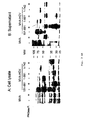
- rMVA Since the generated rMVA should be tested as a vector vaccine candidate for the delivery and immunological characterization of HCV antigens, the production of HCV proteins in tissue culture infection should be carefully determined. All rMVA containing different coding sequences of the core, E1 and E2 proteins piloted the synthesis of the HCV structural proteins as demonstrated by SDS-PAGE analysis and immunoblotting of BHK-21 cell lysates, 24 h of the infection were harvested ( FIG. 2A, B ).
- Monoclonal antibodies raised against the HCV core protein gave specific equal amounts of an approximately 21 kDa core protein produced in MVA-HCV / 1-742 or MVA-HCV / 1-661 infected cells, whereas none such a protein band has been detected after infection with MVA-HCV / 151-661 or non-recombinant MVA ( FIG. 2A ). At later times and with increasing amounts of core protein, a second 23 kDa-specific protein band could be detected. The synthesis of HCV coat protein was monitored with an E2-specific polyclonal rabbit antiserum. All three recombinant viruses produced Fig.
- E2 polypeptides which were visualized as proteins with molecular weights of about 60 kDa for MVA-HCV / 1-742 and about 50 kDa for MVA-HCV / 1-661 and MVA-HCV / 151-661.
- the latter viruses both contain expression cassettes for HCV polyproteins with C-terminally truncated E2 sequences.
- Several other smaller protein bands were also stained by the polyclonal serum and are likely to be proteins that are not HCV encoded, as they were also detected in control lysates from cells infected with wild type MVA.
- HCV envelope proteins E1 and E2 are highly modified by glycosylation and are likely to be type I transmembrane glycoproteins with a carboxy terminal hydrophobic anchor domain. The removal of the E2 transmembrane anchor leads to the secretion of the E2 ectodomain (for overview, see 17).
- the eukaryotic MVA expression system should cause the appearance of authentic post-translational modifications of recombinant proteins.
- the E2 proteins were biochemically characterized, which are synthesized upon infection with rMVAs.
- BHK-21 cells were infected with MVA-HCV / 1-742, MVA-HCV / 1-661 and MVA-HCV / 151-661.
- Total cell lysates ( Fig. 3A ) or precipitates of supernatants from infected cells ( Fig. 3B ) were treated with PNGase F and analyzed by Western blot using polyclonal anti-E2 antiserum.
- E2 proteins (MVA-HCV / 1-742) or ⁇ 50 kDa (MVA-HCV / 1-661 and MVA-HCV / 151-661) in cell lysates were grown to a size of about 33 kDa (full-length -E2) or 31 kDa (truncated E2), corresponding to the unglycosylated peptide backbone of E2 ( FIG. 3A ).
- Secreted E2 could only be detected after infection with the recombinant viruses MVA-HCV / 1-661 and MVA-HCV / 151-661, which produce an E2 truncated in the transmembrane domain.
- Secreted E2 is more strongly modified than cell-associated E2 with an apparent molecular weight of -75 kDa. When treated with PNGase F, these proteins are also reduced to a size of about 31 kDa, corresponding to the peptide backbone of the truncated E2.
- the next step was to characterize the type of glycosylation in these secreted E2 proteins. Cells were infected with MVA-HCV / 1-661 and MVA-HCV / 151-661, cell lysates or supernatants of infected cells were transfected with endoglycosidase H (Fig. Fig. 4 ).
- Intracellular E2 is sensitive to EndoH, as shown by a reduction in MW from -50 kD to -32 kD ( Fig. 4A ), but secreted E2 is largely unaffected ( Fig. 4B ).
- the E2 bands from the PNGase F cleavage had a lower MW than the corresponding bands of the Endo H cleavage, since the latter enzyme leaves a GlcNAc residue on the side chain of Asn after cleavage. This observation is consistent with a more complex glycosylation of C-terminal truncated E2 as it passes through the Golgi apparatus.
- mice with rMVA Inoculation of mice with rMVA elicits HCV and vaccinia virus-specific immune responses.
- HCV-specific humoral or cellular responses were directed against HCV E1 and E2 antigens.
- HCV E1-specific CTL responses were examined.
- Splenocytes derived from animals immunized with MVA / HCV / 151-661 or MVA-HCV / 1-661 showed E1-specific lysis of 75% at an E: T ratio of 9: 1, splenocytes of Animals immunized with MVA-HCV / 1-742 gave specific lysis of 47% when stimulated with the E1 peptide ( Fig. 5 ). Higher E1-specific lysis was observed in cultures with splenocytes from mice immunized with rMVA-expressing C-terminal truncated E2 proteins.
- Serum samples from the same BALB / c mice were also screened for antibody responses against the HCV E2 protein by ELISA ( Fig. 6 ).
- E2-specific antibodies with median titers of 1: 2512 (MVA-HCV / 151-661), 1: 933 (MVA-HCV / 1-661) and 1: 176 (MVA-HCV / 1-742) were detected. Higher levels of anti-E2 antibody were found in animals immunized with MVA-HCV / 151-661, although differences among groups were not statistically significant.
- the HCV core protein may have an immunomodulatory function. It has now been investigated whether the stronger anti-E2 antibody response of MVA-HCV / 151-661-injected mice could be attributed to the large truncation of the core protein in this virus.
- the HCV core was also expected to modulate the induction of vaccinia virus MVA specific immune responses. Therefore, the levels of MVA-neutralizing antibodies in the same mouse sera as used for the. ELISA were used. According to Table 1, no difference in the anti-MVA antibody response among the groups immunized with recombinant or wild-type MVA-expressing full-length or truncated core protein was detected.
- rMVA The formation and isolation of rMVA was based on transient host range gene selection (58) and demonstrated that this technique is particularly well suited when multiple MVA vector viruses need to be generated for comparative study.
- Recombinant MVA expressing the various HCV coding sequences under the control of the vaccinia virus early-late promoter P7.5 could be readily obtained, and initial experiments showed that all constructs consistently produced similar levels of HCV structural proteins.
- HCV core sequence is one of the most conserved regions in the viral genome, reaching as far as viruses from different HCV genotypes and making the core protein interesting as an antigen for immunization (6, 57, 7). Nevertheless, the inclusion of core in vaccine candidates is highly controversial, given its multifunctional properties may be involved in the regulation of host cell apoptosis, transcription, transformation, and immune presentation (43, 4).
- the expression of HCV sequences encoding polyprotein AS 1-742 and 1-661 by recombinant MVA resulted predominantly in the synthesis of the 21 kDa core proteins ( Fig. 2A ). Only at later times of infection, with increasing amounts of recombinant protein produced, did a second core-specific protein band become apparent at 23 kDa size.
- the 23 kDa protein is likely to be the immature 191 amino acid core protein that results from initial polyprotein cleavage, whereas the 21 kDa protein is likely to be the mature core protein present in virus particles (28, 26, 51, 55 , 66). These data demonstrate efficient early synthesis and processing of the core polypeptides in MVA-HCV vector infected cells.
- both E2 proteins migrate after deglycosylation with PGNAse F at the same level.
- secreted E2 was insensitive to treatment with endoglycosidase H, which clearly shows the uptake of complex sugars during transport through the Golgi apparatus.
- HCV E1 protein While the expression of the HCV E1 protein was not formally analyzed, the E2 protein forms a downstream portion of the HCV polyprotein, and the demonstration of E2 synthesis therefore suggests production of the E1 protein sequences. The correctness of this idea was confirmed by the detection of E1-specific T-cell responses after vaccination with recombinant MVA-HCV viruses.
- E1 epitope 312-326 was successfully used for in vitro restimulation of splenocytes from vaccinated BALB / c mice, and epitope-specific CTLs were detected, which were induced by all three rMVA vaccines. Interestingly, detection of the E1-specific CTL was also possible more than 12 weeks after the last immunization.
- E2 transmembrane region inhibits the formation of native E1-E2 complexes and prevents the correct folding of the E1 glycoprotein (11, 48).
- E1 antigen appears to be derived from the endoplasmic reticulum, requires cytoplasmic processing (54), and probably relies on a secondary export pathway for defective transmembrane glycoproteins into the cytosol, as for the influenza nucleoprotein or the cellular glycoprotein tyrosinase has been proposed (2, 47).
- the secretion of E2 could, of course, elicit better antibody responses, whereas the interaction of the full-length E1 and E2 proteins to form intracellularly retained complexes could mask important epitopes and prevent optimal immunoprecipitation.
- the HCV core protein produced by replication-competent vaccinia virus appears to suppress host immune responses, in particular to suppress the production of vaccinia virus-specific immune responses ( Large et al 1999, J Immunol 162: 931-938 ). To fit these data, in the vaccination trials shown here, the best E2-specific antibody responses appeared to be induced by the MVA HCV / 151-661 vaccine, which produced truncated E2 in the absence of core antigen.
- rMVA can be used to efficiently produce posttranslationally mature core and envelope proteins.
- Vaccination of BALB / c mice with rMVA vaccines also induced comparable levels of E2-specific antibodies and long-lasting E1-specific cytotoxic T-cell responses and demonstrated the utility of this viral vector for further evaluation as a promising vaccine against HCV -Infection.
- rMVA was investigated as a potential vaccine candidate against HCV infection.
- MVA vector vaccines were constructed which express all major HCV virion component coding sequences to characterize the synthesis of recombinant HCV proteins upon MVA infection and to evaluate their immunogenicity in use as vaccines.
- Vaccinia virus neutralizing antibodies induced by MVA vaccines ⁇ / b> Vaccine a Titer b (reciprocal mean log2) c d0 d28 d55 d110 MVA ⁇ 5.3 10.3 12.3 12.3 MVA-HCV / 151-661 ⁇ 5.3 9, 5 12.3 12.3 MVA-HCV / 1-661 ⁇ 5.3 10, 1 11.9 12.1 MVA-HCV / 1-742 ⁇ 5.3 9.9 12.3 12.3 a) 10 8 IU MVA vaccine given on the intraperitoneal route on days 0, 38, 81 b) Serum antibody titers were determined by the 50% vaccinia virus plaque reduction assay c) n 5, maximum standard deviation
Landscapes
- Health & Medical Sciences (AREA)
- Life Sciences & Earth Sciences (AREA)
- Chemical & Material Sciences (AREA)
- Virology (AREA)
- General Health & Medical Sciences (AREA)
- Pharmacology & Pharmacy (AREA)
- Veterinary Medicine (AREA)
- Public Health (AREA)
- Medicinal Chemistry (AREA)
- Animal Behavior & Ethology (AREA)
- Organic Chemistry (AREA)
- Engineering & Computer Science (AREA)
- Genetics & Genomics (AREA)
- Immunology (AREA)
- Microbiology (AREA)
- General Chemical & Material Sciences (AREA)
- Chemical Kinetics & Catalysis (AREA)
- Nuclear Medicine, Radiotherapy & Molecular Imaging (AREA)
- Bioinformatics & Cheminformatics (AREA)
- Communicable Diseases (AREA)
- Epidemiology (AREA)
- Mycology (AREA)
- Zoology (AREA)
- Biotechnology (AREA)
- Molecular Biology (AREA)
- Wood Science & Technology (AREA)
- Biomedical Technology (AREA)
- General Engineering & Computer Science (AREA)
- Oncology (AREA)
- Plant Pathology (AREA)
- Biophysics (AREA)
- Physics & Mathematics (AREA)
- Biochemistry (AREA)
- Tropical Medicine & Parasitology (AREA)
- AIDS & HIV (AREA)
- Gastroenterology & Hepatology (AREA)
- Micro-Organisms Or Cultivation Processes Thereof (AREA)
- Medicines Containing Antibodies Or Antigens For Use As Internal Diagnostic Agents (AREA)
- Peptides Or Proteins (AREA)
- Medicines That Contain Protein Lipid Enzymes And Other Medicines (AREA)
Description
- Die Erfindung betrifft rekombinantes Modifiziertes Vaccinia Virus Ankara (MVA), welches in der Lage ist, HCV-Struktur-Antigene, immunogene Abschnitte dieser Struktur-Antigene oder Epitope dieser Struktur-Antigene zu exprimieren. Die Erfindung beschreibt weiterhin eine pharmazeutische Zubereitung, die insbesondere in Form einer Vakzine vorliegt und ein rekombinantes MVA gemäß der Erfindung enthält, Eukaryontenzellen, die das erfindungsgemäße rekombinante MVA enthalten sowie verschiedene Verwendungen des rekombinanten MVA, beispielsweise zur Produktion rekombinanter Strukturproteine, zur Herstellung einer pharmazeutischen Zubereitung, die insbesondere zur Therapie und Prophylaxe von HCV-Infektionen und hierdurch verursachter Erkrankungen geeignet ist, Verfahren zur Herstellung von rekombinantem MVA und von rekombinanten HCV-Struktur-Polypeptiden, die von diesem rekombinanten MVA kodiert werden, sowie DNA oder RNA dieses rekombinanten MVA.
- Das Hepatitis-C-Virus (HCV) ist ein positiv-strängiges RNA-Virus, das zur Familie Flaviviridae gehört. Es ist das hauptsächliche Agens einer nach einer Transfusion und in der Bevölkerung erworbenen Non-A-, Non-B-Hepatitis (8, 35). Mehr als 70% der infizierten Patienten entwickeln eine chronische Hepatitis mit dem Risiko, dass sich diese zur Zirrhose und zum hepatozellulären Karzinom weiter entwickelt (30, 53). Die gängigen Behandlungsmethoden sind eingeschränkt (39, 40, 31, 24, 13). Es wurden zwar viele Mühen in die Impfstoffentwicklung investiert, und es wurden auch einige ermutigende Ergebnisse erzielt (9, 41, 22, 25), jedoch ist bisher noch kein wirksamer und/oder therapeutischer Impfstoff verfügbar.
- Die HCV-Strukturproteine, d.h. der virale Capsidprotein-Kern, und die Hüllglycoproteine E1 und E2 sind vielversprechende Impfstoff-Ziele: Das Core-Antigen ist bei verschiedenen HCV-Genotypen und -Subtypen hoch konserviert (6). Innere Antigene, wie Nukleokapside, gehen in verschiedenen Modellen, bspw. Rabies-Virus, Hepatitis-B-Virus und Influenza-Virus, mit schützenden Immunantworten einher (14, 33, 62). Mit den folgenden verschiedenen Ansätzen konnten HCV-Kern-spezifische Antikörper- und CTL-Antworten induziert und nachgewiesen werden (23, 32). Das Core-Protein scheint dagegen viele regulatorische Funktionen zu besitzen, die an der Modulation der Wirtszell-Apoptose, Transkription, Transformation und Immun-Präsentation beteiligt sind (43, 4). Die Immunisierung von Schimpansen mit rekombinanten E1- und E2-Proteinen induziert einen Schutz gegen einen homologen Challenge vermutlich über die Induktion neutralisierender Antikörper (9). Einer Anzahl von Berichten zufolge könnte die hypervariable Domäne (HVR), die sich am N-Terminus von E2 befindet, eine wichtige Neutralisierungsstelle sein (20, 34, 67, 68).
- Das Fehlen eines geeigneten Tiermodells erschwert das Studium von Wirts-Immunreaktionen gegen HCV-Infektion und die Bewertung des Impfschutzes erheblich. Das Fehlen eines effizienten In-vitro-Zellkultursystems für eine produktive HCV-Vermehrung und geringe Mengen HCV-Teilchen in infizierten Lebergeweben oder Blut behinderten die Erzeugung und die Bewertung konventionellerer Impfstoffe, bspw. auf der Grundlage lebender abgeschwächter oder inaktivierter Viren. Die Verwendung von Vektor-Impfstoffen auf der Basis lebender Trägerviren zur Expression antigencodierender Sequenzen bietet einen alternativen Ansatz bei der HCV-Impfstoffentwicklung.
- Es ist eine Aufgabe der vorliegenden Erfindung, ein neues Mittel bereitzustellen, welches zur Prophylaxe und Therapie von Mensch und Tier egen HCV-Infektionen geeignet ist und die oben beschriebenen Nachteile vermeidet.
- Diese Aufgabe wird erfindungsgemäß durch rekombinantes MVA gemäß den Merkmalen des Anspruchs 1 gelöst, welches so modifiziert, wurde, daß es zur Expression von HCV-Struktur-Antigenen, immunogenen Abschnitten von HCV-Struktur-Antigenen und/oder von Epitopen dieser HCV-Struktur-Antigene, bevorzugt von Oberflächenepitopen, in der Lage ist.
- Bevorzugte Ausgestaltungen der Erfindung sind aus der nachfolgenden Beschreibung sowie den beiliegenden Patentansprüchen zu entnehmen.
- Erfindungsgemäß konnte gezeigt werden, daß rekombinantes MVA mit DNA-Sequenzen, die für HCV-Struktur-Antigene, immunogene Abschnitte hiervon oder für Epitope dieser HCV-Struktur-Antigene kodieren, zur Herstellung einer Vakzine hervorragend geeignet ist. Dies war deshalb überraschend, weil insbesondere die Produktion bzw. die Co-Produktion des HCV Core-Antigens die Immunogenität des Impfstoffs nicht beeinträchtigt. Die Veröffentlichung (19) zeigte nämlich, daß durch die Produktion des Core-Antigens durch ein replikationskompetentes Vaccinia-Virus die Immunantwort in einem infizierten bzw. geimpften Wirt in spezifischer Weise supprimiert wurde. Man konnte deshalb davon ausgehen, daß auch bei Verwendung von MVA als Vektor zur Produktion von HCV-Struktur-Antigenen eine derartige Immunsuppression erfolgt. Unerwarteterweise konnte aber gezeigt werden, daß bei Verwendung von MVA-Vektoren diese Immunsuppression wieder aufgehoben wird. Dieses erstaunliche, experimentell gestützte Resultat bietet eine hervorragende Basis zur Entwicklung von Impfstoffen, insbesondere auf der Grundlage des HCV-Antigens als Impfantigen.
- Der erfindungsgemäß eingesetzte virale Vektor basiert auf dem modifizierten Vaccinia-Virus Ankara (MVA), einem stark abgeschwächten Vaccinia-Virus-Stamm, der sich höchstwahrscheinlich aufgrund seiner Replikationsdefizienz in Säugetier-Wirten für die klinische Verwendung als sicher erwiesen hat (42, 45, 60, 50). Rekombinantes MVA (rMVA) als Impfstoff erwies sich als wirksam bei der Anregung von T-Zell- oder Antikörperantworten auf verschiedene heterologe Antigene und als schützend beim Test in Tier-Challenge-Modellen für Infektionskrankheiten beim Menschen, einschließlich Influenza, AIDS, Masern und Malaria (61, 3, 27, 52, 15, 59, 1).
- Trotz der an sich bekannten Verwendung von rekombinantem MVA zur Herstellung einer Vakzine war somit im vorliegenden Fall die Verwendung in Verbindung mit HCV-Struktur-Antigenen, nämlich dem Core-Antigen von HCV sowie E1 und E2, nicht nahegelegen.
- Die erfindungsgemäß eingesetzten Struktur-Antigene des HCV sind das Kapsid-Protein Core, das Hüll-Glycoprotein E1, und das Hüll-Glycoprotein E2.
- Selbstverständlich ist es nicht notwendig, zur Herstellung einer Vakzine die Struktur-Antigene von HCV vollständig zu exprimieren. Vom Experimentator können je nach Versuchsplänung und gewünschtem Resultat auch nur Teile der Struktur-Antigene zur Expression gebracht werden, insbesondere natürlich immunogene Abschnitte oder Epitope der HCV-Struktur-Antigene. Zu den immunogene Abschnitte der HCV-Struktur-Antigene. Beispiele hierfür sind die C-terminale Core-Proteinregion (AS 127-190 des HCV Polyproteins), die E1-Proteinregion der HCV-Polyprotein-AS 220-371, sowie die 27 AS umfassende hypervariable Region 1 am N-Terminus des Glykoproteins E2 (Rehermann & Chisari 2000, Curr Top Microbiol Immunol 242:299-325, Penin et al 2001, J Virol 75:5703-5710). Zu den funktionellen Teilen der Struktur-Antigene gehören bspw. die für den Zusammenbau von Virus-ähnlichen Nukleokapsid-Partikeln ausreichenden, 124 N-terminalen AS des HCV Core-Proteins (Kunkel et al. J. Virol 75:2119-2129, 2001) und die für die Heterodimerisierung der Hüllproteine E1 und E2 essentiellen Transmembran-Domänen TMD-E1 AS 350-370 bzw. TMD-E2 AS 717-740 (Op de Beeck et al. 2000, J. Biol Chem 275:31428-31437) bzw. sezernierbare Formen der Glykoproteine, die in der Lage sind, das Endoplasmatische Reticulum zu passieren. Dies wird bevorzugt dadurch erreicht, daß die Transmembran-Region von E1 bzw. E2 , durch Deletion oder anderweitige Mutationen so verändert wird, daß die Proteine nicht mehr in der Membran zurückgehalten werden (Michalak et al. 1997, J Gen Virol 78:2299-2306).
- Epitope der HCV-Struktur-Antigene sind bekannt. Hierzu gehören bspw. die MHC-I-restringierten T-Zellepitope core AS 2-10, core AS 35-44, core AS 41-49, core AS 85-98, core AS 132-140, core AS167-176, core AS 178-187, core AS 181-190, E1 AS 220-227, E1 AS 233-242, E1 AS 363-371, E2 AS 401-411, E2 AS 489-496, E2 AS 621-628 oder E2 AS 725-733 (Rehermann & Chisari 2000, Curr Top Microbiol Immunol 242:299-325) Weitere Beispiele sind das E1-Epitop AS 312 - 326. Die Epitope der HCV-Struktur-Antigene können auch durch an sich bekannte Techniken bestimmt werden, die bspw. in Koziel et al 1995, J. Clin Invest 96:2311-2321 beschrieben sind.
- In einer bevorzugten Ausgestaltungsform der Erfindung enthält das rekombinante MVA DNA-Sequenzen, die für E1 und E2 kodieren. Insbesondere vorteilhaft ist die Verwendung des E1-Antigens in Verbindung mit einer verkürzten Form des E2-Antigens, so daß beide Antigene co-produziert werden. "Verkürzt" heißt, daß der lipophile Anteil des E2-Glycoproteins deletiert ist. Es zeigte sich, daß in dieser Ausgestaltungsform des rekombinanten MVA nach Vakzinierung die gegen das E1-Antigen gerichteten zellulären Immunantworten, das heißt die Induktion E1-spezifischer zytotoxischer T-Zellen, deutlich verstärkt war im Vergleich zur Vakzinierung mit rekombinantem MVA, welches das E1-Antigen zusammen mit dem vollständigem E2 Protein exprimierte. Selbstverständlich ist es auch möglich, verschiedene HCV-Struktur-Antigene, insbesondere E1 und E2, nicht auf einem MVA-Vektor anzuordnen, sondern auf zumindest zwei Vektoren. Auch dies führt im Ergebnis zu einer Co-Produktion von HCV-Struktur-Antigenen.
- Erfindungsgemäß wird unter Mutation eine Deletion, Basensubstitution oder eine Basenaddition verstanden. Auch chemische Modifikationen sind möglich.
- Die Anordnung der HCV-Struktur-Antigene im rekombinanten MVA ist so vorzunehmen, daß eine funktionelle Verbindung der kodierenden Sequenzen mit Steuerelementen, bspw. Promotoren und Enhancern, entsteht. Geeignete DNA-Klonierungstechniken stehen dem Fachmann zur Verfügung. Es sei hier nur beispielsweise hingewiesen auf Sambrook, J. et al., 1989, Molecular Cloning: A Laboratory Manual, Cold Spring Harbor Laboratory, NY; oder Ausubel, F.M. et al., 1989, Current Protocols in Molecular Biology, John Wiley & Sons, New York, NY. Auf diese beispielhaft genannten Veröffentlichungen wird hier Bezug genommen. Insbesondere sind die kodierenden DNA-Sequenzen so anzuordnen und auszuwählen, daß eine korrekte Prozessierung der Polypeptide in den mit dem MVA-HCV-Vektorvirus infizierten Zellen erfolgt.
- Die Integration der Fremd-DNA Sequenzen in das MVA-Genom erfolgt bevorzugt in solche Bereiche, die für die Replikation und Infektionsfähigkeit des MVA nicht essentiell sind. Als Integrationsorte kommen bspw. natürlich vorkommende Deletionen im MVA-Genom in Frage. Zu diesen Deletionen, die den Lebenszyklus des Virus nicht essentiell beeinflussen, gehören die Deletionen I, II, III, IV, V, und VI, bevorzugt die Deletionen II und III (Meyer, H., Sutter, G. und Mayr, A., 1991, J. Gen. Virol. 72, 1031-1038). Das so hergestellte rekombinante MVA ist infektiös, d.h. es ist in der Lage, Fremdzellen zu infizieren und die integrierte Fremd-DNA-Sequenz des HCV zu exprimieren. Derartige rekombinante Viren eignen sich als äußerst sichere Lebendvakzine zur Behandlung oder Prophylaxe von durch HCV verursachten Erkrankungen.
- Das erfindungsgemäße rekombinante MVA kann, wie nachfolgend allgemein dargestellt, hergestellt werden. Eine detaillierte Beschreibung findet sich in den Ausführungsbeispielen. Das Einführen der HCV-Strukturantigene in die MVA-DNA erfolgt beispielsweise im Wege einer homologen Rekombination, wobei die zu integrierende Sequenz an beiden Enden mit solchen Sequenzen versehen wird, die mit Sequenzen rekombinierbar sind, die den natürlich vorkommenden Deletionen, beispielsweise Deletion II oder III, im MVA-Genom benachbart angeordnet sind (Antoine et al. 1998, Virology 244:365-396).
- Ein derartiges DNA-Konstrukt, welches ein HCV-Strukturantigen oder funktionelle Teile oder Epitope hiervon enthält, flankiert von MVA-DNA-Sequenzen, die benachbart von den Stellen der Deletionen, beispielsweise Deletion II oder III, im MVA-Genom vorkommen, werden in mit MVA infizierte Zellen eingeführt, um eine homologe Rekombination zu ermöglichen. Das eingeführte DNA-Konstrukt kann linear oder zirkulär sein. Bevorzugt wird ein zirkuläres DNA-Molekül, insbesondere ein Plasmid, eingesetzt.
- Zur Expression der für HCV-Strukturantigene codierenden DNA-Sequenz sind regulatorische Sequenzen notwendig, die eine Transkription der DNA-Sequenz ermöglichen. Derartige regulatorische Sequenzen, beispielsweise Promotoren und Enhancer, sind an sich bekannt. Hierzu gehören beispielsweise auch die Promotoren des Vaccinia 11 kDa-Gens (vergleiche
EP 0 198 328 ), des 7,5 kDa-Gens (vergleicheEP 0 110 385 ) oder synthetische Vacciniavirus-spezifische Promotoren (vergleiche Sutter et al., Vaccine 1994, 12: 1032). - Das DNA-Konstrukt kann in MVA infizierte Zellen durch an sich bekannte Verfahren eingeführt werden, beispielsweise im Wege einer Transfektion durch Calciumphosphatpräzipitation (Graham et al., Virol. 52, 456-467, 1973; Wigler et al., Cell 777-785, 1979, durch Elektroporation (Neumann et al., EMBO J. 1, 841-845,1982), durch Mikroinjektion (Graessmann et al., Meth. Enzymology 101, 482-492, 1983), durch Liposomentechnologie (Straubinger et al., Methods in Enzymology 101, 512-527, 1983), durch Sphäroplastentechnologie (Schaffner, Proc. Natl. Acad. Sci. USA 77, 2163-2167, 1980) oder andere an sich bekannte Methoden. Bevorzugt wird die Calciumphophattechnik eingesetzt.
- Nach Einführen des DNA-Konstrukts in die Eukaryontenzellen und nachher erfolgter Rekombination der HCV-DNA mit der viralen DNA kann das rekombinante MVA in an sich bekannter Weise isoliert werden, bevorzugt mit Hilfe eines Markergens (vergleiche Nakano et al., Proc. Natl. Acad. Sci. USA 79, 1593-1596, 1982; Franke et al., Mol. Cell. Biol. 1918-1924, 1985, Chakrabarti et al., Mol. Cell. Biol. 3403-3409, 1985, Fathi et al., Virology 97-105, 1986).
- Das erfindungsgemäß hergestellte MVA kann ebenfalls ein Markergen tragen. Derartige Markergene erleichtern die Isolierung des rekombinanten Virus unter Zuhilfenahme an sich bekannter Techniken. Derartige Markergene sind an sich bekannt, und hierzu gehören Gene, die für Proteine wie β-Galactosidase, Neomycin, Alkoholdehydrogenase, Luciferase, Puromycin, Hypoxanthin-Phosphoribosyltransferase (HPRT), Hygromycin, sezernierte alkalische Phosphatase oder Grün- und Blau-Fluoreszenzproteine.
- Selbstverständlich sind auch andere Methoden zur Herstellung des rekombinanten Virus der vorliegenden Erfindung möglich, beispielsweise die im nachfolgenden Ausführungsbeispiel näher beschriebene Klonierungsmethode.
- Das MVA-Virus ist ein stark abgeschwächtes Vaccinia-Virus, das vom Vaccinia-Virusstamm Ankara (CVA) durch serielle Langzeit-Passagen auf primären Hühnerembryo-Fibroblasten-(CEF)-Kulturen hergeleitet ist. Für eine allgemeine Übersicht über die Geschichte der Produktion, der Eigenschaften und der Verwendung des MVA-Stammes kann auf die Zusammenfassung Bezug genommen werden, die von Mayr et al. in Infection 3, 6-14 [1975] veröffentlicht wurde. Aufgrund der Abschwächung in CEF repliziert das MVA-Virus zu hohen Titern in dieser Vogel-Wirtszelle. In Säugerzellen ist das Wachstum von MVA jedoch stark gehemmt, und eine typische Plaquebildung durch das Virus ist nicht nachweisbar. Das MVA-Virus wurde daher auf CEF-Zellen gezüchtet. Zur Herstellung von CEF-Zellen wurden 11 Tage alte Embryos aus inkubierten Hühnereiern isoliert, die Extremitäten wurden entfernt, und die Embryos wurden zerkleinert und in einer Lösung, die aus 0,25% Trypsin bestand, 20 min lang bei 37°C dissoziiert. Die resultierende Zellsuspension wurde filtriert, und die Zellen wurden durch Zentrifugation bei 2000 U/min in einer Sorvall RC-3B-Zentrifuge 5 min bei Raumtemperatur sedimentiert, in 10 Volumina Medium A (MEM Eagle, bspw. erhältlich von Life Technologies GmbH Eggenstein, Deutschland) resuspendiert und erneut 5 min durch Zentrifugation bei 2000 U/min in einer Sorvall RC-3B Zentrifuge bei Raumtemperatur sedimentiert. Das Zellpellet wurde in Medium A, das 10 % fötales Kälberserum, (FCS), Penicillin (100 Units/ml), Streptomycin (100 mg/ml) und 2 mM Glutamin wieder aufgeschlämmt, so dass eine Zellsuspension erhalten wurde, die 500 000 Zellen/ml enthielt. Derart erhaltene CEF-Zellen wurden auf Zellkulturschalen verteilt. Diese wurden je nach gewünschter Zelldichte in Medium A in einem CO2-Inkubator 1-2 Tage bei 37°C wachsen gelassen, und wurden entweder direkt oder nach einer weiteren Zellpassage für die Infektion verwendet. Eine eingehende Beschreibung der Herstellung von Primärkulturen befindet sich in dem Buch von R.I. Freshney, "Culture of animal cell", Alan R. Liss Verlag, New York [1983] Kapitel 11, S. 99ff.
- MVA-Viren wurden wie folgt zur Infektion verwendet. Die CEF-Zellen wurden in 175 cm2 Zellkulturflaschen gezüchtet. Bei 90 bis 100%iger Konfluenz wurde das Medium entfernt, und die Zellen wurden für eine Stunde mit einer MVA-Virussuspension (0,01 infektiöse Einheiten (IU) pro Zelle, 0,02 ml/cm2) in Medium A inkubiert. Dann wurde mehr Medium A dazugegeben (0,2 ml/cm2), und die Flaschen wurden 2 bis 3 Tage bei 37°C inkubiert (bis etwa 90% der Zellen cytopathogene Wirkung zeigten). Rohe Virusstammpräparate wurden hergestellt, indem die Zell-Monolayer in das Medium geschabt und das Zellmaterial durch Zentrifugation bei 3000 U/min in einer Sorvall RC-3B Zentrifuge 5 min bei 4°C pelletiert wurden. Das rohe Viruspräparat wurde vor der weiteren Verarbeitung (z.B. Virusreinigung) bei -20°C aufbewahrt.
- Die Reinigungsschritte, die unternommen wurden, um ein Viruspräparat zu erhalten, das so rein wie möglich war und keine für die Wirtszelle spezifischen Komponenten enthielt, ähnelten denjenigen, die von Joklik (Virology 18, 9-18 [1962]) beschrieben wurden. Rohe Virusstammpräparate, die bei -20°C aufbewahrt worden waren, wurden aufgetaut und einmal in PBS suspendiert (das 10 bis 20fache des Sedimentvolumens), und die Suspension wurde wie oben zentrifugiert. Das neue Sediment wurde im 10fachen Volumen Tris-Puffer 1 (10 mM Tris-HCl, pH-Wert 9,0) suspendiert, und die Suspension wurde kurz mit Ultraschall behandelt (Labsonic, L.B. Braun Biotech International, Melsungen, Deutschland; 2 x 10 sec bei 60 Watt und Raumtemperatur), um die Zelltrümmer weiter zu zerkleinern und um die Viruspartikel aus dem zellulären Material freizusetzen. Die Zellkerne und die größeren Zelltrümmer wurden in der nachfolgenden kurzen Zentrifugation der Suspension (Sorvall GSA-Rotor-erhältlich von DuPont Co., D-6353 Bad Nauheim, Deutschland, 3 min bei 3000 U/min und 10°C) entfernt. Das Sediment wurde noch einmal in Tris Puffer 1 suspendiert, mit Ultraschall behandelt und wie vorstehend beschrieben zentrifugiert. Die gesammelten Überstände, die frei von Viruspartikeln sind, wurden vereinigt und über ein Kissen aus 10 ml 36%iger Saccharose in 10 mM Tris-HCl, pH-Wert 9,0 geschichtet und in einem Beckman SW 27/28-Rotor 80 min bei 13500 U/min bei 4°C zentrifugiert. Der Überstand wurde verworfen, und das Sediment, das die Viruspartikel enthielt, wurde in 10 ml 1 mM Tris-HCl, pH-Wert 9,0 aufgenommen, durch kurze Behandlung mit Ultraschall (2 x 10 sec bei Raumtemperatur, Gerät wie vorstehend beschrieben) homogenisiert und auf einen 20-40% Saccharosegradienten (Saccharose in 1 mM Tris-HCl, pH-Wert 9,0) zur weiteren Reinigung aufgetragen. Der Gradient wurde 50 min in einem Beckman SW41 Rotor bei 13000 U/min und 4°C zentrifugiert. Nach der Zentrifugation wurden getrennte Banden, die Viruspartikel enthielten, durch Pipettieren geerntet, nachdem das Volumen über der Bande abgesaugt wurde. Die erhaltene Saccharoselösung wurde mit drei Volumina PBS verdünnt, und die Viruspartikel wurden wiederum durch Zentrifugation (Beckman SW 27/28, 60 min bei 13500 U/min, 4°C) sedimentiert. Das Pellet, das nun überwiegend aus reinen Viruspartikeln bestand, wurde in PBS resuspendiert und auf Virus-Konzentrationen äquilibriert, die im Durchschnitt 1 bis 5 x 10⁹ IU/ml entsprachen. Die gereinigte Virus-Stammlösung wurde bei -80°C aufbewahrt und entweder direkt verwendet oder für die nachfolgenden Experimente mit PBS verdünnt.
- Das modifizierte Vaccinia-Virus Ankara (MVA), ein stark abgeschwächter Vaccinia-Virusstamm mit eingeschränktem Wirtsspektrum, kann sich in vom Menschen stammenden Zellen und den meisten anderen untersuchten Säugerzelllinien nicht vermehren. Da jedoch die virale Genexpression in nichtpermissiven Zellen nicht beeinträchtigt ist, kann das erfindungsgemäße rekombinante MVA als ungewöhnlich sicherer und effizienter Expressionsvektor und rekombinante Vakzine verwendet werden.
- Bei einer erfindungsgemäßen Ausführungsform wurden somit rekombinante MVA-Viren konstruiert, die die Expression der HCV-Strukturantigene unter der Kontrolle des Vaccinia-Virus-Early/Late-Promotors P7.5 ermöglichen.
- Das rekombinante MVA-Virus, welches ein HCV-Strukturantigen exprimiert, kann z.B. zur Immunisierung für die Immuntherapie bei Krebspatienten verwendet werden.
- Darüber hinaus kann das rekombinante MVA-Virus, welches HCV-Strukturantigene exprimiert, in Verbindung mit antigenpräsentierenden Zellen, bspw. Dendritenzellen, Makrophagen und B-Zellen, zur Immunisierung von Menschen für die Immuntherapie verwendet werden.
- Das rekombinante MVA-Virus kann überdies in Verbindung mit antigenpräsentierenden Zellen, bspw. Dendritenzellen, ex vivo zur Erzeugung von Immuneffektorzellen verwendet werden, welche an Menschen zur Immuntherapie applizierbar sind.
- Das rekombinante MVA-Virus kann darüber hinaus zur Herstellung von rekombinanten HCV-Strukturproteinen verwendet werden.
- Die erfindungsgemäßen MVA-Vaccinia-Viren werden zur Herstellung von Vakzinen oder Therapeutika in eine physiologisch verträglich Form umgewandelt. Hier kann man auf die Erfahrung bei der Herstellung von MVA-Vakzinen zurückgreifen, die zur Impfung gegen Pocken verwendet werden (wie beschrieben von Stickl, H., et al. [1974] Dtsch. med. Wschr. 99, 2386-2392). Gewöhnlich werden etwa 106 bis 108 rekombinante MVA-Partikel in 100 ml phosphatgepufferter Salzlösung (PBS) in Gegenwart von 2% Pepton und 1% menschlichem Albumin in einer Ampulle, vorzugsweise einer Glasampulle, gefriergetrocknet. Das Lyophilisat kann Streckmittel (wie Mannitol, Dextran, Zucker, Glycin, Lactose oder Polyvinylpyrrolidon) oder andere Hilfsmittel (wie Antioxidantien, Stabilisatoren, usw.) enthalten, die sich zur parenteralen Verabreichung eignen. Die Glasampulle wird dann verschlossen, und kann dann, vorzugsweise bei Temperaturen unter -20°C, mehrere Monate lang aufbewahrt werden.
- Zur Impfung oder zur Therapie kann das Lyophilisat in 0,1 bis 0,5 ml wässriger Lösung, vorzugsweise in physiologischer Kochsalzlösung, gelöst werden, und entweder parenteral, bspw. durch intramuskuläre Impfung, oder lokal, bspw. durch intradermale Impfung oder durch Impfung in einen Tumor oder an die Stelle eines Tumors, verabreicht werden. Erfindungsgemäße Vakzine oder Therapeutika werden vorzugsweise intramuskulär injiziert (Mayr, A. et al. [1978] Zbl. Bakt. Hyg., I. Abt. Orig. B 167, 375-390). Der Verabreichungsweg, die Dosis und die Anzahl der Verabreichungen kann durch den Fachmann auf bekannte Weise optimiert werden. Geeignetenfalls ist es angebracht, den Impfstoff mehrmals für einen längeren Zeitraum zu verabreichen, damit geeignete Immunantworten gegen das fremde Antigen erhalten werden.
- Die Erfindung umfaßt auch Eukaryontenzellen, die mit den erfindungsgemäßen rekombinanten MVA-Viren infiziert sind. Hierzu gehören bspw. Hühnerembryo-Fibroblastenzellen, Babyhamster-Nierenzellen, bevorzugt BHK-21-Zellen, oder Antigen-präsentierende Zellen wie z. B. dendritische Zellen, Makrophagen oder B-Lymphozyten.
- Die rekombinanten MVA-Viren werden, wie oben näher beschrieben, zur Herstellung einer Vakzine eingesetzt, die zur Therapie und Prophylaxe von HCV-Infektionen und hierdurch verursachter Erkrankungen, insbesondere chronischer Lebererkrankungen und Lebertumoren, einsetzbar ist. Mit Hilfe des rekombinanten Virus können natürlich auch die rekombinanten HCV-Strukturproteine produziert und nach Isolierung und Aufreinigung in reiner Form gewonnen werden. Unter rekombinanten HCV-Proteinen sind erfindungsgemäß HCV-Strukturantigene zu verstehen, wobei dieser Ausdruck auch funktionelle Teile der HCV-Strukturantigene einschließt oder Epitope, insbesondere Oberflächenepitope der HCV-Strukturantigene.
- Die Erfindung umfaßt auch DNA oder RNA des rekombinanten MVA.
- Nachfolgend wird die Erfindung anhand von Ausführungsbeispielen näher beschrieben. Die Erfindung ist jedoch nicht auf diese besondere Ausgestaltung beschränkt sondern kann im Rahmen der Beschreibung in Verbindung mit den beiliegenden Patentansprüchen abgewandelt werden.
- Die beiliegenden Figuren dienen ebenfalls dem näheren Verständnis der Erfindung, insbesondere der Ausführungsbeispiele. Die Figuren zeigen:
-
Fig. 1 Konstruktion und genomische Struktur rekombinanter MVAenthaltender Gensequenzen für HCV-Strukturproteine. (A) Eine schematische Karte des MVA-Genoms für die Restriktionsendonuklease HindIII. Die HCV-codierenden Sequenzen standen unter der transkriptionellen Kontrolle des Vaccinia-Virus-Early/Late Promoters P7.5 und wurden durch homologe Rekombination an der Stelle der Deletion III innerhalb des MVA-Genoms inseriert. Die Flanke 1 und Flanke 2 betreffen MVA-DNA-Sequenzen, die sich neben der Deletionsstelle III befinden und dienen dazu, die Rekombination in das MVA-Genom zu dirigieren. Rec2 zeigt die Positionen der 283 bp repetitiven MVA-DNA-Sequenzen, die dem rechten Ende von Flanke 1 entsprechen und die Entfernung des K1L-selektierbaren Markers aus dem Genom der fertigen rekombinanten Viren durch homologe Rekombination ermöglichen. Die Darstellungen der Genomstrukturen der rekombinanten MVA-HCV-151-661, MVA-HCV-1-661 und MVA-HCV-1-742 sind unten gezeigt. (B) PCR-Analyse viraler DNA zur Überwachung von Gensequenzen, die an der Deletionsstelle III inseriert sind. Genomische DNA von Wildtyp-MVA und rekombinantem MVA-HCV-151-661, MVA-HCV-1-661 und MVA-HCV-1-742 dienten als Matrizen-DNA zur Amplifikation charakteristischer DNA-Fragmente, welche durch Agarosegelelektrophorese aufgetrennt wurden. Die 1-kB-Leiter (Gibco) wurde als Marker (M) für das Molekulargewicht in Basenpaaren (bp) angegeben. -
Fig. 2 . Synthese von HCV-Core- und E2-Proteinen durch rekombinante MVA. BHK-21-Zellen wurden mit 10 IU/Zelle MVA oder MVA-HCV infiziert und nach 24 Std. (A, B) oder zu den angegebenen Zeitpunkten (C, D) nach der Infektion geerntet. Proteine aus Zelllysaten wurden durch SDS-10% PAGE getrennt und durch Western-Blot mittels monoklonalen Maus-anti-HCV-Core (A,C)- oder E2-spezifischen polyklonalen Kaninchen-Antikörpern (B, D) analysiert. HCV-spezifische Proteinbanden sind durch Pfeilspitzen markiert. Die Positionen und Molekülmassen (in kDa) der Proteinstandards sind in den Spuren MW gezeigt. -
Fig. 3 Glycosylierung der durch rekombinante MVA hergestellten zellassoziierten und sezernierten E2-Proteine. Gesamt-Zellproteine (A) oder Proteinpräzipitate von Überständen (B) aus MVA-infizierten BHK-21-Zellen wurden in SDS/β-Mercaptoethanol denaturiert, in Gegenwart (+) oder Abwesenheit (-) von Endoglycosidase PNGase F inkubiert, auf SDS-10%-PAGE getrennt und durch Western-Blot mit polyklonalem Kaninchen-anti-E2-Serum analysiert. HCV-E2-spezifische Proteinbanden sind durch Pfeilspitzen markiert. Die Zahlen in der Spur MW betreffen die Positionen und Molekulargewichte (kDa) der Proteinstandards. -
Fig. 4 Analyse der EndoH-Empfindlichkeit der durch rekombinante MVA hergestellten zellassoziierten und sezernierten E2-Proteine.
BHK-21-Zellen wurden 24 Std. mit rekombinantem MVA-HCV/151-661 oder MVA-HCV/1-661 oder nicht-rekombinantem MVA bei einer MOI von 10 infiziert. Gesamt-Zellproteine (A) oder Proteinpräzipitate der Überstände (B) aus infizierten Kulturen wurden in SDS/β-Mercaptoethanol denaturiert, in Gegenwart (+) oder Abwesenheit (-) von PNGase F oder EndoH inkubiert, auf SDS-10%-PAGE getrennt und durch Western-Blot mit polyklonalem Kaninchen-anti-E2-Serum analysiert. HCV-E2-spezifische Proteinbanden sind durch Pfeilspitzen markiert. Die Zahlen in der Spur MW betreffen die Positionen und Molekulargewichte (kDa) der Proteinstandards. -
Fig. 5 Spezifische Lyse von E1-Peptid-gepulsten Zielzellen durch CTL aus MVA-HCV-geimpften BALB/c-Mäusen. Splenocyten wurden aus Mäusen erhalten, die mit rekombinantem MVA-HCV/151-661 (A), MVA-HCV/1-661 (B) oder MVA-HCV/1-742 (C) geimpft worden waren, und mit E1-Peptid AS 312-326 in vitro restimuliert wurden. Diese Effektoren wurden auf Cytotoxizität im angegebenen E:T-Verhältnis in einem 4stündigen 51Cr-Freisetzungstest gegen P815-Target untersucht, das mit E1-Peptid AS 312-326 (■) oder Flu-Peptid (▲) gepulst worden war. Splenocyten aus Mäusen, die mit nicht-rekombinanten MVA geimpft worden waren, dienten als Kontrollen und wurden auf P815-Targets untersucht, die mit E1-Peptid AS 312-326 (◆) oder Flu-Peptid (X) inkubiert worden waren. -
Fig. 6 E2-spezifische Antikörper-Antwort in BALB/c-Mäusen, die mit rekombinantem MVA geimpft worden waren. Zweifache Verdünnungsreihen von Seren aus BALB/c-Mäusen, die dreimal mit 108 IU rekombinantem oder Wildtyp-MVA immunisiert worden waren, wurden mittels ELISA auf E2-spezifische Antikörper untersucht. Die Log(2)-Werte der normalisierten mittleren anti-E2-Titer sämtlicher serokonvertierender Tiere sind gezeigt. Die Standardabweichungen sind durch Balken ausgedrückt. Die Zahlen oben zeigen die Verhältnisse der serokonvertierenden Tiere zu den insgesamt analysierten Tieren. - Primäre Hühnerembryo-Fibroblasten (CEF), Baby-Hamster-Nieren-BHK-21- (ATCC CCL-10), Kaninchen-Nieren-RK-13- (ATCC CCL-37) und Maus-P815 (H-2d, ATCC TIB64)-Zellen wurden in minimalem essentiellem Medium (MEM) oder RPMI-1640-Medium, das mit 10% fötalem Kälberserum (FCS) supplementiert war, gezüchtet. Die Zellen wurden in einer Feuchtluft-5%-CO2-Atmosphäre bei 37°C gehalten.
- Vaccinia-Virus MVA (kloniertes Isolat F6, 45, 60) wurde routinemäßig vermehrt und sein Titer durch eine Vaccinia-Virus-spezifische Immunfärbung in CEF ermittelt, um die infektiösen Einheiten (infectious units, IU)/ml zu bestimmen. Virus,aus dem 582. CEF-Durchlauf wurde für diese Untersuchung verwendet. Rekombinantes Virus MVA-LZ, das das E. coli lacZ-Reportergen codiert (60), wurde im Plaque-Reduktionsassay verwendet, um Vaccinia-Virus-MVA-spezifische neutralisierende Antikörper zu bestimmen.
- Zur Konstruktion von MVA-Vektorplasmiden, die HCV-Strukturgene enthalten, wurden DNA-Fragmente aus klonierter HCV-cDNA hergestellt, die ursprünglich von Y. Wang (Universität Peking, GeneBank Zugangs-Nr. D10934, 63, 38) zur Verfügung gestellt wurde. Ein DNA-Sement, das die codierende Sequenz für die HCV-Aminosäuren (AS) 1 bis 742 enthielt, wurde durch EcoRI- und HindIII-Spaltung von pUC18/CE1E2 erhalten, mit Klenow-Polymerase zur Erzeugung stumpfer Enden behandelt und in die einzige PmeI-Stelle von pIIIdHR-P7.5 kloniert, so dass pIIIdHR-P7.5-HCV(1-742) erhalten wurde.
- Zur Gewinnung der MVA-Vektorplasmide pIIIdHR-P7.5-HCV(1-661) und pIIIdHR-P7.5-HCV(151-661) wurden DNA-Fragmente, die die HCV-AS 1-661 oder AS 151-651 codieren, mittels Polymerasekettenreaktion (PCR) aus der gleichen HCV-cDNA-Matrize amplifiziert, wobei folgende Oligonukleotide verwendet wurden: HCV-5'-1 5'-CAT GGG AAT TCC CAT GAG-3', HCV-5'-151 5'-GGC GCT GCG AAT TCC ATG GCG CAT GGC GTC CGG-3' und HCV-3'-661 5'-GGG GGG GAA TTC TCA CTC TGA TCT ATC CCT GTC-3' (EcoRI-Stellen sind unterstrichen). Die PCR-Produkte wurden mit EcoRI gespalten, mit Klenow stumpfendig gemacht und in die PmeI-Stelle von pIIIdHR-P7.5 kloniert.
- Monolayer nahezu konfluenter BHK-21-Zellen in Gewebekulturplatten mit sechs Vertiefungen (Corning, Corning, NY, USA) wurden mit MVA bei einer Multiplizität von 0,01 infektiösen Einheiten pro Zelle infiziert und 90 Min nach der Infektion mit 1,5 µg Plasmid-DNA pro Vertiefung mittels FuGENE 6-Transfektions-Reagenz (Roche Molecular Biochemicals, Mannheim, Deutschland) gemäß den Herstellerangaben transfiziert. 24 Std. nach der Infektion wurden die Zellen geerntet und wie zur Isolation von rMVA beschrieben durch Selektion auf transiente Erweiterung des Wirtszellspektrums verarbeitet (58). Rekombinante Viren wurden nach Bildung typischer Infektionsherde in RK-13-Zellmonolayern gesichtet. Die RK-13 Zellen unterstützen das produktive Wachstum parentaler MVA nicht. Nach drei Vermehrungszyklen der Plaquereinigung in RK-13-Zellen wurden rMVA durch BHK-21-Zellen geschickt, um das selektierbare Markergen K1L zu entfernen. Virus-Ausgangsmaterialien wurden in BHK-21-Zellen hergestellt, durch eine Zweischritt-Ultrazentrifugation durch ein Saccharosekissen gereinigt, der Titer auf CEF-Zellen mittels Vaccinia-Virus-spezifischer Immunfärbung bestimmt, aliquotiert und bei -80°C aufbewahrt.
- Monolayer von BHK-21-Zellen wurden mit 10 infektiösen rMVA-Einheiten pro Zelle infiziert. Nach 24 Std. wurden die infizierten Zellen geerntet, durch Zentrifugation aufgefangen und in SDS-Gelbeladungspuffer (50 mM Tris-Cl, pH-Wert 6,8, 2% SDS, 0,04% Bromphenolblau, 84 mM β-Mercaptoethanol, 20% Glycerin) lysiert. Gesamt-Zellproteine wurden in einem SDS-Polyacrylamidgel elektrophoretisch aufgetrennt und auf Nitrocellulose elektrogeblottet. Die Blot-Membranen wurden 1 Std. in phosphatgepuffertem Salinepuffer (PBS) mit 2% (Gew./Vol.) Rinderserumalbumin (BSA) und 0,05% (Vol./Vol.) Tween 20 blockiert, dann 4 Std. mit polyklonalen Kaninchen-anti-HCV-E2-Antikörpern (Serum RE2116, 1:5000), polyklonalen Kaninchen-anti-HCV-Antikörpern (Antigenix Amerika Inc./Bio-Trade, Wien, Österreich, 1:10000) oder monoklonalen Maus-anti-HCV-Core-Antikörpern C1 (1:5000, freundlicherweise von Ramsey C. Cheung, Stanford University School of Medicine, Stanford, CA, USA zur Verfügung gestellt), verdünnt im gleichen PBS-Puffer, inkubiert. Nach Waschen mit 0,05% Tween 20 in PBS, wurden die Blots 1 Std. mit anti-Kaninchen- oder anti-Maus-IgG-Antikörper, konjugiert an Meerrettich-Peroxidase (Dianova, Hamburg, Deutschland), und 10000-fach in Blockierungspuffer verdünnt, inkubiert, erneut gewaschen und mittels Lumi-Light Westerri-Blotting-Substrat (Roche Molecular Biochemicals, Mannheim, Deutschland) entwickelt. Zur Überwachung sezernierter rekombinanter HCV-Proteine wurden zellfreie Überstände aus MVA-infizierten Kulturen, die in serumfreiem optiMEM (Gibco BRL) gezüchtet wurden, 24 Std. nach der Infektion gesammelt. Proteine aus den Überständen wurden mit einem gleichen Volumen eiskaltem Ethanol gefällt, durch einstündige Zentrifugation bei 10000 x g/4°C gewonnen, in PBS resuspendiert und einer Western-Blot-Analyse, wie beschrieben, unterworfen.
- Die Endoglycosidasen EndoH und PNGaseF (New England Biolabs, Frankfurt am Main, Deutschland) wurden zur Deglykosylierung rekombinanter Proteine verwendet. Die aus infizierten Zellen oder aus Kulturüberständen gefällten Gesamtproteine wurden in 0,5% SDS, 1% β-Mercaptoethanol, 10 Min. bei 100°C denaturiert, 2 Std. bei 37°C mit EndoH oder PNGase F, wie vom Hersteller beschrieben, gespalten und durch Western-Blot analysiert.
- Gruppen von fünf BALB/c-Mäusen (6 bis 8 Wochen alt, erhalten von Charles River, Sulzfeld, Deutschland) wurden intraperitoneal mit 1 x 10⁸ infektiösen MVA- oder rMVA-Einheiten (in 0,5 ml sterilem PBS) pro Tier an den Tagen 0, 38 und 81 immunisiert. Die Mäuse wurden unter Anästhesie durch den Retroorbital-Plexus an den Tagen -7, 21, 48, 103 und 158 geblutet. Das Blut wurde in einem Mikrozentrifugenröhrchen gesammelt, 4 Std. bei Raumtemperatur inkubiert und 10 Min. bei 2700 x g/4°C zentrifugiert. Die erhaltenen Seren wurden bei -20°C aufbewahrt.
- Enzym-Immunosorbentassays (ELISA) wurden zur Bestimmung des Vorhandenseins von Antikörpern gegen HCV-Kern- und E2-Antigene in Serumproben verwendet. Die zur Beschichtung von Flachbodenplatten mit 96 Vertiefungen (MaxiSorp Surface, Nunc, Wiesbaden, Deutschland) verwendeten Antigene bei einer Konzentration von 1 µg/ml waren HCV-Kern-Protein (AS 2 bis 192, Bio-Trade, Wien, Austria) oder in E. coli produziertes HCV E2 (AS 450 bis 565). Die Antigene wurden in PBS mit 0,02% Natriumazid suspendiert, mit 50 µl/Vertiefung plattiert und über Nacht bei 4°C inkubiert. Anschließend wurde der Inhalt jeder Vertiefung entfernt und dreimal mit PBS plus 0,05% Tween 20 (PBS-T) gewaschen. Blockierungspuffer (1% BSA in PBS-T) wurde bei 200 µl/Vertiefung dazu gegeben, und die Platten wurden 1 Std. bei 37°C inkubiert. Die Platten wurden mit PBS-T gewaschen, und zweifache serielle Verdünnungen der Serumproben in Blockierungspuffer wurden in einem Volumen von 100 µl/Vertiefung zugegeben und 2 Std. bei 37°C inkubiert. Nach drei Waschschritten mit PBS-T, wurde alkalische Phosphatase-konjugiertes Ziege-anti-Maus-Immunglobulin G (Dianova, Hamburg, Deutschland, 1000fach in Blockierungspuffer verdünnt) zugegeben und 1 Std. bei 37°C inkubiert. Nach achtmaligem Waschen der Platten mit PBS-T wurden die Vertiefungen mit para-Nitrophenylphosphat-Substrat (Sigma, Deisenhofen, Deutschland) entwickelt. Nach 30 Min. Inkubation bei Raumtemperatur wurde die Reaktion durch Zugabe von 0,5 M NaOH gestoppt, und die Extinktion wurde bei 405 nm mit einem Mikroplatten-Lesegerät (Model 550, Bio-Rad Laboratories, München, Deutschland) gemessen. Die Antikörper-Titer wurden als zweifache serielle Verdünnung berechnet, die eine optische Dichte (OD) ergab, die doppelt so groß wie der Cutoff war. Der Cutoff-Wert wurde bestimmt als die mittlere OD von Seren aus Kontrollmäusen, die mit nicht-rekombinantem MVA geimpft worden waren.
- Vaccinia-Virus-MVA-spezifische Antikörper wurden durch einen Plaque-Reduktionstest mittels rekombinantem MVA-LZ analysiert. Zweifache serielle Verdünnungen von Seren aus immunisierten Mäusen wurden mit 200 infektiösen MVA-LZ-Einheiten in einem Gesamtvolumen von 200 µl PBS umgesetzt und 2 Std. bei 37°C inkubiert. Anschließend wurden konfluente BHK-21-Monolayer im Doppelansatz infiziert, und Herde virusinfizierter Zellen wurden 48 Std. nach dem Animpfen durch Färben mit 5-Brom-4-chlor-3-indolyl-β-galactopyranosid-Substrat (X-Gal, Roche Molecular Biochemicals, Mannheim, Deutschland) wie zuvor beschrieben sichtbar gemacht (16). Die blau gefärbten Herde wurden gezählt, und die bei jedem Serum erhaltene Anzahl wurde mit Kontrollen mit Maus-Präimmunseren oder ohne Maus-Serum verglichen. Die Antikörper wurden als zweifache serielle Verdünnung berechnet, die eine 50%ige Reduktion der Herd-Anzahl ergab.
- Die Verfahren erfolgten im Wesentlichen wie zuvor beschrieben (5). Splenozyten, erhalten 12 Wochen nach der letzten Virus-Beimpfung der Mäuse, wurden als In-vitro-Responder-Zellen verwendet. 5 x 10⁶ Zellen wurden in Gegenwart von 5 µg/ml E1-Peptid (HCV AS 312-326) in RPMI1640-Medium gezüchtet, das mit 10% FCS, 25 µM 2-Mercaptoethanol, 1 mM Natriumpyruvat, und 2 mM L-Glutamin supplementiert war. Am zweiten Tag wurde 10% TCGF zu den Kulturen gegeben. Nach 7 Tagen wurden die Effektorzellen geerntet und auf spezifische Cytotoxizität untersucht, wobei ein 51Cr-Freisetzungs-Test verwendet wurde. 1 x 10⁶ P815- oder T2-Targetzellen wurden mit 75 µCi Na51CrO4 in 200 µl RPMI/10% FCS 1 Std. bei 37°C inkubiert. Die Zellen wurden dreimal mit RPMI/10%FCS gewaschen und dreimal mit 5 µg/ml E1-Peptid (HCV AS 312-326) oder 1 µg/ml A/PR/8/34-Influenza-Virus-Matrixprotein-M1-Peptid 30 min bei 37°C inkubiert. Responder- und Target-Zellen wurden in verschiedenen Verhältnissen in Platten mit 96 Vertiefungen 5 Std. bei 37°C cokultiviert. 30 µl Überstand wurde geerntet, um die Menge an freigesetztem 51Cr zu bestimmen. Sämtliche Proben wurden in einem TopCount NXT (Packard, Downers Grove, IL) gezählt, und der Prozentsatz an spezifischer Freisetzung wurde berechnet als: [(experimentelle Freisetzung - spontane Freisetzung)/(Gesamtfreisetzung - spontane Freisetzung)] x 100. Die Freisetzung wurde in Zählimpulsen pro min gemessen. Die Tests erfolgten im Doppelansatz. Zur Restimulation wurden 5 x 10⁶, aus nicht immunisierten BALB/c Mäusen stammende, bestrahlte Splenocyten (3000 rad), die 2 Std. mit 10 µg/ml E1-Peptid (HCV AS 312-326) inkubiert worden waren, zu 1 x 10⁶ Effektorzellen zugegeben und 7 Tage in RPMI1640-Medium, supplementiert mit 10% FCS, 25 µM 2-Mercaptoethanol, 1 mM Natriumpyruvat, 2 mM L-Glutamin und 10% TCGF, gezüchtet.
- Vorhergehende Untersuchungen zeigten eine geeignete Bildung und Isolation von rMVA mittels Vektorplasmiden, die eine Wirstbereichsselektion auf der Basis transienter Expression des Vaccinia-Virus-K1L-Gens ermöglicht (58). Zur Verwendung dieser neuen Technik wurden die HCV-Target-Gensequenzen in MVA-Vektorplasmid pIIIdHR-P7.5 kloniert. Die resultierenden Plasmide pIIIdHR-HCV/1-742, pIIIdHR-HCV/1-661 und pIIIdHR-HCV/151-661 enthielten HCV-cDNA, die die Aminosäuren 1 bis 742, 1 bis 661 oder 151 bis 651 des HCV-Polyproteins codierten. rMVA wurde in BHK-21-Zellen gebildet, die mit MVA infiziert worden waren, und mit pIIIdHR-HCV/1-742, pIIIdHR-HCV/1-661 oder pIIIdHR-HCV/151-661 transfiziert wurden. Verdünnungsreihen der infizierten Zelllysate wurden auf RK-13-Zellen plattiert, die ein selektives Wachstum rekombinanter Viren bei transienter Expression des selektierbaren Markergens K1L ermöglichten. Nach drei Klonierungs-Passagen auf RK-13-Zellen waren nur rMVA nachweisbar, wie durch PCR-Analyse viraler DNA (Daten nicht gezeigt) bestätigt wurde, und die Virusisolate wurden in BHK-21-Zellen weiter plaque-gereinigt. Das Wachstum von rMVA in diesen Zellen war von der K1L-Genexpression unabhängig und führte zum Verlust des Markergens durch homologe Rekombination zwischen repetitiven DNA-Sequenzen, die das K1L-Gen innerhalb der viralen Genome flankierte (
Figur 1A ). Nach mehreren Durchläufen auf BHK-21-Zellen ergab die PCR-Analyse viraler DNA, dass die viralen Genome stabil integrierte HCV-Target-Gensequenzen, jedoch keine K1L-Gene mehr enthielten (Figur 1B ). Die Amplifikation und Reinigung der erhaltenen Viren MVA-HCV/1-742, MVA-HCV/1-661 und MVA-HCV/151-661 ergaben Viruspräparate mit hohen Titern, die zur weiteren Analyse verwendet wurden. - Da das erzeugte rMVA als Vektor-Impfstoff-Kandidat für die Abgabe und immunologische Charakterisierung von HCV-Antigenen untersucht werden sollte, sollte die Produktion von HCV-Proteinen bei Gewebekulturinfektion sorgfältig bestimmt werden. Sämtliche rMVA, die verschiedene codierende Sequenzen der Core-, E1- und E2-Proteine enthielten, steuerten die Synthese der HCV-Strukturproteine, wie durch SDS-PAGE-Analyse und Immunblotting von BHK-21-Zelllysaten gezeigt wurde, die 24 Std. nach der Infektion geerntet wurden (
Figur 2A, B ). Mönoklonale Antikörper, die gegen das HCV-Core-Protein gezüchtet wurden, ergaben spezifisch gleiche Mengen eines etwa 21 kDa Core-Proteins, hergestellt in MVA-HCV/1-742- oder MVA-HCV/1-661-infizierten Zellen, wohingegen keine solche Proteinbande nach Infektion mit MVA-HCV/151-661 oder nicht rekombinantem MVA nachgewiesen wurde (Figur 2A ). An späteren Zeitpunkten und mit steigenden Mengen Core-Protein ließ sich eine zweite 23 kDa-spezifische Proteinbande nachweisen. Die Synthese von HCV-Hüllprotein wurde mit einem E2-spezifischen polyklonalen Kaninchen-Antiserum überwacht. Sämtliche drei rekombinanten Viren erzeugtenFig. 2B zufolge E2-Polypeptide, die als Proteine mit Molekulargewichten von etwa 60 kDa für MVA-HCV/1-742 und etwa 50 kDa für MVA-HCV/1-661 und MVA-HCV/151-661 sichtbar gemacht wurden. Die letzteren Viren enthalten beide Expressions-Cassetten für HCV-Polyproteine mit C-terminal verkürzten E2-Sequenzen. Mehrere andere kleinere Proteinbanden wurden ebenfalls durch das polyklonale Serum gefärbt und stellen wahrscheinlich Proteine dar, die nicht HCV-codiert sind, da sie ebenfalls in Kontrolllysaten aus Zellen nachgewiesen wurden, die mit Wildtyp-MVA infiziert waren. - Zudem sollten die Kinetiken der HCV-Proteinsyynthese bei der rMVA-Infektion überwacht werden. Wir infizierten synchron BHK-21-Zell-Monolayer mit 10 infektiösen Einheiten/Zelle MVA-HCV/1-742 und präparierten Zelllysate für die Western-Blot-Analyse zu mehreren Zeitpunkten während eines Zeitraums von zwei Tagen nach Infektion (
Figur 2C, D ). Bereits 4 Std. nach der Infektion konnte die Synthese von HCV-Core-Protein nachgewiesen werden (Fig. 2C ), wohingegen das erste rekombinante E2-Protein (Fig. 2D ) klar 8 Std. nach Infektion auftrat. Die Mengen beider rekombinanter Proteine stiegen deutlich während einer Zeitspanne von 24 Std. nach der Infektion an. Lysate von Zellen, die 24 Std. mit Wildtyp-MVA infiziert worden waren, dienten als negative Kontrollen. - Posttranslationale Modifikation der abgegebenen Antigene kann die Immunogenität und die Schutzkapazität von Vektor-Impfstoffen entscheidend beeinflussen. Die HCV-Hüllproteine E1 und E2 sind stark durch Glykosylierung modifiziert und sind wahrscheinlich Typ-I-Transmembran-Glykoproteine mit einer carboxyterminalen hydrophoben Anker-Domäne. Die Entfernung des E2-Transmembranankers führt zur Sekretion der E2-Ectodomäne (Übersicht siehe 17). Bei Infektion von Säugetierzellen sollte das eukaryotische MVA-Expressionssystem das Auftreten authentischer posttranslationaler Modifikationen rekombinanter Proteine bewirken. Zur Überwachung diese Prozesses wurden die E2-Proteine biochemisch charakterisiert, die bei Infektion mit rMVAs synthetisiert werden. BHK-21-Zellen wurden mit MVA-HCV/1-742, MVA-HCV/1-661 und MVA-HCV/151-661 infiziert. Gesamt-Zelllysate (
Fig. 3A ) oder Präzipitate von Überständen aus infizierten Zellen (Fig. 3B ) wurden mit PNGase F behandelt und durch Western-Blot mittels polyklonalem anti-E2-Antiserum analysiert. Exprimierte E2-Proteine mit ~60 kDa (MVA-HCV/1-742) oder ~50 kDa (MVA-HCV/1-661 und MVA-HCV/151-661) in Zelllysaten wurden auf eine Größe von etwa 33 kDa (Volllängen-E2) bzw. 31 kDa (verkürztes E2) reduziert, entsprechend dem unglykosylierten Peptid-Gerüst von E2 (Fig. 3A ). Sezerniertes E2 konnte nur nach Infektion mit den rekombinanten Viren MVA-HCV/1-661 und MVA-HCV/151-661 nachgewiesen werden, die ein in der Transmembran-Domäne verkürztes E2 produzieren. Sezerniertes E2 ist stärker modifiziert als zellassoziiertes E2 mit einem apparenten Molekulargewicht von -75 kDa. Bei Behandlung mit PNGase F werden diese Proteine auch auf eine Größe von etwa 31 kDa reduziert, entsprechend dem Peptid-Gerüst des verkürzten E2. Im nächsten Schritt wurde zudem der Glykosylierungstyp in diesen sezernierten E2-Proteinen charakterisiert. Zellen wurden mit MVA-HCV/1-661 und MVA-HCV/151-661 infiziert, Zelllysate oder Überstände infizierter Zellen wurden mit Endoglycosidase H (Fig. 4 ) behandelt. Intrazelluläres E2 ist zwar empfindlich gegenüber EndoH, wie durch eine Reduktion des MW von -50 kD auf -32 kD (Fig. 4A ) nachgewiesen wurde, jedoch ist sezerniertes E2 weitestgehend nicht betroffen (Fig. 4B ). Die E2-Banden von der PNGase F-Spaltung hatten ein niedrigeres MW als die entsprechenden Banden der Endo-H-Spaltung, da das letztere Enzym nach der Spaltung einen GlcNAc-Rest an der Seitenkette von Asn hinterlässt. Diese Beobachtung stimmt mit einer komplexeren Glycosylierung von C-terminal verkürztem E2 beim Durchtritt durch den Golgi-Apparat überein. - Nun wurde die Immunogenität verschiedener rMVA-Impfstoffe in Mäusen untersucht, da bekannt war, dass die Produktion des HCV-Core-Proteins durch Vaccinia-Virus-Rekombinanten eine Immununterdrückung in Mäusen vermittelt (36). Zudem sollte die mögliche Modulation der Immunantworten durch Vollängen-HCV-Core-Protein, hervorgerufen durch zwei unserer rMVA-Impfstoffe, bestimmt werden. Daher wurden HCV-spezifische humorale oder zelluläre Antworten analysiert, die gegen HCV-E1- und -E2-Antigene gerichtet waren. Nach dreimaliger intraperitonealer Immunisierung von BALB/c-Mäusen mit rekombinantem oder Wildtyp-MVA wurden HCV-E1-spezifische CTL-Antworten mit einem E1(312-326)-BALB/c-Epitop untersucht. Splenocyten, die aus Tieren stammten, die mit MVA/HCV/151-661 oder MVA-HCV/1-661 immunisiert worden waren, zeigten E1-spezifische Lyse von 75% bei einem E:T-Verhältnis von 9:1, Splenozyten von Tieren, die mit MVA-HCV/1-742 immunisiert worden waren, ergaben eine spezifische Lyse von 47%, wenn sie mit dem E1-Peptid stimuliert wurden (
Fig. 5 ). Es wurde eine höhere E1-spezifische Lyse in Kulturen mit Splenozyten von Mäusen beobachtet, die mit rMVA-exprimierenden C-terminalen verkürzten E2-Proteinen immunisiert worden waren. - Serumproben der gleichen BALB/c-Mäuse wurden zudem auf Antikörper-Antworten gegen das HCV-E2-Protein durch ELISA untersucht (
Fig. 6 ). Es wurden E2-spezifische Antikörper mit mittleren Titern von 1:2512 (MVA-HCV/151-661), 1:933 (MVA-HCV/1-661) und 1:176 (MVA-HCV/1-742) nachgewiesen. Es wurden höhere Mengen anti-E2-Antikörper in Tieren gefunden, die mit MVA-HCV/151-661 immunisiert worden waren, obgleich die Unterschiede unter den Gruppen nicht statistisch signifikant waren. - Das HCV-Core-Protein kann wie bereits erwähnt eine immunmodulatorische Funktion aufweisen. Nun wurde untersucht, ob die stärkere anti-E2-Antikörperantwort MVA-HCV/151-661-injizierter Mäuse der großen Verkürzung des Core-Proteins in diesem Virus zugeschrieben werden könnte. Es wurde erwartet, dass der HCV-Kern ebenfalls die Induktion der Vaccinia-Virus-MVA-spezifischen Immunantworten moduliert. Daher wurden die Mengen MVA-neutralisierender Antikörper in den gleichen Mausseren, wie sie für den. ELISA verwendet wurden, gemessen. Tabelle 1 zufolge wurde kein Unterschied der anti-MVA-Antikörperantwort unter den Gruppen, die mit rekombinantem oder Wildtyp-MVA-exprimierendem Volllängen- oder verkürztem Core-Protein immunisiert worden waren, entdeckt.
- Vorhergehende Experimente etablierten Daten zur Immunogenität und Schutzwirkung rekombinanter MVA-Impfstoffe, die virale Antigene abgeben, bei einer Reihe von Tier-Modellsystemen (61, 29, 27, 64, 65, 44, 59). Es bestand aber immer noch ein dringender Bedarf an einem Impfstoff gegen HCV-Infektion, und es mußte ein Impfstoff-Kandidat mit ausreichender immunogener und möglicherweise schützender Kapazität identifiziert werden. Zur Untersuchung, der Eignung von MVA zur Produktion von HCV-Antigenen wurde eine Reihe von rMVA mit eingesetzten Expressionskassetten von HCV-Gensequenzen konstruiert, die für die Strukturproteine C-E1-E2 codieren. Wir wollten die Eigenschaften von rekombinantem MVA bestimmen, das so ausgelegt war, dass HCV-E1-E2-Hüllproteine oder sezerniertes E2-Antigen mit oder ohne Volllängen-HCV- Kapsid/Core-Protein produziert wurden. Die Bildung und Isolierung von rMVA beruhte auf der transienten Wirtsbereichs-Genselektion (58) und zeigte, dass sich diese Technik besonders gut eignet, wenn mehrere MVA-Vektor-Viren zur vergleichenden Untersuchung erzeugt werden müssen. Rekombinantes MVA, das die verschiedenen HCV-codierenden Sequenzen unter der Kontrolle des Vaccinia-Virus-Early-Late-Promotors P7.5 exprimiert, konnte leicht erhalten werden, und anfängliche Experimente zeigten, dass sämtliche Konstrukte übereinstimmend ähnliche Mengen an HCV-Strukturproteinen produzierten.
- Die HCV-Core-Sequenz ist eine der am stärksten konservierten Bereiche im viralen Genom, die bis zu Viren aus verschiedenen HCV-Genotypen reicht und das Core-Protein als Antigen zur Immunisierung interessant macht (6, 57, 7). Trotzdem ist der Einschluss von Core in Impfstoff-Kandidaten stark umstritten, da seine multifunktionellen Eigenschaften möglicherweise an der Regulation der Wirtszell-Apoptose, Transkription, Transformation und Immunpräsentation beteiligt sind (43, 4). Die Expression von HCV-Sequenzen, die Polyprotein AS 1-742 und 1-661 codieren, durch rekombinantes MVA führte überwiegend zur Synthese der 21-kDa-Core-Proteine (
Fig. 2A ). Nur zu späteren Zeitpunkten der Infektion, bei steigenden Mengen an produziertem rekombinantem Protein, wurde eine zweite core-spezifische Proteinbande bei 23 kDa Größe sichtbar. Das 23 kDa-Protein ist wahrscheinlich das unreife Core-Protein mit 191 Aminosäuren, das aus einer anfänglichen Polyproteinspaltung hervorgeht, wohingegen das 21 kDa-Protein wahrscheinlich das reife Core-Protein darstellt, das in Viruspartikeln zugegen ist (28, 26, 51, 55, 66). Diese Daten zeigen eine effiziente frühe Synthese und eine Prozessierung der Core-Polypeptide in MVA-HCV-Vektorinfizierten Zellen. - Nach der Infektion mit MVA-HCV/1-742 wurde die Synthese eines etwa 60 kDa E2-Proteins bestätigt, das strikt zellassoziiert blieb (
Fig. 2B ). Die Anreicherung eines 75 kDa C-terminal verkürzten E2-Proteins in Zellkulturüberständen ließ auf die Funktionalität des sekretorischen Wegs in MVA-HCV-infizierten Zellen schließen. Der Vergleich von zellassoziiertem und sezerniertem verkürzten E2, produziert von MVA-HCV/1-661 und MVA-HCV/151-661, durch SDS-PAGE (Figur 3AB ) zeigte, dass die sezernierte Form langsamer wanderte. Dies erfolgte aufgrund unterschiedlicher Glykosylierung. Entsprechend wandern beide E2-Proteine nach Deglykosylierung mit PGNAse F auf gleicher Höhe. Zudem wurde sezerniertes E2 unempfindlich gegenüber einer Behandlung mit Endoglycosidase H, was eindeutig die Aufnahme komplexer Zucker beim Transport durch den Golgi-Apparat zeigt. Die Expression des HCV-E1-Proteins wurde zwar nicht formal analysiert, jedoch bildet das E2-Protein einen stromabwärts gelegenen Teil des HCV-Polyproteins und die Demonstration der E2-Synthese lässt daher auf die Produktion der E1-Proteinsequenzen schließen. Die Richtigkeit dieses Gedankens wurde durch den Nachweis E1-spezifischer T-Zellantworten nach der Impfung mit rekombinanten MVA-HCV-Viren bestätigt. - Zur vergleichenden Untersuchung der Immunogenität der Impfstoffe wurden BALB/c-Mäuse geimpft und auf die Induktion von Immunantworten untersucht, die direkt gegen HCV-Hüllproteine gerichtet waren, die von allen Vektor-Viren erzeugt wurden. Für die Hüll-Antigene E1 und E2 spezifische CTL sind in der Leber und im peripheren Blut HCV-infizierter Menschen und Schimpansen zugegen (Walker 1996, Sem. Virol.). Neuere Daten der Untersuchung von Immunantworten in HCVinfizierten Schimpansen lassen darauf schließen, dass CD8+CTL mit einer akuten Auflösung der Infektion einher gehen (12). Die Analyse der Fein-Spezifität lang beständiger CTL ergab neun verschiedene Peptidepitope der HCV-Proteine E1, E2, P7, NS2, NS3, NS5a, wobei vier dieser Epitope in E1/E2-Sequenzen vorkommen. Das E1-Epitop 312-326 wurde erfolgreich zur In-vitro-Restimulation von Splenozyten aus geimpften BALB/c-Mäusen verwendet, und es wurden epitopspezifische CTL nachgewiesen, die von allen drei rMVA-Impfstoffen induziert wurden. Interessanterweise war der Nachweis der E1-spezifischen CTL auch mehr als 12 Wochen nach der letzten Immunisierung möglich. Dies legt nahe, dass die Immunisierungen über einen langen Zeitraum beständige (Memory-Phase) E1-spezifische T-Zellantworten induzieren konnten.
Die Cosynthese des HCV-Core-Proteins beeinflusste die Induktion E1-spezifischer CTL-Antworten nicht, jedoch schien eine Immunisierung mit Vektor-Viren, die C-terminal verkürzte sezernierte E2-Proteine produzierten, höhere E1-spezifische cytotoxische Aktivitäten hervor zu rufen. Die Glykoproteine E1 und E2 bilden Komplexe, die im Endoplasmatischen Reticulum (ER) aufgrund der Wechselwirkung hydrophober Transmembransequenzen an den C-Termini der Proteine zurückgehalten werden (10). Die Entfernung oder Mutation der E2-Transmembran-Region hemmt die Bildung nativer E1-E2-Komplexe und verhindert die korrekte Faltung des E1-Glycoproteins (11, 48). Für eine Präsentation durch MHC-I-Moleküle scheint das E1-Antigen aus dem Endoplasmatischen Reticulum zu stammen, erfordert eine cytoplasmatische Prozessierung (54) und beruht wahrscheinlich auf einem sekundären Exportweg für defekte Transmembran-Glycoproteine ins Cytosol, wie es für das Influenza-Nucleoprotein oder die zelluläre Glycoprotein-Tyrosinase vorgeschlagen wurde (2, 47). Aufgrund dieses Befunds der höheren CTL-Aktivität nach der Immunisierung mit Vektor-Viren, die sezerniertes E2 produzieren, ist man versucht zu spekulieren, dass die Mobilisierung von verkürztem E2 für den sekretorischen Weg E1 vorzugsweise für den obigen Prozessierungs-Weg verfügbar macht und eine verstärkte MHC-I-restringierte Präsentation von E1-Antigen ermöglicht. - Der Schutz, der nach der prophylaktischen Impfung mit rekombinanten HCV-Hüll-Antigenen im Schimpansenmodell erzielt wurde, wurde mit E2-spezifischen Antikörpern korreliert (9). Folglich wurde dem E2-Protein große Aufmerksamkeit als Impfstoff-Antigen-Kandidat zur Induktion HCVneutralisierender Antikörper geschenkt (68, 56). Bei der Untersuchung auf E2-spezifische humorale Immunantworten in den Experimenten wurden spezifische Antikörper entdeckt, die nach der Immunisierung mit allen drei rMVA-Imfstoffen erzeugt wurden. Höhere Antikörper-Titer wurden in Seren aus Tieren erhalten, die mit Vektoren geimpft wurden, die sezernierte E2-Proteine mit verkürzter Transmembran-Domäne synthetisieren. Die Sekretion von E2 könnte natürlich bessere Antikörper-Antworten hervorrufen, wohingegen die Wechselwirkung der Vollängen-E1- und E2-Proteine zur Bildung intrazellulär zurückgehaltener Komplexe wichtige Epitope maskieren und eine optimale Immunpräsentation verhindern könnte. Das HCV-Core-Protein, hergestellt durch replikationskompetentes Vaccinia-Virus, scheint Wirts-Immunantworten zu supprimieren, insbesondere die Erzeugung von Vaccinia-Virus-spezifischen Immunantworten unterdrücken zu können (Large et al 1999, J Immunol 162:931-938). Zu diesen Daten passend schienen in den hier gezeigten Impfversuchen die besten E2-spezifischen Antikörper-Antworten durch den MVA-HCV/151-661-Impfstoff induziert zu werden, welcher verkürztes E2 in Abwesenheit von Core-Antigen produzierte. Die eingehende Analyse der Vaccinia-Virus-spezifischen neutralisierenden Antikörper-Antworten zeigte aber, dass von allen rMVA-HCV-Impfstoffen nahezu gleichartige Mengen an Vaccinia-spezifischer Immunität hervorgerufen wurde. Die Bewertung der Ergebnisse aller Immunisierungsversuche lässt daher schlussfolgern, dass das HCV-Core-Antigen (zumindest in den vorliegenden Experimenten) mit replikationsdefizientem Vaccinia-Virus MVA als Expressionsvektor keine nachweisbare immunmodulierende Wirkung hatte.
- Diese Untersuchung beweist zusammengefasst, dass rMVA zur effizienten Produktion posttranslational gereifter Core- und Hüllproteine verwendet werden kann. Die Impfung von BALB/c-Mäusen mit rMVA-Impfstoffen induzierte darüber hinaus vergleichbare Mengen E2-spezifischer Antikörper und lang dauernde E1-spezifische cyctotoxische T-Zell-Antworten und belegt die Verwendbarkeit dieses viralen Vektors für die weitere Bewertung als viel versprechenden Impfstoff gegen HCV-Infektion.
- Erfindungsgemäß wurde somit rMVA als potentieller Impfstoffkandidat gegen eine HCV-Infektion untersucht. Im ersten Schritt wurden MVA-Vektor-Impfstoffe konstruiert, welche sämtliche Haupt-HCV-Virionkomponenten codierenden Sequenzen exprimieren, um die Synthese rekombinanter HCV-Proteine bei MVA-Infektion zu charakterisieren, und um ihre Immunogenität beim Gebrauch als Impfstoffe zu bewerten.
- Bei dieser Untersuchung wurde eine starke Synthese und eine effiziente posttranslationale Reifung von Vollängen- oder verkürzten HCV-Strukturproteinen nachgewiesen, die bei In-vitro-Infektion mit rMVA hergestellt wurden. Eine Induktion von HCV-E1- und E2-antigenspezifischen humoralen oder zellulären Immunantworten in BALB/c-Mäusen zur rMVA-Immunisierung konnte gezeigt werden. Sämtliche Impfstoffe riefen ausgeglichene Mengen Vaccinia-Virus-spezifischer zirkulierender Antikörper hervor. Die vorliegenden Daten lassen auf eine hohe Immunogenität rMVA-exprimierter HCV-Strukturantigene, einschließlich des vollständigen HCV-Core-Antigens, schließen.
Tabelle 1: Vaccinia-Virus-neutralisierende Antikörper, induziert durch MVA-Impfstoffe Impfstoffa Titerb (reziproker mittlerer log2)c d0 d28 d55 d110 MVA <5,3 10,3 12,3 12,3 MVA-HCV/151-661 <5,3 9, 5 12,3 12,3 MVA-HCV/1-661 <5,3 10, 1 11,9 12,1 MVA-HCV/1-742 <5,3 9,9 12,3 12,3 a) 108 IU MVA-Impfstoff, gegeben auf dem intraperitonealen Weg an den Tagen 0, 38, 81
b) Serumantikörpertiter wurden bestimmt durch den 50% Vaccinia-Virus-Plaque-Reduktionstest
c) n = 5, Maximale Standardabweichung ist -
- 1. Amara, R. R., F. Villinger, J. D. Altman, S. L. Lydy, O. N. SP, S. I. Staprans, D. C. Montefiori, Y. Xu, J. G. Herndon, L. S. Wyatt, M. A. Candido, N. L. Kozyr, P. L. Earl, J. M. Smith, H. L. Ma, B. D. Grimm, M. L. Hulsey, J. Miller, H. M. McClure, J. M. McNicholl, B. Moss, and H. L. Robinson 2001. Control of a Mucosal Challenge and Prevention of AIDS by a Multiprotein DNA/MVA Vaccine Science. 292:69-74.
- 2. Bacik, I., H. L. Snyder, L. C. Anton, G. Russ, W. Chen, J. R. Bennink, L. Urge, L. Otvos, B. Dudkowska, L. Eisenlohr, and J. W. Yewdell 1997. Introduction of a glycosylation site into a secreted protein provides evidence for an alternative antigen processing pathway: transport of precursors of major histocompatibility complex class I-restricted peptides from the endoplasmic reticulum to the cytosol J Exp Med. 186:479-87.
- 3. Belyakov, I. M., L. S. Wyatt, J. D. Ahlers, P. Earl, C. D. Pendleton, B. L. Kelsall, W. Strober, B. Moss, and J. A. Berzofsky 1998. Induction of a mucosal cytotoxic T-lymphocyte response by intrarectal immunization with a replication-deficient recombinant vaccinia virus expressing human immunodeficiency virus 89.6 envelope protein J Virol. 72:8264-72.
- 4. Bergqvist, A., and C. M. Rice 2001. Transcriptional activation of the interleukin-2 promoter by hepatitis C virus core protein J Virol. 75:772-81.
- 5. Bruna-Romero, O., J. J. Lasarte, G. Wilkinson, K. Grace, B. Clarke, F. Borras-Cuesta, and J. Prieto 1997 induction of cytotoxic T-cell response against hepatitis C virus structural antigens using a defective recombinant adenovirus Hepatology. 25:470-7.
- 6. Bukh, J., R. H. Purcell, and R. H. Miller 1994. Sequence analysis of the core gene of 14 hepatitis C virus genotypes Proc Natl Acad Sei U S A. 91:8239-43.
- 7. Cerny, A., J. G. McHutchison, C. Pasquinelli, M. E. Brown, M. A. Brothers, B. Grabscheid, P. Fowler, M. Houghton, and F. V. Chisari 1995. Cytotoxic T lymphocyte response to hepatitis C virus-derived peptides containing the HLA A2.1 binding motif J Clin Invest. 95:521-30.
- 8. Choo, Q. L., G. Kuo, A. J. Weiner, L. R. Overby, D. W. Bradley, and M. Houghton 1989. Isolation of a cDNA clone derived from a bloodborne non-A, non-B viral hepatitis genome Science. 244:359-62.
- 9. Choo, Q. L., G. Kuo, R. Ralston, A. Weiner, D. Chien, G. Van Nest, J. Han, K. Berger, K. Thudium, C. Kuo, and et al. 1994. Vaccination of chimpanzees against infection by the hepatitis C virus Proc Natl Acad Sci U S A. 91:1294-8.
- 10. Cocquerel, L., J. C. Meunier, A. Pillez, C. Wychowski, and J. Dubuisson 1998. A retention signal necessary and sufficient for endoplasmic reticulum localization maps to the transmembrane domain of hepatitis C virus glycoprotein E2 J Virol. 72:2183-91.
- 11. Cocquerel, L., C. Wychowski, F. Minner, F. Penin, and J. Dubuisson 2000. Charged residues in the transmembrane domains of hepatitis C virus glycoproteins play a major role in the processing, subcellular localization, and assembly of these envelope proteins J Virol. 74:3623-33.
- 12. Cooper, S., A. L. Erickson, E. J. Adams, J. Kansopon, A. J. Weiner, D. Y. Chien, M. Houghton, P. Parham, and C. M. Walker 1999. Analysis of a successful immune response against hepatitis C virus Immunity. 10:439-49.
- 13. Davis, G. L., D. R. Nelson, and G. R. Reyes 1999. Future options for the management of hepatitis C Semin Liver Dis. 19:103-12.
- 14. Dietzschold, B., H. H. Wang, C. E. Rupprecht, E. Celis, M. Tollis, H. Ertl, E. Heber-Katz, and H. Koprowski 1987. Induction of protective immunity against rabies by immunization with rabies virus ribonucleoprotein Proc Natl Acad Sci U S A. 84:9165-9.
- 15. Drexler, I., E. Antunes, M. Schmitz, T. Wolfel, C. Huber, V. Erfle, P. Rieber, M. Theobald, and G. Sutter 1999. Modified vaccinia virus Ankara for delivery of human tyrosinase as melanoma-associated antigen: induction of tyrosinase- and melanoma- specific human leukocyte antigen A*0201-restricted cytotoxic T cells in vitro and in vivo Cancer Res. 59:4955-63.
- 16. Drexler, I., K. Heller, B. Wahren, V. Erfle, and G. Sutter 1998. Highly attenuated modified vaccinia virus Ankara replicates in baby hamster kidney cells, a potential host for virus propagation, but not in various human transformed and primary cells J Gen Virol. 79:347-52.
- 17. Dubuisson, J. 2000. Folding, assembly and subcellular localization of hepatitis C virus glycoproteins Curr Top Microbiol Immunol. 242:135-48.
- 18. Dubuisson, J., H. H. Hsu, R. C. Cheung, H. B. Greenberg, D. G. Russell, and C. M. Rice 1994. Formation and intracellular localization of hepatitis C virus envelope glycoprotein complexes expressed by recombinant vaccinia and Sindbis viruses J Virol. 68:6147-60.
- 19. Duvet, S., L. Cocquerel, A. Pillez, R. Cacan, A. Verbert, D. Moradpour, C. Wychowski, and J. Dubuisson 1998. Hepatitis C virus glycoprotein complex localization in the endoplasmic reticulum involves a determinant for retention and not retrieval J Biol Chem. 273:32088-95.
- 20. Farci, P., A. Shimoda, D. Wong, T. Cabezon, D. De Gioannis, A. Strazzera, Y. Shimizu, M. Shapiro, H. J. Alter, and R. H. Purcell 1996. Prevention of hepatitis C virus infection in chimpanzees by hyperimmune serum against the hypervariable region 1 of the envelope 2 protein Proc Natl Acad Sci U S A. 93:15394-9.
- 21. Flint, M., J. Dubuisson, C. Maidens, R. Harrop, G. R. Guile, P. Borrow, and J. A. McKeating 2000. Functional characterization of intracellular and secreted forms of a truncated hepatitis C virus E2 glycoprotein J Virol. 74:702-9.
- 22. Fournillier, A., E. Depla, P. Karayiannis, O. Vidalin, G. Maertens, C. Trepo, and G. Inchauspe 1999. Expression of noncovalent hepatitis C virus envelope E1-E2 complexes is not required for the induction of antibodies with neutralizing properties following DNA immunization J Virol. 73:7497-504.
- 23. Geissler, M., K. Tokushige, T. Wakita, V. R. Zurawski, and J. R. Wands 1998. Differential cellular and humoral immune responses to HCV core and HBV envelope proteins after genetic immunizations using chimeric constructs Vaccine. 16:857-67.
- 24. Gish, R. G. 1999. Standards of treatment in chronic hepatitis C Semin Liver Dis. 19:35-47.
- 25. Gordon, E. J., R. Bhat, Q. Liu, Y. F. Wang, C. Tackney, and A. M. Prince 2000. Immune responses to hepatitis C virus structural and nonstructural proteins induced by plasmid DNA immunizations J Infect Dis. 181:42-50.
- 26. Grakoui, A., C. Wychowski, C. Lin, S. M. Feinstone, and C. M. Rice 1993. Expression and identification of hepatitis C virus polyprotein cleavage products J Virol. 67:1385-95.
- 27. Hanke, T., T. J. Blanchard, J. Schneider, G. S. Ogg, R. Tan, M. Becker, S. C. Gilbert, A. V. Hill, G. L. Smith, and A. McMichael 1998. lmmunogenicities of intravenous and intramuscular administrations of modified vaccinia virus Ankara-based multi-CTL epitope vaccine for human immunodeficiency virus type 1 in mice J Gen Virol. 79:83-90.
- 28. Harada, S., Y. Watanabe, K. Takeuchi, T. Suzuki, T. Katayama, Y. Takebe, I. Saito, and T. Miyamura 1991. Expression of processed core protein of hepatitis C virus in mammalian cells J Virol. 65:3015-21.
- 29. Hirsch, V. M., T. R. Fuerst, G. Sutter, M. W. Carroll, L. C. Yang, S. Goldstein, M. Piatak, Jr., W. R. Elkins, W. G. Alvord, D. C. Montefiori, B. Moss, and J. D. Lifson 1996. Patterns of viral replication correlate with outcome in simian immunodeficiency virus (SIV)-infected macaques: effect of prior immunization with a trivalent SIV vaccine in modified vaccinia virus Ankara J Virol. 70:3741-52.
- 30. Hollinger 1990. Non-A, non-B hepatitis viruses. Raven Press Ltd, New York.
- 31. Hoofnagle, J. H., and A. M. di Bisceglie 1997. The treatment of chronic viral hepatitis N Engl J Med. 336:347-56.
- 32. Inchauspe, G., L. Vitvitski, M. E. Major, G. Jung, U. Spengler, M. Maisonnas, and C. Trepo 1997. Plasmid DNA expressing a secreted or a nonsecreted form of hepatitis C virus nucleocapsid: comparative studies of antibody and T-helper responses following genetic immunization DNA Cell Biol. 16:185-95.
- 33. Iwarson, S., E. Tabor, H. C. Thomas, P. Snoy, and R. J. Gerety 1985. Protection against hepatitis B virus infection by immunization with hepatitis B core antigen Gastroenterology. 88:763-7.
- 34. Kato, N., Y. Ootsuyama, H. Sekiya, S. Ohkoshi, T. Nakazawa, M. Hijikata, and K. Shimotohno 1994. Genetic drift in hypervariable region 1 of the viral genome in persistent hepatitis C virus infection J Virol. 68:4776-84.
- 35. Kuo, G., Q. L. Choo, H. J. Alter, G. L. Gitnick, A. G. Redeker, R. H. Purcell, T. Miyamura, J. L. Dienstag, M. J. Alter, C. E. Stevens, and et al. 1989. An assay for circulating antibodies to a major etiologic virus of human non-A, non-B hepatitis Science. 244:362-4.
- 36. Large, M. K., D. J. Kittlesen, and Y. S. Hahn 1999. Suppression of host immune response by the core protein of hepatitis C virus: possible implications for hepatitis C virus persistence J Immunol. 162:931-8.
- 37. Lasarte, J. J., F. J. Corrales, N. Casares, A. Lopez-Diaz de Cerio, C. Qian, X. Xie, F. Borras-Cuesta, and J. Pristo 1999. Different doses of adenoviral vector expressing IL-12 enhance or depress the immune response to a coadministered antigen: the role of nitric oxide J Immunol. 162:5270-7.
- 38. Li, L., J. He, and L. Zhao 1998. [Epidemiologic features of viral hepatitis in Fujian] Zhonghua Liu Xing Bing Xue Za Zhi. 19:89-92.
- 39. Liang, T. J. 1998. Combination therapy for hepatitis C infection N Engl J Med. 339:1549-50.
- 40. Llang, T. J., B. Rehermann, L. B. Seeff, and J. H. Hoofnagie 2000. Pathogenesis, natural history, treatment, and prevention of hepatitis C Ann Intern Med. 132:296-305.
- 41. Makimura, M., S. Miyake, N. Akino, K. Takamori, Y. Matsuura, T. Miyamura, and I. Saito 1996. Induction of antibodies against structural proteins of hepatitis C virus in mice using recombinant adenovirus Vaccine. 14:28-36.
- 42. Mayr, A., H. Stickl, H. K. Muller, K. Danner, and H. Singer 1978. [The smallpox vaccination strain MVA: marker, genetic structure, experience gained with the parenteral vaccination and behavior in organisms with a debilitated defence mechanism (author's transl)] Zentralbl Bakteriol [B]. 167:375-90.
- 43. McLauchlan, J. 2000. Properties of the hepatitis C virus core protein: a structural protein that modulates cellular processes J Viral Hepat. 7:2-14.
- 44. Men, R., L. Wyatt, I. Tokimatsu, S. Arakaki, G. Shameem, R. Elkins, R. Chanock, B. Moss, and C. J. Lai 2000. Immunization of rhesus monkeys with a recombinant of modified vaccinia virus Ankara expressing a truncated envelope glycoprotein of dengue type 2 virus induced resistance to dengue type 2 virus challenge Vaccine. 18:3113-22.
- 45. Meyer, H., G. Sutter, and A. Mayr 1991. Mapping of deletions in the genome of the highly attenuated vaccinia virus MVA and their influence on virulence J Gen Virol. 72:1031-8.
- 46. Michalak, J. P., C. Wychowski, A. Choukhi, J. C. Meunier, S. Ung, C. M. Rice, and J. Dubuisson 1997. Characterization of truncated forms of hepatitis C virus glycoproteins J Gen Virol. 78:2299-306.
- 47. Mosse, C. A., L. Meadows, C. J. Luckey, D. J. Kittlesen, E. L. Huczko, C. L. Slingluff, J. Shabanowitz, D. F. Hunt, and V. H. Engelhard 1998. The class I antigen-processing pathway for the membrane protein tyrosinase involves translation in the endoplasmic reticulum and processing in the cytosol J Exp Med. 187:37-48.
- 48. Patel, J., A. H. Patel, and J. McLauchlan 2001. The transmembrane domain of the hepatitis C virus E2 glycoprotein is required for correct folding of the E1 glycoprotein and native complex formation Virology. 279:58-68.
- 49. Ralston, R., K. Thudium, K. Berger, C. Kuo, B. Gervase, J. Hall, M. Selby, G. Kuo, M. Houghton, and Q. L. Choo 1993. Characterization of hepatitis C virus envelope glycoprotein complexes expressed by recombinant vaccinia viruses J Virol. 67:6753-61.
- 50. Ramirez, J. C., M. M. Gherardi, and M. Esteban 2000. Biology of attenuated modified vaccinia virus Ankara recombinant vector in mice: virus fate and activation of B- and T-cell immune responses in comparison with the Western Reserve strain and advantages as a vaccine J Virol. 74:923-33.
- 51. Santolini, E., G. Migliaccio, and N. La Monica 1994. Biosynthesis and biochemical properties of the hepatitis C virus core protein J Virol. 68:3631-41.
- 52. Schneider, J., S. C. Gilbert, T. J. Blanchard, T. Hanke, K. J. Robson, C. M. Hannan, M. Becker, R. Sinden, G. L. Smith, and A. V. Hill 1998. Enhanced immunogeriidty for CD8+ T cell induction and complete protective efficacy of malaria DNA vaccination by boosting with modified vaccinia virus Ankara Nat Med. 4:397-402.
- 53. Seef, L. B. 1999. Natural history of hepatitis C The American Journal of Medicine. 107:10-15.
- 54. Selby, M., A. Erickson, C. Dong, S. Cooper, P. Parham, M. Houghton, and C. M. Walker 1999. Hepatitis C virus envelope glycoprotein E1 originates in the endoplasmic reticulum and requires cytoplasmic processing for presentation by class I MHC molecules J Immunol. 162:669-76.
- 55. Selby, M. J., Q. L. Choo, K. Berger, G. Kuo, E. Glazer, M. Eckart, C. Lee, D. Chien, C. Kuo, and M. Houghton 1993. Expression, identification and subcellular localization of the proteins encoded by the hepatitis C viral genome J Gen Virol. 74:1103-13.
- 56. Shimizu, Y. K., H. Igarashi, T. Kiyohara, T. Cabezon, P. Farci, R. H. Purcell, and H. Yoshikura 1996. A hyperimmune serum against a synthetic peptide corresponding to the hypervariable region 1 of hepatitis C virus can prevent viral infection in cell cultures Virnlngy. 223:409-12.
- 57. Shirai, M., H. Okada, M. Nishioka, T. Akatsuka, C. Wychowski, R. Houghten, C. D. Pendleton, S. M. Feinstone, and J. A. Berzofsky 1994. An epitope in hepatitis C virus core region recognized by cytotoxic T cells in mice and humans J Virol. 68:3334-42.
- 58. Staib, C., I. Drexler, M. Ohlmann, S. Wintersperger, V. Erfle, and G. Sutter 2000. Transient host range selection for genetic engineering of modified vaccinia virus Ankara Biotechniques. 28:1137-42, 1144-6, 1148.
- 59. Stittelaar, K. J., L. S. Wyatt, R. L. de Swart, H. W. Vos, J. Groen, G. van Amerongen, R. S. van Binnendijk, S. Rozenblatt, B. Moss, and A. D. Osterhaus 2000. Protective immunity in macaques vaccinated with a modified vaccinia virus Ankara-based measles virus vaccine in the presence of passively acquired antibodies J Virol. 74:4236-43.
- 60. Sutter, G., and B. Moss 1992. Nonreplicating vaccinia vector efficiently expresses recombinant genes Proc Natl Acad Sei U S A. 89:10847-51.
- 61. Sutter, G., L. S. Wyatt, P. L. Foley, J. R. Bennink, and B. Moss 1994. A recombinant vector derived from the host range-restricted and highly attenuated MVA strain of vaccinia virus stimulates protective immunity in mice to influenza virus Vaccine. 12:1032-40.
- 62. Ulmer, J. B., J. J. Donnelly, S. E. Parker, G. H. Rhodes, P. L. Felgner, V. J. Dwarki, S. H. Gromkowski, R. R. Deck, C. M. DeWitt, A. Friedman, and et al. 1993. Heterologous protection against influenza by injection of DNA encoding a viral protein Science 259:1745-9.
- 63. Wang, Y., H. Okamoto, F. Tsuda, R. Nagayama, Q. M. Tao, and S. Mishiro 1993. Prevalence, genotypes, and an isolate (HC-C2) of hepatitis C virus in Chinese patients with liver disease J Med Virol. 40:254-60.
- 64. Wyatt, L. S., S. T. Shors, B. R. Murphy, and B. Moss 1996. Development of a replication-deficient recombinant vaccinia virus vaccine effective against parainfluenza virus 3 infection in an animal model Vaccine. 14:1451-8.
- 65. Wyatt, L. S., S. S. Whitehead, K. A. Venanzi, B. R. Murphy, and B. Moss 1999. Priming and boosting immunity to respiratory syncytial virus by recombinant replication-defective vaccinia virus MVA Vaccine. 18:392-7.
- 66. Yasui, K., T. Wakita, K. Tsukiyama-Kohara, S. I. Funahashi, M. Ichikawa, T. Kajita, D. Moradpour, J. R. Wands, and M. Kohara 1998. The native form and maturation process of hepatitis C virus core protein J Virol. 72:6048-55.
- 67. Zibert, A., H. Meisel, W. Kraas, A. Schulz, G. Jung, and M. Roggendorf 1997. Early antibody response against hypervariable region 1 is associated with acute self-limiting infections of hepatitis C virus Hepatology. 25:1245-9.
- 68. Zibert, A., E. Schreier, and M. Roggendorf 1995. Antibodies in human sera specific to hypervariable region 1 of hepatitis C virus can block viral attachment Virology. 208:653-61.
Claims (20)
- Rekombinantes, modifiziertes Vaccinia-Virus Ankara (MVA),
dadurch gekennzeichnet, dass
es DNA-Sequenzen enthält, die für die HCV-Strukturantigene des Kapsid-Proteins Core und/oder des Hüll-Glycoproteins E1 und/oder des Hüll-Glycoproteins E2 des Hepatitis C-Virus (HCV), für immunogene Abschnitte oder für Epitope der genannnten HCV-Strukturantigene kodieren. - Rekombinantes MVA nach Anspruch 1,
dadurch gekennzeichnet, dass
es als DNA-Sequenzen die für E1 und E2 kodierenden Sequenzen enthält. - Rekombinantes MVA nach Anspruch 1 oder 2,
dadurch gekennzeichnet, dass
die für E2 kodierenden DNA-Sequenzen für eine mutierte Form von E2 kodieren. - Rekombinantes MVA nach Anspruch 3,
dadurch gekennzeichnet, dass
die mutierte Form von E2 die sezernierbare Form ist, in der zumindest ein Teil des lipophilen Anteils von E2 deletiert ist. - Rekombinantes MVA nach einem oder mehreren der vorhergehenden Ansprüche,
dadurch gekennzeichnet, dass
die DNA-Sequenzen in nicht-essentiellen Bereichen im MVA-Genom integriert sind. - Rekombinantes MVA nach einem oder mehreren der vorhergehenden Ansprüche,
dadurch gekennzeichnet, dass
die DNA-Sequenzen in Bereichen natürlich vorkommender Deletionen im MVA-Genom integriert sind. - Rekombinantes MVA nach Anspruch 6,
dadurch gekennzeichnet, dass
die Stelle der natürlich vorkommenden Deletion die Deletion III oder eine andere Deletion in einem nicht-essentiellen Bereich des MVA-Genoms ist. - Rekombinantes MVA nach einem oder mehreren der vorhergehenden Ansprüche,
dadurch gekennzeichnet, dass
die DNA-Sequenzen unter Transkriptions-Kontrolle Vacciniavirus-spezifischer Promotoren und/oder unter Kontrolle nicht Vaccinia-Virus eigener Promotoren stehen, bevorzugt in Verbindung mit weiteren Kontrollsequenzen wie Enhancerelementen. - Rekombinantes MVA nach Anspruch 8,
dadurch gekennzeichnet, dass
die DNA-Sequenzen unter Transkriptions-Kontrolle des Vacciniavirus-spezifischen Early/Late Promotors P7.5 stehen - Rekombinantes MVA nach einem oder mehreren der vorhergehenden Ansprüche,
dadurch gekennzeichnet, dass
E1 und E2 zur Bildung von Heterodimeren nicht befähigt sind. - Pharmazeutische Zubereitung,
dadurch gekennzeichnet, dass
sie zumindest ein rekombinantes MVA nach einem oder mehreren der vorhergehenden Ansprüche 1 - 10 und pharmazeutisch verträgliche Träger- und Hilfsstoffe enthält. - Pharmazeutische Zubereitung nach Anspruch 11,
dadurch gekennzeichnet, dass
sie in Form einer Vakzine vorliegt. - Isolierte Eukaryontenzelle,
dadurch gekennzeichnet, dass
sie ein rekombinantes MVA nach einem oder mehreren der Ansprüche 1-10 enthält. - Eukaryontenzelle nach Anspruch 13,
dadurch gekennzeichnet, dass
die Zelle eine Hühnerembryofibroblastenzelle oder eine Babyhamster-Nierenzelle, bevorzugt BHK-21, oder eine Antigen-präsentierende Zelle, bevorzugt eine dendritische Zelle, ist. - Verwendung von rekombinantem MVA nach einem oder mehreren der Ansprüche 1 -10 zur Herstellung eines Arzneimittels zur Therapie und Prophylaxe von HCV Infektionen und hierdurch verursachter Erkrankungen, insbesondere chronischer Lebererkrankungen und Lebertumoren.
- Verwendung von rekombinantem MVA nach Anspruch 15 zur Herstellung eines Arzneimittels, enthaltend weiterhin zumindest pharmazeutisch verträgliche Träger- und Hilfsstoffe.
- Verwendung von rekombinantem MVA nach einem oder mehreren der Ansprüche 1 - 10 zur Herstellung einer Vakzine, zur Produktion von rekombinanten HCV-Strukturproteinen oder zur Herstellung das rekombinante HCV-Strukturprotein produzierender Eukaryontenzellen.
- Verfahren zur Herstellung von rekombinanten HCV-Struktur-Polypeptiden oder funktioneller Teile hiervon mit den nachfolgenden Schritten:(a) Kultivieren von Zellen nach Anspruch 13 oder 14 unter geeigneten Bedingungen, und(b) Exprimieren, Isolieren und wahlweise Aufreinigen der rekombinanten HCV-Struktur-Polypeptide, immunogenen Abschnitten hiervon oder Epitopen hiervon.
- Verfahren zur Herstellung von rekombinantem modifiziertem Vaccinia-Virus Ankara(MVA) mit den nachfolgenden Schritten:(a) Einbringen von DNA Sequenzen, die für die Strukturantigene für das Kapsid-Protein Core und/oder das Hüll-Glycoprotein E1 und/oder das Hüll-Glycoprotein E2 des Hepatitis C-Virus (HCV) kodieren, oder immunogener Abschnitte oder Epitope hiervon in einen nicht-essentiellen Bereich eines MVA-Vektors zur Herstellung eines rekombinanten MVA-Vektors;(b) Einbringen des rekombinanten MVA-Vektors in eine Eukaryontenzelle und Vermehren des Vektors in dieser Zelle; und(c) wahlweise Isolieren der Virusteilchen oder deren DNA oder RNA.
- DNA oder die hieraus transkribierte RNA des rekombinanten MVA nach einem oder mehreren der vorhergehenden Ansprüche 1 - 10, bevorzugt in isolierter Form, wobei die DNA oder die RNA das rekombinante MVA umfasst, einschließlich der DNA, die für die HCV-Strukturantigene des Kapsid-Proteins Core und/oder des Hüll-Glycoproteins E1 und/oder des Hüll-Glycoproteins E2 des Hepatitis C-Virus (HCV) oder für Epitope oder immunogene Abschnitteder genannnten HCV-Strukturantigene kodiert oder die hieraus transkribierte RNA.
Applications Claiming Priority (3)
| Application Number | Priority Date | Filing Date | Title |
|---|---|---|---|
| DE10143490 | 2001-09-05 | ||
| DE10143490A DE10143490C2 (de) | 2001-09-05 | 2001-09-05 | Rekombinantes MVA mit der Fähigkeit zur Expression von HCV Struktur-Antigenen |
| PCT/EP2002/009958 WO2003023042A1 (de) | 2001-09-05 | 2002-09-05 | Rekombinantes mva mit der fähigkeit zur expression von hcv struktur-antigenen |
Publications (2)
| Publication Number | Publication Date |
|---|---|
| EP1423525A1 EP1423525A1 (de) | 2004-06-02 |
| EP1423525B1 true EP1423525B1 (de) | 2008-07-30 |
Family
ID=7697788
Family Applications (1)
| Application Number | Title | Priority Date | Filing Date |
|---|---|---|---|
| EP02797955A Expired - Lifetime EP1423525B1 (de) | 2001-09-05 | 2002-09-05 | Rekombinantes mva mit der fähigkeit zur expression von hcv struktur-antigenen |
Country Status (9)
| Country | Link |
|---|---|
| US (4) | US20050019347A1 (de) |
| EP (1) | EP1423525B1 (de) |
| JP (1) | JP4056472B2 (de) |
| CN (1) | CN100375786C (de) |
| AT (1) | ATE403007T1 (de) |
| DE (2) | DE10143490C2 (de) |
| DK (1) | DK1423525T3 (de) |
| RU (1) | RU2270860C2 (de) |
| WO (1) | WO2003023042A1 (de) |
Families Citing this family (9)
| Publication number | Priority date | Publication date | Assignee | Title |
|---|---|---|---|---|
| DE10143490C2 (de) * | 2001-09-05 | 2003-12-11 | Gsf Forschungszentrum Umwelt | Rekombinantes MVA mit der Fähigkeit zur Expression von HCV Struktur-Antigenen |
| GB0226722D0 (en) * | 2002-11-15 | 2002-12-24 | Glaxo Group Ltd | Vaccine |
| EP1518932A1 (de) * | 2003-09-29 | 2005-03-30 | GSF-Forschungszentrum für Umwelt und Gesundheit GmbH | Mutierte Form des Modifizierten Vaccinia Virus Ankara (MVA) sowie ihre Verwendung |
| CU23496A1 (es) * | 2004-09-03 | 2010-02-23 | Ct Ingenieria Genetica Biotech | Composición vacunal contra el virus de la hepatitis c |
| ES2611975T3 (es) * | 2006-06-20 | 2017-05-11 | Transgene S.A. | Procedimiento para producir poxvirus y composiciones de poxvirus |
| JP5584407B2 (ja) * | 2008-03-07 | 2014-09-03 | 公益財団法人東京都医学総合研究所 | C型肝炎ウイルス遺伝子を有する組換えワクシニアウイルス |
| BRPI0822947B1 (pt) * | 2008-11-19 | 2018-09-18 | Laboratorio Avi Mex S A De Cv | vacina recombinante. |
| CN103757032B (zh) * | 2014-01-28 | 2017-06-06 | 中国人民解放军第三0二医院 | 一种以流感病毒为载体的hcv嵌合疫苗及其制备方法 |
| CN114630698A (zh) * | 2019-07-26 | 2022-06-14 | 斯克里普斯研究学院 | 经改造hcv e2免疫原和相关疫苗组合物 |
Family Cites Families (12)
| Publication number | Priority date | Publication date | Assignee | Title |
|---|---|---|---|---|
| US6297048B1 (en) * | 1992-02-04 | 2001-10-02 | Chiron Corporation | Hepatitis therapeutics |
| WO1996004301A2 (en) * | 1994-07-29 | 1996-02-15 | Chiron Corporation | Novel hepatitis c e1 and e2 truncated polypeptides and methods of obtaining the same |
| UA68327C2 (en) * | 1995-07-04 | 2004-08-16 | Gsf Forschungszentrum Fur Unwe | A recombinant mva virus, an isolated eukaryotic cell, infected with recombinant mva virus, a method for production in vitro of polypeptides with use of said cell, a method for production in vitro of virus parts (variants), vaccine containing the recombinant mva virus, a method for immunization of animals |
| PT1119630E (pt) * | 1998-11-05 | 2006-05-31 | Powderject Vaccines Inc | Construcoes de acido nucleico para imunizacao genetica |
| DE60040662D1 (de) * | 1999-05-28 | 2008-12-11 | Helmholtz Zentrum Muenchen | Vektor zur integration von heterologen sequenzen in poxvirusgenomen |
| EP1195381A1 (de) * | 2000-09-28 | 2002-04-10 | Immusystems GmbH | CD4+ T-Lymphozyten spezifische Hepatitis C Virus-Epitope |
| JP4261195B2 (ja) * | 2001-04-24 | 2009-04-30 | イノジェネティックス・ナムローゼ・フェンノートシャップ | 酵母のコア−グリコシル化されたhcvエンベロープタンパク質の発現 |
| DE10143490C2 (de) * | 2001-09-05 | 2003-12-11 | Gsf Forschungszentrum Umwelt | Rekombinantes MVA mit der Fähigkeit zur Expression von HCV Struktur-Antigenen |
| DE10144664B4 (de) * | 2001-09-11 | 2005-06-09 | GSF-Forschungszentrum für Umwelt und Gesundheit GmbH | Vacciniavirus MVA-E3L-Knock-Out-Mutanten und Verwendung hiervon |
| DE602004015418D1 (de) * | 2003-02-18 | 2008-09-11 | Helmholtz Zentrum Muenchen | Rekombinantes mva und verfahren zur erzeugung davon |
| EP1518932A1 (de) * | 2003-09-29 | 2005-03-30 | GSF-Forschungszentrum für Umwelt und Gesundheit GmbH | Mutierte Form des Modifizierten Vaccinia Virus Ankara (MVA) sowie ihre Verwendung |
| US20080286752A1 (en) * | 2005-04-08 | 2008-11-20 | The Trustees Of Columbia University In The City Of New York | Methods for the production of HCV, assaying HCV entry, and screening drugs and cellular receptors for HCV |
-
2001
- 2001-09-05 DE DE10143490A patent/DE10143490C2/de not_active Expired - Fee Related
-
2002
- 2002-09-05 EP EP02797955A patent/EP1423525B1/de not_active Expired - Lifetime
- 2002-09-05 RU RU2004106592/13A patent/RU2270860C2/ru not_active IP Right Cessation
- 2002-09-05 JP JP2003527106A patent/JP4056472B2/ja not_active Expired - Fee Related
- 2002-09-05 DK DK02797955T patent/DK1423525T3/da active
- 2002-09-05 DE DE50212580T patent/DE50212580D1/de not_active Expired - Lifetime
- 2002-09-05 US US10/488,301 patent/US20050019347A1/en not_active Abandoned
- 2002-09-05 CN CNB028222156A patent/CN100375786C/zh not_active Expired - Fee Related
- 2002-09-05 AT AT02797955T patent/ATE403007T1/de active
- 2002-09-05 WO PCT/EP2002/009958 patent/WO2003023042A1/de not_active Ceased
-
2009
- 2009-06-02 US US12/476,696 patent/US20090238843A1/en not_active Abandoned
-
2011
- 2011-05-18 US US13/110,471 patent/US20110293656A1/en not_active Abandoned
-
2012
- 2012-09-21 US US13/624,304 patent/US20130089569A1/en not_active Abandoned
Also Published As
| Publication number | Publication date |
|---|---|
| DE50212580D1 (de) | 2008-09-11 |
| WO2003023042A9 (de) | 2003-12-11 |
| ATE403007T1 (de) | 2008-08-15 |
| HK1074643A1 (zh) | 2005-11-18 |
| US20050019347A1 (en) | 2005-01-27 |
| CN100375786C (zh) | 2008-03-19 |
| CN1610748A (zh) | 2005-04-27 |
| EP1423525A1 (de) | 2004-06-02 |
| JP2005502362A (ja) | 2005-01-27 |
| US20090238843A1 (en) | 2009-09-24 |
| RU2004106592A (ru) | 2005-07-10 |
| JP4056472B2 (ja) | 2008-03-05 |
| RU2270860C2 (ru) | 2006-02-27 |
| DE10143490C2 (de) | 2003-12-11 |
| WO2003023042A1 (de) | 2003-03-20 |
| DK1423525T3 (da) | 2008-12-01 |
| US20130089569A1 (en) | 2013-04-11 |
| US20110293656A1 (en) | 2011-12-01 |
| DE10143490A1 (de) | 2003-04-24 |
Similar Documents
| Publication | Publication Date | Title |
|---|---|---|
| RU2209082C2 (ru) | Химерные флавивирусные вакцины | |
| US20130089569A1 (en) | Recombinant mva capable of expressing structural hcv antigens | |
| US5744140A (en) | Flavivirus recombinant poxvirus vaccine | |
| US8945584B2 (en) | Cell culture system of a hepatitis C genotype 3a and 2a chimera | |
| US8772022B2 (en) | Hepatitis C virus expressing reporter tagged NS5A protein | |
| US6869793B2 (en) | Recombinant MVA virus expressing dengue virus antigens, and the use thereof in vaccines | |
| KR20090008193A (ko) | 가감염성 플라비바이러스 및 이들의 용도 | |
| US8506969B2 (en) | Efficient cell culture system for hepatitis C virus genotype 7a | |
| JP2004507249A (ja) | プラス鎖rnaウイルスレプリコン粒子のパッケージング | |
| CN101397574A (zh) | 黄病毒ns1亚单位疫苗 | |
| WO2008125119A1 (en) | Adaptive mutations allow establishment of jfh1-based cell culture systems for hepatitis c virus genotype 4a | |
| AU2004247457A1 (en) | Composition comprising the polyprotein NS3/NS4 and the polypeptide NS5b of HCV, expression vectors including the corresponding nucleic sequences and their therapeutic use | |
| CN101155826B (zh) | 作为疫苗的复制缺陷型rna病毒 | |
| KR100909846B1 (ko) | Bvdv 바이러스-유사 입자 | |
| US20090197319A1 (en) | Virus-Like Particle Containing A Dengue Recombinant Replicon | |
| WO2018027290A1 (pt) | Cassete de expressão heteróloga, constructo de dna e composição vacinal para imunizar contra flavivirus e/ou outros patógenos | |
| US7244434B2 (en) | Replicons of pestviruses that do not express C and or E1 protein and infectious viral particles containing same, that can be used in vaccines | |
| US20030021805A1 (en) | Generation of HCV-like particles and chimeric HCV virus | |
| EP4547829A1 (de) | Hcv genotyp 1a, 2a und 3a mit hoher ausbeute | |
| HK1074643B (en) | Recombinant mva capable of expressing structural hcv antigens | |
| US20040265324A1 (en) | Recombinant MVA virus expressing dengue virus antigens, and the use thereof in vaccines | |
| HK1126243A (en) | Flavivirus ns1 subunit vaccine |
Legal Events
| Date | Code | Title | Description |
|---|---|---|---|
| PUAI | Public reference made under article 153(3) epc to a published international application that has entered the european phase |
Free format text: ORIGINAL CODE: 0009012 |
|
| 17P | Request for examination filed |
Effective date: 20040312 |
|
| AK | Designated contracting states |
Kind code of ref document: A1 Designated state(s): AT BE BG CH CY CZ DE DK EE ES FI FR GB GR IE IT LI LU MC NL PT SE SK TR |
|
| AX | Request for extension of the european patent |
Extension state: AL LT LV MK RO SI |
|
| 17Q | First examination report despatched |
Effective date: 20061123 |
|
| 17Q | First examination report despatched |
Effective date: 20061123 |
|
| GRAP | Despatch of communication of intention to grant a patent |
Free format text: ORIGINAL CODE: EPIDOSNIGR1 |
|
| RAP1 | Party data changed (applicant data changed or rights of an application transferred) |
Owner name: HELMHOLTZ ZENTRUM MUENCHEN DEUTSCHES F |
|
| GRAS | Grant fee paid |
Free format text: ORIGINAL CODE: EPIDOSNIGR3 |
|
| GRAA | (expected) grant |
Free format text: ORIGINAL CODE: 0009210 |
|
| AK | Designated contracting states |
Kind code of ref document: B1 Designated state(s): AT BE BG CH CY CZ DE DK EE ES FI FR GB GR IE IT LI LU MC NL PT SE SK TR |
|
| REG | Reference to a national code |
Ref country code: GB Ref legal event code: FG4D Free format text: NOT ENGLISH |
|
| REG | Reference to a national code |
Ref country code: CH Ref legal event code: EP |
|
| REF | Corresponds to: |
Ref document number: 50212580 Country of ref document: DE Date of ref document: 20080911 Kind code of ref document: P |
|
| REG | Reference to a national code |
Ref country code: CH Ref legal event code: NV Representative=s name: HEPP WENGER RYFFEL AG |
|
| REG | Reference to a national code |
Ref country code: IE Ref legal event code: FG4D Free format text: LANGUAGE OF EP DOCUMENT: GERMAN |
|
| REG | Reference to a national code |
Ref country code: SE Ref legal event code: TRGR |
|
| REG | Reference to a national code |
Ref country code: DK Ref legal event code: T3 |
|
| PG25 | Lapsed in a contracting state [announced via postgrant information from national office to epo] |
Ref country code: ES Free format text: LAPSE BECAUSE OF FAILURE TO SUBMIT A TRANSLATION OF THE DESCRIPTION OR TO PAY THE FEE WITHIN THE PRESCRIBED TIME-LIMIT Effective date: 20081110 Ref country code: PT Free format text: LAPSE BECAUSE OF FAILURE TO SUBMIT A TRANSLATION OF THE DESCRIPTION OR TO PAY THE FEE WITHIN THE PRESCRIBED TIME-LIMIT Effective date: 20081230 |
|
| PG25 | Lapsed in a contracting state [announced via postgrant information from national office to epo] |
Ref country code: BG Free format text: LAPSE BECAUSE OF FAILURE TO SUBMIT A TRANSLATION OF THE DESCRIPTION OR TO PAY THE FEE WITHIN THE PRESCRIBED TIME-LIMIT Effective date: 20081030 Ref country code: FI Free format text: LAPSE BECAUSE OF FAILURE TO SUBMIT A TRANSLATION OF THE DESCRIPTION OR TO PAY THE FEE WITHIN THE PRESCRIBED TIME-LIMIT Effective date: 20080730 |
|
| PG25 | Lapsed in a contracting state [announced via postgrant information from national office to epo] |
Ref country code: MC Free format text: LAPSE BECAUSE OF NON-PAYMENT OF DUE FEES Effective date: 20080930 Ref country code: EE Free format text: LAPSE BECAUSE OF FAILURE TO SUBMIT A TRANSLATION OF THE DESCRIPTION OR TO PAY THE FEE WITHIN THE PRESCRIBED TIME-LIMIT Effective date: 20080730 |
|
| PG25 | Lapsed in a contracting state [announced via postgrant information from national office to epo] |
Ref country code: CZ Free format text: LAPSE BECAUSE OF FAILURE TO SUBMIT A TRANSLATION OF THE DESCRIPTION OR TO PAY THE FEE WITHIN THE PRESCRIBED TIME-LIMIT Effective date: 20080730 Ref country code: SK Free format text: LAPSE BECAUSE OF FAILURE TO SUBMIT A TRANSLATION OF THE DESCRIPTION OR TO PAY THE FEE WITHIN THE PRESCRIBED TIME-LIMIT Effective date: 20080730 |
|
| PLBE | No opposition filed within time limit |
Free format text: ORIGINAL CODE: 0009261 |
|
| STAA | Information on the status of an ep patent application or granted ep patent |
Free format text: STATUS: NO OPPOSITION FILED WITHIN TIME LIMIT |
|
| 26N | No opposition filed |
Effective date: 20090506 |
|
| PG25 | Lapsed in a contracting state [announced via postgrant information from national office to epo] |
Ref country code: IT Free format text: LAPSE BECAUSE OF FAILURE TO SUBMIT A TRANSLATION OF THE DESCRIPTION OR TO PAY THE FEE WITHIN THE PRESCRIBED TIME-LIMIT Effective date: 20080730 |
|
| PG25 | Lapsed in a contracting state [announced via postgrant information from national office to epo] |
Ref country code: LU Free format text: LAPSE BECAUSE OF NON-PAYMENT OF DUE FEES Effective date: 20080905 Ref country code: CY Free format text: LAPSE BECAUSE OF FAILURE TO SUBMIT A TRANSLATION OF THE DESCRIPTION OR TO PAY THE FEE WITHIN THE PRESCRIBED TIME-LIMIT Effective date: 20080730 |
|
| PG25 | Lapsed in a contracting state [announced via postgrant information from national office to epo] |
Ref country code: TR Free format text: LAPSE BECAUSE OF FAILURE TO SUBMIT A TRANSLATION OF THE DESCRIPTION OR TO PAY THE FEE WITHIN THE PRESCRIBED TIME-LIMIT Effective date: 20080730 |
|
| PG25 | Lapsed in a contracting state [announced via postgrant information from national office to epo] |
Ref country code: GR Free format text: LAPSE BECAUSE OF FAILURE TO SUBMIT A TRANSLATION OF THE DESCRIPTION OR TO PAY THE FEE WITHIN THE PRESCRIBED TIME-LIMIT Effective date: 20081031 |
|
| PGFP | Annual fee paid to national office [announced via postgrant information from national office to epo] |
Ref country code: CH Payment date: 20110923 Year of fee payment: 10 Ref country code: DK Payment date: 20110926 Year of fee payment: 10 |
|
| PGFP | Annual fee paid to national office [announced via postgrant information from national office to epo] |
Ref country code: IE Payment date: 20120920 Year of fee payment: 11 Ref country code: GB Payment date: 20120920 Year of fee payment: 11 Ref country code: SE Payment date: 20120919 Year of fee payment: 11 |
|
| PGFP | Annual fee paid to national office [announced via postgrant information from national office to epo] |
Ref country code: DE Payment date: 20120928 Year of fee payment: 11 |
|
| PGFP | Annual fee paid to national office [announced via postgrant information from national office to epo] |
Ref country code: NL Payment date: 20120920 Year of fee payment: 11 Ref country code: FR Payment date: 20121010 Year of fee payment: 11 Ref country code: BE Payment date: 20120920 Year of fee payment: 11 |
|
| PGFP | Annual fee paid to national office [announced via postgrant information from national office to epo] |
Ref country code: AT Payment date: 20120912 Year of fee payment: 11 |
|
| BERE | Be: lapsed |
Owner name: HELMHOLTZ ZENTRUM MUNCHEN DEUTSCHES FORSCHUNGSZEN Effective date: 20130930 |
|
| REG | Reference to a national code |
Ref country code: NL Ref legal event code: V1 Effective date: 20140401 |
|
| REG | Reference to a national code |
Ref country code: SE Ref legal event code: EUG |
|
| PG25 | Lapsed in a contracting state [announced via postgrant information from national office to epo] |
Ref country code: SE Free format text: LAPSE BECAUSE OF NON-PAYMENT OF DUE FEES Effective date: 20130906 |
|
| REG | Reference to a national code |
Ref country code: CH Ref legal event code: PL |
|
| REG | Reference to a national code |
Ref country code: DK Ref legal event code: EBP Effective date: 20130930 |
|
| REG | Reference to a national code |
Ref country code: AT Ref legal event code: MM01 Ref document number: 403007 Country of ref document: AT Kind code of ref document: T Effective date: 20130905 |
|
| GBPC | Gb: european patent ceased through non-payment of renewal fee |
Effective date: 20130905 |
|
| REG | Reference to a national code |
Ref country code: DE Ref legal event code: R119 Ref document number: 50212580 Country of ref document: DE Effective date: 20140401 |
|
| REG | Reference to a national code |
Ref country code: FR Ref legal event code: ST Effective date: 20140530 |
|
| REG | Reference to a national code |
Ref country code: IE Ref legal event code: MM4A |
|
| PG25 | Lapsed in a contracting state [announced via postgrant information from national office to epo] |
Ref country code: IE Free format text: LAPSE BECAUSE OF NON-PAYMENT OF DUE FEES Effective date: 20130905 Ref country code: CH Free format text: LAPSE BECAUSE OF NON-PAYMENT OF DUE FEES Effective date: 20130930 Ref country code: LI Free format text: LAPSE BECAUSE OF NON-PAYMENT OF DUE FEES Effective date: 20130930 Ref country code: GB Free format text: LAPSE BECAUSE OF NON-PAYMENT OF DUE FEES Effective date: 20130905 Ref country code: BE Free format text: LAPSE BECAUSE OF NON-PAYMENT OF DUE FEES Effective date: 20130930 |
|
| PG25 | Lapsed in a contracting state [announced via postgrant information from national office to epo] |
Ref country code: NL Free format text: LAPSE BECAUSE OF NON-PAYMENT OF DUE FEES Effective date: 20140401 Ref country code: AT Free format text: LAPSE BECAUSE OF NON-PAYMENT OF DUE FEES Effective date: 20130905 Ref country code: DE Free format text: LAPSE BECAUSE OF NON-PAYMENT OF DUE FEES Effective date: 20140401 Ref country code: FR Free format text: LAPSE BECAUSE OF NON-PAYMENT OF DUE FEES Effective date: 20130930 |
|
| PG25 | Lapsed in a contracting state [announced via postgrant information from national office to epo] |
Ref country code: DK Free format text: LAPSE BECAUSE OF NON-PAYMENT OF DUE FEES Effective date: 20130930 |