EP1414231B1 - Bilderzeugungsgerät und Verfahren - Google Patents
Bilderzeugungsgerät und Verfahren Download PDFInfo
- Publication number
- EP1414231B1 EP1414231B1 EP03256441A EP03256441A EP1414231B1 EP 1414231 B1 EP1414231 B1 EP 1414231B1 EP 03256441 A EP03256441 A EP 03256441A EP 03256441 A EP03256441 A EP 03256441A EP 1414231 B1 EP1414231 B1 EP 1414231B1
- Authority
- EP
- European Patent Office
- Prior art keywords
- sample
- light
- input aperture
- radiation beam
- aperture
- Prior art date
- Legal status (The legal status is an assumption and is not a legal conclusion. Google has not performed a legal analysis and makes no representation as to the accuracy of the status listed.)
- Expired - Fee Related
Links
- 238000003384 imaging method Methods 0.000 title claims description 30
- 238000000034 method Methods 0.000 title claims description 16
- 239000000835 fiber Substances 0.000 claims description 125
- 230000005855 radiation Effects 0.000 claims description 89
- 239000000523 sample Substances 0.000 claims description 77
- 238000004020 luminiscence type Methods 0.000 claims description 38
- 230000005284 excitation Effects 0.000 claims description 35
- 239000013307 optical fiber Substances 0.000 claims description 16
- 239000000463 material Substances 0.000 claims description 15
- 230000003287 optical effect Effects 0.000 claims description 15
- 238000013519 translation Methods 0.000 claims description 14
- 230000003993 interaction Effects 0.000 claims description 13
- 239000012472 biological sample Substances 0.000 claims description 10
- 238000001514 detection method Methods 0.000 claims description 9
- 238000010408 sweeping Methods 0.000 claims description 9
- 239000000758 substrate Substances 0.000 claims description 6
- 239000013060 biological fluid Substances 0.000 claims description 5
- 238000000576 coating method Methods 0.000 claims description 5
- 230000033001 locomotion Effects 0.000 claims description 5
- 239000011248 coating agent Substances 0.000 claims description 4
- 230000008569 process Effects 0.000 claims description 4
- 230000005465 channeling Effects 0.000 claims description 2
- 230000000694 effects Effects 0.000 claims description 2
- 210000004027 cell Anatomy 0.000 description 66
- 230000000903 blocking effect Effects 0.000 description 18
- 210000004369 blood Anatomy 0.000 description 11
- 239000008280 blood Substances 0.000 description 11
- 238000004458 analytical method Methods 0.000 description 7
- 230000001605 fetal effect Effects 0.000 description 6
- 230000005540 biological transmission Effects 0.000 description 5
- 230000007547 defect Effects 0.000 description 5
- 239000012530 fluid Substances 0.000 description 5
- 206010028980 Neoplasm Diseases 0.000 description 4
- 201000011510 cancer Diseases 0.000 description 4
- 238000013461 design Methods 0.000 description 4
- 238000005516 engineering process Methods 0.000 description 4
- 230000008774 maternal effect Effects 0.000 description 4
- 235000012431 wafers Nutrition 0.000 description 4
- 238000005452 bending Methods 0.000 description 3
- 238000010894 electron beam technology Methods 0.000 description 3
- 238000000605 extraction Methods 0.000 description 3
- 230000006870 function Effects 0.000 description 3
- 238000000018 DNA microarray Methods 0.000 description 2
- 238000010521 absorption reaction Methods 0.000 description 2
- 230000008901 benefit Effects 0.000 description 2
- 230000001413 cellular effect Effects 0.000 description 2
- 230000008878 coupling Effects 0.000 description 2
- 238000010168 coupling process Methods 0.000 description 2
- 238000005859 coupling reaction Methods 0.000 description 2
- 210000003754 fetus Anatomy 0.000 description 2
- 239000003550 marker Substances 0.000 description 2
- 238000012544 monitoring process Methods 0.000 description 2
- 238000012856 packing Methods 0.000 description 2
- 102000004169 proteins and genes Human genes 0.000 description 2
- 108090000623 proteins and genes Proteins 0.000 description 2
- 238000005070 sampling Methods 0.000 description 2
- 239000004065 semiconductor Substances 0.000 description 2
- 230000001133 acceleration Effects 0.000 description 1
- 238000002669 amniocentesis Methods 0.000 description 1
- 230000003466 anti-cipated effect Effects 0.000 description 1
- 230000009286 beneficial effect Effects 0.000 description 1
- 238000004166 bioassay Methods 0.000 description 1
- 210000000601 blood cell Anatomy 0.000 description 1
- 238000009582 blood typing Methods 0.000 description 1
- 210000001124 body fluid Anatomy 0.000 description 1
- 239000010839 body fluid Substances 0.000 description 1
- -1 but not limited to Substances 0.000 description 1
- 238000012512 characterization method Methods 0.000 description 1
- 238000005253 cladding Methods 0.000 description 1
- 238000013480 data collection Methods 0.000 description 1
- 230000007423 decrease Effects 0.000 description 1
- 230000003247 decreasing effect Effects 0.000 description 1
- 230000001419 dependent effect Effects 0.000 description 1
- 238000001125 extrusion Methods 0.000 description 1
- 210000004700 fetal blood Anatomy 0.000 description 1
- 235000013312 flour Nutrition 0.000 description 1
- 239000011521 glass Substances 0.000 description 1
- 238000005286 illumination Methods 0.000 description 1
- 230000001788 irregular Effects 0.000 description 1
- 238000004519 manufacturing process Methods 0.000 description 1
- 230000000737 periodic effect Effects 0.000 description 1
- 238000005498 polishing Methods 0.000 description 1
- 238000003908 quality control method Methods 0.000 description 1
- 238000002310 reflectometry Methods 0.000 description 1
- 238000011160 research Methods 0.000 description 1
- 238000012216 screening Methods 0.000 description 1
- 230000003595 spectral effect Effects 0.000 description 1
- 238000001228 spectrum Methods 0.000 description 1
- 238000003860 storage Methods 0.000 description 1
- 238000006467 substitution reaction Methods 0.000 description 1
- 238000012360 testing method Methods 0.000 description 1
- 230000001131 transforming effect Effects 0.000 description 1
Images


Classifications
-
- G—PHYSICS
- G01—MEASURING; TESTING
- G01N—INVESTIGATING OR ANALYSING MATERIALS BY DETERMINING THEIR CHEMICAL OR PHYSICAL PROPERTIES
- G01N15/00—Investigating characteristics of particles; Investigating permeability, pore-volume, or surface-area of porous materials
- G01N15/10—Investigating individual particles
- G01N15/14—Electro-optical investigation, e.g. flow cytometers
- G01N15/1468—Electro-optical investigation, e.g. flow cytometers with spatial resolution of the texture or inner structure of the particle
-
- B—PERFORMING OPERATIONS; TRANSPORTING
- B82—NANOTECHNOLOGY
- B82Y—SPECIFIC USES OR APPLICATIONS OF NANOSTRUCTURES; MEASUREMENT OR ANALYSIS OF NANOSTRUCTURES; MANUFACTURE OR TREATMENT OF NANOSTRUCTURES
- B82Y10/00—Nanotechnology for information processing, storage or transmission, e.g. quantum computing or single electron logic
-
- B—PERFORMING OPERATIONS; TRANSPORTING
- B82—NANOTECHNOLOGY
- B82Y—SPECIFIC USES OR APPLICATIONS OF NANOSTRUCTURES; MEASUREMENT OR ANALYSIS OF NANOSTRUCTURES; MANUFACTURE OR TREATMENT OF NANOSTRUCTURES
- B82Y20/00—Nanooptics, e.g. quantum optics or photonic crystals
-
- B—PERFORMING OPERATIONS; TRANSPORTING
- B82—NANOTECHNOLOGY
- B82Y—SPECIFIC USES OR APPLICATIONS OF NANOSTRUCTURES; MEASUREMENT OR ANALYSIS OF NANOSTRUCTURES; MANUFACTURE OR TREATMENT OF NANOSTRUCTURES
- B82Y5/00—Nanobiotechnology or nanomedicine, e.g. protein engineering or drug delivery
-
- G01N15/01—
Landscapes
- Chemical & Material Sciences (AREA)
- Engineering & Computer Science (AREA)
- Nanotechnology (AREA)
- Physics & Mathematics (AREA)
- Crystallography & Structural Chemistry (AREA)
- Life Sciences & Earth Sciences (AREA)
- General Health & Medical Sciences (AREA)
- Biophysics (AREA)
- Health & Medical Sciences (AREA)
- Medicinal Chemistry (AREA)
- Molecular Biology (AREA)
- Analytical Chemistry (AREA)
- Bioinformatics & Cheminformatics (AREA)
- Immunology (AREA)
- Biotechnology (AREA)
- General Engineering & Computer Science (AREA)
- Medical Informatics (AREA)
- Biochemistry (AREA)
- Pathology (AREA)
- Pharmacology & Pharmacy (AREA)
- General Physics & Mathematics (AREA)
- Dispersion Chemistry (AREA)
- Mathematical Physics (AREA)
- Theoretical Computer Science (AREA)
- Optics & Photonics (AREA)
- Investigating, Analyzing Materials By Fluorescence Or Luminescence (AREA)
- Investigating Or Analysing Biological Materials (AREA)
- Investigating Or Analysing Materials By Optical Means (AREA)
Claims (13)
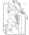
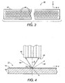
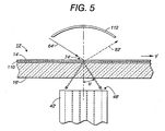
- Gerät, das dazu eingerichtet ist, Zellen in einer biologischen Probe zu erkennen, wobei die Zellen eine charakteristische Lumineszenz emittieren, die auf die Aussetzung einer Anregungsstrahlung reagiert, und das Gerät eine Bilderzeugungsvorrichtung (10) enthält, die umfasst:einen Bilderzeugungsabschnitt (20), der die Probe trägt;eine Abtaststrahlungsquelle (60), die so beschaffen ist, dass sie einen Strahlungsstrahl (64) auf der Probe führt, wobei der Strahlungsstrahl mit der Probe interagiert, um ein Lichtsignal zu erzeugen;einen Fotodetektor (98), der so beschaffen ist, dass er das Lichtsignal erfasst; undeinen Prozessor (80), der das Lichtsignal verarbeitet, das vom Fotodetektor (98) erfasst wird, wobei die Abbildungsvorrichtung weiterhin ein Lichtleitfaserbündel (40) enthält, das ein erstes Bündelende (42) erster Faserenden (46), die so beschaffen sind, dass sie eine Eingangsöffnung (48) eines hohen Bildseitenverhältnisses bilden, die die Probe auf dem Bilderzeugungsabschnitt (20) erkennt, und ein zweites Bündelende (44) zweiter Faserenden (50) hat, die so beschaffen sind, dass sie eine Ausgangsöffnung (52) bilden, die so geformt ist, dass sie kompakter ist als die Eingangsöffnung, und vom Bilderzeugungsabschnitt entfernt angeordnet ist; wobei die Abtaststrahlungsquelle (60) so beschaffen ist, dass sie den Strahlungsstrahl innerhalb eines Erkennungsbereiches der Eingangsöffnung (48) führt, das Lichtsignal von der Eingangsöffnung (48) empfangen und über das Lichtleitfaserbündel zur Ausgangsöffnung (52) übertragen wird und der Fotodetektor (98) das Lichtsignal am zweiten Bündel erfasst, wobei der Bilderzeugungsabschnitt enthält:einen Linearverschiebungsabschnitt (22), der eine plane Oberfläche, die die Probe trägt, in einer ersten Richtung linear verschiebt, wobeidas erste Ende (42) des Lichtleitfaserbündels parallele erste Faserenden hat, die so beschaffen sind, dass sie eine lineare Eingangsöffnung bilden, die senkrecht zur ersten Richtung und parallel zur Oberfläche angeordnet ist, und das zweite Ende (44) eine im wesentlichen kreisförmige Ausgangsöffnung bildet, wobei jedes erste Faserende mit der im wesentlichen kreisförmigen Öffnung (52) optisch in Verbindung steht, und die Bilderzeugungsvorrichtung weiterhin einen Rasterprozessor (80) enthält, der mit dem Bilderzeugungsabschnitt (20) und der Abtaststrahlungsquelle (60) in Verbindung steht, um die Abtastung des Strahlungsstrahls und die lineare Verschiebung der Oberfläche zu koordinieren, um eine Rasterung des Strahlungsstrahls auf der Oberfläche zu bewirken, und wobeidie Strahlungsquelle (60) dazu eingerichtet ist, den Anregungsstrahlungsstrahl linear über die biologische Probe mit einer Abtastrichtung senkrecht zur ersten Richtung zu führen, wobei ein Interaktionsbereich der Strahlungsquelle und der biologischen Probe im Bezug auf die Eingangsöffnung (48) derart eingerichtet ist, dass die charakteristische Lumineszenz, die im Interaktionsbereich erzeugt wird, von der Eingangsöffnung (48) für die Erfassung durch den Fotodetektor (98) gesammelt wird.
- Gerät nach Anspruch 1, bei dem die Probe ein biologischer Abstrich ist und eine Punktgröße des Anregungsstrahlungsstrahls auf dem biologischen Abstrich im wesentlichen mit einer Größe der Zellen übereinstimmt.
- Gerät nach Anspruch 1 oder Anspruch 2, weiterhin enthaltend:eine Zähleinrichtung, die eine Anzahl von Zellen auf der Basis der charakteristischen Lumineszenz zählt, die während des Rasterns erfasst wird.
- Gerät nach Anspruch 1, weiterhin enthaltend:einen Motor (24), der so beschaffen ist, dass er die Probe entweder in einer Verschiebungs- oder einer Drehbewegung bewegt, wobei der Motor mit der Abtaststrahlungsquelle (60) für eine Rasterung des Strahlungsstrahls über einen gewählten Bereich der Probe zusammenwirkt.
- Gerät nach Anspruch 4, bei dem die Eingangsöffnung (48) eine im wesentlichen lineare Form hat, die Abtaststrahlungsquelle (60) den Strahlungsstrahl entlang einer Strahltrajektorie parallel zur im wesentlichen linearen Öffnung führt und der Motor (24) die Probe linear entlang einer Trajektorie verschiebt, die senkrecht zur Strahltrajektorie ist.
- Gerät nach Anspruch 4, bei der der Motor (24) die Probe um eine Achse dreht, die senkrecht zu einer Oberfläche der Probe verläuft, die Eingangsöffnung (48) eine im wesentlichen lineare Form hat, die sich radial von der Drehachse weg erstreckt, und die Abtaststrahlungsquelle (60) den Strahlungsstrahl entlang einer Strahltrajektorie parallel zur im wesentlichen linearen Eingangsöffnung (48) führt.
- Gerät nach einem der vorhergehenden Ansprüche, bei dem die Abtaststrahlungsquelle (60) eine Lichtabtasteinrichtung ist und die Abbildungseinrichtung weiterhin enthält:ein Lichtfilter (94), das so beschaffen ist, das es im wesentlichen das Licht, das von der Lichtabtasteinrichtung erzeugt wird, daran hindert, den Fotodetektor (98) zu erreichen.
- Gerät nach Anspruch 7, weiterhin enthaltend:ein Substrat (16), auf dem die Probe angeordnet ist, wobei das Substrat vom Verschiebungsabschnitt (22) getragen wird und das Lichtfilter (94) als eine Beschichtung (110) auf dem Substrat angeordnet oder durch das Substrat ausgebildet ist.
- Gerät nach Anspruch 7, bei dem das Lichtleitfaserbündel (40) das Lichtfilter ist.
- Gerät nach einem der vorhergehenden Ansprüche, bei dem das Lichtleitfaserbündel (40) weiterhin enthält:einen optischen Koppler (120);eine Vielzahl von Lichtleitfasern, die sich von den ersten Faserenden erstrecken und am optischen Koppler (120) zusammenlaufen; undeinen im wesentlichen kreisförmiger Hohllichtleiter (122), der Licht vom optischen Koppler (120) zum zweiten Ende (44) des Lichtleitfaserbündels (40) leitet, wobei die im wesentlichen kreisförmige Ausgangsöffnung (52) mit einem Ende des Hohllichtleiters korrespondiert, das vom optischen Koppler (120) entfernt ist.
- Gerät nach einem der vorhergehenden Ansprüche, bei dem die Probe eine Gleitoberfläche enthält, die mit einem biologischen Fluid überzogen ist, das Zellen enthält, wobei das biologische Fluid mit einem Material behandelt wird, das sich wahlweise mit dem gewählten Zelltyp verbindet, und das Material das Lichtsignal in Erwiderung auf die Interaktion mit dem Strahlungsstrahl (64) erzeugt.
- Verfahren zum Abbilden einer biologischen Probe, wobei das Verfahren umfasst:Führen eines Strahlungsstrahls (64) entlang eines linearen Weges auf einer biologischen Probe (14);Sammeln von Licht, das durch die Strahlinteraktion mit der Probe erzeugt wird, an der linearen Eingangsöffnung (48), die durch eine Anordnung von Lichtleitfaserenden (46) eines Lichtleitfaserbündels (40) ausgebildet ist, wobei die lineare Eingangsöffnung so beschaffen ist, dass sie eine Eingangsöffnung (48) eines hohen Bildseitenverhältnisses bildet;Senden des gesammelten Lichtes entlang der Fasern des Lichtleitfaserbündels (40), wobei die Fasern das gesammelte Licht zu einer Ausgangsöffnung (52) des Lichtleitfaserbündels (40) kanalisieren und die größte Raumabmessung der Ausgangsöffnung im wesentlichen geringer ist als die größte Raumabmessung der Eingangsöffnung (48);Erfassen des gesammelten Lichtes an der Ausgangsöffnung (52);Bewegen der Probe (14) im wesentlichen senkrecht zum linearen Abtastweg des Strahlungsstrahls (64), wobei die Bewegung der Probe mit der Abtastung zusammenwirkt, um den Strahlungsstrahl auf der Probe zu rastern; undKoordinieren des Abtastens, des Bewegens und des Erfassens, um eine Anordnung von Bildelementen zu erzeugen, die für wenigstens einen Abschnitt der Probe repräsentativ sind.
- Bilderzeugungsverfahren nach Anspruch 12, bei dem die Probe einen biologischen Abstrich enthält und das Abbildungsverfahren weiterhin umfasst:Markieren des biologischen Abstriches mit Hilfe eines fluoreszenten Materials, das wahlweise an einem gewählten Typ einer Zelle haftet, wobei das Licht, das von der Strahlinteraktion erzeugt wird, eine Fluoreszenz enthält, die von dem fluoreszenten Material infolge einer Interaktion mit dem Strahlungsstrahl erzeugt wird.
Applications Claiming Priority (2)
| Application Number | Priority Date | Filing Date | Title |
|---|---|---|---|
| US271347 | 2002-10-15 | ||
| US10/271,347 US7113624B2 (en) | 2002-10-15 | 2002-10-15 | Imaging apparatus and method employing a large linear aperture |
Publications (2)
| Publication Number | Publication Date |
|---|---|
| EP1414231A1 EP1414231A1 (de) | 2004-04-28 |
| EP1414231B1 true EP1414231B1 (de) | 2009-08-26 |
Family
ID=32069136
Family Applications (1)
| Application Number | Title | Priority Date | Filing Date |
|---|---|---|---|
| EP03256441A Expired - Fee Related EP1414231B1 (de) | 2002-10-15 | 2003-10-13 | Bilderzeugungsgerät und Verfahren |
Country Status (4)
| Country | Link |
|---|---|
| US (2) | US7113624B2 (de) |
| EP (1) | EP1414231B1 (de) |
| JP (1) | JP2004138614A (de) |
| DE (1) | DE60328951D1 (de) |
Families Citing this family (68)
| Publication number | Priority date | Publication date | Assignee | Title |
|---|---|---|---|---|
| US6784982B1 (en) * | 1999-11-04 | 2004-08-31 | Regents Of The University Of Minnesota | Direct mapping of DNA chips to detector arrays |
| WO2001040454A1 (en) | 1999-11-30 | 2001-06-07 | Oncosis | Method and apparatus for selectively targeting specific cells within a cell population |
| WO2002037938A2 (en) | 2000-10-24 | 2002-05-16 | Oncosis Llc | Method and device for selectively targeting cells within a three -dimensional specimen |
| US20090182627A1 (en) * | 2001-11-14 | 2009-07-16 | Retaildna, Llc | Self learning method and system for managing a third party subsidy offer |
| US7113624B2 (en) * | 2002-10-15 | 2006-09-26 | Palo Alto Research Center Incorporated | Imaging apparatus and method employing a large linear aperture |
| US20050046848A1 (en) * | 2003-08-26 | 2005-03-03 | Blueshift Biotechnologies, Inc. | Time dependent fluorescence measurements |
| US8064730B2 (en) * | 2003-09-22 | 2011-11-22 | Asml Netherlands B.V. | Device manufacturing method, orientation determination method and lithographic apparatus |
| US20050264805A1 (en) * | 2004-02-09 | 2005-12-01 | Blueshift Biotechnologies, Inc. | Methods and apparatus for scanning small sample volumes |
| SE0401632D0 (sv) * | 2004-06-24 | 2004-06-24 | Innovation Team Ab | Medel och sätt att detektera blodläckage från sår |
| EP1774024A4 (de) * | 2004-07-02 | 2012-04-04 | Blueshift Biotechnologies Inc | Erforschung von fluorophor-mikroumgebungen |
| US7468796B2 (en) * | 2004-08-06 | 2008-12-23 | Compucyte Corporation | Multiple-color monochromatic light absorption and quantification of light absorption in a stained sample |
| US7280261B2 (en) | 2004-12-20 | 2007-10-09 | Palo Alto Research Center Incorporated | Method of scanning and light collection for a rare cell detector |
| US7286224B2 (en) * | 2004-12-21 | 2007-10-23 | Palo Alto Research Center Incorporated | Time-multiplexed scanning light source for multi-probe, multi-laser fluorescence detection systems |
| US7709821B2 (en) * | 2005-04-27 | 2010-05-04 | Advanced Cytometry Instrumentation Systems, Inc. | Flow cytometer acquisition and detection system |
| WO2007014188A2 (en) * | 2005-07-25 | 2007-02-01 | Duke University | Methods, systems, and computer program products for optimization of probes for spectroscopic measurement in turbid media |
| US7842465B2 (en) * | 2006-01-17 | 2010-11-30 | Palo Alto Research Center Incorporated | Immunocytostaining methods for enhanced dye ratio discrimination in rare event detection |
| WO2007089911A2 (en) * | 2006-01-30 | 2007-08-09 | The Scripps Research Institute | Methods for detection of circulating tumor cells and methods of diagnosis of cancer in a mammalian subject |
| EP2001352A4 (de) * | 2006-03-17 | 2010-04-07 | Univ Duke | Auf monte-carlo-simulation basierendes modell für fluoreszenz in trüben medien sowie verfahren und systeme zu seiner verwendung zur bestimmung der intrinsischen fluoreszenz trüber medien |
| US7751039B2 (en) * | 2006-03-30 | 2010-07-06 | Duke University | Optical assay system for intraoperative assessment of tumor margins |
| JP2009537152A (ja) | 2006-05-17 | 2009-10-29 | カリフォルニア インスティテュート オブ テクノロジー | 温度サイクルシステム |
| US7545498B2 (en) * | 2006-12-18 | 2009-06-09 | Palo Alto Research Center Incorporated | System and method for removing auto-fluorescence through the use of multiple detection channels |
| US8821799B2 (en) | 2007-01-26 | 2014-09-02 | Palo Alto Research Center Incorporated | Method and system implementing spatially modulated excitation or emission for particle characterization with enhanced sensitivity |
| US9164037B2 (en) | 2007-01-26 | 2015-10-20 | Palo Alto Research Center Incorporated | Method and system for evaluation of signals received from spatially modulated excitation and emission to accurately determine particle positions and distances |
| US7936463B2 (en) | 2007-02-05 | 2011-05-03 | Palo Alto Research Center Incorporated | Containing analyte in optical cavity structures |
| US7633629B2 (en) | 2007-02-05 | 2009-12-15 | Palo Alto Research Center Incorporated | Tuning optical cavities |
| US7817276B2 (en) * | 2007-02-05 | 2010-10-19 | Palo Alto Research Center Incorporated | Distinguishing objects |
| US20080270091A1 (en) * | 2007-02-23 | 2008-10-30 | Nirmala Ramanujam | Scaling method for fast monte carlo simulation of diffuse reflectance spectra from multi-layered turbid media and methods and systems for using same to determine optical properties of multi-layered turbid medium from measured diffuse reflectance |
| US8717426B2 (en) * | 2007-05-17 | 2014-05-06 | M-I Llc | Liquid and solids analysis of drilling fluids using fractionation and imaging |
| WO2009043050A2 (en) * | 2007-09-27 | 2009-04-02 | Duke University | Optical assay system with a multi-probe imaging array |
| EP2194848A4 (de) * | 2007-09-28 | 2013-08-14 | Univ Duke | Systeme und verfahren für die spektralanalyse einer gewebemasse mit einem instrument, einer optischen sonde und einem monte carlo oder diffusions-algorithmus |
| US7817254B2 (en) | 2008-01-30 | 2010-10-19 | Palo Alto Research Center Incorporated | Obtaining information from time variation of sensing results |
| US7763856B2 (en) * | 2008-01-31 | 2010-07-27 | Palo Alto Research Center Incorporated | Producing time variation in emanating light |
| US8263955B2 (en) * | 2008-12-18 | 2012-09-11 | Palo Alto Research Center Incorporated | Causing relative motion |
| US8629981B2 (en) | 2008-02-01 | 2014-01-14 | Palo Alto Research Center Incorporated | Analyzers with time variation based on color-coded spatial modulation |
| US8373860B2 (en) * | 2008-02-01 | 2013-02-12 | Palo Alto Research Center Incorporated | Transmitting/reflecting emanating light with time variation |
| WO2010042249A2 (en) * | 2008-04-24 | 2010-04-15 | Duke University | A diffuse reflectance spectroscopy device for quantifying tissue absorption and scattering |
| US8983581B2 (en) | 2008-05-27 | 2015-03-17 | Massachusetts Institute Of Technology | System and method for large field of view, single cell analysis |
| KR20110106436A (ko) * | 2009-01-09 | 2011-09-28 | 신텔렉트 인코포레이티드 | 세포의 유전적 분석 |
| US8788213B2 (en) | 2009-01-12 | 2014-07-22 | Intrexon Corporation | Laser mediated sectioning and transfer of cell colonies |
| US9155471B2 (en) | 2009-05-27 | 2015-10-13 | Lumicell, Inc'. | Methods and systems for spatially identifying abnormal cells |
| US20110017915A1 (en) * | 2009-07-23 | 2011-01-27 | Palo Alto Research Center Incorporated | Drift scanner for rare cell detection |
| US8558207B2 (en) * | 2009-09-29 | 2013-10-15 | Carestream Health, Inc. | Photostimulable plate reading device |
| CN115060882A (zh) | 2009-10-21 | 2022-09-16 | 斯克里普斯研究所 | 用非稀有细胞检测稀有细胞的方法 |
| US9091637B2 (en) | 2009-12-04 | 2015-07-28 | Duke University | Smart fiber optic sensors systems and methods for quantitative optical spectroscopy |
| JP5716738B2 (ja) | 2010-03-05 | 2015-05-13 | コニカミノルタ株式会社 | 細胞の検出方法及び細胞検出システム |
| US20110223586A1 (en) * | 2010-03-11 | 2011-09-15 | David Karabinus | Optical particle characterization system |
| US20110223587A1 (en) * | 2010-03-11 | 2011-09-15 | Schulman Joseph D | Optical particle characterization system |
| US8774488B2 (en) | 2010-03-11 | 2014-07-08 | Cellscape Corporation | Method and device for identification of nucleated red blood cells from a maternal blood sample |
| US9314304B2 (en) | 2010-12-08 | 2016-04-19 | Lumicell, Inc. | Methods and system for image guided cell ablation with microscopic resolution |
| US8879065B1 (en) * | 2011-05-10 | 2014-11-04 | The Board Of Trustees Of The University Of Alabama For And On Behalf Of The University Of Alabama In Huntsville | Systems and methods for localized surface plasmon resonance sensing |
| US8723140B2 (en) | 2011-08-09 | 2014-05-13 | Palo Alto Research Center Incorporated | Particle analyzer with spatial modulation and long lifetime bioprobes |
| US9029800B2 (en) | 2011-08-09 | 2015-05-12 | Palo Alto Research Center Incorporated | Compact analyzer with spatial modulation and multiple intensity modulated excitation sources |
| US9335254B2 (en) * | 2011-08-25 | 2016-05-10 | Glory Ltd. | Paper sheet recognition apparatus, light guide and light guide casing for use in spectrometric measurement of paper sheet |
| WO2013163298A1 (en) * | 2012-04-24 | 2013-10-31 | Senseonics, Incorporated | Angle of incidence selective band pass filter for implantable chemical sensor |
| US8964183B2 (en) * | 2012-05-31 | 2015-02-24 | General Electric Company | Systems and methods for screening of biological samples |
| US20140009762A1 (en) * | 2012-06-21 | 2014-01-09 | Nikon Corporation | Measurement assembly with fiber optic array |
| AU2014236561B2 (en) | 2013-03-14 | 2018-08-16 | Lumicell, Inc. | Medical imaging device and methods of use |
| US10147180B2 (en) | 2013-12-19 | 2018-12-04 | Axon Dx, Llc | Cell detection, capture and isolation methods and apparatus |
| JP6194791B2 (ja) * | 2013-12-27 | 2017-09-13 | 富士ゼロックス株式会社 | 画像処理装置及びプログラム |
| US10527624B2 (en) | 2014-01-27 | 2020-01-07 | Epic Sciences, Inc. | Circulating tumor cell diagnostics for prostate cancer biomarkers |
| US10545151B2 (en) | 2014-02-21 | 2020-01-28 | Epic Sciences, Inc. | Methods for analyzing rare circulating cells |
| WO2016118456A1 (en) * | 2015-01-19 | 2016-07-28 | Sri International | Measuring hiv reservoirs with optical scanning |
| CN104568893A (zh) * | 2015-01-24 | 2015-04-29 | 北京中拓机械集团有限责任公司 | 半导体晶片的高速荧光光谱检测装置 |
| JP6730124B2 (ja) * | 2016-08-01 | 2020-07-29 | 株式会社ディスコ | 厚み計測装置 |
| JP6730125B2 (ja) * | 2016-08-01 | 2020-07-29 | 株式会社ディスコ | 計測装置 |
| JP7194202B2 (ja) * | 2018-05-30 | 2022-12-21 | ガタン インコーポレイテッド | 波長分解され角度分解されたカソードルミネッセンスのための装置および方法 |
| EP3745081B1 (de) * | 2019-05-28 | 2023-03-22 | Tecan Trading Ag | Positionsdetektor und verfahren zur 3d-positionsbestimmung |
| CN113405538B (zh) * | 2021-06-07 | 2022-07-26 | 核工业西南物理研究院 | 一种激光散射诊断系统空间测量位置标定装置及标定方法 |
Family Cites Families (33)
| Publication number | Priority date | Publication date | Assignee | Title |
|---|---|---|---|---|
| JPS50154013A (de) | 1974-06-01 | 1975-12-11 | ||
| US4002829A (en) | 1974-08-29 | 1977-01-11 | W. R. Grace & Co. | Autosynchronous optical scanning and recording laser system with fiber optic light detection |
| FR2350596A2 (fr) | 1976-05-04 | 1977-12-02 | Green James E | Procede et appareillage pour l'analyse d'un champ selon deux resolutions |
| US4556903A (en) | 1983-12-20 | 1985-12-03 | At&T Technologies, Inc. | Inspection scanning system |
| US4600951A (en) | 1983-12-20 | 1986-07-15 | At&T Technologies, Inc. | Scanning sample, signal generation, data digitizing and retiming system |
| US4721851A (en) | 1985-04-30 | 1988-01-26 | Ricoh Company, Ltd. | Image reading device using fiber optic bundles configured differently at each end |
| US4886975A (en) | 1986-02-14 | 1989-12-12 | Canon Kabushiki Kaisha | Surface examining apparatus for detecting the presence of foreign particles on two or more surfaces |
| US4875780A (en) | 1988-02-25 | 1989-10-24 | Eastman Kodak Company | Method and apparatus for inspecting reticles |
| US4849645A (en) | 1988-03-04 | 1989-07-18 | American Telephone And Telegraph Company | Substrate inspection system having two scattered light detecting bundles orthogonally positioned to each other |
| US4941309A (en) | 1989-03-02 | 1990-07-17 | Certainteed Corporation | Panel packaging system |
| US5315993A (en) * | 1990-02-16 | 1994-05-31 | The Boc Group, Inc. | Luminescence monitoring with modulation frequency multiplexing |
| JPH04296642A (ja) | 1991-03-27 | 1992-10-21 | Ricoh Co Ltd | 試料表面欠陥検出装置 |
| US5216485A (en) | 1991-09-04 | 1993-06-01 | International Business Machines Corporation | Advanced via inspection tool (avit) |
| US5220617A (en) | 1991-09-04 | 1993-06-15 | International Business Machines Corporation | Method and apparatus for object inspection |
| US5798831A (en) | 1991-12-19 | 1998-08-25 | Nikon Corporation | Defect inspecting apparatus and defect inspecting method |
| JPH06148085A (ja) | 1992-11-12 | 1994-05-27 | Sony Corp | ウェハ異物検査方法およびウェハ異物検査装置 |
| US5313542A (en) | 1992-11-30 | 1994-05-17 | Breault Research Organization, Inc. | Apparatus and method of rapidly measuring hemispherical scattered or radiated light |
| US5651047A (en) * | 1993-01-25 | 1997-07-22 | Cardiac Mariners, Incorporated | Maneuverable and locateable catheters |
| US5471066A (en) | 1993-08-26 | 1995-11-28 | Nikon Corporation | Defect inspection apparatus of rotary type |
| US5732162A (en) | 1993-10-28 | 1998-03-24 | Xerox Corporation | Two dimensional linearity and registration error correction in a hyperacuity printer |
| JP3425615B2 (ja) * | 1994-03-24 | 2003-07-14 | 科学技術庁長官官房会計課長 | 走査型近視野原子間力顕微鏡 |
| GB9418981D0 (en) * | 1994-09-21 | 1994-11-09 | Univ Glasgow | Apparatus and method for carrying out analysis of samples |
| JPH09145631A (ja) | 1995-11-29 | 1997-06-06 | Nikon Corp | 表面異物検査装置 |
| US6545334B2 (en) | 1997-12-19 | 2003-04-08 | Imec Vzw | Device and a method for thermal sensing |
| US6445451B1 (en) * | 1998-02-12 | 2002-09-03 | Hamilton Thorne Research | Colorimeter and assay device |
| CN1311436A (zh) | 2000-03-01 | 2001-09-05 | 上海和泰光电科技有限公司 | 旋转平台上的生物芯片荧光图象的读取 |
| US6582363B2 (en) * | 2000-08-25 | 2003-06-24 | Pentax Corporation | Video endoscope system and illumination optical system |
| WO2002037938A2 (en) | 2000-10-24 | 2002-05-16 | Oncosis Llc | Method and device for selectively targeting cells within a three -dimensional specimen |
| US7072034B2 (en) | 2001-06-08 | 2006-07-04 | Kla-Tencor Corporation | Systems and methods for inspection of specimen surfaces |
| US6636623B2 (en) * | 2001-08-10 | 2003-10-21 | Visiongate, Inc. | Optical projection imaging system and method for automatically detecting cells with molecular marker compartmentalization associated with malignancy and disease |
| US7180862B2 (en) * | 2002-07-18 | 2007-02-20 | Intel Corporation | Apparatus and method for virtual output queue feedback |
| US7305112B2 (en) | 2002-10-15 | 2007-12-04 | The Scripps Research Institute | Method of converting rare cell scanner image coordinates to microscope coordinates using reticle marks on a sample media |
| US7113624B2 (en) | 2002-10-15 | 2006-09-26 | Palo Alto Research Center Incorporated | Imaging apparatus and method employing a large linear aperture |
-
2002
- 2002-10-15 US US10/271,347 patent/US7113624B2/en active Active
-
2003
- 2003-07-09 US US10/616,366 patent/US7277569B2/en active Active
- 2003-10-08 JP JP2003349230A patent/JP2004138614A/ja active Pending
- 2003-10-13 DE DE60328951T patent/DE60328951D1/de not_active Expired - Lifetime
- 2003-10-13 EP EP03256441A patent/EP1414231B1/de not_active Expired - Fee Related
Also Published As
| Publication number | Publication date |
|---|---|
| US20040071332A1 (en) | 2004-04-15 |
| US20040071330A1 (en) | 2004-04-15 |
| JP2004138614A (ja) | 2004-05-13 |
| US7277569B2 (en) | 2007-10-02 |
| EP1414231A1 (de) | 2004-04-28 |
| DE60328951D1 (de) | 2009-10-08 |
| US7113624B2 (en) | 2006-09-26 |
Similar Documents
| Publication | Publication Date | Title |
|---|---|---|
| EP1414231B1 (de) | Bilderzeugungsgerät und Verfahren | |
| EP1674852B1 (de) | Zeitmultiplex-Abtastlichtquelle für Multisonden- und Multilaser-Fluoreszenzdetektionssysteme | |
| EP1672355B1 (de) | Verbessertes Verfahren zum Abtasten und Lichtsammeln für einen Detektor für seltene Zellen | |
| US5248876A (en) | Tandem linear scanning confocal imaging system with focal volumes at different heights | |
| US7305112B2 (en) | Method of converting rare cell scanner image coordinates to microscope coordinates using reticle marks on a sample media | |
| US6355934B1 (en) | Imaging system for an optical scanner | |
| TWI263072B (en) | Method and system for reading microarrays | |
| US20020037149A1 (en) | Fiber optic scanner | |
| JP2001523830A (ja) | 高処理能力光学スキャナー | |
| US8633432B2 (en) | Reflective focusing and transmissive projection device | |
| US6317206B1 (en) | Device for the detection of a fluorescent dye | |
| US7545498B2 (en) | System and method for removing auto-fluorescence through the use of multiple detection channels | |
| KR100615040B1 (ko) | 바이오칩 측정 장치 및 방법 | |
| CN1296700C (zh) | 矿物材料红外荧光分析法 | |
| CN2522855Y (zh) | 带滤光片转盘的生物芯片荧光检测扫描装置 | |
| CA2073344C (en) | Fluorescence assay apparatus | |
| Curry et al. | High-speed detection of occult tumor cells in peripheral blood | |
| JP2001074657A (ja) | 光計測方法および装置 |
Legal Events
| Date | Code | Title | Description |
|---|---|---|---|
| PUAI | Public reference made under article 153(3) epc to a published international application that has entered the european phase |
Free format text: ORIGINAL CODE: 0009012 |
|
| AK | Designated contracting states |
Kind code of ref document: A1 Designated state(s): AT BE BG CH CY CZ DE DK EE ES FI FR GB GR HU IE IT LI LU MC NL PT RO SE SI SK TR |
|
| AX | Request for extension of the european patent |
Extension state: AL LT LV MK |
|
| RIN1 | Information on inventor provided before grant (corrected) |
Inventor name: CURRY, DOUGLAS N. |
|
| RAP1 | Party data changed (applicant data changed or rights of an application transferred) |
Owner name: PALO ALTO RESEARCH CENTER INCORPORATED |
|
| 17P | Request for examination filed |
Effective date: 20041028 |
|
| AKX | Designation fees paid |
Designated state(s): DE FR GB |
|
| 17Q | First examination report despatched |
Effective date: 20050114 |
|
| 17Q | First examination report despatched |
Effective date: 20050114 |
|
| RIN1 | Information on inventor provided before grant (corrected) |
Inventor name: CURRY, DOUGLAS N. |
|
| GRAP | Despatch of communication of intention to grant a patent |
Free format text: ORIGINAL CODE: EPIDOSNIGR1 |
|
| GRAS | Grant fee paid |
Free format text: ORIGINAL CODE: EPIDOSNIGR3 |
|
| GRAA | (expected) grant |
Free format text: ORIGINAL CODE: 0009210 |
|
| AK | Designated contracting states |
Kind code of ref document: B1 Designated state(s): DE FR GB |
|
| REG | Reference to a national code |
Ref country code: GB Ref legal event code: FG4D |
|
| REF | Corresponds to: |
Ref document number: 60328951 Country of ref document: DE Date of ref document: 20091008 Kind code of ref document: P |
|
| PLBE | No opposition filed within time limit |
Free format text: ORIGINAL CODE: 0009261 |
|
| STAA | Information on the status of an ep patent application or granted ep patent |
Free format text: STATUS: NO OPPOSITION FILED WITHIN TIME LIMIT |
|
| 26N | No opposition filed |
Effective date: 20100527 |
|
| REG | Reference to a national code |
Ref country code: DE Ref legal event code: R081 Ref document number: 60328951 Country of ref document: DE Owner name: SRI INTERNATIONAL, MENLO PARK, US Free format text: FORMER OWNER: PALO ALTO RESEARCH CENTER INC., PALO ALTO, CALIF., US Effective date: 20111220 |
|
| REG | Reference to a national code |
Ref country code: FR Ref legal event code: PLFP Year of fee payment: 13 |
|
| REG | Reference to a national code |
Ref country code: FR Ref legal event code: PLFP Year of fee payment: 14 |
|
| REG | Reference to a national code |
Ref country code: FR Ref legal event code: PLFP Year of fee payment: 15 |
|
| REG | Reference to a national code |
Ref country code: FR Ref legal event code: PLFP Year of fee payment: 16 |
|
| PGFP | Annual fee paid to national office [announced via postgrant information from national office to epo] |
Ref country code: DE Payment date: 20191029 Year of fee payment: 17 |
|
| PGFP | Annual fee paid to national office [announced via postgrant information from national office to epo] |
Ref country code: FR Payment date: 20191025 Year of fee payment: 17 |
|
| PGFP | Annual fee paid to national office [announced via postgrant information from national office to epo] |
Ref country code: GB Payment date: 20191028 Year of fee payment: 17 |
|
| REG | Reference to a national code |
Ref country code: DE Ref legal event code: R119 Ref document number: 60328951 Country of ref document: DE |
|
| GBPC | Gb: european patent ceased through non-payment of renewal fee |
Effective date: 20201013 |
|
| PG25 | Lapsed in a contracting state [announced via postgrant information from national office to epo] |
Ref country code: DE Free format text: LAPSE BECAUSE OF NON-PAYMENT OF DUE FEES Effective date: 20210501 Ref country code: FR Free format text: LAPSE BECAUSE OF NON-PAYMENT OF DUE FEES Effective date: 20201031 |
|
| PG25 | Lapsed in a contracting state [announced via postgrant information from national office to epo] |
Ref country code: GB Free format text: LAPSE BECAUSE OF NON-PAYMENT OF DUE FEES Effective date: 20201013 |