EP1391464A1 - Monoklonaler anti-cd-40-antikörper - Google Patents
Monoklonaler anti-cd-40-antikörper Download PDFInfo
- Publication number
- EP1391464A1 EP1391464A1 EP02720634A EP02720634A EP1391464A1 EP 1391464 A1 EP1391464 A1 EP 1391464A1 EP 02720634 A EP02720634 A EP 02720634A EP 02720634 A EP02720634 A EP 02720634A EP 1391464 A1 EP1391464 A1 EP 1391464A1
- Authority
- EP
- European Patent Office
- Prior art keywords
- antibody
- variable region
- chain variable
- cells
- antibodies
- Prior art date
- Legal status (The legal status is an assumption and is not a legal conclusion. Google has not performed a legal analysis and makes no representation as to the accuracy of the status listed.)
- Granted
Links
Images


Classifications
-
- C—CHEMISTRY; METALLURGY
- C07—ORGANIC CHEMISTRY
- C07K—PEPTIDES
- C07K16/00—Immunoglobulins [IGs], e.g. monoclonal or polyclonal antibodies
- C07K16/18—Immunoglobulins [IGs], e.g. monoclonal or polyclonal antibodies against material from animals or humans
- C07K16/28—Immunoglobulins [IGs], e.g. monoclonal or polyclonal antibodies against material from animals or humans against receptors, cell surface antigens or cell surface determinants
- C07K16/2878—Immunoglobulins [IGs], e.g. monoclonal or polyclonal antibodies against material from animals or humans against receptors, cell surface antigens or cell surface determinants against the NGF-receptor/TNF-receptor superfamily, e.g. CD27, CD30, CD40, CD95
-
- A—HUMAN NECESSITIES
- A61—MEDICAL OR VETERINARY SCIENCE; HYGIENE
- A61P—SPECIFIC THERAPEUTIC ACTIVITY OF CHEMICAL COMPOUNDS OR MEDICINAL PREPARATIONS
- A61P35/00—Antineoplastic agents
-
- A—HUMAN NECESSITIES
- A61—MEDICAL OR VETERINARY SCIENCE; HYGIENE
- A61P—SPECIFIC THERAPEUTIC ACTIVITY OF CHEMICAL COMPOUNDS OR MEDICINAL PREPARATIONS
- A61P37/00—Drugs for immunological or allergic disorders
- A61P37/02—Immunomodulators
-
- A—HUMAN NECESSITIES
- A61—MEDICAL OR VETERINARY SCIENCE; HYGIENE
- A61P—SPECIFIC THERAPEUTIC ACTIVITY OF CHEMICAL COMPOUNDS OR MEDICINAL PREPARATIONS
- A61P37/00—Drugs for immunological or allergic disorders
- A61P37/02—Immunomodulators
- A61P37/04—Immunostimulants
-
- A—HUMAN NECESSITIES
- A61—MEDICAL OR VETERINARY SCIENCE; HYGIENE
- A61P—SPECIFIC THERAPEUTIC ACTIVITY OF CHEMICAL COMPOUNDS OR MEDICINAL PREPARATIONS
- A61P37/00—Drugs for immunological or allergic disorders
- A61P37/02—Immunomodulators
- A61P37/06—Immunosuppressants, e.g. drugs for graft rejection
-
- A—HUMAN NECESSITIES
- A61—MEDICAL OR VETERINARY SCIENCE; HYGIENE
- A61P—SPECIFIC THERAPEUTIC ACTIVITY OF CHEMICAL COMPOUNDS OR MEDICINAL PREPARATIONS
- A61P37/00—Drugs for immunological or allergic disorders
- A61P37/08—Antiallergic agents
-
- A—HUMAN NECESSITIES
- A61—MEDICAL OR VETERINARY SCIENCE; HYGIENE
- A61P—SPECIFIC THERAPEUTIC ACTIVITY OF CHEMICAL COMPOUNDS OR MEDICINAL PREPARATIONS
- A61P7/00—Drugs for disorders of the blood or the extracellular fluid
-
- A—HUMAN NECESSITIES
- A61—MEDICAL OR VETERINARY SCIENCE; HYGIENE
- A61P—SPECIFIC THERAPEUTIC ACTIVITY OF CHEMICAL COMPOUNDS OR MEDICINAL PREPARATIONS
- A61P7/00—Drugs for disorders of the blood or the extracellular fluid
- A61P7/04—Antihaemorrhagics; Procoagulants; Haemostatic agents; Antifibrinolytic agents
-
- A—HUMAN NECESSITIES
- A61—MEDICAL OR VETERINARY SCIENCE; HYGIENE
- A61K—PREPARATIONS FOR MEDICAL, DENTAL OR TOILETRY PURPOSES
- A61K39/00—Medicinal preparations containing antigens or antibodies
- A61K2039/505—Medicinal preparations containing antigens or antibodies comprising antibodies
-
- C—CHEMISTRY; METALLURGY
- C07—ORGANIC CHEMISTRY
- C07K—PEPTIDES
- C07K2317/00—Immunoglobulins specific features
- C07K2317/20—Immunoglobulins specific features characterized by taxonomic origin
- C07K2317/21—Immunoglobulins specific features characterized by taxonomic origin from primates, e.g. man
-
- C—CHEMISTRY; METALLURGY
- C07—ORGANIC CHEMISTRY
- C07K—PEPTIDES
- C07K2317/00—Immunoglobulins specific features
- C07K2317/70—Immunoglobulins specific features characterized by effect upon binding to a cell or to an antigen
- C07K2317/73—Inducing cell death, e.g. apoptosis, necrosis or inhibition of cell proliferation
-
- C—CHEMISTRY; METALLURGY
- C07—ORGANIC CHEMISTRY
- C07K—PEPTIDES
- C07K2317/00—Immunoglobulins specific features
- C07K2317/70—Immunoglobulins specific features characterized by effect upon binding to a cell or to an antigen
- C07K2317/74—Inducing cell proliferation
-
- C—CHEMISTRY; METALLURGY
- C07—ORGANIC CHEMISTRY
- C07K—PEPTIDES
- C07K2317/00—Immunoglobulins specific features
- C07K2317/70—Immunoglobulins specific features characterized by effect upon binding to a cell or to an antigen
- C07K2317/75—Agonist effect on antigen
-
- C—CHEMISTRY; METALLURGY
- C07—ORGANIC CHEMISTRY
- C07K—PEPTIDES
- C07K2317/00—Immunoglobulins specific features
- C07K2317/70—Immunoglobulins specific features characterized by effect upon binding to a cell or to an antigen
- C07K2317/76—Antagonist effect on antigen, e.g. neutralization or inhibition of binding
-
- C—CHEMISTRY; METALLURGY
- C07—ORGANIC CHEMISTRY
- C07K—PEPTIDES
- C07K2319/00—Fusion polypeptide
-
- C—CHEMISTRY; METALLURGY
- C07—ORGANIC CHEMISTRY
- C07K—PEPTIDES
- C07K2319/00—Fusion polypeptide
- C07K2319/30—Non-immunoglobulin-derived peptide or protein having an immunoglobulin constant or Fc region, or a fragment thereof, attached thereto
Definitions
- the present invention relates to an antibody or a functional fragment thereof that recognizes a human CD40 antigen present on the surface of human B cells, dendritic cells (DC) and the like. Specifically, the present invention relates to an anti-human CD40 antibody or a functional fragment thereof that is substantially antagonistic to a human CD40 antigen on the dendritic cell (DC) surface, and an agonistic anti-human CD40 antibody or a functional fragment thereof that is expected to have a therapeutic effect higher than those of conventional anti-human CD40 antibodies.
- CD40 is an antigen with a molecular weight of 50 kDa that is present on the cell membrane surface.
- CD40 is expressed on B cells, dendritic cells (DC), certain types of cancer cells, and thymic epithelial cells.
- CD40 is known to play a key role in proliferation and differentiation of B cells and DC.
- CD40 has been identified as an antigen that is expressed on the human B cell surface (E. A. Clark et. al., Proc. Natl. Acad. Sci. USA 83: 4494, 1986, 1. Stamenkovic et. al., EMBO J. 8:1403, 1989).
- CD40 is thought to be a member of the TNF receptor family, to which a low affinity NGF receptor, TNF receptor, CD27, OX40, CD30 and the like belong.
- the gene of a ligand (CD40L) for human and mouse CD40 has been cloned recently, revealing that it is a type II membrane protein, and is expressed on activated CD4+T cells. It has also been shown that CD40L introduces strong activation signals into human and mouse B cells.
- CD40 has been confirmed more often on dendritic cells than on B cells, so that it has become clear that CD40 plays an important role.
- the binding of CD40 with CD40L causes the activation of antigen-presenting cells (APC). Specifically, it enhances the expression of co-stimulation molecules such as CD80 (B7-1) and CD86 (B7-2), or the production of IL-12 (Caux, C., et al.: Activation of human dendritic cells through CD40 cross-linking. J. Exp. Med., 180:1263, 1994), (Shu, U., et al: Activated T cells induce interleukin-12 production by monocyte via CD40-CD40 ligand interaction. Eur. J. Immunol.
- Dendritic cells show strong antigen-presenting ability, and have strong helper T (Th) cell-activating ability. Furthermore, it is thought that dendritic cells control the differentiation of naive Th cells into Th1 or Th2 cells.
- DC1 dendritic cells
- the DC1 in vitro are capable of producing IL-12, stimulate and activate allo-naive Th cells, and thus induce IFN ⁇ -producing T cells (specifically, promotes differentiation into Th1). Since this action is inhibited by anti-IL-12 antibodies, the reaction may be mediated by IL-12.
- DC2 lymphocyte-dendritic cells
- lymphatic tissue T regions or plasmacytoid T cells present in peripheral blood with IL-3 and CD40 ligands DC2 are incapable of producing IL-12, stimulate and activate allo-naive Th cells, induce IL-4-producing T cells, and thus promote differentiation into Th2. It is thought that Th1 cells are involved in the activation of cellular immunity, and Th2 cells are involved in enhancement of the ability for humoral immunity as well as the suppression of the ability for cellular immunity.
- Cytotoxic T cells activated with the help of Th1 cells can remove causative factors (many viruses, Listeria monocytogenes, tubercle bacillus, toxoplasma protozoa and the like) multiplying in the cytoplasm and tumor cells.
- Anti-CD40 monoclonal antibodies that recognize CD40 expressed on the membrane surfaces exert a variety of biological activities on B cells.
- Anti-CD40 monoclonal antibodies are largely classified into agonistic and antagonistic antibodies impacting the interaction between CD40 and CD40L.
- B cells The activation of B cells is known as an action of agonistic antibodies.
- anti-CD40 antibodies have been reported to induce cell adhesion (Barrett et al., J. Immunol. 146: 1722, 1991; Gordon et al., J. Immunol. 140: 1425, 1988), enhance cell size (Gordon et al., J. Immunol. 140: 1425, 1988; Valle et al., Eur. J. Immunol. 19: 1463, 1989), induce the division of B cells that are activated only with anti-IgM antibodies, anti-CD20 antibodies or phorbol ester (Clark and Ledbetter, Proc. Natl. Acad. Sci.
- IgG and IgM (Gascan et al., J. Immunol. 147: 8, 1991) of cells stimulated with IL-4 and cultured without T cells, enhance the secretion and the on-the-cell expression (Challa A, Allergy, 54: 576, 1999) of soluble CD23/Fcs RII from B cells by IL-4 (Gordon and Guy, Immunol. Today 8: 339, 1987; Cairns et al., Eur. J. Immunol. 18: 349, 1988), and promote IL-6 production (Clark and Shu, J. Immunol. 145: 1400, 1990).
- B cell clones are established from human primary culture B cells by adding IL-4 and anti-CD40 antibodies in the presence of CDw32+ adhesion cells (Bancherau et al., Science 241:70,1991), and the inhibition of the apoptosis of germinal center cells is mediated by CD40, regardless of the function of antigen receptors (Liu et al., Nature 342: 929,1989).
- CD40 has been identified as an antigen expressed on the human B cell surface.
- most of the isolated antibodies have been evaluated mainly using function to induce the proliferation and differentiation of human B cells and activity to induce cell death in cancer cells as indicators (Katira, A. et.
- Anti-CD40 antibodies were shown to cause the maturation of DC (Z. H. Zhou et. al., Hybridoma, 18: 471 1999). Moreover, the role of CD4T cells in antigen-specific CD8T cell priming has been reported to activate DC via CD40-CD40L signaling. It was shown that the role of CD4 helper T cells in activation of dendritic cells (DC) can be replaced by that of anti-CD40 monoclonal antibodies (mAb) (Shoenberger, S.P., et al.: T-cell help for cytotoxic T lymphocytes is mediated by CD40-CD40L interactions. Nature, 480, 1998).
- mAb anti-CD40 monoclonal antibodies
- mice Furthermore, it was shown in mice that the organism can be protected not only from tumor cells expressing CD40 but also from tumor cells not expressing the same by the administration of anti-CD40 antibodies (French, R. R., et. al.: CD40 antibody evokes a cytotoxic T-cell response that eradicates lymphoma and bypasses T-cell help. Nature Medicine, 5, 1999).
- anti-CD40 antibodies or CD40 ligands can suppress the proliferation of CD40-expressing lymphoma cell lines and thus can induce the cell death (Funakoshi S et al., Blood, 83: 2782, 1994; Funakoshi S et al., Journal of Immunotherapy, 19, 93, 1996; Z. H. Zhou et. al., Hybridoma, 18: 471 1999; and Joseph A et al., Cancer Research, 60: 3225, 2000).
- agonistic antibodies is that the function of the antibody does not always coincide always with that of CD40L. Action to activate B cells does not also coincide with action to suppress B cell tumor growth.
- both antibodies that inhibit and those that do not inhibit the binding of CD40L to CD40 are present (Challa A et al., Allergy, 54: 576, 1999).
- antibodies produced by G28-5 compete with CD40L, so that there is no effect resulting from the combined use with CD40L.
- the degree of activation of CD40-expressing cells differs depending on antibodies. Even when antibodies exhibit independently weak agonistic activity, the combined use of the antibodies with CD40 ligands may more significantly promote the activity in the presence of the antibodies, than the activity resulting from CD40 ligands alone.
- CD40 ligands may lower the activity in the presence of the antibodies to a greater extent than the activity resulting from CD40 ligands alone (Pound et al., International Immunology, 11:11, 1999). It was shown that with antibodies that do not compete with CD40 ligands, stronger suppression of proliferation can be achieved in the presence of CD40 ligands, although the tumor cell proliferation-suppressing action of the antibody itself is weak (Joseph A et al., Cancer Research, 60: 3225, 2000). Accordingly, it is desired to develop antibodies that bind to CD40 to suppress independently cell proliferation, but that do not inhibit the binding of CD40 ligands to CD40.
- the soluble CD40L is activated by binding with CD40, and at the same time, it suppresses the function of CD40L present in vivo.
- An antibody that does not compete with CD40L, does not cause such suppression, and has better therapeutic effects can be expected by synergistic effect.
- CD40 plays an important role in immune reaction
- therapeutic agents for immune suppression upon organ transplantation and against autoimmune disease can be developed by inhibiting the binding of CD40 with its ligand.
- Sawada-Hase et al. have reported that the proportion of cells strongly expressing CD40 was increased in the peripheral blood monocytes of Crohn's disease patients.
- antibodies that inhibit the binding of CD40 with its ligand have not been well understood
- such antibodies that inhibit the binding may be effective for the functional analysis of CD40, and therapy against disease, for which activation of CD40 is required.
- antibodies that inhibit CD40 ligands have been also shown to have the potential of being effective as agents against diseases with which the binding of CD40 with CD40 ligands is involved.
- CD40L is expressed in activated blood platelets (V. Henn et. al., Nature 391: 591, 1998).
- anti-CD40L antibodies are used as a therapeutic agent (T. Kawai et. al., Nat. Medi. 6: 114, 2000).
- Anti-CD40 antibodies are required to suppress the binding of CD40L to CD40, and not to activate CD40 by the antibody itself.
- anti-humanized anti-CD40 antibodies may be produced after administration. Specifically, there may be a risk that the antibodies would become agonistic antibodies. Even if the antigenicity is low, anti-CD40 antibodies may be cross-linked with antibody receptors (FcR). From these points, a preferred antagonistic antibody is a human antibody, which binds specifically to CD40, suppresses the binding of CD40L, and does not activate CD40 even by cross-linking, and exhibits weak binding to FcR.
- FcR antibody receptors
- the purpose of the present invention is to provide by employing an evaluation system using DC, an anti-human CD40 antibody or a functional fragment thereof, which is substantially antagonistic also to a human CD40 antigen on the dendritic cell (DC) surface, and an agonistic anti-human CD40 antibody or a functional fragment thereof that is expected to have a therapeutic effect higher than that of the conventional anti-human CD40 antibody.
- DC dendritic cell
- the present invention As a result of intensive studies concerning the preparation of antibodies against human CD40, we have completed the present invention by succeeding in producing a novel agonistic antibody and antagonistic antibody that are thought to have a therapeutic effect against disease higher than that of the conventionally known anti-CD40 antibody. That is, the present invention is as follows.
- human CD40 in the present invention means a polypeptide having an amino acid sequence shown by Clark et al. (E. A. Clark et al., Proc. Natl. Acad. Sci. USA 83: 4494, 1986) or Stamenkovic et al. (I. Stamenkovic et al., EMBO J. 8: 1403, 1989).
- the human CD40 is an antigen polypeptide that is expressed on the surface of a B cell, DC, macrophage, endothelial cell, epithelial cell or tumor cells of these cells.
- anti-CD40 monoclonal antibody means any monoclonal antibody against CD40 expressed by a cell, full-length CD40 or partial length CD40.
- a more preferred anti-CD40 monoclonal antibody binds to the extracellular portion of CD40 and provides agonistic or antagonistic action on the cells expressing CD40.
- the term “antibody” in the present invention is derived from a gene (generically called an "antibody gene") encoding a heavy chain variable region, a heavy chain constant region, a light chain variable region and a light chain constant region composing an immunoglobulin.
- the antibody of the present invention encompasses an antibody that is of any immunoglobulin class and has any isotype.
- the term "functional fragment” of the antibody in the present invention is a part (a partial fragment) of an antibody as defined above, and means a fragment retaining one or more actions of the antibody on an antigen.
- Such functional fragment include F(ab') 2 , Fab', Fab, Fv, FVs with disulfide bond, single-stranded FV(scFV), and polymers thereof (D. J. King., Applications and Engineering of Monoclonal Antibodies., 1998 T. J. International Ltd).
- human antibody in the present invention means an antibody which is the expression product of a human-derived antibody gene.
- agonistic means an action to promote the binding of CD40 ligands to CD40 expressed on the surfaces of cells such as B cells, tumor cells or dendritic cells, or an action to provide CD40-expressing cells with one or more effects that are provided by CD40 ligands to CD40-expressing cells.
- agonistic antibody means an antibody having such an agonistic action.
- antagonistic means an action to inhibit the binding of CD40 ligands to CD40 expressed on the surfaces of cells such as B cells, tumor cells or dendritic cells, or an action to neutralize one or more effects that are provided by CD40 ligands to CD40-expressing cells.
- antagonistic antibody means an antibody having such an action.
- dendritic cells indicates a group of cells which are also referred to as dendritic leukocytes having a strong antigen-presenting function.
- Dendritic cells used herein are induced by culturing CD34 positive precursor cells contained in, for example, bone marrow, umbilical cord blood or peripheral blood.
- the dendritic cells can be obtained by culturing CD14 positive monocytes in peripheral blood in the presence of GM-CSF and IL-4.
- immature DC means DC that are CD14 negative, CD1a strongly positive, CD83, CD86 positive, and MHC class II positive.
- mature DC means DC that are CD14 negative, CD1a positive, and have become CD83, CD86 and MHC class II strongly positive.
- activate DC in the present invention means a change that DC induce by responding to the stimulation by CD40. For example, it also means to cause the maturation of immature DC, the high expression of CD80, CD86 and HLA-Class II, and the enhancement of IL-12 production. Alternatively, when T cells co-exist, it also means to stimulate T cells to promote their proliferation.
- activate B cells and a B cell line in the present invention means a change that cells induce by responding to the stimulation by CD40. For example, it means to cause DNA synthesis, promote the incorporation of thymidine, and thus to increase the expression amount of CD95.
- mice it is preferred to immunize mice using as an antigen a gene recombinant mouse cell line expressing a human CD40 or a soluble human CD40 that has been produced and purified with recombinants.
- Mice to be used for immunization are preferred to produce human antibodies (Tomizuka. et al., Proc Natl Acad Sci USA., 2000 Vol 97: 722).
- monoclonal antibodies that bind to soluble human CD40 that has been produced and purified with recombinants antibodies that react also to CD40 expressed on cells other than B cells may be more easily obtained than by a case wherein clones reacting specifically to B cells are selected.
- Hybridomas can be produced by the method of Kohler and Milstein et al. (Nature, 1975 Vol. 256: 495) generally used in monoclonal antibody production using the lymphnode cells or splenocytes of immunized mice.
- soluble CD40L is analyzed using a surface plasmon resonance system such as BIAcore 2000 (Biacore), and then antibodies that do not compete with CD40L are selected.
- antibodies that suppress independently the suppression of the cell growth of B lymphoma are selected.
- antibody selection is performed using the condition of whether or not they act on DC as an indicator. This enables the production and selection of antibodies with advantages of acting on dendritic cells or B cells without competing with CD40L, and suppressing the proliferation of CD40-expressing cancer cells.
- the antibody of the present invention is obtained by culturing the thus obtained hybridoma. Further, a gene encoding a human monoclonal antibody or a variable region thereof is cloned from an antibody-producing cell such as a B cell or a hybridoma, the cloned gene is incorporated into an appropriate vector, and then the vector is introduced into a host (e.g., a mammalian cell line, Escherichia coli , yeast cell, insect cell or plant cell), so that a recombinant antibody produced by gene recombination technology can be prepared (P. J. Delves., ANTIBODY PRODUCTION ESSENTIAL TECHNIQUES., 1997 WILEY, P. Shepherd and C.
- a host e.g., a mammalian cell line, Escherichia coli , yeast cell, insect cell or plant cell
- transgenic cattle, goat, sheep or pigs wherein a target antibody gene is incorporated into the endogenous gene by transgenic animal generation techniques are generated. From the milk of these transgenic animals, monoclonal antibodies derived from the antibody gene can be obtained in large quantities.
- hybridomas When hybridomas are cultured in vitro, they are grown, maintained and stored in a way suitable for various conditions such as the properties of cell species to be cultured, purposes of experiments and studies, and culturing methods. Then, hybridomas can be cultured using any nutrient medium that is induced and prepared from a known nutrient medium or known basic medium that is used for the production of monoclonal antibodies in the culture supernatant.
- Antibodies are further added to a purified DC culture solution, and then antibodies causing maturation are selected. Alternatively, antibodies showing activity to proliferate T cells in a mixed-lymphocyte reaction using immature DC are selected. Furthermore, antibodies are added to mature DC, and then antibodies having action to promote IL-12 production are selected. Furthermore, antibodies having activity to suppress the growth of tumor cells expressing CD40 or activity to induce cell death of the tumor cells are selected.
- Competition with CD40L can be distinguished from other cases based on whether or not the antibody inhibits the binding of soluble CD40 with soluble CD40 ligands using, for example, a surface plasmon resonance system (BIOCore). Alternatively, it can also be distinguished from other cases based on whether or not the antibody enhances the action of CD40 ligands on a B cell line.
- BIOCore surface plasmon resonance system
- Screening for antagonistic antibodies is performed by analysis using human B lymphoma. Further addition of soluble CD40L having FLAG as a tag in the presence of anti-FLAG antibody enables screening for antibodies that inhibit more strongly the binding of soluble CD40L to CD40 on the human B lymphoma cell.
- soluble CD40L By the introduction of a gene encoding CD40L instead of soluble CD40L, it is also possible to use recombinant cells expressing many CD40 ligands on the cell surface.
- human antibodies are cross-linked with anti-human IgG antibodies, so that clones that activate B lymphoma by cross-linking are removed.
- antibodies showing activity to suppress the T cell proliferation in a mixed-lymphocyte reaction using purified and matured DC, or antibodies having action to suppress IL-12 production when CD40 ligands are added to mature DC are selected.
- Antibodies that are obtained as described above have at least any of the following properties that are thought to be therapeutically effective.
- Antibodies having the above properties are produced, for example, by a hybridoma KM302-1 (FERM BP-7578) and a hybridoma KM341-1-19 (FERM BP-7759).
- the nucleotide sequences and amino acid sequences of the heavy chain (H chain) and light chain (L chain) variable regions of a monoclonal antibody produced by the hybridoma KM341-1-19 were determined (Example 17).
- the present invention provides DNA encoding at least the heavy chain variable region, or the full-length heavy chain, and DNA encoding the light chain variable region of the monoclonal antibody produced by the hybridoma KM341-1-19.
- the DNAs also include other DNAs encoding the same amino acid sequences due to codon degeneration in addition to those described in Example 17.
- the present invention provides monoclonal antibodies or functional fragments thereof as specified by the amino acid sequences of at least the heavy chain variable regions or the amino acid sequences of the full-length heavy chains, and the amino acid sequences of the light chain variable regions, as disclosed in Example 17.
- the above antagonistic antibody is produced by, for example, hybridomas KM281-1-10 (FERM BP-7579) and KM281-2-10-1-2 (FERM BP-7580) (May 9, 2001, the International Patent Organism Depositary (IPOD) at the National Institute of Advanced Industrial Science and Technology (Central 6, 1-1-1, Higashi, Tsukuba, Ibaraki) and 4D11 (PERM BP-7758).
- the antibody of the present invention can be altered to an antibody of a different subclass (for example, see EP314161 publication), by modification by genetic engineering techniques known by a person skilled in the art, specifically by substituting a region that defines the subclass of an antibody heavy chain with a region that defines another subclass.
- a heavy chain variable region and the constant region of another subclass can be directly linked.
- an alteration of the subclass of the antibody of the present invention to IgG2 or IgG4 makes it possible to lower the binding degree of the antibody to a Fc receptor.
- Nhe I site (GCTAGC) is introduced into a human antibody heavy chain, EU index 118 (Ala), 119 (Ser) site according to Kabat et al (Sequence of Proteins of Immunological Interest, 5 th Ed. Public Health Service, National Institute of Health, Bethesda, Md. (1991)).
- EU index 118 Al
- Ser Ser
- IgG amino acid sequence of a constant region
- binding of a constant region sequence having such an altered sequence with the variable region of the antibody of the present invention can lower the binding degree to a Fc receptor (Lund J., et al., J. Immunol.
- a pharmaceutical composition containing a pharmaceutical preparation that is the purified antibody of the present invention is also encompassed by the scope of the present invention.
- Such a pharmaceutical composition preferably contains a physiologically acceptable diluent or carrier in addition to the antibody, or may be a mixture with other antibodies or other drugs, such as antibiotics.
- the appropriate carrier include, but are not limited to, a physiological saline solution, a phosphate buffered saline solution, a phosphate buffered saline glucose solution and a buffered physiological saline.
- the antibody is freeze-dried, and then used when necessary by adding the above buffered aqueous solution for reconstruction.
- the route of administration include an oral route and a parenteral route including intravenous, intramuscular, hypodermic and intraperitoneal injections or drug delivery.
- the effective dose to be administered as a combination of the effective dose of the antibody of the present invention, an appropriate diluent, and a pharmacologically acceptable carrier ranges from 0.1 mg to 100 mg per kg of body weight per administration. Administration is performed at intervals of 2 days to 8 weeks.
- the composition is used as an immunopotentiating drug (anti-viral agent and anti-infective drug), anti-tumor agent or anti-autoimmune disease agent. Multiple examples of these diseases may occur together.
- the antibody can also be used as an adjuvant in combination with a vaccine such as a cancer-specific peptide.
- composition contains the antagonistic antibody
- an immunosuppressive agent prophylactic or therapeutic agent against immunological rejection or GVHD upon transplantation of islets of Langerhans, kidneys or the like
- an anti-autoimmune disease agent e.g., against rheumatism, or as a therapeutic agent against arterial sclerosis, disseminated sclerosis, systemic erythematodes, idiopathic thrombocythemia or Crohn's disease
- therapeutic agent against allergies such as asthma, or therapeutic agent against blood coagulation factor VIII-inhibiting syndrome. Multiple examples of these diseases may occur together.
- the anti-CD40 antibody When the anti-CD40 antibody is used as a therapeutic means against a disease in which CD40 is involved, it can be expected that antibodies providing a better therapeutic effect can be obtained by selecting the antibodies using the function of DC as an indicator.
- antibodies having strong immunopotentiation action can be obtained by selecting antibodies that can activate DC more effectively. Furthermore, by using the promotion of IL-12 production by mature DC as an indicator, antibodies having strong CTL-inducing action can be obtained. By the CTL induction, antibodies that are highly effective for removing cells infected with viruses or tumor cells can be obtained. Moreover, since synergistic effects can be expected, preferred antibodies bind to CD40 without inhibiting the binding of CD40 ligands to CD40.
- antibodies that directly induce cell death of CD40-expressing cancer cells or suppress their proliferation, and effectively activate DC are present, synergistic effects are expected therefrom, and such antibodies can be a therapeutic agent that can be used against tumors that do not express CD40. These antibodies are considered to be useful as a therapeutic agents against viral diseases or anti-tumor agents.
- antibodies that specifically bind to CD40 and suppress the binding of CD40L without activating CD40 are also expected to be able to suppress not only the action of ligands for B cells, but also the action on DC.
- antibodies have been so far obtained using as an indicator their effect on B cells.
- the anti-CD40 antibody can have a totally opposite action by cross-linking as described above.
- antibodies that do not activate CD40 even by cross-linking are required.
- a human antibody is desirable as an antibody to inhibit the binding with CD40 ligands.
- EL-4 cells are of a mouse-derived established T cell line, and can be easily obtained (ATCC No.: TIB-39).
- Ramos B cells ATCC No.: CRL-1596
- mouse anti-CD40 antibody-producing hybridoma G28-5 HB-9110
- 5D12 HB-11339
- Extracellular regions were amplified by PCR using human CD40 cDNA (Genbank Accession Number: NM_001250) as a template and the following primers under conditions of 20 cycles of 95°C for 5 seconds, 55°C for 30 seconds and 72°C for 30 seconds.
- the amplified cDNA was inserted following the melittin signal sequence and before the human IgG1-derived FC or mouse IgG2a-derived FC region of a pFastBac vector (Gibco BRL).
- recombinant baculoviruses were prepared according to the instruction. Th5 cells were infected with the recombinant viruses, and then cultured for 4 days. The supernatant was treated with a 0.22 nm filter, ProteinG sepharose (Amersham Pharmacia) was added thereto, and then the mixture was gently shaken at 4°C. After one night, sepharose was transferred to a column and then washed with a 20 ⁇ volume of PBS.
- a human CD40 FC protein was eluted with a 20 mM glycine buffer (pH 3.0).
- the vector to express CD40 on cell surfaces was obtained from Randolph J. Noelle (Inui, S et al., EJI, 20, 1747-1753, 1990).
- the full-length cDNA was cleaved with the Xba I enzyme, and then inserted into pCDNA3 (INVITROGEN).
- the vector was introduced into EL-4 cells, and then the cells were cultured in the presence of 0.5 mg/ml G418 (Gibco BRL), thereby obtaining a stable expression strain.
- the expression of CD40 was confirmed by FACS analysis using FITC-conjugated anti-human CD40 antibodies (Pharmingen).
- mice used for immunization had a genetic background whereby they were homozygotes for both disrupted endogenous lg heavy chain and ⁇ light chain, and the mice harbored at the same time chromosome 14 fragment (SC20) containing human Ig heavy chain gene locus, and human Ig ⁇ chain transgene (KCo5). These mice were generated by crossing mice of a line A having a human Ig heavy chain gene locus with mice of a line B having a human Ig ⁇ chain transgene.
- SC20 chromosome 14 fragment
- KCo5 human Ig ⁇ chain transgene
- mice of line A are homozygous for both disrupted endogenous Ig heavy chain and ⁇ light chain, and harbor chromosome 14 fragment (SC20), which is transmittable to progeny, as is described, for example, in the report of Tomizuka et al. (Tomizuka. et al., Proc Natl Acad Sci USA., 2000 Vol 97: 722).
- SC20 chromosome 14 fragment
- mice of line B are homozygotes for both disrupted endogenous Ig heavy chain and ⁇ light chain, and harbor a human Ig ⁇ chain transgene (KCo5), as described, for example, in the report of Fishwild et al. (Nat Biotechnol., 1996 Vol 14:845).
- KCo5 human Ig ⁇ chain transgene
- mice By immunizing the above mice, the following hybridomas KM302-1, KM341-1-19, KM643-4-11, 2053, 2105, 3821, 3822, 285, 110, 115, KM281-1-10, KM281-2-10-1-2, KM283-5, KM292-1-24, KM225-2-56, KM341-6-9, 4D11, 5H10, 11E1, 5G3, 3811, 3411 and 3417 were obtained. Moreover, chimeric mice (Kuroiwa et al., Nat Biotechnol., 2000 vol 18:1086) harboring human antibody Lambda chain reported by Kuroiwa et al. were also used for the following immunization experiment. A hybridoma F4-465 was obtained from the mouse.
- Monoclonal antibodies in this example were prepared according to a general method described in the Introduction of Experimental Procedures for Monoclonal Antibodies (written by Tamie ANDO et al., KODANSHA, 1991).
- the human CD40 used as an immunogen herein were the human CD40 human FC and CD40-expressing EL-4 cells prepared in Example 1.
- Animals used herein for immunization were human antibody-producing mice that produce the human immunoglobulin prepared in Example 2.
- the human antibody-producing mice were immunized with 2 to 100 ⁇ g/immunization of CD40: hFc per mouse. Excluding the first immunization, an antigen solution was mixed with an equivalent volume of Freund's incomplete adjuvant (Sigma), and then injected subcutaneously into several separate positions. Immunization was performed 3 to 4 times approximately every 10 days to 3 weeks. For the first immunization, Freund's incomplete adjuvant (Sigma) was used. Blood was collected from the mouse tail, and then human antibody ⁇ and ⁇ against CD40 in the serum were measured using ELISA. 3 to 4 days before excision of the spleen, final immunization was performed by injecting 20 ⁇ g of CD40: Fc dissolved in PBS via the caudal vein.
- mice The human antibody-producing mice were immunized with human CD40-expressing mouse EL-4 cells.
- EL-4 cells (10 8 cells/ml) were suspended in PBS, and then gently mixed with an equivalent volume of RIBI adjuvant previously emulsified with PBS. Immunization was performed with the cells 3 to 5 times approximately every 10 days to 3 weeks. When the adjuvant was not used, the cells were irradiated with X-rays with 8000 rad for use.
- the spleen was surgically obtained from the immunized mice.
- the collected splenocytes were mixed with mouse myeloma SP2/0 (ATCC No.: CRL1581) at a ratio of 5 to 1.
- the cells were fused using polyethylene glycol 1500 (Boehringer Mannheim) as an agent for cell fusion, thereby preparing a large number of hybridomas.
- the selection of hybridomas was performed by culturing in HAT-containing DMEM media (Gibco BRL) supplemented with 10% fetal calf serum (FCS), hypoxanthine (H), aminopterin (A) and thymidine (T).
- FCS fetal calf serum
- H hypoxanthine
- A aminopterin
- T thymidine
- single clones were obtained by the limiting dilution method using HT-containing DMEM media. Culturing was performed in a 96-well microtiter plate (Beckton Dickinson). Screening for hybridma clones producing anti-human CD40 human monoclonal antibodies was performed by measurement using enzyme-linked immuno adsorbent assay (ELISA) and fluorescence activated cell sorter (FACS), as described later in Example 4.
- ELISA enzyme-linked immuno adsorbent assay
- FACS fluorescence activated cell sorter
- a clone represented by the symbols followed by "antibody” means an antibody that is produced by each of the hybridomas, or a recombinant antibody that is produced by a host cell carrying an antibody gene (full-length or a variable region) isolated from the hybridoma.
- the name of a hybridoma clone may express the name of an antibody.
- the following hybridoma clones represent single clones.
- the human CD40 mouse FC (1 ⁇ g/ml) prepared in Example 1 was added at 50 ⁇ l/well to each well of a 96-well microplate for ELISA (Maxisorp, Nunc) and incubated at 4°C for the human CD40 mouse FC to be adsorbed to the microplate. Next, the supernatant was discarded, and then a blocking reagent (Block Ace, DAINIPPON PHARMACEUTICAL) was added to each well, followed by incubation at room temperature for blocking. The culture supernatant (50 ⁇ l) of each hybridoma was added to each well for reaction, and then each well was washed with a 0.1% Tween20-containing phosphate buffer (PBS-T).
- PBS-T 0.1% Tween20-containing phosphate buffer
- Goat anti-human IgG ( ⁇ ) antibody (Sigma, A0170) labeled with peroxydase was then diluted 5,000-fold with 1% FBS-containing PBS-T. The solution was added (50 ⁇ l/well) to each well, and then incubation was performed. The microplate was washed 3 times with PBS-T, and then a chromogenic substrate solution (TMB, 50 ⁇ l/well, SUMITOMO BAKELITE) was added to each well, followed by incubation at room temperature for 30 minutes. A stop solution was added (50 ⁇ l/well) to each well to stop reaction. Absorbance at a wavelength of 450 nm was measured with a microplate reader.
- TMB chromogenic substrate solution
- the culture supernatant of positive wells was analyzed by FACS, an then the wells wherein Ramos cells were stained were selected.
- the cells in the wells were cloned by the limiting dilution method, and then the cells of 1 clone were obtained per well. h ⁇ -positive status was confirmed by ELISA using the human CD40 mouse FC.
- anti-human CD40 antibodies of 173 clones were obtained from 20 mice. Some of these antibodies were shown in Table 3 (agonistic antibodies) and Table 4 (antagonistic antibodies).
- the agonistic antibodies at least KM341-1-19 and 2105 did not significantly compete with ligands in a competitive test using CD40L-expressing cells, CD40-expressing cells and the antibodies.
- Monoclonal antibodies having human immunoglobulin light chain ⁇ (Ig ⁇ ) were detected in a manner similar to the above described ELISA method for human immunoglobulin ⁇ chain except that goat anti-human Ig ⁇ antibodies (diluted 1,000-fold, 50 ⁇ l/well, Southern Biotechnology) labeled with peroxydase were used.
- each monoclonal antibody was identified in a manner similar to the above ELISA method for human immunoglobulin ⁇ chain, except that a sheep anti-human IgG1 antibody, sheep anti-human IgG2 antibody, sheep anti-human IgG3 antibody or sheep anti-human IgG4 antibody (each diluted 2,000-fold, 50 ⁇ l/well, The Binding Site) labeled with peroxydase, was used. Reaction test of each monoclonal antibody against human CD40-expressing cells
- the Ramos cell line was suspended at a concentration of 2 ⁇ 10 6 /ml in a staining buffer (SB) of 0.1% NaN 3 and 2% FCS-containing PBS.
- the cell suspension 100 ⁇ l/well was apportioned to a 96-well round bottom plate (Beckton Dickinson).
- the culture supernatant (50 ⁇ l) of each hybridoma was added, and then incubation was performed at ice temperature for 30 minutes.
- Human IgG1 antibodies against human serum albumin were used as a negative control, and prepared at a concentration of 2 ⁇ g/ml with a hybridoma culture medium. 50 ⁇ l of the solution was added, and then incubation was performed at ice temperature for 15 minutes.
- the culture supernatant containing monoclonal antibodies was prepared by the following method.
- a G28-5 antibody-producing hybridoma was obtained from ATCC (ATCC No. HB-9110).
- Anti-CD40 antibody-producing hybridomas were acclimatized in eRDF media (Kyokutoseiyaku) containing bovine insulin (5 ⁇ g/ml, Gibco BRL), human transferrin (5 ⁇ g/ml, Gibco BRL), ethanolamine (0.01 mM, Sigma) and sodium selenite (2.5x10 -5 nM, Sigma).
- the hybridomas were cultured in a spinner flask. When the viable cell rate of the hybridomas reached 90%, the culture supernatant was collected. The collected supernatant was applied to a 10 ⁇ m and 0.2 ⁇ m filters (German Science) so as to eliminate miscellaneous debris such as hybridomas.
- Anti-CD40 antibodies were purified from the above culture supernatant by the following method.
- the culture supernatant containing the anti-CD40 antibodies was subjected to affinity purification using a Hyper D Protein A column (NGK INSULATORS, LTD) or a Protein G column (for purifying mouse IgG1, Amersham Pharmacia Biotech) according to the attached instruction using PBS (-) as an adsorption buffer and 0.1 M sodium citrate buffer (pH 3) as an elution buffer.
- PBS -
- sodium citrate buffer pH 3
- 1 M Tris-HCl (pH 8.0) or Na 2 HPO 4 solution was added to adjust the elution fraction to have a pH of around 7.2.
- the prepared antibody solution was substituted with PBS (-) using a dialysis membrane (10000 cut, Spectrum Laboratories) or SP column (Amersham Pharmacia Biotech), and then sterilization by filtration was performed using a membrane filter MILLEX-GV (MILLIPORE) with a pore size of 0.22 ⁇ m.
- the concentration of the purified antibody was found by measuring absorbance at 280 nm and then calculating with 1 mg/ml at 1.45 OD.
- a 5.0 ⁇ 10 5 cells/ml Ramos cell suspension was inoculated at 100 ⁇ l/well (5 ⁇ 10 4 cells per well) to a 96-well plate.
- the hybridoma culture supernatant or the purified antibody was diluted to 20 ⁇ g/ml with a medium, and then the solution was added at a concentration of 100 ⁇ l/well to a 96-well plate. After overnight culture, the cells were collected and then analyzed by FACSCan or FACSsort (Beckton Dickinson) using R-PE-labeled anti-CD95 antibodies (Pharmingen NJ).
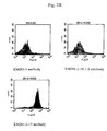
- Figure 1 shows the result.
- the horizontal axes in Fig. 1 indicate the expression intensity of CD95.
- a 1.0x10 6 cells/ml Ramos cell suspension was inoculated at 50 ⁇ l/well to a 96-well plate.
- the hybridoma culture supernatant or the purified antibody was adjusted at 2 ⁇ g/ml with a medium, and then added at 100 ⁇ l/well to a 96-well plate.
- Soluble CD40 ligands (4 ⁇ g/ml, ALEXIS CORPORATION) and anti-FLAG antibodies (4 ⁇ g/ml, M2, Sigma) were added to media, and then the media were added at 50 ⁇ l/well to the 96-well plate. After overnight culture, the cells were collected and then analyzed by FACS using R-PE-labeled anti-CD95 antibodies (Pharmingen NJ).
- Figures 2A and 2B, and 3 show the results.
- the horizontal axes in the figures indicate the expression intensity of CD95.
- CD95 expression was suppressed to the same degree as that of a negative control by the antibodies produced by each of the following hybridomas: KM281-1-10, KM281-2-10-1-2, KM283-5, KM292-1-24 and KM225-2-56.
- KM281-1-10 antibodies (lower panel) suppressed CD95 expression more effectively than that the 5D12 antibodies (central panel), the known antibody, only slightly suppressed CD95 expression. Specifically, the KM281-1-10 antibody was shown to be more effectively antagonistic. Thus, the human monoclonal antibody was shown to be an antagonistic antibody.
- a 1.0x10 6 cells/ml Ramos cell suspension was inoculated at 50 ⁇ l/well to a 96-well plate.
- the hybridoma culture supernatant or the purified antibody was adjusted to 2 ⁇ g/ml with a medium, and then added at 100 ⁇ l/well to a 96-well plate.
- Anti-human IgG antibodies Sigma, 13382
- Anti-mouse IgG antibodies Biosource, AMI3401
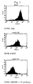
- Figures 4 and 5 show the results.
- the horizontal axes in the figures indicate the expression intensity of CD95.
- CD95 expression was suppressed by the antibodies produced by each of the hybridomas KM281-1-10 and KM281-2-10-1-2.
- CD95 expression was enhanced by the antibodies produced by each of the following hybridomas, 5D12, KM283-5, KM292-1-24 and KM225-2-56.
- a 1.0x10 5 cells/ml Ramos and HS-Sulton cell suspension was inoculated at 100 ⁇ l/well to a 96-well plate.
- a mixture of equivalent amount of the purified antibodies or soluble CD40 ligands, and anti-FLAG antibodies (M2) was added to media.
- 10 ⁇ l of 100 ⁇ Ci/ml 3 H-Thymidine was added.
- the culture product was harvested in a Printed Filtermat A (Wallac) using a Macro 96 Harvester (SKATRON), dried, and then immersed well in Betap;Scint (Wallac).
- FIG. 6 shows the results.
- the longitudinal axes indicate the amount of 3 H thymidine incorporated by cells
- the horizontal axes indicate the concentration of the antibody or CD40L in the culture solution.
- Recombinant human IL-4 was purchased from Genzyme techne.
- Anti-human CD14 MACS beads were purchased from Miltenyi Biotech GmbH. Lymphoprep was purchased from Nycomed Pharma AS.
- the medium used for culturing was RPMI1640 (Gibco BRL) supplemented with 10% heat inactivated FCS (Cell Culture Technologies), 10mM HEPES (Sigma), 55 ⁇ M 2-mercaptoethanol (Gibco BRL) and streptomycin sulfate (MEIJI SEIKA KAISHA, LTD.), when DC were induced.
- the cells in a staining process were washed with PBS (Sigma) supplemented with 2% FCS (Cell Culture Technologies) and 0.02% Azaid. When the cells were frozen, Cell banker (Nippon Zenyaku Kogyo) was used.
- Mononuclear cells were prepared (PBMC) from peripheral blood by density gradient centrifugation using Lymphoprep. The cells were subjected to positive selection using anti-human CD14 MACS beads, so as to separate the cells into a CD14 positive fraction and negative fraction. Recombinant human GM-CSF (50 ng/ml) and recombinant human IL-4 (100 ng/ml) were added to the positive fraction, followed by culturing in RPMI1640 media supplemented with 10% FCS in a 6-well plate. At the start of culturing, the cells were cultured at a concentration of 1 ⁇ 10 6 /ml (3 ml per well). During culturing, the media were exchanged once every 2 days.
- PBMC peripheral blood by density gradient centrifugation using Lymphoprep. The cells were subjected to positive selection using anti-human CD14 MACS beads, so as to separate the cells into a CD14 positive fraction and negative fraction.
- anti-HLA-DR antibodies isotype control: rat IgG2a
- anti-CD86 antibodies isotype control: rat IgG1
- anti-CD83 antibodies isotype control: rat IgG2b
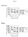
- Figure 7 shows the effect of KM302-1 antibodies, the agonistic antibodies, on DC maturation
- Figure 8 shows the effect of KM302-1 antibodies on IL-12 production of mature DC.
- the degree of maturation was compared with G28-5 antibodies as a control.
- the expression of CD86 and that of HLA-DR were examined, the expression was further elevated, that is, the degree of maturation was increased in the case of KM302-1 antibodies compared with the case of G28-5 antibodies.
- IL-12 secretion was increased by the treatment of mature DC with KM302-1 antibodies. Accordingly, it was shown that the KM302-1 antibodies acted as agonistic antibodies on DC.
- Blood peripheral blood collected from a normal human was centrifuged at 2000 rpm for 10 minutes, and then the serum was absorbed. The blood cell fraction was re-suspended with PBS, and then gently placed on Ficoll (Amersham Pharmacia). Centrifugation was performed at 2000 rpm for 30 minutes, so that a PBMC portion in the intermediate layer was collected, washed twice with PBS, and then used for a certain cell separation process using MACS.
- Monocyte separation for culturing DC was performed according to the attached instruction using MACS (Miltenyi Biotec GmbH). This is briefly explained as follows. 800 ⁇ l of MACS Buffer and 200 ⁇ l of MACS CD 14 (Miltenyi Biotec GmbH, 502-01) were added to PBMC (1 ⁇ 10 8 ), and then treated at 4°C for 15 minutes. The cells were adsorbed to a MACS LS column, and then washed. The cells adsorbed to the column were collected as monocytes. MACS HLA-DR (Miltenyi Biotec GmbH, 461-01) was added to the cells that were not adsorbed to the column. HLR-DR positive cells were removed with a BS column, thereby preparing a T cell fraction.
- MACS Motenyi Biotec GmbH
- the proportion of CD3 positive cells was measured by FACS, and then substantial number of T cells was calculated from the total cell number in the T cell fraction.
- the obtained monocytes were cultured in R0 media (PPMI medium supplemented with ⁇ -mercapto ethanol (Gibco) and HEPES (SIGMA)) containing 100 ng/ml IL-4 (R&D system), 50 ng/ml G-CSF (KIRIN) and 10% FCS (SIGMA) at a concentration of 1 ⁇ 10 6 cells/ml in a 6-well culture plate.
- 10 ng/ml LPS DIFCO was added for the cells to differentiate into mature DC.
- MLR was performed by mixing T cells and mature DC, which had been isolated from different humans.
- the cell ratio of T cells to DC was determined as 1:80, and number of T cells was determined as 2 ⁇ 10 5 cells/well.
- antibodies were added to DC for reaction to proceed for 30 minutes.
- T cells were added, culturing was performed for 4 days, and then 10 ⁇ l of 100 ⁇ Ci/ml 3 H-Thymidine (Amersham Pharmacia) was added. 14 hours later, the cells were harvested in Printed Filtermat A (Wallac) using a Macro 96 Harvester (SKATRON), dried, and then immersed well in Betap;Scint (Wallac).
- Anti-CD40 antibodies were caused to bind to immobilized CD40 human FC using BIAcore 2000 (Biacore), and then changes in the binding amount of soluble CD40L to CD40 were measured.
- soluble CD40 human FC was immobilized on a CM chip (CM5, Biacore).
- CM5, Biacore CM5, Biacore
- 25 ⁇ g/ml anti-CD40 antibodies were added to bind to CD40.
- 10 ⁇ g/ml soluble CD40L was added for binding. A difference between the binding amounts before and after addition of CD40L was measured.
- control IgG was added, the binding amount of CD40L was 100 RU.
- the binding amount of CD40L was 110 RU, and after addition of KM283-5 antibodies the binding amount of CD40L was 18RU. Thus, it was shown that the KM302-1 antibody does not inhibit the binding of CD40L to CD40.
- Example 12 Purified antibodies of the hybridomas obtained in Example 4 were analyzed according to the method of Example 6, and then clones producing agonistic antibodies were selected (number of cells per well: 5 ⁇ 10 4 ; cell concentration: 2.5 ⁇ 10 5 /ml).
- Figure 12 shows the results.
- the horizontal axis indicates the antibody concentration in culture solutions, and the longitudinal axis indicates average fluorescence intensities, that is, CD95 expression intensities.
- KM341-1-19 and 2105 antibodies were shown to promote CD95 expression of Ramos cells more effectively than G28-5 antibodies, which are known mouse antibodies.
- KM341-1-19 and 2105 antibodies were shown to be more effective agonistic antibodies.
- the agonistic activity (to increase CD95 expression of Ramos cells) of KM341-1-19 and 2105 antibodies (0.01 ⁇ g/ml) was higher than that of G28-5 antibodies (10 ⁇ g/ml) (Fig. 12).
- Table 5 summarizes that at each antibody concentration, CD95 expression level is how many times greater or less than that expressed by the addition of G28-5 antibodies.
- Antibody concentration ( ⁇ g/ml) KM341-1-19 2105 F5-77 F2-103 0.01 5.7 3.9 0.1 7.0 7.1 1.2 1.2 1 5.7 5.1 1.7 1.8 10 4.5 3.3 2.0 1.7
- IL-12 was measured by the ELISA (Pharmingen) method.
- Figures 13 and 14 show the results. It was shown that IL-12 secretion was increased by treatment with KM341-1-19 antibodies.
- CD40 ligand-expressing recombinant L cells (2 ⁇ 10 5 cells/ml) that had been irradiated with X-rays (5000 rad) were allowed to co-exist, the concentrations of IL-12 and IL-10 in culture solutions were 254 and 51 pg/ml, respectively. They were lower than that when 1 ⁇ g/ml KM341-1-19 antibodies were added.
- KM341-1-19 antibodies act on DC as effective agonistic antibodies.
- the agonistic activity of KM341-1-19 antibodies (0.1 ⁇ g/ml) to cause mature DC to secrete IL-12 was higher than that of G28-5 antibodies (100 ⁇ g/ml).
- the agonistic activity of KM341-1-19 antibodies (1 ⁇ g/ml) to cause mature DC to secrete IL-12 was 100 times or more greater than that by G28-5 antibodies (100 ⁇ g/ml) (Fig. 13).
- the agonistic activity of KM341-1-19 antibodies (1 ⁇ g/ml) to cause mature DC to secrete IL-10 was 10 times or more greater than that by G28-5 antibodies (100 ⁇ g/ml) (Fig.
- the antibody since the subclass of KM341-1-19 antibody was IgG2, the antibody has lower binding ability to the Fc receptor than that of IgG1 or IgG3. Its ability to sensitize the killer activity of NK cells and ability to activate the complement system are also weak. Accordingly, there may be a low risk that the function of CD40-expressing cells or the number of the cells themselves decreases due to the antibody. Furthermore, the antibody is not easily cross-linked by an Fc receptor, so that it can be expected that the drug effect is easily controlled without any large fluctuation in in vivo agonistic activity due to cross-linking.
- Ramos cell suspension was inoculated at 50 ⁇ l/well to a flat bottom 96-well plate (number of cells per well: 5 ⁇ 10 4 ).
- Purified antibodies diluted with media were added at 100 ⁇ l/well to a 96-well plate.
- Human CD40 ligand-expressing recombinant mouse L cells (see Spriggs, M.K. et. al., J. Exp. Med., 176: 1543, 1992; Garrone, P. et. al., J. Exp. Med., 182: 1265, 1995 and the like) were prepared at 1.0 ⁇ 10 5 cells/ml.
- the prepared cells were added at 50 ⁇ l/well (the number of Ramos cells per well: 5 ⁇ 10 4 ; Ramos cell concentration: 2.5 ⁇ 10 4 cells/ml; the number of mouse L cells per well: 5 ⁇ 10 3 ; mouse cell concentration: 2.5 ⁇ 10 4 cells/ml). After overnight culture, the cells were collected, and then analyzed by FACS using R-PE-labeled anti-CD95 antibodies.
- Figure 15 shows the results. In the figure, the longitudinal axis indicates the average fluorescence intensity, that is, CD95 expression intensity.
- 4D11 antibodies suppressed, even at a concentration of 0.1 ⁇ g/ml, CD95 expression to the same degree as that of a case of a negative control wherein no CD40L-expressing cells had been added.
- 4D11, F4-465 and KM281-1-10 suppressed CD95 expression to the same degree as that of the case of the negative control wherein no CD40L-expressing cells had been added.
- Table 6 shows relative values of the average fluorescence intensity corresponding to each antibody concentration, when the value of the control case wherein no antagonistic antibodies were added is determined as 100.
- Anti-asialo GM1 antibodies were intravenously injected to 5-week-old C.B.17/Icr-scidJc1 mice (CLEA JAPAN). 1 day later, 5 ⁇ 10 6 Ramos cells per mouse were intravenously injected as tumor cells. 1 day later, KM302-1 antibodies or anti-human albumin human IgG antibodies as a negative control were intravenously injected. The doses per mouse of KM302-1 antibodies were 1, 10 and 100 ⁇ g, and the same of the negative control antibodies was 100 ⁇ g. Each of these antibodies was administered once to 5 mice. Figure 16 shows the results.
- KM302-1 antibodies By day 34 after transplantation, all the mice of the negative control-administered group had died, whereas all the 5 mice each of the groups administered with 10 ⁇ g and 100 ⁇ g of KM302-1 antibodies had been administered to which survived. Thus, the anti-tumor effect of KM302-1 antibodies was confirmed.
- the KM302-1 antibody is of the IgG4 subclass, so that the Fc receptor-mediated antibody dependent cellular cytotoxicity (ADCC) and activation of the complement system are weak. Despite these characteristics, it was observed that single administration of 10 ⁇ g of KM302-1 antibodies prolonged the survival time of tumor-bearing mice.
- a Ramos cell suspension was prepared at 1x10 4 cells/ml in an RPMI1640 medium supplemented with 10%FBS, and then 100 ⁇ l of the suspension was apportioned to a 96-well plate.
- a KM341-1-19 antibody or soluble ligand solution prepared at 20 ⁇ g/ml using media was added.
- Anti-FLAG antibodies (M2) with the same concentration as that of the ligands were allowed to co-exist with the soluble ligands (the concentration in the reaction solution was 10 ⁇ g/ml), thereby enhancing the activity.
- 20 ⁇ l of MTS reagent Promega
- Hybridomas producing KM341-1-19, 2105, 110, 115, KM281-1-10, 4D11, KM643-4-11, F4-465, F2-103 and F5-77 antibodies were cultured, and then the cells were collected by centrifugation.
- TRIZOL Gibco BRL
- Total RNA was extracted according to the attached instructions.
- Cloning of the variable regions of the antibody cDNA was performed according to the attached instructions using a SMART RACE cDNA amplification Kit (CLONTECH). Using 5 ⁇ g of total RNA as a template, 1st Strand cDNA was constructed.
- H chain Heavy chains (H chain) of KM341-1-19, 2105, 110, 115, KM281-1-10, 4D11, KM643-4-11, F2-103 and F5-77
- Z-Taq (Takara) and UMP and hh6 primers were used, and a cycle of 98°C for 1 second and 68°C for 30 seconds was repeated 30 times.
- a cycle of 98°C for 1 second and 68°C for 30 seconds was repeated 20 times.
- UMP and hh2 primers and an Advantage 2 PCR kit (Clonthech, cat#1910) were used, and 5 cycles of 94°C for 5 seconds and 72°C for 3 minutes, 5 cycles of 94°C for 5 seconds, 70°C for 0 seconds and 72°C for 3 minutes, and 25 cycles of 94°C for 5 seconds, 68°C for 10 seconds and 72°C for 3 minutes were performed.
- the amplified PCR product was purified using a PCR purification kit (QIAGEN), and then the nucleotide sequence was determined using hh4 as a primer.
- the product was subcloned to PCR-Script (Stratagene, Lajolla, CA) or PCR-Blunt (Invitrogene, Carlsbad, CA), and then sequencing was performed. Based on the sequence information, antibody heavy-chain-specific primers were synthesized.
- a 341H primer was synthesized in the case of KM341-1-19, a 2105Hsal primer in the case of 2105, a 110Hsal primer in the case of 110 and 115, a 281Hsal primer in the case of KM281-1-10, a 4D11Sal primer in the case of 4D11, a 643Hsal primer in the case of KM643-4-11, H11-9 5' primer in the case of F4-465, a F2-103H primer in the case of F2-103 and F5-77H primer in the case of F5-77.
- cDNA was amplified from the 1st Strand cDNA, and then the sequence from the opposite direction was determined using the amplified product as a template and the antibody-specific primers.
- the light chains (L chains) of KM341-1-19, 2105, 110, 115, KM281-1-10, 4D11, KM643-4-11, F2-103 and F5-77 were amplified using UMP and hk2 primers and by repeating 30 times a cycle of 98°C for 1 second and 68°C for 30 seconds.
- the light chain of F4-465 was amplified using UMP and hL2 primers and by repeating 30 times a cycle of 98°C for 1 second and 68°C for 30 seconds.
- the amplified PCR product was purified using a PCR purification kit, and then the nucleotide sequence was determined using hk6 or hL2 primers.
- a 341K primer was synthesized in the case of KM341-1-19, 2053KBgl primer in the case of 2105, 110KBgl primer in the case of 110 and 115, 281KBgl primer in the case of KM281-1-10, 4D11KBg1 in the case of 4D11, 643KBgl primer in the case of KM643-4-11, Lamda 5' primer in the case of F4-465, and F2-103K primer in the case of F-103 and F5-77.
- cDNA was amplified from the 1st Strand cDNA using the light chain specific primer and hk6 primer. The sequence was then determined from both directions using the amplified product as a template. For F4-465, F2-103 and F5-77, subcloning to PCR-Script (Stratagene, Lajolla, CA) or PCR-Blunt (Invitrogene, Carlsbad, CA) was performed to determine the sequence.
- the translation initiation point of the H-chain DNA is an ATG codon that begins from the 50th adenine (A) from the 5' end of SEQ ID NO: 27, and the termination codon is TGA beginning from the 1472nd thymine (T).
- the boundary of the antibody variable region and the constant region is located between the 493rd adenine (A) and the 494th guanine (G) from the 5'end.
- the H-chain variable region ranges from the N-terminus to the 148th serine (S) residue of SEQ ID NO: 28, and the constant region is of the 149th alanine (A) and the following residues.
- the translation initiation point of the L-chain DNA is an ATG codon that begins from the 29th A from the 5' end of SEQ ID NO: 29, and the variable region ranges from the 5' end to the 400th adenine (A).
- the variable region ranges from the N-terminus to the 124th lysine (K) of SEQ ID NO: 30.
- Analysis of the N-terminus of the purified L-chain protein revealed that the L-chain signal sequence ranges from the N-terminus to the 20th glycine (G) of SEQ ID NO: 30, and the N-terminus of the mature protein is the 21 st glutamic acid (E) of SEQ ID NO: 30.
- the translation initiation point of the H-chain DNA is an ATG codon that begins from the 70th adenine (A) from the 5' end of SEQ ID NO: 31.
- the boundary of the antibody variable region and the constant region is located between the 495th adenine (A) and the 496th guanine (G) from the 5' end.
- the H-chain variable region ranges from the N-terminus to the 142nd serine (S) residue of SEQ ID NO: 32, and the constant region is of the 149th alanine (A) and the following residues.
- H-chain signal sequence ranges from the N-terminus to the 19th cystein (C) of SEQ ID NO: 32. It is thought that the N-terminus of the mature protein is the 20th glutamic acid (E) of SEQ ID NO: 32.
- the translation initiation point of the L-chain DNA is an ATG codon that begins from the 28th A from the 5' end of SEQ ID NO: 33, and the variable region ranges from the 5' end to the 405th adenine (A).
- the variable region ranges from the N-terminus to the 126th lysine (K) of SEQ ID NO: 34. It was predicted by gene sequence prediction software (Signal P ver.2) that the L-chain signal sequence ranges from the N-terminus to the 20th glycine (G) of SEQ ID NO: 34. It is thought that the N-terminus of the mature protein is the 21st glutamic acid (E) of SEQ ID NO: 34.
- DNAs of 110 encoding the H-chain variable region and L-chain variable region and the amino acid sequences of the H-chain and L-chain are respectively shown below.
- the translation initiation point of the H-chain DNA is an ATG codon that begins from the 60th adenine (A) from the 5' end of SEQ ID NO: 35.
- the boundary of the antibody variable region and the constant region is located between the 479th adenine (A) and the 480th guanine (G) from the 5'end.
- the H-chain variable region ranges from the N-terminus of SEQ ID NO: 36 to the 140th serine (S) residue, and the constant region is of the 141 st alanine (A) and the following residues.
- the translation initiation point of the L-chain DNA is an ATG codon that begins from the 35th A from the 5' end of SEQ ID NO: 37, and the variable region ranges from the 5' end to the 421 st adenine (A).
- the variable region ranges from the N-terminus to the 129th lysine (K) of SEQ ID NO: 38. It was predicted by gene sequence prediction software (Signal P ver.2) that the L-chain signal sequence ranges from the N-terminus to the 22nd cystein (C) of SEQ ID NO: 38. It is thought that the N-terminus of the mature protein is the 23rd valine (V) of SEQ ID NO: 38.
- the translation initiation point of the H-chain DNA is an ATG codon that begins from the 60th adenine (A) from the 5' end of SEQ ID NO: 39.
- the boundary of the antibody variable region and the constant region is located between the 479th adenine (A) and the 480th guanine (G) from the 5'end.
- the H-chain variable region ranges from the N-terminus of SEQ ID NO: 40 to the 140th serine (S) residue, and the constant region is of the 141 st alanine (A) and the following residues.
- the translation initiation point of the L-chain DNA is an ATG codon that begins from the 35th A from the 5' end of SEQ ID NO: 41, and the variable region ranges from the 5' end to the 421 st adenine (A).
- the variable region ranges from the N-terminus to the 129th lysine (K) of SEQ ID NO: 42. It was predicted by gene sequence prediction software (Signal P ver.2) that the L-chain signal sequence ranges from the N-terminus to the 22nd cystein (C) of SEQ ID NO: 42. It is thought that the N-terminus of the mature protein is the 23rd valine (V) of SEQ ID NO: 42.
- DNAs of 281-1-10 encoding the H-chain variable region and L-chain variable region and the amino acid sequences of the H-chain and L-chain are respectively shown below.
- the translation initiation point of the H-chain DNA is an ATG codon that begins from the 52nd adenine (A) from the 5' end of SEQ ID NO: 43.
- the boundary of the antibody variable region and the constant region is located between the 468th adenine (A) and the 469th guanine (G) from the 5'end.
- the H-chain variable region ranges from the N-terminus of SEQ ID NO: 44 to the 139th serine (S) residue, and the constant region is of the 140th alanine (A) and the following residues.
- the translation initiation point of the L-chain DNA is an ATG codon that begins from the 41 st A from the 5' end of SEQ ID NO: 45, and the variable region ranges from the 5' end to the 424th adenine (A).
- the variable region ranges from the N-terminus to the 128th lysine (K) of SEQ ID NO: 46. It was predicted by gene sequence prediction software (Signal P ver.2) that the L-chain signal sequence ranges from the N-terminus to the 20th glycine (G) of SEQ ID NO: 46. It is thought that the N-terminus of the mature protein is the 21 st glutamic acid (E) of SEQ ID NO: 46.
- DNAs of 4D11 encoding the H-chain variable region and L-chain variable region and the amino acid sequences of the H-chain and L-chain are respectively shown below.
- the translation initiation point of the H-chain DNA is an ATG codon that begins from the 16th adenine (A) from the 5' end of SEQ ID NO: 47.
- the boundary of the antibody variable region and the constant region is located between the 456th adenine (A) and the 457th guanine (G) from the 5'end.
- the H-chain variable region ranges from the N-terminus of SEQ ID NO: 48 to the 147th serine (S) residue, and the constant region is of the 148th alanine (A) and the following residues.
- the translation initiation point of the L-chain DNA is an ATG codon that begins from the 59th A from the 5' end of SEQ ID NO: 49, and the variable region ranges from the 5' end to the 442nd adenine (A).
- the variable region ranges from the N-terminus to the 128th lysine (K) of SEQ ID NO: 50. It was predicted by gene sequence prediction software (Signal P ver.2) that the L-chain signal sequence ranges from the N-terminus to the 22nd cystein (C) of SEQ ID NO: 50. It is thought that the N-terminus of the mature protein is the 21 st alanine (A) of SEQ ID NO: 50.
- DNAs of KM643-4-11 encoding the H-chain variable region and L-chain variable region and the amino acid sequences of the H-chain and L-chain are respectively shown below.
- the translation initiation point of the H-chain DNA is an ATG codon that begins from the 1 st adenine (A) from the 5' end of SEQ ID NO: 51.
- the boundary of the antibody variable region and the constant region is located between the 447th adenine (A) and the 448th guanine (G) from the 5'end.
- the H-chain variable region ranges from the N-terminus of SEQ ID NO: 52 to the 149th serine (S) residue, and the constant region is of the 150th alanine (A) and the following residues.
- the translation initiation point of the L-chain DNA is an ATG codon that begins from the 38th A from the 5' end of SEQ ID NO: 53, and the variable region ranges from the 5' end to the 409th adenine (A).
- the variable region ranges from the N-terminus to the 124th lysine (K) of SEQ ID NO: 54. It was predicted by gene sequence prediction software (Signal P ver.2) that the L-chain signal sequence ranges from the N-terminus to the 20th glycine (G) of SEQ ID NO: 54. It is thought that the N-terminus of the mature protein is the 21st Glutamic acid (E) of SEQ ID NO: 54.
- the translation initiation point of the H-chain DNA is an ATG codon that begins from the 47th adenine (A) from the 5' end of SEQ ID NO: 55.
- the boundary of the antibody variable region and the constant region is located between the 484th adenine (A) and the 445th guanine (G) from the 5'end.
- the H-chain variable region ranges from the N-terminus of SEQ ID NO: 56 to the 146th serine (S) residue, and the constant region is of the 147th alanine (A) and the following residues.
- the translation initiation point of the L-chain DNA is an ATG codon that begins from the 81 st A from the 5' end of SEQ ID NO: 57, and the variable region ranges from the 5' end to the 440th (C).
- the variable region ranges from the N-terminus to the 120th Threonine (T) of SEQ ID NO: 58.
- T Threonine
- the translation initiation point of the H-chain DNA is an ATG codon that begins from the 32nd adenine (A) from the 5' end of SEQ ID NO: 59.
- the boundary of the antibody variable region and the constant region is located between the 463rd adenine (A) and the 464th Guanine (G) from the 5'end.
- the H-chain variable region ranges from the N-terminus of SEQ ID NO: 60 to the 144th Serine (S) residue, and the constant region is of the ]45th Alanine (A) and the following residues.
- the translation initiation point of the L-chain DNA is an ATG codon that begins from the 29th A from the 5' end of SEQ ID NO: 61, and the variable region ranges from the 5' end to the 415th adenine (A).
- the variable region ranges from the N-terminus to the 129th Lysine (K) of SEQ ID NO: 62. It was predicted by gene sequence prediction software (Signal P ver.2) that the L-chain signal sequence ranges from the N-terminus to the 22nd Cystein (C) of SEQ ID NO: 62. It is thought that the N-terminus of the mature protein is the 23rd Asp (D) of SEQ ID NO: 62.
- the translation initiation point of the H-chain DNA is an ATG codon that begins from the 100th adenine (A) from the 5' end of SEQ ID NO: 63.
- the boundary of the antibody variable region and the constant region is located between the 528th adenine (A) and the 529th Guanine (G) from the 5'end.
- the H-chain variable region ranges from the N-terminus of SEQ ID NO: 64 to the 143rd Serine (S) residue, and the constant region is of the 144th Alanine (A) and the following residues.
- the translation initiation point of the L-chain DNA is an ATG codon that begins from the 59th A from the 5' end of SEQ ID NO: 65, and the variable region ranges from the 5' end to the 445th adenine (A).
- the variable region ranges from the N-terminus to the 129th Lysine (K) of SEQ ID NO: 66. It was predicted by gene sequence prediction software (Signal P ver.2) that the L-chain signal sequence ranges from the N-terminus to the 22nd Cystein (C) of SEQ ID NO: 66. It is thought that the N-terminus of the mature protein is the 23rd Asp (D) of SEQ ID NO: 66.
- the above obtained DNA fragment containing the variable region of the antibody was incorporated into an appropriate vector such as NSKG1 (IDEC Pharmaceuticals, US patent 6001358), thereby preparing an antibody expression vector.
- a host cell for expression for example, CHO-Ras (Katakura Y., et al., Cytotechnology, 31: 103-109, 1999) is appropriately used.
- the vector can be introduced into the host cell by, for example, electroporation. Approximately 2 ⁇ g of the antibody expression vector was linearized with a restriction enzyme.
- the gene was introduced into 4x10 7 CHO-Ras cells under conditions of 350V and 500 ⁇ F using a Bio-Rad electrophoreter, and then inoculated to a 96-well culture plate. A drug corresponding to the selection marker of the expression vector was added, and culturing was continued. When colonies were observed, antibody-expressing lines were selected by the method described in Example 4. Antibodies can be purified from the selected cells according to Example 5.
- mice having a genetic background whereby they were homozygotes for mouse endogenous disrupted CD40 and harboring a transgene of a human CD40 gene were sensitized by intraperitoneally injecting 100 ⁇ g (in an amount of NP-CGG) of a complex of 4-hydroxy-3-nitrophenylacetyl-chicken ⁇ -globulin conjugates (NP-CGG : distributed by Hitoshi KIKUTANI, Professor, Research Institute for Microbial Diseases, Osaka University) and ARAM (ARAM: Antigen Recognition Activation Motif).
- NP-BSA NP-bound bovine serum albumin
- a solution prepared by diluting 1,000-fold alkaline phosphatase-labeled goat anti-mouse IgG1 or IgM antibody (COSMO BIO, 1070-04 or 1020-04) with 10% BlockAce-containing PBS-T was added (50 ⁇ l/well) to each well, followed by 2 hours of incubation at 37°C. Next, the microplate was washed 3 times with PBS-T, a chromogenic substrate solution (50 ⁇ l/well, Sigma104, phosphatase substrate) was added to each well, and then absorbance at a wavelength of 405 nm was measured with a microplate reader.
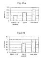
- Figures 18 and 19 show the results.
- longitudinal axes indicate values obtained by conversion using as a unit the serum diluted 10,000-fold in the case of IgG1 antibody, and the serum diluted 100-fold in the case of IgM antibody.
- the serum was prepared by injecting NP-CGG twice into C57BL/6 mice, collecting blood from the mice, and pooling the serum.
- Administration of 100 ⁇ g each of F4-465, 4D11 and KM281-1-10 antibodies strongly suppressed NP-specific IgG 1 and IgM antibody production.
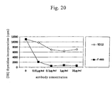
- 1x10 5 cells were added to a 96-well plate, and then anti-human CD40 antibodies were added at concentrations of 0.01, 0.1, 1.0 and 10 ⁇ g/ml. The test was conducted in triplicate at each concentration. 1 ⁇ g/ml flag-labeled CD40L (Alexis) and 1 ⁇ g/ml CD40L enhancer antibodies (Alexis) were added to each well, followed by 3 days of culturing. Then, 1 ⁇ Ci [ 3 H] thymidine was added to each well. 12 to 15 hours later, the cells were collected, and then the proliferation of tonsillar B cells was measured using a liquid scintillation counter.
- 1 ⁇ g/ml flag-labeled CD40L (Alexis) and 1 ⁇ g/ml CD40L enhancer antibodies (Alexis) were added to each well, followed by 3 days of culturing. Then, 1 ⁇ Ci [ 3 H] thymidine was added to each well. 12 to 15 hours later, the cells
- the count obtained when B cells had proliferated due to the stimulation of CD40L and no antibody had been added was determined as 100, and the count obtained when no CD40L had been added and B cells had not been not stimulated was determined as 0. For example, when the relative count measured was 30, this was expressed in this experiment as that 70% proliferation inhibition had occurred.
- 5D12 which is the known antagonistic antibody, did not show more than 50% proliferation inhibition even with the antibody concentration of 100 ⁇ g/ml.
- F4-465 showed approximately 80% proliferation suppression even with the antibody concentration as low as 0.01 ⁇ g/ml, and showed approximately 95% proliferation suppression with an antibody concentration of 0.1 to 10 ⁇ g/ml (Fig. 20).
- the present invention provides an antibody against CD40.
- the antibody of the present invention includes both an antibody that acts agonistically on CD40 and an antibody that acts antagonistically on CD40.
- these antibodies are useful as, for example, an immunopotentiating agent and immunosuppressive agent, respectively.
Priority Applications (3)
| Application Number | Priority Date | Filing Date | Title |
|---|---|---|---|
| EP07014664A EP1914243A3 (de) | 2001-04-27 | 2002-04-26 | Monoglonaler Antikörper gegen CD40 |
| EP08017500A EP2011802A3 (de) | 2001-04-27 | 2002-04-26 | Monoklonaler Antikörper gegen CD40 |
| EP08017499.8A EP2009027B1 (de) | 2001-04-27 | 2002-04-26 | Monoklonaler Antikörper gegen CD40 |
Applications Claiming Priority (11)
| Application Number | Priority Date | Filing Date | Title |
|---|---|---|---|
| US13672 | 2001-04-27 | ||
| PCT/US2001/013672 WO2001083755A2 (en) | 2000-04-28 | 2001-04-27 | Human anti-cd40 antibodies and methods of making and using same |
| US844684 | 2001-04-27 | ||
| US09/844,684 US7063845B2 (en) | 2000-04-28 | 2001-04-27 | Human anti-CD40 antibodies |
| JP2001142482 | 2001-05-11 | ||
| JP2001142482 | 2001-05-11 | ||
| JP2001310535 | 2001-10-05 | ||
| JP2001310535 | 2001-10-05 | ||
| US10/040,244 US20030059427A1 (en) | 2000-04-28 | 2001-10-26 | Isolation and characterization of highly active anti-CD40 antibody |
| US40244 | 2001-10-26 | ||
| PCT/JP2002/004292 WO2002088186A1 (en) | 2001-04-27 | 2002-04-26 | Anti-cd40 monoclonal antibody |
Related Child Applications (2)
| Application Number | Title | Priority Date | Filing Date |
|---|---|---|---|
| EP07014664A Division EP1914243A3 (de) | 2001-04-27 | 2002-04-26 | Monoglonaler Antikörper gegen CD40 |
| EP08017499.8A Division EP2009027B1 (de) | 2001-04-27 | 2002-04-26 | Monoklonaler Antikörper gegen CD40 |
Publications (3)
| Publication Number | Publication Date |
|---|---|
| EP1391464A1 true EP1391464A1 (de) | 2004-02-25 |
| EP1391464A4 EP1391464A4 (de) | 2005-04-27 |
| EP1391464B1 EP1391464B1 (de) | 2007-09-26 |
Family
ID=31192420
Family Applications (2)
| Application Number | Title | Priority Date | Filing Date |
|---|---|---|---|
| EP07014664A Withdrawn EP1914243A3 (de) | 2001-04-27 | 2002-04-26 | Monoglonaler Antikörper gegen CD40 |
| EP02720634A Expired - Lifetime EP1391464B1 (de) | 2001-04-27 | 2002-04-26 | Monoklonaler anti-cd-40-antikörper |
Family Applications Before (1)
| Application Number | Title | Priority Date | Filing Date |
|---|---|---|---|
| EP07014664A Withdrawn EP1914243A3 (de) | 2001-04-27 | 2002-04-26 | Monoglonaler Antikörper gegen CD40 |
Country Status (8)
| Country | Link |
|---|---|
| US (1) | US20030059427A1 (de) |
| EP (2) | EP1914243A3 (de) |
| JP (1) | JP3847715B2 (de) |
| CN (2) | CN101289510B (de) |
| AU (1) | AU2002251562B2 (de) |
| DE (1) | DE60222658T2 (de) |
| ES (1) | ES2292746T3 (de) |
| WO (1) | WO2002088186A1 (de) |
Cited By (23)
| Publication number | Priority date | Publication date | Assignee | Title |
|---|---|---|---|---|
| WO2005044854A3 (en) * | 2003-11-04 | 2005-08-11 | Chiron Corp | Antagonist anti-cd40 monoclonal antibodies and methods for their use |
| WO2005044304A3 (en) * | 2003-11-04 | 2005-08-25 | Chiron Corp | Use of antagonist anti-cd40 antibodies for treatment of chronic lymphocytic leukemia |
| WO2005044307A3 (en) * | 2003-11-04 | 2005-08-25 | Chiron Corp | Methods of therapy for b cell-related cancers |
| WO2005044855A3 (en) * | 2003-11-04 | 2005-09-15 | Chiron Corp | Use of antagonist anti-cd40 monoclonal antibodies for treatment of multiple myeloma |
| WO2006073443A2 (en) * | 2004-04-27 | 2006-07-13 | Novartis Vaccines And Diagnostics, Inc. | Antagonist anti-cd40 monoclonal antibodies and methods for their use |
| EP1768999A2 (de) * | 2004-06-30 | 2007-04-04 | Mayo Foundation For Medical Education And Research | B7-dc-bindender antikörper |
| EP1847602A1 (de) * | 2005-01-12 | 2007-10-24 | Kirin Pharma Kabushiki Kaisha | Stabilisierte menschliche igg1- und igg3-antikörper |
| US7501119B2 (en) | 2004-06-30 | 2009-03-10 | Mayo Foundation For Medical Education And Research | Methods and molecules for modulating an immune response |
| AU2004309275B2 (en) * | 2003-12-25 | 2010-12-23 | Kyowa Kirin Co., Ltd. | Mutants of anti-CD40 antibody |
| WO2010104761A3 (en) * | 2009-03-10 | 2011-01-06 | Baylor Research Institute | Anti-cd40 antibodies and uses thereof |
| US8188238B2 (en) | 2004-11-05 | 2012-05-29 | Mayo Foundation For Medical Education And Research | Recombinantly produced antibody |
| US8277810B2 (en) | 2003-11-04 | 2012-10-02 | Novartis Vaccines & Diagnostics, Inc. | Antagonist anti-CD40 antibodies |
| US8518410B2 (en) | 2009-03-10 | 2013-08-27 | Baylor Research Institute | Fusion protein with HIV antigen |
| US8828396B2 (en) | 2010-11-15 | 2014-09-09 | Novartis Ag | Silent Fc variants of anti-CD40 antibodies |
| US8911726B2 (en) * | 2004-09-22 | 2014-12-16 | Kyowa Hakko Kirin Co., Ltd | Stabilized human Igg4 antibodies |
| WO2014200349A1 (en) | 2013-06-13 | 2014-12-18 | Fast Forward Pharmaceutical B.V. | Cd40 signalling inhibitor and a further compound, wherein the further compound is a bile acid, a bile acid derivative, an tgr5-receptor agonist, an fxr agonist or a combination thereof, for the treatment of chronic inflammation, and the prevention of gastrointestinal cancer or fibrosis. |
| US8926979B2 (en) | 2005-11-01 | 2015-01-06 | Novartis Ag | Treatment of cancer or pre-malignant conditions using anti-CD40 antibodies |
| US8945564B2 (en) | 2006-04-21 | 2015-02-03 | Novartis Ag | Antagonist anti-CD40 antibody pharmaceutical compositions |
| US9102734B2 (en) | 2009-03-10 | 2015-08-11 | Baylor Research Institute | Fusion proteins comprising antibody and HPV E6 and E7 antigens |
| US9885017B2 (en) | 2011-03-25 | 2018-02-06 | Baylor Research Institute | Compositions and methods to immunize against hepatitis C virus |
| US10610585B2 (en) | 2017-09-26 | 2020-04-07 | Inserm (Institut National De La Sante Et De La Recherche Medicale) | Methods and compositions for treating and preventing HIV |
| US10940195B2 (en) | 2014-01-13 | 2021-03-09 | Baylor Research Institute | Vaccines against HPV and HPV-related diseases |
| RU2779128C2 (ru) * | 2017-06-01 | 2022-09-02 | Цзянсу Хэнжуй Медисин Ко., Лтд. | Антитело к cd40, его антигенсвязывающий фрагмент и его медицинское применение |
Families Citing this family (51)
| Publication number | Priority date | Publication date | Assignee | Title |
|---|---|---|---|---|
| US20070160615A1 (en) * | 1999-06-01 | 2007-07-12 | Jun Tan | Methods and compounds for disruption of CD40R/CD40L signaling in the treatment of Alzheimer's disease |
| US6787318B1 (en) * | 1999-06-01 | 2004-09-07 | Roskamp Research Institute, Llc | Assay for evaluating the therapeutic effectiveness of agents in reducing Alzheimer's disease pathology |
| ATE541860T1 (de) * | 2000-10-02 | 2012-02-15 | Novartis Vaccines & Diagnostic | Humane antikörper gegen cd40 zur therapie von b- zell tumoren |
| WO2003033538A1 (fr) * | 2001-10-15 | 2003-04-24 | Kirin Beer Kabushiki Kaisha | Anticorps anti-hlr-dr |
| AR039067A1 (es) * | 2001-11-09 | 2005-02-09 | Pfizer Prod Inc | Anticuerpos para cd40 |
| WO2004037204A2 (en) * | 2002-10-25 | 2004-05-06 | University Of South Florida | Methods and compounds for disruption of cd40r/cd40l signaling in the treatment of alzheimer's disease |
| US7628986B2 (en) | 2003-06-27 | 2009-12-08 | Amgen Fremont Inc. | Antibodies directed to the deletion mutants of epidermal growth factor receptor and uses thereof |
| TWI353992B (en) * | 2003-07-22 | 2011-12-11 | Schering Ag | Rg1 antibodies and uses thereof |
| AR045563A1 (es) | 2003-09-10 | 2005-11-02 | Warner Lambert Co | Anticuerpos dirigidos a m-csf |
| US20050136055A1 (en) | 2003-12-22 | 2005-06-23 | Pfizer Inc | CD40 antibody formulation and methods |
| KR20060132006A (ko) | 2004-03-23 | 2006-12-20 | 비오겐 아이덱 엠에이 아이엔씨. | 수용체 커플링제 및 이의 치료적 용도 |
| EP1809737B9 (de) | 2004-10-07 | 2011-10-05 | Argos Therapeutics, Inc. | Zusammensetzungen reifer dendritischer zellen und verfahren zu deren kultivierung |
| US8513008B2 (en) | 2004-10-07 | 2013-08-20 | Argos Therapeutics, Inc. | Mature dendritic cell compositions and methods for culturing same |
| EP2096166A1 (de) * | 2005-03-31 | 2009-09-02 | Osaka University | Verfahren zur Herstellung von gegen das Zellmembranoberflächen-Antigenepitop gerichteten Antikörpern und Assay-Verfahren |
| AU2006247134B2 (en) | 2005-05-18 | 2012-05-10 | Novartis Ag | Methods for diagnosis and treatment of diseases having an autoimmune and/or inflammatory component |
| EP2589654A1 (de) | 2006-05-03 | 2013-05-08 | The Regents of the University of Colorado, a body corporate | Synergistische Hilfsstoffkombination aus CD40-Agonisten-Antikörper/Typ1-Interferon, diese enthaltende Konjugate und ihre Verwendung als Therapeutika zur Verstärkung der Zellimmunität |
| ES2828627T3 (es) | 2008-04-25 | 2021-05-27 | Kyowa Kirin Co Ltd | Anticuerpo multivalente estable |
| CN102625714B (zh) * | 2008-07-16 | 2015-08-19 | 贝勒研究院 | 基于将最大化的gag和nef靶向树突细胞的hiv疫苗 |
| AU2013202410B2 (en) * | 2008-07-16 | 2014-10-09 | Institute For Research In Biomedicine | Human cytomegalovirus neautralizing antibodies and use thereof |
| US9234044B2 (en) | 2009-04-20 | 2016-01-12 | Kyowa Hakko Kirin Co., Ltd | Agonistic anti-CD40 IGG2 antibodies having amino acid mutations introduced therein |
| AR081750A1 (es) | 2010-03-31 | 2012-10-17 | Boehringer Ingelheim Int | Anticuerpos anti-cd40 |
| US20140031250A1 (en) | 2010-10-07 | 2014-01-30 | David Tsai Ting | Biomarkers of Cancer |
| WO2012075111A1 (en) | 2010-11-30 | 2012-06-07 | Novartis Ag | Uses of anti-cd40 antibodies in combination therapy for b cell-related cancers |
| ES2734076T3 (es) | 2011-02-17 | 2019-12-04 | Kyowa Hakko Kirin Co Ltd | Preparación farmacéutica de anticuerpo anti-CD40 muy concentrada |
| GB201115280D0 (en) * | 2011-09-05 | 2011-10-19 | Alligator Bioscience Ab | Antibodies, uses and methods |
| SI3303395T1 (sl) * | 2015-05-29 | 2020-03-31 | Abbvie Inc. | Protitelesa proti CD40 in njihove uporabe |
| EP3744340A3 (de) | 2015-07-16 | 2021-03-03 | Biokine Therapeutics Ltd. | Zusammensetzungen und verfahren zur behandlung von krebs |
| BR112018001907A2 (pt) * | 2015-09-01 | 2018-09-25 | Boehringer Ingelheim Int | uso de anticorpos anti-cd40 para o tratamento de nefrite lúpica |
| CN109069627A (zh) | 2016-01-14 | 2018-12-21 | 纪念斯隆-凯特琳癌症中心 | 对foxp3衍生肽特异性的t细胞受体样抗体 |
| EA039944B1 (ru) * | 2016-01-27 | 2022-03-30 | Бёрингер Ингельхайм Интернациональ Гмбх | Применение антител к cd40 для лечения волчаночного нефрита |
| CN108778301A (zh) | 2016-03-10 | 2018-11-09 | 永恒生物科技股份有限公司 | 通过联合疗法来治疗实体瘤或淋巴瘤的方法 |
| CA3021328A1 (en) * | 2016-04-18 | 2017-10-26 | Celldex Therapeutics, Inc. | Agonistic antibodies that bind human cd40 and uses thereof |
| JP2019523221A (ja) * | 2016-05-27 | 2019-08-22 | アッヴィ・バイオセラピューティクス・インコーポレイテッド | 抗cd40抗体とその使用 |
| IL263834B2 (en) | 2016-06-20 | 2024-01-01 | Kymab Ltd | Antibodies against PD-L1 |
| TWI746590B (zh) | 2016-06-30 | 2021-11-21 | 日商協和麒麟股份有限公司 | 核酸複合體 |
| WO2018089688A1 (en) | 2016-11-09 | 2018-05-17 | Jinjun Shi | Restoration of tumor suppression using mrna-based delivery system |
| MX2019014375A (es) | 2017-06-01 | 2020-01-23 | Jiangsu Hengrui Medicine Co | Anticuerpo anti-cd40, fragmento de union a antigeno del mismo, y uso medico del mismo. |
| WO2019157087A1 (en) | 2018-02-06 | 2019-08-15 | The General Hospital Corporation | Repeat rna as biomarkers of tumor immune response |
| JP2021528393A (ja) | 2018-06-15 | 2021-10-21 | フラグシップ パイオニアリング イノベーションズ ブイ,インコーポレーテッド | 後細胞シグナル伝達因子の調節による免疫活性の上昇 |
| WO2020004492A1 (ja) | 2018-06-26 | 2020-01-02 | 協和キリン株式会社 | Cell Adhesion Molecule3に結合する抗体 |
| CN112424357A (zh) | 2018-06-26 | 2021-02-26 | 协和麒麟株式会社 | 与硫酸软骨素蛋白聚糖5结合的抗体 |
| MX2021006360A (es) | 2018-11-30 | 2021-08-11 | Jiangsu Hengrui Medicine Co | Composicion farmaceutica de anticuerpo cd40 y uso de la misma. |
| TW202039558A (zh) | 2018-11-30 | 2020-11-01 | 大陸商江蘇恆瑞醫藥股份有限公司 | 抗cd40抗體、其抗原結合片段及其醫藥用途 |
| MA55805A (fr) | 2019-05-03 | 2022-03-09 | Flagship Pioneering Innovations V Inc | Métodes de modulation de l'activité immunitaire |
| WO2021127217A1 (en) | 2019-12-17 | 2021-06-24 | Flagship Pioneering Innovations V, Inc. | Combination anti-cancer therapies with inducers of iron-dependent cellular disassembly |
| CA3184366A1 (en) | 2020-06-29 | 2022-01-06 | Darby Rye Schmidt | Viruses engineered to promote thanotransmission and their use in treating cancer |
| CN112409486B (zh) * | 2020-11-26 | 2022-05-20 | 杭州百凌生物科技有限公司 | 一种抗cd40抗体及其应用 |
| WO2022212784A1 (en) | 2021-03-31 | 2022-10-06 | Flagship Pioneering Innovations V, Inc. | Thanotransmission polypeptides and their use in treating cancer |
| AU2022301422A1 (en) * | 2021-06-28 | 2024-02-01 | Jiangsu Hengrui Pharmaceuticals Co., Ltd. | Anti-cd40 antibody, antigen-binding fragment and medical use thereof |
| EP4363059A1 (de) | 2021-06-29 | 2024-05-08 | Flagship Pioneering Innovations V, Inc. | Zur förderung der apo-übertragung manipulierte immunzellen und verwendungen davon |
| WO2024077191A1 (en) | 2022-10-05 | 2024-04-11 | Flagship Pioneering Innovations V, Inc. | Nucleic acid molecules encoding trif and additionalpolypeptides and their use in treating cancer |
Citations (5)
| Publication number | Priority date | Publication date | Assignee | Title |
|---|---|---|---|---|
| WO1999055369A1 (en) * | 1998-04-28 | 1999-11-04 | Smithkline Beecham Corporation | Monoclonal antibodies with reduced immunogenicity |
| WO2001016180A2 (en) * | 1999-08-27 | 2001-03-08 | Board Of Regents, The University Of Texas System | Cd40 agonist compositions and methods of use |
| WO2001083755A2 (en) * | 2000-04-28 | 2001-11-08 | La Jolla Institute For Allergy And Immunology | Human anti-cd40 antibodies and methods of making and using same |
| WO2002012501A2 (en) * | 2000-08-07 | 2002-02-14 | Centocor, Inc. | Anti-dual integrin antibodies, compositions, methods and uses |
| WO2003040170A2 (en) * | 2001-11-09 | 2003-05-15 | Pfizer Products Inc. | Antibodies to cd40 |
Family Cites Families (12)
| Publication number | Priority date | Publication date | Assignee | Title |
|---|---|---|---|---|
| US5247069A (en) * | 1986-06-13 | 1993-09-21 | Oncogen | Ligands and methods for augmenting B-cell proliferation |
| US6150584A (en) * | 1990-01-12 | 2000-11-21 | Abgenix, Inc. | Human antibodies derived from immunized xenomice |
| EP0618970A1 (de) * | 1991-12-10 | 1994-10-12 | Dana Farber Cancer Institute | Reaktive neutralisierende humane anti-gp120 rekombinante antikörper, entsprechend kodierende dna und ihre verwendung |
| US5874082A (en) * | 1992-07-09 | 1999-02-23 | Chiron Corporation | Humanized anti-CD40 monoclonal antibodies and fragments capable of blocking B cell proliferation |
| US5397703A (en) * | 1992-07-09 | 1995-03-14 | Cetus Oncology Corporation | Method for generation of antibodies to cell surface molecules |
| EP0724456B1 (de) * | 1993-10-01 | 2003-12-10 | Immunex Corporation | Antikörper gegen cd40 |
| IL130785A0 (en) * | 1997-01-10 | 2001-01-28 | Biogen Inc | Use of an anti-cd40l compound in the manufacture of a medicament for treating a patient with an autoimmune disease or chronic immune system disorder and pharmaceutical compositions containing the same |
| DE69841452D1 (de) * | 1997-02-28 | 2010-03-04 | Kyowa Hakko Kirin Co Ltd | Humanes Chromosomfragment und ein Vektor, der dies enthält |
| US6051228A (en) * | 1998-02-19 | 2000-04-18 | Bristol-Myers Squibb Co. | Antibodies against human CD40 |
| US6946129B1 (en) * | 1999-06-08 | 2005-09-20 | Seattle Genetics, Inc. | Recombinant anti-CD40 antibody and uses thereof |
| US7063845B2 (en) * | 2000-04-28 | 2006-06-20 | Gemini Science, Inc. | Human anti-CD40 antibodies |
| CA2658221C (en) * | 2001-04-27 | 2012-11-27 | Kyowa Hakko Kirin Co., Ltd. | Anti-cd40 monoclonal antibody |
-
2001
- 2001-10-26 US US10/040,244 patent/US20030059427A1/en not_active Abandoned
-
2002
- 2002-04-26 DE DE60222658T patent/DE60222658T2/de not_active Expired - Lifetime
- 2002-04-26 WO PCT/JP2002/004292 patent/WO2002088186A1/ja active Application Filing
- 2002-04-26 ES ES02720634T patent/ES2292746T3/es not_active Expired - Lifetime
- 2002-04-26 AU AU2002251562A patent/AU2002251562B2/en not_active Expired
- 2002-04-26 JP JP2002585483A patent/JP3847715B2/ja not_active Expired - Lifetime
- 2002-04-26 CN CN2008100967211A patent/CN101289510B/zh not_active Expired - Lifetime
- 2002-04-26 CN CNB028130642A patent/CN100430419C/zh not_active Expired - Lifetime
- 2002-04-26 EP EP07014664A patent/EP1914243A3/de not_active Withdrawn
- 2002-04-26 EP EP02720634A patent/EP1391464B1/de not_active Expired - Lifetime
Patent Citations (5)
| Publication number | Priority date | Publication date | Assignee | Title |
|---|---|---|---|---|
| WO1999055369A1 (en) * | 1998-04-28 | 1999-11-04 | Smithkline Beecham Corporation | Monoclonal antibodies with reduced immunogenicity |
| WO2001016180A2 (en) * | 1999-08-27 | 2001-03-08 | Board Of Regents, The University Of Texas System | Cd40 agonist compositions and methods of use |
| WO2001083755A2 (en) * | 2000-04-28 | 2001-11-08 | La Jolla Institute For Allergy And Immunology | Human anti-cd40 antibodies and methods of making and using same |
| WO2002012501A2 (en) * | 2000-08-07 | 2002-02-14 | Centocor, Inc. | Anti-dual integrin antibodies, compositions, methods and uses |
| WO2003040170A2 (en) * | 2001-11-09 | 2003-05-15 | Pfizer Products Inc. | Antibodies to cd40 |
Non-Patent Citations (5)
| Title |
|---|
| FRANCISCO J A ET AL: "Agonistic properties and in vivo antitumor activity of the anti-CD40 antibody SGN-14." CANCER RESEARCH. 15 JUN 2000, vol. 60, no. 12, 15 June 2000 (2000-06-15), pages 3225-3231, XP002302208 ISSN: 0008-5472 * |
| KOOTEN C V ET AL: "Functions of CD40 on B cells, dendritic cells and other cells" CURRENT OPINION IN IMMUNOLOGY, CURRENT BIOLOGY LTD, XX, vol. 9, no. 3, June 1997 (1997-06), pages 330-337, XP004313522 ISSN: 0952-7915 * |
| See also references of WO02088186A1 * |
| STOUT R D ET AL: "The many roles of CD40 in cell-mediated inflammatory responses" IMMUNOLOGY TODAY, ELSEVIER PUBLICATIONS, CAMBRIDGE, GB, vol. 17, no. 10, 1 October 1996 (1996-10-01), pages 487-492, XP004034711 ISSN: 0167-5699 * |
| YAMADA N ET AL: "Generation of mature dendritic cells from a CD14+ cell line (XS52) by IL-4, TNF-alpha, IL-1 beta, and agonistic anti-CD40 monoclonal antibody." JOURNAL OF IMMUNOLOGY (BALTIMORE, MD. : 1950) 15 NOV 1999, vol. 163, no. 10, 15 November 1999 (1999-11-15), pages 5331-5337, XP002302209 ISSN: 0022-1767 * |
Cited By (70)
| Publication number | Priority date | Publication date | Assignee | Title |
|---|---|---|---|---|
| EP2248830A1 (de) * | 2003-11-04 | 2010-11-10 | Novartis Vaccines and Diagnostics, Inc. | Verwendung von antagonistischen-anti-CD40-Antikörpern zur Behandlung von Autoimmunkrankheiten und entzündlichen Erkrankungen und von Organtransplantat-Abstossung |
| EP2236172A1 (de) * | 2003-11-04 | 2010-10-06 | Novartis Vaccines and Diagnostics, Inc. | Kombinationstherapie mit anti-CD20 und anti-CD40 Antikörper zur Behandlung von B-Zell Krebsarten |
| WO2005044307A3 (en) * | 2003-11-04 | 2005-08-25 | Chiron Corp | Methods of therapy for b cell-related cancers |
| EP2255828A1 (de) * | 2003-11-04 | 2010-12-01 | Novartis Vaccines and Diagnostics, Inc. | Verfahren zur Behandlung von Krebs mit Expression des CD40-Antigens |
| WO2005044294A3 (en) * | 2003-11-04 | 2005-09-15 | Chiron Corp | Method of therapy for cancers expressing the cd40 antigen |
| WO2005044855A3 (en) * | 2003-11-04 | 2005-09-15 | Chiron Corp | Use of antagonist anti-cd40 monoclonal antibodies for treatment of multiple myeloma |
| US8637032B2 (en) | 2003-11-04 | 2014-01-28 | Novartis Vaccines And Diagnostics, Inc. | Antagonist anti-CD40 monoclonal antibodies and methods for their use |
| WO2005044304A3 (en) * | 2003-11-04 | 2005-08-25 | Chiron Corp | Use of antagonist anti-cd40 antibodies for treatment of chronic lymphocytic leukemia |
| US8277810B2 (en) | 2003-11-04 | 2012-10-02 | Novartis Vaccines & Diagnostics, Inc. | Antagonist anti-CD40 antibodies |
| EP1844815A1 (de) * | 2003-11-04 | 2007-10-17 | Novartis Vaccines and Diagnostics, Inc. | Kombinationstherapie von anti-CD20 und anti-CD40 Antikörpern zur Behandlung von B-Zell-bedingtem Krebs |
| WO2005044854A3 (en) * | 2003-11-04 | 2005-08-11 | Chiron Corp | Antagonist anti-cd40 monoclonal antibodies and methods for their use |
| WO2005044306A3 (en) * | 2003-11-04 | 2005-09-15 | Chiron Corp | Use of antagonist anti-cd40 antibodies for treatment of autoimmune and inflammatory diseases and organ transplant rejection |
| EP2243492A1 (de) * | 2003-11-04 | 2010-10-27 | Novartis Vaccines and Diagnostics, Inc. | Verwendung von antagonistischen-anti-CD40-monoklonalen Antikörpern zur Behandlung von multiplem Myelom |
| EP2243491A1 (de) * | 2003-11-04 | 2010-10-27 | Novartis Vaccines and Diagnostics, Inc. | Verwendung von antagonistischen anti-CD40-Antikörpern zur Behandlung von chronischer lymphatischer Leukämie |
| EP2149585A1 (de) * | 2003-11-04 | 2010-02-03 | Novartis Vaccines and Diagnostics, Inc. | Antagonisten-anti-CD40-monoklonale Antikörper und Verwendungsverfahren dafür |
| US8568725B2 (en) | 2003-12-25 | 2013-10-29 | Kyowa Hakko Kirin Co., Ltd. | Method of treating transplant rejection with an anti-CD40 antibody |
| US9598494B2 (en) | 2003-12-25 | 2017-03-21 | Kyowa Hakko Kirin Co., Ltd. | Anti-CD40 antibody mutants |
| US9023361B2 (en) | 2003-12-25 | 2015-05-05 | Kyowa Hakko Kirin Co., Ltd. | Methods for treating transplant rejection by administering anti-CD40 antibody |
| US9023360B2 (en) | 2003-12-25 | 2015-05-05 | Kyowa Hakko Kirin Co., Ltd. | Methods of treating autoimmune disease with anti-CD40 antibodies |
| AU2004309275B2 (en) * | 2003-12-25 | 2010-12-23 | Kyowa Kirin Co., Ltd. | Mutants of anti-CD40 antibody |
| NO344608B1 (no) * | 2003-12-25 | 2020-02-10 | Kyowa Kirin Co Ltd | Farmasøytisk sammensetning omfattende et monoklonalt antistoff og anvendelse av et monoklonalt antistoff for å fremstille en farmasøytisk sammensetning |
| WO2006073443A3 (en) * | 2004-04-27 | 2006-10-12 | Chiron Corp | Antagonist anti-cd40 monoclonal antibodies and methods for their use |
| WO2006073443A2 (en) * | 2004-04-27 | 2006-07-13 | Novartis Vaccines And Diagnostics, Inc. | Antagonist anti-cd40 monoclonal antibodies and methods for their use |
| EP1768999A2 (de) * | 2004-06-30 | 2007-04-04 | Mayo Foundation For Medical Education And Research | B7-dc-bindender antikörper |
| US7501119B2 (en) | 2004-06-30 | 2009-03-10 | Mayo Foundation For Medical Education And Research | Methods and molecules for modulating an immune response |
| EP1768999A4 (de) * | 2004-06-30 | 2008-09-17 | Mayo Foundation | B7-dc-bindender antikörper |
| EP2481750A1 (de) * | 2004-06-30 | 2012-08-01 | Mayo Foundation for Medical Education and Research | B7-DC-bindender Antikörper |
| US9688766B2 (en) | 2004-09-22 | 2017-06-27 | Kyowa Hakko Kirin Co., Ltd | Stabilized human IgG4 antibodies |
| US8911726B2 (en) * | 2004-09-22 | 2014-12-16 | Kyowa Hakko Kirin Co., Ltd | Stabilized human Igg4 antibodies |
| US9255147B2 (en) | 2004-11-05 | 2016-02-09 | Mayo Foundation For Medical Education & Research | Recombinantly produced antibody |
| US10253097B2 (en) | 2004-11-05 | 2019-04-09 | Mayo Foundation For Medical Education And Research | Isolated antibody |
| US8188238B2 (en) | 2004-11-05 | 2012-05-29 | Mayo Foundation For Medical Education And Research | Recombinantly produced antibody |
| US8716451B2 (en) | 2005-01-12 | 2014-05-06 | Kyowa Hakko Kirin Co., Ltd | Stabilized human IgG2 and IgG3 antibodies |
| US10093719B2 (en) | 2005-01-12 | 2018-10-09 | Kyowa Hakko Kirin Co., Ltd | Methods for producing stabilized human IgG2 and IgG3 antibodies |
| EP1847602A4 (de) * | 2005-01-12 | 2008-08-27 | Kirin Pharma Kk | Stabilisierte menschliche igg1- und igg3-antikörper |
| EP1847602A1 (de) * | 2005-01-12 | 2007-10-24 | Kirin Pharma Kabushiki Kaisha | Stabilisierte menschliche igg1- und igg3-antikörper |
| US8926979B2 (en) | 2005-11-01 | 2015-01-06 | Novartis Ag | Treatment of cancer or pre-malignant conditions using anti-CD40 antibodies |
| US8945564B2 (en) | 2006-04-21 | 2015-02-03 | Novartis Ag | Antagonist anti-CD40 antibody pharmaceutical compositions |
| CN102448989A (zh) * | 2009-03-10 | 2012-05-09 | 贝勒研究院 | 抗-cd40抗体及其用途 |
| WO2010104761A3 (en) * | 2009-03-10 | 2011-01-06 | Baylor Research Institute | Anti-cd40 antibodies and uses thereof |
| CN102448989B (zh) * | 2009-03-10 | 2015-09-30 | 贝勒研究院 | 抗-cd40抗体及其用途 |
| AU2010222942B2 (en) * | 2009-03-10 | 2013-01-10 | Baylor Research Institute | Anti-CD40 antibodies and uses thereof |
| US10988544B2 (en) | 2009-03-10 | 2021-04-27 | Baylor Research Institute | Fusion proteins comprising an anti-CD40 antibody and HIV antigenic peptides |
| US9416186B2 (en) | 2009-03-10 | 2016-08-16 | Baylor Research Institute | Antigen presenting cell targeted anti-viral vaccines |
| US8961991B2 (en) | 2009-03-10 | 2015-02-24 | Baylor Research Institute | Anti-CD40 targeted fusion proteins |
| US9567401B2 (en) | 2009-03-10 | 2017-02-14 | Baylor Research Institute | Anti-CD40 antibodies |
| CN106432493A (zh) * | 2009-03-10 | 2017-02-22 | 贝勒研究院 | 抗‑cd40抗体及其用途 |
| US10980869B2 (en) | 2009-03-10 | 2021-04-20 | Baylor Research Institute | Fusion proteins comprising an anti-CD40 antibody and cancer antigens |
| US8518410B2 (en) | 2009-03-10 | 2013-08-27 | Baylor Research Institute | Fusion protein with HIV antigen |
| US10683361B2 (en) | 2009-03-10 | 2020-06-16 | Baylor Research Institute | Anti-CD40 antibodies |
| US10087256B2 (en) | 2009-03-10 | 2018-10-02 | Baylor Research Institute | Method of making an anti-CD40 antibody |
| US11267895B2 (en) | 2009-03-10 | 2022-03-08 | Baylor Research Institute | Nucleic acids encoding anti-CD40 antibodies |
| US11806390B2 (en) | 2009-03-10 | 2023-11-07 | Baylor Research Institute | Fusion proteins comprising an anti-CD40 antibody and cancer antigens |
| US9102734B2 (en) | 2009-03-10 | 2015-08-11 | Baylor Research Institute | Fusion proteins comprising antibody and HPV E6 and E7 antigens |
| US9562104B2 (en) | 2009-03-10 | 2017-02-07 | Baylor Research Institute | Anti-CD40 antibodies |
| CN106432493B (zh) * | 2009-03-10 | 2020-01-31 | 贝勒研究院 | 抗-cd40抗体及其用途 |
| US10323096B2 (en) | 2010-11-15 | 2019-06-18 | Novartis Ag | Nucleic acids encoding silent Fc variants of anti-CD40 antibodies |
| US9828433B2 (en) | 2010-11-15 | 2017-11-28 | Novartis Ag | Nucleic acids encoding silent Fc variants of anti-CD40 antibodies |
| US9688768B2 (en) | 2010-11-15 | 2017-06-27 | Novartis Ag | Silent Fc variants of anti-CD40 antibodies |
| US8828396B2 (en) | 2010-11-15 | 2014-09-09 | Novartis Ag | Silent Fc variants of anti-CD40 antibodies |
| US11124578B2 (en) | 2010-11-15 | 2021-09-21 | Novartis Ag | Method of treating transplant rejection with silent Fc variants of anti-CD40 antibodies |
| US9221913B2 (en) | 2010-11-15 | 2015-12-29 | Novartis Ag | Silent Fc variants of anti-CD40 antibodies |
| US9885017B2 (en) | 2011-03-25 | 2018-02-06 | Baylor Research Institute | Compositions and methods to immunize against hepatitis C virus |
| WO2014200349A1 (en) | 2013-06-13 | 2014-12-18 | Fast Forward Pharmaceutical B.V. | Cd40 signalling inhibitor and a further compound, wherein the further compound is a bile acid, a bile acid derivative, an tgr5-receptor agonist, an fxr agonist or a combination thereof, for the treatment of chronic inflammation, and the prevention of gastrointestinal cancer or fibrosis. |
| US11717567B2 (en) | 2014-01-13 | 2023-08-08 | Baylor Research Institute | Vaccines against HPV and HPV-related diseases |
| US10940195B2 (en) | 2014-01-13 | 2021-03-09 | Baylor Research Institute | Vaccines against HPV and HPV-related diseases |
| RU2779128C2 (ru) * | 2017-06-01 | 2022-09-02 | Цзянсу Хэнжуй Медисин Ко., Лтд. | Антитело к cd40, его антигенсвязывающий фрагмент и его медицинское применение |
| US11219683B2 (en) | 2017-09-26 | 2022-01-11 | INSTITUT NATIONAL DE LA SANTÉ ET DE LA RECHERCHE M{circumflex over (D)}DICALE (INSERM) | Methods and compositions for treating and preventing HIV |
| US10722572B2 (en) | 2017-09-26 | 2020-07-28 | Institut National De La Sante Et De La Recherche Medicale Inserm | Methods and compositions for treating and preventing HIV |
| US10610585B2 (en) | 2017-09-26 | 2020-04-07 | Inserm (Institut National De La Sante Et De La Recherche Medicale) | Methods and compositions for treating and preventing HIV |
Also Published As
| Publication number | Publication date |
|---|---|
| AU2002251562B2 (en) | 2006-12-14 |
| DE60222658T2 (de) | 2008-06-26 |
| US20030059427A1 (en) | 2003-03-27 |
| JPWO2002088186A1 (ja) | 2004-08-19 |
| EP1391464B1 (de) | 2007-09-26 |
| DE60222658D1 (de) | 2007-11-08 |
| CN101289510A (zh) | 2008-10-22 |
| JP3847715B2 (ja) | 2006-11-22 |
| CN100430419C (zh) | 2008-11-05 |
| CN101289510B (zh) | 2013-04-17 |
| ES2292746T3 (es) | 2008-03-16 |
| EP1391464A4 (de) | 2005-04-27 |
| EP1914243A3 (de) | 2008-06-18 |
| EP1914243A2 (de) | 2008-04-23 |
| CN1522264A (zh) | 2004-08-18 |
| WO2002088186A1 (en) | 2002-11-07 |
Similar Documents
| Publication | Publication Date | Title |
|---|---|---|
| EP1391464B1 (de) | Monoklonaler anti-cd-40-antikörper | |
| EP2009027B1 (de) | Monoklonaler Antikörper gegen CD40 | |
| CA2445349C (en) | Anti-cd40 monoclonal antibody | |
| CA2447602C (en) | Anti-trail-r antibodies | |
| JP4242388B2 (ja) | 抗cd40抗体の変異体 | |
| JP4242437B2 (ja) | 抗cd40モノクローナル抗体 | |
| AU2006203620B2 (en) | Anti-CD40 monoclonal anitbody | |
| KR100937068B1 (ko) | 항-cd40 모노클로날 항체 |
Legal Events
| Date | Code | Title | Description |
|---|---|---|---|
| PUAI | Public reference made under article 153(3) epc to a published international application that has entered the european phase |
Free format text: ORIGINAL CODE: 0009012 |
|
| 17P | Request for examination filed |
Effective date: 20031120 |
|
| AK | Designated contracting states |
Kind code of ref document: A1 Designated state(s): AT BE CH CY DE DK ES FI FR GB GR IE IT LI LU MC NL PT SE TR |
|
| AX | Request for extension of the european patent |
Extension state: AL LT LV MK RO SI |
|
| A4 | Supplementary search report drawn up and despatched |
Effective date: 20050307 |
|
| 17Q | First examination report despatched |
Effective date: 20050429 |
|
| 17Q | First examination report despatched |
Effective date: 20050429 |
|
| GRAP | Despatch of communication of intention to grant a patent |
Free format text: ORIGINAL CODE: EPIDOSNIGR1 |
|
| GRAS | Grant fee paid |
Free format text: ORIGINAL CODE: EPIDOSNIGR3 |
|
| GRAA | (expected) grant |
Free format text: ORIGINAL CODE: 0009210 |
|
| AK | Designated contracting states |
Kind code of ref document: B1 Designated state(s): AT BE CH CY DE DK ES FI FR GB GR IE IT LI LU MC NL PT SE TR |
|
| REG | Reference to a national code |
Ref country code: GB Ref legal event code: FG4D |
|
| RIN1 | Information on inventor provided before grant (corrected) |
Inventor name: TAKAHASHI, NOBUAKI,C/O PHARMACEUT. RESEARCH LABORA Inventor name: FORCE, WALKER, R,C/O GEMINI SCIENCE INC. Inventor name: YOSHIDA, HITOSHI,C/O GEMINI SCIENCE INC. Inventor name: CHEN, XINGJIE Inventor name: MIKAYAMA,TOSHIFUMI,C/O PHARMAC. RESEARCH LABORAT. |
|
| RAP2 | Party data changed (patent owner data changed or rights of a patent transferred) |
Owner name: KIRIN PHARMA KABUSHIKI KAISHA |
|
| REG | Reference to a national code |
Ref country code: CH Ref legal event code: EP |
|
| REF | Corresponds to: |
Ref document number: 60222658 Country of ref document: DE Date of ref document: 20071108 Kind code of ref document: P |
|
| REG | Reference to a national code |
Ref country code: IE Ref legal event code: FG4D |
|
| REG | Reference to a national code |
Ref country code: PT Ref legal event code: SC4A Free format text: AVAILABILITY OF NATIONAL TRANSLATION Effective date: 20071029 |
|
| NLT2 | Nl: modifications (of names), taken from the european patent patent bulletin |
Owner name: KIRIN PHARMA KABUSHIKI KAISHA Effective date: 20071003 |
|
| REG | Reference to a national code |
Ref country code: SE Ref legal event code: TRGR |
|
| REG | Reference to a national code |
Ref country code: DK Ref legal event code: T3 |
|
| REG | Reference to a national code |
Ref country code: CH Ref legal event code: NV Representative=s name: BOVARD AG PATENTANWAELTE |
|
| REG | Reference to a national code |
Ref country code: GR Ref legal event code: EP Ref document number: 20070403820 Country of ref document: GR |
|
| REG | Reference to a national code |
Ref country code: ES Ref legal event code: FG2A Ref document number: 2292746 Country of ref document: ES Kind code of ref document: T3 |
|
| ET | Fr: translation filed | ||
| PLBE | No opposition filed within time limit |
Free format text: ORIGINAL CODE: 0009261 |
|
| STAA | Information on the status of an ep patent application or granted ep patent |
Free format text: STATUS: NO OPPOSITION FILED WITHIN TIME LIMIT |
|
| 26N | No opposition filed |
Effective date: 20080627 |
|
| PG25 | Lapsed in a contracting state [announced via postgrant information from national office to epo] |
Ref country code: MC Free format text: LAPSE BECAUSE OF NON-PAYMENT OF DUE FEES Effective date: 20080430 |
|
| REG | Reference to a national code |
Ref country code: GB Ref legal event code: 732E |
|
| REG | Reference to a national code |
Ref country code: CH Ref legal event code: PUE Owner name: KYOWA HAKKO KIRIN CO., LTD. Free format text: KIRIN PHARMA KABUSHIKI KAISHA#26-1, JINGUMAE 6-CHOME,#SHIBUYA-KU TOKYO (JP) -TRANSFER TO- KYOWA HAKKO KIRIN CO., LTD.#1-6-1, OHTEMACHI, CHIYODA-KU#TOKYO (JP) |
|
| REG | Reference to a national code |
Ref country code: PT Ref legal event code: PC4A Owner name: KYOWA HAKKO KIRIN CO., LTD., JP Effective date: 20090212 |
|
| REG | Reference to a national code |
Ref country code: FR Ref legal event code: TP Ref country code: FR Ref legal event code: CD |
|
| NLS | Nl: assignments of ep-patents |
Owner name: KYOWA HAKKO KIRIN CO., LTD. Effective date: 20090318 |
|
| NLXE | Nl: other communications concerning ep-patents (part 3 heading xe) |
Free format text: PAT. BUL. 02/2008, PAGE 238: CORR.: KIRIN PHARMA KABUSHIKI KAISHA. |
|
| PG25 | Lapsed in a contracting state [announced via postgrant information from national office to epo] |
Ref country code: CY Free format text: LAPSE BECAUSE OF FAILURE TO SUBMIT A TRANSLATION OF THE DESCRIPTION OR TO PAY THE FEE WITHIN THE PRESCRIBED TIME-LIMIT Effective date: 20070926 |
|
| REG | Reference to a national code |
Ref country code: ES Ref legal event code: PC2A |
|
| PG25 | Lapsed in a contracting state [announced via postgrant information from national office to epo] |
Ref country code: LU Free format text: LAPSE BECAUSE OF NON-PAYMENT OF DUE FEES Effective date: 20080426 |
|
| REG | Reference to a national code |
Ref country code: CH Ref legal event code: PFA Owner name: KYOWA HAKKO KIRIN CO., LTD. Free format text: KYOWA HAKKO KIRIN CO., LTD.#1-6-1, OHTEMACHI, CHIYODA-KU#TOKYO (JP) -TRANSFER TO- KYOWA HAKKO KIRIN CO., LTD.#1-6-1, OHTEMACHI, CHIYODA-KU#TOKYO (JP) |
|
| REG | Reference to a national code |
Ref country code: FR Ref legal event code: PLFP Year of fee payment: 14 |
|
| REG | Reference to a national code |
Ref country code: FR Ref legal event code: PLFP Year of fee payment: 15 |
|
| REG | Reference to a national code |
Ref country code: FR Ref legal event code: PLFP Year of fee payment: 16 |
|
| REG | Reference to a national code |
Ref country code: FR Ref legal event code: PLFP Year of fee payment: 17 |
|
| REG | Reference to a national code |
Ref country code: DE Ref legal event code: R082 Ref document number: 60222658 Country of ref document: DE Representative=s name: HOFFMANN - EITLE PATENT- UND RECHTSANWAELTE PA, DE Ref country code: DE Ref legal event code: R081 Ref document number: 60222658 Country of ref document: DE Owner name: KYOWA KIRIN CO., LTD., JP Free format text: FORMER OWNER: KYOWA HAKKO KIRIN CO.LTD., TOKYO, CHIYODA-KU, JP |
|
| REG | Reference to a national code |
Ref country code: NL Ref legal event code: HC Owner name: KYOWA KIRIN CO., LTD.; JP Free format text: DETAILS ASSIGNMENT: CHANGE OF OWNER(S), CHANGE OF OWNER(S) NAME; FORMER OWNER NAME: KYOWA HAKKO KIRIN CO., LTD. Effective date: 20191108 |
|
| REG | Reference to a national code |
Ref country code: CH Ref legal event code: PFA Owner name: KYOWA KIRIN CO., LTD., JP Free format text: FORMER OWNER: KYOWA HAKKO KIRIN CO., LTD., JP |
|
| REG | Reference to a national code |
Ref country code: FI Ref legal event code: PCE Owner name: KYOWA KIRIN CO., LTD. |
|
| REG | Reference to a national code |
Ref country code: BE Ref legal event code: HC Owner name: KYOWA KIRIN CO., LTD.; JP Free format text: DETAILS ASSIGNMENT: CHANGE OF OWNER(S), CHANGEMENT DE NOM DU PROPRIETAIRE Effective date: 20191227 |
|
| PGFP | Annual fee paid to national office [announced via postgrant information from national office to epo] |
Ref country code: FR Payment date: 20210309 Year of fee payment: 20 Ref country code: GR Payment date: 20210311 Year of fee payment: 20 Ref country code: IT Payment date: 20210310 Year of fee payment: 20 Ref country code: NL Payment date: 20210312 Year of fee payment: 20 |
|
| PGFP | Annual fee paid to national office [announced via postgrant information from national office to epo] |
Ref country code: GB Payment date: 20210331 Year of fee payment: 20 Ref country code: BE Payment date: 20210316 Year of fee payment: 20 |
|
| PGFP | Annual fee paid to national office [announced via postgrant information from national office to epo] |
Ref country code: DE Payment date: 20210330 Year of fee payment: 20 Ref country code: FI Payment date: 20210409 Year of fee payment: 20 Ref country code: PT Payment date: 20210423 Year of fee payment: 20 |
|
| PGFP | Annual fee paid to national office [announced via postgrant information from national office to epo] |
Ref country code: SE Payment date: 20210413 Year of fee payment: 20 Ref country code: IE Payment date: 20210409 Year of fee payment: 20 Ref country code: AT Payment date: 20210325 Year of fee payment: 20 Ref country code: TR Payment date: 20210421 Year of fee payment: 20 Ref country code: ES Payment date: 20210507 Year of fee payment: 20 Ref country code: CH Payment date: 20210416 Year of fee payment: 20 Ref country code: DK Payment date: 20210412 Year of fee payment: 20 |
|
| REG | Reference to a national code |
Ref country code: DE Ref legal event code: R071 Ref document number: 60222658 Country of ref document: DE |
|
| REG | Reference to a national code |
Ref country code: NL Ref legal event code: MK Effective date: 20220425 |
|
| REG | Reference to a national code |
Ref country code: DK Ref legal event code: EUP Expiry date: 20220426 |
|
| REG | Reference to a national code |
Ref country code: BE Ref legal event code: MK Effective date: 20220426 |
|
| REG | Reference to a national code |
Ref country code: FI Ref legal event code: MAE |
|
| REG | Reference to a national code |
Ref country code: CH Ref legal event code: PL |
|
| REG | Reference to a national code |
Ref country code: GB Ref legal event code: PE20 Expiry date: 20220425 |
|
| REG | Reference to a national code |
Ref country code: IE Ref legal event code: MK9A |
|
| REG | Reference to a national code |
Ref country code: SE Ref legal event code: EUG |
|
| REG | Reference to a national code |
Ref country code: AT Ref legal event code: MK07 Ref document number: 374214 Country of ref document: AT Kind code of ref document: T Effective date: 20220426 |
|
| PG25 | Lapsed in a contracting state [announced via postgrant information from national office to epo] |
Ref country code: PT Free format text: LAPSE BECAUSE OF EXPIRATION OF PROTECTION Effective date: 20220504 Ref country code: IE Free format text: LAPSE BECAUSE OF EXPIRATION OF PROTECTION Effective date: 20220426 Ref country code: GB Free format text: LAPSE BECAUSE OF EXPIRATION OF PROTECTION Effective date: 20220425 |
|
| REG | Reference to a national code |
Ref country code: ES Ref legal event code: FD2A Effective date: 20220803 |
|
| PG25 | Lapsed in a contracting state [announced via postgrant information from national office to epo] |
Ref country code: ES Free format text: LAPSE BECAUSE OF EXPIRATION OF PROTECTION Effective date: 20220427 |