EP1337850B1 - Verfahren der pibf-konzentrationsbestimmung zur diagnose eines tumors in einem patienten - Google Patents
Verfahren der pibf-konzentrationsbestimmung zur diagnose eines tumors in einem patienten Download PDFInfo
- Publication number
- EP1337850B1 EP1337850B1 EP01994744A EP01994744A EP1337850B1 EP 1337850 B1 EP1337850 B1 EP 1337850B1 EP 01994744 A EP01994744 A EP 01994744A EP 01994744 A EP01994744 A EP 01994744A EP 1337850 B1 EP1337850 B1 EP 1337850B1
- Authority
- EP
- European Patent Office
- Prior art keywords
- pibf
- fragment
- tumor
- concentration
- sample
- Prior art date
- Legal status (The legal status is an assumption and is not a legal conclusion. Google has not performed a legal analysis and makes no representation as to the accuracy of the status listed.)
- Expired - Lifetime
Links
Images


Classifications
-
- C—CHEMISTRY; METALLURGY
- C07—ORGANIC CHEMISTRY
- C07K—PEPTIDES
- C07K14/00—Peptides having more than 20 amino acids; Gastrins; Somatostatins; Melanotropins; Derivatives thereof
- C07K14/435—Peptides having more than 20 amino acids; Gastrins; Somatostatins; Melanotropins; Derivatives thereof from animals; from humans
- C07K14/46—Peptides having more than 20 amino acids; Gastrins; Somatostatins; Melanotropins; Derivatives thereof from animals; from humans from vertebrates
- C07K14/47—Peptides having more than 20 amino acids; Gastrins; Somatostatins; Melanotropins; Derivatives thereof from animals; from humans from vertebrates from mammals
- C07K14/4701—Peptides having more than 20 amino acids; Gastrins; Somatostatins; Melanotropins; Derivatives thereof from animals; from humans from vertebrates from mammals not used
- C07K14/4715—Pregnancy proteins, e.g. placenta proteins, alpha-feto-protein, pregnancy specific beta glycoprotein
-
- A—HUMAN NECESSITIES
- A61—MEDICAL OR VETERINARY SCIENCE; HYGIENE
- A61P—SPECIFIC THERAPEUTIC ACTIVITY OF CHEMICAL COMPOUNDS OR MEDICINAL PREPARATIONS
- A61P35/00—Antineoplastic agents
-
- C—CHEMISTRY; METALLURGY
- C07—ORGANIC CHEMISTRY
- C07K—PEPTIDES
- C07K16/00—Immunoglobulins [IGs], e.g. monoclonal or polyclonal antibodies
- C07K16/18—Immunoglobulins [IGs], e.g. monoclonal or polyclonal antibodies against material from animals or humans
-
- G—PHYSICS
- G01—MEASURING; TESTING
- G01N—INVESTIGATING OR ANALYSING MATERIALS BY DETERMINING THEIR CHEMICAL OR PHYSICAL PROPERTIES
- G01N33/00—Investigating or analysing materials by specific methods not covered by groups G01N1/00 - G01N31/00
- G01N33/48—Biological material, e.g. blood, urine; Haemocytometers
- G01N33/50—Chemical analysis of biological material, e.g. blood, urine; Testing involving biospecific ligand binding methods; Immunological testing
- G01N33/53—Immunoassay; Biospecific binding assay; Materials therefor
- G01N33/574—Immunoassay; Biospecific binding assay; Materials therefor for cancer
Definitions
- the present invention concerns a recombinant protein with a Progesterone-Induced Immunomodulatory Protein-(PIBF-)activity, a nucleic acid molecule encoding a recombinant protein with a PIBF activity, a nucleic acid vector comprising said nucleic acid sequence, a cell comprising said vector and a method for diagnosing a tumor in a patient.
- PIBF- Progesterone-Induced Immunomodulatory Protein-
- progesterone - a steroid hormone with wide range of immunosuppressive effects - is absolutely essential.
- Peripheral lymphocytes from healthy pregnant women express nuclear receptors to sense this hormone (Szekeres-Bartho et al. J.Reprod.Immunol. 16, 239 (1989); Szekeres-Bartho et al. Cell. Immunol. 125, 273 (1990)), and produce a mediator protein named the Progesterone Induced Blocking Factor (PIBF) (Szekeres-Bartho et al. Am.J.Reprod.Immunol.Microbiol. 9, 15 (1985)).
- PIBF Progesterone Induced Blocking Factor
- the sequence of the PIBF cDNA from human liver showed no significant homology to that of any of the known proteins (HSPIBF, Acc. No. Y09631).
- the encoded precursor protein is highly hydrophilic and has a molecular weight of 89 kDa.
- the naturally occuring PIBF as discoverred originally is a 34-36 kDa immunomodulatory protein with a sequence length of 757 amino acids.
- the concentration of PIBF in urine samples of healthy persons is about 1-10 ng/ml whereas the concentration of PIBF in pregnant women from the 2 nd trimester ranges from about 70-150 ng/ml. These high levels quickly return to normal following abortion or labor.
- PIBF which mediates the effects of progesterone is shown to have very potent immune modulatory function, both in vitro, as well as in vivo. Indeed, PIBF is shown to be essential for pregnancy in a mouse model, since PIBF isolated from culture supernatants of mouse lymphocytes protects fetuses from resorption induced by antiprogesterones. In addition, neutralizing antibodies against the mouse PIBF cause resorption of embryos and consequently abortus. The important role of PIBF in human reproduction has also been substantiated by measuring low levels in body fluids from pathologic pregnancies. PIBF plays an important role in the maintenance of pregnancy most likely by inhibiting natural killer lymphocytes.
- NK natural killer cells
- NK cells kill their target cells by exocytosis of perforin and serine esterase-containing granules in the contact area between effector and target cells.
- Decidual lymphocytes - of which 60% carry NK surface markers - have high perforin content, however, they exert low cytotoxic activity.
- NK cells Although activated NK cells find and bind their targets in the presence of PIBF, they fail to release perforin from the storage granules, and as a result, there is no lysis of target cells. It seems that PIBF paralyses NK cells and holds the cytotoxic machinery under restraint by inhibiting degranulation and thus the release of killing substances.
- TNFa Tumor Necrosis Factor ⁇
- T H2 dominance contributes to decreased cell mediated responses and B cell enhancement, whereas T H1 dominance results in decreased humoral responses and favors cellular immunological mechanisms.
- the secreted PIBF facilitates the production of T H2 cytokines, such as IL-3, IL-4 and IL-10, while it suppresses T H1 cytokines, such as IL-12 and IFN-y both in vitro and in vivo.
- T H1 cytokines such as IL-12 and IFN-y both in vitro and in vivo.
- Neutralization of PIBF by specific antibodies results in a T H1 shift in vivo, which is also a characteristics of failed pregnancies.
- PIBF PIgosaccharide residue on one of the Fab arms of the molecule
- ab a population of antibodies (ab) which, owing to the presence of a mannose-rich oligosaccharide residue on one of the Fab arms of the molecule, possesses an asymmetric structure, and has no or low effector functions; however, these ab might act as blocking antibodies.
- the ratio of asymmetric IgG was significantly higher in supernatants of hybridoma cells cultured in the presence of PIBF than in those cultured in the absence of PIBF. Further studies revealed a positive relationship between asymmetric antibody content of the sera and PIBF expression on lymphocytes. Furthermore, blocking of progesterone receptors by RU 486 or neutralizing endogenous PIBF activity by specific anti-PIBF antibodies significantly reduced the production of asymmetric antibodies in pregnant mice.
- Malignant tumors i.e., cancers
- Cancer is characterized primarily by an increase in the number of abnormal, or neoplastic, cells derived from a normal tissue which proliferate to form a tumor mass, the invasion of adjacent tissues by these neoplastic tumor cells, and the generation of malignant cells which spread via the blood or lymphatic system to regional lymph nodes and to distant sites. The latter progression to malignancy is referred to as metastasis.
- Cancer can result from a breakdown in the communication between neoplastic cells and their environment, including their normal neighbouring cells. Signals, both growth-stimulatory and growth-inhibitory, are routinely exchanged between cells within a tissue. Normally, cells do not divide in the absence of stimulatory signals, and, likewise, will cease dividing in the presence of inhibitory signals. In a cancerous, or neoplastic, state, a cell acquires the ability to "override" these signals and to proliferate under conditions in which normal cells would not grow.
- Tumor cells must acquire a number of distinct aberrant traits to proliferate. Reflecting this requirement is the fact that the genomes of certain well-studied tumors carry several different independently altered genes, including activated oncogenes and inactivated tumor suppressor genes. Each of these genetic changes appears to be responsible for imparting some of the traits that, in aggregate, represent the full neoplastic phenotype.
- Tumor cells carry antigens that can be recognized as being foreign to the body and that is one of the major functions of the immune system to eliminate such cells before they can form large tumors. This immune surveillance is clearly ineffective in patients with progressive malignant diseases. There is a range of protective measures identified that suppresses self-reactivity and may represent a major barrier in the immune system's ability to eradicate tumor cells. There are a number of mechanisms exerted by tumor cells such as 1. non-expression of classical and expression of non-classical self-identifying class I MHC molecules (such as HLA-G), which undermines the killing effect of (tumor)antigen specific class I MHC-restricted CTL S ; 2.
- An aim of the present invention is a novel method for diagnosing a tumor in a patient which can be carried out easily and safely, which method does not require high-tech equipment, does not cause any particular trouble to the patient, which can be carried out quickly and which leads to results which allow to distinguish between a patient with a tumor and a healthy patient.
- a further object of the present invention is to provide a kit for carrying out the method for diagnosing a tumor in a patient.
- a still further object of the present invention is to provide an efficient anti-tumor medicine.
- the method according to the present invention for diagnosing a tumor in a patient with which the above object is solved comprises taking a sample from the patient, measuring the concentration of PIBF (Progesterone Induced Blocking Factor) or a derivative thereof or a fragment thereof in the sample and determining whether the concentration of PIBF in the sample is above or below a predetermined threshold value, whereby the concentration above the threshold value identifies a patient with a tumor.
- PIBF Progesterone Induced Blocking Factor
- PIBF is involved in the development or maintenance of immunological tolerance towards malignantly transformed cells and constitutes therefore a useful marker for tumor cells.
- the method according to the present invention uses the fact that the concentration of PIBF in a sample taken from the patient to be tested is higher than the concentration of PIBF in a sample taken from a-healthy person.
- the sample taken from the patient may be any kind of sample, fluid or not, from practically any part of the body.
- the concentration of the PIBF can be measured according to any method known in the art which enables to quantify the concentration of PIBF in a sample. This may comprise chemical, microbiologic, physical techniques, staining, etc. on fluids, tissue samples, etc. Possible methods include in vivo imaging with Computer Tomograph (CT) and Magnetic Resonance Image (MRI), after labeling with radionucleic and paramagnetic (e.g. gadolinium) labels, respectively, etc.
- CT Computer Tomograph
- MRI Magnetic Resonance Image
- the PIBF may be submitted to metabolic or other changes in the body of the patient, the PIBF may comprise modifications depending on which sample has been taken from the patient.
- the PIBF may for example have been cleaved so that only a fragment of the PIBF is present in the given sample.
- the PIBF may furthermore have been modified so that a derivative of the PIBF is present in that sample or also a fragment of that derivative.
- alternatively processed PIBF mRNA are present in tumor cells in a different concentration compared to normal cells, therefore, proteins or fragments translated from these different forms of mRNA molecules or fragments thereof are also comprised by the the term "fragments".
- the PIBF derivative, or a fragment of the PIBF or of the PIBF derivative or PIBF related substances can be used as an indication of the concentration of the PIBF in the patient and therefore the respective concentration can be used for the method for diagnosing a tumor in a patient according to the present invention.
- the term "PIBF or fragments thereof” refers to - without being limited - to sequences according to SEQ ID Nos: 1, 3, 4, 6, 8, 10, 14, 15, 17, 19, 20, 23, 25, 27, 29, 31, 32, 34 and 36 or fragments or derivatives thereof.
- examples of PIBF or fragments thereof which can be detected or quantified according to the present invention are these above mentioned sequences. Since it has been shown that exons 17 and 18 are included in almost all mRNA forms which have been identified PIBF fragments comprising exons 17 and 18 (see figs.) are preferably used for the detection or quantification of PIBF in a sample.
- the fragment of the PIBF or of the PIBF-derivative may comprise for example less than 715 amino acids, preferably less than 500 amino acids, still preferred less than 200 amino acids, and most preferred less than 50 amino acids.
- derivative comprises for example any naturally or even non naturally occurring modifications, e.g. cleavage, glycosylations, methylations, acetylations, amidations, phosphorylations, sulfatations, deletions, substitutions, etc.
- threshold value relates to a concentration value which will generally be the median sample concentration of PIBF in healthy sample donors. It is possible to take a known general median PIBF concentration in healthy people according to the literature or also to determine the sample concentration of PIBF in healthy donors when carrying out the present invention.
- the threshold value may also be determined in healthy (normal) samples taken earlier (in a healthy state) from the same person. Examples of such threshold values may be for example between 1 and 10 ng/ml, preferably between 1 and 5 ng/ml, whereby the concentration depends on the method of detection as well as the type of tumor.
- the threshold value may be zero in the case that alternatively processed PIBF mRNA products are present only in tumor cells and not in healthy cells. Therefore, the threshold value also depends on the PIBF molecule and must be determined for each specific PIBF molecule individually.
- the threshold value it is important that the sample from the healthy person is not taken from a pregnant woman since the PIBF concentration in samples of pregnant women is higher than the PIBF concentration in samples of non-pregnant women.
- the concentration of PIBF measured in the sample taken from the patient which is above the predetermined threshold value identifies individuals with suspected tumor.
- a "tumor” as used herein refers to all neoplastic cell growth and proliferation, whether malignant or benign, and all pre-cancerous and cancerous cells and tissues.
- patient in the scope of the present invention, patients with a tumor but also patients susceptible of having a tumor as well as healthy people who are having a general routine check are comprised.
- patient may comprise also any animal, in particular mouse, rat, guinea pig, monkey, which animal is preferably a laboratory animal used for analyses, e.g. for detecting specific tumors, testing anti-tumor substances or carcinogenic substances.
- the animal may be a genetically modified animal which has a pre-disposition for developing tumors.
- pregnancy-related malignancies are related to the uncontrolled growth of pregnancy-related tissues (such as trophoblast cells in mola hydatidosa), extremely high levels (> 150-200 ng/ml) of PIBF might indicate tumor growth with or without a viable baby present.
- the tumor to be diagnosed by the method according to the present invention is an epithelial carcinoma. Since the vast majority of human tumors (based on world wide morbidity data) are epithelial carcinomas (lung, breast, colon, etc.) the method according to the present invention is particularly advantageous for the diagnosis of this kind of tumor.
- the epithelial carcinoma is preferably a lung carcinoma, colon carcinoma and breast carcinoma, respectively.
- the PIBF concentration in samples taken from patients with the above mentioned tumors is particularly high compared to the PIBF concentration in samples from healthy patients. Therefore, if the method according to the present invention is used for diagnosing one of the above mentioned tumors a concentration above a threshold value identifies individuals with a suspected tumor. However, a concentration below the threshold value does not necessarily exclude the presence of a tumor in certain cases.
- the sample is a body fluid, preferably urine and serum, respectively.
- the body fluid can be taken in any laboratory or even at the patient's home and is especially advantageous for a routine diagnosis, a diagnosis on a patient who is very weak and for regular checking of the progression of the tumor in a patient.
- the PIBF concentration can for example be measured with a dry chemistry method, e.g. a strip which will change its colour according to the PIBF concentration in a sample into which it is dipped.
- the sample is a tissue sample.
- a method according to the present invention using a tissue sample from the patient enables the direct location of the tumor, especially if different tissue samples are taken and compared with each other. Furthermore, it is possible to directly follow the progressing of the tumor. Furthermore, by detecting the tissues with a tumor the method according to the present invention can further be used at least as an additional method for deciding which tissues and which parts of a body of the patient must be surgically removed.
- the threshold value is the concentration of PIBF in a sample of a healthy person.
- the threshold value is particularly precise if it is the median concentration of PIBF of a plurality of samples of healthy persons.
- the threshold value is determined by measuring the concentration of PIBF in a sample of at least one healthy person parallel to the determination of the concentration of PIBF in a sample of the patient. Since the measured concentration depends on the method of measuring the PIBF concentration, the diagnosis is more specific and exact if the method for measuring the concentration of PIBF in the sample of the patient and in the sample of the healthy person are identical.
- the sample from the patient and the sample from the healthy person are preferably measured in parallel, e.g. at the same time in order to eliminate any interfering parameters, e.g. temperature, buffers etc. which have an influence on the result.
- the sample of the healthy person will preferably be measured in parallel as the "negative sample".
- the concentration of PIBF or a derivative thereof or a fragment thereof in a sample comprising a defined concentration of PIBF or a derivative thereof or a fragment thereof is measured parallel to the determination of the concentration of PIBF in the sample of the patient.
- the measuring in parallel of the positive control allows to control the results and detect any divergence in the method.
- the concentration of PIBF in the sample is measured immunologically, in particlular by a competitive assay, by a sandwich assay, by immunostaining or combinations of these methods.
- Any immunological method known to the person skilled in the art may be applied. Immunological methods are highly sensitive methods for detecting molecules and therefore particularly advantageous for the measuring of the PIBF concentration in the sample.
- To carry out the immunological method it is necessary to have at least one anti-PIBF antibody which will specifically bind to PIBF, derivatives thereof or fragments thereof.
- the antibody may be monoclonal or polyclonal and may further be recombinant.
- humanized monoclonal or phage encoded single-chain monoclonal antibodies may be used.
- Examples of recombinant monoclonal anti-human PIBF antibodies which can be used as described above are deposited at the hybridoma cell bank at the University Medical School of Pécs, Department of Immunology and Biotechnology, Hungary, under the deposition Nos. 11 to 14/2001, cell line codes HYB.255-258.
- Single-chain antibodies are structurally defined as comprising the binding portion of a first polypeptide from the variable region of an antibody, associated with the binding portion of a second polypeptide from the variable region of an antibody, the two polypeptides being joined by a peptide linker linking the first and second polypeptides into a single polypeptide chain.
- the single polypeptide chain thus comprises a pair of variable regions connected by a polypeptide linker.
- the regions may associate to form a functional antigen-binding site, as in the case wherein the regions comprise a light-chain and a heavy-chain variable region pair with appropriately paired complementarity determining regions (CDRs).
- CDRs complementarity determining regions
- humanized antibody refers to antibody molecules in which amino acids have been replaced in the known antigen binding reagents in order to more closely resemble a human antibody and still retaining the original binding ability.
- the antibodies may be generated using methods that are well known in the art. Such antibodies may include but are not limited to polyclonal, monoclonal, recombinant, chimeric, single chain, Fab fragments, and fragments produced by a Fab expression library. Neutralizing antibodies, (i.e., those which inhibit dimer formation) are especially preferred for therapeutic use.
- various hosts including goats, rabbits, rats, mice, chicken (Yab), humans, and others, may be immunized by injection with PIBF natural or recombinant protein or any fragment or oligopeptide thereof which has immunogenic properties or a PIBF-DNA (fragment).
- various adjuvants may be used to increase immunological response.
- adjuvants include but are not limited to Freund's, mineral gels such as aluminium hydroxide, and surface active substances such as lysolecithin, pluronic polyols, polyanions, aluminium, polycations (e.g. polyArg), peptides, oil emulsions, keyhole limpet hemocyanin, and dinitrophenol.
- BCG Bacilli Calmette-Guerin
- Corynebacterium parvum are especially preferable.
- the peptides, fragments, or oligopeptides used to induce antibodies to PIBF have an amino acid sequence consisting of at least five amino acids, and more preferably at least 10 amino acids. It is also preferable that they are identical to a portion of the amino acid sequence of the natural protein. Short stretches of PIBF amino acids may be fused with those of another protein such as keyhole limpet hemocyanin and antibodies will be produced against the chimeric molecule.
- Monoclonal antibodies to PIBF may be prepared using any technique which provides for the production of antibody molecules by continuous cell lines in culture. These include but are not limited to the hybridoma technique, the human B-cell hybridoma technique, and the EBV-hybridoma technique.
- chimeric antibodies the splicing of mouse antibody genes to human antibody genes to obtain a molecule with appropriate antigen specificity and biological activity can be used.
- techniques described for the production of single chain antibodies may be adapted, using methods known in the art, to produce PIBF-specific single chain antibodies.
- Antibodies with related specificity, but of distinct idiotypic composition maybe generated by chain shuffling from random combinatorial immunoglobulin libraries.
- Antibodies may also be produced by inducing in vivo production in the lymphocyte population or by screening recombinant immunoglobulin libraries or panels of highly specific binding reagents.
- Antibody fragments which contain specific binding sites for PIBF may also be generated.
- such fragments include but are not limited to the F(ab')2 fragments which can be produced by pepsin digestion of the antibody molecule and the Fab fragments which can be generated by reducing the disulfide bridges of the F(ab')2 fragments.
- Fab expression libraries may be constructed to allow rapid and easy identification of monoclonal Fab fragments with the desired specificity.
- immunoassays may be used for screening to identify antibodies having the desired specificity.
- Numerous protocols for competitive binding or immunoradiometric assays using either polyclonal or monoclonal antibodies with established specificities are well known in the art.
- Such immunoassays typically involve the measurement of complex formation between PIBF and its specific antibody.
- a two-site, monoclonal-based immunoassay utilizing monoclonal antibodies reactive to two non-interfering PIBF epitopes is preferred, but a competitive binding assay may also be employed.
- the concentration of PIBF in the sample is measured by a competitive assay.
- a solid phase is covered with preferably recombinant human PIBF (or its variants) at a specific concentration.
- Labelled anti-PIBF antibodies are added together with the examples to be measured. The higher is the PIBF concentration in the sample the lower is the corresponding detected value. Based on these readings the absolute concentration of the PIBF can be determined. This is a particularly precise method especially when the sample is a body fluid and can be carried out for example with an ELISA.
- the concentration of PIBF in a sample is measured by a sandwich assay.
- the first anti-PIBF antibody is preferably immobilized to a solid support after which the sample which is to be measured is added so that the PIBF present in the sample binds to the first anti-PIBF antibody.
- a second anti-PIBF antibody which is preferably labelled is added so that it binds to the bound PIBF.
- the amount of bound second anti-PIBF antibody is measured and is used as indication for the absolute concentration of the PIBF in the sample.
- this method is preferably used when the sample to be measured is a body fluid of the patient and can be carried out by ELISA.
- the concentration of PIBF in a sample is measured by immunostaining.
- This method is preferably used when the sample which is to be measured is a tissue sample of the patient.
- anti-PIBF antibody is directly added to the tissue sample of the patient where it binds to PIBF present in the tissue sample.
- the bound antibody is quantified there by directly indicating the concentration of PIBF in the tissue sample. This method allows localisation of PIBF in a sample.
- the concentration of PIBF is measured indirectly by measuring the concentration of PIBF-mRNA in the sample.
- polynucleotides may be used, including oligonucleotide sequences, antisense RNA and DNA molecules, and PNAs.
- the polynucleotides may be used to detect and quantitate gene expression in samples in which expression of PIBF will be correlated with a tumor.
- a kit can be provided comprising a reagent comprising the above mentioned (labelled) polynucleotides in order to carry out PIBF-mRNA measurement in the given sample.
- concentration of alternatively processed mRNA can give information with respect to whether or not the cells are tumor cells.
- hybridization with nucleotide probes may be used to identify PIBF-mRNA sequences.
- Nucleotide sequences complementary to the PIBF-mRNA may be labelled by standard methods, and added to a fluid or tissue sample from a patient under conditions suitable for the formation of hybridization complexes. After a suitable incubation period, the sample is washed and the signal is quantitated and compared with the threshold value.
- the specificity of the probe whether it is made from a highly specific region or a less specific region and the stringency of the hybridization (maximal, high, intermediate, or low) will determine whether the probe identifies only naturally occurring sequences encoding PIBF, alleles, or related sequences.
- Probes which are used for the hybridization of PIBF-mRNA (related) sequences should preferably show at least 50%, preferably 70%, still preferred 90%, homology to the PIBF encoding sequence or fragments thereof.
- the hybridization probes of the subject invention may be DNA or RNA and derived from the nucleotide sequence of SEQ ID. NO 3 or 5 (PIBF-cDNA).
- PIBF-mRNA molecules to be detected and/or quantified are for example those detected by DNA or RNA derived from the nucleotide sequence of SEQ ID NO: 5, 7, 9, 11, 12, 13, 16, 18, 21, 22, 24, 26, 28, 30, 33, 35 and 37. Since it has been shown that exons 17 and 18 are included in almost all mRNA forms which have been identified DNA or RNA derived from a sequence coding exons 17 and 18 (see figs.) are preferably used for the detection or quantification of PIBF-mRNA in a sample.
- Hybridization probes may be labelled by a variety of reporter groups, for example, radionucleotides such as 32P or 35S, or enzymatic labels, such as alkaline phosphatase coupled to the probe via avidin/biotin coupling systems, and the like.
- reporter groups for example, radionucleotides such as 32P or 35S, or enzymatic labels, such as alkaline phosphatase coupled to the probe via avidin/biotin coupling systems, and the like.
- polynucleotide sequences encoding PIBF may further be used in northern blot analysis, dot blot, or other membrane-based technologies; in dip stick, pin, ELISA or (micro-) chip assays utilizing fluids or tissues from patient biopsies to detect PIBF-mRNAs. Such methods are well known in the art.
- PIBF-mRNA can be detected and measured by RT-PCR:
- a first step the mRNA is transcribed by reverse transcriptase into cDNA after which the cDNA is detected and quantified by PCR.
- the oligomers for the PCR may be chemically synthesized, generated enzymatically, or produced from a recombinant source. Oligomers will preferably consist of two nucleotide sequences, one with sense orientation and another with antisense, employed under optimized conditions for identification of the specific sequence. The same two oligomers, nested sets of oligomers, or even a degenerate pool of oligomers may be employed under less stringent conditions for detection and/or quantitation of closely related sequences.
- a further aspect of the present invention relates to a method for determining the positive or negative progression of a tumor in a patient comprising diagnosing a tumor in a patient according to one of the above mentioned methods according to the present invention and determining whether the measured concentration of PIBF or a derivative thereof or a fragment thereof in the sample is above or below at least one previously measured concentration of PIBF or a derivative thereof or a fragment thereof in at least one sample previously taken from the same patient, whereby the concentration above the previously measured concentration identifies a positive progression. Since the concentration of PIBF in a sample is directly proportional to the progression of the tumor, e.g. size, development etc., the method according to the present invention allows direct analysing of the course of the illness.
- a further aspect of the present invention relates to the use of an anti-PIBF antibody or a fragment thereof in an above described method according to the present invention.
- the anti-PIBF antibody may be monoclonal, polyclonal, it further may be recombinant, humanized or phage encoded single-chain antibody. If only a fragment of the antibody is used, this fragment comprises the epitope of the anti-PIBF antibody which recognizes the PIBF.
- the monoclonal antibody may be produced as mentioned above, and the examples given above also apply here.
- a further aspect of the present invention relates to the use of PIBF or a derivative thereof or a fragment thereof in one of the above mentioned methods according to the present invention.
- the fragment may be a fragment of the PIBF or a fragment of the PIBF derivative.
- the same definitions and preferred embodiments or examples as mentioned above apply.
- the PIBF is recombinant, meaning that also the derivative or the fragment may be recombinant.
- a further aspect of the present invention relates to a kit comprising a first reagent comprising at least one anti-PIBF antibody or a fragment thereof and a second reagent comprising PIBF or a derivative thereof or a fragment thereof at a defined concentration.
- the anti-PIBF antibody and the PIBF are present in a form which allows their storage, e.g. in dry, lyophilized, frozen or dissolved form.
- the kit may comprise any further buffers, enzymes, salts etc., which are necessary for the above mentioned method to be carried out.
- the kit comprises a solid phase to which the at least one anti-PIBF antibody or the fragment thereof or the PIBF or the derivative thereof or the fragment thereof is bound.
- the solid phase may be any solid phase known to the person skilled in the art, e.g. any insoluble material which can provide a substrate upon which to immobilize proteins or peptides, for example in the form of a dry strip. Such substrates may include nylon, amino acids, glass, cellulose and the like.
- This kit may preferably be used for competitive or a sandwich assay whereby the further reagent comprising either the antibody or the PIBF, depending on which is immobilized to the solid phase, and the sample are added to the solid phase.
- the PIBF present in the above mentioned kit is recombinant meaning of course that also the derivative thereof and the fragment thereof, respectively, are recombinant.
- a preferred kit comprises a further reagent comprising a second anti-PIBF antibody or a fragment thereof which binds to an epitope of the PIBF which is distinct to the epitope recognized by the first anti-PIBF antibody or the fragment thereof.
- This kit is particularly advantageous in order to carry out a sandwich assay.
- the above mentioned kit according to the present invention is preferably for diagnosing a tumor in a patient and for determining the progression of a tumor in a patient, respectively.
- the methods are the same as described above, whereby the reagent comprising the PIBF or a derivative thereof or a fragment thereof at the defined concentration is used either as a positive control as described above or to carry out a competitive assay as described above (whereby it is used in competition to the PIBF present in the sample of the patient) or both.
- a further aspect of the present invention concerns the use of an anti-PIBF antibody or a fragment thereof for the preparation of an anti-tumor medicine.
- the anti-PIBF antibody or the fragment thereof specifically blocks or neutralizes PIBF, thereby specifically abolishing PIBF activity in tumors thus rendering the tumors susceptible to NK (and potentially CD8+ and other T cell mediated lysis).
- mono and bi-specific antibodies can specifically recognize PIBF on a surface of tumor cells and can be used to deliver toxic substances to the tumorous compartment of the body of the patient.
- the main strategy of the anti-tumor medicine is the use of the knowledge that tumor cells produce higher concentrations of PIBF. With this information which is the basis of the present invention various strategies can be developed for fighting a tumor in a patient.
- the antibody is a monoclonal, humanized, and single chain antibody, respectively.
- the above mentioned deposited antibodies may also be used for this aspect of the present invention.
- the antibody has attached thereto a molecule.
- the anti-PIBF antibody is used as a targeting or delivery mechanism for bringing a molecule, e.g. a pharmaceutical agent, to cells or tissues which express PIBF.
- the antibody which is administered to the patient binds to the tumor expressing PIBF and thereby brings the molecule which is toxic for the tumor into direct contact with the tumor.
- a toxic molecule can be used which will enter the tumorous cells and interfere with for example essential metabolic steps thereby killing the cells.
- the toxic molecule may induce cell lysis or act as a receptor for other toxic substances or enzymes which will kill the tumorous cells.
- the main point is that the molecule is specifically directed to the tumorous cells by the anti-PIBF antibody and does not interfere with healthy cells.
- the molecule may preferably be a toxic substance and a prodrug, respectively, in particular a radionuclide, a toxin and a chemotherapeutic drug, respectively.
- a further aspect of the present invention concerns the use of PIBF or a derivative thereof or a fragment thereof for the preparation of an anti-tumor medicine.
- PIBF-derivative or a fragment thereof is used as inhibitory protein or peptide which interferes with the PIBF action through binding and thereby blocking or inactivating putative receptors for PIBF present on cells, e.g. NK cells, or inhibit signalling components down-stream of receptor binding.
- the medicine is a vaccine.
- the PIBF derivative or the fragment thereof comprises the immunogenic peptide of PIBF and can be used for vaccination either to induce antigen specific anti-tumor cytotoxic T cell responses and/or to stimulate the production of neutralizing antibodies by the immune system of the cancer patient himself which would release the NK cells from suppression by PIBF.
- the vaccine comprises an adjuvant.
- an adjuvant may be but is not limited to for example Freund's mineral gels such as aluminiumhydroxide and surface active substances such as lysolecitin, pluronic polyols, polyanions, polycations (e.g. polyArg), peptides, oil emulsions, keyhole limpet hemocyanin, and dinitrophenol.
- adjuvants preferably used in humans are BCG (bacilli Calmette-Guerin) and Corynebacterium parvum.
- the PIBF or the derivative thereof or the fragment thereof is recombinant and a chemically synthesized molecule, respectively.
- polynucleotide encoding PIBF or a derivative thereof or a fragment thereof or PIBF-antisense molecule for the preparation of an anti-tumor medicine.
- polynucleotide encoding PIBF or "nucleotide sequences complementary to PIBF-mRNA” relates to a sequence derived from a sequence preferably selected from the group consisting of SEQ ID No. 3, 5, 7, 9, 11, 12, 13, 16, 18, 21, 22, 24,26,28,30,33,35,37 or fragemnts or derivatives thereof.
- Genes encoding PIBF can be turned off by transforming a cell or tissue with expression vectors which express high levels of a polynucleotide or a derivative thereof or a fragment thereof which encodes PIBF. Such constructs may be used to introduce untranslatable sense or antisense sequences into a cell. Even in the absence of integration into the DNA, such vectors may continue to transcribe RNA molecules until they are disabled by endogenous nucleases. Transient expression may last for a month or more with a non-replicating vector and even longer if appropriate replication elements are part of the vector system.
- Modifications of gene expression can be obtained by designing antisense molecules, DNA, RNA, or PNA, to the control regions of the gene encoding PIBF, i.e., the promoters, enhancers, and introns. Oligonucleotides derived from the transcription initiation site, e.g., between positions -10 and +10 from the start site, are preferred. Similarly, inhibition can be achieved using "triple helix" base-pairing methodology. Triple helix pairing is useful because it causes inhibition of the ability of the double helix to open sufficiently for the binding of polymerases, transcription factors, or regulatory molecules.
- the antisense molecules may also be designed to block translation of mRNA by preventing the transcript from binding to ribosomes.
- antisense refers to nucleotide sequences which are complementary to a specific DNA or RNA sequence. Antisense molecules may be produced by any method including synthesis by ligating the gene(s) of interest in a reverse orientation to a viral promoter which permits the synthesis of a complementary strand. Once introduced into a cell this transcribed strand combines with natural sequences produced by the cell to form duplexes. These duplexes then block either the further transcription or translation.
- antisense molecules to the polynucleotide encoding PIBF may be used in situations in which it would be desirable to block the transcription of the mRNA.
- cells may be transformed with sequences complementary to polynucleotides encoding PIBF.
- antisense molecules may be used to modulate PIBF activity, or to achieve regulation of gene function.
- sense or antisense oligomers or larger fragments can be designed from various locations along the coding or control regions of sequences encoding PIBF.
- Expression vectors derived from retroviruses, adenovirus, herpes or vaccinia viruses, or from various bacterial plasmids may be used for delivery of nucleotide sequences to the targeted tumorous organ, tissue or cell population. Methods which are well known to the person skilled in the art can be used to construct recombinant vectors which will express antisense molecules complementary to the polynucleotides of the gene encoding PIBF.
- Ribozymes enzymatic RNA molecules, may also be used to catalyze the specific cleavage of RNA.
- the mechanism of ribozyme action involves sequence-specific hybridization of the ribozyme molecule to complementary target RNA, followed by endonucleolytic cleavage. Examples which may be used include engineered hammerhead motif ribozyme molecules that can specifically and efficiently catalyze endonucleolytic cleavage of sequences encoding PIBF.
- ribozyme cleavage sites within any potential RNA target are initially identified by scanning the target molecule for ribozyme cleavage sites which include the following sequences: GUA, GUU, and GUC. Once identified, short RNA sequences of between 15 and ,20 ribonucleotides corresponding to the region of the target gene containing the cleavage site may be evaluated for secondary structural features which may render the oligonucleotide inoperable. The suitability of candidate targets may also be evaluated by testing accessibility to hybridization with complementary oligonucleotides using ribonuclease protection assays.
- Antisense molecules and ribozymes of the invention may be prepared by any method known in the art for the synthesis of nucleic acid molecules. These include techniques for chemically synthesizing oligonucleotides such as solid phase phosphoramidite chemical synthesis. Alternatively, RNA molecules may be generated by in vitro and in vivo transcription of DNA sequences encoding PIBF. Such DNA sequences may be incorporated into a wide variety of vectors with suitable RNA polymerase promoters such as T7 or SP6. Alternatively, these cDNA constructs that synthesize antisense RNA constitutively or inducibly can be introduced into cell lines, cells, or tissues.
- RNA molecules may be modified to increase intracellular stability and half-life. Possible modifications include but are not limited to the addition of flanking sequences at the 5' and/or 3' ends of the molecule or the use of phosphorothioate or 2' O-methyl rather than phosphodiesterase linkages within the backbone of the molecule.
- vectors may be introduced into stem cells taken from the patient and clonally propagated for autologous transplant back into that same patient (allogeneic stem cell transplantation). Delivery by transfection and by liposome injections may be achieved using methods which are well known in the art..
- any of the anti-tumor medicines described above may be applied to any suitable subject including, for example, mammals such as dogs, cats, cows, horses, rabbits, monkeys, and most preferably, humans.
- a further aspect of the present invention is a method for treating a patient with a tumor said method comprising administering to the patient an effective amount of an anti-PIBF antibody or a fragment thereof.
- an anti-PIBF antibody or a fragment thereof In two publications (Szekeres-Bartho et al., Am.J.Reprod.Immuno. 24, 105, 1990; Szekeres-Bartho et al., Cell.Immunol.177, 194, 1997) it was demonstrated that addition of neutralizing anti-PIBF antibody interferes with a successful pregnancy outcome in mice. Moreover, PIBF isolated from culture supernatants of progesterone treated murine lymphocytes when injected in vivo, prevented the abortive effect of anti-progesterone drugs. These data suggest that these reagents could act in a similar way in patients with cancer or autoimmune diseases.
- Another aspect of the present invention concerns a method for the treatment of a tumor in a patient said method comprising administering an effective amount of PIBF or a derivative thereof or a fragment thereof.
- Another preferred aspect of the present invention relates to a method for the treatment of a tumor in a patient said method comprising administering an effective amount of a polynucleotide encoding PIBF or a derivative thereof or a fragment thereof or PIBF antisense molecule.
- Another aspect of the present invention refers to a pharmaceutical preparation for the treatment of a tumor in a patient said preparation comprising anti-PIBF antibody or a fragment thereof, PIBF or a derivative thereof or a fragment thereof, and polynucleotide encoded PIBF or a derivative thereof or a fragment thereof or PIBF antisense molecule, respectively.
- anti-PIBF antibody or a fragment thereof PIBF or a derivative thereof or a fragment thereof
- polynucleotide encoded PIBF or a derivative thereof or a fragment thereof or PIBF antisense molecule respectively.
- the pharmaceutical preparation may be administered alone or in combination with at least one other agent such as a stabilizing compound which may be administered in any sterile biocompatible pharmaceutical carrier including but not limited to saline, buffered saline, dextrose and water.
- a stabilizing compound which may be administered in any sterile biocompatible pharmaceutical carrier including but not limited to saline, buffered saline, dextrose and water.
- the pharmaceutical preparations may be administered to a patient alone or in combination with other agents, drugs or hormones.
- the pharmaceutical preparations utilized for the method for the treatment of a tumor in a patient may be administered by any number of routes including but not limited to oral intravenous, intramuscular, intra-arterial, intramedullary, intrathecal, intraventricular, transdermal, subcutaneous, intraperitonial, intranasal, enteral, topical, sublingual or rectal means.
- compositions may comprise suitable pharmaceutically acceptable carriers comprising excipients and auxiliaries which facilitate processing of the active compounds into preparations which can be used pharmaceutically.
- suitable pharmaceutically acceptable carriers comprising excipients and auxiliaries which facilitate processing of the active compounds into preparations which can be used pharmaceutically.
- the carriers enable the pharmaceutical preparations to be formulated as tablets, pills, dragees, capsules, liquids, gels, syrups, slurries, suspensions and the like.
- the present invention also relates to a recombinant protein with a PIBF activity according to SEQ.ID.NO 1 and derivatives thereof.
- This PIBF protein according to the present invention has full PIBF activity comparable to natural PIBF (comprising the sequence according to SEQ.ID.NO 2) but does not show the exact amino acids 595 to 614 as well as amino acid No. 333 according to the natural PIBF sequence (SEQ.ID.NO 2).
- the protein sequence of the recombinant PIBF protein according to the present invention (SEQ. ID. NO 1) comprises 757 amino acid residues.
- the present invention therefore provides for novel recombinant PIBF proteins having the sequence of SEQ.ID.NO 1 or derivatives or homologs thereof. Therefore, the recombinant protein with a PIBF activity according to the present invention comprises
- the PIBF activity may be defined and quantified as NK or CTL inhibition.
- NK inhibition is considered when in the presence of PIBF the otherwise efficient effector cells (tested in the absence of PIBF) are paralized, that is either the recognition and binding (conjugation) or killing of the target cells is reduced as a function of PIBF concentration.
- the activity can be expressed as percentage inhibition/ ⁇ g PIBF or similar substances compared to no PIBF. This similarly applies to CTL inhibitory activity (Szekeres-Bartho et al., Cell.Immunol.177 (1997), 194-199), Szekeres-Bartho et al., Am.J.Reprod.Immunol. 24, 105, (1990)).
- the PIBF activity may be defined and quantified as Th2 enhancement which is measured by quantifying Th2 (IL-3, IL-4, IL-6, IL-10) vs. Th1 (IL-12, IFN-g) lymphokines, either at the protein or at the mRNA level, and then taking the ratio of the Th2 vs. Th1 signals.
- Th2 IL-3, IL-4, IL-6, IL-10
- Th1 IL-12, IFN-g lymphokines
- An increase in the Th2 and a concomitant decrease in the Th1 cytokines indicate a Th2 enhancement. It can be expressed as an increase in the percentage of Th2 cytokine positive or a decrease in the percentage of Th1 cytokine positive peripheral blood mononuclear cells (PBMCs)/ ⁇ g PIBF.
- PBMCs peripheral blood mononuclear cells
- cytokine mRNAs can be measured by standard quantitative RT-PCR based assays, Szekeres-Bartho et al., AJRI 35 (1996), 348-351, Szekeres-Bartho et al., Am.J.Reprod.Immunol. 23, 26, (1990), Szekeres-Bartho et al., Am.J.Ob.Gyn. 163, 1320, (1990).
- the recombinant protein comprises an amino acid sequence as given from amino acid residues 300 to 350 in SEQ.ID.NO 1.
- Amino acid No. 333 in the natural human PIBF protein (SEQ.ID.NO 2) is Cys instead of Arg in the recombinant PIBF protein according to the present invention (SEQ.ID.NO 1). Therefore, the recombinant protein according to the present invention preferably comprises an Arg as amino acid No. 333 according to SEQ.ID.NO 1 and a considerable PIBF activity (>50%).
- it may comprise on either one or both ends further amino acid residues which are identical to, homologue to or different from the amino acid residues in SEQ.ID.NO 1 as long as the recombinant protein shows a PIBF activity of at least 50% of the natural human PIBF molecule.
- the recombinant protein comprises an amino acid sequence as given from amino acid residues 580 to 630 in SEQ.ID.NO 1 and a considerable PIBF activity (>50%).
- This recombinant protein therefore comprises the sequence of the inventive PIBF between amino acid residues 580 to 630 in SEQ.ID.NO 1. It may further comprise on either one or both ends further amino acid residues which are identical to, homologue to or different from the amino acid residues in SEQ.ID.NO 1 as long as the recombinant protein shows a PIBF activity of at least 50% of a natural human PIBF molecule.
- a protein with a PIBF activity comprising
- a preferred aspect of the present invention relates to a protein comprising an amino acid sequence with an identity of at least 85%,preferably at least 90%, still preferred at least 95% as determined by FAST/A algorithm to a sequence selected from the group consisting of SEQ.ID.NOs 6, 8, 10, 14, 15, 17, 19, 20, 23, 25, 27, 29, 31, 32, 34 and 36 said protein being an alternatively processed PIBF protein.
- SEQ.ID.NOs 6, 8, 10, 14, 15, 17, 19, 20, 23, 25, 27, 29, 31, 32, 34 and 36 said protein being an alternatively processed PIBF protein.
- alternatively processed mRNA molecules are present in different tissues and therefore express also alternatively processed proteins.
- these alternatively processed proteins are present in tumor tissues at another concentration compared to the healthy tissues. This can be advantageously used for detecting and analyzing tumors in a sample whereby particularly SEQ.ID.
- NOs 6 and 8 are preferred since these are two smaller PIBF forms found in human primary tumors, SEQ.ID.NO 6 in gastric adenocarcinoma and SE-Q.ID.NO 8 in endometrial adenocarcinoma.
- Normal tissue counterparts from the same patients did not express detectable levels of these PIBF-mRNA splice variants. It has, however, also been shown that exons 17 and 18 are included in almost all forms identified. Therefore, peptides comprising these exons are particularly advantageous.
- the term "alternatively spliced PIBF proteins” refers to proteins which are derived from proteins with PIBF activity.
- the present invention provides a nucleic acid molecule encoding the above described recombinant protein with a PIBF activity according to the present invention.
- the nucleic acid molecule may comprise an additional sequence encoding at least one second protein other than the PIBF protein thereby providing a nucleic acid sequence encoding a fusion protein comprising at least in one part a peptide with PIBF activity.
- mice e.g. mice, in which the expression of the PIBF gene or a fragment thereof is blocked or inhibited.
- This will for example be carried out by providing an antisense mouse PIBF nucleic acid molecule or fragment thereof which strategy is described above.
- Another aspect of the present invention therefore relates to knock out mice which show an inhibited or reduced expression of active PIBF protein.
- a nucleic acid molecule encoding an alternatively processed PIBF protein comprising a nucleic acid sequence with an identity of at least 80%, preferably at least 90%, still preferred at least 95% to a sequence elected from the group comprising SEQ.ID. NOs 7, 9, 11, 12, 13, 16, 18, 21, 22, 24, 26, 28, 30, 33, 35 and 37 or
- the present invention relates to a nucleic acid vector comprising an inventive nucleic acid sequence.
- the regulatory element can be any suitable element known by the skilled person in the art in particular a specific promotor which is chosen in accordance with the specific host into which the vector is to be introduced in order to achieve a maximum production of recombinant protein.
- the regulatory element can further comprise enhancers which enhance the transcription.
- the nucleic acid vector further comprises a selection marker.
- the selection marker can be any suitable marker which is well-known by the skilled person in the art in order to select cells or host organisms into which the vector has been introduced.
- selection markers may be for example any gene encoding an antibiotic resistance conferring protein, or a gene encoding a protein necessary for the cell metabolism, whereby the cells or host organisms in which the above mentioned vector is to be introduced show a deficiency for this protein.
- the selection marker may further be any gene which will change the phenotype of the cell or host organism which has taken up the above mentioned vector, e.g. the colour.
- the present invention relates to a cell comprising the above mentioned vector according to the present application.
- the vector may be integrated into the genome of the cell or also present as exogenous DNA in the cytoplasma as long as transcription of the complementary nucleic acid molecule is provided.
- the term "cell” comprises any procaryotic or eucaryotic cell. These cells will be preferably used to produce recombinant proteins with PIBF activity according to the present invention. These produced recombinant proteins can be isolated and purified according to methods well known in the art and used further, e.g. for the production of pharmaceutical preparations comprising recombinant proteins with PIBF activity according to the present invention.
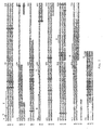
- Fig. 1 shows the alignment of recombinant (human) and mouse (natural) PIBF, A being the recombinant sequence, B the IC mouse sequence (cloned from mouse testis library), C being the EST mouse, pieced together from DEST libraries based on the human sequences, and D being the bovine sequence.
- X represents the signal sequence according to the PSG prediction method, y the signal sequence according to the GvH prediction, z the ER membrane retention signal, w the leucine zipper pattern - DNA binding motif, v the peroxisomal targeting signal, and u the nuclear localisation signal.
- the PIBF gene is located to chromosome 13.
- a number of introns are present in the PIBF gene (s. Fig. 2), whereby in intron 2 there are multiple copies of the Alu repeat element which serves as a site for alternative splicing.
- A shows a gap between genomic contigs.
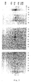
- Fig. 3 shows a nothern blot for the detection of PIBF-mRNA in various normal tissues: stomach (A), thyroid gland (B), spinal cord (C), lymph node (D), trachea (E), adrenal gland (F), bone marrow (G), spleen (H), thymus (I), prostate (J), testis (K), uterus (L), small intestine (M), colon (N), PBL (O), heart (P), brain (Q), placenta (R), lung (S), liver (T), skeletal muscle (U), kidney (V), pancreas (W).
- the arrows in Fig. 4 indicate 3 different mRNA forms.
- Example 1 ESTs entries matching the human PIBF sequence
- ESTs entries in human cDNA libraries were searched which match the human PIBF sequence. 43 entries having PIBF sequences were found out of 2.2 million dESTs deposited in 3776 human cDNA libraries. These 43 entries belong to'27 different libraries. 7 of the 27 (25%) libraries originate for normal (non-pregnant, non-tumors adult) tissues. Importantly, testis, which is a immuno-privileged tissue, frequently indicated the presence of PIBF mRNA. 13 of the 27 libraries contain mRNAs expressed in tumorous tissues (-50%). The rest is from foetal or pregnant tissues. This shows that PIBF is preferentially expressed during development, pregnancy and malignancy.
- Example 2 Determination of PIBF concentration in urine samples from cancer patients
- a competitive ELISA-based assay to measure PIBF in the urine of cancer patients was established.
- ELISA plates were coated with recombinant human PIBF at a concentration of 2 ⁇ g/ml.
- Biotin labelled polyclonal anti-PIBF IgG was added together with the samples to be determined for PIBF content. The higher is the PIBF concentration in the sample, the lower is the corresponding ELISA value. Based on these ELISA readings the absolute concentration of the PIBF can be determined.
- Urine samples were collected from cancer patients, and used fresh or frozen shortly after collection and stored at -20°C until analysis. It was determined previously that PIBF levels in the serum are significantly higher in healthy pregnant women relative to non-pregnant or pathologic pregnancy levels.
- urine samples from healthy pregnant women and normal healthy non-pregnant individuals served as positive and negative controls, respectively. The results are summarized in Table II. Normal (healthy, non-pregnant) individuals have low urinary PIBF concentrations (5 ng/ml). Urine from pregnant women was characterized by an average of 110 ng/ml PIBF concentration. Importantly, the high PIBF levels quickly returned to normal following abortion or labor.
- the most prevalent tumor type among the patients was lung cancer with 23 cases. Most of the lung cancer patients had high PIBF concentration. The lack of high PIBF in the urine could be correlated with clinical disease status, namely after the removal of the primary tumor and in remission PIBF concentrations were significantly lower or even normal.
- MCF-7 human mammary epithelial carcinoma
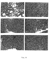
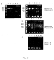
- Fig. 4 left column designated “A”
- the normal tissue is shown
- tumor tissues are shown in the right column.
- lung cancer small cell
- urinary bladder carcinoma transitorial cell
- stomach cancer adenocarcinoma
- PIBF positivity is a result of expression by the tumor cells themselves, and not the binding of PIBF from the extracellular fluid (secreted by infiltrating lymphocytes).
- PIBF production is a quite general phenomenon of the malignant or undifferentiated state, and as a consequence, PIBF can serve as a tumor marker.
- Example 4 Modulating NK activity by the presence of PIBF
- PIBF produced - secreted or cell surface expressed - by tumor cells will inhibit killer cell activity systemically or locally. It has been long known that there are cell lines which are good targets in NK assays, others are not.
- the human tumor cell line, MCF-7 belongs to the poor target category. It is certainly a possibility that the low killing activity against these cells is the result of PIBF production, which inhibits NK activity, since the MCF-7 cell line is shown to produce PIBF.
- PIBF-expressing MCF-7 cells were used as targets in a 4 hours single cell cytotoxicity assay, according to Grimm and Bonavida "Frequency determination of killer cells by a single-cell cytotoxic assay”; Methods Enzymol 93, 270 (1983).
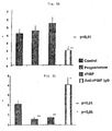
- Fig. 5 it is shown that anti-PIBF treatment enhances NK cell target killing of tumor cells: The minus and the plus indicate the treatment with or without anti-PIBF IgG, the numbers are the percentage of NK activity for Figures 5A and 5B and the percentage of inhibition of NK activity for Figures 5C and 5D.
- the source of NK cells were freshly isolated PBMCs from healthy individuals.
- this assay allows to test the effectiveness of exogenous PIBF to reduce PIBF- target cell killing, and more importantly to evaluate the power of neutralizing anti-PIBF antibodies in stimulating the lysis of PIBF+ tumor cells.
- Neutralizing rabbit anti-human and also anti-mouse antisera are generated whereby these polyclonal antibodies are able to inactivate natural PIBF in vitro (human lymphocyte cultures in NK assay) or in vivo (pregnant mice, as animal model).
- T H2 cytokines One of the main mechanism of the pregnancy promoting action of PIBF is the induction of the T H2 cytokines.
- the recombinant form of PIBF is also active to modulate cytokine expression by peripheral blood lymphocytes in vitro.
- rPIBF was added to non-pregnancy peripheral lymphocytes isolated by Ficoll-Paque gradient and cultured at 10 6 /ml cell density.
- the production of the prototype T H2 lymphokine, IL-10 was measured by detecting and counting the number of IL-10 positive lymphocytes (by immunohistochemistry on cytospins) after 24 h treatment.
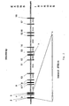
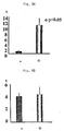
- the percent of IL-10 positive lymphocytes increased as the function of rPIBF concentration from 0.35 +/- 0.15 to 3.5 +/- 1.5%. At the highest rPIBF concentration (10 ⁇ g/ml) 10x more IL-10 positive lymphocytes were present than in control cultures (Fig. 6A). The opposite effect was seen on IL-12 (T H1 lymphokine)-producing lymphocytes by the same treatment. The number of IL-12 positive lymphocytes decreased as the function of PIBF concentration resulting in an approximately 8fold reduction at the highest amount of PIBF. Neutralization of the effect of natural PIBF on cytokine production was also successful.
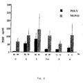
- the diagnostic value of urinary PIBF levels in malignancies is further exemplified by using urine samples of patients with non-adenocarcinoma tumors and nonsolid tumors (Fig. 7); dots represent results with individual sera. Average numbers are shown. N means number of patients.
- polyclonal antibodies are replaced in a similar antigen capture sandwich assay with a pair of monoclonal antibodies (ab) produced in mice using the N-terminal 48-kDa rPIBF as antigen.
- ab monoclonal antibodies
- Approximately twenty different hybridoma clones were tested, then four stable and well producing clones selected for ELISA assay, and also for other diagnostic methods (immunohistochemistry). These hybridoma clones are deposited at the Hybridoma Cell Bank at the University Medical School of Pécs. (Deposition numbers are 11-14/2001, cell line codes: HYB255-258).
- mouse full length PIBF mRNA and protein sequence was identified (SEQ.ID.No 4,5).
- the mouse PIBF is also organized into 18 exons, and shows an amino acid homology of 89%.
- the different,PIBF forms are differentially expressed and have different functional attributes.
- the full length mRNA (exons 1-18) encodes for a nuclear 89-kDa protein.
- Bioinformatics predicts a nuclear compartmentalization based on the two nuclear localization signals in exons 7 and 13, and a nucleic acid binding domain in exons 14-16.
- the 89-kDa protein indeed localizes exclusively to the nuclear fractions. Smaller molecular weight forms are present in the cytosolic and secreted fractions of different human and mouse primary tumors, embryo and cell lines.
- mRNA The full length mRNA can be found in almost all tissues based on Northern (Fig. 3) and RT-PCR analysis (Fig. 10), however, in different amounts.
- Semiquantitative RT-PCR was performed on matched tumor/normal tissue pairs. The same amount of.RNA was amplified with either PIBF specific exon 1/exon 18 (Fig. 10A) and exon 2/exon 18 primer pairs (Fig. 10B) or with ribosomal protein S9 specific primers (for loading control) of the same samples.
- Fast growing cell e.g.
- PIBF mRNAs in tumors and embryo, cell of immune priviledged tissues (testis, placenta) contain more full length PIBF mRNA.
- RT-PCR analysis of human primary tumors and subsequent cloning and DNA sequencing of PIBF cDNA reveal that the alternatively processed PIBF mRNA forms are expressed differently. It is exemplified by two smaller PIBF forms found in human primary tumors. Expression of the exons (1-5)-(17-18) form in gastric adenocarcinoma and that of the exons 1-(13-18) form in endometrial adenocarcinoma is restricted to the tumor tissues, since the normal tissue counterparts from the same patients do not express detectable levels of these PIBF mRNA splice variants. Importantly, both of these and other alternatively processed PIBF mRNAs are also found in tissues of immune priviledge (placenta, embryo, testis) and in immune cells.
- PIBF The function of PIBF also depends on the structure of the mature protein from.
- One of the most interesting forms is encoded by exons 2-3-4-5-17-18 mRNA frequently found in human and mouse tissues, such as human and mouse placenta, human lymphocytes, mouse embryo and human gastric tumor. It encodes for a 298 and 297 amino acid long polypeptide with predicted 35-kDa molecular weight. (SEQ.ID.No 6,7 for human, SEQ.ID.No 8,9 for mouse). The two proteins are 86% homologous. FACS analysis reveals that the human 35-kDa form binds specifically to human immune cells (Fig.
- adenocc Exon 1-(13-18) 87aa 10-kDa Pregnancy lymphocytes MCF-7 human mammary adenocc. cell line Exon.1-(15-18) Leukocytes Exon 14'-15-18 No 14 Alternatively spliced human 14-kDa Pregnancy lymphocytes Exon 1-(9-10)-(12-15)-(17-18) 118aa 14-kDa MCF-7 tumor cell line No 15,16 Alternatively spliced human 8-kDa Pregnancy lymphocytes Exon 1-(9-10)-(12-15)-(17-18) 70aa 8-kDa MCF-7 tumor cell line No 17,18 Alternatively spliced human 20-kDa MCF-7 tumor cell line Exon 1-(3-7)-(9-10)-12-(17-18) 185aa 20-kDa No 19,21, 22 Alternatively spliced human 37-kDa MCF-7 tumor cell line Exon (1-7)-(9-15)-(17-18) 308
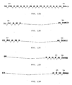
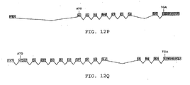
- Figs. 12A to 12Q show the alternatively processed PIBF proteins whereby the exons are schematically represented:
- Fig. 12A shows mouse full length 89-kDA PIBF (SEQ ID No. 4)
- Fig. 12B shows exons (1-5)-(17-18) which is found in stomach tumor, human terminal placenta, male and female lymphocytes, female pregnancy lymphocytes (SEQ ID No. 6)
- Fig. 12C shows exons (1-5)-(17-18), found in mouse placenta and embryo (SEQ.ID No. 8)
- Fig. 12D shows exons 1-(13-18) (SEQ. ID No. 11)
- Fig. 12E shows exons 1-(15-18), found in MCF-7 cells and pregnancy lymphocytes (SEQ.ID No. 12), Fig.
- FIG. 12F shows a part of intron 14 and exons 15-18, found in leukocyte cDNA library (SEQ. ID No. 13)
- Fig. 12G shows exons 1+(9-10)+(12-15)+(17-18), found in MCF-7 cells and pregnancy lymphocytes (SEQ. ID No. 14)
- Fig. 12H shows exons 1+(3-7)+(9-10)+12+(17-18), found in MCF-7 cells - human mammary tumor cell line (SEQ. ID No. 17)
- Fig. 12I shows exon (1-7)+(9-15)+(17-18), found in MCF-7 cells - human mammary tumor cell line (SEQ. ID No. 19), Fig.
- FIG. 12J shows exon (1-7)+(9-10)+12+(17-18), found in MCF-7 cells - human mammary tumor cell line (SEQ. ID No. 22)
- Fig. 12K shows exon (1-2)-(17-18) found in mouse embryo and placenta (SEQ. ID No. 23)
- Fig. 12L shows exon (1-4)-(16-18) found in mouse placenta (SEQ. ID No. 25)
- Fig. 12M shows exons (1-2)-(15-18) found in mouse testis (SEQ. ID No. 27)
- Fig. 12N shows exon 1-11 18 found in mouse embryo (SEQ. ID No. 29)
- Fig. 120 shows exons (1-11)-18 found in mouse embryo (SEQ. ID No.
- Fig. 12P shows exons 1-(8-14)-(17-18) found in mouse embryo (SEQ. ID No. 34) and Fig. 12Q shows exons (1-1)-(15-18) found in mouse testis (SEQ. ID No. 36).
Landscapes
- Health & Medical Sciences (AREA)
- Chemical & Material Sciences (AREA)
- Life Sciences & Earth Sciences (AREA)
- Organic Chemistry (AREA)
- Immunology (AREA)
- Molecular Biology (AREA)
- General Health & Medical Sciences (AREA)
- Medicinal Chemistry (AREA)
- Engineering & Computer Science (AREA)
- Biochemistry (AREA)
- Urology & Nephrology (AREA)
- Biomedical Technology (AREA)
- Genetics & Genomics (AREA)
- Biophysics (AREA)
- Hematology (AREA)
- Proteomics, Peptides & Aminoacids (AREA)
- Pathology (AREA)
- Gastroenterology & Hepatology (AREA)
- Reproductive Health (AREA)
- General Physics & Mathematics (AREA)
- Analytical Chemistry (AREA)
- Physics & Mathematics (AREA)
- Food Science & Technology (AREA)
- Gynecology & Obstetrics (AREA)
- Microbiology (AREA)
- Hospice & Palliative Care (AREA)
- Oncology (AREA)
- Pregnancy & Childbirth (AREA)
- Toxicology (AREA)
- Biotechnology (AREA)
- Cell Biology (AREA)
- Zoology (AREA)
- Chemical Kinetics & Catalysis (AREA)
- Animal Behavior & Ethology (AREA)
- Pharmacology & Pharmacy (AREA)
- Nuclear Medicine, Radiotherapy & Molecular Imaging (AREA)
- General Chemical & Material Sciences (AREA)
- Public Health (AREA)
- Veterinary Medicine (AREA)
- Measuring Or Testing Involving Enzymes Or Micro-Organisms (AREA)
Claims (38)
- Verfahren zur Diagnostizierung eines Tumors bei einem Patienten, welches Verfahren das Messen der Konzentration von PIBF (Progesteron-induzierter Blockierungs-Faktor) oder eines Derivats davon oder Fragments davon in einer vom Patienten entnommenen Probe und die Bestimmung, ob die PIBF-Konzentration in der Probe über oder unter einem vorbestimmten Schwellenwert liegt, umfasst, wobei die Konzentration über dem Schwellenwert einen Patienten mit einem Tumor identifiziert.
- Verfahren nach Anspruch 1, dadurch gekennzeichnet, dass der Tumor ein Epithel-Carcinom, insbesondere ein Lungen-Carcinom, ein Colon-Carcinom oder ein Brust-Carcinom ist.
- Verfahren nach Anspruch 1 oder 2, dadurch gekennzeichnet, dass die Probe eine Körperflüssigkeit, insbesondere Harn oder Serum ist.
- Verfahren nach Anspruch 1 oder 2, dadurch gekennzeichnet, dass die Probe eine Gewebsprobe ist.
- Verfahren nach einem der Ansprüche 1 bis.4, dadurch gekennzeichnet, dass der Schwellenwert die Konzentration des PIBF in einer Probe einer gesunden Person ist.
- Verfahren nach einem der Ansprüche 1 bis 5, dadurch gekennzeichnet, dass der Schwellenwert durch Messen der PIBF-Konzentration in mindestens einer gesunden Person parallel zur Bestimmung der PIBF-Konzentration in einer Probe des Patienten bestimmt wird.
- Verfahren nach einem der Ansprüche 1 bis 6, dadurch gekennzeichnet, dass als positive Kontrolle die Konzentration von PIBF oder eines Derivats davon oder eines Fragments davon in der Probe, die eine festgelegte Konzentration von PIBF oder eines Derivats davon oder eines Fragments davon aufweist, gemessen wird, parallel zur Bestimmung der Konzentration des PIBF in der Probe des Patienten.
- Verfahren nach einem der Ansprüche 1 bis 7, dadurch gekennzeichnet, dass die PIBF-Konzentration in der Probe immunologisch, insbesondere durch einen konkurrierenden Test, durch einen Sandwich-Test, durch Immunfärbung oder durch Kombinationen dieser Methoden gemessen wird.
- Verfahren nach einem der Ansprüche 1 bis 8, dadurch gekennzeichnet, dass die PIBF-Konzentration in der Probe indirekt, durch Messung der Konzentration der PIBF-mRNA in der Probe, gemessen wird.
- Verfahren zur Bestimmung der positiven oder negativen Progression eines Tumors bei einem Patienten, welches die Diagnostizierung eines Tumors bei einem Patienten gemäß einem Verfahren nach einem der Ansprüche 1 bis 9 und die Feststellung, ob die gemessene Konzentration des PIBF oder eines Derivats davon oder eines Fragments davon in der Probe über oder unter mindestens einer zuvor gemessenen Konzentration von PIBF oder einem Derivat davon oder einem Fragment davon in mindestens einer zuvor vom selben Patienten genommenen Probe liegt, wobei eine über der zuvor gemessenen Konzentration liegende Konzentration eine positive Progression identifiziert.
- Verwendung eines anti-PIBF-Antikörpers, vorzugsweise eines monoklonalen Antikörpers oder eines Fragments davon bei einem Verfahren nach einem der Ansprüche 8 bis 10.
- Verwendung von PIBF oder einem Derivat davon oder einem Fragment davon bei einem Verfahren nach einem der Ansprüche 7 bis 10.
- Verwendung nach Anspruch 12, dadurch gekennzeichnet, dass der PIBF ein rekombinanter PIBF ist.
- Set, umfassend ein erstes Reagens, das mindestens einen anti-PIBF-Antikörper oder ein Fragment desselben aufweist, und ein zweites Reagens, das PIBF oder ein Derivat davon oder ein Fragment davon mit einer bestimmten Konzentration aufweist.
- Set nach Anspruch 14, dadurch gekennzeichnet, dass es eine feste Phase aufweist, an die der mindestens eine anti-PIBF-Antikörper oder das Fragment davon oder das PIBF oder das Derivat davon oder das Fragment davon gebunden ist.
- Set nach Anspruch 14 oder 15, dadurch gekennzeichnet, dass der PIBF rekombinant ist.
- Set nach einem der Ansprüche 14 bis 16, dadurch gekennzeichnet, dass es mindestens ein weiteres Reagens umfasst, welches einen zweiten anti-PIBF-Antikörper oder ein Fragment davon aufweist, der (das) an ein Epitop des PIBF bindet, welches von dem vom ersten anti-PIBF-Antikörper oder seinem Fragment erkannten Epitop verschieden ist.
- Set nach einem der Ansprüche 14 bis 17 zur Diagnostizierung eines Tumors bei einem Patienten bzw. zur Feststellung der Progression eines Tumors bei einem Patienten.
- Verwendung eines anti-PIBF-Antikörpers, vorzugsweise eines monoklonalen anti-PIBF-Antikörpers, insbesondere eines humanisierten anti-PIBF-Antikörpers oder eines Fragments davon zur Herstellung eines anti-Tumor-Medikaments.
- Verwendung nach Anspruch 19, dadurch gekennzeichnet, dass der Antikörper ein Einzelketten-Antikörper ist.
- Verwendung nach Anspruch 19 oder 20, dadurch gekennzeichnet, dass der Antikörper ein an ihm haftendes Molekül aufweist.
- Verwendung nach Anspruch 21, dadurch gekennzeichnet, dass das Molekül eine toxische Substanz ist, insbesondere ein Radionuklid, ein chemotherapeutischer Arzneistoff oder ein Toxin, bzw. ein Prodrug.
- Verwendung von PIBF oder eines Derivats davon oder eines Fragments davon zur Herstellung eines Anti-Tumor-Medikaments.
- Verwendung nach Anspruch 23, dadurch gekennzeichnet, dass das Medikament ein Vakzin ist.
- Verwendung nach Anspruch 24, dadurch gekennzeichnet, dass das Vakzin weiters ein Adjuvans aufweist.
- Verwendung nach Anspruch 23 oder 24, dadurch gekennzeichnet, dass der PIBF oder das Derivat davon oder das Fragment davon rekombinant ist.
- Verwendung nach Anspruch 23 bis 24, dadurch gekennzeichnet, dass der PIBF oder das Derivat davon oder das Fragment davon ein chemisch synthetisiertes Molekül ist.
- Verwendung eines Polynukleotids, das für PIBF oder ein Derivat davon oder Fragment davon oder für ein PIBF-antisense-Molekül codiert, zur Herstellung eines Anti-Tumor-Medikaments.
- Rekombinantes Protein mit einer Progesteron-induzierten, immunmodulierenden Protein(PIBF-)-Aktivität, welches- die Aminosäuresequenz gemäß SEQ.ID.NO 1 oder- eine Aminosäuresequenz mit einer Aminosäure-Identität von mindestens 98% mit der Sequenz gemäß SEQ.ID.NO 1, wie mittels FAST/A-Algorithmus bestimmt, oder- eine Aminosäuresequenz mit einer Aminosäure-Identität von mindestens 95% mit der Sequenz von Aminosäurerest 580 bis 630 von SEQ.ID.NO 1, wie mittels FAST/A-Algorithmus bestimmt, und- eine PIBF-Aktivität von mindestens 50% des natürlichen Human-PIBF-Moleküls aufweist.
- Rekombinantes Protein nach Anspruch 29, dadurch gekennzeichnet, dass es eine Aminosäuresequenz, wie durch die Aminosäurereste 300 bis 350 in SEQ.ID.NO 1 angegeben, aufweist.
- Rekombinantes Protein nach Anspruch 29 oder 30, dadurch gekennzeichnet, dass es eine Aminosäuresequenz, wie durch die Aminosäurereste 580 bis 630 in SEQ.ID.NO 1 angegeben, aufweist.
- Protein mit PIBF-Aktivität, welches umfasst:- die Aminosäuresequenz gemäß SEQ.ID.NO 4 oder- eine Aminosäure mit einer Aminosäure-Identität von mindestens 90%, vorzugsweise mindestens 95%, noch mehr bevorzugt mindestens 99% mit der Sequenz gemäß SEQ.ID.NO 4, wie mittels FAST/A-Algorithmus bestimmt.
- Protein, dadurch gekennzeichnet, dass es eine Aminosäuesequenz mit einer Identität von mindestens 85%, vorzugsweise mindestens 90%, noch mehr bevorzugt mindestens 95%, wie mittels FAST/A-Algorithmus bestimmt, mit einer Sequenz ausgewählt aus der Gruppe bestehend aus SEQ.ID.NO. 6, 8, 10, 14, 15, 17, 19, 20, 23, 25, 27, 29, 31, 32, 34 und 36 aufweist, wobei dieses Protein ein alternativ prozessiertes PIBF-Protein ist.
- Nukleinsäure-Molekül, das für ein rekombinantes Protein mit einer PIBF-Aktivität nach einem der Ansprüche 29 bis 32 codiert.
- Nukleinsäure-Molekül, das für ein alternativ prozessiertes PIBF-Protein codiert, dadurch gekennzeichnet, dass es eine Nukleinsäuresequenz aufweist, die eine Identität von mindestens 80%, vorzugsweise mindestens 90%, noch mehr bevorzugt mindestens 95% hat mit- einer Sequenz gewählt aus der Gruppe bestehend aus SEQ.ID.NO. 7, 9, 11, 12, 13, 16, 18, 21, 22, 24, 26, 28, 30, 33, 35 und 37 oder- einer Sequenz, die unter stringenten Bedingungen mit einer der obigen Sequenzen hybridisiert, oder- einer Sequenz, die infolge des genetischen Codes zu einer der obigen Sequenzen degeneriert ist.
- Nukleinsäure-Vektor, umfassend eine Nukleinsäuresequenz nach Anspruch 34 oder 35 und ein geeignetes Regulierungselement.
- Nukleinsäure-Vektor nach Anspruch 36, dadurch gekennzeichnet, dass er weiters einen Selektions-Marker aufweist.
- Zelle, umfassend einen Vektor nach einem der Ansprüche 34 bis 36.
Applications Claiming Priority (3)
| Application Number | Priority Date | Filing Date | Title |
|---|---|---|---|
| AT19972000 | 2000-11-28 | ||
| AT0199700A AT410753B (de) | 2000-11-28 | 2000-11-28 | Pibf für die diagnose von tumoren |
| PCT/EP2001/013876 WO2002044734A1 (en) | 2000-11-28 | 2001-11-28 | Method for diagnosing a tumor in a patient determining the concentration of pibf |
Publications (2)
| Publication Number | Publication Date |
|---|---|
| EP1337850A1 EP1337850A1 (de) | 2003-08-27 |
| EP1337850B1 true EP1337850B1 (de) | 2007-03-21 |
Family
ID=3689570
Family Applications (1)
| Application Number | Title | Priority Date | Filing Date |
|---|---|---|---|
| EP01994744A Expired - Lifetime EP1337850B1 (de) | 2000-11-28 | 2001-11-28 | Verfahren der pibf-konzentrationsbestimmung zur diagnose eines tumors in einem patienten |
Country Status (9)
| Country | Link |
|---|---|
| US (1) | US20040142394A1 (de) |
| EP (1) | EP1337850B1 (de) |
| JP (2) | JP4025645B2 (de) |
| AT (2) | AT410753B (de) |
| AU (2) | AU2490102A (de) |
| CA (1) | CA2431837A1 (de) |
| DE (1) | DE60127442T2 (de) |
| ES (1) | ES2283461T3 (de) |
| WO (1) | WO2002044734A1 (de) |
Families Citing this family (5)
| Publication number | Priority date | Publication date | Assignee | Title |
|---|---|---|---|---|
| ATA8092002A (de) * | 2002-05-28 | 2005-01-15 | Intercell Ag | Pibf |
| JP2015109806A (ja) * | 2012-03-22 | 2015-06-18 | アステラス製薬株式会社 | 新規ret融合体の検出法 |
| GB201702392D0 (en) * | 2017-02-14 | 2017-03-29 | Univ Edinburgh | Methods for cancer diagnosis using a gene expression signature |
| CN108447537A (zh) * | 2018-03-23 | 2018-08-24 | 广西科技大学 | 一种妊娠期及哺乳期妇女用药安全和健康管理系统 |
| KR102490714B1 (ko) * | 2018-12-28 | 2023-01-26 | 재단법인 아산사회복지재단 | Pibf 단백질을 유효성분으로 함유하는 염증성 질환 예방 또는 치료용 약학 조성물 |
Family Cites Families (5)
| Publication number | Priority date | Publication date | Assignee | Title |
|---|---|---|---|---|
| US5648340A (en) * | 1991-10-31 | 1997-07-15 | Barnea; Eytan R. | Gestational agents for controlling cell proliferation |
| US5646003A (en) * | 1994-03-23 | 1997-07-08 | Barnea; Eytan R. | Preimplantation factor |
| US6207380B1 (en) * | 1997-09-15 | 2001-03-27 | Abbott Laboratories | Reagents and methods useful for detecting diseases of the urinary tract |
| IL140625A0 (en) * | 1998-07-02 | 2002-02-10 | Envision Biomedical Consulting | Antiproliferative and antiviral proteins and peptides |
| WO2001027633A1 (en) * | 1999-10-08 | 2001-04-19 | Envision Biomedical Consulting | Gestational agents which modulate cell proliferation |
-
2000
- 2000-11-28 AT AT0199700A patent/AT410753B/de not_active IP Right Cessation
-
2001
- 2001-11-28 EP EP01994744A patent/EP1337850B1/de not_active Expired - Lifetime
- 2001-11-28 DE DE60127442T patent/DE60127442T2/de not_active Expired - Fee Related
- 2001-11-28 AU AU2490102A patent/AU2490102A/xx active Pending
- 2001-11-28 CA CA002431837A patent/CA2431837A1/en not_active Abandoned
- 2001-11-28 JP JP2002546225A patent/JP4025645B2/ja not_active Expired - Fee Related
- 2001-11-28 AT AT01994744T patent/ATE357662T1/de not_active IP Right Cessation
- 2001-11-28 AU AU2002224901A patent/AU2002224901B2/en not_active Ceased
- 2001-11-28 WO PCT/EP2001/013876 patent/WO2002044734A1/en active IP Right Grant
- 2001-11-28 ES ES01994744T patent/ES2283461T3/es not_active Expired - Lifetime
-
2003
- 2003-05-23 US US10/444,693 patent/US20040142394A1/en not_active Abandoned
-
2006
- 2006-02-20 JP JP2006043156A patent/JP2006230407A/ja active Pending
Also Published As
| Publication number | Publication date |
|---|---|
| JP2006230407A (ja) | 2006-09-07 |
| ES2283461T3 (es) | 2007-11-01 |
| US20040142394A1 (en) | 2004-07-22 |
| DE60127442T2 (de) | 2007-11-29 |
| DE60127442D1 (de) | 2007-05-03 |
| EP1337850A1 (de) | 2003-08-27 |
| ATE357662T1 (de) | 2007-04-15 |
| AU2002224901B2 (en) | 2006-11-09 |
| AU2490102A (en) | 2002-06-11 |
| ATA19972000A (de) | 2002-12-15 |
| JP4025645B2 (ja) | 2007-12-26 |
| JP2004514909A (ja) | 2004-05-20 |
| CA2431837A1 (en) | 2002-06-06 |
| AT410753B (de) | 2003-07-25 |
| WO2002044734A1 (en) | 2002-06-06 |
Similar Documents
| Publication | Publication Date | Title |
|---|---|---|
| JP6490581B2 (ja) | 修飾型自己エピトープに対する抗腫瘍免疫応答 | |
| US8486903B2 (en) | TAZ/WWTR1 for diagnosis and treatment of cancer | |
| JP5291641B2 (ja) | 癌抗原及びその利用 | |
| US10066014B2 (en) | Anti CD84 antibodies, compositions comprising same and uses thereof | |
| JP2009514528A (ja) | Nkg2d陽性cd4+細胞による免疫応答の負の免疫調節法 | |
| US20120034196A1 (en) | Gene encoding a multidrug resistance human p-glycoprotein homologue on chromosome 7p15-21 and uses thereof (frank) | |
| EP1723970A1 (de) | Medizinische zusammensetzung mit cxcr3-hemmer | |
| JPH08504170A (ja) | 進行性免疫抑制症患者の評価と治療 | |
| EP1337850B1 (de) | Verfahren der pibf-konzentrationsbestimmung zur diagnose eines tumors in einem patienten | |
| AU2002224901A1 (en) | Method for diagnosing a tumor in a patient determining the concentration of PIBF | |
| MXPA06014972A (es) | Metodos y composiciones para el tratamiento de enfermedades poliquisticas. | |
| CN113398270A (zh) | 一种治疗骨巨细胞瘤的方法 | |
| WO2003100428A2 (de) | Verfahren zu diagnostizierung eines tumors bei einem patienten durch bestimmung von pibf |
Legal Events
| Date | Code | Title | Description |
|---|---|---|---|
| PUAI | Public reference made under article 153(3) epc to a published international application that has entered the european phase |
Free format text: ORIGINAL CODE: 0009012 |
|
| 17P | Request for examination filed |
Effective date: 20030526 |
|
| AK | Designated contracting states |
Designated state(s): AT BE CH CY DE DK ES FI FR GB GR IE IT LI LU MC NL PT SE TR |
|
| AX | Request for extension of the european patent |
Extension state: AL LT LV MK RO SI |
|
| RAP1 | Party data changed (applicant data changed or rights of an application transferred) |
Owner name: INTERCELL AG |
|
| GRAP | Despatch of communication of intention to grant a patent |
Free format text: ORIGINAL CODE: EPIDOSNIGR1 |
|
| GRAS | Grant fee paid |
Free format text: ORIGINAL CODE: EPIDOSNIGR3 |
|
| GRAA | (expected) grant |
Free format text: ORIGINAL CODE: 0009210 |
|
| AK | Designated contracting states |
Kind code of ref document: B1 Designated state(s): AT BE CH CY DE DK ES FI FR GB GR IE IT LI LU MC NL PT SE TR |
|
| AX | Request for extension of the european patent |
Extension state: AL LT LV MK RO SI |
|
| PG25 | Lapsed in a contracting state [announced via postgrant information from national office to epo] |
Ref country code: BE Free format text: LAPSE BECAUSE OF FAILURE TO SUBMIT A TRANSLATION OF THE DESCRIPTION OR TO PAY THE FEE WITHIN THE PRESCRIBED TIME-LIMIT Effective date: 20070321 Ref country code: FI Free format text: LAPSE BECAUSE OF FAILURE TO SUBMIT A TRANSLATION OF THE DESCRIPTION OR TO PAY THE FEE WITHIN THE PRESCRIBED TIME-LIMIT Effective date: 20070321 Ref country code: NL Free format text: LAPSE BECAUSE OF FAILURE TO SUBMIT A TRANSLATION OF THE DESCRIPTION OR TO PAY THE FEE WITHIN THE PRESCRIBED TIME-LIMIT Effective date: 20070321 Ref country code: LI Free format text: LAPSE BECAUSE OF FAILURE TO SUBMIT A TRANSLATION OF THE DESCRIPTION OR TO PAY THE FEE WITHIN THE PRESCRIBED TIME-LIMIT Effective date: 20070321 Ref country code: CH Free format text: LAPSE BECAUSE OF FAILURE TO SUBMIT A TRANSLATION OF THE DESCRIPTION OR TO PAY THE FEE WITHIN THE PRESCRIBED TIME-LIMIT Effective date: 20070321 |
|
| REG | Reference to a national code |
Ref country code: GB Ref legal event code: FG4D |
|
| RIN1 | Information on inventor provided before grant (corrected) |
Inventor name: NAGY, ESZTER Inventor name: KLADE, CHRISTOPH Inventor name: SZEKERES-BARTHO, JULIA Inventor name: HENICS, TAMAS Inventor name: POLGAR, BEATA Inventor name: KOCH, MARGIT Inventor name: PALKOVICS, TAMAS |
|
| REG | Reference to a national code |
Ref country code: CH Ref legal event code: EP |
|
| REF | Corresponds to: |
Ref document number: 60127442 Country of ref document: DE Date of ref document: 20070503 Kind code of ref document: P |
|
| REG | Reference to a national code |
Ref country code: IE Ref legal event code: FG4D |
|
| PG25 | Lapsed in a contracting state [announced via postgrant information from national office to epo] |
Ref country code: SE Free format text: LAPSE BECAUSE OF FAILURE TO SUBMIT A TRANSLATION OF THE DESCRIPTION OR TO PAY THE FEE WITHIN THE PRESCRIBED TIME-LIMIT Effective date: 20070621 |
|
| PG25 | Lapsed in a contracting state [announced via postgrant information from national office to epo] |
Ref country code: PT Free format text: LAPSE BECAUSE OF FAILURE TO SUBMIT A TRANSLATION OF THE DESCRIPTION OR TO PAY THE FEE WITHIN THE PRESCRIBED TIME-LIMIT Effective date: 20070821 |
|
| LTIE | Lt: invalidation of european patent or patent extension |
Effective date: 20070321 |
|
| ET | Fr: translation filed | ||
| REG | Reference to a national code |
Ref country code: CH Ref legal event code: PL |
|
| NLV1 | Nl: lapsed or annulled due to failure to fulfill the requirements of art. 29p and 29m of the patents act | ||
| REG | Reference to a national code |
Ref country code: ES Ref legal event code: FG2A Ref document number: 2283461 Country of ref document: ES Kind code of ref document: T3 |
|
| PLBE | No opposition filed within time limit |
Free format text: ORIGINAL CODE: 0009261 |
|
| STAA | Information on the status of an ep patent application or granted ep patent |
Free format text: STATUS: NO OPPOSITION FILED WITHIN TIME LIMIT |
|
| PG25 | Lapsed in a contracting state [announced via postgrant information from national office to epo] |
Ref country code: DK Free format text: LAPSE BECAUSE OF FAILURE TO SUBMIT A TRANSLATION OF THE DESCRIPTION OR TO PAY THE FEE WITHIN THE PRESCRIBED TIME-LIMIT Effective date: 20070321 |
|
| PGFP | Annual fee paid to national office [announced via postgrant information from national office to epo] |
Ref country code: ES Payment date: 20071128 Year of fee payment: 7 |
|
| 26N | No opposition filed |
Effective date: 20071227 |
|
| PGFP | Annual fee paid to national office [announced via postgrant information from national office to epo] |
Ref country code: IT Payment date: 20071123 Year of fee payment: 7 Ref country code: AT Payment date: 20071126 Year of fee payment: 7 |
|
| PG25 | Lapsed in a contracting state [announced via postgrant information from national office to epo] |
Ref country code: GR Free format text: LAPSE BECAUSE OF FAILURE TO SUBMIT A TRANSLATION OF THE DESCRIPTION OR TO PAY THE FEE WITHIN THE PRESCRIBED TIME-LIMIT Effective date: 20070622 |
|
| PGFP | Annual fee paid to national office [announced via postgrant information from national office to epo] |
Ref country code: FR Payment date: 20071031 Year of fee payment: 7 Ref country code: GB Payment date: 20071130 Year of fee payment: 7 |
|
| PGFP | Annual fee paid to national office [announced via postgrant information from national office to epo] |
Ref country code: DE Payment date: 20071228 Year of fee payment: 7 |
|
| PG25 | Lapsed in a contracting state [announced via postgrant information from national office to epo] |
Ref country code: MC Free format text: LAPSE BECAUSE OF NON-PAYMENT OF DUE FEES Effective date: 20071130 |
|
| PG25 | Lapsed in a contracting state [announced via postgrant information from national office to epo] |
Ref country code: IE Free format text: LAPSE BECAUSE OF NON-PAYMENT OF DUE FEES Effective date: 20071128 |
|
| GBPC | Gb: european patent ceased through non-payment of renewal fee |
Effective date: 20081128 |
|
| PG25 | Lapsed in a contracting state [announced via postgrant information from national office to epo] |
Ref country code: CY Free format text: LAPSE BECAUSE OF FAILURE TO SUBMIT A TRANSLATION OF THE DESCRIPTION OR TO PAY THE FEE WITHIN THE PRESCRIBED TIME-LIMIT Effective date: 20070321 |
|
| PG25 | Lapsed in a contracting state [announced via postgrant information from national office to epo] |
Ref country code: AT Free format text: LAPSE BECAUSE OF NON-PAYMENT OF DUE FEES Effective date: 20081128 Ref country code: LU Free format text: LAPSE BECAUSE OF NON-PAYMENT OF DUE FEES Effective date: 20071128 Ref country code: IT Free format text: LAPSE BECAUSE OF NON-PAYMENT OF DUE FEES Effective date: 20081128 |
|
| REG | Reference to a national code |
Ref country code: FR Ref legal event code: ST Effective date: 20090731 |
|
| PG25 | Lapsed in a contracting state [announced via postgrant information from national office to epo] |
Ref country code: TR Free format text: LAPSE BECAUSE OF FAILURE TO SUBMIT A TRANSLATION OF THE DESCRIPTION OR TO PAY THE FEE WITHIN THE PRESCRIBED TIME-LIMIT Effective date: 20070321 |
|
| PG25 | Lapsed in a contracting state [announced via postgrant information from national office to epo] |
Ref country code: DE Free format text: LAPSE BECAUSE OF NON-PAYMENT OF DUE FEES Effective date: 20090603 |
|
| PG25 | Lapsed in a contracting state [announced via postgrant information from national office to epo] |
Ref country code: GB Free format text: LAPSE BECAUSE OF NON-PAYMENT OF DUE FEES Effective date: 20081128 |
|
| REG | Reference to a national code |
Ref country code: ES Ref legal event code: FD2A Effective date: 20081129 |
|
| PG25 | Lapsed in a contracting state [announced via postgrant information from national office to epo] |
Ref country code: ES Free format text: LAPSE BECAUSE OF NON-PAYMENT OF DUE FEES Effective date: 20081129 |
|
| PG25 | Lapsed in a contracting state [announced via postgrant information from national office to epo] |
Ref country code: FR Free format text: LAPSE BECAUSE OF NON-PAYMENT OF DUE FEES Effective date: 20081130 |