EP1274989B1 - Procede d'amelioration d'un examen radiologique et dispositif pour la mise en oeuvre de ce procede - Google Patents
Procede d'amelioration d'un examen radiologique et dispositif pour la mise en oeuvre de ce procede Download PDFInfo
- Publication number
- EP1274989B1 EP1274989B1 EP00985379A EP00985379A EP1274989B1 EP 1274989 B1 EP1274989 B1 EP 1274989B1 EP 00985379 A EP00985379 A EP 00985379A EP 00985379 A EP00985379 A EP 00985379A EP 1274989 B1 EP1274989 B1 EP 1274989B1
- Authority
- EP
- European Patent Office
- Prior art keywords
- examination
- ray
- radiological
- characteristic points
- reference system
- Prior art date
- Legal status (The legal status is an assumption and is not a legal conclusion. Google has not performed a legal analysis and makes no representation as to the accuracy of the status listed.)
- Expired - Lifetime
Links
- 238000000034 method Methods 0.000 title claims abstract description 21
- 238000005259 measurement Methods 0.000 claims abstract description 17
- 210000000988 bone and bone Anatomy 0.000 abstract description 23
- 238000000326 densiometry Methods 0.000 abstract description 13
- 238000006073 displacement reaction Methods 0.000 abstract description 6
- 230000005855 radiation Effects 0.000 abstract description 2
- 229920000297 Rayon Polymers 0.000 description 14
- 239000002964 rayon Substances 0.000 description 14
- 210000003484 anatomy Anatomy 0.000 description 7
- 238000013519 translation Methods 0.000 description 6
- 230000037182 bone density Effects 0.000 description 4
- 238000003384 imaging method Methods 0.000 description 4
- 230000009466 transformation Effects 0.000 description 4
- 238000009547 dual-energy X-ray absorptiometry Methods 0.000 description 3
- 230000006870 function Effects 0.000 description 3
- 238000003325 tomography Methods 0.000 description 3
- 208000031968 Cadaver Diseases 0.000 description 2
- 208000001132 Osteoporosis Diseases 0.000 description 2
- 238000011156 evaluation Methods 0.000 description 2
- 238000000605 extraction Methods 0.000 description 2
- 230000004907 flux Effects 0.000 description 2
- 238000009206 nuclear medicine Methods 0.000 description 2
- 238000012545 processing Methods 0.000 description 2
- 238000002604 ultrasonography Methods 0.000 description 2
- 208000010392 Bone Fractures Diseases 0.000 description 1
- 238000010521 absorption reaction Methods 0.000 description 1
- 238000013459 approach Methods 0.000 description 1
- 210000003679 cervix uteri Anatomy 0.000 description 1
- 238000007596 consolidation process Methods 0.000 description 1
- 238000012937 correction Methods 0.000 description 1
- 238000001739 density measurement Methods 0.000 description 1
- 238000001514 detection method Methods 0.000 description 1
- 201000010099 disease Diseases 0.000 description 1
- 208000037265 diseases, disorders, signs and symptoms Diseases 0.000 description 1
- 230000009977 dual effect Effects 0.000 description 1
- 230000000694 effects Effects 0.000 description 1
- 210000000245 forearm Anatomy 0.000 description 1
- 230000000877 morphologic effect Effects 0.000 description 1
- 238000005457 optimization Methods 0.000 description 1
- 230000008520 organization Effects 0.000 description 1
- 238000010422 painting Methods 0.000 description 1
- 230000002093 peripheral effect Effects 0.000 description 1
- 230000000284 resting effect Effects 0.000 description 1
- 238000012552 review Methods 0.000 description 1
- 230000003595 spectral effect Effects 0.000 description 1
- 238000001228 spectrum Methods 0.000 description 1
- 230000001225 therapeutic effect Effects 0.000 description 1
- 210000001519 tissue Anatomy 0.000 description 1
- 210000000689 upper leg Anatomy 0.000 description 1
Images
Classifications
-
- A—HUMAN NECESSITIES
- A61—MEDICAL OR VETERINARY SCIENCE; HYGIENE
- A61B—DIAGNOSIS; SURGERY; IDENTIFICATION
- A61B6/00—Apparatus or devices for radiation diagnosis; Apparatus or devices for radiation diagnosis combined with radiation therapy equipment
- A61B6/50—Apparatus or devices for radiation diagnosis; Apparatus or devices for radiation diagnosis combined with radiation therapy equipment specially adapted for specific body parts; specially adapted for specific clinical applications
- A61B6/505—Apparatus or devices for radiation diagnosis; Apparatus or devices for radiation diagnosis combined with radiation therapy equipment specially adapted for specific body parts; specially adapted for specific clinical applications for diagnosis of bone
-
- A—HUMAN NECESSITIES
- A61—MEDICAL OR VETERINARY SCIENCE; HYGIENE
- A61B—DIAGNOSIS; SURGERY; IDENTIFICATION
- A61B6/00—Apparatus or devices for radiation diagnosis; Apparatus or devices for radiation diagnosis combined with radiation therapy equipment
- A61B6/48—Diagnostic techniques
- A61B6/482—Diagnostic techniques involving multiple energy imaging
-
- A—HUMAN NECESSITIES
- A61—MEDICAL OR VETERINARY SCIENCE; HYGIENE
- A61B—DIAGNOSIS; SURGERY; IDENTIFICATION
- A61B6/00—Apparatus or devices for radiation diagnosis; Apparatus or devices for radiation diagnosis combined with radiation therapy equipment
- A61B6/48—Diagnostic techniques
- A61B6/488—Diagnostic techniques involving pre-scan acquisition
Definitions
- the present invention relates to a method for improving a radiological examination of an area of a body and a device for carrying out this method.
- body is meant as well an object (for example a painting or a mummy) as a person or even an animal.
- the invention is applicable to any radiological examination using a two-dimensional X-ray detector and, in particular, cone-beam bi-energy X-ray bone densitometry.
- the invention relates more particularly to the positioning of a patient prior to such a radiological examination and the adjustment of the dose of X-rays to be transmitted to this patient during this examination.
- X-ray bone densitometry is a technique for measuring bone masses and densities from radiographic acquisitions made at a plurality of energies.
- the systems of the first two families require a mechanical scan to obtain an overall image of an anatomical zone while the systems of the third family allow to directly establish a complete image.
- the invention more particularly relates to cone beam bi-energy X-ray bone densitometry systems.
- a first positioning of the patient is carried out using a laser pointer which marks the zone examination from external morphological observations. Then the X-ray scan starts. If the patient is well positioned the examination continues but if the observation on the screen of the first lines acquired positioning is not good, the operator stops everything and makes a new positioning and then restart the examination.
- a mechanical positioning aid system for example a handle for the forearm and a bowl formed for the heel.
- the goal is to get the best reconstructed picture quality possible. Indeed, as the tomographic systems are studied so that the maximum attenuation is at the center of the acquisition area and as the spectrum hardening corrections depend on the size of the acquisition field, the quality of the reconstructed image depends on the proper centering and size of the acquisition field.
- the object of the present invention is to improve the reproducibility of the measurements made during any radiological examination using a two-dimensional detector.
- the invention also aims to optimize the dose of X-rays transmitted to the patient during such an examination.
- the object of the present invention is to improve a cone-beam bi-energy X-ray bone densitometry examination and, more particularly, to increase the reproducibility of bone density measurements in anatomical areas of a subject patient. such an examination.
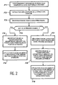
- the subject of the present invention is a method for improving a radiological examination of an area of a body, this radiological examination being carried out by means of a radiological device comprising a two-dimensional x-ray detector. characterized in that prior to the radiological examination, a two-dimensional x-ray (and thus a low dose X-ray) x-ray is taken of the area of the body to be examined with a single energy for these X-rays. this plate is used to determine first characteristic points which define a measurement frame of reference, as well as geometric parameters of a displacement able to correspond substantially this measurement frame with a pre-established frame of reference from equivalent characteristic points corresponding to the first characteristic points .
- This radiological examination may be a bone densitometry examination.
- first characteristic points are generally precisely identifiable points of the image, for example contour points, inflection points or points of extreme density.
- the pre-established reference system is a theoretical reference, adapted to the examined body.
- the preset reference system is a reference system which is defined on a previous image from the equivalent characteristic points.
- the two-dimensional image is usable (only or in addition) for adjusting the dose of X-rays to be transmitted to the body during the radiological examination by adapting the operating point of the radiological device to the morphology of the body to be examined. or by adjusting, to the morphology of the body to be examined, the area to be irradiated by the X-rays during the examination.
- the patient is moved relative to the source-detector system that comprises the bone densitometry device, or the source-detector system relative to the patient, by manual or automatic control.
- this is radiology (with a two-dimensional detector) and not tomography (with a fan beam detector, rotated as described in document [6]). ] mentioned above).
- the final image is a two-dimensional projection and not a reconstructed cut.
- a single pre-shot and not two shots at 90 ° from each other is used.
- a purpose of the example considered of the invention is the reproducibility of the measurement of bone mass calculated from the image, and not the quality of the image itself.
- this recentering is automatic in height, that is to say perpendicular to the plane of the table that supports the patient or parallel to the axis of the beam X, but it is not in width, it is -to say according to the smallest dimension of this table, because the lateral displacement of the table is not planned or is not necessary.
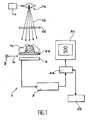
- FIG. 1 a bone osteodensitometry system 1 that is also used to make the prior radioscopy plate according to the invention.
- This system comprises an X-ray source 1a capable of sending a conical beam 1b of X-rays to the body of a patient 1c to be examined.
- This source is able to emit corresponding X-radiation respectively at two distinct levels of energy. These two levels are used to obtain two separate images of the patient.
- a removable filter ld is interposable between the source 1a and the patient 1c and serves to improve the spectral qualities of the beam.
- the system 1 also comprises a two-dimensional detector 2 which is very schematically shown in cross section on the figure 1 and intended to detect X-rays emitted by the source and having passed through the patient 1c.
- This detector 2 is parallel to a plane defined by two orthogonal directions x and y and is perpendicular to the axis of the X-ray beam.
- the patient is placed on a suitable support 2a, for example a bed, which is transparent to X-rays.
- a suitable support 2a for example a bed, which is transparent to X-rays.
- the source la (provided with the possible filter 1d) is placed above the patient resting on the support while the detector is placed below this support.
- Means not shown are provided for moving the support 2a relative to the source 1a and the detector 2, which are then fixed, or are provided to move the source 1a and the detector 2 relative to the support 2a which is then fixed, these displacements occurring parallel to the x and y directions.
- any type of two-dimensional detector for example an X-ray sensitive sensor and able to directly provide an electronic signal representative of the image acquired by the detector in the form of pixels.
- a scintillator screen can be used to receive X-rays passed through the patient and to convert these X-rays into visible light. The latter is then sent, via a mirror, to a CCD sensor provided with an objective and comprising an array of photosensitive pixels.
- FIG. 1 we also see a device 3 of the CCD controller type or the like which reads, pixel by pixel, the image representation provided by the detector and which digitizes this representation.
- the representation thus digitized is stored in a memory 3a.
- a computer 3b is provided for processing the images thus stored.
- a display device 3c for example comprising a cathode ray tube, is provided for displaying the images before or after this treatment.
- Such a system can therefore be used to implement a method according to the invention, according to which, in order to obtain good reproducibility of the measurement of bone density, a first, low-dose, radioscopic-type x-ray is used. to help position the patient in the bone densitometry system.
- this X-ray is used to retroact on the mechanics of the system (that is to say to control the mechanics of the image acquisition device so that it is positioned correctly relative to the patient or vice versa) in order to position the anatomical zone in relation to a preset reference system.
- the examination is a follow-up examination of the patient
- the X-ray is used to put the anatomical area in the same position as it was during the previous examination.
- the measurement depends on the position of the anatomical area in this beam.
- the X-ray zone 1a is provided with control means allowing it to vary the X-ray dose transmitted to the patient 1c.
- these means are adjusted so that the source sends a low dose to the patient, for example a dose equal to 1 ⁇ Sv.
- the source 1a is capable of emitting low energy X-rays and high energy X-rays.
- an X-ray beam is used which leads to a good image contrast and to a minimum dose for the examined body, with an energy for example equal to 80 keV.
- the X-ray is useful not only to position the patient before the examination but also to optimize the dose of X-rays that will be transmitted to the patient during his examination.
- the operating point of the source is adapted to the morphology (in particular to the thickness) of the patient.
- the radiophotometer is used to determine the order of magnitude of the thickness of the exposed body.
- the X-ray irradiation zone is adjusted to the morphology of the patient.
- the invention is not limited to improving a bone densitometry examination. It applies to any other radiological examination using a two-dimensional detector, for example a follow-up examination of bone fracture consolidation.
- the invention is not limited to improving the radiological examination of a patient. It can be implemented with any living or inert body, for example a table, before performing an X-ray to this table.
Landscapes
- Health & Medical Sciences (AREA)
- Life Sciences & Earth Sciences (AREA)
- Medical Informatics (AREA)
- Engineering & Computer Science (AREA)
- Pathology (AREA)
- Surgery (AREA)
- Veterinary Medicine (AREA)
- Biophysics (AREA)
- High Energy & Nuclear Physics (AREA)
- Public Health (AREA)
- Nuclear Medicine, Radiotherapy & Molecular Imaging (AREA)
- Optics & Photonics (AREA)
- General Health & Medical Sciences (AREA)
- Radiology & Medical Imaging (AREA)
- Biomedical Technology (AREA)
- Heart & Thoracic Surgery (AREA)
- Molecular Biology (AREA)
- Physics & Mathematics (AREA)
- Animal Behavior & Ethology (AREA)
- Orthopedic Medicine & Surgery (AREA)
- Dentistry (AREA)
- Oral & Maxillofacial Surgery (AREA)
- Apparatus For Radiation Diagnosis (AREA)
- Length-Measuring Devices Using Wave Or Particle Radiation (AREA)
Applications Claiming Priority (3)
| Application Number | Priority Date | Filing Date | Title |
|---|---|---|---|
| FR9915273A FR2801978B1 (fr) | 1999-12-03 | 1999-12-03 | Procede d'amelioration d'un examen radiologique et dispositif pour la mise en oeuvre de ce procede |
| FR9915273 | 1999-12-03 | ||
| PCT/FR2000/003357 WO2001040754A2 (fr) | 1999-12-03 | 2000-12-01 | Procede d'amelioration d'un examen radiologique et dispositif pour la mise en oeuvre de ce procede |
Publications (2)
| Publication Number | Publication Date |
|---|---|
| EP1274989A2 EP1274989A2 (fr) | 2003-01-15 |
| EP1274989B1 true EP1274989B1 (fr) | 2010-09-01 |
Family
ID=9552867
Family Applications (1)
| Application Number | Title | Priority Date | Filing Date |
|---|---|---|---|
| EP00985379A Expired - Lifetime EP1274989B1 (fr) | 1999-12-03 | 2000-12-01 | Procede d'amelioration d'un examen radiologique et dispositif pour la mise en oeuvre de ce procede |
Country Status (6)
| Country | Link |
|---|---|
| US (1) | US20030048873A1 (enExample) |
| EP (1) | EP1274989B1 (enExample) |
| JP (1) | JP2004500173A (enExample) |
| DE (1) | DE60044911D1 (enExample) |
| FR (1) | FR2801978B1 (enExample) |
| WO (1) | WO2001040754A2 (enExample) |
Families Citing this family (13)
| Publication number | Priority date | Publication date | Assignee | Title |
|---|---|---|---|---|
| FR2825610B1 (fr) | 2001-06-06 | 2004-02-20 | Diagnostic Medical Systems Dms | Procede et dispositif d'examen d'osteodensitometrie par rayons x |
| JP4943631B2 (ja) * | 2001-09-05 | 2012-05-30 | コーニンクレッカ フィリップス エレクトロニクス エヌ ヴィ | Ct画像における線量制御 |
| US6827489B2 (en) * | 2001-11-01 | 2004-12-07 | Ge Medical Systems Global Technology Company, Llc | Low-dose exposure aided positioning (LEAP) for digital radiography |
| EP1487342A2 (en) * | 2001-12-28 | 2004-12-22 | Koninklijke Philips Electronics N.V. | Medical examination apparatus having means for performing correction of settings |
| FR2861283B1 (fr) * | 2003-10-22 | 2006-01-21 | Diagnostic Medical Systems Dms | Procede pour la determination d'au moins une caracteristique geometrique d'une partie reperable sous rayonnement x, situee dans une zone d'examen d'un patient |
| DE102004063995A1 (de) * | 2004-10-25 | 2006-08-17 | Siemens Ag | Tomographiegerät und Verfahren für ein Tomographiegerät zur Erzeugung von Mehrfachenergie-Bildern |
| US20080159477A1 (en) * | 2006-12-29 | 2008-07-03 | General Electric Company | System and method for radiographic inspection without a-priori information of inspected object |
| DE102007016370A1 (de) * | 2007-04-03 | 2008-10-09 | Carl Zeiss Industrielle Messtechnik Gmbh | Verfahren und eine Messanordnung zum Erzeugen von dreidimensionalen Bildern von Messobjekten mittels invasiver Strahlung |
| RU2495623C1 (ru) | 2012-03-11 | 2013-10-20 | Федеральное государственное бюджетное учреждение науки Институт космических исследований Российской академии наук (ИКИ РАН) | Способ двухэнергетической делительно-разностной маммографии |
| US9105087B2 (en) * | 2012-07-20 | 2015-08-11 | Lawrence Livermore National Security, Llc | System for uncollimated digital radiography |
| JP5904548B2 (ja) * | 2012-08-29 | 2016-04-13 | 富士フイルム株式会社 | 骨塩定量分析方法および骨塩定量分析システム、並びに記録媒体 |
| US9962134B2 (en) * | 2015-10-28 | 2018-05-08 | Medtronic Navigation, Inc. | Apparatus and method for maintaining image quality while minimizing X-ray dosage of a patient |
| CN113662567B (zh) * | 2020-05-14 | 2024-08-27 | 镇江慧影科技发展有限公司 | 一种结合表面几何采集的x射线骨密度测量系统及方法 |
Family Cites Families (7)
| Publication number | Priority date | Publication date | Assignee | Title |
|---|---|---|---|---|
| US4773087A (en) * | 1986-04-14 | 1988-09-20 | University Of Rochester | Quality of shadowgraphic x-ray images |
| US5150394A (en) * | 1989-12-05 | 1992-09-22 | University Of Massachusetts Medical School | Dual-energy system for quantitative radiographic imaging |
| US6031892A (en) * | 1989-12-05 | 2000-02-29 | University Of Massachusetts Medical Center | System for quantitative radiographic imaging |
| US5838765A (en) * | 1993-11-22 | 1998-11-17 | Hologic, Inc. | Whole-body x-ray bone densitometry using a narrow-angle fan beam, including variable fan beam displacement between scan passes |
| US5457724A (en) * | 1994-06-02 | 1995-10-10 | General Electric Company | Automatic field of view and patient centering determination from prescan scout data |
| CA2163504A1 (en) * | 1994-11-25 | 1996-05-26 | Jay A. Stein | X-ray bone densitometry |
| CA2184237A1 (en) * | 1995-09-08 | 1997-03-09 | Jay A. Stein | X-ray bone densitometry |
-
1999
- 1999-12-03 FR FR9915273A patent/FR2801978B1/fr not_active Expired - Fee Related
-
2000
- 2000-12-01 DE DE60044911T patent/DE60044911D1/de not_active Expired - Lifetime
- 2000-12-01 WO PCT/FR2000/003357 patent/WO2001040754A2/fr not_active Ceased
- 2000-12-01 JP JP2001542170A patent/JP2004500173A/ja active Pending
- 2000-12-01 US US10/148,617 patent/US20030048873A1/en not_active Abandoned
- 2000-12-01 EP EP00985379A patent/EP1274989B1/fr not_active Expired - Lifetime
Also Published As
| Publication number | Publication date |
|---|---|
| EP1274989A2 (fr) | 2003-01-15 |
| WO2001040754A3 (fr) | 2002-10-31 |
| FR2801978A1 (fr) | 2001-06-08 |
| JP2004500173A (ja) | 2004-01-08 |
| FR2801978B1 (fr) | 2002-06-14 |
| DE60044911D1 (de) | 2010-10-14 |
| US20030048873A1 (en) | 2003-03-13 |
| WO2001040754A2 (fr) | 2001-06-07 |
Similar Documents
| Publication | Publication Date | Title |
|---|---|---|
| EP1233700B1 (fr) | Procede d'utilisation d'un systeme d'osteodensitometrie, par rayonnement x bi-energie | |
| EP1274989B1 (fr) | Procede d'amelioration d'un examen radiologique et dispositif pour la mise en oeuvre de ce procede | |
| US6751285B2 (en) | Dose management system for mammographic tomosynthesis | |
| JP5942266B2 (ja) | X線ct装置および管電流決定方法 | |
| US6233304B1 (en) | Methods and apparatus for calcification scoring | |
| JP5274812B2 (ja) | X線ct装置及び画像処理装置 | |
| JP3999176B2 (ja) | X線ct装置、情報処理方法ならびに記憶媒体、プログラム | |
| US6366638B1 (en) | Methods and apparatus for CT scout image processing | |
| EP1016375A1 (en) | Imaging system for generating high quality images | |
| EP2078216B1 (en) | Imaging system for imaging an object | |
| EP1306807A2 (en) | A tomographic image reconstruction method for tomosynthesis | |
| EP2091437B1 (en) | Ct imaging system | |
| FR2896607A1 (fr) | Procede et dispositif de controle de la qualite d'images de tomosynthese. | |
| JP5220374B2 (ja) | X線ct装置 | |
| JP4675753B2 (ja) | X線ct装置 | |
| FR2849241A1 (fr) | Procede et dispositif d'imagerie radiographique | |
| JP7462433B2 (ja) | 医用診断システム、医用診断装置、および医用情報処理装置 | |
| US20080008372A1 (en) | A method and system for reducing artifacts in a tomosynthesis imaging system | |
| US20050152502A1 (en) | Alignment systems and methods for radiographic imaging systems | |
| US6269139B1 (en) | Methods and apparatus for pre-filtering weighting in image reconstruction | |
| JP3466678B2 (ja) | X線ctスキャナ | |
| KR100685561B1 (ko) | X선 ct 장치 및 촬상 방법 | |
| JP4582997B2 (ja) | 高速コンピュータ断層撮影方法 | |
| EP1103221A1 (en) | Methods and apparatus for optimizing CT image quality with optimized data acquisition | |
| FR2733142A1 (fr) | Procede et dispositif d'imagerie d'elastographie ultrasonore et appareil de mammographie comprenant ce dispositif. |
Legal Events
| Date | Code | Title | Description |
|---|---|---|---|
| PUAI | Public reference made under article 153(3) epc to a published international application that has entered the european phase |
Free format text: ORIGINAL CODE: 0009012 |
|
| 17P | Request for examination filed |
Effective date: 20020517 |
|
| AK | Designated contracting states |
Kind code of ref document: A2 Designated state(s): AT BE CH CY DE DK ES FI FR GB GR IE IT LI LU MC NL PT SE TR |
|
| RBV | Designated contracting states (corrected) |
Designated state(s): AT BE CH DE GB IT LI |
|
| RAP1 | Party data changed (applicant data changed or rights of an application transferred) |
Owner name: COMMISSARIAT A L'ENERGIE ATOMIQUE |
|
| 17Q | First examination report despatched |
Effective date: 20080407 |
|
| GRAP | Despatch of communication of intention to grant a patent |
Free format text: ORIGINAL CODE: EPIDOSNIGR1 |
|
| RIC1 | Information provided on ipc code assigned before grant |
Ipc: G01N 23/04 20060101AFI20100303BHEP Ipc: A61B 6/00 20060101ALI20100303BHEP |
|
| RAP1 | Party data changed (applicant data changed or rights of an application transferred) |
Owner name: COMMISSARIAT A L'ENERGIE ATOMIQUE ET AUX ENERGIES |
|
| GRAS | Grant fee paid |
Free format text: ORIGINAL CODE: EPIDOSNIGR3 |
|
| GRAA | (expected) grant |
Free format text: ORIGINAL CODE: 0009210 |
|
| AK | Designated contracting states |
Kind code of ref document: B1 Designated state(s): DE GB IT |
|
| REG | Reference to a national code |
Ref country code: GB Ref legal event code: FG4D Free format text: NOT ENGLISH |
|
| REF | Corresponds to: |
Ref document number: 60044911 Country of ref document: DE Date of ref document: 20101014 Kind code of ref document: P |
|
| PG25 | Lapsed in a contracting state [announced via postgrant information from national office to epo] |
Ref country code: IT Free format text: LAPSE BECAUSE OF FAILURE TO SUBMIT A TRANSLATION OF THE DESCRIPTION OR TO PAY THE FEE WITHIN THE PRESCRIBED TIME-LIMIT Effective date: 20100901 |
|
| PLBE | No opposition filed within time limit |
Free format text: ORIGINAL CODE: 0009261 |
|
| STAA | Information on the status of an ep patent application or granted ep patent |
Free format text: STATUS: NO OPPOSITION FILED WITHIN TIME LIMIT |
|
| 26N | No opposition filed |
Effective date: 20110606 |
|
| REG | Reference to a national code |
Ref country code: DE Ref legal event code: R097 Ref document number: 60044911 Country of ref document: DE Effective date: 20110606 |
|
| PGFP | Annual fee paid to national office [announced via postgrant information from national office to epo] |
Ref country code: DE Payment date: 20121207 Year of fee payment: 13 |
|
| PGFP | Annual fee paid to national office [announced via postgrant information from national office to epo] |
Ref country code: GB Payment date: 20121219 Year of fee payment: 13 |
|
| REG | Reference to a national code |
Ref country code: DE Ref legal event code: R119 Ref document number: 60044911 Country of ref document: DE |
|
| GBPC | Gb: european patent ceased through non-payment of renewal fee |
Effective date: 20131201 |
|
| REG | Reference to a national code |
Ref country code: DE Ref legal event code: R119 Ref document number: 60044911 Country of ref document: DE Effective date: 20140701 |
|
| PG25 | Lapsed in a contracting state [announced via postgrant information from national office to epo] |
Ref country code: DE Free format text: LAPSE BECAUSE OF NON-PAYMENT OF DUE FEES Effective date: 20140701 |
|
| PG25 | Lapsed in a contracting state [announced via postgrant information from national office to epo] |
Ref country code: GB Free format text: LAPSE BECAUSE OF NON-PAYMENT OF DUE FEES Effective date: 20131201 |