EP1255556B1 - Neuroprotective, antithrombotic and anti-inflammatory uses of activated protein c (apc) - Google Patents
Neuroprotective, antithrombotic and anti-inflammatory uses of activated protein c (apc) Download PDFInfo
- Publication number
- EP1255556B1 EP1255556B1 EP01910427A EP01910427A EP1255556B1 EP 1255556 B1 EP1255556 B1 EP 1255556B1 EP 01910427 A EP01910427 A EP 01910427A EP 01910427 A EP01910427 A EP 01910427A EP 1255556 B1 EP1255556 B1 EP 1255556B1
- Authority
- EP
- European Patent Office
- Prior art keywords
- apc
- protein
- brain
- stroke
- mice
- Prior art date
- Legal status (The legal status is an assumption and is not a legal conclusion. Google has not performed a legal analysis and makes no representation as to the accuracy of the status listed.)
- Expired - Lifetime
Links
- 229960000856 protein c Drugs 0.000 title claims abstract description 34
- 230000000324 neuroprotective effect Effects 0.000 title description 12
- 239000003146 anticoagulant agent Substances 0.000 title description 10
- 230000003110 anti-inflammatory effect Effects 0.000 title description 8
- 230000002785 anti-thrombosis Effects 0.000 title description 5
- 101800004937 Protein C Proteins 0.000 claims abstract description 34
- 102000017975 Protein C Human genes 0.000 claims abstract description 34
- 101800001700 Saposin-D Proteins 0.000 claims abstract description 32
- 229940096437 Protein S Drugs 0.000 claims abstract description 26
- 102000029301 Protein S Human genes 0.000 claims abstract description 26
- 108010066124 Protein S Proteins 0.000 claims abstract description 26
- 208000037265 diseases, disorders, signs and symptoms Diseases 0.000 claims abstract description 22
- 230000002981 neuropathic effect Effects 0.000 claims abstract description 7
- 238000001802 infusion Methods 0.000 claims description 15
- 210000002569 neuron Anatomy 0.000 claims description 13
- 239000007924 injection Substances 0.000 claims description 9
- 238000002347 injection Methods 0.000 claims description 9
- 208000028867 ischemia Diseases 0.000 claims description 9
- 241000894007 species Species 0.000 claims description 6
- 230000030833 cell death Effects 0.000 claims description 3
- 208000006011 Stroke Diseases 0.000 abstract description 35
- 238000000034 method Methods 0.000 abstract description 26
- 210000004556 brain Anatomy 0.000 abstract description 20
- 208000035475 disorder Diseases 0.000 abstract description 10
- 230000002792 vascular Effects 0.000 abstract description 8
- 230000002757 inflammatory effect Effects 0.000 abstract description 5
- 206010012601 diabetes mellitus Diseases 0.000 abstract description 2
- 230000004770 neurodegeneration Effects 0.000 abstract description 2
- 208000015122 neurodegenerative disease Diseases 0.000 abstract description 2
- 230000009885 systemic effect Effects 0.000 abstract description 2
- 208000019553 vascular disease Diseases 0.000 abstract 2
- 208000024827 Alzheimer disease Diseases 0.000 abstract 1
- 208000014644 Brain disease Diseases 0.000 abstract 1
- 208000023105 Huntington disease Diseases 0.000 abstract 1
- 206010020772 Hypertension Diseases 0.000 abstract 1
- 201000009906 Meningitis Diseases 0.000 abstract 1
- 208000018737 Parkinson disease Diseases 0.000 abstract 1
- 208000018262 Peripheral vascular disease Diseases 0.000 abstract 1
- 208000025698 brain inflammatory disease Diseases 0.000 abstract 1
- 206010014599 encephalitis Diseases 0.000 abstract 1
- 230000006764 neuronal dysfunction Effects 0.000 abstract 1
- 241000699670 Mus sp. Species 0.000 description 53
- 230000003727 cerebral blood flow Effects 0.000 description 45
- 229950003499 fibrin Drugs 0.000 description 42
- 102000009123 Fibrin Human genes 0.000 description 39
- 108010073385 Fibrin Proteins 0.000 description 39
- BWGVNKXGVNDBDI-UHFFFAOYSA-N Fibrin monomer Chemical compound CNC(=O)CNC(=O)CN BWGVNKXGVNDBDI-UHFFFAOYSA-N 0.000 description 39
- 241001465754 Metazoa Species 0.000 description 39
- 230000000302 ischemic effect Effects 0.000 description 32
- 210000003657 middle cerebral artery Anatomy 0.000 description 32
- 210000001519 tissue Anatomy 0.000 description 25
- 230000010410 reperfusion Effects 0.000 description 24
- 239000000203 mixture Substances 0.000 description 21
- 230000006378 damage Effects 0.000 description 20
- 210000000265 leukocyte Anatomy 0.000 description 18
- 230000002829 reductive effect Effects 0.000 description 15
- 208000027418 Wounds and injury Diseases 0.000 description 14
- 208000014674 injury Diseases 0.000 description 14
- 210000000440 neutrophil Anatomy 0.000 description 14
- 230000004083 survival effect Effects 0.000 description 14
- 230000009467 reduction Effects 0.000 description 13
- 238000010186 staining Methods 0.000 description 13
- 208000007536 Thrombosis Diseases 0.000 description 12
- 230000000694 effects Effects 0.000 description 12
- 210000004369 blood Anatomy 0.000 description 11
- 239000008280 blood Substances 0.000 description 11
- 206010008118 cerebral infarction Diseases 0.000 description 11
- 102000001554 Hemoglobins Human genes 0.000 description 10
- 108010054147 Hemoglobins Proteins 0.000 description 10
- 201000010099 disease Diseases 0.000 description 10
- 230000004054 inflammatory process Effects 0.000 description 10
- 206010061216 Infarction Diseases 0.000 description 9
- 206010030113 Oedema Diseases 0.000 description 9
- 108090000190 Thrombin Proteins 0.000 description 9
- 201000008247 brain infarction Diseases 0.000 description 9
- 230000034994 death Effects 0.000 description 9
- 231100000517 death Toxicity 0.000 description 9
- 230000007574 infarction Effects 0.000 description 9
- 235000002639 sodium chloride Nutrition 0.000 description 9
- 230000001225 therapeutic effect Effects 0.000 description 9
- 229960004072 thrombin Drugs 0.000 description 9
- 239000003981 vehicle Substances 0.000 description 9
- 206010061218 Inflammation Diseases 0.000 description 8
- 239000004480 active ingredient Substances 0.000 description 8
- 210000005013 brain tissue Anatomy 0.000 description 8
- 230000008021 deposition Effects 0.000 description 8
- 108090000623 proteins and genes Proteins 0.000 description 8
- 230000004913 activation Effects 0.000 description 7
- 208000029028 brain injury Diseases 0.000 description 7
- -1 form amine hydrochlorides Chemical class 0.000 description 7
- 230000006872 improvement Effects 0.000 description 7
- 230000008595 infiltration Effects 0.000 description 7
- 238000001764 infiltration Methods 0.000 description 7
- 102000004169 proteins and genes Human genes 0.000 description 7
- 108010039209 Blood Coagulation Factors Proteins 0.000 description 6
- 102000015081 Blood Coagulation Factors Human genes 0.000 description 6
- 206010048962 Brain oedema Diseases 0.000 description 6
- WZUVPPKBWHMQCE-UHFFFAOYSA-N Haematoxylin Chemical compound C12=CC(O)=C(O)C=C2CC2(O)C1C1=CC=C(O)C(O)=C1OC2 WZUVPPKBWHMQCE-UHFFFAOYSA-N 0.000 description 6
- 208000032843 Hemorrhage Diseases 0.000 description 6
- 241000282412 Homo Species 0.000 description 6
- 229940127219 anticoagulant drug Drugs 0.000 description 6
- 239000003114 blood coagulation factor Substances 0.000 description 6
- 208000006752 brain edema Diseases 0.000 description 6
- 210000004027 cell Anatomy 0.000 description 6
- 230000015271 coagulation Effects 0.000 description 6
- 238000005345 coagulation Methods 0.000 description 6
- 230000000926 neurological effect Effects 0.000 description 6
- 108090000765 processed proteins & peptides Proteins 0.000 description 6
- 150000003839 salts Chemical class 0.000 description 6
- 238000002560 therapeutic procedure Methods 0.000 description 6
- XLYOFNOQVPJJNP-UHFFFAOYSA-N water Substances O XLYOFNOQVPJJNP-UHFFFAOYSA-N 0.000 description 6
- 238000001262 western blot Methods 0.000 description 6
- 102000004190 Enzymes Human genes 0.000 description 5
- 108090000790 Enzymes Proteins 0.000 description 5
- 108090000371 Esterases Proteins 0.000 description 5
- PEDCQBHIVMGVHV-UHFFFAOYSA-N Glycerine Chemical compound OCC(O)CO PEDCQBHIVMGVHV-UHFFFAOYSA-N 0.000 description 5
- 101001046686 Homo sapiens Integrin alpha-M Proteins 0.000 description 5
- 102100022338 Integrin alpha-M Human genes 0.000 description 5
- 206010047249 Venous thrombosis Diseases 0.000 description 5
- 238000009825 accumulation Methods 0.000 description 5
- 230000015572 biosynthetic process Effects 0.000 description 5
- 230000000740 bleeding effect Effects 0.000 description 5
- 230000023555 blood coagulation Effects 0.000 description 5
- 238000001514 detection method Methods 0.000 description 5
- 229940120124 dichloroacetate Drugs 0.000 description 5
- JXTHNDFMNIQAHM-UHFFFAOYSA-N dichloroacetic acid Chemical compound OC(=O)C(Cl)Cl JXTHNDFMNIQAHM-UHFFFAOYSA-N 0.000 description 5
- 210000002889 endothelial cell Anatomy 0.000 description 5
- 229940088598 enzyme Drugs 0.000 description 5
- 238000012744 immunostaining Methods 0.000 description 5
- 230000006698 induction Effects 0.000 description 5
- 210000004088 microvessel Anatomy 0.000 description 5
- 102000004196 processed proteins & peptides Human genes 0.000 description 5
- 239000000126 substance Substances 0.000 description 5
- 239000000758 substrate Substances 0.000 description 5
- 230000000451 tissue damage Effects 0.000 description 5
- 231100000827 tissue damage Toxicity 0.000 description 5
- 102100034540 Adenomatous polyposis coli protein Human genes 0.000 description 4
- 101710112282 Adenomatous polyposis coli protein Proteins 0.000 description 4
- 201000006474 Brain Ischemia Diseases 0.000 description 4
- OYPRJOBELJOOCE-UHFFFAOYSA-N Calcium Chemical compound [Ca] OYPRJOBELJOOCE-UHFFFAOYSA-N 0.000 description 4
- 206010053567 Coagulopathies Diseases 0.000 description 4
- 206010051055 Deep vein thrombosis Diseases 0.000 description 4
- WQZGKKKJIJFFOK-GASJEMHNSA-N Glucose Natural products OC[C@H]1OC(O)[C@H](O)[C@@H](O)[C@@H]1O WQZGKKKJIJFFOK-GASJEMHNSA-N 0.000 description 4
- 102000015271 Intercellular Adhesion Molecule-1 Human genes 0.000 description 4
- 108010064593 Intercellular Adhesion Molecule-1 Proteins 0.000 description 4
- 108010022999 Serine Proteases Proteins 0.000 description 4
- 102000012479 Serine Proteases Human genes 0.000 description 4
- 102000012607 Thrombomodulin Human genes 0.000 description 4
- 108010079274 Thrombomodulin Proteins 0.000 description 4
- 230000001154 acute effect Effects 0.000 description 4
- 125000000539 amino acid group Chemical group 0.000 description 4
- 239000000427 antigen Substances 0.000 description 4
- 108091007433 antigens Proteins 0.000 description 4
- 102000036639 antigens Human genes 0.000 description 4
- 239000011575 calcium Substances 0.000 description 4
- 229910052791 calcium Inorganic materials 0.000 description 4
- 239000000969 carrier Substances 0.000 description 4
- 230000001413 cellular effect Effects 0.000 description 4
- 230000024203 complement activation Effects 0.000 description 4
- 150000001875 compounds Chemical class 0.000 description 4
- 210000003038 endothelium Anatomy 0.000 description 4
- 239000012634 fragment Substances 0.000 description 4
- 239000000463 material Substances 0.000 description 4
- 210000003622 mature neutrocyte Anatomy 0.000 description 4
- 230000005012 migration Effects 0.000 description 4
- 238000013508 migration Methods 0.000 description 4
- 208000010125 myocardial infarction Diseases 0.000 description 4
- YBYRMVIVWMBXKQ-UHFFFAOYSA-N phenylmethanesulfonyl fluoride Chemical compound FS(=O)(=O)CC1=CC=CC=C1 YBYRMVIVWMBXKQ-UHFFFAOYSA-N 0.000 description 4
- 229920001184 polypeptide Polymers 0.000 description 4
- 230000008569 process Effects 0.000 description 4
- 230000001681 protective effect Effects 0.000 description 4
- 230000002797 proteolythic effect Effects 0.000 description 4
- 238000001356 surgical procedure Methods 0.000 description 4
- 230000001052 transient effect Effects 0.000 description 4
- 206010008111 Cerebral haemorrhage Diseases 0.000 description 3
- 206010008120 Cerebral ischaemia Diseases 0.000 description 3
- 108090000317 Chymotrypsin Proteins 0.000 description 3
- 108010009900 Endothelial Protein C Receptor Proteins 0.000 description 3
- 102100030024 Endothelial protein C receptor Human genes 0.000 description 3
- 102000010911 Enzyme Precursors Human genes 0.000 description 3
- 108010062466 Enzyme Precursors Proteins 0.000 description 3
- LFQSCWFLJHTTHZ-UHFFFAOYSA-N Ethanol Chemical compound CCO LFQSCWFLJHTTHZ-UHFFFAOYSA-N 0.000 description 3
- 101000924577 Homo sapiens Adenomatous polyposis coli protein Proteins 0.000 description 3
- 238000012313 Kruskal-Wallis test Methods 0.000 description 3
- HNDVDQJCIGZPNO-YFKPBYRVSA-N L-histidine Chemical compound OC(=O)[C@@H](N)CC1=CN=CN1 HNDVDQJCIGZPNO-YFKPBYRVSA-N 0.000 description 3
- 241000699666 Mus <mouse, genus> Species 0.000 description 3
- 208000008457 Neurologic Manifestations Diseases 0.000 description 3
- 239000007864 aqueous solution Substances 0.000 description 3
- 238000003556 assay Methods 0.000 description 3
- 230000008499 blood brain barrier function Effects 0.000 description 3
- 230000017531 blood circulation Effects 0.000 description 3
- 210000001218 blood-brain barrier Anatomy 0.000 description 3
- 239000000872 buffer Substances 0.000 description 3
- 238000006243 chemical reaction Methods 0.000 description 3
- 239000003795 chemical substances by application Substances 0.000 description 3
- 238000000546 chi-square test Methods 0.000 description 3
- 229960002376 chymotrypsin Drugs 0.000 description 3
- 230000035602 clotting Effects 0.000 description 3
- 238000012217 deletion Methods 0.000 description 3
- 230000037430 deletion Effects 0.000 description 3
- 230000001419 dependent effect Effects 0.000 description 3
- 238000012137 double-staining Methods 0.000 description 3
- 238000002474 experimental method Methods 0.000 description 3
- UHBYWPGGCSDKFX-VKHMYHEASA-N gamma-carboxy-L-glutamic acid Chemical group OC(=O)[C@@H](N)CC(C(O)=O)C(O)=O UHBYWPGGCSDKFX-VKHMYHEASA-N 0.000 description 3
- HNDVDQJCIGZPNO-UHFFFAOYSA-N histidine Natural products OC(=O)C(N)CC1=CN=CN1 HNDVDQJCIGZPNO-UHFFFAOYSA-N 0.000 description 3
- 102000055691 human APC Human genes 0.000 description 3
- 210000004969 inflammatory cell Anatomy 0.000 description 3
- 239000003112 inhibitor Substances 0.000 description 3
- 230000003993 interaction Effects 0.000 description 3
- 208000020658 intracerebral hemorrhage Diseases 0.000 description 3
- 238000007912 intraperitoneal administration Methods 0.000 description 3
- 238000001990 intravenous administration Methods 0.000 description 3
- 230000003447 ipsilateral effect Effects 0.000 description 3
- 239000007788 liquid Substances 0.000 description 3
- 210000002540 macrophage Anatomy 0.000 description 3
- 238000005259 measurement Methods 0.000 description 3
- 230000001404 mediated effect Effects 0.000 description 3
- 201000006417 multiple sclerosis Diseases 0.000 description 3
- 210000001577 neostriatum Anatomy 0.000 description 3
- 230000001537 neural effect Effects 0.000 description 3
- 210000000056 organ Anatomy 0.000 description 3
- 230000010412 perfusion Effects 0.000 description 3
- 238000002360 preparation method Methods 0.000 description 3
- 239000000047 product Substances 0.000 description 3
- 239000000523 sample Substances 0.000 description 3
- 208000024891 symptom Diseases 0.000 description 3
- 230000002195 synergetic effect Effects 0.000 description 3
- 239000003826 tablet Substances 0.000 description 3
- 208000037816 tissue injury Diseases 0.000 description 3
- 230000009466 transformation Effects 0.000 description 3
- 101710172562 Cobra venom factor Proteins 0.000 description 2
- 102000004127 Cytokines Human genes 0.000 description 2
- 108090000695 Cytokines Proteins 0.000 description 2
- WSFSSNUMVMOOMR-UHFFFAOYSA-N Formaldehyde Chemical compound O=C WSFSSNUMVMOOMR-UHFFFAOYSA-N 0.000 description 2
- HTTJABKRGRZYRN-UHFFFAOYSA-N Heparin Chemical compound OC1C(NC(=O)C)C(O)OC(COS(O)(=O)=O)C1OC1C(OS(O)(=O)=O)C(O)C(OC2C(C(OS(O)(=O)=O)C(OC3C(C(O)C(O)C(O3)C(O)=O)OS(O)(=O)=O)C(CO)O2)NS(O)(=O)=O)C(C(O)=O)O1 HTTJABKRGRZYRN-UHFFFAOYSA-N 0.000 description 2
- DGAQECJNVWCQMB-PUAWFVPOSA-M Ilexoside XXIX Chemical compound C[C@@H]1CC[C@@]2(CC[C@@]3(C(=CC[C@H]4[C@]3(CC[C@@H]5[C@@]4(CC[C@@H](C5(C)C)OS(=O)(=O)[O-])C)C)[C@@H]2[C@]1(C)O)C)C(=O)O[C@H]6[C@@H]([C@H]([C@@H]([C@H](O6)CO)O)O)O.[Na+] DGAQECJNVWCQMB-PUAWFVPOSA-M 0.000 description 2
- KDXKERNSBIXSRK-UHFFFAOYSA-N Lysine Natural products NCCCCC(N)C(O)=O KDXKERNSBIXSRK-UHFFFAOYSA-N 0.000 description 2
- 239000004472 Lysine Substances 0.000 description 2
- 241000124008 Mammalia Species 0.000 description 2
- 241001529936 Murinae Species 0.000 description 2
- 208000012902 Nervous system disease Diseases 0.000 description 2
- 208000025966 Neurological disease Diseases 0.000 description 2
- 208000010378 Pulmonary Embolism Diseases 0.000 description 2
- 206010063837 Reperfusion injury Diseases 0.000 description 2
- FAPWRFPIFSIZLT-UHFFFAOYSA-M Sodium chloride Chemical compound [Na+].[Cl-] FAPWRFPIFSIZLT-UHFFFAOYSA-M 0.000 description 2
- 238000000692 Student's t-test Methods 0.000 description 2
- XSQUKJJJFZCRTK-UHFFFAOYSA-N Urea Chemical compound NC(N)=O XSQUKJJJFZCRTK-UHFFFAOYSA-N 0.000 description 2
- 229930003448 Vitamin K Natural products 0.000 description 2
- 230000009471 action Effects 0.000 description 2
- 238000007792 addition Methods 0.000 description 2
- 125000003277 amino group Chemical group 0.000 description 2
- 238000004458 analytical method Methods 0.000 description 2
- 230000002429 anti-coagulating effect Effects 0.000 description 2
- 230000004872 arterial blood pressure Effects 0.000 description 2
- 208000006673 asthma Diseases 0.000 description 2
- 230000009286 beneficial effect Effects 0.000 description 2
- WQZGKKKJIJFFOK-VFUOTHLCSA-N beta-D-glucose Chemical compound OC[C@H]1O[C@@H](O)[C@H](O)[C@@H](O)[C@@H]1O WQZGKKKJIJFFOK-VFUOTHLCSA-N 0.000 description 2
- 208000034158 bleeding Diseases 0.000 description 2
- 238000004159 blood analysis Methods 0.000 description 2
- 229940019700 blood coagulation factors Drugs 0.000 description 2
- 230000036765 blood level Effects 0.000 description 2
- 210000004204 blood vessel Anatomy 0.000 description 2
- 230000036760 body temperature Effects 0.000 description 2
- 230000004856 capillary permeability Effects 0.000 description 2
- 239000002775 capsule Substances 0.000 description 2
- 125000003178 carboxy group Chemical group [H]OC(*)=O 0.000 description 2
- 210000000269 carotid artery external Anatomy 0.000 description 2
- 210000003169 central nervous system Anatomy 0.000 description 2
- 230000002490 cerebral effect Effects 0.000 description 2
- 239000003153 chemical reaction reagent Substances 0.000 description 2
- 238000012937 correction Methods 0.000 description 2
- 230000001054 cortical effect Effects 0.000 description 2
- 230000007423 decrease Effects 0.000 description 2
- 239000008121 dextrose Substances 0.000 description 2
- 239000003085 diluting agent Substances 0.000 description 2
- 238000010790 dilution Methods 0.000 description 2
- 239000012895 dilution Substances 0.000 description 2
- 231100000673 dose–response relationship Toxicity 0.000 description 2
- 210000003979 eosinophil Anatomy 0.000 description 2
- 210000003743 erythrocyte Anatomy 0.000 description 2
- 238000009472 formulation Methods 0.000 description 2
- 230000006870 function Effects 0.000 description 2
- 239000008103 glucose Substances 0.000 description 2
- 235000011187 glycerol Nutrition 0.000 description 2
- 238000005534 hematocrit Methods 0.000 description 2
- 230000023597 hemostasis Effects 0.000 description 2
- 229960002897 heparin Drugs 0.000 description 2
- 229920000669 heparin Polymers 0.000 description 2
- 210000003630 histaminocyte Anatomy 0.000 description 2
- 230000001771 impaired effect Effects 0.000 description 2
- 238000000338 in vitro Methods 0.000 description 2
- 238000001727 in vivo Methods 0.000 description 2
- 208000027866 inflammatory disease Diseases 0.000 description 2
- 230000028709 inflammatory response Effects 0.000 description 2
- 239000007791 liquid phase Substances 0.000 description 2
- 230000007246 mechanism Effects 0.000 description 2
- 210000004379 membrane Anatomy 0.000 description 2
- 239000012528 membrane Substances 0.000 description 2
- 238000010172 mouse model Methods 0.000 description 2
- 230000007971 neurological deficit Effects 0.000 description 2
- 239000012188 paraffin wax Substances 0.000 description 2
- 230000008506 pathogenesis Effects 0.000 description 2
- 230000001575 pathological effect Effects 0.000 description 2
- 230000035515 penetration Effects 0.000 description 2
- WEXRUCMBJFQVBZ-UHFFFAOYSA-N pentobarbital Chemical compound CCCC(C)C1(CC)C(=O)NC(=O)NC1=O WEXRUCMBJFQVBZ-UHFFFAOYSA-N 0.000 description 2
- 239000008194 pharmaceutical composition Substances 0.000 description 2
- 239000000546 pharmaceutical excipient Substances 0.000 description 2
- 239000012071 phase Substances 0.000 description 2
- SHUZOJHMOBOZST-UHFFFAOYSA-N phylloquinone Natural products CC(C)CCCCC(C)CCC(C)CCCC(=CCC1=C(C)C(=O)c2ccccc2C1=O)C SHUZOJHMOBOZST-UHFFFAOYSA-N 0.000 description 2
- 239000006187 pill Substances 0.000 description 2
- 230000036470 plasma concentration Effects 0.000 description 2
- 239000002243 precursor Substances 0.000 description 2
- 102000005962 receptors Human genes 0.000 description 2
- 108020003175 receptors Proteins 0.000 description 2
- 239000011734 sodium Substances 0.000 description 2
- 229910052708 sodium Inorganic materials 0.000 description 2
- 239000011780 sodium chloride Substances 0.000 description 2
- 239000007787 solid Substances 0.000 description 2
- 239000000243 solution Substances 0.000 description 2
- 210000000278 spinal cord Anatomy 0.000 description 2
- 238000007920 subcutaneous administration Methods 0.000 description 2
- 239000006228 supernatant Substances 0.000 description 2
- 239000000725 suspension Substances 0.000 description 2
- 230000008961 swelling Effects 0.000 description 2
- 201000000596 systemic lupus erythematosus Diseases 0.000 description 2
- 238000012876 topography Methods 0.000 description 2
- GETQZCLCWQTVFV-UHFFFAOYSA-N trimethylamine Chemical compound CN(C)C GETQZCLCWQTVFV-UHFFFAOYSA-N 0.000 description 2
- 210000003462 vein Anatomy 0.000 description 2
- 235000019168 vitamin K Nutrition 0.000 description 2
- 239000011712 vitamin K Substances 0.000 description 2
- 150000003721 vitamin K derivatives Chemical class 0.000 description 2
- 229940046010 vitamin k Drugs 0.000 description 2
- 238000005303 weighing Methods 0.000 description 2
- UKAUYVFTDYCKQA-UHFFFAOYSA-N -2-Amino-4-hydroxybutanoic acid Natural products OC(=O)C(N)CCO UKAUYVFTDYCKQA-UHFFFAOYSA-N 0.000 description 1
- KJCVRFUGPWSIIH-UHFFFAOYSA-N 1-naphthol Chemical compound C1=CC=C2C(O)=CC=CC2=C1 KJCVRFUGPWSIIH-UHFFFAOYSA-N 0.000 description 1
- MIJDSYMOBYNHOT-UHFFFAOYSA-N 2-(ethylamino)ethanol Chemical compound CCNCCO MIJDSYMOBYNHOT-UHFFFAOYSA-N 0.000 description 1
- HSTOKWSFWGCZMH-UHFFFAOYSA-N 3,3'-diaminobenzidine Chemical compound C1=C(N)C(N)=CC=C1C1=CC=C(N)C(N)=C1 HSTOKWSFWGCZMH-UHFFFAOYSA-N 0.000 description 1
- WTRRIQCGCGCMQA-CBZIJGRNSA-N 3-Hydroxyestra-1,3,5(10),6-tetraen-17-one Chemical compound OC1=CC=C2[C@H]3CC[C@](C)(C(CC4)=O)[C@@H]4[C@@H]3C=CC2=C1 WTRRIQCGCGCMQA-CBZIJGRNSA-N 0.000 description 1
- BRMWTNUJHUMWMS-UHFFFAOYSA-N 3-Methylhistidine Natural products CN1C=NC(CC(N)C(O)=O)=C1 BRMWTNUJHUMWMS-UHFFFAOYSA-N 0.000 description 1
- 229940117976 5-hydroxylysine Drugs 0.000 description 1
- SLXKOJJOQWFEFD-UHFFFAOYSA-N 6-aminohexanoic acid Chemical compound NCCCCCC(O)=O SLXKOJJOQWFEFD-UHFFFAOYSA-N 0.000 description 1
- 208000035657 Abasia Diseases 0.000 description 1
- 206010001052 Acute respiratory distress syndrome Diseases 0.000 description 1
- 208000000884 Airway Obstruction Diseases 0.000 description 1
- QGZKDVFQNNGYKY-UHFFFAOYSA-O Ammonium Chemical compound [NH4+] QGZKDVFQNNGYKY-UHFFFAOYSA-O 0.000 description 1
- 108010039627 Aprotinin Proteins 0.000 description 1
- 208000023275 Autoimmune disease Diseases 0.000 description 1
- 102000004506 Blood Proteins Human genes 0.000 description 1
- 108010017384 Blood Proteins Proteins 0.000 description 1
- 241000283690 Bos taurus Species 0.000 description 1
- 238000011740 C57BL/6 mouse Methods 0.000 description 1
- 101100323406 Caenorhabditis elegans apc-10 gene Proteins 0.000 description 1
- 241001092081 Carpenteria Species 0.000 description 1
- 101710091342 Chemotactic peptide Proteins 0.000 description 1
- 206010009900 Colitis ulcerative Diseases 0.000 description 1
- 208000011231 Crohn disease Diseases 0.000 description 1
- 208000025962 Crush injury Diseases 0.000 description 1
- 241000271032 Daboia russelii Species 0.000 description 1
- 201000004624 Dermatitis Diseases 0.000 description 1
- QZKRHPLGUJDVAR-UHFFFAOYSA-K EDTA trisodium salt Chemical compound [Na+].[Na+].[Na+].OC(=O)CN(CC([O-])=O)CCN(CC([O-])=O)CC([O-])=O QZKRHPLGUJDVAR-UHFFFAOYSA-K 0.000 description 1
- 208000005189 Embolism Diseases 0.000 description 1
- 206010048554 Endothelial dysfunction Diseases 0.000 description 1
- 101710089384 Extracellular protease Proteins 0.000 description 1
- 108010074860 Factor Xa Proteins 0.000 description 1
- 108010049003 Fibrinogen Proteins 0.000 description 1
- 102000008946 Fibrinogen Human genes 0.000 description 1
- 108010088842 Fibrinolysin Proteins 0.000 description 1
- PMMYEEVYMWASQN-DMTCNVIQSA-N Hydroxyproline Chemical compound O[C@H]1CN[C@H](C(O)=O)C1 PMMYEEVYMWASQN-DMTCNVIQSA-N 0.000 description 1
- 208000022559 Inflammatory bowel disease Diseases 0.000 description 1
- 208000008574 Intracranial Hemorrhages Diseases 0.000 description 1
- 201000001429 Intracranial Thrombosis Diseases 0.000 description 1
- 208000032382 Ischaemic stroke Diseases 0.000 description 1
- AHLPHDHHMVZTML-BYPYZUCNSA-N L-Ornithine Chemical compound NCCC[C@H](N)C(O)=O AHLPHDHHMVZTML-BYPYZUCNSA-N 0.000 description 1
- UKAUYVFTDYCKQA-VKHMYHEASA-N L-homoserine Chemical compound OC(=O)[C@@H](N)CCO UKAUYVFTDYCKQA-VKHMYHEASA-N 0.000 description 1
- 108010030317 Macrophage-1 Antigen Proteins 0.000 description 1
- 102000006386 Myelin Proteins Human genes 0.000 description 1
- 108010083674 Myelin Proteins Proteins 0.000 description 1
- JDHILDINMRGULE-LURJTMIESA-N N(pros)-methyl-L-histidine Chemical compound CN1C=NC=C1C[C@H](N)C(O)=O JDHILDINMRGULE-LURJTMIESA-N 0.000 description 1
- 206010028813 Nausea Diseases 0.000 description 1
- 108010025020 Nerve Growth Factor Proteins 0.000 description 1
- 102000007072 Nerve Growth Factors Human genes 0.000 description 1
- 239000004677 Nylon Substances 0.000 description 1
- AHLPHDHHMVZTML-UHFFFAOYSA-N Orn-delta-NH2 Natural products NCCCC(N)C(O)=O AHLPHDHHMVZTML-UHFFFAOYSA-N 0.000 description 1
- UTJLXEIPEHZYQJ-UHFFFAOYSA-N Ornithine Natural products OC(=O)C(C)CCCN UTJLXEIPEHZYQJ-UHFFFAOYSA-N 0.000 description 1
- 239000002033 PVDF binder Substances 0.000 description 1
- 206010033799 Paralysis Diseases 0.000 description 1
- 108091005804 Peptidases Proteins 0.000 description 1
- 102000035195 Peptidases Human genes 0.000 description 1
- 108090000113 Plasma Kallikrein Proteins 0.000 description 1
- 102000003827 Plasma Kallikrein Human genes 0.000 description 1
- 108010001014 Plasminogen Activators Proteins 0.000 description 1
- 102000001938 Plasminogen Activators Human genes 0.000 description 1
- 239000002202 Polyethylene glycol Substances 0.000 description 1
- ZLMJMSJWJFRBEC-UHFFFAOYSA-N Potassium Chemical compound [K] ZLMJMSJWJFRBEC-UHFFFAOYSA-N 0.000 description 1
- WCUXLLCKKVVCTQ-UHFFFAOYSA-M Potassium chloride Chemical class [Cl-].[K+] WCUXLLCKKVVCTQ-UHFFFAOYSA-M 0.000 description 1
- 239000004792 Prolene Substances 0.000 description 1
- ONIBWKKTOPOVIA-UHFFFAOYSA-N Proline Natural products OC(=O)C1CCCN1 ONIBWKKTOPOVIA-UHFFFAOYSA-N 0.000 description 1
- 239000004365 Protease Substances 0.000 description 1
- 101710111620 Protein C activator Proteins 0.000 description 1
- 201000004681 Psoriasis Diseases 0.000 description 1
- 208000013616 Respiratory Distress Syndrome Diseases 0.000 description 1
- MTCFGRXMJLQNBG-UHFFFAOYSA-N Serine Natural products OCC(N)C(O)=O MTCFGRXMJLQNBG-UHFFFAOYSA-N 0.000 description 1
- 208000032851 Subarachnoid Hemorrhage Diseases 0.000 description 1
- 210000001744 T-lymphocyte Anatomy 0.000 description 1
- 102000003790 Thrombin receptors Human genes 0.000 description 1
- 108090000166 Thrombin receptors Proteins 0.000 description 1
- 208000001435 Thromboembolism Diseases 0.000 description 1
- YXFVVABEGXRONW-UHFFFAOYSA-N Toluene Chemical compound CC1=CC=CC=C1 YXFVVABEGXRONW-UHFFFAOYSA-N 0.000 description 1
- 108090000631 Trypsin Proteins 0.000 description 1
- 102000004142 Trypsin Human genes 0.000 description 1
- 206010067584 Type 1 diabetes mellitus Diseases 0.000 description 1
- 201000006704 Ulcerative Colitis Diseases 0.000 description 1
- 208000024248 Vascular System injury Diseases 0.000 description 1
- 208000012339 Vascular injury Diseases 0.000 description 1
- 206010047115 Vasculitis Diseases 0.000 description 1
- FMVKYSCWHDVMGO-UHFFFAOYSA-N [3-[(2-methylphenyl)carbamoyl]naphthalen-2-yl] 2-chloroacetate Chemical compound CC1=CC=CC=C1NC(=O)C1=CC2=CC=CC=C2C=C1OC(=O)CCl FMVKYSCWHDVMGO-UHFFFAOYSA-N 0.000 description 1
- SXEHKFHPFVVDIR-UHFFFAOYSA-N [4-(4-hydrazinylphenyl)phenyl]hydrazine Chemical compound C1=CC(NN)=CC=C1C1=CC=C(NN)C=C1 SXEHKFHPFVVDIR-UHFFFAOYSA-N 0.000 description 1
- GELXFVQAWNTGPQ-UHFFFAOYSA-N [N].C1=CNC=N1 Chemical compound [N].C1=CNC=N1 GELXFVQAWNTGPQ-UHFFFAOYSA-N 0.000 description 1
- 239000002253 acid Substances 0.000 description 1
- 108010042591 activated protein C receptor Proteins 0.000 description 1
- 239000011149 active material Substances 0.000 description 1
- 230000009692 acute damage Effects 0.000 description 1
- 206010069351 acute lung injury Diseases 0.000 description 1
- 208000011341 adult acute respiratory distress syndrome Diseases 0.000 description 1
- 201000000028 adult respiratory distress syndrome Diseases 0.000 description 1
- 230000002411 adverse Effects 0.000 description 1
- 230000002776 aggregation Effects 0.000 description 1
- 238000004220 aggregation Methods 0.000 description 1
- 201000009961 allergic asthma Diseases 0.000 description 1
- 150000003862 amino acid derivatives Chemical class 0.000 description 1
- 150000001413 amino acids Chemical class 0.000 description 1
- 229960002684 aminocaproic acid Drugs 0.000 description 1
- 238000000540 analysis of variance Methods 0.000 description 1
- 229940124599 anti-inflammatory drug Drugs 0.000 description 1
- 239000004019 antithrombin Substances 0.000 description 1
- 229960004405 aprotinin Drugs 0.000 description 1
- 239000008365 aqueous carrier Substances 0.000 description 1
- 150000001483 arginine derivatives Chemical class 0.000 description 1
- 125000000637 arginyl group Chemical group N[C@@H](CCCNC(N)=N)C(=O)* 0.000 description 1
- 210000002565 arteriole Anatomy 0.000 description 1
- 210000001367 artery Anatomy 0.000 description 1
- 206010003246 arthritis Diseases 0.000 description 1
- 210000003651 basophil Anatomy 0.000 description 1
- 210000000227 basophil cell of anterior lobe of hypophysis Anatomy 0.000 description 1
- 125000001584 benzyloxycarbonyl group Chemical group C(=O)(OCC1=CC=CC=C1)* 0.000 description 1
- 230000031018 biological processes and functions Effects 0.000 description 1
- 230000033228 biological regulation Effects 0.000 description 1
- 230000000903 blocking effect Effects 0.000 description 1
- 239000010836 blood and blood product Substances 0.000 description 1
- 208000015294 blood coagulation disease Diseases 0.000 description 1
- 230000036772 blood pressure Effects 0.000 description 1
- 229940125691 blood product Drugs 0.000 description 1
- 230000036770 blood supply Effects 0.000 description 1
- 238000009529 body temperature measurement Methods 0.000 description 1
- 239000000337 buffer salt Substances 0.000 description 1
- 239000004202 carbamide Substances 0.000 description 1
- 230000002612 cardiopulmonary effect Effects 0.000 description 1
- 210000001168 carotid artery common Anatomy 0.000 description 1
- 210000004004 carotid artery internal Anatomy 0.000 description 1
- 238000010523 cascade reaction Methods 0.000 description 1
- 230000003197 catalytic effect Effects 0.000 description 1
- 230000036755 cellular response Effects 0.000 description 1
- 238000005119 centrifugation Methods 0.000 description 1
- 210000001638 cerebellum Anatomy 0.000 description 1
- 210000001627 cerebral artery Anatomy 0.000 description 1
- 230000035605 chemotaxis Effects 0.000 description 1
- 125000002668 chloroacetyl group Chemical group ClCC(=O)* 0.000 description 1
- 230000001684 chronic effect Effects 0.000 description 1
- 210000000275 circle of willis Anatomy 0.000 description 1
- 238000003776 cleavage reaction Methods 0.000 description 1
- 230000008045 co-localization Effects 0.000 description 1
- 230000001149 cognitive effect Effects 0.000 description 1
- 238000002648 combination therapy Methods 0.000 description 1
- 230000000295 complement effect Effects 0.000 description 1
- 235000012343 cottonseed oil Nutrition 0.000 description 1
- 239000002385 cottonseed oil Substances 0.000 description 1
- 230000001186 cumulative effect Effects 0.000 description 1
- WZHCOOQXZCIUNC-UHFFFAOYSA-N cyclandelate Chemical compound C1C(C)(C)CC(C)CC1OC(=O)C(O)C1=CC=CC=C1 WZHCOOQXZCIUNC-UHFFFAOYSA-N 0.000 description 1
- 230000003247 decreasing effect Effects 0.000 description 1
- 230000007123 defense Effects 0.000 description 1
- 230000007812 deficiency Effects 0.000 description 1
- 230000002939 deleterious effect Effects 0.000 description 1
- YSMODUONRAFBET-UHFFFAOYSA-N delta-DL-hydroxylysine Natural products NCC(O)CCC(N)C(O)=O YSMODUONRAFBET-UHFFFAOYSA-N 0.000 description 1
- 238000000326 densiometry Methods 0.000 description 1
- 210000004207 dermis Anatomy 0.000 description 1
- 230000001627 detrimental effect Effects 0.000 description 1
- 238000011161 development Methods 0.000 description 1
- 230000018109 developmental process Effects 0.000 description 1
- 239000012954 diazonium Substances 0.000 description 1
- 150000001989 diazonium salts Chemical class 0.000 description 1
- LOKCTEFSRHRXRJ-UHFFFAOYSA-I dipotassium trisodium dihydrogen phosphate hydrogen phosphate dichloride Chemical compound P(=O)(O)(O)[O-].[K+].P(=O)(O)([O-])[O-].[Na+].[Na+].[Cl-].[K+].[Cl-].[Na+] LOKCTEFSRHRXRJ-UHFFFAOYSA-I 0.000 description 1
- 239000012153 distilled water Substances 0.000 description 1
- 208000002173 dizziness Diseases 0.000 description 1
- PMMYEEVYMWASQN-UHFFFAOYSA-N dl-hydroxyproline Natural products OC1C[NH2+]C(C([O-])=O)C1 PMMYEEVYMWASQN-UHFFFAOYSA-N 0.000 description 1
- 239000002552 dosage form Substances 0.000 description 1
- 239000003937 drug carrier Substances 0.000 description 1
- 238000001378 electrochemiluminescence detection Methods 0.000 description 1
- 230000008030 elimination Effects 0.000 description 1
- 238000003379 elimination reaction Methods 0.000 description 1
- 239000003995 emulsifying agent Substances 0.000 description 1
- 239000000839 emulsion Substances 0.000 description 1
- 230000008694 endothelial dysfunction Effects 0.000 description 1
- 108091007231 endothelial receptors Proteins 0.000 description 1
- 210000003989 endothelium vascular Anatomy 0.000 description 1
- 230000002255 enzymatic effect Effects 0.000 description 1
- 230000036566 epidermal hyperplasia Effects 0.000 description 1
- 210000005081 epithelial layer Anatomy 0.000 description 1
- YSMODUONRAFBET-UHNVWZDZSA-N erythro-5-hydroxy-L-lysine Chemical compound NC[C@H](O)CC[C@H](N)C(O)=O YSMODUONRAFBET-UHNVWZDZSA-N 0.000 description 1
- 238000010931 ester hydrolysis Methods 0.000 description 1
- 150000002148 esters Chemical class 0.000 description 1
- 125000004494 ethyl ester group Chemical group 0.000 description 1
- 230000007717 exclusion Effects 0.000 description 1
- 230000001747 exhibiting effect Effects 0.000 description 1
- 239000011536 extraction buffer Substances 0.000 description 1
- 210000003414 extremity Anatomy 0.000 description 1
- 208000024711 extrinsic asthma Diseases 0.000 description 1
- 210000001105 femoral artery Anatomy 0.000 description 1
- 230000001605 fetal effect Effects 0.000 description 1
- 229940012952 fibrinogen Drugs 0.000 description 1
- 239000003527 fibrinolytic agent Substances 0.000 description 1
- 230000003480 fibrinolytic effect Effects 0.000 description 1
- 210000004744 fore-foot Anatomy 0.000 description 1
- 125000002485 formyl group Chemical group [H]C(*)=O 0.000 description 1
- 239000007789 gas Substances 0.000 description 1
- 210000004211 gastric acid Anatomy 0.000 description 1
- 230000002496 gastric effect Effects 0.000 description 1
- 210000000224 granular leucocyte Anatomy 0.000 description 1
- 210000004884 grey matter Anatomy 0.000 description 1
- 238000010438 heat treatment Methods 0.000 description 1
- 230000002439 hemostatic effect Effects 0.000 description 1
- 210000001320 hippocampus Anatomy 0.000 description 1
- 238000010231 histologic analysis Methods 0.000 description 1
- 229940042795 hydrazides for tuberculosis treatment Drugs 0.000 description 1
- 235000011167 hydrochloric acid Nutrition 0.000 description 1
- 150000004679 hydroxides Chemical class 0.000 description 1
- 125000002887 hydroxy group Chemical group [H]O* 0.000 description 1
- 229960002591 hydroxyproline Drugs 0.000 description 1
- 238000003384 imaging method Methods 0.000 description 1
- 230000036039 immunity Effects 0.000 description 1
- 230000002163 immunogen Effects 0.000 description 1
- 239000007943 implant Substances 0.000 description 1
- 230000002779 inactivation Effects 0.000 description 1
- 238000011534 incubation Methods 0.000 description 1
- 208000015181 infectious disease Diseases 0.000 description 1
- 239000004615 ingredient Substances 0.000 description 1
- 230000002401 inhibitory effect Effects 0.000 description 1
- 230000005764 inhibitory process Effects 0.000 description 1
- ZPNFWUPYTFPOJU-LPYSRVMUSA-N iniprol Chemical compound C([C@H]1C(=O)NCC(=O)NCC(=O)N[C@H]2CSSC[C@H]3C(=O)N[C@@H](CCCCN)C(=O)N[C@@H](C)C(=O)N[C@@H](CCCNC(N)=N)C(=O)N[C@H](C(N[C@H](C(=O)N[C@@H](CCCNC(N)=N)C(=O)N[C@@H](CC=4C=CC(O)=CC=4)C(=O)N[C@@H](CC=4C=CC=CC=4)C(=O)N[C@@H](CC=4C=CC(O)=CC=4)C(=O)N[C@@H](CC(N)=O)C(=O)N[C@@H](C)C(=O)N[C@@H](CCCCN)C(=O)N[C@@H](C)C(=O)NCC(=O)N[C@@H](CC(C)C)C(=O)N[C@@H](CSSC[C@H](NC(=O)[C@H](CC(O)=O)NC(=O)[C@H](CCC(O)=O)NC(=O)[C@H](C)NC(=O)[C@H](CO)NC(=O)[C@H](CCCCN)NC(=O)[C@H](CC=4C=CC=CC=4)NC(=O)[C@H](CC(N)=O)NC(=O)[C@H](CC(N)=O)NC(=O)[C@H](CCCNC(N)=N)NC(=O)[C@H](CCCCN)NC(=O)[C@H](C)NC(=O)[C@H](CCCNC(N)=N)NC2=O)C(=O)N[C@@H](CCSC)C(=O)N[C@@H](CCCNC(N)=N)C(=O)N[C@@H]([C@@H](C)O)C(=O)N[C@@H](CSSC[C@H](NC(=O)[C@H](CC=2C=CC=CC=2)NC(=O)[C@H](CC(O)=O)NC(=O)[C@H]2N(CCC2)C(=O)[C@@H](N)CCCNC(N)=N)C(=O)N[C@@H](CC(C)C)C(=O)N[C@@H](CCC(O)=O)C(=O)N2[C@@H](CCC2)C(=O)N2[C@@H](CCC2)C(=O)N[C@@H](CC=2C=CC(O)=CC=2)C(=O)N[C@@H]([C@@H](C)O)C(=O)NCC(=O)N2[C@@H](CCC2)C(=O)N3)C(=O)NCC(=O)NCC(=O)N[C@@H](C)C(O)=O)C(=O)N[C@@H](CCC(N)=O)C(=O)N[C@H](C(=O)N[C@@H](CC=2C=CC=CC=2)C(=O)N[C@H](C(=O)N1)C(C)C)[C@@H](C)O)[C@@H](C)CC)=O)[C@@H](C)CC)C1=CC=C(O)C=C1 ZPNFWUPYTFPOJU-LPYSRVMUSA-N 0.000 description 1
- 150000007529 inorganic bases Chemical class 0.000 description 1
- 238000003780 insertion Methods 0.000 description 1
- 230000037431 insertion Effects 0.000 description 1
- 238000007689 inspection Methods 0.000 description 1
- 230000000968 intestinal effect Effects 0.000 description 1
- 238000001361 intraarterial administration Methods 0.000 description 1
- 230000004068 intracellular signaling Effects 0.000 description 1
- 238000007918 intramuscular administration Methods 0.000 description 1
- 238000007914 intraventricular administration Methods 0.000 description 1
- 238000005342 ion exchange Methods 0.000 description 1
- JJWLVOIRVHMVIS-UHFFFAOYSA-N isopropylamine Chemical compound CC(C)N JJWLVOIRVHMVIS-UHFFFAOYSA-N 0.000 description 1
- 210000003734 kidney Anatomy 0.000 description 1
- 125000001909 leucine group Chemical group [H]N(*)C(C(*)=O)C([H])([H])C(C([H])([H])[H])C([H])([H])[H] 0.000 description 1
- 239000006193 liquid solution Substances 0.000 description 1
- 239000006194 liquid suspension Substances 0.000 description 1
- 238000011068 loading method Methods 0.000 description 1
- 230000004807 localization Effects 0.000 description 1
- 210000000627 locus coeruleus Anatomy 0.000 description 1
- 230000007774 longterm Effects 0.000 description 1
- 210000004698 lymphocyte Anatomy 0.000 description 1
- 230000003211 malignant effect Effects 0.000 description 1
- 238000004519 manufacturing process Methods 0.000 description 1
- 210000001259 mesencephalon Anatomy 0.000 description 1
- 239000002207 metabolite Substances 0.000 description 1
- RFKMCNOHBTXSMU-UHFFFAOYSA-N methoxyflurane Chemical compound COC(F)(F)C(Cl)Cl RFKMCNOHBTXSMU-UHFFFAOYSA-N 0.000 description 1
- 229960002455 methoxyflurane Drugs 0.000 description 1
- 125000002496 methyl group Chemical group [H]C([H])([H])* 0.000 description 1
- 230000007570 microbleeding Effects 0.000 description 1
- 210000000274 microglia Anatomy 0.000 description 1
- 150000007522 mineralic acids Chemical class 0.000 description 1
- 238000002156 mixing Methods 0.000 description 1
- 230000004048 modification Effects 0.000 description 1
- 238000012986 modification Methods 0.000 description 1
- 238000012544 monitoring process Methods 0.000 description 1
- 210000000337 motor cortex Anatomy 0.000 description 1
- 210000003097 mucus Anatomy 0.000 description 1
- 210000003205 muscle Anatomy 0.000 description 1
- 210000005012 myelin Anatomy 0.000 description 1
- 210000004165 myocardium Anatomy 0.000 description 1
- 230000008693 nausea Effects 0.000 description 1
- 230000014508 negative regulation of coagulation Effects 0.000 description 1
- 230000007830 nerve conduction Effects 0.000 description 1
- 230000007658 neurological function Effects 0.000 description 1
- 230000003955 neuronal function Effects 0.000 description 1
- 230000004112 neuroprotection Effects 0.000 description 1
- 239000004090 neuroprotective agent Substances 0.000 description 1
- 239000002858 neurotransmitter agent Substances 0.000 description 1
- 239000003900 neurotrophic factor Substances 0.000 description 1
- 229920001778 nylon Polymers 0.000 description 1
- 239000003921 oil Substances 0.000 description 1
- 210000003977 optic chiasm Anatomy 0.000 description 1
- 230000004413 optic chiasma Effects 0.000 description 1
- 150000007524 organic acids Chemical class 0.000 description 1
- 235000005985 organic acids Nutrition 0.000 description 1
- 150000007530 organic bases Chemical class 0.000 description 1
- 229960003104 ornithine Drugs 0.000 description 1
- 239000007800 oxidant agent Substances 0.000 description 1
- 150000002926 oxygen Chemical class 0.000 description 1
- 239000006179 pH buffering agent Substances 0.000 description 1
- 210000000496 pancreas Anatomy 0.000 description 1
- 238000007911 parenteral administration Methods 0.000 description 1
- 239000011236 particulate material Substances 0.000 description 1
- 230000007170 pathology Effects 0.000 description 1
- 229960001412 pentobarbital Drugs 0.000 description 1
- 210000004976 peripheral blood cell Anatomy 0.000 description 1
- 230000002093 peripheral effect Effects 0.000 description 1
- 150000002978 peroxides Chemical class 0.000 description 1
- 210000001539 phagocyte Anatomy 0.000 description 1
- 239000008196 pharmacological composition Substances 0.000 description 1
- 239000008363 phosphate buffer Substances 0.000 description 1
- 239000002953 phosphate buffered saline Substances 0.000 description 1
- 235000011007 phosphoric acid Nutrition 0.000 description 1
- 150000003016 phosphoric acids Chemical class 0.000 description 1
- 230000001766 physiological effect Effects 0.000 description 1
- 239000002504 physiological saline solution Substances 0.000 description 1
- 229940012957 plasmin Drugs 0.000 description 1
- 229940127126 plasminogen activator Drugs 0.000 description 1
- 229920001223 polyethylene glycol Polymers 0.000 description 1
- 229920001296 polysiloxane Polymers 0.000 description 1
- 229920002981 polyvinylidene fluoride Polymers 0.000 description 1
- 239000011591 potassium Substances 0.000 description 1
- 229910052700 potassium Inorganic materials 0.000 description 1
- 235000011164 potassium chloride Nutrition 0.000 description 1
- 239000000843 powder Substances 0.000 description 1
- 230000035935 pregnancy Effects 0.000 description 1
- 230000001292 preischemic effect Effects 0.000 description 1
- 238000009101 premedication Methods 0.000 description 1
- 230000002265 prevention Effects 0.000 description 1
- MFDFERRIHVXMIY-UHFFFAOYSA-N procaine Chemical compound CCN(CC)CCOC(=O)C1=CC=C(N)C=C1 MFDFERRIHVXMIY-UHFFFAOYSA-N 0.000 description 1
- 229960004919 procaine Drugs 0.000 description 1
- 239000003805 procoagulant Substances 0.000 description 1
- 210000004129 prosencephalon Anatomy 0.000 description 1
- 230000004224 protection Effects 0.000 description 1
- 230000007420 reactivation Effects 0.000 description 1
- 239000003642 reactive oxygen metabolite Substances 0.000 description 1
- 238000011084 recovery Methods 0.000 description 1
- 230000001105 regulatory effect Effects 0.000 description 1
- 238000009877 rendering Methods 0.000 description 1
- 230000035806 respiratory chain Effects 0.000 description 1
- 230000004044 response Effects 0.000 description 1
- 230000007017 scission Effects 0.000 description 1
- 239000013049 sediment Substances 0.000 description 1
- 230000009758 senescence Effects 0.000 description 1
- 230000035939 shock Effects 0.000 description 1
- 210000002027 skeletal muscle Anatomy 0.000 description 1
- 210000000813 small intestine Anatomy 0.000 description 1
- 230000016160 smooth muscle contraction Effects 0.000 description 1
- 238000002415 sodium dodecyl sulfate polyacrylamide gel electrophoresis Methods 0.000 description 1
- 239000001488 sodium phosphate Substances 0.000 description 1
- 229910000162 sodium phosphate Inorganic materials 0.000 description 1
- 239000012064 sodium phosphate buffer Substances 0.000 description 1
- 210000004092 somatosensory cortex Anatomy 0.000 description 1
- 238000007619 statistical method Methods 0.000 description 1
- 210000002784 stomach Anatomy 0.000 description 1
- 230000002739 subcortical effect Effects 0.000 description 1
- 210000003523 substantia nigra Anatomy 0.000 description 1
- 238000013268 sustained release Methods 0.000 description 1
- 239000012730 sustained-release form Substances 0.000 description 1
- 208000011580 syndromic disease Diseases 0.000 description 1
- 238000012360 testing method Methods 0.000 description 1
- 230000008719 thickening Effects 0.000 description 1
- 230000002885 thrombogenetic effect Effects 0.000 description 1
- 230000002537 thrombolytic effect Effects 0.000 description 1
- 230000001732 thrombotic effect Effects 0.000 description 1
- 230000000699 topical effect Effects 0.000 description 1
- 231100000331 toxic Toxicity 0.000 description 1
- 230000002588 toxic effect Effects 0.000 description 1
- 238000013519 translation Methods 0.000 description 1
- 238000002054 transplantation Methods 0.000 description 1
- RYFMWSXOAZQYPI-UHFFFAOYSA-K trisodium phosphate Chemical compound [Na+].[Na+].[Na+].[O-]P([O-])([O-])=O RYFMWSXOAZQYPI-UHFFFAOYSA-K 0.000 description 1
- 239000012588 trypsin Substances 0.000 description 1
- VBEQCZHXXJYVRD-GACYYNSASA-N uroanthelone Chemical compound C([C@@H](C(=O)N[C@H](C(=O)N[C@@H](CS)C(=O)N[C@@H](CC(N)=O)C(=O)N[C@@H](CS)C(=O)N[C@H](C(=O)N[C@@H]([C@@H](C)CC)C(=O)NCC(=O)N[C@@H](CC=1C=CC(O)=CC=1)C(=O)N[C@@H](CO)C(=O)NCC(=O)N[C@@H](CC(O)=O)C(=O)N[C@@H](CCCNC(N)=N)C(=O)N[C@@H](CS)C(=O)N[C@@H](CCC(N)=O)C(=O)N[C@@H]([C@@H](C)O)C(=O)N[C@@H](CCCNC(N)=N)C(=O)N[C@@H](CC(O)=O)C(=O)N[C@@H](CC(C)C)C(=O)N[C@@H](CCCNC(N)=N)C(=O)N[C@@H](CC=1C2=CC=CC=C2NC=1)C(=O)N[C@@H](CC=1C2=CC=CC=C2NC=1)C(=O)N[C@@H](CCC(O)=O)C(=O)N[C@@H](CC(C)C)C(=O)N[C@@H](CCCNC(N)=N)C(O)=O)C(C)C)[C@@H](C)O)NC(=O)[C@H](CO)NC(=O)[C@H](CC(O)=O)NC(=O)[C@H](CC(C)C)NC(=O)[C@H](CO)NC(=O)[C@H](CCC(O)=O)NC(=O)[C@@H](NC(=O)[C@H](CC=1NC=NC=1)NC(=O)[C@H](CCSC)NC(=O)[C@H](CS)NC(=O)[C@@H](NC(=O)CNC(=O)CNC(=O)[C@H](CC(N)=O)NC(=O)[C@H](CC(C)C)NC(=O)[C@H](CS)NC(=O)[C@H](CC=1C=CC(O)=CC=1)NC(=O)CNC(=O)[C@H](CC(O)=O)NC(=O)[C@H](CC=1C=CC(O)=CC=1)NC(=O)[C@H](CO)NC(=O)[C@H](CO)NC(=O)[C@H]1N(CCC1)C(=O)[C@H](CS)NC(=O)CNC(=O)[C@H]1N(CCC1)C(=O)[C@H](CC=1C=CC(O)=CC=1)NC(=O)[C@H](CO)NC(=O)[C@@H](N)CC(N)=O)C(C)C)[C@@H](C)CC)C1=CC=C(O)C=C1 VBEQCZHXXJYVRD-GACYYNSASA-N 0.000 description 1
- 210000005167 vascular cell Anatomy 0.000 description 1
- 230000008728 vascular permeability Effects 0.000 description 1
- 210000005166 vasculature Anatomy 0.000 description 1
- 235000015112 vegetable and seed oil Nutrition 0.000 description 1
- 239000008158 vegetable oil Substances 0.000 description 1
- 210000000264 venule Anatomy 0.000 description 1
- 239000002821 viper venom Substances 0.000 description 1
- 230000000007 visual effect Effects 0.000 description 1
- 238000009736 wetting Methods 0.000 description 1
- 239000000080 wetting agent Substances 0.000 description 1
Images


Classifications
-
- A—HUMAN NECESSITIES
- A61—MEDICAL OR VETERINARY SCIENCE; HYGIENE
- A61K—PREPARATIONS FOR MEDICAL, DENTAL OR TOILETRY PURPOSES
- A61K38/00—Medicinal preparations containing peptides
- A61K38/16—Peptides having more than 20 amino acids; Gastrins; Somatostatins; Melanotropins; Derivatives thereof
- A61K38/43—Enzymes; Proenzymes; Derivatives thereof
- A61K38/46—Hydrolases (3)
- A61K38/48—Hydrolases (3) acting on peptide bonds (3.4)
- A61K38/49—Urokinase; Tissue plasminogen activator
-
- A—HUMAN NECESSITIES
- A61—MEDICAL OR VETERINARY SCIENCE; HYGIENE
- A61K—PREPARATIONS FOR MEDICAL, DENTAL OR TOILETRY PURPOSES
- A61K38/00—Medicinal preparations containing peptides
- A61K38/16—Peptides having more than 20 amino acids; Gastrins; Somatostatins; Melanotropins; Derivatives thereof
- A61K38/43—Enzymes; Proenzymes; Derivatives thereof
- A61K38/46—Hydrolases (3)
- A61K38/48—Hydrolases (3) acting on peptide bonds (3.4)
- A61K38/482—Serine endopeptidases (3.4.21)
- A61K38/4866—Protein C (3.4.21.69)
-
- A—HUMAN NECESSITIES
- A61—MEDICAL OR VETERINARY SCIENCE; HYGIENE
- A61P—SPECIFIC THERAPEUTIC ACTIVITY OF CHEMICAL COMPOUNDS OR MEDICINAL PREPARATIONS
- A61P25/00—Drugs for disorders of the nervous system
-
- A—HUMAN NECESSITIES
- A61—MEDICAL OR VETERINARY SCIENCE; HYGIENE
- A61P—SPECIFIC THERAPEUTIC ACTIVITY OF CHEMICAL COMPOUNDS OR MEDICINAL PREPARATIONS
- A61P25/00—Drugs for disorders of the nervous system
- A61P25/08—Antiepileptics; Anticonvulsants
-
- A—HUMAN NECESSITIES
- A61—MEDICAL OR VETERINARY SCIENCE; HYGIENE
- A61P—SPECIFIC THERAPEUTIC ACTIVITY OF CHEMICAL COMPOUNDS OR MEDICINAL PREPARATIONS
- A61P25/00—Drugs for disorders of the nervous system
- A61P25/14—Drugs for disorders of the nervous system for treating abnormal movements, e.g. chorea, dyskinesia
- A61P25/16—Anti-Parkinson drugs
-
- A—HUMAN NECESSITIES
- A61—MEDICAL OR VETERINARY SCIENCE; HYGIENE
- A61P—SPECIFIC THERAPEUTIC ACTIVITY OF CHEMICAL COMPOUNDS OR MEDICINAL PREPARATIONS
- A61P25/00—Drugs for disorders of the nervous system
- A61P25/28—Drugs for disorders of the nervous system for treating neurodegenerative disorders of the central nervous system, e.g. nootropic agents, cognition enhancers, drugs for treating Alzheimer's disease or other forms of dementia
-
- A—HUMAN NECESSITIES
- A61—MEDICAL OR VETERINARY SCIENCE; HYGIENE
- A61P—SPECIFIC THERAPEUTIC ACTIVITY OF CHEMICAL COMPOUNDS OR MEDICINAL PREPARATIONS
- A61P29/00—Non-central analgesic, antipyretic or antiinflammatory agents, e.g. antirheumatic agents; Non-steroidal antiinflammatory drugs [NSAID]
-
- A—HUMAN NECESSITIES
- A61—MEDICAL OR VETERINARY SCIENCE; HYGIENE
- A61P—SPECIFIC THERAPEUTIC ACTIVITY OF CHEMICAL COMPOUNDS OR MEDICINAL PREPARATIONS
- A61P43/00—Drugs for specific purposes, not provided for in groups A61P1/00-A61P41/00
-
- A—HUMAN NECESSITIES
- A61—MEDICAL OR VETERINARY SCIENCE; HYGIENE
- A61P—SPECIFIC THERAPEUTIC ACTIVITY OF CHEMICAL COMPOUNDS OR MEDICINAL PREPARATIONS
- A61P9/00—Drugs for disorders of the cardiovascular system
- A61P9/02—Non-specific cardiovascular stimulants, e.g. drugs for syncope, antihypotensives
-
- A—HUMAN NECESSITIES
- A61—MEDICAL OR VETERINARY SCIENCE; HYGIENE
- A61P—SPECIFIC THERAPEUTIC ACTIVITY OF CHEMICAL COMPOUNDS OR MEDICINAL PREPARATIONS
- A61P9/00—Drugs for disorders of the cardiovascular system
- A61P9/10—Drugs for disorders of the cardiovascular system for treating ischaemic or atherosclerotic diseases, e.g. antianginal drugs, coronary vasodilators, drugs for myocardial infarction, retinopathy, cerebrovascula insufficiency, renal arteriosclerosis
-
- C—CHEMISTRY; METALLURGY
- C12—BIOCHEMISTRY; BEER; SPIRITS; WINE; VINEGAR; MICROBIOLOGY; ENZYMOLOGY; MUTATION OR GENETIC ENGINEERING
- C12N—MICROORGANISMS OR ENZYMES; COMPOSITIONS THEREOF; PROPAGATING, PRESERVING, OR MAINTAINING MICROORGANISMS; MUTATION OR GENETIC ENGINEERING; CULTURE MEDIA
- C12N9/00—Enzymes; Proenzymes; Compositions thereof; Processes for preparing, activating, inhibiting, separating or purifying enzymes
- C12N9/14—Hydrolases (3)
- C12N9/48—Hydrolases (3) acting on peptide bonds (3.4)
- C12N9/50—Proteinases, e.g. Endopeptidases (3.4.21-3.4.25)
- C12N9/64—Proteinases, e.g. Endopeptidases (3.4.21-3.4.25) derived from animal tissue
- C12N9/6421—Proteinases, e.g. Endopeptidases (3.4.21-3.4.25) derived from animal tissue from mammals
- C12N9/6424—Serine endopeptidases (3.4.21)
- C12N9/6464—Protein C (3.4.21.69)
-
- C—CHEMISTRY; METALLURGY
- C12—BIOCHEMISTRY; BEER; SPIRITS; WINE; VINEGAR; MICROBIOLOGY; ENZYMOLOGY; MUTATION OR GENETIC ENGINEERING
- C12Y—ENZYMES
- C12Y304/00—Hydrolases acting on peptide bonds, i.e. peptidases (3.4)
- C12Y304/21—Serine endopeptidases (3.4.21)
- C12Y304/21069—Protein C activated (3.4.21.69)
Definitions
- the present invention relates generally to uses of activated protein C (APC) and protein S for the treatment of certain neuropathological disorders.
- APC activated protein C
- protein S protein S for the treatment of certain neuropathological disorders.
- Serine proteases are a class of proteins that proteolytically cleave other proteins. Members of this class of proteins contribute to important biological processes including the proteolytic cascade reactions of complement activation and blood coagulation. Cleavage of a blood coagulation factor contributes to the coagulation cascade, resulting in blood coagulation. A variety of medical conditions can arise where it is advantageous to inhibit the coagulation cascade at the level of one or another proteolytic step. In addition, procedures involving blood product manipulation can activate members of the cascade, and therefore their specific inhibition is advantageous. The neuroprotective effects of serine-proteases have not been so far recognized.
- Protein C is a member of the class of vitamin K-dependent serine protease coagulation factors. Unlike the majority of coagulation factors, such as Factors VIIA, IXa, Xa, XIIa, thrombin, plasmin or plasma kallikrein which are procoagulants, Protein C regulates blood coagulation by acting as a natural anticoagulant that circulates in the blood in an inactive form that requires proteolytic activation to generate the anticoagulant enzyme. The activated form of Protein C, APC, inhibits blood coagulation at the levels of Factors V and VIII in the clotting cascade.
- coagulation factors such as Factors VIIA, IXa, Xa, XIIa, thrombin, plasmin or plasma kallikrein which are procoagulants.
- Protein C regulates blood coagulation by acting as a natural anticoagulant that circulates in the blood in an inactive form that requires proteolytic activation to generate the anticoagulant enzyme.
- Protein C has the core structure of the chymotrypsin family, having insertions and N-terminus extensions that enable regulation of the zymogen and the enzyme (See Owen W., in Hemostasis and Thrombosis: Basic Principles and Clinical Practice, Colman et al., eds, pp. 235-241, J.B. Lippincott Co. (Philadelphia), 1987 ).
- Protein C is composed of domains with discrete structure and function (See Foster et al., Proc. Natl. Acad Sci. USA. 82:4673-4677 (1985 ) and Plutzky et al., Proc. Natl. Acad. Sci. USA, 83:546-550 (1986 )).
- the light chain contains an amino-terminal gamma-carboxyglutamic acid (Gla) region, which is followed by two domains that are homologous to domains in the epidermal growth factor (EGF) precursor.
- GEF epidermal growth factor
- the zymogen is activated by the action of thrombin at the site between the arginine residue at position number 15 of the heavy chain and the leucine residue at position 16 (chymotrypsin numbering) (See Kisiel, J. Clin. Invest., 64:761-769, (1976 ); Marlar et al., Blood, 59:1067-1072 (1982 ); Fisher et al. Protein Science, 3:588-599 (1994 )).
- Other proteins including Factor Xa ( Haley et al., J. Biol. Chem., 264:16303-16310 (1989 ), Russell's viper venom and trypsin ( Esmon et al., J. Biol.
- Activated protein C hydrolyzes arginine esters and related substrates via a core triad of catalytic amino acid residues that occur at Ser-195, His-57, and Asp-102 of the heavy chain (chymotrypsin numbering).
- the enzyme's specificity is restricted to a small number of protein substrates; blood coagulation cofactors, activated Factors V and VIII, as well as Factors V and VIII are the known macromolecular substrates for the proteolytic inactivation by activated protein C (See Kisiel et al., Biochem., 16:5824-5831 (1977 ); Vehar et al., Biochem., 19:401-410 (1980 ); and Walker et al., Biochim. Biophys. Acta., 571:333-342 (1979 )).
- thrombomodulin A membrane-bound thrombin receptor called thrombomodulin which accelerates protein C activation. Thrombin binds to thrombomodulin on the luminal surface of endothelial cells and undergoes an increase in specificity for protein C. Calcium is required for this process.
- EPCR Endothelial protein C receptor
- thrombosis and thromboembolism the occurrence of occlusive thrombi in the vasculature of human patients, poses a significant clinical problem and is a significant cause of morbidity and mortality.
- Arterial thrombi are responsible for myocardial infarction (MI) and cerebral ischemia (stroke), while venous thrombi cause deep vein thrombosis (DVT) and pulmonary embolism (PE).
- MI myocardial infarction
- stroke cerebral ischemia
- PE deep vein thrombosis
- PE pulmonary embolism
- Deep vein thrombosis is a common disease.
- Well established risk factors include recent surgery, malignant disorders, pregnancy and labor, long term immobilization, and deficiency of one of the main inhibitors of the clotting system.
- the main inhibitors are known to be protein C, protein S and antithrombin.
- APC activated protein C
- Inflammation is the body's reaction to injury and infection.
- Three major events are involved in inflammation: (1) increased blood supply to the injured or infected area; (2) increased capillary permeability enabled by retraction of endothelial cells; and (3) migration of leukocytes out of the capillaries and into the surrounding tissue (hereinafter referred to as cellular infiltration) ( Roitt et al., Immunology, Grower Medical Publishing, New York, 1989 ).
- Increased capillary permeability allows larger molecules to cross the endothelium that are not ordinarily capable of doing so, thereby allowing mediators of immunity such as leukocytes to reach the injured or infected site.
- Leukocytes primarily neutrophil polymorphs (also known as polymorphonuclear leukocytes, neutrophils or PMN) and macrophages, migrate to the injured site by a process known as chemotaxis.
- tissue damage and complement activation cause the release of chemotactic peptides, such as C5a.
- Complement activation products are also responsible for causing degranulation of phagocytic cells, mast cells and basophils, smooth muscle contraction and increases in vascular permeability ( Mulligan et al. J. Immunol. 148:1479-1485 (1991 )).
- leukocyte traversal of vessel walls to extravascular tissue is necessary for host defense against foreign antigens and organisms, leukocyte-endothelial interactions often have deleterious consequences for the host. For example, during the process of adherence and transendothelial migration, leukocytes release oxidants, proteases and cytokines that directly damage endothelium or cause endothelial dysfunction. Once at the extravascular site, emigrated leukocytes further contribute to tissue damage by releasing a variety of inflammatory mediators. Moreover, single leukocytes sticking within the capillary lumen or aggregation of leukocytes within larger vessels are responsible for microvascular occlusion and ischemia.
- Leukocyte-mediated vascular and tissue injury has been implicated in pathogenesis of a wide variety of clinical disorders, such as acute and chronic allograft rejection, vasculitis, rheumatoid and other forms of inflammatory based arthritis, inflammatory skin diseases, adult respiratory distress syndrome, ischemiareperfusion syndromes such as myocardial infarction, shock, stroke, organ transplantation, crush injury and limb replantation.
- MS multiple sclerosis
- SLE Systemic lupus erythematosus
- Reperfusion injury is another condition associated with activation of the inflammatory system and enhanced leukocyte-endothelial cell (EC) adhesion.
- adhesion-promoting molecules facilitate interactions between leukocytes and endothelial cells and play important roles in acute inflammatory reaction and accompanying tissue injury.
- CVF cobra venom factor
- neutrophil activation and the generation of toxic oxygen metabolites cause acute injury ( Mulligan et al., J. Immunol. 150(6):2401-2405 (1992 )).
- Neutrophils (PMNs) are also known to mediate ischemia/reperfusion injury in skeletal and cardiac muscle, kidney and other tissues ( Pemberton et al., J. Immunol. 150:5104-5113 (1993 )).
- Neutrophils, eosinophils, mast cells, lymphocytes and macrophages contribute to the inflammatory response. Minute microabcesses of neutrophils in the upper epithelial layers of the dermis accompany the characteristic epidermal hyperplasia/thickening and scaling in psoriasis.
- the invention provides a combination of activated protein C (APC) and protein S for use in protecting neuronal cells from cell death in a subject having or at risk of having a neuropathological disorder selected hom stroke and ischemia, wherein the APC and protein S are hom the same species.
- APC activated protein C
- protein S for use in protecting neuronal cells from cell death in a subject having or at risk of having a neuropathological disorder selected hom stroke and ischemia, wherein the APC and protein S are hom the same species.
- the present invention is based on the seminal discovery that APC has significant - neuroprotective, antithrombotic and anti-inflammatory effects of APC in a murine model of transient focal cerebral ischemia with cerebrovascular thrombosis whether administered before or after the ischemic event.
- CBF cerebral blood flow
- MCA middle cerebral artery
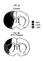
- APC beneficial effects of APC in the present invention were associated with marked improvement of post-ischemic re-circulation, i.e., 79% of baseline CBF values in comparison to only 32% in control animals. It is possible that the observed CBF improvement involves, in part, alleviation of post-ischemic coagulopathy by APC.
- Previous studies of global ischemia revealed massive intravascular coagulation in association with complement activation.
- Studies in focal ischemia models also revealed that significant obstructions in CBF might result from massive microvascular occlusions due to vascular accumulation of polymorphonuclear (PMNs) leukocytes and fibrin deposition.
- PMNs polymorphonuclear
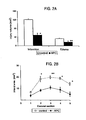
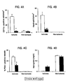
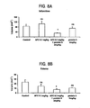
- APC in addition to significantly reducing fibrin deposition in the ischemic hemisphere, also prevents intravascular accumulation of peripheral blood cells, i.e., PMNs, in brain by preventing their transport across the blood-brain barrier.
- PMNs peripheral blood cells
- the mechanisms of anti-inflammatory effects of APC are still not completely understood, the absence of significant leukocyte-endothelial interactions in ischemic APC-treated animals may not only reduce fibrin formation in ischemic brain, but could also be related to improvements in the CBF and reduced neuronal injury. It has been shown that blocking PMN penetration across the blood-brain barrier results in considerable improvement of the neurological outcome and also limits neuronal injury.
- APC Alzheimer's disease
- APC APC to abolish almost completely leukocyte accumulation within the vascular system and prevent PMN penetration into brain parenchyma may be responsible, in part, for its neuroprotective effects. This may increase not only the rate of re-circulation, but may also importantly alleviate post-ischemic generation of reactive oxygen species from PMNs, which in turn may protect neurons from injury.
- the neuroprotective effects of APC in stroke can be rationalized by its anti-inflammatory action and antithrombotic effects, we cannot rule out the possibility that APC itself may also have direct neuroprotective effects on neurons.
- APC does not adversely affect hemostatic function or produce increased bleeding in the brain or intracerebral hemorrhage (ICH).
- ICH intracerebral hemorrhage
- Endogenous protein C zymogen may be protective in stroke in humans.
- Low levels of plasma protein C or APC, and/or resistance to the anticoagulant effects of APC were related to poor outcome after stroke.
- Low plasma levels of protein C observed in stroke patients may be caused by lower levels of protein C biosynthesis and/or by protein C depletion due to excessive thrombin generation and rapid APC clearance, while low circulating APC may result from depletion of protein C zymogen precursor, increased levels of circulating APC inhibitors, or reduced APC generating capacity due to either low levels of intravascular thrombin, or reduced thrombomodulin and/or endothelial cell protein C receptor.
- the present invention provides a combination of activated protein C (APC) and protein S for use in protecting neuronal cells from cell death in a subject having or at risk of having a neuropathological disorder selected from stroke and ischemia, wherein the APC and protein S are hom the same species.
- a neuroprotective effective amount of activated protein C (APC) for example, in a pharmaceutically acceptable carrier, may be administered to the subject thereby providing neuroprotection to the subject.
- One "having or at risk of having” a neuropathological disorder as described herein is a subject either exhibiting symptoms of the disorder or diagnosed as being at risk for developing the disorder. Such subjects include those subjects having undergone or preparing for surgical procedures as described below.
- treatment refers to reducing the symptoms of the disease.
- treatment or “ameliorate” denotes a lessening of the detrimental effect of the neurological disease in the subject receiving therapy.
- treatment when referring to neurological disease used hereinafter does not necessarily mean that the neurodegenerative disease is completely eliminated, but rather that the cognitive facilities damaged by the disease are improved.
- “Therapeutically effective” as used herein, refers to that amount of APC that is of sufficient quantity to ameliorate the cause or symptoms of the disease.
- the subject of the invention is preferably a human, however, it can be envisioned that any animal can be treated using the invention.
- One aspect of the invention includes the "neuroprotective" activity of APC.
- the term “neuron” includes hundreds of different types of neurons, each with distinct properties. Each type of neuron produces and responds to different combinations of neurotransmitters and neurotrophic factors. Neurons are thought not to divide in the adult brain, nor do they generally survive long in vitro.
- the invention provides for the protection from death or senescence of neurons from virtually any region of the brain and spinal cord.
- Neurons include those in embryonic, fetal or adult neural tissue, including tissue from the hippocampus, cerebellum, spinal cord, cortex (e.g., motor or somatosensory cortex), striatum, basal forebrain (cholenergic neurons), ventral mesencephalon (cells of the substantia nigra), and the locus ceruleus (neuroadrenaline cells of the central nervous system).
- cortex e.g., motor or somatosensory cortex
- striatum e.g., basal forebrain (cholenergic neurons), ventral mesencephalon (cells of the substantia nigra), and the locus ceruleus (neuroadrenaline cells of the central nervous system).
- APC acts on brain endothelial cells (i.e., vascular cells), via endothelial receptor(s) for protein C and APC, that mediates its effects on endothelium, both central and peripheral. This, in turns affects intracellular signaling systems that in a cascade turn on and off different genes in vascular endothelium that may interfere with normal endothelial cell response to inflammation. It is also possible that receptors for APC on neurons may mediate neuroprotective effects of APC.
- APC utilized in the present invention include those described in US Patent Nos. 5, 084, 274 , 6, 037,322 and 6,156,734 .
- Activated Protein C refers to Protein C that is cleaved proteolytically by thrombin to yield an activated protein C (APC) which inactivates coagulation Factors Va and VIIIa thus inhibiting coagulation.
- APC activated protein C
- the use of APC described herein include "fragments of APC, as long as they retain the activities described herein.
- Such fragments, or APC include recombinantly produced, human plasma-derived and synthetically produced, for example, "Synthetic peptide” refers to a chemically produced chain of amino acid residues linked together by peptide bonds that is free of naturally occurring proteins and fragments thereof. "Anticoagulant” refers to an agent that interrupts coagulation and thereby inhibits fibrin formation. "Coagulation” refers to the sequential process in which the multiple coagulation factors of the blood interact resulting in the formation of fibrin. Protein C consists of a 155 amino acid residue light chain and a 262 amino acid residue heavy chain and is fully described in US Patent No. 5,679,639
- “Chemical derivative” refers to a subject polypeptide having one or more residues chemically derivatized by reaction of a functional side group.
- Such derivatized molecules include for example, those molecules in which free amino groups have been derivatized to form amine hydrochlorides, p-toluene sulfonyl groups, carbobenzoxy groups, t-butyloxycarbonyl groups, chloroacetyl groups or formyl groups.
- Free carboxyl groups may be derivatized to form salts, methyl and ethyl esters or other types of esters or hydrazides.
- Free hydroxyl groups may be derivatized to form O-acyl or O-alkyl derivatives.
- the imidazole nitrogen of histidine may be derivatized to form N-im-benzylhistidine.
- chemical derivatives those peptides which contain one or more naturally occurring amino acid derivatives of the twenty standard amino acids. For examples: 4-hydroxyproline may be substituted for proline; 5-hydroxylysine may be substituted for lysine; 3-methylhistidine may be substituted for histidine; homoserine may be substituted for serine; and ornithine may be substituted for lysine.
- APC of the present invention also include any polypeptide having one or more additions and/or deletions or residues relative to the sequence of a polypeptide whose sequence is shown herein, so long as the requisite activity, e.g., anti-inflammatory or neuroprotective, is maintained.
- compositions described herein contain a physiologically tolerable carrier together with APC, dissolved or dispersed therein as an active ingredient.
- the therapeutic composition is not immunogenic when administered to a mammal or human patient for therapeutic purposes.
- compositions, carriers, diluents and reagents are used interchangeably and represent that the materials are capable of administration to or upon a mammal without the production of undesirable physiological effects such as nausea, dizziness, and gastric upset .
- compositions that contains active ingredients dissolved or dispersed therein are well understood in the art.
- compositions are prepared as injectables either as liquid solutions or suspensions, however, solid forms suitable for solution, or suspensions, in liquid prior to use can also be prepared.
- the preparation can also be emulsified.
- the active ingredient can be mixed with excipients which are pharmaceutically acceptable and compatible with the active ingredient and in amounts suitable for use in the therapeutic methods described herein.
- Suitable excipients are, for example, water, saline, dextrose, glycerol, ethanol and combinations thereof.
- the composition can contain minor amounts of auxiliary substances such as wetting or emulsifying agents, pH buffering agents which enhance the effectiveness of the active ingredient.
- a therapeutic composition can include pharmaceutically acceptable salts of the components therein.
- Pharmaceutically acceptable salts include the acid addition salts (formed with the free amino groups of the polypeptide) that are formed with inorganic acids such as, for example, hydrochloric or phosphoric acids, or such organic acids as acetic, tartaric, and mandelic . Salts formed with the free carboxyl groups can also be derived from inorganic bases such as, for example, sodium, potassium, ammonium, calcium or ferric hydroxides, and such organic bases as isopropylamine, trimethylamine, 2-ethylamino ethanol, histidine, and procaine.
- Physiologically tolerable carriers are well known in the art.
- Exemplary of liquid carriers are sterile aqueous solutions that contain no materials in addition to the active ingredients and water, or contain a buffer such as sodium phosphate at physiological pH value, physiological saline or both, such as phosphate-buffered saline.
- aqueous carriers can contain more than one buffer salt, as well as salts such as sodium and potassium chlorides, dextrose, polyethylene glycol and other solutes.
- Liquid compositions can also contain liquid phases in addition to and to the exclusion of water.
- additional liquid phases are glycerin, vegetable oils such as cottonseed oil, and water-oil emulsions.
- APC is a very species specific moiety.
- the dosage for human or recombinant human APC in a human is much lower than the appropriate dosage in a mouse, for example.
- the normal baseline level of APC in a human is typically about 2.2 ng/ml of blood.
- a therapeutically effective amount of human APC is typically administered to a human at a dosage sufficient to raise the blood level of APC by from about 1.0 ng/ml to about 500 ng/ml, preferably, from about 5 ng/ml to 200 ng/ml.
- the APC can be formulated according to known methods to prepare pharmaceutically useful compositions.
- the APC is preferably administered parenterally to ensure its delivery into the bloodstream in an effective form by injecting the appropriate dose as continuous infusion for about 4 to about 96 hours.
- the appropriate dose of APC will be administered by continuous infusion for about 4 to 72 hours. More preferably, the appropriate dose of APC will be administered by continuous infusion for about 4 to about 48 hours. More preferably, the appropriate dose of APC will be administered by continuous infusion for about 12 to 48 hours. More preferably, the appropriate dose of APC will be administered by continuous infusion for about 12 to 36 hours. More preferably, the appropriate dose of APC will be administered by continuous infusion for about 4 to 36 hours.
- the appropriate dose of APC will be administered by continuous infusion for about 12 to 24 hours. Most preferably, the appropriate dose of APC will be administered by continuous infusion for about 24 hours.
- An appropriate loading dose of APC may be given by bolus injection with or without subsequent APC infusion.
- the amount of APC administered can be from about 0.01 mg/kg/hr to 0.10 mg/kg/hr which is equivalent to about 17 mg/70 kg/24 hours to 170mg/70 kg/24 hours. While the dose level is identified as a specific amount per 24 hours, one skilled in the art would recognize that this is a designation of the dose level and is not necessarily limited to a 24 hour infusion but may include continuous infusion for various times, for example, from about four hours to ninety-six hours. More preferably the amount of APC administered is about 0.01 mg/kg/hr to 0.05 mg/kg/hr (about 17 mg/70 kg/24 hours to 84 mg/70 kg/24 hours).
- the amount of APC administered will be about 0.01 mg/kg/hr to 0.03 mg/kg/hr (about 17 mg/70 kg/24 hours to 50 mg/70 kg/24 hours). Furthermore, the amount of APC administered is from about 0.02 mglkg/hr to 0.05 mg/kg/hr which is equivalent to about 34 mg/70 kg/24 hours to 84 mg/70 kg/24 hours. More preferably the amount of APC administered is about 0.024 mg/kg/hr to 0.048 mg/kg/hr (about 40 mg/70 kg/24 hours to 80 mg/70 kg/24 hours).
- the amount of APC administered will be about 0.027 mg/kg/hr to 0.045 mg/kg/hr (about 45 mg/70 kg/24 hours to 75 mg/70 kg/24 hours). While more preferably the amount of APC administered will be about 0.030 mg/kg/hr to 0.042 mg/kg/hr (about 50 mg/70 kg/24 hours to 70 mg/70 kg/24 hours). While more preferably the amount of APC administered will be about 0.033 mg/kg/hr to 0.039 mg/kg/hr (about 55 mg/70 kg/24 hours to 65 mg/70 kg/24 hours).
- Preferable amounts of APC administered are about 0.024 mg/kg/hr (about 40 mg/70 kg/24 hours), about 0.027 mg/kg/hr (about 45 mg/70 kg/24 hours) or, about 0.030 mglkg/hr to 0.042 mg/kg/hr (about 50 mg/70 kg/24 hours).
- the amount of APC can be reduced when administered with a co-factor such as Protein S, as described herein.
- the APC will be administered by injecting a portion of the appropriate dose per hour as a bolus injection over a time from about 5 minutes to 120 minutes, followed by continuous infusion of the appropriate dose for about twenty three hours to 96 hours which results in the appropriate dose administered over 24 hours to 96 hours.
- the most preferable dose level of APC to be administered for thrombotic occlusion (e.g. stroke) as described herein will be about 0.024mg/kg/hr.
- a therapeutically effective amount of an anticoagulant antibody that may be used in conjunction with the methods of the invention is typically an amount of antibody such that when administered in a physiologically tolerable composition is sufficient to achieve a plasma concentration of from about 0.1 microgram (ug) per milliliter (ml) to 100 ug/ml, preferably from about 1 ug/ml to 5 ug/ml, and usually about 5 ug/ml.
- the therapeutic compositions containing APC are conventionally administered intravenously, as by injection of a unit dose, for example.
- unit dose when used in reference to a therapeutic composition of the present invention refers to physically discrete units suitable as unitary dosage for the subject, each unit containing a predetermined quantity of active material calculated to produce the desired therapeutic effect in association with the required diluent; i.e., carrier, or vehicle.
- the compositions are administered in a manner compatible with the dosage formulation, and in a therapeutically effective amount.
- the quantity to be administered depends on the subject to be treated, capacity of the subject's system to utilize the active ingredient, and degree of therapeutic effect desired. Precise amounts of active ingredient required to be administered depend on the judgement of the practitioner and are peculiar to each individual.
- suitable dosage ranges for systemic application are disclosed herein and depend on the route of administration. Suitable regimes for initial administration and booster shots are also variable, but are typified by an initial administration followed by repeated doses at one or more hour intervals by a subsequent injection or other administration. Alternatively, continuous intravenous infusion sufficient to maintain concentrations in the blood in the ranges specified for in vivo therapies are contemplated.
- the pharmaceutical composition may conveniently be presented in unit dosage form and may be prepared by any of the methods well known in the art of pharmacy. All methods include the step of bringing the compounds into association with a carrier which may contain one or more accessory ingredients.
- a carrier which may contain one or more accessory ingredients.
- the composition for pills, tablets, or capsules (e.g., for oral administration) or powders are prepared by uniformly and intimately blending the compounds with finely divided solid carriers, and then, if necessary as in the case of tablets, forming the product into the desired shape and size.
- the pill, tablet, or capsule can be coated with a substance capable of protecting the composition from the gastric acid or intestinal enzymes in the subject's stomach for a period of time sufficient to allow the composition to pass undigested into the subject's small intestine.
- the pharmaceutical compositions may be also directly targeted to the brain by an intercerebroventricular pump.
- compositions suitable for parenteral administration conveniently comprise sterile aqueous solutions of the compound(s) in water or saline to produce an aqueous solution, and rendering said solution sterile.
- the composition may be presented in unit or multi-dose containers, for example, sealed ampoules or vials.
- Example 3 illustrates that administration of a combination of protein S and APC in the treatment of stroke in a mouse model is more effective for reducing the area of brain infarction and volume of edema in the ischemic hemisphere in mice than is a therapeutically effective low amount of APC administered alone, even when therapy is administered after the ischemic event.
- This unexpected result is obtained even when the amount of the APC administered (e.g., 0.1 mg/kg) in the combination therapy is far less than is generally used when APC alone is used in treatment of stroke in this model.
- APC APC-derived from the same non-human species as the APC K is to be co-administered.
- APC APC, analogs or biologically active fragments thereof may be administered in a manner compatible with the dosage formulation and in such amount as will be therapeutically effective.
- invention treatment methods are equally effective whether the APC is administered during, before or after an ischemic event, such as stroke, e.g., up to three to six hours after a stroke.
- compositions of the invention may be administered in any way which is medically acceptable which may depend on the disease condition or injury being treated.
- Possible administration routes include injections, by parenteral routes such as intravascular, intravenous, intra-arterial, subcutaneous, intramuscular, intratumor, intraperitoneal, intraventricular, intraepidural or others, as well as oral, nasal, ophthalmic, rectal, topical, or by inhalation.
- the compositions may also be directly applied to tissue surfaces during surgery.
- Sustained release administration is also specifically included in the invention, by such means as depot injections or erodible implants.
- mice were subjected to a modified middle cerebral artery (MCA) occlusion technique ( E.S. Connolly et al., Neurosurgery. 38(3):523-531 (1996 ); H. Hara et al, J. Cereb. Blood Flow Metab.16:605-611, (1996 ); and P. Tabrizi et al., Arterioscler. Thromb. Vasc. Biol. 19:2801-2806 (1999 )) to induce acute focal ischemic stroke with cerebrovascular thrombosis, as described below.
- MCA middle cerebral artery
- mice were initially anesthetized with metofane and maintained with 50 mg/kg i.p. pentobarbital. Atropine methyl nitrate (0.18 mg/kg i.p.) was given as pre-medication to prevent airway obstruction by mucus formation. Animals were allowed to breath spontaneously. Rectal temperature was maintained at 37 ⁇ 1 °C by a thermostatically regulated heating pad. The right femoral artery was cannulated with a PE-10 catheter for continuous monitoring of blood pressure and blood analysis.
- the suture was introduced into the external carotid artery lumen, and gently advanced into the internal carotid artery to occlude the MCA at its origin from the circle of Willis. MCA occlusion was maintained for 1 hr followed by 24 hrs of reperfusion.
- CBF was monitored by Laser Doppler Flowmetry (LDF) using tissue perfusion monitor (Transonic BLF21, Ithaca, NY). CBF measurements were performed in all animals subjected to neuropathological and neurologic analysis. For CBF determinations, animals were placed in a stereotactic head frame, and readings were obtained 2 mm posterior to the bregma, both 3 and 6 mm to each side of midline, using a stereotactic micromanipulator and keeping the angle of the probe perpendicular to the cortical surface. LDF probes (0.8 mm diameter) positioned on the cortical surface were connected to a tissue perfusion monitor (Transonic BLF21).
- LDF Laser Doppler Flowmetry
- the procedure was considered to be technically successful if ⁇ 88% reduction in relative CBF was observed immediately after placement of the occluding suture.
- the procedure was successful in all studied control animals producing levels of ischemia sufficient to render consistently large infarction volumes and microvascular thrombosis in the ischemic hemisphere. Head temperature was monitored with a 36-gauge thermocouple temperature probe in the temporalis muscle connected to a digital thermometer/thermoregulator model (model 9000, Omega, CT).
- Arterial blood gases (pH, PaO 2 , PaCO 2 ) were measured before and during MCA occlusion using ABL 30 Acid-Base Analyzer (Radiometer, Copenhagen, Denmark).
- the area of injury was delineated by incubation of unfixed 1-mm coronal brain slices in 2% TTC in phosphate buffer (pH 7.4). Serial coronal sections were displayed on a digitizing video screen using the imaging system of Jandel Scientific (San Rafael, CA). The volume of injury was calculated by summing up affected areas from each coronal section and multiplying by the thickness of each section. Brain infarction and edema were calculated using Swanson correction (Tabrizi et al., 1999).
- Detection of fibrin by immunostaining was performed on brain tissue sections previously treated with TTC. Tissue fixed in 10% buffer formalin was processed and 4 ⁇ m thick paraffin coronal sections from each block cut and stained. Fibrin was localized using anti-fibrin II antibody (NYB-T2G1, Accurate Chemical Sci. Corp., Westbury, NY) (1:500 dilution) and graded according to the following scale (P. Tabrizi et al., 1999 and Y. Okada et al., Stroke.
- Paraffin sections were also stained using the anti-CD11b antibody (DAKO corporation, Carpenteria, CA) (1:250 dilution) directed against the leukocyte adhesion receptor macrophage-1 antigen.
- This antibody detects CAD11b antigen on polymorphonuclear leucocytes (PMNs), as well as on activated macrophages and/or microglia in the tissue.
- PMNs polymorphonuclear leucocytes
- the number of CD11b positive cells was counted in ten random fields by two independent blinded observers and expressed as number per mm 2 of section. Simultaneous detection of fibrin and leukocytes was done by sequential immunostaining.
- Fibrin was localized first with the NYB-T2G1 antibody and detected using 3, 3' diaminobenzidine (DAB) substrate (Vector Labs, CA) followed by detection of leukocytes using the CD11b antibody and the Vector SG peroxide substrate (Vector Labs, CA). Routine control sections included deletion of primary antibody, deletion of secondary antibody and the use of an irrelevant primary antibody. Neutrophils were also detected in brain sections by dichloroacetate esterase staining using the esterase staining kit (Sigma, MO) as reported ( S.G. Soriano et al, Stroke. 30:134-139 (1999 )). Sections were deparaffinized and incubated with naphthol AS-D chloroacetate. The free naphthol released on ester hydrolysis by enzymes on neutrophils was detected using freshly formed diazonium salt and counterstained with Gill No.3 hematoxylin. The number of neutrophils was counted as above.
- DAB 3, 3' diamino
- TTC staining a 1 mm section of brain tissue was divided into contralateral and ipsilateral hemisphere. Tissue was homogenized in 10 mM sodium phosphate buffer, pH 7.5, 0.1 mM aminocaproic acid, 5 mM trisodium EDTA, 10 U aprotinin/ml, 10 U heparin/ml, and 2 mM PMSF.
- the homogenate was agitated for 14 hour at 4 °C, and the particulate material was sedimented by centrifugation at 10,000 g for 10 min, resuspended in extraction buffer without PMSF, sedimented again, and finally dispersed in 3 M urea.
- the suspension was agitated for 2 hours at 37 °C, vigorously vortexed, and centrifuged at 14,000 g for 15 min.
- the supernatant was aspirated and the sediment dissolved at 65 °C in reducing SDS buffer, subjected to SDS-PAGE (8%), and transferred to a PVDF membrane (Immobilon-P; Millipore Corp., MA) by electroblotting.
- Fibrin was visualized with anti-human fibrin II antibody (given above) and enhanced chemiluminescence system (Amersham Corp., IL). Fibrin standards were prepared by clotting a known amount of murine fibrinogen (Sigma Chemical Co., MO) with an excess of thrombin in the absence of calcium. The films were scanned with a Hoefer GS 300 scanning densitometer interfaced to an IBM PC computer with a DT 2805 analog and digital system (Data Translation, MA) and data were converted into ⁇ g fibrin/0.1 g of tissue.
- This assay was performed to detect microhemorrhages in the brain tissue. The procedure was as described previously ( T.F. Choudhri et al.,Stroke. 28(11):2296-2302 (1997 )). Briefly, a section of brain tissue 1 mm thick was divided into ipsilateral and contralateral hemisphere after TTC staining. Distilled water (200 ⁇ l) was added, tissue homogenized for 30 sec, sonicated on ice for 1 min, and centrifuged at 13,000 rpm for 30 min.
- Hemoglobin-containing supernatant was collected, and 80 ⁇ l of Drabkin's reagent (Sigma Diagnostics, MO) added to a 20- ⁇ l aliquot and allowed to mix for 15 min. This reaction converts hemoglobin to cyanmethemoglobin, which has an absorbency peak at 540 nm. To validate that the measured absorbency reflects the amount of hemoglobin, known quantities of bovine erythrocyte hemoglobin (Sigma) and incremental aliquots of mouse blood added to freshly homogenized brain tissue were analyzed in parallel.
- Physiological variables, infarction, and edema volumes were compared between groups using Student's t-test and ANOVA.
- Non-parametric data (neurologic outcome scores) was subjected to the Chi-square test with Fisher's transformation. Survival was compared by the Kruskal-Wallis test. A value ofp ⁇ 0.05 was considered statistically significant.
- the mean time of survival in control group was 10.2 hrs. In contrast, 5 out of 6 APC-treated animals survived 24 hrs, and only 1 animal died at 23 hrs. APC-treated animals were sacrificed at 24 hrs to determine the volume of brain injury, and the mean time of survival in APC-treated group was 23.7 hrs. The motor neurological score in APC-treated animals was about 2-fold lower than in control group.
- mice treated with APC had a significant reduction in the volume of brain injury compared to control mice ( Figure 2 ).
- the total volume of injury of gray matter (corrected for edema), i.e., the brain infarction volume, was significantly decreased by 59% (p ⁇ 0.02) in the APC-treated group relative to control mice ( Figure 2A ).
- the edema volume in the lesioned ischemic hemisphere was also reduced by APC treatment by 50% (p ⁇ 0.05).
- Studies of the infarct area for each of the five coronal sections of the same brains as in Figure 2A confirmed significant reductions of injury at all brain levels in APC-treated mice ( Figure 2B ).
- Figure 3 illustrates that 100% of control mice had injury that involved significant ipsilateral portion of the hemisphere including cortex, subcortical structures and lateral striatum; ⁇ 50% mice exhibited changes in the medial striatum and ⁇ 50% showed changes in the dorsomedial and ventromedial cortex.
- APC-treated animals there was a significant reduction of the injured area, and all animals developed injury only in small well-localized area in the lateral cortex, with significant reduction of injury in each region.
- microvessels including small veins, arterioles and numerous capillaries contained intraluminal fibrin deposits corresponding to grade 1 according to the scale of fibrin localization in an MCA model (P. Tabrizi et al., 1999 and Y. Okada et al., 1994). Extravascular deposition of fibrin grade 2 was also found around some microvessels in control mice. The migration of leucocytes into parenchymal tissues was frequently observed in control animals, and PMNs were identified either by morphology or by positive staining for CD11b in the tissue, and/or positive staining for dichloroacetate esterase. In addition to a single antibody staining, sections were also counter-stained with hematoxylin.
- Double staining for fibrin and leukocytes showed complete thrombosis of a large venule.
- the expansion of so-called "white thrombus" from the vein into a capillary bed was also shown.
- the nuclear morphology of neutrophils could not be seen as hematoxylin staining was not performed simultaneously with double staining.
- Staining with dichloroacetate esterase confirmed infiltration of ischemic parenchymal tissue with PMNs in control mice. It is noteworthy that deposition of fibrin in control mice was much more significant in the present model than in a previous MCA model ( Y. Okada et al., Stroke. 25:1847-1853, 1994 ).
- the difference could be related to significantly higher reductions in the CBF in the present vs. previous model, both during occlusion, i.e., 88% vs. 63%, and reperfusion, 68% vs. 30%, respectively.
- Double staining confirmed co-localization of fibrin and leukocytes in cerebral vessels in the ischemic hemisphere in control mice.
- APC treatment significantly reduced fibrin deposition, as well as vascular accumulation and parenchymal infiltration with PMNs.
- Figures 5A and 5B illustrate 8.2-fold decrease in the amount of deposited fibrin in ischemic hemisphere of APC-treated vs. control mice determined by quantitative Western blot analysis.
- Table 3 shows the cumulative results of experiments conducted with mice treated with APC (2 mg/kg) 15 min prior to or 10 min after the MCA occlusion had no significant differences in mean arterial blood pressure, PaO 2 , PaCO 2 , pH, hematocrit, head temperature, and blood glucose when compared with control animals (data not shown).
- APC administration did not influence CBF under basal conditions in the absence of occlusion/reperfusion.
- TABLE 3 Survival and motor scores at 24 hrs post MCA occlusion/reperfusion Group Survival Time (hr) Scores at 24 hours post MCAO No.
- mice Mean Score ( ⁇ SD) Control (mean ⁇ SD) 0 1 2 3 4 5 APC 15 min prior to MCAO 13.6 ⁇ 3.24 0 0 0 2 2 8 4.50 ⁇ 0.79 2 mg/kg 23.7 ⁇ 0.33* 0 2 2 1 0 1 2.33 ⁇ 1.51**
- the mean survival time for control group animals was 13.6 hours. Ten of 11 mice treated with 2 mg/kg APC either 15 min prior to occlusion or 10 min after occlusion survived 24 hrs, and 1 APC-treated animal died at 23 hours. All 3 animals treated with 0.5 mg/kg APC at 10 min after stroke induction survived 24 hours. APC-treated animals were sacrificed at 24 hrs to determine the volume of brain injury, thus data beyond 24 hours is not available.
- mice given 2 mg/kg APC 15 min prior to and 10 min after the MCA occlusion were improved by 2 to 2.7-fold compared with control group.
- 0.5 mg/kg APC given 10 min after MCA occlusion improved neurological outcome significantly.
- the protective effect of APC was not apparent at an APC dose of 0.1 mg/kg based on survival time and neurologic function (Table 1) and on the volume of brain injury and effects on CBF ( Fig. 7 ).
Landscapes
- Health & Medical Sciences (AREA)
- Engineering & Computer Science (AREA)
- Life Sciences & Earth Sciences (AREA)
- Chemical & Material Sciences (AREA)
- Bioinformatics & Cheminformatics (AREA)
- Organic Chemistry (AREA)
- General Health & Medical Sciences (AREA)
- Medicinal Chemistry (AREA)
- Veterinary Medicine (AREA)
- Pharmacology & Pharmacy (AREA)
- Animal Behavior & Ethology (AREA)
- Biomedical Technology (AREA)
- Public Health (AREA)
- Nuclear Medicine, Radiotherapy & Molecular Imaging (AREA)
- General Chemical & Material Sciences (AREA)
- Chemical Kinetics & Catalysis (AREA)
- Neurology (AREA)
- Zoology (AREA)
- Neurosurgery (AREA)
- Wood Science & Technology (AREA)
- Genetics & Genomics (AREA)
- Biochemistry (AREA)
- Gastroenterology & Hepatology (AREA)
- Immunology (AREA)
- Proteomics, Peptides & Aminoacids (AREA)
- Epidemiology (AREA)
- General Engineering & Computer Science (AREA)
- Heart & Thoracic Surgery (AREA)
- Cardiology (AREA)
- Molecular Biology (AREA)
- Pain & Pain Management (AREA)
- Biotechnology (AREA)
- Microbiology (AREA)
- Psychiatry (AREA)
- Hospice & Palliative Care (AREA)
- Psychology (AREA)
- Vascular Medicine (AREA)
- Urology & Nephrology (AREA)
- Rheumatology (AREA)
- Medicines That Contain Protein Lipid Enzymes And Other Medicines (AREA)
Applications Claiming Priority (3)
| Application Number | Priority Date | Filing Date | Title |
|---|---|---|---|
| US18022700P | 2000-02-04 | 2000-02-04 | |
| US180227P | 2000-02-04 | ||
| PCT/US2001/003758 WO2001056532A2 (en) | 2000-02-04 | 2001-02-05 | Neuroprotective, antithrombotic and anti-inflammatory uses of activated protein c (apc) |
Related Child Applications (1)
| Application Number | Title | Priority Date | Filing Date |
|---|---|---|---|
| EP10162400.5 Division-Into | 2010-05-10 |
Publications (3)
| Publication Number | Publication Date |
|---|---|
| EP1255556A2 EP1255556A2 (en) | 2002-11-13 |
| EP1255556A4 EP1255556A4 (en) | 2003-07-02 |
| EP1255556B1 true EP1255556B1 (en) | 2011-04-06 |
Family
ID=22659693
Family Applications (1)
| Application Number | Title | Priority Date | Filing Date |
|---|---|---|---|
| EP01910427A Expired - Lifetime EP1255556B1 (en) | 2000-02-04 | 2001-02-05 | Neuroprotective, antithrombotic and anti-inflammatory uses of activated protein c (apc) |
Country Status (8)
| Country | Link |
|---|---|
| US (1) | US7074402B2 (enExample) |
| EP (1) | EP1255556B1 (enExample) |
| JP (1) | JP2004508280A (enExample) |
| AT (1) | ATE504310T1 (enExample) |
| AU (1) | AU2001238034A1 (enExample) |
| CA (1) | CA2398929C (enExample) |
| DE (1) | DE60144366D1 (enExample) |
| WO (1) | WO2001056532A2 (enExample) |
Families Citing this family (22)
| Publication number | Priority date | Publication date | Assignee | Title |
|---|---|---|---|---|
| US20040096443A1 (en) * | 2002-03-08 | 2004-05-20 | Traynelis Stephen Francis | Treatment of neurodegenerative diseases and conditions using par1 antagonists |
| ATE550073T1 (de) | 2001-06-13 | 2012-04-15 | Univ Sydney | Protein c für die wundheilung |
| US20030073636A1 (en) * | 2001-09-19 | 2003-04-17 | Oklahoma Medical Research Foundation | Method of treating diabetes |
| CA2499458C (en) * | 2002-09-30 | 2013-11-12 | Socratech L.L.C. | Protein s protects the nervous system from injury |
| CA2508276C (en) * | 2002-12-05 | 2018-03-27 | Socratech L.L.C. | Neuroprotective activity of activated protein c is independent of its anticoagulant activity |
| WO2004056309A2 (en) * | 2002-12-05 | 2004-07-08 | Socratech L.L.C. | Neuroprotective activity of activated protein c is independent of its anticoagulant activity |
| US20070142272A1 (en) * | 2003-01-24 | 2007-06-21 | Zlokovic Berislav V | Neuroprotective activity of activated protein c independent of its anticoagulant activity |
| US9192657B2 (en) * | 2003-07-08 | 2015-11-24 | The Scripps Research Institute | Activated protein C variants with normal cytoprotective activity but reduced anticoagulant activity |
| CA2574598A1 (en) * | 2004-07-23 | 2006-02-09 | The University Of Rochester | Activated protein c inhibits undesirable effects of plasminogen activator in the brain |
| US20080293631A1 (en) * | 2005-01-07 | 2008-11-27 | Christopher John Jackson | Treatment for Autoimmune and Inflammatory Conditions |
| US7785857B2 (en) * | 2006-08-31 | 2010-08-31 | Saint Louis University | Protein C variant |
| AU2008280782B2 (en) | 2007-07-20 | 2014-01-23 | Diamedica Inc. | Tissue kallikrein for the treatment of diseases associated with amyloid protein |
| WO2009099642A2 (en) * | 2008-02-05 | 2009-08-13 | The Board Of Trustees Of The Leland Stanford Junior University | Proteomic analysis of active multiple sclerosis lesions |
| CN102458454A (zh) * | 2009-04-22 | 2012-05-16 | 森诺姆尼有限公司 | 用于治疗亨廷顿病的组织激肽释放酶 |
| US20150150954A1 (en) | 2012-07-04 | 2015-06-04 | The University Of Sydney | Treatment of inflammatory skin disorders |
| CN104122396A (zh) * | 2013-04-23 | 2014-10-29 | 中国科学院上海生命科学研究院 | 维生素k依赖性蛋白s作为糖尿病标志物的应用 |
| DK3137102T3 (da) | 2014-04-16 | 2021-10-11 | Zz Biotech Llc | Apc til anvendelse til behandling af unormal kutan ardannelse |
| ES2890623T3 (es) | 2014-04-16 | 2022-01-20 | Zz Biotech Llc | Uso del análogo de APC para curar heridas |
| CN105806818B (zh) * | 2016-04-01 | 2019-11-22 | 南京医科大学 | 检测血小板nmda受体活性的方法及其应用 |
| WO2017212476A1 (en) | 2016-06-05 | 2017-12-14 | Tel Hashomer Medical Research Infrastructure And Services Ltd. | Novel molecules for the treatment of inflammation |
| KR20190122706A (ko) | 2017-03-09 | 2019-10-30 | 다이어메디카 인코포레이티드 | 조직 칼리크레인 1의 투약 형태 |
| CA3176941A1 (en) * | 2020-03-24 | 2021-09-30 | Talengen International Limited | Method and medicine for treating huntington's disease |
Family Cites Families (7)
| Publication number | Priority date | Publication date | Assignee | Title |
|---|---|---|---|---|
| US5084274A (en) * | 1987-11-17 | 1992-01-28 | Scripps Clinic And Research Foundation | Inhibition of arterial thrombotic occlusion or thromboembolism |
| US5009889A (en) * | 1987-12-31 | 1991-04-23 | Oklahoma Medical Research Foundation | Treatment of dysfunctional vascular endothelium using activated protein C |
| AT402153B (de) * | 1989-06-26 | 1997-02-25 | Immuno Ag | Protein-s-hältige pharmazeutische präparation |
| US5254532A (en) * | 1989-06-26 | 1993-10-19 | Immuno Aktiengesellschaft Fur Chemisch-Medizinische Produkte | Preparation for treating and preventing thromboses and thromboembolic complications, use of such a preparation and a method of producing the same |
| IL131615A0 (en) * | 1997-03-24 | 2001-01-28 | Lilly Co Eli | Methods for treating vascular disorders |
| CN1265598A (zh) * | 1997-06-05 | 2000-09-06 | 伊莱利利公司 | 治疗血栓形成性疾病的方法 |
| HUP0001237A3 (en) * | 1997-10-20 | 2002-01-28 | Lilly Co Eli | Methods for treating vascular disorders |
-
2001
- 2001-02-05 US US09/777,484 patent/US7074402B2/en not_active Expired - Fee Related
- 2001-02-05 DE DE60144366T patent/DE60144366D1/de not_active Expired - Lifetime
- 2001-02-05 WO PCT/US2001/003758 patent/WO2001056532A2/en not_active Ceased
- 2001-02-05 CA CA2398929A patent/CA2398929C/en not_active Expired - Fee Related
- 2001-02-05 AT AT01910427T patent/ATE504310T1/de not_active IP Right Cessation
- 2001-02-05 AU AU2001238034A patent/AU2001238034A1/en not_active Abandoned
- 2001-02-05 JP JP2001556226A patent/JP2004508280A/ja active Pending
- 2001-02-05 EP EP01910427A patent/EP1255556B1/en not_active Expired - Lifetime
Also Published As
| Publication number | Publication date |
|---|---|
| EP1255556A4 (en) | 2003-07-02 |
| CA2398929C (en) | 2011-05-31 |
| WO2001056532A2 (en) | 2001-08-09 |
| JP2004508280A (ja) | 2004-03-18 |
| DE60144366D1 (de) | 2011-05-19 |
| WO2001056532A3 (en) | 2001-12-06 |
| CA2398929A1 (en) | 2001-08-09 |
| ATE504310T1 (de) | 2011-04-15 |
| US20020028199A1 (en) | 2002-03-07 |
| US7074402B2 (en) | 2006-07-11 |
| EP1255556A2 (en) | 2002-11-13 |
| AU2001238034A1 (en) | 2001-08-14 |
| WO2001056532A9 (en) | 2003-01-09 |
Similar Documents
| Publication | Publication Date | Title |
|---|---|---|
| EP1255556B1 (en) | Neuroprotective, antithrombotic and anti-inflammatory uses of activated protein c (apc) | |
| US6268337B1 (en) | Methods for treating vascular disorders using activated protein C | |
| US6315995B1 (en) | Methods for treating an ischemic disorder and improving stroke outcome | |
| US8071091B2 (en) | Non-neurotoxic plasminogen activating factors for treating stroke | |
| JPH11500408A (ja) | 改良型▲vii▼因子 | |
| JP2006525270A (ja) | 脳梗塞の処置のための非神経毒性プラスミノーゲン活性剤の静脈内注射 | |
| JP2006525270A5 (enExample) | ||
| US20030175264A1 (en) | Method of thrombolysis by local delivery of reversibly inactivated acidified plasmin | |
| JP2012077090A (ja) | 脳梗塞の処置のための非神経毒性プラスミノーゲン活性剤の静脈内注射 | |
| AU733756B2 (en) | Methods for treating vascular disorders | |
| US20080057050A1 (en) | Intravenous injection of plasminogen non-neurotoxic activators for treating cerebral stroke | |
| JP6629744B2 (ja) | 第Xa因子の半減期を延ばす組成物および方法 | |
| EP2501399A2 (en) | Treatment of acute ischemic stroke or intracranial bleeding with tpa and carbamylated erythropoietin | |
| US20060135425A1 (en) | Intravenous injection of plasminogen non-neurotoxic activators for treating cerebral stroke | |
| WO2005074979A1 (fr) | Utilisation de la prourokinase pour le traitement du syndrome pulmonaire thrombo-embolique ou de l'occlusion de l'artere centrale de la retine | |
| JP2008507561A (ja) | 活性化プロテインcによる、脳内のプラスミノゲン活性化因子の不都合な作用の阻害 | |
| Diaz-Ricart | Desmoteplase | |
| AU2008201735A1 (en) | Non-neurotoxic plasminogen activating factors for treating stroke | |
| MXPA99008727A (en) | Methods for treating vascular disorders |
Legal Events
| Date | Code | Title | Description |
|---|---|---|---|
| PUAI | Public reference made under article 153(3) epc to a published international application that has entered the european phase |
Free format text: ORIGINAL CODE: 0009012 |
|
| 17P | Request for examination filed |
Effective date: 20020904 |
|
| AK | Designated contracting states |
Kind code of ref document: A2 Designated state(s): AT BE CH CY DE DK ES FI FR GB GR IE IT LI LU MC NL PT SE TR |
|
| AX | Request for extension of the european patent |
Free format text: AL;LT;LV;MK;RO;SI |
|
| RIN1 | Information on inventor provided before grant (corrected) |
Inventor name: ZLOKOVIC, BERISLAV, Y. Inventor name: GRIFFIN, JOHN, H. |
|
| RIC1 | Information provided on ipc code assigned before grant |
Ipc: 7A 61P 25/00 B Ipc: 7A 61K 38/48 A Ipc: 7C 12N 9/64 B |
|
| A4 | Supplementary search report drawn up and despatched |
Effective date: 20030515 |
|
| 17Q | First examination report despatched |
Effective date: 20051007 |
|
| GRAP | Despatch of communication of intention to grant a patent |
Free format text: ORIGINAL CODE: EPIDOSNIGR1 |
|
| GRAS | Grant fee paid |
Free format text: ORIGINAL CODE: EPIDOSNIGR3 |
|
| RAP1 | Party data changed (applicant data changed or rights of an application transferred) |
Owner name: THE SCRIPPS RESEARCH INSTITUTE Owner name: THE UNIVERSITY OF SOUTHERN CALIFORNIA |
|
| GRAA | (expected) grant |
Free format text: ORIGINAL CODE: 0009210 |
|
| AK | Designated contracting states |
Kind code of ref document: B1 Designated state(s): AT BE CH CY DE DK ES FI FR GB GR IE IT LI LU MC NL PT SE TR |
|
| REG | Reference to a national code |
Ref country code: GB Ref legal event code: FG4D |
|
| REG | Reference to a national code |
Ref country code: CH Ref legal event code: EP |
|
| REG | Reference to a national code |
Ref country code: IE Ref legal event code: FG4D |
|
| REF | Corresponds to: |
Ref document number: 60144366 Country of ref document: DE Date of ref document: 20110519 Kind code of ref document: P |
|
| REG | Reference to a national code |
Ref country code: DE Ref legal event code: R096 Ref document number: 60144366 Country of ref document: DE Effective date: 20110519 |
|
| REG | Reference to a national code |
Ref country code: NL Ref legal event code: VDEP Effective date: 20110406 |
|
| PG25 | Lapsed in a contracting state [announced via postgrant information from national office to epo] |
Ref country code: PT Free format text: LAPSE BECAUSE OF FAILURE TO SUBMIT A TRANSLATION OF THE DESCRIPTION OR TO PAY THE FEE WITHIN THE PRESCRIBED TIME-LIMIT Effective date: 20110808 Ref country code: SE Free format text: LAPSE BECAUSE OF FAILURE TO SUBMIT A TRANSLATION OF THE DESCRIPTION OR TO PAY THE FEE WITHIN THE PRESCRIBED TIME-LIMIT Effective date: 20110406 |
|
| PG25 | Lapsed in a contracting state [announced via postgrant information from national office to epo] |
Ref country code: BE Free format text: LAPSE BECAUSE OF FAILURE TO SUBMIT A TRANSLATION OF THE DESCRIPTION OR TO PAY THE FEE WITHIN THE PRESCRIBED TIME-LIMIT Effective date: 20110406 Ref country code: AT Free format text: LAPSE BECAUSE OF FAILURE TO SUBMIT A TRANSLATION OF THE DESCRIPTION OR TO PAY THE FEE WITHIN THE PRESCRIBED TIME-LIMIT Effective date: 20110406 Ref country code: FI Free format text: LAPSE BECAUSE OF FAILURE TO SUBMIT A TRANSLATION OF THE DESCRIPTION OR TO PAY THE FEE WITHIN THE PRESCRIBED TIME-LIMIT Effective date: 20110406 Ref country code: GR Free format text: LAPSE BECAUSE OF FAILURE TO SUBMIT A TRANSLATION OF THE DESCRIPTION OR TO PAY THE FEE WITHIN THE PRESCRIBED TIME-LIMIT Effective date: 20110707 Ref country code: ES Free format text: LAPSE BECAUSE OF FAILURE TO SUBMIT A TRANSLATION OF THE DESCRIPTION OR TO PAY THE FEE WITHIN THE PRESCRIBED TIME-LIMIT Effective date: 20110717 Ref country code: CY Free format text: LAPSE BECAUSE OF FAILURE TO SUBMIT A TRANSLATION OF THE DESCRIPTION OR TO PAY THE FEE WITHIN THE PRESCRIBED TIME-LIMIT Effective date: 20110406 |
|
| PG25 | Lapsed in a contracting state [announced via postgrant information from national office to epo] |
Ref country code: NL Free format text: LAPSE BECAUSE OF FAILURE TO SUBMIT A TRANSLATION OF THE DESCRIPTION OR TO PAY THE FEE WITHIN THE PRESCRIBED TIME-LIMIT Effective date: 20110406 |
|
| PLBE | No opposition filed within time limit |
Free format text: ORIGINAL CODE: 0009261 |
|
| STAA | Information on the status of an ep patent application or granted ep patent |
Free format text: STATUS: NO OPPOSITION FILED WITHIN TIME LIMIT |
|
| PG25 | Lapsed in a contracting state [announced via postgrant information from national office to epo] |
Ref country code: DK Free format text: LAPSE BECAUSE OF FAILURE TO SUBMIT A TRANSLATION OF THE DESCRIPTION OR TO PAY THE FEE WITHIN THE PRESCRIBED TIME-LIMIT Effective date: 20110406 |
|
| 26N | No opposition filed |
Effective date: 20120110 |
|
| PGFP | Annual fee paid to national office [announced via postgrant information from national office to epo] |
Ref country code: FR Payment date: 20120306 Year of fee payment: 12 |
|
| REG | Reference to a national code |
Ref country code: DE Ref legal event code: R097 Ref document number: 60144366 Country of ref document: DE Effective date: 20120110 |
|
| PGFP | Annual fee paid to national office [announced via postgrant information from national office to epo] |
Ref country code: DE Payment date: 20120228 Year of fee payment: 12 |
|
| PGFP | Annual fee paid to national office [announced via postgrant information from national office to epo] |
Ref country code: GB Payment date: 20120224 Year of fee payment: 12 |
|
| PG25 | Lapsed in a contracting state [announced via postgrant information from national office to epo] |
Ref country code: MC Free format text: LAPSE BECAUSE OF NON-PAYMENT OF DUE FEES Effective date: 20120229 |
|
| REG | Reference to a national code |
Ref country code: CH Ref legal event code: PL |
|
| PG25 | Lapsed in a contracting state [announced via postgrant information from national office to epo] |
Ref country code: LI Free format text: LAPSE BECAUSE OF NON-PAYMENT OF DUE FEES Effective date: 20120229 Ref country code: CH Free format text: LAPSE BECAUSE OF NON-PAYMENT OF DUE FEES Effective date: 20120229 |
|
| REG | Reference to a national code |
Ref country code: IE Ref legal event code: MM4A |
|
| PG25 | Lapsed in a contracting state [announced via postgrant information from national office to epo] |
Ref country code: IE Free format text: LAPSE BECAUSE OF NON-PAYMENT OF DUE FEES Effective date: 20120205 |
|
| GBPC | Gb: european patent ceased through non-payment of renewal fee |
Effective date: 20130205 |
|
| REG | Reference to a national code |
Ref country code: FR Ref legal event code: ST Effective date: 20131031 |
|
| REG | Reference to a national code |
Ref country code: DE Ref legal event code: R119 Ref document number: 60144366 Country of ref document: DE Effective date: 20130903 |
|
| PG25 | Lapsed in a contracting state [announced via postgrant information from national office to epo] |
Ref country code: GB Free format text: LAPSE BECAUSE OF NON-PAYMENT OF DUE FEES Effective date: 20130205 Ref country code: DE Free format text: LAPSE BECAUSE OF NON-PAYMENT OF DUE FEES Effective date: 20130903 Ref country code: FR Free format text: LAPSE BECAUSE OF NON-PAYMENT OF DUE FEES Effective date: 20130228 |
|
| PG25 | Lapsed in a contracting state [announced via postgrant information from national office to epo] |
Ref country code: TR Free format text: LAPSE BECAUSE OF FAILURE TO SUBMIT A TRANSLATION OF THE DESCRIPTION OR TO PAY THE FEE WITHIN THE PRESCRIBED TIME-LIMIT Effective date: 20110406 |
|
| PG25 | Lapsed in a contracting state [announced via postgrant information from national office to epo] |
Ref country code: LU Free format text: LAPSE BECAUSE OF NON-PAYMENT OF DUE FEES Effective date: 20120205 |
|
| PG25 | Lapsed in a contracting state [announced via postgrant information from national office to epo] |
Ref country code: IT Free format text: LAPSE BECAUSE OF FAILURE TO SUBMIT A TRANSLATION OF THE DESCRIPTION OR TO PAY THE FEE WITHIN THE PRESCRIBED TIME-LIMIT Effective date: 20110406 |