EP1175508B1 - Optische sonden und assays zur messung der proteinphosphorylierung - Google Patents
Optische sonden und assays zur messung der proteinphosphorylierung Download PDFInfo
- Publication number
- EP1175508B1 EP1175508B1 EP00932098A EP00932098A EP1175508B1 EP 1175508 B1 EP1175508 B1 EP 1175508B1 EP 00932098 A EP00932098 A EP 00932098A EP 00932098 A EP00932098 A EP 00932098A EP 1175508 B1 EP1175508 B1 EP 1175508B1
- Authority
- EP
- European Patent Office
- Prior art keywords
- polypeptide
- optical probe
- protease
- protein
- moiety
- Prior art date
- Legal status (The legal status is an assumption and is not a legal conclusion. Google has not performed a legal analysis and makes no representation as to the accuracy of the status listed.)
- Expired - Lifetime
Links
Images



Classifications
-
- G—PHYSICS
- G01—MEASURING; TESTING
- G01N—INVESTIGATING OR ANALYSING MATERIALS BY DETERMINING THEIR CHEMICAL OR PHYSICAL PROPERTIES
- G01N33/00—Investigating or analysing materials by specific methods not covered by groups G01N1/00 - G01N31/00
- G01N33/48—Biological material, e.g. blood, urine; Haemocytometers
- G01N33/50—Chemical analysis of biological material, e.g. blood, urine; Testing involving biospecific ligand binding methods; Immunological testing
- G01N33/68—Chemical analysis of biological material, e.g. blood, urine; Testing involving biospecific ligand binding methods; Immunological testing involving proteins, peptides or amino acids
- G01N33/6803—General methods of protein analysis not limited to specific proteins or families of proteins
- G01N33/6842—Proteomic analysis of subsets of protein mixtures with reduced complexity, e.g. membrane proteins, phosphoproteins, organelle proteins
-
- A—HUMAN NECESSITIES
- A61—MEDICAL OR VETERINARY SCIENCE; HYGIENE
- A61P—SPECIFIC THERAPEUTIC ACTIVITY OF CHEMICAL COMPOUNDS OR MEDICINAL PREPARATIONS
- A61P43/00—Drugs for specific purposes, not provided for in groups A61P1/00-A61P41/00
-
- C—CHEMISTRY; METALLURGY
- C12—BIOCHEMISTRY; BEER; SPIRITS; WINE; VINEGAR; MICROBIOLOGY; ENZYMOLOGY; MUTATION OR GENETIC ENGINEERING
- C12Q—MEASURING OR TESTING PROCESSES INVOLVING ENZYMES, NUCLEIC ACIDS OR MICROORGANISMS; COMPOSITIONS OR TEST PAPERS THEREFOR; PROCESSES OF PREPARING SUCH COMPOSITIONS; CONDITION-RESPONSIVE CONTROL IN MICROBIOLOGICAL OR ENZYMOLOGICAL PROCESSES
- C12Q1/00—Measuring or testing processes involving enzymes, nucleic acids or microorganisms; Compositions therefor; Processes of preparing such compositions
- C12Q1/34—Measuring or testing processes involving enzymes, nucleic acids or microorganisms; Compositions therefor; Processes of preparing such compositions involving hydrolase
- C12Q1/37—Measuring or testing processes involving enzymes, nucleic acids or microorganisms; Compositions therefor; Processes of preparing such compositions involving hydrolase involving peptidase or proteinase
-
- C—CHEMISTRY; METALLURGY
- C12—BIOCHEMISTRY; BEER; SPIRITS; WINE; VINEGAR; MICROBIOLOGY; ENZYMOLOGY; MUTATION OR GENETIC ENGINEERING
- C12Q—MEASURING OR TESTING PROCESSES INVOLVING ENZYMES, NUCLEIC ACIDS OR MICROORGANISMS; COMPOSITIONS OR TEST PAPERS THEREFOR; PROCESSES OF PREPARING SUCH COMPOSITIONS; CONDITION-RESPONSIVE CONTROL IN MICROBIOLOGICAL OR ENZYMOLOGICAL PROCESSES
- C12Q1/00—Measuring or testing processes involving enzymes, nucleic acids or microorganisms; Compositions therefor; Processes of preparing such compositions
- C12Q1/34—Measuring or testing processes involving enzymes, nucleic acids or microorganisms; Compositions therefor; Processes of preparing such compositions involving hydrolase
- C12Q1/42—Measuring or testing processes involving enzymes, nucleic acids or microorganisms; Compositions therefor; Processes of preparing such compositions involving hydrolase involving phosphatase
-
- C—CHEMISTRY; METALLURGY
- C12—BIOCHEMISTRY; BEER; SPIRITS; WINE; VINEGAR; MICROBIOLOGY; ENZYMOLOGY; MUTATION OR GENETIC ENGINEERING
- C12Q—MEASURING OR TESTING PROCESSES INVOLVING ENZYMES, NUCLEIC ACIDS OR MICROORGANISMS; COMPOSITIONS OR TEST PAPERS THEREFOR; PROCESSES OF PREPARING SUCH COMPOSITIONS; CONDITION-RESPONSIVE CONTROL IN MICROBIOLOGICAL OR ENZYMOLOGICAL PROCESSES
- C12Q1/00—Measuring or testing processes involving enzymes, nucleic acids or microorganisms; Compositions therefor; Processes of preparing such compositions
- C12Q1/48—Measuring or testing processes involving enzymes, nucleic acids or microorganisms; Compositions therefor; Processes of preparing such compositions involving transferase
-
- C—CHEMISTRY; METALLURGY
- C40—COMBINATORIAL TECHNOLOGY
- C40B—COMBINATORIAL CHEMISTRY; LIBRARIES, e.g. CHEMICAL LIBRARIES
- C40B30/00—Methods of screening libraries
- C40B30/04—Methods of screening libraries by measuring the ability to specifically bind a target molecule, e.g. antibody-antigen binding, receptor-ligand binding
-
- C—CHEMISTRY; METALLURGY
- C40—COMBINATORIAL TECHNOLOGY
- C40B—COMBINATORIAL CHEMISTRY; LIBRARIES, e.g. CHEMICAL LIBRARIES
- C40B40/00—Libraries per se, e.g. arrays, mixtures
- C40B40/04—Libraries containing only organic compounds
- C40B40/10—Libraries containing peptides or polypeptides, or derivatives thereof
-
- C—CHEMISTRY; METALLURGY
- C40—COMBINATORIAL TECHNOLOGY
- C40B—COMBINATORIAL CHEMISTRY; LIBRARIES, e.g. CHEMICAL LIBRARIES
- C40B60/00—Apparatus specially adapted for use in combinatorial chemistry or with libraries
- C40B60/12—Apparatus specially adapted for use in combinatorial chemistry or with libraries for screening libraries
-
- G—PHYSICS
- G01—MEASURING; TESTING
- G01N—INVESTIGATING OR ANALYSING MATERIALS BY DETERMINING THEIR CHEMICAL OR PHYSICAL PROPERTIES
- G01N33/00—Investigating or analysing materials by specific methods not covered by groups G01N1/00 - G01N31/00
- G01N33/48—Biological material, e.g. blood, urine; Haemocytometers
- G01N33/50—Chemical analysis of biological material, e.g. blood, urine; Testing involving biospecific ligand binding methods; Immunological testing
- G01N33/53—Immunoassay; Biospecific binding assay; Materials therefor
- G01N33/536—Immunoassay; Biospecific binding assay; Materials therefor with immune complex formed in liquid phase
- G01N33/542—Immunoassay; Biospecific binding assay; Materials therefor with immune complex formed in liquid phase with steric inhibition or signal modification, e.g. fluorescent quenching
-
- G—PHYSICS
- G01—MEASURING; TESTING
- G01N—INVESTIGATING OR ANALYSING MATERIALS BY DETERMINING THEIR CHEMICAL OR PHYSICAL PROPERTIES
- G01N33/00—Investigating or analysing materials by specific methods not covered by groups G01N1/00 - G01N31/00
- G01N33/48—Biological material, e.g. blood, urine; Haemocytometers
- G01N33/50—Chemical analysis of biological material, e.g. blood, urine; Testing involving biospecific ligand binding methods; Immunological testing
- G01N33/58—Chemical analysis of biological material, e.g. blood, urine; Testing involving biospecific ligand binding methods; Immunological testing involving labelled substances
- G01N33/582—Chemical analysis of biological material, e.g. blood, urine; Testing involving biospecific ligand binding methods; Immunological testing involving labelled substances with fluorescent label
-
- G—PHYSICS
- G01—MEASURING; TESTING
- G01N—INVESTIGATING OR ANALYSING MATERIALS BY DETERMINING THEIR CHEMICAL OR PHYSICAL PROPERTIES
- G01N33/00—Investigating or analysing materials by specific methods not covered by groups G01N1/00 - G01N31/00
- G01N33/48—Biological material, e.g. blood, urine; Haemocytometers
- G01N33/50—Chemical analysis of biological material, e.g. blood, urine; Testing involving biospecific ligand binding methods; Immunological testing
- G01N33/68—Chemical analysis of biological material, e.g. blood, urine; Testing involving biospecific ligand binding methods; Immunological testing involving proteins, peptides or amino acids
- G01N33/6803—General methods of protein analysis not limited to specific proteins or families of proteins
-
- G—PHYSICS
- G01—MEASURING; TESTING
- G01N—INVESTIGATING OR ANALYSING MATERIALS BY DETERMINING THEIR CHEMICAL OR PHYSICAL PROPERTIES
- G01N2333/00—Assays involving biological materials from specific organisms or of a specific nature
- G01N2333/435—Assays involving biological materials from specific organisms or of a specific nature from animals; from humans
- G01N2333/43504—Assays involving biological materials from specific organisms or of a specific nature from animals; from humans from invertebrates
- G01N2333/43595—Assays involving biological materials from specific organisms or of a specific nature from animals; from humans from invertebrates from coelenteratae, e.g. medusae
-
- G—PHYSICS
- G01—MEASURING; TESTING
- G01N—INVESTIGATING OR ANALYSING MATERIALS BY DETERMINING THEIR CHEMICAL OR PHYSICAL PROPERTIES
- G01N2333/00—Assays involving biological materials from specific organisms or of a specific nature
- G01N2333/90—Enzymes; Proenzymes
- G01N2333/91—Transferases (2.)
- G01N2333/912—Transferases (2.) transferring phosphorus containing groups, e.g. kinases (2.7)
- G01N2333/91205—Phosphotransferases in general
- G01N2333/9121—Phosphotransferases in general with an alcohol group as acceptor (2.7.1), e.g. general tyrosine, serine or threonine kinases
Definitions
- the present invention relates generally to the fields of chemistry and biology. More particularly, the present invention relates to optical probes for protein phosphorylation activities, such as phosphorylation, and methods for their use.
- One of the largest and most important classes of intracellular activities for which drugs may be particularly valuable are those involved in post-translational modification activities. These activities are typically directed to the modification of proteins and nucleic acids within living cells to effect changes in the biological activity and function of these molecules.
- the major methods of protein or polypeptide, post-translational modification include protein phosphorylation, methylation, prenylation, glycosylation, ubiquitination sulfation and proteolysis (see generally Cells. A Laboratory Manual, Cold Spring Harbor Laboratory Press (1998) review ).
- Major methods of nucleic acid modification include methylation, ADP-ribsoylation and restriction digestion.
- a variety of environmental stimuli such as the presence of growth factors, hormones, changes in the cell cycle and toxins can transiently modulate the post-translational state of many intracellular components.
- the rapid development of specific, and effective inhibitors for a particular post-translational activity requires the development of suitable assays that can reliably and sensitively detect these activities in a high throughput screening system.
- Protein prenylation involves the addition of isoprenoid moieties such as farnesyl and geranylgeranyl to proteins, and is a major mechanism of post-translational modification for many membrane-associated proteins.
- isoprenoid moieties such as farnesyl and geranylgeranyl to proteins
- Clark, 1992 Protein isoprenylation and methylation at carboxyl-terminal cysteine residues. Annu. Rev. Biochem. 61 355-386 .
- the amino acid derivatized with the isoprenoid is a cysteine, or cysteines close to the carboxyl-terminus of the protein.
- Radioactive precursors such as [ 3 H]-mevalonate or [ 3 H]-S-adenosylmethionine, isolation of the protein of interest and measurement of radioactive incorporation.
- Protein glycosylation plays an extremely important role in the function of a significant number of proteins ( Varki, 1993, Biological roles of oligosaccharides. Glycobiology 3 97-130 ). Protein glycosylation, unlike most other types of post-translational modification provides a wide diversity in the oligosaccharides added to a protein because of the potential for branching after the addition of the first sugar residue.
- Present methods of measuring glycosylation typically involve determining radioactive incorporation of a precursor oligosaccharide into a protein, isolating the protein and then measuring specific radioactive incorporation into a protein. There is thus a need for fluorescence or luminescence based assays for these activities that are adaptable to high throughput screening methods. It is one objective of the present invention to provide optical probes and methods of use that meet this need.
- Protein kinases and phosphatases are generally recognized as one of the more important general mechanisms of regulating protein function.
- a recent review and analysis of diseases associated with genetic defects in protein kinases (www.nih.go.jp/mirror/pkr/pk medicine.html ) lists over 400 specific disease states associated with these activities alone.
- Protein kinases act on proteins via the addition of phosphate groups (phosphorylation) primarily on the amino acids, tyrosine, serine or threonine. Protein phosphatases in contrast, act to remove these phosphate groups thereby reversing the effects of phosphorylation.
- Changes in the phosphorylation state of proteins can regulate the enzymatic activity, protein localization and protein-protein interactions of a particular protein within a cell. Such changes can subsequently modulate virtually every aspect of cellular metabolism, regulation, growth and differentiation.
- the overall balance of kinase and phosphatase activities in a cell is a primary determinant of the phosphorylation state of a protein at any one time.
- WO 98/11251 discloses a method which can be utilized to screen a resin-bound or a solution-phase phosphotyrosine peptide library for substance turnover by a selected protein tyrosine phosphatase or kinase.
- the invention provides a fluorescent or bioluminescent substrate useful as an optical probe or sensor of protein phosphorylation, such as phosphorylation.
- the invention comprises a polypeptide moiety, which contains a recognition motif for a protein phosphorylation activity and a protease site, which is coupled to a probe moiety.
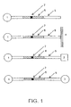
- the presence of a modification at the recognition motif alters protease activity at the protease site resulting in a modulation of the cleavage rate of the protease. Cleavage is sensed by a measurable change in at least one optical property of the optical probe upon cleavage at the protease site, FIG. 1.
- the probe is a fluorescent or luminescent moiety.
- the invention further comprises a fluorescent quencher coupled to the polypeptide that quenches emission from the first probe moiety.
- the first probe moiety and the quencher moiety are coupled to the polypeptide such that the recognition motif and the protease site are located between them (FIG. 1). In this case, cleavage of the polypeptide by a protease results in an alteration he fluorescence emission of the first probe moiety that may be used to determine protein phosphorylation activity.
- the optical probe may further comprise a second probe moiety coupled to the polypeptide that participates in energy transfer with the first probe moiety.
- the first probe moiety and the second probe moiety are coupled to the polypeptide such that recognition motif and the protease site are located between them. In this case, cleavage of the polypeptide by a protease results in an alteration in energy transfer between the first probe moiety and the second probe moiety that may be used to determine protein phosphorylation activity.
- the invention also provides methods for using the optical probes of the invention to determine whether a sample contains a protein phosphorylation activity such as protein phosphorylation.
- the method consisting of; i), contacting the optical probe with a sample, usually containing or suspected of containing a protein phosphorylation activity; ii), contacting the sample and optical probe with a protease, and iii), determining at least one optical property of said optical probe, or product thereof.
- the invention provides methods for using the optical probes of the invention to determine whether a test chemical modulates the activity of a protein phosphorylation activity.
- the invention provides a library of optical probes, each with a unique peptide sequence for use in selecting an optimal sequence specificity of a protein phosphorylation activity.
- a further aspect of the present invention includes various systems for spectroscopic measurements.
- the system typically includes at least one reagent for an assay and a device, said device comprising a container and a platform.
- the container can include the optical sensor compounds of the present invention, and additional reagents necessary for the protein phosphorylation activity. Addition of a sample to the container, followed by the addition of a protease after a given time results in a change in at least one fluorescent property of the optical probes of the present invention that can be used to determine the protein phosphorylation activity of the sample.
- the system can include a microfluidic spectroscopic system comprising at least one fluid containing structure with at least one electro-osmotic or electrophoretic system to control fluid movement within that structure.
- optical probes can be designed to act as optical sensors of protein phosphorylation activities through the creation of engineered molecules.
- protein phosphorylation modification of a polypeptide results in the modulation of the rate and efficiency of cleavage of the modified polypeptide compared to the non-modified peptide.
- the attachment of at least one probe moiety to the peptide couples the cleavage of the optical probe to a change in a fluorescence property of the substrate that may be used to determine the amount of protein phosphorylation activity in a sample, FIG. 1.
- t-Boc tert-butyloxycarbonyl
- Bzl benzyl
- CaMK calmodulin dependent kinase
- CKI casein kinase 1
- PDGF platelet derived growth factor
- Fmoc fluorenylmethyloxycarbonyl
- EGF epidermal growth factor
- ELISA enzyme-linked immuno absorbant assay
- FGF fibroblast growth factor
- HF hydrogen fluoride
- HOBT N-Hydroxybenzotriazole
- PyBop Benzotriazole-1-yl-oxy-tris-pyyrolidino-phosphonium hexafluorophosphate
- TFA trifluoroacteic acid.
- acceptor refers to a quencher that operates via energy transfer. Acceptors may re-emit the transferred energy as fluorescence and are "acceptor fluorescent moieties". Examples of acceptors include coumarins and related fluorophores, xanthenes such as fluoresceins, fluorescent proteins rhodols, and rhodamines, resorufins, cyanines, difluoroboradiazaindacenes, and phthalocyanines. Other chemical classes of acceptors generally do not re-emit the transferred energy. Examples include indigos, benzoquinones, anthraquinones, azo compounds, nitro compounds, indoanilines, and di- and triphenylmethanes.
- Bead refers to a substantially spherical particle such as a sphere or microsphere. Beads may be used within a wide size range. Preferred beads are typically within the range of 0.01 to 100 ⁇ m in diameter. Beads may be composed of any material and may be substantially inert or comprise fluorescent, luminescence, electro-luminescent, chemo-luminescent, magnetic or paramagnetic probes. Such beads are commercially available from a variety of sources including Molecular Probes, Sigma or Polysciences.
- cleavage site refers to the bond cleaved by the protease (e.g. a scissile bond) and typically the surrounding three amino acids of either side of the bond.
- protease e.g. a scissile bond
- P 1 refers to the amino acid positions, 1 amino acid, 2 amino acids and 3 amino acids N-terminal to the scissile bond.
- P' 1 refers to the amino acids positions 1 amino acid, 2 amino acids and 3 amino acids C-terminal to the scissile bond, as shown below; Scissile bond P 3 P 2 P 1 ⁇ P' 1 P' 2 P' 3
- engineered recognition motif refers to a recognition motif that has been modified from the naturally existing sequence by at least one amino acid substitution.
- engineered protease site refers to a protease site that has been modified from the naturally existing sequence by at least one amino acid substitution.
- fluorescent moiety refers to a moiety that can absorb electromagnetic energy and is capable of at least partially remitting some fraction of that energy as electromagnetic radiation over some time period.
- Suitable fluorescent moieties include, but are not limited to, coumarins and related dyes, xanthene dyes such as fluoresceins, rhodols, and rhodamines, resorufins, cyanine dyes, bimanes, acridines, isoindoles, dansyl dyes, aminophthalic hydrazides such as luminol, and isoluminol derivatives, aminophthalimides, aminonaphthalimides, aminobenzofurans, aminoquinolines, dicyanohydroquinones, semiconductor fluorescent nanocrystals, fluorescent proteins and fluorescent europium and terbium complexes and related compounds.
- fluorescent property refers to any one of the following, the molar extinction coefficient at an appropriate excitation wavelength, the fluorescent quantum efficiency, the shape of the excitation or emission spectrum, the excitation wavelength maximum, or the emission magnitude at any wavelength during, or at one or more times after excitation of the fluorescent moiety, the ratio of excitation amplitudes at two different wavelengths, the ratio of emission amplitudes at two different wavelengths, the excited state lifetime, the fluorescent anisotropy or any other measurable property of a fluorescent moiety and the like.
- fluorescent property refers to fluorescence emission, or the fluorescence emission ratio at two or more wavelengths.
- homolog refers to two sequences or parts thereof, that are greater than, or equal to 75% identical when optimally aligned using the ALIGN program.
- Homology or sequence identity refers to the following. Two amino acid sequences are homologous if there is a partial or complete identity between their sequences. For example, 85% homology means that 85% of the amino acids are identical when the two sequences are aligned for maximum matching. Gaps (in either of the two sequences being matched) are allowed in maximizing matching; gap lengths of 5 or less are preferred with 2 or less being more preferred.
- two protein sequences are homologous, as this term is used herein, if they have an alignment score of more than 5 (in standard deviation units) using the program ALIGN with the mutation data matrix and a gap penalty of 6 or greater. See Dayhoff, M.O., in Atlas of Protein Sequence and Structure, 1972, volume 5, National Biomedical Research Foundation, pp. 101-110 , and Supplement 2 to this volume, pp. 1-10 .
- modulates refers to either the enhancement or inhibition (e.g. attenuation of the rate or efficiency) partially or complete of an activity or process.
- modulator refers to a chemical compound (naturally occurring or non-naturally occurring), such as a biological macromolecule (e.g., nucleic acid, protein, non-peptide, or organic molecule), or an extract made from biological materials such as bacteria, plants, fungi, or animal (particularly mammalian, including human) cells or tissues.
- Modulators are evaluated for potential activity as inhibitors or activators (directly or indirectly) of a biological process or processes (e.g., agonist, partial antagonist, partial agonist, inverse agonist, antagonist, antineoplastic agents, cytotoxic agents, inhibitors of neoplastic transformation or cell proliferation, cell proliferation-promoting agents, and the like) by inclusion in screening assays described herein.
- the activity of a modulator may be known, unknown or partially known.
- non-naturally occurring refers to the fact that an object cannot be found in nature.
- a polypeptide or polynucleotide sequence that is present in an organism (including viruses) that can be isolated from a source in nature and which has not been intentionally modified by man in the laboratory is naturally-occurring, while such a sequence that has been intentionally modified by man is non-naturally occurring.
- optical property refers to a physical property of light, including the molar extinction coefficient at an appropriate excitation wavelength, the fluorescent or luminescent quantum efficiency, the shape of the excitation spectrum or emission spectrum, the excitation wavelength maximum or emission wavelength maximum, the ratio of excitation amplitudes at two different wavelengths, the ratio of emission amplitudes at two different wavelengths, the excited state lifetime, the fluorescent anisotropy or any other measurable optical property of a compound, or any product or emission derived from that compound, either spontaneously or in response to electrical or chemical stimulation or reaction.
- polypeptide refers to a polymer in which the monomers are amino acids and are joined together through amide bonds, alternatively referred to as a peptide. Additionally, unnatural amino acids, for example, beta-alanine, phenylglycine and homoarginine are also meant to be included. Commonly encountered amino acids, which are not gene-encoded, may also be used in the present invention. For a general review see Spatola, A.F., in Chemistry and Biochemistry of Amino Acids. Peptides and Proteins, B. Weinstein, eds., Marcel Dekker, New York, p. 267 (1983 ). All of the amino acids used in the present invention may be either the D- or L-isomer. The L-isomers are preferred. Chemically modified or substituted amino acids including phosphorylated, sulfated, methylated, or prenylated residues may also be used to create polypeptides for specific applications.
- post-translational type modification refers to the enzymatic or non-enzymatic modification of an amino acid residue (preferably enzymatic). Such covalent modifications includes phosphorilation.
- a preferred post-translational type modification includes phosphorylation
- post-translational includes non-covalent type modifications including protein-protein interactions, and the binding of allosteric, or other modulators or second messengers such as calcium, or cAMP or inositol phosphates to the recognition motif.
- probe moiety refers to a chemical moiety useful as a marker or indicator, or contrast agent for, absorption spectroscopy, luminescence spectroscopy, fluorescence spectroscopy, or magnetic detection.
- quencher refers to a molecule or part of a compound that is capable of reducing the emission from a probe moiety. Such reduction includes reducing the light after the time when a photon is normally emitted from a fluorescent moiety. Quenching may occur by any of several mechanisms, including fluorescence resonance energy transfer, photoinduced electron transfer, paramagnetic enhancement of intersystem crossing. Dexter exchange coupling, and excitation coupling, such as the formation of dark complexes. Preferred quenchers include those that operate by fluorescence resonance energy transfer.
- recognition motif refers to all or part of a polypeptide sequence recognized by a phosphorylation activity to enable a polypeptide to become modified by that protein phosphorylation activity.
- affinity of a protein, e.g. enzyme, for the recognition motif is about 1 mM (apparent K d ), preferably a greater affinity of about 10 ⁇ M or less, more preferably, I ⁇ M or most preferably has an apparent K d of about 0.1 ⁇ M.
- the term is not meant to be limited to optimal or preferred recognition motifs, but encompasses all sequences that can specifically confer substrate recognition to a peptide.
- the recognition motif is a phosphorylated recognition motif (e.g. includes a phosphate group).
- the recognition motif will, at least partially, comprise a protease site. The protease site may be located at any location within recognition motif.
- test chemical refers to a chemical to be tested by one or more screening method(s) of the invention as a putative modulator.
- a test chemical can be any chemical, such as an inorganic chemical, an organic chemical, a protein, a peptide, a carbohydrate, a lipid, or a combination thereof.
- various predetermined concentrations of test chemicals are used for screening, such as 0.01 micromolar, 1 micromolar and 10 micromolar.
- Test chemical controls can include the measurement of a signal in the absence of the test compound or comparison to a compound known to modulate the target.
- the present invention recognizes for the first time that optical probes can be designed to measure a range of protein phosphorylation activities.
- the advantages of the present invention include compositions that can be used in methods, particularly methods for high throughput and miniaturized screening systems for drug discovery and profiling.
- Theoretical probes provide for assays, that typically exhibit a large dynamic range, increased sensitivity and allow ratiometric readouts for the detection of protein phosphorylation activities.
- the invention includes several general and useful aspects, including:
- peptide sequences for measuring a protein phosphorylation activity encompass a protein phosphorylation recognition motif that contains a residue that, when modified, modulates the rate of cleavage of the substrate by a protease as compared to the unmodified form.
- peptides typically contain a single scissile bond (bond that is cleaved within the substrate) for a specific protease and exhibit reasonable solubility (e.g. 0.1 mg/ml or greater) in aqueous solution.
- solubility e.g. 0.1 mg/ml or greater
- the peptide separating the fluorescent or luminescent moieties will typically be in the range of 5 to 50 amino acids in length, preferably 10 to 25 amino acids in length, or more preferably 10 to 15 amino acids in length.
- the peptide may be significantly larger, up to and including entire protein domains, for example 50 to 100 amino acids in length. Smaller peptides, in the range of 5 to 50 amino acids may also be used.
- the protease site may be located at any position either completely or partially within the recognition motif.
- the recognition motif and protease site may be located at any position within the peptide with respect to the optical probe moiety.
- protein kinases act on proteins via the addition of phosphate groups (phosphorylation) primarily on the amino acids, tyrosine, serine or threonine through a free hydroxyl group.
- phosphate groups phosphorylation
- the protein kinases that enzymatically catalyze these reactions may be classified into a number of distinct families based on shared structural and functional properties.
- kinases within a family have a similar overall topology, have similar modes of regulation and have similar substrate specificity's (see, Table 1 , and may be used with the invention, as well as those recognition motifs developed in the future).
- members of the AGC protein kinase A, G or C families of kinases typically prefer phosphorylation recognition motifs with basic amino acids (R or K), those in the CMGC group typically prefer proline containing motifs, etc.
- Blank cells in the "Substrate Preference" column indicate that the complete information for every member of a particular class was not available, or indicates that the family was too small to define a clear substrate preference, or indicates that no clearly defined substrate preference yet exists.
- Eukaryotic protein phosphatases are structurally and functionally diverse enzymes that are represented by three distinct gene families. Two of these, dephosphorylate phosphoserine and phosphothreonine residues, whereas the protein tyrosine phosphatases (PTPs) dephosphorylate phosphotyrosine amino acids. A subfamily of the PTPs, the dual specificity phosphatases, dephosphorylates all three phosphoamino acids. Within each family, the catalytic domains are highly conserved, with functional diversity endowed by regulatory domains and subunits.
- the protein serine or threonine phosphatases type 1 and 2A account for as much as 95% of the phosphatase activity in cell extracts ( Brautigan and Shriner, Methods. Enzymol. 159: 339-346 (1988) . These enzymes have broad substrate specificities and may be regulated in vivo through targeting of the enzymes to discrete sub-cellular localizations.
- the total number of protein tyrosine phosphatases encoded in the mammalian genome has been estimated at between 500 and approximately 2000. These estimates are imprecise due to the large number of sequence database entries that are different splice forms or duplicates of the same PTP sequence.
- Optical probes for detecting tyrosine kinase activity are designed by incorporating the desired phosphorylation motif into a peptide, and by ensuring that the only aromatic residue (Tyr, Trp or Phe) in the substrate is the tyrosine that is phosphorylated. It may also be preferable in certain cases to eliminate or reduce the number of negatively charged amino acids in the P' 1 , P' 2 or P' 3 positions. If this is the case, then phosphorylation of the tyrosine residue by the tyrosine directed protein kinase activity modulates the rate of optical probe hydrolysis by chymotrypsin compared to the non-phosphorylated optical probe.
- the present inventors have recognized that elimination of negatively charged residues in the optical probe C-terminal to the scissile bond improves the efficiency of cleavage of non-phosphorylated optical probe, on occasion significantly increasing the utility of the optical probes for measuring kinase or phosphatase activities.
- This approach can be used to create specific optical probes for virtually all known tyrosine kinase activities by routine optimization of the reaction conditions as described herein.
- Specific illustrative examples for different tyrosine kinase classes are shown in Table 2, below for use with chymotrypsin.
- Insulin receptor XEEE Y MMMF (1) EEE Y MMM M SEQ. ID. NO: 6 PDGF receptor E EEE Y V FIX (1) E EEE Y V V IX SEQ.ID. NO:7 EGF receptor E EEE Y F ELV (1) E EEE Y V L LV SEQ. ID. NO: 8 FGF receptor AE EE Y FFL F (1) AE EE Y F V L M SEQ. ID. NO: 9
- peptides are designed to incorporate a single aromatic amino acid (Tyr, Trp or Phe) that is typically located within about three amino acids of a serine or threonine residue, which is phosphorylated by an appropriate serine or threonine specific kinase. It is also preferable in certain cases (depending on the protease selected) to eliminate or reduce the number of negatively charged amino acids (e.g.
- proteases for use in the present invention typically have the following characteristics: They are commonly available at high purity, are substantially stable, and recognize a substrate recognition motif that comprises at least one position in which the presence, or absence, of a post-translationally modified residue modulates the activity of the protease towards that substrate.
- Preferred substrates possess well defined protease sites, and exhibit a significant modulation e.g. at least 2 fold, or more preferably at least 5 fold modulation of activity towards a phosphorylated residue compared to a non-modified residue.
- Proteases that may be used to measure peptide phosphorylation include those that recognize a substrate recognition motif that comprises at least one position in which the presence or absence of a phosphorylated residue modulates the activity of the protease towards that substrate. For example like those in ( Table 6 ), or other such proteases developed now or in the future.
- a contemplated version of the method is to use inducible controlling nucleotide sequences to produce a sudden increase in the expression of the protease within a cell, for the development of a cell based assay.
- An appropriate optical property would typically be monitored at one or more time intervals after the onset of increased expression of the protease.
- the choice of the probe moiety is governed by a number of factors including, the type of measurements being made, the availability of specific instrumentation and the ease of coupling of the probe moiety to the peptide. Additionally, other factors that are specific to a particular application are also relevant and include, the effect of labeling on the solubility of the peptide, kinetics of the optical probe with respect to the protein phosphorylation activity or protease, and the required detection sensitivity of the assay. Fortunately numerous probe moieties are commercially available or can be readily made so that availability of probe moieties to meet a desired situation is not limiting.
- preferred fluorophores typically exhibit good quantum yields, lifetimes, and extinction coefficients, are resistant to collisional quenching and bleaching, and should preferably be easily conjugated to the ligand.
- fluorophores that show absorbance and emission in the red and near infrared range, which are useful in whole animal studies, because of reduced scattering background fluorescence, and greater transmission through tissues.
- moieties include cyanines, oxazines, thiazines, porphyrins, phthalocyanines, fluorescent infrared-emitting polynuclear aromatic hydrocarbons such as violanthrones, fluorescent proteins, near IR squaraine dyes.
- fluoroscein isothicyanate especially fluorescein-5-isothiocyanate
- dichlorotriazinylaminofluorescein tetramethylrhodamine-5 (and -6)-isothiocyanate
- 1,3-bis-(2-dialkylamino-5-thienyl)-substituted squarines and the succinimidyl esters of: 5 (and 6) carboxyfluoroscein; 5 (and 6)-carboxytetramethylrhodamine; and 7-amino-4-methylcoumarin-3-acetic acid.
- Semiconductor fluorescent nanocrystals are available with a range of emission spectra, are highly fluorescent and are also preferred, (see Bruchez et al., Science 281: 2013-2016 ).
- Preferred luminescent probes include chemi-luminescent, electro-luminescent and bioluminescent compounds.
- Preferred bioluminescent compounds include bioluminescent proteins such as firefly, bacterial or click beetle luciferases, aequorins and other photoproteins, for example as described in U.S. Patents 5,221,623, issued June 22, 1989 to Thompson et al. , and 5,683,888 issued November 4, 1997 to Campbell , 5,674,713 issued September 7 1997 to DeLuca et al. , 5,650,289 issued July 22, 1997 to Wood and U.S. patent 5,843,746 issued December 1, 1998 to Tatsumi et al.
- Preferred electro-luminescent probes include ruthenium complexes, as for example described in U.S. Patent 5,597,910 issued to Jan 28, 1997 to Gudibande .
- Preferred chemi-luminescent substrates include those based on 1,2-dioxetanes, as for example described in U.S. Patents 4,372,745 issued February 8, 1983 to Mandle et al. , 5,656,207 issued August 12, 1997 to Woodhead et al. , and 5,800,999 issued September 1, 1998 issued to Bronstein et al .
- Preferred probes for use as NMR contrast agents include chelates of paramagnetic, ferromagnetic or diamagnetic metal ions complexed to lipophilic complexes as described in U.S. patents 5,628,982, issued May 13, 1997 to Lauffer et al. and U.S. patent 5,242,681, issued September 7, 1993 to Elgavish et al. , and fluorine-18-and 19 containing compounds J. Nucl. Med. 39 1884-91 (1998) .
- any phenolic hydroxyls or free amines in the dye structures are acylated with C 1 -C 4 acyl groups (e.g. formyl, acetyl, n-butryl) or converted to various esters and carbonates, as described in Bundgaard, H., Design of Prodrugs, Elsevier Science Publishers (1985), Chapter 1, page 3 et seq., Further modification of the fluorescent moieties may also be accomplished, as required as described in U.S. patent No. 5,741,657 issued 4/21/98 to Tsien et al.
- the probe may be attached to the polypeptide by a linker that provides a spacer between the probe and the peptide thereby preventing sterric interference of the probe on the interaction between the recognition motif and the protein phosphorylation activity.
- Preferred spacers are substantially stable under cellular conditions and easily coupled to the peptide and probe.
- Preferred examples include flexible aliphatic linkers such as y-amino n-butyric acid (GABA), diaminopentane, and aminohexanoyl as well as rigid aromatic linkers.
- GABA y-amino n-butyric acid
- diaminopentane diaminopentane
- aminohexanoyl as well as rigid aromatic linkers.
- Such linkers are known in the art and described for example in the Handbook of Fluorescent Probes and Research Chemicals, by Richard Haugland, published by Molecular Probes.
- the peptide may be designed to encompass a specific binding site for a fluorescent moiety as described in the pending U.S. Patent applications, identified by serial number 08/955,050, filed October 21, 1997 , entitled Methods of using synthetic molecules and target sequences; 08/955,859, filed October 21, 1997 , entitled Synthetic molecules that specifically react with target sequences, and 08/955,206, filed October 21, 1997 , entitled Target sequences for synthetic molecules.
- Labeling may then be achieved by incubation of the peptide with the membrane permeate fluorescent binding partner, which has the advantages of enabling the expression of peptides within intact living cells, and the subsequent labeling of these peptides in situ to create optical probes within intact living cells.
- preferred fluorescent moieties include endogenously fluorescent proteins, functional engineered fluorescent proteins, and homologs thereof. Because the entire fluorophore and peptide can be expressed within intact living cells without the addition of other co-factors or fluorophores, such optical probes provide the ability to monitor protein phosphorylation activities within defined cell populations, tissues or an entire transgenic organism. For example by the use of inducible controlling nucleotide sequences to produce a sudden increase in the expression of the optical probe and suitable protease. Endogenously fluorescent proteins have been isolated and cloned from a number of marine species including the sea pansies Renilla reniformis, R. kollikeri and R.
- fluorescent proteins have also been observed in other organisms, although in most cases these require the addition of some exogenous factor to enable fluorescence development.
- yellow fluorescent protein from Vibrio fischeri strain Y-1 has been described by T.O. Baldwin et al., Biochemistry (1990) 29:5509-15 .
- This protein requires flavins as fluorescent co-factors.
- the cloning of Peridinin-chlorophyll a binding protein from the dinoflagellate Symbiodinium sp. was described by B.J. Morris et al., Plant Molecular Biology, (1994) 24:673:77 .
- One useful aspect of this protein is that it fluoresces in red.
- the optical probe comprises a first fluorescent protein, a peptide containing a protein phosphorylation recognition motif and a protease site, and a second fluorescent protein fused together as a single polypeptide chain.
- Nucleic acids encoding fluorescent proteins can be obtained by methods known in the art. For example, a nucleic acid encoding the protein can be isolated by polymerase chain reaction of cDNA from a suitable organism using primers based on the DNA sequence of the fluorescent protein. PCR methods are described in, for example, U.S. Pat. No. 4,683,195 ; Mullis et al. (1987) Cold Spring Harbor Symp. Quant. Biol. 51:263 ; and Erlich, ed., PCR Technology, (Stockton Press, NY, 1989 ).
- Suitable clones expressing the optical probes of the invention may then be identified, isolated and characterized by fluorescence activated cell sorting (FACS) typically enabling the analysis of a few thousand cells per second.
- FACS fluorescence activated cell sorting
- Nucleic acids used to transfect cells with sequences coding for expression of the polypeptide of interest generally will be in the form of an expression vector including expression control sequences operatively linked to a nucleotide sequence coding for expression of the polypeptide comprising the optical probe.
- Methods that are preferred with the present invention include, fluorescence spectroscopy, luminescence spectroscopy, absorption spectroscopy and magnetic detection
- Fluorescent methods that are preferred with the present invention include, continuous or time resolved fluorescence spectroscopy, fluorescence correlation spectroscopy, fluorescence polarization spectroscopy , and resonance energy based fluorescence spectroscopy.
- Methods of performing such assays on fluorescent materials are well known in the art and are described in, e.g., Lakowicz, J.R., Topics in Fluorescence Spectroscopy, volumes 1 to 3, New York: Plenum Press (1991 ); Herman, B., Resonance energy transfer microscopy, in: Fluorescence Microscopy of Living Cells in Culture, Part B, Methods in Cell Biology, vol. 30, ed. Taylor, D.L.
- Methods for determining whether a sample has an activity typically involve contacting the sample with an optical probe, incubating the mixture under conditions to enable protein phosphorylation of the substrate, and then adding a protease. Finally the degree of protein phosphorylation activity in the sample is detected by determining at least one optical property of the optical probe or product thereof.
- the optical probe and the protease may be added to a sample at the same time.
- the method would typically involve stimulation of the cells, and then either lyzing the cells in the presence of the substrate, or in the case where the substrate is expressed within the cells, lyzing the cells in the presence of a protease to measure substrate modification.
- the method used to determine the degree of protein phosphorylation activity is dependent on the assay format used.
- the method may be based on the difference in fluorescence anisotropy of the optical probe before and after cleavage with a protease.
- the optical probe typically comprises a polypeptide moiety, which contains a recognition motif for a protein phosphorylation activity and a protease site, which is coupled to a fluorescent moiety (FIG. 1), Modification of the polypeptide, by the protein phosphorylation activity, results in a modulation of the rate at which a protease cleaves the polypeptide which is sensed by a measurable change in fluorescence polarization of the optical probe upon cleavage.
- FOG. 1 fluorescent moiety
- Polarization measurements are based on the relative rotational movement of the fluorophore compared to the excited state life-time of that fluorophore.
- the relationship between polarization ( p ) and the degree of rotational movement can be readily derived (see Weber, Polarization of the fluorescence of solutions, in Fluorescence and Phosphorescence Analysis, Don Hercules (ed.), Interscience Publishers New York. Chapter 8, pages 217-240 (1966) ).
- Rotational movement can be related to the rotational diffusion constant of the molecule, and hence to the molecular volume. In practice there is a close correlation between the molecular size and relative polarization of emitted light from a fluorophore.
- Polarization based measurements are relatively easy to set up, and can be obtained over a wide concentration, temperature, and ionic strength range.
- fluorescence anisotropy measurements may be enhanced by attaching one end of the peptide to a solid matrix, or a bead. In either case, cleavage of the optical probe results in a larger drop in fluorescence polarization because of the increased rotational flexibility of the optical probe once separated from the solid matrix or bead.
- the present invention takes advantage of resonance energy transfer either between two fluorescent moieties (FRET), or a bioluminescent moiety and fluorescent moiety (bioluminescent resonance energy transfer, BRET), or a fluorescent moiety and a quencher (resonance energy transfer, RET) to provide a fluorescent readout.
- FRET fluorescent moieties
- BRET bioluminescent resonance energy transfer
- RET quencher
- the optical probe typically comprises a first fluorescent moiety and a second fluorescent moiety coupled to the polypeptide such that the recognition motif and the protease site are located between them (FIG. 1).
- cleavage of the polypeptide by a protease results in an alteration in energy transfer between the first fluorescent moiety and the second fluorescent moiety that may be used to determine protein phosphorylation activity.
- the fluorescent moieties are typically chosen such that the excitation spectrum of one of the moieties (the acceptor fluorescent moiety) overlaps with the emission spectrum of the donor fluorescent moiety.
- the donor fluorescent moiety is excited by light of appropriate intensity within the donor fluorescent moiety's excitation spectrum and under conditions in which direct excitation of the acceptor fluorophore is minimized.
- the donor fluorescent moiety then transfers the absorbed energy by non radiative means to the acceptor, which subsequently re-emits some of the absorbed energy as fluorescence emission, at a characteristic wavelength.
- FRET can be manifested as a reduction in the intensity of the fluorescent signal from the donor, reduction in the lifetime of its excited state, and an increase in emission of fluorescence from the acceptor fluorescent moiety.
- the peptide substrate that connects the donor fluorescent moiety and acceptor fluorescent moiety is cleaved, the donor fluorescent moiety and the acceptor fluorescent moiety physically separate, and FRET is diminished or eliminated. Under these circumstances, fluorescence emission from the donor increases and fluorescence emission from the acceptor decreases.
- E the efficiency of FRET
- F and F 0 are the fluorescence intensities of the donor in the presence and absence of the acceptor, respectively
- R is the distance between the donor and the acceptor.
- K 2 is an orientation factor having an average value close to 0.67 for freely mobile donors and acceptors
- Q is the quantum yield of the unquenched fluorescent donor
- n is the refractive index of the intervening medium
- J is the overlap integral, which expresses in quantitative terms the degree of spectral overlap
- J °° 0 ⁇ F 4 ⁇ D / °° 0 ⁇ F d
- F is the donor fluorescence at wavelength measured in cm.
- the characteristic distance R 0 at which FRET is 50% efficient depends on the quantum yield of the donor, the extinction coefficient of the acceptor, the overlap between the donor's emission spectrum and the acceptor's excitation spectrum and the orientation factor between the two fluorophores.
- changes in the degree of FRET are determined as a function of the change in the ratio of the amount of fluorescence from the donor and acceptor moieties, a process referred to as "ratioing.”
- ratioing a process referred to as "ratioing.”
- a contemplated variation of the above assay is to either introduce, or express, the optical probe into living eukaryotic or prokaryotic cells to enable the measurement of intracellular post-translational activities.
- the method would involve an optical probe comprising a first fluorescent protein, a peptide containing a post-translational modification recognition motif and a protease site, and a second fluorescent protein fused together as a single polypeptide chain.
- the first fluorescent protein and the second fluorescent protein would be selected to enable FRET to occur as described above.
- a preferred pair of functional engineered fluorescent proteins for example being, Topaz (S65G, S72A, K79R, T203Y) and W1B (F64L, S65T, Y66W, N146I, M153T,V163A) ( Table 7 ).
- the method would involve an optical probe comprising a peptide containing one or more binding sites for a fluorescent moiety, a post-translational modification recognition motif and a protease site.
- the binding site could comprises a sequence that recognizes a fluorescent moiety as described in the pending U.S. Patent applications, identified by serial number 08/955,050, filed October 21, 1997 , entitled Methods of using synthetic molecules and target sequences; 08/955,859, filed October 21, 1997 , entitled Synthetic molecules that specifically react with target sequences, and 08/955,206, filed October 21, 1997 , entitled Target sequences for synthetic molecules.
- expression of the peptide comprising the post-translational recognition motif, protease site and binding site could be accomplished using genetic means as described above.
- the addition of a membrane permeate fluorescent moiety capable of binding to the binding site would enable the creation, in situ of an optical probe.
- a contemplated version of the method is to use inducible controlling nucleotide sequences to produce a sudden increase in the expression of either the optical probe or the post-translational activity being assayed, e.g., by inducing expression of the construct.
- a suitable protease could be expressed within the cell, or induced, or introduced using a membrane translocating sequence U.S. patent No. 5,807,746, issued Sept 15 1998 to Lin et al. The efficiency of FRET is typically monitored at one or more time intervals after the onset of increased expression of the protease.
- the method would involve the introduction of an optical probe of the present invention into the cell through the use of a membrane translocating sequence, as described herein.
- the optical probe typically comprises a luminescent moiety and a fluorescent moiety coupled to the polypeptide such that the recognition motif and the protease site are located between them (FIG. 1).
- cleavage of the polypeptide by a protease results in an alteration in energy transfer between the luminescent moiety and the fluorescent moiety that may be used to determine post-translational type activities.
- the luminescent and fluorescent moieties are typically chosen such that the emission spectrum of the luminescent moiety overlaps with the excitation spectrum of the fluorescent moiety.
- the luminescent moiety provides light through a chemi-luminescent, electro-luminescent or bioluminescent reaction, there is no requirement for direct light excitation to create the excited state in the luminescent moiety. Instead appropriate substrates, or voltage must be provided to the luminescent moiety, to create an excited state within the luminescent moiety.
- substrates are, generically referred to as luciferins (for example see U.S. patent 5,650,289 issued July 22, 1997 to Wood ). If BRET occurs, the energy from the excited state of the luminescent moiety is transferred to the fluorescent moiety by non radiative means, rather than being emitted as light from the luminescent moiety.
- the emission ratio of the two can provide a ratiometric readout as described for FRET based applications.
- BRET can be manifested as a reduction in the intensity of the fluorescent signal from the luminescent moiety, reduction in the lifetime of its excited state, and an increase in emission of fluorescence from the fluorescent moiety.
- the peptide substrate that connects the luminescent moiety and fluorescent moiety is cleaved, the luminescent moiety and the fluorescent moiety physically separate, and BRET is diminished or eliminated. Under these circumstances light emission from the luminescent moiety increases and fluorescence emission from the fluorescent moiety decreases.
- the efficiency of BRET is dependent on the same separation and orientation factors as described above for FRET.
- the optical probe typically comprises a first fluorescent moiety and a quencher moiety coupled to the polypeptide such that the recognition motif and the protease site are located between them (FIG. 1).
- cleavage of the polypeptide by a protease results in an alteration in energy transfer between the first fluorescent moiety and the quencher moiety that may be used to determine post-translational activity.
- the fluorescent moiety and the quencher moiety are typically chosen such that the absorption spectrum of one of the quencher (the acceptor moiety) overlaps with the emission spectrum of the donor fluorescent moiety.
- the donor fluorescent moiety is excited by light of appropriate intensity within the donor fluorescent moiety's excitation spectrum.
- the donor fluorescent moiety then transfers the absorbed energy by non radiative means to the quencher, which in this case does not re-emit any of the absorbed energy as light.
- RET can be manifested as a reduction in the intensity of the fluorescent signal from the donor or a reduction in the lifetime of its excited state.
- the protein phosphorylation modification assays of the present invention can be used in drug screening assays to identify compounds that alter a protein phosphorylation activity.
- the assay is performed on a sample in vitro (e.g. in a sample isolated from a cell, or cell lysate or purified enzyme) containing the activity.
- a sample containing a known amount of activity is mixed with an optical probe of the invention and with a test chemical.
- the amount of the activity in the sample is then determined after addition of a protease as described herein, for example, by determining at least one optical property of the probe.
- the optical property of the sample in the presence of the test chemical is compared with the optical property of the sample in the absence of the test compound. A difference indicates that the test compound alters the activity.
- the ability of a test chemical to alter a protein phosphorylation activity, in a cell based assay may be determined.
- cells transfected with an expression vector encoding an optical probe of the invention, as described above are exposed to different amounts of the test chemical, and the effect on FRET or fluorescence polarization in each cell can be determined after induction or introduction of a suitable protease.
- the difference in FRET or polarization of treated cells is compared to that of untreated controls.
- libraries of optical probes can be created by producing peptides containing a diverse population of amino acid sequences. Such libraries are useful for the identification and characterization of novel protein phosphorylation activities that have unknown or poorly defined substrate specificities.
- a "library” refers to a collection containing at least 5 different members, preferably at least 100 different members and more preferably at least 200 different members.
- the amino acid sequences for the peptide will typically be in the range or 10 to 20 amino acids in length and may be completely random or biased towards a particular sequence based on a particular structural motif, for example based on a known substrate for a particular protein phosphorylation activity.
- the library will created genetically and the individual members expressed in bacterial or a mammalian cells. Suitable clones expressing the optical probes of the invention may then be identified, isolated and characterized by fluorescence activated cell sorting (FACS) typically enabling the analysis of a few thousand cells per second.
- FACS fluorescence activated cell sorting
- the peptides may be chemically synthesized and individual members attached to a solid matrix and arranged within a two dimensional array.
- the library will contain variable peptides in which only a few, e.g., one to ten, amino acid positions are varied, but in which the probability of substitution is very high.
- each member of the optical probe library wilt contain a single defined protease site, and a variable protein phosphorylation recognition motif, such that randomized sequences comply with the design considerations for the particular protein phosphorylation activity (described above).
- the array includes systematically substituted amino acids attached to a substrate, as described in U.S. Patent No. 5,770,456, issued June 23, 1998 to Holmes .
- Screening of the library to identify optimal substrates may be achieved by incubating the array with a sample containing the protein phosphorylation activity, adding an appropriate protease, and then detecting at least one optical property from each member of the library Those library members that are more efficiently modified by the protein phosphorylation activity may then be identified by the degree to which the optical property of each library member is altered after exposure to the protein phosphorylation activity.
- libraries of knwon recognition motifs may be created in order to create an activity profile of protein phosphorylation activities in a sample.
- screening of the library is used to characterize the relative protein phosphorylation activities within by incubating the array with a sample containing the protein phosphorylation activities, adding an appropriate protease, and then detecting at least one optical property from each member of the library.
- Those library members that are more efficiently modified after exposure to the sample may then be identified by the degree to which the optical property of each library member is altered after exposure to the sample to determine the protein phosphorylation activities present within the sample.
- the system comprises: a reagent for an assay, and a device comprising at least one plate or container, preferably a multi-well platform, and a second platform to hold said plate or container for detecting a signal from a sample.
- the system can further comprise a detector, such as a detector appropriate for detecting a signal from a sample or a plate on in a container as such detectors are known in the art or are later developed.
- the system can comprise multiple plates or containers or multi-well platforms.
- a reagent for an assay includes any reagent useful to perform biochemical or biological in vitro or in vivo testing procedures, such as, for example, buffers, co-factors, proteins such as enzymes or proteases, carbohydrates, lipids, nucleic acids, active fragments thereof, organic solvents such as DMSO, chemicals, analytes, therapeutics, compositions, cells, antibodies, ligands, and the like.
- an active fragment is a portion of a reagent that has substantially the activity of the parent reagent.
- the choice of optical probe depends on the type of assay to be performed. For example, FRET based assays would typically comprise an optical probe with two fluorophores. Fluorescent polarization based assays would typically be completed with optical probes comprising one fluorescent moiety (FIG. 1).
- optical probes of the present invention are suited for use with systems and methods that utilize automated and integratable workstations for identifying modulators, and chemicals having useful activity.
- Such systems are described generally in the art (see, U.S. Patent Nos: 4,000,976 to Kramer et al. (issued January 4, 1977 ), 5,104,621 to Pfost et al. (issued April 14, 1992 ), 5,125,748 to Bjornson et al. (issued June 30, 1992 ), 5,139,744 to Kowalski (issued August 18, 1992 ), 5,206,568 Bjornson et al. (issued April 27, 1993 ), 5,350,564 to Mazza et al.
- such a system includes: A) a storage and retrieval module comprising storage locations for storing a plurality of chemicals in solution in addressable chemical wells, a chemical well retriever and having programmable selection and retrieval of the addressable chemical wells and having a storage capacity for at least 100,000 addressable wells, B) a sample distribution module comprising a liquid handler to aspirate or dispense solutions from selected addressable chemical wells, the chemical distribution module having programmable selection of, and aspiration from, the selected addressable chemical wells and programmable dispensation into selected addressable sample wells (including dispensation into arrays of addressable wells with different densities of addressable wells per centimeter squared) or at locations, preferably pre-selected, on a plate, C) a sample transporter to transport the selected addressable chemical wells to the sample distribution module and optionally having programmable control of transport of the selected addressable chemical wells or locations on a plate (including adaptive routing and parallel processing), D) a reaction module comprising either
- the storage and retrieval module, the sample distribution module, and the reaction module are integrated and programmably controlled by the data processing and integration module.
- the storage and retrieval module, the sample distribution module, the sample transporter, the reaction module and the data processing and integration module are operably linked to facilitate rapid processing of the addressable sample wells or locations on a plate.
- devices of the invention can process at least 100,000 addressable wells or locations on a plate in 24 hours. This type of system is described in the PCT application WO/98/52047 by Stylli et al. , entitled “Systems and method for rapidly identifying useful chemicals in liquid samples. "
- each separate module is integrated and programmably controlled to facilitate the rapid processing of liquid samples, as well as being operably linked to facilitate the rapid processing of liquid samples.
- the invention provides for a reaction module that is a fluorescence detector to monitor fluorescence.
- the fluorescence detector is integrated to other workstations with the data processing and integration module and operably linked with the sample transporter.
- the fluorescence detector is of the type described herein and can be used for epifluorescence.
- Other fluorescence detectors that are compatible with the data processing and integration module and the sample transporter, if operable linkage to the sample transporter is desired, can be used as known in the art or developed in the future.
- detectors are available for integration into the system. Such detectors are described in U.S. Patent 5,589,351 (Harootunian ), U.S. Patent 5,355,215 (Schroeder ), U.S. Patent Application (serial number pending), entitled “Detector and Screening Device for Ion Channels” filed 7/17/98, and PCT patent application WO 93/13423 (Akong ).
- an entire plate may be "read” using an imager, such as a Molecular Dynamics Fluor-Imager 595 (Sunnyvale, CA).
- Multi-well platforms having greater than 864 wells, including 3,456 wells, can also be used in the present invention (see, for example, the PCT Application PCT/US98/11061, filed 6/2/98 . These higher density well plates require miniaturized assay volumes that necessitate the use of highly sensitivity assays that do not require washing. The present invention provides such assays as described herein.
- the system comprises a microvolume liquid handling system that uses electrokinetic forces to control the movement of fluids through channels of the system, for example as described in U.S. patent No., 5,800,690 issued September 1, 1998 to Chow et al. , European patent application EP 0 810 438 A2 filed May 5 1997, by Pelc et al. and PCT application WO 98/00231 filed 24 June 1997 by Parce et al.
- These systems use "chip" based analysis systems to provide massively parallel miniaturized analysis. Such systems are preferred systems of spectroscopic measurements in some instances that require miniaturized analysis.
- the system may comprise a two dimensional array of optical probes dispersed on a substratum, for example as described in U.S. patents nos., 4,216,245 issued August 5, 1980 to Johnson , 5,721,435 issued February 24, 1998 to Troll , and 5,601,980 issued February 11, 1997 issued to Gordon et al .
- Such a system provides the ability to rapidly profile large numbers of optical probes and or large numbers of samples in a simple, miniaturized high throughput format.
- the optical probes of the present invention can also be used for testing a therapeutic for useful therapeutic activity or toxicological activity.
- a therapeutic is identified by contacting a test chemical suspected of having a modulating activity of a biological process or target with a biological process or target on a plate or in a container, such as at least one well of a multi-well platform, that also comprises an optical probe.
- the test chemical can be part of a library of test chemicals that is screened for activity, such as biological activity.
- the library can have individual members that are tested individually or in combination, or the library can be a combination of individual members.
- Such libraries can have at least two members, preferably greater than about 100 members or greater than about 1,000 members, more preferably greater than about 10,000 members, and most preferably greater than about 100,000 or 1,000,000 members.
- a protease is added and at least one optical property (such as FRET or polarization) of the sample is determined and compared to a non-treated control. If the sample having the test chemical exhibits increased or decreased FRET or polarization relative to that of the control or background levels, then a candidate modulator has been identified
- the candidate modulator can be further characterized and monitored for structure, potency, toxicology, and pharmacology using well-known methods.
- the structure of a candidate modulator identified by the invention can be determined or confirmed by methods known in the art, such as mass spectroscopy. For putative modulators stored for extended periods of time, the structure, activity, and potency of the putative modulator can be confirmed.
- the candidate modulator will have putative pharmacological activity. For example, if the candidate modulator is found to inhibit a protein tyrosine phosphatase involved, for example in T-cell proliferation in vitro, then the candidate modulator would have presumptive pharmacological properties as an immunosuppressant or anti-inflammatory (see, Suthanthiran et al., Am. J. Kidney Disease, 28:159-172 (1996) ).
- Such nexuses are known in the art for several disease states, and more are expected to be discovered over time. Based on such nexuses, appropriate confirmatory in vitro and in vivo models of pharmacological activity, as well as toxicology, can be selected.
- the optical probes, and methods of use described herein, enable rapid pharmacological profiling to assess selectivity and specificity, and toxicity. This data can subsequently be used to develop new candidates with improved characteristics.
- candidate modulators can be evaluated for bioavailability and toxicological effects using known methods (see, Lu, Basic Toxicology, Fundamentals, Target Organs, and Risk Assessment, Hemisphere Publishing Corp., Washington (1985 ); U.S. Patent Nos: 5,196,313 to Culbreth (issued March 23, 1993 ) and U.S. Patent No. 5,567,952 to Benet (issued October 22, 1996 ).
- toxicology of a candidate modulator can be established by determining in vitro toxicity towards a cell line, such as a mammalian i.e. human, cell line.
- Candidate modulators can be treated with, for example, tissue extracts, such as preparations of liver, such as microsomal preparations, to determine increased or decreased toxicological properties of the chemical after being metabolized by a whole organism.
- tissue extracts such as preparations of liver, such as microsomal preparations
- the results of these types of studies are often predictive of toxicological properties of chemicals in animals, such as mammals, including humans.
- the toxicological activity can be measured using reporter genes that are activated during toxicological activity or by cell lysis (see WO 98/13353, published 4/2/98 ).
- Preferred reporter genes produce a fluorescent or luminescent translational product (such as, for example, a Green Fluorescent Protein (see, for example, U.S. Patent No. 5,625,048 to Tsien et al., issued 4/29/98 ; U.S. Patent No.
- Cell lysis can be detected in the present invention as a reduction in a fluorescence signal from at least one photon-producing agent within a cell in the presence of at least one photon reducing agent.
- Such toxicological determinations can be made using prokaryotic or eukaryotic cells, optionally using toxicological profiling, such as described in PCT/US94/00583, filed 1/21/94 ( WO 94/17208 ), German Patent No 69406772.5-08, issued 11/25/97 ; EPC 0680517, issued 11/12/94 ; U.S. Patent No. 5,589,337, issued 12/31/96 ; EPO 651825, issued 1/14/98 ; and U.S. Patent No. 5,585,232, issued 12/17/96 ).
- the bioavailability and toxicological properties of a candidate modulator in an animal model can be determined using established methods (see, Lu, supra (1985); and Creasey, Drug Disposition in Humans, The Basis of Clinical Pharmacology, Oxford University Press, Oxford (1979 ), Osweiler, Toxicology, Williams and Wilkins, Baltimore, MD (1995 ), Yang, Toxicology of Chemical Mixtures; Case Studies, Mechanisms, and Novel Approaches, Academic Press, Inc., San Diego, CA (1994 ), Burrell et al., Toxicology of the Immune System; A Human Approach, Van Nostrand Reinhld, Co.
- Efficacy of a candidate modulator can be established using several art-recognized methods, such as in vitro methods, animal models, or human clinical trials (see, Creasey, supra (1979)). Recognized in vitro models exist for several diseases or conditions. For example, the ability of a chemical to extend the life-span of HIV-infected cells in vitro is recognized as an acceptable model to identify chemicals expected to be efficacious to treat HIV infection or AIDS (see, Daluge et al., Antimicro. Agents Chemother, 41:1082-1093 (1995) ).
- CsA cyclosporin A
- the rabbit knee is an accepted model for testing chemicals for efficacy in treating arthritis (see, Shaw and Lacy, J. Bone Joint Surg. (Br) 55:197-205 (1973 )).
- Hydrocortisone which is approved for use in humans to treat arthritis, is efficacious in this model which confirms the validity of this model (see, McDonough, Phys. Ther. 62:835-839 (1982) ).
- the skilled artisan can be guided by the state of the art to choose an appropriate model, dose, and route of administration, regime, and endpoint and as such would not be unduly burdened.
- the in vitro and in vivo methods described above also establish the selectivity of a candidate modulator. It is recognized that chemicals can modulate a wide variety of biological processes or be selective. Panels of enzymes or panels of cells based on the present invention, or a combination of both, can be used to determine the specificity of the candidate modulator. Selectivity is evident, for example, in the field of chemotherapy, where the selectivity of a chemical to be toxic towards cancerous cells, but not towards non-cancerous cells, is obviously desirable. Selective modulators are preferable because they have fewer side effects in the clinical setting.
- the selectivity of a candidate modulator can be established in vitro by testing the toxicity and effect of a candidate modulator on a plurality of cell lines that exhibit a variety of cellular pathways and sensitivities.
- the data obtained from these in vitro toxicity studies can be extended into in vivo animal model studies, including human clinical trials, to determine toxicity, efficacy, and selectivity of the candidate modulator suing art-recognized methods.
- arrays of kinase or phosphatase optical probes may be used to rapidly profile the selectivity of a test chemical with respect to its ability to inhibit related kinases or phosphatases.
- Such arrays may be located within a microtiter plate, or as a printed array, for example as disclosed in U.S. Patents Nos., 4,216,245 issued August 5, 1980 to Johnson , 5,721,435 issued February 24, 1998 to Troll , and 5,601,980 issued February 11, 1997 issued to Gordon et al.
- Such a system provides the ability to rapidly profile large numbers of kinases or phosphatases in the presence or absence of a test chemical in order to profile in a simple, miniaturized high throughput format the selectivity of a candidate modulator.
- Example 1 Measurement of tyrosine kinase activity using optical probes .
- the bags were washed 3 times, first with 5% diisopropylethylamine (DIEA) in DCM (neutralization step) for 2 minutes, and then twice with 100 % DCM (each for one minute) to remove excess base. After neutralization, the bags were sorted and placed into a Nalgene TM bottle containing a solution of the amino acid of interest in DCM, an equal amount of diisopropylcarbodiimide (DIC) in DCM was added to activate the coupling reaction. A 5-fold excess of amino acid and DIC was used for all of the couplings. The bottle was shaken for one hour to ensure completion of the reaction.
- DIEA diisopropylethylamine
- DIC diisopropylcarbodiimide
- the reaction mixture was discarded and the packets were washed in DMF twice for 1 minute to remove excess amino acids and by-products like diisopropylurea. Two final washes with DCM were performed to remove any excess DMF.
- the N- ⁇ -t-Boc was removed by acidolysis using a solution of 55% TFA in DCM for 30 minutes leaving the TFA salt of the ⁇ -amino group.
- the bags were washed successively with DCM (1 X 1 minute), isopropanol (2 x 1 minute) and DCM (1 X 1 minute). The synthesis was completed by repeating the same procedure while substituting for the corresponding amino acid at the coupling step.
- the bags were washed 3 times, 2 minutes each, with 5% DIEA in DCM, then with DCM (3 X 2 minutes).
- the bags were sorted, placed in a Nalgene TM bottle containing a solution of fluorescein isothiocyanate (FITC) in DCM/DMF (80/20) and shaken for 2 minutes (2-fold excess). Neat DIEA was then added to the FITC solution. The bottle was shaken for 3 hours to ensure completion of the reaction. The reaction mixture was discarded and the bags were washed in DCM (4 X 2 minutes) and DMF (1X2 minutes).
- FITC fluorescein isothiocyanate
- the Fmoc group on the side chain of the C-terminal lysine residue was removed using a solution of 20% piperidine in DMF for 25 minutes.
- the bags were washed successively with DMF (2 X 2 minutes), DCM (1 X 2 minutes) and DMF (1 X 2 minutes). Bags were then placed in a Nalgene TM bottle containing a solution of 7-hydroxycoumarin-3-carboxylic acid (1.5-fold excess) in DMF and shaken for 2 minutes.
- a solution of PyBop/HOBt in DMF was added to the bottle and the mixture was shaken for 2 minutes. Neat DIEA was then added and the mixture was shaken for 2 hours.
- the reaction mixture was discarded, the packets were washed with DMF (3 X 2 minutes) and DCM (3 X 2 minutes), and placed in a desiccator and dried under vacuum in preparation for cleavage.
- fluorescent peptide substrates were made using an automated peptide synthesizer (ABI 432A, Applied Biosystems, Foster City, CA) using Fmoc/ t-Boc chemistry. See Fields, G. B., et al., Principles and practice of solid-phase peptide synthesis, pages 77-183 in Synthetic Peptides: A Users Guide, Grant, G.R., ed., W. H. Freeman and Co. New York, (1992 ).
- the synthesis column was removed from the ABI432A synthesizer.
- the column containing the peptide attached to the resin was manually flushed with DMF to swell the resin.
- a two ml solution of 100 ⁇ M FITC (5 fold excess) in 10% DIEA/DMF was then slowly injected through the synthesis column over a period of 2 hours using a syringe pump.
- the synthesis column was washed with (5 x 10 ml) DMF and THF (3 x 5 ml) and finally dried with a stream of dry nitrogen.
- the dried resin was suspended in 1 ml of trifluoroacetic (TFA) containing 50 ⁇ l of ethanedithiol and 50 ⁇ l of thioanisole, this mixture was stirred under nitrogen for 4 hours.
- TFA trifluoroacetic
- the peptide was precipitated from the TFA solution by the addition of 20 ml of ether.
- the solid was further washed with ether (3 x 20 ml) to remove the thiol scavengers.
- the precipitated peptide (still mixed with the cleaved resin) was dried under vacuum.
- the C-terminal lysine of the peptide in the case of the abl-specific peptide (AEAIYAAPL, SEQ. ID.
- N-hydroxysuccimidyl ester of 7-hydroxycoumarin-3-carboxylic acid (NHS-coumarin ester). This was accomplished by incubating a 5-fold excess of the NHS-coumarin ester with the peptide overnight at room temperature in a solution of DMF containing 10% DIEA. After removal of the solvent the peptide was purified as described below.
- a fluorescein/rhodamine fluorescent substrate was produced using an identical procedure to that described above with the exception that an amine reactive rhodamine fluorophore (Lissamine rhodamine B sulfonyl chloride) was used to label the C-terminal lysine.
- an amine reactive rhodamine fluorophore Lisamine rhodamine B sulfonyl chloride
- the crude peptides were purified by reversed-phase high-performance liquid chromatography on a C 18 column using established methods.
- the mobile phase solvents were 0.1% TFA in water (Solvent A) and 0.1% TFA in Acetonitrile (Solvent B).
- the fractions containing the purified material were pooled and lyophilized and the purified peptides were characterized by analytical reverse phase-HPLC and by mass spectral analysis.
- Peptide concentrations were determined by absorbance spectroscopy, using coumarin and fluorescein extinction coefficients of 35,000 and 75,000 M -1 cm -1 , respectively.
- Peptides were stable at 4 °C for at least one month and indefinitely at -20 °C.
- the peptides were incubated with excess tyrosine kinase activity for a sufficient time to ensure complete phosphorylation of the peptide.
- the reaction buffer consisted of: 0.1x phosphate buffered saline (PBS), 5 mM MgCl 2 , 200 ⁇ M ATP and not more than 10% of the total reaction volume of the tyrosine kinase enzyme. Reaction volumes were typically 20 ⁇ L, but were also performed at 10 ⁇ L and 100 ⁇ L. Recombinant v-Abl kinase was typically purchased from Calbiochem. Kinase reactions were quenched by the addition of 20 mM EDTA, pH 8. The degree of phosphorylation of the peptide was monitored over time by removing samples of the reaction mixture and analyzing them by reverse-phase high-performance liquid chromatography.
- phosphorylated optical probes could be prepared directly during the peptide synthesis by simply using the O-benzyl protected phosphate derivative of the desired hydroxyl containing amino acid.
- N ⁇ -Fmoc-O-benzyl-L-phosphotyrosine is commercially available and is compatible with standard Fmoc solid phase peptide synthesis. See White, P. et al. in "Innovations & Perspectives in Solid Phase Synthesis and Combinatorial Libraries, 4th International Symposium", R. Epton (Ed.), Mayflower Scientific Ltd. Birmingham, (1966), pp557 .
- phosphorylated optical probes could be readily produced using protocols similar to those described above for automated peptide synthesis using Fmoc chemistry and purified as described below.
- a Dionex HPLC apparatus and a C18 reverse-phase column by running a gradient elution profile consisting of either 5 to 80% acetonitrile / 0.1% trifluoroacetic acid ( ⁇ pH 3) or 5 to 80 % acetonitrile / 0.1% triethylamine (-pH 7.5) over 25 minutes.
- the degree of phosphorylation was determined by mass spectroscopy. Using both methods, the degree of peptide phosphorylation was typically greater than 95 % after incubation with the kinase. Negative control peptides were incubated under identical conditions to those for the phosphorylated peptides, but were incubated in the absence of ATP.
- fluorescence emission measurements were made in a cuvette, using a steady-state fluorimeter (SPEX).
- SPEX steady-state fluorimeter
- emission spectra between 420 and 600 nm were obtained by excitation at 405 nm, (where coumarin absorbs maximally and there is little direct excitation of fluorescein).
- concentration of the optical probes was 100 nM, and the total reaction volume was 700 ⁇ L.
- FIG. 2 shows that cleavage of the non-phosphorylated optical probes by chymotrypsin results in a large increase in fluorescence emission at around 460 nm, and smaller decrease in emission at 530 nm that is caused by the loss of fluorescence resonance energy transfer (FRET) between the donor (coumarin) and acceptor (fluorescein).
- FRET fluorescence resonance energy transfer
- the phosphorylated optical probe is not degraded by chymotrypsin, and exhibits almost no change in emission characteristics at either wavelength upon incubation with the protease.
- the substantial 30-fold difference in emission ratios of phosphorylated (non-cleaved) substrate and non-phosphorylated (cleaved) substrate provides the basis for one aspect of the present invention.
- the phosphorylated optical probe does not begin to exhibit a comparable change in emission ratio until exposed to a 1000-fold higher concentration of protease (10 ⁇ M).
- Example 3 Measurement of other tyrosine kinase activities using optical probes Measurement of Src kinase activity
- Src-1 GEEEI Y GEIEK, SEQ. ID. NO: 3
- Src-2 GEEEI Y GVIEK
- Src kinase (Upstate Biotechnology) reaction conditions were the same as described in Example 1, except 25 mM glycerol phosphate and 1 mM DTT were also added.
- Incubation of the optical probe with chymotrypsin (100 nM) and measurement of fluorescence emission ratios were as described in Example 1.
- the apparent K m s for the two substrates, (determined by fluorescence measurements after protease incubation) with respect to the Src kinase were 11 ⁇ M and 19 ⁇ M respectively.
- Other optical probes, designed as described herein, can be generated to create specific optical probes for a range of tyrosine kinase activities that can subsequently be optimized using the methods described herein.
- the Src-2 substrate is more readily de-phosphorylated than the abl substrate.
- Analysis of the enzyme kinetics by virtue of a Michaelis-Menten plot, demonstrates that the apparent K m for the Src-2optical probe is 1.3 ⁇ M and the k cat is 79 s.
- the K cat /K m for this substrate is nearly 10 8 M -1 sec -1 , indicating extremely efficient recognition of the phosphotyrosine containing optical probe.
- peptides was designed that could be effectively recognized and phosphorylated by protein kinase A.
- the substrate was designed with a single aromatic amino acid (F) that was located immediately N-terminal to the phosphorylation site for protein kinase A, underlined in SEQ. ID. NO: 12, below, (the P' 1 position with respect to the protease cleavage site of chymotrypsin). This results in a modulation of the rate of optical probe cleavage by chymotrypsin after phosphorylation.
- the peptide (RRRKF S LRRKA, SEQ. ID. NO: 12 ) was labeled with fluorescein isothiocyanate at the N-terminus and 7-hydroxycoumarin-3-carboxamide at the C-terminus as described above.
- fluorescein isothiocyanate at the N-terminus
- 7-hydroxycoumarin-3-carboxamide at the C-terminus as described above.
- samples of both were prepared.
- 10 microM of the substrate in a total volume of 10 ⁇ L, was phosphorylated to completion by incubation with excess protein kinase A for one hour at 30 °C in a buffer consisting of 50 mM TRIS-Cl, pH 7.5, 10 mM MgCl 2 , and 200 ⁇ M ATP.
- Mock kinase reactions with no ATP were used to create non-phosphorylated, control samples.
- the samples were diluted 10 fold with buffer containing 50 mM HEPES, pH 7.5, 10 mM CaCl 2 and 0.01% Brij-35, and incubated with 0.8 nM chymotrypsin. Fluorescence emission ratios were monitored for one hour and are shown in Table 12.
- Non-phosphorylated Optical probe Phosphorylated Optical probe Fold Difference in Ratios 0 0.34 0.01 0.33 0.01 1.0 10 3.15 0.06 0.54 0.01 5.8 20 6.71 0.29 0.78 0.01 8.6 30 9.86 0.43 1.02 0.02 9.7 40 12.00 0.4 1.27 0.03 9.5 50 13.34 0.40 1.53 0.03 8.7 60 14.02 0.27 1.78 0.03 7.9
- the maximum fold difference in 460/530 ratio was about 9.7 after 30 minutes of treatment with chymotrypsin. This ratio change therefore provides a robust and sensitive measure of protein phosphorylation that by virtue of its high signal to noise ratio is well suited for high throughput screening applications.
- inhibitor For the substrate competitive inhibitor PKI, inhibitor (2.8 ⁇ M) was pre-incubated with enzyme before the addition of optical probe and ATP (100 ⁇ M) in the buffer described above. After one hour incubation at 30 C, chymotrypsin to a final concentration of 0.8 nM was added and the 460/530 ratio was determined after one hour, as described above (Example 6). The results showed almost complete inhibition of protein kinase A activity at the concentrations of inhibitor tested ( Table 13A and 13B ).
- a screen was performed in a 96-well plate.
- the experiment was set up with randomly spiked wells containing a known protein kinase A inhibitor, staurosporin, under conditions where approximately 20 % of the substrate was converted to phosphorylated product.
- the 96-well plate was set up with appropriate no-ATP and no-inhibitor controls. Fifteen wells were chosen at random and received 5 ⁇ L of 360 nM staurosporin (in 3% DMSO). The final concentration of staurosporin in the five spiked wells was 60 nM, equal to the IC 80 determined empirically.
- kinase reaction mix which included the optical probe (final concentration in the kinase reaction was 3.3 ⁇ M), buffer, and protein kinase A. After a 5 min pre-incubation, kinase reactions were started by the addition of 15 ⁇ L of 20 ⁇ M ATP, and incubated for 15 minutes at 30 C. Final kinase reaction concentrations were: 3.3 ⁇ M fluorescent substrate ( SEQ. ID.
- casein kinase 1 activity an optical probe was designed as described above.
- the kinase substrate was designed so that the point of phosphorylation was located at the P' 2 position (underlined in SEQ. I.D. NO: 17 below) with respect to the scissile bond cleaved by chymotrypsin, which enables the creation of a recognition motif suitable for casein kinase 1.
- a substrate peptide (GDQDYLSLDK, SEQ. ID. NO: 17 ) was synthesized and labeled with fluorescein isothiocyanate at the N-terminus and 7-hydroxycoumarin-3-carboxamide at the C-terminus as described in Example 1.
- VAPF S PGGRAK extracellular signal-regulated kinase
- This substrate (100 ⁇ M) was phosphorylated by incubation at 30 C for 3 hours in a 100 ⁇ L reaction containing 500 ng ERK (Biomol) and 500 ⁇ M ATP in a buffer consisting of 50 mM Tris-Cl, pH 7.5, 10 mM MgCl 2 , 1 mM EGTA, 2 mM DTT, and 0.01 % Brij-35.
- a peptide (RRRKF S LRRKA, SEQ. ID. NO: 12) was designed in which phosphorylation by protein kinase C occurred at the P' 1 position (underlined in SEQ. ID. NO: 12) with respect to the scissile bond cleaved by chymotrypsin.
- casein kinase 1 specific phosphorylated optical probes were prepared and treated with various serine/threonine protein phosphatases.
- the casein kinase 1 optical probe (GDQDYLSLDK, SEQ. ID. NO: 17) (100 ⁇ M) was phosphorylated to completion with CKI (2000 units) in a 100 ⁇ L reaction containing 200 ⁇ M ATP for 5 hours at 30 °C.
- a mock kinase reaction was performed which contained no ATP for preparation of control, non-phosphorylated optical probes.
- the resistance of the phosphorylated optical probe to chymotrypsin cleavage confirmed complete phosphorylation of the CKI-treated optical probe.
- Phosphorylated and non-phosphorylated samples of the optical probe (2 ⁇ M) were incubated in a 50 ⁇ L volume with or without 1 unit of the serine/threonine phosphatase protein phosphatase I (PPI) for 2 hours at 30 °C in a buffer consisting of 50 mM Tris-Cl, pH 7.0, 0.1 mM EDTA, 5 mM DTT, 0.01% Brij-35, and 1 mM MnCl 2 .
- the reactions were diluted to 100 ⁇ L with 50 mM HEPES, pH 7.5, incubated with chymotrypsin (0.2 ⁇ M) for 1 hour at room temperature, and fluorescence values were measured on the Cytofluor as described above. Before chymotrypsin addition all reactions had similar 460/530 ratios of about 1.2.
- the optical probes were also evaluated to determine if they could be used to measure protein phosphatase 2A (PP2A) activity.
- Phosphorylated and non-phosphorylated samples of the CKI optical probes were prepared and incubated with 0.03 units of PP2A as described above, except MnCl 2 was not included.
- fluorescence values were measured using a 96 well plate reader (Cytofluor). After a 1.5 hour chymotrypsin incubation, the 460/530 emission ratio of the phosphorylated optical probe was 1.8 ⁇ 0.2 ( Table 18 ).
- the phosphatase assay was performed in the presence or absence of 1 ⁇ M microcystin-LR, a potent inhibitor of PP1.
- Phosphatase assays were set up as described above except the phosphatase was allowed to pre-incubate with microcystin-LR for 10 minutes before the addition of phosphorylated or non-phosphorylated optical probes.
- PP1 reactions were incubated at 30 C for 1 hour and then diluted to 100 ⁇ L in 50 mM HEPES, pH 7.5 followed by the addition of chymotrypsin to 0.2 ⁇ M.
- the phosphatase assay was performed in the presence or absence of 100 nM microcystin-LR.
- PP2A assays were set up as described above, and were incubated at 30 C for 2 hours. Reactions were diluted to 100 ⁇ L with 50 mM HEPES, pH 7.5 followed by the addition of chymotrypsin to 0.2 ⁇ M. Fluorescence values were measured on the Cytofluor after a 1.5 hour incubation at room temperature. As described above, treatment of the phosphorylated optical probe with PP2A followed by chymotrypsin gave a final 460/530 ratio of 8.8 ⁇ 0.1.
- Example 11 Use of other proteases to measure kinase activity
- Mock kinase reactions were performed for preparation of control (non-phosphorylated) optical probe as above, except ATP was omitted.
- 10 ⁇ L volumes of phosphorylated and control (non-phosphorylated) samples of the optical probe were placed in individual wells of a 96-well multiwell plate.
- Caspase-3 cleavage reactions were carried out in these samples after dilution to 100 ⁇ l in a buffer consisting of 100 mM HEPES, pH 7.5, 5 mM DTT, 0.5 mM EDTA, 20% glycerol, 0.01% Brij-35, and 50-100 ng caspase-3 (Upstate Biotechnology), and incubated at room temperature. Emission readings were taken at 5 minute intervals during the course of the caspase-3 incubation using a Cytofluor plate reader as described in Example 1.
- the 460/530 emission ratio which as described above, indicates increased cleavage of the optical probe, changes more rapidly for the control (non-phosphorylated) optical probe than it does for the phosphorylated substrate.
- ERK kinase a known ERK kinase inhibitor
- ERK kinase 50 ng was pre-incubated with the indicated amounts of roscovitine (Calbiochem), in the presence of 100 ⁇ M ATP. After 10 minutes optical probes (to a final concentration of 0.7 ⁇ M) were added and the incubations continued for an additional 2 h at 30°C. After incubation, reactions were diluted to 100 ⁇ l and fluorescence measurements made as described above in Example 6.
Landscapes
- Life Sciences & Earth Sciences (AREA)
- Health & Medical Sciences (AREA)
- Chemical & Material Sciences (AREA)
- Engineering & Computer Science (AREA)
- Molecular Biology (AREA)
- Organic Chemistry (AREA)
- Immunology (AREA)
- Biochemistry (AREA)
- Physics & Mathematics (AREA)
- General Health & Medical Sciences (AREA)
- Proteomics, Peptides & Aminoacids (AREA)
- Biomedical Technology (AREA)
- Hematology (AREA)
- Urology & Nephrology (AREA)
- Medicinal Chemistry (AREA)
- Biotechnology (AREA)
- Microbiology (AREA)
- Analytical Chemistry (AREA)
- Bioinformatics & Cheminformatics (AREA)
- Zoology (AREA)
- Wood Science & Technology (AREA)
- Biophysics (AREA)
- General Physics & Mathematics (AREA)
- Pathology (AREA)
- Cell Biology (AREA)
- Food Science & Technology (AREA)
- Chemical Kinetics & Catalysis (AREA)
- General Chemical & Material Sciences (AREA)
- Bioinformatics & Computational Biology (AREA)
- General Engineering & Computer Science (AREA)
- Genetics & Genomics (AREA)
- Animal Behavior & Ethology (AREA)
- Pharmacology & Pharmacy (AREA)
- Nuclear Medicine, Radiotherapy & Molecular Imaging (AREA)
- Public Health (AREA)
- Veterinary Medicine (AREA)
- Measuring Or Testing Involving Enzymes Or Micro-Organisms (AREA)
- Investigating Or Analysing Biological Materials (AREA)
- Investigating Or Analysing Materials By The Use Of Chemical Reactions (AREA)
- Medicines That Contain Protein Lipid Enzymes And Other Medicines (AREA)
Claims (35)
- Eine modifizierte optische Sonde zum Messen von Protein phosphorylierungsaktivität, die folgendes umfasst:ein nicht natürlich vorkommendes Polypeptid, das eine erste fluoreszierende Gruppe umfasst,wobei dieses Polypeptid ein Erkennungsmotiv für eine Proteinphosphorylierung innerhalb des Polypeptids und
eine Spaltstelle für Protease innerhalb dieses nicht natürlich vorkommenden Polypeptids umfasst; und
wobei die Proteinphosphorylierung dieses Erkennungmotivs die Spaltgeschwindigkeit des nicht natürlich vorkommenden Polypeptids durch eine Protease mit Spezifität für diese Spaltstelle für Protease reguliert und die erste fluoreszierende Gruppe die Detektion des nicht natürlich vorkommenden Polypeptids oder eines Polypeptidfragments davon ermöglicht. - Die modifizierte optische Sonde gemäß Anspruch 1, wobei dieses Polypeptid des Weiteren eine zweite, an dieses Polypeptid gebundene, fluoreszierende Gruppe umfasst, wobei sich das Erkennungsmotiv und die Spaltstelle für Protease innerhalb dieses Polypeptids zwischen der ersten fluoreszierenden Gruppe und dieser zweiten fluoreszierenden Gruppe befinden.
- Die modifizierte optische Sonde gemäß Anspruch 1, wobei dieses Polypeptid des Weiteren eine an dieses Polypeptid gebundene lumineszierende Gruppe umfasst, wobei sich das Erkennungsmotiv und die Spaltstelle für Protease innerhalb dieses Polypeptids zwischen der ersten fluoreszierenden Gruppe und dieser lumineszierenden Gruppe befinden.
- Die modifizierte optische Sonde gemäß Anspruch 1, wobei dieses Polypeptid des Weiteren eine an dieses Polypeptid gebundene Quenchergruppe umfasst,
wobei sich das Erkennungsmotiv und die Spaltstelle für Protease innerhalb dieses Polypeptids zwischen der ersten fluoreszierenden Gruppe und dieser Quenchergruppe befinden. - Die modifizierte optische Sonde gemäß Anspruch 1, wobei es sich bei dem Erkennungsmotiv um ein modifiziertes Erkennungsmotiv handelt.
- Die modifizierte optische Sonde gemäß Anspruch 1, wobei es sich bei der Spaltstelle für Protease um eine modifizierte Spaltstelle für Protease handelt.
- Die modifizierte optische Sonde gemäß Anspruch 1, wobei dieses Polypeptid in der Länge nicht mehr als 15 Aminosäuren aufweist.
- Die modifizierte optische Sonde gemäß Anspruch 1, wobei dieses Polypeptid in der Länge nicht mehr als 50 Aminosäuren aufweist.
- Die modifizierte optische Sonde gemäß Anspruch 5, wobei dieses Polypeptid des Weiteren eine Quenchergruppe umfasst, wobei sich das modifizierte Erkennungsmotiv und die Spaltstelle für Protease innerhalb dieses Polypeptids zwischen der ersten fluoreszierenden Gruppe und dieser Quenchergruppe befinden.
- Die modifizierte optische Sonde gemäß Anspruch 5, wobei dieses Polypeptid des Weiteren eine zweite, an dieses Polypeptid gebundene, fluoreszierende Gruppe umfasst, wobei sich das modifizierte Erkennungsmotiv und die Spaltstelle für Protease innerhalb dieses Polypeptids zwischen der ersten fluoreszierenden Gruppe und dieser zweiten fluoreszierenden Gruppe befinden.
- Die modifizierte optische Sonde gemäß Anspruch 10, wobei die erste fluoreszierende Gruppe ein erstes fluoreszierendes Protein umfasst.
- Die modifizierte optische Sonde gemäß Anspruch 10, wobei es sich bei der Phosphorylierung um Phosphorylierung durch Tyrosinkinaseaktivität handelt.
- Die modifizierte optische Sonde gemäß Anspruch 10, wobei es sich bei der Phosphorylierung um Phosphorylierung durch Protein-Serin- oder Threoninkinaseaktivität handelt.
- Die modifizierte optische Sonde gemäß Anspruch 10, wobei die Phosphorylierung an der Aminosäureposition, die 1 Aminosäure N-terminal zu der leicht spaltbaren Bindung (P1 Position) mit Bezug auf diese Protease liegt, stattfindet.
- Die modifizierte optische Sonde gemäß Anspruch 10, wobei die Phosphorylierung an der Aminosäureposition, die 2 Aminosäuren N-terminal zu der leicht spaltbaren Bindung (P2-Position) mit Bezug auf diese Protease liegt, stattfindet.
- Die modifizierte optische Sonde gemäß Anspruch 11, wobei es sich bei dem fluoreszierenden Protein um ein grün fluoreszierendes Protein aus Aequorea handelt.
- Ein Verfahren zur Bestimmung, ob eine Probe eine Phosphorylierungsaktivität aufweist, das folgendes umfasst:a) Kontaktieren einer Probe mit einer modifizierten optischen Sonde, wobei diese modifizierte optische Sonde folgendes umfasst:ein Polypeptid umfassend eine erste Gruppe der Sonde,wobei das Polypeptid ein Erkennungsmotiv für eine Proteinphosphorylierung innerhalb dieses Polypetids und
eine Spaltstelle für Protease innerhalb dieses Polypeptids umfasst; und
wobei Proteinphosphorylierung dieses Erkennungmotivs die Geschwindigkeit der Spaltung des Polypeptids durch eine Protease mit Spezifität für diese Spaltstelle für Protease reguliert;b) Kontaktieren der Probe und der modifizierten optischen Sonde mit einer Protease mit Spezifität für diese Spaltstelle für Protease; undc) Detektieren mindestens einer optischen Eigenschaft der modifizierten optischen Sonde oder eines Produkts derselben. - Das Verfahren gemäß Anspruch 17, wobei das Polypeptid in der Länge nicht mehr als 15 Aminosäuren aufweist.
- Das Verfahren gemäß Anspruch 17, wobei das Polypeptid in der Länge nicht mehr als 50 Aminosäuren aufweist.
- Das Verfahren gemäß Anspruch 17, wobei die optische Eigenschaft eine fluoreszierende Eigenschaft umfasst.
- Das Verfahren gemäß Anspruch 20, wobei es sich bei der fluoreszierenden Eigenschaft um Fluoreszenzanisotropie handelt.
- Das Verfahren gemäß Anspruch 21, wobei das Polypeptid an eine feste Matrix gebunden ist.
- Das Verfahren gemäß Anspruch 17, das des Weiteren einen an dieses Polypeptid gebundenen Quencher umfasst, wobei sich das Erkennungsmotiv und die Proteasestelle zwischen der ersten Gruppe der Sonde und diesem Quencher befinden.
- Das Verfahren gemäß Anspruch 22, wobei es sich bei der optischen Eigenschaft um Fluoreszenzemission handelt.
- Das Verfahren gemäß Anspruch 24, wobei das Polypeptid an ein Kügelchen gebunden ist.
- Das Verfahren gemäß Anspruch 17, das des Weiteren eine zweite, an dieses Polypeptid gebundene Gruppe der Sonde umfasst, wobei das Erkennungsmotiv und die Spaltstelle für Protease sich zwischen der ersten Gruppe der Sonde und dieser zweiten Gruppe der Sonde befinden.
- Das Verfahren gemäß Anspruch 26, wobei es sich bei der optischen Eigenschaft um Fluorescence resonance energy transfer handelt.
- Das Verfahren gemäß Anspruch 24, wobei es sich bei der Protease um eine der folgenden handelt: Caspase 3, Cathepsin G, Chymotrypsin, Elastase, Endoproteinase Asp-N oder Endoproteinase Glu-N.
- Das Verfahren gemäß Anspruch 24, wobei es sich bei der Proteinphosphorylierungsaktivität um Proteintyrosinkinaseaktivität handelt.
- Das Verfahren gemäß Anspruch 24, wobei es sich bei der Proteinphosphorylierungsaktivität um Protein-Serin- oder Threoninkinaseaktivität handelt.
- Ein Verfahren zur Bestimmung, ob eine Testchemikalie die Aktivität einer Proteinphosphorylierungsaktivität reguliert, wobei das Verfahren die folgenden Schritte umfasst:a) Kontaktieren einer Probe enthaltend ein Enzym, das Proteinphosphorylierungsaktivität aufweist, mit einer Testzusammensetzung und einer modifizierten optischen Sonde, wobei diese modifizierte optische Sonde folgendes umfasst:eine erste, an ein Polypeptid gebundene Gruppe der Sonde,wobei das Polypeptid ein Erkennungsmotiv für eine Proteinphosphorylierungsaktivität und
eine Spaltstelle für Protease innerhalb dieses Polypeptids umfasst; und
wobei Proteinphosphorylierung des Erkennungmotivs innerhalb dieses Polypeptids durch diese Proteinphosphorylierungsaktivität die Geschwindigkeit der Spaltung dieses Polypeptids durch eine Protease mit Spezifität für diese Spaltstelle für Protease reguliert,b) Kontaktieren dieser Probe enthaltend das Enzym mit einer Testzusammensetzung und einer modifizierten optischen Sonde mit einer Protease; undc) Bestimmen mindestens einer optischen Eigenschaft dieser modifizierten optischen Sonde. - Ein Verfahren zur Bestimmung der Sequenzspezifität einer Proteinphosphorylierungsaktivität, das folgendes umfasst:a) Kontaktieren einer Probe enthaltend die Proteinphosphorylierungsaktivität mit einer Bibliothek von modifizierten optischen Sonden, wobei jede dieser modifizierten optischen Sonden folgendes umfasst:eine erste Gruppe der Sonde und einen fluoreszierenden Quencher, gebunden an ein Polypeptid,wobei das Polypeptid ein Erkennungsmotiv für eine Proteinphosphorylierungsaktivität und eine Spaltstelle für Protease innerhalb dieses Polypeptids umfasst; und
wobei Proteinphosphorylierung des Erkennungmotivs innerhalb des Polypeptids durch diese Proteinphosphorylierungsaktivität die Geschwindigkeit der Spaltung dieses Polypeptids durch eine Protease mit Spezifität für diese Spaltstelle für Protease reguliert; und
wobei sich das Erkennungsmotiv und die Spaltstelle für Protease zwischen der ersten Gruppe der Sonde und dem fluoreszierenden Quencher befinden,b) Kontaktieren dieser Probe enthaltend die Proteinphosphorylierungsaktivität und der modifizierten optischen Sonde mit einer Protease; undc) Bestimmen einer optischen Eigenschaft dieser Bibliothek der modifizierten optischen Sonden. - Ein System für spektroskopische Messungen, das folgendes umfasst: mindestens ein Reagens für einen Assay, wobei das Reagens für die posttranslationsartige Aktivität erforderlich ist, und eine Vorrichtung, wobei diese Vorrichtung einen Behälter und eine Plattform umfasst,
wobei der Behälter folgendes umfasst:a) eine optische Sonde umfassend: eine erste, an ein Polypeptid gebundene Gruppe der Sonde,
wobei das Polypeptid ein Erkennungsmotiv für eine Proteinphosphorylierungsaktivität und eine Spaltstelle für Protease innerhalb dieses Polypeptids umfasst; und
wobei Proteinphosphorylierung des Erkennungmotivs innerhalb dieses Polypeptids durch diese Proteinphosphorylierungsaktivität die Geschwindigkeit der Spaltung dieses Polypeptids durch eine Protease mit Spezifität für diese Spaltstelle für Protease reguliertb) eine Probec) eine Protease mit Spezifität für die Spaltstelle für Protease. - Das System gemäß Anspruch 33, wobei es sich bei dem Behälter um einen Well in einer Multiwellplatte handelt.
- Ein System für mikrofluidische, spektroskopische Messungen, das folgendes umfasst:1) eine Vorrichtung, wobei diese Vorrichtung eine flüssigkeitsgefüllte Struktur mit mindestens einem elektro-osmotischen oder elektrophoretischen System zur Steuerung der Flüssigkeitsbewegung innerhalb der Vorrichtung umfasst;2) eine optische Sonde, umfassend: eine erste, an ein Polypeptid gebundene Gruppe der Sonde,
wobei das Polypeptid ein Erkennungsmotiv für eine Proteinphosphorylierungsaktivität und eine Spaltstelle für Protease innerhalb dieses Polypeptids umfasst; und
wobei Proteinphosphorylierung des Erkennungmotivs innerhalb dieses Polypeptids durch diese Proteinphosphorylierungsaktivität die Geschwindigkeit der Spaltung dieses Polypeptids durch eine Protease mit Spezifität für diese Spaltstelle für Protease reguliert.3) mindestens eine Probe, und4) eine Protease mit Spezifität für die Spaltstelle für Protease
Applications Claiming Priority (3)
| Application Number | Priority Date | Filing Date | Title |
|---|---|---|---|
| US306542 | 1994-09-14 | ||
| US09/306,542 US6410255B1 (en) | 1999-05-05 | 1999-05-05 | Optical probes and assays |
| PCT/US2000/012290 WO2000066766A1 (en) | 1999-05-05 | 2000-05-05 | Optical probes and assays |
Publications (2)
| Publication Number | Publication Date |
|---|---|
| EP1175508A1 EP1175508A1 (de) | 2002-01-30 |
| EP1175508B1 true EP1175508B1 (de) | 2007-07-25 |
Family
ID=23185763
Family Applications (1)
| Application Number | Title | Priority Date | Filing Date |
|---|---|---|---|
| EP00932098A Expired - Lifetime EP1175508B1 (de) | 1999-05-05 | 2000-05-05 | Optische sonden und assays zur messung der proteinphosphorylierung |
Country Status (8)
| Country | Link |
|---|---|
| US (3) | US6410255B1 (de) |
| EP (1) | EP1175508B1 (de) |
| JP (1) | JP2003503013A (de) |
| AT (1) | ATE368127T1 (de) |
| AU (1) | AU785136B2 (de) |
| CA (1) | CA2370111A1 (de) |
| DE (1) | DE60035681T2 (de) |
| WO (1) | WO2000066766A1 (de) |
Families Citing this family (79)
| Publication number | Priority date | Publication date | Assignee | Title |
|---|---|---|---|---|
| US8669074B2 (en) * | 1996-01-31 | 2014-03-11 | The Regents Of The University Of California | Chimeric phosphorylation indicator |
| US6037137A (en) | 1997-02-20 | 2000-03-14 | Oncoimmunin, Inc. | Fluorogenic peptides for the detection of protease activity |
| US6893868B2 (en) * | 1997-02-20 | 2005-05-17 | Onco Immunin, Inc. | Homo-doubly labeled compositions for the detection of enzyme activity in biological samples |
| US7312302B2 (en) | 1997-02-20 | 2007-12-25 | Oncolmmunin, Inc. | Compositions for the detection of enzyme activity in biological samples and methods of use thereof |
| US6607829B1 (en) | 1997-11-13 | 2003-08-19 | Massachusetts Institute Of Technology | Tellurium-containing nanocrystalline materials |
| US6207392B1 (en) | 1997-11-25 | 2001-03-27 | The Regents Of The University Of California | Semiconductor nanocrystal probes for biological applications and process for making and using such probes |
| WO2000023785A2 (en) * | 1998-10-20 | 2000-04-27 | Ljl Biosystems, Inc. | Improvements in luminescence assays |
| US6420130B1 (en) * | 1998-12-14 | 2002-07-16 | Aurora Biosciences Corporation | Optical molecular sensors for cytochrome P450 activity |
| US6514687B1 (en) * | 1998-12-14 | 2003-02-04 | Vertex Pharmaceuticals (San Diego), Llc | Optical molecular sensors for cytochrome P450 activity |
| CA2352631A1 (en) * | 1998-12-14 | 2000-06-22 | Life Technologies Corporation | Optical molecular sensors for cytochrome p450 activity |
| US7166475B2 (en) * | 1999-02-26 | 2007-01-23 | Cyclacel Ltd. | Compositions and methods for monitoring the modification state of a pair of polypeptides |
| US6410255B1 (en) * | 1999-05-05 | 2002-06-25 | Aurora Biosciences Corporation | Optical probes and assays |
| US20060040319A1 (en) * | 2000-01-14 | 2006-02-23 | Tom Muir | Multiple sensor-containing active modified polypeptides, preparation and uses thereof |
| CA2401782A1 (en) * | 2000-01-31 | 2001-08-02 | John T. Mcdevitt | Portable sensor array system |
| US6528318B1 (en) * | 2000-03-06 | 2003-03-04 | The Johns Hopkins University | Scatter controlled emission for optical taggants and chemical sensors |
| US7198924B2 (en) | 2000-12-11 | 2007-04-03 | Invitrogen Corporation | Methods and compositions for synthesis of nucleic acid molecules using multiple recognition sites |
| US7211414B2 (en) | 2000-12-01 | 2007-05-01 | Visigen Biotechnologies, Inc. | Enzymatic nucleic acid synthesis: compositions and methods for altering monomer incorporation fidelity |
| US6934630B2 (en) * | 2000-12-20 | 2005-08-23 | The John Hopkins University | GPS receiver tracking system |
| US7354735B2 (en) * | 2001-05-24 | 2008-04-08 | Hong Kong University Of Science And Technology | Nucleic acid encoding fluorescent protein constructs and methods for detecting apoptosis |
| GB0208989D0 (en) * | 2002-04-19 | 2002-05-29 | Amersham Biosciences Uk Ltd | Methods for measuring enzyme activity |
| WO2003097867A1 (en) * | 2002-05-17 | 2003-11-27 | Ciphergen Biosystems, Inc. | Detection of kinase substrates and products using ion mass spectrometry |
| US7195884B2 (en) * | 2002-07-19 | 2007-03-27 | Promega Corp. | Methods and kits for transferases |
| DE60331580D1 (de) * | 2002-11-18 | 2010-04-15 | Panomics Inc | Caged-sensoren, -regulatoren und-verbindungen sowie verwendungen davon |
| WO2004046691A2 (en) * | 2002-11-18 | 2004-06-03 | Genospectra, Inc. | Uncaging devices |
| US20050020922A1 (en) * | 2003-03-04 | 2005-01-27 | Frangioni John V. | Materials and methods for near-infrared and infrared intravascular imaging |
| US7181266B2 (en) * | 2003-03-04 | 2007-02-20 | Massachusetts Institute Of Technology | Materials and methods for near-infrared and infrared lymph node mapping |
| CN1768268A (zh) * | 2003-04-02 | 2006-05-03 | 比奥斯考拉有限公司 | 检测翻译后修饰活性的方法及实施该方法的装置系统 |
| ATE393915T1 (de) * | 2003-04-10 | 2008-05-15 | Tecan Trading Ag | Verfahren zum messen von an biologische moleküle gebundenen, chemischen gruppen |
| DE10318048A1 (de) * | 2003-04-17 | 2004-11-04 | Axaron Bioscience Ag | Nicht fluoreszierende, durch Proteolyse zur Fluoreszenz aktivierbare Reporterproteine und ihre Verwendung zur Detektion Protease-abhängiger Ereignisse |
| SE0301663D0 (sv) * | 2003-06-10 | 2003-06-10 | Oncolog Medical Qa Ab | Analysmetod för infusionsläkemedel |
| EP1494007B8 (de) * | 2003-06-30 | 2014-09-24 | Tecan Trading AG | Vorrichtung und Verfahren zum Analysieren von Proben |
| CA2445420A1 (en) * | 2003-07-29 | 2005-01-29 | Invitrogen Corporation | Kinase and phosphatase assays |
| US7619059B2 (en) | 2003-07-29 | 2009-11-17 | Life Technologies Corporation | Bimolecular optical probes |
| US7727752B2 (en) | 2003-07-29 | 2010-06-01 | Life Technologies Corporation | Kinase and phosphatase assays |
| US20050069929A1 (en) | 2003-08-08 | 2005-03-31 | Invitrogen Corporation | Methods and compositions for seamless cloning of nucleic acid molecules |
| ATE458200T1 (de) * | 2003-09-12 | 2010-03-15 | Life Technologies Corp | Anwendungen des resonanzenergietransfers zwischen terbium und gfp |
| WO2005029041A2 (en) * | 2003-09-19 | 2005-03-31 | Applera Corporation | High density sequence detection methods and apparatus |
| US20060029948A1 (en) * | 2003-09-19 | 2006-02-09 | Gary Lim | Sealing cover and dye compatibility selection |
| US7531317B2 (en) * | 2003-11-25 | 2009-05-12 | Wisconsin Alumni Research Foundation | Fluorescence polarization assay to detect protease cleavage |
| US8304189B2 (en) | 2003-12-01 | 2012-11-06 | Life Technologies Corporation | Nucleic acid molecules containing recombination sites and methods of using the same |
| WO2005054805A2 (en) * | 2003-12-01 | 2005-06-16 | Raymond And Lae Engineering, Inc. | Fluid detection cable |
| WO2005071095A2 (en) | 2004-01-16 | 2005-08-04 | Applera Corporation | Fluorogenic kinase assays and substrates for kinases and phosphatases |
| WO2005077125A2 (en) * | 2004-02-11 | 2005-08-25 | Applera Corporation | Methods and compositions for detecting nucleic acids |
| WO2005111229A1 (ja) * | 2004-05-13 | 2005-11-24 | Kitakyushu Foundation For The Advancement Of Industry, Science And Technology | 酵素活性検出用基板及びそれを用いた酵素活性の検出方法 |
| US20050255485A1 (en) * | 2004-05-14 | 2005-11-17 | Livak Kenneth J | Detection of gene duplications |
| US20060003337A1 (en) * | 2004-06-30 | 2006-01-05 | John Brandis | Detection of small RNAS |
| EP1869206A4 (de) * | 2005-03-02 | 2011-08-10 | Einstein Coll Med | Enzymsensoren mit umweltsensitiven oder fluoreszierenden etiketten und anwendungen davon |
| EP1924262B1 (de) | 2005-08-22 | 2011-11-16 | Melior Pharmaceuticals I, Inc. | Verfahren und formulierungen zur modulierung von lyn-kinaseaktivität und behandlung assoziierter erkrankungen |
| JP4527080B2 (ja) * | 2005-11-23 | 2010-08-18 | コリア インスティチュート オブ サイエンス アンド テクノロジー | ラボオンアチップでの蛍光偏光測定方法 |
| US7858386B2 (en) * | 2006-03-07 | 2010-12-28 | The United States Of America As Represented By The Secretary Of The Navy | Method of controlling quantum dot photoluminescence and other intrinsic properties through biological specificity |
| WO2007120638A2 (en) * | 2006-04-12 | 2007-10-25 | President And Fellows Of Harvard College | Methods and compositions for modulating glycosylation |
| JP2010518860A (ja) * | 2007-02-20 | 2010-06-03 | メリオール・ファーマスーティカルズ・ワン・インコーポレイテッド | Lynキナーゼの活性化剤の同定方法 |
| WO2008109459A1 (en) * | 2007-03-02 | 2008-09-12 | University Of Massachusetts | Luciferins |
| US20080280310A1 (en) * | 2007-05-09 | 2008-11-13 | Louis Panagopoulos | Testing for Blood Group Immunological Reaction Without the Use of Anti-Human Globulin |
| US8524444B2 (en) | 2007-06-15 | 2013-09-03 | President And Fellows Of Harvard College | Methods and compositions for detections and modulating O-glycosylation |
| CA2693809A1 (en) * | 2007-07-23 | 2009-01-29 | Melior Discovery, Inc. | Methods of activating irs-1 and akt |
| US8552184B2 (en) * | 2008-07-03 | 2013-10-08 | Melior Pharmaceuticals I, Inc. | Compounds and methods for treating disorders related to glucose metabolism |
| US8323978B2 (en) * | 2008-11-07 | 2012-12-04 | Mocon, Inc. | Calibration system and technique for photoluminescent oxygen sensors with zero point maintained with a metal-air battery |
| US8241911B2 (en) * | 2008-11-07 | 2012-08-14 | Mocon, Inc. | Calibration card for photoluminescent oxygen sensors with zero point maintained with a metal-air battery |
| EP2350614A4 (de) * | 2008-11-07 | 2017-01-11 | Mocon, Inc. | Kalibrierungskarte für optische sauerstoffsensoren |
| WO2010141074A2 (en) | 2009-06-01 | 2010-12-09 | President And Fellows Of Harvard College | O-glcnac transferase inhibitors and uses thereof |
| US20110136247A1 (en) | 2009-12-07 | 2011-06-09 | Dmitri Boris Papkovsky | Photoluminescent oxygen probe with reduced cross-sensitivity to humidity |
| EP2529201A1 (de) | 2010-01-27 | 2012-12-05 | Luxcel Biosciences Ltd. | Fotolumineszente drucksonde |
| US20130158055A1 (en) | 2010-05-28 | 2013-06-20 | Andrew G. Reaume | Prevention Of Pancreatic Beta Cell Degeneration |
| US20120129268A1 (en) | 2010-11-19 | 2012-05-24 | Mayer Daniel W | Photoluminescent oxygen probe with reduced cross-sensitivity to humidity |
| CN103476944B (zh) * | 2011-03-25 | 2016-01-20 | 艾尔玛可科学(苏格兰)有限公司 | 荧光寿命表观遗传学测定 |
| US9121827B2 (en) | 2011-06-30 | 2015-09-01 | Mocon, Inc. | Method of contemporaneously monitoring changes in analyte concentration in a plurality of samples on individual schedules |
| WO2013023697A1 (en) | 2011-08-17 | 2013-02-21 | Luxcel Biosciences Limited | Tool and method for validating operational performance of a photoluminesence based analytical instrument |
| US20150177154A1 (en) | 2011-09-06 | 2015-06-25 | Dmitri Boris Papkovsky | Dry laminated photoluminescent probe and method of manufacture and use |
| US20140329332A1 (en) | 2011-11-22 | 2014-11-06 | Luxcel Biosciences Ltd. | Device and method for rapid assay of multiple biological samples for oxygen consumption |
| DE102012101744B4 (de) * | 2012-03-01 | 2021-06-24 | BAM Bundesanstalt für Materialforschung und -prüfung | Verfahren zur Bestimmung der Helligkeit eines lumineszenten Teilchens |
| US11293866B2 (en) | 2012-03-22 | 2022-04-05 | John EASTMAN | Fiber optic analyte sensor |
| US9057687B2 (en) | 2012-04-20 | 2015-06-16 | Mocon, Inc. | Calibration vial and technique for calibrating a fiber optic oxygen sensing needle |
| US8658429B1 (en) | 2012-08-06 | 2014-02-25 | Mocon, Inc. | Photoluminescent oxygen probe tack |
| EP2929323B1 (de) * | 2012-12-05 | 2024-05-01 | Agilent Technologies, Inc. | Individuell und flexibel einsetzbare targetanalytempfindliche partikelsonden und verfahren zur herstellung und verwendung |
| US9316554B1 (en) | 2014-12-23 | 2016-04-19 | Mocon, Inc. | Fiber optic analyte sensor with integrated in situ total pressure correction |
| AU2018250795B2 (en) | 2017-04-10 | 2022-02-10 | Board Of Supervisors Of Louisiana State University And Agricultural And Mechanical College | Treatment of adipocytes |
| WO2020047251A1 (en) | 2018-08-29 | 2020-03-05 | President And Fellows Of Harvard College | O-glcnac transferase inhibitors and uses thereof |
| GB202215117D0 (en) | 2022-10-13 | 2022-11-30 | Norwegian Univ Sci & Tech Ntnu | Compound |
Family Cites Families (97)
| Publication number | Priority date | Publication date | Assignee | Title |
|---|---|---|---|---|
| US4000976A (en) * | 1972-12-07 | 1977-01-04 | G. D. Searle & Co. | Apparatus and method for preparing, diluting and replicating laboratory samples |
| IL60888A (en) | 1978-06-16 | 1984-07-31 | Yeda Res & Dev | Substrates and process for the quantitative assay of enzymes |
| US4216245A (en) * | 1978-07-25 | 1980-08-05 | Miles Laboratories, Inc. | Method of making printed reagent test devices |
| US4372745A (en) * | 1979-12-19 | 1983-02-08 | Electro-Nucleonics, Inc. | Chemical luminescence amplification substrate system for immunochemistry involving microencapsulated fluorescer |
| US4670572A (en) * | 1981-07-01 | 1987-06-02 | Eastman Kodak Company | Phenolic fluorescent labels |
| US5196313A (en) * | 1983-01-03 | 1993-03-23 | American Cyanamid Company | Method for the evaluation of anticoccidial activity of compounds using 2,3,5-triphenyltetrazolium chloride and trypan blue indicators |
| DK365785A (da) * | 1984-09-17 | 1986-03-18 | Hoffmann La Roche | Metalcomplexer |
| US4683195A (en) * | 1986-01-30 | 1987-07-28 | Cetus Corporation | Process for amplifying, detecting, and/or-cloning nucleic acid sequences |
| US5583024A (en) | 1985-12-02 | 1996-12-10 | The Regents Of The University Of California | Recombinant expression of Coleoptera luciferase |
| US5104621A (en) * | 1986-03-26 | 1992-04-14 | Beckman Instruments, Inc. | Automated multi-purpose analytical chemistry processing center and laboratory work station |
| US5206568A (en) * | 1986-03-26 | 1993-04-27 | Beckman Instruments, Inc. | Coordinated control of stepper motors |
| US5139744A (en) * | 1986-03-26 | 1992-08-18 | Beckman Instruments, Inc. | Automated laboratory work station having module identification means |
| US5125748A (en) * | 1986-03-26 | 1992-06-30 | Beckman Instruments, Inc. | Optical detection module for use in an automated laboratory work station |
| US5221623A (en) | 1986-07-22 | 1993-06-22 | Boyce Thompson Institute For Plant Research, Inc. | Use of bacterial luciferase structural genes for cloning and monitoring gene expression in microorganisms and for tagging and identification of genetically engineered organisms |
| GB8701635D0 (en) * | 1987-01-26 | 1987-03-04 | Unilever Plc | Stamping detergent bar |
| US5641414A (en) * | 1987-01-30 | 1997-06-24 | Baxter International Inc. | Blood processing systems and methods which restrict in flow of whole blood to increase platelet yields |
| US5141852A (en) | 1988-12-12 | 1992-08-25 | The United States Of America As Represented By The Department Of Health And Human Services | Assay of protein kinases with peptide substrates |
| US5133974A (en) * | 1989-05-05 | 1992-07-28 | Kv Pharmaceutical Company | Extended release pharmaceutical formulations |
| US5527681A (en) * | 1989-06-07 | 1996-06-18 | Affymax Technologies N.V. | Immobilized molecular synthesis of systematically substituted compounds |
| US5656207A (en) * | 1989-06-24 | 1997-08-12 | Gen Probe Incorporated | Detecting or quantifying multiple analytes using labelling techniques |
| GB8916806D0 (en) | 1989-07-22 | 1989-09-06 | Univ Wales Medicine | Modified proteins |
| US5683888A (en) | 1989-07-22 | 1997-11-04 | University Of Wales College Of Medicine | Modified bioluminescent proteins and their use |
| EP0428000A1 (de) | 1989-11-03 | 1991-05-22 | Abbott Laboratories | Fluorogene Substrate zum Nachweis von proteolytischer Enzymaktivität |
| US5242681A (en) * | 1990-03-12 | 1993-09-07 | Research Corporation Technologies, Inc. | Lipophilic contrast agents for diagnostic image analysis |
| US5599906A (en) | 1990-04-13 | 1997-02-04 | Schering Corporation | Protease assays |
| US5276182A (en) * | 1990-07-09 | 1994-01-04 | The Dow Chemical Company | Process for preparing polyurea oligomers |
| US5264563A (en) | 1990-08-24 | 1993-11-23 | Ixsys Inc. | Process for synthesizing oligonucleotides with random codons |
| US5283179A (en) | 1990-09-10 | 1994-02-01 | Promega Corporation | Luciferase assay method |
| DE4039602A1 (de) * | 1990-12-12 | 1992-06-17 | Bauer Kurt Heinz Prof Dr | Pharmazeutische formulierungen |
| FR2672128B1 (fr) * | 1991-01-28 | 1995-08-18 | Cis Bio Int | Procede de mesure de la luminescence emise dans un dosage par luminescence. |
| DE4105354A1 (de) * | 1991-02-21 | 1992-08-27 | Basf Ag | Waessrige polymerdispersionen, enthaltend organische verbindungen mit carbonat- und carbonylgruppen, sowie damit hergestellte anstrich- und beschichtungssysteme |
| US5610276A (en) | 1991-05-17 | 1997-03-11 | Chiron Corporation | Polypeptides and antibodies derived from GAP associated protein, p62 |
| KR100221695B1 (ko) * | 1991-08-12 | 1999-09-15 | 그린 마틴, 브라이언 쥐 테슬리 | 약학적 구상 제형 |
| AU3129493A (en) | 1991-11-12 | 1993-06-15 | Promega Corporation | Non-radioactive enzyme assay |
| ZA929351B (en) * | 1991-12-11 | 1993-06-04 | Igen Inc | Electrochemiluminescent label for DNA assays. |
| US5653962A (en) * | 1991-12-12 | 1997-08-05 | Glaxo Group Limited | Aerosol formulations containing P134a and particulate medicaments |
| CA2087413A1 (en) * | 1992-01-17 | 1993-07-18 | Joseph R. Lakowicz | Fluorescent energy transfer immunoassay |
| CA2139667C (en) * | 1992-07-06 | 2005-03-29 | Spencer B. Farr | Methods and diagnostic kits for determining toxicity utilizing bacterial stress promoters fused to reporter genes |
| US5288514A (en) * | 1992-09-14 | 1994-02-22 | The Regents Of The University Of California | Solid phase and combinatorial synthesis of benzodiazepine compounds on a solid support |
| US5355215A (en) * | 1992-09-30 | 1994-10-11 | Environmental Research Institute Of Michigan | Method and apparatus for quantitative fluorescence measurements |
| US5445935A (en) * | 1992-11-23 | 1995-08-29 | Royer; Catherine A. | Quantitative detection of macromolecules with fluorescent oligonucleotides |
| ES2224200T3 (es) * | 1992-11-27 | 2005-03-01 | Mayne Pharma (Usa) Inc. | Composicion inyectable estable de paclitaxel. |
| US5491074A (en) | 1993-04-01 | 1996-02-13 | Affymax Technologies Nv | Association peptides |
| US5654286A (en) * | 1993-05-12 | 1997-08-05 | Hostetler; Karl Y. | Nucleotides for topical treatment of psoriasis, and methods for using same |
| GB9310978D0 (en) | 1993-05-27 | 1993-07-14 | Zeneca Ltd | Compounds |
| US5350564A (en) * | 1993-06-28 | 1994-09-27 | Baxter Diagnostics Inc. | Automated chemical analyzer with apparatus and method for conveying and temporary storage of sample tubes |
| FI96697C (fi) | 1993-08-18 | 2001-05-16 | Valio Oy | Uudet Streptococcus thermophilus -kannat ja niiden käyttö antibioottie n määrittämiseen |
| US5491084A (en) | 1993-09-10 | 1996-02-13 | The Trustees Of Columbia University In The City Of New York | Uses of green-fluorescent protein |
| PL177323B1 (pl) * | 1993-10-01 | 1999-10-29 | Syntex Inc | Kompozycja farmaceutyczna zawierająca mykofenolan mofetylu lub kwas mykofenolowy i sposób wytwarzania kompozycji farmaceutycznej zawierającej mykofenolan mofetylu lub kwas mykofenolowy |
| US5532167A (en) * | 1994-01-07 | 1996-07-02 | Beth Israel Hospital | Substrate specificity of protein kinases |
| FR2715762B1 (fr) * | 1994-02-01 | 1996-03-29 | Transnucleaire | Dispositif de fixation du fond d'un conteneur de transport et/ou stockage de matières radioactives. |
| WO1995021191A1 (en) | 1994-02-04 | 1995-08-10 | William Ward | Bioluminescent indicator based upon the expression of a gene for a modified green-fluorescent protein |
| JP3198842B2 (ja) * | 1994-03-24 | 2001-08-13 | トヨタ自動車株式会社 | ゲラニルゲラニル二リン酸合成酵素およびそれをコードするdna |
| US5527688A (en) | 1994-04-08 | 1996-06-18 | Pierce Chemical Company | Rapid assays for protein kinase activity |
| US5807746A (en) * | 1994-06-13 | 1998-09-15 | Vanderbilt University | Method for importing biologically active molecules into cells |
| US5622821A (en) * | 1994-06-29 | 1997-04-22 | The Regents Of The University Of California | Luminescent lanthanide chelates and methods of use |
| JP3466765B2 (ja) | 1994-07-27 | 2003-11-17 | キッコーマン株式会社 | ビオチン化ホタルルシフェラーゼ、ビオチン化ホタルルシフェラーゼ遺伝子、新規な組み換え体dna、ビオチン化ホタルルシフェラーゼの製造法及び生物発光分析法 |
| US5601980A (en) * | 1994-09-23 | 1997-02-11 | Hewlett-Packard Company | Manufacturing method and apparatus for biological probe arrays using vision-assisted micropipetting |
| US5707641A (en) * | 1994-10-13 | 1998-01-13 | Pharmaderm Research & Development Ltd. | Formulations comprising therapeutically-active proteins or polypeptides |
| US5605809A (en) | 1994-10-28 | 1997-02-25 | Oncoimmunin, Inc. | Compositions for the detection of proteases in biological samples and methods of use thereof |
| US5625048A (en) | 1994-11-10 | 1997-04-29 | The Regents Of The University Of California | Modified green fluorescent proteins |
| US5777079A (en) | 1994-11-10 | 1998-07-07 | The Regents Of The University Of California | Modified green fluorescent proteins |
| US5589351A (en) * | 1994-12-06 | 1996-12-31 | Nps Pharmaceuticals, Inc. | Fluorescence detection apparatus |
| US5602021A (en) | 1994-12-29 | 1997-02-11 | Catalytic Antibodies, Inc. | Method for generating proteolytic enzymes specific against a selected peptide sequence |
| ATE211506T1 (de) | 1995-01-31 | 2002-01-15 | Bioimage As | Ein verfahren zum nachweis biologisch aktiver substanzen |
| WO1996027027A1 (en) | 1995-03-02 | 1996-09-06 | Rutgers The State University Of New Jersey | Improved method for purifying green fluorescent protein |
| GB9504446D0 (en) | 1995-03-06 | 1995-04-26 | Medical Res Council | Improvements in or relating to gene expression |
| US5614191A (en) | 1995-03-15 | 1997-03-25 | The United States Of America As Represented By The Department Of Health And Human Services | IL-13 receptor specific chimeric proteins and uses thereof |
| US5741657A (en) * | 1995-03-20 | 1998-04-21 | The Regents Of The University Of California | Fluorogenic substrates for β-lactamase and methods of use |
| IE80468B1 (en) * | 1995-04-04 | 1998-07-29 | Elan Corp Plc | Controlled release biodegradable nanoparticles containing insulin |
| US5653987A (en) * | 1995-05-16 | 1997-08-05 | Modi; Pankaj | Liquid formulations for proteinic pharmaceuticals |
| US5726181A (en) * | 1995-06-05 | 1998-03-10 | Bionumerik Pharmaceuticals, Inc. | Formulations and compositions of poorly water soluble camptothecin derivatives |
| US5667809A (en) * | 1995-06-07 | 1997-09-16 | Alliance Pharmaceutical Corp. | Continuous fluorochemical microdispersions for the delivery of lipophilic pharmaceutical agents |
| US6511811B1 (en) * | 1995-06-07 | 2003-01-28 | The Regents Of The University Of California | Protein kinase C antagonist related to insulin receptor |
| DE69604298T2 (de) | 1995-09-22 | 2000-05-18 | Bioimage A/S, Soeborg | Varianten des grünen fluoreszenzproteins, gfp |
| US5804395A (en) | 1995-12-01 | 1998-09-08 | The United States Of America As Represented By The Secretary Of The Navy | Fluorescence polarization assays of enzymes and substrates therefore |
| US6803188B1 (en) | 1996-01-31 | 2004-10-12 | The Regents Of The University Of California | Tandem fluorescent protein constructs |
| US5804387A (en) | 1996-02-01 | 1998-09-08 | The Board Of Trustees Of The Leland Stanford Junior University | FACS-optimized mutants of the green fluorescent protein (GFP) |
| US5721435A (en) * | 1996-04-09 | 1998-02-24 | Hewlett Packard Company | Methods and apparatus for measuring optical properties of biological and chemical substances |
| US6027881A (en) | 1996-05-08 | 2000-02-22 | The United States Of America As Represented By The Secretary Of The Department Of Health And Human Services | Mutant Aequorea victoria fluorescent proteins having increased cellular fluorescence |
| US5800690A (en) * | 1996-07-03 | 1998-09-01 | Caliper Technologies Corporation | Variable control of electroosmotic and/or electrophoretic forces within a fluid-containing structure via electrical forces |
| US5759787A (en) | 1996-08-26 | 1998-06-02 | Tularik Inc. | Kinase assay |
| GB9619168D0 (en) | 1996-09-13 | 1996-10-23 | Univ Cambridge Tech | Assay |
| WO1998021355A1 (en) | 1996-11-15 | 1998-05-22 | Life Technologies, Inc. | Mutants of green fluorescent protein |
| US5800999A (en) * | 1996-12-16 | 1998-09-01 | Tropix, Inc. | Dioxetane-precursor-labeled probes and detection assays employing the same |
| WO1998030715A1 (en) | 1997-01-07 | 1998-07-16 | California Institute Of Technology | Optical sensors of cell signaling |
| US6245564B1 (en) | 1997-01-23 | 2001-06-12 | Cornell Research Foundation, Inc. | Method for separating cells |
| WO1998036099A1 (en) | 1997-02-18 | 1998-08-20 | Oncor, Inc. | Detection of targets with green fluorescent protein and fluorescent variants thereof |
| US5917012A (en) * | 1997-04-03 | 1999-06-29 | Wako Pure Chemical Industries, Ltd. | Peptide derivatives |
| US5842974A (en) * | 1997-04-29 | 1998-12-01 | Stubbs; R. Clay | Arcade speculum and method of use |
| US5910287A (en) * | 1997-06-03 | 1999-06-08 | Aurora Biosciences Corporation | Low background multi-well plates with greater than 864 wells for fluorescence measurements of biological and biochemical samples |
| US5882977A (en) * | 1997-10-03 | 1999-03-16 | International Business Machines Corporation | Method of forming a self-aligned, sub-minimum isolation ring |
| US6054271A (en) * | 1997-10-21 | 2000-04-25 | The Regents Of The University Of California | Methods of using synthetic molecules and target sequences |
| US6008378A (en) * | 1997-10-21 | 1999-12-28 | The Regents Of The University Of California | Synthetic molecules that specifically react with target sequences |
| US5932474A (en) * | 1997-10-21 | 1999-08-03 | The Regents Of The University Of California | Target sequences for synthetic molecules |
| US6972198B2 (en) * | 1999-02-26 | 2005-12-06 | Cyclacel, Ltd. | Methods and compositions using protein binding partners |
| US6410255B1 (en) * | 1999-05-05 | 2002-06-25 | Aurora Biosciences Corporation | Optical probes and assays |
-
1999
- 1999-05-05 US US09/306,542 patent/US6410255B1/en not_active Expired - Lifetime
-
2000
- 2000-05-05 WO PCT/US2000/012290 patent/WO2000066766A1/en not_active Ceased
- 2000-05-05 EP EP00932098A patent/EP1175508B1/de not_active Expired - Lifetime
- 2000-05-05 DE DE60035681T patent/DE60035681T2/de not_active Expired - Lifetime
- 2000-05-05 CA CA002370111A patent/CA2370111A1/en not_active Abandoned
- 2000-05-05 JP JP2000615788A patent/JP2003503013A/ja not_active Ceased
- 2000-05-05 AU AU49875/00A patent/AU785136B2/en not_active Expired
- 2000-05-05 AT AT00932098T patent/ATE368127T1/de not_active IP Right Cessation
-
2002
- 2002-03-22 US US10/105,735 patent/US20030087328A1/en not_active Abandoned
-
2007
- 2007-07-19 US US11/780,059 patent/US20080146460A1/en not_active Abandoned
Non-Patent Citations (1)
| Title |
|---|
| None * |
Also Published As
| Publication number | Publication date |
|---|---|
| US20080146460A1 (en) | 2008-06-19 |
| US6410255B1 (en) | 2002-06-25 |
| DE60035681T2 (de) | 2008-04-30 |
| WO2000066766A1 (en) | 2000-11-09 |
| CA2370111A1 (en) | 2000-11-09 |
| AU785136B2 (en) | 2006-10-05 |
| AU4987500A (en) | 2000-11-17 |
| ATE368127T1 (de) | 2007-08-15 |
| US20030087328A1 (en) | 2003-05-08 |
| EP1175508A1 (de) | 2002-01-30 |
| JP2003503013A (ja) | 2003-01-28 |
| DE60035681D1 (de) | 2007-09-06 |
Similar Documents
| Publication | Publication Date | Title |
|---|---|---|
| US6410255B1 (en) | Optical probes and assays | |
| US8487078B2 (en) | Kinase and phosphatase assays | |
| EP1664332B1 (de) | Kinase- und phosphatasetests | |
| US9150899B2 (en) | Identification of compounds modifying a cellular response | |
| US6004757A (en) | Substrate specificity of a protein kinases | |
| JP4340544B2 (ja) | プロテインキナーゼをアッセイするための方法および試薬 | |
| US7619059B2 (en) | Bimolecular optical probes | |
| US7135325B2 (en) | Short enzyme donor fragment | |
| US20020009762A1 (en) | Assay for protein tyrosine phosphatases | |
| DE60015319T2 (de) | Test zur gleichzeitigen Messung von verschiedenen enzymischen Aktivitäten | |
| AU2006203399A1 (en) | Optical probes and assays | |
| Perez | High-Throughput Identification of Oncogenic Tyrosine Kinase Substrate Preferences to Improve Methods of Detection | |
| EP1199370A1 (de) | Verfahren zur Auffindung von Dephosphorylierung phosphorylierte Threonin oder Serin durch Phosphataseaktivität |
Legal Events
| Date | Code | Title | Description |
|---|---|---|---|
| PUAI | Public reference made under article 153(3) epc to a published international application that has entered the european phase |
Free format text: ORIGINAL CODE: 0009012 |
|
| 17P | Request for examination filed |
Effective date: 20011105 |
|
| AK | Designated contracting states |
Kind code of ref document: A1 Designated state(s): AT BE CH CY DE DK ES FI FR GB GR IE IT LI LU MC NL PT SE |
|
| AX | Request for extension of the european patent |
Free format text: AL;LT;LV;MK;RO;SI |
|
| RIN1 | Information on inventor provided before grant (corrected) |
Inventor name: RODEMS, STEVEN, M. Inventor name: HAMMAN, BRIAN, D. Inventor name: POLLOK, BRIAN, A. Inventor name: MAKINGS, LEWIS, R. |
|
| RAP1 | Party data changed (applicant data changed or rights of an application transferred) |
Owner name: PANVERA LLC |
|
| RAP1 | Party data changed (applicant data changed or rights of an application transferred) |
Owner name: INVITROGEN CORPORATION |
|
| 17Q | First examination report despatched |
Effective date: 20041214 |
|
| RTI1 | Title (correction) |
Free format text: OPTICAL PROBES AND ASSAYS FOR MEASURING PROTEIN PHOSPHORYLATION |
|
| GRAP | Despatch of communication of intention to grant a patent |
Free format text: ORIGINAL CODE: EPIDOSNIGR1 |
|
| GRAS | Grant fee paid |
Free format text: ORIGINAL CODE: EPIDOSNIGR3 |
|
| GRAA | (expected) grant |
Free format text: ORIGINAL CODE: 0009210 |
|
| AK | Designated contracting states |
Kind code of ref document: B1 Designated state(s): AT BE CH CY DE DK ES FI FR GB GR IE IT LI LU MC NL PT SE |
|
| REG | Reference to a national code |
Ref country code: GB Ref legal event code: FG4D |
|
| REG | Reference to a national code |
Ref country code: CH Ref legal event code: EP |
|
| REG | Reference to a national code |
Ref country code: IE Ref legal event code: FG4D |
|
| REF | Corresponds to: |
Ref document number: 60035681 Country of ref document: DE Date of ref document: 20070906 Kind code of ref document: P |
|
| ET | Fr: translation filed | ||
| PG25 | Lapsed in a contracting state [announced via postgrant information from national office to epo] |
Ref country code: NL Free format text: LAPSE BECAUSE OF FAILURE TO SUBMIT A TRANSLATION OF THE DESCRIPTION OR TO PAY THE FEE WITHIN THE PRESCRIBED TIME-LIMIT Effective date: 20070725 Ref country code: PT Free format text: LAPSE BECAUSE OF FAILURE TO SUBMIT A TRANSLATION OF THE DESCRIPTION OR TO PAY THE FEE WITHIN THE PRESCRIBED TIME-LIMIT Effective date: 20071226 Ref country code: FI Free format text: LAPSE BECAUSE OF FAILURE TO SUBMIT A TRANSLATION OF THE DESCRIPTION OR TO PAY THE FEE WITHIN THE PRESCRIBED TIME-LIMIT Effective date: 20070725 Ref country code: ES Free format text: LAPSE BECAUSE OF FAILURE TO SUBMIT A TRANSLATION OF THE DESCRIPTION OR TO PAY THE FEE WITHIN THE PRESCRIBED TIME-LIMIT Effective date: 20071105 |
|
| NLV1 | Nl: lapsed or annulled due to failure to fulfill the requirements of art. 29p and 29m of the patents act | ||
| PG25 | Lapsed in a contracting state [announced via postgrant information from national office to epo] |
Ref country code: AT Free format text: LAPSE BECAUSE OF FAILURE TO SUBMIT A TRANSLATION OF THE DESCRIPTION OR TO PAY THE FEE WITHIN THE PRESCRIBED TIME-LIMIT Effective date: 20070725 |
|
| PG25 | Lapsed in a contracting state [announced via postgrant information from national office to epo] |
Ref country code: BE Free format text: LAPSE BECAUSE OF FAILURE TO SUBMIT A TRANSLATION OF THE DESCRIPTION OR TO PAY THE FEE WITHIN THE PRESCRIBED TIME-LIMIT Effective date: 20070725 |
|
| PG25 | Lapsed in a contracting state [announced via postgrant information from national office to epo] |
Ref country code: DK Free format text: LAPSE BECAUSE OF FAILURE TO SUBMIT A TRANSLATION OF THE DESCRIPTION OR TO PAY THE FEE WITHIN THE PRESCRIBED TIME-LIMIT Effective date: 20070725 Ref country code: GR Free format text: LAPSE BECAUSE OF FAILURE TO SUBMIT A TRANSLATION OF THE DESCRIPTION OR TO PAY THE FEE WITHIN THE PRESCRIBED TIME-LIMIT Effective date: 20071026 |
|
| PLBE | No opposition filed within time limit |
Free format text: ORIGINAL CODE: 0009261 |
|
| STAA | Information on the status of an ep patent application or granted ep patent |
Free format text: STATUS: NO OPPOSITION FILED WITHIN TIME LIMIT |
|
| PG25 | Lapsed in a contracting state [announced via postgrant information from national office to epo] |
Ref country code: SE Free format text: LAPSE BECAUSE OF FAILURE TO SUBMIT A TRANSLATION OF THE DESCRIPTION OR TO PAY THE FEE WITHIN THE PRESCRIBED TIME-LIMIT Effective date: 20071025 |
|
| 26N | No opposition filed |
Effective date: 20080428 |
|
| PG25 | Lapsed in a contracting state [announced via postgrant information from national office to epo] |
Ref country code: MC Free format text: LAPSE BECAUSE OF NON-PAYMENT OF DUE FEES Effective date: 20080531 |
|
| PG25 | Lapsed in a contracting state [announced via postgrant information from national office to epo] |
Ref country code: IE Free format text: LAPSE BECAUSE OF NON-PAYMENT OF DUE FEES Effective date: 20080505 |
|
| REG | Reference to a national code |
Ref country code: GB Ref legal event code: 732E Free format text: REGISTERED BETWEEN 20090618 AND 20090624 |
|
| PG25 | Lapsed in a contracting state [announced via postgrant information from national office to epo] |
Ref country code: CY Free format text: LAPSE BECAUSE OF FAILURE TO SUBMIT A TRANSLATION OF THE DESCRIPTION OR TO PAY THE FEE WITHIN THE PRESCRIBED TIME-LIMIT Effective date: 20070725 |
|
| REG | Reference to a national code |
Ref country code: GB Ref legal event code: 732E Free format text: REGISTERED BETWEEN 20090903 AND 20090909 |
|
| PG25 | Lapsed in a contracting state [announced via postgrant information from national office to epo] |
Ref country code: LU Free format text: LAPSE BECAUSE OF NON-PAYMENT OF DUE FEES Effective date: 20080505 |
|
| PG25 | Lapsed in a contracting state [announced via postgrant information from national office to epo] |
Ref country code: IT Free format text: LAPSE BECAUSE OF NON-PAYMENT OF DUE FEES Effective date: 20080531 |
|
| REG | Reference to a national code |
Ref country code: FR Ref legal event code: PLFP Year of fee payment: 17 |
|
| REG | Reference to a national code |
Ref country code: FR Ref legal event code: PLFP Year of fee payment: 18 |
|
| REG | Reference to a national code |
Ref country code: FR Ref legal event code: PLFP Year of fee payment: 19 |
|
| PGFP | Annual fee paid to national office [announced via postgrant information from national office to epo] |
Ref country code: DE Payment date: 20190423 Year of fee payment: 20 |
|
| PGFP | Annual fee paid to national office [announced via postgrant information from national office to epo] |
Ref country code: FR Payment date: 20190410 Year of fee payment: 20 |
|
| PGFP | Annual fee paid to national office [announced via postgrant information from national office to epo] |
Ref country code: CH Payment date: 20190516 Year of fee payment: 20 |
|
| PGFP | Annual fee paid to national office [announced via postgrant information from national office to epo] |
Ref country code: GB Payment date: 20190501 Year of fee payment: 20 |
|
| REG | Reference to a national code |
Ref country code: DE Ref legal event code: R071 Ref document number: 60035681 Country of ref document: DE |
|
| REG | Reference to a national code |
Ref country code: CH Ref legal event code: PL |
|
| REG | Reference to a national code |
Ref country code: GB Ref legal event code: PE20 Expiry date: 20200504 |
|
| PG25 | Lapsed in a contracting state [announced via postgrant information from national office to epo] |
Ref country code: GB Free format text: LAPSE BECAUSE OF EXPIRATION OF PROTECTION Effective date: 20200504 |