EP1173550B1 - Verfahren zur reduzierung von spezifischen immunreaktionen - Google Patents
Verfahren zur reduzierung von spezifischen immunreaktionen Download PDFInfo
- Publication number
- EP1173550B1 EP1173550B1 EP00936709A EP00936709A EP1173550B1 EP 1173550 B1 EP1173550 B1 EP 1173550B1 EP 00936709 A EP00936709 A EP 00936709A EP 00936709 A EP00936709 A EP 00936709A EP 1173550 B1 EP1173550 B1 EP 1173550B1
- Authority
- EP
- European Patent Office
- Prior art keywords
- antigen
- presenting cell
- der
- receptors
- protein
- Prior art date
- Legal status (The legal status is an assumption and is not a legal conclusion. Google has not performed a legal analysis and makes no representation as to the accuracy of the status listed.)
- Expired - Lifetime
Links
- 238000000034 method Methods 0.000 title claims description 49
- 230000008105 immune reaction Effects 0.000 title claims description 20
- 230000003467 diminishing effect Effects 0.000 title 1
- 210000000612 antigen-presenting cell Anatomy 0.000 claims abstract description 78
- 239000000427 antigen Substances 0.000 claims abstract description 39
- 108091007433 antigens Proteins 0.000 claims abstract description 38
- 102000036639 antigens Human genes 0.000 claims abstract description 38
- 108020003175 receptors Proteins 0.000 claims abstract description 36
- 101150013553 CD40 gene Proteins 0.000 claims abstract description 30
- 230000006870 function Effects 0.000 claims abstract description 15
- 108090000623 proteins and genes Proteins 0.000 claims description 85
- 210000004027 cell Anatomy 0.000 claims description 69
- 210000001616 monocyte Anatomy 0.000 claims description 53
- 241000700605 Viruses Species 0.000 claims description 48
- 102000005962 receptors Human genes 0.000 claims description 35
- 102000004169 proteins and genes Human genes 0.000 claims description 34
- 235000018102 proteins Nutrition 0.000 claims description 32
- 230000014509 gene expression Effects 0.000 claims description 31
- 102000039446 nucleic acids Human genes 0.000 claims description 28
- 108020004707 nucleic acids Proteins 0.000 claims description 28
- 150000007523 nucleic acids Chemical class 0.000 claims description 28
- 238000011282 treatment Methods 0.000 claims description 26
- 108020004414 DNA Proteins 0.000 claims description 23
- 206010020751 Hypersensitivity Diseases 0.000 claims description 21
- 108091032973 (ribonucleotides)n+m Proteins 0.000 claims description 19
- 239000013566 allergen Substances 0.000 claims description 19
- 102100040245 Tumor necrosis factor receptor superfamily member 5 Human genes 0.000 claims description 18
- 239000013598 vector Substances 0.000 claims description 18
- 230000001629 suppression Effects 0.000 claims description 16
- 208000023275 Autoimmune disease Diseases 0.000 claims description 14
- 239000003814 drug Substances 0.000 claims description 14
- 210000001744 T-lymphocyte Anatomy 0.000 claims description 13
- 230000007815 allergy Effects 0.000 claims description 13
- 230000036961 partial effect Effects 0.000 claims description 13
- 102000012673 Follicle Stimulating Hormone Human genes 0.000 claims description 12
- 108010079345 Follicle Stimulating Hormone Proteins 0.000 claims description 12
- 229940028334 follicle stimulating hormone Drugs 0.000 claims description 12
- 239000012634 fragment Substances 0.000 claims description 12
- 238000012546 transfer Methods 0.000 claims description 12
- 210000004340 zona pellucida Anatomy 0.000 claims description 12
- 108090000765 processed proteins & peptides Proteins 0.000 claims description 11
- 210000001519 tissue Anatomy 0.000 claims description 11
- 108091008034 costimulatory receptors Proteins 0.000 claims description 10
- NOESYZHRGYRDHS-UHFFFAOYSA-N insulin Chemical compound N1C(=O)C(NC(=O)C(CCC(N)=O)NC(=O)C(CCC(O)=O)NC(=O)C(C(C)C)NC(=O)C(NC(=O)CN)C(C)CC)CSSCC(C(NC(CO)C(=O)NC(CC(C)C)C(=O)NC(CC=2C=CC(O)=CC=2)C(=O)NC(CCC(N)=O)C(=O)NC(CC(C)C)C(=O)NC(CCC(O)=O)C(=O)NC(CC(N)=O)C(=O)NC(CC=2C=CC(O)=CC=2)C(=O)NC(CSSCC(NC(=O)C(C(C)C)NC(=O)C(CC(C)C)NC(=O)C(CC=2C=CC(O)=CC=2)NC(=O)C(CC(C)C)NC(=O)C(C)NC(=O)C(CCC(O)=O)NC(=O)C(C(C)C)NC(=O)C(CC(C)C)NC(=O)C(CC=2NC=NC=2)NC(=O)C(CO)NC(=O)CNC2=O)C(=O)NCC(=O)NC(CCC(O)=O)C(=O)NC(CCCNC(N)=N)C(=O)NCC(=O)NC(CC=3C=CC=CC=3)C(=O)NC(CC=3C=CC=CC=3)C(=O)NC(CC=3C=CC(O)=CC=3)C(=O)NC(C(C)O)C(=O)N3C(CCC3)C(=O)NC(CCCCN)C(=O)NC(C)C(O)=O)C(=O)NC(CC(N)=O)C(O)=O)=O)NC(=O)C(C(C)CC)NC(=O)C(CO)NC(=O)C(C(C)O)NC(=O)C1CSSCC2NC(=O)C(CC(C)C)NC(=O)C(NC(=O)C(CCC(N)=O)NC(=O)C(CC(N)=O)NC(=O)C(NC(=O)C(N)CC=1C=CC=CC=1)C(C)C)CC1=CN=CN1 NOESYZHRGYRDHS-UHFFFAOYSA-N 0.000 claims description 10
- 102000004196 processed proteins & peptides Human genes 0.000 claims description 10
- 108091093037 Peptide nucleic acid Proteins 0.000 claims description 9
- 230000000692 anti-sense effect Effects 0.000 claims description 9
- 102000053642 Catalytic RNA Human genes 0.000 claims description 8
- 108090000994 Catalytic RNA Proteins 0.000 claims description 8
- 108010076504 Protein Sorting Signals Proteins 0.000 claims description 8
- 230000030741 antigen processing and presentation Effects 0.000 claims description 8
- 210000000170 cell membrane Anatomy 0.000 claims description 8
- 238000006243 chemical reaction Methods 0.000 claims description 8
- 238000004520 electroporation Methods 0.000 claims description 8
- 230000001105 regulatory effect Effects 0.000 claims description 8
- 108091092562 ribozyme Proteins 0.000 claims description 8
- 102000003886 Glycoproteins Human genes 0.000 claims description 7
- 108090000288 Glycoproteins Proteins 0.000 claims description 7
- 239000000463 material Substances 0.000 claims description 7
- VOXZDWNPVJITMN-ZBRFXRBCSA-N 17β-estradiol Chemical compound OC1=CC=C2[C@H]3CC[C@](C)([C@H](CC4)O)[C@@H]4[C@@H]3CCC2=C1 VOXZDWNPVJITMN-ZBRFXRBCSA-N 0.000 claims description 6
- 102000008214 Glutamate decarboxylase Human genes 0.000 claims description 6
- 108091022930 Glutamate decarboxylase Proteins 0.000 claims description 6
- 239000000579 Gonadotropin-Releasing Hormone Substances 0.000 claims description 6
- 108010000123 Myelin-Oligodendrocyte Glycoprotein Proteins 0.000 claims description 6
- 101000857870 Squalus acanthias Gonadoliberin Proteins 0.000 claims description 6
- MUMGGOZAMZWBJJ-DYKIIFRCSA-N Testostosterone Chemical compound O=C1CC[C@]2(C)[C@H]3CC[C@](C)([C@H](CC4)O)[C@@H]4[C@@H]3CCC2=C1 MUMGGOZAMZWBJJ-DYKIIFRCSA-N 0.000 claims description 6
- 108700005077 Viral Genes Proteins 0.000 claims description 6
- 230000001580 bacterial effect Effects 0.000 claims description 6
- XLXSAKCOAKORKW-AQJXLSMYSA-N gonadorelin Chemical compound C([C@@H](C(=O)NCC(=O)N[C@@H](CC(C)C)C(=O)N[C@@H](CCCNC(N)=N)C(=O)N1[C@@H](CCC1)C(=O)NCC(N)=O)NC(=O)[C@H](CO)NC(=O)[C@H](CC=1C2=CC=CC=C2NC=1)NC(=O)[C@H](CC=1N=CNC=1)NC(=O)[C@H]1NC(=O)CC1)C1=CC=C(O)C=C1 XLXSAKCOAKORKW-AQJXLSMYSA-N 0.000 claims description 6
- 229940035638 gonadotropin-releasing hormone Drugs 0.000 claims description 6
- 239000013612 plasmid Substances 0.000 claims description 6
- 238000002360 preparation method Methods 0.000 claims description 6
- 230000004044 response Effects 0.000 claims description 6
- 238000013518 transcription Methods 0.000 claims description 6
- 230000035897 transcription Effects 0.000 claims description 6
- 239000013603 viral vector Substances 0.000 claims description 6
- 102100039498 Cytotoxic T-lymphocyte protein 4 Human genes 0.000 claims description 5
- -1 Der f 3 Proteins 0.000 claims description 5
- 108010061573 Dermatophagoides pteronyssinus antigen p 5 Proteins 0.000 claims description 5
- 101000889276 Homo sapiens Cytotoxic T-lymphocyte protein 4 Proteins 0.000 claims description 5
- 102000004877 Insulin Human genes 0.000 claims description 5
- 108090001061 Insulin Proteins 0.000 claims description 5
- 108091034117 Oligonucleotide Proteins 0.000 claims description 5
- JLCPHMBAVCMARE-UHFFFAOYSA-N [3-[[3-[[3-[[3-[[3-[[3-[[3-[[3-[[3-[[3-[[3-[[5-(2-amino-6-oxo-1H-purin-9-yl)-3-[[3-[[3-[[3-[[3-[[3-[[5-(2-amino-6-oxo-1H-purin-9-yl)-3-[[5-(2-amino-6-oxo-1H-purin-9-yl)-3-hydroxyoxolan-2-yl]methoxy-hydroxyphosphoryl]oxyoxolan-2-yl]methoxy-hydroxyphosphoryl]oxy-5-(5-methyl-2,4-dioxopyrimidin-1-yl)oxolan-2-yl]methoxy-hydroxyphosphoryl]oxy-5-(6-aminopurin-9-yl)oxolan-2-yl]methoxy-hydroxyphosphoryl]oxy-5-(6-aminopurin-9-yl)oxolan-2-yl]methoxy-hydroxyphosphoryl]oxy-5-(6-aminopurin-9-yl)oxolan-2-yl]methoxy-hydroxyphosphoryl]oxy-5-(6-aminopurin-9-yl)oxolan-2-yl]methoxy-hydroxyphosphoryl]oxyoxolan-2-yl]methoxy-hydroxyphosphoryl]oxy-5-(5-methyl-2,4-dioxopyrimidin-1-yl)oxolan-2-yl]methoxy-hydroxyphosphoryl]oxy-5-(4-amino-2-oxopyrimidin-1-yl)oxolan-2-yl]methoxy-hydroxyphosphoryl]oxy-5-(5-methyl-2,4-dioxopyrimidin-1-yl)oxolan-2-yl]methoxy-hydroxyphosphoryl]oxy-5-(5-methyl-2,4-dioxopyrimidin-1-yl)oxolan-2-yl]methoxy-hydroxyphosphoryl]oxy-5-(6-aminopurin-9-yl)oxolan-2-yl]methoxy-hydroxyphosphoryl]oxy-5-(6-aminopurin-9-yl)oxolan-2-yl]methoxy-hydroxyphosphoryl]oxy-5-(4-amino-2-oxopyrimidin-1-yl)oxolan-2-yl]methoxy-hydroxyphosphoryl]oxy-5-(4-amino-2-oxopyrimidin-1-yl)oxolan-2-yl]methoxy-hydroxyphosphoryl]oxy-5-(4-amino-2-oxopyrimidin-1-yl)oxolan-2-yl]methoxy-hydroxyphosphoryl]oxy-5-(6-aminopurin-9-yl)oxolan-2-yl]methoxy-hydroxyphosphoryl]oxy-5-(4-amino-2-oxopyrimidin-1-yl)oxolan-2-yl]methyl [5-(6-aminopurin-9-yl)-2-(hydroxymethyl)oxolan-3-yl] hydrogen phosphate Polymers Cc1cn(C2CC(OP(O)(=O)OCC3OC(CC3OP(O)(=O)OCC3OC(CC3O)n3cnc4c3nc(N)[nH]c4=O)n3cnc4c3nc(N)[nH]c4=O)C(COP(O)(=O)OC3CC(OC3COP(O)(=O)OC3CC(OC3COP(O)(=O)OC3CC(OC3COP(O)(=O)OC3CC(OC3COP(O)(=O)OC3CC(OC3COP(O)(=O)OC3CC(OC3COP(O)(=O)OC3CC(OC3COP(O)(=O)OC3CC(OC3COP(O)(=O)OC3CC(OC3COP(O)(=O)OC3CC(OC3COP(O)(=O)OC3CC(OC3COP(O)(=O)OC3CC(OC3COP(O)(=O)OC3CC(OC3COP(O)(=O)OC3CC(OC3COP(O)(=O)OC3CC(OC3COP(O)(=O)OC3CC(OC3COP(O)(=O)OC3CC(OC3CO)n3cnc4c(N)ncnc34)n3ccc(N)nc3=O)n3cnc4c(N)ncnc34)n3ccc(N)nc3=O)n3ccc(N)nc3=O)n3ccc(N)nc3=O)n3cnc4c(N)ncnc34)n3cnc4c(N)ncnc34)n3cc(C)c(=O)[nH]c3=O)n3cc(C)c(=O)[nH]c3=O)n3ccc(N)nc3=O)n3cc(C)c(=O)[nH]c3=O)n3cnc4c3nc(N)[nH]c4=O)n3cnc4c(N)ncnc34)n3cnc4c(N)ncnc34)n3cnc4c(N)ncnc34)n3cnc4c(N)ncnc34)O2)c(=O)[nH]c1=O JLCPHMBAVCMARE-UHFFFAOYSA-N 0.000 claims description 5
- 229940088597 hormone Drugs 0.000 claims description 5
- 239000005556 hormone Substances 0.000 claims description 5
- 230000003053 immunization Effects 0.000 claims description 5
- 229940125396 insulin Drugs 0.000 claims description 5
- 230000003834 intracellular effect Effects 0.000 claims description 5
- 210000002540 macrophage Anatomy 0.000 claims description 5
- 230000009466 transformation Effects 0.000 claims description 5
- 238000013519 translation Methods 0.000 claims description 5
- 102000004127 Cytokines Human genes 0.000 claims description 4
- 108090000695 Cytokines Proteins 0.000 claims description 4
- 102000004190 Enzymes Human genes 0.000 claims description 4
- 108090000790 Enzymes Proteins 0.000 claims description 4
- 101000914514 Homo sapiens T-cell-specific surface glycoprotein CD28 Proteins 0.000 claims description 4
- 102000003996 Interferon-beta Human genes 0.000 claims description 4
- 108090000467 Interferon-beta Proteins 0.000 claims description 4
- 102100027213 T-cell-specific surface glycoprotein CD28 Human genes 0.000 claims description 4
- 229960004784 allergens Drugs 0.000 claims description 4
- 238000002649 immunization Methods 0.000 claims description 4
- PXGPLTODNUVGFL-BRIYLRKRSA-N (E,Z)-(1R,2R,3R,5S)-7-(3,5-Dihydroxy-2-((3S)-(3-hydroxy-1-octenyl))cyclopentyl)-5-heptenoic acid Chemical compound CCCCC[C@H](O)C=C[C@H]1[C@H](O)C[C@H](O)[C@@H]1CC=CCCCC(O)=O PXGPLTODNUVGFL-BRIYLRKRSA-N 0.000 claims description 3
- ZIIUUSVHCHPIQD-UHFFFAOYSA-N 2,4,6-trimethyl-N-[3-(trifluoromethyl)phenyl]benzenesulfonamide Chemical compound CC1=CC(C)=CC(C)=C1S(=O)(=O)NC1=CC=CC(C(F)(F)F)=C1 ZIIUUSVHCHPIQD-UHFFFAOYSA-N 0.000 claims description 3
- KISWVXRQTGLFGD-UHFFFAOYSA-N 2-[[2-[[6-amino-2-[[2-[[2-[[5-amino-2-[[2-[[1-[2-[[6-amino-2-[(2,5-diamino-5-oxopentanoyl)amino]hexanoyl]amino]-5-(diaminomethylideneamino)pentanoyl]pyrrolidine-2-carbonyl]amino]-3-hydroxypropanoyl]amino]-5-oxopentanoyl]amino]-5-(diaminomethylideneamino)p Chemical compound C1CCN(C(=O)C(CCCN=C(N)N)NC(=O)C(CCCCN)NC(=O)C(N)CCC(N)=O)C1C(=O)NC(CO)C(=O)NC(CCC(N)=O)C(=O)NC(CCCN=C(N)N)C(=O)NC(CO)C(=O)NC(CCCCN)C(=O)NC(C(=O)NC(CC(C)C)C(O)=O)CC1=CC=C(O)C=C1 KISWVXRQTGLFGD-UHFFFAOYSA-N 0.000 claims description 3
- 108091006112 ATPases Proteins 0.000 claims description 3
- 102000057290 Adenosine Triphosphatases Human genes 0.000 claims description 3
- 108010088751 Albumins Proteins 0.000 claims description 3
- 101710191958 Amino-acid acetyltransferase Proteins 0.000 claims description 3
- 102000013455 Amyloid beta-Peptides Human genes 0.000 claims description 3
- 108010090849 Amyloid beta-Peptides Proteins 0.000 claims description 3
- 102000009042 Argininosuccinate Lyase Human genes 0.000 claims description 3
- 102000007350 Bone Morphogenetic Proteins Human genes 0.000 claims description 3
- 108010007726 Bone Morphogenetic Proteins Proteins 0.000 claims description 3
- 102000011682 Centromere Protein A Human genes 0.000 claims description 3
- 108010076303 Centromere Protein A Proteins 0.000 claims description 3
- 102000019034 Chemokines Human genes 0.000 claims description 3
- 108010012236 Chemokines Proteins 0.000 claims description 3
- 108010009685 Cholinergic Receptors Proteins 0.000 claims description 3
- 108010055622 Dermatophagoides farinae antigen f 1 Proteins 0.000 claims description 3
- 108010082995 Dermatophagoides farinae antigen f 2 Proteins 0.000 claims description 3
- 108010061629 Dermatophagoides pteronyssinus antigen p 1 Proteins 0.000 claims description 3
- 108010061608 Dermatophagoides pteronyssinus antigen p 2 Proteins 0.000 claims description 3
- 108010061612 Dermatophagoides pteronyssinus antigen p 3 Proteins 0.000 claims description 3
- 108010061569 Dermatophagoides pteronyssinus antigen p 4 Proteins 0.000 claims description 3
- 108060003199 Glucagon Proteins 0.000 claims description 3
- 102000012153 HLA-B27 Antigen Human genes 0.000 claims description 3
- 108010061486 HLA-B27 Antigen Proteins 0.000 claims description 3
- 108010036012 Iodide peroxidase Proteins 0.000 claims description 3
- 102100031413 L-dopachrome tautomerase Human genes 0.000 claims description 3
- 102100027064 Lipoamide acyltransferase component of branched-chain alpha-keto acid dehydrogenase complex, mitochondrial Human genes 0.000 claims description 3
- 102100022742 Lupus La protein Human genes 0.000 claims description 3
- 102000009151 Luteinizing Hormone Human genes 0.000 claims description 3
- 108010073521 Luteinizing Hormone Proteins 0.000 claims description 3
- 102100034574 P protein Human genes 0.000 claims description 3
- 101710181008 P protein Proteins 0.000 claims description 3
- 241000746983 Phleum pratense Species 0.000 claims description 3
- 102000015439 Phospholipases Human genes 0.000 claims description 3
- 108010064785 Phospholipases Proteins 0.000 claims description 3
- 101710177166 Phosphoprotein Proteins 0.000 claims description 3
- 108010010974 Proteolipids Proteins 0.000 claims description 3
- 102000016202 Proteolipids Human genes 0.000 claims description 3
- 102100031874 Spectrin alpha chain, non-erythrocytic 1 Human genes 0.000 claims description 3
- 101710172711 Structural protein Proteins 0.000 claims description 3
- 102000009843 Thyroglobulin Human genes 0.000 claims description 3
- 108010034949 Thyroglobulin Proteins 0.000 claims description 3
- 102000014267 Thyroid peroxidases Human genes 0.000 claims description 3
- 108060008539 Transglutaminase Proteins 0.000 claims description 3
- 108091026838 U1 spliceosomal RNA Proteins 0.000 claims description 3
- 102000034337 acetylcholine receptors Human genes 0.000 claims description 3
- 102000030619 alpha-1 Adrenergic Receptor Human genes 0.000 claims description 3
- 108020004102 alpha-1 Adrenergic Receptor Proteins 0.000 claims description 3
- 239000003098 androgen Substances 0.000 claims description 3
- 102000016967 beta-1 Adrenergic Receptors Human genes 0.000 claims description 3
- 108010014494 beta-1 Adrenergic Receptors Proteins 0.000 claims description 3
- 210000004443 dendritic cell Anatomy 0.000 claims description 3
- 108010003123 dihydrolipoamide acyltransferase Proteins 0.000 claims description 3
- 108010051081 dopachrome isomerase Proteins 0.000 claims description 3
- 201000010063 epididymitis Diseases 0.000 claims description 3
- 229960005309 estradiol Drugs 0.000 claims description 3
- 229930182833 estradiol Natural products 0.000 claims description 3
- 239000000262 estrogen Substances 0.000 claims description 3
- 108010006620 fodrin Proteins 0.000 claims description 3
- MASNOZXLGMXCHN-ZLPAWPGGSA-N glucagon Chemical compound C([C@@H](C(=O)N[C@H](C(=O)N[C@@H](CCC(N)=O)C(=O)N[C@@H](CC=1C2=CC=CC=C2NC=1)C(=O)N[C@@H](CC(C)C)C(=O)N[C@@H](CCSC)C(=O)N[C@@H](CC(N)=O)C(=O)N[C@@H]([C@@H](C)O)C(O)=O)C(C)C)NC(=O)[C@H](CC(O)=O)NC(=O)[C@H](CCC(N)=O)NC(=O)[C@H](C)NC(=O)[C@H](CCCNC(N)=N)NC(=O)[C@H](CCCNC(N)=N)NC(=O)[C@H](CO)NC(=O)[C@H](CC(O)=O)NC(=O)[C@H](CC(C)C)NC(=O)[C@H](CC=1C=CC(O)=CC=1)NC(=O)[C@H](CCCCN)NC(=O)[C@H](CO)NC(=O)[C@H](CC=1C=CC(O)=CC=1)NC(=O)[C@H](CC(O)=O)NC(=O)[C@H](CO)NC(=O)[C@@H](NC(=O)[C@H](CC=1C=CC=CC=1)NC(=O)[C@@H](NC(=O)CNC(=O)[C@H](CCC(N)=O)NC(=O)[C@H](CO)NC(=O)[C@@H](N)CC=1NC=NC=1)[C@@H](C)O)[C@@H](C)O)C1=CC=CC=C1 MASNOZXLGMXCHN-ZLPAWPGGSA-N 0.000 claims description 3
- 229960004666 glucagon Drugs 0.000 claims description 3
- 229940046528 grass pollen Drugs 0.000 claims description 3
- 230000002163 immunogen Effects 0.000 claims description 3
- 239000000893 inhibin Substances 0.000 claims description 3
- ZPNFWUPYTFPOJU-LPYSRVMUSA-N iniprol Chemical compound C([C@H]1C(=O)NCC(=O)NCC(=O)N[C@H]2CSSC[C@H]3C(=O)N[C@@H](CCCCN)C(=O)N[C@@H](C)C(=O)N[C@@H](CCCNC(N)=N)C(=O)N[C@H](C(N[C@H](C(=O)N[C@@H](CCCNC(N)=N)C(=O)N[C@@H](CC=4C=CC(O)=CC=4)C(=O)N[C@@H](CC=4C=CC=CC=4)C(=O)N[C@@H](CC=4C=CC(O)=CC=4)C(=O)N[C@@H](CC(N)=O)C(=O)N[C@@H](C)C(=O)N[C@@H](CCCCN)C(=O)N[C@@H](C)C(=O)NCC(=O)N[C@@H](CC(C)C)C(=O)N[C@@H](CSSC[C@H](NC(=O)[C@H](CC(O)=O)NC(=O)[C@H](CCC(O)=O)NC(=O)[C@H](C)NC(=O)[C@H](CO)NC(=O)[C@H](CCCCN)NC(=O)[C@H](CC=4C=CC=CC=4)NC(=O)[C@H](CC(N)=O)NC(=O)[C@H](CC(N)=O)NC(=O)[C@H](CCCNC(N)=N)NC(=O)[C@H](CCCCN)NC(=O)[C@H](C)NC(=O)[C@H](CCCNC(N)=N)NC2=O)C(=O)N[C@@H](CCSC)C(=O)N[C@@H](CCCNC(N)=N)C(=O)N[C@@H]([C@@H](C)O)C(=O)N[C@@H](CSSC[C@H](NC(=O)[C@H](CC=2C=CC=CC=2)NC(=O)[C@H](CC(O)=O)NC(=O)[C@H]2N(CCC2)C(=O)[C@@H](N)CCCNC(N)=N)C(=O)N[C@@H](CC(C)C)C(=O)N[C@@H](CCC(O)=O)C(=O)N2[C@@H](CCC2)C(=O)N2[C@@H](CCC2)C(=O)N[C@@H](CC=2C=CC(O)=CC=2)C(=O)N[C@@H]([C@@H](C)O)C(=O)NCC(=O)N2[C@@H](CCC2)C(=O)N3)C(=O)NCC(=O)NCC(=O)N[C@@H](C)C(O)=O)C(=O)N[C@@H](CCC(N)=O)C(=O)N[C@H](C(=O)N[C@@H](CC=2C=CC=CC=2)C(=O)N[C@H](C(=O)N1)C(C)C)[C@@H](C)O)[C@@H](C)CC)=O)[C@@H](C)CC)C1=CC=C(O)C=C1 ZPNFWUPYTFPOJU-LPYSRVMUSA-N 0.000 claims description 3
- 239000002502 liposome Substances 0.000 claims description 3
- 229940040129 luteinizing hormone Drugs 0.000 claims description 3
- 206010028417 myasthenia gravis Diseases 0.000 claims description 3
- 239000000816 peptidomimetic Substances 0.000 claims description 3
- 102000040430 polynucleotide Human genes 0.000 claims description 3
- 108091033319 polynucleotide Proteins 0.000 claims description 3
- 239000002157 polynucleotide Substances 0.000 claims description 3
- PXGPLTODNUVGFL-UHFFFAOYSA-N prostaglandin F2alpha Natural products CCCCCC(O)C=CC1C(O)CC(O)C1CC=CCCCC(O)=O PXGPLTODNUVGFL-UHFFFAOYSA-N 0.000 claims description 3
- 230000002207 retinal effect Effects 0.000 claims description 3
- 210000003705 ribosome Anatomy 0.000 claims description 3
- 229960002175 thyroglobulin Drugs 0.000 claims description 3
- 102000003601 transglutaminase Human genes 0.000 claims description 3
- 108010029697 CD40 Ligand Proteins 0.000 claims description 2
- 102100032937 CD40 ligand Human genes 0.000 claims description 2
- 102100032912 CD44 antigen Human genes 0.000 claims description 2
- 102100022641 Coagulation factor IX Human genes 0.000 claims description 2
- 102000008186 Collagen Human genes 0.000 claims description 2
- 108010035532 Collagen Proteins 0.000 claims description 2
- 229920000858 Cyclodextrin Polymers 0.000 claims description 2
- 108010076282 Factor IX Proteins 0.000 claims description 2
- 108010054218 Factor VIII Proteins 0.000 claims description 2
- 102000001690 Factor VIII Human genes 0.000 claims description 2
- 102000018899 Glutamate Receptors Human genes 0.000 claims description 2
- 108010027915 Glutamate Receptors Proteins 0.000 claims description 2
- 101000868273 Homo sapiens CD44 antigen Proteins 0.000 claims description 2
- 101000972485 Homo sapiens Lupus La protein Proteins 0.000 claims description 2
- 102000014150 Interferons Human genes 0.000 claims description 2
- 108010050904 Interferons Proteins 0.000 claims description 2
- 102000015696 Interleukins Human genes 0.000 claims description 2
- 108010063738 Interleukins Proteins 0.000 claims description 2
- 108010064548 Lymphocyte Function-Associated Antigen-1 Proteins 0.000 claims description 2
- 102100037608 Spectrin alpha chain, erythrocytic 1 Human genes 0.000 claims description 2
- 101710130405 Spectrin alpha chain, erythrocytic 1 Proteins 0.000 claims description 2
- 239000003659 bee venom Substances 0.000 claims description 2
- 239000000227 bioadhesive Substances 0.000 claims description 2
- 229920001436 collagen Polymers 0.000 claims description 2
- 229940097362 cyclodextrins Drugs 0.000 claims description 2
- 210000002472 endoplasmic reticulum Anatomy 0.000 claims description 2
- 239000003623 enhancer Substances 0.000 claims description 2
- 229940011871 estrogen Drugs 0.000 claims description 2
- 229960004222 factor ix Drugs 0.000 claims description 2
- 229960000301 factor viii Drugs 0.000 claims description 2
- 210000002288 golgi apparatus Anatomy 0.000 claims description 2
- 239000000017 hydrogel Substances 0.000 claims description 2
- 238000001802 infusion Methods 0.000 claims description 2
- 229940047124 interferons Drugs 0.000 claims description 2
- 229940047122 interleukins Drugs 0.000 claims description 2
- 238000007912 intraperitoneal administration Methods 0.000 claims description 2
- 238000001990 intravenous administration Methods 0.000 claims description 2
- 210000004153 islets of langerhan Anatomy 0.000 claims description 2
- 239000004005 microsphere Substances 0.000 claims description 2
- 239000002088 nanocapsule Substances 0.000 claims description 2
- 229960003604 testosterone Drugs 0.000 claims description 2
- 210000003412 trans-golgi network Anatomy 0.000 claims description 2
- 239000003981 vehicle Substances 0.000 claims description 2
- 102000011022 Chorionic Gonadotropin Human genes 0.000 claims 2
- 108010062540 Chorionic Gonadotropin Proteins 0.000 claims 2
- 102000002233 Myelin-Oligodendrocyte Glycoprotein Human genes 0.000 claims 2
- 229940015047 chorionic gonadotropin Drugs 0.000 claims 2
- SNICXCGAKADSCV-JTQLQIEISA-N (-)-Nicotine Chemical compound CN1CCC[C@H]1C1=CC=CN=C1 SNICXCGAKADSCV-JTQLQIEISA-N 0.000 claims 1
- 102000009027 Albumins Human genes 0.000 claims 1
- 102400000321 Glucagon Human genes 0.000 claims 1
- 101000738765 Homo sapiens Receptor-type tyrosine-protein phosphatase N2 Proteins 0.000 claims 1
- 102100022339 Integrin alpha-L Human genes 0.000 claims 1
- 102100037404 Receptor-type tyrosine-protein phosphatase N2 Human genes 0.000 claims 1
- 229960002715 nicotine Drugs 0.000 claims 1
- SNICXCGAKADSCV-UHFFFAOYSA-N nicotine Natural products CN1CCCC1C1=CC=CN=C1 SNICXCGAKADSCV-UHFFFAOYSA-N 0.000 claims 1
- 230000004936 stimulating effect Effects 0.000 claims 1
- 241001430294 unidentified retrovirus Species 0.000 description 32
- 239000002299 complementary DNA Substances 0.000 description 30
- 230000000694 effects Effects 0.000 description 18
- 210000000987 immune system Anatomy 0.000 description 17
- 210000004698 lymphocyte Anatomy 0.000 description 17
- 230000020382 suppression by virus of host antigen processing and presentation of peptide antigen via MHC class I Effects 0.000 description 17
- 238000004519 manufacturing process Methods 0.000 description 16
- 230000028993 immune response Effects 0.000 description 14
- 239000002609 medium Substances 0.000 description 14
- 102100034343 Integrase Human genes 0.000 description 13
- 241000699666 Mus <mouse, genus> Species 0.000 description 13
- 238000010790 dilution Methods 0.000 description 12
- 239000012895 dilution Substances 0.000 description 12
- 238000004806 packaging method and process Methods 0.000 description 12
- 241000282414 Homo sapiens Species 0.000 description 10
- 210000004369 blood Anatomy 0.000 description 10
- 239000008280 blood Substances 0.000 description 10
- 208000037265 diseases, disorders, signs and symptoms Diseases 0.000 description 10
- 238000001415 gene therapy Methods 0.000 description 10
- 108010055044 Tetanus Toxin Proteins 0.000 description 9
- 230000015572 biosynthetic process Effects 0.000 description 9
- 201000010099 disease Diseases 0.000 description 9
- 238000010353 genetic engineering Methods 0.000 description 9
- 210000002443 helper t lymphocyte Anatomy 0.000 description 9
- 230000016784 immunoglobulin production Effects 0.000 description 9
- 229940118376 tetanus toxin Drugs 0.000 description 9
- 230000009467 reduction Effects 0.000 description 8
- 208000024891 symptom Diseases 0.000 description 8
- 102100038132 Endogenous retrovirus group K member 6 Pro protein Human genes 0.000 description 7
- 108010058846 Ovalbumin Proteins 0.000 description 7
- 108091005804 Peptidases Proteins 0.000 description 7
- 239000004365 Protease Substances 0.000 description 7
- 108010092799 RNA-directed DNA polymerase Proteins 0.000 description 7
- 208000006673 asthma Diseases 0.000 description 7
- 230000010354 integration Effects 0.000 description 7
- 210000000056 organ Anatomy 0.000 description 7
- 229940092253 ovalbumin Drugs 0.000 description 7
- 230000001177 retroviral effect Effects 0.000 description 7
- 239000000126 substance Substances 0.000 description 7
- 239000006228 supernatant Substances 0.000 description 7
- 210000002845 virion Anatomy 0.000 description 7
- 108090000565 Capsid Proteins Proteins 0.000 description 6
- 102100023321 Ceruloplasmin Human genes 0.000 description 6
- 108060003393 Granulin Proteins 0.000 description 6
- NTYJJOPFIAHURM-UHFFFAOYSA-N Histamine Chemical compound NCCC1=CN=CN1 NTYJJOPFIAHURM-UHFFFAOYSA-N 0.000 description 6
- 108020000999 Viral RNA Proteins 0.000 description 6
- 210000005006 adaptive immune system Anatomy 0.000 description 6
- 230000001413 cellular effect Effects 0.000 description 6
- 229940079593 drug Drugs 0.000 description 6
- 230000003993 interaction Effects 0.000 description 6
- 239000000047 product Substances 0.000 description 6
- 230000002829 reductive effect Effects 0.000 description 6
- 101710177291 Gag polyprotein Proteins 0.000 description 5
- 241001295925 Gegenes Species 0.000 description 5
- 102000009438 IgE Receptors Human genes 0.000 description 5
- 108010073816 IgE Receptors Proteins 0.000 description 5
- 108010061833 Integrases Proteins 0.000 description 5
- 101710125418 Major capsid protein Proteins 0.000 description 5
- 241000124008 Mammalia Species 0.000 description 5
- 108020005202 Viral DNA Proteins 0.000 description 5
- 230000000172 allergic effect Effects 0.000 description 5
- 208000003455 anaphylaxis Diseases 0.000 description 5
- 208000010668 atopic eczema Diseases 0.000 description 5
- 210000000234 capsid Anatomy 0.000 description 5
- 210000003630 histaminocyte Anatomy 0.000 description 5
- 238000001727 in vivo Methods 0.000 description 5
- 230000001404 mediated effect Effects 0.000 description 5
- 239000002243 precursor Substances 0.000 description 5
- 210000002966 serum Anatomy 0.000 description 5
- 238000003786 synthesis reaction Methods 0.000 description 5
- 102000053602 DNA Human genes 0.000 description 4
- 102100023302 Myelin-oligodendrocyte glycoprotein Human genes 0.000 description 4
- 241001068263 Replication competent viruses Species 0.000 description 4
- 230000004913 activation Effects 0.000 description 4
- 238000001994 activation Methods 0.000 description 4
- 208000026935 allergic disease Diseases 0.000 description 4
- 238000013459 approach Methods 0.000 description 4
- 210000003719 b-lymphocyte Anatomy 0.000 description 4
- 210000004204 blood vessel Anatomy 0.000 description 4
- 230000020411 cell activation Effects 0.000 description 4
- 229960004407 chorionic gonadotrophin Drugs 0.000 description 4
- 238000002474 experimental method Methods 0.000 description 4
- 108010027225 gag-pol Fusion Proteins Proteins 0.000 description 4
- 230000002068 genetic effect Effects 0.000 description 4
- 238000000338 in vitro Methods 0.000 description 4
- 230000006698 induction Effects 0.000 description 4
- 208000015181 infectious disease Diseases 0.000 description 4
- 230000005764 inhibitory process Effects 0.000 description 4
- 210000001165 lymph node Anatomy 0.000 description 4
- 230000007246 mechanism Effects 0.000 description 4
- 210000004379 membrane Anatomy 0.000 description 4
- 239000012528 membrane Substances 0.000 description 4
- 230000025308 nuclear transport Effects 0.000 description 4
- 230000001575 pathological effect Effects 0.000 description 4
- 238000010839 reverse transcription Methods 0.000 description 4
- 239000000243 solution Substances 0.000 description 4
- 238000012360 testing method Methods 0.000 description 4
- 230000001225 therapeutic effect Effects 0.000 description 4
- 102000040650 (ribonucleotides)n+m Human genes 0.000 description 3
- 102100034540 Adenomatous polyposis coli protein Human genes 0.000 description 3
- 206010002198 Anaphylactic reaction Diseases 0.000 description 3
- 241000713826 Avian leukosis virus Species 0.000 description 3
- 241000714266 Bovine leukemia virus Species 0.000 description 3
- 241000713666 Lentivirus Species 0.000 description 3
- 241000714177 Murine leukemia virus Species 0.000 description 3
- 241000699670 Mus sp. Species 0.000 description 3
- 102000011931 Nucleoproteins Human genes 0.000 description 3
- 108010061100 Nucleoproteins Proteins 0.000 description 3
- 206010039085 Rhinitis allergic Diseases 0.000 description 3
- 206010070834 Sensitisation Diseases 0.000 description 3
- 108010067390 Viral Proteins Proteins 0.000 description 3
- 201000010105 allergic rhinitis Diseases 0.000 description 3
- 230000036783 anaphylactic response Effects 0.000 description 3
- 210000003651 basophil Anatomy 0.000 description 3
- 210000000601 blood cell Anatomy 0.000 description 3
- 210000000621 bronchi Anatomy 0.000 description 3
- 238000004113 cell culture Methods 0.000 description 3
- 108091092356 cellular DNA Proteins 0.000 description 3
- 238000005119 centrifugation Methods 0.000 description 3
- 238000010367 cloning Methods 0.000 description 3
- 230000034994 death Effects 0.000 description 3
- 108700004025 env Genes Proteins 0.000 description 3
- 210000001035 gastrointestinal tract Anatomy 0.000 description 3
- 229960001340 histamine Drugs 0.000 description 3
- 230000028996 humoral immune response Effects 0.000 description 3
- 238000011534 incubation Methods 0.000 description 3
- 230000016507 interphase Effects 0.000 description 3
- 208000032839 leukemia Diseases 0.000 description 3
- 108020004999 messenger RNA Proteins 0.000 description 3
- 239000002245 particle Substances 0.000 description 3
- 230000002265 prevention Effects 0.000 description 3
- 230000008313 sensitization Effects 0.000 description 3
- 238000010361 transduction Methods 0.000 description 3
- 230000026683 transduction Effects 0.000 description 3
- 238000002054 transplantation Methods 0.000 description 3
- 102100027211 Albumin Human genes 0.000 description 2
- 206010002199 Anaphylactic shock Diseases 0.000 description 2
- 108020005544 Antisense RNA Proteins 0.000 description 2
- 208000023328 Basedow disease Diseases 0.000 description 2
- 101710132601 Capsid protein Proteins 0.000 description 2
- 108010078791 Carrier Proteins Proteins 0.000 description 2
- 241000282693 Cercopithecidae Species 0.000 description 2
- 206010009192 Circulatory collapse Diseases 0.000 description 2
- 206010056370 Congestive cardiomyopathy Diseases 0.000 description 2
- PMATZTZNYRCHOR-CGLBZJNRSA-N Cyclosporin A Chemical compound CC[C@@H]1NC(=O)[C@H]([C@H](O)[C@H](C)C\C=C\C)N(C)C(=O)[C@H](C(C)C)N(C)C(=O)[C@H](CC(C)C)N(C)C(=O)[C@H](CC(C)C)N(C)C(=O)[C@@H](C)NC(=O)[C@H](C)NC(=O)[C@H](CC(C)C)N(C)C(=O)[C@H](C(C)C)NC(=O)[C@H](CC(C)C)N(C)C(=O)CN(C)C1=O PMATZTZNYRCHOR-CGLBZJNRSA-N 0.000 description 2
- 229930105110 Cyclosporin A Natural products 0.000 description 2
- 108010036949 Cyclosporine Proteins 0.000 description 2
- 201000010046 Dilated cardiomyopathy Diseases 0.000 description 2
- 101710091045 Envelope protein Proteins 0.000 description 2
- 241000714165 Feline leukemia virus Species 0.000 description 2
- 241000713813 Gibbon ape leukemia virus Species 0.000 description 2
- 102000051325 Glucagon Human genes 0.000 description 2
- 208000015023 Graves' disease Diseases 0.000 description 2
- 241000282412 Homo Species 0.000 description 2
- 101000914484 Homo sapiens T-lymphocyte activation antigen CD80 Proteins 0.000 description 2
- 206010061218 Inflammation Diseases 0.000 description 2
- 102100034349 Integrase Human genes 0.000 description 2
- 102100034353 Integrase Human genes 0.000 description 2
- 101710203526 Integrase Proteins 0.000 description 2
- 108010064593 Intercellular Adhesion Molecule-1 Proteins 0.000 description 2
- 102100037877 Intercellular adhesion molecule 1 Human genes 0.000 description 2
- 241001465754 Metazoa Species 0.000 description 2
- 206010028980 Neoplasm Diseases 0.000 description 2
- 108700026244 Open Reading Frames Proteins 0.000 description 2
- 108010076039 Polyproteins Proteins 0.000 description 2
- 101710188315 Protein X Proteins 0.000 description 2
- 108091034057 RNA (poly(A)) Proteins 0.000 description 2
- 239000012980 RPMI-1640 medium Substances 0.000 description 2
- 241000714474 Rous sarcoma virus Species 0.000 description 2
- 206010039491 Sarcoma Diseases 0.000 description 2
- 241000713675 Spumavirus Species 0.000 description 2
- 230000006044 T cell activation Effects 0.000 description 2
- 230000006052 T cell proliferation Effects 0.000 description 2
- 102100027222 T-lymphocyte activation antigen CD80 Human genes 0.000 description 2
- 210000004241 Th2 cell Anatomy 0.000 description 2
- 108091036066 Three prime untranslated region Proteins 0.000 description 2
- 208000024799 Thyroid disease Diseases 0.000 description 2
- 102000001999 Transcription Factor Pit-1 Human genes 0.000 description 2
- 108010040742 Transcription Factor Pit-1 Proteins 0.000 description 2
- 108010003533 Viral Envelope Proteins Proteins 0.000 description 2
- UCTWMZQNUQWSLP-UHFFFAOYSA-N adrenaline Chemical compound CNCC(O)C1=CC=C(O)C(O)=C1 UCTWMZQNUQWSLP-UHFFFAOYSA-N 0.000 description 2
- 208000030961 allergic reaction Diseases 0.000 description 2
- 238000010171 animal model Methods 0.000 description 2
- 230000000890 antigenic effect Effects 0.000 description 2
- 229940125715 antihistaminic agent Drugs 0.000 description 2
- 239000000739 antihistaminic agent Substances 0.000 description 2
- 230000001363 autoimmune Effects 0.000 description 2
- 230000008901 benefit Effects 0.000 description 2
- 230000033228 biological regulation Effects 0.000 description 2
- 210000001772 blood platelet Anatomy 0.000 description 2
- 230000034303 cell budding Effects 0.000 description 2
- 230000008859 change Effects 0.000 description 2
- 229960001265 ciclosporin Drugs 0.000 description 2
- 239000003184 complementary RNA Substances 0.000 description 2
- 238000004132 cross linking Methods 0.000 description 2
- 210000000805 cytoplasm Anatomy 0.000 description 2
- 230000007123 defense Effects 0.000 description 2
- 238000011161 development Methods 0.000 description 2
- 206010012601 diabetes mellitus Diseases 0.000 description 2
- 108010078428 env Gene Products Proteins 0.000 description 2
- 210000000981 epithelium Anatomy 0.000 description 2
- 210000003743 erythrocyte Anatomy 0.000 description 2
- 210000002950 fibroblast Anatomy 0.000 description 2
- 238000001943 fluorescence-activated cell sorting Methods 0.000 description 2
- 230000004927 fusion Effects 0.000 description 2
- 210000003714 granulocyte Anatomy 0.000 description 2
- 239000003018 immunosuppressive agent Substances 0.000 description 2
- 230000004054 inflammatory process Effects 0.000 description 2
- 230000000977 initiatory effect Effects 0.000 description 2
- 230000008611 intercellular interaction Effects 0.000 description 2
- 238000002955 isolation Methods 0.000 description 2
- 239000003446 ligand Substances 0.000 description 2
- 210000004324 lymphatic system Anatomy 0.000 description 2
- 210000003519 mature b lymphocyte Anatomy 0.000 description 2
- 230000009456 molecular mechanism Effects 0.000 description 2
- 239000002773 nucleotide Substances 0.000 description 2
- 125000003729 nucleotide group Chemical group 0.000 description 2
- 244000045947 parasite Species 0.000 description 2
- 239000008188 pellet Substances 0.000 description 2
- 210000005259 peripheral blood Anatomy 0.000 description 2
- 239000011886 peripheral blood Substances 0.000 description 2
- 210000004180 plasmocyte Anatomy 0.000 description 2
- 239000004033 plastic Substances 0.000 description 2
- 108010089520 pol Gene Products Proteins 0.000 description 2
- 108700004029 pol Genes Proteins 0.000 description 2
- 229920001184 polypeptide Polymers 0.000 description 2
- 230000003389 potentiating effect Effects 0.000 description 2
- 230000010076 replication Effects 0.000 description 2
- 238000003757 reverse transcription PCR Methods 0.000 description 2
- 239000000523 sample Substances 0.000 description 2
- 206010040560 shock Diseases 0.000 description 2
- 230000008961 swelling Effects 0.000 description 2
- 238000002560 therapeutic procedure Methods 0.000 description 2
- 208000021510 thyroid gland disease Diseases 0.000 description 2
- 230000032258 transport Effects 0.000 description 2
- 230000001960 triggered effect Effects 0.000 description 2
- OUYCCCASQSFEME-UHFFFAOYSA-N tyrosine Chemical compound OC(=O)C(N)CC1=CC=C(O)C=C1 OUYCCCASQSFEME-UHFFFAOYSA-N 0.000 description 2
- 230000003612 virological effect Effects 0.000 description 2
- 238000005406 washing Methods 0.000 description 2
- FUFLCEKSBBHCMO-UHFFFAOYSA-N 11-dehydrocorticosterone Natural products O=C1CCC2(C)C3C(=O)CC(C)(C(CC4)C(=O)CO)C4C3CCC2=C1 FUFLCEKSBBHCMO-UHFFFAOYSA-N 0.000 description 1
- PXFBZOLANLWPMH-UHFFFAOYSA-N 16-Epiaffinine Natural products C1C(C2=CC=CC=C2N2)=C2C(=O)CC2C(=CC)CN(C)C1C2CO PXFBZOLANLWPMH-UHFFFAOYSA-N 0.000 description 1
- PZEUTLIKVUEDLB-UHFFFAOYSA-N 2-[[[2-[[6-amino-2-[[2-[[6-amino-2-[[2-[[2-[[2-[[2-[[2-[2-[[1-[2-[[2-[[2-[[2-[[2-[[2-[[6-amino-2-[[2-[[2-[2-[[2-[[2-[(2-aminoacetyl)amino]-3-methylpentanoyl]amino]acetyl]amino]propanoylamino]-3-methylbutanoyl]amino]-4-methylpentanoyl]amino]hexanoyl]amino]-3-methylbutanoyl]amino]-4-methylpentanoyl]amino]-3-hydroxybutanoyl]amino]-3-methylbutanoyl]amino]acetyl]amino]-4-methylpentanoyl]pyrrolidine-2-carbonyl]amino]propanoylamino]-4-methylpentanoyl]amino]-3-methylpentanoyl]amino]-3-hydroxypropanoyl]amino]-3-(1H-indol-3-yl)propanoyl]amino]-3-methylpentanoyl]amino]hexanoyl]amino]-5-carbamimidamidopentanoyl]amino]hexanoyl]amino]-3-(carbamoylamino)propanoyl]-(3-amino-3-oxopropyl)carbamoyl]amino]pentanediamide Chemical compound CCC(C)C(NC(=O)CN)C(=O)NCC(=O)NC(C)C(=O)NC(C(C)C)C(=O)NC(CC(C)C)C(=O)NC(CCCCN)C(=O)NC(C(C)C)C(=O)NC(CC(C)C)C(=O)NC(C(C)O)C(=O)NC(C(C)C)C(=O)NCC(=O)NC(CC(C)C)C(=O)N1CCCC1C(=O)NC(C)C(=O)NC(CC(C)C)C(=O)NC(C(C)CC)C(=O)NC(CO)C(=O)NC(Cc1c[nH]c2ccccc12)C(=O)NC(C(C)CC)C(=O)NC(CCCCN)C(=O)NC(CCCNC(N)=N)C(=O)NC(CCCCN)C(=O)NC(CNC(N)=O)C(=O)N(CCC(N)=O)C(=O)NC(CCC(N)=O)C(N)=O PZEUTLIKVUEDLB-UHFFFAOYSA-N 0.000 description 1
- SVDVJBWDBYSQLO-UHFFFAOYSA-N 5-(4-hydroxy-3-methoxyphenyl)-5-phenylimidazolidine-2,4-dione Chemical compound C1=C(O)C(OC)=CC(C2(C(NC(=O)N2)=O)C=2C=CC=CC=2)=C1 SVDVJBWDBYSQLO-UHFFFAOYSA-N 0.000 description 1
- 241001136792 Alle Species 0.000 description 1
- 208000035285 Allergic Seasonal Rhinitis Diseases 0.000 description 1
- 241001128034 Amphotropic murine leukemia virus Species 0.000 description 1
- 108050000824 Angiotensin II receptor Proteins 0.000 description 1
- 102000015427 Angiotensins Human genes 0.000 description 1
- 108020000948 Antisense Oligonucleotides Proteins 0.000 description 1
- 206010003402 Arthropod sting Diseases 0.000 description 1
- 206010003497 Asphyxia Diseases 0.000 description 1
- 108010045634 B7 Antigens Proteins 0.000 description 1
- 102000005738 B7 Antigens Human genes 0.000 description 1
- 108010077805 Bacterial Proteins Proteins 0.000 description 1
- 108091003079 Bovine Serum Albumin Proteins 0.000 description 1
- 208000031229 Cardiomyopathies Diseases 0.000 description 1
- 241000701489 Cauliflower mosaic virus Species 0.000 description 1
- 208000017667 Chronic Disease Diseases 0.000 description 1
- 101710094648 Coat protein Proteins 0.000 description 1
- 108091026890 Coding region Proteins 0.000 description 1
- MFYSYFVPBJMHGN-ZPOLXVRWSA-N Cortisone Chemical compound O=C1CC[C@]2(C)[C@H]3C(=O)C[C@](C)([C@@](CC4)(O)C(=O)CO)[C@@H]4[C@@H]3CCC2=C1 MFYSYFVPBJMHGN-ZPOLXVRWSA-N 0.000 description 1
- MFYSYFVPBJMHGN-UHFFFAOYSA-N Cortisone Natural products O=C1CCC2(C)C3C(=O)CC(C)(C(CC4)(O)C(=O)CO)C4C3CCC2=C1 MFYSYFVPBJMHGN-UHFFFAOYSA-N 0.000 description 1
- 241000450599 DNA viruses Species 0.000 description 1
- 108010014303 DNA-directed DNA polymerase Proteins 0.000 description 1
- 102000016928 DNA-directed DNA polymerase Human genes 0.000 description 1
- 241000238740 Dermatophagoides pteronyssinus Species 0.000 description 1
- 206010012735 Diarrhoea Diseases 0.000 description 1
- 239000006144 Dulbecco’s modified Eagle's medium Substances 0.000 description 1
- 101710170658 Endogenous retrovirus group K member 10 Gag polyprotein Proteins 0.000 description 1
- 101710186314 Endogenous retrovirus group K member 21 Gag polyprotein Proteins 0.000 description 1
- 101710162093 Endogenous retrovirus group K member 24 Gag polyprotein Proteins 0.000 description 1
- 101710094596 Endogenous retrovirus group K member 8 Gag polyprotein Proteins 0.000 description 1
- 101710177443 Endogenous retrovirus group K member 9 Gag polyprotein Proteins 0.000 description 1
- 101800001467 Envelope glycoprotein E2 Proteins 0.000 description 1
- 208000010201 Exanthema Diseases 0.000 description 1
- 229920001917 Ficoll Polymers 0.000 description 1
- 108010010803 Gelatin Proteins 0.000 description 1
- 102100021181 Golgi phosphoprotein 3 Human genes 0.000 description 1
- 208000031886 HIV Infections Diseases 0.000 description 1
- 241000238631 Hexapoda Species 0.000 description 1
- 108010027412 Histocompatibility Antigens Class II Proteins 0.000 description 1
- 102000018713 Histocompatibility Antigens Class II Human genes 0.000 description 1
- 101000937642 Homo sapiens Malonyl-CoA-acyl carrier protein transacylase, mitochondrial Proteins 0.000 description 1
- 241000598436 Human T-cell lymphotropic virus Species 0.000 description 1
- 241000725303 Human immunodeficiency virus Species 0.000 description 1
- 241000713772 Human immunodeficiency virus 1 Species 0.000 description 1
- 241000713340 Human immunodeficiency virus 2 Species 0.000 description 1
- 108060003951 Immunoglobulin Proteins 0.000 description 1
- 208000006877 Insect Bites and Stings Diseases 0.000 description 1
- 102100025390 Integrin beta-2 Human genes 0.000 description 1
- 102100027329 Malonyl-CoA-acyl carrier protein transacylase, mitochondrial Human genes 0.000 description 1
- 241000713333 Mouse mammary tumor virus Species 0.000 description 1
- 101100341510 Mus musculus Itgal gene Proteins 0.000 description 1
- 208000009525 Myocarditis Diseases 0.000 description 1
- 201000002481 Myositis Diseases 0.000 description 1
- 206010028813 Nausea Diseases 0.000 description 1
- 108090001074 Nucleocapsid Proteins Proteins 0.000 description 1
- 101710141454 Nucleoprotein Proteins 0.000 description 1
- 206010053159 Organ failure Diseases 0.000 description 1
- 101150054854 POU1F1 gene Proteins 0.000 description 1
- 229930182555 Penicillin Natural products 0.000 description 1
- JGSARLDLIJGVTE-MBNYWOFBSA-N Penicillin G Chemical compound N([C@H]1[C@H]2SC([C@@H](N2C1=O)C(O)=O)(C)C)C(=O)CC1=CC=CC=C1 JGSARLDLIJGVTE-MBNYWOFBSA-N 0.000 description 1
- 102000004160 Phosphoric Monoester Hydrolases Human genes 0.000 description 1
- 108090000608 Phosphoric Monoester Hydrolases Proteins 0.000 description 1
- 239000004743 Polypropylene Substances 0.000 description 1
- 101710083689 Probable capsid protein Proteins 0.000 description 1
- 102000002727 Protein Tyrosine Phosphatase Human genes 0.000 description 1
- 241000713824 Rous-associated virus Species 0.000 description 1
- 241000713656 Simian foamy virus Species 0.000 description 1
- 102100029797 Sodium-dependent phosphate transporter 1 Human genes 0.000 description 1
- 102000005890 Spectrin Human genes 0.000 description 1
- 108010019965 Spectrin Proteins 0.000 description 1
- 229930006000 Sucrose Natural products 0.000 description 1
- CZMRCDWAGMRECN-UGDNZRGBSA-N Sucrose Chemical compound O[C@H]1[C@H](O)[C@@H](CO)O[C@@]1(CO)O[C@@H]1[C@H](O)[C@@H](O)[C@H](O)[C@@H](CO)O1 CZMRCDWAGMRECN-UGDNZRGBSA-N 0.000 description 1
- 101800001271 Surface protein Proteins 0.000 description 1
- 208000000389 T-cell leukemia Diseases 0.000 description 1
- 208000028530 T-cell lymphoblastic leukemia/lymphoma Diseases 0.000 description 1
- 108700026226 TATA Box Proteins 0.000 description 1
- 206010043376 Tetanus Diseases 0.000 description 1
- 210000000447 Th1 cell Anatomy 0.000 description 1
- 102000003911 Thyrotropin Receptors Human genes 0.000 description 1
- 108090000253 Thyrotropin Receptors Proteins 0.000 description 1
- 108091023040 Transcription factor Proteins 0.000 description 1
- 102000040945 Transcription factor Human genes 0.000 description 1
- 206010067584 Type 1 diabetes mellitus Diseases 0.000 description 1
- 206010047700 Vomiting Diseases 0.000 description 1
- 230000003187 abdominal effect Effects 0.000 description 1
- 230000009471 action Effects 0.000 description 1
- 230000001154 acute effect Effects 0.000 description 1
- 230000001464 adherent effect Effects 0.000 description 1
- 239000002671 adjuvant Substances 0.000 description 1
- 230000002009 allergenic effect Effects 0.000 description 1
- 201000009961 allergic asthma Diseases 0.000 description 1
- 229940037003 alum Drugs 0.000 description 1
- 239000005557 antagonist Substances 0.000 description 1
- 239000000074 antisense oligonucleotide Substances 0.000 description 1
- 238000012230 antisense oligonucleotides Methods 0.000 description 1
- 230000002917 arthritic effect Effects 0.000 description 1
- QVGXLLKOCUKJST-UHFFFAOYSA-N atomic oxygen Chemical compound [O] QVGXLLKOCUKJST-UHFFFAOYSA-N 0.000 description 1
- 230000006472 autoimmune response Effects 0.000 description 1
- 230000008499 blood brain barrier function Effects 0.000 description 1
- 230000036772 blood pressure Effects 0.000 description 1
- 210000001218 blood-brain barrier Anatomy 0.000 description 1
- 210000000746 body region Anatomy 0.000 description 1
- 210000004556 brain Anatomy 0.000 description 1
- 239000004044 bronchoconstricting agent Substances 0.000 description 1
- 230000003435 bronchoconstrictive effect Effects 0.000 description 1
- 229940124630 bronchodilator Drugs 0.000 description 1
- 239000000168 bronchodilator agent Substances 0.000 description 1
- 201000011510 cancer Diseases 0.000 description 1
- 210000004413 cardiac myocyte Anatomy 0.000 description 1
- 230000015556 catabolic process Effects 0.000 description 1
- 230000001364 causal effect Effects 0.000 description 1
- 230000030833 cell death Effects 0.000 description 1
- 238000001516 cell proliferation assay Methods 0.000 description 1
- 239000006285 cell suspension Substances 0.000 description 1
- 230000008614 cellular interaction Effects 0.000 description 1
- 230000033077 cellular process Effects 0.000 description 1
- 210000003850 cellular structure Anatomy 0.000 description 1
- 239000003153 chemical reaction reagent Substances 0.000 description 1
- 239000003795 chemical substances by application Substances 0.000 description 1
- 230000001684 chronic effect Effects 0.000 description 1
- 208000037976 chronic inflammation Diseases 0.000 description 1
- 230000006020 chronic inflammation Effects 0.000 description 1
- 230000000295 complement effect Effects 0.000 description 1
- 230000009850 completed effect Effects 0.000 description 1
- 230000009918 complex formation Effects 0.000 description 1
- 238000010276 construction Methods 0.000 description 1
- 229960004544 cortisone Drugs 0.000 description 1
- 239000012228 culture supernatant Substances 0.000 description 1
- 229930182912 cyclosporin Natural products 0.000 description 1
- 239000000824 cytostatic agent Substances 0.000 description 1
- 230000001085 cytostatic effect Effects 0.000 description 1
- 230000006378 damage Effects 0.000 description 1
- 230000006735 deficit Effects 0.000 description 1
- 238000006731 degradation reaction Methods 0.000 description 1
- 230000001419 dependent effect Effects 0.000 description 1
- 238000000586 desensitisation Methods 0.000 description 1
- 238000003745 diagnosis Methods 0.000 description 1
- 230000010339 dilation Effects 0.000 description 1
- 238000006471 dimerization reaction Methods 0.000 description 1
- 208000035475 disorder Diseases 0.000 description 1
- 238000000635 electron micrograph Methods 0.000 description 1
- 230000008030 elimination Effects 0.000 description 1
- 238000003379 elimination reaction Methods 0.000 description 1
- 230000012202 endocytosis Effects 0.000 description 1
- 210000001163 endosome Anatomy 0.000 description 1
- 101150030339 env gene Proteins 0.000 description 1
- 244000309457 enveloped RNA virus Species 0.000 description 1
- 230000002255 enzymatic effect Effects 0.000 description 1
- 210000002919 epithelial cell Anatomy 0.000 description 1
- 210000003527 eukaryotic cell Anatomy 0.000 description 1
- 201000005884 exanthem Diseases 0.000 description 1
- 239000012894 fetal calf serum Substances 0.000 description 1
- 239000012997 ficoll-paque Substances 0.000 description 1
- 239000012530 fluid Substances 0.000 description 1
- 235000013305 food Nutrition 0.000 description 1
- 238000009472 formulation Methods 0.000 description 1
- 108020001507 fusion proteins Proteins 0.000 description 1
- 102000037865 fusion proteins Human genes 0.000 description 1
- 239000008273 gelatin Substances 0.000 description 1
- 229920000159 gelatin Polymers 0.000 description 1
- 235000019322 gelatine Nutrition 0.000 description 1
- 235000011852 gelatine desserts Nutrition 0.000 description 1
- 108010084724 gibbon ape leukemia virus receptor Proteins 0.000 description 1
- 230000036541 health Effects 0.000 description 1
- 210000005260 human cell Anatomy 0.000 description 1
- 230000002209 hydrophobic effect Effects 0.000 description 1
- 230000005965 immune activity Effects 0.000 description 1
- 210000002865 immune cell Anatomy 0.000 description 1
- 102000018358 immunoglobulin Human genes 0.000 description 1
- 230000001506 immunosuppresive effect Effects 0.000 description 1
- 229960003444 immunosuppressant agent Drugs 0.000 description 1
- 229940124589 immunosuppressive drug Drugs 0.000 description 1
- 238000002650 immunosuppressive therapy Methods 0.000 description 1
- 238000009169 immunotherapy Methods 0.000 description 1
- 239000000411 inducer Substances 0.000 description 1
- 230000002458 infectious effect Effects 0.000 description 1
- 239000003978 infusion fluid Substances 0.000 description 1
- 229940125369 inhaled corticosteroids Drugs 0.000 description 1
- 208000030603 inherited susceptibility to asthma Diseases 0.000 description 1
- 210000000936 intestine Anatomy 0.000 description 1
- 108010028930 invariant chain Proteins 0.000 description 1
- 150000002617 leukotrienes Chemical class 0.000 description 1
- 150000002632 lipids Chemical class 0.000 description 1
- 230000007774 longterm Effects 0.000 description 1
- 239000012139 lysis buffer Substances 0.000 description 1
- 230000002101 lytic effect Effects 0.000 description 1
- 201000005857 malignant hypertension Diseases 0.000 description 1
- 239000011159 matrix material Substances 0.000 description 1
- 230000035800 maturation Effects 0.000 description 1
- 238000005259 measurement Methods 0.000 description 1
- 238000013160 medical therapy Methods 0.000 description 1
- 238000002483 medication Methods 0.000 description 1
- 239000000203 mixture Substances 0.000 description 1
- 230000004001 molecular interaction Effects 0.000 description 1
- 210000003097 mucus Anatomy 0.000 description 1
- 201000006417 multiple sclerosis Diseases 0.000 description 1
- 210000004165 myocardium Anatomy 0.000 description 1
- 230000008693 nausea Effects 0.000 description 1
- 210000004492 nuclear pore Anatomy 0.000 description 1
- 210000004940 nucleus Anatomy 0.000 description 1
- 238000005457 optimization Methods 0.000 description 1
- 230000002018 overexpression Effects 0.000 description 1
- 238000012261 overproduction Methods 0.000 description 1
- 229910052760 oxygen Inorganic materials 0.000 description 1
- 239000001301 oxygen Substances 0.000 description 1
- 229940094443 oxytocics prostaglandins Drugs 0.000 description 1
- 230000001717 pathogenic effect Effects 0.000 description 1
- 229940049954 penicillin Drugs 0.000 description 1
- 239000012466 permeate Substances 0.000 description 1
- 210000001539 phagocyte Anatomy 0.000 description 1
- 239000008194 pharmaceutical composition Substances 0.000 description 1
- 238000000053 physical method Methods 0.000 description 1
- 239000002504 physiological saline solution Substances 0.000 description 1
- 239000003058 plasma substitute Substances 0.000 description 1
- 238000002616 plasmapheresis Methods 0.000 description 1
- 230000008488 polyadenylation Effects 0.000 description 1
- 229920001155 polypropylene Polymers 0.000 description 1
- 235000020004 porter Nutrition 0.000 description 1
- 230000002028 premature Effects 0.000 description 1
- 125000002924 primary amino group Chemical group [H]N([H])* 0.000 description 1
- 230000001566 pro-viral effect Effects 0.000 description 1
- 230000008569 process Effects 0.000 description 1
- 230000001915 proofreading effect Effects 0.000 description 1
- 150000003180 prostaglandins Chemical class 0.000 description 1
- 108020000494 protein-tyrosine phosphatase Proteins 0.000 description 1
- 206010037844 rash Diseases 0.000 description 1
- 230000009257 reactivity Effects 0.000 description 1
- 230000006798 recombination Effects 0.000 description 1
- 238000005215 recombination Methods 0.000 description 1
- 230000008439 repair process Effects 0.000 description 1
- 230000000241 respiratory effect Effects 0.000 description 1
- 210000001533 respiratory mucosa Anatomy 0.000 description 1
- 230000000717 retained effect Effects 0.000 description 1
- 230000002441 reversible effect Effects 0.000 description 1
- 206010039083 rhinitis Diseases 0.000 description 1
- 238000000926 separation method Methods 0.000 description 1
- 230000035939 shock Effects 0.000 description 1
- 230000019491 signal transduction Effects 0.000 description 1
- 231100000046 skin rash Toxicity 0.000 description 1
- 210000002460 smooth muscle Anatomy 0.000 description 1
- 206010041232 sneezing Diseases 0.000 description 1
- 230000000392 somatic effect Effects 0.000 description 1
- 241000894007 species Species 0.000 description 1
- 238000001228 spectrum Methods 0.000 description 1
- 238000011301 standard therapy Methods 0.000 description 1
- 239000005720 sucrose Substances 0.000 description 1
- 230000002459 sustained effect Effects 0.000 description 1
- 230000009885 systemic effect Effects 0.000 description 1
- 210000001685 thyroid gland Anatomy 0.000 description 1
- 231100000331 toxic Toxicity 0.000 description 1
- 230000002588 toxic effect Effects 0.000 description 1
- 238000001890 transfection Methods 0.000 description 1
- 230000001052 transient effect Effects 0.000 description 1
- 238000005199 ultracentrifugation Methods 0.000 description 1
- 241000701161 unidentified adenovirus Species 0.000 description 1
- 230000002792 vascular Effects 0.000 description 1
- 239000002435 venom Substances 0.000 description 1
- 231100000611 venom Toxicity 0.000 description 1
- 210000001048 venom Anatomy 0.000 description 1
- 230000008673 vomiting Effects 0.000 description 1
- 238000001262 western blot Methods 0.000 description 1
Images



Classifications
-
- A—HUMAN NECESSITIES
- A61—MEDICAL OR VETERINARY SCIENCE; HYGIENE
- A61K—PREPARATIONS FOR MEDICAL, DENTAL OR TOILETRY PURPOSES
- A61K40/00—Cellular immunotherapy
- A61K40/10—Cellular immunotherapy characterised by the cell type used
- A61K40/17—Monocytes; Macrophages
-
- A—HUMAN NECESSITIES
- A61—MEDICAL OR VETERINARY SCIENCE; HYGIENE
- A61K—PREPARATIONS FOR MEDICAL, DENTAL OR TOILETRY PURPOSES
- A61K40/00—Cellular immunotherapy
- A61K40/20—Cellular immunotherapy characterised by the effect or the function of the cells
- A61K40/24—Antigen-presenting cells [APC]
-
- A—HUMAN NECESSITIES
- A61—MEDICAL OR VETERINARY SCIENCE; HYGIENE
- A61K—PREPARATIONS FOR MEDICAL, DENTAL OR TOILETRY PURPOSES
- A61K40/00—Cellular immunotherapy
- A61K40/40—Cellular immunotherapy characterised by antigens that are targeted or presented by cells of the immune system
- A61K40/45—Bacterial antigens
- A61K40/453—Clostridium, e.g. Clostridium tetani
-
- A—HUMAN NECESSITIES
- A61—MEDICAL OR VETERINARY SCIENCE; HYGIENE
- A61P—SPECIFIC THERAPEUTIC ACTIVITY OF CHEMICAL COMPOUNDS OR MEDICINAL PREPARATIONS
- A61P37/00—Drugs for immunological or allergic disorders
- A61P37/02—Immunomodulators
- A61P37/06—Immunosuppressants, e.g. drugs for graft rejection
-
- A—HUMAN NECESSITIES
- A61—MEDICAL OR VETERINARY SCIENCE; HYGIENE
- A61P—SPECIFIC THERAPEUTIC ACTIVITY OF CHEMICAL COMPOUNDS OR MEDICINAL PREPARATIONS
- A61P37/00—Drugs for immunological or allergic disorders
- A61P37/08—Antiallergic agents
-
- C—CHEMISTRY; METALLURGY
- C12—BIOCHEMISTRY; BEER; SPIRITS; WINE; VINEGAR; MICROBIOLOGY; ENZYMOLOGY; MUTATION OR GENETIC ENGINEERING
- C12N—MICROORGANISMS OR ENZYMES; COMPOSITIONS THEREOF; PROPAGATING, PRESERVING, OR MAINTAINING MICROORGANISMS; MUTATION OR GENETIC ENGINEERING; CULTURE MEDIA
- C12N5/00—Undifferentiated human, animal or plant cells, e.g. cell lines; Tissues; Cultivation or maintenance thereof; Culture media therefor
- C12N5/06—Animal cells or tissues; Human cells or tissues
- C12N5/0602—Vertebrate cells
- C12N5/0634—Cells from the blood or the immune system
- C12N5/0645—Macrophages, e.g. Kuepfer cells in the liver; Monocytes
-
- A—HUMAN NECESSITIES
- A61—MEDICAL OR VETERINARY SCIENCE; HYGIENE
- A61K—PREPARATIONS FOR MEDICAL, DENTAL OR TOILETRY PURPOSES
- A61K35/00—Medicinal preparations containing materials or reaction products thereof with undetermined constitution
- A61K35/12—Materials from mammals; Compositions comprising non-specified tissues or cells; Compositions comprising non-embryonic stem cells; Genetically modified cells
- A61K2035/122—Materials from mammals; Compositions comprising non-specified tissues or cells; Compositions comprising non-embryonic stem cells; Genetically modified cells for inducing tolerance or supression of immune responses
-
- C—CHEMISTRY; METALLURGY
- C12—BIOCHEMISTRY; BEER; SPIRITS; WINE; VINEGAR; MICROBIOLOGY; ENZYMOLOGY; MUTATION OR GENETIC ENGINEERING
- C12N—MICROORGANISMS OR ENZYMES; COMPOSITIONS THEREOF; PROPAGATING, PRESERVING, OR MAINTAINING MICROORGANISMS; MUTATION OR GENETIC ENGINEERING; CULTURE MEDIA
- C12N2510/00—Genetically modified cells
Definitions
- the present invention relates to antigen presenting cells, methods for preparing antigen presenting cells, drugs containing antigen presenting cells, and using the antigen presenting cells
- IgE immunoglobulin E antibodies
- an allergen that enters the bloodstream in any way can cause anaphylaxis: an allergic reaction far from its entry point distant body regions. Severe anaphylactic shock can confound all normal bodily functions and result in death.
- allergic reactions vary, they are always triggered by the same mechanism: sensitization. This may already be the one-time contact with an allergen, typically a protein meet.
- an allergen typically a protein meet.
- the allergy-causing substance hits so-called phagocytes or macrophages. These gobble up the foreign matter, dismember it and present the fragments on the cell surface with MHC II molecules.
- T-helper lymphocytes recognize the presented fragments and bind to it.
- the T helper lymphocytes are activated by the macrophages and then in turn activate some B lymphocytes, which also recognize the allergen.
- the B cells then mature to antibody producing plasma cells. First, these are antibodies of the so-called IgM type; however, at some point, plasma cells switch to IgE antibodies.
- IgE molecules may attach to IgE receptors of two different classes of immune system cells.
- mast cells which usually settle in the body tissue near blood vessels and epithelial cells.
- the epithelium is in contact with the outside world (including the respiratory epithelium and gastrointestinal tract).
- IgE antibodies also bind to basophilic granulocytes (basophils). However, these cells circulate in the bloodstream.
- IgE production has started, it apparently lasts for months, sometimes even years. Consequently, they constantly occupy IgE receptors on mast cells and basophils - ready to go into action immediately on the next allergen contact.
- the second exposure initiates a stage of hypersensitivity reaction, which also appears externally.
- the allergenic agent binds to the mast cell IgEs. If he attaches to two or more IgE molecules at the same time, he forms a bridge between them. Such cross-links cause the affected IgE receptors to contract closer together, and this activates the cell to release highly potent substances that directly cause allergic symptoms. (The release can also be caused in other ways, allergic reactions are only mentioned if IgEs are involved). The most important of these substances is histamine.
- the second group of mediators consists mainly of prostaglandins and leukotrienes. They are generated only after the allergen molecules have attached to the IgEs on the cells. Like histamine, they narrow the bronchi and dilate the blood vessels. However, their effect lasts longer.
- stimulated mast cells release a variety of potentially toxic enzymes. They also seem to release cytokines that regulate the activities of other immune cells.
- Antihistamines are usually effective and still serve as standard therapy. The newest variants are no longer able to pass the blood-brain barrier easily and do not make patients tired anymore. If antihistamines are ineffective in severe inflammation, inhaled corticosteroids, which are commonly prescribed to relieve chronic inflammation in asthma, are often helpful.
- the immunotherapy or hyposensitization also known as allergy shots or desensitization
- the immunotherapy or hyposensitization introduced as early as 1911 may provide relief in the long term.
- physicians inject patients with increasing doses of the allergen to which they are sensitive.
- the dose is crucial: too little allergen does not confer tolerance. Besides, the protection is seldom complete.
- Bronchodilators are the most widely used drugs in asthma. They relieve the symptoms of histamine and other bronchoconstrictors very quickly but are unlikely to affect the underlying inflammation. In addition, their excessive use can cause a backlash of the body, so that after their effect has subsided, the respiratory flow is more hampered than before. In addition, the methods listed under 1. are used.
- Another new strategy is the use of humanized monoclonal anti-IgE antibodies against the Fc ⁇ RI binding region for IgE. This prevents the binding of IgE to the IgE receptor, so that no mediators of the allergic reaction of mast cells or basophils can be released. This strategy has shown in clinical trials in patients with allergic rhinitis and allergic asthma that these antibodies are well tolerated and reduce allergic reactions.
- Autoimmune diseases are among the chronic diseases. These include rheumatism in a variety of clinical manifestations, diabetes, multiple sclerosis, certain types of heart muscle inflammation and thyroid disease. The series of autoimmune diseases could be Why about 90% epidemiologically make up only a small percentage.
- the clinical course of autoimmune diseases even with one and the same diagnosis can be very different. At times, relapsing disease courses are observed. Accordingly, the treatments are personalized by the doctor.
- autoimmune diseases are antibody-mediated.
- immunology this is understood to mean that the organism produces antibodies which are directed against the body's own cellular structures (autoantigens) for reasons that are generally unknown. Once these antibodies are formed, they trigger a cell- and tissue-destroying immune system reaction through their interaction and binding to the respective organ or cell-specific structures. Ultimately, this leads to the clinical picture of the disease (Steinman 1994).
- WO-A-96/18736 discloses a method and a reagent for the treatment of arthritic conditions, induction of engraftment tolerance and reversal of immune responses.
- the possibility to suppress the expression of B7-1, B7-2 and B7-3 by ribozymes or antisense RNA is theoretically discussed. Although suppression of B7 levels by a B7 ribozyme and B7 antisense RNA are not verified by experiment, mere suppression of B7 would, at best, cause a general reduction in the overall response of the adaptive immune system.
- WO-A-95/32734 describes the prevention of co-stimulatory activities of antigen-presenting cells by cross-linking Fc ⁇ RII receptors by aggregated IgG molecules. Suppression of T-cell mediated diseases is to be achieved by simultaneously cross-linking the Fc ⁇ RII receptors and administering specific antigens.
- WO-A-98/29124 relates to injected antisense oligonucleotides designed to reduce B7 levels.
- the expression of B7 can be hindered at most unspecific in all cells.
- WO-A-97/44450 discloses the enhancement or suppression of processes based on sequence-specific interactions of nucleic acids with the cell machinery.
- gene promoter-suppressing nucleic acids can be used that interact with gene promoter motifs to increase or suppress the transcription of a target gene.
- the expression of MHCs of Classes I and II, as well as of ICAM-1, B7-1, B7-2 and Fc ⁇ R is hindered by a Utron nucleic acid, the TSU.
- Utron nucleic acids are from the 3 'untranslated regions of mRNAs.
- the cellular machinery is promoters according to WO-A-97/44450. Although it is there speculated on the possibility of suppressing B7 expression (1 and 2) by genetic engineering methods, but such inhibition can be used with the indicated means only in connection with the inhibition of other genes (MHCs of classes I and II, as well as ICAM-1 and Fc ⁇ R). Such far-reaching inhibition is counterproductive, since one relies on high MHC expression is. In addition, regulation at the promoter level is disadvantageous because there is no possibility to act sufficiently specific.
- WO-A-99/50394 discloses dividable monocyte-derived cells. No monocytes are described which predominantly present predefined antigens. No gene transfer is provided which provides for a reduced number of CD80, CD86, CD40 at the cell surface. Rather, their reduced presence by treatment with z. B. physical methods such as UV light, whereby also the described other characteristics are caused.
- the problem underlying the invention is, inter alia, to provide genetic engineering, therapeutically useful products for the reduction of specific immune reactions in which targets (antigens, autoantigens) and antibodies, autoantibodies are known.
- the antigen-presenting cell according to the invention which is obtainable by transformation of an antigen-presenting cell with nucleic acid-containing material, wherein the nucleic acid-containing material causes an increased expression of a previously determined antigen as well as a suppression of functions of the B7 and / or CD40. Receptors causes.
- the antigen presenting cell of the present invention is a monocyte, a dendritic cell and / or a macrophage.
- the APCs are the interfaces of the adaptive immune system. Only they can trigger such an immune response. This is shown in the following figures using the example of monocytes and the antibody production triggered by T helper cells:
- the antibodies usually play an important role as defense molecules ("humoral immune reaction”). They are produced by mature B lymphocytes. However, the induction and production of the antibodies does not occur independently of the remaining cells of the immune system; rather, this humoral immune response is controlled by other cells of the immune system.
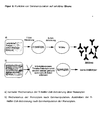
- Antibody production - and thus also the production of the pathological autoantibodies - can not be maintained without the help and mediation of monocytes and T helper lymphocytes, which in turn activate the B lymphocytes antigen-specifically (Figure 1).
- the antigen is e.g. a bacterial protein
- the immune response is useful to the organism.
- the antigen is an endogenous structure, it is called a (pathological) autoimmune reaction.
- the antigen presenting cell of the present invention exhibits increased expression of an antigen by transformation of an antigen presenting cell with nucleic acid containing material, wherein the antigen presenting cell presents substantially only one kind of antigen.
- the designed gene therapy method is based on two parallel genetic engineering interventions on the patient's own blood monocytes.
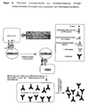
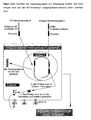
- the interventions are carried out by means of suitable probes and cause the reduction of B7 molecules by the obstruction and thus the prevention of the formation of the molecule on the surface of the monocytes ( Figure 3) and at the same time a strong presentation of the autoantigen (Figure 4).
- the production of pathological autoantibodies is specifically switched off by the manipulation of monocytes.
- the co-receptor B7 without which the antigen presentation or the induction cascade for antibody production does not start, is suppressed.
- the term B7 harbors two different co-receptors, termed CD80 (B7-1) and CD86 (B7-2). Their structures are known.
- the cells After in vitro manipulation of the monocytes, the cells are returned to the patient's bloodstream.
- the genetically modified monocytes now switch off the corresponding T helper lymphocytes present in the organism.
- the genetically manipulated cells compete directly with the already present in the organism (preferably bloodstream and lymphatic system) autoantigen-presenting monocytes, however, the B7 on the cell surface and normally activate the T helper lymphocytes and thus the antibody production (Figure 5).
- the cDNA of a protein that produces the production of pathological autoantibodies as an autoantigen is integrated into the monocyte genome. This genetic information then serves to overproduction of the autoantigen. Peptides of this autoantigen are then preferably presented to MHC II and / or MHC I (see also Figure 4 (II)). MHC II presents the peptides to T helper cells and tries to find those that specifically recognize the proteins presented. If the co-receptor B7 does not appear on the cell surface at the same time, the T helper cells are immobilized and subject to premature cell death.
- the antigen-presenting cell according to the invention can display an increased number of homing receptors, such as CD44. Overexpression of homing receptors will direct the antigen-presenting cell into the lymph nodes, causing the engineered APCs to multiply and accumulate more rapidly in lymph nodes.
- the lymph nodes are where the vast majority of responses from the adaptive immune system are produced. There, the gene-manipulated APCs develop a much higher effect than outside the lymph nodes.
- a transfonation causes an increase in the number of homing receptors and / or a suppression of functions of the B7, CD40 receptors.
- B7 and / or CD40 receptor expression in the antigen-presenting cell of the invention can be prevented or reduced.
- suppression of expression of the B7 and / or CD40 receptors by co-suppression in the antigen-presenting cell of the present invention can be effected with nucleic acids.
- the antigen-presenting cell according to the invention may contain or be transformed nucleic acids which cause expression of proteins or peptides having affinity with B7 and / or CD40 receptors.
- proteins are formed which virtually neutralize by complex formation with B7 and / or CD40 receptors.
- CTLA4, CD28, antibodies, F (ab) 2 , scFv and / or F ab fragments are suitable as proteins.
- the antigen-presenting cell according to the invention contains nucleic acids which code for a signal sequence or expression products of a signal sequence which effects the fate of the expression products in the endoplasmic reticulum, the Golgi apparatus, the trans-Golgi network or intracellular vesicles.
- the antigen-presenting cell according to the invention is transformed to express antigens with nucleic acids which enable the transport of the expressed antigens into MHC II compartments of the cells.
- All genetically engineered monocytes present the autoantigen on most of their MHC complexes, while very few "normal” monocytes even present the autoantigen and then only on a few MHC complexes.
- the aim of the treatment is to displace the "normal" monocytes that present the autoantigen and activate the T helper cells by the gene manipulated monocytes programmed to shut down the antibody products. It is very important to administer the antigen (s) also in a form that allows transport into the MHC II endosomes. Only then can a presentation to MHC II be reliably achieved. In a genetic engineering this is usually done by manipulation of the open reading frame, so that the reading frame is preceded by a signal sequence for this compartment. Genetic manipulation has the additional advantage of having a much longer half-life than e.g. Oligonucleotides or peptides. Stable integration of genes even leads to a permanent change in the property of the target cells.
- the corresponding nucleic acids may be DNA, RNA, oligonucleotides, polynucleotides, ribozymes, peptide nucleic acids (PNA).
- PNA peptide nucleic acids
- the DNA has regulatory elements such as enhancers, promoters, polyA-encoding 3 'ends for transcription of the DNA into RNA, the RNA regulatory elements for translation of the RNA into protein.
- the regulatory elements ensure efficient expression of the genes.
- an antigen presenting cell is ex vivo by treatment with viral vectors, bacterial vectors, plasmids, transformed by electroporation techniques, iontophoresis, ballistic methods and / or other techniques for introducing molecules into an antigen presenting cell.
- an antigen presenting cell may be prepared by treatment with viruses, viral vectors, bacterial vectors, plasmids produced by electroporation techniques, iontophoresis, ballistic methods and / or other techniques for introducing molecules into a cell having a suppressed function of co-stimulatory receptors or expression of co-stimulatory receptors by preventing their expression or the co-stimulatory receptors are prevented from reacting with affine structures to stimulate T cells bound to the antigen-presenting cell.
- molecules such as antibodies, proteins, peptides, peptidomimetics, CTLA4, CD28, CD40L and / or components and / or combinations of these molecules, e.g. B7-1, B7-2, CD40, which blocks co-stimulation of the T cell in the presence of antigen presentation, is brought into contact with the antigen-presenting cell.
- the molecules may be advantageous to use the molecules by means of vehicles such as liposomes, hydrogels, cyclodextrins, biodegradable nanocapsules, bioadhesive microspheres and / or by electroporation techniques, iontophoresis, ballistic methods and / or other techniques for introducing molecules into the antigen presenting cell Cell to transfer.
- vehicles such as liposomes, hydrogels, cyclodextrins, biodegradable nanocapsules, bioadhesive microspheres and / or by electroporation techniques, iontophoresis, ballistic methods and / or other techniques for introducing molecules into the antigen presenting cell Cell to transfer.
- Nucleic acids can be transferred, in particular, by viruses, viral vectors, bacterial vectors, plasmids, which are transferred by electroporation techniques, iontophoresis, ballistic methods and / or other techniques for introducing molecules into the antigen-presenting cell.
- the invention relates to a pharmaceutical composition containing at least one antigen-presenting cell according to the invention.
- the medicament according to the invention is preferably formulated as an infusion solution for intravenous or intraperitoneal administration.
- the formulation is chosen so that upon administration of the drug no significant impairment of the efficacy of the antigen-presenting cell according to the invention takes place.
- the antigen-presenting cell according to the invention can be used in particular for the production of a medicament for the treatment of unwanted immune reactions, such as autoimmune diseases and allergies or intentionally induced immune reactions, such as in immunizations.
- the antigen-presenting cell according to the invention can be used for the preparation of a medicament for the treatment of immune reactions against allologic and / or xenologic tissue features.
- the invention claims a use in which the immune reactions to be treated in conjunction with allologic and / or xenologic tissue characteristics whose gene sequences and / or partial sequences, in particular MHC I, MHC II, Rhesus factor are.
- the method presented here for reducing immune responses e.g. for the treatment of autoimmune diseases with a clearly defined autoantibody profile based on the genetic manipulation of certain blood cells, preferably outside the body, which are then subsequently returned to the organism.
- the aim of this treatment is, inter alia, the specific shutdown of a chronic, driven by the immune system production of autoantibodies, which cause the disease.
- the antibodies play an important role as defense molecules ("humoral immune reaction”). They are produced by mature B lymphocytes. However, the induction and production of the antibodies does not occur detached from the remaining cells of the immune system; rather, this humoral immune response is controlled by other cells of the immune system. The immune response can not be sustained without the help and mediation of antigen presenting cells and T helper lymphocytes.
- the molecular mechanisms of interaction - cell-cell contact - of antigen-presenting cells with T helper lymphocytes is known in detail at the receptor level.
- B7-1 and B7-2 An essential molecule of intercellular interaction in antigen presentation (contact of antigen presenting cells with T helper lymphocytes) are costimulatory receptors designated B7-1 and B7-2.
- CD40 also plays a role in this signal transduction.
- B7 and CD40 are the contents of numerous publications (Daikh et al 1997, Greenfield et al 1998, Johnson-Leger et al 1998, McAdam et al 1998, Vyth-Dreese et al 1995) and are also extensively reviewed in Textbooks (see, for example, Janeway and Travers 1997).
- the genetically modified monocytes now switch off the corresponding T helper lymphocytes present in the organism.
- the genetically manipulated cells compete directly with the autoantigen-presenting monocytes already present in the organism (preferably the bloodstream and the lymphatic system), but carry B7 on the cell surface and normally activate the T helper lymphocytes and, thus, among others, antibody production.
- Immunosuppressive Preparations such as cortisone and its derivatives, cyclosporin, beta-interferon or cytostatics (methothrexate).
- antibodies, antagonists and oligonucleotides are being tested against various signal components of the immune system for the purpose of preventing the activation of T cells.
- Such drugs are selective at best but never specifically directed against the misprogrammed autoimmune responses.
- Immunosuppressants therefore have a negative effect on important, necessary and useful parts of the immune system; because of this and their side effects their use is limited.
- the presented methodology is intended as a general concept for the reduction of immune responses. It should be possible with this basic concept to put an end to any immune reaction of the adaptive immune system again. In addition, such immune reactions can be induced as desired and then turned off again.
- liposome-mediated gene transfer plays only a minor role.
- Retroviruses are single-stranded, enveloped RNA viruses.
- the retroviridiae family is divided into three subfamilies: oncoviruses, lentiviruses and spumaviruses (Fields 1996).
- Lentiviruses include HIV-1 and HIV-2, and spumaviruses include simian foamy virus.
- the group of oncoviruses is divided into 5 genera (Table 1). ⁇ B> Table.
- MMTV HervK family Mouse Mammary Tumor Virus
- MMTV C-type viruses (mammals) Erv-3 S71 family Moloney mouse leukemia virus (MoMuLV) Harvey Mouse Sarcoma Virus (HaMSV) feline leukemia virus (FeLV) Gibbon Ape Leukemia Virus (GALV) Monkey Sarcoma Virus (SSV) ALV-related avian leukosis virus (ALV) (C-type viruses) Rous Sarcoma Virus (RSV) Rous associated virus (RAV 1-50) D-type viruses Mason Pfitzer monkey virus HTLV / BLV group human T-cell leukemia viruses (HTLV1 / HTLV2) Bovine Leukemia Virus (BLV)
- Electron micrographs show that retroviruses are coated particles about 100 nm in size.
- the capsid is surrounded by a membrane in which outward facing envelope proteins (env) are incorporated. Inside, the membrane is provided with a layer of matrix proteins (MA). Inside the particle is the nucleocapsid, which consists of capsid proteins (CA) is constructed.
- Matrix protein and capsid protein belong to the so-called group-specific antigens (gag).
- RT reverse transcriptase
- integrase coded by the sequences designated as pol
- PR Protease
- the genome of the retroviruses consists of two identical single-stranded RNAs of 7 - 10 kb in length.
- the RNA starts with the m 7 GPPP cap structure, which is important for the translation of the structural proteins, and ends with the approximately 200 base long poly (A) sequence. Between cap structure and poly (A) sequence are regulatory sequences, including the splice donor, followed by the sequences coding for the gag, pro- and pol proteins, the splice acceptor, and the coding sequence for the env gene.
- the simple retroviruses such as the C-type viruses, only need these proteins for their life cycle. More complex viruses, such as the lentiviruses, contain other genes, either by shifting the reading frames or by further sequences between the env region and the 3 'regulatory sequences.
- amphotropic mouse leukemia virus belongs to the class of C-type viruses.
- Murine leukemia viruses are further subdivided according to their receptor utilization and thus their host range (Table 2).
- Tab.2 Classification of murine leukemia viruses class host range receptor function amphotropic all mammals Pit-2 (ram-1) PO 4 3 transporter ecotropic mouse MCAT AS transporter polytropically all mammals ? ? xenotropic all mammals except mouse ? ? MCF mouse ? ? 10A1 all mammals Pit-1 (Glvr-1) and Pit-2 (ram-1)
- Cells infected with the viruses of a group can not be over-fished by viruses of the same class because the cellular receptors are already occupied by the env proteins of that virus. This phenomenon is called interference. However, the cells can be infected with viruses of a different class.
- the 5 'capping region Following the 5 'capping region is the 5' R region, followed by the U5 region, the Primer Binding Site (PBS), the leader, the sequences for the viral proteins, the polypurine tract, the U3 region, the 3 'R Region and the poly A sequence.
- PBS Primer Binding Site
- the leader the sequences for the viral proteins, the polypurine tract, the U3 region, the 3 'R Region and the poly A sequence.
- the two identical R regions Directly after the capping structure and before the poly A strand are the two identical R regions. They are about 60 base pairs long and important for reverse transcription.
- the 5 'end-subsequent U5 region is the first transcribed region in reverse transcription and contains important sequences for initiation the transcription. It is about 75 bases long and becomes the 3 'end of the 5' Long Terminal Repeats (LTR) of the Provirus.
- the primerbinding site is 18 nucleotides long. Here, a molecule of tRNA is bound as a primer.
- the leader sequence is about 475 bases long and contains the splice donor sequences.
- the packaging signal is located here. It causes the genomic RNA to be packaged into the virions when assembling the virions, while other RNA is not packaged. This is followed by the sequences for the gag, pro and pol genes, followed by the splice acceptor and the sequences for the env genes.
- the env sequence is followed immediately before the U3 region by the polypurine region. Here the initiation site for the plus strand synthesis is located.
- the U3 region is about 500 bases long and becomes the 5 'area of the LTR in Provirus. It contains all sequences necessary for the transcription of viral proteins, such as. B. Binding sites for various transcription factors or the TATA box. Cell lines specific regulation of expression z. B. is encoded by the U3 region.
- the U3 region is followed by the 3 'R region, as well as the polyadenylation signal and the 200 nucleotide poly A sequence.
- the matrix, capsid and nucleoproteins are synthesized by two common polyprotein precursors.
- the Pr180 gag-pol contains not only the gag proteins but also the pol domain, the Pr gag contains only the gag proteins. These precursor proteins are sequentially cleaved by the viral protease (PR) into the mature proteins: matrix protein (p15), capsid protein (p30) and nucleoprotein (p10); Protease (p14), reverse transcriptase (p80) and integrase (p46).
- the matrix protein is most closely associated with the lipid membrane via amino terminal attached myristic residues.
- the capsid or core is composed of the hydrophobic capsid protein. The small basic nucleoprotein lies within the core.
- gag proteins are located in the first reading frame of the viral genome and are synthesized in large quantities in the cell since they are generated by both the Pr180 gag-pol and the Prag gag (Fields 1996).
- the following pro and pol genes responsible for the enzymatic activities are in the next reading frame and are produced in smaller quantities (only from the Pr180 gag-pol ).
- the protease is responsible for the splitting of the gag and pol polyprotein precursors into the mature proteins.
- the reverse transcriptase contains DNA polymerase activity and RNase H activity, but has no proofreading activity.
- Reverse transcriptase is one of the most conserved regions of the retrovirus genome. Not only is there agreement within the retrovirus family, but certain areas are also found in genetically distant viruses, such as hepadnaviruses (double-stranded DNA viruses) and cauliflower mosaic virus.
- the integrase is the enzyme that inserts the proviral DNA into the genome of the host cell.
- the env genes have their own reading frame and are transcribed via a splice acceptor. They are also transcribed as polyprotein precursors (Pr80 env ) but cleaved by a cellular protease: transmembrane region (p15E) and external glycoprotein (gp70).
- the env protein consists of two polypeptide chains linked by disulfide bridges. These chains are synthesized as a common precursor; the amino-terminal part gives the external polypeptide chain (SU), the carboxy-terminal part the transmembrane region (TM).
- the SU protein is glycosylated and responsible for interacting with the receptor; the TM protein is not glycosylated and anchors the proteins in the membrane. He is also probably responsible for the dimerization of env molecules.
- the SU protein Since the binding between the SU and the TM protein is not particularly strong, the SU protein is often "lost". Among other things, this is due to the lack of stability of infectious viruses during ultracentrifugation and the large number of infection-defective particles.
- the infection of a cell occurs by the specific attachment of the virus to its receptor on the cell surface and the subsequent uptake of the virus into the cell. Only cells that express this receptor can become infected.
- the interaction between the cellular receptor and the external viral envelope protein (SU) causes a conformational change in the latter and allows fusion with the cell membrane through the transmembrane domain of the virus.
- the uptake of virions is either by pH-dependent fusion (ecotropic virus) or by endocytosis (amphotropic virus).
- the viruses are uncoated; this releases the viral RNA and activates the reverse transcriptase.
- the capsid remains in close association with the viral RNA and is necessary for the correct transcription of the viral RNA into DNA.
- PB primer binding site
- the tRNA primer which is located close to the 5 'end at the primer binding site (PB)
- the minus DNA strand is synthesized. It the 5 'located U5 and R region is transcribed (strong stop DNA), whereby at the same time by the RNAse H activity of the reverse transcriptase the RNA template is degraded.
- the thus released DNA "jumps" to the 3 'end of the viral RNA and there is an attachment of the newly synthesized R region to the existing R region there.
- the remaining viral RNA is transcribed into DNA and degraded to the RNAse H resistant polypurine area in front of the U3 region.
- This area serves as a template for the synthesis of the plus DNA strand on the already synthesized minus DNA strand, whose 3 'end is the first to be transcribed.
- the remaining RNA residues are degraded and the primer binding site of the plus strand can bind to the primer binding site of the minus strand and the synthesis of the plus strand can proceed to completion.
- both DNA strands are completed.
- the newly synthesized DNA contains the Long Terminal Repeats (LTR) on both sides.
- LTR Long Terminal Repeats
- the newly synthesized in the cytoplasm viral DNA migrates as a pre-integration complex in the nucleus and there attaches itself to the cellular DNA.
- MuLV can only infect dividing cells as they have a disrupted cell membrane. They have no nuclear transport signal or protein (such as HIV) that could effect nuclear transport.
- the pre-integration complex is also too large to diffuse through nuclear pores.
- the viral DNA is in linear form.
- the virus-own integrase cleaves the cellular DNA at an arbitrary position and generates overhanging ends of 6 base pairs.
- the 3 'ends of the viral DNA are ligated to one strand of cellular DNA; previously, 2 bases each are removed at the 3 'ends of the viral DNA.
- the missing sequences in the second strand are filled in by the cell's own repair system, whereby the two supernumerary bases at the 5 'end of the viral DNA are removed.
- Retroviruses belong to the non-lytic viruses, have a simple genome, are well characterized, stably integrate into the genome of the infected cell and can permanently express the inserted gene there - all reasons to make them attractive for gene therapy approaches.
- vectors with amphotropic env or pseudotype vectors with the env of other viruses are used. It is being worked on the development of cell-type-specific vectors, either by choosing a suitable LTR (Hunt 1987), a particular coat protein or by introducing antibodies or receptors into the virus envelope.
- Retroviral vectors are made on the basis of retroviruses. To prevent the formation of replication-competent viruses, the viral genes are deleted. Instead of this, the gene to be expressed with the necessary regulatory sequences is cloned between the LTRs and provided with the packaging signal. The vectors are packaged in special packaging cell lines that provide all the proteins necessary for packaging. The DNA to be transferred is limited by the packaging capacity of the virions. Retroviral vectors can transfer up to 9 kb of foreign DNA. By selecting a suitable LTR or an additional promoter, an attempt is made to obtain the highest possible expression of the transferred gene in the desired target cell. Various strategies allow multiple genes to be encoded by a vector.
- the packaging cells contain one construct encoding the gag pol genes and another encoding the envelope proteins (Danos 1988). They are stably integrated into the genome of the cells. The constructs included no packaging signal, so that only empty virions are formed. Only when vector DNA is present in the cells do RNA-containing viruses arise. The use of two different constructs for the expression of gag, pol and env is said to reduce the risk of replication-competent virus formation. At least three recombination events are required before a replication-competent virus (RCR) can arise. Nevertheless, packaging cells for clinical applications must be constantly checked for RCRs.
- Peripheral blood monocytes were isolated from buffy coats or mouse blood, preferably by density centrifugation over lymphocyte separation medium followed by selective adhesion.
- the lymphocytes / monocytes contained in the interphase were washed twice at 200 ⁇ g, 15 min, 20 ° C. Then the cells were seeded at a density of 5x10 6 / ml to 2 x 10 7 / ml in sterile, gelatin-coated cell culture dishes in medium 2. After two hours of incubation at 37 ° C non-adherent cells could be removed by washing three times with medium 3 evenly. The monocytes were then cultured in medium 2 at 37 ° C, 5% CO 2 . The mouse blood was treated in the same way.
- the buffy coat was diluted 1: 4 with PBS without (Ca and Mg), centrifuged at 100 g for 15 minutes.
- the upper yellowish part (plasma) contains mainly platelets. He is carefully removed and discarded. The remainder is diluted to about 120 ml with PBS without Ca & Mg.
- the interphase milky ring
- the interphase is aspirated (lymphocytes, monocytes, platelets) and washed twice.
- the pellet (erythrocytes, granulocytes) is discarded.
- the monocytes are then isolated by adherence to gelatinized plastic vessels as the other cell types are removed during washing. For this purpose, the interphase was incubated for two hours in the gelatinized plastic tubes.
- the cells were transfected using the Ca-PO 4 method and the supernatants collected on three consecutive days.
- the cells were seeded confluent and the medium changed after 6 h. After 16 h, the supernatants were collected and filtered through a 0.8 ⁇ m filter. The virus supernatant from amphotropoen packaging lines was used directly.
- the supernatants of the virus-producing cells were collected and filtered through a 0.8 micron filter. Then they were transferred to a centrifugation tube and centrifuged at 100,000 g and 5 ° C for 2 h. The supernatant was removed and the pelleted virus taken up in a little DMEM medium with 2% BSA. The concentrated viruses were either used directly in tests or frozen at -80 ° C. For optimization, 5 ml of a 15% sucrose solution were placed in centrifugation tubes and these were overlaid with the cell culture supernatant. Then centrifuged as described above. For Western blot analyzes, the supernatant was split into two tubes and centrifuged. One of the virus pellets was taken in media for titer determination, the other was taken up in the same amount of virus lysis buffer and frozen at -80 ° C until use.
- 5x10 3 SC-1 fibroblasts were plated in 1 ml of medium in a 24-well plate. After 4 hours of incubation, the medium was removed, replaced by 1 ml of a virus dilution (1: 5 dilution steps, triplicate) and incubated for a further 16 h. The expression of the additional genetic information was quantified in FACS or by RT-PCR. The clones of the dilution step were counted containing 1 to 6 clones per well.
- the gene ferry was an amphotropic murine leukemia virus.
- the genes used in the experiments aim on the one hand to reduce the B7 and / or CD40 molecules appearing on the cell surface and additionally to provide for the expression of an antigen.
- CTLA4Ig for the suppression of B7 and / or CD40 antisense cDNA and an antigen are used at the beginning.
- human monocytes are the C fragment of tetanus toxin.
- CTLA4Ig is a fusion protein from the Fc part of an antibody and the part of CTLA4 that binds to B7.
- the supernatant of the packaging cell line was used. With this, the monocytes were incubated.
- the T cell proliferation assay was performed with the EZ4U kit (BIOMEDICA, Vienna, Austria). Reaction of T cells in relative units: to 1.: 0 to 2 .: 0 to 3.: +++ to 4. + to 5 .: ++ to 6 .: +
- the experiment shows that the totality of T-cell-mediated immune responses to tetanus toxin in this case can be reduced if the monocytes not only specifically present antigenic determinants of the tetanus toxin, but additionally the binding of B7 and / or CD40 molecules to the corresponding ligands is prevented.
- both Th1 and Th2 T-cell activations have been reduced.
- the blockade of B7 is more efficient than that of CD40.
- OvalbuminIi means that the ovalbumin was provided with a signal sequence for the MHC II compartment to allow its fragments to be presented to MHC II with greater efficiency.
- the experiment shows that the humoral immune response against ovalbumin in this case can be reduced if the monocytes not only specifically present antigenic determinants of the ovalbumin, but in addition the binding of B7 and / or CD40 molecules to the corresponding ligands is prevented.
- the blockade of B7 is more efficient than that of CD40.
- the use of the signal sequence of the invariant chain is well suited to obtain the desired effect.
- the system was tested on human and mouse blood. It was shown that a suppression of specific immune responses is possible both in vitro and in vivo.
Landscapes
- Health & Medical Sciences (AREA)
- Life Sciences & Earth Sciences (AREA)
- Engineering & Computer Science (AREA)
- General Health & Medical Sciences (AREA)
- Veterinary Medicine (AREA)
- Animal Behavior & Ethology (AREA)
- Immunology (AREA)
- Public Health (AREA)
- Bioinformatics & Cheminformatics (AREA)
- Epidemiology (AREA)
- Organic Chemistry (AREA)
- Biomedical Technology (AREA)
- Chemical & Material Sciences (AREA)
- Zoology (AREA)
- Biotechnology (AREA)
- Wood Science & Technology (AREA)
- Genetics & Genomics (AREA)
- Gastroenterology & Hepatology (AREA)
- Biochemistry (AREA)
- Pharmacology & Pharmacy (AREA)
- Hematology (AREA)
- Chemical Kinetics & Catalysis (AREA)
- General Chemical & Material Sciences (AREA)
- Cell Biology (AREA)
- Medicinal Chemistry (AREA)
- Microbiology (AREA)
- Nuclear Medicine, Radiotherapy & Molecular Imaging (AREA)
- General Engineering & Computer Science (AREA)
- Transplantation (AREA)
- Pulmonology (AREA)
- Medicines Containing Material From Animals Or Micro-Organisms (AREA)
- Medicines That Contain Protein Lipid Enzymes And Other Medicines (AREA)
- Medicines Containing Antibodies Or Antigens For Use As Internal Diagnostic Agents (AREA)
- Organic Low-Molecular-Weight Compounds And Preparation Thereof (AREA)
- Pharmaceuticals Containing Other Organic And Inorganic Compounds (AREA)
- Micro-Organisms Or Cultivation Processes Thereof (AREA)
- Peptides Or Proteins (AREA)
Description
- Gegenstand der vorliegenden Erfindung sind Antigen präsentierende Zellen, Verfahren zur Herstellung Antigen präsentierender Zellen, Arzneimittel enthaltend Antigen präsentierende Zellen sowie Verwendung der Antigen präsentierenden Zellen
- Seit knapp 10 Jahren werden für verschiedene Krankheiten und in Tiermodellen gentherapeutische Verfahren entwickelt und angewendet. Bisher sind sie noch nicht in die Routine überführt. Meist handelt es sich dabei um die Behandlung von genetisch bedingten, schweren Erkrankungen, für die andere Therapien nicht zur Verfügung stehen. Weiterhin werden Gentherapien zur Behandlung von schweren und ebenfalls nicht therapierbaren Krebserkrankungen eingesetzt.
- Wenig Aufmerksamkeit hat die experimentelle Medizin bisher der Behandlung von Allergien und Autoimmunerkrankungen mittels gentechnischer Methoden geschenkt.
- Allergien verursachen beträchtliche Kosten im Gesundheitswesen der Industrieländer. Immerhin sind schätzungsweise mindestens 20% der Bevölkerung gegen irgendeine Substanz allergisch. Die meisten Betroffenen leiden an allergischer Rhinitis, zu der insbesondere der Heuschnupfen zählt, oder an Bronchialasthma: Sie niesen oder ringen nach Luft, nachdem sie bestimmte Pollen oder andere im allgemeinen harmlose Substanzen eingeatmet haben. Viele Kinder und einige Erwachsene reagieren auch allergisch auf Nahrungsmittel. Andere erleiden Hautausschläge oder sogar einen allergischen Schock, nachdem sie Medikamente wie Penizillin erhalten haben. Bei wieder anderen rufen Bienenstiche starke lokale Schwellungen oder schwere systemische - den gesamten Organismus erfassende - Störungen hervor. Im Extremfall können allergische Anfälle sogar zum Tod führen. Allein die unmittelbare medizinische Versorgung von Asthmapatienten hat in den USA 1990 schätzungsweise 3,6 Milliarden Dollar verschlungen und damit rund ein Prozent aller Kosten im Gesundheitswesen ausgemacht.
- Mittlerweile ist bekannt, dass einige der zellulären und molekularen Wechselwirkungen bei allergischen Reaktionen oft ähnlich ablaufen, unabhängig davon, auf welche Substanzen der einzelne anspricht und welche Symptome er entwickelt. Gewisse Begleiterscheinungen der Allergien treten normalerweise ausschließlich auf, wenn das Immunsystem Parasiten bekämpft. So reagiert der Körper auf Schmarotzer ebenso wie auf Allergene mit der massiven Produktion von Molekülen, die als Immunglobulin-E-Antikörper (IgE) bezeichnet werden. Die Produktion dieser Antikörper wird durch T-Helfer-Zellen induziert, die wiederum von antigen-präsentierenden Zellen aktiviert werden.
- Unterschiedliche Allergene rufen unter anderem deswegen verschiedenartige Symptome hervor, weil sie mit dem Immunsystem in verschiedenen Körperregionen in Berührung kommen. In den oberen Luftwegen erzeugt die fehlgeleitete Immunreaktion Niesen und eine verstopfte Nase. In den unteren Luftwegen können dagegen die Bronchien sich verengen und verschleimen, so dass typische asthmatische Symptome auftreten. Entsprechend rufen Immunaktivitäten in den Geweben des Magen-Darm-Traktes Übelkeit, Bauchkrämpfe, Durchfall oder Erbrechen hervor.
- Schließlich vermag ein Allergen, dass auf irgendeinem Weg ins Blut gelangt, eine Anaphylaxe auszulösen: eine allergische Reaktion in weit von seiner Eintrittsstelle entfernten Körperregionen. Schwere anaphylaktische Schocks können alle normalen Körperfunktionen durcheinanderbringen und tödlich enden.
- Auch wenn sich die allergischen Reaktionen unterschiedlich äußern, werden sie doch stets durch den gleichen Mechanismus in Gang gesetzt: die Sensibilisierung. Dazu kann bereits der einmalige Kontakt mit einem Allergen, typischerweise einem Eiweißstoff, genügen. In den Luftwegen oder anderen Geweben trifft die allergieauslösende Substanz auf sogenannte Fresszellen oder Makrophagen. Diese verschlingen den Fremdstoff, zerstückeln ihn und präsentieren die Fragmente auf der Zelloberfläche mit MHC II-Molekülen. Im weiteren Verlauf erkennen einige T-Helfer-Lymphozyten die dargebotenen Bruchstücke und binden daran. Die T-Helfer-Lymphozyten werden von den Makrophagen aktiviert und aktivieren dann ihrerseits einige B-Lymphozyten, die gleichfalls das Allergen erkennen. Die B-Zellen reifen dann zu Antikörper produzierenden Plasmazellen aus. Zunächst sind dies Antikörper vom sogenannten IgM-Typ; ab einem bestimmten Zeitpunkt schalten die Plasmazellen jedoch auf IgE-Antikörper um.
- Bis die Antikörper hergestellt sind, können Tage oder Wochen vergehen, und das Allergen, welches ihre Produktion in Gang gesetzt hat, ist dann womöglich schon lange verschwunden. Nicht so die IgE-Moleküle. Mit ihrer Fc-Region heften sie sich an IgE-Rezeptoren zweier unterschiedlicher Klassen von Immunsystemzellen. Bei der einen handelt es sich um Mastzellen, die sich im Körpergewebe für gewöhnlich in der Nähe von Blutgefäßen und Epithelzellen ansiedeln. Über das Epithel besteht Kontakt mit der Außenwelt (darunter fällt auch das Epithel der Atemwege und des Magen-Darm-Traktes). IgE-Antikörper binden außerdem an basophile Granulozyten (Basophile). Diese Zellen zirkulieren allerdings im Blutstrom.
- Hat die Produktion der IgEs einmal begonnen, hält sie offenbar Monate, ja manchmal sogar Jahre an. Folglich besetzen sie unablässig IgE-Rezeptoren auf Mastzellen und Basophilen - bereit beim nächsten Allergenkontakt augenblicklich in Aktion zu treten.
- Während also die erste Begegnung mit einem Allergen selbst bei Personen, die sich später als Allergiker entpuppen, keine Symptome hervorruft, leitet die Zweitexposition ein Stadium der Überempfindlichkeitsreaktion ein, welches auch äußerlich in Erscheinung tritt. Innerhalb von Sekunden nach dem Kontakt mit menschlichem Gewebe bindet der allergieauslösende Stoff an die IgEs der Mastzellen. Heftet er sich dabei an zwei oder mehr IgE-Moleküle zugleich, bildet er eine Brücke zwischen ihnen. Solche Quervernetzungen lassen die betroffenen IgE-Rezeptoren dichter zusammenrücken, und dies aktiviert die Zelle, so dass sie hochwirksame Substanzen ausschüttet, die auf direktem Wege allergische Symptome erzeugen. (Die Freisetzung kann auch auf andere Arten hervorgerufen werden; von allergischen Reaktionen spricht man nur, wenn IgEs beteiligt sind). Die wichtigste dieser Substanzen ist Histamin. Es kann sowohl die Schleimbildung in den Epithelien anregen und so zur Verstopfung der Luftwege beitragen als auch die glatte Muskulatur, die wie ein elastisches Band Bronchien und Därme umschlingt, kontrahieren lassen. Ferner vermag es die feinen Blutgefäße zu weiten und durchlässiger zu machen, so dass Flüssigkeit ins Gewebe sickern kann. Rötungen und Schwellungen sind die Folge. Betreffen diese Gefäßveränderungen große Teile des Körpers, können sie ein tödliches Kreislaufversagen auslösen: Bei einem solchen Schock fällt der Blutdruck jäh so stark ab, dass die Sauerstoffversorgung von Herz und Gehirn nicht mehr gewährleistet ist.
- Die zweite Gruppe von Mediatoren besteht hauptsächlich aus Prostaglandinen und Leukotrienen. Sie werden erst erzeugt, nachdem die Allergenmoleküle sich an die IgEs auf den Zellen angelagert haben. Wie Histamin verengen sie die Bronchien und erweitern die Blutgefäße. Ihre Wirkung hält allerdings länger an.
- Zusätzlich stoßen stimulierte Mastzellen eine Vielzahl potentiell toxischer Enzyme aus. Offenbar setzen sie ferner Cytokine frei, die die Aktivitäten anderer Immunzellen regulieren.
- Antihistaminika erweisen sich in der Regel als wirksam und dienen immer noch als Standardtherapie. Die neuesten Varianten können die Blut-Hirn-Schranke nicht mehr ohne weiteres passieren und machen die Patienten nicht mehr müde. Wenn bei einer schweren Entzündung Antihistaminika wirkungslos bleiben, helfen oft inhalierbare Corticosteroide, die gewöhnlich zur Linderung der chronischen Entzündung bei Asthma verschrieben werden.
- In schweren Fällen kann die schon im Jahre 1911 eingeführte Immuntherapie oder Hyposensibilisierung (auch als Allergiespritzen oder Desensibilisierung bekannt) auf lange Sicht Erleichterung verschaffen. Bei dieser Behandlung injizieren Ärzte den Patienten steigende Dosen des Allergens, auf das diese empfindlich reagieren. In allen Fällen ist die Dosis ausschlaggebend: Zu wenig Allergen verleiht keine Toleranz. Außerdem ist der Schutz selten vollständig.
- Bronchodilatatoren sind die meistverwendeten Arzneimittel bei Asthma. Sie lindern die durch Histamin und andere Bronchokonstriktoren hervorgerufenen Symptome sehr schnell, beeinflussen die zugrundeliegende Entzündung jedoch wahrscheinlich nicht. Außerdem kann ihr übermäßiger Gebrauch eine Gegenreaktion des Körpers hervorrufen, so dass nach Abklingen ihrer Wirkung der Atemstrom stärker behindert ist als zuvor. Zusätzlich kommen die unter 1. aufgezählten Methoden zur Anwendung.
- Insektenstiche rufen bei manchen Menschen Anaphylaxien aus. In schweren Fällen führen diese zum Tod durch z.B. Kreislaufversagen oder Ersticken. Jede schwere Anaphylaxie - gleich ob sie zum ersten oder fünfzehnten Mal auftritt - ist ein Notfall, bei dem zunächst versucht werden muss, die bedrohlichsten Symptome zu beherrschen. Meist geschieht das durch Injektion von Adrenalin, welches die Freisetzung der Mediatoren hemmt, die Luftwege öffnet und der Erweiterung der Blutgefäße entgegenwirkt. Man kann vorbeugend durch eine Immunisierung mit dem Gift des gesundheitsbedrohenden Insekts agieren.
- In der Erprobung im Tiermodell befindet sich die auf DNA basierende Immunisierung mit einem Allergen (Der p 5) der Milbe Dermatophagoides pteronyssinus. Diese Immunisierung resultiert in einer Produktion von IgG, aber nicht IgE, und resultiert in einer 90%tigen Reduktion der Mengen an spezifischem IgE, welche durch klassische Sensibilisierung mit Der p 5 und Alaun als Adjuvant oder allergen-induzierte Rhinitis hervorgerufen wurden.
- Eine weitere neue Strategie ist die Verwendung von humanisierten monoklonalen Anti-IgE-Antikörpern gegen die FcεRI-Binderegion für IgE. Dadurch wird die Bindung von IgE an den IgE-Rezeptor verhindert, so dass keine Mediatoren der allergischen Reaktion von Mastzellen oder Basophilen ausgeschüttet werden können. Diese Strategie hat in klinischen Studien bei Patienten mit allergischer Rhinitis und allergischem Asthma gezeigt, dass diese Antikörper gut toleriert werden und die allergischen Reaktionen reduzieren.
- (siehe auch L.M. Lichtenstein: "Allergie und Immunsystem"; in Spektrum der Wissenschaft Spezial: Das Immunsystem; 1994; 74-83; S-K Huang, K-Y Chua und K-H Hsieh: Allergen gene transfer. Current Opinion in Immunology 1997; 800-804; C. Heusser und P. Jardieu: Therapeutic potential of anti-IgE antibodies. Current Opinion in Immunology 1997; 805-814).
- Die Transplantation von Geweben, um kranke Organe zu ersetzen, ist heute eine wichtige medizinische Therapie. In den meisten Fällen stellt eine Reaktion des anpassungsfähigen Immunsystems gegen das Transplantat die größte Bedrohung für eine erfolgreiche Behandlung dar. Reaktionen des anpassungsfähigen Immunsystems werden von Antigen-präsentierenden Zellen durch Aktivierung von T-Helfer-Lymphozyten induziert. Bei der Transfusionen von Blut, welches das erste und am häufigsten verwendete Transplantat ist, müssen die ABO und Rh Blutgruppenantigene abgeglichen werden, damit die schnelle Zerstörung unpassender Erythrozyten vermieden wird. Bei anderen Geweben müssen die sehr polymorphen Haupthistokompatibilätskomplexe (MHC) aufeinander abgeglichen werden, da diese fast immer die Immunreaktion auslösen. Leider ist der perfekte Abgleich der MHCs außer bei Verwandten fast unmöglich.
- Obwohl die Immunreaktion Organtransplantationen schwierig macht, gibt es wenige Alternativen bei Organausfällen. Die Verwendung von potenten immunsuppressiven Drogen, besonders Cyclosporin A und FK-506, die die Aktivierung von T-Zellen verhindern, macht Organtransplantationen erfolgreich. Trotzdem laufen hier einige Probleme auf, da das Leiden, welches das eigene Organ zerstört hat, auch das fremde Organ zerstört. Außerdem steigt durch die Unterdrückung des Immunsystems, das Risiko an Krebs oder Infektionen zu erkranken. Zudem ist die Prozedur sehr kostspielig (Janeway and Travers 1997).
- Autoimmunerkrankungen gehören zu den chronischen Erkrankungen. Zu ihnen zählen Rheuma in verschiedensten klinischen Ausprägungen, Diabetes, Multiple Sklerose, bestimmte Formen von Herzmuskelentzündungen und von Schilddrüsenerkrankungen. Die Reihe von Autoimmunerkrankungen ließe sich beliebig fortsetzen, wobei ca. 90% allerdings epidemiologisch nur einen kleinen Prozentsatz ausmachen.
- Die klinischen Verläufe von Autoimmunerkrankungen selbst bei ein- und derselben Diagnose können sehr unterschiedlich sein. Z. T. werden schubartige Krankheitsverläufe beobachtet. Entsprechend werden die Behandlungen vom Arzt individuell gestaltet.
- Einige der Autoimmunerkrankungen sind antikörpervermittelt. Darunter wird in der Immunologie verstanden, dass der Organismus aus meist nicht bekannten Ursachen Antikörper produziert, welche sich gegen körpereigene zelluläre Strukturen (Autoantigene) richten. Sind diese Antikörper einmal gebildet, lösen sie durch ihre Interaktion und Bindung an die jeweiligen organ- oder zellspezifischen Strukturen eine zell- und gewebszerstörende Reaktion des Immunsystems aus. Letztendlich führt diese dann zu dem klinischen Bild der Erkrankung (Steinman 1994).
- Beispiel hierfür ist die Schilddrüsenerkrankung Morbus Basedow, aber auch eine Form der Herzmuskelentzündung, die zur Dilatativen Cardiomyopathie führt. In einigen Fällen sind sowohl die Targets, gegen die sich die Autoantikörper richten, als auch die Autoantikörper selbst, genau charakterisiert.
- WO-A-96/18736 offenbart ein Verfahren und ein Reagenz zur Behandlung von arthritischen Bedingungen, Induktion von Toleranz bei Verpflanzungen und Umkehrung von Immunreaktionen. Dabei wird die Möglichkeit, die Expression von B7-1, B7-2 und B7-3 durch Ribozyme oder Antisense-RNA zu unterdrücken, theoretisch erörtert. Die Unterdrückung der B7-Mengen durch ein B7-Ribozym und B7-Antisense-RNA werden zwar nicht durch Experimente verifiziert, aber eine reine Unterdrückung von B7 würde im besten Falle eine allgemeine Reduzierung der gesamten Reaktionen des anpassungsfähigen Immunsystems bewirken.
- Die WO-A-95/32734 beschreibt die Verhinderung co-stimulatorischer Aktivitäten von antigen-präsentierenden Zellen, indem FcγRII-Rezeptoren durch aggregierte IgG-Moleküle vernetzt werden. Eine Unterdrückung von T-Zell-vermittelten Krankheiten soll erreicht werden, indem gleichzeitig die FcγRII-Rezeptoren vernetzt und spezifische Antigene verabreicht werden.
- Die WO-A-98/29124 betrifft injizierte Antisense-Oligonukleotide, mit denen die B7-Mengen reduziert werden sollen. Damit kann die Expression von B7 höchstens unspezifisch in allen Zellen behindert werden.
- WO-A-97/44450 offenbart die Erhöhung oder Unterdrückung von Prozessen, die auf sequenzspezifischen Interaktionen von Nukleinsäuren mit der Zellmaschinerie beruhen. Insbesondere können Genpromotor-unterdrückende Nukleinsäuren eingesetzt werden, die mit Genpromotormotiven für die Erhöhung oder Unterdrückung der Transkription eines Zielgens interagieren. Im Detail werden mit einer Utron-Nukleinsäure, dem TSU, die Expression von MHCs der Klassen I und II, sowie von ICAM-1, B7-1, B7-2 und FcγR behindert. Ein solcher Eingriff könnte insbesondere für Organtransplantate von Nutzen sein, bei denen die Expression von MHC-Molekülen die entscheidenden Abstoßungsreaktionen hervorruft. Utron-Nukleinsäuren stammen aus den 3'-untranslatierten Regionen von mRNAs. Diese 3'-untranslatierten Regionen werden so genannt, wenn sie in vivo Aktivitäten entfalten, die zelluläre Prozesse durch sequenzspezifische Interaktionen mit einem Teil der zellulären Maschinerie stimulieren oder inhibieren. Bei der zellulären Maschinerie handelt es sich gemäß WO -A-97/44450 um Promotoren. Es wird dort zwar über die Möglichkeit der Unterdrückung der B7-Expression (1 und 2) mit gentechnischen Methoden spekuliert, aber eine solche Inhibition kann mit dem aufgezeigten Mittel nur in Zusammenhang mit der Inhibition weiterer Gene (MHCs der Klassen I und II, sowie von ICAM-1 und FcγR) geschehen. Eine solch weitreichende Inhibition ist kontraproduktiv, da man auf eine hohe MHC-Expression angewiesen ist. Außerdem ist eine Regulation auf der Promotorebene nachteilig, da keine Möglichkeit besteht, um hinreichend spezifisch zu wirken.
- WO-A-99/50394 offenbart teilungsfähige aus Monozyten gewonnene Zellen. Es werden keine Monozyten beschrieben, die vorwiegend vordefinierte Antigene präsentieren. Es wird kein Gentransfer vorgesehen, der für eine verminderte Anzahl von CD80, CD86, CD40 an der Zelloberfläche sorgt. Vielmehr wird deren verminderte Präsenz durch die Behandlung mit z. B. physikalischen Methoden wie UV-Licht erreicht, wodurch auch die beschriebenen anderen Charakteristika hervorgerufen werden.
- Das der Erfindung zu Grunde liegende Problem besteht unter anderem darin, gentechnologische, therapeutisch nutzbare Produkte für die Reduktion von spezifischen Immunreaktionen zur Verfügung zustellen, bei denen Targets (Antigene, Autoantigene) und Antikörper, Autoantikörper bekannt sind.
- Gelöst wird dieses Problem durch die erfindungsgemäße antigen-präsentierende Zelle, die durch Transformation einer antigen-präsentierenden Zelle mit nukleinsäurehaltigem Material erhältlich ist, wobei das nukleinsäurehaltige Material eine erhöhte Expression eines vorher bestimmten Antigens sowie eine Suppression von Funktionen der B7- und/oder CD40-Rezeptoren bewirkt.
- Vorteilhaft an der antigen-präsentierenden Zelle gemäß der Erfindung ist, dass man die Zellen vermehren und z. B. Makrophagen und dendritischen Zellen differenzieren kann. Durch Erhalt der Teilungsfähigkeit werden diese Zellen im Grunde erst für den Gentransfer durch die gängigen nicht replikationsfähigen Retroviren geeignet, da diese erst bei der Teilung ins Genom eingebaut werden.
- Figur 1 zeigt ein Schema zur Funktion der Genmanipulation auf zellulärer Ebene.
- a) normaler Mechanismus der T-Helfer-Zell-Aktivierung über Monozyten
- b) Mechanismus der Monozyten nach Genmanipulation. Ausbleiben der T-Helfer-Zell-Aktivierung nach Genmanipulation der Monozyten.
- Figur 2 zeigt eine "Normale" Immunreaktion zur Antikörperbildung, Antigen-präsentierende Monozyten als Induktoren der Antikörperproduktion. Monozyten nehmen als fremd erkannte Moleküle auf und bringen sie auf die Zelloberfläche (Antigenpräsentation). Gemeinsam mit einem Oberflächenmolekül (B7) wird das Antigen den T-Helferzellen präsentiert. Das präsentierte Antigen wird von T-Helfer-Lymphozyten erkannt, die ihrerseits B-Zellen zur Antikörperproduktion anregen. Das Schema ist stark vereinfacht.
- Figur 3 zeigt schematisch die Funktion der Genmanipulation auf molekularer Ebene: der Co-stimulierende Rezeptor B7 wird inaktiviert.
- Figur 4 (I) zeigt die Funktion der Genmanipulation auf molekularer Ebene: das Autoantigen wird von den B7-inhibierten antigenpräsentierenden Zellen synthetisiert
- Figur 4 (II) zeigt einen Vergleich der Antigenpräsentation von "normalen" und gentechnisch veränderten Monozyten.
- Figur 5 demonstriert die Funktion der genetisch veränderten Monozyten im Körper.
- Vorzugsweise ist die erfindungsgemäße antigen-präsentierende Zelle ein Monozyt, eine dendritische Zelle und/oder ein Makrophage.
- Die APCs sind die Schaltstellen des anpassungsfähigen Immunsystems. Nur sie können eine solche Immunantwort auslösen. Dies sei in folgenden Abbildungen am Beispiel von Monozyten und der durch T-Helfer-Zellen ausgelösten Antikörperproduktion gezeigt:
- Innerhalb des Immunsystems spielen die Antikörper normalerweise als Abwehrmoleküle ("humorale Immunreaktion") eine wichtige Rolle. Sie werden von ausgereiften B-Lymphozyten produziert. Die Induktion und Produktion der Antikörper erfolgt allerdings nicht unabhängig von den restlichen Zellen des Immunsystems; vielmehr wird diese humorale Immunreaktion von anderen Zellen des Immunsystems gesteuert.
- Die Antikörperproduktion - und so auch die Produktion der pathologischen Autoantikörper - lässt sich nicht aufrechterhalten ohne die Hilfe und Vermittlung von Monozyten und T-Helfer-Lymphozyten, die ihrerseits antigenspezifisch die B-Lymphozyten aktivieren (Fig.1).
- Die molekularen Mechanismen der Interaktion - des Zell-Zell-Kontakts - von Monozyten mit den T-Helfer-Lymphozyten ist auf Rezeptorebene bis ins Detail bekannt (Figur 2).
- Handelt es sich bei dem Antigen z.B. um ein Bakterienprotein, ist die Immunreaktion für den Organismus nützlich. Ist das Antigen allerdings eine körpereigene Struktur, spricht man von einer (pathologischen) Autoimmunreaktion.
- Neben dem zu präsentierenden Antigen (gegen welches sich die zu produzierenden Antikörper richten) sind auf der Zelloberfläche verschiedene Rezeptoren und Hilfsrezeptoren am Zell-Zell-Kontakt und der Zellaktivierung beteiligt. Ein essentielles Molekül der interzellulären Wechselwirkung bei der Antigenpräsentation (Kontakt von Monozyt mit den T-Helfer-Lymphozyten) ist ein costimulierender Rezeptor mit der Bezeichnung B7 (Figur 2)
- Die erfindungsgemäße antigen-präsentierende Zelle weist vorzugsweise durch eine Transformation einer antigen-präsentierenden Zelle mit nukleinsäurehaltigem Material eine erhöhte Expression eines Antigens auf, wobei die antigen-präsentierende Zelle im wesentlichen nur eine Sorte eines Antigens präsentiert.
- Das konzipierte gentherapeutische Verfahren beruht auf zwei parallelen gentechnologischen Eingriffen an patienteneigenen Blutmonozyten. Die Eingriffe erfolgen mittels geeigneter Sonden und bewirken die Verminderung von B7-Molekülen durch die Behinderung und damit der Verhinderung der Ausbildung des Moleküls auf der Oberfläche der Monozyten (Figur 3) und gleichzeitig eine starke Präsentation des Autoantigens (Figur 4).
- Die Produktion pathologischer Autoantikörper wird durch die Manipulation von Monozyten spezifisch abgeschaltet. Der Co-Rezeptor B7, ohne den die Antigenpräsentation bzw. die Induktionskaskade zur Antikörperproduktion nicht anläuft, wird unterdrückt. Hinter dem Begriff B7 verbergen sich zwei unterschiedliche Co-Rezeptoren, die als CD80 (B7-1) und CD86 (B7-2) bezeichnet werden. Ihre Strukturen sind bekannt.
- Nach dem Abschalten der Antikörperproduktion verschwinden die im Blut zirkulierenden Autoantikörper aufgrund des natürlichen Abbaus und des fehlenden Nachschubs.
- Nach der in vitro Manipulation der Monozyten werden die Zellen in die Blutbahn des Patienten zurückgegeben. Die genmanipulierten Monozyten schalten nunmehr die im Organismus befindlichen entsprechenden T-Helfer-Lymphozyten ab. Die genmanipulierten Zellen treten dabei unmittelbar in Konkurrenz zu den bereits im Organismus (vorzugsweise Blutbahn und Lymphsystem) vorhandenen Autoantigen-präsentierenden Monozyten, die allerdings B7 auf der Zelloberfläche tragen und normalerweise die T-Helfer-Lymphozyten und damit die Antikörperproduktion aktivieren (Figur 5).
- Die cDNA eines Proteins, das als Autoantigen die Produktion pathologischer Autoantikörper hervoruft, wird in das Monozytengenom integriert. Diese Erbinformation dient dann zur Überproduktion des Autoantigens. Peptide dieses Autoantigens werden daraufhin bevorzugt an MHC II und/oder MHC I präsentiert (siehe auch Figur 4(II)). MHC II präsentiert die Peptide den T-Helferzellen und versucht solche zu finden, die spezifisch die präsentierten Proteine erkennen. Wenn der Co-Rezeptor B7 nicht gleichzeitig an der Zelloberfläche erscheint, werden die T-Helferzellen stillgestellt und unterliegen einem vorzeitigen Zelltod.
- Alle genmanipulierten Monozyten präsentieren an den meisten ihrer MHC II-Komplexe das Autoantigen, während in vivo nur sehr wenige "normale" Monozyten das Autoantigen präsentieren und dann auch nur an wenigen MHC II-Komplexen. Es ist Ziel der Behandlung, in vivo die "normalen" Monozyten, die das Autoantigen präsentieren und die T-Helfer-Zellen aktivieren, zu verdrängen durch die auf Abschaltung der Antikörperprodukte programmierten, genmanipulierten Monozyten.
- Die erfindungsgemäße antigen-präsentierende Zelle kann insbesondere eine erhöhte Anzahl von Homing-Rezeptoren, wie CD44, aufzeigen. Eine Überexpression von Homing-Rezeptoren wird der Antigen-präsentierenden Zelle den Weg in die Lymphknoten weisen, wodurch die genmanipulierten APCs sich vermehrt und schneller in Lymphknoten ansammeln. Die Lymphknoten sind der Ort, an dem die allermeisten Reaktionen des anpassungsfähigen Immunsystems hervorgerufen werden. Dort entfalten die genmanipulierten APCs daher eine um ein vielfaches höhere Wirkung als außerhalb der Lymphknoten.
- In einer bevorzugten Ausführungsform der erfindungsgemäßen antigen-präsentierenden Zelle bewirkt eine Transfon-nation eine Erhöhung der Anzahl von Homing-Rezeptoren und/oder eine Suppression von Funktionen der B7-, CD40-Rezeptoren.
- Insbesondere mit Antisense-Nukleinsäuren kann die B7- und/oder CD40-Rezeptor Expression in der erfindungsgemäßen antigen-präsentierenden Zelle verhindert oder reduziert werden.
- Alternativ kann mit Nukleinsäuen eine Unterdrückung der Expression der B7-und/oder CD40-Rezeptoren durch Co-Suppression in der erfindungsgemäßen antigen-präsentierende Zelle bewirkt werden.
- Die erfindungsgemäße antigen-präsentierende Zelle kann Nukleinsäuren enthalten oder transformiert sein, die eine Expression von mit B7-und/oder CD40-Rezeptoren affinen Strukturen aufweisenden Proteinen oder Peptiden bewirken. Damit werden Proteine gebildet, die durch Komplexbildung mit B7 - und/oder CD40-Rezeptoren praktisch neutralisieren. Insbesondere kommen dazu CTLA4, CD28, Antikörper, F(ab)2, scFv und/oder Fab-Fragmente als Proteine in Betracht.
- In einer weiteren bevorzugten Ausführungsform enthält die erfindungsgemäße antigen-präsentierende Zelle Nukleinsäuren, die für eine Signalsequenz kodieren oder Expressionsprodukte einer Signalsequenz, die den Verbleib der Expressionsprodukte im endoplasmatischen Retikulum, dem Golgi-Apparat, dem Trans-Golgi-Netzwerk oder intrazellulären Vesikeln bewirkt.
- Die erfindungsgemäße antigen-präsentierende Zelle wird zur Expression von Antigenen mit Nukleinsäuren transformiert, die den Transport der exprimierten Antigene in MHC II-Kompartimente der Zellen ermöglichen.
- Alle genmanipulierten Monozyten präsentieren an den meisten ihrer MHC-Komplexe das Autoantigen, während nur sehr wenige "normale" Monozyten überhaupt das Autoantigen präsentieren und dann auch nur an wenigen MHC-Komplexen. Es ist Ziel der Behandlung, die "normalen" Monozyten, die das Autoantigen präsentieren und die T-Helfer-Zellen aktivieren, zu verdrängen durch die auf Abschaltung der Antikörperprodukte programmierten, genmanipulierten Monozyten. Es ist sehr wichtig, das Antigen (die Antigene) auch in einer Form zu verabreichen, die den Transport in die MHC II-Endosomen ermöglicht. Nur so kann zuverlässig eine Präsentation an MHC II erreicht werden. Bei einem gentechnischen Eingriff erfolgt dies in der Regel durch Manipulation des offenen Leserasters, so daß dem Leseraster eine Signalsequenz für dieses Kompartiment vorgeschaltet wird. Eine genetische Manipulation hat den zusätzlichen Vorteil, daß sie eine viel längere Halbwertzeit hat, als z.B. Oligonukleotide oder Peptide. Eine stabile Integration von Genen führt sogar zu einer permanenten Veränderung der Eigenschaft der Zielzellen.
- Die entsprechenden Nukleinsäuren können dabei DNA, RNA, Oligonukleotide, Polynukleotide, Ribozyme, Peptidnukleinsäuren (PNA) sein.
- Vorzugsweise besitzt die DNA Regulationselemente wie Enhancer, Promotoren, polyA-kodierende 3'-Enden zur Transkription der DNA in RNA, die RNA Regulationselemente zur Translation der RNA in Protein. Die Regulationselemente sorgen für eine effiziente Expression der Gene.
- Die Herstellung der erfindungsgemäßen antigen-präsentierenden Zelle erfolgt zum Beispiel durch ex vivo Verfahren. Dabei wird vorzugsweise eine antigen-präsentierende Zelle ex vivo durch Behandlung mit Viren viralen Vektoren, bakteriellen Vektoren, Plasmiden, die durch Elektroporationstechniken, Iontophorese, ballistischen Methoden und/oder anderen Techniken zur Einschleusung von Molekülen in eine antigen-präsentierende Zelle transformiert.
- In einer weiteren Ausführungsform kann eine antigen-präsentierende Zelle durch Behandlung mit Viren, viralen Vektoren, bakteriellen Vektoren, Plasmiden, die durch Elektroporationstechniken, Iontophorese, ballistischen Methoden und/oder anderen Techniken zur Einschleusung von Molekülen in eine Zelle mit supprimierter Funktion co-stimutatorischer Rezeptoren transformiert werden oder die Expression co-stimulatorischer Rezeptoren durch Verhinderung von deren Expression oder die co-stimulatorischen Rezeptoren werden durch Reaktion mit affinen Strukturen an einer Stimulation von T-Zellen, die an die antigen-präsentierende Zelle gebunden sind, gehindert.
- Für die Unterdrückung der Co-Stimulation, die letztendlich ein Unterdrücken der Produktion eines oder mehrerer Proteine oder die Behinderung des Proteins oder der Proteine bedeutet, kommen verschiedene Strategien in Frage:
- 1. Antisense-Ansatz
- 2. Co-Suppression
- 3. Bindung des co-stimulatorischen Moleküls.
- Zu 1.:
- In diesem Fall müssen Antisense-Nukleinsäuren in Kontakt mit der mRNA des co-stimulatorischen Moleküls kommen. Sie binden dann wahrscheinlich das Molekül und verhindern die Translation. Als Nukleinsäuren kommen eine große Bandbreite von Molekülen, wie RNAs, DNAs, PNAs, Ribozyme, in Frage. Auch hier würde ein gentechnischer Eingriff für die größte Halbwertszeit des Effekts sorgen.
- Zu 2.:
- Es hat sich inzwischen herausgestellt, dass man die Produktion eines Genproduktes auch durch Integration einer möglichst homologen Sense-Gensequenz erreichen kann. Der Mechanismus ist noch völlig unbekannt.
- Zu 3.:
- Die Bindung des co-stimulatorischen Moleküls z.B. durch spezifische Antikörper verhindert, daß dieses Molekül Kontakt mit dem avisierten Rezeptor auf einer T-Zelle aufnimmt und verhindert so die Aktivierung der T-Zelle. Die externe Zugabe solcher Bindemoleküle hat den Nachteil, dass sie auf alle Antigen-präsentierenden Zellen wirken und damit jede Immunreaktion verhindern. Um Immunreaktionen spezifisch zu behindern, haben wir ein Modell entworfen, bei dem die co-stimulatorischen Moleküle bereits in der Zelle, in einem intrazellulären Kompartiment gebünden werden. In einer Erweiterung dieses Modells soll dafür gesorgt werden, dass das Bindemolekül im intrazellulären Kompartiment zurückgehalten wird, so dass der co-stimulatorische Rezeptor erst gar nicht zur Plasmamembran gelangt. Hierfür sind Signalsequenzen bekannt, die dem offenen Leseraster des Bindemoleküls zugefügt werden müssen. Dieser intrazelluläre Ansatz lässt sich gut an isolierten Zellen durchführen. Auch hier ist ein gentechnischer Eingriff zu bevorzugen.
- Die gewünschten Effekte können auch erreicht werden, wenn nur eines der beiden Ziele mit einem gentechnischen Eingriff vorgenommen wird. In diesem Fall können statt der Gene z.B. folgende anderen Moleküle eingesetzt werden:
- 1. Behinderung der Co-Stimulation durch
Nukleinsäuren (zumeist komplementär zur Zielsequenz) bei denen es sich z.B. um Oligonukleotide, Polynukleotide, Ribozyme, Peptidnukleinsäuren (PNAs) handeln kann,
Antikörper oder andere Moleküle, die die co-stimulatorischen Moleküle binden. - 2. Antigen Kontaktierung durch
Proteine, Peptide, Peptidomimetica. - Es werden nach dem erfindungsgemäßen Verfahren insbesondere Moleküle wie Antikörper, Proteine, Peptide, Peptidomimetica, CTLA4, CD28, CD40L und/oder Bestandteile und/oder Kombinationen dieser Moleküle, die z.B. B7-1, B7-2, CD40 binden, welche eine in Gegenwart einer Antigenpräsentation stattfindende Co-Stimulation der T-Zelle behindert, mit der antigen-präsentierenden Zelle in Kontakt gebracht.
- Es kann vorteilhaft sein, die Moleküle mittels Vehikeln, wie Liposomen, Hydrogele, Zyklodextrine, biologisch abbaubare Nanokapsein, bio-adhäsive Mikrokugeln und/oder durch Elektroporationstechniken, Iontophorese, ballistische Methoden und/oder andere Techniken zur Einschleusung von Molekülen in die antigen-präsentierende Zelle Zelle zu transferieren.
- Nukleinsäuren können insbesondere durch Viren, virale Vektoren, bakterielle Vektoren, Plasmide, die durch Elektroporationstechniken, Iontophorese, ballistische Methoden und/oder andere Techniken zur Einschleusung von Molekülen in die antigen-präsentierende Zelle transferiert werden.
- Erfindungsgemäß beansprucht wird ein Arzneimittel, enthaltend mindestens eine erfindungsgemäße antigen-präsentierende Zelle. Vorzugsweise ist das erfindungsgemäße Arzneimittel als Infusionslösung zur intravenösen oder intraperitonealen Applikation formuliert. Die Formulierung ist so gewählt, dass bei Verabreichung des Arzneimittels keine wesentliche Beeinträchtigung der Wirksamkeit der erfindungsgemäßen antigen-präsentierenden Zelle erfolgt.
- So kommt als Infusionslösung vorzugsweise physiologische Kochsalzlösung in Betracht. Grundsätzlich sind auch andere Lösungen, die einen pH-Wert von 5,5 bis 8,5 aufweisen, geeignet. Auch Serum, beispielsweise humanes Serum, authologes Serum oder Serum anderer Species, Lösungen mit Plasmaersatzstoffen, wie Polyvinylpyrolidon, kommen in Betracht. Typischerweise sollen 0,5 ml bis 500 ml appliziert werden. Diese Mengen sind selbstverständlich nicht absolut, sondern können vom Fachmann, je nach Bedingungen und Anforderungen, variiert und an die spezifischen Bedürfnisse eines Patienten angepasst werden.
- Die erfindungsgemäße antigen-präsentierende Zelle kann insbesondere zur Herstellung eines Arzneimittels zur Behandlung von ungewollten Immunreaktionen, wie Autoimmunerkrankungen und Allergien oder gewollt hervorgerufenen Immunreaktionen, wie bei Immunisierungen verwendet werden.
- Weiterhin kann die erfindungsgemäße antigen-präsentierende Zelle zur Herstellung eines Arzneimittels zur Behandlung von Immunreaktionen gegen allologe und/oder xenologe Gewebsmerkmale verwendet werden.
- Erfindungsgemäß beansprucht werden insbesondere Verwendungen, bei denen die zu behandelnden Immunreaktionen in Verbindung mit Antigenen oder deren Gensequenzen und/oder Teilen davon stehen und ausgewählt sind aus der Gruppe bestehend aus
- Enzymen, deren Gensequenzen und/oder Teilsequenzen, insbesondere Glutamic acid decarboxylase (GAD), Rezeptor-Typ Protein Tyrosin Phosphatase IA-2Beta, Antigen: H+K+ATPase, U1RNP, Transglutaminase, Argininosuccinatlyase (ASL), Tyrosinase-related protein-2, Thyroid Peroxidase, Faktor VIII, Faktor IX;
- Rezeptoren, deren Gensequenzen und/oder Teilsequenzen, insbesondere Acetylcholinrezeptor vom Nicotintyp, β1-adrenerger-Rezeptor, α1-adrenerger-Rezeptor, Angiotensin-2-AT1-Rezeptor, Glutamat-Rezeptor, Thyrotropin-stimulierendes Hormon (TSH)-Rezeptor, LFA-1, HLA-B27, Epididymal Protein DE, Zona Pellucida (ZP)-3 Glycoprotein, Zona Pellucida (ZP)-4 Glycoprotein, Follicle-Stimulating Hormone (FSH) Rezeptor, Sperm Immunogen SP-10 oder Sperm Protein SP-10;
- Hormone oder Botenstoffe, deren Gensequenzen und/oder Teilsequenzen, insbesondere Insulin, Thyroglobulin, Follicle-Stimulating Hormone (FSH), Prostaglandin F2 alpha, Gonadotropin-Releasing Hormone (GnRH), Oestradiol-17beta, Oestrogen, Luteinizing Hormon (LH) Rezeptor, Inhibin, Testosteron, Androgen, Chorionic Gonadotrophin (CG), Interleukine, Interferone, Cytokine, Chemokine, Bone Morphogenetic Factors, β-Interferon, Estradiol;
- Strukturproteine, deren Gensequenzen und/oder Teilsequenzen, insbesondere Myelin Basic Protein (MBP), Proteolipid protein (PLP), Myelin oligodendrocyte glycoprotein (MOG), α-Fodrin, Nicht-erythroides α-Spectrin, Beta-Amyloid Precursor Protein (beta-APP), Typ 2 Kollagen, Sperm Plasma Membran Protein PH-20;
- Antigene, deren Gensequenzen und/oder Teilsequenzen, insbesondere CENP-A Autoantigen, Beta2GP-I, ribosomales P Protein, Ro/SSA, La/SSB, Sm/RNP, Sm, Scl-70, Jo-1, BCOADC-E2, Albumin, Glucagon, Inselzellantigene, Retinal S Ag;
- Allergene, die eine IgE-Antwort auslösen, deren Gensequenzen und/oder Teilsequenzen, insbesondere Der f 1, Der f 2, Der f 3, Der p 1, Der p 2, Der p 3, Der p 4, Der p 5, Der p 8, Eur m 1, Lep d 2, Fel d 1, Can f 1, Can f 2, Mus m 1, Rat n 1, Bla g 1, Bla g 2, Bla g 4, Bla g 5, Per a 1, Bienengift Phospholipase A2 (PLA2), Group V major allergen Phl p 5b von Timothy Grass Pollen, Hom s 1.
- Weiterhin wird erfindungsgemäß beansprucht eine Verwendung bei der die zu behandelnden Immunreaktionen in Verbindung mit allologen und/oder xenologen Gewebsmerkmalen, deren Gensequenzen und/oder Teilsequenzen, insbesondere MHC I, MHC II, Rhesus Faktor stehen.
- Das hier vorgestellte Verfahren zur Reduzierung von Immunantworten z.B. zur Behandlung von Autoimmunerkrankungen mit klar definiertem Autoantikörperprofil basiert auf der gentechnischen Manipulation von bestimmten Blutzellen vorzugsweise außerhalb des Körpers, die dann anschließend wieder in den Organismus zurückgeführt werden. Ziel dieser Behandlung ist unter anderem das spezifische Abschalten einer chronischen, durch das Immunsystem getriebenen Produktion von Autoantikörpern, welche die Krankheit auslösen.
- Das Verfahren zur spezifischen Abschaltung von Immunreaktionen ist immer dort sinnvoll, wo ein oder mehrere molekular definierte Targets (Antigene, Autoantigene) bekannt sind. Diese können beispielsweise mit einer Autoimmunerkrankung oder Allergie assoziiert seien.
- Z.B. handelt es sich dabei um Krankheiten mit Autoantikörper-vermittelten Autoimmunreaktionen, d.h. um Krankheiten, bei denen die "Bindungsstrukturen" dieser Autoantikörper (Autoantigene, Targets) bekannt sind und pathogenetische Bedeutung haben.
- Beispiele für Krankheiten mit bekannten, die Autoantikörper-bindenden molekularen Strukturen (Epitope) sind:
- Myasthenia gravis (Autoantikörper gegen den Acetylcholinrezeptor)
- Morbus Basedow (Autoantikörper gegen den TSH-Rezeptor der Schilddrüse)
- Dilatative Cardiomyopathie (DCM, Autoantikörper gegen den β1-adrenergen Rezeptor der Herzmuskelzellen)
- bestimmte Formen des insulinabhängigen Diabetes (Autoantikörper gegen Insulin)
- bestimmte Formen des malignen Bluthochdrucks (Autoantikörper gegen den Angiotensin- und/oder α1-adrenerger-Rezeptor.
- Innerhalb des Immunsystems spielen die Antikörper als Abwehrmoleküle ("humorale Immunreaktion") eine wichtige Rolle. Sie werden von ausgereiften B-Lymphozyten produziert. Die Induktion und Produktion der Antikörper erfolgt allerdings nicht losgelöst von den restlichen Zellen des Immunsystems; vielmehr wird diese humorale Immunreaktion von anderen Zellen des Immunsystems gesteuert. Die Immunreaktion lässt sich nicht aufrechterhalten ohne die Hilfe und Vermittlung von Antigen-präsentierenden Zellen und T-Helfer-Lymphozyten. Die molekularen Mechanismen der Interaktion - des Zell-Zell-Kontakts - von Antigen-präsentierenden Zellen mit den T-Helfer-Lymphozyten ist auf Rezeptorebene bis ins Detail bekannt.
- Neben dem zu präsentierenden Antigen sind auf der Zelloberfläche verschiedene Rezeptoren und Hilfsrezeptoren am Zell-Zell-Kontakt und der Zellaktivierung beteiligt. Ein essentielles Molekül der interzellutären Wechselwirkung bei der Antigenpräsentation (Kontakt von Antigen-präsentierenden Zellen mit den T-Helfer-Lymphozyten) sind costimulierende Rezeptoren mit den Bezeichnungen B7-1 und B7-2. Auch CD40 spielt bei dieser Signaltransduktion eine Rolle.
- Die Funktionen von B7 und CD40 sind Inhalt zahlreicher Publikationen (Daikh et al. 1997; Greenfield et al. 1998; Johnson-Leger et al. 1998; McAdam et al. 1998; Vyth-Dreese et al. 1995) und werden auch umfangreich in Lehrbüchern abgehandelt (s. z.B. Janeway and Travers 1997).
- Das konzipierte Verfahren beruht auf zwei parallelen Eingriffen an patienteneigenen Antigen-präsentierenden Zellen. Die Eingriffe erfolgen mittels geeigneter Sonden und bewirken
- die Abschaltung von B7 und damit die Verhinderung der Ausbildung des Moleküls auf der Oberfläche der Antigen-präsentierenden Zellen und/oder die Abschaltung von CD40 und
- gleichzeitig eine starke Präsentation des Autoantigens auf den Antigen-präsentierenden Zellen.
- Sollte die Manipulation der Zellen, die z.B. Monozyten sind, in vitro erfolgen, werden die Zellen in die Blutbahn des Spenders zurückgegeben. Die genmanipulierten Monozyten schalten nunmehr die im Organismus befindlichen entsprechenden T-Helfer-Lymphozyten ab. Die genmanipulierten Zellen treten dabei unmittelbar in Konkurrenz zu den bereits im Organismus (vorzugsweise Blutbahn und Lymphsystem) vorhandenen Autoantigen-präsentierenden Monozyten, die allerdings B7 auf der Zelloberfläche tragen und normalerweise die T-Helfer-Lymphozyten und damit unter anderem die Antikörperproduktion aktivieren.
- Kausaltherapien zur Behandlung von Autoimmunerkrankungen und Allergien existieren nicht. Von wenigen Ausnahmen abgesehen (extrakorporale Elimination der Antikörper aus dem Blut der Patienten bzw. Plasmapherese), erfolgt die Behandlung von Autoimmunerkrankungen und Allergien medikamentös durch eine Hemmung von Reaktionen des Immunsystems.
- Nach wie vor am gebräuchlichsten ist die medikamentöse Behandlung von Autoimmunerkrankungen, Allergien und Transplantationen mit immunsuppressiven Präparaten wie Cortison und dessen Abkömmlingen, Cyclosporin, Beta-Interferon oder Zytostatika (Methothrexat). Desweiteren werden Antikörper, Antagonisten und Oligonukleotide gegen verschiedene Signalkomponenten des Immunsystems mit dem Zweck erprobt, die Aktivierung von T-Zellen zu verhindern. Solche Arzneimittel sind bestenfalls selektiv, nie aber spezifisch gegen die falsch programmierten Autoimmunreaktionen gerichtet. Immunsuppressiva haben daher auf wichtige, notwendige und nützliche Teile des Immunsystems eine negative Wirkung; aus diesem Grund und ihrer Nebenwirkungen wegen ist ihr Einsatz begrenzt.
- Von den immunsuppressiven Therapien unterscheidet sich das vorgelegte Konzept einer Gentherapie zur Reduktion von spezifischen Immunreaktionen grundlegend. Es wird eine spezifische Abschaltung fehlgeleiteter immunologischer Reaktionen durch gentechnisch umprogrammierte patienteneigene Blutzellen durchgeführt.
- Die aufgezeigte Methodik ist als allgemein gültiges Konzept zur Reduzierung von Immunantworten gedacht. Es sollte mit diesem Grundkonzept möglich sein jede Immunreaktion des anpassungsfähigen Immunsystems wieder abzustellen. Außerdem können nach belieben solche Immunreaktionen hervorgerufen und dann wieder abgestellt werden.
- Als Hilfsmittel für den Gentransfer werden hauptsächlich retrovirale (Hunt 1987; Porter 1996) oder adenovirus-assoziierte Vektoren eingesetzt (Bertran 1996), liposomen-vermittelter Gentransfer spielt nur eine untergeordnete Rolle.
- Retroviren sind einzelsträngige, umhüllte RNA Viren. Die Familie der Retroviridiae wird in drei Unterfamilien eingeteilt: Onkoviren, Lentiviren und Spumaviren (Fields 1996). Zu den Lentiviren gehören HIV-1 und HIV-2, zu den Spumaviren das Simian Foamy Virus. Die Gruppe der Onkoviren wird in 5 Genera unterteilt (Tabelle 1).
Tab. 1: Einteilung der Onkoviren und ihre wichtigsten Vertreter bei Menschen und Tieren Genus Mensch Tier B-Typ Viren HervK Familie Maus Mamma Tumor Virus (MMTV) C-Typ Viren (Säuger) Erv-3 S71 Familie Moloney Maus Leukämie Virus(MoMuLV) Harvey Maus Sarkom Virus (HaMSV) felines Leukämie Virus (FeLV) Gibbon Ape Leukämie Virus (GALV) Affen Sarkom Virus (SSV) ALV-related aviäres Leukose Virus (ALV) (C-Typ Viren) Rous Sarkom Virus (RSV) Rous assoziertes Virus (RAV 1-50) D-Typ Viren Mason Pfitzer Affen Virus HTLV/BLV Gruppe humane T- Zell Leukämie Viren (HTLV1/HTLV2) Rinder Leukämie Virus (BLV) - Elektronenmikroskopische Bilder zeigen, daß Retroviren umhüllte Partikel mit einer Grösse von etwa 100 nm sind. Das Capsid ist von einer Membran umgeben, in die nach außen weisende Hüllproteine (env) eingelagert sind. Innen ist die Membran mit einer Schicht von Matrixproteinen (MA) versehen. Im Innern des Partikels befindet sich das Nucleocapsid, welches aus Capsidproteinen (CA) aufgebaut ist. Matrixprotein und Capsidprotein gehören zu den sogenannten gruppenspezifischen Antigenen (gag). Innerhalb des Capsids befinden sich zwei Moleküle einzelsträngige RNA sowie die Enzyme Reverse Transkriptase (RT), Integrase (IN) (durch die als pol bezeichneten Sequenzen kodiert) und
Protease (PR) (durch die als pro bezeichneten Sequenzen kodiert) (Fields 1996). - Das Genom der Retroviren besteht aus zwei identischen Einzetstrang-RNAs von 7 - 10 kb Länge.
- Die RNA beginnt mit der m7GPPP Cap Struktur, die wichtig für die Translation der Strukturproteine ist, und endet mit der ca. 200 Basen langen Poly(A) Sequenz. Zwischen Cap Struktur und Poly(A) Sequenz findet man regulatorische Sequenzen, unter anderem den Spleißdonor, dann folgen die für die gag-, pro- und pol-Proteine kodierenden Sequenzen, der Spleißakzeptor und die kodierende Sequenz für das env Gen.
Die einfachen Retroviren, wie z.B. die C-Typ Viren, benötigen nur diese Proteine für ihren Lebenszyklus. Komplexere Viren,wie z.B. die Lentiviren, enthalten noch weitere Gene, entweder durch Verschiebung der Leserahmen oder durch weitere Sequenzen zwischen der env-Region und den 3' regulatorischen Sequenzen. - Das für den Gentransfer am häufigsten benutzte Virus, das amphotrope Maus Leukämie Virus (MuLV), gehört zur Klasse der C-Typ Viren. Murine Leukämie Viren werden anhand ihrer Rezeptornutzung und damit ihres Wirtsbereiches weiter unterteilt (Tab. 2).
Tab.2: Einteilung der murinen Leukämie Viren Klasse Wirtsbereich Rezeptor Funktion amphotrop alle Mammalia Pit-2 (ram-1) PO4 3- Transporter ecotrop Maus mCat AS Transporter polytrop alle Mammalia ? ? xenotrop alle Mammalia au- ßer Maus ? ? MCF Maus ? ? 10A1 alle Mammalia Pit-1 (Glvr-1) und Pit-2 (ram-1) - Zellen, die mit den Viren einer Gruppe infiziert sind, können nicht von Viren derselben Klasse überinfiziert werden, da die zellulären Rezeptoren bereits von den env Proteinen dieses Virus besetzt sind. Dieses Phänomen wird Interferenz genannt. Die Zellen können jedoch mit Viren einer anderen Klasse infiziert werden.
- Anschließend an die 5' Capping Region befindet sich die 5' R Region, gefolgt von der U5 Region, der Primer Binding Site (PBS), dem Leader, den Sequenzen für die viralen Proteine, dem Polypurintrakt, der U3 Region, der 3' R Region und der Poly A Sequenz.
Direkt an die Capping Struktur anschließend und vor dem Poly A Strang befinden sich jeweils die beiden identischen R Regionen. Sie sind ca. 60 Basenpaare lang und wichtig für die reverse Transkription. - Die am 5' Ende anschließende U5 Region ist die erste transkribierte Region bei der reversen Transkription und enthält wichtige Sequenzen für die Initiation der Transkription. Sie ist ca. 75 Basen lang und wird zum 3' Ende der 5' Long Terminal Repeats (LTR) des Provirus.
Die Primerbinding Site ist 18 Nucleotide lang. Hier wird ein Molekül tRNA als Primer gebunden.
Die Leader Sequenz ist ca. 475 Basen lang und enthält die Splice Donor Sequenzen. Außerdem ist das Verpackungssignal hier lokalisiert. Es bewirkt beim Zusammenbau der Virionen die Verpackung der genomischen RNA in die Virionen, während andere RNA nicht verpackt wird.
Daran schließen sich die Sequenzen für die gag, pro und pol Gene an, gefolgt vom Splice Akzeptor und den Sequenzen für die env Gene.
An die env Sequenz schließt sich direkt vor der U3 Region der Polypurin Bereich an. Hier ist die Initiationsstelle für die Plus Strang Synthese lokalisiert.
Die U3 Region ist ca. 500 Basen lang und wird der 5' Bereich der LTR im Provirus. Sie enthält alle für die Transkription der viralen Proteine erforderlichen Sequenzen, wie z. B. Bindungsstellen für verschiedene Transkriptionsfaktoren oder die TATA Box. Zellinien spezifische Regulation der Expression z. B. wird von der U3 Region codiert.
Die U3 Region wird von der 3' R Region gefolgt, sowie dem Polyadenylierungs Signal und der 200 Nucleotid langen Poly A Sequenz. - Die Matrix-, Capsid- und Nucleoproteine werden von zwei gemeinsamen Polyprotein-Vorläufern synthetisiert. Das Pr180gag-pol enthäl neben den gag Proteinen auch die pol Domäne, das Pr65gag enthält nur die gag Proteine. Diese Vorläuferproteine werden von der viralen Protease (PR) sequentiell in die reifen Proteine gespalten: Matrixprotein (p15), Capsidprotein (p30) und Nucleoprotein (p10); Protease (p14), Reverse Transcriptase (p80) und Integrase (p46).
Das Matrixprotein ist über aminoterminal angefügte Myristinsäurereste mit der Lipidmembran am engsten assoziert. Das Capsid oder Core ist aus dem hydrophoben Capsidprotein aufgebaut. Das kleine basische Nucleoprotein liegt innerhalb des Cores. Werden diese Proteine in eukaryontischen Zellen exprimiert, lagern sie sich dort in die Zytoplasmamembran und generieren nach Ausknospung fast normal aussehende Virionen. Die gag Proteine liegen im ersten Leserahmen des Virusgenom und werden in der Zelle in großen Mengen synthetisiert, da sie sowohl vom Pr180gag-pol als auch vom Pr65gag generiert werden (Fields 1996).
Die folgenden pro und pol Gene, die für die enzymatischen Aktivitäten verantwortlich sind, liegen im nächsten Leserahmen und werden in geringeren Mengen hergestellt (nur vom Pr180gag-pol).
Die Protease ist verantwortlich für die Aufspaltung der gag und pol Polyprotein-Vorläufer in die reifen Proteine.
Die reverse Transkriptase enthält die DNA Polymerase-Aktivität und eine RNase H-Aktivität, sie besitzt jedoch keine "proof reading" Aktivität. Reverse Transkriptase gehört zu den am stärksten konservierten Bereichen des Retrovirusgenoms. Man findet nicht nur Übereinstimmung innerhalb der Retrovirus Familie, sondern bestimmte Bereiche sind auch bei genetisch weit entfernten Viren, wie z.B. den Hepadnaviren (doppelsträngige DNA-Viren) und dem Blumenkohl-Mosaic Virus zu finden.
Die Integrase ist das Enzym, das die provirale DNA in das Genom der Wirtszelle einfügt.
Die env Gene haben einen eigenen Leserahmen und werden über einen Spleißakzeptor transkribiert. Sie werden ebenfalls als Polyprotein-Vorläufer transkribiert (Pr80env), aber durch eine zelluläre Protease gespalten: Transmembran Region (p15E) und externes Glykoprotein (gp70).
Das env Protein besteht aus zwei Polypeptidketten, die durch Disulfid -Brücken miteinander verbunden sind. Diese Ketten werden als gemeinsamer Vorläufer synthetisiert; der aminoterminale Teil ergibt die externe Polypeptidkette (SU), der carboxyterminale Teil die Transmembranregion (TM). Das SU Protein ist glykosyliert und für die Interaktion mit dem Rezeptor zuständig; das TM Protein ist nicht glykosyliert und sorgt für die Verankerung der Proteine in der Membran. Wahrscheinlich ist er auch für die Dimerisierung der env Moleküle verantwortlich. - Da die Bindung zwischen dem SU und dem TM Protein nicht besonders fest ist, geht das SU Protein häufig "verloren". Dies bedingt unter anderem die mangelnde Stabilität infektiöser Viren während der Ultrazentrifugation und die große Anzahl an infektionsdefekten Partikeln.
- Der Replikationszyklus des murinen Leukämie Virus entspricht dem allgemeinen Schema der Retroviren und unterteilt sich in folgende Schritte:
- Anheftung an den Rezeptor und Zelleintritt
- Reverse Transkription
- Kerntransport und Integration
- Synthese viraler RNA und Proteine
- Ausknospung und Reifung der Virionen
- Die Infektion einer Zelle erfolgt durch die spezifische Anlagerung des Virus an seinen Rezeptor auf der Zelloberfläche und die anschließende Aufnahme des Virus in die Zelle. Nur Zellen, die diesen Rezeptor exprimieren, können infiziert werden.
Die Interaktion zwischen dem zellulären Rezeptor und dem externen viralen Hüllprotein (SU) bewirkt eine Konformationsänderung bei letzterem und ermöglicht die Fusion mit der Zellmembran durch die Transmembrandomäne des Virus. Die Aufnahme der Virionen erfolgt entweder durch pH abhängige Fusion (ecotropes Virus) oder durch Endozytose (amphotropes Virus). - Im Cytoplasma der Zelle erfolgt das Uncoating der Viren; dadurch wird die virale RNA freigesetzt und die Reverse Transkriptase aktiviert. Das Capsid verbleibt jedoch in enger Assoziation mit der viralen RNA und ist notwendig für die korrekte Transkription der viralen RNA in DNA.
Ausgehend von dem tRNA Primer, der nahe dem 5'Ende an der Primerbindungsstelle (PB) sitzt, wird zunächst der Minus-DNA-Strang synthetisiert. Es wird die 5' gelegene U5 und R-Region transkribiert (strong stop DNA), wobei gleichzeitig durch die RNAse H Aktivität der Reversen Transcriptase die RNA Matritze abgebaut wird. Die so freigewordene DNA "springt" an das 3' Ende der viralen RNA und es erfolgt eine Anlagerung der neu synthetisierten R-Region an die dort vorhandene R-Region. Ausgehend von dieser Matritze wird die restliche virale RNA in DNA transkribiert und bis auf den RNAse H resistenten Polypurin Bereich vor der U3 Region abgebaut. Dieser Bereich dient am bereits synthetisierten Minus-DNA-Strang als Matritze für die Synthese des Plus-DNA-Stranges, dessen 3' Ende als erstes transkribiert wird. Dann werden die verbleibenden RNA Reste abgebaut und die Primerbindungsstelle des Plus-Stranges kann an die Primerbindungsstelle des Minus-Stranges binden und die Synthese des Plus-Stranges kann vollständig ablaufen. Als letzter Schritt werden beide DNA Stränge vervollständigt. Die neu synthetisierte DNA enthält an beiden Seiten die Long Terminal Repeats (LTR). - Die im Cytoplasma neu synthetisierte virale DNA wandert als Präintegrationskomplex in den Kern und lagert sich dort an die zelluläre DNA an. MuLV können nur sich teilende Zellen infizieren, da diese eine aufgelöste Zellmembran haben. Sie besitzen kein Kerntransportsignal oder Protein (wie z.B. HIV), daß den Kerntransport bewirken könnte. Der Präintegrationskomplex ist auch zu groß, um durch Kernporen zu diffundieren. Die virale DNA liegt in linearer Form vor. Die viruseigene Integrase spaltet die zelluläre DNA an einer beliebigen Stelle und erzeugt überhängende Enden von 6 Basenpaaren. Die 3' Enden der viralen DNA werden mit einem Strang der zellulären DNA ligiert; vorher werden jeweils 2 Basen an den 3' Enden der viralen DNA entfernt. Die fehlenden Sequenzen im zweiten Strang werden durch das zelleigene Reparatursystem aufgefüllt, wobei auch die beiden überzähligen Basen am 5' Ende der viralen DNA entfernt werden.
- Retroviren gehören zu den nicht lytischen Viren, haben ein einfaches Genom, sind gut charakterisiert, integrieren stabil in das Genom der infizierten Zelle und können dort dauerhaft das inserierte Gen exprimieren- alles Gründe, um sie für gentherapeutische Ansätze attraktiv zu machen.
Für die Transduktion von humanen Zellen werden Vektoren mit amphotropem env eingesetzt oder Pseudotypvektoren mit dem env anderer Viren. Es wird an der Entwicklung zelltypspezifischer Vektoren gearbeitet, entweder durch Wahl einer geeigneten LTR (Hunt 1987), eines besonderen Hüllproteins oder durch Einbringen von Antikörpern oder Rezeptoren in die Virushülle. - Retrovirale Vektoren werden auf der Basis von Retroviren hergestellt. Um die Bildung von replikationskompetenten Viren zu verhindern, werden die viralen Gene deletiert. Anstelle dieser wird zwischen die LTRs das zu exprimierende Gen mit den nötigen Regulationssequenzen kloniert und mit dem Verpackungssignal versehen. Die Verpackung der Vektoren erfolgt in speziellen Verpackungszellinien, die alle zur Verpackung nötigen Proteine bereitstellen. Die zu transferierende DNA wird durch die Verpackungskapazität der Virionen begrenzt. Retrovirale Vektoren können bis 9 kb Fremd DNA übertragen.
Durch Wahl eines geeigneten LTR oder eines zusätzlichen Promotors wird versucht, eine möglichst hohe Expression des transferierten Gens in der gewünschten Zielzelle zu erhalten. Durch verschiedene Strategien können mehrere Gene von einem Vektor aus kodiert werden. - Da bei retroviralen Vektoren alle viralen Gene deletiert wurden, müssen die für die Replikation benötigten Proteine von geeigneten Verpackungszellen bereit gestellt werden.
Die zur Zeit benutzten Verpackungszellen enthalten ein Konstrukt, das für die gag pol Gene kodiert, und ein weiteres, das für die Hüllproteine kodiert (Danos 1988). Sie sind stabil in das Genom der Zellen integriert. Die Konstrukte enthalten kein Verpackungssignal, so daß nur leere Virionen gebildet werden. Erst wenn Vektor DNA in den Zellen vorliegt, entstehen RNA haltige Viren.
Die Benutzung von zwei verschiedenen Konstrukten zur Expression von gag, pol und env soll die Gefahr der Bildung von replikationskompetenten Viren vermindern. Es sind mindestens drei Rekombinationsereignisse erforderlich, bevor ein replikationskompetentes Virus (RCR) entstehen kann. Trotzdem müssen Verpackungszellen für klinische Anwendungen ständig auf RCRs überprüft werden. - Die grundsätzlichen Methoden der Molekularbiologie sind in "Current Protocols" (Fred M. Ausubel 1999; John E. Coligan 1999; Juan S. Bonifacino 1999) offenbart. Hierauf wird ausdrücklich Bezug genommen.
- Alle Zellinien wurden in Brutschränken bei 37°C und 5 % CO2 kultiviert.
- Folgende Methoden wurden verwandt, um humane Blutmonozyten oder solche aus Mäusen aufzureinigen. Dem Fachmann sind noch weitere Methoden an sich bekannt.
-
- Medium 1:
- Dulbecco's PBS ohne (Ca und Mg)
- Medium 2:
- RPMI 1640
10% fetales Kälberserum - Medium 3:
- RPMI 1640, serumfrei
- Periphere Blutmonozyten wurden aus buffy coats oder Mäuseblut vorzugsweise durch Dichtezentrifugation über ein Lymphozytentrennmedium und anschließende selektive Adhäsion isoliert.
Buffy coats, präpariert aus ca. 400 ml Zitratblut von gesunden Spendern, wurden von einer lokalen Blutspende erhalten. Frisch präparierte buffy coats wurden jeweils mit Medium 1 auf ca. 120 ml aufgefüllt. 30 bis 35 ml der Zellsuspension wurden in sterilen 50ml-Polypropylenröhrchen mit 15 ml Ficoll-Paque (d = 1,077 mg/ml) vorsichtig mit einer Spritze unterschichtet und bei 400 x g, 30 min, 24°C zentrifugiert. Die in der Interphase enthaltenen Lymphozyten/Monozyten wurden zweimal bei 200 x g, 15 min, 20°C gewaschen. Dann wurden die Zellen mit einer Dichte von 5x 10⁶/ml bis 2 x 10⁷/ml in sterile, gelatinebeschichtete Zellkulturschalen in Medium 2 ausgesät. Nach zweistündiger Inkubation bei 37°C konnten nicht adhärente Zellen durch dreimaliges, gleichmäßiges Waschen mit Medium 3 entfernt werden. Die Monozyten wurden dann in Medium 2 bei 37°C, 5% CO2 kultiviert. Das Mäuseblut wurde in gleicher Weise behandelt. - Der Buffy coat bzw. das Mäuseblut wurde 1:4 mit PBS ohne (Ca und Mg) verdünnt, 15 Minuten bei 100 g zentrifugiert. Der obere gelbliche Teil (Plasma) enthält hauptsächlich Thrombozyten. Er wird vorsichtig abgenommen und verworfen. Der Rest wird mit PBS ohne Ca & Mg auf ca. 120 ml verdünnt. die Lösung wird auf vier Falcontubes mit jeweils 15 ml Ficoll (d = 1,077 mg/ml) überschichtet. Dann wird 20 Minuten bei 700 g ohne Bremse zentrifugiert. Die Interphase (milchiger Ring) wird abgesaugt (Lymphozyten, Monozyten, Thrombozyten) und zweimal gewaschen. Das Pellet (Erythrozyten, Granulozyten) wird verworfen.
Die Monozyten werden dann durch Adherenz an gelatinierte Plastikgefäße isoliert, da die anderen Zelltypen beim waschen entfernt werden. Hierzu wurde die Interphase zwei Stunden in den gelatinierten Plastikgefäßen inkubiert. - Für transiente Virusproduktion wurden die Zellen unter Verwendung der Ca-PO4-Methode transfiziert und die Überstände an drei aufeinanderfolgenden Tagen abgesammelt.
Bei stabilen Producer Linien wurden die Zellen konfluent ausgesät und das Medium nach 6 h gewechselt. Nach 16 h wurden die Überstände abgesammelt und durch einen 0,8 µm Filter filtriert. Der Virusüberstand von amphotropoen Verpackungslinien wurde direkt eingesetzt. - Zur Virusanreicherung wurden die Überstände der virusproduzierenden Zellen abgesammelt und durch einen 0,8 µm Filter filtriert. Anschließend wurden sie in ein Zentrifugationsröhrchen überführt und bei 100.000 g und 5°C 2 h abzentrifugiert. Der Überstand wurde entfernt und das pelletierte Virus in wenig DMEM-Medium mit 2 % BSA aufgenommen. Die konzentrierten Viren wurden entweder direkt in Teste eingesetzt oder bei -80°C eingefroren.
Zur Optimierung wurde in Zentrifugationsröhrchen 5 ml einer 15% Sucroselösung vorgelegt und diese mit dem Zellkulturüberstand überschichtet. Dann wurde wie oben beschrieben zentrifugiert.
Für Westernblot-Analysen wurde der Überstand auf zwei Röhrchen aufgeteilt und zentrifugiert. Eines der Virus-Pellets wurde in Medium aufgenommen zur Titerbestimmung, das andere wurde in der gleichen Menge Virus-Lysepuffer aufgenommen und bis zur Verwendung bei -80°C eingefroren. - Je 5x10³ SC-1 Fibroblasten wurden in 1 ml Medium in die Weil einer 24 Weil Platte ausplattiert. Nach 4 Stunden Inkubation wurde das Medium entfernt, durch 1 ml einer Virusverdünnung (1:5 Verdünnungsschritte, dreifach Ansatz) ersetzt und weitere 16 h inkubiert. Die Expression der zusätzlichen genetischen Informationen wurde im FACS oder per RT-PCR quantifiziert. Es wurden die Klone der Verdünnungsstufe ausgezählt, die 1 - 6 Klone pro Well enthielt.
- Je 1x10⁶ SC-1 Fibroblasten wurden in 3 ml in die Wells einer 6-Well Platte ausplattiert. Nach 4 Stunden Inkubation wurde das Medium entfernt, durch 2 ml einer Virusverdünnung ersetzt und weitere 16 h inkubiert. Die Zellen wurden trypsiniert und in Verdünnungen von 102, 3x10² bis 1x10⁵ Zellen/Well in 4 ml Medium ausplattiert. Es wurden je zwei Well pro Verdünnung angesetzt. Die Expression der zusätzlichen genetischen Informationen wurde im FACS oder per RT-PCR quantifiziert. Es wurden die Klone der Verdünnungsstufe ausgezählt, die 1 - 6 Klone pro Well enthielt.
Die Klonierungseffizienz berechnete sich aus den Klonen der Klonierungskontrolle: - Der Virustiter berechnete sich aus den Klonen der einzelnen Virusverdünnung, der Verdünnungsstufe und der Klonierungseffizienz.
- Zur Titerbestimmung wurden nur Verdünnungen verwendet, in denen die Anzahl der Klone linear mit den Verdünnungen anstieg.
- Als Genfähre diente ein amphotropes, murines Leukämievirus. Die in den Versuchen verwendeten Gene bezwecken einerseits die Reduktion der an der Zelloberfläche erscheinenden B7- und/oder CD40-Moleküle und sollen zusätzlich für die Expression eines Antigens sorgen. Dazu werden zu Beginn CTLA4Ig zur Unterdrückung von B7 und/oder CD40-Antisense-cDNA und ein Antigen verwendet. Bei dem Antigen handelt es sich bei humanen Monozyten um das C-Fragment des Tetanustoxins. Bei den Mäusemonozyten um Ovalbumin. CTLA4Ig ist ein Fusionsprotein aus dem Fc-Teil eines Antikörpers und dem Teil von CTLA4, der an B7 bindet. Es ist gezeigt worden, daß dieses Molekül sehr effektiv beide B7 (1 und 2)- Moleküle bindet und auch neutralisieren kann (Linsley, et al.; Patent; 1993).
Da Menschen gegen Tetanus geimpft werden, kann in humanem Blut eine starke Reaktion gegen Fragmente des Tetanustoxins (z.B. das C-Fragment) auch ex vivo beobachtet werden. Dies wurde genutzt, um die ex vivo T-Zellproliferation zu beobachten.
Die Mäuse wiederum können gegen Ovalbumin geimpft werden. Danach kann die Reduktion der Immunantwort gegen Ovalbumin beobachtet werden.
Als Promotor wurde der CMV-Promotor (Boshart et al. 1985; Nelson et al. 1987) verwendet. Die Gene waren durch IRES-Sequenzen transkriptional gekoppelt (Hildinger et al. 1999). - Hierfür wurde der Überstand der Verpackungszellinie verwendet. Mit diesem wurden die Monozyten inkubiert.
- Die Buffy Coats wurden vorweg auf ihre Reaktivität gegen Tetanustoxin getestet. Der Versuchsansatz enthält Tests, mit Monozyten und T-Lymphozyten. Die Monozyten wurden
- 1. nicht genetisch verändert;
- 2. mit einem Retrovirus behandelt, der keine therapeutischen Gene enthält;
- 3. mit einem Retrovirus behandelt, der mit der C-Fragment-TetanustoxincDNA versehen ist;
- 4. mit einem Retrovirus behandelt, der mit der C-Fragment-TetanustoxincDNA und der CTLA4Ig-cDNA versehen ist;
- 5. mit einem Retrovirus behandelt, der mit der C-Fragment-TetanustoxincDNA und der CD40-Antisense-cDNA versehen ist;
- 6. mit einem Retrovirus behandelt, der mit der C-Fragment-TetanustoxincDNA, der CD40-Antisense-cDNA und der CTLA4Ig-cDNA versehen ist.
- Die Messungen wurden sieben Tage nach Beginn der Behandlungen durchgeführt. Der T-Zell-Proliferation-Assay wurde mit dem EZ4U-Kit (BIOMEDICA, Wien, Österreich) durchgeführt.
Reaktion der T-Zellen in relativen Einheiten: zu 1.: 0 zu 2.: 0 zu 3.: +++ zu 4. + zu 5.: ++ zu 6.: + - Der Versuch zeigt, daß die Gesamtheit der T-Zell-vermittelten Immunreaktionen gegen in diesem Falle Tetanustoxin vermindert werden kann, wenn die Monozyten nicht nur spezifisch antigene Determinaten des Tetanustoxins präsentieren, sondern zusätzlich die Bindung von B7- und/oder CD40-Molekülen an die entsprechenden Liganden verhindert wird. Hier sind sowohl Th1 als auch Th2 T-Zell-Aktivierungen vermindert worden. Die Blockade von B7 ist dabei effizienter als die von CD40.
- Der Versuchsansatz enthält Tests, mit Monozyten und T-Lymphozyten. Die Monozyten wurden
- 1. nicht genetisch verändert;
- 2. mit einem Retrovirus behandelt, der keine therapeutischen Gene enthält;
- 3. mit einem Retrovirus behandelt, der mit der OvalbuminIi-cDNA versehen ist;
- 4. mit einem Retrovirus behandelt, der mit der OvalbuminIi-cDNA und der CTLA4Ig-cDNA versehen ist;
- 5. mit einem Retrovirus behandelt, der mit der OvalbuminIi-cDNA und der CD40-Antisense-cDNA versehen ist;
- 6. mit einem Retrovirus behandelt, der mit der OvalbuminIi-cDNA, der CD40-Antisense-cDNA und der CTLA4Ig-cDNA versehen ist.
- OvalbuminIi bedeutet, daß das Ovalbumin mit einer Signalsequenz für das MHC II-Kompartiment versehen wurde, um seine Fragmente mit größerer Effizienz an MHC II präsentieren zu lassen.
-
Zeit in Wochen 0 1 4 8 zu 1.: +++++ +++++ +++++ ++++ zu 2.: +++++ +++++ +++++ ++++ zu 3. +++++ +++++ +++++++ ++++++++ zu 4.: +++++ +++++ +++ ++ zu 5.: +++++ +++++ ++++ +++ zu 6.: +++++ +++++ +++ ++ - Der Versuch zeigt, dass die humorale Immunantwort gegen in diesem Falle Ovalbumin vermindert werden kann, wenn die Monozyten nicht nur spezifisch antigene Determinaten des Ovalbumins präsentieren, sondern zusätzlich die Bindung von B7- und/oder CD40-Molekülen an die entsprechenden Liganden verhindert wird. Wir haben hier vor allem Th2 T-Zell-Aktivierungen beobachtet. Die Blockade von B7 ist dabei effizienter als die von CD40. Es hat sich auch gezeigt, das die Verwendung der Signalsequenz der Invarianten Kette gut geeignet ist, um den gewünschten Effekt zu erhalten. In den beiden gezeigten Beispielen wurde das System an Menschen- und Mäuseblut getestet. Es zeigte sich, dass sowohl in vitro, als auch in vivo eine Unterdrückung spezifischer Immunantworten möglich ist.
- Neben der erfindungsgemäß beschriebenen Transduktion von Antigen-präsentierenden Zellen sind dem Fachmann auch andere Möglichkeiten zur Transduktion, wie die Verwendung anderer Viren oder der nicht-virale Gentransfer an sich bekannt.
- Daikh D., Wofsy D. and Imboden J. (1997) The CD28-B7 constimulatory pathway and its role in autoimmune disease. J Leukoc Biol 62, 165-62.
- Greenfield E., Nguyen K. and Kuchroo V. (1998) CD28/B7 costimulation: a review. Crit Rev Immunol 18, 389-418
- Janeway J.C.A. and Travers P. (1997) Immunobiology - The immune system in health and disease. Current Biology Ltd./Garland Publishing Inc., London, San Francisco and New York
- Johnson-Leger C., Christensen J. and Klaus G. (1998) CD28 co-stimulation stabilizes the expression of the CD40 lingand on T cells. Int Immunol 10, 1083-91
- McAdam A., Schweitzer A. and Sharpe A. (1998) The role of B7 costimulation in activation and differentiation of CD4+ and CD8+ T cells. Immunol Rev 165, 231-47
- Steinman L. (1994) Autoimmunerkrankungen. Spektrum der Wissenschaften (Spezial: Das Immunsystem) 64-73.
- Vyth-Dreese F., Dellemijn T., Majoor D. and de Jong D. (1995) Localization in situ of the co-stimulatory molecules B7.1, B7.2, DC40 and their ligands in normal human lymphoid tissue. Eur J Immunol 25, 3023-9
- Linsley Peter S; Ledbetter Jeffrey A; Damle Nitin K; Brady William (Publication date: 1993-01-07) CTL4A RECEPTOR, FUSION PROTEINS CONTAINING IT AND USES THEREOF. Patent Number: WO9300431
- Bertran J. Miller, J., Yang, Y., et al., . (1996) Recombinant adeno-associated virus-mediated high-efficiency, transient expression of the murine cationic amino acid transporter (ecotropic retroviral receptor) permits stable transduction of human HeLa cells by ecotropic retroviral vectors. J-Virol. 70, 6759-66.
- Boshart M., Weber F., Jahn G., Dorsch-Hasler K., Fleckenstein B. and Schaffner W. (1985) A very strong enhancer is located upstream of an immediate early gene of human cytomegalovirus. Cell 41, 521-30.
- Danos O.a.M. R.C. (1988) Safe and efficient generation of recombinant retroviruses with amphotropic and ecotropic host ranges. Genetics 85, 6460-6464.
- Fields B.N. (1996) Fields Virology. Lippincot-Raven, Philadelphia
- Fred M. Ausubel R.B. Robert E. Kingston, David D. Moore, J.G. Seidman, John A. Smith, Kevin Struhl (1999) Current Protocols in Molecular Biology. John Wiley & Sons, New York
- Hildinger M., Schilz A., Eckert H.G., Bohn W., Fehse B., Zander A., Ostertag W. and Baum C. (1999) Bicistronic retroviral vectors for combining myeloprotection with cell-surface marking. Gene Ther 6, 1222-30.
- Hunt N. Laker, C., Stocking, C., et al. (1987) Retroviral vectors for gene transfer into and gene expression in hematopoietic cells. In Advanced Research Workshop " Molecular and Cellular Aspects of Erythropoiesis", (Rich I.N., eds) Berlin: Springer-Verlag, pp. 103-121.
- John E. Coligan A.M.K. David H. Margulies, Ethan M. Shevach, Warren Strober (1999) Current Protocols in Immunology. John Wiley & Sons, New York
- Juan S. Bonifacino M.D. Jennifer Lippincott-Schwartz, Joe B. Harford, and Kenneth M. Yamada (1999) Current Protocols in Cell Biology. John Wiley & Sons, New York
- Nelson J.A., Reynolds-Kohler C. and Smith B.A. (1987) Negative and positive regulation by a short segment in the 5'-flanking region of the human cytomegalovirus major immediate-early gene. Mol Cell Biol 7, 4125-9.
- Porter C.D. Collins, M., Tailor, C., et al. (1996) Comparison of efficiency of infection of human gene therapy target cells via four different retroviral receptors. Hum Gene Ther 7(), 913-9.
Claims (24)
- Antigen-präsentierende Zelle, erhältlich durch Transformation einer antigen-präsentierenden Zelle mit nukleinsäurehaltigem Material, wobei das nukleinsäurehaltige Material eine erhöhte Expression eines vorher bestimmten Antigens sowie eine Suppression von Funktionen der B7-, und/oder CD40-Rezeptoren bewirkt.
- Antigen-präsentierende Zelle nach Anspruch 1, dadurch gekennzeichnet, dass die antigen-präsentierende Zelle ein Monozyt, eine dendritische Zelle und/oder ein Makrophage ist.
- Antigen-präsentierende Zelle nach Anspruch 1 und/oder 2, dadurch gekennzeichnet, dass die antigen-präsentierende Zelle eine erhöhte Anzahl von Homing-Rezeptoren, wie CD44, aufweist.
- Antigen-präsentierende Zelle nach mindestens einem der Ansprüche 1 bis 3, dadurch gekennzeichnet, dass eine Transformation eine Suppression von Funktionen der B7-, CD40-Rezeptoren und/oder eine Erhöhung der Anzahl von Homing-Rezeptoren bewirkt.
- Antigen-präsentierende Zelle nach mindestens einem der Ansprüche 1 bis 4 enthaltend Antisense-Nukleinsäuren zur Verhinderung der B7-und/oder CD40-Rezeptor Expression.
- Antigen-präsentierende Zelle nach mindestens einem der Ansprüche 1 bis 4 enthaltend Nukleinsäuren, die eine Unterdrückung der Expression der B7- und/oder CD40-Rezeptoren durch Co-Suppression bewirken.
- Antigen-präsentierende Zelle nach mindestens einem der Ansprüche 1 bis 6 enthaltend Nukleinsäuren, die eine Expression von mit B7-und/oder CD40-Rezeptoren affinen Strukturen aufweisenden Proteinen oder Peptiden bewirken.
- Antigen-präsentierende Zelle nach Anspruch 7, wobei die Proteine, die affine Strukturen zu B7-Rezeptoren aufweisen CTLA4, CD28, Antikörper, F(ab)2, scFv und/oder Fab-Fragmente sind.
- Antigen-präsentierende Zelle nach Anspruch 7 und/oder 8, wobei die Nukleinsäuren für eine Signalsequenz kodieren oder die Expressionsprodukte eine Signalsequenz besitzen, die den Verbleib der Expressionsprodukte im endoplasmatischen Retikulum, dem Golgi-Apparat, dem Trans-Golgi-Netzwerk oder intrazellulären Vesikeln bewirkt.
- Antigen-präsentierende Zelle nach mindestens einem der Ansprüche 1 bis 9, dadurch gekennzeichnet, dass die antigen-präsentierende Zelle zur Expression von Antigenen mit Nukleinsäuren transformiert ist, die den Transport der exprimierten Antigene in MHC II-Kompartimente der Zellen ermöglicht.
- Antigen-präsentierende Zelle nach mindestens einem der Ansprüche 1 bis 10, dadurch gekennzeichnet, dass die Nukleinsäuren DNA, RNA, Oligonukleotide, Polynukleotide, Ribozyme, Peptidnukleinsäuren (PNA) sind. Transformation einer antigen-präsentierenden Zelle mit nukleinsäurehaltigem Material, wobei das nukleinsäurehaltige Material eine erhöhte Expression eines vorher bestimmten Antigens sowie eine Suppression von Funktionen der B7-, und/oder CD40-Rezeptoren bewirkt.
- Antigen-präsentierende Zelle nach Anspruch 11, dadurch gekennzeichnet, dass die DNA Regulationselemente wie Enhancer, Promotoren, polyA-kodierende 3'-Enden zur Transkription der DNA in RNA enthält, die RNA Regulationselemente zur Translation der RNA in Protein enthält.
- Verfahren zur Herstellung der antigen-präsentierenden Zelle nach mindestens einem der Ansprüche 1 bis 12 durch ex vivo
- Verfahren nach Anspruch 13, wobei eine antigen-präsentierende Zelle ex vivo durch Behandlung mit Viren, viralen Vektoren, bakteriellen Vektoren, Plasmiden, die durch viralen Gentransfer, Elektroporationstechniken, Iontophorese, ballistischen Methoden und/oder anderen Techniken zur Einschleusung von Molekülen in eine antigen-präsentierende Zelle transformiert wird.
- Verfahren nach Anspruch 13 und/oder 14, wobei eine antigen-präsentierende Zelle oder eine antigen-präsentierende Zelle durch Behandlung mit Viren, viralen Vektoren, bakteriellen Vektoren, Plasmiden die durch viralen Gentransfer, Elektroporationstechniken, Iontophorese, ballistischen Methoden und/oder anderen Techniken zur Einschleusung von Molekülen in eine Zelle mit supprimierter Funktion co-stimulatorischer Rezeptoren transformiert, die Expression co-stimulatorischer Rezeptoren durch Verhinderung deren Expression oder die co-stimulatorischen Rezeptoren durch Reaktion mit affinen Strukturen an einer Stimulation von T-Zellen, die an die antigen-präsentierende Zelle gebunden sind, verhindert wird.
- Verfahren nach mindestens einem der Ansprüche 13 bis 15, wobei Moleküle wie Antikörper, Proteine, Peptide, Peptidomimetica, CTLA4, CD28, CD40L und/oder Bestandteile und/oder Kombinationen dieser Moleküle, die z.B. B7-1, B7-2, CD40 binden, welche eine in Gegenwart einer Antigenpräsentation stattfindende Co-Stimulation der T-Zelle behindert, mit der antigen-präsentierenden Zelle in Kontakt gebracht werden.
- Verfahren nach mindestens einem der Ansprüche 13 bis 16, wobei die Moleküle gemäß Anspruch 16 durch Vehikel, wie Liposomen, Hydrogele, Zyklodextrine, biologisch abbaubare Nanokapseln, bio-adhäsive Mikrokugeln und/oder durch Elektroporationstechniken, Iontophorese, ballistische Methoden und/oder andere Techniken zur Einschleusung von Molekülen in die antigen-präsentierende Zelle oder die antigen-präsentierende Zelle transferiert werden.
- Verfahren nach mindestens einem der Ansprüche 13 bis 17, wobei Nukleinsäuren durch Viren, virale Vektoren, bakterielle Vektoren, Plasmide die durch viralen Gentransfer, Elektroporationstechniken, Iontophorese, ballistische Methoden und/oder andere Techniken zur Einschleusung von Molekülen in die antigen-präsentierende Zelle oder die antigen-präsentierende Zelle transferiert werden.
- Arzneimittel enthaltend mindestens eine antigen-präsentierende Zelle nach mindestens einem der Ansprüche 1 bis 12.
- Arzneimittel nach Anspruch 19, wobei mindestens eine antigen-präsentierende Zelle als Infusionslösung zur intravenösen oder intraperitonealen Applikation formuliert ist.
- Verwendung mindestens einer antigen-präsentierenden Zelle nach mindestens einem der Ansprüche 1 bis 12 zur Herstellung eines Arzneimittels zur Behandlung von ungewollten Immunreaktionen, wie Autoimmunerkrankungen und Allergien oder gewollt hervorgerufenen Immunreaktionen, wie bei Immunisierungen.
- Verwendung mindestens einer antigen-präsentierenden Zelle nach mindestens einem der Ansprüche 1 bis 12 zur Herstellung eines Arzneimittels zur Behandlung von Immunreaktionen gegen allologe und/oder xenologe Gewebsmerkmale.
- Verwendung nach Anspruch 21, wobei die zu behandelnden Immunreaktionen in Verbindung mit Antigenen oder deren Gensequenzen und/oder Teilen davon stehen ausgewählt aus der Gruppe bestehend aus- Enzymen, deren Gensequenzen und/oder Teilsequenzen, insbesondere Glutamic acid decarboxylase (GAD), Rezeptor-Typ Protein Tyrosin Phosphatase IA-2Beta, H+K+ATPase, U1RNP, Transglutaminase, Argininosuccinatelyase (ASL), Tyrosinase-related protein-2, Thyroid Peroxidase, Faktor VIII, Faktor IX;- Rezeptoren, deren Gensequenzen und/oder Teilsequenzen, insbesondere Acetylcholinrezeptor vom Nicotintyp, Myasthenia gravis, β1-adrenerger Rezeptor, α1-adrenerger-Rezeptor, Angiotensin-2-AT1-Rezeptor, Glutamat-Rezeptor, Thyrotropin-stimulierendes Hormon (TSH)-Rezeptor, LFA-1, HLA-B27, Epididymal Protein DE, Zona Pellucida (ZP)-3 Glycoprotein, Zona Pellucida (ZP)-4 Glycoprotein, Follicle-Stimulating Hormone (FSH) Rezeptor, Sperm Immunogen SP-10 oder Sperm Protein SP-10;- Hormone oder Botenstoffe, deren Gensequenzen und/oder Teilsequenzen, insbesondere Insulin, Thyroglobulin, Follicle-Stimulating Hormone (FSH), Prostaglandin F2 alpha, Gonadotropin-Releasing Hormone (GnRH), Oestradiol-17beta, Oestrogen, Luteinizing Hormon (LH) Rezeptor, Inhibin, Testosteron, Androgen, Chorionic Gonadotrophin (CG), Interleukine, Interferone, Cytokine, Chemokine, Bone Morphogenetic Factors, β-Interferon, Estradiol;- Strukturproteine, deren Gensequenzen und/oder Teilsequenzen, insbesondere Myelin Basic Protein (MBP), Proteolipid protein (PLP), Myelin oligodendrocyte glycoprotein (MOG), α-Fodrin, Nicht-erythroides α- Spectrin, Beta-Amyloid Precursor Protein (beta-APP), Typ 2 Kollagen, Sperm Plasma Membran Protein PH-20;- Antigene, deren Gensequenzen und/oder Teilsequenzen, insbesondere CENP- A Autoantigen, Beta2GP-I, ribosomales P Protein, Ro/SSA, La/SSB, Sm/RNP, Sm, Scl-70, Jo-1, BCOADC-E2, Albumin, Glucagon, Inselzellantigene, Retinal S Ag;- Allergene, die eine IgE-Antwort auslösen, deren Gensequenzen und/oder Teilsequenzen, insbesondere Der f 1, Der f 2, Der f 3, Der p 1, Der p 2, Der p 3, Der p 4, Der p 5, Der p 8, Eur m 1, Lep d 2, Fel d 1, Can f 1, Can f 2, Mus m 1, Rat n 1, Bla g 1, Bla g 2, Bla g 4, Bla g 5, Per a 1, Bienengift Phospholipase A2 (PLA2), Group V major allergen Phl p 5b von Timothy Grass Pollen, Hom s 1.
- Verwendung nach Anspruch 22, wobei die zu behandelnden Immunreaktionen in Verbindung mit allologen und/oder xenologen Gewebsmerkmalen, deren Gensequenzen und/oder Teilsequenzen, insbesondere MHC I, MHC II, Rhesus Faktor steht.
Priority Applications (1)
| Application Number | Priority Date | Filing Date | Title |
|---|---|---|---|
| EP00936709A EP1173550B1 (de) | 1999-05-04 | 2000-05-04 | Verfahren zur reduzierung von spezifischen immunreaktionen |
Applications Claiming Priority (8)
| Application Number | Priority Date | Filing Date | Title |
|---|---|---|---|
| DE19920412 | 1999-05-04 | ||
| DE19920412 | 1999-05-04 | ||
| EP99118518A EP1085085A1 (de) | 1999-09-18 | 1999-09-18 | Verfahren zur Reduzierung von spezifischen Immunreaktionen |
| DE19944858A DE19944858A1 (de) | 1999-09-18 | 1999-09-18 | Verfahren zur Reduzierung von spezifischen Immunreaktionen |
| EP99118518 | 1999-09-18 | ||
| DE19944858 | 1999-09-18 | ||
| EP00936709A EP1173550B1 (de) | 1999-05-04 | 2000-05-04 | Verfahren zur reduzierung von spezifischen immunreaktionen |
| PCT/EP2000/003984 WO2000066715A1 (de) | 1999-05-04 | 2000-05-04 | Verfahren zur reduzierung von spezifischen immunreaktionen |
Publications (2)
| Publication Number | Publication Date |
|---|---|
| EP1173550A1 EP1173550A1 (de) | 2002-01-23 |
| EP1173550B1 true EP1173550B1 (de) | 2006-07-26 |
Family
ID=27219128
Family Applications (1)
| Application Number | Title | Priority Date | Filing Date |
|---|---|---|---|
| EP00936709A Expired - Lifetime EP1173550B1 (de) | 1999-05-04 | 2000-05-04 | Verfahren zur reduzierung von spezifischen immunreaktionen |
Country Status (7)
| Country | Link |
|---|---|
| EP (1) | EP1173550B1 (de) |
| JP (1) | JP2002542819A (de) |
| AT (1) | ATE334193T1 (de) |
| AU (1) | AU5211200A (de) |
| CA (1) | CA2373016A1 (de) |
| DE (1) | DE50013230D1 (de) |
| WO (1) | WO2000066715A1 (de) |
Cited By (4)
| Publication number | Priority date | Publication date | Assignee | Title |
|---|---|---|---|---|
| US7815941B2 (en) | 2004-05-12 | 2010-10-19 | Baxter Healthcare S.A. | Nucleic acid microspheres, production and delivery thereof |
| US7884085B2 (en) | 2004-05-12 | 2011-02-08 | Baxter International Inc. | Delivery of AS-oligonucleotide microspheres to induce dendritic cell tolerance for the treatment of autoimmune type 1 diabetes |
| US7964574B2 (en) | 2006-08-04 | 2011-06-21 | Baxter International Inc. | Microsphere-based composition for preventing and/or reversing new-onset autoimmune diabetes |
| US8808747B2 (en) | 2007-04-17 | 2014-08-19 | Baxter International Inc. | Nucleic acid microparticles for pulmonary delivery |
Families Citing this family (8)
| Publication number | Priority date | Publication date | Assignee | Title |
|---|---|---|---|---|
| KR100361933B1 (ko) | 1993-09-08 | 2003-02-14 | 라 졸라 파마슈티칼 컴파니 | 화학적으로정의된비중합성결합가플랫폼분자및그것의콘주게이트 |
| US6858210B1 (en) | 1998-06-09 | 2005-02-22 | La Jolla Pharmaceutical Co. | Therapeutic and diagnostic domain 1 β2GPI polypeptides and methods of using same |
| CA2414076A1 (en) | 2000-06-08 | 2001-12-13 | La Jolla Pharmaceutical Company | Multivalent platform molecules comprising high molecular weight polyethylene oxide |
| DE10038722C2 (de) * | 2000-08-09 | 2002-07-25 | Genethor Gmbh | Verfahren zur Reduzierung von spezifischen Immunreaktionen |
| WO2002077208A1 (de) * | 2001-03-27 | 2002-10-03 | Genethor Gmbh | Antigen-abhängige reduktion von spezifischen immunreaktionen durch beeinflussung der co-stimulation |
| WO2002092795A2 (de) * | 2001-05-16 | 2002-11-21 | Genethor Gmbh | Intrazelluläre bindung costimulatorischer moleküle |
| WO2003006636A1 (de) * | 2001-07-12 | 2003-01-23 | Genethor Gmbh | Reduktion der stimulationsfähigkeit von antigen präsentierenden zellen |
| EP2000531A1 (de) * | 2007-06-06 | 2008-12-10 | Biomay AG | Antigenpräsentierende Zellen |
Family Cites Families (3)
| Publication number | Priority date | Publication date | Assignee | Title |
|---|---|---|---|---|
| US6077833A (en) * | 1996-12-31 | 2000-06-20 | Isis Pharmaceuticals, Inc. | Oligonucleotide compositions and methods for the modulation of the expression of B7 protein |
| AU1220599A (en) * | 1997-11-14 | 1999-06-07 | Hemosol Inc. | Method for the production and use of dendritic cells |
| JP2002509717A (ja) * | 1998-03-30 | 2002-04-02 | イ・デ・エム・イミュノ−デジネ・モレキュル | 抑制された単球由来細胞、それらの調製法及びそれらの医薬組成物における使用 |
-
2000
- 2000-05-04 CA CA002373016A patent/CA2373016A1/en not_active Abandoned
- 2000-05-04 AU AU52112/00A patent/AU5211200A/en not_active Abandoned
- 2000-05-04 WO PCT/EP2000/003984 patent/WO2000066715A1/de not_active Ceased
- 2000-05-04 JP JP2000615740A patent/JP2002542819A/ja active Pending
- 2000-05-04 EP EP00936709A patent/EP1173550B1/de not_active Expired - Lifetime
- 2000-05-04 AT AT00936709T patent/ATE334193T1/de not_active IP Right Cessation
- 2000-05-04 DE DE50013230T patent/DE50013230D1/de not_active Expired - Fee Related
Cited By (7)
| Publication number | Priority date | Publication date | Assignee | Title |
|---|---|---|---|---|
| US7815941B2 (en) | 2004-05-12 | 2010-10-19 | Baxter Healthcare S.A. | Nucleic acid microspheres, production and delivery thereof |
| US7884085B2 (en) | 2004-05-12 | 2011-02-08 | Baxter International Inc. | Delivery of AS-oligonucleotide microspheres to induce dendritic cell tolerance for the treatment of autoimmune type 1 diabetes |
| US9115357B2 (en) | 2004-05-12 | 2015-08-25 | Baxter International Inc. | Delivery of AS-oligonucleotide microspheres to induce dendritic cell tolerance for the treatment of autoimmune type 1 diabetes |
| US9339465B2 (en) | 2004-05-12 | 2016-05-17 | Baxter International, Inc. | Nucleic acid microspheres, production and delivery thereof |
| US7964574B2 (en) | 2006-08-04 | 2011-06-21 | Baxter International Inc. | Microsphere-based composition for preventing and/or reversing new-onset autoimmune diabetes |
| US8389493B2 (en) | 2006-08-04 | 2013-03-05 | Baxter International Inc. | Microsphere-based composition for preventing and/or reversing new-onset autoimmune diabetes |
| US8808747B2 (en) | 2007-04-17 | 2014-08-19 | Baxter International Inc. | Nucleic acid microparticles for pulmonary delivery |
Also Published As
| Publication number | Publication date |
|---|---|
| JP2002542819A (ja) | 2002-12-17 |
| WO2000066715A1 (de) | 2000-11-09 |
| EP1173550A1 (de) | 2002-01-23 |
| CA2373016A1 (en) | 2000-11-09 |
| AU5211200A (en) | 2000-11-17 |
| ATE334193T1 (de) | 2006-08-15 |
| DE50013230D1 (de) | 2006-09-07 |
Similar Documents
| Publication | Publication Date | Title |
|---|---|---|
| DE69910362T2 (de) | Anwendung der mesenchymalen stammzellen als immunsuppressiva | |
| DE60011761T2 (de) | Arpe-19 als plattform zellinie zur verabreichung eingekapselter zellen | |
| DE69936577T2 (de) | Lentivirale Verpackungszellen | |
| Borlongan et al. | CNS immunological modulation of neural graft rejection and survival | |
| DE69231250T2 (de) | Kombinierte zelluläre und immunsuppressive therapien | |
| EP2220214A2 (de) | Verfahren zur erhöhung der immunreaktivität | |
| DE102020111571A1 (de) | Wpre-mutantenkonstrukte, zusammensetzungen und zugehörige verfahren | |
| WO2000066715A1 (de) | Verfahren zur reduzierung von spezifischen immunreaktionen | |
| EP0859628B9 (de) | Medikament, insbesondere zur modulation der immunantwort bei der bekämpfung von viren, tumoren, bakterien und parasiten | |
| DE60038555T2 (de) | Verpackungszelle | |
| KR20000053291A (ko) | 이식에서 이식편 거부를 예방하고, 보편적인 유전자 치료 숙주세포를 생산하는 방법 | |
| DE69627644T2 (de) | Vektoren, die therapeutische gene fuer antimikrobielle peptide enthalten, zur verwendung in der gentherapie | |
| EP1308167A1 (de) | Antigenpräsentierende Vesikel | |
| EP1778295A1 (de) | Gentherapie solider tumore durch retrovirale, mit arenavirus-glykoprotein pseudotypisierte vektoren | |
| DE69505376T2 (de) | Alpha b crystallin zur verwendung in diagnose und therapie von autoimmunkrankheiten, besonders multipler skerose | |
| US8309070B2 (en) | Use of umbilical cord blood stem cells to treat ischemic event | |
| WO2026056440A1 (zh) | T细胞特异性膜融合型仿病毒颗粒及其制备方法和应用 | |
| JP2000500986A (ja) | レトロウイルスベクターおよび遺伝子治療におけるその用途 | |
| CA2158932A1 (en) | Methods of suppressing autoimmune response | |
| DE10038722C2 (de) | Verfahren zur Reduzierung von spezifischen Immunreaktionen | |
| WO2003006636A1 (de) | Reduktion der stimulationsfähigkeit von antigen präsentierenden zellen | |
| DE10120505A1 (de) | Verwendung von stimulierten mononukleären Zellen des peripheren Blutes zur Behandlung von Krebserkrankungen | |
| WO2002077208A1 (de) | Antigen-abhängige reduktion von spezifischen immunreaktionen durch beeinflussung der co-stimulation | |
| EP1179587A1 (de) | Verfahren zur Reduzierung von spezifischen Immunreaktionen | |
| WO2002092792A2 (de) | Antigen-abhängige reduktion von spezifischen immunreaktionen durch beeinflussung der co-stimulation |
Legal Events
| Date | Code | Title | Description |
|---|---|---|---|
| PUAI | Public reference made under article 153(3) epc to a published international application that has entered the european phase |
Free format text: ORIGINAL CODE: 0009012 |
|
| 17P | Request for examination filed |
Effective date: 20011031 |
|
| AK | Designated contracting states |
Kind code of ref document: A1 Designated state(s): AT BE CH CY DE DK ES FI FR GB GR IE IT LI LU MC NL PT SE |
|
| AX | Request for extension of the european patent |
Free format text: AL;LT;LV;MK;RO;SI |
|
| 17Q | First examination report despatched |
Effective date: 20031105 |
|
| GRAP | Despatch of communication of intention to grant a patent |
Free format text: ORIGINAL CODE: EPIDOSNIGR1 |
|
| RIN1 | Information on inventor provided before grant (corrected) |
Inventor name: GEBAUER, FRANK Inventor name: SHERIFF, AHMED |
|
| GRAS | Grant fee paid |
Free format text: ORIGINAL CODE: EPIDOSNIGR3 |
|
| RIN1 | Information on inventor provided before grant (corrected) |
Inventor name: SHERIFF, AHMED Inventor name: GEBAUER, FRANK |
|
| GRAA | (expected) grant |
Free format text: ORIGINAL CODE: 0009210 |
|
| AK | Designated contracting states |
Kind code of ref document: B1 Designated state(s): AT BE CH CY DE DK ES FI FR GB GR IE IT LI LU MC NL PT SE |
|
| PG25 | Lapsed in a contracting state [announced via postgrant information from national office to epo] |
Ref country code: IT Free format text: LAPSE BECAUSE OF FAILURE TO SUBMIT A TRANSLATION OF THE DESCRIPTION OR TO PAY THE FEE WITHIN THE PRESCRIBED TIME-LIMIT;WARNING: LAPSES OF ITALIAN PATENTS WITH EFFECTIVE DATE BEFORE 2007 MAY HAVE OCCURRED AT ANY TIME BEFORE 2007. THE CORRECT EFFECTIVE DATE MAY BE DIFFERENT FROM THE ONE RECORDED. Effective date: 20060726 Ref country code: NL Free format text: LAPSE BECAUSE OF FAILURE TO SUBMIT A TRANSLATION OF THE DESCRIPTION OR TO PAY THE FEE WITHIN THE PRESCRIBED TIME-LIMIT Effective date: 20060726 Ref country code: FI Free format text: LAPSE BECAUSE OF FAILURE TO SUBMIT A TRANSLATION OF THE DESCRIPTION OR TO PAY THE FEE WITHIN THE PRESCRIBED TIME-LIMIT Effective date: 20060726 |
|
| REG | Reference to a national code |
Ref country code: GB Ref legal event code: FG4D Free format text: NOT ENGLISH |
|
| REG | Reference to a national code |
Ref country code: CH Ref legal event code: EP |
|
| REG | Reference to a national code |
Ref country code: IE Ref legal event code: FG4D Free format text: LANGUAGE OF EP DOCUMENT: GERMAN |
|
| REF | Corresponds to: |
Ref document number: 50013230 Country of ref document: DE Date of ref document: 20060907 Kind code of ref document: P |
|
| PG25 | Lapsed in a contracting state [announced via postgrant information from national office to epo] |
Ref country code: DK Free format text: LAPSE BECAUSE OF FAILURE TO SUBMIT A TRANSLATION OF THE DESCRIPTION OR TO PAY THE FEE WITHIN THE PRESCRIBED TIME-LIMIT Effective date: 20061026 Ref country code: SE Free format text: LAPSE BECAUSE OF FAILURE TO SUBMIT A TRANSLATION OF THE DESCRIPTION OR TO PAY THE FEE WITHIN THE PRESCRIBED TIME-LIMIT Effective date: 20061026 |
|
| PG25 | Lapsed in a contracting state [announced via postgrant information from national office to epo] |
Ref country code: ES Free format text: LAPSE BECAUSE OF FAILURE TO SUBMIT A TRANSLATION OF THE DESCRIPTION OR TO PAY THE FEE WITHIN THE PRESCRIBED TIME-LIMIT Effective date: 20061106 |
|
| GBT | Gb: translation of ep patent filed (gb section 77(6)(a)/1977) |
Effective date: 20061025 |
|
| REG | Reference to a national code |
Ref country code: CH Ref legal event code: NV Representative=s name: SCHMAUDER & PARTNER AG PATENTANWALTSBUERO |
|
| PG25 | Lapsed in a contracting state [announced via postgrant information from national office to epo] |
Ref country code: PT Free format text: LAPSE BECAUSE OF FAILURE TO SUBMIT A TRANSLATION OF THE DESCRIPTION OR TO PAY THE FEE WITHIN THE PRESCRIBED TIME-LIMIT Effective date: 20061226 |
|
| NLV1 | Nl: lapsed or annulled due to failure to fulfill the requirements of art. 29p and 29m of the patents act | ||
| EN | Fr: translation not filed | ||
| PLBE | No opposition filed within time limit |
Free format text: ORIGINAL CODE: 0009261 |
|
| STAA | Information on the status of an ep patent application or granted ep patent |
Free format text: STATUS: NO OPPOSITION FILED WITHIN TIME LIMIT |
|
| PGFP | Annual fee paid to national office [announced via postgrant information from national office to epo] |
Ref country code: DE Payment date: 20070627 Year of fee payment: 8 |
|
| 26N | No opposition filed |
Effective date: 20070427 |
|
| BERE | Be: lapsed |
Owner name: GENETHOR G.M.B.H. Effective date: 20070531 |
|
| REG | Reference to a national code |
Ref country code: CH Ref legal event code: PL |
|
| GBPC | Gb: european patent ceased through non-payment of renewal fee |
Effective date: 20070504 |
|
| PG25 | Lapsed in a contracting state [announced via postgrant information from national office to epo] |
Ref country code: MC Free format text: LAPSE BECAUSE OF NON-PAYMENT OF DUE FEES Effective date: 20070531 |
|
| PG25 | Lapsed in a contracting state [announced via postgrant information from national office to epo] |
Ref country code: LI Free format text: LAPSE BECAUSE OF NON-PAYMENT OF DUE FEES Effective date: 20070531 Ref country code: CH Free format text: LAPSE BECAUSE OF NON-PAYMENT OF DUE FEES Effective date: 20070531 |
|
| PG25 | Lapsed in a contracting state [announced via postgrant information from national office to epo] |
Ref country code: BE Free format text: LAPSE BECAUSE OF NON-PAYMENT OF DUE FEES Effective date: 20070531 |
|
| PG25 | Lapsed in a contracting state [announced via postgrant information from national office to epo] |
Ref country code: GR Free format text: LAPSE BECAUSE OF FAILURE TO SUBMIT A TRANSLATION OF THE DESCRIPTION OR TO PAY THE FEE WITHIN THE PRESCRIBED TIME-LIMIT Effective date: 20061027 Ref country code: FR Free format text: LAPSE BECAUSE OF FAILURE TO SUBMIT A TRANSLATION OF THE DESCRIPTION OR TO PAY THE FEE WITHIN THE PRESCRIBED TIME-LIMIT Effective date: 20070511 |
|
| PG25 | Lapsed in a contracting state [announced via postgrant information from national office to epo] |
Ref country code: GB Free format text: LAPSE BECAUSE OF NON-PAYMENT OF DUE FEES Effective date: 20070504 Ref country code: IE Free format text: LAPSE BECAUSE OF NON-PAYMENT OF DUE FEES Effective date: 20070504 |
|
| PG25 | Lapsed in a contracting state [announced via postgrant information from national office to epo] |
Ref country code: AT Free format text: LAPSE BECAUSE OF NON-PAYMENT OF DUE FEES Effective date: 20070504 |
|
| PG25 | Lapsed in a contracting state [announced via postgrant information from national office to epo] |
Ref country code: FR Free format text: LAPSE BECAUSE OF FAILURE TO SUBMIT A TRANSLATION OF THE DESCRIPTION OR TO PAY THE FEE WITHIN THE PRESCRIBED TIME-LIMIT Effective date: 20060726 |
|
| PG25 | Lapsed in a contracting state [announced via postgrant information from national office to epo] |
Ref country code: DE Free format text: LAPSE BECAUSE OF NON-PAYMENT OF DUE FEES Effective date: 20081202 |
|
| PG25 | Lapsed in a contracting state [announced via postgrant information from national office to epo] |
Ref country code: CY Free format text: LAPSE BECAUSE OF FAILURE TO SUBMIT A TRANSLATION OF THE DESCRIPTION OR TO PAY THE FEE WITHIN THE PRESCRIBED TIME-LIMIT Effective date: 20060726 Ref country code: LU Free format text: LAPSE BECAUSE OF NON-PAYMENT OF DUE FEES Effective date: 20070504 |