EP1125585A1 - Traitements de maladies immunitaires - Google Patents
Traitements de maladies immunitaires Download PDFInfo
- Publication number
- EP1125585A1 EP1125585A1 EP00956800A EP00956800A EP1125585A1 EP 1125585 A1 EP1125585 A1 EP 1125585A1 EP 00956800 A EP00956800 A EP 00956800A EP 00956800 A EP00956800 A EP 00956800A EP 1125585 A1 EP1125585 A1 EP 1125585A1
- Authority
- EP
- European Patent Office
- Prior art keywords
- ailim
- cells
- antibody
- pharmaceutical composition
- substance
- Prior art date
- Legal status (The legal status is an assumption and is not a legal conclusion. Google has not performed a legal analysis and makes no representation as to the accuracy of the status listed.)
- Granted
Links
Images


Classifications
-
- A—HUMAN NECESSITIES
- A61—MEDICAL OR VETERINARY SCIENCE; HYGIENE
- A61K—PREPARATIONS FOR MEDICAL, DENTAL OR TOILETRY PURPOSES
- A61K38/00—Medicinal preparations containing peptides
-
- A—HUMAN NECESSITIES
- A61—MEDICAL OR VETERINARY SCIENCE; HYGIENE
- A61K—PREPARATIONS FOR MEDICAL, DENTAL OR TOILETRY PURPOSES
- A61K38/00—Medicinal preparations containing peptides
- A61K38/16—Peptides having more than 20 amino acids; Gastrins; Somatostatins; Melanotropins; Derivatives thereof
- A61K38/17—Peptides having more than 20 amino acids; Gastrins; Somatostatins; Melanotropins; Derivatives thereof from animals; from humans
- A61K38/1703—Peptides having more than 20 amino acids; Gastrins; Somatostatins; Melanotropins; Derivatives thereof from animals; from humans from vertebrates
- A61K38/1709—Peptides having more than 20 amino acids; Gastrins; Somatostatins; Melanotropins; Derivatives thereof from animals; from humans from vertebrates from mammals
-
- A—HUMAN NECESSITIES
- A61—MEDICAL OR VETERINARY SCIENCE; HYGIENE
- A61P—SPECIFIC THERAPEUTIC ACTIVITY OF CHEMICAL COMPOUNDS OR MEDICINAL PREPARATIONS
- A61P1/00—Drugs for disorders of the alimentary tract or the digestive system
- A61P1/16—Drugs for disorders of the alimentary tract or the digestive system for liver or gallbladder disorders, e.g. hepatoprotective agents, cholagogues, litholytics
-
- A—HUMAN NECESSITIES
- A61—MEDICAL OR VETERINARY SCIENCE; HYGIENE
- A61P—SPECIFIC THERAPEUTIC ACTIVITY OF CHEMICAL COMPOUNDS OR MEDICINAL PREPARATIONS
- A61P19/00—Drugs for skeletal disorders
- A61P19/02—Drugs for skeletal disorders for joint disorders, e.g. arthritis, arthrosis
-
- A—HUMAN NECESSITIES
- A61—MEDICAL OR VETERINARY SCIENCE; HYGIENE
- A61P—SPECIFIC THERAPEUTIC ACTIVITY OF CHEMICAL COMPOUNDS OR MEDICINAL PREPARATIONS
- A61P29/00—Non-central analgesic, antipyretic or antiinflammatory agents, e.g. antirheumatic agents; Non-steroidal antiinflammatory drugs [NSAID]
-
- A—HUMAN NECESSITIES
- A61—MEDICAL OR VETERINARY SCIENCE; HYGIENE
- A61P—SPECIFIC THERAPEUTIC ACTIVITY OF CHEMICAL COMPOUNDS OR MEDICINAL PREPARATIONS
- A61P37/00—Drugs for immunological or allergic disorders
-
- A—HUMAN NECESSITIES
- A61—MEDICAL OR VETERINARY SCIENCE; HYGIENE
- A61P—SPECIFIC THERAPEUTIC ACTIVITY OF CHEMICAL COMPOUNDS OR MEDICINAL PREPARATIONS
- A61P37/00—Drugs for immunological or allergic disorders
- A61P37/02—Immunomodulators
- A61P37/06—Immunosuppressants, e.g. drugs for graft rejection
-
- C—CHEMISTRY; METALLURGY
- C07—ORGANIC CHEMISTRY
- C07K—PEPTIDES
- C07K16/00—Immunoglobulins [IGs], e.g. monoclonal or polyclonal antibodies
- C07K16/18—Immunoglobulins [IGs], e.g. monoclonal or polyclonal antibodies against material from animals or humans
-
- C—CHEMISTRY; METALLURGY
- C07—ORGANIC CHEMISTRY
- C07K—PEPTIDES
- C07K16/00—Immunoglobulins [IGs], e.g. monoclonal or polyclonal antibodies
- C07K16/18—Immunoglobulins [IGs], e.g. monoclonal or polyclonal antibodies against material from animals or humans
- C07K16/28—Immunoglobulins [IGs], e.g. monoclonal or polyclonal antibodies against material from animals or humans against receptors, cell surface antigens or cell surface determinants
- C07K16/2803—Immunoglobulins [IGs], e.g. monoclonal or polyclonal antibodies against material from animals or humans against receptors, cell surface antigens or cell surface determinants against the immunoglobulin superfamily
- C07K16/2818—Immunoglobulins [IGs], e.g. monoclonal or polyclonal antibodies against material from animals or humans against receptors, cell surface antigens or cell surface determinants against the immunoglobulin superfamily against CD28 or CD152
-
- A—HUMAN NECESSITIES
- A61—MEDICAL OR VETERINARY SCIENCE; HYGIENE
- A61K—PREPARATIONS FOR MEDICAL, DENTAL OR TOILETRY PURPOSES
- A61K39/00—Medicinal preparations containing antigens or antibodies
- A61K2039/505—Medicinal preparations containing antigens or antibodies comprising antibodies
-
- C—CHEMISTRY; METALLURGY
- C07—ORGANIC CHEMISTRY
- C07K—PEPTIDES
- C07K2317/00—Immunoglobulins specific features
- C07K2317/70—Immunoglobulins specific features characterized by effect upon binding to a cell or to an antigen
- C07K2317/73—Inducing cell death, e.g. apoptosis, necrosis or inhibition of cell proliferation
Definitions
- the present invention relates to a pharmaceutical composition
- a pharmaceutical composition comprising a substance having an activity which modulates biological activity of AILIM (activation inducible lymphocyte immunomodulatory molecule; alternatively called "JTT-1 antigen”, “JTT-2 antigen”, “ICOS (inducible costimulator” or “8F4") , especially a signal transduction mediated by the AILIM.
- AILIM activation inducible lymphocyte immunomodulatory molecule
- JTT-1 antigen activation inducible lymphocyte immunomodulatory molecule
- JTT-2 antigen JTT-2 antigen
- ICOS inducible costimulator
- the present invention relates to a pharmaceutical composition
- a pharmaceutical composition comprising a substance having an activity which modulates (for example, inhibits) proliferation of AILIM-expressing cells or modulates (for example, inhibits) production of a cytokine (for example, interferon ⁇ , or interleukin 4) by AILIM-expressing cells.
- a cytokine for example, interferon ⁇ , or interleukin 4
- the present invention relates to (1) a pharmaceutical composition for preventing, treating, or prophylaxis of arthrosis (for example, rheumatoid arthritis; RA, osteoarthritis: OA), (2) a pharmaceutical composition for preventing, treating, or prophylaxis of inflammation (for example, hepatitis), (3) a pharmaceutical composition for preventing, treating, or prophylaxis of graft versus host reaction (GVH reaction) , graft versus host disease (GVHD) , or immune rejection accompanying transplantation of a tissue or organ, (4) a pharmaceutical composition for preventing or prophylaxis of immune response triggered by an foreign antigen or autoantigen (for example, the production of an antibody against the antigen, cell proliferation, production of a cytokine).
- a pharmaceutical composition for preventing, treating, or prophylaxis of arthrosis for example, rheumatoid arthritis; RA, osteoarthritis: OA
- a living body of mammals has immune response systems that excludes pathogenic microorganisms (viruses, bacteria, parasites, etc.) or foreign bodies (both are called "antigen” in the following) that have invaded the living body.
- pathogenic microorganisms viruses, bacteria, parasites, etc.
- foreign bodies both are called "antigen” in the following
- One of them is called natural immune response system, another acquired immune response system.
- the former is an exclusion mechanism comprising phagocytosis by phagocytes (polymorphonuclear leukocytes, monocytes, macrophages, etc.) , attack by natural killer (NK) cells, and non-specific recognition such as opsonization of antigen by complements.
- NK natural killer
- the latter, acquired immune response system is an exclusion mechanism by lymphocytes (mainly, T cells and B cells) that acquired the specificity to the antigen (namely, activated lymphocytes).
- T cells that acquired antigen specificity attack the antigen existing outside of the cells through production of antibodies specific to the antigen.
- T cells that acquired antigen specificity namely, activated T cells
- helper T cells cytotoxic T cells
- CTL cytotoxic lymphocyte
- the helper T cells regulate a differentiation of B cells and a production of antibodies, and destroy the antigen cooperating with phagocytes.
- the latter, CTLs attack virus-infected cells and so on by themselves (Experimental Medicine: SUPPLEMENT, "Bio Science Term Library, Immunity", Yodosha, pp.14-17 (1995)).
- T cells This acquisition of antigen specificity by T cells (namely, activation of T cells) is initiated through recognition by T cells the antigen presented by antigen-presenting cells (APC) such as macrophage, B cells, or dendritic cells.
- APC antigen-presenting cells
- MHC major histocompatibility complex
- T cells receive primary signal for activation of the cells (or acquisition of specificity) by recognizing the processed antigens presented by antigen-presenting cells through a complex between T cell receptor (TcR) and CD3 antigen existing on the surface of the cell membrane (TcR/CD3 complex).
- TcR T cell receptor
- CD3 antigen existing on the surface of the cell membrane
- the TcR/CD3 complex-mediated primary signal alone cannot activate T cells sufficiently and leads to unresponsiveness or clonal anergy, so that the cells can not react with any stimulation received thereafter.
- the autocrine of interleukin 2 (IL-2) is necessary for T cells to be activated, to be differentiated into antigen specific T cell clones, and to be proliferated.
- IL-2 interleukin 2
- clonal anergy T cells are inactivated due to no production of IL-2 and such and no cell division. Namely, the activation of T cells accompanied by production of cytokines such as IL-2 requires the secondary signal following the first signal through TcR/CD3 complex. This secondary signal is called costimulatory signal.
- T cells receive this secondary signal and transmit it into the cells by interacting (cell adhesion) with molecules other than MHC on antigen-presenting cells through molecules other than TcR/CD3 complex on the T cell surface.
- This secondary signal avoids cell anergy (clonal anergy) and activates the cells.
- CD28 also named Tp44, T44, or 9.3 antigen
- CD80 also named B7-1, B7, BB1, or B7/BB1
- CD86 also named B7-2 or B70
- CTLA-4 Cytolytic T lymphocyte associated antigen 4
- CD80 B7-1
- CD86 B7-2
- the regulation of T cell activation by the transmission of the secondary signal involves at least the interaction between CD28 and CD80/CD86, the enhancement of CTLA-4 expression, which is thought to depend on the interaction, and the interaction between CTLA-4 and CD80/CD86.
- CD28 is known to be a costimulator molecule transmitting the secondary signal (costimulatory signal) required for the activation of T cells and for the avoidance of anergy.
- the expression of CTLA-4 is induced by the primary signal transmitted through TcR/CD3, and the expression is also enhanced by the secondary signal transmitted by the binding between CD28 and CD80. It is being revealed that CTLA-4 receives these signals to work to inhibit T cell function, which is contrary to the activation of T cells by the secondary signal transmitted by CD28.
- Human CD28 and CTLA-4 are type I glycoproteins whose molecular weights are 44 kD and 41 to 43 kD, respectively. Both have an immunoglobulin-like domain, belong to the immunoglobulin superfamily, and have both function as a cell adhesion molecule and function as a signal transmission molecule.
- Human CD28 forms a homodimer with a disulfide bond while CTLA-4 exists as a monomer.
- Both CD28 and CTLA-4 genes are located at "2q33" on human chromosome and "1C” on mouse chromosome, and are composed of four (4) exons.
- Human CD28 and CTLA-4 are composed of 220 and 223 amino acids, respectively, including the leader sequences, and amino acid homology between them is 20 to 30%.
- the ligands for CD28 and CTLA-4 are CD80 (B7-1) and CD86 (B7-2) in human and mice.
- CTLA-4 has about 20 times as high affinity to both ligands as CD28. It has been elucidated that the amino acid sequence structures "MYPPPY (Met-Tyr-Pro-Pro-Pro-Tyr)" conserved through animal species is important for the binding of CD28 and CTLA-4 to CD80 (B7-1).
- PI3 kinase phosphoinositide 3 kinase, PI3K
- PI3 kinase phosphoinositide 3 kinase
- CTLA-4 also has a sequence represented by "YxxM,” namely "YVKM (Tyr-Val-Lys-Met)" in its cytoplasmic region and that, after being stimulated, SYP associates with this sequence.
- CD28 is expressed specifically in thymocytes and peripheral blood T cells
- CTLA-4 is expressed specifically in activated T cells
- a living body activates its acquired immune response system against antigens that are foreign bodies to the living body (self), it also has immunological tolerance so as to show no immune response against its own component (autoantigen). If immunological tolerance breaks down by some reason, immune response to the autoantigen occurs, autoantigen-reactive T cells are induced by the same mechanism as mentioned above to fall into abnormal state of immunity, and various autoimmune diseases are caused.
- non-stimulated antigen presenting cells in normal tissues do not express costimulatory molecules when the immune system of a living body is normal, T cells are in the unresponsiveness state to maintain immunological tolerance even if autoantigen-reactive T cells, which reacts with autoantigen, exist. It has been suggested that in abnormal state of immunity, more autoantigen-reactive T cells are activated due to abnormal excess and continuous expression of costimulatory molecules to thereby cause autoimmune diseases.
- the present inventors have successfully identified and isolated a novel cell membrane surface molecule derived from mammals (human, mouse and rat) considered as a molecule which transmits the secondary signal (a costimulatory signal) necessary for the activation of lymphocytes, for example T cells, and controls the function of the activated lymphocytes, for example, activated T cells, by working with the signal, in the same manner as in the above "CD28" and "CTLA-4" and designated the molecule "JTT-1 antigen" or "JTT-2" (Unexamined Published Japanese Patent Application (JP-A) No. Hei 11-29599; WO98/38216; Int. Immunology, Vol.12, No.1, pp.51-55, 2000). The present inventors later change the name of these molecules to AILIM (activation inducible lymphocytes immunomodulatory molecule).
- AILIM activation inducible lymphocytes immunomodulatory molecule
- lymphocytes such as T cells
- an objective of the present invention is to reveal biological functions of the novel molecule AILIM, considered, like "CD28” and "CTLA-4", as a molecule which transmits the secondary signal (costimulatory signal) essential for the activation of lymphocytes, such as T cells, and which controls the functions of activated lymphocytes, such as activated T cells, by working with the signal; to reveal relationships between the expression of AILIM and diseases; and to provide a method and a pharmaceutical which inhibit the development of the various diseases dependent on the expression pattern. of AILIM or which treat the diseases by controlling the biological functions of the AILIM using the medical and pharmaceutical methods (for example, a drug such as a low molecular compound and an antibody) .
- AILIM or which treat the diseases by controlling the biological functions of the AILIM using the medical and pharmaceutical methods (for example, a drug such as a low molecular compound and an antibody) .
- the present inventors have studied the biological functions of mammalian AILIM, the expression pattern for AILIM in various cells, the relationships between the expression of AILIM and diseases to find the following knowledge in addition to the above knowledge obtained so far, completing the present invention.
- a pharmaceutical composition of the present invention is useful as a pharmaceutical for modulating various reactions in vivo in which transduction of a costimulatory signal to AILIM-expressing cells mediated by AILIM is involved (for example, cell proliferation of AILIM-expressing cells, production of cytokine(s) by AILIM-expressing cells, immune cytolysis or apoptosis of AILIM-expressing cells, and an activity in inducing antibody-dependent cellular cytotoxicity against AILIM-expressing cells) and/or as a pharmaceutical for preventing sideration and/or progression of various diseases in which the signal transduction mediated by AILIM is involved and treating or prophylaxis of the diseases.
- a costimulatory signal to AILIM-expressing cells mediated by AILIM for example, cell proliferation of AILIM-expressing cells, production of cytokine(s) by AILIM-expressing cells, immune cytolysis or apoptosis of AILIM-expressing cells, and an activity in inducing antibody-dependent cellular cytotoxicity against
- a pharmaceutical composition of the present invention can modulate the proliferation of AILIM-expressing cells (inhibition or promotion) , or can modulate (inhibition or promotion) production of cytokines by AILIM-expressing cells (for example, interferon ⁇ , or interleukin 4), and can prevent various diseased condition triggered by various physiological phenomena in which signal transduction mediated by AILIM is involved, and may treat or prevent the various diseases.
- AILIM-expressing cells for example, interferon ⁇ , or interleukin 4
- arthrosis for example rheumatoid arthritis; RA, osteoarthritis: OA
- inflammation for example, hepatitis
- GVH graft versus host reaction
- GVHD graft versus host disease
- immune rejection response accompanying transplantation of tissues or organ immune response triggered by a foreign antigen or an autoantigen (for example, the production of antibody against the antigen, cell proliferation, production of cytokine (s)) can be inhibited, prevented and/or treated.
- a foreign antigen or an autoantigen for example, the production of antibody against the antigen, cell proliferation, production of cytokine (s)
- composition of the present invention can be applied for treating or prophylaxis of an arbitrary inflammation to which various steroids are applied as an anti-inflammatory.
- the pharmaceutical composition of the present invention can be applied for treating or preventing, inflammatory diseases, for example, inflammation accompanying various arthritis (for example, rheumatoid arthritis, osteoarthritis), pneumonia, hepatitis (including viral hepatitis), inflammation accompanying infectious diseases, inflammatory bowel diseases, intestinal enteritis, nephritis (inflammation accompanying glomerular nephritis, nephrofibrosis), gastritis, angiitis, pancreatitis, peritonitis, bronchitis, myocarditis, cerebritis, inflammation in postischemic reperfusion injury (myocardial ischemic reperfusion injury), inflammation attributed to immune rejection after transplantation of tissue and organ, burn, various skin inflammation (psoriasis, allergic contact-type dermatitis, lichen planus which is chronic inflammatory skin disease), inflammation in multiple organ failure, inflammation after operation of PTCA or PTCR, and inflammation accompanying arteriosclerosis, and autoimmune diseases,
- the present invention is the invention described from the following (1) to (32).
- mammal means human, bovine, goat, rabbit, mouse, rat, hamster, and guinea pig; preferred is human, rabbit, rat, mouse, or hamster, and particularly preferred is human.
- AILIM used herein stands for "activation inducible lymphocyte immunomodulatory molecule”. This AILIM means a mammalian novel cell membrane surface molecule which has been recently identified, isolated, reported in JP-A Hei 11-29599 (Japanese Patent Application No. Hei 10-62217), which corresponds to WO98/38216 (PCT/JP98/00837), and named "JTT-1 antigen” or "JTT-2 antigen" by the present inventors.
- AILIM' used herein also includes a polypeptide having substantially the same amino acid sequence as that of AILIM of each mammal described in the references, and particularly preferably, that of human AILIM (the amino acid sequence of SEQ ID NO: 2 in JP-A Hei 11-29599, which corresponds to WO98/38216).
- “having substantially the same amino acid sequence” means that a polypeptide having an amino acid sequence where multiple amino acids, preferably 1 to 10 amino acids, particularly preferably 1 to 5 amino acids, in the amino acid sequence shown in the references are substituted, deleted, and/or modified, and a polypeptide having an amino acid sequence where multiple amino acids, preferably 1 to 10 amino acids, particularly preferably 1 to 5 amino acids, are added to the amino acid sequence shown in the references are also included in "AILIM" of the present invention as long as the polypeptide has substantially the same biological properties as the polypeptide comprising the amino acid sequence shown in the references.
- Examples thereof are synthetic oligonucleotide site-directed mutagenesis (gapped duplex method), point mutagenesis by which a point mutation is introduced at random by treatment with nitrite or sulfite, the method by which a deletion mutant is prepared with Ba131 enzyme and the'like, cassette mutagenesis, linker scanning method, miss incorporation method, mismatch primer method, DNA segment synthesis method, etc.
- Synthetic oligonucleotide site-directed mutagenesis can be, for example, performed as follows.
- the region desired to be mutagenized is cloned into M13 phage vector having amber mutation to prepare the single-stranded phage DNA.
- RF I DNA of M13 vector without amber mutation is linearized by restriction enzyme treatment, DNA is mixed with the single stranded phage DNA mentioned above, denatured, and annealed thereby forming "gapped duplex DNA.”
- a synthetic oligonucleotide into which mutations are introduced is hybridized with the gapped duplex DNA and the closed-circular double-stranded DNAs are prepared by the reactions with DNA polymerase and DNA ligase.
- E. coli mutS cells deficient in mismatch repair activity, are transfected with this DNA.
- E. coli cells without suppressor activity are infected with the grown phages, and only phages without amber mutation are screened.
- the method by which a point mutation is introduced with nitrite utilizes, for example the principle as mentioned below. If DNA is treated with nitrite, bases are deaminated to change adenine into hypoxanthine, cytosine into uracil, and guanine into xanthine. If deaminated DNA is introduced into cells, "A:T” and “G:C” are replaced with “G:C” and “A:T”, respectively, because hypoxanthine, uracil, and xanthine form a base pair with cytosine, adenine, and thymine, respectively, in the DNA replication.
- single-stranded DNA fragments treated with nitrite are hybridized with "gapped duplex DNA", and thereafter mutant strains are separated by manipulating in the same way as synthetic oligonucleotide site-directed mutagenesis (gapped duplex method).
- Mitogen used herein is also called mitogenic factor and means a substance which induces cell division. Immunologically, it means a substance inducing blastogenesis of lymphocytes polyclonally and inducing cell division. Examples thereof are lectins such as PHA and PWM, Concanavalin A (ConA), lipopolysaccharides, streptolysin S, and anti-lymphocyte antibody. It is known that Concanavalin A and PHA act only on T lymphocytes, that lipopolysaccharides act only on B lymphocytes, and that PWM acts on both lymphocytes.
- lectins such as PHA and PWM, Concanavalin A (ConA), lipopolysaccharides, streptolysin S, and anti-lymphocyte antibody. It is known that Concanavalin A and PHA act only on T lymphocytes, that lipopolysaccharides act only on B lymphocytes, and that PWM acts on both lymphocytes.
- lymphoblast cell used herein is also called a large lymphocyte, lymphoblast, or immunoblast, and means a lymphocyte belonging to a large lymphocyte among lymphocytes existing in lymphoid tissues (lymph node, spleen, thymus, bone marrow, lymphoduct, tonsil, etc.) and blood.
- activated lymphocyte means, for example, a lymphocyte as mentioned below, but is not limited thereto.
- the term means a lymphocyte activated by some stimulation.
- Lymphocytes are classified into T cells, B cells, and natural killer cells.
- T cells are classified into CD4-positive cells and CD8-positive cells. Therefore, the "activated lymphocytes" of the present invention include mainly activated T cells, activated B cells, and activated natural killer cells, and activated T cells include activated CD4-positive cells and activated CD8-positive cells.
- CD4-positive T cells Upon reacting with antigens presented by antigen-presenting cells, CD4-positive T cells secrete various cytokines (IFN ⁇ , IL-4, etc.), newly express receptors for these cytokines, enlarge their own size, start cell dividing, proliferate, and are activated. Activated CD4-positive T cells include those in such a state.
- cytokines IFN ⁇ , IL-4, etc.
- CD8-positive T cells express IL-2R when they react with antigens.
- IL-2 acts on IL-2R
- the cells are differentiated into CTL, which has cellular cytotoxicity.
- CTL destroy its target cells to kill them when they meet the same antigen peptide/MHC class I complex.
- CD8-positive T cells are differentiated into CTL, granules increase in the cytoplasm. These granules comprise various high molecular weight proteins, represented by perforin. Perforin resembles MAC composed of the fifth to ninth components of complement, and makes holes in the cell membrane of target cells.
- the granules also comprise serine proteases, LT, and proteoglycan. If CD8-positive cells receive antigen stimulation and are differentiated into CTL, they also secrete lymphokines such as IFN ⁇ , LT, TNF, or IL-2. Activated CD8-positive T cells include those in such a state.
- T cells show blast formation phenomenon when they react with hemagglutinin (phytohemagglutinin, PHA) or Concanavalin A (ConA).
- Activated T cells include those in such a state.
- B cells express B7 molecules, activate helper T cells by stimulating CD28 on their surface with TCR, allow the helper T cells to express CD40L, or produce lymphokines. When the cells receive stimulation, they change to expand their cell size or proliferate.
- Activated B cells include those in such a state.
- activated B cells include those secreting antibodies (antibody-secreting cells and plasma cells).
- Activated natural killer cells mean those showing cytotoxic action on tumor cells or virus-infected cells as mentioned above.
- activated lymphocytes include thymus cells stimulated by Concanavalin A (ConA).
- the "activated lymphoblast cell” used herein includes an activated “lymphoblast” that is generated when the lymphoblast mentioned above is stimulated with "mitogen” mentioned above such as Concanavalin A.
- resting lymphocyte means, in some case, an non-activated lymphocyte, which has not received the stimulation to activate cells, in contrast to an activated lymphocyte mentioned above.
- Cytokine in "production of a cytokine by AILIM-expressing cells” constituting the present invention means an arbitrary cytokine which is produced by AILIM-expressing cells (especially, T cells).
- T cells are T cells of Th1 type and of Th2 type
- the cytokine of the present invention is specifically meant by the cytokine produced by the T cells of the Th1 type and/or an arbitrary cytokine produced by T cells of Th2 type.
- Examples of a cytokine produced by T cells of Th1 type are IFN- ⁇ , IL-2, TNF, IL-3, and those of a cytokine produced by T cells of Th2 type are IL-3, IL-4, IL-5, IL-10, TNF (Cell, Vol.30, No.9, pp.343-346, 1998).
- a substance composing the present invention, specifically "a substance having an activity in modulating the signal transduction mediated by AILIM", and more specifically "a substance having an activity in inhibiting proliferation of AILIM-expressing cells, or in inhibiting production of a cytokine by AILIM-expressing cells” means a natural substance present in the nature, or a artificially prepared arbitrary substance.
- the signal transduction mediated by AILIM means the signal transduction through AILIM, leading to a change of an arbitrary phenotype in the AILIM-expressing cells described above or in the following Examples (cell proliferation, activation of cells, inactivation of cells, apoptosis, and/or a change of an ability for producing an arbitrary cytokine from AILIM-expressing cells).
- the substance can be mainly classified into “a protein substance” and "a non-protein substance”.
- protein substances are the following polypeptide, antibody (a polyclonal antibody, a monoclonal antibody, or a portion of a monoclonal antibody) .
- the substance is preferably a monoclonal antibody.
- the substance includes not only a non-human mammal derived monoclonal antibody, but also the following recombinant chimeric monoclonal antibody, a recombinant humanized monoclonal antibody and human monoclonal antibody.
- the substance when the substance is a polypeptide, the substance includes the following polypeptide, a fragment of the polypeptide (an oligopeptide), a fusion polypeptide, a chemically modified one thereof.
- an oligopeptide are a peptide comprising 5 to 30 amino acids, preferably 5 to 20 amino acids.
- the chemical modification can be designed depending on various purposes, for example, the increased half-life in blood in the case of administering in vivo , or the increased tolerance against the degradation or increased absorption in digestive tract at the oral administration.
- polypeptide examples of the polypeptide are as follows:
- non-protein examples include DNA, RNA, and a chemically synthesized compound.
- DNA means "DNA comprising a partial nucleotide sequence of the DNA or chemically modified DNA thereof" useful as an antisense DNA pharmaceutical designed based on a nucleotide sequence of DNA (including cDNA and genomic DNA) encoding the above AILIM (preferably human AILIM).
- the antisense DNA can inhibit transcription of DNA encoding the AILIM into mRNA, or translation of the mRNA into a protein by hybridizing DNA or RNA encoding AILIM.
- the "partial nucleotide sequence" as referred to here indicates a partial nucleotide sequence comprising an arbitrary number of nucleotides in an arbitrary region.
- the partial nucleotide sequence consists of 5 to 100 consecutive nucleotides, preferably 5 to 70 consecutive nucleotides, more preferably 5 to 50 consecutive nucleotides , and still more preferably 5 to 30 consecutive nucleotides.
- the DNA sequence can be modified chemically in part for extending the half-life (stability) of the blood concentration of the DNA administered to patients, for increasing the intracytoplasmic-membrane permeability of the DNA, or for increasing the degradation resistance or the absorption of the orally administered DNA in the digestive organs.
- the chemical modification includes, for example, the modification of the phosphate bonds, the riboses, the nucleotide bases, the sugar moiety, the 3' end and/or the 5' end in the structure of the oligonucleotide DNA.
- the modification of phosphate bond includes, for example, the conversion of one or more of the bonds to phosphodiester bonds (D-oligo), phosphorothioate bonds, phosphorodithioate bonds (S-oligo), methyl phosphonate (MP-oligo), phosphoroamidate bonds, non-phosphate bonds or methyl phosphonothioate bonds, or combinations thereof.
- the modification of the ribose includes, for example, the conversion to 2'-fluororibose or 2'-O-methylribose.
- the modification of the nucleotide base includes, for example, the conversion to 5-propynyluracil or 2-aminoadenine.
- RNA means "RNA comprising a partial nucleotide sequence of the RNA or chemically modified RNA thereof" useful as an antisense RNA pharmaceutical designed based on a nucleotide sequence of RNA encoding the above AILIM (preferably human AILIM).
- the antisense RNA can inhibit transcription of DNA encoding the AILIM into mRNA, or translation of the mRNA into a protein by hybridizing DNA or RNA encoding AILIM.
- the "partial nucleotide sequence" as referred to here indicates a partial nucleotide sequence comprising an arbitrary number of nucleotides in an arbitrary region.
- the partial nucleotide sequence consists of 5 to 100 consecutive nucleotides, preferably 5 to 70 consecutive nucleotides, more preferably 5 to 50 consecutive nucleotides , and still more preferably 5 to 30 consecutive nucleotides.
- the sequence of antisense RNA can be modified chemically in part for extending the half-life (stability) of the blood concentration of the RNA administered to patients, for increasing the intracytoplasmic-membrane permeability of the RNA, or for increasing the degradation resistance or the absorption of the orally administered RNA in the digestive organ.
- An example of chemical modification is the chemical modification applied to the above antisense DNA.
- Examples of "a chemically synthesized compound” are an arbitrary compound except for the above DNA, RNA and protein substances, having the molecular weight of about 100 to about 1000, preferably a compound having the molecular weight of about 100 to about 800, and more preferably the molecular weight of about 100 to about 600.
- a "polypeptide” included in the definition of the above “substance” means a portion (a fragment) of a polypeptide chain constituting AILIM (preferably human AILIM), preferably the whole or a portion of an extracellular region of the polypeptide constituting AILIM (1 to 5 amino acids may be optionally added into the N-terminus and/or C-terminus of the region).
- AILIM involving in the present invention is a transmembrane molecule penetrating cell membrane, comprising 1 or 2 polypeptide chains.
- a transmembrane protein means a protein that connects with membrane through the hydrophobic peptide region penetrating the lipid bilayer of the membrane once or several times and whose structure is, as a whole, composed of three main regions, that is, extracellular region, transmembrane region, and cytoplasmic region, as seen in many receptors or cell surface molecules.
- Such a transmembrane protein constitutes each receptor or cell surface molecule in the form of a monomer, homodimer, heterodimer or oligomer with another chain(s) having the same or different amino acid sequence.
- an "extracellular region” means the whole or a portion from the partial structure (partial region) from the entire structure of the above-mentioned transmembrane protein where the partial structure exists outside of the membrane. In other words, it means the whole or a portion of the region of the transmembrane protein except the region incorporated into the membrane (transmembrane region) and the region existing in the cytoplasm following the transmembrane region (cytoplasmic region).
- a fusion polypeptide included in the above “protein substance” means a fusion polypeptide comprising the whole or a portion of an extracellular region of a polypeptide constituting AILIM (preferably human AILIM), and "the whole or a portion of a constant region of immunoglobulin heavy chain (Ig, preferably human Ig)".
- the fusion polypeptide is a fusion polypeptide with an extracellular region of AILIM and a portion of a constant region of human IgG heavy chain and particularly preferably, a fusion polypeptide of an extracellular region of AILIM and a region (Fc) of human IgG heavy chain comprising a hinge region, CH2 domain and CH3 domain.
- IgG IgG1 is preferable, and as AILIM, human, mouse, or rat AILIM is preferable (preferably human).
- the whole or a portion of a constant region of human immunoglobulin (Ig) heavy chain used herein means the constant region or the Fc region of human-derived immunoglobulin heavy chain (H chain) as described, or a portion thereof.
- the immunoglobulin can be any immunoglobulin belonging to any class and any subclass. Specifically, examples of the immunoglobulin are IgG (IgG1, IgG2, IgG3, and IgG4), IgM, IgA (IgA1 and IgA2), IgD, and IgE. Preferably, the immunoglobulin is IgG (IgG1, IgG2, IgG3, or IgG4), or IgM. Examples of particularly preferable immunoglobulin of the present invention are those belonging to human-derived IgG (IgG1, IgG2, IgG3, or IgG4).
- Immunoglobulin has a Y-shaped structural unit in which four chains composed of two homologous light chains (L chains) and two homologous heavy chains (H chains) are connected through disulfide bonds (S-S bonds).
- the light chain is composed of the light chain variable region (V L ) and the light chain constant region (C L ).
- the heavy chain is composed of the heavy chain variable region (V H ) and the heavy chain constant region (C H ).
- the heavy chain constant region is composed of some domains having the amino acid sequences inherent in each class (IgG, IgM, IgA, IgD, and IgE) and each subclass (IgG1, IgG2, IgG3, and IgG4, IgA1, and IgA2).
- the heavy chain of IgG (IgG1, IgG2, IgG3, and IgG4) is composed of V H , CH1 domain, hinge region, CH2 domain, and CH3 domain in this order from N terminus.
- the heavy chain of IgG1 is composed of V H , C ⁇ 1 1 domain, hinge region, C ⁇ 1 2 domain, and C ⁇ 1 3 domain in this order from N terminus.
- the heavy chain of IgG2 is composed of V H , C ⁇ 2 1 domain, hinge region, C ⁇ 2 2 domain, and C ⁇ 2 3 domain in this order from N terminus.
- the heavy chain of IgG3 is composed of V H , C ⁇ 3 1 domain, hinge region, C ⁇ 3 2 domain, and C ⁇ 3 3 domain in this order from N terminus.
- the heavy chain of IgG4 is composed of V H , C ⁇ 4 1 domain, hinge region, C ⁇ 4 2 domain, and C ⁇ 4 3 domain in this order from N terminus.
- the heavy chain of IgA is composed of V H , C ⁇ 1 domain, hinge region, C ⁇ 2 domain, and C ⁇ 3 domain in this order from N terminus.
- the heavy chain of IgA1 is composed of V H , C ⁇ 1 1 domain, hinge region, C ⁇ 1 2 domain, and C ⁇ 1 3 domain in this order fromN terminus.
- the heavy chain of IgA2 is composed of V H , C ⁇ 2 1 domain, hinge region, C ⁇ 2 2 domain, and C ⁇ 2 3 domain in this order from N terminus.
- the heavy chain of IgD is composed of V H , C ⁇ 1 domain, hinge region, C ⁇ 2 domain, and C ⁇ 3 domain in this order from N terminus.
- the heavy chain of IgM is composed of V H , C ⁇ 1 domain, C ⁇ 2 domain, C ⁇ 3 domain, and C ⁇ 4 domain in this order from N terminus and have no hinge region as seen in IgG, IgA, and IgD.
- the heavy chain of IgE is composed of V H , C ⁇ 1 domain, C ⁇ 2 domain, C ⁇ 3 domain, and C ⁇ 4 domain in this order from N terminus and have no hinge region as seen in IgG, IgA, and IgD.
- IgG is treated with papain, it is cleaved at the slightly N terminal side beyond the disulfide bonds existing in the hinge region where the disulfide bonds connect the two heavy chains to generate two homologous Fab, in which a heavy chain fragment composed of V H and CH1 is connected with one light chain through a disulfide bond, and one Fc, in which two homologous heavy chain fragments composed of the hinge region, CH2 domain, and CH3 domain are connected through disulfide bonds (See “Immunology Illustrated", original 2nd ed., Nankodo, pp.65-75 (1992); and "Focus of Newest Medical Science 'Recognition Mechanism of Immune System'", Nankodo, pp.4-7 (1991); and so on).
- a portion of a constant region of immunoglobulin heavy chain means a portion of a constant region of an immunoglobulin heavy chain having the structural characteristics as mentioned above, and preferably, is the constant region without C1 domain, or the Fc region.
- example thereof is the region composed of hinge region, C2 domain, and C3 domain from each of IgG, IgA, and IgD, and is the region composed of C2 domain, C3 domain, and C4 domain from each of IgM and IgE.
- a particularly preferable example thereof is the Fc region of human-derived IgG1.
- the fusion polypeptide mentioned above has the advantage that the fusion polypeptide can be purified extremely easily by using affinity column chromatography using the property of protein A, which binds specifically to the immunoglobulin fragment because the fusion polypeptide of the present invention has a portion of a constant region (for example Fc) of an immunoglobulin such as IgG as mentioned above as a fusion partner. Moreover, since various antibodies against the Fc of various immunoglobulins are available, an immunoassay for the fusion polypeptides can be easily performed with antibodies against the Fc.
- Fc constant region
- a polypeptide which binds to AILIM is included in “a polypeptide” included in the definition of the above “substance”.
- a polypeptide which binds to AILIM are the whole or a portion of a polypeptide constituting known B7h, B7RP-1, GL50 or a molecule called LICOS which are ligands interacting with AILIM (Nature, Vol.402, No.6763, pp.827-832, 1999; Nature Medicine, Vol.5, No. 12, pp. 1365-1369, 1999; J. Immunology, Vol.164, pp. 1653-1657, 2000; Curr. Biol., Vol.10 No 6, pp.333-336, 2000).
- the polypeptide is a polypeptide comprising the whole or a portion of an extracellular region of the above ligands (B7h, B7RP-1, GL50, LICOS), or a fusion polypeptide comprising the polypeptide, and the whole or a portion of a constant region of immunoglobulin heavy chain (preferably human immunoglobulin) .
- a constant region of immunoglobulin heavy chain preferably human immunoglobulin
- polypeptide, a portion of the polypeptide (fragment), and fusion polypeptide mentioned above can be produced not only by recombinant DNA technology as mentioned below but also by a method well known in the art such as a chemical synthetic method and a cell culture method, or a modified method thereof.
- the "antibody” of the present invention can be a polyclonal antibody (antiserum) or a monoclonal antibody against mammalian AILIM (particularly preferably human AILIM) defined above, and preferably a monoclonal antibody.
- the antibody is an antibody having an activity in inhibiting proliferation of AILIM-expressing cells by biding to AILIM, or inhibiting production of interferon ⁇ or interleukin 4 by AILIM-expressing cells through biding to AILIM.
- the antibody of the present invention can be natural antibodies obtained by immunizing mammals such as mice, rats, hamsters, guinea pigs, and rabbits with the antigen, such as cells (natural cells, cell lines, tumor cells, etc.) expressing AILIM of the present invention, transformants prepared using recombinant DNA technology so as to overexpress AILIM on the surface thereof, polypeptides constituting AILIM, or the above-mentioned fusion polypeptides comprising the AILIM polypeptide or the extracellular region of AILIM.
- the antibody of the present invention also includes chimeric antibodies and humanized antibodies (CDR-grafted antibodies) that can be produced by recombinant DNA technology, and human antibodies that can be produced using human antibody-producing transgenic animals.
- the monoclonal antibody includes those having any one isotype of IgG, IgM, IgA, IgD, or IgE. IgG or IgM is preferable.
- the polyclonal antibody (antisera) or monoclonal antibody can be produced by the known methods. Namely, a mammal, preferably, a mouse, rat, hamster, guinea pig, rabbit, cat, dog, pig, goat, horse, or cattle, or more preferably, a mouse, rat, hamster, guinea pig, or rabbit is immunized, for example, with an antigen mentioned above with Freund's adjuvant, if necessary.
- the polyclonal antibody can be obtained from the serum obtained from the animal so immunized.
- the monoclonal antibodies are produced as follows. Hybridomas are prepared from the antibody-producing cells obtained from the animal so immunized and myeloma cells that are not capable of producing autoantibodies. The hybridomas are cloned, and clones producing the monoclonal antibodies showing the specific affinity to the antigen used for immunizing the mammal are screened.
- the monoclonal antibody can be produced as follows. Immunizations are performed by inj ecting or implanting once or several times the antigen as mentioned above as an immunogen, if necessary, with Freund's adjuvant, subcutaneously, intramuscularly, intravenously, through the footpad, or intraperitoneally into a non-human mammal, specifically a mouse, rat, hamster, guinea pig, or rabbit, preferably a mouse, rat, or hamster (including a transgenic animal generated so as to produce antibodies derived from another animal such as the transgenic mouse producing human antibody mentioned below) . Usually, immunizations are performed once to four times every one to fourteen days after the first immunization.
- Antibody-producing cells are obtained from the mammal so immunized in about one to five days after the last immunization.
- the frequency and interval of immunizations can be appropriately arranged depending on, e.g., property of the immunogen used.
- Hybridomas that secrete a monoclonal antibody can be prepared by the method of Köhler and Milstein (Nature, Vol.256, pp.495-497 (1975)) and by its modified method.
- hybridomas are prepared by fusing antibody-producing cells contained in a spleen, lymph node, bone marrow, or tonsil obtained from the non-human mammal immunized as mentioned above, preferably a spleen, with myelomas without autoantibody-producing ability, which are derived from, preferably, a mammal such as a mouse, rat, guinea pig, hamster, rabbit, or human, or more preferably, a mouse, rat, or human.
- a mammal such as a mouse, rat, guinea pig, hamster, rabbit, or human, or more preferably, a mouse, rat, or human.
- mouse-derived myeloma P3/X63-AG8.653 (653), P3/NSI/1-Ag4-1 (NS-1), P3/X63-Ag8.U1 (P3U1), SP2/0-Ag14 (Sp2/0, Sp2), PAI, F0, or BW5147, rat-derived myeloma 210RCY3-Ag.2.3., or human-derived myeloma U-266AR1, GM1500-6TG-A1-2, UC729-6, CEM-AGR, D1R11, or CEM-T15 can be used as a myeloma used for the cell fusion.
- Hybridoma clones producing monoclonal antibodies can be screened by cultivating hybridomas, for example, in microtiter plates and by measuring the reactivity of the culture supernatant in the well in which hybridoma growth is observed, to the immunogen used for the immunization mentioned above, for example, by enzyme immunoassay such as RIA and ELISA.
- the monoclonal antibodies can be produced from hybridomas by cultivating the hybridomas in vitro or in vivo such as in the ascites fluid of a mouse, rat, guinea pig, hamster, or rabbit, preferably a mouse or rat, more preferably mouse and isolating the antibodies from the resulting the culture supernatant or ascites fluid of a mammal.
- Cultivating hybridomas in vitro can be performed depending on, e.g., the property of cells to be cultured, the object of a test study, and the various conditions of a cultivating method, by using known nutrient media or any nutrient media derived from known basal media for growing, maintaining, and storing the hybridomas to produce monoclonal antibodies in culture supernatant.
- basal media examples include low calcium concentration media such as Ham' F12 medium, MCDB153 medium, or low calcium concentration MEM medium, and high calcium concentration media such as MCDB104 medium, MEM medium, D-MEMmedium, RPMI1640 medium, ASF104 medium, or RD medium.
- the basal media can contain, for example, sera, hormones, cytokines, and/or various inorganic or organic substances depending on the objective.
- Monoclonal antibodies can be isolated and purified from the culture supernatant or ascites fluid mentioned above by saturated ammonium sulfate precipitation, euglobulin precipitation method, caproic acid method, caprylic acid method, ion exchange chromatography (DEAE or DE52), affinity chromatography using anti-immunoglobulin column or protein A column.
- the "recombinant chimeric monoclonal antibody” is a monoclonal antibody prepared by genetic engineering, and specifically means a chimeric antibody such as mouse/human chimeric monoclonal antibody whose variable regions are derived from immunoglobulin of an non-human mammal (mouse, rat, hamster, etc.) and whose constant regions are derived from human immunoglobulin.
- the constant region derived from human immunoglobulin has the amino acid sequence inherent in each isotype such as IgG (IgG1, IgG2, IgG3, IgG4), IgM, IgA, IgD, and IgE.
- the constant region of the recombinant chimeric monoclonal antibody can be that of human immunoglobulin belonging to any isotype. Preferably, it is the constant region of human IgG.
- the chimeric monoclonal antibody can be produced, for example, as follows. Needless to say, the production method is not limited thereto.
- a mouse/human chimeric monoclonal antibody can be prepared, referring to Experimental Medicine: SUPPLEMENT, Vol.1.6, No.10 (1988); and Examined Published Japanese Patent Application (JP-B) No. Hei 3-73280. Namely, it can be prepared by operably inserting CH gene (C gene encoding the constant region of H chain) obtained from the DNA encoding human immunoglobulin downstream of active V H genes (rearranged VDJ gene encoding the variable region of H chain) obtained from the DNA encoding a mouse monoclonal antibody isolated from the hybridoma producing the mouse monoclonal antibody, and C L gene (C gene encoding the constant region of L chain) obtained from the DNA encoding human immunoglobulin downstream of active V L genes (rearranged VJ gene encoding the variable region of L chain) obtained from the DNA encoding the mouse monoclonal antibody isolated from the hybridoma, into the same or different vectors so as for them to be expressed, following by transforming host cells with the expression vector, and then by cultiv
- DNAs are first extracted from mouse monoclonal antibody-producing hybridomas by the usual method, digested with appropriate restriction enzymes (for example, EcoRI and HindIII), electrophoresed (using, for example, 0.7% agarose gel), and analyzed by Southern blotting. After an electrophoresed gel is stained, for example, with ethidium bromide and photographed, the gel is given with marker positions, washed twice with water, and soaked in 0.25 M HC1 for 15 minutes. Then, the gel is soaked in 0.4 N NaOH solution for 10 minutes with gently stirring. The DNAs are transferred to a filter for 4 hours by the usual method. The filter is recovered and washed twice with 2xSSC.
- appropriate restriction enzymes for example, EcoRI and HindIII
- electrophoresed using, for example, 0.7% agarose gel
- Southern blotting After an electrophoresed gel is stained, for example, with ethidium bromide and photographed, the gel is given with marker positions, washed twice with water, and soaked
- the filter After the filter is sufficiently dried, it is baked at 75°C for 3 hours. After baking, the filter is treated with 0.1 x SSC/0.1% SDS at 65°C for 30 minutes. Then, it is soaked in 3 x SSC/0.1% SDS. The filter obtained is treated with prehybridization solution in a plastic bag at 65°C for 3 to 4 hours.
- 32 P-labeled probe DNA and hybridization solution are added to the bag and reacted at 65°C about 12 hours.
- the filter is washed under appropriate salt concentration, reaction temperature, and time (for example, 2 x SSC-0.1% SDS, room temperature, 10 minutes).
- the filter is put into a plastic bag with a little 2 x SSC, and subjected to autoradiography after the bag is sealed.
- Rearranged VDJ gene and VJ gene encoding H chain and L chain of a mouse monoclonal antibody are identified by Southern blotting mentioned above.
- the region comprising the identified DNA fragment is fractioned by sucrose density gradient centrifugation and inserted into a phage vector (for example, Charon 4A, Charon 28, ⁇ EMBL3, ⁇ EMBL4, etc.).
- E. coli for example, LE392, NM539, etc. is transformed with the phage vector to generate a genomic library.
- the genomic library is screened by plaque hybridization such as Benton-Davis method (Science, Vol.196, pp.180-182 (1977)) using appropriate probes (H chain J gene, L chain (K) J gene, etc.) to obtain positive clones comprising rearranged VDJ gene or VJ gene.
- plaque hybridization such as Benton-Davis method (Science, Vol.196, pp.180-182 (1977)) using appropriate probes (H chain J gene, L chain (K) J gene, etc.) to obtain positive clones comprising rearranged VDJ gene or VJ gene.
- human C H gene and human C L gene used for chimerization are isolated.
- C ⁇ 1 gene as a C H gene, and C ⁇ gene as a C L gene are isolated.
- These genes can be isolated from human genomic library with mouse C ⁇ 1 gene and mouse C ⁇ gene, corresponding to human C ⁇ 1 gene and human CK gene, respectively, as probes, taking advantage of high homology between the nucleotide sequences of mouse immunoglobulin gene and that of human immunoglobulin gene.
- DNA fragments comprising human C ⁇ gene and an enhancer region are isolated from human ⁇ Charon 4A HaeIII-AluI genomic library (Cell, Vol.15, pp.1157-1174 (1978)), for example, with a 3 kb HindIII-BamHI fragment of clone Ig146 (Proc. Natl. Acad. Sci. USA, Vol.75, pp.4709-4713 (1978)) and a 6.8 kb EcoRI fragment of clone MEP10 (Proc. Natl. Acad. Sci. USA, Vol. 78, pp. 474-478 (1981)) as probes.
- human C H gene is inserted downstream mouse V H gene and human C L gene is inserted downstream mouse V L gene into an expression vector such as pSV2gpt or pSV2neo with appropriate restriction enzymes and DNA ligase by the usual method.
- chimeric genes of mouse V H gene/human C H gene and mouse V L gene/human C L gene can be respectively inserted in the same expression vector or in different expression vectors.
- Chimeric gene-inserted expression vector(s) thus prepared are introduced into myelomas that do not produce antibodies, for example, P3X63 ⁇ Ag8 ⁇ 653 cells or SP210 cells by protoplast fusion method, DEAE-dextran method, calcium phosphate method, or electroporation method.
- the transformants are screened by cultivating in media containing a drug corresponding to the drug resistance gene inserted into the expression vector and, then, cells producing desired chimeric monoclonal antibodies are obtained.
- Desired chimeric monoclonal antibodies are obtained from the culture supernatant of antibody-producing cells thus screened.
- the "humanized monoclonal antibody (CDR-grafted antibody)" of the present invention is a monoclonal antibody prepared by genetic engineering and specifically means a humanized monoclonal antibody wherein a portion or the whole of the complementarity determining regions of the hypervariable region are derived from the complementarity determining regions of the hypervariable region from a monoclonal antibody of an non-human mammal (mouse, rat, hamster, etc.), the framework regions of the variable region are derived from the framework regions of the variable region from human immunoglobulin, and the constant region is derived from human a constant region from immunoglobulin.
- the complementarity determining regions of the hypervariable region exists in the hypervariable region in the variable region of an antibody and means three regions which directly and complementary binds to an antigen (complementarity-determining residues, CDR1, CDR2, and CDR3).
- the framework regions of the variable region mean four comparatively conserved regions lying upstream, downstream or between the three complementarity determining regions (framework region, FR1, FR2, FR3, and FR4).
- a humanized monoclonal antibody means that in which all the regions except a portion or the whole of the complementarity determining regions of the hypervariable region of a non-human mammal-derived monoclonal antibody have been replaced with their corresponding regions derived from a human immunoglobulin.
- the constant region derived from human immunoglobulin has the amino acid sequence inherent in each isotype such as IgG (IgG1, IgG2, IgG3, IgG4), IgM, IgA, IgD, and IgE.
- the constant region of a humanized monoclonal antibody in the present invention can be that from human immunoglobulin belonging to any isotype. Preferably, it is the constant region of human IgG.
- the framework regions of the constant region derived from human immunoglobulin are not particularly limited.
- the humanized monoclonal antibody can be produced, for example, as follows. Needless to say, the production method is not limited thereto.
- a recombinant humanized monoclonal antibody derived from mouse monoclonal antibody can be prepared by genetic engineering, referring to Published Japanese Translation of International Publication (JP-WA) No. Hei 4-506458 and JP-A Sho 62-296890. Namely, at least one mouse H chain CDR gene and at least one mouse L chain CDR gene corresponding to the mouse H chain CDR gene are isolated from hybridomas producing mouse monoclonal antibody, and human H chain gene encoding the whole regions except human H chain CDR corresponding to mouse H chain CDR mentioned above and human L chain gene encoding the whole region except human L chain CDR corresponding to mouse L chain CDR mentioned above are isolated from human immunoglobulin genes.
- mouse H chain CDR gene(s) and the human H chain gene(s) so isolated are operably inserted into an appropriate vector so that they can be expressed.
- the mouse L chain CDR gene(s) and the human L chain gene(s) are operably inserted into another appropriate vector so that they can be expressed.
- the mouse H chain CDR gene(s)/human H chain gene(s) and mouse L chain CDR gene(s)/human L chain gene(s) can be operably inserted into the same expression vector so that they can be expressed.
- Host cells are transformed with the expression vector thus prepared to obtain transformants producing humanized monoclonal antibody. By cultivating the transformants, desired humanized monoclonal antibody is obtained from culture supernatant.
- the "human monoclonal antibody” is immunoglobulin in which the entire regions comprising the variable and constant region of H chain, and the variable and constant region of L chain constituting immunoglobulin are derived from the genes encoding human immunoglobulin.
- the human antibody (preferably human monoclonal antibody) can be produced in the same way as the production method of polyclonal or monoclonal antibodies mentioned above by immunizing, with an antigen, a transgenic animal which for example, at least human immunoglobulin gene(s) have been integrated into the locus of a non-human mammal such as a mouse by the usual method.
- a transgenic mouse producing human antibodies is prepared by the methods described in Nature Genetics, Vol.7, pp.13-21 (1994); Nature Genetics, Vol.15, pp.146-156 (1997); JP-WA Hei 4-504365; JP-WA Hei 7-509137; Nikkei Science, No.6, pp.40-50 (1995); WO94/25585; Nature, Vol.368, pp.856-859 (1994); and JP-WA No. Hei 6-500233.
- portion of an antibody used in the present invention means a partial region of the monoclonal antibody as mentioned above, and specifically, means F(ab') 2 , Fab', Fab, Fv (variable fragment of antbody), sFv, dsFv (disulfide stabilized Fv), or dAb (single domain antibody) (Exp. Opin. Ther. Patents, Vol.6, No.5, pp.441-456 (1996)).
- F(ab') 2 " and “Fab'” can be produced by treating immunoglobulin (monoclonal antibody) with a protease such as pepsin and papain, and means an antibody fragment generated by digesting immunoglobulin near the disulfide bonds in the hinge regions existing between each of the two H chains.
- a protease such as pepsin and papain
- papain cleaves IgG upstream of the disulfide bonds in the hinge regions existing between each of the two H chains to generate two homologous antibody fragments in which an L chain composed of V L (L chain variable region) and C L (L chain constant region) , and an H chain fragment composed of V H (H chain variable region) and C H ⁇ 1 ( ⁇ 1 region in the constant region of H chain) are connected at their C terminal regions through a disulfide bond.
- Fab' an L chain composed of V L (L chain variable region) and C L (L chain constant region)
- Pepsin also cleaves IgG downstream of the disulfide bonds in the hinge regions existing between each of the two H chains to generate an antibody fragment slightly larger than the fragment in which the two above-mentioned Fab' are connected at the hinge region.
- This antibody fragment is called F(ab') 2 .
- a pharmaceutical composition of the present invention is the pharmaceutical composition comprising "the substance” defined above, specifically “a substance having an activity in modulating the signal transduction mediated by AILIM", more specifically “a substance having an activity in inhibiting proliferation of AILIM-expressing cells, or in inhibiting production of a cytokine by AILIM-expressing cells” as well as a pharmaceutically acceptable carrier.
- the pharmaceutical composition of the present invention is a pharmaceutical composition comprising "the protein substance” or “the non-protein substance” defined above and a pharmaceutically acceptable carrier.
- the pharmaceutical composition of the present invention is a pharmaceutical composition comprising any one of the polypeptide, a portion of the polypeptide (a fragment), the fusion polypeptide, the polyclonal antibody, the monoclonal antibody or a portion of the monoclonal antibody defined above, and a pharmaceutically acceptable carrier.
- the "pharmaceutically acceptable carrier” includes a excipient, a diluent, an expander, a decomposition agent, a stabilizer, a preservative, a buffer, an emulsifier, an aromatic, a colorant, a sweetener, a viscosity increasing agent, a flavor, a solubility increasing agent, or other additives.
- a pharmaceutical composition can be formulated into tablets, pills, powders, granules, injections, solutions, capsules, troches, elixirs, suspensions, emulsions, or syrups.
- the pharmaceutical composition can be administered orally or parenterally.
- Other forms for parenteral administration include a solution for external application, suppository for rectal administration, and pessary, prescribed by the usual method, which comprises one or more active ingredient.
- the dosage can vary depending on the age, sex, weight, and symptom of a patient, effect of treatment, administration route, period of treatment, or the kind of active ingredient (polypeptide or antibody mentioned above) contained in the pharmaceutical composition.
- the pharmaceutical composition can be administered to an adult in a dose of 10 ⁇ g to 1000 mg (or 10 ⁇ g to 500 mg) per one administration.
- the dosage less than that mentioned above may be sufficient in some cases, and the dosage more than that mentioned above may be necessary in other cases.
- the injection can be produced by dissolving or suspending the antibody in a non-toxic, pharmaceutically acceptable carrier such as physiological saline or commercially available distilled water for injection with adjusting a concentration to 0.1 ⁇ g antibody/ml carrier to 10 mg antibody/ml carrier.
- a non-toxic, pharmaceutically acceptable carrier such as physiological saline or commercially available distilled water for injection with adjusting a concentration to 0.1 ⁇ g antibody/ml carrier to 10 mg antibody/ml carrier.
- the injection thus produced can be administered to a human patient in need of treatment in a dose of 1 ⁇ g to 100 mg/kg body weight, preferably 50 ⁇ g to 50 mg/kg body weight once or more times a day.
- administration route are medically appropriate administration routes such as intravenous injection, subcutaneous injection, intradermal injection, intramuscular injection, or intraperitoneal injection, preferably intravenous injection.
- the injection can also be prepared into a non-aqueous diluent (for example, propylene glycol, polyethylene glycol, vegetable oil such as olive oil, and alcohol such as ethanol) , suspension, or emulsion.
- a non-aqueous diluent for example, propylene glycol, polyethylene glycol, vegetable oil such as olive oil, and alcohol such as ethanol
- the injection can be sterilized by filtration with a bacteria-non-penetrated filter, by mixing bacteriocide, or by irradiation.
- the injection can be produced in the form that is prepared upon use. Namely, it is freeze-dried to be a sterile solid composition, and can be dissolved in sterile distilled water for injection or another solvent before use.
- the pharmaceutical composition of the present invention is useful for treating or prophylaxis of various autoimmune diseases, allergic diseases, or inflammatory diseases caused by the activation of lymphocytes such as T cells and the abnormality of regulation of activated lymphocyte functions.
- arthrosis for example, rheumatoid arthritis, osteoarthritis
- inflammation for example, cerebritis, bronchitis, angiitis, pneumonia, hepatitis, myocarditis, pancreatis, intestinal enteritis, gastritis, peritonitis, nephritis (for example, glomerular nephritis), arthritis (for example, rheumatoid arthritis), inflammation in postischemic reperfusion injury (myocardial ischemic reperfusion injury), inflammation attributed to immune rejection, inflammatory bowel diseases, burn, inflammation in multiple organ failure, inflammation after operation of PTCA or PTCR, inflammation accompanying arteriosclerosis], various conditions caused by bacterial or viral infection (for example, inflammation) , graft versus host reaction, immune rejection accompanying graft versus host reaction, transplantation of tissue (s) and organ (s), various diseases accompanied by excessive production of an antibody against a foreign antigen, caused by imm
- inflammation Both acute and chronic inflammations are included in "inflammation" of the present invention.
- acute inflammation is the inflammation in which the expression of inflammatory response is relatively rapid and the progression is also rapid, and the termination thereof is obvious.
- chronic inflammation means the inflammation in which the expression of inflammatory response is relatively slow, or gradual, or even the presence of the expression is too weak to be detected clearly and the expression prolongs from several weeks to several years and the termination is unclear.
- Inflammation caused in an arbitrary tissue is included in the inflammation of the present invention.
- the therapeutic effect of the pharmaceutical composition of the present invention for symptom of various diseases can be tested by the usual method by administering it to a known disease model animal.
- Figure 1 shows the expression pattern for CD3, CD28, and AILIM (alternatively called ThA), in normal mouse thymus derived T cells.
- Sub figure (a) shows the expression pattern of CD3 and AILIM (alternatively called ThA).
- Sub figure (b) shows the expression pattern of CD3 and CD28.
- Figure 2 shows the expression pattern of CD28 and AILIM in normal mouse thymus derived T cells at each differentiation stage of T cells, graded using the expression of CD4 and CD8 as an index.
- R2 to R8 show the following:
- R2 The expression pattern of AILIM and CD28 in CD4-negative CD8-negative T cells.
- R3 The expression pattern of AILIM and CD28 in CD4-weakly positive CD8-weakly positive T cells.
- R4 The expression pattern of AILIM and CD28 in CD4-positive CD8-positive cells.
- R5 The expression pattern of AILIM and CD28 in CD4-positive CD8-weakly positive T cells.
- R6 The expression pattern of AILIM and CD28 in CD4-positive CD8-negative cells.
- R7 The expression pattern of AILIM and CD28 in CD4-weakly positive CD8-positive T cells.
- R8 The expression pattern of AILIM and CD28 in CD4-negative CD8-positive T cells.
- Figure 3 shows the expression pattern for AILIM in CD4-positive T cells contained in normal mouse spleen tissues.
- Figure 4 shows the expression pattern for AILIM in liver tissue-infiltrating CD4-positive T cells in a host suffering from hepatitis.
- Figure 5 shows the expression pattern for AILIM and CD28 in each of CD4-positive T cells and CD4-negative T cells contained in peripheral blood T cells and synovial fluid-infiltrating T cells from a patient suffering from rheumatoid arthritis, respectively.
- Figure 6 shows the expression pattern for AILIM in the time course in lymphoid tissue-derived T cells from a normal mouse activated by stimulating with various stimulators.
- Figure 7 schematically shows the expression pattern and various properties for AILIM in various T cell strains and T cell derived hybridoma from a mouse.
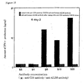
- Figure 8 shows the activation (induction of IFN ⁇ production) of mouse spleen-derived T cells by crosslink of CD3 and AILIM realized by using a plate coated with anti-CD3 antibody and anti-AILIM antibody.
- Figure 9 shows the activation (induction of IFN ⁇ production) of rat spleen-derived T cells by crosslink of CD3 and AILIM realized by using a plate coated with anti-CD3 antibody and anti-AILIM antibody.
- Figure 10 shows the activation (induction of IFN ⁇ production) of human peripheral blood-derived T cells by crosslink of CD3 and AILIM realized using a plate coated with anti-CD3 antibody and anti-AILIM antibody.
- Figure 11 shows the inhibitory effect of anti-AILIM antibody on the increased production of IFN ⁇ , one of the T-cell responses, in human peripheral blood-derived T cells activated by stimulating with anti-CD3 antibody.
- Figure 12 shows the inhibitory effect of anti-AILIM antibody on the increased production of IL-4, one of the T-cell responses, in human peripheral blood-derived T cells activated by stimulating with anti-CD3 antibody.
- Figure 13 shows the inhibitory effect of anti-AILIM antibody on the increased production of IL-4, one of the T-cell responses, in mouse thymus-derived T cells activated by stimulating with anti-CD3 antibody.
- Figure 14 shows the inhibitory effect of anti-AILIM antibody on the increased production of IL-4, one of the T-cell responses, in mouse spleen-derived T cells activated by stimulating with anti-CD3 antibody.
- Figure 15 shows the therapeutic effect on paw swelling which is a parameter of arthrosis in a host suffering from arthrosis by administering anti-AILIM antibody several times.
- Figure 16 shows the therapeutic effect of anti-AILIM antibody on increased production of IFN ⁇ which is a parameter for the worsen condition in a host suffering from hepatitis.
- Figure 17 shows the inhibitory effect of anti-AILIM antibody on increased production of GPT and GOT which are a parameter for the worsen condition in a host suffering from hepatitis.
- Figure 18 shows the inhibitory effect of anti-AILIM antibody on increased production of IgG which is one of graft versus host reactions (GVH reactions) in graft versus host disease (GVHD).
- Figure 19 shows the inhibitory effect of anti-AILIM antibody on increased production of IgE which is one of graft versus host reactions (GVH reactions) in graft versus host disease (GVHD).
- Figure 20 shows the inhibitory effect of anti-AILIM antibody on increased anti-dsDNA antibody titer which is one of graft versus host reactions (GVH reactions) in graft versus host disease (GVHD).
- Figure 21 shows the inhibitory effect of anti-AILIM antibody in vivo in a host immunized with SRBC which is a foreign antigen on production of an antibody (administered immediately after antigen sensitization) against the foreign antigen.
- Figure 22 shows the inhibitory effect of anti-AILIM antibody in vivo in a host sensitized with SRBC which is a foreign antigen on production of an antibody (administered 7 days after antigen sensitization) against a foreign antigen.
- Figure 23 schematically shows the expression pattern of AILIM and of CD28 in normal tissues (thymus, lymph node and peripheral blood) and lesion parts.
- Figure 24 shows the expression patterns for AILIM, CD28, CD4, CD8, CD19, and CTLA-4 in T cells derived from peripheral blood of a normal healthy person and in AILIM-positive cells separated from the T cells, respectively.
- Sub figure (a) shows the distribution of various peripheral blood-derived T cells.
- Sub figure (b) shows the distribution of AILIM-positive cells isolated from peripheral blood-derived T cells.
- Sub figure (c) shows the expression pattern of CD4 and CD8 in peripheral blood-derived T cells.
- Sub figure (d) shows the expression patterns of CD4 and CD8 in AILIM-positive cells separated from peripheral blood derived T cells.
- Sub figure (e) shows the expression patterns of AILIM and CD4 in peripheral blood-derived T cells.
- Sub figure (f) shows the expression patterns of AILIM and CD4 in AILIM-positive cells separated from peripheral blood-derived T cells.
- Sub figure (g) shows the expression patterns of AILIM and CD28 in the peripheral blood-derived T cells.
- Sub figure (h) shows the expression patterns of AILIM and CD28 in AILIM-positive cells separated from the peripheral blood-derived T cells.
- Sub figure (i) shows the expression patterns of AILIM and CTLA-4 in the peripheral blood-derived T cells.
- Sub figure (j) shows the expression patterns of AILIM and CTLA-4 in AILIM-positive cells separated from the peripheral blood-derived T cells.
- Sub figure (k) shows the expression patterns of AILIM and CD19 in the peripheral blood-derived T cells.
- Sub figure (l) shows the expression patterns of AILIM and CD19 in AILIM-positive cells separated from the peripheral blood-derived T cells.
- Figure 25 shows strength of AILIM expression in T cells derived from peripheral blood of a normal healthy person, CD4-positive T cell, CD8-positive T cells, and each AILIM-positive cells separated from each of the T cells.
- Sub figure (a) shows the strength of AILIM expression in peripheral blood T cells, and AILIM-positive cells separated from the T cells, respectively.
- Sub figure (b) shows the strength of AILIM expression in peripheral blood CD4 positive T cells, and CD4-positive AILIM-positive T cells separated from the T cells, respectively.
- Sub figure (c) shows the strength of AILIM expression in peripheral blood CD8 positive T cells, and CD8-positive AILIM-positive T cells separated from the T cells, respectively.
- Figure 26 shows the expression patterns of AILIM in peripheral blood derived T cells and synovial fluid-derived T cells from each patient suffering from chronic rheumatoid arthritis (RA) and osteoarthritis (OA), and in peripheral blood T cells from each patient suffering from progressive systemic sclerosis (scleroderma; PSS) and systemic lupus erythematosus (SLE), respectively
- RA chronic rheumatoid arthritis
- OA osteoarthritis
- PSS systemic sclerosis
- SLE systemic lupus erythematosus
- Figure 27 shows the expression patterns of AILIM in T cells (CD4-positive T cells, CD8-positive T cells) derived from each thymus, spleen, lymph node and peripheral blood in adjuvant-induced arthritis model rats.
- Figure 28 shows expression patterns of each AILIM and CTLA-4 in normal healthy person peripheral blood-derived T cells activated by various stimuli.
- Sub figure (a) shows the expression strength of AILIM in T cells activated by stimulating with PMA and Ionophore.
- Sub figure (b) shows the expression strength of CTLA-4 in T cells activated by stimulating with PMA and Ionophore.
- Sub figure (c) shows the expression strength for AILIM in CD4-positive T cells activated by stimulating with PMA and Ionophore.
- Sub figure (d) shows the expression strength for CTLA-4 in CD4-positive T cells activated by stimulating with PMA and Ionophore.
- Sub figure (e) shows the expression strength of AILIM in T cells activated by stimulating with either anti-CD3 antibody and anti-AILIM antibody or anti-CD3 antibody and anti-CD28 antibody.
- Sub figure (f) shows the expression strength of CTLA-4 in T cells activated by stimulating with either anti-CD3 antibody and anti-AILIM antibody or anti-CD3 antibody and anti-CD28 antibody.
- Figure 29 shows the induction of production of various cytokines from each human peripheral blood T cell by stimulating with various antibodies.
- Figure 30 shows induction of cell proliferation of each human peripheral blood T cell by stimulating with various antibodies at the various concentrations.
- Figure 31 shows induction of cell proliferation in a time course of each human peripheral blood T cells by stimulating with various antibodies.
- Figure 32 shows the levels of cell proliferation in each mouse spleen cells and mouse spleen-derived T cells cultured on a microplate coated with anti-CD3 antibody and anti-AILIM antibody.
- Sub figure (a) shows the levels of proliferation of mouse spleen cells.
- Sub figure (b) shows levels of proliferation of mouse spleen-derived T cells.
- Figure 33 shows levels of proliferation of each mouse spleen cell cultured using microbeads (constant concentration) , coated with anti-CD3 antibody (constant concentration), and anti-AILIM antibody (various concentration).
- Figure 34 shows levels of proliferation of each mouse spleen cell cultured using microbeads (various concentrations) , coated with anti-CD3 antibody (constant concentration), and anti-AILIM antibody (constant concentration).
- Figure 35 shows levels of proliferation of each mouse spleen-derived T cell cultured using microbeads (various concentrations), coated with anti-CD3 antibody (constant concentration), and anti-AILIM antibody (constant concentration).
- Figure 36 shows the induction of cell proliferation of each rat lymph node-derived T cell by the stimulating with various antibodies at the various concentrations.
- Figure 37 shows the therapeutic effect of the single administration (constant concentration) of anti-AILIM antibody on paw swelling which is a parameter for arthrosis in a host suffering from arthrosis.
- Figure 38 shows the therapeutic effect of the single administration (various concentration) of anti-AILIM antibody on paw swelling which is a parameter for arthrosis in a host suffering from arthrosis.
- Figure 39 shows the inhibitory effect of anti-AILIM antibody on the increased production of IgG which is one of the graft versus host reactions (GVH reactions) in graft versus host disease (GVHD).
- Figure 40 shows the inhibitory effect of anti-AILIM antibody on the increased production of IgE which is one of the graft versus host reactions (GVH reactions) in graft versus host disease (GVHD).
- Figure 41 shows the inhibitory effect of anti-AILIM antibody on the increased anti-dsDNA antibody titer which is one of the graft versus host reactions (GVH reactions) in graft versus host disease (GVHD).
- Figure 42 shows the inhibitory effect of anti-AILIM antibody in a host in vivo sensitized with NP-KLH which is a foreign antigen on the production of IgG1 antibody against the foreign antigen.
- Figure 43 shows the inhibitory effect of anti-AILIM antibody in a host in vivo sensitized with NP-KLH which is the foreign antigen on the production of IgM antibody against the foreign antigen.
- Figure 44 shows the inhibitory effect of anti-AILIM antibody in a host in vivo sensitized with NP-KLH which is the foreign antigen on the production of IgG1 antibody against the foreign antigen.
- Figure 45 shows the inhibitory effect of anti-AILIM antibody in a host in vivo sensitized with NP-KLH which is the foreign antigen on the production of IgG2b antibody against the foreign antigen.
- Figure 46 shows the inhibitory effect of anti-AILIM antibody in a host in vivo sensitized with NP-KLH which is the foreign antigen on the production of IgG2a antibody against the foreign antigen.
- Figure 47 shows the inhibitory effect on T cell proliferation in the case of culturing T cells from a normal healthy person "donor A", with PBMC of a normal healthy person “donor D” by various test samples in the proliferation test of the T cells through the mixed lymphocyte reactions (MLR).
- MLR mixed lymphocyte reactions
- the vertical axis indicates the amount of incorporation of [ 3 H] thymidine as an index showing a level of cell proliferation, and the horizontal axis shows the concentration of the test samples.
- Figure 48 shows the inhibitory effect on T cell proliferation in the case of culturing T cells from a normal healthy person "donor D", with PBMC of a normal healthy person “donor B” by various test samples in the proliferation test of the T cells through the mixed lymphocyte reaction (MLR).
- MLR mixed lymphocyte reaction
- the vertical axis indicates the amount of incorporation of [ 3 H] thymidine as an index showing a level of cell proliferation, and the horizontal axis shows the concentration of the test samples.
- Figure 49 shows the inhibitory effect on T cell proliferation in the case of culturing T cells from a normal healthy person "donor C", with PBMC of a normal healthy person "donor A” by various test samples in the proliferation test of the T cells through the mixed lymphocyte reaction (MLR).
- MLR mixed lymphocyte reaction
- the vertical axis indicates the amount of incorporation of [ 3 H] thymidine as an index showing a level of cell proliferation, and the horizontal axis shows the concentration of the test samples.
- Figure 50 shows the inhibitory effect on T cell proliferation in the case of culturing T cells from a normal healthy person "donor E", with PBMC of a normal healthy person “donor G” by various test samples in the proliferation test of the T cells through the mixed lymphocyte reaction (MLR).
- MLR mixed lymphocyte reaction
- the vertical axis indicates the amount of incorporation of [ 3 H] thymidine as an index showing a level of cell proliferation, and the horizontal axis shows the concentration of the test samples.
- Figure 51 shows the inhibitory effect on T cell proliferation in the case of culturing T cells from a normal healthy person "donor F", with PBMC of a normal healthy person “donor E” by various test samples in the proliferation test of the T cells through the mixed lymphocyte reaction (MLR).
- MLR mixed lymphocyte reaction
- the vertical axis indicates the amount of incorporation of [ 3 H] thymidine as an index showing a level of cell proliferation, and the horizontal axis shows the concentration of the test samples.
- Figure 52 shows the inhibitory effect on T cell proliferation in the case of culturing T cells from a normal healthy person "donor G", with PBMC of a normal healthy person “donor F” by various test samples in the proliferation test of the T cells through the mixed lymphocyte reaction (MLR).
- MLR mixed lymphocyte reaction
- the vertical axis indicates the amount of incorporation of [ 3 H] thymidine as an index showing a level of cell proliferation, and the horizontal axis shows the concentration of the test samples.
- mice Male, 5- to 8-week-old
- BALB/c mice Male, 5- to 8-week-old
- Wistar rats Male, 5- to 6-week-old were purchased from Charles River Japan Inc.
- Hybridomas designated as "JTT-1” and "JTT-2" capable of producing mouse anti-rat AILIM monoclonal antibody (mouse anti-rat JTT-1 antigen monoclonal antibody) which were created and reported previously by the present inventors, have been deposited internationally (as of October 11, 1996, both hybridomas have been deposited internationally in The National Institute of Bioscience and Human-Technology, The Agency of Industrial Science and Technology, The Ministry of International Trade and Industry (1-1-3 Higashi, Tsukuba, Ibaraki, Japan (Zip Code: 305-8566)), which is an international depositary authority under the Budapest Treaty) .
- JTT-1 antibody and “JTT-2 antibody” (IgG1), respectively.
- JTT-1 antibody and JTT-2 antibody are also referred to as “JMab49” and “JMab50,” respectively.
- JTT-2 antibody also referred to as JMab50; IgG1
- mice Female, 7- to 8-week-old mice
- intraperitoneal injection 10 6 to 10 7 cells/0.5ml/mouse for each hybridoma
- the ascites was collected from each mouse by laparotomy under anesthesia according to a commonly used method.
- the respective mouse anti-human AILIM monoclonal antibodies were thus prepared on a large scale (JP-A 11-29599 (Example 12), and WO98/38216 (Example 12)).
- SA12 antibody IgG1
- SG430 antibody IgG1
- the antibodies were prepared as follows:
- a transformed cell capable of expressing mouse AILIM was prepared using the cDNA encoding the full-length amino acid sequence of mouse AILIM (mouse JTT-1 antigen) (JP-A 11-29599 (SEQ ID NO: 5), and WO98/38216 (SEQ ID NO: 5)), which had previously been cloned by the present inventors.
- the transformed cells were homogenized and then subjected to ultracentrifugation (100,000 x g).
- the resulting precipitate containing cell membrane fraction was recovered and then suspended in PBS.
- the obtained cell membrane fraction was given to Wistar rats by footpad injection for primary immunization (0 day) .
- the cell membrane fraction antigen was repeatedly given to the rats by footpad injection, on seventh day, fourteenth day and twenty-eighth day after the primary administration. Two days after the final immunization, the lymph node cells were collected from the rats.
- the lymph node cells were mixed with mouse myeloma PAI cells (JCR No. B0113; Res. Disclosure, Vol.217, pp.155, 1982) at a ratio of 5:1.
- the cells were fused to each other by using polyethylene glycol 4000 (Boehringer Mannheim) as a fusing agent to create hybridomas capable of producing monoclonal antibodies.
- Selection of hybridoma was performed by culturing the fused cells in an ASF104 medium (Ajinomoto) containing in HAT, 10% fetal calf serum and aminopterin.
- mice JTT-1 antigen The reactivity against mouse AILIM (mouse JTT-1 antigen) of each monoclonal antibody in culture supernatant of hybridoma was tested as follows: each culture supernatant was allowed to react to the transformed cells expressing the above-mentioned recombinant mouse AILIM; and then the cells were further allowed to react to FITC-labeled anti-rat IgG (Cappel); fluorescence intensities of the stained cells were measured in an EPICS-ELITE flow cytometer. The screening yielded multiple hybridomas capable of producing monoclonal antibodies reactive to mouse AILIM (mouse JTT-1 antigen).
- hybridomas Two distinct hybridomas selected from them were named "B10.5" and "B9B6.”
- Each of the hybridomas (10 6 to 10 7 cells/0.5 ml/mouse for each) was given to ICR nu/nu mice (female, 7- to 8-week old) by intraperitoneal injection. 10 to 20 days after the injection, the ascites was collected from each mouse by laparotomy under anesthesia according to a commonly used method.
- the rat anti-mouse AILIM monoclonal antibodies were thus prepared on a large scale.
- thymus and peripheral lymphoid tissues were taken out according to a commonly used method. The tissues were cut into small blocks on a stainless mesh.
- Cell suspensions were prepared by suspending the tissue blocks in RPMI1640 medium containing 10 % fetal calf serum (FCS). The suspensions of cell (1 x 10 7 to 3 x 10 7 cells/ml for each) were plated in a dish and then cultured for 2 hours in a CO 2 incubator. After the culture was completed, non-adhesive cells were carefully recovered from the dish and then washed with RPMI1640 medium. Mouse cells were thus obtained from various tissues.
- Rat cells from various tissues were also prepared from thymus and peripheral lymphoid tissues (spleen and lymph node) by the same method as described above.
- Human peripheral blood T cells (from healthy normal persons and patients) were prepared according to a commonly used method. Specifically, heparinated blood sample collected from a healthy normal person or patient was subjected to treatment of separation with lymphoprep (Nycomed) to yield peripheral blood mononuclear cells. Subsequently, T cells were recovered by using a Pan T Cell Isolation kit (Miltenyi).
- mice T cell D10, MS202, CD28KO, EL-4, 2L2, and BC3C13
- mouse T cell-derived hybridomas KV24, D0.11.10, 8-4-31, 3H10-11, 61-21-25, 1-2-66, and 6-13-64 that originate from a mouse T cell-derived hybridoma BW5147, all of which were established by the present inventors.
- Cells prepared from various tissues by the methods described above as well as T cells or various T cell lines unstimulated or stimulated by each of stimulating substances (anti-CD3 antibody, ConA, or PMA and ionophore, etc.) were re-suspended in phosphate buffer (Ca 2+ , Mg 2+ -free PBS; PBS-) containing 0.5% bovine serum albumin (BSA) and 5 mM EDTA.
- phosphate buffer Ca 2+ , Mg 2+ -free PBS; PBS-
- BSA bovine serum albumin
- 5 mM EDTA 5 mM EDTA
- the cells were washed 3 times with the above-mentioned phosphate buffer, and then re-suspended in the same buffer.
- the primary antibody used was an unlabeled anti-AILIM antibody (namely, one of the above-mentioned (B) antibodies)
- further FITC-, PE- orbiotin-labeled anti-mouse Ig antibody or anti-rat Ig antibody was added as a secondary antibody to the cell suspensions.
- the suspensions were incubated in the same manner as described above.
- the secondary antibody was a biotin-labeled antibody
- a PE-labeled streptavidin Pharmingen
- the sizes of the cells stained by the above-mentioned method and their fluorescence intensities were measured in a FACSort (Becton Dekinson) .
- the distribution of AILIM expression was analyzed by using Lysis II software.
- AILIM in T cells isolated from mouse thymus, being a normal lymphoid tissue, was analyzed according to the same method described above in ⁇ 2-1>. At the same time, similar measurements were carried out to analyze the correlation between the expression pattern of AILIM and the expression of other molecules (CD3 molecule functioning in the primary signaling to T cell, CD28 molecule functioning in the secondary stimulatory signaling, CD4 and CD8 that are T cell surface markers).
- thymus T cell The differentiation and maturation of thymus T cell is known to be achieved through the following major steps.
- AILIM nor CD28 was recognizably expressed in CD4-negative CD8-negative cells but weak levels of expression were observed in CD4 weak-positive CD8 weak-positive cells. The expression of CD28 was maximized in CD4-positive CD8-positive cells and then decreased as the cells further differentiated to maturity.
- AILIM low-level expression of AILIM was found even in the CD4-positive CD8-positive cells.
- the level of AILIM expression was however elevated during the subsequent cell differentiation processes; namely, as the level of CD4 or CD8 expression decreased, the level of AILIM expression was elevated and then maximized at the final differentiation stage of lymphocyte of which positive selection is completed; the mature lymphocyte is called SP cell (CD4-positive CD8-negative cell or CD4-negative CD8-positive cell).
- the experimental result shows that the expression pattern of AILIM is different from that of CD28 with respect to the correlation to the expression of CD4 and CD8 as well as that of CD3.
- AILIM in T cells of spleen and lymph node, which are mouse normal lymphoid tissues, was analyzed by the above-mentioned method.
- AILIM-positive T cell among spleen-derived T cells is smaller than that in the thymus, and the percentage was about 1 to 3%.
- Majority of the AILIM-positive cells were CD4-positive and CD8-negative.
- the expression pattern of AILIM in lymph node-derived T cells and the percentage population were comparable to those in the above-mentioned spleen-derived T cells.
- Hepatitis model mice were prepared as follows:
- a suspension of Propionibacterium acnes ( P. acnes ; 5 mg/ml) in phosphate buffer (PBS- ; 0.2 ml) was intravenously injected into the tail of C57BL/6 mouse. After one week, a solution of LPS (lipopolysaccaride ; 1.5 ⁇ g/ml) in phosphate buffer (PBS- ; 0.2 ml) was intravenously injected into the mouse to induce hepatitis. The mouse was used as a hepatitis model animal.
- T cells were prepared from the cells by the above-mentioned method to analyze AILIM expression.
- the expression level of AILIM was highly elevated in the T cells (mononuclear cells) derived from the liver tissue of the hepatitis model mouse; most of the T cells expressed AILIM at markedly high levels.
- the level of AILIM expression in T cells derived from the liver of the hepatitis model was considerably higher than those in spleen-derived T cells (CD4 positive cells) and lymph node-derived T cells from normal mouse.
- AILIM-expressing T cells were obtained as follows: a fraction of mononuclear cells separated from peripheral blood of a healthy normal person were suspended in PBS- that contained 0.5% BSA and 5 mM EDTA, and anti-AILIM antibody (SG430 ; 50 ⁇ g) was added thereto. The mixture was incubated at 4°C for 30 minutes. Subsequently, after the cells were washed 3 times with the same buffer, microbeads with immobilized goat anti-mouse IgG (100 to 500 ⁇ l; Miltenyi) were added thereto. The mixture was incubated in the same manner and then washed with the same buffer.
- the cells were subjected to a treatment using magnetic separation column (twice) according to a usual method to isolate AILIM-positive cells.
- the isolated AILIM-positive cells were stained with each of a variety of labeled antibodies and anti-AILIM antibodies; the cells stained were analyzed by flow cytometer.
- Peripheral blood T cells are primarily grouped into CD4-positive CD8-negative cells and CD4-negative CD8-positive cells.
- the expression of AILIM was recognized mainly in CD4-positive cells.
- Double staining with the anti-AILIM antibody and anti-CD28 antibody revealed that almost all the peripheral blood-derived AILIM-positive cells expressed CD28.
- the percentage population of AILIM-positive cell in peripheral blood T cells was roughly estimated to 0.5 to 5%.
- AILIM IL-12
- OA osteoarthritis
- SLE systemic lupus erythematosus
- T cells were separated from each of synovial fluid and peripheral blood of arthritis patients; T cells were also isolated from peripheral blood of patients with autoimmune disease. In addition, T cells of peripheral blood from healthy normal persons were used as a control.
- AILIM peripheral blood-derived T cells from RA patients was compared with that in the cells from healthy normal persons. Between the patients and normal persons, the expression levels were not significantly different in CD4-positive T cells and also in CD4-negative T cells (namely CD4-negative CD8-positive T cells).
- the population of AILIM-expressing cells was significantly increased among CD4-positive T cells and also among CD4-negative T cells in T cells derived from synovial fluids of RA patients.
- the averaged proportion of AILIM-expressing cells was increased to about 20% of total CD4-positive T cells.
- the expression level of AILIM was significantly elevated in CD4-positive T cells and CD4-negative T cells from synovial fluids of RA patients as compared with those in CD4-positive T cells and CD4-negative T cells derived from peripheral blood of healthy normal persons.
- the expression level of CD28 was not altered in CD4-positive T cells from RA patients.
- the population of AILIM-positive cells among CD4-negative T cells from peripheral blood of patients with autoimmune diseases was comparable to that amongthe cells from healthy normal persons.
- the population of AILIM-positive cell was significantly elevated among CD4-positive T cells from PSS patients as compared with that among the cells from healthy normal persons.
- Dead tubercle bacillus (M. Tuberculosis H37Ra; Difco) of 10 mg/ml in liquid paraffin (Wako pure chemical) was used as an adjuvant.
- the adjuvant was intracutaneously injected into Wistar rats (male, 5-week-old, Charles River) at a concentration of 1 mg/0.1 ml/individual in a tail head area to induce arthritis.
- the volume of both hind legs was measured by using a plethysmometer. The volume determined was used as an index for the onset of arthritis.
- T cell suspensions were prepared according to the above-mentioned method.
- the T cells were stained with each of anti-CD4 antibody, anti-CD8 antibody and anti-AILIM antibody by the above-mentioned method. Then the expression of CD4, CD8 and AILIM was analyzed by flow cytometer.
- Control T cells used were those derived from the thymus, spleen, lymph node and peripheral blood of normal rats.
- the population of AILIM-positive cell was significantly increased among lymph node-derived CD4-positive T cells as well as among CD8-positive T cells when compare to that among the control cells.
- the expression of AILIM reached a peak level on the fifth day after the adj uvant administration in CD4-positive T cells.
- Mouse T cell derived from lymphoid tissues were activated under a variety of conditions. The varying level of AILIM expression associated with T cell activation was analyzed.
- T cells were activated by the addition of anti-CD3 antibody (final concentration: 1 to 10 ⁇ g/ml), concanavalin A (ConA ; final concentration: 1 to 5 ⁇ g/ml), or PMA (phorbol myristate acetate; final concentration: 20 ng/ml) and Ca ionophore (final concentration: 200 ng/ml) to the suspension of T cells in 10% FCS-containing RPMI1640 medium for the stimulation.
- the expression of AILIM was analyzed over time (after 0, 6, 12, 24, and 48 hours) after addition of the activating agent.
- the AILIM expression started to be up-regulated about 3 to 6 hours after the stimulation under any of the activation conditions and was maximized 12 hours after the stimulation.
- the level of AILIM expression remained to be high about 24 hours after the stimulation, and the high level was still maintained 48 hours after the stimulation.
- T cells derived from peripheral blood of healthy normal persons in the same manner as described above were activated by the method of (A) or (B) as descried below.
- the analysis was carried out for the T cell activation-associated expression of AILIM and CTLA-4 that is a costimulatory molecule.
- peripheral blood-derived human T cells (1 x 10 5 cells/ml, 0.1 ml/well) in 10% FCS-containing RPMI1640 medium was added to each plate. The cells were harvested after cultured for 2 to 3 days. The expression of AILIM and CTLA-4 was analyzed by using a flow cytometer according to the above-mentioned method.
- AILIM expression was induced 8 hours after the stimulation for T cell activation with PMA and the ionophore. The expression level was much higher than that of CTLA-4 expression that was also induced by the stimulation. Furthermore, AILIM expression was induced in almost all the T cells. In addition, double staining test for CD4 and AILIM, or for CD8 and AILIM, showed that the activation induced a significant level of AILIM expression in CD4-positive T cells as well as in CD8-positive T cells.
- T cell lines are known to be established chiefly by spontaneous immortalization, or as T cell lines immortalized with chemical agent or as T cell hybridoma by fusing T cell with myeloma cell.
- T cell lines are grouped into two classes based on the property of cytokine production, namely Th1-type T cell lines and Th2-type T cell lines.
- AILIM was recognized to be expressed constitutively in T cell lines exhibiting the property of Th2-type T cell line in regard to cytokine production (D10, MS202, CD28KO, EL-4, etc.). The level of AILIM expression was comparable or higher relative to that of CD28 in these cell lines.
- the anti-AILIM antibody of the present invention had the activities in modulating (enhancing and/or suppressing) T cell response (production of a cytokine such as IFN- ⁇ and IL-4, cell proliferation, etc.), namely whether or not the antibody had the capability in regulating the transduction of AILIM-mediated costimulatory signal into cell.
- the analysis was carried out by using the quantities of cytokines (IFN- ⁇ and IL-4) produced by the cells and the degree of cell proliferation as indices.
- one or two types of antibodies (anti-CD3 antibody alone, anti-CD28 antibody alone, anti-CD3 antibody and anti-AILIM antibody, or anti-CD3 antibody and anti-CD28 antibody) prepared above were selected and added to wells of a 96-well microplate.
- the plate was incubated at 37°C for 1 hour or more to coat the plate with one or two antibodies.
- the plate was then sufficiently washed with PBS. Subsequently, thymus cells (5 x 10 5 cells/well), spleen cells (2 x 10 5 cells/well) or purified T cells (1 x 10 5 to 3 x 10 5 cells/well), which had been prepared in the above method, were added to the plate.
- CTLA4-Ig fusion protein between the soluble region of CTLA4 and IgFc
- the plate was incubated for 2 to 4 days in a CO 2 incubator.
- concentrations of cytokines (IFN- ⁇ or IL-4) in the culture supernatant were determined by a commonly used ELISA method. Further, the degree of cell proliferation during the culture was evaluated by a common method utilizing tritium-labeled thymidine ( 3 H-TdR) incorporation.
- T cells produce specific cytokines in response to T cell receptor-mediated primary signal and secondary signal mediated by costimulatory molecules such as CD28 and CTLA-4.
- cytokines induced by the stimulation with a variety of antibodies was analyzed by using peripheral blood T cells, thymus cells or spleen cells isolated from each of mouse, rat and human according to the above-mentioned test method as described in ⁇ 3-1>.
- T cells derived from mouse spleen were added to (1) a microplate coated with anti-CD3 antibody (clone 145-2C11 ; Pharmingen: 0 to 3 ⁇ g/ml) and anti-CD28 antibody (clone CD28.2; 1 ⁇ g/well) ; (2) a microplate coated with anti-CD3 antibody and anti-mouse AILIM antibody (clone B10.5; 1 ⁇ g/well); and (3) a microplate coated with anti-CD3 antibody alone. The plates were incubated to cultivate the cells and then quantities of IFN ⁇ in the culture supernatants were determined by ELISA.
- IFN ⁇ The production of IFN ⁇ was not induced by the stimulation with anti-CD3 antibody alone. However, the production of IFN ⁇ was significantly induced by the stimulation with anti-CD3 antibody/anti-AILIM antibody or anti-CD3 antibody/anti-CD28 antibody. The induction was enhanced depending on the concentration of anti-CD3 antibody.
- T cells derived from rat spleen were added to (1) a microplate coated with anti-CD3 antibody (clone G4.18; 50 ng/well) and anti-CD28 antibody (clone JJ316; 1 ⁇ g/well); (2) a microplate coated with anti-CD3 antibody and anti-rat AILIM antibody (clone JTT1; 1 ⁇ g/well); (3) a microplate coated with anti-CD3 antibody alone; (4) a microplate coated with anti-AILIM antibody alone; and (5) a microplate coated with anti-CD28 antibody alone. The plates were incubated to cultivate the cells and then quantities of IFN ⁇ in the culture supernatants were determined by ELISA.
- IFN ⁇ The production of IFN ⁇ was not significantly induced by the stimulation with anti-CD3 antibody alone, anti-AILIM antibody alone, or anti-CD28 antibody alone. However, the production of IFN ⁇ was significantly induced by the stimulation with anti-CD3 antibody/anti-AILIM antibody or anti-CD3 antibody/anti-CD28 antibody. The level of production resulted from the induction was elevated over time.
- Human T cells derived from peripheral blood were added to (1) a microplate coated with anti-CD3 antibody (clone OKT3; constant concentration) and anti-AILIM antibody (clone SA12; various concentrations); and (2) a microplate coated with anti-CD3 antibody alone.
- the plates were incubated to cultivate the cells and then quantities of IFN ⁇ in the culture supernatants were determined by ELISA.
- the solution of anti-AILIM antibody was added after the cells were added.
- T cells derived from peripheral blood each of two unrelated healthy normal donors were added to (1) amicroplatecoatedwithanti-CD3 antibody (clone OKT3; 200 ng/well) alone; (2) a microplate coated with anti-CD28 antibody (clone CD28.2; 1 ⁇ g/well) alone; (3) a microplate coated with anti-human AILIM antibody (clone SA12; 1 ⁇ g/well); (4) a microplate coated with anti-CD3 antibody and anti-CD28 antibody; (5) a microplate coated with anti-CD3 antibody and anti-AILIM antibody; and (6) a microplate coated with anti-CD3 antibody, anti-AILIM antibody and anti-CD28 antibody.
- amicroplatecoatedwithanti-CD3 antibody clone OKT3; 200 ng/well
- a microplate coated with anti-CD28 antibody clone CD28.2; 1 ⁇ g/well
- anti-human AILIM antibody clone SA12; 1 ⁇ g/well
- TNF ⁇ tumor necrosis factor- ⁇
- IFN ⁇ interferon- ⁇
- IL-2 interleukin-2
- IL-4 interleukin-4
- IL-10 interleukin-10
- TNF ⁇ , IFN ⁇ and IL-2 are cytokines produced by Th1-type T cells; and IL-4 and IL-10 are cytokines produced by Th2-type T cells.
- the above-mentioned test indicates that the anti-CD3 antibody immobilized on the plate functioned as MHC on antigen-presenting cell and also the anti-AILIM antibody immobilized on the plate functioned as an AILIM ligand, resulting the primary signal and secondary stimulatory signal (costimulatory signal) , which are responsible for T cell activation, were transduced into the added T cells.
- anti-AILIM antibody and anti-CD28 antibody were capable of inhibiting the induction of IFN ⁇ and IL-4 production as a T cell response induced when T cells were cultured in a microplate coated with anti-CD3 antibody.
- Peripheral blood-derived T cells, thymus-derived T cells or spleen-derived T cells were plated in a microplate coated with anti-CD3 antibody alone, and then any one of anti-AILIM antibody (various concentrations), anti-CD28 antibody (various concentrations) or CTLA4-IgFc (control) was added thereto.
- the amounts of IFN ⁇ or IL-4 in the culture supernatant were determined according to the method as described above in ⁇ 3-1>.
- anti-AILIM antibody significantly inhibited the production of both IFN ⁇ and IL-4 induced by anti-CD3 antibody stimulation in peripheral blood-derived T cells ( Figures 11 and 12).
- the addition of anti-AILIM antibody also inhibited the proliferation of cells.
- the addition of anti-CD28 antibody inhibited neither the cytokine production nor the cell proliferation.
- anti-AILIM antibody markedly inhibited anti-CD3 antibody-induced IL-4 production in thymus-derived T cells ( Figure 13).
- anti-AILIM antibody also inhibited the cell proliferation.
- CTLA4-IgFc as a control resulted in neither significant inhibition of IL-4 production nor significant inhibition of cell proliferation.
- anti-AILIM antibody markedly inhibited anti-CD3 antibody-induced IL-4 production in spleen-derived T cells ( Figure 14).
- anti-AILIM antibody also inhibited the cell proliferation.
- CTLA4-IgFc as a control resulted in neither significant inhibition of IL-4 production nor significant inhibition of cell proliferation.
- T cells proliferate in response to T cell receptor-mediated primary signal and secondary signal mediate by costimulatory molecules such as CD28 and CTLA-4.
- the proliferation of cells induced by the stimulation with a variety of antibodies was analyzed by using T cells derived from peripheral blood of healthy normal persons, mouse spleen cells, mouse spleen-derived T cells, and rat lymph node T cells according to the above-mentioned test method as described in ⁇ 3-1>.
- Human T cells derived from peripheral blood were added to (1) a microplate coated with anti-CD3 antibody (clone OKT3; 200 ng/well; Ortho Diagnostic Systems) alone; (2) a microplate coated with anti-CD3 antibody and anti-CD28 antibody (clone CD28.2; various concentrations; Pharmingen); (3) a microplate coated with anti-CD3 antibody (200 ng/well) and anti-AILIM antibody (clone SA12; various concentrations) ; and (4) a microplate coated with anti-CD3 antibody (200 ng/well), anti-human AILIM antibody (various concentrations) and anti-CD28 antibody (1 ⁇ g/well).
- the plates were incubated to cultivate the cells and then the degree of cell proliferation was evaluated over time by a test of tritium-labeled thymidine ( 3 H-TdR) incorporation according to a commonly used method.
- 3 H-TdR tritium-labeled thymidine
- the degree of T cell proliferation was evaluated by using the microplates as indicated below in (5), (6) and (7) in the same manner as described above, in order to investigate the time course of proliferation of human peripheral blood-derived T cells induced by the stimulation with the above-mentioned various antibodies.
- T cells Proliferation of T cells was recognizable 18 hours after the stimulation in any of the antibody combinations.
- the stimulation with the combination of anti-CD3 antibody and antibody CD28 antibody (as described above in (5)) induced the highest level of T cell proliferation 40 hours after the antibody stimulation, but the activity of inducing T cell proliferation previously reached equilibrium with the combination.
- T cell proliferation induced by the stimulation with anti-CD3 antibody and anti-AILIM antibody reached a peak 60 hours after the stimulation.
- T cell proliferation induced 60 hours after the stimulation with the antibodies were significantly higher than that in the combination of anti-CD3 antibody and anti-CD28 antibody.
- 96-well microplates were coated with anti-CD3 antibody (clone 145-2C11; Pharmingen; 50 ng/well). Then, the plates were further coated with various concentrations of anti-mouse AILIM antibody (clone B10.5) or anti-NP-KLH antibody as a control antibody.
- Anti-mouse AILIM antibody clone B10.5
- Anti-NP-KLH antibody anti-NP-KLH antibody
- the anti-NP-KLH antibody used as a control antibody was prepared by using as antigen NP-KLH, which is KLH (keyhole limpet hemocyanin; PIERCE) linked with NP (Nitrophenol) that is a hapten.
- NP-KLH which is KLH (keyhole limpet hemocyanin; PIERCE) linked with NP (Nitrophenol) that is a hapten.
- latex microbead was used as a carrier on which antibody is immobilized.
- Cell proliferation test was carried out by using the bead in the same manner as described above.
- D-PBS 1 x 10 7 microbeads were treated with (1) 1 ⁇ g/ml of anti-CD3 antibody (clone 145-2C11; Pharmingen) and various concentrations of anti-AILIM antibody (clone B10.5), or (2) 1 ⁇ g/ml of anti-CD3 antibody and various concentrations of anti-NP-KLH antibody.
- the mixtures containing beads were incubated for 1 hour or more, and then the beads were washed with D-PBS. The antibodies were thus immobilized on the beads.
- C57BL/6 mouse spleen cells (1 x 10 5 /well) suspended in 10% FCS-containing RPMI1640 medium were added to each well of a 96-well microplate and the beads (1 x 10 5 particles/well) were added thereto. The mixture was incubated for 56 hours. The degree of cell proliferation after the reaction was determined by tritium-labeled thymidine ( 3 H-TdR) incorporation test in a usual manner.
- 3 H-TdR tritium-labeled thymidine
- the proliferation of C57BL/6 mouse spleen cells was induced even either by the stimulation with anti-CD3 antibody and anti-AILIM antibody or by the stimulation with anti-CD3 antibody and anti-CD28 antibody.
- the degree of cell proliferation increased depending on increase of the concentration of anti-AILIM antibody or anti-CD28 antibody immobilized on the beads (increase in the ratio of concentration of anti-AILIM antibody or anti-CD28 antibody against anti-CD3 antibody concentration).
- the degree of cell proliferation was maximized when both ratio of anti-CD3 antibody concentration vs. anti-AILIM antibody concentration and ratio of anti-CD3 antibody concentration vs. anti-CD28 antibody concentration were 1:9.
- mouse cells used in the test were BALB/C mouse spleen cells and BALB/C mouse spleen-derived T cells.
- BALB/C mouse spleen cells The proliferation of both BALB/C mouse spleen cells and BALB/C mouse spleen-derived T cells was induced even either (1) by the stimulation with anti-CD3 antibody alone; (2) by the stimulation with anti-CD3 antibody and anti-AILIM antibody; or (3) by the stimulation with anti-CD3 antibody and anti-CD28 antibody.
- the degree of cell proliferation increased depending on increase of the concentration of microbeads (namely, antibody concentration) added to the cell culture.
- the proliferation of both BALB/C mouse spleen cell and BALB/C mouse spleen-derived T cell was maximized when the concentration of microbead added to the cell culture was 30,000 particles/well.
- the same result was obtained by using beads coated with anti-CD3 antibody and anti-AILIM antibody as well as by using microbeads coated with anti-CD3 antibody and anti-CD28 antibody.
- Rat lymph node T cells (1 x 10 5 cells/well) suspended in 10% FCS-containing RPMI1640 medium were added to (1) a microplate coated with anti-CD3 antibody (clone G4.18; 50 ng/well) and anti-CD28 antibody (clone JJ319; various concentrations ; Pharmingen); (2) a microplate coated with anti-CD3 antibody (50 ng/well) and anti-AILIM antibody (various concentrations); and (3) a microplate coated with anti-CD3 antibody (50 ng/well) and negative control antibody MOPC21 (various concentrations; Pharmingen). The plates were incubated in order to cultivate the cells at 37°C for 44 hours.
- tritium-labeled thymidine 3 H-TdR
- 3 H-TdR tritium-labeled thymidine
- rat lymph node T cells Although the proliferation of rat lymph node T cells was not induced by anti-CD3 antibody alone, the proliferation was significantly enhanced even either by the stimulation with anti-CD3 antibody and anti-AILIM antibody or by the stimulation with anti-CD3 antibody and anti-CD28 antibody. The proliferation depended on the concentration of anti-AILIM antibody or anti-CD28 antibody that was immobilized on the plate.
- anti-rat AILIM antibody JTT-2 antibody; also referred to as JMab50 ; 20 mg/kg
- JMab50 anti-rat AILIM antibody
- a control experiment was carried out in a group of normal rats (4 individuals) to which neither adjuvant nor antibody was given and in a negative-control group of rats to which mouse anti-human CETP antibody was given (clone JHC1; also referred to as JMab109; JP-A 9-20800), instead of anti-rat AILIM antibody.
- clone JHC1 also referred to as JMab109; JP-A 9-20800
- clone JHC1 also referred to as JMab109; JP-A 9-20800
- anti-AILIM antibody or negative control antibody was intravenously administered only once on the third, fifth or seventh day after the adjuvant administration (0 day) ;
- the anti-AILIM antibody and negative control antibody used were, respectively, anti-rat AILIM antibody (JTT-2 antibody; also referred to as JMab50; 20 mg/kg) and anti-rat CETP antibody (JHC1; 20 mg/kg).
- the administration of the anti-AILIM antibody was conducted even only once, but the paw swelling was significantly suppressed in any cases where the antibody was given once on the third, fifth or seventh day after the adjuvant administration.
- the paw swelling was almost completely inhibited.
- the degree of the inhibition was substantially the same as that in the group of normal rats to which arthritis was not induced.
- anti-rat AILIM antibody JTT-2 antibody; also referred to as JMab50
- JMab50 anti-rat AILIM antibody
- JTT-2 another anti-rat AILIM antibody (JTT-1; also referred to as JMab-49; 20 mg/kg) was administered only once in the same manner for comparison.
- JTT-1 also referred to as JMab-49; 20 mg/kg
- anti-rat CETP antibody JHC1 ; 20 mg/kg was intravenously administered only once.
- P . acnes Propionibacterium acnes
- PBS phosphate buffer
- LPS Lipopolysaccaride
- plasma IFN- ⁇ concentration was determined by ELISA.
- concentrations of plasma GOT (glutamic-oxaloacetic transaminase) and GPT (glutamic-pyruvic transaminase) were measured in a biochemical analyzer (Fara).
- anti-mouse AILIM monoclonal antibody (B10.5 antibody; 5, 50, or 500 ⁇ g/individual) was intraperitoneally administered to the mice, and then the relieving effect of anti-AILIM antibody on hepatitis was evaluated.
- the anti-mouse AILIM antibody was not given to a control group.
- anti-AILIM antibody significantly inhibited the elevation of blood IFN- ⁇ level in an antibody concentration-dependent fashion.
- the administration of anti-AILIM antibody (50 ⁇ g/individual) also significantly inhibited the elevation of GOT and GPT levels.
- BALB/c mouse spleen cells (8 x 10 7 cells/individual) were intravenously given to F1 mice (8- to 10-week-old, 3 individuals) obtained by the bleeding of BALB/c mouse and C57BL/6 mouse.
- anti-mouse AILIM monoclonal antibody (B10.5 antibody; 400 ⁇ g/individual) was intravenously administered to the mice.
- B10.5 antibody 200 ⁇ g/individual was intraperitoneally given to the mice.
- mice Immediately, 1, 2, 3 and 6 weeks after the administration of the spleen cells (0 day), blood was collected from the mice.
- the IgG1, IgE and anti-dsDNA antibody titer in the sera were determined according to a usual method.
- the values of anti-dsDNA antibody titer were normalized by using an anti-dsDNA antibody in the serum from a spontaneous autoimmune disease mouse as a standard.
- hCTLA4-Ig fusion protein between the soluble region of human CTLA4 and the constant region of immunoglobulin
- PBS was given to a negative-control group in the same manner.
- the increase of serum IgG, IgE, and anti-dsDNA antibody titer as an index of GVH reaction was significantly suppressed in the group subjected to the administration of anti-AILIM antibody as compared with the negative control. Further, the suppressing effect was comparable to that observed in the positive control group subjected to the administration of hCTLA4-Ig.
- BALB/c mouse spleen cells (1 x 10 8 cells/individual) were intravenously given to F1 mice (8- to 10-week-old, 3 individuals) obtained by the bleeding of BALB/c mouse and C57BL/6 mouse.
- anti-mouse AILIM monoclonal antibody (B10.5 antibody; 200 ⁇ g/individual) was intravenously administered to the mice.
- B10.5 antibody 100 ⁇ g/individual was intraperitoneally given to the mice.
- mice Immediately, 1, 2, 3, 6, 9 and 12 weeks after the administration of the spleen cells (0 day), blood was collected from the mice.
- the IgG1, IgE and anti-dsDNA antibody titer in the sera were determined according to a usual method.
- the values of anti-dsDNA antibody titer were normalized by using an anti-dsDNA antibody in the serum from a spontaneous autoimmune disease mouse as a standard.
- hCTLA4-Ig fusion protein between the soluble region of human CTLA4 and the constant region of immunoglobulin
- PBS was given to a negative-control group in the same manner.
- the increase of serum IgG, IgE, and anti-dsDNA antibody titer as an index of GVH reaction was significantly suppressed in the group subjected to the administration of anti-AILIM antibody as compared with the negative-control group. Further, the suppressing effect was comparable to that observed in the positive control group subjected to the administration of hCTLA4-Ig.
- Sheep red blood cells (SRBC; 1 x 10 8 cells/individual) were intravenously given to BALB/c mice (female, 5-week-old). Immediately or 7 days after the administration of SRBC (0 day), anti-mouse AILIM monoclonal antibody (B10.5 antibody; 50 or 500 ⁇ g/individual) was intravenously administered to the mice. Blood was collected over time after the administration of SRBC. The anti-SRBC antibody produced in the serum was evaluated by a commonly used ELISA method.
- hCTLA4-Ig fusion protein between the soluble region of human CTLA4 and the constant region of immunoglobulin
- PBS was given to a negative-control group in the same manner.
- IgG antibody specific to the foreign antigen SRBC was significantly inhibited in the group subjected to the administration of anti-AILIM antibody, in each case where the antibody was given immediately after the SRBC sensitization or where it was given 7 days after the sensitization, as compared with that in the negative-control group.
- the inhibitory effect was stronger than that in the positive-control group subjected to the administration of hCTLA4-Ig.
- NP-KLH KLH (keyhole limpet hemocyanin) chemically linked to a hapten NP (Nitrophenol); 100 ⁇ g/mouse
- antigen either of clone B10.5 and B9.B6; 200 ⁇ g/mouse
- 24 and 48 hours after the administration of the antigen either of the two anti-AILIM antibodies was intraperitoneally given to the mice.
- NP-KLH-specific antibody IgG1, IgG2a, IgG2b or IgM
- BSA bovine serum albumin
- the amount of anti-NP-KLH antibody produced was increased over time in a group subjected to the administration of negative-control antibody. Thus the negative-control antibody did not inhibit the production of anti-NP-KLH antibody.
- the production of anti-NP-KLH antibody was significantly inhibited in the group subjected to the administration of anti-AILIM antibody.
- the degree of inhibition was almost comparable to the effect of inhibiting production of anti-NP-KLH antibody exerted by CTLA4-IgFc that was a positive control.
- anti-AILIM antibody had the activities in modulating (enhancing and/or suppressing) T cell response (production of a cytokine such as IFN- ⁇ and IL-4, cell proliferation, etc.), namely whether or not the antibody had the capability in modulating the transduction of AILIM-mediated costimulatory signal into cell.
- the analysis was carried out by utilizing as an index the presence or absence of the activity in modulating T cell proliferation (namely, DNA synthesis in cell) in response to allogenic mixed lymphocyte reaction (allogenic MLR).
- Peripheral blood samples 200 ml collected from healthy normal subjects (7 individuals; referred to as donors A, B, C, D, E, F and G) were placed on the layers of lymphoprep (15 ml ; Nycomed) in microtubes (50 ml; Falcon) . Subsequently, the tubes were centrifuged (at 1600 rpm for 10 minutes) and then the resulting intermediate layers were recovered. The recovered cells were diluted 2-fold or more with phosphate buffer and then centrifuged (at 1,800 rpm for 10 minutes) to prepare PBMC (peripheral blood mononuclear cells; 2 x 10 8 to 5 x 10 8 cells) . The cell count was determined with a hemocytometer. An aliquot of the cells (1.08 x 10 8 cells in 9 microplates) was taken for the MLR test and stored on ice. The remaining cells were used to separate T cells as follows:
- the separation of T cells from PBMCs was carried out by using a Pan T Isolation kit (Miltenyi Biotech). According to the instruction manual attached to the kit, the remaining PBMCs were added and allowed to react in the solution contained in the kit. Subsequently, the cells were washed with PBS containing 5 mM EDTA and 0.5% BSA, and then re-suspended in PBS. The cell suspension was then added to Positive Selection Column VS+ (Miltenyi Biotech) swollen with PBS. The unadsorbed fraction was recovered. The column was washed with PBS. The washing solution was recovered. The washing treatment was carried out again. The recovered solutions were combined together to yield a T cell fraction. The T cell fraction was centrifuged and then the cells were re-suspended in PBS. The resulting T cells were counted by using a hemocytometer. The cells were used in the following test.
- the proliferation of T-- cell in response to mixed lymphocyte reaction can be also induced by the signal transduction through each of the two known pathways.
- this test was conducted to analyze (1) the inhibition of MLR by blocking the CTLA4-mediated signaling pathway; (2) the inhibition of MLR by blocking the CD80 (B7-1)/CD86(B7-2)-mediated signaling pathway; and (3) the inhibition of MLRby blocking the tertiary signaling pathway associated with AILIM.
- the mixed lymphocyte reaction was conducted in the following 6 combinations using PBMCs and T cells prepared from the donors described above in ⁇ 8-1>.
- PBMCs and T cells to be used in the test were adjusted as described below.
- PBMCs peripheral blood mononuclear cells
- PBMCs peripheral blood mononuclear cells
- the cells were recovered, centrifuged and then added to 10% FCS-containing RPMI1640 medium. The cell count was adjusted to 2 x 10 5 cells/50 ⁇ l.
- the resulting T cells from each donor were also added to 10% FCS-containing RPMI1640 medium and the cell count was adjusted to 1 x 10 5 cells/50 ⁇ l.
- PRMI1640 medium containing 10% FCS was added to each well of a 96-well microplate having U-shaped wells.
- a solution of mouse anti-human AILIM monoclonal antibody SA12 was diluted with 10 % FCS-containing RPMI1640 medium to prepare solutions with various concentrations of the antibody.
- the diluted antibody solutions were added to the wells (final concentration: 0,0.31, 1.25, 5 and 20 ⁇ g/ml).
- T cells 50 ⁇ l were added to the wells.
- the plate was incubated at 37°C for 1 hour in a CO 2 incubator (NAPCO). After the reaction was completed, PBMCs (50 ⁇ l) derived from a different donor were added to the wells to initiate the MLR.
- T cells derived from a different donor were allowed to react after the incubation of PBMCs with the test substance.
- tritium-labeled thymidine 3 H-Thymidine ; 20 ⁇ l ; 1 ⁇ Ci/well
- FCS-containing RPMI1640 medium 10% FCS-containing RPMI1640 medium
- costimulatory signaling pathways required for T cell activation include the tertiary pathway through which the signal is mediated by AILIM and the ligand thereof in addition to the known pathways mediated by CTLA4/CD80/CD86 and mediated by CD28/CD80/CD86 as well as that the AILIM-mediated signaling pathway is inhibited by antibody against AILIM.
- AILIM-mediated pathway to the signal transduction may be comparable to those of CTLA4/CD80/CD86-mediated pathway and CD28/CD80/CD86-mediated pathway.
- the pharmaceutical composition of the present invention is useful for treating or prophylaxis of below-mentioned various autoimmune diseases, allergic diseases, or inflammatory diseases caused by the activation of lymphocytes such as T cells and the abnormality of regulation of activated lymphocyte functions.
- arthrosis for example, rheumatoid arthritis, osteoarthritis
- inflammation for example, cerebritis, bronchitis, angiitis, pneumonia, hepatitis, myocarditis, pancreatis, intestinal enteritis, gastritis, peritonitis, nephritis (for example, glomerular nephritis), arthritis (for example, rheumatoid arthritis), inflammation in postischemic reperfusion injury (myocardial ischemic reperfusion injury), inflammation attributed to immune rejection, inflammatory bowel diseases, burn, inflammation in multiple organ failure, inflammation after operation of PTCA or PTCR, inflammation accompanying arteriosclerosis], various conditions caused by bacterial or viral infection (for example, inflammation), graft versus host reaction, immune rejection accompanying graft versus host reaction, transplantation of tissue(s) and organ (s), various diseases accompanied by excessive production of an antibody against a foreign antigen, caused by immunization
- a pharmaceutical composition comprising a human antibody against AILIM included in the pharmaceutical composition of the present invention is extremely useful as medicine because it completely excludes any side effects, for example, allergy at the administration of a mouse-derived antibody to human.
Landscapes
- Health & Medical Sciences (AREA)
- Chemical & Material Sciences (AREA)
- Life Sciences & Earth Sciences (AREA)
- Immunology (AREA)
- Organic Chemistry (AREA)
- Medicinal Chemistry (AREA)
- General Health & Medical Sciences (AREA)
- Engineering & Computer Science (AREA)
- Bioinformatics & Cheminformatics (AREA)
- Pharmacology & Pharmacy (AREA)
- Animal Behavior & Ethology (AREA)
- Public Health (AREA)
- Veterinary Medicine (AREA)
- Proteomics, Peptides & Aminoacids (AREA)
- Chemical Kinetics & Catalysis (AREA)
- General Chemical & Material Sciences (AREA)
- Nuclear Medicine, Radiotherapy & Molecular Imaging (AREA)
- Biophysics (AREA)
- Genetics & Genomics (AREA)
- Molecular Biology (AREA)
- Biochemistry (AREA)
- Epidemiology (AREA)
- Gastroenterology & Hepatology (AREA)
- Rheumatology (AREA)
- Zoology (AREA)
- Marine Sciences & Fisheries (AREA)
- Pain & Pain Management (AREA)
- Physical Education & Sports Medicine (AREA)
- Orthopedic Medicine & Surgery (AREA)
- Transplantation (AREA)
- Medicines That Contain Protein Lipid Enzymes And Other Medicines (AREA)
- Medicines Containing Antibodies Or Antigens For Use As Internal Diagnostic Agents (AREA)
- Peptides Or Proteins (AREA)
- Medicines Containing Material From Animals Or Micro-Organisms (AREA)
- Pharmaceuticals Containing Other Organic And Inorganic Compounds (AREA)
- Acyclic And Carbocyclic Compounds In Medicinal Compositions (AREA)
- Preparation Of Compounds By Using Micro-Organisms (AREA)
- Micro-Organisms Or Cultivation Processes Thereof (AREA)
Applications Claiming Priority (5)
| Application Number | Priority Date | Filing Date | Title |
|---|---|---|---|
| JP24267299 | 1999-08-30 | ||
| JP24267299 | 1999-08-30 | ||
| JP2000254680 | 2000-08-24 | ||
| JP2000254680 | 2000-08-24 | ||
| PCT/JP2000/005868 WO2001015732A1 (fr) | 1999-08-30 | 2000-08-30 | Traitements de maladies immunitaires |
Publications (3)
| Publication Number | Publication Date |
|---|---|
| EP1125585A1 true EP1125585A1 (fr) | 2001-08-22 |
| EP1125585A4 EP1125585A4 (fr) | 2007-08-29 |
| EP1125585B1 EP1125585B1 (fr) | 2011-07-27 |
Family
ID=26535864
Family Applications (1)
| Application Number | Title | Priority Date | Filing Date |
|---|---|---|---|
| EP00956800A Expired - Lifetime EP1125585B1 (fr) | 1999-08-30 | 2000-08-30 | Traitement de la réaction de greffe contre hôte en ciblant ailim |
Country Status (21)
| Country | Link |
|---|---|
| US (2) | US7465445B2 (fr) |
| EP (1) | EP1125585B1 (fr) |
| JP (1) | JP3871503B2 (fr) |
| KR (2) | KR100609441B1 (fr) |
| CN (2) | CN1359302A (fr) |
| AT (1) | ATE517925T1 (fr) |
| AU (1) | AU6865000A (fr) |
| BR (1) | BR0007047A (fr) |
| CA (1) | CA2348954C (fr) |
| CZ (1) | CZ304013B6 (fr) |
| HK (1) | HK1040489A1 (fr) |
| HU (1) | HU228575B1 (fr) |
| ID (1) | ID30042A (fr) |
| IL (2) | IL142691A0 (fr) |
| NO (2) | NO332264B1 (fr) |
| NZ (1) | NZ529577A (fr) |
| RU (1) | RU2203682C2 (fr) |
| SK (1) | SK288074B6 (fr) |
| TR (1) | TR200101205T1 (fr) |
| WO (1) | WO2001015732A1 (fr) |
| ZA (1) | ZA200103314B (fr) |
Cited By (58)
| Publication number | Priority date | Publication date | Assignee | Title |
|---|---|---|---|---|
| WO2001087981A2 (fr) * | 2000-05-18 | 2001-11-22 | Japan Tobacco, Inc. | Anticorps monoclonal humain dirige contre une molecule de transduction du signal de co-stimulation ailim et utilisation pharmaceutique de cet anticorps |
| EP1374902A4 (fr) * | 2001-03-27 | 2005-09-28 | Japan Tobacco Inc | Remedes contre les affections intestinales inflammatoires |
| US7030225B1 (en) | 1997-02-27 | 2006-04-18 | Japan Tobacco, Inc. | Antibodies to JTT-1 protein, cells secreting such antibodies, and methods of making such antibodies |
| US7045615B2 (en) | 1997-02-27 | 2006-05-16 | Japan Tobacco, Inc. | Nucleic acids encoding JTT-1 protein |
| US7435796B1 (en) | 1999-02-03 | 2008-10-14 | Amgen Inc. | Antibodies which bind B7RP1 |
| US7438905B2 (en) | 2001-03-01 | 2008-10-21 | Japan Tobacco, Inc. | Methods of suppressing, treating, or preventing graft rejection with an antibody or a portion thereof that binds to AILIM |
| US7465445B2 (en) | 1999-08-30 | 2008-12-16 | Japan Tobacco Inc. | Methods of preventing or treating graft versus host reaction by administering an antibody or portion thereof that binds to AILIM |
| US7708993B2 (en) | 1999-02-03 | 2010-05-04 | Amgen Inc. | Polypeptides involved in immune response |
| WO2012131004A3 (fr) * | 2011-03-31 | 2012-11-22 | INSERM (Institut National de la Santé et de la Recherche Médicale) | Anticorps dirigés contre icos et utilisation de ceux-ci |
| WO2012136534A3 (fr) * | 2011-04-05 | 2012-11-29 | Mat-Malta Advanced Technologies Limited | Utilisation d'un agent sélectionné parmi des anticorps et/ou des antagonistes du facteur de croissance analogue à l'insuline |
| US8470317B2 (en) | 1999-02-03 | 2013-06-25 | Amgen Inc. | B7RP-1 antagonists |
| US8624010B1 (en) | 1999-02-03 | 2014-01-07 | Steven K. Yoshinaga | Nucleic acids encoding B7RP1 |
| WO2017093933A1 (fr) | 2015-12-03 | 2017-06-08 | Glaxosmithkline Intellectual Property Development Limited | Dinucléotides cycliques de purine à titre de modulateurs du sting |
| WO2017098421A1 (fr) | 2015-12-08 | 2017-06-15 | Glaxosmithkline Intellectual Property Development Limited | Composés benzothiadiazine |
| US9738718B2 (en) | 2015-01-28 | 2017-08-22 | Glaxosmithkline Intellectual Property Development Limited | ICOS binding proteins |
| WO2017153952A1 (fr) | 2016-03-10 | 2017-09-14 | Glaxosmithkline Intellectual Property Development Limited | Dérivés de 5-sulfamoyl-2-hydroxybenzamide |
| WO2017175147A1 (fr) | 2016-04-07 | 2017-10-12 | Glaxosmithkline Intellectual Property Development Limited | Amides hétérocycliques utiles en tant que modulateurs de protéine |
| WO2017175156A1 (fr) | 2016-04-07 | 2017-10-12 | Glaxosmithkline Intellectual Property Development Limited | Amides hétérocycliques utiles en tant que modulateurs de protéine |
| WO2017191545A1 (fr) | 2016-05-05 | 2017-11-09 | Glaxosmithkline Intellectual Property (No.2) Limited | Activateur d'inhibiteurs de l'homologue 2 de zeste |
| WO2018015879A1 (fr) | 2016-07-20 | 2018-01-25 | Glaxosmithkline Intellectual Property Development Limited | Dérivés d'isoquinoléine utilisés comme inhibiteurs de perk |
| WO2018029474A2 (fr) | 2016-08-09 | 2018-02-15 | Kymab Limited | Anticorps anti-icos |
| US9957323B2 (en) | 2016-06-20 | 2018-05-01 | Kymab Limited | Anti-ICOS antibodies |
| WO2018154520A1 (fr) | 2017-02-27 | 2018-08-30 | Glaxosmithkline Intellectual Property Development Limited | Amides hétérocycliques en tant qu'inhibiteurs de kinase |
| WO2018225033A1 (fr) | 2017-06-09 | 2018-12-13 | Glaxosmithkline Intellectual Property Development Limited | Polythérapie |
| WO2018225035A1 (fr) | 2017-06-09 | 2018-12-13 | Glaxosmithkline Intellectual Property Development Limited | Polythérapie à l'aide d'un agoniste icos et d'un agoniste ox40 pour le traitement du cancer |
| WO2018225034A1 (fr) | 2017-06-09 | 2018-12-13 | Glaxosmithkline Intellectual Property Development Limited | Polythérapie avec un agoniste icos et un agoniste ox40 pour traiter le cancer |
| WO2019021208A1 (fr) | 2017-07-27 | 2019-01-31 | Glaxosmithkline Intellectual Property Development Limited | Dérivés d'indazole utiles en tant qu'inhibiteurs de perk |
| WO2019053617A1 (fr) | 2017-09-12 | 2019-03-21 | Glaxosmithkline Intellectual Property Development Limited | Composés chimiques |
| WO2019053613A2 (fr) | 2017-09-14 | 2019-03-21 | Glaxosmithkline Intellectual Property Development Limited | Polythérapie pour le traitement du cancer |
| WO2019069269A1 (fr) | 2017-10-05 | 2019-04-11 | Glaxosmithkline Intellectual Property Development Limited | Modulateurs de stimulateur des gènes (sting) d'interféron utiles dans le traitement du vih |
| WO2019069270A1 (fr) | 2017-10-05 | 2019-04-11 | Glaxosmithkline Intellectual Property Development Limited | Modulateurs de stimulateur des gènes (sting) de l'interféron |
| WO2019122884A1 (fr) | 2017-12-19 | 2019-06-27 | Kymab Limited | Anticorps anti-icos |
| US10421823B2 (en) | 2013-03-13 | 2019-09-24 | Amgen Inc. | Proteins specific for BAFF and B7RP1 and uses thereof |
| US10421824B2 (en) | 2013-03-13 | 2019-09-24 | Amgen Inc. | Proteins specific for BAFF and B7RP1 |
| WO2019219820A1 (fr) | 2018-05-16 | 2019-11-21 | Ctxt Pty Limited | Thiophènes condensés substitués utilisés en tant que modulateurs de sting |
| WO2019229614A1 (fr) | 2018-05-31 | 2019-12-05 | Glaxosmithkline Intellectual Property Development Limited | Association d'un inhibiteur des protéine arginine méthyltransférases de type ii et d'une protéine se liant à icos pour traiter le cancer |
| WO2019229613A1 (fr) | 2018-05-31 | 2019-12-05 | Glaxosmithkline Intellectual Property Development Limited | Polythérapie avec des protéines de liaison à icos et des inhibiteurs d'arginine méthyltransférase |
| WO2020031087A1 (fr) | 2018-08-06 | 2020-02-13 | Glaxosmithkline Intellectual Property Development Limited | Polythérapie |
| WO2020086476A1 (fr) | 2018-10-22 | 2020-04-30 | Glaxosmithkline Intellectual Property Development Limited | Schéma posologique |
| WO2021009365A1 (fr) | 2019-07-18 | 2021-01-21 | Ctxt Pty Limited | Dérivés de benzothiophène, de thiénopyridine et de thiénopyrimidine pour la modulation de sting |
| WO2021009362A1 (fr) | 2019-07-18 | 2021-01-21 | Ctxt Pty Limited | Dérivés de benzothiophène, de thiénopyridine et de thiénopyrimidine permettant la modulation d'une piqûre |
| WO2021018941A1 (fr) | 2019-07-31 | 2021-02-04 | Glaxosmithkline Intellectual Property Development Limited | Méthodes de traitement du cancer |
| WO2021046289A1 (fr) | 2019-09-06 | 2021-03-11 | Glaxosmithkline Intellectual Property Development Limited | Schéma posologique pour le traitement du cancer avec un anticorps agoniste anti-cos et de l'ipilimumab |
| WO2021043961A1 (fr) | 2019-09-06 | 2021-03-11 | Glaxosmithkline Intellectual Property Development Limited | Schéma posologique pour le traitement du cancer avec un anticorps agoniste anti-icos et une chimiothérapie |
| WO2021058711A2 (fr) | 2019-09-27 | 2021-04-01 | Glaxosmithkline Intellectual Property Development Limited | Protéines de liaison à un antigène |
| WO2021209358A1 (fr) | 2020-04-14 | 2021-10-21 | Glaxosmithkline Intellectual Property Development Limited | Traitement combiné pour le cancer à base d'un anticorps icos et d'une protéine de fusion du récepteur tgf-bêta d'anticorps pd-l1 |
| WO2021209357A1 (fr) | 2020-04-14 | 2021-10-21 | Glaxosmithkline Intellectual Property Development Limited | Traitement combiné pour le cancer impliquant des anticorps anti-icos et anti-pd1, impliquant éventuellement en outre des anticorps anti-tim3 |
| WO2021209356A1 (fr) | 2020-04-14 | 2021-10-21 | Glaxosmithkline Intellectual Property Development Limited | Traitement combiné pour le cancer |
| WO2022185160A1 (fr) | 2021-03-02 | 2022-09-09 | Glaxosmithkline Intellectual Property Development Limited | Pyridines substituées utiles en tant qu'inhibiteurs de la dnmt1 |
| US11440960B2 (en) | 2017-06-20 | 2022-09-13 | Kymab Limited | TIGIT antibodies, encoding nucleic acids and methods of using said antibodies in vivo |
| WO2022208353A1 (fr) | 2021-03-31 | 2022-10-06 | Glaxosmithkline Intellectual Property Development Limited | Protéines de liaison à l'antigène et leurs combinaisons |
| WO2022243378A1 (fr) | 2021-05-18 | 2022-11-24 | Kymab Limited | Utilisations d'anticorps anti-icos |
| WO2022254227A1 (fr) | 2021-06-04 | 2022-12-08 | Kymab Limited | Traitement du cancer pd-l1 négatif ou à faible à faible expression de pd-l1 avec des anticorps anti-icos |
| US11629189B2 (en) | 2017-12-19 | 2023-04-18 | Kymab Limited | Bispecific antibody for ICOS and PD-L1 |
| US11753479B2 (en) | 2014-03-04 | 2023-09-12 | Kymab Limited | Nucleic acids encoding anti-OX40L antibodies |
| US11779604B2 (en) | 2016-11-03 | 2023-10-10 | Kymab Limited | Antibodies, combinations comprising antibodies, biomarkers, uses and methods |
| WO2023222854A1 (fr) | 2022-05-18 | 2023-11-23 | Kymab Limited | Utilisations d'anticorps anti-icos |
| US11858996B2 (en) | 2016-08-09 | 2024-01-02 | Kymab Limited | Anti-ICOS antibodies |
Families Citing this family (21)
| Publication number | Priority date | Publication date | Assignee | Title |
|---|---|---|---|---|
| US8022058B2 (en) | 2000-05-10 | 2011-09-20 | The Trustees Of Columbia University In The City Of New York | Agents for preventing and treating disorders involving modulation of the RyR receptors |
| US7718644B2 (en) | 2004-01-22 | 2010-05-18 | The Trustees Of Columbia University In The City Of New York | Anti-arrhythmic and heart failure drugs that target the leak in the ryanodine receptor (RyR2) and uses thereof |
| US7879840B2 (en) | 2005-08-25 | 2011-02-01 | The Trustees Of Columbia University In The City Of New York | Agents for preventing and treating disorders involving modulation of the RyR receptors |
| US7220822B2 (en) * | 2000-11-22 | 2007-05-22 | The Ohio State University | CD28 mimetics for blocking T cell mediated immune reactions |
| US8710045B2 (en) | 2004-01-22 | 2014-04-29 | The Trustees Of Columbia University In The City Of New York | Agents for preventing and treating disorders involving modulation of the ryanodine receptors |
| US7704990B2 (en) | 2005-08-25 | 2010-04-27 | The Trustees Of Columbia University In The City Of New York | Agents for preventing and treating disorders involving modulation of the RyR receptors |
| KR20100017514A (ko) * | 2007-05-07 | 2010-02-16 | 메디뮨 엘엘씨 | 항 icos 항체, 및 종양, 이식 및 자가면역성 질환 치료에서의 이의 용도 |
| EP2717887B1 (fr) | 2011-06-09 | 2017-12-27 | University of Florida Research Foundation, Inc. | Méthodes de traitement ou de prévention d'une maladie du greffon contre l'hôte |
| US9695247B2 (en) | 2012-09-03 | 2017-07-04 | Inserm (Institut National De La Sante Et De La Recherche Medicale) | Antibodies directed against ICOS for treating graft-versus-host disease |
| IL263834B2 (en) | 2016-06-20 | 2024-01-01 | Kymab Ltd | Antibodies against PD-L1 |
| MX2019007347A (es) | 2016-12-22 | 2019-12-05 | Daiichi Sankyo Co Ltd | Anticuerpo anti-cd3 y moleculas que comprenden el anticuerpo. |
| TWI788340B (zh) | 2017-04-07 | 2023-01-01 | 美商必治妥美雅史谷比公司 | 抗icos促效劑抗體及其用途 |
| KR20210035805A (ko) | 2018-06-15 | 2021-04-01 | 플래그쉽 파이어니어링 이노베이션스 브이, 인크. | 세포후 신호전달 인자의 조절을 통한 면역 활성의 증가 |
| EP3962493A2 (fr) | 2019-05-03 | 2022-03-09 | Flagship Pioneering Innovations V, Inc. | Métodes de modulation de l'activité immunitaire |
| EP4076434A1 (fr) | 2019-12-17 | 2022-10-26 | Flagship Pioneering Innovations V, Inc. | Polythérapies anticancéreuses ayant des inducteurs de désassemblage cellulaire dépendant du fer |
| GB202007099D0 (en) | 2020-05-14 | 2020-07-01 | Kymab Ltd | Tumour biomarkers for immunotherapy |
| EP4172323A1 (fr) | 2020-06-29 | 2023-05-03 | Flagship Pioneering Innovations V, Inc. | Virus modifiés pour favoriser la thanotransmission et leur utilisation dans le traitement du cancer |
| WO2022212784A1 (fr) | 2021-03-31 | 2022-10-06 | Flagship Pioneering Innovations V, Inc. | Polypeptides de thanotransmission et leur utilisation dans le traitement du cancer |
| AU2022303363A1 (en) | 2021-06-29 | 2024-01-18 | Flagship Pioneering Innovations V, Inc. | Immune cells engineered to promote thanotransmission and uses thereof |
| US20240174732A1 (en) | 2022-10-05 | 2024-05-30 | Flagship Pioneering Innovations V, Inc. | Nucleic acid molecules encoding trif and additional polypeptides and their use in treating cancer |
| US20240269251A1 (en) | 2023-01-09 | 2024-08-15 | Flagship Pioneering Innovations V, Inc. | Genetic switches and their use in treating cancer |
Citations (3)
| Publication number | Priority date | Publication date | Assignee | Title |
|---|---|---|---|---|
| WO1998030232A1 (fr) * | 1997-01-10 | 1998-07-16 | The John P. Robarts Research Institute | Procedes et compositions de prevention de maladies auto-immunes |
| WO1998052606A1 (fr) * | 1997-05-17 | 1998-11-26 | Biogen, Inc. | Utilisation d'un interrupteur de fixation cd40:cd154 pour prevenir les reponses immunitaires indesirables, en particulier le rejet de greffe |
| EP0984023A1 (fr) * | 1997-02-27 | 2000-03-08 | Japan Tobacco Inc. | Molecule de surface cellulaire induisant l'adhesion cellulaire et la transmission de signaux |
Family Cites Families (35)
| Publication number | Priority date | Publication date | Assignee | Title |
|---|---|---|---|---|
| US5506126A (en) | 1988-02-25 | 1996-04-09 | The General Hospital Corporation | Rapid immunoselection cloning method |
| US6075181A (en) | 1990-01-12 | 2000-06-13 | Abgenix, Inc. | Human antibodies derived from immunized xenomice |
| US5580756A (en) | 1990-03-26 | 1996-12-03 | Bristol-Myers Squibb Co. | B7Ig fusion protein |
| US5770197A (en) | 1991-06-27 | 1998-06-23 | Bristol-Myers Squibb Company | Methods for regulating the immune response using B7 binding molecules and IL4-binding molecules |
| JP3162438B2 (ja) | 1991-09-12 | 2001-04-25 | 住友製薬株式会社 | 高感度特異的抗体測定法 |
| US5484892A (en) | 1993-05-21 | 1996-01-16 | Dana-Farber Cancer Institute, Inc. | Monoclonal antibodies that block ligand binding to the CD22 receptor in mature B cells |
| US6719972B1 (en) | 1994-06-03 | 2004-04-13 | Repligen Corporation | Methods of inhibiting T cell proliferation or IL-2 accumulation with CTLA4- specific antibodies |
| US5747461A (en) | 1994-07-26 | 1998-05-05 | Markov; Angel K. | Synergistic administration of cyclosporine and fructose diphosphate |
| US6440418B1 (en) * | 1995-11-07 | 2002-08-27 | Idec Pharmaceuticals Corporation | Methods of treating autoimmune diseases with gp39-specific antibodies |
| US5914112A (en) | 1996-01-23 | 1999-06-22 | Genentech, Inc. | Anti-CD18 antibodies in stroke |
| EP1297847A3 (fr) | 1996-01-23 | 2003-05-07 | Genentech, Inc. | Anticorps dirigé contre le CD18 dans l'ictus cérébral |
| AR009952A1 (es) | 1996-09-18 | 2000-05-17 | Zetesis Spa | Uso de proteinas como agentes contra las enfermedades autoinmunes. |
| BR9713498A (pt) | 1996-11-08 | 2000-02-29 | Idec Pharma Corp | Identificação de interações de ligação singulares entre alguns anticorpos e os antìgenos co-estimulatórios b7.1 e b7.2 humanos |
| AU4145597A (en) | 1997-02-20 | 1998-09-09 | Cedars-Sinai Medical Center | Ulcerative colitis panca secretory vesicle antigen and methods of using sa me |
| US7112655B1 (en) | 1997-02-27 | 2006-09-26 | Japan Tobacco, Inc. | JTT-1 protein and methods of inhibiting lymphocyte activation |
| CA2286330C (fr) | 1997-04-07 | 2008-06-10 | Genentech, Inc. | Anticorps anti-vegf |
| CA2305350C (fr) | 1997-09-23 | 2015-04-07 | Bundesrepublik Deutschland Letztvertreten Durch Den Direktor Des Robert-Koch-Instituts | Polypeptide costimulant de lymphocytes t, anticorps monoclonaux, leur preparation et leur utilisation |
| DE19821060A1 (de) | 1997-09-23 | 1999-04-15 | Bundesrepublik Deutschland Let | Ko-stimulierendes Polypeptid von T-Zellen, monoklonale Antikörper sowie die Herstellung und deren Verwendung |
| JPH11228442A (ja) | 1998-02-09 | 1999-08-24 | Cypros Pharmaceut Corp | 臓器移植後のシクロスポリン投与量を低減するためのフルクトース二リン酸の使用 |
| JP2000154151A (ja) | 1998-09-14 | 2000-06-06 | Kyo Jo | 免疫抑制剤 |
| AU767241B2 (en) | 1998-09-14 | 2003-11-06 | Qiang Xu | Immunosuppressive agents |
| WO2000019988A1 (fr) | 1998-10-07 | 2000-04-13 | Millennium Pharmaceuticals, Inc. | NOUVELLES MOLECULES SPECIFIQUES DE Th2 ET LEURS UTILISATIONS |
| NZ513349A (en) | 1999-02-03 | 2004-03-26 | Amgen Inc | Polypeptides involved in immune response |
| WO2000067788A2 (fr) | 1999-05-06 | 2000-11-16 | Genetics Institute, Inc. | Emploi de molecules de co-stimulation solubles favorisant les reponses immunitaires |
| WO2001008700A1 (fr) | 1999-07-28 | 2001-02-08 | Genetics Institute, Inc. | Prevention d'avortements d'origine immunologique par l'inhibition de signaux de costimulation |
| WO2001012658A2 (fr) | 1999-08-11 | 2001-02-22 | Isis Innovations Limited | Acides nucleiques, polypeptides, analyses, moyens et techniques therapeutiques |
| JP4210454B2 (ja) | 2001-03-27 | 2009-01-21 | 日本たばこ産業株式会社 | 炎症性腸疾患治療剤 |
| JP3871503B2 (ja) | 1999-08-30 | 2007-01-24 | 日本たばこ産業株式会社 | 免疫性疾患治療剤 |
| JP2003508088A (ja) | 1999-09-03 | 2003-03-04 | ヒューマン ジノーム サイエンシーズ, インコーポレイテッド | 52個のヒト分泌タンパク質 |
| ES2290052T3 (es) | 1999-09-21 | 2008-02-16 | Genetics Institute, Llc | Moleculas gl50 y usos para las mismas. |
| EP1224201A4 (fr) | 1999-10-29 | 2005-03-02 | Human Genome Sciences Inc | 32 proteines humaines secretees |
| AU2001245396A1 (en) | 2000-03-02 | 2001-09-12 | Mayo Foundation For Medical Education And Research | Hb7-h2, a novel co-stimulatory molecule |
| JP3597140B2 (ja) | 2000-05-18 | 2004-12-02 | 日本たばこ産業株式会社 | 副刺激伝達分子ailimに対するヒトモノクローナル抗体及びその医薬用途 |
| AU1796902A (en) | 2000-11-28 | 2002-06-11 | Amgen Inc | Novel polypeptides involved in immune response |
| JP4212278B2 (ja) | 2001-03-01 | 2009-01-21 | 日本たばこ産業株式会社 | 移植片拒絶反応抑制剤 |
-
2000
- 2000-08-29 JP JP2000258470A patent/JP3871503B2/ja not_active Expired - Fee Related
- 2000-08-30 TR TR2001/01205T patent/TR200101205T1/xx unknown
- 2000-08-30 HU HU0200994A patent/HU228575B1/hu not_active IP Right Cessation
- 2000-08-30 BR BR0007047-5A patent/BR0007047A/pt active Search and Examination
- 2000-08-30 CA CA2348954A patent/CA2348954C/fr not_active Expired - Fee Related
- 2000-08-30 CN CN00802420A patent/CN1359302A/zh active Pending
- 2000-08-30 AU AU68650/00A patent/AU6865000A/en not_active Abandoned
- 2000-08-30 KR KR1020017005279A patent/KR100609441B1/ko not_active IP Right Cessation
- 2000-08-30 ID IDW00200101165A patent/ID30042A/id unknown
- 2000-08-30 CZ CZ20011720A patent/CZ304013B6/cs not_active IP Right Cessation
- 2000-08-30 WO PCT/JP2000/005868 patent/WO2001015732A1/fr active IP Right Grant
- 2000-08-30 AT AT00956800T patent/ATE517925T1/de not_active IP Right Cessation
- 2000-08-30 KR KR10-2004-7001435A patent/KR20040028960A/ko not_active Application Discontinuation
- 2000-08-30 NZ NZ529577A patent/NZ529577A/en not_active IP Right Cessation
- 2000-08-30 IL IL14269100A patent/IL142691A0/xx unknown
- 2000-08-30 CN CN2006101006481A patent/CN101028519B/zh not_active Expired - Fee Related
- 2000-08-30 EP EP00956800A patent/EP1125585B1/fr not_active Expired - Lifetime
- 2000-08-30 RU RU2001114516/14A patent/RU2203682C2/ru not_active IP Right Cessation
- 2000-08-30 SK SK732-2001A patent/SK288074B6/sk not_active IP Right Cessation
-
2001
- 2001-04-19 IL IL142691A patent/IL142691A/en not_active IP Right Cessation
- 2001-04-24 ZA ZA200103314A patent/ZA200103314B/en unknown
- 2001-04-27 NO NO20012105A patent/NO332264B1/no not_active IP Right Cessation
-
2002
- 2002-02-22 HK HK02101307.4A patent/HK1040489A1/zh unknown
-
2003
- 2003-12-05 US US10/729,880 patent/US7465445B2/en not_active Expired - Fee Related
-
2008
- 2008-08-26 NO NO20083674A patent/NO20083674L/no not_active Application Discontinuation
- 2008-11-11 US US12/268,832 patent/US7998478B2/en not_active Expired - Fee Related
Patent Citations (3)
| Publication number | Priority date | Publication date | Assignee | Title |
|---|---|---|---|---|
| WO1998030232A1 (fr) * | 1997-01-10 | 1998-07-16 | The John P. Robarts Research Institute | Procedes et compositions de prevention de maladies auto-immunes |
| EP0984023A1 (fr) * | 1997-02-27 | 2000-03-08 | Japan Tobacco Inc. | Molecule de surface cellulaire induisant l'adhesion cellulaire et la transmission de signaux |
| WO1998052606A1 (fr) * | 1997-05-17 | 1998-11-26 | Biogen, Inc. | Utilisation d'un interrupteur de fixation cd40:cd154 pour prevenir les reponses immunitaires indesirables, en particulier le rejet de greffe |
Non-Patent Citations (7)
| Title |
|---|
| FABRIS C ET AL: "RELATIONSHIP AMONG HEPATIC INFLAMMATORY CHANGES, CIRCULATING LEVELSOF CYTOKINES, AND RESPONSE TO IFN-ALPHA IN CHRONIC HEPATITIS C" JOURNAL OF INTERFERON AND CYTOKINE RESEARCH, MARY ANN LIEBERT, NEW YORK, NY, US, vol. 18, no. 9, 1998, pages 705-709, XP000929124 ISSN: 1079-9907 * |
| HUTLOFF ANDREAS ET AL: "ICOS is an inducible T-cell co-stimulator-structurally and functionally related to CD28" NATURE (LONDON), vol. 397, no. 6716, 21 January 1999 (1999-01-21), pages 263-266, XP002156736 ISSN: 0028-0836 * |
| IWAI HIDEYUKI ET AL: "Amelioration of collagen-induced arthritis by blockade of inducible costimulator-B7 homologous protein costimulation." JOURNAL OF IMMUNOLOGY (BALTIMORE, MD. : 1950) 15 OCT 2002, vol. 169, no. 8, 15 October 2002 (2002-10-15), pages 4332-4339, XP002336753 ISSN: 0022-1767 * |
| NANJI S A ET AL: "Combination therapy with anti-ICOS and cyclosporine enhances cardiac but not islet allograft survival" TRANSPLANTATION PROCEEDINGS, ORLANDO, FL, US, vol. 35, no. 7, November 2003 (2003-11), pages 2477-2478, XP002288913 ISSN: 0041-1345 * |
| NURIEVA ROZA I ET AL: "Inducible costimulator is essential for collagen-induced arthritis." THE JOURNAL OF CLINICAL INVESTIGATION. MAR 2003, vol. 111, no. 5, March 2003 (2003-03), pages 701-706, XP002336752 ISSN: 0021-9738 * |
| OZKAYNAK E ET AL: "Importance of ICOS-B7RP-1 costimulation in acute and chronic allograft rejection." NATURE IMMUNOLOGY JUL 2001, vol. 2, no. 7, July 2001 (2001-07), pages 591-596, XP008019252 ISSN: 1529-2908 * |
| See also references of WO0115732A1 * |
Cited By (100)
| Publication number | Priority date | Publication date | Assignee | Title |
|---|---|---|---|---|
| US7247612B2 (en) | 1997-02-27 | 2007-07-24 | Japan Tobacco Inc. | Methods of treating an inflammatory disease with a JJT-1 polypeptide |
| US7932358B2 (en) | 1997-02-27 | 2011-04-26 | Japan Tobacco Inc. | Antibodies to JTT-1 protein and cells secreting such antibodies |
| US7294473B2 (en) | 1997-02-27 | 2007-11-13 | Japan Tobacco Inc. | Methods of identifying substances that interact with JTT-1 protein |
| US8389690B2 (en) | 1997-02-27 | 2013-03-05 | Japan Tobacco Inc. | Antibodies to JTT-1 protein and cells secreting such antibodies |
| US7279560B2 (en) | 1997-02-27 | 2007-10-09 | Japan Tobacco Inc. | Antibody fragments to JTT-1 protein and cells secreting such antibody fragments |
| US7030225B1 (en) | 1997-02-27 | 2006-04-18 | Japan Tobacco, Inc. | Antibodies to JTT-1 protein, cells secreting such antibodies, and methods of making such antibodies |
| US7045615B2 (en) | 1997-02-27 | 2006-05-16 | Japan Tobacco, Inc. | Nucleic acids encoding JTT-1 protein |
| US7112655B1 (en) | 1997-02-27 | 2006-09-26 | Japan Tobacco, Inc. | JTT-1 protein and methods of inhibiting lymphocyte activation |
| US7259147B2 (en) | 1997-02-27 | 2007-08-21 | Japan Tobacco Inc. | Methods of treating multiple sclerosis with a JTT-1 polypeptide |
| US7196175B2 (en) | 1997-02-27 | 2007-03-27 | Japan Tobacco Inc. | Antibodies to JTT-1 protein and cells secreting such antibodies |
| US7217792B2 (en) | 1997-02-27 | 2007-05-15 | Japan Tobacco Inc. | JTT-1 protein and methods of inhibiting lymphocyte activation |
| US7226909B2 (en) | 1997-02-27 | 2007-06-05 | Japan Tobacco Inc. | Methods of inhibiting transmission of a costimulatory signal of lymphocytes |
| US8309083B2 (en) | 1999-02-03 | 2012-11-13 | Amgen, Inc. | Polypeptides involved in immune response |
| US7708993B2 (en) | 1999-02-03 | 2010-05-04 | Amgen Inc. | Polypeptides involved in immune response |
| US8470317B2 (en) | 1999-02-03 | 2013-06-25 | Amgen Inc. | B7RP-1 antagonists |
| US7435796B1 (en) | 1999-02-03 | 2008-10-14 | Amgen Inc. | Antibodies which bind B7RP1 |
| US8624010B1 (en) | 1999-02-03 | 2014-01-07 | Steven K. Yoshinaga | Nucleic acids encoding B7RP1 |
| US7998478B2 (en) | 1999-08-30 | 2011-08-16 | Japan Tobacco, Inc. | Pharmaceutical composition for treating immune diseases |
| US7465445B2 (en) | 1999-08-30 | 2008-12-16 | Japan Tobacco Inc. | Methods of preventing or treating graft versus host reaction by administering an antibody or portion thereof that binds to AILIM |
| US7988965B2 (en) | 2000-05-18 | 2011-08-02 | Japan Tobacco, Inc. | Methods of treating systemic lupus erythematosus with an antibody against costimulatory signal transduction molecule ailim |
| EP1158004A3 (fr) * | 2000-05-18 | 2002-07-24 | Japan Tobacco Inc. | Anticorps monoclonal humain dirige contre la molécule de co-stimulation de transduction du signal AILIM et LEUR UTILISATION PHARMACEUTIQUE |
| EP2216343A1 (fr) * | 2000-05-18 | 2010-08-11 | Japan Tobacco, Inc. | Anticorps monoclonal humain dirige contre la molécule de co-stimulation de transduction du signal AILIM et leur utilisation pharmaceutique |
| WO2001087981A2 (fr) * | 2000-05-18 | 2001-11-22 | Japan Tobacco, Inc. | Anticorps monoclonal humain dirige contre une molecule de transduction du signal de co-stimulation ailim et utilisation pharmaceutique de cet anticorps |
| US7166283B2 (en) | 2000-05-18 | 2007-01-23 | Japan Tobacco Inc. | Methods of treating an inflammatory disorder and prohibiting proliferation, cytokine production, and signal transduction with antibody against costimulatory signal transduction molecule AILIM |
| WO2001087981A3 (fr) * | 2000-05-18 | 2002-06-13 | Japan Tobacco Inc | Anticorps monoclonal humain dirige contre une molecule de transduction du signal de co-stimulation ailim et utilisation pharmaceutique de cet anticorps |
| US6803039B2 (en) | 2000-05-18 | 2004-10-12 | Japan Tobacco Inc. | Human monoclonal antibody against a costimulatory signal transduction molecule AILIM |
| US7438905B2 (en) | 2001-03-01 | 2008-10-21 | Japan Tobacco, Inc. | Methods of suppressing, treating, or preventing graft rejection with an antibody or a portion thereof that binds to AILIM |
| US8052972B2 (en) | 2001-03-27 | 2011-11-08 | Japan Tobacco, Inc. | Remedies for inflammatory bowel diseases |
| EP1374902A4 (fr) * | 2001-03-27 | 2005-09-28 | Japan Tobacco Inc | Remedes contre les affections intestinales inflammatoires |
| US7465444B2 (en) | 2001-03-27 | 2008-12-16 | Japan Tobacco, Inc. | Methods of suppressing or treating an inflammatory bowel disease by administering an antibody or portion thereof that binds to AILIM |
| US9376493B2 (en) | 2011-03-31 | 2016-06-28 | INSERM (Institut National de la Sante et de la Recherche Mediacale) | Antibodies directed against ICOS and uses thereof |
| US9975950B2 (en) | 2011-03-31 | 2018-05-22 | Inserm (Institut National De La Sante Et De La Recherche Medicale) | Antibodies directed against ICOS and uses thereof |
| EP3590969A1 (fr) * | 2011-03-31 | 2020-01-08 | INSERM (Institut National de la Santé et de la Recherche Médicale) | Anticorps dirigés contre l'icos et leurs utilisations |
| EP3147297A1 (fr) * | 2011-03-31 | 2017-03-29 | INSERM (Institut National de la Santé et de la Recherche Médicale) | Anticorps dirigés contre l'icos et leurs utilisations |
| WO2012131004A3 (fr) * | 2011-03-31 | 2012-11-22 | INSERM (Institut National de la Santé et de la Recherche Médicale) | Anticorps dirigés contre icos et utilisation de ceux-ci |
| US9676852B2 (en) | 2011-03-31 | 2017-06-13 | Inserm (Institut National De La Sante Et De La Recherche Medicale) | Antibodies directed against ICOS and uses thereof |
| AU2016273829B2 (en) * | 2011-03-31 | 2018-11-01 | Centre Leon Berard | Antibodies directed against icos and uses thereof |
| EP3461846A1 (fr) * | 2011-03-31 | 2019-04-03 | INSERM (Institut National de la Santé et de la Recherche Médicale) | Anticorps dirigés contre l'icos et leurs utilisations |
| EA027623B1 (ru) * | 2011-03-31 | 2017-08-31 | Инсэрм (Инститют Насиональ Де Ля Сантэ Э Де Ля Решерш Медикаль) | Антитела, направленные против icos, и их применения |
| WO2012136534A3 (fr) * | 2011-04-05 | 2012-11-29 | Mat-Malta Advanced Technologies Limited | Utilisation d'un agent sélectionné parmi des anticorps et/ou des antagonistes du facteur de croissance analogue à l'insuline |
| US9913904B2 (en) | 2011-04-05 | 2018-03-13 | Ignova Gmbh | Use of an agent consisting of antibodies and/or insulin-like growth factor antagonists |
| US9801938B2 (en) | 2011-04-05 | 2017-10-31 | Mat-Malta Advanced Technologies Limited | Use of an agent consisting of antibodies and/or insulin-like growth factor antagonists |
| US11492417B2 (en) | 2013-03-13 | 2022-11-08 | Amgen Inc. | Proteins specific for BAFF and B7RP1 and uses thereof |
| US10421823B2 (en) | 2013-03-13 | 2019-09-24 | Amgen Inc. | Proteins specific for BAFF and B7RP1 and uses thereof |
| US10421824B2 (en) | 2013-03-13 | 2019-09-24 | Amgen Inc. | Proteins specific for BAFF and B7RP1 |
| US11773175B2 (en) | 2014-03-04 | 2023-10-03 | Kymab Limited | Antibodies, uses and methods |
| US11753479B2 (en) | 2014-03-04 | 2023-09-12 | Kymab Limited | Nucleic acids encoding anti-OX40L antibodies |
| US10351627B2 (en) | 2015-01-28 | 2019-07-16 | Glaxosmithkline Intellectual Property Development Limited | ICOS binding proteins |
| US11130811B2 (en) | 2015-01-28 | 2021-09-28 | Glaxosmithkline Intellectual Property Development Limited | ICOS binding proteins |
| US9771424B2 (en) | 2015-01-28 | 2017-09-26 | Glaxosmithkline Intellectual Property Development Limited | ICOS binding proteins |
| US9738718B2 (en) | 2015-01-28 | 2017-08-22 | Glaxosmithkline Intellectual Property Development Limited | ICOS binding proteins |
| EP3366691A1 (fr) | 2015-12-03 | 2018-08-29 | GlaxoSmithKline Intellectual Property Development Limited | Dinucléotides cycliques de purine utilisés comme modulateurs de sting |
| WO2017093933A1 (fr) | 2015-12-03 | 2017-06-08 | Glaxosmithkline Intellectual Property Development Limited | Dinucléotides cycliques de purine à titre de modulateurs du sting |
| WO2017098421A1 (fr) | 2015-12-08 | 2017-06-15 | Glaxosmithkline Intellectual Property Development Limited | Composés benzothiadiazine |
| WO2017153952A1 (fr) | 2016-03-10 | 2017-09-14 | Glaxosmithkline Intellectual Property Development Limited | Dérivés de 5-sulfamoyl-2-hydroxybenzamide |
| WO2017175147A1 (fr) | 2016-04-07 | 2017-10-12 | Glaxosmithkline Intellectual Property Development Limited | Amides hétérocycliques utiles en tant que modulateurs de protéine |
| EP4032885A1 (fr) | 2016-04-07 | 2022-07-27 | GlaxoSmithKline Intellectual Property Development Limited | Amides hétérocycliques utiles en tant que modulateurs de protéine |
| WO2017175156A1 (fr) | 2016-04-07 | 2017-10-12 | Glaxosmithkline Intellectual Property Development Limited | Amides hétérocycliques utiles en tant que modulateurs de protéine |
| WO2017191545A1 (fr) | 2016-05-05 | 2017-11-09 | Glaxosmithkline Intellectual Property (No.2) Limited | Activateur d'inhibiteurs de l'homologue 2 de zeste |
| US11965026B2 (en) | 2016-06-20 | 2024-04-23 | Kymab Limited | Anti-PD-L1 and IL-2 cytokines |
| US9957323B2 (en) | 2016-06-20 | 2018-05-01 | Kymab Limited | Anti-ICOS antibodies |
| US10604576B2 (en) | 2016-06-20 | 2020-03-31 | Kymab Limited | Antibodies and immunocytokines |
| WO2018015879A1 (fr) | 2016-07-20 | 2018-01-25 | Glaxosmithkline Intellectual Property Development Limited | Dérivés d'isoquinoléine utilisés comme inhibiteurs de perk |
| US11858996B2 (en) | 2016-08-09 | 2024-01-02 | Kymab Limited | Anti-ICOS antibodies |
| WO2018029474A2 (fr) | 2016-08-09 | 2018-02-15 | Kymab Limited | Anticorps anti-icos |
| US11779604B2 (en) | 2016-11-03 | 2023-10-10 | Kymab Limited | Antibodies, combinations comprising antibodies, biomarkers, uses and methods |
| WO2018154520A1 (fr) | 2017-02-27 | 2018-08-30 | Glaxosmithkline Intellectual Property Development Limited | Amides hétérocycliques en tant qu'inhibiteurs de kinase |
| WO2018225033A1 (fr) | 2017-06-09 | 2018-12-13 | Glaxosmithkline Intellectual Property Development Limited | Polythérapie |
| WO2018225035A1 (fr) | 2017-06-09 | 2018-12-13 | Glaxosmithkline Intellectual Property Development Limited | Polythérapie à l'aide d'un agoniste icos et d'un agoniste ox40 pour le traitement du cancer |
| WO2018225034A1 (fr) | 2017-06-09 | 2018-12-13 | Glaxosmithkline Intellectual Property Development Limited | Polythérapie avec un agoniste icos et un agoniste ox40 pour traiter le cancer |
| US11440960B2 (en) | 2017-06-20 | 2022-09-13 | Kymab Limited | TIGIT antibodies, encoding nucleic acids and methods of using said antibodies in vivo |
| WO2019021208A1 (fr) | 2017-07-27 | 2019-01-31 | Glaxosmithkline Intellectual Property Development Limited | Dérivés d'indazole utiles en tant qu'inhibiteurs de perk |
| WO2019053617A1 (fr) | 2017-09-12 | 2019-03-21 | Glaxosmithkline Intellectual Property Development Limited | Composés chimiques |
| WO2019053613A2 (fr) | 2017-09-14 | 2019-03-21 | Glaxosmithkline Intellectual Property Development Limited | Polythérapie pour le traitement du cancer |
| WO2019069269A1 (fr) | 2017-10-05 | 2019-04-11 | Glaxosmithkline Intellectual Property Development Limited | Modulateurs de stimulateur des gènes (sting) d'interféron utiles dans le traitement du vih |
| WO2019069270A1 (fr) | 2017-10-05 | 2019-04-11 | Glaxosmithkline Intellectual Property Development Limited | Modulateurs de stimulateur des gènes (sting) de l'interféron |
| US11629189B2 (en) | 2017-12-19 | 2023-04-18 | Kymab Limited | Bispecific antibody for ICOS and PD-L1 |
| WO2019122884A1 (fr) | 2017-12-19 | 2019-06-27 | Kymab Limited | Anticorps anti-icos |
| WO2019219820A1 (fr) | 2018-05-16 | 2019-11-21 | Ctxt Pty Limited | Thiophènes condensés substitués utilisés en tant que modulateurs de sting |
| US11613525B2 (en) | 2018-05-16 | 2023-03-28 | Ctxt Pty Limited | Substituted condensed thiophenes as modulators of sting |
| WO2019229613A1 (fr) | 2018-05-31 | 2019-12-05 | Glaxosmithkline Intellectual Property Development Limited | Polythérapie avec des protéines de liaison à icos et des inhibiteurs d'arginine méthyltransférase |
| WO2019229614A1 (fr) | 2018-05-31 | 2019-12-05 | Glaxosmithkline Intellectual Property Development Limited | Association d'un inhibiteur des protéine arginine méthyltransférases de type ii et d'une protéine se liant à icos pour traiter le cancer |
| WO2020031087A1 (fr) | 2018-08-06 | 2020-02-13 | Glaxosmithkline Intellectual Property Development Limited | Polythérapie |
| WO2020086479A1 (fr) | 2018-10-22 | 2020-04-30 | Glaxosmithkline Intellectual Property Development Limited | Dosage |
| WO2020086476A1 (fr) | 2018-10-22 | 2020-04-30 | Glaxosmithkline Intellectual Property Development Limited | Schéma posologique |
| WO2021009362A1 (fr) | 2019-07-18 | 2021-01-21 | Ctxt Pty Limited | Dérivés de benzothiophène, de thiénopyridine et de thiénopyrimidine permettant la modulation d'une piqûre |
| WO2021009365A1 (fr) | 2019-07-18 | 2021-01-21 | Ctxt Pty Limited | Dérivés de benzothiophène, de thiénopyridine et de thiénopyrimidine pour la modulation de sting |
| WO2021018941A1 (fr) | 2019-07-31 | 2021-02-04 | Glaxosmithkline Intellectual Property Development Limited | Méthodes de traitement du cancer |
| WO2021046293A1 (fr) | 2019-09-06 | 2021-03-11 | Glaxosmithkline Intellectual Property Development Limited | Schéma posologique pour le traitement du cancer avec un anticorps agoniste anti-icos et du trémélimumab |
| WO2021043961A1 (fr) | 2019-09-06 | 2021-03-11 | Glaxosmithkline Intellectual Property Development Limited | Schéma posologique pour le traitement du cancer avec un anticorps agoniste anti-icos et une chimiothérapie |
| WO2021046289A1 (fr) | 2019-09-06 | 2021-03-11 | Glaxosmithkline Intellectual Property Development Limited | Schéma posologique pour le traitement du cancer avec un anticorps agoniste anti-cos et de l'ipilimumab |
| WO2021058711A2 (fr) | 2019-09-27 | 2021-04-01 | Glaxosmithkline Intellectual Property Development Limited | Protéines de liaison à un antigène |
| WO2021209356A1 (fr) | 2020-04-14 | 2021-10-21 | Glaxosmithkline Intellectual Property Development Limited | Traitement combiné pour le cancer |
| WO2021209357A1 (fr) | 2020-04-14 | 2021-10-21 | Glaxosmithkline Intellectual Property Development Limited | Traitement combiné pour le cancer impliquant des anticorps anti-icos et anti-pd1, impliquant éventuellement en outre des anticorps anti-tim3 |
| WO2021209358A1 (fr) | 2020-04-14 | 2021-10-21 | Glaxosmithkline Intellectual Property Development Limited | Traitement combiné pour le cancer à base d'un anticorps icos et d'une protéine de fusion du récepteur tgf-bêta d'anticorps pd-l1 |
| WO2022185160A1 (fr) | 2021-03-02 | 2022-09-09 | Glaxosmithkline Intellectual Property Development Limited | Pyridines substituées utiles en tant qu'inhibiteurs de la dnmt1 |
| WO2022208353A1 (fr) | 2021-03-31 | 2022-10-06 | Glaxosmithkline Intellectual Property Development Limited | Protéines de liaison à l'antigène et leurs combinaisons |
| WO2022243378A1 (fr) | 2021-05-18 | 2022-11-24 | Kymab Limited | Utilisations d'anticorps anti-icos |
| WO2022254227A1 (fr) | 2021-06-04 | 2022-12-08 | Kymab Limited | Traitement du cancer pd-l1 négatif ou à faible à faible expression de pd-l1 avec des anticorps anti-icos |
| WO2023222854A1 (fr) | 2022-05-18 | 2023-11-23 | Kymab Limited | Utilisations d'anticorps anti-icos |
Also Published As
Similar Documents
| Publication | Publication Date | Title |
|---|---|---|
| US7998478B2 (en) | Pharmaceutical composition for treating immune diseases | |
| EP1769807B1 (fr) | Dispositifs de rejet du greffon | |
| CA2441577C (fr) | Agents therapeutiques contre les affections intestinales inflammatoires | |
| TWI316546B (en) | Human monoclonal antibody against a costimulatory signal transduction molecule ailim and pharmaceutical use thereof | |
| AU2003262479B2 (en) | A Pharmaceutical Composition for Treating Immune Diseases | |
| MXPA01004315A (en) | Remedies for immunological diseases | |
| JP3543966B2 (ja) | 副刺激伝達分子ailimに対するヒトモノクローナル抗体及びその医薬用途 |
Legal Events
| Date | Code | Title | Description |
|---|---|---|---|
| PUAI | Public reference made under article 153(3) epc to a published international application that has entered the european phase |
Free format text: ORIGINAL CODE: 0009012 |
|
| 17P | Request for examination filed |
Effective date: 20010517 |
|
| AK | Designated contracting states |
Kind code of ref document: A1 Designated state(s): AT BE CH CY DE DK ES FI FR GB GR IE IT LI LU MC NL PT SE |
|
| AX | Request for extension of the european patent |
Free format text: AL PAYMENT 20010517;LT PAYMENT 20010517;LV PAYMENT 20010517;MK PAYMENT 20010517;RO PAYMENT 20010517;SI PAYMENT 20010517 |
|
| RIN1 | Information on inventor provided before grant (corrected) |
Inventor name: ABE, RYO Inventor name: TEZUKA, KATSUNARI PHA. FRONTIER RES.LAB. OF JAPAN Inventor name: WATANABE, YOSHIHIRO, PHA. FRONTIER RES. LAB. OF |
|
| A4 | Supplementary search report drawn up and despatched |
Effective date: 20050803 |
|
| RA4 | Supplementary search report drawn up and despatched (corrected) |
Effective date: 20070726 |
|
| 17Q | First examination report despatched |
Effective date: 20080208 |
|
| GRAP | Despatch of communication of intention to grant a patent |
Free format text: ORIGINAL CODE: EPIDOSNIGR1 |
|
| RIC1 | Information provided on ipc code assigned before grant |
Ipc: A61P 37/06 20060101ALI20100908BHEP Ipc: C07K 16/28 20060101AFI20100908BHEP |
|
| RTI1 | Title (correction) |
Free format text: TREATMENT OF GRAFT-VERSUS-HOST DISEASE TARGETING AILIM |
|
| GRAS | Grant fee paid |
Free format text: ORIGINAL CODE: EPIDOSNIGR3 |
|
| GRAA | (expected) grant |
Free format text: ORIGINAL CODE: 0009210 |
|
| AK | Designated contracting states |
Kind code of ref document: B1 Designated state(s): AT BE CH CY DE DK ES FI FR GB GR IE IT LI LU MC NL PT SE |
|
| AX | Request for extension of the european patent |
Extension state: AL LT LV MK RO SI |
|
| REG | Reference to a national code |
Ref country code: GB Ref legal event code: FG4D |
|
| REG | Reference to a national code |
Ref country code: CH Ref legal event code: EP |
|
| REG | Reference to a national code |
Ref country code: DE Ref legal event code: R096 Ref document number: 60046271 Country of ref document: DE Effective date: 20110922 |
|
| REG | Reference to a national code |
Ref country code: NL Ref legal event code: VDEP Effective date: 20110727 |
|
| LTIE | Lt: invalidation of european patent or patent extension |
Effective date: 20110727 |
|
| REG | Reference to a national code |
Ref country code: AT Ref legal event code: MK05 Ref document number: 517925 Country of ref document: AT Kind code of ref document: T Effective date: 20110727 |
|
| PG25 | Lapsed in a contracting state [announced via postgrant information from national office to epo] |
Ref country code: NL Free format text: LAPSE BECAUSE OF FAILURE TO SUBMIT A TRANSLATION OF THE DESCRIPTION OR TO PAY THE FEE WITHIN THE PRESCRIBED TIME-LIMIT Effective date: 20110727 Ref country code: SE Free format text: LAPSE BECAUSE OF FAILURE TO SUBMIT A TRANSLATION OF THE DESCRIPTION OR TO PAY THE FEE WITHIN THE PRESCRIBED TIME-LIMIT Effective date: 20110727 Ref country code: PT Free format text: LAPSE BECAUSE OF FAILURE TO SUBMIT A TRANSLATION OF THE DESCRIPTION OR TO PAY THE FEE WITHIN THE PRESCRIBED TIME-LIMIT Effective date: 20111128 Ref country code: FI Free format text: LAPSE BECAUSE OF FAILURE TO SUBMIT A TRANSLATION OF THE DESCRIPTION OR TO PAY THE FEE WITHIN THE PRESCRIBED TIME-LIMIT Effective date: 20110727 Ref country code: BE Free format text: LAPSE BECAUSE OF FAILURE TO SUBMIT A TRANSLATION OF THE DESCRIPTION OR TO PAY THE FEE WITHIN THE PRESCRIBED TIME-LIMIT Effective date: 20110727 |
|
| PG25 | Lapsed in a contracting state [announced via postgrant information from national office to epo] |
Ref country code: AT Free format text: LAPSE BECAUSE OF FAILURE TO SUBMIT A TRANSLATION OF THE DESCRIPTION OR TO PAY THE FEE WITHIN THE PRESCRIBED TIME-LIMIT Effective date: 20110727 Ref country code: CY Free format text: LAPSE BECAUSE OF FAILURE TO SUBMIT A TRANSLATION OF THE DESCRIPTION OR TO PAY THE FEE WITHIN THE PRESCRIBED TIME-LIMIT Effective date: 20110727 Ref country code: GR Free format text: LAPSE BECAUSE OF FAILURE TO SUBMIT A TRANSLATION OF THE DESCRIPTION OR TO PAY THE FEE WITHIN THE PRESCRIBED TIME-LIMIT Effective date: 20111028 |
|
| PG25 | Lapsed in a contracting state [announced via postgrant information from national office to epo] |
Ref country code: MC Free format text: LAPSE BECAUSE OF NON-PAYMENT OF DUE FEES Effective date: 20110831 |
|
| REG | Reference to a national code |
Ref country code: CH Ref legal event code: PL |
|
| PG25 | Lapsed in a contracting state [announced via postgrant information from national office to epo] |
Ref country code: CH Free format text: LAPSE BECAUSE OF NON-PAYMENT OF DUE FEES Effective date: 20110831 Ref country code: LI Free format text: LAPSE BECAUSE OF NON-PAYMENT OF DUE FEES Effective date: 20110831 |
|
| REG | Reference to a national code |
Ref country code: IE Ref legal event code: MM4A |
|
| PG25 | Lapsed in a contracting state [announced via postgrant information from national office to epo] |
Ref country code: IT Free format text: LAPSE BECAUSE OF FAILURE TO SUBMIT A TRANSLATION OF THE DESCRIPTION OR TO PAY THE FEE WITHIN THE PRESCRIBED TIME-LIMIT Effective date: 20110727 |
|
| PLBE | No opposition filed within time limit |
Free format text: ORIGINAL CODE: 0009261 |
|
| STAA | Information on the status of an ep patent application or granted ep patent |
Free format text: STATUS: NO OPPOSITION FILED WITHIN TIME LIMIT |
|
| PG25 | Lapsed in a contracting state [announced via postgrant information from national office to epo] |
Ref country code: DK Free format text: LAPSE BECAUSE OF FAILURE TO SUBMIT A TRANSLATION OF THE DESCRIPTION OR TO PAY THE FEE WITHIN THE PRESCRIBED TIME-LIMIT Effective date: 20110727 |
|
| 26N | No opposition filed |
Effective date: 20120502 |
|
| PG25 | Lapsed in a contracting state [announced via postgrant information from national office to epo] |
Ref country code: IE Free format text: LAPSE BECAUSE OF NON-PAYMENT OF DUE FEES Effective date: 20110830 |
|
| REG | Reference to a national code |
Ref country code: DE Ref legal event code: R097 Ref document number: 60046271 Country of ref document: DE Effective date: 20120502 |
|
| REG | Reference to a national code |
Ref country code: HK Ref legal event code: WD Ref document number: 1040489 Country of ref document: HK |
|
| PG25 | Lapsed in a contracting state [announced via postgrant information from national office to epo] |
Ref country code: ES Free format text: LAPSE BECAUSE OF FAILURE TO SUBMIT A TRANSLATION OF THE DESCRIPTION OR TO PAY THE FEE WITHIN THE PRESCRIBED TIME-LIMIT Effective date: 20111107 |
|
| PG25 | Lapsed in a contracting state [announced via postgrant information from national office to epo] |
Ref country code: LU Free format text: LAPSE BECAUSE OF NON-PAYMENT OF DUE FEES Effective date: 20110830 |
|
| PGFP | Annual fee paid to national office [announced via postgrant information from national office to epo] |
Ref country code: DE Payment date: 20140827 Year of fee payment: 15 |
|
| PGFP | Annual fee paid to national office [announced via postgrant information from national office to epo] |
Ref country code: FR Payment date: 20140808 Year of fee payment: 15 Ref country code: GB Payment date: 20140827 Year of fee payment: 15 |
|
| REG | Reference to a national code |
Ref country code: DE Ref legal event code: R119 Ref document number: 60046271 Country of ref document: DE |
|
| GBPC | Gb: european patent ceased through non-payment of renewal fee |
Effective date: 20150830 |
|
| REG | Reference to a national code |
Ref country code: FR Ref legal event code: ST Effective date: 20160429 |
|
| PG25 | Lapsed in a contracting state [announced via postgrant information from national office to epo] |
Ref country code: GB Free format text: LAPSE BECAUSE OF NON-PAYMENT OF DUE FEES Effective date: 20150830 Ref country code: DE Free format text: LAPSE BECAUSE OF NON-PAYMENT OF DUE FEES Effective date: 20160301 |
|
| PG25 | Lapsed in a contracting state [announced via postgrant information from national office to epo] |
Ref country code: FR Free format text: LAPSE BECAUSE OF NON-PAYMENT OF DUE FEES Effective date: 20150831 |