EP1125130B1 - Nachweis von säure-resistenten mikroorganismen im stuhl - Google Patents
Nachweis von säure-resistenten mikroorganismen im stuhl Download PDFInfo
- Publication number
- EP1125130B1 EP1125130B1 EP99971515A EP99971515A EP1125130B1 EP 1125130 B1 EP1125130 B1 EP 1125130B1 EP 99971515 A EP99971515 A EP 99971515A EP 99971515 A EP99971515 A EP 99971515A EP 1125130 B1 EP1125130 B1 EP 1125130B1
- Authority
- EP
- European Patent Office
- Prior art keywords
- antibody
- antigen
- fragment
- epitope
- cdr3
- Prior art date
- Legal status (The legal status is an assumption and is not a legal conclusion. Google has not performed a legal analysis and makes no representation as to the accuracy of the status listed.)
- Expired - Lifetime
Links
Images


Classifications
-
- G—PHYSICS
- G01—MEASURING; TESTING
- G01N—INVESTIGATING OR ANALYSING MATERIALS BY DETERMINING THEIR CHEMICAL OR PHYSICAL PROPERTIES
- G01N33/00—Investigating or analysing materials by specific methods not covered by groups G01N1/00 - G01N31/00
- G01N33/48—Biological material, e.g. blood, urine; Haemocytometers
- G01N33/50—Chemical analysis of biological material, e.g. blood, urine; Testing involving biospecific ligand binding methods; Immunological testing
- G01N33/53—Immunoassay; Biospecific binding assay; Materials therefor
- G01N33/569—Immunoassay; Biospecific binding assay; Materials therefor for microorganisms, e.g. protozoa, bacteria, viruses
- G01N33/56911—Bacteria
- G01N33/56922—Campylobacter
-
- C—CHEMISTRY; METALLURGY
- C07—ORGANIC CHEMISTRY
- C07K—PEPTIDES
- C07K16/00—Immunoglobulins [IGs], e.g. monoclonal or polyclonal antibodies
- C07K16/12—Immunoglobulins [IGs], e.g. monoclonal or polyclonal antibodies against material from bacteria
- C07K16/1203—Immunoglobulins [IGs], e.g. monoclonal or polyclonal antibodies against material from bacteria from Gram-negative bacteria
- C07K16/121—Immunoglobulins [IGs], e.g. monoclonal or polyclonal antibodies against material from bacteria from Gram-negative bacteria from Helicobacter (Campylobacter) (G)
-
- G—PHYSICS
- G01—MEASURING; TESTING
- G01N—INVESTIGATING OR ANALYSING MATERIALS BY DETERMINING THEIR CHEMICAL OR PHYSICAL PROPERTIES
- G01N2333/00—Assays involving biological materials from specific organisms or of a specific nature
- G01N2333/195—Assays involving biological materials from specific organisms or of a specific nature from bacteria
- G01N2333/205—Assays involving biological materials from specific organisms or of a specific nature from bacteria from Campylobacter (G)
Definitions
- the invention relates to a method for detecting infection of a mammal with an acid-resistant microorganism, comprising: (a) incubating a stool sample of the mammal with at least two different monoclonal antibodies, fragments or derivatives thereof or aptamers under conditions which complex the antigens the acid-resistant microorganism with the antibodies, fragments or derivatives thereof or the aptamers, and wherein (aa) the first monoclonal antibody or the fragment or derivative thereof or the first aptamer specifically binds an epitope of the first antigen which is at least part of the mammal after the intestinal passage has a structure corresponding to the native structure or structure against which a mammal after infection or immunization with the acid-resistant microorganism or an extract or lysate thereof or a protein thereof or a fragment thereof or a synthetic peptide Antikörpe r produces; (ab) the second monoclonal antibody or the fragment or derivative thereof or the second aptamer specifically binds an epi
- the acid-resistant microorganism is a bacterium, in particular Helicobacter pylori, Helicobacter hepaticus or Mycobacterium tuberculosis or Campylobacter jejuni or Campylobacter pylori .
- the two epitopes are epitopes of a urease and a heat shock protein, preferably Hsp60, an alkyl hydroperoxide reductase, preferably the 26kDa protein, the 20kDa protein (3-dehydroquinase type II), the 16,9kDa protein (Neutrophil activating protein) or the 33.8 kDa protein (fructose bisphosphate aldolase).
- the stool sample is assayed with three different monoclonal antibodies, fragments or derivatives thereof or aptamers, wherein the first antibody, the fragment or derivative thereof or the first aptamer an epitope of a urease and the second antibody and the third antibody (or the derivative, fragment or aptamer) each specifically an epitope of different aforementioned proteins, preferably Hsp60 (when one of the first two epitopes is an epitope of an alkyl hydroperoxide reductase) or an alkyl hydroperoxide reductase specifically binds when one of the first two epitopes Epitope of Hsp60 is.
- the invention relates to diagnostic and pharmaceutical compositions and test devices containing the aforementioned components and packages containing them.
- Detection of infection of a mammalian organism with a microbial pathogen or parasite can today be conducted in a variety of invasive and non-invasive direct and indirect ways.
- invasive techniques When invasive techniques are used, the physical integrity of the person being examined is violated, eg when taking a biopsy or serum.
- Non-invasive diagnostic techniques detect changes in parameters that can be measured without intervention in the organism.
- body fluids and excretions such as respiratory air, urine, saliva, sweat or stool are preferably sampled and analyzed.
- direct Methods are used to detect the presence of the pathogen or parasite, its components or their degradation products, eg by staining and microscopic examination or specific enzymatic reactions.
- Indirect methods rely on the detection of host organism responses to the pathogen or parasite, eg the presence of antibodies to antigens of the pathogen in serum, urine or saliva of the host.
- indicator substances must be supplied to the patient, which are modified by the microbial pathogen or parasite in a specific manner, wherein the modified form is detected in samples (breath test).
- a diagnostic procedure should also be optimized for other aspects: high reproducibility, sensitivity and specificity, guaranteed availability of the materials to be used in constant quality, low production and implementation costs, and ease of use independent of complex equipment are parameters to be considered here ,
- Dis 155: 1167-1171 uses the combination of two monoclonal antibodies directed against different enteric adenovirus serotypes to control these serotypes unaffected by the presence of others to detect non-enteric adenoviruses. In this case, the detection with knowledge of the adenovirus serotype, with which a patient is identified, also succeeds reliably with the respective monoclonal antibody alone.
- pathogens described above have in common that they in the intestine, in the intestinal mucosa or in the intestinal lumen of their host, in all cases of man, are capable of life and reproduction. So they have mechanisms that allow you to survive and reproduce in the presence of gut digestive and digestive systems. It is therefore probable that a high number of intact or almost intact pathogens or parasites will pass when excreted with the stool. With detection reagents, such as antibodies, which recognize components of the pathogens or parasites, they can be easily detected in the stool or in prepared stool samples usually.
- pathogens and parasites which, on the one hand, may appear in the stools due to the relationships of the tissues affected by them (eg lung, stomach, pancreas, duodenum, liver) to the gastrointestinal tract but, on the other hand, are not alive in the intestine itself - and / or reproducible.
- These pathogens and parasites are referred to herein as acid-resistant microorganisms.
- These pathogens and parasites include, for example, Helicobacter pylori (H.
- WO 96/34624 describes immunogenic compositions against Helicobacter infections to produce antibodies directed against Helicobacter.
- none of the cited references disclose antibodies that might be considered suitable for detecting H. pylori in a stool sample.
- the object of the present invention was thus to improve the aforementioned disadvantageous methods from the prior art and to provide an easily standardizable and cost-effective method for the reliable detection of acid-resistant microorganisms.
- acid-resistant microorganism in the sense of this invention encompasses any microorganism which, by virtue of its properties / adaptation mechanisms to the host, resists the physical and chemical influences of the digestive tract so that it is detectable by preferably immunological detection or by using aptamers.
- acid-resistant microorganisms are Helicobacter pylori, Helicobacter hepaticum, Mycobacterium tuberculosis, Mycobacterium pseudotuberculosis, Mycobacterium cansassii, Camplyobacter jejuni and Campylobacter pylori .
- stool sample of the mammal means any stool sample that can be used for the detection method according to the invention.
- stool specimens which have been prepared according to methods known per se for diagnostic tests are included. The treatment takes place, for example, according to RIDASCREEN® Entamoeba enzyme immunoassay (R-Biopharm GmbH, Darmstadt).
- fragments or derivatives of monoclonal antibodies for the purposes of this invention have the same binding specificity as the monoclonal antibodies.
- Such fragments or derivatives can be prepared by conventional methods become; see. eg Harlow and Lane “Antibodies, A Laboratory Manual", CSH Press, Cold Spring Harbor, USA, 1988.
- fragments are Fab, F (ab ') 2 or Fv fragments.
- derivatives are scFv fragments.
- Derivatives may also be chemically synthesized substances that have the same or improved binding properties as the antibodies. Such substances can be prepared, for example, by peptidomimetics or by various rounds of phage display and subsequent selection for improved binding properties.
- ssDNA single-stranded
- modified RNA single-stranded
- ssDNA single-stranded
- ssDNA single-stranded
- ssDNA single-stranded
- RNA single-stranded
- modified RNA or modified ssDNA which bind a large number of target sequences with high specificity and affinity.
- aptamer is known in the art and defines, for example, in Osborne et al., Curr. Opin. Chem. Biol. 1 (1997), 5-9, or in Stull and Szoka, Pharm. Res. 12 (1995), 465-483.
- the term "at least two” is not limited to the number of antibodies used, etc. upwards, and means, for example, three, four, five, six, seven, eight, nine, or ten antibodies, etc.
- the term "after the intestinal passage has a structure corresponding to the native structure" in the sense of this invention means that the epitope of an antigen after intestinal passage is recognized by a monoclonal antibody, derivative or fragment thereof or the aptamer, the same Antigen or epitope was or binds to this, which has not passed through the intestinal passage.
- the epitope / antigen specifically bound by the above-mentioned antibody or fragments or derivatives thereof or aptamers has survived the intestinal transit in terms of its structure unscathed or substantially unscathed and has not been degraded.
- the native structure of the epitope / antigen for example, one with a French Press digested bacterial extract further purified by conventional methods (see, eg, Sambrook et al., Molecular Cloning, A Laboratory Manual, 2nd ed., 1989, CSH Press, Cold Spring Harbor, USA) or a bacterial lysate prepared by standard procedures was further purified (eg Sambrook et al., supra) serve.
- the term "after intestinal transit has a structure corresponding to the structure against which a mammal produces antibodies after infection or immunization with the acid-resistant microorganism or an extract or lysate thereof or a protein thereof or a fragment thereof or a synthetic peptide” means according to the invention that the epitope recognized by the monoclonal antibody, fragment, derivative or aptamer corresponds to an epitope presented by the immune system of a mammal, preferably a human.
- the mechanisms of antigen presentation as well as mechanisms leading to the processing of antigens and the resulting antibody diversity are known in the art and described for example in Janeway and Travers, Immunology, 2nd edition 1997, Spektrum Akademischer Verlag GmbH, Heidelberg.
- epitopes may be different from the native epitopes.
- Contact of the mammal with the microorganisms or the proteins or fragments or the synthetic peptides may be by natural infection (except for the synthetic peptides) or by immunization. Extracts, lysates, synthetic peptides etc. of the microorganism / protein can also be used for the immunization. Suitable immunization schemes are known in the art and described, for example, in Harlow and Lane, supra. Suitable antibodies can also be obtained, for example, by immunization and / or screening for surrogates such as synthetic peptides, recombinantly produced proteins, extracts, lysates or partially digested proteins.
- the term "specifically binds" means according to the invention that the monoclonal antibody, the fragment or derivative thereof or aptamer no or in the essentially has no reactivity with other epitopes in samples of uninfected mammals.
- the term "wherein the parts of the mammals may overlap according to (aa) and (ab) and in total make up substantially the total number of infected mammals” means that at least one, but optionally also two or more epitopes / antigens occur in the stool which are specifically bound by the monoclonal antibodies, fragments or derivatives or the aptamers and that the monoclonal antibodies, fragments or derivatives or the aptamers detect at least one epitope of an antigen in substantially every infected mammal.
- substantially the total number means that the epitopes and thus a corresponding infection with the microorganism at more than 70%, preferably at least 75%, more preferably more than 85%, more preferably more than 90% and most preferably more than 95% of those affected can be covered. Ideally, infections are detected in 100% of those affected.
- antigen-antibody complex for the purposes of this invention includes not only complexes that the antigen with the native antibody, but also those that it enters with its fragments or derivatives.
- antigens of a purified stool sample can for example be bound to a solid phase and the infecting agent with the limited number of monoclonal antibodies present in labeled form, fragments or derivatives thereof or aptamers detected become.
- the same monoclonal antibodies, fragments or derivatives thereof or aptamers can be used both as a scavenger and as a detector.
- This embodiment also includes the possibility that an epitope occurs several times on the same subunit.
- the invention includes embodiments in which further epitopes having the aforementioned properties are recognized by further monoclonal antibodies or fragments or derivatives thereof or by further aptamers.
- an antigen of various monoclonal antibodies or aptamers recognizing the same or different epitopes of the antigen can be detected. For example, three monoclonal antibodies or three aptamers can recognize three different epitopes on two protein fragments.
- the same monoclonal antibody can also bind several epitopes in the same multimeric protein in the method according to the invention. Examples of such embodiments will be described in more detail below.
- first monoclonal antibody with a second antibody derivative or a first aptamer with a second antibody fragment
- first and second refer to the first and second detection reagents. This does not mean that always two antibodies, derivatives or fragments thereof or always two aptamers are used.
- the method according to the invention is particularly advantageous over the cited prior art, since a relatively reliable diagnosis is made possible with only two monoclonal antibodies or fragments or derivatives thereof or aptamers.
- the two antibodies of the couple binding the same or different epitopes on the same antigen.
- H. pylori urease forms multimeric structures of several identical as well as different subunits.
- the same antibodies, fragments or derivatives thereof or aptamers can be used as capture antibodies / aptamers as well as detection antibodies / aptamers in ELISA or other assays.
- the acid-resistant microorganism is an acid-resistant bacterium.
- the acid-resistant bacterium is a bacterium of the genus Helicobacter or the genus Mycobacterium or the genus Campylobacter.
- the bacterium is a bacterium of the species Helicobacter pylori, Helicobacter hepaticum or a bacterium of the species Mycobacterium tuberculosis or a bacterium of the species Campylobacterjejuni or Campylobacter pylori.
- the epitope of the first antigen is an epitope of a urease, e.g. an ⁇ -urease or a ⁇ -urease
- the epitope of the second antigen an epitope of a heat shock protein, an alkyl hydroperoxide reductase or the 20kDa protein (3-dehydroquinase type II), the 16,9kDa protein (neutrophil activating Protein) or the 33.8 kDa protein (fructose-bisphosphate aldolase).
- the urease is the ⁇ -urease of H. pylori .
- the heat shock protein is an Hsp60.
- Hsp60 is Hsp 60 of H. pylori .
- the alkyl hydroperoxide reductase is the 26kDa protein of H. pylori .
- H. pylori proteins are H. pylori proteins.
- the method according to the invention for detecting infection of a mammal with an acid-resistant microorganism comprises the following steps, comprising (a) a stool sample of the mammal having three different monoclonal antibodies, fragments or derivatives thereof or aptamers under conditions which allow complexation of antigens from the acid-resistant microorganism with the antibodies, fragments or derivatives thereof or aptamers, and wherein (aa) the first monoclonal antibody or the fragment or derivative thereof or the first aptamer specific for an epitope of the first antigen which has at least a portion of the mammals after intestinal transit has a structure corresponding to the native structure or structure against which a mammal is infected or immunized with the acid-resistant microorganism or an extract or lysate thereof or a protein thereof or a a fragment thereof or a synthetic peptide produces antibodies; (ab) the second monoclonal antibody or the fragment or derivative thereof or the second aptamer specifically bind
- the epitope of the first antigen is an epitope of a urease, preferably the ⁇ -urease of H. pylori
- the epitope of the second antigen is an epitope of a heat shock protein, preferably Hsp60, preferably H. pylori
- the epitope of the third antigen is an epitope an alkyl hydroperoxide reductase, preferably the 26kDa protein of H. pylori .
- the epitopes of the second and third antigens are epitopes of the 20kDa protein (3-dehydroquinase type II), the 16,9kDa protein (neutrophil activating protein) or the 33.8kDa protein (fructose Bisphosphate aldolase), in combination with the urease as first and optionally the Hsp60 or the 26kDa protein as second or third antigen.
- the epitopes of the second antigen are epitopes of Hsp60 and the Epitopes of the third antigen Epitope of the 20kDa protein (3-dehydroquinase type II), the 16,9kDa protein (neutrophil activating protein) or the 33,8 kDa protein (fructose bisphosphate aldolase). Further embodiments with detection of combinations of the abovementioned proteins or their epitopes, even without urease, are possible.
- the monoclonal antibodies, fragments or derivatives thereof or the aptamers can according to the invention recognize and specifically bind to linear or conformational epitopes.
- at least one of the monoclonal antibodies or one of the fragments, derivatives or aptamers binds a conformation epitope.
- all monoclonal antibodies etc. bind conformational epitopes.
- the invention in another embodiment, relates to a method of detecting Helicobacter pylori infection in a mammalian stool, comprising: (a) incubating a stool sample with at least two different monoclonal antibodies, fragments, derivatives thereof or aptamers under conditions containing an antigenic antibody Allow antigen-aptamer complex formation, wherein (aa) the first monoclonal antibody, the fragment, derivative thereof or the first aptamer specifically binds ⁇ -urease or a fragment thereof; (ab) the second monoclonal antibody, the fragment, derivative thereof or the second aptamer specifically binds the 26kDa antigen or a fragment thereof or specifically binds Hsp60 or a fragment thereof; and (b) detecting the formation of at least one antigen-antibody complex / antigen aptamer complex according to (aa) or (ab).
- the invention further relates to a method of detecting Helicobacter pylori infection in a mammalian stool, comprising: (a) incubating a stool sample with three different monoclonal antibodies, fragments, derivatives thereof or aptamers under conditions comprising an antigen-antibody / antigen aptamer Allowing (aa) the first monoclonal antibody, the fragment, derivative thereof or the first aptamer to specifically bind ⁇ -urease or a fragment thereof; (ab) the second monoclonal antibody, the fragment, derivative thereof or the second aptamer specifically binds Hsp60 or a fragment thereof; and (ac) the third monoclonal antibody, the fragment, derivative thereof or the third aptamer specifically binds the 26kDa antigen or a fragment thereof; and (b) detecting the formation of at least one antigen-antibody / antigen aptamer complex according to (aa), (ab) or (ac).
- the said proteins in the two above embodiments are of course H. pylori proteins.
- the Hsp60-specific antibody is that of the hybridoma HP16m / 2A5-E6-E5 deposited with the German Collection of Microorganisms and Cell Cultures (DSMZ) on June 23, 1998 under the statutes of the Budapest Treaty under the accession number DSM ACC2356 produced antibodies.
- the 26kDa antigen-specific antibody is that of the hybridoma HP15m / 3E8-D9 deposited at the German Collection of Microorganisms and Cell Cultures (DSMZ) on June 23, 1998, according to the statutes of the Budapest Treaty under the accession number DSM ACC2355 D6 produced antibodies.
- the ⁇ -urease-specific antibody is that of hybridomas HP8m deposited with the German Collection of Microorganisms and Cell Cultures (DSMZ) on June 23, 1998 under the statutes of the Budapest Treaty under the accession numbers DSM ACC2360 or DSM ACC2362 / 4H5-D4-C9 or HP9.1m / 3C2-F8-E2 produced antibodies.
- the ⁇ -urease-specific antibody HP8m / 1H5-G2-B4 described in the examples / figures is produced by a daughter clone of the deposited hybridoma HP8m / 4H5-D4-C9.
- the two antibodies produced by the parent and daughter clones are encoded by identical DNA sequences and have the same properties.
- the heavy and light chains having the above CDRs occur together in an antibody, fragment or derivative thereof, the hsp60, the 26kDa protein or the ⁇ -urease, or a fragment thereof, preferably specifically binds from H. pylori .
- the invention also includes embodiments in which these heavy or light chains are combined with other light or heavy chains, wherein the bonding properties can be substantially maintained or improved.
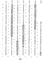
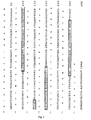
- Particularly preferred antibodies in the variable regions of the light and heavy chains have the amino acid sequences shown in FIGS. 1 and 2, FIGS. 3 and 4, FIGS. 5 and 6 or FIGS. 7 and 8, and the regions are shown there Coded DNA sequences.
- the stool sample is preceded by the following steps prior to incubation with the antibodies:
- the stool sample is resuspended 1: 3 to 1:25, preferably about 1:10, in a resuspension buffer and then mixed on a vortex mixer.
- An example of a resuspension buffer is 150 mM PBS, 0.1% SDS.
- step (b) the detection of the formation of the at least one antigen-antibody complex / antigen aptamer complex in step (b) takes place by means of an immunological method.
- the detection of the formation of the at least one antigen-antibody complex / antigen aptamer complex in step (b) is carried out by ELISA, RIA, Western blot or an immunochromatographic method.
- the same antibody or its fragment or derivative or the aptamer is used for binding to the solid phase as well as for detecting the epitope.
- the capture antibody / the capture aptamer can be bound in unmodified form to the solid phase, for example a microtiter plate, the antibody used for detection, the fragment or derivative thereof or the aptamer is optionally provided with a label.
- this antibody, the fragment or derivative thereof or this aptamer may also be unmarked and thus the epitope of the microorganism, preferably the bacterial epitope, also be detected via a third labeled antibody, the fragment or derivative thereof, a third labeled aptamer, said antibody , the fragment or derivative thereof or this aptamer may be a species-specific or Ig-class specific antibody or aptamer.
- Labels of antibodies, for example, with radioactive or fluorescent markers are known in the art; see. Harlow and Lane a.a.O. The same applies to aptamers.
- urease preferably ⁇ -urease
- ⁇ -urease which may also be present after the intestinal passage as a dimer, possibly in a multimeric version.
- combinations of antibodies, fragments, derivatives and aptamers may also be used in this embodiment, e.g. Combinations of antibodies, etc. that bind to different epitopes of the same antigen.
- the monoclonal antibody is a mouse antibody.
- the antibodies, fragments or derivatives thereof or the aptamers are fixed to a carrier.
- the fixation of the antibodies, fragments or derivatives thereof or the aptamer to a carrier is particularly advantageous for the performance of routine checks.
- the combination antibody carrier / aptamer carrier can also be packaged well as a test kit or in kit form.
- the carrier material is a porous carrier material.
- the carrier material is a test strip.
- the carrier material is cellulose or a cellulose derivative.
- the mammal whose stool can be examined by the method of the invention may use an animal, for example a pet such as a cat or a dog, a farm animal, e.g. a pig or other animal like a mouse, a tiger or a ferret.
- an animal for example a pet such as a cat or a dog, a farm animal, e.g. a pig or other animal like a mouse, a tiger or a ferret.
- the mammal is a human.
- the invention relates to a monoclonal antibody, a fragment or derivative thereof, having a V region comprising a combination of the CDRs set forth above or produced by any of the hybridomas set forth above.
- a monoclonal antibody, fragment or derivative thereof which has at least one of the V regions shown in FIGS. 1 to 8 is preferred.
- this antibody has two of the V regions shown in Figures 1 and 2, 3 and 4, 5 and 6 or Figures 7 and 8. It is also preferred that these V regions are encoded by the DNA sequences shown in Figs. 1-8.
- the monoclonal antibody, the fragment or derivative thereof is a mouse antibody or a fragment or derivative thereof or a chimeric, preferably a humanized antibody or a fragment or derivative thereof.
- the derivative may also be a fusion protein.
- the antibody is labeled, for example with a colloid, with a radioactive, fluorescent, phosphorescent or chemiluminescent label.
- the preparation of chimerized humanized and human antibodies and the other derivatives is well known in the art (eg, Vaughan et al., 1998, Orlandi et al., 1989, Harlow and Lane, supra).
- the invention also relates to an aptamer which specifically binds the same epitope as the monoclonal antibody, fragment or derivative thereof.
- the preparation of such aptamers can be carried out by methods known in the art.
- the invention relates to further antibodies, derivatives or fragments thereof which specifically bind the epitope according to the invention.
- These antibodies may be, for example, monoclonal antibodies generated using the Epitops as hapten / component of an antigen can be prepared by conventional methods.
- the present invention further relates to a diagnostic composition
- a diagnostic composition comprising at least two monoclonal antibodies, fragments or derivatives thereof or aptamers as defined above, optionally fixed to a carrier material.
- the present invention relates to a test device for detecting at least one epitope as defined above comprising (a) at least two monoclonal antibodies, fragments or derivatives thereof or aptamers as defined above, fixed to a support material; (b) a device for the preparation and analysis of stool samples and optionally (c) a mixture of at least two monoclonal antibodies, fragments or derivatives thereof or aptamers.
- the invention further relates to a test device comprising (a) at least two monoclonal antibodies, fragments or derivatives thereof or aptamers as defined above, wherein the antibodies, fragments or derivatives thereof or aptamers are conjugated with colloidal gold, latex particles or other coloring particles whose Size is typically in the range between 5nm and 100nm, preferably between 20nm and 60nm; (b) a device for the preparation and analysis of stool samples and optionally (c) a mixture of at least two monoclonal antibodies, fragments or derivatives thereof or aptamers.
- the present invention relates to a kit comprising (a) at least two monoclonal antibodies, fragments or derivatives thereof or aptamers as defined above, optionally fixed to a carrier material; optionally (b) a device for the preparation and analysis of stool samples and optionally (c) a mixture of at least two monoclonal antibodies, fragments or derivatives thereof or aptamers.
- the invention relates to a pack containing the diagnostic composition according to the invention, the test device according to the invention or the kit according to the invention.
- the components of the diagnostic composition according to the invention, the test device according to the invention and / or the kit according to the invention can in containers such as vials or tubes, optionally packed in buffers and / or solutions. Under certain circumstances, one or more of the ingredients may be packaged in the same container.
- Example 1 Isolation of H. pylori antigens
- H. pylori strain NCTC 11637 was streaked in Petri dishes on Wilkins-chalkern agar with the addition of 10% horse blood and amphotericin B, vancomycin and cefsoludin (Sigma Chemicals) and 3-4 days in a microaerophilic atmosphere (Anaerocult GasPAk, Merck). incubated at 37 ° C. The contents of 2 dishes were suspended in 350 ml BHIB medium with the addition of the antibiotics as above in a 11 bottle (Schott), the medium gassed for 10 min with a gas mixture of 10% CO 2 , 5% O 2 , 85% N 2 and the bottle closed.
- the culture was shaken for 2 days at 37 ° C on a rotary shaker, after 24 hours the bottle's cap was opened.
- the contents of the bottle were then placed sterile in a 101 bottle, filled with 4.7 l of BHIB medium, the medium gassed for 10 min with a gas mixture of 10% CO 2 , 5% O 2 , 85% N 2 and the bottle capped.
- the cap of the bottle was opened.
- the entire volume is then centrifuged at 11000g for 10 min, supernatant decanted and the bacterial pellet weighed.
- the pellet was resuspended in a physiological saline solution with the addition of 15% glycerol in the ratio 2: 1 (wt: vol.) And frozen at -80 ° C.
- a visual inspection of the bacteria and tests for urease, oxidase and catalase activity was performed. The freedom from contamination was evidenced by culture of some suspension taken from freezing at 37 ° C in air for 48 hours.
- Example 2 General procedure for selecting suitable antigens for detection on microorganisms
- a lysate of the microorganism is produced.
- the separation of the lysate takes place by means of gel filtration.
- those proteins are isolated which are present in large quantities in the lysate.
- These proteins are identified by protein sequencing and comparison with sequences from appropriate databases. As a result, characteristic or specific proteins for the respective microorganism can be determined. For these selected antigens then a suitable cleaning scheme is developed.
- Urease-positive fractions were added to a chromatography column filled with Sephacryl S200 50/100 (Pharmacia) and separated (buffer: 50 mM Tris, 100 mM NaCl, 0.05% NaN 3 , pH 7.3). The protein concentration in the effluent was monitored by measuring the optical density at 280nm. The fractions of the first protein peak contained oligomeric urease (molecular weight about 550-600 kDa), followed by a second peak attributable to the alkyl hydroperoxide reductase (also as an oligomer).
- the HSP60 antigen could not be recovered in sufficient quantity by the butanol extraction method described above. That's why the Bacteria unlocked with ultrasound. Fresh bacterial pellet or pellet obtained after extraction with 10% butanol was thawed, resuspended 1:10 in 150 mM PBS, pH 7.5 and placed in a Falcon tube on ice-water in a sonicator (Sonifire) at 25-30% level. (Level 7) Ultrasound-treated (4x90s with each 90s break).
- Polyclonal antisera were prepared by pab Productions (Hebertshausen) according to standard procedures against antigens purified as above. Monoclonal antibodies are produced by methods known to those skilled in the art (Harlow & Lane, 1988, Peters & Baumgarten, 1990).
- the purified proteins used were urease, HSP60 and alkyl hydroperoxide reductase as well as recombinant ⁇ - and ⁇ -urease (Austral Biologics).
- 4-6 mice (BALB / cx C57 Black, F1 generation, 8-12 weeks old) were immunized (primed) and boosted 3-4x at intervals of approximately four weeks.
- 200-300 ⁇ l antigen solutions 25-50 ⁇ g antigen / animal) per injection were emulsified 1: 1 with complete (priming) or incomplete (Boost) Freund's adjuvant (Difco) and injected intraperitoneally with 100 ⁇ l / mouse.
- Boost incomplete Freund's adjuvant
- mice Prior to fusion, the mice were retro-blooded for the virus and the antibody titer was determined from the antiserum obtained by enzyme-linked immunosorbent assay (ELISA, see below).
- the spleen cells of the immunized mice were fused with the myeloma cells P3x63Ag8.653 (ATCC CRL-1580, Kearney et al., 1979) in the ratio 5: 1 with polyethylene glycol 4000.
- the fused cells were suspended in cloning medium (RPMI 1640 medium + 20% FCS + 200 U IL-6 / ml) with hypoxanthine-aminopterin-thymidine supplement (100x concentrate, Sigma) and with a cell density of 2-6x10 4 cells / well on 96 -Napf microtiter plates taken in culture (37 ° C, 6% CO 2 and 95% relative humidity).
- the culture supernatant was screened for the presence of antibodies of the desired specificity by ELISA (see below).
- Clones producing antigen-specific antibodies were recloned twice in the limiting dilution (Coller & Coller, 1983) cloning medium with hypoxanthine-thymidine supplement (100x concentrate, Sigma). The monoclonal end-clone was then adapted to the flat-bottle culture in RPMI 1640 medium with 10% FCS and expanded for the cryopreservation of 5-10 aliquots each containing 2-5x10 6 cells and the production of antibody-containing culture supernatants.
- the detection of the bound antibodies is carried out by adding a horseradish peroxidase (POD) conjugated secondary antibody (rabbit anti-mouse IgG-POD, DAKO).
- POD horseradish peroxidase
- TMB colorless substrate tetramethylbenzidine
- the intensity of the color reaction was measured in the ELISA reader (MWG spectral). The measurement is carried out at 455 nm against the reference wavelength 620 nm. Between the individual steps, the ELISA plate was washed 2-3x with 250 ⁇ l PBS with 0.025% Tween 20 (v / v).
- a total of 24 mAb against ⁇ -urease, 6 mAb against ⁇ -urease, 8 mAb against 26kDa protein and 10 mAb against HSP60 were generated. From this antibody repertoire, those mAb with the lowest detection limits for the respective antigens were selected.
- the purification of mAbs from hybridoma culture supernatants is carried out by means of protein G affinity chromatography (modified according to: Pharmacia Biotech, 1994).
- the culture supernatants were filtered (0.8 .mu.m) and passed directly over the protein G matrix. Was washed with Tris / HCl until the signal at the detector had returned to the background. Elution was carried out with 0.1 M glycine / HCl, pH 3.0, and the protein concentration was detected in the eluate over optical density at 280 nm. All fractions in the range of the single elution signal can be used.
- the murine mAbs were isotyped with the isotyping kit IsoStrip from Boehringer Mannheim (Mannheim).
- the proteins (antigens) separated in the gel were then immobilized on a nitrocellulose membrane by the semidry blot method.
- the membrane was blocked with 2% skimmed milk powder in PBS for 30 min at room temperature and washed three times with TBS / Tween 20 (0.2%) for 5 min.
- the membrane was clamped into the Accutran Cross Blot Screening Unit (Schleicher and Schuell) using a grid plate with 34 transverse channels. 250 ⁇ l of TBS / Tween 20 were initially introduced into each of the resulting transverse channels and 250 ⁇ l each were added to the hybridoma culture supernatants to be tested. The incubation was carried out for 2 hours at room temperature under agitation.
- the membrane was incubated for 1 h with the POD-conjugated secondary antibody (rabbit anti-mouse IgG-POD, DAKO).
- the membrane was washed three times and the immune complex visualized by addition of the 3,3-diaminobenzidine substrate solution (DAB, Sigma).
- DAB 3,3-diaminobenzidine substrate solution
- H. pylori proteins ⁇ - and ⁇ -urease, HSP60 and 26kDa protein were synthesized with 12 amino acid residues on a solid phase and covalently attached to one Cellulose acetate membrane coupled (Jerini GmbH, Berlin). These membranes were incubated with hybridoma culture supernatant and the bound antibodies then blotted on nitrocellulose membranes by the Semidry-Blot method. The membrane was blocked and the immobilized antibodies were detected as described under 5.2 with a POD-labeled secondary antibody (rabbit anti mouse IgG-POD, DAKO).
- the ELISA plate was washed 2-3x with 300 ⁇ l PBS with 0.025% Tween (wash buffer) (v: v).
- the coating of the ELISA plates (MaxiSorb; Nunc) was carried out for 1 h at 37 ° C. with 100 ⁇ l of a solution of a polyclonal rabbit anti- H. pylori antigen antibody (pAK; approximately 10 ⁇ g of antibody / ml of carbonate buffer, 0.1M, pH 9, 5).
- pAK polyclonal rabbit anti- H. pylori antigen antibody
- 200 ⁇ l of 150 mM PBS with 2% skim milk powder were pipetted per well and incubated for 30 min at room temperature. 50ng / ml purified H.
- pylori antigens dissolved in 150mM PBS with the addition of 0.1% skimmed milk powder were diluted in 1: 2 increments. Buffer served as a negative control. Subsequently, 100 ⁇ l 1:10 diluted culture supernatant of the mAb to be examined against the same antigen was added and incubated for 30-60min at RT. The detection of the bound antibody was carried out as described under 3.4. The lowest Concentration at which an extinction greater than or equal to twice the control was detected was accepted as the detection limit.
- Table 1 summarizes the results of isotyping, immunoblot analyzes, and detection limit determination for the mAb listed in Table 1.
- Table 2 ⁇ / b> Results of epitope mapping of monoclonal antibodies against ⁇ i> H. pylori ⁇ / i> antigens Ag-specificity Fusion / clone Epitope (AS position) AA sequence ⁇ -urease HP8m / 1H5-G2-B4 negative (large subunit) HPBM / 4H5-D4-C9 negative HP9m / 2B12 G7 B12 369-375 VGEVITR ⁇ -urease HP9m / 2E7-B8-G10 negative (small subunit) HP9m / 1H7-C6-D5 negative Alkylhydroperoxid- HP15m / 3E8-D9-D6 negative reductase HP15m / 3F5-D5-B6 negative HP15m / 4H12-A4 negative D5 143
- the epitope overlap measurement with SPR spectroscopy provides information on the simultaneous accessibility of antibody epitopes. This makes it possible to find suitable antibody pairs for the development of ELISA and rapid test (Fägerstam LG et al., 1990, Malmqvist M., 1996).
- mAK set in the percentage ratio ( ⁇ RU 2 / ⁇ RU 1 ).
- overlapping the epitopes of an antigen are called when a first antibody has occupied all the epitopes of an antigen to be bound by it and prevents or prevents the subsequent binding of a second antibody.
- Kinetic measurements are still possible calculate the values for rate constants of adsorption and desorption of antibodies.
- a combination of antibodies, some of which recognize overlapping epitopes can be explained by the fact that oligomeric proteins such as urease several times present the same epitope. This allows in ELISA the binding of a capture antibody to an epitope and the simultaneous binding of one or more detection antibodies to one or more identical epitopes of the oligomeric protein.
- Example 8 Selection of antibody pairs for use in human stool ELISA
- epitope overlaps were determined by surface plasmon resonance.
- the combinations which showed promising results in these measurements were tested for their detection limit in the stool ELISA.
- Detector Detection antibody
- Catcher Catching antibody
- the ELISA plate was washed 2-3 times with 250 ⁇ l PBS with the addition of 0.025% (washing buffer 1) or 0.2% Tween 20 (washing buffer 2, VoL: Vol.).
- the coating of the ELISA plates (MaxiSorb; Nunc) was carried out for 1 h at 37 ° C with 100 ⁇ l of a mAb solution (2.5 ⁇ g antibody / ml carbonate buffer, 0.1 M, pH 9.5).
- 200 ⁇ l of 150 mM PBS with 0.2% fish gelatin or 1% skim milk powder (weight: volume) were pipetted per well and incubated for 30 min at room temperature. Was washed with washing buffer 1.
- Human chair was suspended in the ratio 1:10 (wt.: Vol.) With 150mM PBS with the addition of 2% skimmed milk powder, purified H. pylori antigens dissolved in 150mM PBS added in known concentrations. Per 100 ⁇ l of the suspensions were incubated per well for 1h. The plate was first rinsed by hand and washed 4x with Wash Buffer 2. Subsequently, 100 ⁇ l of a solution with biotin-coupled mAb (0.5 ⁇ g antibody / ml PBS) was added against the same antigen and incubated for 30-60 min at RT. The detection of the bound antigens is carried out by adding a conjugate of streptavidin with POD (DAKO).
- DAKO conjugate of streptavidin with POD
- the POD converts the colorless substrate TMB (Sigma) into a blue product. After 5 to 10 minutes, or as soon as the negative control showed a slight blue coloration, the reaction was stopped by the addition of 1 N sulfuric acid (100 ⁇ l / well). The intensity of the color reaction was determined in the ELISA reader (MWG Spectral). The measurement takes place at 455 nm against the reference wavelength 620 nm.
- Table 4 Detection of H. pylori antigens from the stool of definitely negative (category 0) or safe positive (category 4) patients by ELISA using monoclonal antibodies: Table 4a: Category 0 samples: 0 means false-positive, 1 means true-negative [ ⁇ g Ag / g stool] resulting truth value sample urease 26 kDa HSP60 urease 26 kDa HSP60 3-combination cut-off 1 15 5 0047 0.6 2.3 1 1 1 0048 0.6 2.3 1 1 1 0051 2.1 22 0 0 0 0057 0.6 2.3 1 1 1 0069 0.6 2.3 1 1 1 0074 0.6 2.3 1 1 1 0087 0.6 2.3 1 1 1 0099 0.6 2.3 1 1 1 0185 0.6 2.3 1 1 1 0186 0.6 2.3 1 1 1 0189 0.6 2.3 1 1 1 0265 0.6 2.3 1 1 1 0298 0.6 2.3 1 1 1 0305 0.6 2.3 1 1
- 26kDa means 26 kDa protein
- 3 combination means combination of all three antigens (urease, 26kDa protein and HSP60)
- urease HP8m / 4H5 + HP16m / XG1 was used as catcher antibodies and detection antibodies.
- the detection limit was 0.075 ng / ml.
- HP15m / 4H12 was used as the capture antibody and HP15m / 3E8 as the detection antibody.
- the detection limit was 1.5 ng / ml.
- HP18.1m / 3F11 was used as capture antibody and HP16m / 2A5 + HP18.1 m / 4D9 as detection antibody.
- the detection limit was 6 ng / ml.
- Tables 4a-4c show the results of stool specimens for urease, alkyl hydroperoxide reductase and HSP60.
- the last column (3 combination) shows in each case the values for the resulting truth value of the combination of all three antigens (urease, 26kDa protein, HSP60).
- Table 5 shows the result of examining 3 samples taken from the stool of a patient at three different sites. The numbers correspond to the measured absorbance at 650 nm divided by the absorbance of a blank sample, ie the multiple of the background (H). It showed an inhomogeneous distribution of the detected antigens.
- Table 5 Table 5: ⁇ / b> Distribution ⁇ i> H. pylori-specific antigens in stool specimens collected at various sites in the stool of an infected person Detected antigen ⁇ -urease 26 kDa protein HSP60 Sample 1 8.6 x H 2.5 x H 1 x H Sample 2 11 x H 1 x H 1 x H Sample 3 3.5 x H 1 x H 1 x H
- Example 10 Cloning and sequence determination of the functional variable regions of immunoglobulins from hybridoma cell lines
- the DNA regions encoding the kappa light chain as well as the heavy chain Fd segment (VH + CH1) of the respective antibodies were amplified by PCR.
- the oligonucleotide primer set listed in Table 6 was used.
- the cDNA isolated from the individual hybridoma cell lines served as template.
- the primer set used leads to a 5'- XhoI and a 3'- Spe I interface in the heavy chain Fd fragments as well as to a 5'- Sac I and a 3'-Xba I interface in the kappa light chains.
- 11 different 5'-VH primers (MVH 1-8 and MULH1-3) were each combined with the 3'-VH primer MlgG1.
- 11 different 5 'VK primers (MUVK 1-7 and MULK1-4) each were combined with the 3'-VK primer 3'MUCK.
- the following temperature program was used in all PCR amplifications: initial denaturation at 94 ° C for 3 min, denaturation at 94 ° C for 25 sec, primer annealing at 52 ° C for 60 sec, polymerization at 72 ° C for 90 sec , This program was maintained for 40 cycles, followed by a 10 min. final completion of the fragments at 72 ° C.
- the results of the PCR amplifications were separated by agarose gel electrophoresis and isolated DNA bands of the expected molecular weight.
- the isolated bands were then subjected to a restriction digestion using the enzymes Xhol and Spel (heavy chains) or Sacl and XbaI (light chains) and the fragments obtained were cloned into the plasmid vector Bluescript KS (Stratagene), after this first with the restriction enzymes Xhol and Spel or Sacl and Xbal had been split.
- Plasmid preparations of the cloned heavy and light chain fragments were then sequence analyzed. Sequences were selected for each hybridoma cell line encoding immunoglobulin heavy and light chain functional variable regions (VH and VL, respectively). In this way, exactly one functional VH and one functional VL region could be identified for each hybridoma cell line.
- the functional VH and VL sequences are shown in Figures 1-8. Cloning and sequencing were performed by standard methods (Sambrook et al., 1989).
- Harlow & Lane, 1988 Harlow, E., Lane, D., Antibodies: A laboratory manual, Cold Spring Harbor Laboratory, New York
Landscapes
- Health & Medical Sciences (AREA)
- Life Sciences & Earth Sciences (AREA)
- Chemical & Material Sciences (AREA)
- Immunology (AREA)
- Molecular Biology (AREA)
- Engineering & Computer Science (AREA)
- Urology & Nephrology (AREA)
- General Health & Medical Sciences (AREA)
- Biochemistry (AREA)
- Medicinal Chemistry (AREA)
- Hematology (AREA)
- Organic Chemistry (AREA)
- Biomedical Technology (AREA)
- Genetics & Genomics (AREA)
- Physics & Mathematics (AREA)
- Tropical Medicine & Parasitology (AREA)
- Biotechnology (AREA)
- Cell Biology (AREA)
- Proteomics, Peptides & Aminoacids (AREA)
- Microbiology (AREA)
- Biophysics (AREA)
- Food Science & Technology (AREA)
- Virology (AREA)
- Analytical Chemistry (AREA)
- General Physics & Mathematics (AREA)
- Pathology (AREA)
- Peptides Or Proteins (AREA)
- Preparation Of Compounds By Using Micro-Organisms (AREA)
- Measuring Or Testing Involving Enzymes Or Micro-Organisms (AREA)
- Agricultural Chemicals And Associated Chemicals (AREA)
- Investigating Or Analysing Biological Materials (AREA)
Priority Applications (2)
| Application Number | Priority Date | Filing Date | Title |
|---|---|---|---|
| EP99971515A EP1125130B1 (de) | 1998-10-29 | 1999-10-29 | Nachweis von säure-resistenten mikroorganismen im stuhl |
| EP06015257A EP1734366A3 (de) | 1998-10-29 | 1999-10-29 | Neues Verfahren zum Nachweis von Säure-resistenten Mikroorganismen im Stuhl |
Applications Claiming Priority (6)
| Application Number | Priority Date | Filing Date | Title |
|---|---|---|---|
| EP98120517 | 1998-10-29 | ||
| EP98120517 | 1998-10-29 | ||
| EP98120687 | 1998-11-06 | ||
| EP98120687 | 1998-11-06 | ||
| EP99971515A EP1125130B1 (de) | 1998-10-29 | 1999-10-29 | Nachweis von säure-resistenten mikroorganismen im stuhl |
| PCT/EP1999/008212 WO2000026671A1 (de) | 1998-10-29 | 1999-10-29 | Nachweis von säure-resistenten mikroorganismen im stuhl |
Related Child Applications (1)
| Application Number | Title | Priority Date | Filing Date |
|---|---|---|---|
| EP06015257A Division EP1734366A3 (de) | 1998-10-29 | 1999-10-29 | Neues Verfahren zum Nachweis von Säure-resistenten Mikroorganismen im Stuhl |
Publications (2)
| Publication Number | Publication Date |
|---|---|
| EP1125130A1 EP1125130A1 (de) | 2001-08-22 |
| EP1125130B1 true EP1125130B1 (de) | 2006-07-26 |
Family
ID=26149749
Family Applications (2)
| Application Number | Title | Priority Date | Filing Date |
|---|---|---|---|
| EP06015257A Withdrawn EP1734366A3 (de) | 1998-10-29 | 1999-10-29 | Neues Verfahren zum Nachweis von Säure-resistenten Mikroorganismen im Stuhl |
| EP99971515A Expired - Lifetime EP1125130B1 (de) | 1998-10-29 | 1999-10-29 | Nachweis von säure-resistenten mikroorganismen im stuhl |
Family Applications Before (1)
| Application Number | Title | Priority Date | Filing Date |
|---|---|---|---|
| EP06015257A Withdrawn EP1734366A3 (de) | 1998-10-29 | 1999-10-29 | Neues Verfahren zum Nachweis von Säure-resistenten Mikroorganismen im Stuhl |
Country Status (9)
| Country | Link |
|---|---|
| US (2) | US7122320B2 (es) |
| EP (2) | EP1734366A3 (es) |
| JP (1) | JP2002529705A (es) |
| AT (1) | ATE334395T1 (es) |
| AU (1) | AU1157100A (es) |
| DE (1) | DE59913716D1 (es) |
| DK (1) | DK1125130T3 (es) |
| ES (1) | ES2270638T3 (es) |
| WO (1) | WO2000026671A1 (es) |
Families Citing this family (26)
| Publication number | Priority date | Publication date | Assignee | Title |
|---|---|---|---|---|
| JP2002529705A (ja) * | 1998-10-29 | 2002-09-10 | コンネクス・ゲーエムベーハー | 糞便中の耐酸性微生物を検出するための新規方法 |
| AU1137301A (en) | 1999-10-12 | 2001-04-23 | Connex Gesellschaft Zur Optimierung Von Forschung Und Entwicklung Mbh | Immuno-chromatographic rapid assay in order to detect acid-resistant microorganisms in the stool |
| JP3504633B2 (ja) | 1999-10-29 | 2004-03-08 | わかもと製薬株式会社 | ヘリコバクター・ピロリへの感染を判定する検査方法及び検査試薬 |
| EP1227159B1 (en) | 1999-10-29 | 2011-04-20 | Wakamoto Pharmaceutical Co., Ltd. | Monoclonal antibody, hybridoma, immunoassay method and diagnosis kit |
| DE60138981D1 (de) * | 2000-05-18 | 2009-07-30 | Meridian Bioscience Inc | Immunoassay für H. pylori in Fäkalienproben mit Hilfe von gattungsspezifischen Antikörpern |
| DE10043161A1 (de) * | 2000-09-01 | 2002-03-14 | Connex Ges Zur Optimierung Von | Lösung zur Aufbereitung von Stuhlproben für diagnostische Zwecke |
| CN1489474A (zh) * | 2001-01-26 | 2004-04-14 | Ӣϣ��̩��˹��˾ | 针对clfa蛋白质的单克隆抗体和在治疗或预防感染中的利用方法 |
| FI118061B (fi) | 2001-09-24 | 2007-06-15 | Beanor Oy | Menetelmä ja bioanturi analyysiä varten |
| FI115166B (fi) | 2001-12-31 | 2005-03-15 | Biofons Oy | Diagnostisia menetelmiä |
| US20040023415A1 (en) * | 2002-03-05 | 2004-02-05 | Konstantin Sokolov | Biospecific contrast agents |
| DE10219741A1 (de) * | 2002-05-02 | 2003-11-13 | Georg S Wengler | Verfahren zur Vorbehandlung von Stuhlproben |
| GB0210783D0 (en) | 2002-05-10 | 2002-06-19 | Polonelli Luciano | Anti-microbial polypeptides |
| US20090191213A9 (en) | 2003-07-02 | 2009-07-30 | Novo Nordisk A/S | Compositions and methods for regulating NK cell activity |
| RU2404993C2 (ru) * | 2003-07-02 | 2010-11-27 | Иннейт Фарма | Композиции и способы регуляции клеточной активности nk |
| KR101299167B1 (ko) * | 2004-07-01 | 2013-08-30 | 노보 노르디스크 에이/에스 | 인간 항-kir 항체 |
| SI2287195T1 (sl) * | 2004-07-01 | 2019-08-30 | Novo Nordisk A/S | Pan-kir2dl nk-receptor protitelesa in njihova uporaba pri diagnostiki in terapiji |
| JP4625318B2 (ja) * | 2004-12-06 | 2011-02-02 | 財団法人野田産業科学研究所 | 相同組換え頻度が上昇した形質転換菌 |
| JP5295568B2 (ja) | 2005-01-06 | 2013-09-18 | ノヴォ ノルディスク アー/エス | Kir結合剤およびその使用方法 |
| WO2009088460A2 (en) * | 2008-01-03 | 2009-07-16 | Ask Diagnostics, Inc. | Methods of diagnosing latent and active malignancies |
| US20110065086A1 (en) * | 2008-02-21 | 2011-03-17 | Otc Biotechnologies, Llc | Methods of producing homogeneous plastic-adherent aptamer-magnetic bead-fluorophore and other sandwich assays |
| EP2699257A4 (en) * | 2011-04-21 | 2014-10-15 | Univ Louisiana State | PEPTIDE AND CONJUGATED VACCINES FOR FUNGAL INFECTIONS |
| WO2016176176A1 (en) * | 2015-04-29 | 2016-11-03 | 3M Innovative Properties Company | Culture device for anaerobic microorganisms |
| CN109628457B (zh) * | 2019-02-03 | 2021-12-10 | 淮海工学院 | 单链dna核酸适配体及其筛选方法与用途 |
| CN110618270B (zh) * | 2019-09-10 | 2020-08-25 | 深圳市鸿美诊断技术有限公司 | 一种用于定量测定粪便中幽门螺杆菌抗原试剂的制备方法 |
| WO2022047125A1 (en) * | 2020-08-28 | 2022-03-03 | Board Of Regents, The University Of Texas System | Antibodies specific to ccl21 and methods of use |
| WO2022271771A2 (en) * | 2021-06-22 | 2022-12-29 | The General Hospital Corporation | Therapeutic targeting of cadherin 11 in cancer |
Family Cites Families (9)
| Publication number | Priority date | Publication date | Assignee | Title |
|---|---|---|---|---|
| US4879213A (en) * | 1986-12-05 | 1989-11-07 | Scripps Clinic And Research Foundation | Synthetic polypeptides and antibodies related to Epstein-Barr virus early antigen-diffuse |
| US5200344A (en) * | 1990-11-13 | 1993-04-06 | Blaser Martin J | Diagnostic testing for campylobacter jejuni or campylobacter coli infections using novel antigens |
| AU5693496A (en) * | 1995-05-02 | 1996-11-21 | Institut National De La Sante Et De La Recherche Medicale | Immunogenic compositions against helicobacter infection, pol ypeptides for use in the compositions, and nucleic acid sequ ences encoding said polypeptides |
| GB2307987A (en) * | 1995-12-06 | 1997-06-11 | Univ Manchester | Epitopes of the urease of Helicobacter pylori as dignostic agents; pharmaceuticals comprising such epitopes or the antibodies thereto |
| US5932430A (en) * | 1996-05-09 | 1999-08-03 | Meridian Diagnostics, Inc. | Immunoassay for H. pylori in fecal specimens |
| US5716791A (en) * | 1996-05-09 | 1998-02-10 | Meridian Diagnostics, Inc. | Immunoassay for H. pylori in fecal specimens |
| US6383763B1 (en) * | 1996-07-26 | 2002-05-07 | Case Western Reserve University | Detection of mycobacteria |
| IT1289578B1 (it) * | 1996-12-06 | 1998-10-15 | Sanitaria Scaligera Spa | Immunopurificazione di un antigene dall'apparente peso molecolare di 16 +- 2 kda dell' helicobacter pylori e metodi per la sua |
| JP2002529705A (ja) * | 1998-10-29 | 2002-09-10 | コンネクス・ゲーエムベーハー | 糞便中の耐酸性微生物を検出するための新規方法 |
-
1999
- 1999-10-29 JP JP2000580001A patent/JP2002529705A/ja not_active Ceased
- 1999-10-29 AT AT99971515T patent/ATE334395T1/de not_active IP Right Cessation
- 1999-10-29 EP EP06015257A patent/EP1734366A3/de not_active Withdrawn
- 1999-10-29 DE DE59913716T patent/DE59913716D1/de not_active Expired - Lifetime
- 1999-10-29 EP EP99971515A patent/EP1125130B1/de not_active Expired - Lifetime
- 1999-10-29 DK DK99971515T patent/DK1125130T3/da active
- 1999-10-29 WO PCT/EP1999/008212 patent/WO2000026671A1/de active IP Right Grant
- 1999-10-29 AU AU11571/00A patent/AU1157100A/en not_active Abandoned
- 1999-10-29 ES ES99971515T patent/ES2270638T3/es not_active Expired - Lifetime
-
2001
- 2001-04-27 US US09/842,776 patent/US7122320B2/en not_active Expired - Lifetime
-
2006
- 2006-08-31 US US11/513,129 patent/US7736859B2/en not_active Expired - Fee Related
Also Published As
| Publication number | Publication date |
|---|---|
| US20070009975A1 (en) | 2007-01-11 |
| EP1125130A1 (de) | 2001-08-22 |
| DK1125130T3 (da) | 2006-11-27 |
| US7122320B2 (en) | 2006-10-17 |
| EP1734366A2 (de) | 2006-12-20 |
| WO2000026671A1 (de) | 2000-05-11 |
| ES2270638T3 (es) | 2007-04-01 |
| JP2002529705A (ja) | 2002-09-10 |
| AU1157100A (en) | 2000-05-22 |
| DE59913716D1 (de) | 2006-09-07 |
| US7736859B2 (en) | 2010-06-15 |
| ATE334395T1 (de) | 2006-08-15 |
| US20040023316A1 (en) | 2004-02-05 |
| EP1734366A3 (de) | 2007-11-07 |
Similar Documents
| Publication | Publication Date | Title |
|---|---|---|
| EP1125130B1 (de) | Nachweis von säure-resistenten mikroorganismen im stuhl | |
| EP1232392B2 (de) | Verbessertes verfahren zum nachweis von säure-resistenten bakterien der gattung helicobacter im stuhl | |
| DE69626026T2 (de) | Differentialtest für ulzerative kolitis, primäre sklerosierende cholangitis und autoimmun-hepatitis vom type 1. | |
| CA1216808A (en) | Monoclonal antibody having specificity for the double -stranded conformation of native dna and diagnostic methods using same | |
| Adams et al. | Development and use of monoclonal antibody probes forimmunohistochemistry, ELISA and IFAT to detect bacterial and parasitic fish pathogens | |
| CA2132421A1 (en) | Broadly reactive opsonic antibodies that react with common staphylococcal antigens | |
| EP2459588B1 (de) | Verfahren zum nachweis und zur identifikation eines varianten c. difficile stammes in einer probe | |
| DE4134297A1 (de) | Monoclonale antikoerper gegen mycoplasma pneumoniae, diese produzierende hybridome, verfahren zu deren herstellung sowie deren verwendung | |
| US20210324055A1 (en) | Methods, Devices, Kits and Compositions for Detecting Tapeworm | |
| EP2561362B1 (de) | Verfahren zur erkennung einer salmonelleninfektion | |
| EP1913394B1 (de) | Testsystem zum nachweis von salmonellen | |
| DE20023767U1 (de) | Mittel zum Nachweis von Säure-resistenten Mikroorganismen im Stuhl | |
| Ghosh et al. | Assay dependent specificities of monoclonal antibodies to bacterial antigens. | |
| RU2782463C1 (ru) | Способы, устройства, наборы и композиции для обнаружения ленточных червей | |
| KR102212636B1 (ko) | 페스트균 f1 캡슐 단백질에 특이적인 항체, 이를 생산하는 하이브리도마 세포주 1h4, 및 이를 이용한 페스트균 진단 키트 | |
| EP1221045A2 (de) | Immunchromatographischer schnelltest zum nachweis von säure-resistenten mikroorganismen im stuhl | |
| DE102008029688B4 (de) | Verfahren zum Nachweis und zur Identifikation eines varianten C. difficile Stammes in einer Probe | |
| EP1829894A1 (de) | Monoklonale Antikörper gegen Kollagen XVII | |
| Schmidt et al. | Production and characterization of monoclonal antibodies specific for pathogenic serogroups O: 3, O: 8, and O: 9 of Yersinia enterocolitica | |
| DE10035668A1 (de) | Diagnose und Therapie von HHV-8-assoziierten Erkrankungen auf der Grundlage des HHV-8-kodierten Chemokinrezeptors | |
| WO2014188763A1 (en) | An antibody that binds to leptospiral antigen |
Legal Events
| Date | Code | Title | Description |
|---|---|---|---|
| PUAI | Public reference made under article 153(3) epc to a published international application that has entered the european phase |
Free format text: ORIGINAL CODE: 0009012 |
|
| 17P | Request for examination filed |
Effective date: 20010529 |
|
| AK | Designated contracting states |
Kind code of ref document: A1 Designated state(s): AT BE CH CY DE DK ES FI FR GB GR IE IT LI LU MC NL PT SE |
|
| AX | Request for extension of the european patent |
Free format text: AL;LT;LV;MK;RO;SI |
|
| 17Q | First examination report despatched |
Effective date: 20030703 |
|
| RAP1 | Party data changed (applicant data changed or rights of an application transferred) |
Owner name: DAKOCYTOMATION DENMARK A/S |
|
| GRAP | Despatch of communication of intention to grant a patent |
Free format text: ORIGINAL CODE: EPIDOSNIGR1 |
|
| TPAC | Observations filed by third parties |
Free format text: ORIGINAL CODE: EPIDOSNTIPA |
|
| GRAS | Grant fee paid |
Free format text: ORIGINAL CODE: EPIDOSNIGR3 |
|
| GRAA | (expected) grant |
Free format text: ORIGINAL CODE: 0009210 |
|
| STAA | Information on the status of an ep patent application or granted ep patent |
Free format text: STATUS: THE PATENT HAS BEEN GRANTED |
|
| AK | Designated contracting states |
Kind code of ref document: B1 Designated state(s): AT BE CH CY DE DK ES FI FR GB GR IE IT LI LU MC NL PT SE |
|
| PG25 | Lapsed in a contracting state [announced via postgrant information from national office to epo] |
Ref country code: IT Free format text: LAPSE BECAUSE OF FAILURE TO SUBMIT A TRANSLATION OF THE DESCRIPTION OR TO PAY THE FEE WITHIN THE PRESCRIBED TIME-LIMIT;WARNING: LAPSES OF ITALIAN PATENTS WITH EFFECTIVE DATE BEFORE 2007 MAY HAVE OCCURRED AT ANY TIME BEFORE 2007. THE CORRECT EFFECTIVE DATE MAY BE DIFFERENT FROM THE ONE RECORDED. Effective date: 20060726 |
|
| REG | Reference to a national code |
Ref country code: GB Ref legal event code: FG4D Free format text: NOT ENGLISH |
|
| PLBI | Opposition filed |
Free format text: ORIGINAL CODE: 0009260 |
|
| REG | Reference to a national code |
Ref country code: CH Ref legal event code: EP |
|
| REG | Reference to a national code |
Ref country code: IE Ref legal event code: FG4D Free format text: LANGUAGE OF EP DOCUMENT: GERMAN |
|
| REF | Corresponds to: |
Ref document number: 59913716 Country of ref document: DE Date of ref document: 20060907 Kind code of ref document: P |
|
| 26 | Opposition filed |
Opponent name: MERIDIAN BIOSCIENCE, INC. Effective date: 20060731 |
|
| PGFP | Annual fee paid to national office [announced via postgrant information from national office to epo] |
Ref country code: AT Payment date: 20061024 Year of fee payment: 8 |
|
| PG25 | Lapsed in a contracting state [announced via postgrant information from national office to epo] |
Ref country code: MC Free format text: LAPSE BECAUSE OF NON-PAYMENT OF DUE FEES Effective date: 20061031 |
|
| NLR1 | Nl: opposition has been filed with the epo |
Opponent name: MERIDIAN BIOSCIENCE, INC. |
|
| REG | Reference to a national code |
Ref country code: SE Ref legal event code: TRGR |
|
| GBT | Gb: translation of ep patent filed (gb section 77(6)(a)/1977) |
Effective date: 20061025 |
|
| REG | Reference to a national code |
Ref country code: CH Ref legal event code: NV Representative=s name: BRAUNPAT BRAUN EDER AG |
|
| REG | Reference to a national code |
Ref country code: DK Ref legal event code: T3 |
|
| PG25 | Lapsed in a contracting state [announced via postgrant information from national office to epo] |
Ref country code: PT Free format text: LAPSE BECAUSE OF FAILURE TO SUBMIT A TRANSLATION OF THE DESCRIPTION OR TO PAY THE FEE WITHIN THE PRESCRIBED TIME-LIMIT Effective date: 20061226 |
|
| REG | Reference to a national code |
Ref country code: GB Ref legal event code: 732E |
|
| ET | Fr: translation filed | ||
| REG | Reference to a national code |
Ref country code: ES Ref legal event code: FG2A Ref document number: 2270638 Country of ref document: ES Kind code of ref document: T3 |
|
| REG | Reference to a national code |
Ref country code: CH Ref legal event code: PFA Owner name: OXOID (ELY) LIMITED Free format text: DAKOCYTOMATION DENMARK A/S#PRODUKTIONSVEJ 42#2600 GLOSTRUP (DK) -TRANSFER TO- OXOID (ELY) LIMITED#DENMARK HOUSE, ANGEL DROVE#ELY, CAMBRIDGESHIRE CB7 4ET (GB) |
|
| REG | Reference to a national code |
Ref country code: FR Ref legal event code: TP Ref country code: FR Ref legal event code: CD |
|
| NLS | Nl: assignments of ep-patents |
Owner name: DAKOCYTOMATION LTD. Effective date: 20070731 |
|
| NLT1 | Nl: modifications of names registered in virtue of documents presented to the patent office pursuant to art. 16 a, paragraph 1 |
Owner name: OXOID (ELY) LIMITED |
|
| BECA | Be: change of holder's address |
Owner name: *OXOID ELY LTDDENMARK HOUSE, ANGEL DROVE, ELY, CB7 Effective date: 20060726 |
|
| BECH | Be: change of holder |
Owner name: *OXOID ELY LTD Effective date: 20060726 |
|
| BECN | Be: change of holder's name |
Owner name: *OXOID ELY LTDDENMARK HOUSE, ANGEL DROVE, ELY, CB7 Effective date: 20070614 |
|
| PG25 | Lapsed in a contracting state [announced via postgrant information from national office to epo] |
Ref country code: GR Free format text: LAPSE BECAUSE OF FAILURE TO SUBMIT A TRANSLATION OF THE DESCRIPTION OR TO PAY THE FEE WITHIN THE PRESCRIBED TIME-LIMIT Effective date: 20061027 |
|
| PLAZ | Examination of admissibility of opposition: despatch of communication + time limit |
Free format text: ORIGINAL CODE: EPIDOSNOPE2 |
|
| PG25 | Lapsed in a contracting state [announced via postgrant information from national office to epo] |
Ref country code: LU Free format text: LAPSE BECAUSE OF NON-PAYMENT OF DUE FEES Effective date: 20061029 |
|
| PG25 | Lapsed in a contracting state [announced via postgrant information from national office to epo] |
Ref country code: AT Free format text: LAPSE BECAUSE OF NON-PAYMENT OF DUE FEES Effective date: 20071029 |
|
| PLBJ | Opposition found inadmissible |
Free format text: ORIGINAL CODE: 0009275 |
|
| PLAB | Opposition data, opponent's data or that of the opponent's representative modified |
Free format text: ORIGINAL CODE: 0009299OPPO |
|
| 26U | Opposition found inadmissible |
Opponent name: MERIDIAN BIOSCIENCE, INC. Effective date: 20080916 |
|
| PG25 | Lapsed in a contracting state [announced via postgrant information from national office to epo] |
Ref country code: CY Free format text: LAPSE BECAUSE OF FAILURE TO SUBMIT A TRANSLATION OF THE DESCRIPTION OR TO PAY THE FEE WITHIN THE PRESCRIBED TIME-LIMIT Effective date: 20060726 |
|
| REG | Reference to a national code |
Ref country code: ES Ref legal event code: PC2A |
|
| PGFP | Annual fee paid to national office [announced via postgrant information from national office to epo] |
Ref country code: NL Payment date: 20101009 Year of fee payment: 12 Ref country code: IE Payment date: 20101015 Year of fee payment: 12 Ref country code: DK Payment date: 20101014 Year of fee payment: 12 |
|
| PGFP | Annual fee paid to national office [announced via postgrant information from national office to epo] |
Ref country code: FI Payment date: 20101014 Year of fee payment: 12 Ref country code: CH Payment date: 20101012 Year of fee payment: 12 |
|
| PGFP | Annual fee paid to national office [announced via postgrant information from national office to epo] |
Ref country code: BE Payment date: 20101020 Year of fee payment: 12 Ref country code: SE Payment date: 20101012 Year of fee payment: 12 |
|
| PGFP | Annual fee paid to national office [announced via postgrant information from national office to epo] |
Ref country code: ES Payment date: 20101122 Year of fee payment: 12 |
|
| BERE | Be: lapsed |
Owner name: *OXOID ELY LTD Effective date: 20111031 |
|
| REG | Reference to a national code |
Ref country code: NL Ref legal event code: V1 Effective date: 20120501 |
|
| REG | Reference to a national code |
Ref country code: CH Ref legal event code: PL |
|
| REG | Reference to a national code |
Ref country code: DK Ref legal event code: EBP |
|
| REG | Reference to a national code |
Ref country code: SE Ref legal event code: EUG |
|
| PG25 | Lapsed in a contracting state [announced via postgrant information from national office to epo] |
Ref country code: LI Free format text: LAPSE BECAUSE OF NON-PAYMENT OF DUE FEES Effective date: 20111031 Ref country code: CH Free format text: LAPSE BECAUSE OF NON-PAYMENT OF DUE FEES Effective date: 20111031 Ref country code: BE Free format text: LAPSE BECAUSE OF NON-PAYMENT OF DUE FEES Effective date: 20111031 Ref country code: NL Free format text: LAPSE BECAUSE OF NON-PAYMENT OF DUE FEES Effective date: 20120501 |
|
| REG | Reference to a national code |
Ref country code: IE Ref legal event code: MM4A |
|
| PG25 | Lapsed in a contracting state [announced via postgrant information from national office to epo] |
Ref country code: FI Free format text: LAPSE BECAUSE OF NON-PAYMENT OF DUE FEES Effective date: 20111029 |
|
| PG25 | Lapsed in a contracting state [announced via postgrant information from national office to epo] |
Ref country code: IE Free format text: LAPSE BECAUSE OF NON-PAYMENT OF DUE FEES Effective date: 20111029 Ref country code: SE Free format text: LAPSE BECAUSE OF NON-PAYMENT OF DUE FEES Effective date: 20111030 Ref country code: DK Free format text: LAPSE BECAUSE OF NON-PAYMENT OF DUE FEES Effective date: 20111031 |
|
| REG | Reference to a national code |
Ref country code: ES Ref legal event code: FD2A Effective date: 20130605 |
|
| PG25 | Lapsed in a contracting state [announced via postgrant information from national office to epo] |
Ref country code: ES Free format text: LAPSE BECAUSE OF NON-PAYMENT OF DUE FEES Effective date: 20111030 |
|
| REG | Reference to a national code |
Ref country code: FR Ref legal event code: PLFP Year of fee payment: 18 |
|
| REG | Reference to a national code |
Ref country code: FR Ref legal event code: PLFP Year of fee payment: 19 |
|
| REG | Reference to a national code |
Ref country code: FR Ref legal event code: PLFP Year of fee payment: 20 |
|
| PGFP | Annual fee paid to national office [announced via postgrant information from national office to epo] |
Ref country code: FR Payment date: 20180913 Year of fee payment: 20 |
|
| PGFP | Annual fee paid to national office [announced via postgrant information from national office to epo] |
Ref country code: DE Payment date: 20181016 Year of fee payment: 20 |
|
| PGFP | Annual fee paid to national office [announced via postgrant information from national office to epo] |
Ref country code: IT Payment date: 20181018 Year of fee payment: 20 Ref country code: GB Payment date: 20181024 Year of fee payment: 20 |
|
| REG | Reference to a national code |
Ref country code: DE Ref legal event code: R071 Ref document number: 59913716 Country of ref document: DE |
|
| REG | Reference to a national code |
Ref country code: GB Ref legal event code: PE20 Expiry date: 20191028 |
|
| PG25 | Lapsed in a contracting state [announced via postgrant information from national office to epo] |
Ref country code: GB Free format text: LAPSE BECAUSE OF EXPIRATION OF PROTECTION Effective date: 20191028 |
|
| PLAB | Opposition data, opponent's data or that of the opponent's representative modified |
Free format text: ORIGINAL CODE: 0009299OPPO |
|
| R26U | Opposition found inadmissible (corrected) |
Opponent name: MERIDIAN BIOSCIENCE, INC. Effective date: 20080916 |