EP1095275B1 - Endogenous constitutively activated g protein-coupled orphan receptors - Google Patents
Endogenous constitutively activated g protein-coupled orphan receptors Download PDFInfo
- Publication number
- EP1095275B1 EP1095275B1 EP99941990A EP99941990A EP1095275B1 EP 1095275 B1 EP1095275 B1 EP 1095275B1 EP 99941990 A EP99941990 A EP 99941990A EP 99941990 A EP99941990 A EP 99941990A EP 1095275 B1 EP1095275 B1 EP 1095275B1
- Authority
- EP
- European Patent Office
- Prior art keywords
- receptor
- protein
- seq
- endogenous
- gpr6
- Prior art date
- Legal status (The legal status is an assumption and is not a legal conclusion. Google has not performed a legal analysis and makes no representation as to the accuracy of the status listed.)
- Expired - Lifetime
Links
- 102000016978 Orphan receptors Human genes 0.000 title claims description 36
- 108070000031 Orphan receptors Proteins 0.000 title claims description 36
- 108090000623 proteins and genes Proteins 0.000 title description 28
- 102000004169 proteins and genes Human genes 0.000 title description 22
- 150000001875 compounds Chemical class 0.000 claims abstract description 90
- 102000003688 G-Protein-Coupled Receptors Human genes 0.000 claims abstract description 59
- 108090000045 G-Protein-Coupled Receptors Proteins 0.000 claims abstract description 59
- 239000000556 agonist Substances 0.000 claims abstract description 47
- 238000000034 method Methods 0.000 claims abstract description 33
- 239000004031 partial agonist Substances 0.000 claims abstract description 18
- 102000005962 receptors Human genes 0.000 claims description 160
- 108020003175 receptors Proteins 0.000 claims description 160
- 102100033861 G-protein coupled receptor 6 Human genes 0.000 claims description 86
- 101001069613 Homo sapiens G-protein coupled receptor 6 Proteins 0.000 claims description 86
- 102100033047 G-protein coupled receptor 3 Human genes 0.000 claims description 43
- 108091006027 G proteins Proteins 0.000 claims description 42
- 102000030782 GTP binding Human genes 0.000 claims description 42
- 108091000058 GTP-Binding Proteins 0.000 claims description 42
- 101000871088 Homo sapiens G-protein coupled receptor 3 Proteins 0.000 claims description 41
- 108020001507 fusion proteins Proteins 0.000 claims description 31
- 101001015106 Homo sapiens G-protein coupled receptor 12 Proteins 0.000 claims description 30
- 102100033012 G-protein coupled receptor 12 Human genes 0.000 claims description 29
- 101001071363 Homo sapiens Probable G-protein coupled receptor 21 Proteins 0.000 claims description 20
- 102100036934 Probable G-protein coupled receptor 21 Human genes 0.000 claims description 19
- 229940125425 inverse agonist Drugs 0.000 claims description 17
- 102100033045 G-protein coupled receptor 4 Human genes 0.000 claims description 16
- 101000871138 Homo sapiens G-protein coupled receptor 4 Proteins 0.000 claims description 16
- 108010016122 Ghrelin Receptors Proteins 0.000 claims description 11
- 101001120710 Homo sapiens Ovarian cancer G-protein coupled receptor 1 Proteins 0.000 claims description 11
- 102100026070 Ovarian cancer G-protein coupled receptor 1 Human genes 0.000 claims description 11
- 102100025361 G-protein coupled receptor 161 Human genes 0.000 claims description 9
- 101000857756 Homo sapiens G-protein coupled receptor 161 Proteins 0.000 claims description 9
- 210000004962 mammalian cell Anatomy 0.000 claims description 7
- 238000005259 measurement Methods 0.000 claims description 4
- 102100039256 Growth hormone secretagogue receptor type 1 Human genes 0.000 claims 1
- 239000008194 pharmaceutical composition Substances 0.000 abstract description 8
- 108020004414 DNA Proteins 0.000 description 60
- 108020004707 nucleic acids Proteins 0.000 description 49
- 102000039446 nucleic acids Human genes 0.000 description 49
- 150000007523 nucleic acids Chemical class 0.000 description 49
- 210000001519 tissue Anatomy 0.000 description 48
- 210000004027 cell Anatomy 0.000 description 38
- 150000001413 amino acids Chemical class 0.000 description 33
- 239000000523 sample Substances 0.000 description 32
- 238000003556 assay Methods 0.000 description 31
- 230000014509 gene expression Effects 0.000 description 31
- 238000012216 screening Methods 0.000 description 29
- 239000013598 vector Substances 0.000 description 29
- XOFLBQFBSOEHOG-UUOKFMHZSA-N γS-GTP Chemical compound C1=2NC(N)=NC(=O)C=2N=CN1[C@@H]1O[C@H](COP(O)(=O)OP(O)(=O)OP(O)(O)=S)[C@@H](O)[C@H]1O XOFLBQFBSOEHOG-UUOKFMHZSA-N 0.000 description 29
- 235000001014 amino acid Nutrition 0.000 description 27
- 229940024606 amino acid Drugs 0.000 description 27
- 239000012528 membrane Substances 0.000 description 27
- 238000013459 approach Methods 0.000 description 25
- 241001465754 Metazoa Species 0.000 description 24
- 230000027455 binding Effects 0.000 description 24
- 239000012634 fragment Substances 0.000 description 21
- 239000003446 ligand Substances 0.000 description 21
- 239000006274 endogenous ligand Substances 0.000 description 20
- 235000018102 proteins Nutrition 0.000 description 19
- 239000002299 complementary DNA Substances 0.000 description 18
- 208000037265 diseases, disorders, signs and symptoms Diseases 0.000 description 18
- 102000054930 Agouti-Related Human genes 0.000 description 16
- LFQSCWFLJHTTHZ-UHFFFAOYSA-N Ethanol Chemical compound CCO LFQSCWFLJHTTHZ-UHFFFAOYSA-N 0.000 description 15
- 241000700159 Rattus Species 0.000 description 15
- XLYOFNOQVPJJNP-UHFFFAOYSA-N water Chemical compound O XLYOFNOQVPJJNP-UHFFFAOYSA-N 0.000 description 15
- 210000004556 brain Anatomy 0.000 description 14
- 239000000872 buffer Substances 0.000 description 14
- 230000003834 intracellular effect Effects 0.000 description 14
- 239000006144 Dulbecco’s modified Eagle's medium Substances 0.000 description 13
- 201000010099 disease Diseases 0.000 description 13
- 108091008880 orphan GPCRs Proteins 0.000 description 13
- 230000004044 response Effects 0.000 description 13
- 108091034117 Oligonucleotide Proteins 0.000 description 12
- 230000000694 effects Effects 0.000 description 12
- 239000000463 material Substances 0.000 description 12
- 239000002773 nucleotide Substances 0.000 description 12
- 125000003729 nucleotide group Chemical group 0.000 description 12
- 239000000243 solution Substances 0.000 description 12
- 230000000692 anti-sense effect Effects 0.000 description 11
- 238000006243 chemical reaction Methods 0.000 description 11
- 239000000203 mixture Substances 0.000 description 11
- 230000037361 pathway Effects 0.000 description 11
- 102000000393 Ghrelin Receptors Human genes 0.000 description 10
- 239000005557 antagonist Substances 0.000 description 10
- 238000007901 in situ hybridization Methods 0.000 description 10
- 238000011065 in-situ storage Methods 0.000 description 10
- 238000011534 incubation Methods 0.000 description 10
- 238000002360 preparation method Methods 0.000 description 10
- 238000001890 transfection Methods 0.000 description 10
- 108010052285 Membrane Proteins Proteins 0.000 description 9
- 102000008318 Type 3 Melanocortin Receptor Human genes 0.000 description 9
- 108010021433 Type 3 Melanocortin Receptor Proteins 0.000 description 9
- 238000004458 analytical method Methods 0.000 description 9
- 238000009826 distribution Methods 0.000 description 9
- 239000003814 drug Substances 0.000 description 9
- 230000019439 energy homeostasis Effects 0.000 description 9
- 239000013604 expression vector Substances 0.000 description 9
- JKMHFZQWWAIEOD-UHFFFAOYSA-N 2-[4-(2-hydroxyethyl)piperazin-1-yl]ethanesulfonic acid Chemical compound OCC[NH+]1CCN(CCS([O-])(=O)=O)CC1 JKMHFZQWWAIEOD-UHFFFAOYSA-N 0.000 description 8
- 239000007995 HEPES buffer Substances 0.000 description 8
- FAPWRFPIFSIZLT-UHFFFAOYSA-M Sodium chloride Chemical compound [Na+].[Cl-] FAPWRFPIFSIZLT-UHFFFAOYSA-M 0.000 description 8
- 230000004634 feeding behavior Effects 0.000 description 8
- 239000013612 plasmid Substances 0.000 description 8
- 239000012131 assay buffer Substances 0.000 description 7
- 108010005774 beta-Galactosidase Proteins 0.000 description 7
- 239000012148 binding buffer Substances 0.000 description 7
- 230000001605 fetal effect Effects 0.000 description 7
- WFDIJRYMOXRFFG-UHFFFAOYSA-N Acetic anhydride Chemical compound CC(=O)OC(C)=O WFDIJRYMOXRFFG-UHFFFAOYSA-N 0.000 description 6
- TWRXJAOTZQYOKJ-UHFFFAOYSA-L Magnesium chloride Chemical compound [Mg+2].[Cl-].[Cl-] TWRXJAOTZQYOKJ-UHFFFAOYSA-L 0.000 description 6
- 241000124008 Mammalia Species 0.000 description 6
- 208000008589 Obesity Diseases 0.000 description 6
- 102000005936 beta-Galactosidase Human genes 0.000 description 6
- 239000007853 buffer solution Substances 0.000 description 6
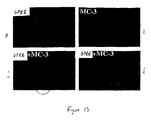
- 230000008045 co-localization Effects 0.000 description 6
- 238000012790 confirmation Methods 0.000 description 6
- 238000011161 development Methods 0.000 description 6
- 238000010249 in-situ analysis Methods 0.000 description 6
- 235000020824 obesity Nutrition 0.000 description 6
- KWTSXDURSIMDCE-QMMMGPOBSA-N (S)-amphetamine Chemical compound C[C@H](N)CC1=CC=CC=C1 KWTSXDURSIMDCE-QMMMGPOBSA-N 0.000 description 5
- 108060001084 Luciferase Proteins 0.000 description 5
- JLCPHMBAVCMARE-UHFFFAOYSA-N [3-[[3-[[3-[[3-[[3-[[3-[[3-[[3-[[3-[[3-[[3-[[5-(2-amino-6-oxo-1H-purin-9-yl)-3-[[3-[[3-[[3-[[3-[[3-[[5-(2-amino-6-oxo-1H-purin-9-yl)-3-[[5-(2-amino-6-oxo-1H-purin-9-yl)-3-hydroxyoxolan-2-yl]methoxy-hydroxyphosphoryl]oxyoxolan-2-yl]methoxy-hydroxyphosphoryl]oxy-5-(5-methyl-2,4-dioxopyrimidin-1-yl)oxolan-2-yl]methoxy-hydroxyphosphoryl]oxy-5-(6-aminopurin-9-yl)oxolan-2-yl]methoxy-hydroxyphosphoryl]oxy-5-(6-aminopurin-9-yl)oxolan-2-yl]methoxy-hydroxyphosphoryl]oxy-5-(6-aminopurin-9-yl)oxolan-2-yl]methoxy-hydroxyphosphoryl]oxy-5-(6-aminopurin-9-yl)oxolan-2-yl]methoxy-hydroxyphosphoryl]oxyoxolan-2-yl]methoxy-hydroxyphosphoryl]oxy-5-(5-methyl-2,4-dioxopyrimidin-1-yl)oxolan-2-yl]methoxy-hydroxyphosphoryl]oxy-5-(4-amino-2-oxopyrimidin-1-yl)oxolan-2-yl]methoxy-hydroxyphosphoryl]oxy-5-(5-methyl-2,4-dioxopyrimidin-1-yl)oxolan-2-yl]methoxy-hydroxyphosphoryl]oxy-5-(5-methyl-2,4-dioxopyrimidin-1-yl)oxolan-2-yl]methoxy-hydroxyphosphoryl]oxy-5-(6-aminopurin-9-yl)oxolan-2-yl]methoxy-hydroxyphosphoryl]oxy-5-(6-aminopurin-9-yl)oxolan-2-yl]methoxy-hydroxyphosphoryl]oxy-5-(4-amino-2-oxopyrimidin-1-yl)oxolan-2-yl]methoxy-hydroxyphosphoryl]oxy-5-(4-amino-2-oxopyrimidin-1-yl)oxolan-2-yl]methoxy-hydroxyphosphoryl]oxy-5-(4-amino-2-oxopyrimidin-1-yl)oxolan-2-yl]methoxy-hydroxyphosphoryl]oxy-5-(6-aminopurin-9-yl)oxolan-2-yl]methoxy-hydroxyphosphoryl]oxy-5-(4-amino-2-oxopyrimidin-1-yl)oxolan-2-yl]methyl [5-(6-aminopurin-9-yl)-2-(hydroxymethyl)oxolan-3-yl] hydrogen phosphate Polymers Cc1cn(C2CC(OP(O)(=O)OCC3OC(CC3OP(O)(=O)OCC3OC(CC3O)n3cnc4c3nc(N)[nH]c4=O)n3cnc4c3nc(N)[nH]c4=O)C(COP(O)(=O)OC3CC(OC3COP(O)(=O)OC3CC(OC3COP(O)(=O)OC3CC(OC3COP(O)(=O)OC3CC(OC3COP(O)(=O)OC3CC(OC3COP(O)(=O)OC3CC(OC3COP(O)(=O)OC3CC(OC3COP(O)(=O)OC3CC(OC3COP(O)(=O)OC3CC(OC3COP(O)(=O)OC3CC(OC3COP(O)(=O)OC3CC(OC3COP(O)(=O)OC3CC(OC3COP(O)(=O)OC3CC(OC3COP(O)(=O)OC3CC(OC3COP(O)(=O)OC3CC(OC3COP(O)(=O)OC3CC(OC3COP(O)(=O)OC3CC(OC3CO)n3cnc4c(N)ncnc34)n3ccc(N)nc3=O)n3cnc4c(N)ncnc34)n3ccc(N)nc3=O)n3ccc(N)nc3=O)n3ccc(N)nc3=O)n3cnc4c(N)ncnc34)n3cnc4c(N)ncnc34)n3cc(C)c(=O)[nH]c3=O)n3cc(C)c(=O)[nH]c3=O)n3ccc(N)nc3=O)n3cc(C)c(=O)[nH]c3=O)n3cnc4c3nc(N)[nH]c4=O)n3cnc4c(N)ncnc34)n3cnc4c(N)ncnc34)n3cnc4c(N)ncnc34)n3cnc4c(N)ncnc34)O2)c(=O)[nH]c1=O JLCPHMBAVCMARE-UHFFFAOYSA-N 0.000 description 5
- 229940025084 amphetamine Drugs 0.000 description 5
- 230000006399 behavior Effects 0.000 description 5
- 238000001514 detection method Methods 0.000 description 5
- 238000010586 diagram Methods 0.000 description 5
- 208000035475 disorder Diseases 0.000 description 5
- 238000010230 functional analysis Methods 0.000 description 5
- 210000001320 hippocampus Anatomy 0.000 description 5
- 210000003016 hypothalamus Anatomy 0.000 description 5
- 230000006742 locomotor activity Effects 0.000 description 5
- 239000008188 pellet Substances 0.000 description 5
- 210000002966 serum Anatomy 0.000 description 5
- 239000011780 sodium chloride Substances 0.000 description 5
- 238000001356 surgical procedure Methods 0.000 description 5
- 238000011144 upstream manufacturing Methods 0.000 description 5
- ZHNUHDYFZUAESO-UHFFFAOYSA-N Formamide Chemical compound NC=O ZHNUHDYFZUAESO-UHFFFAOYSA-N 0.000 description 4
- 101100337651 Rattus norvegicus Gpr12 gene Proteins 0.000 description 4
- 108091027981 Response element Proteins 0.000 description 4
- 240000004808 Saccharomyces cerevisiae Species 0.000 description 4
- GSEJCLTVZPLZKY-UHFFFAOYSA-N Triethanolamine Chemical compound OCCN(CCO)CCO GSEJCLTVZPLZKY-UHFFFAOYSA-N 0.000 description 4
- 239000000074 antisense oligonucleotide Substances 0.000 description 4
- 238000012230 antisense oligonucleotides Methods 0.000 description 4
- 238000000376 autoradiography Methods 0.000 description 4
- 239000004568 cement Substances 0.000 description 4
- 239000003153 chemical reaction reagent Substances 0.000 description 4
- 238000010168 coupling process Methods 0.000 description 4
- 238000005859 coupling reaction Methods 0.000 description 4
- 102000037865 fusion proteins Human genes 0.000 description 4
- 238000001727 in vivo Methods 0.000 description 4
- QWTDNUCVQCZILF-UHFFFAOYSA-N isopentane Chemical compound CCC(C)C QWTDNUCVQCZILF-UHFFFAOYSA-N 0.000 description 4
- 230000003137 locomotive effect Effects 0.000 description 4
- 210000004940 nucleus Anatomy 0.000 description 4
- 238000003757 reverse transcription PCR Methods 0.000 description 4
- 125000006850 spacer group Chemical group 0.000 description 4
- 239000008223 sterile water Substances 0.000 description 4
- 238000013518 transcription Methods 0.000 description 4
- 230000035897 transcription Effects 0.000 description 4
- 108091032973 (ribonucleotides)n+m Proteins 0.000 description 3
- CZMRCDWAGMRECN-UHFFFAOYSA-N 2-{[3,4-dihydroxy-2,5-bis(hydroxymethyl)oxolan-2-yl]oxy}-6-(hydroxymethyl)oxane-3,4,5-triol Chemical compound OCC1OC(CO)(OC2OC(CO)C(O)C(O)C2O)C(O)C1O CZMRCDWAGMRECN-UHFFFAOYSA-N 0.000 description 3
- 238000009010 Bradford assay Methods 0.000 description 3
- 108091026890 Coding region Proteins 0.000 description 3
- KCXVZYZYPLLWCC-UHFFFAOYSA-N EDTA Chemical compound OC(=O)CN(CC(O)=O)CCN(CC(O)=O)CC(O)=O KCXVZYZYPLLWCC-UHFFFAOYSA-N 0.000 description 3
- DHMQDGOQFOQNFH-UHFFFAOYSA-N Glycine Chemical compound NCC(O)=O DHMQDGOQFOQNFH-UHFFFAOYSA-N 0.000 description 3
- 239000005089 Luciferase Substances 0.000 description 3
- 102000008834 Orexin receptor Human genes 0.000 description 3
- 230000004913 activation Effects 0.000 description 3
- 108060000200 adenylate cyclase Proteins 0.000 description 3
- 102000030621 adenylate cyclase Human genes 0.000 description 3
- 230000008512 biological response Effects 0.000 description 3
- 230000037396 body weight Effects 0.000 description 3
- 210000003710 cerebral cortex Anatomy 0.000 description 3
- 230000006552 constitutive activation Effects 0.000 description 3
- 230000008878 coupling Effects 0.000 description 3
- 230000003247 decreasing effect Effects 0.000 description 3
- 238000013461 design Methods 0.000 description 3
- 229940079593 drug Drugs 0.000 description 3
- 238000007876 drug discovery Methods 0.000 description 3
- 210000005153 frontal cortex Anatomy 0.000 description 3
- 238000000265 homogenisation Methods 0.000 description 3
- 238000009396 hybridization Methods 0.000 description 3
- 238000000338 in vitro Methods 0.000 description 3
- 238000002372 labelling Methods 0.000 description 3
- 150000002632 lipids Chemical class 0.000 description 3
- 210000004072 lung Anatomy 0.000 description 3
- 229910001629 magnesium chloride Inorganic materials 0.000 description 3
- 239000002609 medium Substances 0.000 description 3
- 108020004999 messenger RNA Proteins 0.000 description 3
- 230000004048 modification Effects 0.000 description 3
- 238000012986 modification Methods 0.000 description 3
- IXRNQIKIVWWFBH-UHFFFAOYSA-N n-(1-phenylethenyl)acetamide Chemical compound CC(=O)NC(=C)C1=CC=CC=C1 IXRNQIKIVWWFBH-UHFFFAOYSA-N 0.000 description 3
- 230000002123 temporal effect Effects 0.000 description 3
- 238000012360 testing method Methods 0.000 description 3
- 230000001225 therapeutic effect Effects 0.000 description 3
- 238000010361 transduction Methods 0.000 description 3
- 230000026683 transduction Effects 0.000 description 3
- 238000012546 transfer Methods 0.000 description 3
- 238000011680 zucker rat Methods 0.000 description 3
- 108091093088 Amplicon Proteins 0.000 description 2
- DCXYFEDJOCDNAF-UHFFFAOYSA-N Asparagine Natural products OC(=O)C(N)CC(N)=O DCXYFEDJOCDNAF-UHFFFAOYSA-N 0.000 description 2
- 241000972773 Aulopiformes Species 0.000 description 2
- KRKNYBCHXYNGOX-UHFFFAOYSA-K Citrate Chemical compound [O-]C(=O)CC(O)(CC([O-])=O)C([O-])=O KRKNYBCHXYNGOX-UHFFFAOYSA-K 0.000 description 2
- 102000016928 DNA-directed DNA polymerase Human genes 0.000 description 2
- 108010014303 DNA-directed DNA polymerase Proteins 0.000 description 2
- 108010067770 Endopeptidase K Proteins 0.000 description 2
- 102000013446 GTP Phosphohydrolases Human genes 0.000 description 2
- 108091006109 GTPases Proteins 0.000 description 2
- 108010001515 Galectin 4 Proteins 0.000 description 2
- 102100039556 Galectin-4 Human genes 0.000 description 2
- 102100034013 Gamma-glutamyl phosphate reductase Human genes 0.000 description 2
- 101710198928 Gamma-glutamyl phosphate reductase Proteins 0.000 description 2
- 101710159101 Green-light absorbing proteorhodopsin Proteins 0.000 description 2
- DCXYFEDJOCDNAF-REOHCLBHSA-N L-asparagine Chemical compound OC(=O)[C@@H](N)CC(N)=O DCXYFEDJOCDNAF-REOHCLBHSA-N 0.000 description 2
- CKLJMWTZIZZHCS-REOHCLBHSA-N L-aspartic acid Chemical compound OC(=O)[C@@H](N)CC(O)=O CKLJMWTZIZZHCS-REOHCLBHSA-N 0.000 description 2
- AGPKZVBTJJNPAG-WHFBIAKZSA-N L-isoleucine Chemical compound CC[C@H](C)[C@H](N)C(O)=O AGPKZVBTJJNPAG-WHFBIAKZSA-N 0.000 description 2
- ROHFNLRQFUQHCH-YFKPBYRVSA-N L-leucine Chemical compound CC(C)C[C@H](N)C(O)=O ROHFNLRQFUQHCH-YFKPBYRVSA-N 0.000 description 2
- COLNVLDHVKWLRT-QMMMGPOBSA-N L-phenylalanine Chemical compound OC(=O)[C@@H](N)CC1=CC=CC=C1 COLNVLDHVKWLRT-QMMMGPOBSA-N 0.000 description 2
- OUYCCCASQSFEME-QMMMGPOBSA-N L-tyrosine Chemical compound OC(=O)[C@@H](N)CC1=CC=C(O)C=C1 OUYCCCASQSFEME-QMMMGPOBSA-N 0.000 description 2
- 108091026898 Leader sequence (mRNA) Proteins 0.000 description 2
- KDXKERNSBIXSRK-UHFFFAOYSA-N Lysine Natural products NCCCCC(N)C(O)=O KDXKERNSBIXSRK-UHFFFAOYSA-N 0.000 description 2
- CSNNHWWHGAXBCP-UHFFFAOYSA-L Magnesium sulfate Chemical compound [Mg+2].[O-][S+2]([O-])([O-])[O-] CSNNHWWHGAXBCP-UHFFFAOYSA-L 0.000 description 2
- 102000018697 Membrane Proteins Human genes 0.000 description 2
- 108091028043 Nucleic acid sequence Proteins 0.000 description 2
- 108050000742 Orexin Receptor Proteins 0.000 description 2
- 108020002230 Pancreatic Ribonuclease Proteins 0.000 description 2
- 102000005891 Pancreatic ribonuclease Human genes 0.000 description 2
- 101100122770 Rattus norvegicus Gpr3 gene Proteins 0.000 description 2
- 101100449397 Rattus norvegicus Gpr6 gene Proteins 0.000 description 2
- 108700008625 Reporter Genes Proteins 0.000 description 2
- 102000006382 Ribonucleases Human genes 0.000 description 2
- 108010083644 Ribonucleases Proteins 0.000 description 2
- 229920005654 Sephadex Polymers 0.000 description 2
- 239000012507 Sephadex™ Substances 0.000 description 2
- 102000005157 Somatostatin Human genes 0.000 description 2
- 108010056088 Somatostatin Proteins 0.000 description 2
- 108091023040 Transcription factor Proteins 0.000 description 2
- 102000040945 Transcription factor Human genes 0.000 description 2
- XSQUKJJJFZCRTK-UHFFFAOYSA-N Urea Chemical compound NC(N)=O XSQUKJJJFZCRTK-UHFFFAOYSA-N 0.000 description 2
- KZSNJWFQEVHDMF-UHFFFAOYSA-N Valine Natural products CC(C)C(N)C(O)=O KZSNJWFQEVHDMF-UHFFFAOYSA-N 0.000 description 2
- 241000700605 Viruses Species 0.000 description 2
- 238000009825 accumulation Methods 0.000 description 2
- 239000004480 active ingredient Substances 0.000 description 2
- 210000004727 amygdala Anatomy 0.000 description 2
- 238000000137 annealing Methods 0.000 description 2
- 230000008901 benefit Effects 0.000 description 2
- 210000004899 c-terminal region Anatomy 0.000 description 2
- 238000013262 cAMP assay Methods 0.000 description 2
- 239000003795 chemical substances by application Substances 0.000 description 2
- 210000000349 chromosome Anatomy 0.000 description 2
- 230000000052 comparative effect Effects 0.000 description 2
- 239000003184 complementary RNA Substances 0.000 description 2
- 229960000633 dextran sulfate Drugs 0.000 description 2
- 230000004069 differentiation Effects 0.000 description 2
- AFABGHUZZDYHJO-UHFFFAOYSA-N dimethyl butane Natural products CCCC(C)C AFABGHUZZDYHJO-UHFFFAOYSA-N 0.000 description 2
- 208000037765 diseases and disorders Diseases 0.000 description 2
- VHJLVAABSRFDPM-QWWZWVQMSA-N dithiothreitol Chemical compound SC[C@@H](O)[C@H](O)CS VHJLVAABSRFDPM-QWWZWVQMSA-N 0.000 description 2
- -1 e.g. Proteins 0.000 description 2
- 239000013613 expression plasmid Substances 0.000 description 2
- 210000002458 fetal heart Anatomy 0.000 description 2
- 239000011888 foil Substances 0.000 description 2
- 235000013305 food Nutrition 0.000 description 2
- 230000006870 function Effects 0.000 description 2
- 238000003197 gene knockdown Methods 0.000 description 2
- 239000007943 implant Substances 0.000 description 2
- 210000003734 kidney Anatomy 0.000 description 2
- 210000004185 liver Anatomy 0.000 description 2
- 208000020442 loss of weight Diseases 0.000 description 2
- 230000004060 metabolic process Effects 0.000 description 2
- 238000002156 mixing Methods 0.000 description 2
- 230000010004 neural pathway Effects 0.000 description 2
- 210000000118 neural pathway Anatomy 0.000 description 2
- 239000000047 product Substances 0.000 description 2
- 230000012743 protein tagging Effects 0.000 description 2
- 210000002637 putamen Anatomy 0.000 description 2
- 230000000384 rearing effect Effects 0.000 description 2
- 230000009467 reduction Effects 0.000 description 2
- 108091008146 restriction endonucleases Proteins 0.000 description 2
- 239000003161 ribonuclease inhibitor Substances 0.000 description 2
- 235000019515 salmon Nutrition 0.000 description 2
- 238000007423 screening assay Methods 0.000 description 2
- 230000011664 signaling Effects 0.000 description 2
- 239000011734 sodium Substances 0.000 description 2
- 239000012064 sodium phosphate buffer Substances 0.000 description 2
- NHXLMOGPVYXJNR-ATOGVRKGSA-N somatostatin Chemical compound C([C@H]1C(=O)N[C@H](C(N[C@@H](CO)C(=O)N[C@@H](CSSC[C@@H](C(=O)N[C@@H](CCCCN)C(=O)N[C@@H](CC(N)=O)C(=O)N[C@@H](CC=2C=CC=CC=2)C(=O)N[C@@H](CC=2C=CC=CC=2)C(=O)N[C@@H](CC=2C3=CC=CC=C3NC=2)C(=O)N[C@@H](CCCCN)C(=O)N[C@H](C(=O)N1)[C@@H](C)O)NC(=O)CNC(=O)[C@H](C)N)C(O)=O)=O)[C@H](O)C)C1=CC=CC=C1 NHXLMOGPVYXJNR-ATOGVRKGSA-N 0.000 description 2
- 229960000553 somatostatin Drugs 0.000 description 2
- 210000000952 spleen Anatomy 0.000 description 2
- 238000012453 sprague-dawley rat model Methods 0.000 description 2
- 230000006641 stabilisation Effects 0.000 description 2
- 238000011105 stabilization Methods 0.000 description 2
- 239000006228 supernatant Substances 0.000 description 2
- 239000000725 suspension Substances 0.000 description 2
- 210000001541 thymus gland Anatomy 0.000 description 2
- 239000011534 wash buffer Substances 0.000 description 2
- WHTVZRBIWZFKQO-AWEZNQCLSA-N (S)-chloroquine Chemical compound ClC1=CC=C2C(N[C@@H](C)CCCN(CC)CC)=CC=NC2=C1 WHTVZRBIWZFKQO-AWEZNQCLSA-N 0.000 description 1
- ISPYQTSUDJAMAB-UHFFFAOYSA-N 2-chlorophenol Chemical compound OC1=CC=CC=C1Cl ISPYQTSUDJAMAB-UHFFFAOYSA-N 0.000 description 1
- BFSVOASYOCHEOV-UHFFFAOYSA-N 2-diethylaminoethanol Chemical compound CCN(CC)CCO BFSVOASYOCHEOV-UHFFFAOYSA-N 0.000 description 1
- 108700021677 Agouti-Related Proteins 0.000 description 1
- 206010002091 Anaesthesia Diseases 0.000 description 1
- 108020000948 Antisense Oligonucleotides Proteins 0.000 description 1
- 239000004475 Arginine Substances 0.000 description 1
- 108091003079 Bovine Serum Albumin Proteins 0.000 description 1
- 102000000844 Cell Surface Receptors Human genes 0.000 description 1
- 108010001857 Cell Surface Receptors Proteins 0.000 description 1
- 108020004705 Codon Proteins 0.000 description 1
- 102000004420 Creatine Kinase Human genes 0.000 description 1
- 108010042126 Creatine kinase Proteins 0.000 description 1
- 101710095468 Cyclase Proteins 0.000 description 1
- 102000005636 Cyclic AMP Response Element-Binding Protein Human genes 0.000 description 1
- 108010045171 Cyclic AMP Response Element-Binding Protein Proteins 0.000 description 1
- IVOMOUWHDPKRLL-KQYNXXCUSA-N Cyclic adenosine monophosphate Chemical compound C([C@H]1O2)OP(O)(=O)O[C@H]1[C@@H](O)[C@@H]2N1C(N=CN=C2N)=C2N=C1 IVOMOUWHDPKRLL-KQYNXXCUSA-N 0.000 description 1
- 102000052510 DNA-Binding Proteins Human genes 0.000 description 1
- 101710096438 DNA-binding protein Proteins 0.000 description 1
- 229920002307 Dextran Polymers 0.000 description 1
- 206010013654 Drug abuse Diseases 0.000 description 1
- 102000004190 Enzymes Human genes 0.000 description 1
- 108090000790 Enzymes Proteins 0.000 description 1
- 108091006101 Gi proteins Proteins 0.000 description 1
- 102000034354 Gi proteins Human genes 0.000 description 1
- WHUUTDBJXJRKMK-UHFFFAOYSA-N Glutamic acid Natural products OC(=O)C(N)CCC(O)=O WHUUTDBJXJRKMK-UHFFFAOYSA-N 0.000 description 1
- 239000004471 Glycine Substances 0.000 description 1
- 108091006065 Gs proteins Proteins 0.000 description 1
- 241000282412 Homo Species 0.000 description 1
- 101100392414 Homo sapiens GHSR gene Proteins 0.000 description 1
- 101000598921 Homo sapiens Orexin Proteins 0.000 description 1
- 206010062767 Hypophysitis Diseases 0.000 description 1
- ONIBWKKTOPOVIA-BYPYZUCNSA-N L-Proline Chemical compound OC(=O)[C@@H]1CCCN1 ONIBWKKTOPOVIA-BYPYZUCNSA-N 0.000 description 1
- QNAYBMKLOCPYGJ-REOHCLBHSA-N L-alanine Chemical compound C[C@H](N)C(O)=O QNAYBMKLOCPYGJ-REOHCLBHSA-N 0.000 description 1
- FFEARJCKVFRZRR-BYPYZUCNSA-N L-methionine Chemical compound CSCC[C@H](N)C(O)=O FFEARJCKVFRZRR-BYPYZUCNSA-N 0.000 description 1
- QIVBCDIJIAJPQS-VIFPVBQESA-N L-tryptophane Chemical compound C1=CC=C2C(C[C@H](N)C(O)=O)=CNC2=C1 QIVBCDIJIAJPQS-VIFPVBQESA-N 0.000 description 1
- KZSNJWFQEVHDMF-BYPYZUCNSA-N L-valine Chemical compound CC(C)[C@H](N)C(O)=O KZSNJWFQEVHDMF-BYPYZUCNSA-N 0.000 description 1
- 102000016267 Leptin Human genes 0.000 description 1
- 108010092277 Leptin Proteins 0.000 description 1
- ROHFNLRQFUQHCH-UHFFFAOYSA-N Leucine Natural products CC(C)CC(N)C(O)=O ROHFNLRQFUQHCH-UHFFFAOYSA-N 0.000 description 1
- 239000004472 Lysine Substances 0.000 description 1
- 229910021380 Manganese Chloride Inorganic materials 0.000 description 1
- GLFNIEUTAYBVOC-UHFFFAOYSA-L Manganese chloride Chemical compound Cl[Mn]Cl GLFNIEUTAYBVOC-UHFFFAOYSA-L 0.000 description 1
- 102000004378 Melanocortin Receptors Human genes 0.000 description 1
- 108090000950 Melanocortin Receptors Proteins 0.000 description 1
- 108010008364 Melanocortins Proteins 0.000 description 1
- 101710151321 Melanostatin Proteins 0.000 description 1
- 101100122769 Mus musculus Gpr3 gene Proteins 0.000 description 1
- 102400000064 Neuropeptide Y Human genes 0.000 description 1
- 108090000189 Neuropeptides Proteins 0.000 description 1
- 108700026244 Open Reading Frames Proteins 0.000 description 1
- 102000002512 Orexin Human genes 0.000 description 1
- BELBBZDIHDAJOR-UHFFFAOYSA-N Phenolsulfonephthalein Chemical compound C1=CC(O)=CC=C1C1(C=2C=CC(O)=CC=2)C2=CC=CC=C2S(=O)(=O)O1 BELBBZDIHDAJOR-UHFFFAOYSA-N 0.000 description 1
- 229920005372 Plexiglas® Polymers 0.000 description 1
- ONIBWKKTOPOVIA-UHFFFAOYSA-N Proline Natural products OC(=O)C1CCCN1 ONIBWKKTOPOVIA-UHFFFAOYSA-N 0.000 description 1
- 238000010240 RT-PCR analysis Methods 0.000 description 1
- 101100015376 Rattus norvegicus Gnaz gene Proteins 0.000 description 1
- 101000633010 Rattus norvegicus Somatostatin Proteins 0.000 description 1
- MTCFGRXMJLQNBG-UHFFFAOYSA-N Serine Natural products OCC(N)C(O)=O MTCFGRXMJLQNBG-UHFFFAOYSA-N 0.000 description 1
- 108091036066 Three prime untranslated region Proteins 0.000 description 1
- AYFVYJQAPQTCCC-UHFFFAOYSA-N Threonine Natural products CC(O)C(N)C(O)=O AYFVYJQAPQTCCC-UHFFFAOYSA-N 0.000 description 1
- 239000004473 Threonine Substances 0.000 description 1
- 108700009124 Transcription Initiation Site Proteins 0.000 description 1
- 229920004890 Triton X-100 Polymers 0.000 description 1
- 239000013504 Triton X-100 Substances 0.000 description 1
- QIVBCDIJIAJPQS-UHFFFAOYSA-N Tryptophan Natural products C1=CC=C2C(CC(N)C(O)=O)=CNC2=C1 QIVBCDIJIAJPQS-UHFFFAOYSA-N 0.000 description 1
- IVOMOUWHDPKRLL-UHFFFAOYSA-N UNPD107823 Natural products O1C2COP(O)(=O)OC2C(O)C1N1C(N=CN=C2N)=C2N=C1 IVOMOUWHDPKRLL-UHFFFAOYSA-N 0.000 description 1
- 108010046516 Wheat Germ Agglutinins Proteins 0.000 description 1
- 238000002835 absorbance Methods 0.000 description 1
- 210000004100 adrenal gland Anatomy 0.000 description 1
- 239000011543 agarose gel Substances 0.000 description 1
- 235000004279 alanine Nutrition 0.000 description 1
- 230000037005 anaesthesia Effects 0.000 description 1
- 238000010171 animal model Methods 0.000 description 1
- 230000008485 antagonism Effects 0.000 description 1
- 210000000709 aorta Anatomy 0.000 description 1
- 210000003295 arcuate nucleus Anatomy 0.000 description 1
- ODKSFYDXXFIFQN-UHFFFAOYSA-N arginine Natural products OC(=O)C(N)CCCNC(N)=N ODKSFYDXXFIFQN-UHFFFAOYSA-N 0.000 description 1
- 238000003491 array Methods 0.000 description 1
- 235000009582 asparagine Nutrition 0.000 description 1
- 229960001230 asparagine Drugs 0.000 description 1
- 235000003704 aspartic acid Nutrition 0.000 description 1
- 230000003305 autocrine Effects 0.000 description 1
- 239000011324 bead Substances 0.000 description 1
- 230000009286 beneficial effect Effects 0.000 description 1
- OQFSQFPPLPISGP-UHFFFAOYSA-N beta-carboxyaspartic acid Natural products OC(=O)C(N)C(C(O)=O)C(O)=O OQFSQFPPLPISGP-UHFFFAOYSA-N 0.000 description 1
- 238000010256 biochemical assay Methods 0.000 description 1
- 230000033228 biological regulation Effects 0.000 description 1
- 230000000903 blocking effect Effects 0.000 description 1
- 210000001185 bone marrow Anatomy 0.000 description 1
- 210000005013 brain tissue Anatomy 0.000 description 1
- 230000011496 cAMP-mediated signaling Effects 0.000 description 1
- 238000010805 cDNA synthesis kit Methods 0.000 description 1
- 239000004202 carbamide Substances 0.000 description 1
- 210000001159 caudate nucleus Anatomy 0.000 description 1
- 238000004113 cell culture Methods 0.000 description 1
- 210000000170 cell membrane Anatomy 0.000 description 1
- 239000013553 cell monolayer Substances 0.000 description 1
- 230000001413 cellular effect Effects 0.000 description 1
- 238000005119 centrifugation Methods 0.000 description 1
- 210000001638 cerebellum Anatomy 0.000 description 1
- 238000001311 chemical methods and process Methods 0.000 description 1
- 238000007385 chemical modification Methods 0.000 description 1
- 229960003677 chloroquine Drugs 0.000 description 1
- WHTVZRBIWZFKQO-UHFFFAOYSA-N chloroquine Natural products ClC1=CC=C2C(NC(C)CCCN(CC)CC)=CC=NC2=C1 WHTVZRBIWZFKQO-UHFFFAOYSA-N 0.000 description 1
- 238000010367 cloning Methods 0.000 description 1
- ZPUCINDJVBIVPJ-LJISPDSOSA-N cocaine Chemical compound O([C@H]1C[C@@H]2CC[C@@H](N2C)[C@H]1C(=O)OC)C(=O)C1=CC=CC=C1 ZPUCINDJVBIVPJ-LJISPDSOSA-N 0.000 description 1
- 210000001072 colon Anatomy 0.000 description 1
- 230000037011 constitutive activity Effects 0.000 description 1
- 238000010276 construction Methods 0.000 description 1
- 238000011109 contamination Methods 0.000 description 1
- 229940095074 cyclic amp Drugs 0.000 description 1
- 235000018417 cysteine Nutrition 0.000 description 1
- XUJNEKJLAYXESH-UHFFFAOYSA-N cysteine Natural products SCC(N)C(O)=O XUJNEKJLAYXESH-UHFFFAOYSA-N 0.000 description 1
- RGWHQCVHVJXOKC-SHYZEUOFSA-J dCTP(4-) Chemical compound O=C1N=C(N)C=CN1[C@@H]1O[C@H](COP([O-])(=O)OP([O-])(=O)OP([O-])([O-])=O)[C@@H](O)C1 RGWHQCVHVJXOKC-SHYZEUOFSA-J 0.000 description 1
- 235000021316 daily nutritional intake Nutrition 0.000 description 1
- 238000003936 denaturing gel electrophoresis Methods 0.000 description 1
- 210000001947 dentate gyrus Anatomy 0.000 description 1
- 229960002086 dextran Drugs 0.000 description 1
- 206010012601 diabetes mellitus Diseases 0.000 description 1
- 230000029087 digestion Effects 0.000 description 1
- 239000003937 drug carrier Substances 0.000 description 1
- 238000009510 drug design Methods 0.000 description 1
- 238000009509 drug development Methods 0.000 description 1
- 238000012912 drug discovery process Methods 0.000 description 1
- 238000007877 drug screening Methods 0.000 description 1
- 206010015037 epilepsy Diseases 0.000 description 1
- 230000001037 epileptic effect Effects 0.000 description 1
- 238000002474 experimental method Methods 0.000 description 1
- 210000001723 extracellular space Anatomy 0.000 description 1
- 239000012091 fetal bovine serum Substances 0.000 description 1
- 210000003754 fetus Anatomy 0.000 description 1
- 230000004927 fusion Effects 0.000 description 1
- 238000001415 gene therapy Methods 0.000 description 1
- 230000008303 genetic mechanism Effects 0.000 description 1
- 235000013922 glutamic acid Nutrition 0.000 description 1
- 239000004220 glutamic acid Substances 0.000 description 1
- ZDXPYRJPNDTMRX-UHFFFAOYSA-N glutamine Natural products OC(=O)C(N)CCC(N)=O ZDXPYRJPNDTMRX-UHFFFAOYSA-N 0.000 description 1
- BCQZXOMGPXTTIC-UHFFFAOYSA-N halothane Chemical compound FC(F)(F)C(Cl)Br BCQZXOMGPXTTIC-UHFFFAOYSA-N 0.000 description 1
- 229960003132 halothane Drugs 0.000 description 1
- HNDVDQJCIGZPNO-UHFFFAOYSA-N histidine Natural products OC(=O)C(N)CC1=CN=CN1 HNDVDQJCIGZPNO-UHFFFAOYSA-N 0.000 description 1
- 230000013632 homeostatic process Effects 0.000 description 1
- 102000046099 human GPR12 Human genes 0.000 description 1
- 102000044097 human GPR21 Human genes 0.000 description 1
- 230000002209 hydrophobic effect Effects 0.000 description 1
- 238000001802 infusion Methods 0.000 description 1
- 230000002401 inhibitory effect Effects 0.000 description 1
- 238000002347 injection Methods 0.000 description 1
- 239000007924 injection Substances 0.000 description 1
- 238000003780 insertion Methods 0.000 description 1
- 230000037431 insertion Effects 0.000 description 1
- 230000003993 interaction Effects 0.000 description 1
- 230000002452 interceptive effect Effects 0.000 description 1
- 230000031146 intracellular signal transduction Effects 0.000 description 1
- 238000003874 inverse correlation nuclear magnetic resonance spectroscopy Methods 0.000 description 1
- 238000011835 investigation Methods 0.000 description 1
- AGPKZVBTJJNPAG-UHFFFAOYSA-N isoleucine Natural products CCC(C)C(N)C(O)=O AGPKZVBTJJNPAG-UHFFFAOYSA-N 0.000 description 1
- 229960000310 isoleucine Drugs 0.000 description 1
- 238000005304 joining Methods 0.000 description 1
- 210000003140 lateral ventricle Anatomy 0.000 description 1
- 239000010410 layer Substances 0.000 description 1
- 150000002611 lead compounds Chemical class 0.000 description 1
- 229940039781 leptin Drugs 0.000 description 1
- NRYBAZVQPHGZNS-ZSOCWYAHSA-N leptin Chemical compound O=C([C@H](CO)NC(=O)[C@H](CC(C)C)NC(=O)[C@H](CC(O)=O)NC(=O)[C@H](CC(C)C)NC(=O)[C@H](CCC(N)=O)NC(=O)[C@H](CC=1C2=CC=CC=C2NC=1)NC(=O)[C@H](CC(C)C)NC(=O)[C@@H](NC(=O)[C@H](CC(O)=O)NC(=O)[C@H](CCC(N)=O)NC(=O)[C@H](CC(C)C)NC(=O)[C@H](CO)NC(=O)CNC(=O)[C@H](CCC(N)=O)NC(=O)[C@@H](N)CC(C)C)CCSC)N1CCC[C@H]1C(=O)NCC(=O)N[C@@H](CS)C(O)=O NRYBAZVQPHGZNS-ZSOCWYAHSA-N 0.000 description 1
- 210000000265 leukocyte Anatomy 0.000 description 1
- 239000007788 liquid Substances 0.000 description 1
- 238000004020 luminiscence type Methods 0.000 description 1
- 210000001165 lymph node Anatomy 0.000 description 1
- 239000012139 lysis buffer Substances 0.000 description 1
- 229910052943 magnesium sulfate Inorganic materials 0.000 description 1
- 210000005075 mammary gland Anatomy 0.000 description 1
- 239000011565 manganese chloride Substances 0.000 description 1
- 230000007246 mechanism Effects 0.000 description 1
- 230000007721 medicinal effect Effects 0.000 description 1
- 210000001767 medulla oblongata Anatomy 0.000 description 1
- 239000002865 melanocortin Substances 0.000 description 1
- 229930182817 methionine Natural products 0.000 description 1
- 244000005700 microbiome Species 0.000 description 1
- 238000012544 monitoring process Methods 0.000 description 1
- 230000008450 motivation Effects 0.000 description 1
- 230000001537 neural effect Effects 0.000 description 1
- 210000002569 neuron Anatomy 0.000 description 1
- BPGXUIVWLQTVLZ-OFGSCBOVSA-N neuropeptide y(npy) Chemical compound C([C@@H](C(=O)N[C@@H]([C@@H](C)CC)C(=O)N[C@@H](CC(N)=O)C(=O)N[C@@H](CC(C)C)C(=O)N[C@@H]([C@@H](C)CC)C(=O)N[C@@H]([C@@H](C)O)C(=O)N[C@@H](CCCN=C(N)N)C(=O)N[C@@H](CCC(N)=O)C(=O)N[C@@H](CCCN=C(N)N)C(=O)N[C@@H](CC=1C=CC(O)=CC=1)C(O)=O)NC(=O)[C@H](CC=1N=CNC=1)NC(=O)[C@H](CCCN=C(N)N)NC(=O)[C@H](CC(C)C)NC(=O)[C@H](C)NC(=O)[C@H](CO)NC(=O)[C@H](CC=1C=CC(O)=CC=1)NC(=O)[C@H](CC=1C=CC(O)=CC=1)NC(=O)[C@H](CCCN=C(N)N)NC(=O)[C@H](C)NC(=O)[C@H](CC(C)C)NC(=O)[C@H](CC(O)=O)NC(=O)[C@H](CCC(O)=O)NC(=O)[C@H](C)NC(=O)[C@H]1N(CCC1)C(=O)[C@H](C)NC(=O)[C@H](CC(O)=O)NC(=O)[C@H](CCC(O)=O)NC(=O)CNC(=O)[C@H]1N(CCC1)C(=O)[C@H](CC(N)=O)NC(=O)[C@H](CC(O)=O)NC(=O)[C@H]1N(CCC1)C(=O)[C@H](CCCCN)NC(=O)[C@H](CO)NC(=O)[C@H]1N(CCC1)C(=O)[C@@H](N)CC=1C=CC(O)=CC=1)C1=CC=C(O)C=C1 BPGXUIVWLQTVLZ-OFGSCBOVSA-N 0.000 description 1
- URPYMXQQVHTUDU-OFGSCBOVSA-N nucleopeptide y Chemical compound C([C@@H](C(=O)N[C@@H]([C@@H](C)CC)C(=O)N[C@@H](CC(N)=O)C(=O)N[C@@H](CC(C)C)C(=O)N[C@@H]([C@@H](C)CC)C(=O)N[C@@H]([C@@H](C)O)C(=O)N[C@@H](CCCNC(N)=N)C(=O)N[C@@H](CCC(N)=O)C(=O)N[C@@H](CCCNC(N)=N)C(=O)N[C@@H](CC=1C=CC(O)=CC=1)C(N)=O)NC(=O)[C@H](CC=1NC=NC=1)NC(=O)[C@H](CCCNC(N)=N)NC(=O)[C@H](CC(C)C)NC(=O)[C@H](C)NC(=O)[C@H](CO)NC(=O)[C@H](CC=1C=CC(O)=CC=1)NC(=O)[C@H](CC=1C=CC(O)=CC=1)NC(=O)[C@H](CCCNC(N)=N)NC(=O)[C@H](C)NC(=O)[C@H](CC(C)C)NC(=O)[C@H](CC(O)=O)NC(=O)[C@H](CCC(O)=O)NC(=O)[C@H](C)NC(=O)[C@H]1N(CCC1)C(=O)[C@H](C)NC(=O)[C@H](CC(O)=O)NC(=O)[C@H](CCC(O)=O)NC(=O)CNC(=O)[C@H]1N(CCC1)C(=O)[C@H](CC(N)=O)NC(=O)[C@H](CC(O)=O)NC(=O)[C@H]1N(CCC1)C(=O)[C@H](CCCCN)NC(=O)[C@H](CO)NC(=O)[C@H]1N(CCC1)C(=O)[C@@H](N)CC=1C=CC(O)=CC=1)C1=CC=C(O)C=C1 URPYMXQQVHTUDU-OFGSCBOVSA-N 0.000 description 1
- 210000001009 nucleus accumben Anatomy 0.000 description 1
- 108060005714 orexin Proteins 0.000 description 1
- 210000000056 organ Anatomy 0.000 description 1
- 230000003204 osmotic effect Effects 0.000 description 1
- 210000001672 ovary Anatomy 0.000 description 1
- 230000002018 overexpression Effects 0.000 description 1
- 210000002741 palatine tonsil Anatomy 0.000 description 1
- 210000000496 pancreas Anatomy 0.000 description 1
- 229960001412 pentobarbital Drugs 0.000 description 1
- WEXRUCMBJFQVBZ-UHFFFAOYSA-N pentobarbital Chemical compound CCCC(C)C1(CC)C(=O)NC(=O)NC1=O WEXRUCMBJFQVBZ-UHFFFAOYSA-N 0.000 description 1
- 102000014187 peptide receptors Human genes 0.000 description 1
- 108010011903 peptide receptors Proteins 0.000 description 1
- 239000000825 pharmaceutical preparation Substances 0.000 description 1
- 229940127557 pharmaceutical product Drugs 0.000 description 1
- 230000000144 pharmacologic effect Effects 0.000 description 1
- 229960003531 phenolsulfonphthalein Drugs 0.000 description 1
- COLNVLDHVKWLRT-UHFFFAOYSA-N phenylalanine Natural products OC(=O)C(N)CC1=CC=CC=C1 COLNVLDHVKWLRT-UHFFFAOYSA-N 0.000 description 1
- 230000004962 physiological condition Effects 0.000 description 1
- 210000003635 pituitary gland Anatomy 0.000 description 1
- 210000002826 placenta Anatomy 0.000 description 1
- 239000004033 plastic Substances 0.000 description 1
- 229920003023 plastic Polymers 0.000 description 1
- 229920002401 polyacrylamide Polymers 0.000 description 1
- 239000004926 polymethyl methacrylate Substances 0.000 description 1
- 125000002924 primary amino group Chemical group [H]N([H])* 0.000 description 1
- 230000008569 process Effects 0.000 description 1
- 230000001737 promoting effect Effects 0.000 description 1
- 210000002307 prostate Anatomy 0.000 description 1
- 239000012264 purified product Substances 0.000 description 1
- 239000000700 radioactive tracer Substances 0.000 description 1
- 239000002464 receptor antagonist Substances 0.000 description 1
- 229940044551 receptor antagonist Drugs 0.000 description 1
- 230000002829 reductive effect Effects 0.000 description 1
- 230000029556 regulation of feeding behavior Effects 0.000 description 1
- 230000001105 regulatory effect Effects 0.000 description 1
- 238000003571 reporter gene assay Methods 0.000 description 1
- 238000011160 research Methods 0.000 description 1
- 230000000284 resting effect Effects 0.000 description 1
- 210000003079 salivary gland Anatomy 0.000 description 1
- 238000002821 scintillation proximity assay Methods 0.000 description 1
- 239000012679 serum free medium Substances 0.000 description 1
- 230000019491 signal transduction Effects 0.000 description 1
- 239000002356 single layer Substances 0.000 description 1
- 210000002027 skeletal muscle Anatomy 0.000 description 1
- 210000000813 small intestine Anatomy 0.000 description 1
- 229910000162 sodium phosphate Inorganic materials 0.000 description 1
- 239000001488 sodium phosphate Substances 0.000 description 1
- 210000000278 spinal cord Anatomy 0.000 description 1
- 238000009987 spinning Methods 0.000 description 1
- 238000013222 sprague-dawley male rat Methods 0.000 description 1
- 230000004936 stimulating effect Effects 0.000 description 1
- 230000000638 stimulation Effects 0.000 description 1
- 210000002784 stomach Anatomy 0.000 description 1
- 239000000126 substance Substances 0.000 description 1
- 208000011117 substance-related disease Diseases 0.000 description 1
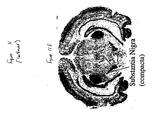
- 210000003523 substantia nigra Anatomy 0.000 description 1
- 239000000758 substrate Substances 0.000 description 1
- 210000004281 subthalamic nucleus Anatomy 0.000 description 1
- 230000005062 synaptic transmission Effects 0.000 description 1
- 210000001550 testis Anatomy 0.000 description 1
- 230000000542 thalamic effect Effects 0.000 description 1
- 210000001103 thalamus Anatomy 0.000 description 1
- 229940126585 therapeutic drug Drugs 0.000 description 1
- RYYWUUFWQRZTIU-UHFFFAOYSA-K thiophosphate Chemical compound [O-]P([O-])([O-])=S RYYWUUFWQRZTIU-UHFFFAOYSA-K 0.000 description 1
- 210000001685 thyroid gland Anatomy 0.000 description 1
- 210000003437 trachea Anatomy 0.000 description 1
- 230000009466 transformation Effects 0.000 description 1
- 102000027257 transmembrane receptors Human genes 0.000 description 1
- 108091008578 transmembrane receptors Proteins 0.000 description 1
- RYFMWSXOAZQYPI-UHFFFAOYSA-K trisodium phosphate Chemical compound [Na+].[Na+].[Na+].[O-]P([O-])([O-])=O RYFMWSXOAZQYPI-UHFFFAOYSA-K 0.000 description 1
- OUYCCCASQSFEME-UHFFFAOYSA-N tyrosine Natural products OC(=O)C(N)CC1=CC=C(O)C=C1 OUYCCCASQSFEME-UHFFFAOYSA-N 0.000 description 1
- 241000701161 unidentified adenovirus Species 0.000 description 1
- 210000004291 uterus Anatomy 0.000 description 1
- 239000004474 valine Substances 0.000 description 1
- 239000003981 vehicle Substances 0.000 description 1
- 210000000575 ventromedial hypothalamic nucleus Anatomy 0.000 description 1
- 238000005303 weighing Methods 0.000 description 1
- 210000005253 yeast cell Anatomy 0.000 description 1
- DGVVWUTYPXICAM-UHFFFAOYSA-N β‐Mercaptoethanol Chemical compound OCCS DGVVWUTYPXICAM-UHFFFAOYSA-N 0.000 description 1
Images


Classifications
-
- C—CHEMISTRY; METALLURGY
- C07—ORGANIC CHEMISTRY
- C07D—HETEROCYCLIC COMPOUNDS
- C07D403/00—Heterocyclic compounds containing two or more hetero rings, having nitrogen atoms as the only ring hetero atoms, not provided for by group C07D401/00
- C07D403/02—Heterocyclic compounds containing two or more hetero rings, having nitrogen atoms as the only ring hetero atoms, not provided for by group C07D401/00 containing two hetero rings
- C07D403/12—Heterocyclic compounds containing two or more hetero rings, having nitrogen atoms as the only ring hetero atoms, not provided for by group C07D401/00 containing two hetero rings linked by a chain containing hetero atoms as chain links
-
- A—HUMAN NECESSITIES
- A61—MEDICAL OR VETERINARY SCIENCE; HYGIENE
- A61P—SPECIFIC THERAPEUTIC ACTIVITY OF CHEMICAL COMPOUNDS OR MEDICINAL PREPARATIONS
- A61P43/00—Drugs for specific purposes, not provided for in groups A61P1/00-A61P41/00
-
- C—CHEMISTRY; METALLURGY
- C07—ORGANIC CHEMISTRY
- C07D—HETEROCYCLIC COMPOUNDS
- C07D233/00—Heterocyclic compounds containing 1,3-diazole or hydrogenated 1,3-diazole rings, not condensed with other rings
- C07D233/04—Heterocyclic compounds containing 1,3-diazole or hydrogenated 1,3-diazole rings, not condensed with other rings having one double bond between ring members or between a ring member and a non-ring member
- C07D233/28—Heterocyclic compounds containing 1,3-diazole or hydrogenated 1,3-diazole rings, not condensed with other rings having one double bond between ring members or between a ring member and a non-ring member with hetero atoms or with carbon atoms having three bonds to hetero atoms with at the most one bond to halogen, e.g. ester or nitrile radicals, directly attached to ring carbon atoms
- C07D233/30—Oxygen or sulfur atoms
- C07D233/42—Sulfur atoms
-
- C—CHEMISTRY; METALLURGY
- C07—ORGANIC CHEMISTRY
- C07D—HETEROCYCLIC COMPOUNDS
- C07D405/00—Heterocyclic compounds containing both one or more hetero rings having oxygen atoms as the only ring hetero atoms, and one or more rings having nitrogen as the only ring hetero atom
- C07D405/02—Heterocyclic compounds containing both one or more hetero rings having oxygen atoms as the only ring hetero atoms, and one or more rings having nitrogen as the only ring hetero atom containing two hetero rings
- C07D405/12—Heterocyclic compounds containing both one or more hetero rings having oxygen atoms as the only ring hetero atoms, and one or more rings having nitrogen as the only ring hetero atom containing two hetero rings linked by a chain containing hetero atoms as chain links
-
- C—CHEMISTRY; METALLURGY
- C07—ORGANIC CHEMISTRY
- C07D—HETEROCYCLIC COMPOUNDS
- C07D409/00—Heterocyclic compounds containing two or more hetero rings, at least one ring having sulfur atoms as the only ring hetero atoms
- C07D409/02—Heterocyclic compounds containing two or more hetero rings, at least one ring having sulfur atoms as the only ring hetero atoms containing two hetero rings
- C07D409/12—Heterocyclic compounds containing two or more hetero rings, at least one ring having sulfur atoms as the only ring hetero atoms containing two hetero rings linked by a chain containing hetero atoms as chain links
-
- C—CHEMISTRY; METALLURGY
- C07—ORGANIC CHEMISTRY
- C07D—HETEROCYCLIC COMPOUNDS
- C07D413/00—Heterocyclic compounds containing two or more hetero rings, at least one ring having nitrogen and oxygen atoms as the only ring hetero atoms
- C07D413/02—Heterocyclic compounds containing two or more hetero rings, at least one ring having nitrogen and oxygen atoms as the only ring hetero atoms containing two hetero rings
- C07D413/12—Heterocyclic compounds containing two or more hetero rings, at least one ring having nitrogen and oxygen atoms as the only ring hetero atoms containing two hetero rings linked by a chain containing hetero atoms as chain links
-
- C—CHEMISTRY; METALLURGY
- C07—ORGANIC CHEMISTRY
- C07D—HETEROCYCLIC COMPOUNDS
- C07D513/00—Heterocyclic compounds containing in the condensed system at least one hetero ring having nitrogen and sulfur atoms as the only ring hetero atoms, not provided for in groups C07D463/00, C07D477/00 or C07D499/00 - C07D507/00
- C07D513/02—Heterocyclic compounds containing in the condensed system at least one hetero ring having nitrogen and sulfur atoms as the only ring hetero atoms, not provided for in groups C07D463/00, C07D477/00 or C07D499/00 - C07D507/00 in which the condensed system contains two hetero rings
- C07D513/04—Ortho-condensed systems
-
- G—PHYSICS
- G01—MEASURING; TESTING
- G01N—INVESTIGATING OR ANALYSING MATERIALS BY DETERMINING THEIR CHEMICAL OR PHYSICAL PROPERTIES
- G01N33/00—Investigating or analysing materials by specific methods not covered by groups G01N1/00 - G01N31/00
- G01N33/48—Biological material, e.g. blood, urine; Haemocytometers
- G01N33/50—Chemical analysis of biological material, e.g. blood, urine; Testing involving biospecific ligand binding methods; Immunological testing
- G01N33/68—Chemical analysis of biological material, e.g. blood, urine; Testing involving biospecific ligand binding methods; Immunological testing involving proteins, peptides or amino acids
-
- G—PHYSICS
- G01—MEASURING; TESTING
- G01N—INVESTIGATING OR ANALYSING MATERIALS BY DETERMINING THEIR CHEMICAL OR PHYSICAL PROPERTIES
- G01N2500/00—Screening for compounds of potential therapeutic value
Definitions
- transmembrane receptors more particularly to endogenous, constitutively active G protein-coupled receptors for which the endogenous ligand is unknown, and most particularly to the use of such receptors for the direct identification of candidate compounds via screening as agonists, partial agonists or inverse agonists to such receptors.
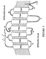
- G protein-coupled receptors share a common structural motif. All these receptors have seven sequences of between 22 to 24 hydrophobic amino acids that form seven alpha helices, each of which spans the membrane. The transmembrane helices are joined by strands of amino acids having a larger loop between the fourth and fifth transmembrane helix on the extracellular side of the membrane. Another larger loop, composed primarily of hydrophilic amino acids, joins transmembrane helices five and six on the intracellular side of the membrane. The carboxy terminus of the receptor lies intracellularly with the amino terminus in the extracellular space. It is thought that the loop joining helices five and six, as well as the carboxy terminus, interact with the G protein. Currently, Gq, Gs, Gi, and Go are G proteins that have been identified. The general structure of G protein-coupled receptors is shown in Figure 1 .
- G protein-coupled receptors exist in the cell membrane in equilibrium between two different states or conformations: an "inactive" state and an “active” state. As shown schematically in Figure 2 , a receptor in an inactive state is unable to link to the intracellular transduction pathway to produce a biological response. Changing the receptor conformation to the active state allows linkage to the transduction pathway and produces a biological response.
- a receptor may be stabilized in an active state by an endogenous ligand or an exogenous agonist ligand.
- Recent discoveries such as, including but not exclusively limited to, modifications to the amino acid sequence of the receptor provide means other than ligands to stabilize the active state conformation. These means effectively stabilize the receptor in an active state by simulating the effect of a ligand binding to the receptor. Stabilization by such ligand-independent means is termed "constitutive receptor activation.”
- a receptor for which the endogenous ligand is unknown or not identified is referred to as an "orphan receptor.”
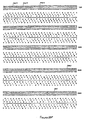
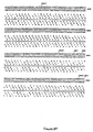
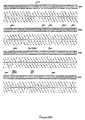
- GPR3 is a 330 amino acid G protein coupled receptor for which the endogenous ligand is unknown.
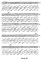
- Genomics 23:609 see also , Iismaa, T.P. et al (1994) Genomics 24:391 ; see Figure 1 for reported nucleic acid and amino acid sequence.
- GPR3 is constitutively active in its endogenous form.
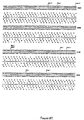
- GPR12 is a 334 amino acid homolog of GPR3; the endogenous ligand for GPR12 is unknown ( Song, Z.
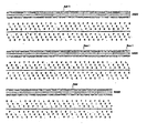
- GPR6 is a 362 amino acid homolog of GPR3; the endogenous ligand for GPR6 is unknown (Song, Z.-H. et al, supra .; see Figure 1 for reported amino acid sequence).
- GPR6 transcripts are reported to be abundant in the human putamen and to a lesser extent in the frontal cortex, hippocampus, and hypothalamus ( Heiber, M. et al. DNA and Cell Biology (1995) 14(1): 25 ; see Figure 1 for reported nucleic acid and amino acid sequences for GPR6).
- GPR4 has also been identified as an orphan GPCR ( Heiber, M.
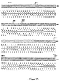
- GPR21 is a 349 amino acid G protein coupled receptor for which the endogenous ligand is unknown ( see GenBank Accession # U66580 for nucleic acid and deduced amino acid sequence). GPR21 has been reported to be located at chromosome 9q33. O'Dowd B. et al., 187 Gene 75 (1997 ). AL022171 is a human DNA sequence from clone 384F21 on chromosome 1q24.
- AL022171 has been identified to contain an open reading frame of 1,086 bp encoding for a 361 amino acid protein. ( see GenBank Accession number AL022171). AL022171 is 68% homologous to GPR21 ( see Figure 5B ). GHSR is also identified as an orphan GPCR ( Howard, A.D. et al, 273 Science 974 (1996 )).
- WO97/21731 discloses a method for identifying agonists, partial agonists, inverse agonists or antagonists of a peptide hormone receptor (a G-protein coupled receptor), comprising measuring the binding affinity of a candidate compound to a constitutively active form of the receptor.
- a G-protein coupled receptor a G-protein coupled receptor
- GPCRs G protein-coupled orphan receptors
- an endogenous, constitutively activated orphan GPCR:G protein - fusion protein is utilized.
- AGONISTS shall mean materials ( e . g ., ligands, candidate compounds) that activate the intracellular response when they bind to the receptor, or enhance GTP binding to membranes.
- AMINO ACID ABBREVIATIONS used herein are set out in Table 1: TABLE 1 ALANINE ALA A ARGININE ARG R ASPARAGINE ASN N ASPARTIC ACID ASP D CYSTEINE CYS C GLUTAMIC ACID GLU E GLUTAMINE GLN Q GLYCINE GLY G HISTIDINE HIS H ISOLEUCINE ILE I LEUCINE LEU L LYSINE LYS K METHIONINE MET M PHENYLALANINE PHE F PROLINE PRO P SERINE SER S THREONINE THR T TRYPTOPHAN TRP W TYROSINE TYR Y VALINE VAL V PARTIAL AGONISTS shall mean materials ( e .
- ANTAGONIST shall mean materials ( e . g ., ligands, candidate compounds) that competitively bind to the receptor at the same site as the agonists but which do not activate the intracellular response initiated by the active form of the receptor, and can thereby inhibit the intracellular responses by agonists or partial agonists.
- ANTAGONISTS do not diminish the baseline intracellular response in the absence of an agonist or partial agonist.
- CANDIDATE COMPOUND shall mean a molecule (for example, and not limitation, a chemical compound) which is amenable to a screening technique.
- the phrase "candidate compound” does not include compounds which were publicly known to be compounds selected from the group consisting of inverse agonist, agonist or antagonist to a receptor, as previously determined by an indirect identification process ("indirectly identified compound”); more preferably, not including an indirectly identified compound which has previously been determined to have therapeutic efficacy in at least one mammal; and, most preferably, not including an indirectly identified compound which has previously been determined to have therapeutic utility in humans.
- COMPOSITION means a material comprising at least one component; a "pharmaceutical composition” is an example of a composition.
- COMPOUND EFFICACY shall mean a measurement of the ability of a compound to inhibit or stimulate receptor functionality, as opposed to receptor binding affinity. A most preferred means of detecting compound efficacy is via measurement of GTP (via [ 35 S]GTP ⁇ S) or cAMP, as further disclosed in the Example section of this patent document.
- CONSTITUTIVELY ACTIVATED RECEPTOR shall mean a receptor subject to constitutive receptor activation. A constitutively activated receptor can be endogenous or non-endogenous.
- CONSTITUTIVE RECEPTOR ACTIVATION shall mean stabilization of a receptor in the active state by means other than binding of the receptor with its endogenous ligand or a chemical equivalent thereof.
- CONTACT or CONTACTING shall mean bringing at least two moieties together, whether in an in vitro system or an in vivo system.
- DIRECTLY IDENTIFYING or DIRECTLY IDENTIFIED in relationship to the phrase "candidate compound”, shall mean the screening of a candidate compound against a constitutively activated receptor, preferably a constitutively activated orphan receptor, and most preferably against a constitutively activated G protein-coupled cell surface orphan receptor, and assessing the compound efficacy of such compound.
- This phrase is, under no circumstances, to be interpreted or understood to be encompassed by or to encompass the phrase "indirectly identifying" or “indirectly identified.”
- ENDOGENOUS shall mean a material that a mammal naturally produces.
- ENDOGENOUS in reference to, for example and not limitation, the term "receptor,” shall mean that which is naturally produced by a mammal (for example, and not limitation, a human) or a virus.
- the term NON-ENDOGENOUS in this context shall mean that which is not naturally produced by a mammal (for example, and not limitation, a human) or a virus.
- a receptor which is not constitutively active in its endogenous form, but when manipulated becomes constitutively active is most preferably referred to herein as a "non-endogenous, constitutively activated receptor.” Both terms can be utilized to describe both "in vivo" and “in vitro" systems.
- the endogenous or non-endogenous receptor may be in reference to an in vitro screening system.
- screening of a candidate compound by means of an in vivo system is viable.
- G PROTEIN COUPLED RECEPTOR FUSION PROTEIN and GPCR FUSION PROTEIN in the context of the invention disclosed herein, each mean a non-endogenous protein comprising an endogenous, constitutively activated orphan GPCR fused to at least one G protein, most preferably, the alpha ( ⁇ ) subunit of such G protein (this being the subunit that binds GTP), with the G protein preferably being of the same type as the G protein that naturally couples with endogenous orphan GPCR
- the G protein "Gs ⁇ " is the predominate G protein that couples with GPR6 such that a GPCR Fusion Protein based upon GPR6 would be a non-endogenous protein comprising GPR6 fused to Gs ⁇ .
- the G protein can be fused directly to the c-terminus of the endogenous, constitutively active orphan GPCR, or there may be spacers between the two.
- INDIRECTLY IDENTIFYING or INDIRECTLY IDENTIFIED means the traditional approach to the drug discovery process involving identification of an endogenous ligand specific for an endogenous receptor, screening of candidate compounds against the receptor for determination of those which interfere and/or compete with the ligand-receptor interaction, and assessing the efficacy of the compound for affecting at least one second messenger pathway associated with the activated receptor.
- INHIBIT or INHIBITING in relationship to the term "response” shall mean that a response is decreased or prevented in the presence of a compound as opposed to in the absence of the compound.
- INVERSE AGONISTS shall mean materials (e . g ., ligand, candidate compound) which bind to either the endogenous form of the receptor or to the constitutively activated form of the receptor, and which inhibit the baseline intracellular response initiated by the active form of the receptor below the normal base level of activity which is observed in the absence of agonists or partial agonists, or decrease GTP binding to membranes.
- the baseline intracellular response is inhibited in the presence of the inverse agonist by at least 30%, more preferably by at least 50%, and most preferably by at least 75%, as compared with the baseline response in the absence of the inverse agonist.
- LIGAND shall mean an endogenous, naturally occurring molecule specific for an endogenous, naturally occurring receptor.
- ORPHAN RECEPTOR shall mean an endogenous receptor for which the endogenous ligand specific for that receptor has not been identified or is not known.
- PHARMACEUTICAL COMPOSITION shall mean a composition comprising at least one active ingredient, whereby the composition is amenable to investigation for a specified, efficacious outcome in a mammal (for example, and not limitation, a human).
- NON-ORPHAN RECEPTOR shall mean an endogenous naturally occurring molecule specific for an endogenous naturally occurring ligand wherein the binding of a ligand to a receptor activates an intracellular signaling pathway.
- STIMULATE or STIMULATING in relationship to the term "response” shall mean that a response is increased in the presence of a compound as opposed to in the absence of the compound.
- response in relationship to the term "response” shall mean that a response is increased in the presence of a compound as opposed to in the absence of the compound.
- any search for therapeutic compounds should start by screening compounds against the ligand-independent active state. The search, then, is for an inverse agonist to the active state receptor.
- Screening candidate compounds against the endogenous, constitutively activated orphan receptors allows for the direct identification of candidate compounds which act at these orphan cell surface receptors, without requiring any prior knowledge or use of the receptor's endogenous ligand.
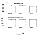
- By determining areas within the body where such receptors are expressed and/or over-expressed it is possible to determine related disease/disorder states which are associated with the expression and/or over-expression of these receptors; such an approach is disclosed in this patent document.
- inverse agonists to endogenous, constitutively activated orphan receptors e . g ., such as those set forth herein (GPR3, GPR4, GPR6, GPR12, GPR21, GHSR, OGR1, RE2 and AL022171) can be identified by the methodologies of this invention.
- Such inverse agonists are ideal candidates as lead compounds in drug discovery programs for treating diseases related to these receptors. Indeed, an antagonist to such a receptor (even if the ligand were known) may be ineffective given that the receptor is activated even in the absence of ligand-receptor binding.
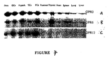
- tissue scans can be conducted across a broad range of healthy and diseased tissues. Such tissue scans provide a preferred first step in associating a specific receptor with a disease and/or a disorder.
- the DNA sequence of the endogenous, constitutively activated GPCR is used to make a probe for RT-PCR identification of the expression of the receptor in tissue samples.
- the presence of a receptor in a diseased tissue, or the presence of the receptor at elevated concentrations in diseased tissue compared to normal tissue, can be utilized to identify a correlation with that disease.
- Receptors can equally well be localized to regions of organs by this technique. Based on the known functions of the specific tissues to which the receptor is localized, the putative functional role of the receptor can be deduced.
- the identification and association of an orphan receptor with diseases and/or disorders can be beneficially enhanced via identification of additional receptors having homology with the original orphan receptor.
- This approach was utilized in the identification of both GPR6 and GPR12, based upon their sequence homology with GPR3, and in the identification of AL022171, having sequence homology to GPR21.
- GPR3 was previously identified as a constitutively activated orphan receptor ( see Eggerick, supra ). What was not known, prior to this invention, was that GPR6, GPR12, GPR21 and AL022171 are also constitutively active in their endogenous states. Using known computerized databases ( e . g ., dbEST), GPR6, GPR12, GPR21 and AL022171 were identified.
- G protein receptor When a G protein receptor becomes constitutively active, it binds to a G protein (eg., Gq, Gs, Gi, Go) and stimulates the binding of GTP to the G protein. The G protein then acts as a GTPase and slowly hydrolyzes the GTP to GDP, whereby the receptor, under normal conditions, becomes deactivated. However, constitutively activated receptors continue to exchange GDP to GTP.
- GTP Gq, Gs, Gi, Go
- G protein-coupled receptor assay i.e. an assay to select compounds that are agonists, partial agonists, or inverse agonists
- further screening to confirm that the compounds have interacted at the receptor site is preferred.
- a compound identified by the "generic” assay may not bind to the receptor, but may instead merely "uncouple" the G protein from the intracellular domain.
- GPR3, GPR4, GPR6, GPR12, GPR21, GHSR, OGR1, RE2 and AL022171 it has been determined that these receptors couple the G protein Gs. Gs stimulates the enzyme adenylyl cyclase (Gi, on the other hand, inhibits this enzyme).
- Adenylyl cyclase catalyzes the conversion of ATP to cAMP; thus, because these receptors are activated in their endogenous forms, increased levels of cAMP are associated therewith (on the other hand, endogenously activated receptors which couple the Gi protein are associated with decreased levels of cAMP). See, generally, " Indirect Mechanisms of Synaptic Transmission," Chpt. 8, From Neuron To Brain (3rd Ed.) Nichols, J.G. et al eds. Sinauer Associates, Inc. (1992 ). Thus, assays that detect cAMP can be utilized to determine if a candidate compound is an inverse agonist to the receptor ( i .
- a compound which contacts the receptor would decrease the levels of cAMP relative to the uncontacted receptor).
- a variety of approaches known in the art for measuring cAMP can be utilized; a most preferred approach relies upon the use of anti-cAMP antibodies.
- Another type of assay that can be utilized is a whole cell second messenger reporter system assay. Promoters on genes drive the expression of the proteins that a particular gene encodes. Cyclic AMP drives gene expression by promoting the binding of a cAMP-responsive DNA binding protein or transcription factor (CREB) which then binds to the promoter at specific sites called cAMP response elements and drives the expression of the gene.
- CREB cAMP-responsive DNA binding protein or transcription factor
- Reporter systems can be constructed which have a promoter containing multiple cAMP response elements before the reporter gene, e.g., ⁇ -galactosidase or luciferase.
- an activated Gs receptor such as GPR3 causes the accumulation of cAMP which then activates the gene and expression of the reporter protein.
- the reporter protein such as ⁇ -galactosidase or luciferase can then be detected using standard biochemical assays (see, for example, Chen et al. 1995). A cAMP assay is particularly preferred.
- an endogenous, constitutively activated orphan GPCR for use in screening of candidate compounds for the direct identification of inverse agonists, agonists and partial agonists provides a unique challenge in that, by definition, the endogenous receptor is active even in the absence of an endogenous ligand bound thereto.
- an approach be utilized that can enhance such differentiation.
- a preferred approach is the use of a GPCR Fusion Protein.
- an endogenous orphan GPCR is constitutively active, using the assay techniques set forth above (as well as others), it is possible to determine the predominant G protein that couples with the endogenous GPCR. Coupling of the G protein to the GPCR provides a signaling pathway that can be assessed. Because it is most preferred that screening take place by use of a mammalian expression system, such a system will be expected to have endogenous G protein therein. Thus, by definition, in such a system, the endogenous, constitutively active orphan GPCR will continuously signal. In this regard, it is preferred that this signal be enhanced such that in the presence of, e . g ., an inverse agonist to the receptor, it is more likely that one will be able to more readily differentiate, particularly in the context of screening, between the receptor when it is or is not contacted with the inverse agonist.
- this signal be enhanced such that in the presence of, e . g ., an inverse agonist to the receptor, it is more likely that one will be able
- the GPCR Fusion Protein is intended to enhance the efficacy of G protein coupling with the endogenous GPCR.
- the GPCR Fusion Protein appears to be important for screening with an endogenous, constitutively activated GPCR because such an approach increases the signal that is most preferably utilized in such screening techniques. Facilitating a significant "signal to noise" ratio is important for the screening of candidate compounds as disclosed herein.
- GPCR Fusion Protein The construction of a construct useful for expression of a GPCR Fusion Protein is within the purview of those having ordinary skill in the art. Commercially available expression vectors and systems offer a variety of approaches that can fit the particular needs of an investigator.
- One important criterion for such a GPCR Fusion Protein construct is that the endogenous GPCR sequence and the G protein sequence both be in-frame (preferably, the sequence for the endogenous GPCR is upstream of the G protein sequence) and that the "stop" codon of the GPCR must be deleted or replaced such that upon expression of the GPCR, the G protein can also be expressed.
- the GPCR can be linked directly to the G protein, or there can be spacer residues between the two (preferably no more than about 12, although this number can be readily ascertained by one of ordinary skill in the art).
- the results are substantially the same; however, there is a preference (based upon convenience) of use of a spacer in that some restriction sites that are not used will, effectively, upon expression, become a spacer.
- the G protein that couples to the endogenous GPCR will have been identified prior to the creation of the GPCR Fusion Protein construct. Because there are only a few G proteins that have been identified, it is preferred that a construct comprising the sequence of the G protein ( i . e ., a universal G protein construct) be available for insertion of an endogenous GPCR sequence therein; this provides for efficiency in the context of large-scale screening of a variety of different endogenous GPCRs having different sequences.
- direct identification of candidate compounds is preferably conducted in conjunction with compounds generated via combinatorial chemistry techniques, whereby thousands of compounds are randomly prepared for such analysis.
- the results of such screening will be compounds having unique core structures; thereafter, these compounds are preferably subjected to additional chemical modification around a preferred core structure(s) to further enhance the medicinal properties thereof.
- inverse agonists, agonists and/or partial agonists that are directly identified can be beneficially improved upon prior to development of pharmaceutical compositions comprising such compounds.
- the binding affinity of a directly identified compound selected for further refinement into a pharmaceutical composition have a binding affinity for the receptor of less than 100nM, although this is generally a preference selection based upon the particular needs of the artisan.
- Such techniques are known to those in the art and will not be addressed in detail in this patent document.
- Candidate compounds selected for further development can be formulated into pharmaceutical compositions using techniques well known to those in the art. Suitable pharmaceutically-acceptable carriers are available to those in the art; for example, see Remington's Pharmaceutical Sciences, 16th Edition, 1980, Mack Publishing Co., (Oslo et al., eds .).
- In situ probes for GPR3, GPR6, and GPR12 were prepared.
- the following PCR protocol was utilized for all three probes: the reaction condition utilized was 1X rTth DNA polymerase buffer II, 1.5 mM Mg(OAc) 2 , 0.2 mM each of the 4 nucleotides, 0.228 ⁇ g rat genomic DNA, 0.25 ⁇ M of each primer (see below) and 1 unit of rTth DNA polymerase (Perkin Elmer) in 50 ⁇ l reaction volume.
- the cycle condition was 30 cycles of 94°C for 1 min, 55 °C for 1 min and 72 °C for 45 sec with a Perkin Elmer Cetus 2400 thermal cycler.
- the DNA fragment for the in situ probe was obtained by PCR using a 3' degenerate oligonucleotide based on the published human and mouse GPR3 sequences in the middle of the transmembrane domain 3, and a 5' degenerate oligonucleotide near the beginning of the 5' extracellular domain.
- a 537 bp PCR fragment containing nucleotide 24 through to the middle of transmembrane 3 was digested with Bam HI and Hind III and was subcloned into a Bam HI-Hind III site of pBluescript.
- the in situ probe DNA fragment of rat GPR6 was obtained by PCR based on the published rat GPR6 cDNA sequences.
- the sequences of the oligonucleotides utilized were as follows: 5'-GGAGAAGCTTCTGGCGGCGATGAACGCTAG-3' (SEQ.ID.NO.: 3; 5' oligo) 5'-ACAGGATCCAGGTGGCTGCTAGCAAGAG-3' (SEQ.ID.NO.: 4; 3' oligo)
- a 608 bp PCR fragment containing nucleotide -10 through to the middle of transmembrane domain 4 was digested with Bam HI and Hind III and was subcloned into Bam HI-Hind III site of pBluescript.
- the in situ probe DNA fragment of rat GPR12 was obtained by PCR based on the published rat GPR12 cDNA sequences.
- the sequences of the oligonucleotides utilized were as follows: 5'-CTTAAGCTTAAAATGAACGAAGACCCGAAG-3' (SEQ.ID.NO.: 5; 5' oligo) 5'-GGAGGATCCCCAGAGCATCACTAGCAT-3' (SEQ.ID.NO.: 6; 3' oligo)
- a 516 bp PCR fragment containing nucleotide -5 through to the middle of transmembrane domain 4 was digested with Bam HI and Hind III and subcloned into a Bam HI-Hind III site of pBluescript.
- GPR12 cDNA was prepared using the following protocol: Human GPR12 cDNA was obtained by PCR using human genomic DNA and a 5' primer from the 5' untranslated region with a Hind III restriction site, and a 3' primer from the 3' untranslated region containing a Bam HI site'.
- Primers had the following sequences: 5'-CTTAAGCTTGTGGCATTTGGTACT-3' (SEQ.ID.NO.: 10; 5' oligo) 5'-TCTGGATCCTTGGCCAGGCAGTGGAAGT-3 (SEQ.ID.NO.: 11; 3' oligo) PCR was performed using rTth polymerase (Perkin Elmer) with the buffer system provided by the manufacturers, 0.25 ⁇ M of each primer, 0.2 ⁇ M of each of the four nucleotides and 0.2 ⁇ g of genomic DNA as template. The cycle condition was 30 cycles of 94°C for 1 min, 57 °C for 1 min and 72°C for 1.5 min.
- the 1.2 kb PCR fragment was digested with Hind III and Bam HI, and subcloned into Hind III-Bam HI site of pCMV expression vector. The resulting cDNA clones were fully sequenced and consistent with published sequences.
- PCR was performed using genomic DNA as template and rTth polymerase (Perkin Elmer) with the buffer system provided by the manufacturer, 0.25 ⁇ M of each primer, and 0.2 mM of each of the four nucleotides.
- the cycle condition was 30 cycles of 94°C for 1 min, 62°C for 1min and 72°C for 1 min and 20 sec.
- the 5' PCR primer was kinased with the sequence: 5'-GAGAATTCACTCCTGAGCTCAAGATGAACT-3' (SEQ.ID.NO.:12) and the 3' primer contained a BamHI site with the sequence: 5'-CGGGATCCCCGTAACTGAGCCACTTCAGAT-3' (SEQ.ID.NO.:13).
- the resulting 1.1 kb PCR fragment was digested with BamHI and cloned into EcoRV-BamHI site of pCMV expression vector. Nucleic acid (SEQ.ID.NO.:14) and amino acid (SEQ.ID.NO.:15) sequences for human GPR21 were thereafter determined.
- PCR was performed using genomic DNA as template and rTth polymerase (Perkin Elmer) with the buffer system provided by the manufacturer, 0.25 ⁇ M of each primer, and 0.2 mM of each of the four nucleotides.
- the cycle condition was 30 cycles of 94°C for 1 min, 54°C for 1min and 72 °C for 1 min and 20 sec.
- the 5' primer contains an HindIII site with the following sequence: 5'-AGGAAGCTTTAAATTTCCAAGCCATGAATG-3' (SEQ.ID.NO.:16) and the 3' primer contained a EcoRI site with the following sequence: 5'-ACCGAATTCAGATTACATTTGATTTACTATG-3' (SEQ.ID.NO.: 17).
- the resulting 1.15 kb PCR fragment was digested with HindIII and EcoRI and cloned into HindIII-EcoRI site ofpCMV expression vector.
- Nucleic acid (SEQ.ID.NO.: 18) and amino acid (SEQ.ID.NO.: 19) sequences for human AL022171 were thereafter determined and verified.
- GPR4 GeneBank accession number L36148
- expression vectors comprising the cDNA was generously supplied by Brian O'Dowd (University of Toronto).
- the vector for GPR4 cDNA was pcDNA3 and this subcloned into pCMV vector at a Hind III-Xbal site (the 5' untranslated region between HindIII and an ApaI site was trimmed by conducting digestion/self ligation).
- PCR was performed using human brain cDNA as template and rTth polymerase (Perkin Elmer) with the buffer system provided by the manufacturer, 0.25 ⁇ M of each primer, and 0.2 mM of each of the four nucleotides.
- the cycle condition was 30 cycles of 94°C for 1 min, 62°C for 1min and 72°C for 1 min and 30 sec.
- the 5' PCR primer contained an EcoRI site with the sequence 5' -AGCGAATTCTGCCCACCCCACGCCGAGGTGCT-3' (SEQ. ID. No.
- PCR was performed using human genomic DNA as template and rTth polymerase (Perkin Elmer) with the buffer system provided by the manufacturer, 0.25 ⁇ M of each primer, and 0.2 mM of each of the four nucleotides.
- the cycle condition was 30 cycles of 94°C for 1 min, 62°C for 1min and 72°C for 1 min and 20 sec.
- the 5' PCR primer contained a HindIII site with the sequence 5'-GGAAGCTTCAGGCCCAAAGATGGGGAACAT-3' (SEQ. ID. No.
- PCR was performed using hippocampus cDNA as template and TaqPlus Precision polymerase (Stratagene) with the buffer system provided by the manufacturer, 0.25 ⁇ M of each primer, and 0.2 mM of each 4 nucleotides.
- the cycle condition was 30 cycles of 94°C for 1 min, 68°C for 1min and 72°C for 1 min and 10 sec.
- the 5' PCR primer sequence 5'-ATGTGGAACGCGACGCCCAGCG-3' (SEQ.ID.NO.40) and the 3' primer sequence: 5'-TCATGTATTAATACTAGATTCT-3' (SEQ.ID.NO.41).
- tube A was prepared by mixing between 8-20 ⁇ g DNA (e.g., pCMV vector; pCMV vector with receptor cDNA; pCMV with GPCR Fusion Protein, supra ) in 1-2ml serum free DMEM (Irvine Scientific, Irvine, CA); tube B was prepared by mixing 50-120 ⁇ l lipofectamine (Gibco BRL) in 1-2ml serum free DMEM. Tubes A and B were then admixed by inversions (several times), followed by incubation at room temperature for 30-45min.
- the admixture is referred to as the "transfection mixture”.
- Plated cells were washed with 1XPBS, followed by addition of 10-12ml serum free DMEM. 2.4ml of the transfection mixture was then added to the cells, followed by incubation for 4hrs at 37°C/5% CO 2 . The transfection mixture was then removed by aspiration, followed by the addition of 25ml of DMEM/10% Fetal Bovine Serum. Cells were then incubated at 37°C/5% CO 2 .
- GPCR Fusion Protein preferred amounts to the above are as follows: 12 ⁇ g DNA; 2ml serum free DMEM; 60 ⁇ l lipofectamine; 293 cells 9 and an addition of 12ml serum free DMEM).
- the entire coding region of the receptor is used to generate a radiolabeled probe using a Prime-It IITM Random Primer Labeling Kit (Stratagene, #300385), according to the manufacturer's instructions.
- a Prime-It IITM Random Primer Labeling Kit (Stratagene, #300385), according to the manufacturer's instructions.
- this approach was utilized for GPR4.
- a G protein-coupled receptor When a G protein-coupled receptor is in its active state, either as a result of ligand binding or constitutive activation, the receptor binds to a G protein (in the case of GPR3, GPR4, GPR6, GPR12, GPR21, GHSR, OGR1, RE2 and AL022171, Gs) and stimulates the binding of GTP to the G protein.
- the trimeric G protein-receptor complex acts as a GTPase and slowly hydrolyzes the GTP to GDP, at which point the receptor normally is deactivated. Constitutively activated receptors continue to exchange GDP for GTP.
- the non-hydrolyzable GTP analog, [ 35 S]GTP ⁇ S can be utilized to demonstrate enhanced binding of [ 35 S]GTP ⁇ S to membranes expressing constitutively activated receptors.
- the advantage of using [ 35 S]GTPyS binding to measure constitutive activation is that: (a) it is generically applicable to all G protein-coupled receptors; (b) it is proximal at the membrane surface making it less likely to pick-up molecules which affect the intracellular cascade.
- the assay utilizes the ability of G protein coupled receptors to stimulate [ 35 S]GTP ⁇ S binding to membranes expressing the relevant receptors.
- the assay can, therefore, be used in the direct identification method to screen candidate compounds to known, orphan and constitutively activated G protein coupled receptors.
- the assay is generic and has application to drug discovery at all G protein coupled receptors.
- the [ 35 S]GTP ⁇ S assay was incubated in 20 mM HEPES, pH 7.4, binding buffer with 12 nM [ 35 S]GTP ⁇ S and 75 ⁇ g membrane protein [e.g., 293T cells expressing GPR3] and 1 ⁇ M GDP for 1 hour.
- Wheatgerm agglutinin beads (25 ⁇ l; Amersham) were then added and the mixture was incubated for another 30 minutes at room temperature.
- the tubes were then centrifuged at 1500 X g for 5 minutes at room temperature and then counted in a scintillation counter.
- GPR3 receptor was determined to have increased activity as compared to control; this heightened activity is not the result of autocrine stimulation in that the data were obtained from membrane preparations, as opposed to whole cell preparations.
- GPR3 is a constitutively activated receptor
- mammalian cells Although a variety of cells are available to the art for the expression of proteins, it is most preferred that mammalian cells be utilized. The primary reason for this is predicated upon practicalities, i . e ., utilization of, e.g., yeast cells for the expression of a GPCR, while possible, introduces into the protocol a non-mammalian cell which may not (indeed, in the case of yeast, does not) include the receptor-coupling, genetic-mechanism and secretary pathways that have evolved for mammalian systems - thus, results obtained in non-mammalian cells, while of potential use, are not as preferred as that obtained from mammalian cells.
- COS-7, 293 and 293T cells are particularly preferred, although the specific mammalian cell utilized can be predicated upon the particular needs of the artisan.
- a Bgl II/ HindIII fragment was removed from the somatostatin promoter-containing plasmid 1.4(5xGal)CAT ( Leonard, J. et al (1992) PNAS USA 89:6247-6251 ) and cloned into p ⁇ gal-Basic (Promega).
- the Bgl II/ HindIII fragment contains a variant of the minimal somatostatin promoter (from -71 bp to +50 bp relative to the transcription start site) in which the core 4bp of the cAMP Response Element (-46 to -43) were replaced with 5 copies of the recognition sequence for the yeast transcription factor Gal4.
- VIP2.0Zc is a cell line that has been stably transfected with the reporter gene ⁇ -galactosidase under the control of a cAMP responsive VIP promoter ( Konig et al. Mol. Cell.Neuro. 1991, 2, 331-337 ). The cell line was used here to indirectly measure the accumulation of intracellular cAMP. Approximately 2 million cells were plated in 6 cm plate the day before transfection. DNA (5 ⁇ g), for each receptor, was mixed with 2.5 ml serum-free DMEM containing 200 ⁇ g/ml DEAE dextran and 100 ⁇ M chloroquine, and added to a rinsed cell monolayer. After incubation for 90 min in a CO 2 incubator, the transfection medium was removed.
- the cells were washed with serum-free medium and supplemented with fresh complete medium. Twenty four hours after transfection, the cells were replated into 96-well plate at a density of 50 - 100 K per well and the ⁇ -galactosidase activity was assayed 48 to 72 hours after transfection.
- the assay buffer contained 100 mM sodium phosphate, 2 mM MgSO 4 , 0.1 mM MnCl 2 , pH 8.0.
- the cells were washed with PBS, and 25 ⁇ l /well of hypotonic lysis buffer consisting of 0.1 X assay buffer was added.
- 100 ⁇ l of assay buffer containing 0.5% Triton X-100 and 40 mM ⁇ -mercaptoethanol was added to each well and incubation at room temperature continued for 10 minutes.
- the substrate solution containing 5 mg/ml chlorophenol red- ⁇ -D-galactopyranoside (CPRG) in assay buffer was added at 25 ⁇ l/well and the plate was incubated at 37°C for 30 minutes before absorbance at 595 nm was measured with a plate reader.
- CPRG chlorophenol red- ⁇ -D-galactopyranoside
- GPR3, GPR6 and GPR 12 were assayed using the foregoing system, and it was determined that both GPR6 and GPR12 are constitutively active. See Figure 6A .
- 293 and 293T cells were plated-out on 96 well plates at a density of 2 x 10 4 cells per well and were transfected, using Lipofectamine Reagent (BRL), the following day according to manufacturer instructions.
- a DNA/lipid mixture was prepared for each 6-well transfection as follows: 260ng of plasmid DNA in 100 ⁇ l of DMEM were gently mixed with 2 ⁇ l of lipid in 100 ⁇ l of DMEM (the 260ng of plasmid DNA consisted of 200ng of a 8xCRE-Luc reporter plasmid, 50ng of pCMV comprising endogenous receptor or non-endogenous receptor or pCMV alone, and 10ng of a GPRS expression plasmid (GPRS in pcDNA3 (Invitrogen)).
- the 8XCRE-Luc reporter plasmid was prepared as follows: vector SRIF- ⁇ -gal was obtained by cloning the rat somatostatin promoter (-71/+51) at BgIV-HindIII site in the p ⁇ gal-Basic Vector (Clontech). Eight (8) copies of cAMP response element were obtained by PCR from an adenovirus template AdpCF126CCRE8 ( see 7 Human Gene Therapy 1883 (1996 )) and cloned into the SRIF- ⁇ -gal vector at the Kpn-BgIV site, resulting in the 8xCRE- ⁇ -gal reporter vector.
- the 8xCRE-Luc reporter plasmid was generated by replacing the beta-galactosidase gene in the 8xCRE- ⁇ -gal reporter vector with the luciferase gene obtained from the pGL3-basic vector (Promega) at the HindIII-BamHI site. Following 30 min. incubation at room temperature, the DNA/lipid mixture was diluted with 400 ⁇ l of DMEM and 100 ⁇ l of the diluted mixture was added to each well. 100 ⁇ l of DMEM with 10% FCS were added to each well after a 4hr incubation in a cell culture incubator. The following day the transfected cells were changed with 200 ⁇ l/well of DMEM with 10% FCS.
- GPR21 and AL022171 were assayed using the foregoing system, and based upon these results, it was determined that both GPR21 and AL022171 are constitutively active in their endogenous forms. See Figure 6B and 6C .
- GPR4, RE2, OGR1 and GHSR were analyzed and determined to be constitutively active in their endogenous forms (data not shown).
- Tissue samples were examined for expression of these orphan receptors by comparative RT-PCR, using the following primers:
- amplicons were designed to be non-overlapping, i.e., there is no sequence similarity between them, and to have similar Tm's, such that each primer pair amplifies its respective target at the same optimal annealing temperature. This diminishes the chance that an amplicon from one primer pair will act as an annealing target for the other primers in the multiplex reaction, therefore reducing the chance of interference with other primer pairs.
- PCR products were visualized by denaturing gel electrophoresis (7M urea, 5% polyacrylamide (Long RangerTM Solution, AT Biochemical, 0.6 XTBE) and subsequent autoradiography.
- Figures 7A, 7B, and 7C show the distribution of GPR3, GPR6 and GPR12 across human tissues. This information allows for assessing disease states that are associated with such tissue, as well as determining specific regions within such tissue where such expression predominates, thus allowing for correlating such receptor expression with particular disease states. This, in tum, then allows for direct identification of compounds that impact such receptors, without the need to understand or know the endogenous ligand for such receptor. Further screening reveals that GPR3 is expressed in much higher levels in human epilepsy tissue samples (tissue source: temporal cortex), as compared with controls, as evidenced by RT-PCR analysis ( Figure 8 ).
- GPR6 probe was produced from a 450bp HindIII-ScaI fragment of the GPR6 receptor cloned into the HindIII-SmaI site of pBluescriptSK+.
- Riboprobes were produced using a T7 transcription system in a standard labeling reaction consisting of: 1 ⁇ g of linearized plasmid, 2 ⁇ l of 5x transcription buffer, 125 ⁇ Ci of 35 S-UTP, 150 ⁇ M of GTP, CTP and ATP, 12.5mM dithiothreitol, 20U of RNase inhibitor and 6U of appropriate polymerase. The reaction was incubated at 37°C for 90 min., labeled probe being separated from free nucleotides over Sephadex G-50 spin columns.
- Dissected tissue was frozen in isopentane cooled to -42°C and subsequently stored at -80°C prior to sectioning on a cryostat maintained at -20°C. Slide-mounted tissue sections were then stored at -80 °C.
- Tissue sections were removed from the -80°C freezer and incubated with a 1 ⁇ g/ml solution of proteinase-K to permeabilize the tissue and inactivate endogenous RNase. After this treatment, sections were incubated in succession in water (1 min), 0.1 M triethanolamine (pH 8.0; 1 min), and 0.25% acetic anhydride in 0.1 M triethanolamine (10 min). The tissue was then washed in 2 x SSC (0.3 mM NaCl, 0.03 nM Na citrate, pH 7.2; 5 min) and dehydrated through graded concentrations of ethanol.
- 2 x SSC 0.3 mM NaCl, 0.03 nM Na citrate, pH 7.2; 5 min
- Sections were then hybridized with 1.5 x 10 6 dpm of [ 35 S]UTP-labeled cRNA probes in 20 ⁇ l of a hybridization buffer containing 75% formamide, 10% dextran sulfate, 3 x SSC, 50 mM sodium phosphate buffer (pH 7.4), 1 x Denhart's solution, 0.1 mg/ml yeast tRNA, and 0.1 mg/ml sheared salmon sperm DNA.
- Tissue sections were covered with coverslips that were sealed with rubber cement. The slides were incubated overnight at 50°C. On the following day, the rubber cement was removed, the coverslips were soaked-off with 2 x SSC, and the tissue sections were washed for 10 min in fresh 2 x SSC solution.
- Single stranded probe not hybridized with endogenous mRNAs was removed by incubating the sections for 30 min in 200 ⁇ g/ml solution of RNase-A at 37°C. The tissue was then washed in increasingly stringent SSC solutions (2, 1 and 0.5 x SSC; 10 min each), followed by a 1 hr wash in 0.5 x SSC at 60°C. After this final wash, the tissue sections were dehydrated using graded concentrations of ethanol, air dried and prepared for detection by x-ray autoradiography on Kodak XAR-5 film.
- GPR6 is expressed in the following areas of the brain: hypothalamus, hippocampus, nucleus accumbens, caudate and cerebral cortex. See Figure 9A for a representative tissue section (GPR6 receptor is presented in the dark areas; Figure 9B provides a reference map of the rat brain.)
- FIG. 10A provides a representative tissue section of GPR6 receptor expression in the lean Zucker animals
- Figure 10B provides a representative tissue section of GPR6 receptor expression in the obese Zucker animals
- Figure 10C is a reference map of this section of the rat brain.
- GPR12 probe was produced from a 515bp (NT5 - NT520) HindIII-BamHI fragment of the rat GPR12 receptor cloned into the HindIII-BamHI site of pBluescriptSK+.
- Riboprobes were produced using a T3/T7 transcription system in a standard labeling reaction consisting of 1 ⁇ g of linearized plasmid, 2 ⁇ l of 5x transcription buffer, 125 ⁇ Ci of 35 S-UTP, 150 ⁇ M of GTP, CTP and ATP, 12.5mM dithiothreitol, 20U of Rnase inhibitor and 6U of appropriate polymerase. The reaction was incubated at 37°C for 90 min., labeled probe being separated from free nucleotides over Sephadex G-50 spin columns.
- Dissected tissue was frozen in isopentane cooled to -42°C and subsequently stored at -80°C prior to sectioning on a cryostat maintained at -20°C. Slide-mounted tissue sections were then stored at -80 °C.
- Tissue sections were removed from the -80°C freezer and incubated with a 1 ⁇ g/ml solution of proteinase-K to permeabilize the tissue and inactivate endogenous RNase. After this treatment, sections are incubated in succession in water (1 min), 0.1 M triethanolamine (pH 8.0; 1 min), and 0.25% acetic anhydride in 0.1 M triethanolamine (10 min). The tissue was then washed in 2 x SSC (0.3 mM NaCl, 0.03 nM Na citrate, pH 7.2; 5 min) and dehydrated through graded concentrations of ethanol.
- 2 x SSC 0.3 mM NaCl, 0.03 nM Na citrate, pH 7.2; 5 min
- Sections were then hybridized with 1.5 x 10 6 dpm of [ 35 S]UTP-labeled cRNA probes in 20 ⁇ l of a hybridization buffer containing 75% formamide, 10% dextran sulfate, 3 x SSC, 50 mM sodium phosphate buffer (pH 7.4), 1 x Denhart's solution, 0.1 mg/ml yeast tRNA, and 0.1 mg/ml sheared salmon sperm DNA.
- Tissue sections were covered with coverslips that were sealed with rubber cement. The slides were incubated overnight at 50°C. On the following day, the rubber cement was removed, the coverslips were soaked-offwith 2 x SSC, and the tissue sections were washed for 10 min in fresh 2 x SSC solution.
- Single stranded probe not hybridized with endogenous mRNAs was removed by incubating the sections for 30 min in 200 ⁇ g/ml solution of RNase-A at 37°C. The tissue was then washed in increasingly stringent SSC solutions (2, 1 and 0.5 x SSC; 10 min each), followed by a 1 hr wash in 0.5 x SSC at 60°C. After this final wash, the tissue sections were dehydrated using graded concentrations of ethanol, air dried and prepared for detection by x-ray autoradiography on Kodak XAR-5 film.
- GPR12 is expressed in the following areas of the brain: hippocampus (particularly in regions CA 3 , CA 4 and the dentate gyrus; outer layers of the cerebral cortex; and the amygdala - all of these regions are well known in the art as associated with regions important for learning and memory); and thalamic relay nuclei, including the lateral geniculate nucleus, the medial geniculate nucleus and the lateral thelamic nucleus (regions related to lateral relay functions, e.g., vision and hearing). See Figures 11A-F for representative tissue sections (GPR12 receptor is presented in the dark areas).
- the human orexin receptor OX 1 R previously an orphan GPCR (a.k.a., "HFGAN72"), has been localized in the lateral hypothalamic region of the brain and has been hypothesized to be involved in regulation of feeding behavior.
- Sakurai T. et al 92(4) Cell 573 (1998 ).
- "pharmacological intervention directed at the orexin receptors may prove to be an attractive avenue toward the discovery of novel therapeutics for diseases involving disregulation of energy homeostasis, such as obesity and diabetes mellitus.” Id at 582.
- the melanocortin-3 receptor (MC-3) has also been identified, Gantz, I. Et al, 268(11) J. Biol. Chem. 8246 (1993 ), and is similarly associated with energy homeostasis.
- OX 1 and MC-3 receptors are also, in some manner, involved with energy homeostasis
- development of pharmaceuticals based upon the traditional receptor "antagonist" approach may prove to be more frustrating than fruitful if, for example, these receptors are not constitutively active in their endogenous forms, and, within the energy homeostasis pathway, there is a receptor that is constitutively active in its endogenous state.
- the endogenous, constitutively active receptor would, by definition, continually signal whereas the endogenous, non-constitutively active receptors would require ligand-binding for such signaling.
- GPR6 which is not only constitutively active in its endogenous form, but also appears to be significantly up-regulated in an animal model of obesity
- GPR6 would, in essence, "trump" other energy homeostasis related receptors in that even with complete blockage via receptor antagonists to these receptors, GPR6 would continue signaling.
- a determination of whether these receptors (and others within the energy homeostasis pathway) are co-localized within discrete, neuronal regions, is useful in providing a more refined receptor target for drug development.
- Results are presented in Figure 12 (GPR6 and OX 1 R) and Figure 13 (GPR6 and MC-3), where a red filter was used for GPR6 hybridization and a green filter was used for OX 1 R ( Figure 12B ) and MC-3 ( Figure 13B ).
- Figures 12C and 12D (a magnified version of 12A) are generated by overlay of Figures 12A and 12B ; co-localization is evidenced by areas having an orange color (from the combination of red and green).
- FIG 12C it can be seen that GPR6 and OX 1 R are co-localized in a sub-set of cells in the lateral arcuate and in the ventromedial hypothalamic nucleus, both of these regions being involved in the energy homeostasis pathways.
- a similar overlay-procedure for Figures 13A (GPR6) and 13B (MC-3) provides evidence that these receptors are co-localized primarily in the lateral arcuate.
- AGRP neuropeptide agouti-related peptide
- ART agouti-related transcript
- AGRP appears to be involved in at least two pathways associated with feeding behavior.
- the GPR6 receptor is co-localized within cells that produce AGRP, and based upon the results set forth below in Example 8, coupled with the fact that GPR6 is an endogenous, constitutively activated GPCR, it is apparent that GPR6 is in some manner a potential "regulator" of the system - when expression of the GPR6 receptor is reduced via the use of antisense protocols (Example 9) there was a exceedingly rapid loss in body weight of the animals tested, suggesting that GPR6 may regulate the expression of AGRP.
- Antisense oligonucleotide 5'-GsCTAGCGTTCATCGCCGsC-3' SEQ.ID.NO.:34; antisense
- missense oligonucleotide 5'-CsTGGACTGTATCGCCCCsG-3' SEQ.ID.NO.: 35; missense
- Oligonucleotides were synthesized by Genset Corp and diluted to 2 ⁇ g/ ⁇ l in sterile water.
- animals received 48 ⁇ g/day of antisense or missense oligonucleotides, or 24 ⁇ l/day of sterile water. Pumps were primed prior to implant by incubation in sterile saline at 37°C for at least four hours prior to implant.
- mice Five days after surgery, animals were treated with d-amphetamine sulfate; six days after surgery, baseline and amphetamine-stimulated locomotor behavior were examined; seven days after surgery, animals were euthanized and brains rapidly removed and frozen for histological analysis.
- Animals were taken from the vivarium to the testing room, placed into an open field enclosure ( see below), and baseline activity assessed for 30 minutes. At the end of 30 minutes, animals were briefly removed from the enclosure, injected with d-amphetamine sulfate (1.0 mg/kg s.c., diluted in sterile saline; National Institute on Drug Abuse Drug Supply Program), and immediately returned to the enclosure for 150 minutes. Locomotor behavior was quantified at 10 minute intervals in order to follow the time-course of baseline and amphetamine-stimulated activity.
- Results are presented in Figures 15 and 16 .
- animals receiving the antisense oligonucleotide GPR6 "knock-down" animals
- results of Figure 16 support the position that the base-line and amphetamine-treatment locomotor activities were substantially the same across all three groups.
- the modified pcDNA3.1(-) containing the rat Gs ⁇ gene at HindIII sequence was then verified; this vector was now available as a "universal" Gs ⁇ protein vector.
- the pcDNA3.1(-) vector contains a variety of well-known restriction sites upstream of the HindIII site, thus beneficially providing the ability to insert, upstream of the Gs protein, the coding sequence of an endogenous, constitutively active GPCR.
- This same approach can be utilized to create other "universal" G protein vectors, and, of course, other commercially available or proprietary vectors known to the artisan can be utilized - the important criteria is that the sequence for the GPCR be upstream and in-frame with that of the G protein.
- primers were designed for both the GPR3 and GPR6.
- the primers were as follows: 5'-gatcTCTAGAATGATGTGGGGTGCAGGCAGCC-3' (SEQ. ID. NO. 36; sense) 5'-ctagGGTACCCGGACATCACTGGGGGAGCGGGATC-3' (SEQ. ID. NO. 37, antisense)
- the sense and anti-sense primers included the restriction sites for XbaI and KpnI, respectively.
- the primers were as follows: 5'-gatcTCTAGAATGCAGGGTGCAAATCCGGCC-3' (SEQ. ID. NO.
- the sense and anti-sense primers also contained restriction sites for XbaI and KpnI, respectively. These restriction sites are available upstream of the HindIII site in the pcDNA3.1(-) vector.
- PCR was then utilized to secure the respective receptor sequences for fusion within the Gs ⁇ universal vector disclosed above, using the following protocol for each: 100ng cDNA for GPR3 and GPR6 was added to separate tubes containing 2ul of each primer (sense and anti-sense), 3uL of 10mM dNTPs, 10uL of 10XTaqPlusTM Precision buffer, 1uL of TaqPlusTM Precision polymerase (Stratagene: #600211), and 80uL of water. Reaction temperatures and cycle times for GPR3 were as follows: the initial denaturing step was done it 94°C for five minutes, and a cycle of 94°C for 30 seconds; 55°C for 30 seconds; 72°C for two minutes (repeated 30 times for GPR3).
- a final extension time was done at 72°C for ten minutes.
- the initial denaturing step was done at 96°C for seven minutes, and a cycle of 96°C for 30seconds, 55°C for 30 seconds, and 72°C for two minutes was repeated 30 times.
- a final extension time of ten minutes at 72°C was done for GPR6.
- Both PCR products for GPR3 and GPR6 were ran on a 1% agarose gel and then purified (data not shown). Each purified product was digested with XbaI and KpnI (New England Biolabs) and the desired inserts were isolated, purified and ligated into the Gs universal vector at the respective restriction site.
- the positive clones were isolated following transformation and determined by restriction enzyme digest; expression using 293 cells was accomplished following the protocol set forth infra. Each positive clone for GPR3:Gs - Fusion Protein and GPR6:Gs - Fusion Protein was sequenced and made available for the direct identification of candidate compounds.
- GPCR Fusion Proteins were analyzed as above and verified to be constitutively active (data not shown).
- GPCR Fusion Protein as disclosed above, is utilized. We have determined that when such a protein is used, intra-assay variation appears to be substantially stabilized, whereby an effective signal-to-noise ratio is obtained. This has the beneficial result of allowing for a more robust identification of candidate compounds.
- Membranes comprising the endogenous, constitutively active orphan GPCR fusion protein of interest ( see Examples 2 and 10) and for use in the direct identification of candidate compounds as inverse agonists, agonists or partial agonists were prepared as follows:
- Membrane Scrape Buffer was comprised of 20mM HEPES and 10mM EDTA, pH 7.4;
- Membrane Wash Buffer was comprised of 20 mM HEPES and 0.1 mM EDTA, pH 7.4;
- Binding Buffer was comprised of 20mM HEPES, 100 mM NaCl, and 10 mM MgCl 2 , pH 7.4
- protein concentration of the membranes was determined using the Bradford Protein Assay (protein can be diluted to about 1.5mg/ml, aliquoted and frozen (-80°C) for later use; when frozen, protocol for use is as follows: on the day of the assay, frozen Membrane Protein is thawed at room temperature, followed by vortex and then homogenized with a polytron at about 12 x 1,000 rpm for about 5-10 seconds; it is noted that for multiple preparations, the homogenizor should be thoroughly cleaned between homoginezation of different preparations).
- Binding Buffer (as per above); Bradford Dye Reagent; Bradford Protein Standard were utilized, following manufacturer instructions (Biorad, cat. no. 500-0006).
- Duplicate tubes were prepared, one including the membrane, and one as a control "blank". Each contained 800ul Binding Buffer. Thereafter, 10ul of Bradford Protein Standard (1mg/ml) was added to each tube, and 10ul of membrane Protein was then added to just one tube (not the blank). Thereafter, 200ul of Bradford Dye Reagent was added to each tube, followed by vortex of each. After five (5) minutes, the tubes were re-vortexed and the material therein was transferred to cuvettes. The cuvettes were then read using a CECIL 3041 spectrophotometer, at wavelength 595.
- GDP Buffer consisted of 37.5 ml Binding Buffer and 2mg GDP (Sigma, cat. no. G-7127), followed by a series of dilutions in Binding Buffer to obtain 0.2 uM GDP (final concentration of GDP in each well was 0.1 uM GDP); each well comprising a candidate compound, had a final volume of 200ul consisting of 100ul GDP Buffer (final concentration, 0.1uM GDP), 50ul Membrane Protein in Binding Buffer, and 50ul [ 35 S]GTP ⁇ S (0.6 nM) in Binding Buffer (2.5 ul [ 35 S]GTP ⁇ S per 10ml Binding Buffer).
- Candidate compounds (Tripos, Inc., St. Louis, MO) were received in 96-well plates (these can be frozen at -80°C).
- Membrane Protein or membranes with expression vector excluding the GPCR Fusion Protein, as control), were homogenized briefly until in suspension. Protein concentration was then determined using the Bradford Protein Assay set forth above. Membrane Protein (and control) was then diluted to 0.25mg/ml in Binding Buffer (final assay concentration, 12.5ug/well). Thereafter, 100 ul GDP Buffer was added to each well of a Wallac ScintistripTM (Wallac). A 5ul pin-tool was then used to transfer 5 ul of a candidate compound into such well ( i .
- IC 50 inverse agonist
- EC 50 agonist
- the preferred confirmation assay is a cyclase-based assay.
- a modified Flash PlateTM Adenylyl Cyclase kit (New England Nuclear, Cat. No. SMP004A) was utilized for confirmation of candidate compounds directly identified as inverse agonists and agonists to endogenous, constitutively activated orphan GPCRs in accordance with the following protocol.
- Transfected cells were harvested approximately three days after transfection.
- Membranes were prepared by homogenization of suspended cells in buffer containing 20mM HEPES, pH 7.4 and 10mM MgCl 2 . Homogenization was performed on ice using a Brinkman PolytronTM for approximately 10 seconds. The resulting homogenate was centrifuged at 49,000 X g for 15 minutes at 4°C. The resulting pellet was then resuspended in buffer containing 20mM HEPES, pH 7.4 and 0.1 mM EDTA, homogenized for 10 seconds, followed by centrifugation at 49,000 X g for 15 minutes at 4°C. The resulting pellet can be stored at -80°C until utilized.
- the membrane pellet On the day of direct identification screening, the membrane pellet is slowly thawed at room temperature, resuspended in buffer containing 20mM HEPES, pH 7.4 and 10mM MgCL2, to yield a final protein concentration of 0.60mg/ml (the resuspended membranes are placed on ice until use).
- cAMP standards and Detection Buffer comprising 2 ⁇ Ci of tracer [ 125 I cAMP (100 ⁇ l] to 11 ml Detection Buffer) were prepared and maintained in accordance with the manufacturer's instructions.
- Assay Buffer was prepared fresh for screening and contained 20mM HEPES, pH 7.4, 10mM MgCl 2 , 20mM phospocreatine (Sigma), 0.1 units/ml creatine phosphokinase (Sigma), 50 ⁇ M GTP (Sigma), and 0.2 mM ATP (Sigma); Assay Buffer can be stored on ice until utilized.
- Candidate compounds identified as per above if frozen, thawed at room temperature) were added to plate wells (3 ⁇ l/well; 12 ⁇ M final assay concentration), together with 40 ⁇ l Membrane Protein (30 ⁇ g/well) and 50 ⁇ l of Assay Buffer. This admixture was then incubated for 30 minutes at room temperature, with gentle shaking.
- the vector utilized be pCMV.
- This vector has been deposited with the American Type Culture Collection (ATCC) on October 13, 1998 (10801 University Boulevard., Manassas, VA 20110-2209 USA) under the provisions of the Budapest Treaty for the International Recognition of the Deposit of Microorganisms for the Purpose of Patent Procedure. The vector was tested by the ATCC and determined to be viable. The ATCC has assigned the following deposit number to pCMV: ATCC #203351. A diagram of pCMV (including restriction sites) is set forth in Figure 18 .
Landscapes
- Chemical & Material Sciences (AREA)
- Organic Chemistry (AREA)
- Health & Medical Sciences (AREA)
- Life Sciences & Earth Sciences (AREA)
- Engineering & Computer Science (AREA)
- General Health & Medical Sciences (AREA)
- Urology & Nephrology (AREA)
- Molecular Biology (AREA)
- Immunology (AREA)
- Medicinal Chemistry (AREA)
- Hematology (AREA)
- Biomedical Technology (AREA)
- Cell Biology (AREA)
- Bioinformatics & Cheminformatics (AREA)
- Veterinary Medicine (AREA)
- Proteomics, Peptides & Aminoacids (AREA)
- Animal Behavior & Ethology (AREA)
- Biotechnology (AREA)
- Pharmacology & Pharmacy (AREA)
- Nuclear Medicine, Radiotherapy & Molecular Imaging (AREA)
- General Chemical & Material Sciences (AREA)
- Microbiology (AREA)
- Chemical Kinetics & Catalysis (AREA)
- Public Health (AREA)
- Food Science & Technology (AREA)
- Physics & Mathematics (AREA)
- Analytical Chemistry (AREA)
- Biochemistry (AREA)
- General Physics & Mathematics (AREA)
- Pathology (AREA)
- Investigating Or Analysing Biological Materials (AREA)
- Measuring Or Testing Involving Enzymes Or Micro-Organisms (AREA)
- Peptides Or Proteins (AREA)
- Medicines That Contain Protein Lipid Enzymes And Other Medicines (AREA)
- Cosmetics (AREA)
- Preparation Of Compounds By Using Micro-Organisms (AREA)
Applications Claiming Priority (9)
| Application Number | Priority Date | Filing Date | Title |
|---|---|---|---|
| US9487998P | 1998-07-31 | 1998-07-31 | |
| US94879P | 1998-07-31 | ||
| US10630098P | 1998-10-30 | 1998-10-30 | |
| US106300P | 1998-10-30 | ||
| US11090698P | 1998-12-04 | 1998-12-04 | |
| US110906P | 1998-12-04 | ||
| US12185199P | 1999-02-26 | 1999-02-26 | |
| US121851P | 1999-02-26 | ||
| PCT/US1999/017425 WO2000006597A2 (en) | 1998-07-31 | 1999-07-30 | Endogenous constitutively activated g protein-coupled orphan receptors |
Publications (2)
| Publication Number | Publication Date |
|---|---|
| EP1095275A2 EP1095275A2 (en) | 2001-05-02 |
| EP1095275B1 true EP1095275B1 (en) | 2009-05-27 |
Family
ID=27492754
Family Applications (1)
| Application Number | Title | Priority Date | Filing Date |
|---|---|---|---|
| EP99941990A Expired - Lifetime EP1095275B1 (en) | 1998-07-31 | 1999-07-30 | Endogenous constitutively activated g protein-coupled orphan receptors |
Country Status (13)
| Country | Link |
|---|---|
| EP (1) | EP1095275B1 (enExample) |
| JP (1) | JP2002521681A (enExample) |
| CN (1) | CN1231762C (enExample) |
| AT (1) | ATE432472T1 (enExample) |
| AU (1) | AU751080B2 (enExample) |
| CA (1) | CA2338543C (enExample) |
| DE (1) | DE69940923D1 (enExample) |
| DK (1) | DK1095275T3 (enExample) |
| IL (1) | IL140858A0 (enExample) |
| MX (1) | MXPA01001176A (enExample) |
| NO (1) | NO20010509L (enExample) |
| NZ (1) | NZ509429A (enExample) |
| WO (1) | WO2000006597A2 (enExample) |
Families Citing this family (7)
| Publication number | Priority date | Publication date | Assignee | Title |
|---|---|---|---|---|
| US6555339B1 (en) * | 1997-04-14 | 2003-04-29 | Arena Pharmaceuticals, Inc. | Non-endogenous, constitutively activated human protein-coupled receptors |
| MXPA02009902A (es) | 2000-04-07 | 2003-06-17 | Arena Pharm Inc | Receptores acoplados a proteina g, conocidos, activados constitutivamente, no endogenos. |
| AU2003218234A1 (en) * | 2002-03-15 | 2003-09-29 | Arena Pharmaceuticals, Inc. | Methods of expressing non-endogenous g protein coupled receptors in cells |
| WO2003081256A2 (en) * | 2002-03-22 | 2003-10-02 | Bayer Healthcare Ag | Diagnostics and therapeutics for diseases associated with human g protein-coupled orphan receptor 21 (gpr21) |
| WO2004015422A2 (en) * | 2002-08-05 | 2004-02-19 | Bayer Healthcare Ag | Diagnostics and therapeutics for diseases associated with ovarian cancer g-protein coupled receptor 1 (ogr-1) |
| CA2511144A1 (en) * | 2002-12-20 | 2004-07-08 | 7Tm Pharma A/S | Ghrelin receptor inverse agonist for regulation of feeding behaviours |
| WO2005095990A1 (en) * | 2004-03-25 | 2005-10-13 | Bayer Healthcare Ag | Diagnostics and therapeutics for diseases associated with g-protein coupled receptor 52 (gpr52) |
Family Cites Families (3)
| Publication number | Priority date | Publication date | Assignee | Title |
|---|---|---|---|---|
| CA2220715C (en) * | 1995-06-07 | 2004-11-02 | Praecis Pharmaceuticals Incorporated | Functional bioassay for g-protein coupled receptor agonists and antagonists |
| WO1997011159A1 (en) * | 1995-09-20 | 1997-03-27 | Molecular Geriatrics Corporation | Yeast receptor and g-protein fusion protein |
| NZ336479A (en) * | 1997-04-14 | 2001-10-26 | Arena Pharm Inc | A method of identifying modulators of cell surface membrane receptors useful in the treatment of disease |
-
1999
- 1999-07-30 CN CNB998122653A patent/CN1231762C/zh not_active Expired - Fee Related
- 1999-07-30 NZ NZ509429A patent/NZ509429A/en not_active IP Right Cessation
- 1999-07-30 MX MXPA01001176A patent/MXPA01001176A/es unknown
- 1999-07-30 WO PCT/US1999/017425 patent/WO2000006597A2/en not_active Ceased
- 1999-07-30 AU AU55459/99A patent/AU751080B2/en not_active Ceased
- 1999-07-30 IL IL14085899A patent/IL140858A0/xx unknown
- 1999-07-30 CA CA002338543A patent/CA2338543C/en not_active Expired - Fee Related
- 1999-07-30 EP EP99941990A patent/EP1095275B1/en not_active Expired - Lifetime
- 1999-07-30 DE DE69940923T patent/DE69940923D1/de not_active Expired - Lifetime
- 1999-07-30 DK DK99941990T patent/DK1095275T3/da active
- 1999-07-30 JP JP2000562393A patent/JP2002521681A/ja not_active Ceased
- 1999-07-30 AT AT99941990T patent/ATE432472T1/de not_active IP Right Cessation
-
2001
- 2001-01-30 NO NO20010509A patent/NO20010509L/no unknown
Also Published As
| Publication number | Publication date |
|---|---|
| DK1095275T3 (da) | 2009-09-28 |
| JP2002521681A (ja) | 2002-07-16 |
| CN1323396A (zh) | 2001-11-21 |
| IL140858A0 (en) | 2002-02-10 |
| MXPA01001176A (es) | 2005-07-25 |
| NZ509429A (en) | 2002-06-28 |
| AU751080B2 (en) | 2002-08-08 |
| WO2000006597A9 (en) | 2000-07-13 |
| DE69940923D1 (de) | 2009-07-09 |
| NO20010509L (no) | 2001-03-19 |
| WO2000006597A2 (en) | 2000-02-10 |
| ATE432472T1 (de) | 2009-06-15 |
| CA2338543A1 (en) | 2000-02-10 |
| EP1095275A2 (en) | 2001-05-02 |
| AU5545999A (en) | 2000-02-21 |
| WO2000006597A3 (en) | 2000-05-18 |
| CN1231762C (zh) | 2005-12-14 |
| CA2338543C (en) | 2009-12-29 |
| NO20010509D0 (no) | 2001-01-30 |
Similar Documents
| Publication | Publication Date | Title |
|---|---|---|
| US7097969B2 (en) | Non-endogenous, constitutively activated known G protein-coupled receptors | |
| JP4672866B2 (ja) | 非内因性の構成的に活性化されたヒトgタンパク質共役受容体 | |
| KR20010080882A (ko) | 구성적으로 활성화된 비내생성 인간 g 단백질 결합형수용체 | |
| AU1769601A (en) | Endogenous and non-endogenous versions of human G protein-coupled receptors | |
| EP1412372A2 (en) | Endogenous and non-endogenous versions of human g protein-coupled receptors | |
| US20130165633A1 (en) | Endogenous and Non-Endogenous Versions of Human G Protein-Coupled Receptors | |
| US20060240523A1 (en) | Endogenous and non-endogenous versions of human G protein-coupled receptors | |
| US20130309766A1 (en) | Endogenous and Non-Endogenous Versions of Human G Protein-Coupled Receptors | |
| EP1095275B1 (en) | Endogenous constitutively activated g protein-coupled orphan receptors | |
| JP2003530821A (ja) | メラニン凝集ホルモン受容体 | |
| US7119190B2 (en) | Endogenous and non-endogenous versions of human G protein-coupled receptors | |
| US8440391B2 (en) | Constitutively activated human G protein coupled receptors | |
| EP1757691A1 (en) | Novel monkey gpr103 and monkey qrfp and method of evaluating compound by using gpr103 | |
| EP1676861A2 (en) | Non-endogenous, constitutively activated human G protein-coupled receptors | |
| HK1091214A (en) | Non-endogenous, constitutively activated human g protein-coupled receptors | |
| US20070072248A1 (en) | Constitutively activated human G protein coupled receptors | |
| AU2004202476A1 (en) | Human G Protein-Coupled Receptors | |
| US20150153327A1 (en) | Endogenous and Non-Endogenous Versions of Human G Protein-Coupled Receptors | |
| EP1688494A1 (en) | Monkey npy y4 recepotr and method of evaluating compound | |
| AU2002248497A1 (en) | Endogenous and non-endogenous versions of human G protein-coupled receptors | |
| AU2007216752A1 (en) | Endogenous and non-endogenous versions of human G protein-coupled receptor hRUP35 | |
| CA2732120A1 (en) | Endogenous and non-endogenous versions of human g protein-coupled receptors | |
| HK1077081A1 (en) | Human orphan g protein-coupled receptor rup3 |
Legal Events
| Date | Code | Title | Description |
|---|---|---|---|
| PUAI | Public reference made under article 153(3) epc to a published international application that has entered the european phase |
Free format text: ORIGINAL CODE: 0009012 |
|
| 17P | Request for examination filed |
Effective date: 20010202 |
|
| AK | Designated contracting states |
Kind code of ref document: A2 Designated state(s): AT BE CH CY DE DK ES FI FR GB GR IE IT LI LU MC NL PT SE |
|
| AX | Request for extension of the european patent |
Free format text: AL;LT;LV;MK;RO;SI |
|
| 17Q | First examination report despatched |
Effective date: 20050309 |
|
| 17Q | First examination report despatched |
Effective date: 20050309 |
|
| REG | Reference to a national code |
Ref country code: HK Ref legal event code: WD Ref document number: 1035228 Country of ref document: HK |
|
| RIC1 | Information provided on ipc code assigned before grant |
Ipc: G01N 33/68 20060101AFI20080603BHEP |
|
| GRAP | Despatch of communication of intention to grant a patent |
Free format text: ORIGINAL CODE: EPIDOSNIGR1 |
|
| DAX | Request for extension of the european patent (deleted) | ||
| GRAS | Grant fee paid |
Free format text: ORIGINAL CODE: EPIDOSNIGR3 |
|
| GRAA | (expected) grant |
Free format text: ORIGINAL CODE: 0009210 |
|
| AK | Designated contracting states |
Kind code of ref document: B1 Designated state(s): AT BE CH CY DE DK ES FI FR GB GR IE IT LI LU MC NL PT SE |
|
| REG | Reference to a national code |
Ref country code: GB Ref legal event code: FG4D |
|
| REG | Reference to a national code |
Ref country code: CH Ref legal event code: EP |
|
| REG | Reference to a national code |
Ref country code: IE Ref legal event code: FG4D |
|
| REF | Corresponds to: |
Ref document number: 69940923 Country of ref document: DE Date of ref document: 20090709 Kind code of ref document: P |
|
| REG | Reference to a national code |
Ref country code: SE Ref legal event code: TRGR |
|
| REG | Reference to a national code |
Ref country code: DK Ref legal event code: T3 |
|
| PG25 | Lapsed in a contracting state [announced via postgrant information from national office to epo] |
Ref country code: PT Free format text: LAPSE BECAUSE OF FAILURE TO SUBMIT A TRANSLATION OF THE DESCRIPTION OR TO PAY THE FEE WITHIN THE PRESCRIBED TIME-LIMIT Effective date: 20090927 Ref country code: FI Free format text: LAPSE BECAUSE OF FAILURE TO SUBMIT A TRANSLATION OF THE DESCRIPTION OR TO PAY THE FEE WITHIN THE PRESCRIBED TIME-LIMIT Effective date: 20090527 Ref country code: AT Free format text: LAPSE BECAUSE OF FAILURE TO SUBMIT A TRANSLATION OF THE DESCRIPTION OR TO PAY THE FEE WITHIN THE PRESCRIBED TIME-LIMIT Effective date: 20090527 |
|
| PG25 | Lapsed in a contracting state [announced via postgrant information from national office to epo] |
Ref country code: ES Free format text: LAPSE BECAUSE OF FAILURE TO SUBMIT A TRANSLATION OF THE DESCRIPTION OR TO PAY THE FEE WITHIN THE PRESCRIBED TIME-LIMIT Effective date: 20090907 |
|
| PG25 | Lapsed in a contracting state [announced via postgrant information from national office to epo] |
Ref country code: BE Free format text: LAPSE BECAUSE OF FAILURE TO SUBMIT A TRANSLATION OF THE DESCRIPTION OR TO PAY THE FEE WITHIN THE PRESCRIBED TIME-LIMIT Effective date: 20090527 Ref country code: MC Free format text: LAPSE BECAUSE OF NON-PAYMENT OF DUE FEES Effective date: 20090731 |
|
| PLBE | No opposition filed within time limit |
Free format text: ORIGINAL CODE: 0009261 |
|
| STAA | Information on the status of an ep patent application or granted ep patent |
Free format text: STATUS: NO OPPOSITION FILED WITHIN TIME LIMIT |
|
| 26N | No opposition filed |
Effective date: 20100302 |
|
| PG25 | Lapsed in a contracting state [announced via postgrant information from national office to epo] |
Ref country code: GR Free format text: LAPSE BECAUSE OF FAILURE TO SUBMIT A TRANSLATION OF THE DESCRIPTION OR TO PAY THE FEE WITHIN THE PRESCRIBED TIME-LIMIT Effective date: 20090828 |
|
| PG25 | Lapsed in a contracting state [announced via postgrant information from national office to epo] |
Ref country code: IT Free format text: LAPSE BECAUSE OF FAILURE TO SUBMIT A TRANSLATION OF THE DESCRIPTION OR TO PAY THE FEE WITHIN THE PRESCRIBED TIME-LIMIT Effective date: 20090527 |
|
| PG25 | Lapsed in a contracting state [announced via postgrant information from national office to epo] |
Ref country code: LU Free format text: LAPSE BECAUSE OF NON-PAYMENT OF DUE FEES Effective date: 20090730 |
|
| PG25 | Lapsed in a contracting state [announced via postgrant information from national office to epo] |
Ref country code: CY Free format text: LAPSE BECAUSE OF FAILURE TO SUBMIT A TRANSLATION OF THE DESCRIPTION OR TO PAY THE FEE WITHIN THE PRESCRIBED TIME-LIMIT Effective date: 20090527 |
|
| PGFP | Annual fee paid to national office [announced via postgrant information from national office to epo] |
Ref country code: IE Payment date: 20110712 Year of fee payment: 13 Ref country code: CH Payment date: 20110712 Year of fee payment: 13 Ref country code: FR Payment date: 20110727 Year of fee payment: 13 Ref country code: DK Payment date: 20110712 Year of fee payment: 13 |
|
| PGFP | Annual fee paid to national office [announced via postgrant information from national office to epo] |
Ref country code: SE Payment date: 20110712 Year of fee payment: 13 Ref country code: GB Payment date: 20110727 Year of fee payment: 13 Ref country code: DE Payment date: 20110727 Year of fee payment: 13 |
|
| PGFP | Annual fee paid to national office [announced via postgrant information from national office to epo] |
Ref country code: NL Payment date: 20110715 Year of fee payment: 13 |
|
| REG | Reference to a national code |
Ref country code: NL Ref legal event code: V1 Effective date: 20130201 |
|
| REG | Reference to a national code |
Ref country code: CH Ref legal event code: PL |
|
| REG | Reference to a national code |
Ref country code: SE Ref legal event code: EUG |
|
| GBPC | Gb: european patent ceased through non-payment of renewal fee |
Effective date: 20120730 |
|
| REG | Reference to a national code |
Ref country code: DK Ref legal event code: EBP |
|
| REG | Reference to a national code |
Ref country code: FR Ref legal event code: ST Effective date: 20130329 |
|
| PG25 | Lapsed in a contracting state [announced via postgrant information from national office to epo] |
Ref country code: FR Free format text: LAPSE BECAUSE OF NON-PAYMENT OF DUE FEES Effective date: 20120731 Ref country code: LI Free format text: LAPSE BECAUSE OF NON-PAYMENT OF DUE FEES Effective date: 20120731 Ref country code: GB Free format text: LAPSE BECAUSE OF NON-PAYMENT OF DUE FEES Effective date: 20120730 Ref country code: CH Free format text: LAPSE BECAUSE OF NON-PAYMENT OF DUE FEES Effective date: 20120731 Ref country code: NL Free format text: LAPSE BECAUSE OF NON-PAYMENT OF DUE FEES Effective date: 20130201 Ref country code: DE Free format text: LAPSE BECAUSE OF NON-PAYMENT OF DUE FEES Effective date: 20130201 Ref country code: SE Free format text: LAPSE BECAUSE OF NON-PAYMENT OF DUE FEES Effective date: 20120731 |
|
| REG | Reference to a national code |
Ref country code: IE Ref legal event code: MM4A |
|
| REG | Reference to a national code |
Ref country code: DE Ref legal event code: R119 Ref document number: 69940923 Country of ref document: DE Effective date: 20130201 |
|
| PG25 | Lapsed in a contracting state [announced via postgrant information from national office to epo] |
Ref country code: IE Free format text: LAPSE BECAUSE OF NON-PAYMENT OF DUE FEES Effective date: 20120730 Ref country code: DK Free format text: LAPSE BECAUSE OF NON-PAYMENT OF DUE FEES Effective date: 20120731 |