EP1006186A1 - Genes humains - Google Patents
Genes humains Download PDFInfo
- Publication number
- EP1006186A1 EP1006186A1 EP98909733A EP98909733A EP1006186A1 EP 1006186 A1 EP1006186 A1 EP 1006186A1 EP 98909733 A EP98909733 A EP 98909733A EP 98909733 A EP98909733 A EP 98909733A EP 1006186 A1 EP1006186 A1 EP 1006186A1
- Authority
- EP
- European Patent Office
- Prior art keywords
- gene
- present
- cdna
- dna
- sequence
- Prior art date
- Legal status (The legal status is an assumption and is not a legal conclusion. Google has not performed a legal analysis and makes no representation as to the accuracy of the status listed.)
- Withdrawn
Links
- IAQUBQHHIIUMIT-UHFFFAOYSA-N C(C1)C11C=CCC1 Chemical compound C(C1)C11C=CCC1 IAQUBQHHIIUMIT-UHFFFAOYSA-N 0.000 description 1
- 0 CC(*)C(CC(C(C)*C*1)=C1C1)C(C)C1C(CC*(C)C1)C1*(C)C Chemical compound CC(*)C(CC(C(C)*C*1)=C1C1)C(C)C1C(CC*(C)C1)C1*(C)C 0.000 description 1
Images


Classifications
-
- C—CHEMISTRY; METALLURGY
- C07—ORGANIC CHEMISTRY
- C07K—PEPTIDES
- C07K14/00—Peptides having more than 20 amino acids; Gastrins; Somatostatins; Melanotropins; Derivatives thereof
- C07K14/435—Peptides having more than 20 amino acids; Gastrins; Somatostatins; Melanotropins; Derivatives thereof from animals; from humans
- C07K14/46—Peptides having more than 20 amino acids; Gastrins; Somatostatins; Melanotropins; Derivatives thereof from animals; from humans from vertebrates
- C07K14/47—Peptides having more than 20 amino acids; Gastrins; Somatostatins; Melanotropins; Derivatives thereof from animals; from humans from vertebrates from mammals
-
- A—HUMAN NECESSITIES
- A61—MEDICAL OR VETERINARY SCIENCE; HYGIENE
- A61P—SPECIFIC THERAPEUTIC ACTIVITY OF CHEMICAL COMPOUNDS OR MEDICINAL PREPARATIONS
- A61P35/00—Antineoplastic agents
-
- A—HUMAN NECESSITIES
- A61—MEDICAL OR VETERINARY SCIENCE; HYGIENE
- A61K—PREPARATIONS FOR MEDICAL, DENTAL OR TOILETRY PURPOSES
- A61K38/00—Medicinal preparations containing peptides
Definitions
- the present invention relates to genes which are useful for prevention of human diseases and for establishing guidelines for diagnosis and therapeutic treatment, and more particularly, to human genes which are transcriptionally regulated specifically by a tumor suppressor gene p53, as well as to genes which can feasibly be used for gene diagnosis and development of new therapeutic methods.
- p53 a tumor suppressor gene p53
- the p53 gene functions as a transcription factor (Vogelstein B., et al ., Cell, 70 : 523-526, 1992), and it has been confirmed that, upon its binding to a specific DNA sequence, p53 can activate various genes, including p21/WAF1, MDM2, GADD45, BAX, cyclin G, IGF-BP3, PCNA, and GML (EI-Deiry, W.
- identification of genes regulated by p53 is vital for understanding biological and physiological functions of p53. Furthermore, identification of p53-target genes and elucidation of their functions are eagerly awaited not only by cancer researchers but also by researchers who hope to develop new methods of diagnosis and treatment of cancer through use of such target genes.
- the present inventors have already designed and established a method of finding candidates for p53-target genes in the vicinity of functional p53-binding sites (p53-tagged sites) in the human genome. Using the method, the present inventors have successfully demonstrated isolation of the GML gene, whose expression is believed to be positively correlated with sensitivity to anticancer drugs (Furuhata T., et al. , Oncogene, 13 : 1965-1970, 1996).
- An object of the present invention is to provide the demanded information which is vitally important to the above fields; that is, to provide information which can enable finding and identification of target genes for the tumor suppressor gene p53 (p53-target genes) or p53-inducible genes, i.e. novel human genes whose expressions fall under specific transcriptional regulation by p53.
- the present inventors Upon cloning functional p53-tagged sites from the human genome, the present inventors have isolated a novel human gene which can be induced by wild-type p53, and have further demonstrated that the isolated novel gene satisfies the above object, thus successfully completing the invention.
- the present invention provides a human gene comprising a nucleotide sequence encoding the entirety of or a portion of the amino acid sequence shown in SEQ ID No: 1, and, in particular, a gene which comprises the entirety of or a portion of the nucleotide sequence shown in SEQ ID No: 2.
- Examples of the gene of the present invention include genes deduced from the DNA sequence of a clone termed "P2XM,” which will be described later in the Example section, and whose nucleotide sequence is shown in the sequence list attached hereto.
- the gene of the present invention is shown in a nucleotide sequence of a single-strand DNA.
- the present invention includes not only the nucleotide sequence of a single-strand DNA, but also its complementary DNA sequence, as well as components containing both.
- the gene sequence of the present invention shown in SEQ ID No: 2 is an example combination of various codons encoding an amino acid residue, and thus the gene of the present invention should not be construed as being limited thereto; for example, the gene of the present invention may also include a nucleotide sequence resulting from an arbitrary combination of codons encoding an amino acid residue. Construction of such a combination can be accomplished by use of conventional methods; for example, by use of codon usage frequencies in an employed host (Nucleic Acid Res., 9 : 43-74, 1981).
- the gene of present invention also includes the DNA nucleotide sequences encoding proteins having similar functions and exhibiting similar effects, of which some amino acid residue(s) or part(s) of the amino acid sequences described above are altered by replacement, deletion, addition, etc. Syntheses or alterations (mutations) of such polypeptides can occur spontaneously or can be induced by posttranscriptional modifications or by use of genetic engineering techniques. In the latter case, in order to obtain such mutated DNA, a natural gene, for example, (such as a gene according to the embodiment of the present invention) can be modified by, for example, site-specific mutagenesis (Methods in Enzymology, 154 : p.
- the gene shown in SEQ ID No: 2 according to the present invention is transcriptionally regulated specifically by p53; and its expression is activated in vivo by p53 and is thought to play a role in tumor suppression.
- the in vivo gene therapy carried out by means of expressing the gene of the present invention or in vivo administration of the gene product could be very useful for cancer prevention as well as for cancer treatment.
- use of the gene of the present invention or a product thereof would be favored.
- the above-mentioned gene therapy employing the gene of the present invention or a gene product thereof need not always employ the entire gene of the present invention or its entire encoded product; that is, the entire DNA nucleotide sequence or its entire encoded amino acid sequence may not be necessary for such treatment, but instead the above-mentioned modified genes or sequences of portions of the gene or their encoded products can be favorably employed, so long as they are capable of fulfilling the same basic function fulfilled by the gene described in SEQ ID No: 2.
- p53-related proteins encoded by any of the various genes mentioned above can be easily and steadily manufactured.
- component proteins used as antigens can be produced on a large scale by use of the above-mentioned genetic engineering techniques; thus, either polyclonal or monoclonal antibodies can be raised, and the antibodies can then be used advantageously for purification, measurement, identification, etc. of the corresponding proteins.
- the gene of the present invention can be easily manufactured by use of general genetic engineering techniques (see, for example, Molecular Cloning 2nd Ed., Cold Spring Harbor Laboratory Press (1989); "Zoku-Seikagaku Jikken Koza 'Idenshi Kenkyuho I, II, and III'” edited by the Japanese Biochemical Society, 1986).
- a desired clone can be selected by use of appropriate probes or antibodies specific for the gene of the present invention or its encoded protein (Proc. Natl. Acad. Sci. USA, 78 : 6613, 1981; Science, 222 : 778, 1983, etc.).
- expressing cells used for cDNA preparation various types of cells, tissue, and their deriving cultured cells capable of expressing target genes are exemplified. Preparation of total RNA from these cells, isolation and purification of mRNA, subsequent conversion of mRNA into cDNA (synthesis of cDNA), and cDNA cloning can be carried out by use of conventional methods. Further, various cDNA libraries are commercially available. For cloning the gene of the present invention, various types of cDNA libraries available from Clontech Lab. Inc. and other commercial enterprises can be used, for example.
- Screening of the gene of the present invention from a cDNA library can be carried out by use of a conventional method as described above.
- screening methods include the immunological method, with which a corresponding cDNA clone is selected by screening proteins synthesized from cDNAs by use of specific antibodies against the protein; the plaque hybridization method and the colony hybridization method, wherewith a probe capable of binding specifically to the corresponding targeted DNA sequence is employed; and combinations of these methods.
- Concerning the above-mentioned probes, DNA sequences, etc. which are chemically synthesized based on DNA sequence information of the gene of the present invention are typically used. Needless to say, the entirety of the gene of the present invention may be used, or fragments thereof may be used.
- sense primers as well as anti-sense primers can be synthesized and then used as probes for screening.
- amplification of DNA or RNA of the gene with PCR can be advantageously carried out (Science, 230 : 1350-1354, 1985).
- RACE methods may be advantageously used (RACE: rapid amplification of cDNA ends; Experimental Medicine, 12 (6): 35-38, 1994); particularly the 5'-RACE method (Frohman M. A., et al ., Proc. Natl. Acad. Sci. USA, 8 : 8998-9002, 1988).
- Primers used for carrying out PCR can be properly designed on the basis of the sequence information of the gene of the present invention, which is clarified by the present invention, and can be synthesized by use of a conventional method.
- Isolation and purification of the amplified fragments of the DNA or RNA can be carried out by conventional methods, as mentioned above, such as by gel electrophoresis.
- nucleotide sequencing of the gene of the present invention or its various DNA fragments as obtained above can be carried out by use of a conventional method, such as the dideoxy method (Proc. Natl. Acad. Sci. USA, 74 : 5463-5467, 1977) or the Maxam-Gilbert method (Methods in Enzymology, 65 : 499, 1980).
- a conventional method such as the dideoxy method (Proc. Natl. Acad. Sci. USA, 74 : 5463-5467, 1977) or the Maxam-Gilbert method (Methods in Enzymology, 65 : 499, 1980).
- Such DNA sequencing can also be easily performed by use of a commercially available sequencing kit.
- recombinant proteins can be obtained through use of conventional gene recombination techniques and the gene of the present invention (for gene recombination techniques, see, for example, Science, 224 : p. 1431, 1984; Biochem. Biophys. Res. Comm., 130 : p. 692, 1985; Proc. Natl. Acad. Sci. USA, 80 : p. 5990, 1983; and the previously quoted references).
- recombinant DNA constructs capable of being expressed in host cells are first obtained from the gene of the present invention, the DNA constructs are subsequently introduced into host cells for transformation, and resultant transformants are then cultured.
- Eukaryotic cells include vertebrate and yeast cells. Vertebrate cells such as COS cells originating form monkey cells (Cell, 23 : 175-182, 1981), or Chinese hamster ovarian cells and their dihydro-folate reductase-deficient cell line (Proc. Natl. Acad. Sci. USA, 77 : 4216-4220, 1980) are very often used, but the host cells are not limited to these examples.
- an expression vector used for vertebrates there may be used one that typically contains a promoter located upstream of the gene to be expressed, RNA splicing sites, a polyadenylation site, a transcription terminating sequence, etc. If necessary, the vector may further have multiple replication sites, as exemplified by pSV2dhfr, etc. carrying the SV40 early promoter (Mol. Cell. Biol., 1 : 854, 1981).
- yeast is often used; in particular, yeast belonging to the Saccharomyces genus is preferably used.
- Examples of an expression vector for the eukaryotic microorganisms include pAM82 carrying the promoter of the acid phosphatase gene (Proc. Natl. Acad. Sci. USA, 80 : 1-5, 1983). Also, prokaryotic gene-fusion vectors are preferable examples of an expression vector for the gene of the present invention. Specific examples of such vectors include pGEX-2TK and pGEX-4T-2, each carrying the GST domain having a molecular weight of 2600 and derived from S . japonicum .
- Escherichia coli and Bacillus subtilis are generally used as host cells for prokaryotic organisms.
- plasmid expression vectors capable of replicating in the host cells are preferred.
- Expression of the gene is preferably carried out by use of a vector that carries a promoter and the SD (Shine-Dalgarno) nucleotide sequence, both of which are required for expression of the gene of the present invention and must be located upstream of the expressing gene.
- each carries an initiation codon (for example, ATG) which is required for initiation of protein synthesis.
- Escherichia coli K12 is often used; and as a vector, pBR322 and its improved types of modified vectors are usually used.
- pBR322 and its improved types of modified vectors are usually used.
- these are nonlimiting examples; and various types of known bacterial strains and vectors can also be used.
- the promoter which may be used include tryptophan (trp) promoter, lpp promoter, lac promoter, and PL/PR promoter.
- the resultant transformants can be cultured by use of a conventional method; and through cultivation, the protein encoded by the gene of the present invention is synthesized and expressed.
- Various types of conventionally used media for culturing the host cells can be appropriately used as the culture medium for the transformants; and the transformants can be cultured under the same culture conditions optimally employed for growth of the host cells.
- the target recombinant protein can be expressed, produced, and accumulated inside the cell or on cell membrane, or can be secreted outside the cell.
- Each recombinant protein can be separated and purified, as desired, by use of various separation techniques selected in accordance with the physical and chemical nature of the recombinant protein (see, for example, "Biochemical Data book II” 1175-1259, 1st printing, 1st Edition, June 23, 1980, published by Tokyo Kagaku Dojin; Biochemistry, 25 (25): 8274-8277, 1986; Eur. J. Biochem., 163 : 313-321, 1987).
- Such methods include typical reconstitution treatment; a salting-out method employing protein-precipitation reagents; a centrifugation separation method; an osmotic shock method; an ultrasonic disruption method; an ultrafiltration method; various liquid chromatography techniques, including molecular sieve chromatography (gel filtration), adsorption chromatography, ion exchange chromatography, and high performance liquid chromatography (HPLC); dialysis; and various combinations of these methods.
- a salting-out method employing protein-precipitation reagents
- a centrifugation separation method an osmotic shock method
- an ultrasonic disruption method an ultrafiltration method
- various liquid chromatography techniques including molecular sieve chromatography (gel filtration), adsorption chromatography, ion exchange chromatography, and high performance liquid chromatography (HPLC); dialysis; and various combinations of these methods.
- affinity column chromatography employing a column to which appropriate desired proteins are bound.
- expression levels of the gene of the present invention can be determined in various types of human tissue. This can be satisfactorily accomplished by means of a conventional method; for example, by carrying out RNA amplification by means of RT-PCR (reverse transcribed-polymerase chain reaction: Kawasaki E. S., et al ., "Amplification of RNA. In PCR Protocol, A Guide to Methods and Applications", Academic Press, Inc., San Diego, 21-27, 1991), or by carrying out northern blotting analysis (Molecular Cloning, Cold Spring Harbor Laboratory, 1989).
- RT-PCR reverse transcribed-polymerase chain reaction: Kawasaki E. S., et al ., "Amplification of RNA. In PCR Protocol, A Guide to Methods and Applications", Academic Press, Inc., San Diego, 21-27, 1991
- the above-mentioned PCR can employ any set of primers, so long as they are specific to the gene of the present invention and can amplify the gene of the present invention alone in a selective manner.
- the primer sequences can be appropriately set on the basis of the sequence information of the gene of the present invention.
- one primer can have a partial sequence consisting of 20-30 nucleotides.
- the present invention also provides primers and/or probes useful for detection of characteristics of these novel human genes.
- a p53-tagged site (clone p53-191) has been identified by use of the method of Tokino, et al . (Tokino T., et al ., Hum. Mol. Gene. 3 : 1537-1542, 1994). By use of a [ 32 P]-labeled probe containing the p53-tagged site, a cosmid cDNA library prepared from human peripheral lymphocytes was screened.
- the obtained cosmid p53-cos191 was digested with EcoRI; then the resultant EcoRI fragment was subcloned into pBluescriptIISK (-) (Stratagene); and its DNA sequence was determined by use of a ABI377 DNA sequencer and a DNA sequencing kit (Taq DyeDeoxy Terminator Cycle Sequencing Kit; ABI).
- Cosmid p53-cos191 was digested with BamH I and Bgl II; then the resultant digested fragment was inserted into the BamH I site of an exon-trapping vector pSPL3 (Gibco-BRL); and the resultant DNA was introduced to COS7 cells by use of LipofectACE (Gibco-BRL). After cultivation for 24 hr, total RNA was prepared with TRIZOL (Gibco-BRL). The first stranded cDNA fragment synthesized, as well as spliced fragments, was subjected to PCR amplification according to the method of North et al . (North M. A., et al ., Mamm.
- each PCR reaction was carried out by use of cDNA generated from 200 ng of total RNA.
- the PCR reaction mixture described in literature was used (Han H-J., et al ., Hum. Mol. Genet., 4 : 237-242, 1995); and the reaction was carried out at 94°C for 2 min at the initial denaturation step, followed by 30 cycles for 191E1 or 25 cycles for p21/WAF1, and GAPDH with a cycling step of 94°C for 30 sec. 55-60°C for 30 sec, and 72°C for 1 min (GeneAmp PCR system 9600; Perkin Elmer).
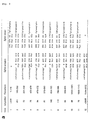
- the nucleotide sequences of primers used are shown in Table 1.
- the amplified cDNA was separated on a 3% NuSieveGTG (2:1) agarose gel.
- the blot was washed at 50°C with a solution containing 0.1XSSC and 0.1% SDS, followed by autoradiography at -80°C for 24h.
- FISH was carried out according to the method of Inazawa. et al. (Inazawa J., et al ., Genomics, 17 : 153-162, 1993).
- Human metaphase chromosomes were prepared through a conventional method (thymidine synchronization/bromodeoxyuridine release technique). Prior to hybridization, the metaphase cells were subjected to staining with Hoechst 33258 and UV-irradiation. The cosmid clone p53-cos191 was nick-translated and labeled with biotin-16-dUTP, and used for hybridization to denatured metaphase chromosomes. In order to remove noise signals generated by scattered repeat sequences such as Alu repeats, the chromosomal in situ suppression technique was used. Hybridized signals were detected by use of FITC-avidin, and their precise locations were determined by visualization of replication-G bands.
- cosmid clone p53-cos191 was obtained.
- RT-PCR analysis was carried out.
- expression vector DNA carrying wt p53 or a mutant p53 cDNA was transiently introduced to SW480 cells (SW-480-wt53 or SW-480-mt53, respectively), and RNA prepared from these cells were used as a template.
- RNA samples were subjected to reverse transcription in the presence (+) or absence (-) of reverse transcriptase (RT).
- RT reverse transcriptase
- the cDNA termed P2XM contained an open reading frame of 1293 bp encoding a protein of 431 amino acid residues. The entire DNA sequence is shown in SEQ ID No: 3.
- the coding region of the cDNA P2XM was present at nucleotide positions 46-1338; potential transmembrane domains (M1 and M2) were at amino acid residues 33-49 and 324-344, respectively; and a segment (H) homologous to voltage-gated K+ channel H5 was present at amino acid residues 306-319.
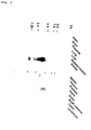
- FIG. 2 is a photo showing the results of northern blot analysis of P2XM expression in various human tissue.
- a blot with poly(A)+ RNA (2 ⁇ g/lane) prepared from various tissue was hybridized with P2XM cDNA.
- Figs. 3 and 4 depict the genome structure of the P2XM gene.
- Fig. 3(a) shows nucleotide sequences of exon-intron boundaries of the 191 gene. Exon and intron sequences are expressed in uppercase and lowercase letters, respectively.
- Fig. 4(b) exon positions are drawn to scale according to their sizes, and exons are represented as numbered boxes.
- Fig. 4(c) shows a sequence comparison between the p53-binding site of cosmid p53-191 and the consensus sequences of p53-binding sites. Each arrow shows the consensus sequence (pentamer) of p53-binding sites; sequences in uppercase letters agree with the consensus, whereas those in the lowercase letters disagree with the consensus.
- P2X receptor (P2X1-X6) family members contain two transmembrane domains (M1 and M2), a segment (H5) homologous to the H5 domain of voltage-gated K+ channel, N-glycosylation sites, and 11 cysteine residues conserved evolutionarily (see Figs. 5 and 6).
- Figs. 5 and 6 show the amino acid sequences of various P2X receptors.
- boxed amino acid residues indicate those conserved commonly among P2XM and rat P2X1-P2X7 receptors.
- Lines on top of the figures indicate regions of two conserved hydrophobic domains (M1 and M2) and the H5 domain.
- a star indicates a potential N-glycosylation site.
- amino acid sequence encoded by the gene of the present invention shares the basic features of the P2X receptor family, thus implying that the gene of the present invention is a new member belonging to the P2XM family.
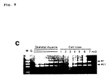
- RT-PCR of RNA prepared from skeletal muscle, followed by direct DNA sequencing confirmed 3 different in-frame transcripts (AL1, AL2, and AL3) resulting from alternative splicing, which lacked part of exon 1 from the donor site of exon 1 to 18 bases downstream, exon 10, and exons 10 and 11, respectively (see Fig. 7).
- Fig. 7 shows a schematic figure of the alternative splicing.
- the major RT-PCR amplified products from normal skeletal muscle are shown as N1 and N2, whereas the three types of variants are shown as AL1, AL2, and Al3.
- exons 1-2 and exon 11 correspond to the transmembrane domains M1 and M2, respectively.
- the M1 and M2 domains are believed to form the ion pore and ion-binding site (Valera S., et al ., Nature, 371 : 516-519, 1994; Brake A. J., et al ., Nature, 371 : 519-523, 1994). From the structural point of view, these exons appear to encode biologically important domains.
- Figs. 8 and 9 show the results of N1, AL1, N2, AL2, and AL3, whose PCR products are of sizes 392, 314, 450, 384, and 306 bp, respectively. Note that amounts of RNA templates used for the above analysis were normalized with respect to PCR amplification of GAPDH, and thus all the samples applied showed similar levels of the GAPDH signal (Fig. 10).
- the obtained results show that expression of the gene of the present invention decreased remarkably in one rhabdomyosarcoma (A673) among the 7 cell lines.
- the following findings of alternative splicing were observed with these cell lines: the profiles of transcripts lacking exon 10 and exons 10-11 in the cancer cell lines were similar to that in normal skeletal muscle; on the other hand, as compared with a relatively low level of the transcript variant lacking exon 1 in normal skeletal muscle, relatively high levels of the transcript were observed with one rhabdomyosarcoma (RD) and one osteosarcoma (Hu03N1).
- a new p53-inducible gene a new member belonging to P2X family encoding ATP-gated ion channels, was isolated.
- the p53-binding site was located 1.6 kb downstream of the above gene, as revealed by sequencing the entire fragment of the cosmid DNA insert that also contained the above gene.
- the functional p53-binding sites of the genes regulated by p53 have been all located within their introns or promoter regions.
- the functional p53-binding site was located downstream of the gene.
- the amino acid sequence deduced from the cDNA of the present invention showed homology to the P2X receptor family members, particularly to rat P2X6 (80% homology).
- rat P2X6 mRNA is distributed widely over the brain, whereas the gene of the present invention was specifically expressed only in skeletal muscle. Hence, the gene of the present invention is unlikely to be the human counterpart of rat P2X6.
- the P2X receptor is classified in the group of ATP-gated ion channels, and is believed to function as a mediator for extracellular ATP-inducible biological activity, such as cell death and synaptic transmission (Zheng L. M., et al ., J.
- RP-2 a partial cDNA
- RP-2 was isolated by subtractive hybridization of mRNAs in rat thymocytes undergoing apoptosis induced by gamma-irradiation (Owens G. P., et al ., Mol. Cell. Biol., 11 : 4177-4188, 1991).
- ATP Upon increasing intracellular Calcium concentrations, ATP induces death of thymocytes, hepatocytes, and various types of lymphocyte cell lines. These phenomena imply that the gene of the present invention is closely involved in p53-dependent apoptosis of skeletal muscle, possibly mediated through extracellular ATP.
- Northern blot analysis revealed the presence of the 3.6-kb transcript in skeletal muscle.
- the expression level of the transcript decreased remarkably in one of the 4 rhabdomyosarcoma cell lines.
- an expression level of the spliced variant transcript lacking part of exon 1 encoding part of transmembrane domain M1 was a minor transcript in normal skeletal muscle, the expression level was relatively high in two of the remaining cancer cell lines.
- the ratio of the abnormal transcripts resulting from alternative splicing was relatively high in the cancer cell lines tested; and thus it is important to clarify the biological significance of the observed heterogeneity at the amino-terminal region.
- the DNA sequence of the cDNA contains an open reading frame encoding a peptide of 431 amino acid residues that has homology to the major characteristics of the members of P2X receptor family (ATP-gated ion channels).
- the peptide has also homology to RP-2, a gene that can be activated in the thymus undergoing induced programmed cell death.
- the gene of the present invention is expressed mainly in skeletal muscle, was named P2XM (P2X specifically expressed in skeletal muscle), and is considered to be involved in suppression of cell growth and/or apoptosis of skeletal muscle.
- transcript in one of the 4 rhabdomyosarcoma cell lines was remarkably suppressed. Furthermore, levels of the splicing variant transcript lacking part of exon 1 encoding transmembrane domain M1, which was a minor species in normal muscle, were relatively high in two out of the seven cancer cell lines tested, implying that the production rate of such altered transcripts drastically increases in cancer cell lines. Further, the gene of the present invention was located at chromosome band 22q11, which is known to undergo deletion in rhabdoid tumors.
- a new human gene transcriptionally regulated specifically by the tumor suppressor gene p53 is provided.
- the gene By use of the gene, its expression in various tissue can be detected, and the structure and function of the product encoded by the gene can be analyzed.
- the gene product can be manufactured by means of gene engineering techniques.
Landscapes
- Health & Medical Sciences (AREA)
- Chemical & Material Sciences (AREA)
- Organic Chemistry (AREA)
- Life Sciences & Earth Sciences (AREA)
- General Health & Medical Sciences (AREA)
- Medicinal Chemistry (AREA)
- Genetics & Genomics (AREA)
- Molecular Biology (AREA)
- Proteomics, Peptides & Aminoacids (AREA)
- Biophysics (AREA)
- Biochemistry (AREA)
- Gastroenterology & Hepatology (AREA)
- Zoology (AREA)
- Toxicology (AREA)
- Veterinary Medicine (AREA)
- Public Health (AREA)
- General Chemical & Material Sciences (AREA)
- Animal Behavior & Ethology (AREA)
- Chemical Kinetics & Catalysis (AREA)
- Pharmacology & Pharmacy (AREA)
- Nuclear Medicine, Radiotherapy & Molecular Imaging (AREA)
- Peptides Or Proteins (AREA)
- Measuring Or Testing Involving Enzymes Or Micro-Organisms (AREA)
- Medicines That Contain Protein Lipid Enzymes And Other Medicines (AREA)
- Preparation Of Compounds By Using Micro-Organisms (AREA)
- Saccharide Compounds (AREA)
Applications Claiming Priority (3)
| Application Number | Priority Date | Filing Date | Title |
|---|---|---|---|
| JP09304497A JP3885177B2 (ja) | 1997-03-26 | 1997-03-26 | ヒト遺伝子 |
| JP9304497 | 1997-03-26 | ||
| PCT/JP1998/001146 WO1998042835A1 (fr) | 1997-03-26 | 1998-03-18 | Genes humains |
Publications (2)
| Publication Number | Publication Date |
|---|---|
| EP1006186A1 true EP1006186A1 (fr) | 2000-06-07 |
| EP1006186A4 EP1006186A4 (fr) | 2000-12-06 |
Family
ID=14071517
Family Applications (1)
| Application Number | Title | Priority Date | Filing Date |
|---|---|---|---|
| EP98909733A Withdrawn EP1006186A4 (fr) | 1997-03-26 | 1998-03-18 | Genes humains |
Country Status (6)
| Country | Link |
|---|---|
| US (1) | US6255472B1 (fr) |
| EP (1) | EP1006186A4 (fr) |
| JP (1) | JP3885177B2 (fr) |
| AU (1) | AU724681B2 (fr) |
| CA (1) | CA2284859C (fr) |
| WO (1) | WO1998042835A1 (fr) |
Cited By (9)
| Publication number | Priority date | Publication date | Assignee | Title |
|---|---|---|---|---|
| US7531171B2 (en) | 2001-01-17 | 2009-05-12 | Intreat Pty Limited | Antibodies to non-functional P2X7 receptor |
| US8067550B2 (en) | 2006-10-10 | 2011-11-29 | Biosceptre International Limited | Hybridomas producing antibodies against non functional P2X7 receptor |
| US8440186B2 (en) | 2007-09-14 | 2013-05-14 | Biosceptre International Limited | P2X7 epitopes |
| US8597643B2 (en) | 2008-07-04 | 2013-12-03 | Biosceptre International Limited | Antibodies for binding to non-functional P2X7 receptors in trimeric form |
| US8658385B2 (en) | 2007-09-14 | 2014-02-25 | Biosceptre International Limited | Purinergic (P2X) receptors in extra-cellular body fluid |
| US8835609B2 (en) | 2009-12-24 | 2014-09-16 | Biosceptre International Limited | Antigen binding sites to non-functional oligomeric P2X7 receptors and methods of use thereof |
| US9127059B2 (en) | 2009-08-20 | 2015-09-08 | Biosceptre International Limited | Anti P2X7 receptor antibodies and fragments thereof |
| US9562094B2 (en) | 2010-09-10 | 2017-02-07 | Biosceptre (Aust) Pty Ltd | Companion animal treatments |
| US9566318B2 (en) | 2011-07-01 | 2017-02-14 | Biosceptre (Aust) Pty Ltd | Combination therapy |
Families Citing this family (17)
| Publication number | Priority date | Publication date | Assignee | Title |
|---|---|---|---|---|
| US6242216B1 (en) | 1997-11-14 | 2001-06-05 | Abbott Laboratories | Nucleic acids encoding a functional human purinoreceptor P2X2 and P2X4, and methods of production and use thereof |
| US6214581B1 (en) | 1998-01-16 | 2001-04-10 | Abbott Laboratories | Nucleic acids encoding a functional human purinoreceptor P2X3 and P2X6, and methods of production and use thereof |
| WO1999036539A1 (fr) * | 1998-01-16 | 1999-07-22 | Abbott Laboratories | Acides nucleiques codant un purinorecepteur fonctionnel humain p2x3 et procedes de production et d'utilisation de ces derniers |
| CA2339871A1 (fr) * | 1998-08-20 | 2000-03-02 | Abbott Laboratories | Acides nucleiques codant un purinorecepteur fonctionnel humain p2x2, et procedes de production et d'utilisation de ce dernier |
| AUPP991199A0 (en) | 1999-04-21 | 1999-05-13 | University Of Sydney, The | Methods for diagnosing pre-cancerous and cancerous conditions |
| US8022058B2 (en) | 2000-05-10 | 2011-09-20 | The Trustees Of Columbia University In The City Of New York | Agents for preventing and treating disorders involving modulation of the RyR receptors |
| US7393652B2 (en) | 2000-05-10 | 2008-07-01 | The Trustees Of Columbia University In The City Of New York | Methods for identifying a chemical compound that directly enhances binding of FKBP12.6 to PKA-phosphorylated type 2 ryanodine receptor (RyR2) |
| US7879840B2 (en) | 2005-08-25 | 2011-02-01 | The Trustees Of Columbia University In The City Of New York | Agents for preventing and treating disorders involving modulation of the RyR receptors |
| US7718644B2 (en) | 2004-01-22 | 2010-05-18 | The Trustees Of Columbia University In The City Of New York | Anti-arrhythmic and heart failure drugs that target the leak in the ryanodine receptor (RyR2) and uses thereof |
| US7569351B2 (en) | 2000-08-03 | 2009-08-04 | Oncotherapy Science, Inc. | P53 dependent apoptosis-associated gene and protein |
| US7186812B2 (en) * | 2001-03-29 | 2007-03-06 | Applera Corporation | Isolated human G-protein coupled receptors, nucleic acid molecules encoding human GPCR proteins, and uses thereof |
| WO2003072014A2 (fr) | 2002-02-25 | 2003-09-04 | Mpex Bioscience, Inc. | Compositions minicellulaires et methodes associees |
| US7544678B2 (en) | 2002-11-05 | 2009-06-09 | The Trustees Of Columbia University In The City Of New York | Anti-arrythmic and heart failure drugs that target the leak in the ryanodine receptor (RyR2) |
| EP1603450A4 (fr) | 2003-03-07 | 2009-07-29 | Univ Columbia | Procedes utilisant le recepteur de la ryanodine de type 1 |
| US8710045B2 (en) | 2004-01-22 | 2014-04-29 | The Trustees Of Columbia University In The City Of New York | Agents for preventing and treating disorders involving modulation of the ryanodine receptors |
| WO2006042147A2 (fr) * | 2004-10-08 | 2006-04-20 | The Regents Of The University Of California | Production a large echelle de proteines transmembranaires recombinantes et de proteines cytosoliques |
| US7704990B2 (en) | 2005-08-25 | 2010-04-27 | The Trustees Of Columbia University In The City Of New York | Agents for preventing and treating disorders involving modulation of the RyR receptors |
Family Cites Families (1)
| Publication number | Priority date | Publication date | Assignee | Title |
|---|---|---|---|---|
| US5985603A (en) * | 1994-05-27 | 1999-11-16 | Glaxo Group Limited | P2x receptor DNA and protein sequence |
-
1997
- 1997-03-26 JP JP09304497A patent/JP3885177B2/ja not_active Expired - Fee Related
-
1998
- 1998-03-18 EP EP98909733A patent/EP1006186A4/fr not_active Withdrawn
- 1998-03-18 WO PCT/JP1998/001146 patent/WO1998042835A1/fr not_active Application Discontinuation
- 1998-03-18 US US09/381,681 patent/US6255472B1/en not_active Expired - Fee Related
- 1998-03-18 CA CA002284859A patent/CA2284859C/fr not_active Expired - Fee Related
- 1998-03-18 AU AU64184/98A patent/AU724681B2/en not_active Ceased
Non-Patent Citations (5)
| Title |
|---|
| COLLO G ET AL: "Cloning OF P2X5 and P2X6 receptors and the distribution and properties of an extended family of ATP-gated ion channels." JOURNAL OF NEUROSCIENCE, (1996 APR 15) 16 (8) 2495-507. , XP000940405 * |
| See also references of WO9842835A1 * |
| SEGUELA P ET AL: "A NOVEL NEURONAL P2X ATP RECEPTOR ION CHANNEL WITH WIDESPREAD DISTRIBUTION IN THE BRAIN" JOURNAL OF NEUROSCIENCE,US,NEW YORK, NY, vol. 16, no. 2, 15 January 1996 (1996-01-15), pages 448-455, XP000616491 ISSN: 0270-6474 * |
| SOTO FLORENTINA ET AL: "Cloning and tissue distribution of a novel P2X receptor from rat brain." BIOCHEMICAL AND BIOPHYSICAL RESEARCH COMMUNICATIONS, vol. 223, no. 2, 1996, pages 456-460, XP000918892 ISSN: 0006-291X * |
| VALERA S ET AL: "CHARACTERIZATION AND CHROMOSOMAL LOCALIZATION OF A HUMAN P2X RECEPTOR FROM THE URINARY BLADDER" RECEPTORS AND CHANNELS,CH,HARWOOD ACADEMIC PUBLISHERS, vol. 3, no. 4, 1995, pages 283-289, XP002916431 ISSN: 1060-6823 * |
Cited By (27)
| Publication number | Priority date | Publication date | Assignee | Title |
|---|---|---|---|---|
| US7531171B2 (en) | 2001-01-17 | 2009-05-12 | Intreat Pty Limited | Antibodies to non-functional P2X7 receptor |
| US7888473B2 (en) | 2001-01-17 | 2011-02-15 | Intreat Pty Limited | Non-functional P2X7 receptor |
| US8080635B2 (en) | 2001-01-17 | 2011-12-20 | Biosceptre International Limited | Non-functional P2X7 receptor |
| US8399617B2 (en) | 2001-01-17 | 2013-03-19 | Biosceptre International Limited | Non-functional P2X7 receptor |
| US9663584B2 (en) | 2001-01-17 | 2017-05-30 | Biosceptre (Aust) Pty Ltd | Antibodies to non-functional P2X7 receptor |
| US8709425B2 (en) | 2001-01-17 | 2014-04-29 | Biosceptre International Limited | Antibodies to non-functional P2X7 receptor |
| US10450380B2 (en) | 2001-01-17 | 2019-10-22 | Biosceptre (Aust) Pty Ltd | Polypeptide immunogen for generating an antibody to non-functional P2X7 receptor |
| US8067550B2 (en) | 2006-10-10 | 2011-11-29 | Biosceptre International Limited | Hybridomas producing antibodies against non functional P2X7 receptor |
| US8440186B2 (en) | 2007-09-14 | 2013-05-14 | Biosceptre International Limited | P2X7 epitopes |
| US8658385B2 (en) | 2007-09-14 | 2014-02-25 | Biosceptre International Limited | Purinergic (P2X) receptors in extra-cellular body fluid |
| US10597451B2 (en) | 2007-09-14 | 2020-03-24 | Biosceptre (Aust) Pty Ltd | Methods of treating cancer with a P2X7 peptide |
| US9181320B2 (en) | 2007-09-14 | 2015-11-10 | Biosceptre International Limited | Peptides for generating an antibody selectively binding to a non-ATP-binding P2X7 receptor but not to an ATP-binding P2X7 receptor |
| US9944701B2 (en) | 2007-09-14 | 2018-04-17 | Biosceptre (Aust) Pty Ltd | Methods of treating cancer with antibodies that bind P2X7 receptors |
| US8597643B2 (en) | 2008-07-04 | 2013-12-03 | Biosceptre International Limited | Antibodies for binding to non-functional P2X7 receptors in trimeric form |
| US9328155B2 (en) | 2008-07-04 | 2016-05-03 | Biosceptre (Aust) Pty Ltd | Peptides for inducing antibodies to a non-functional P2X7 receptor |
| US10238716B2 (en) | 2008-07-04 | 2019-03-26 | Biosceptre (Aust) Pty Ltd | Anti-P2X7 peptides and epitopes |
| US9127059B2 (en) | 2009-08-20 | 2015-09-08 | Biosceptre International Limited | Anti P2X7 receptor antibodies and fragments thereof |
| US9688771B2 (en) | 2009-08-20 | 2017-06-27 | Biosceptre (Aust) Pty Ltd | Anti P2X7 receptor antibodies and fragments thereof |
| US10053508B2 (en) | 2009-08-20 | 2018-08-21 | Biosceptre (Aust) Pty Ltd | Anti P2X7 receptor antibodies and fragments thereof |
| US10988532B2 (en) | 2009-08-20 | 2021-04-27 | Biosceptre (Aust) Pty Ltd | Anti P2X7 receptor antibodies and fragments thereof |
| US9428587B2 (en) | 2009-12-24 | 2016-08-30 | Biosceptre International Limited | Antibodies to non-functional oligomeric P2X7 receptors and methods of use thereof |
| US8835609B2 (en) | 2009-12-24 | 2014-09-16 | Biosceptre International Limited | Antigen binding sites to non-functional oligomeric P2X7 receptors and methods of use thereof |
| US10232025B2 (en) | 2010-09-10 | 2019-03-19 | Biosceptre (Ausi) Pty Ltd | Method for minimising progression of cancer in companion animals |
| US9562094B2 (en) | 2010-09-10 | 2017-02-07 | Biosceptre (Aust) Pty Ltd | Companion animal treatments |
| US9566318B2 (en) | 2011-07-01 | 2017-02-14 | Biosceptre (Aust) Pty Ltd | Combination therapy |
| US10245308B2 (en) | 2011-07-01 | 2019-04-02 | Biosceptre (Aust) Pty Ltd | Combination therapy utilizing P2X7 peptides |
| US10543262B2 (en) | 2011-07-01 | 2020-01-28 | Biosceptre (Aust) Pty Ltd | Combination therapy utilizing P2X7 peptides |
Also Published As
| Publication number | Publication date |
|---|---|
| US6255472B1 (en) | 2001-07-03 |
| JP3885177B2 (ja) | 2007-02-21 |
| AU724681B2 (en) | 2000-09-28 |
| CA2284859A1 (fr) | 1998-10-01 |
| EP1006186A4 (fr) | 2000-12-06 |
| WO1998042835A1 (fr) | 1998-10-01 |
| JPH10262681A (ja) | 1998-10-06 |
| AU6418498A (en) | 1998-10-20 |
| CA2284859C (fr) | 2007-01-30 |
Similar Documents
| Publication | Publication Date | Title |
|---|---|---|
| EP1006186A1 (fr) | Genes humains | |
| Urano et al. | Cloning of P2XM, a novel human P2X receptor gene regulated by p53 | |
| KR100959199B1 (ko) | 신규한 피53 타겟 유전자 에스아이에스피-1 및 그의 용도 | |
| US20070082345A1 (en) | Secretory protein or membrane protein | |
| JP2010042012A (ja) | Cadasilに関与する遺伝子、診断方法および治療への適用 | |
| US20040053262A1 (en) | Supressor gene | |
| EP1012333A1 (fr) | ISOLEMENT D'UN NOUVEAU GENE p23 DU FACTEUR DE SENESCENCE | |
| Yang et al. | Genomic structure and mutational analysis of the human KIF1B gene which is homozygously deleted in neuroblastoma at chromosome 1p36. 2 | |
| KR20040073588A (ko) | 종양 치료용 조성물 및 방법 | |
| US7964358B2 (en) | Sphingosine 1-phosphate receptor gene, SPPR | |
| Liu et al. | Molecular cloning and characterization of the human ASB-8 gene encoding a novel member of ankyrin repeat and SOCS box containing protein family | |
| Kothapalli et al. | Characterization of a human sphingosine-1-phosphate receptor gene (S1P5) and its differential expression in LGL leukemia | |
| JPH07143884A (ja) | 腫瘍サプレッサー遺伝子メルリンおよびその用途 | |
| Tanaka et al. | Characterization of tissue-and cell-type-specific expression of a novel human septin family gene, Bradeion | |
| Fiucci et al. | Genomic organization and expression of mouse Tpt1 gene☆ | |
| JP2004180540A (ja) | 哺乳動物のToll様受容体3に結合する新規アダプタータンパク質およびその遺伝子 | |
| US6689584B1 (en) | Transcriptional regulatory factor | |
| US20030109027A1 (en) | TSLL2 gene | |
| Mykkänen et al. | Promoter analysis of the human SLC7A7 gene encoding y+ L amino acid transporter-1 (y+ LAT-1) | |
| WO2003102028A1 (fr) | Proteine induite par le gene rb1 (rb1cc1) et gene | |
| US7364876B2 (en) | ADIP protein and use thereof | |
| CA2493263A1 (fr) | Nouveau gene associe a l'arthrite rhumatoide | |
| JP2001505424A (ja) | マウス・グアニンヌクレオチド交換因子(mngef)及びそのヒト相同体 | |
| US20030109016A1 (en) | TSLL1 gene | |
| Liang | United States Patent te |
Legal Events
| Date | Code | Title | Description |
|---|---|---|---|
| PUAI | Public reference made under article 153(3) epc to a published international application that has entered the european phase |
Free format text: ORIGINAL CODE: 0009012 |
|
| 17P | Request for examination filed |
Effective date: 19990924 |
|
| AK | Designated contracting states |
Kind code of ref document: A1 Designated state(s): AT BE CH DE DK ES FI FR GB GR IE IT LI LU MC NL PT SE |
|
| A4 | Supplementary search report drawn up and despatched |
Effective date: 20001019 |
|
| AK | Designated contracting states |
Kind code of ref document: A4 Designated state(s): AT BE CH DE DK ES FI FR GB GR IE IT LI LU MC NL PT SE |
|
| 17Q | First examination report despatched |
Effective date: 20040127 |
|
| STAA | Information on the status of an ep patent application or granted ep patent |
Free format text: STATUS: THE APPLICATION IS DEEMED TO BE WITHDRAWN |
|
| 18D | Application deemed to be withdrawn |
Effective date: 20040807 |