EP0955883B1 - Detecting thermal discrepancies in vessel walls - Google Patents
Detecting thermal discrepancies in vessel walls Download PDFInfo
- Publication number
- EP0955883B1 EP0955883B1 EP96935912A EP96935912A EP0955883B1 EP 0955883 B1 EP0955883 B1 EP 0955883B1 EP 96935912 A EP96935912 A EP 96935912A EP 96935912 A EP96935912 A EP 96935912A EP 0955883 B1 EP0955883 B1 EP 0955883B1
- Authority
- EP
- European Patent Office
- Prior art keywords
- fiber
- radiation
- vessel
- plaque
- balloon
- Prior art date
- Legal status (The legal status is an assumption and is not a legal conclusion. Google has not performed a legal analysis and makes no representation as to the accuracy of the status listed.)
- Expired - Lifetime
Links
Images


Classifications
-
- A—HUMAN NECESSITIES
- A61—MEDICAL OR VETERINARY SCIENCE; HYGIENE
- A61B—DIAGNOSIS; SURGERY; IDENTIFICATION
- A61B5/00—Measuring for diagnostic purposes; Identification of persons
- A61B5/0059—Measuring for diagnostic purposes; Identification of persons using light, e.g. diagnosis by transillumination, diascopy, fluorescence
- A61B5/0082—Measuring for diagnostic purposes; Identification of persons using light, e.g. diagnosis by transillumination, diascopy, fluorescence adapted for particular medical purposes
- A61B5/0084—Measuring for diagnostic purposes; Identification of persons using light, e.g. diagnosis by transillumination, diascopy, fluorescence adapted for particular medical purposes for introduction into the body, e.g. by catheters
- A61B5/0086—Measuring for diagnostic purposes; Identification of persons using light, e.g. diagnosis by transillumination, diascopy, fluorescence adapted for particular medical purposes for introduction into the body, e.g. by catheters using infrared radiation
-
- A—HUMAN NECESSITIES
- A61—MEDICAL OR VETERINARY SCIENCE; HYGIENE
- A61B—DIAGNOSIS; SURGERY; IDENTIFICATION
- A61B5/00—Measuring for diagnostic purposes; Identification of persons
- A61B5/01—Measuring temperature of body parts ; Diagnostic temperature sensing, e.g. for malignant or inflamed tissue
-
- A—HUMAN NECESSITIES
- A61—MEDICAL OR VETERINARY SCIENCE; HYGIENE
- A61B—DIAGNOSIS; SURGERY; IDENTIFICATION
- A61B5/00—Measuring for diagnostic purposes; Identification of persons
- A61B5/01—Measuring temperature of body parts ; Diagnostic temperature sensing, e.g. for malignant or inflamed tissue
- A61B5/015—By temperature mapping of body part
-
- A—HUMAN NECESSITIES
- A61—MEDICAL OR VETERINARY SCIENCE; HYGIENE
- A61B—DIAGNOSIS; SURGERY; IDENTIFICATION
- A61B5/00—Measuring for diagnostic purposes; Identification of persons
- A61B5/02—Detecting, measuring or recording for evaluating the cardiovascular system, e.g. pulse, heart rate, blood pressure or blood flow
- A61B5/02007—Evaluating blood vessel condition, e.g. elasticity, compliance
-
- A—HUMAN NECESSITIES
- A61—MEDICAL OR VETERINARY SCIENCE; HYGIENE
- A61B—DIAGNOSIS; SURGERY; IDENTIFICATION
- A61B5/00—Measuring for diagnostic purposes; Identification of persons
- A61B5/05—Detecting, measuring or recording for diagnosis by means of electric currents or magnetic fields; Measuring using microwaves or radio waves
- A61B5/0507—Detecting, measuring or recording for diagnosis by means of electric currents or magnetic fields; Measuring using microwaves or radio waves using microwaves or terahertz waves
-
- A—HUMAN NECESSITIES
- A61—MEDICAL OR VETERINARY SCIENCE; HYGIENE
- A61B—DIAGNOSIS; SURGERY; IDENTIFICATION
- A61B5/00—Measuring for diagnostic purposes; Identification of persons
- A61B5/41—Detecting, measuring or recording for evaluating the immune or lymphatic systems
- A61B5/413—Monitoring transplanted tissue or organ, e.g. for possible rejection reactions after a transplant
-
- A—HUMAN NECESSITIES
- A61—MEDICAL OR VETERINARY SCIENCE; HYGIENE
- A61B—DIAGNOSIS; SURGERY; IDENTIFICATION
- A61B5/00—Measuring for diagnostic purposes; Identification of persons
- A61B5/68—Arrangements of detecting, measuring or recording means, e.g. sensors, in relation to patient
- A61B5/6846—Arrangements of detecting, measuring or recording means, e.g. sensors, in relation to patient specially adapted to be brought in contact with an internal body part, i.e. invasive
- A61B5/6847—Arrangements of detecting, measuring or recording means, e.g. sensors, in relation to patient specially adapted to be brought in contact with an internal body part, i.e. invasive mounted on an invasive device
- A61B5/6852—Catheters
- A61B5/6853—Catheters with a balloon
-
- A—HUMAN NECESSITIES
- A61—MEDICAL OR VETERINARY SCIENCE; HYGIENE
- A61B—DIAGNOSIS; SURGERY; IDENTIFICATION
- A61B17/00—Surgical instruments, devices or methods
- A61B2017/00017—Electrical control of surgical instruments
- A61B2017/00022—Sensing or detecting at the treatment site
- A61B2017/00084—Temperature
-
- A—HUMAN NECESSITIES
- A61—MEDICAL OR VETERINARY SCIENCE; HYGIENE
- A61B—DIAGNOSIS; SURGERY; IDENTIFICATION
- A61B17/00—Surgical instruments, devices or methods
- A61B17/22—Implements for squeezing-off ulcers or the like on inner organs of the body; Implements for scraping-out cavities of body organs, e.g. bones; for invasive removal or destruction of calculus using mechanical vibrations; for removing obstructions in blood vessels, not otherwise provided for
- A61B2017/22001—Angioplasty, e.g. PCTA
-
- A—HUMAN NECESSITIES
- A61—MEDICAL OR VETERINARY SCIENCE; HYGIENE
- A61B—DIAGNOSIS; SURGERY; IDENTIFICATION
- A61B5/00—Measuring for diagnostic purposes; Identification of persons
- A61B5/0059—Measuring for diagnostic purposes; Identification of persons using light, e.g. diagnosis by transillumination, diascopy, fluorescence
- A61B5/0062—Arrangements for scanning
- A61B5/0066—Optical coherence imaging
-
- A—HUMAN NECESSITIES
- A61—MEDICAL OR VETERINARY SCIENCE; HYGIENE
- A61B—DIAGNOSIS; SURGERY; IDENTIFICATION
- A61B5/00—Measuring for diagnostic purposes; Identification of persons
- A61B5/0059—Measuring for diagnostic purposes; Identification of persons using light, e.g. diagnosis by transillumination, diascopy, fluorescence
- A61B5/0071—Measuring for diagnostic purposes; Identification of persons using light, e.g. diagnosis by transillumination, diascopy, fluorescence by measuring fluorescence emission
-
- A—HUMAN NECESSITIES
- A61—MEDICAL OR VETERINARY SCIENCE; HYGIENE
- A61B—DIAGNOSIS; SURGERY; IDENTIFICATION
- A61B5/00—Measuring for diagnostic purposes; Identification of persons
- A61B5/0059—Measuring for diagnostic purposes; Identification of persons using light, e.g. diagnosis by transillumination, diascopy, fluorescence
- A61B5/0073—Measuring for diagnostic purposes; Identification of persons using light, e.g. diagnosis by transillumination, diascopy, fluorescence by tomography, i.e. reconstruction of 3D images from 2D projections
-
- A—HUMAN NECESSITIES
- A61—MEDICAL OR VETERINARY SCIENCE; HYGIENE
- A61B—DIAGNOSIS; SURGERY; IDENTIFICATION
- A61B5/00—Measuring for diagnostic purposes; Identification of persons
- A61B5/0059—Measuring for diagnostic purposes; Identification of persons using light, e.g. diagnosis by transillumination, diascopy, fluorescence
- A61B5/0075—Measuring for diagnostic purposes; Identification of persons using light, e.g. diagnosis by transillumination, diascopy, fluorescence by spectroscopy, i.e. measuring spectra, e.g. Raman spectroscopy, infrared absorption spectroscopy
Definitions
- This invention relates to the medical diagnosis and treatment of arterial disease by means of temperature differential sensing, and particularly, infrared-sensing with devices such as temperature probes, cameras, and catheters.
- the invention provides catheters to diagnose arterial diseases detectable by thermal discrepancies in the arterial wall.
- Atherosclerotic coronary artery disease is the leading cause of death in industrialized countries.
- An atherosclerotic plaque is a thickened area in the wall of an artery.
- patients who have died of coronary disease may exhibit as many as several dozen atherosclerotic plaques; however, in most instances of myocardial infarction, cardiac arrest, or stroke, it is found that only one of these potential obstructions has, in fact, ruptured, fissured, or ulcerated.
- the rupture, fissure, or ulcer causes a large thrombus (blood clot) to form on the inside of the artery, which may completely occlude the flow of blood through the artery, thereby injuring the heart or brain.
- a major prognostic and diagnostic dilemma for the cardiologist is how to predict which placque is about to rupture.
- ruptured plaques are characterized by a large pool of cholesterol or necrotic debris and a thin fibrous cap with a dense infiltration of macrophages.
- the release of matrix-digesting enzymes by the cells is thought to contribute to plaque rupture.
- Other thromboses are found on non-ruptured but inflamed plaque surfaces.
- Inflammation in an arterial plaque is the result of a series of biochemical and mechanical changes in the arterial wall. Plaque, a thickening in the arterial vessel wall results from the accumulation of cholesterol, proliferation of smooth muscle cells, secretion of a collagenous extracellular matrix by the cells, and accumulation of macrophages and, eventually, hemorrhage (bleeding), thrombosis (clotting) and calcification.
- the consensus theory is that atherosclerotic plaque develops as a result of irritation or biochemical damage of the endothelial cells.
- plaques most likely to rupture are those that have both a thin collagen cap (fibrous scar) and a point of physical weakness in the underlying plaque. It is known that plaques with inflamed surfaces or a high density of activated macrophages and a thin overlying cap are at risk of thrombosis. Van der Wal, et al., Circulation 89:36-44 (1994); Shah, et al., Circulation 244 (1995); Davies, et al., Br Heart J 53:363-373 (1985); Farb, et al., Circulation 92:1701-1709 (1995); Van Damme, et al., Cardiovasc Pathol 3:9-17 (1994).
- Such points are thought to be located (as determined by modeling studies and pathologic analysis) at junctures where pools of cholesterol meet a more cellular and fibrous part of the plaque.
- macrophages inflammatory cells
- these inflammatory cells release enzymes capable of degrading the collagen and other components of the extracellular matrix, it is thought that they are crucial to the process of plaque rupture or fissuring.
- Inflammation also plays an important role in the rejection of transplanted organs, a process which begins by an attack of host T lymphocytes in the grafted donor organ's endothelial cells.
- Recruitment and proliferation of inflammatory and smooth muscle cells are heat-generating processes, whose effects are detectable in advance of the detection of vessel narrowing using stress tests, ultrasound, or angiography. Johnson et al. J. Am. Coll. Cardiol. 17: 449-57 (191); St.
- restenosis a renarrowing of an artery that has undergone one or more interventional techniques to relieve an original stenosis (caused by plaque).
- interventional techniques include balloon angioplasty, atherectomy (shaving or cutting the plaque), and laser angioplasty.
- Balloon angioplasty of the coronary arteries is a major advance in treatment and has been performed on hemodynamically significant coronary stenoses (those that are 70% to 99% of the cross-sectional diameter of the vessel) with a success rate of 90%.
- restenosis occurs in the vessel and most of the benefit gained by the procedure is lost.
- Another major diagnostic and prognostic dilemma for cardiologists not readily addressed by prior art devices or methods is predicting which patients will develop restenosis.
- Restenosis may occur when the removal of plaque by angioplasty or atherectomy injures the artery wall.
- the injury to the vessel wall causes the smooth muscle cells at that site to proliferate and to secrete an extracellular matrix which again narrows the artery. Both cell proliferation and secretion are exergonic (heat-generating) processes. Additionally, it is known that macrophage concentration in a vessel is correlated to the risk of restenosis.
- U.S. Patent No. 3,866,599 relates to a fiberoptic catheter for insertion into the cardiovascular system for the measurement of oxygen in blood.
- optical fibers are used to first project infra-red and red light at the catheter tip into the blood.
- the infra-red and red light reflected by the blood is then returned through the optical fibers to an oximeter.
- the ratio of infra-red light reflected to that absorbed by the blood is proportional to the oxygen saturation in the blood.
- This catheter design is also one wherein there is at the distal end of the element a recess preventing the ends of the fibers from contacting the vessel wall and an exterior sleeve which can be expanded to further space the fibers from the wall of the vessel.
- the fiberoptic catheter of this patent does not permit detection of heat.
- U.S. Patent No. 4,752,141 relates to fiberoptic sensing of temperature and/or other physical parameters using a system comprising (1) an optical fiber (2) means including a source of visible or near visible electromagnetic radiation pulses at one end of the fiber for directing the radiation along the fiber to another end of the fiber (3) a sensor positioned at or near the end of the fiber in a manner to receive the radiation, modulate it by the temperature, and redirect the modulated radiation back through the optical fiber to the sensor (4) the sensor comprising at least one optical element in the path of the source of radiation whose optical properties vary in response to the magnitude of temperature changes and (5) means positioned at the end of the fiber receiving the modulated radiation for measuring a function related to the time of the resulting luminescent radiation intensity decay after an excitation pulse indicating the temperature of the sensor.
- These temperature sensors were designed to physically contact a surface and were built with an elastomeric substance at the end of the fiber to which a thin layer of phosphor material had been deposited. The phosphor reacts to the temperature and emits radiation which travels up the fiber and is detected by the sensor. Contact temperature determinations require the ability of the catheter to be placed in contact with the locus whose temperature is to be measured.
- U.S. Patent No. 4,986,671 relates to a fiber optic probe with a single sensor formed by a elastomeric lens coated with a light reflective and temperature dependent material over which is coated a layer of material that is absorptive of infrared radiation thereby allowing a determination of characteristics of heat or heat transfer.
- One application is in a catheter for providing pressure, flow and temperature of the blood in a blood vessel.
- U.S. Pat. No. 4,140,393 relates to the use of birefringement material as a temperature probe.
- U.S. Pat. No. 4,136,566 suggests the use of the temperature dependent light absorption characteristics of gallium arsenide for a temperature sensor.
- U.S. Pat. No. 4,179,927 relates to a gaseous material having a temperature dependent light absorption.
- U.S. Pat. No. 4,075,493 relates to the use of a luminescent material as a temperature sensor, exciting radiation of one wavelength range being passed along the optical fiber from the measuring instrument, and temperature dependent luminescent radiation being emitted from the sensor back along the communicating optical fiber for detection and measurement by the instrument. It is the luminescent sensor technology which has found the greatest commercial applicability in fiber optic measurements, primarily for reasons of stability, wide temperature range, ability to minimize the effect of non-temperature light variations, small sensor size and the like.
- U.S. Patent No. 4,995,398 relates to the use of thermography during the course of by-pass heart surgery for the purpose of checking the success of the operation before closing the chest cavity.
- This patent describes the use of a scanning thermal camera, image processing, temperature differentials and displaying images in real time.
- the invention relies on the use of a cold injectate which when it mixes with warmer blood provides images captured on a thermal camera focusing on the heart to detect stenoses at the sites of distal anastomoses.
- U.S. Patent No. 5,046,501 relates to a method of identifying atherosclerotic plaque versus structurally viable tissues using a fluorescent beam at a single excitation wavelength of between 350 and 390 nm preferably from a laser which allows differentiation of these tissues.
- No catheter was used in the examples of the patent.
- in situ imaging is not disclosed or taught by this patent.
- no technique is described by this patent for predicting plaque rupture, restenosis or transplant vasculopathy.
- U.S. Patent No. 5,057,105 relates to a hot tip catheter assembly comprising a heater, a cap, a thermocouple, power leads, and a central distal lumen to position the catheter in the artery.
- the thermocouple is included to continuously monitor the heating of the catheter tip in order to prevent overheating. The tip when properly placed on a plaque build up, melts the plaque.
- U.S. Patent No. 5,109,859 relates to ultrasound guided laser angioplasty comprising a laser at the tip of a catheter, an ultrasound device also at the tip of the laser for guidance, and a proximal occlusion balloon to provide stabilization and a blood free environment.
- This patent apparently also relates to estimating the mass of a plaque tissue.
- the ultrasound device of the patent can distinguish histological features (i.e. , what cells and extracellular matrix are within the plaque). Thus, it is not likely that such a device could be used to predict plaque rupture. Indeed, recent studies have found that intravascular ultrasound cannot identify which plaques are at risk of rupturing. de Feytia Circulation 92:1408-13 (1995).
- U.S. Patent No. 5,217,456 relates to an intra-vascular guidewire-compatible catheter which has a source of illumination and a synchronous fluorescence detector.
- Light in a wavelength that induces fluorescence in tissues emanates radially from an aperture on the side of the catheter. Fluorescence emitted from the tissues enters the catheter through the same aperture and is conveyed to a spectral analyzer.
- This information can be used to differentiate healthy tissue from atherosclerotic plaque.
- this device does not distinguish between plaque on the basis of heat differential.
- U.S. Patent No. 5,275,594 relates to methods and apparatus for distinguishing between atherosclerotic plaque and normal tissue by analyzing photoemissions from a target site.
- the system includes a source of laser energy for stimulation of fluorescence by non-calcified versus calcified atherosclerotic plaque, and an analyzing means for determining whether a spectrum of fluorescence emitted by a tissue represents calcified or non-calcified atherosclerotic plaque at a target site, based upon the time domain signal of calcium photoemission following fluorescent excitation of the calcium.
- laser energy is used to ablate the plaque.
- Prior art approaches to intravascular arterial diagnosis and repair have been numerous yet have failed to provide certain capabilities.
- such prior art catheters and methods have failed to provide means for detecting and treating atherosclerotic plaque since they have not been able to differentiate between those plaques at risk of rupturing and occluding and those that are not presently at such risk even if they are capable of determining the presence or absence of calcification of the plaque.
- prior art approaches have not provided effective means for identifying specific arterial sites at risk for arterial restenosis after angioplasty or atherectomy.
- Prior art approaches have also failed to provide practical and effective means for detecting transplant vasculopathy.
- the present invention overcomes at least some of the failures of the prior art by providing an infrared-sensing catheter for detecting heat-producing inflammatory cells and vessel wall cells, and thus predicting the behavior of injured blood vessels in medical patients.
- the catheters of the present invention provide effective means for detecting and treating atherosclerotic plaque which is capable of differentiating between, among other pathologies, those plaques at risk of rupturing arid occluding and those that are not presently at risk.
- the catheters of the present invention also provide effective means for identifying specific arterial sites at risk for arterial restenosis after angioplasty or atherectomy, or which patients are at risk due to vasculopathy, or tissue rejection.
- the catheters of the present invention also are capable of effectively identifying patients who have arterial wall areas of unusually low temperature and which represent previously undetected arterial at-risk areas-just as excess heat can identity regions at risk due to inflammation, sub-normal heat (areas cooler than the rest of a vessel) indicates a lack of actively metabolizing healthy cells (since heat in the body results from actively metabolizing cells).
- Non-cellular areas are typically regions of hemorrhage, thrombosis, cholesterol pools, or calcium-all indicators of high risk plaques.
- the invention's devices achieve these ends by identifying and analyzing thermal discrepancies in the wall temperature of blood vessels.
- the invention relates to apparatus for analyzing optical radiation of a vessel. There are also described methods for analyzing optical radiation, which methods are best preferably achieved using the apparatus of the invention.
- Optical radiation of particular interest in the invention is that radiation which falls in the optical spectrum in the wavelength interval from about 2-14 ⁇ m.
- An attractive feature of infrared is its penetration through calcium (relative to white light and ultrasound). Benaron, et al., J Clin Monit 11:109-117 (1995).
- the vessels of particular interest in the invention are those vessels where the access to a surface of which is problematic.
- a traditional temperature probe i.e., a contact thermometer or thermocouple
- the apparatus of the invention will find utility.
- the vessel, while of sufficiently large internal diameter for access by a temperature probe has one or others of its openings narrowed, occluded or otherwise blocked, the apparatus of the invention will find utility.
- vessels of the body including vessels circulating and transporting sera (i.e. blood) such as arteries, veins, sinuses, heart cavities and chambers.
- the invention relates to apparatus in which there is at least one optical fiber used which is capable of transmitting optical radiation from a distal end of the fiber, usually inside a vessel, to a proximal end of the fiber, usually outside the vessel.
- Optical fibers used will exhibit certain key parameters related to their ability to transmit wavelengths in the region of 2-14 ⁇ m. These key parameters include optical transparency, flexibility and strength.
- the optical fibers used are those which may be extruded in ultrathin diameters and which transmit over the appropriate infra-red spectral region.
- the infrared fiberoptic can be constructed from a variety of substances known to those of skill in the art such as zirconium fluoride (ZrF 4 ), silica, or chalcogenide (AsSeTe).
- ZrF 4 fibers are well suited to the apparatus of the invention because such fibers have >90% transmission capabilities over 1 meter for small diameters.
- optical fibers useful in the apparatus of the invention will also be ones capable of placement proximate to a locus of a wall of the vessel being investigated. This criterion is achieved in part by the flexibility of the fiber optic. In additional part, this criterion is met by the ultrathin nature of the diameter of the fiber optic.
- the apparatus of the invention also utilize a balloon which encases a distal end of the fiber.
- the balloon in one embodiment, may be one which is transparent to the optical radiation of interest. In that instance, optical radiation originating outside the balloon is transmitted through the outer surface of the balloon to the inner surface of the balloon and on to the entry point for optical radiation into the optical fiber. It is important, in this embodiment, for there to be little if any absorption, reflection or other diversion of the optical radiation emanating from the source (i.e., the vessel wall, a locus such as a plaque locus) during its transmission through the surfaces of the balloon. Such unwanted absorption may be caused by blood or other body fluids. Therefore, transparency for purposes of the invention means an ability to transmit substantially all optical radiation from a particular source through the balloon surfaces to the optical fiber.
- opacity for purposes of the invention means an ability to absorb substantially all optical radiation from a particular source on the outer balloon surface. Thereafter, the inner surface of the balloon will re-emit a proportional amount of radiation to that absorbed on the outer surface immediately adjacent the locus originating the radiation. This re-emitted radiation will be detectable by the fiber optic system encased inside the balloon.
- the apparatus of the invention also utilize a detector capable of detecting a difference in the optical radiation of interest, between the locus and the average optical radiation along the vessel wall being investigated.
- the detector of the invention is one which has a sensitivity capable of detection of differences in infra-red radiation as small as 50 °mK, and in the range of 10 to 100 °mK.
- the detector will be one capable of detecting the radiation which is transmitted through the balloon's outer and inner surfaces.
- the detector will be one capable of detecting the radiation which is re-emitted from the balloon's inner surface opposite the balloon's outer surface which is directly in contact with the locus.
- the apparatus of the invention will rely on detection of optical radiation in the infra-red radiation ranges.
- ranges of 2-14 micrometers are of particular interest in the apparatus and methods of the invention.
- T 1 , T 2 and T 3 each refer to temperatures in the range of 300-310 °K which vary from one another increasingly by 1 °K) in the wavelength range of 3 up to 6 micrometers. It can also be seen in the inset to Fig.
- black bodies held at constant temperatures in the range of 300-310 °K and differing from one another by only a single degree appear as easily distinguishable curve segments, emitting photons from these black bodies in the range of approximately 0.21 x 10 17 to 0.40 x 10 17 photons.
- the apparatus of the invention may comprise at least two fibers, although the use of greater than two fibers is clearly possible where merited, such as when detection along the axis of the vessel is preferred at greater than a single position simultaneously.
- at least one of the fibers is a reference fiber and another of the fibers is a signal fiber.
- the signal fiber is a fiber designed to transmit all optical radiation focused into its length from its distal end to its proximal end.
- the reference fiber is a fiber which is used as a control against which the signal fiber transmissions may be compared.
- optical radiation exiting the proximal end of the signal fiber is compared to that exiting the proximal end of the reference finer, a determination can be readily made as to relative amounts of optical radiation exiting the signal fiber which is due to other than optical radiation emitted by the locus of interest.
- the apparatus of the invention may also be optically connected at the distal end of the signal fiber to an optically reflective surface capable of directing optical radiation arising radially to said distal end, and on into said fiber.
- an optically reflective surface capable of directing optical radiation arising radially to said distal end, and on into said fiber.
- the reference fiber will typically be coated on its distal end with a material that substantially prevents optical radiation from entering it.
- the apparatus of the invention is also one in which the inner surface of the opaque occluding balloon emits a black body spectrum modulated by the transmission spectrum of the balloon.
- the balloon upon inflation, will substantially limit flow of fluids within the vessel.
- the flow limitation required is one in which only so much flow occurs as will not cause a rise or fall in average background IR radiation along the vessel wall immediately distal the inflated balloon.
- the apparatus of the invention is one where the balloon, upon inflation, substantially excludes the presence of intervesicular fluids between the fibers inside the balloon and the wall of the vessel most proximate to the test locus.
- the apparatus of the invention will be placed along an axis of the vessel. in this manner, it will be possible to bring the diagnostic fiber array into close proximity with a locus to be diagnosed.
- the locus will be one which contains plaque.
- the apparatus as previously noted will be useful in detecting among those plaques with which it is brought into proximity, whether a given plaque is one at risk of rupturing. In most instances, the apparatus of the invention will be used to diagnose thermal discrepancies on the interior wall of a vessel.
- the apparatus of the invention is in its most preferred embodiments a catheter.
- the catheter of the invention will be one designed for use with a guidewire.
- the guidewire will allow optional removal and reinsertion at the discretion of the surgeon, for example where after diagnosing a plaque at risk of rupturing using the catheter of the invention, the surgeon may wish to bring another diagnostic device or a therapeutic device such as a laser into the same position next to the problematic plaque.
- the apparatus of the invention is also one where the detector is preferably optically connected to a proximal end of the fiber, and if there is more than one fiber, to a proximal end of each of the fibers.
- the detector will be a multi-wavelength radiometer.
- Such a radiometer will preferably be a spinning circular variable filter whose transmission wavelength is a function of its angle of rotation.
- a filter it is possible to prevent transmission of all but a narrow band of wavelengths of light by adjusting the rotational angle.
- such a filter can be made to be transparent to highly selected wavelengths by its rotational characteristics.
- the filter will be one transparent to radiation with a wavelength of approximately between 2 to 6 micrometers.
- the filter will be transparent to radiation with a wavelength of approximately 3 micrometers.
- One of the keys to this invention as it relates to the apparatus is that it automatically provides a reference for each spectrum by sampling approximately 3 ⁇ m. For the range of temperatures expected in biological organisms, 300-310 °K, the blackbody spectrum at this wavelength is essentially the same. This provides a zero for each signal and locks down the low wavelength side of the signal. Without this, there would be no way to fit a signal to a blackbody spectrum since the vertical scale would be "unfixed".
- the apparatus of the invention utilizes the transmitted information from more than one fiber through a filter for comparative purposes, it will be preferred to utilize an offset in the distal fiber ends.
- the offset will be at a distance sufficient to allow sampling of radiation emitted from either fiber to pass the filter at a substantially identical location on the filter.
- the apparatus of the invention when used in conjunction with a radiometer will preferably be one optically connected to at least one photoelectric device capable of converting the transmitted radiation into an electrical signal.
- the photoelectric device is preferably one electrically connected to a device capable of digitizing the electrical signal (a digitizer).
- the digitized signal is mathematically fitted to a curve selected from a spectrum of curves for black bodies held at temperatures between approximately 300-310° K.
- the curves of the controlled black bodies are those plotted as numbers of photons emitted from each black body for each wavelengths.
- the particular selection of black body control curves will be made with the knowledge of typical temperatures of the human body.
- the apparatus of the invention will be a catheter for analyzing infra-red radiation of a blood vessel.
- a preferred device will comprise at least two fibers capable of transmitting the radiation and capable of placement along an axis of the vessel proximate to a plaque-containing locus of an interior wall of the vessel.
- At least one of the fibers will be a reference fiber coated on its distal end with a material that substantially prevents optical radiation from entering it, and at least one of the other of the fibers will be a signal fiber whose distal end is optically connected to an optically reflective surface capable of directing optical radiation arising radially to its distal end into and along its shaft.
- Such a preferred device will also have a balloon encasing the distal ends of each of the fibers, which balloon upon inflation will substantially limit the flow of fluids within the blood vessel.
- the balloon will substantially exclude fluids between the fibers and the wall of the vessel most proximate to the locus to be tested.
- the balloon will be transparent to or opaque to the radiation arising inside the vessel and will have an inner surface exhibiting spatially constant optical radiation emissivity. This inner surface of the opaque balloon will be one which emits a black body spectrum.
- the catheter will be one having a guidewire. It will also have a detector, optically connected to a proximal end of each of the fibers, and capable of detecting a difference in the radiation between the locus and average optical radiation along the wall of the vessel.
- the detector will further comprise a multi-wavelength radiometer with a spinning circular variable filter, the filter being such that its transmission wavelength is a function of its angle of rotation.
- the distal ends of the fibers will be offset from one another a distance sufficient to allow sampling of radiation emitted from either fiber to pass the filter at a substantially identical position on the filter.
- the radiometer will be optically connected to at least one photoelectric device capable of converting the transmitted and filtered radiation into an electrical signal, which signal is capable of being digitized, and which digitized signal is mathematically fitted to a curve selected from a spectrum of curves for black bodies held at temperatures between approximately 300-310° K, where the curves are plotted as numbers of photons emitted from each of the black bodies for each of the wavelengths.
- an analytical method suitable in certain embodiments for diagnosing medical conditions.
- a method for analyzing optical radiation of a locus in a vessel wall comprises placing at least one optical fiber capable of transmitting radiation proximate to the locus.
- the placement of the fiber and balloon is accomplished by catheterization.
- a balloon encasing a distal end of the fiber is inflated within the vessel to cause the balloon to limit flow of fluids within the vessel.
- the balloon is transparent to or opaque to the thermal radiation and has an inner surface exhibiting spatially constant optical radiation emissivity.
- the methods described d further call for transmitting the radiation along the fiber to a detector capable of detecting a difference in the radiation between the locus and the average optical radiation along the vessel wall.
- a method of detecting plaque at risk of rupturing along a blood vessel comprises inserting a guidewire into the blood vessel to be diagnosed and then catheterizing the vessel along the guidewire with at least two fibers capable of transmitting infra-red radiation along an axis of the vessel proximate to a plaque-containing locus of an interior wall of the vessel. Thereafter, the steps of the method described is carried out as described above.
- a method of surgically treating a patient with a plurality of plaque loci within a vessel comprises determining which one or more of the plurality of plaque loci has a temperature elevated above that of the average vessel wall temperature. Once such a determination is made, the surgeon removes or reduces the plaque loci found to have an elevated temperature.
- This method has as its determination step the methods described above for analyzing optical radiation of plaque locus in a vessel wall. Once plaque at risk is identified, a number of therapies may be used to reduce the risk.
- Another goal in using the present invention is to identify patients who have arterial areas of lower rather than higher temperature, such as an area of extensive scarring, a lipid pool with no cellular infiltration, or an area of hemorrhage and thrombosis which has yet to be colonized by inflammatory cells.
- the delineation of a cholesterol pool is useful in following the regression of plaques. Identifying such areas for follow-up study will localize those likely to be inflamed in the future.
- Yet another goal in using the present invention is to deliver specific local therapy to the injured areas identified by the catheter.
- These therapies include, but are not limited to, therapies which prevent or limit inflammation (recruitment, attachment, activation, and proliferation of inflammatory cells), smooth muscle cell proliferation, or endothelial cell infection, including antibodies, transforming growth factor- ⁇ (TGF- ⁇ ), nitric oxide (NO), NO synthase, glucocorticoids, interferon gamma, and heparan and heparin sulfate proteoglycans, and the various complementary DNAs that encode them.
- TGF- ⁇ transforming growth factor- ⁇
- NO nitric oxide
- NO synthase glucocorticoids
- interferon gamma interferon gamma
- heparan and heparin sulfate proteoglycans and the various complementary DNAs that encode them.
- the invention's devices will have a number of utilities. Each will reduce morbidity and mortality from coronary and carotid artery atherosclerosis. Each will reduce the incidence of restenosis and thus the need for repeated angioplasties or atherectomies. Each will also reduce the incidence of vasculopathy in organ-transplant patients. In turn, these outcomes will produce the benefits of better health care, improved public health, and reduced health care costs.
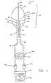
- Figure I is a schematic representation of the apparatus of the present invention with its infra-red detection unit at its proximal end and the sensor tipped distal end of the catheter as well as the guide wire disposed within a flexible outer catheter (not shown) which surrounds the optical fibers.
- Figure 2 is a black body curve spectrum for temperatures T1, T2, and T3 (differing sequentially by a single degres Kelvin) plotted as emitted radiation in photons (x1E17) versus wavelength (micrometers).
- Figure 3 is a length-wise cross sectional view of the catheter tip of Figure 1 in place within a blood vessel near a plaque at risk of rupture.
- Figure 4(a) is a graph depicting surface temperature of living carotid artery plaque in relation to cell density. Relative cell density equals the ratio of call density in the area of interest to that of the background area. Temperature measurements were made at room temperature (20°C) on 24 samples from 22 patients 10-15 minutes after removal. Point >(0°C difference in temperature) represents 27 observations.
- Figure 4(b) shows the correlation between living human carotid plaque temperature and cell density when measured in a 37 °C chamber.
- Figure 5 is a graph depicting plaque surface temperature as a function of cap thickness. Samples that had a non-inflamed fibrous cap were subjected to planimetry to measure distance from the lumen to the center of the underlying cell cluster.
- Figure 7 shows the correlation of IR radiation with cell density in the specimens described in Fig. 6, above.
- FIG. 1 shows a preferred embodiment of the apparatus of the invention in use.
- a catheter apparatus 10 is shown, which can be placed inside an artery (not shown) having with an interior arterial wall (not shown) which possesses a plurality of plaque loci (not shown). The risk of rupture of either of the plaque loci is unknown until the apparatus of the invention is applied.
- Guidewire 20 has been surgically inserted into the artery and can be seen to extend both proximally 22 and distally 24 . Guidewire 20 can also be seen to proceed through catheter apparatus 10 . Guidewire 20 is used to guide the placement of catheter apparatus 10 to the area of the artery which contains plaque loci.
- Catheter apparatus 10 comprises at its distal end (the end farthest from the detector) an inflatable balloon 40, a signal fiber 50, and a reference fiber 60.
- Inflatable balloon 40 is shown in its inflated state, which would cause it to rest primly against an interior wall of an artery and against plaque loci.
- inflation of balloon 40 would substantially limit flow of blood either at position 32 or 34 or any of the similar points around the perimeter of the generally circular series of contact points between the balloon wall 42 and an interior artery wall, allowing measurements being conducted by catheter apparatus 10 to proceed without interference.
- Balloon 40 comprises a wall 42 which is made of an elastic material.
- the perimeters of balloon 40 are such that inflation causes sealing or closure of the balloon 40 at points along the arterial wall.
- balloon 40 retreats from its contact of the arterial wall, allowing reestablishment of natural blood flow within the artery, and allowing facile movement of catheter apparatus 10 in the artery to a next position, for instance to a position at which catheter apparatus 10 may be used to measure radiation emitted from another plaque locus.
- Activation of inflation/deflation of balloon 40 may be accomplished in any of a number of ways known well to those of skill in the art of building angioplasty or embolectomy catheters or balloon-tipped catheters.
- balloon 40 The purpose of balloon 40 is to avoid problems associated with absorption of infra-red radiation by water between the source of infra-red radiation being measured and the distal catheter portion.
- the balloon wall 42 Upon inflation and contact of the artery wall, the balloon wall 42 assumes the temperature of the portions of the artery with which it is most proximate.
- the void area 46 excludes all water between the balloon wall interior and the distal signal fiber tip 56 .
- Signal fiber 50 has a translucent tip region 52 and an opaque body region 54 which is capable or incapable, respectively, of transmitting infra-red radiation efficiently.
- Opaque body region 54 may be a region in which signal fiber 50 is covered over by a cladding or sleeve 56 which causes the region to become opaque and incapable of efficiently transmitting or absorbing infra-red radiation.
- Translucent region 52 may simply be an area in which signal fiber 50 is exposed.
- Signal fiber 50 is an optical fiber which can efficiently transmit infra-red radiation. In order to collect such radiation from the surrounding milieu, signal fiber 50 may be fitted or otherwise used at its distal end with a collecting device 58 which focuses the infra-red radiation of the surrounding milieu into the fiber for subsequent transmission.
- reference fiber 60 has no translucent region. Rather, reference fiber 60 has an opaque end 62 and an opaque region 64, both of which are incapable of transmitting infra-red radiation efficiently.
- opaque region 64 may be a region in which reference fiber 60 is covered over by a cladding or sleeve 66 which causes the region to become opaque and incapable of efficiently transmitting or absorbing infra-red radiation.
- Opaque end 62 may be an area in which reference fiber 60 is coated with an infra-red reflective coating such as polished silver or aluminum.
- reference fiber 60 is identical to signal fiber 50 in its ability to function as an optical fiber which can efficiently transmit infra-red radiation.
- reference fiber 60 is offset from signal fiber 50 in the proximal direction. This offset (which can be equally well accomplished by offsetting distally) physically introduces a time delay between the radiation received and transmitted by each fiber. As will be discussed immediately below, this time delay is introduced in order to ensure that the signal and reference beams issuing from the proximal ends of each fiber strike the filter on the same spatial portion. By doing so, it is possible to eliminate alignment problems or bandpass dissimilarites arising from a multi-filter system.
- the fiber-balloon array 70 collects thermal radiation which is transmitted proximally through signal fiber 50 and reference fiber 60 . Both fibers are positioned to transmit through spinning radiometer 80 at identical radial position 82 to impinge on digitizers 92 or 90, respectively. Once a digitized signal is generated from each of the optical fiber transmissions, the background signal created by the reference fiber 60 is subtracted by computer 94 from the digitized signal transmitted by the signal fiber 50 . The resulting adjusted signal is mathematically fitted by computer 94 to a spectrum of black body curves 96 in order to ascertain the temperature of the particular locus.
- optical transparency Several options for materials for the other various components of the catheter devices described herein exist.
- the key parameters for the optical components are optical transparency, flexibility and strength.
- Materials such as high strength polyester and polyethylene terephthalate (PET) are very clear and easily extruded in ultrathin wall sizes.
- PET polyethylene terephthalate
- a high strength braided polyester is useful for translating twisting motions over long distances as may be required in certain embodiments.
- Spacers/bearings can be made from Teflon®.
- the overall flexibility of the catheter will be approximately the same as similar-sized cardiovascular laser, fiberoptic, angioplasty and atherectomizing catheters. These devices should therefore be deliverable to small diameter coronary arteries.
- a detector will be positioned at the proximal end of the catheter (outside the patient) utilizing InSb or, alternatively, HgCdTe, TeO 2 or TAS detection systems.
- Tne elongated flexible fiberoptic element will be connected at one end to an optical connector through a protective sheath.
- the optical connector is a standard item adapted to be slidably inserted into a thermal detector, and will include a plurality of openings in one side through which fluids or gases, including air, can be introduced into the catheter and emitted therefrom.
- the connector will also include a coupling element for connecting to a pressure transducer to measure pressure, there being an opening in the connector communicating with the coupling element and the pressure lumen of the catheter.
- the coupling element may also be connected to a syringe to take a blood sample or to use a saline solution to flush the catheter.
- catheters may be any of those commonly used, including flexible plastics such as nylon, TeflonTM, vinyls such as polyvinyl chloride, polyurethane, and polyethylene, or various rubber compounds.
- flexible plastics such as nylon, TeflonTM, vinyls such as polyvinyl chloride, polyurethane, and polyethylene, or various rubber compounds.
- the catheter will typically be 127 to 1016 mm (5 to 40 inches) long and have an outer diameter of about 1 to 2 millimeters.
- the lumen inside the catheter can vary but typically will be about one half to 1 millimeter in diameter.
- the minimum detectible heat differential using the devices and materials of the present invention will be about 0.1 °C. While the devices of the invention will be capable of finer thermal discrimination, biological variables are apt to introduce noise into the system. In most instances, plaques which are in danger of rupturing will vary from those less at risk by at least 1.5°C.
- the infrared-sensing catheter 100 has identified an ulcerated atherosclerotic plaque 102 which is accompanied by platelet aggregation 103 and vasoconstriction 104. Because of the presence of inflammatory cells 105 in this plaque 102, its temperature is higher than that of the immediately adjacent vessel 107, and this change is sensed by the catheter 100. Some endothelial cells 108 have been lost (as a result of senescence, inflammation, infarction, toxins, or balloon injury) causing platelets 109 to become activated and to adhere to the damaged vessel wall 110.
- the activated platelets 109 release mediators that cause vasoconstriction, platelet aggregation, and growth of smooth muscle cells; these mediators include ADP, serotonin, thromboxane A 2 , platelet-derived growth factor, transforming growth factor- ⁇ , and PF4.
- mediators include ADP, serotonin, thromboxane A 2 , platelet-derived growth factor, transforming growth factor- ⁇ , and PF4.
- the exposure of subendothelial collagen 111 and lipid 112 and the activation of platelets promote enzymatic activation of coagulation enzymes, which result in the release of plasma mitogens and the activation of thrombin, an enzyme which cleaves fibrinogen to form fibrin.
- the culmination of this process may be complete occlusion of the artery and consequent injury to the heart (or brain, in the case of a carotid, vertebral or cerebral artery).
- monocyte 114 which has attached itself to adhesion molecules on the surface of activated endothelial cells.
- the monocyte becomes a macrophage involved in uptake of modified cholesterol and the release, as by-products, of mitogens and proteolytic enzymes that may promote rupture.
- a Cole-Parmer model 8402-20 thermistor with a 24-gauge needle tip (accuracy, 0.1° C: time constant, 0.15) was used to measure the temperature of tile luminal surface in 20 locations. Temperatures were reproducible ( ⁇ 0.1° C), and most measurements were found to be within 0.2° C of each other and thus were designated as the background temperature.
- cap thickness and the cell density in a 300 x 4O0- ⁇ m region beneath the dyed regions was measured using a Mackintosh Centris 650 and NIH Image software (version 1.43), available on the Internet from the National Institutes of Health, Bethesda, Maryland.
- a minority of plaques (approximately 20%) exhibited no detectable thermal heterogeneity. Regions of deep or superficial inflammation in these specimens were not marked with dye, indicating that the overlying temperature had not been measured. In a few of the regions containing cellular infiltrates, temperatures had been measured, and they were no warmer than less cellular adjacent areas. This finding was believed by the inventors to possibly reflect decreased metabolic activity in specimens that were kept at room temperature for a longer interval after removal.
- the inventors also studied several specimens using a platinum silicide, cooled, infrared camera with a thermal resolution of 0.1° C and a spatial resolution of 0. 1 mm.
- This camera detected thermal heterogeneity in ex vivo specimens.
- Fig. 7 shows that this correlation of the IR camera measured temperatures was also observed with cell density measurements. It is noted by the inventors that cooled staring array cameras have even better thermal resolution, and spatial resolutions are as low as 10 ⁇ m.
- a thermistor with a 1-mm tip was able to detect differences as great as 4° C within different parts of the same plaque that were only 10 mm apart. Temperatures were highest when the cells were closest to the probe (i.e., at or just beneath the lumen itself). Most of the lumenal surfaces of the plaques had several regions characterized by superficial inflammation and endothelial denudation.
- plaque temperature in vivo could enable one to identify such plaques.
- plaque angiogenesis neovascularization
- the inventors studied living plaques ex vivo. In vivo, the presence and tone of the vasae vasorum might influence the temperature. However, since plaque angiogenesis correlates with inflammation, (Nikkari, et al., Circulation 92:1393-1398 (1995) and both are considered risk factors for plaque rupture, it is likely that temperature will still be predictive in vivo.
- Infrared spectroscopy could prove useful in several ways. First, it should be able to corroborate the location of macrophages by the massive amounts of nitric oxide they produce, since nitric oxide has a characteristic near-infrared spectrum. Ohdan, et al., Transplantation 57:1674-1677 (1994). Near-infrared imaging of cholesterol has already been demonstrated. Cassis, et al., Anal Chem 65:1247-1256 (1993). Second, since infrared and near-infrared wavelengths penetrate tissue more deeply as wavelength increases, longer wavelengths should reveal metabolic activity in deeper (0.1- to 1-mm) regions.
- This phenomenon could be used to develop computed infrared tomography, possibly in conjunction with interferometry, in which an incident beam is split by a moving mirror to produce a reference beam and a beam that is variably scattered and absorbed by the tissue.
- the nonsynchronous reflected wavelengths are reconstituted to reveal structural detail with 20- ⁇ m resolution. Benaron, et al., Science 259:1463-1466 (1993); Brezinski, et al., Circulation 92:1-149 (1995).
- infrared detection is also desirable since infrared absorption, convection, and tissue emissivity differences are likely to preclude non-invasive infrared tomography.
- Such alternatives include imaging the inflammatory cells with gallium, (Pasterkamp, et al., Circulation 91:1444-1449(1995)) 18 FDG positron scanning, radiolabeled anti-macrophage antibody fragments, or magnetic resonance (to take advantage of the temperature-dependence of proton-spin relaxation).
- gallium Pasterkamp, et al., Circulation 91:1444-1449(1995)
- FDG positron scanning radiolabeled anti-macrophage antibody fragments
- magnetic resonance to take advantage of the temperature-dependence of proton-spin relaxation.
- Medical therapies would depend, in part, on whether the inflammation is on the surface or beneath an intact cap. This distinction may one day be made by angioscopy (especially with the use of light-emitting antibodies) or by sampling blood for soluble markers of inflammation (P-selectin, VCAM-1, and others). Magnetic resonance imaging, ultrasound, and near-infrared imaging may also prove helpful.
- Therapies might include local delivery of agents (peptides, peptide mimetics, oligonucleotides, and others) that prevent monocyte recruitment, attachment, activation, or DNA synthesis.
- Collagen synthesis might be stimulated with ascorbic acid or transforming growth factor ⁇ (which also acts to inhibit angiogenesis, inflammation, and smooth muscle proliferation in most models, though it can also provoke inflammation in non-inflamed tissue and delay endothelial regeneration).
- ascorbic acid or transforming growth factor ⁇ which also acts to inhibit angiogenesis, inflammation, and smooth muscle proliferation in most models, though it can also provoke inflammation in non-inflamed tissue and delay endothelial regeneration.
- Endothelial regeneration can be enhanced by basic or acidic fibroblast growth factor or by vascular endothelial growth factor, among others.
- Casscells Circulation 91:2699-2702 (1995).
Landscapes
- Health & Medical Sciences (AREA)
- Life Sciences & Earth Sciences (AREA)
- General Health & Medical Sciences (AREA)
- Heart & Thoracic Surgery (AREA)
- Public Health (AREA)
- Biophysics (AREA)
- Pathology (AREA)
- Engineering & Computer Science (AREA)
- Biomedical Technology (AREA)
- Animal Behavior & Ethology (AREA)
- Medical Informatics (AREA)
- Molecular Biology (AREA)
- Surgery (AREA)
- Veterinary Medicine (AREA)
- Physics & Mathematics (AREA)
- Vascular Medicine (AREA)
- Physiology (AREA)
- Transplantation (AREA)
- Immunology (AREA)
- Cardiology (AREA)
- Nuclear Medicine, Radiotherapy & Molecular Imaging (AREA)
- Radiology & Medical Imaging (AREA)
- Measuring And Recording Apparatus For Diagnosis (AREA)
- Measuring Pulse, Heart Rate, Blood Pressure Or Blood Flow (AREA)
- Devices For Indicating Variable Information By Combining Individual Elements (AREA)
- Filling Or Discharging Of Gas Storage Vessels (AREA)
- Measurement Of The Respiration, Hearing Ability, Form, And Blood Characteristics Of Living Organisms (AREA)
- Investigating Or Analyzing Materials Using Thermal Means (AREA)
- Optical Measuring Cells (AREA)
Abstract
Description
- This invention relates to the medical diagnosis and treatment of arterial disease by means of temperature differential sensing, and particularly, infrared-sensing with devices such as temperature probes, cameras, and catheters. In particular, the invention provides catheters to diagnose arterial diseases detectable by thermal discrepancies in the arterial wall.
- Atherosclerotic coronary artery disease is the leading cause of death in industrialized countries. An atherosclerotic plaque is a thickened area in the wall of an artery. Typically, patients who have died of coronary disease may exhibit as many as several dozen atherosclerotic plaques; however, in most instances of myocardial infarction, cardiac arrest, or stroke, it is found that only one of these potential obstructions has, in fact, ruptured, fissured, or ulcerated. The rupture, fissure, or ulcer causes a large thrombus (blood clot) to form on the inside of the artery, which may completely occlude the flow of blood through the artery, thereby injuring the heart or brain. A major prognostic and diagnostic dilemma for the cardiologist is how to predict which placque is about to rupture.
- Most ruptured plaques are characterized by a large pool of cholesterol or necrotic debris and a thin fibrous cap with a dense infiltration of macrophages. The release of matrix-digesting enzymes by the cells is thought to contribute to plaque rupture. Other thromboses are found on non-ruptured but inflamed plaque surfaces.
- Inflammation in an arterial plaque is the result of a series of biochemical and mechanical changes in the arterial wall. Plaque, a thickening in the arterial vessel wall results from the accumulation of cholesterol, proliferation of smooth muscle cells, secretion of a collagenous extracellular matrix by the cells, and accumulation of macrophages and, eventually, hemorrhage (bleeding), thrombosis (clotting) and calcification. The consensus theory is that atherosclerotic plaque develops as a result of irritation or biochemical damage of the endothelial cells.
- The endothelial cells which line the interior of the vessel prevent inappropriate formation of blood clots and inhibit contraction and proliferation of the underlying smooth muscle cells. Most investigators believe that atherosclerotic plaques can develop when endothelial cells are damaged or dysfunctional. Dysfunction in endothelial cells is typically produced as a result of injury by cigarette smoke, high serum cholesterol (especially oxidized low density lipoprotein), hemodynamic alterations (such as those found at vessel branch points), some viruses (herpes simplex, cytomegalovirus) or bacteria (e.g., Chlamydia), hypertension, some hormonal factors in the plasma (including angiotensisn II, norepinephrine), and other factors as yet unknown. As a result of these gradual injuries to the endothelial cells, an atherosclerotic plaque may grow slowly over many years. However, it is now well documented that if a plaque ruptures, it often grows abruptly.
- When plaque rupture develops, there is hemorrhage into the plaque through the fissure where the surface of the plaque meets the bloodstream. Blood coagulates (forms a thrombus) quickly upon contact with the collagen and lipid of the plaque. This blood clot may then grow to completely occlude the vessel or it may remain only partially occlusive. In the latter case, the new clot quite commonly becomes incorporated into the wall of the plaque, creating a larger plaque.
- Considerable evidence indicates that plaque rupture triggers 60% to 70% of fatal myocardial infarctions and that monocyte-macrophages contribute to rupture by releasing metalloproteinases (e.g., collagenases, stromelysin) which can degrade and thereby weaken the overly fibrous cap. Van der Wal, et al., Circulation 89:36-44(1994); Nikkari, et al., Circulation 92:1393-1398 (1995); Falk, et al., Circulation 92:2033-20335 (1995); Shah, et al., Circulation 244 (1995); Davies, et al., Br Heart J 53:363-373 (1985); Constantinides, J Atheroscler Res 6:1-17 (1966). In another 25% to 30% of fatal infarctions, the plaque does not rupture, but beneath the thrombus the endothelium is replaced by monocytes and inflammatory cells. Van der Wal, et al., Circulation 89:36-44 (1994); Farb, et al., Circulation 92:1701-1709 (1995). These cells may both respond to and aggravate intimal injury, promoting thrombosis and vasocoristriction. Baju, et al., Circulation 89:503-505 (1994).
- Unfortunately, neither plaque rupture nor plaque erosion is predicable by clinical means. Soluble markers (P-selectin, von Willebrand factor, angiotensen-converting enzyme, C-reactive protein, D-dimer; Ikeda, et al., Circulation 92:1693-1696 (1995); Merlini, et al., Circulation 90:61-8 (1994); Berk, et al., Am J Cardiol 65:168-172 (1990)) and activated circulating inflammatory cells are found in patients with unstable angina pectoris, but it is not yet known whether these substances predict infarction or death. Mazzone, et al., Circulation 88:358-363 (1993). It is known, however, that the presence of such substances cannot be used to locate the involved lesion.
- Low-shear regions opposite flow dividers are more likely to develop atherosclerosis, (Ku, et al., Arteriosclerosis 5:292-302 (1985)), but most patients who develop acute myocardial infarction or sudden cardiac death have not had prior symptoms, much less an angiogram. Farb, et al., Circulation 92:1701-1709 (1995).
- Certain angiographic data has revealed that an irregular plaque profile is a fairly specific, though insensitive, indicator of thrombosis. Kaski, et al., Circulation 92:2058-2065 (1995). These stenoses are likely to progress to complete occlusion, while less severe stenoses are equally likely to progress, but less often to the point of complete occlusion. Aldeman, et al., J Am Coll Cardiol 22:1141-1154 (1993). However, because hemodynamically nonsignificant stenoses more numerous than critical stenoses and have not triggered collateral development, those which do abruptly occlude actually account for most myocardial infarctions. Ambrose, et al., J Am Coll Cardiol 12:56-62 (1988); Little, et al., Circulation 78:1157-1166 (1988).
- Moreover, in postmortem studies, most occlusive thrombi are found over a ruptured or ulcerated plaque that is estimated to have produced a stenosis of less than 50% in diameter. Shah, et al., Circulation 244 (1995). Such stenoses are not likely to cause angina or a positive treadmill test. (In fact, most patients who die of myocardial infarction do not have three-vessel disease or severe left ventricular dysfunction.) Farb, et al., Circulation 92:1701-1709 (1995).
- In the vast majority of plaques causing a stenosis less than or equal to 50%, the surface outline is uniform, but the deep structure is highly variable and does not correlate directly with either the size of the plaque or the severity of the stenosis. Pasterkamp, et al., Circulation 91:1444-1449 (1995); Mann and Davies Circulation 94:928-931 (1996).
- Certain studies have been conducted to determine the ability to identify plaques likely to rupture using intracoronary ultrasound. It is known that (1) angiography underestimates the extent of coronary atherosclerosis, (2) high echo-density usually indicates dense fibrous tissue, (3) low echo-density is a feature of hemorrhage, thrombosis, or cholesterol, and (4) shadowing indicates calcification. Yock, et al., Cardio 11-14 (1994); McPerhson, et al., N Engl J Med 316:304-309 (1987). However, recent studies indicate that intra-vascular ultrasound technology currently cannot discriminate between table and unstable plaque. De Feyter, et al., Circulation 92:1408-1413 (1995).
- The rupture process is not completely understood, but it is known that the plaques most likely to rupture are those that have both a thin collagen cap (fibrous scar) and a point of physical weakness in the underlying plaque. It is known that plaques with inflamed surfaces or a high density of activated macrophages and a thin overlying cap are at risk of thrombosis. Van der Wal, et al., Circulation 89:36-44 (1994); Shah, et al., Circulation 244 (1995); Davies, et al., Br Heart J 53:363-373 (1985); Farb, et al., Circulation 92:1701-1709 (1995); Van Damme, et al., Cardiovasc Pathol 3:9-17 (1994). Such points are thought to be located (as determined by modeling studies and pathologic analysis) at junctures where pools of cholesterol meet a more cellular and fibrous part of the plaque. Typically, macrophages (inflammatory cells), which produce heat, have been found at these junctures. Since these inflammatory cells release enzymes capable of degrading the collagen and other components of the extracellular matrix, it is thought that they are crucial to the process of plaque rupture or fissuring.
- Inflammation also plays an important role in the rejection of transplanted organs, a process which begins by an attack of host T lymphocytes in the grafted donor organ's endothelial cells. Yeung et al. J. Heart Lung Transplant. 14:S215-220 (1995); Pucci et al. J. Heart Transplant. 9:339-45 (1990); Crisp et al. J. Heart Lung Transplant. 13:1381-1392 (1994). Recruitment and proliferation of inflammatory and smooth muscle cells are heat-generating processes, whose effects are detectable in advance of the detection of vessel narrowing using stress tests, ultrasound, or angiography. Johnson et al. J. Am. Coll. Cardiol. 17: 449-57 (191); St. Goar et al. Circulation 85:979- 987 (1992). In addition to the host attack of "non-self" antigens of the donor organs, many transplanted tissues develop cytomegalovirus infections, an event that is also heat-generating. Grattan et al. JAMA 261:3561-3566 (1989). These events in transplant physiology are ones for which it would be valuable to track in patients recovering from such surgery.
- Another serious problem in diagnostic cardiology is restenosis, a renarrowing of an artery that has undergone one or more interventional techniques to relieve an original stenosis (caused by plaque). Such techniques include balloon angioplasty, atherectomy (shaving or cutting the plaque), and laser angioplasty. Balloon angioplasty of the coronary arteries is a major advance in treatment and has been performed on hemodynamically significant coronary stenoses (those that are 70% to 99% of the cross-sectional diameter of the vessel) with a success rate of 90%. In about 40% of the patients, however, restenosis occurs in the vessel and most of the benefit gained by the procedure is lost. Thus, another major diagnostic and prognostic dilemma for cardiologists not readily addressed by prior art devices or methods is predicting which patients will develop restenosis.
- Restenosis may occur when the removal of plaque by angioplasty or atherectomy injures the artery wall. The injury to the vessel wall causes the smooth muscle cells at that site to proliferate and to secrete an extracellular matrix which again narrows the artery. Both cell proliferation and secretion are exergonic (heat-generating) processes. Additionally, it is known that macrophage concentration in a vessel is correlated to the risk of restenosis.
- Many factors have been reported to predict which patients will develop restenosis. However, these studies are markedly at odds with each other and no factor has been strongly predictive of the restenosis process. Thus, cigarette smoking, hypertension, hypercholesterolemia, unstable angina, age, sex, and many other factors have been only weakly predictive, at best.
- A number of approaches and devices have been proposed to diagnose or treat vascular obstructions. U.S. Patent No. 3,866,599 relates to a fiberoptic catheter for insertion into the cardiovascular system for the measurement of oxygen in blood. For the purpose of detecting oxygenation levels in the blood, optical fibers are used to first project infra-red and red light at the catheter tip into the blood. The infra-red and red light reflected by the blood is then returned through the optical fibers to an oximeter. The ratio of infra-red light reflected to that absorbed by the blood is proportional to the oxygen saturation in the blood. This catheter design is also one wherein there is at the distal end of the element a recess preventing the ends of the fibers from contacting the vessel wall and an exterior sleeve which can be expanded to further space the fibers from the wall of the vessel. However, the fiberoptic catheter of this patent does not permit detection of heat.
- In some prior art devices, temperature sensing elements have been used. U.S. Patent No. 4,752,141 relates to fiberoptic sensing of temperature and/or other physical parameters using a system comprising (1) an optical fiber (2) means including a source of visible or near visible electromagnetic radiation pulses at one end of the fiber for directing the radiation along the fiber to another end of the fiber (3) a sensor positioned at or near the end of the fiber in a manner to receive the radiation, modulate it by the temperature, and redirect the modulated radiation back through the optical fiber to the sensor (4) the sensor comprising at least one optical element in the path of the source of radiation whose optical properties vary in response to the magnitude of temperature changes and (5) means positioned at the end of the fiber receiving the modulated radiation for measuring a function related to the time of the resulting luminescent radiation intensity decay after an excitation pulse indicating the temperature of the sensor. These temperature sensors were designed to physically contact a surface and were built with an elastomeric substance at the end of the fiber to which a thin layer of phosphor material had been deposited. The phosphor reacts to the temperature and emits radiation which travels up the fiber and is detected by the sensor. Contact temperature determinations require the ability of the catheter to be placed in contact with the locus whose temperature is to be measured.
- U.S. Patent No. 4,986,671 relates to a fiber optic probe with a single sensor formed by a elastomeric lens coated with a light reflective and temperature dependent material over which is coated a layer of material that is absorptive of infrared radiation thereby allowing a determination of characteristics of heat or heat transfer. One application is in a catheter for providing pressure, flow and temperature of the blood in a blood vessel.
- Other methods utilizing differing means for heat detection are known. The sensitivity and/or toxicity of these devices is unknown. U.S. Pat. No. 4,140,393 relates to the use of birefringement material as a temperature probe. U.S. Pat. No. 4,136,566 suggests the use of the temperature dependent light absorption characteristics of gallium arsenide for a temperature sensor. U.S. Pat. No. 4,179,927 relates to a gaseous material having a temperature dependent light absorption.
- Other approaches utilize excitation techniques to detect heat. U.S. Pat. No. 4,075,493 relates to the use of a luminescent material as a temperature sensor, exciting radiation of one wavelength range being passed along the optical fiber from the measuring instrument, and temperature dependent luminescent radiation being emitted from the sensor back along the communicating optical fiber for detection and measurement by the instrument. It is the luminescent sensor technology which has found the greatest commercial applicability in fiber optic measurements, primarily for reasons of stability, wide temperature range, ability to minimize the effect of non-temperature light variations, small sensor size and the like.
- An example of a luminescent fiberoptic probe that can be used to measure the velocity of fluid flow, among other related parameters, is given in U.S. Pat. No. 4,621,929. Infrared radiation is directed to the sensor along the optical fiber and is absorbed by a layer of material provided for that purpose. Once heated, the sensor is then allowed to be cooled by a flow of fluid, such cooling being measured by the luminescent sensor. The rate of cooling is proportional to the heat transfer characteristics and flow of the surrounding liquid.
- U.S. Patent No. 4,995,398 relates to the use of thermography during the course of by-pass heart surgery for the purpose of checking the success of the operation before closing the chest cavity. This patent describes the use of a scanning thermal camera, image processing, temperature differentials and displaying images in real time. The invention relies on the use of a cold injectate which when it mixes with warmer blood provides images captured on a thermal camera focusing on the heart to detect stenoses at the sites of distal anastomoses.
- U.S. Patent No. 5,046,501 relates to a method of identifying atherosclerotic plaque versus structurally viable tissues using a fluorescent beam at a single excitation wavelength of between 350 and 390 nm preferably from a laser which allows differentiation of these tissues. No catheter was used in the examples of the patent. Thus, in situ imaging is not disclosed or taught by this patent. Moreover, no technique is described by this patent for predicting plaque rupture, restenosis or transplant vasculopathy.
- U.S. Patent No. 5,057,105 relates to a hot tip catheter assembly comprising a heater, a cap, a thermocouple, power leads, and a central distal lumen to position the catheter in the artery. The thermocouple is included to continuously monitor the heating of the catheter tip in order to prevent overheating. The tip when properly placed on a plaque build up, melts the plaque.
- U.S. Patent No. 5,109,859 relates to ultrasound guided laser angioplasty comprising a laser at the tip of a catheter, an ultrasound device also at the tip of the laser for guidance, and a proximal occlusion balloon to provide stabilization and a blood free environment. This patent apparently also relates to estimating the mass of a plaque tissue. There is no teaching that the ultrasound device of the patent can distinguish histological features (i.e., what cells and extracellular matrix are within the plaque). Thus, it is not likely that such a device could be used to predict plaque rupture. Indeed, recent studies have found that intravascular ultrasound cannot identify which plaques are at risk of rupturing. de Feytia Circulation 92:1408-13 (1995).
- U.S. Patent No. 5,217,456 relates to an intra-vascular guidewire-compatible catheter which has a source of illumination and a synchronous fluorescence detector. Light in a wavelength that induces fluorescence in tissues emanates radially from an aperture on the side of the catheter. Fluorescence emitted from the tissues enters the catheter through the same aperture and is conveyed to a spectral analyzer. This information can be used to differentiate healthy tissue from atherosclerotic plaque. However, this device does not distinguish between plaque on the basis of heat differential.
- U.S. Patent No. 5,275,594 relates to methods and apparatus for distinguishing between atherosclerotic plaque and normal tissue by analyzing photoemissions from a target site. The system includes a source of laser energy for stimulation of fluorescence by non-calcified versus calcified atherosclerotic plaque, and an analyzing means for determining whether a spectrum of fluorescence emitted by a tissue represents calcified or non-calcified atherosclerotic plaque at a target site, based upon the time domain signal of calcium photoemission following fluorescent excitation of the calcium. When atherosclerotic plaque is identified, laser energy is used to ablate the plaque.
- Prior art approaches to intravascular arterial diagnosis and repair have been numerous yet have failed to provide certain capabilities. In particular, such prior art catheters and methods have failed to provide means for detecting and treating atherosclerotic plaque since they have not been able to differentiate between those plaques at risk of rupturing and occluding and those that are not presently at such risk even if they are capable of determining the presence or absence of calcification of the plaque. Similarly, prior art approaches have not provided effective means for identifying specific arterial sites at risk for arterial restenosis after angioplasty or atherectomy. Prior art approaches have also failed to provide practical and effective means for detecting transplant vasculopathy. Neither have prior art approaches been able to effectively identify patients who have arterial wall areas of lower rather than higher temperature, such as areas of extensive scarring, lipid pools where there is no cellular infiltration, or areas of hemorrhage and thrombosis which have yet to be colonized by inflammatory cells.
- The present invention overcomes at least some of the failures of the prior art by providing an infrared-sensing catheter for detecting heat-producing inflammatory cells and vessel wall cells, and thus predicting the behavior of injured blood vessels in medical patients. The catheters of the present invention provide effective means for detecting and treating atherosclerotic plaque which is capable of differentiating between, among other pathologies, those plaques at risk of rupturing arid occluding and those that are not presently at risk. The catheters of the present invention also provide effective means for identifying specific arterial sites at risk for arterial restenosis after angioplasty or atherectomy, or which patients are at risk due to vasculopathy, or tissue rejection. The catheters of the present invention also are capable of effectively identifying patients who have arterial wall areas of unusually low temperature and which represent previously undetected arterial at-risk areas-just as excess heat can identity regions at risk due to inflammation, sub-normal heat (areas cooler than the rest of a vessel) indicates a lack of actively metabolizing healthy cells (since heat in the body results from actively metabolizing cells). Non-cellular areas are typically regions of hemorrhage, thrombosis, cholesterol pools, or calcium-all indicators of high risk plaques. The invention's devices achieve these ends by identifying and analyzing thermal discrepancies in the wall temperature of blood vessels.
- The invention relates to apparatus for analyzing optical radiation of a vessel. There are also described methods for analyzing optical radiation, which methods are best preferably achieved using the apparatus of the invention.
- Optical radiation of particular interest in the invention is that radiation which falls in the optical spectrum in the wavelength interval from about 2-14 µm. An attractive feature of infrared is its penetration through calcium (relative to white light and ultrasound). Benaron, et al., J Clin Monit 11:109-117 (1995).
- The vessels of particular interest in the invention are those vessels where the access to a surface of which is problematic. Thus, where the internal diameter of a vessel is too small for ready access by a traditional temperature probe (i.e., a contact thermometer or thermocouple), the apparatus of the invention will find utility. Similarly where the vessel, while of sufficiently large internal diameter for access by a temperature probe, has one or others of its openings narrowed, occluded or otherwise blocked, the apparatus of the invention will find utility. Thus, of particular interest in application of the apparatus of the invention are vessels of the body, including vessels circulating and transporting sera (i.e. blood) such as arteries, veins, sinuses, heart cavities and chambers.
- The invention relates to apparatus in which there is at least one optical fiber used which is capable of transmitting optical radiation from a distal end of the fiber, usually inside a vessel, to a proximal end of the fiber, usually outside the vessel. Optical fibers used will exhibit certain key parameters related to their ability to transmit wavelengths in the region of 2-14 µm. These key parameters include optical transparency, flexibility and strength. The optical fibers used are those which may be extruded in ultrathin diameters and which transmit over the appropriate infra-red spectral region. The infrared fiberoptic can be constructed from a variety of substances known to those of skill in the art such as zirconium fluoride (ZrF4), silica, or chalcogenide (AsSeTe). ZrF4 fibers are well suited to the apparatus of the invention because such fibers have >90% transmission capabilities over 1 meter for small diameters.
- The optical fibers useful in the apparatus of the invention will also be ones capable of placement proximate to a locus of a wall of the vessel being investigated. This criterion is achieved in part by the flexibility of the fiber optic. In additional part, this criterion is met by the ultrathin nature of the diameter of the fiber optic.
- The apparatus of the invention also utilize a balloon which encases a distal end of the fiber. The balloon, in one embodiment, may be one which is transparent to the optical radiation of interest. In that instance, optical radiation originating outside the balloon is transmitted through the outer surface of the balloon to the inner surface of the balloon and on to the entry point for optical radiation into the optical fiber. It is important, in this embodiment, for there to be little if any absorption, reflection or other diversion of the optical radiation emanating from the source (i.e., the vessel wall, a locus such as a plaque locus) during its transmission through the surfaces of the balloon. Such unwanted absorption may be caused by blood or other body fluids. Therefore, transparency for purposes of the invention means an ability to transmit substantially all optical radiation from a particular source through the balloon surfaces to the optical fiber.
- It is important, in this embodiment, for there to be substantially total conduction of the heat, while having substantially no loss of the heat emanating from the source (i.e., the vessel wall, a locus such as a plaque locus) as it contacts the outer surface of the balloon. Therefore, opacity (opaque) for purposes of the invention means an ability to absorb substantially all optical radiation from a particular source on the outer balloon surface. Thereafter, the inner surface of the balloon will re-emit a proportional amount of radiation to that absorbed on the outer surface immediately adjacent the locus originating the radiation. This re-emitted radiation will be detectable by the fiber optic system encased inside the balloon.
- The apparatus of the invention also utilize a detector capable of detecting a difference in the optical radiation of interest, between the locus and the average optical radiation along the vessel wall being investigated. In certain preferred embodiments, the detector of the invention is one which has a sensitivity capable of detection of differences in infra-red radiation as small as 50 °mK, and in the range of 10 to 100 °mK.
- Where the balloon is one which is transparent to the radiation directly emitted from the locus or from the vessel wall portions outside the specific locus, the detector will be one capable of detecting the radiation which is transmitted through the balloon's outer and inner surfaces. Where the balloon is one which is opaque to the radiation directly emitted from the locus or from the vessel wall portions outside the specific locus, the detector will be one capable of detecting the radiation which is re-emitted from the balloon's inner surface opposite the balloon's outer surface which is directly in contact with the locus.
- In preferred embodiments the apparatus of the invention will rely on detection of optical radiation in the infra-red radiation ranges. In particular, as noted above, ranges of 2-14 micrometers are of particular interest in the apparatus and methods of the invention. Referring to Figure 2, it can be seen that it is possible to plot curves for radiation (numbers of photons x 1 x 10¹⁷) being emitted by black bodies held at differing constant temperatures (T1, T2 and T3 each refer to temperatures in the range of 300-310 °K which vary from one another increasingly by 1 °K) in the wavelength range of 3 up to 6 micrometers. It can also be seen in the inset to Fig. 2, that in the range of approximately 5.3 to 5.6 micrometers, black bodies held at constant temperatures in the range of 300-310 °K and differing from one another by only a single degree, appear as easily distinguishable curve segments, emitting photons from these black bodies in the range of approximately 0.21 x 10¹⁷ to 0.40 x 10¹⁷ photons. Thus, it is preferred to select a wavelength for sampling the radiation from the wall and specific locus on the wall of a vessel which will provide similarly distinguishable curves.
- In certain preferred embodiments, the apparatus of the invention may comprise at least two fibers, although the use of greater than two fibers is clearly possible where merited, such as when detection along the axis of the vessel is preferred at greater than a single position simultaneously. In other preferred embodiments, where at least two fibers are utilized, at least one of the fibers is a reference fiber and another of the fibers is a signal fiber. The signal fiber is a fiber designed to transmit all optical radiation focused into its length from its distal end to its proximal end. Conversely, the reference fiber is a fiber which is used as a control against which the signal fiber transmissions may be compared. Thus, where optical radiation exiting the proximal end of the signal fiber is compared to that exiting the proximal end of the reference finer, a determination can be readily made as to relative amounts of optical radiation exiting the signal fiber which is due to other than optical radiation emitted by the locus of interest.
- The apparatus of the invention may also be optically connected at the distal end of the signal fiber to an optically reflective surface capable of directing optical radiation arising radially to said distal end, and on into said fiber. U.S. Patent Application No. 08/434,477 in which certain of the present inventors are named co-inventors, now granted under No. 5,935,075, describes such an optically reflective surface. As opposed to the signal fiber, the reference fiber will typically be coated on its distal end with a material that substantially prevents optical radiation from entering it.
- The apparatus of the invention is also one in which the inner surface of the opaque occluding balloon emits a black body spectrum modulated by the transmission spectrum of the balloon. The balloon, upon inflation, will substantially limit flow of fluids within the vessel. The flow limitation required is one in which only so much flow occurs as will not cause a rise or fall in average background IR radiation along the vessel wall immediately distal the inflated balloon. In addition, in preferred embodiments, the apparatus of the invention is one where the balloon, upon inflation, substantially excludes the presence of intervesicular fluids between the fibers inside the balloon and the wall of the vessel most proximate to the test locus.
- In use, the apparatus of the invention will be placed along an axis of the vessel. in this manner, it will be possible to bring the diagnostic fiber array into close proximity with a locus to be diagnosed. In certain preferred embodiments, the locus will be one which contains plaque. In particular, the apparatus as previously noted will be useful in detecting among those plaques with which it is brought into proximity, whether a given plaque is one at risk of rupturing. In most instances, the apparatus of the invention will be used to diagnose thermal discrepancies on the interior wall of a vessel.
- The apparatus of the invention is in its most preferred embodiments a catheter. Typical of catheters used inside of blood vessels, the catheter of the invention will be one designed for use with a guidewire. The guidewire will allow optional removal and reinsertion at the discretion of the surgeon, for example where after diagnosing a plaque at risk of rupturing using the catheter of the invention, the surgeon may wish to bring another diagnostic device or a therapeutic device such as a laser into the same position next to the problematic plaque.
- The apparatus of the invention is also one where the detector is preferably optically connected to a proximal end of the fiber, and if there is more than one fiber, to a proximal end of each of the fibers. In preferred embodiments, the detector will be a multi-wavelength radiometer.
- Such a radiometer will preferably be a spinning circular variable filter whose transmission wavelength is a function of its angle of rotation. In such a filter, it is possible to prevent transmission of all but a narrow band of wavelengths of light by adjusting the rotational angle. Said differently, such a filter can be made to be transparent to highly selected wavelengths by its rotational characteristics. Thus, in certain embodiments, the filter will be one transparent to radiation with a wavelength of approximately between 2 to 6 micrometers. In highly preferred embodiments, the filter will be transparent to radiation with a wavelength of approximately 3 micrometers.
- One of the keys to this invention as it relates to the apparatus, is that it automatically provides a reference for each spectrum by sampling approximately 3 µm. For the range of temperatures expected in biological organisms, 300-310 °K, the blackbody spectrum at this wavelength is essentially the same. This provides a zero for each signal and locks down the low wavelength side of the signal. Without this, there would be no way to fit a signal to a blackbody spectrum since the vertical scale would be "unfixed".
- Where the apparatus of the invention utilizes the transmitted information from more than one fiber through a filter for comparative purposes, it will be preferred to utilize an offset in the distal fiber ends. Thus, where the distal ends of the signal fiber and the reference fiber are offset from one another, the offset will be at a distance sufficient to allow sampling of radiation emitted from either fiber to pass the filter at a substantially identical location on the filter.
- The apparatus of the invention when used in conjunction with a radiometer, will preferably be one optically connected to at least one photoelectric device capable of converting the transmitted radiation into an electrical signal. The photoelectric device is preferably one electrically connected to a device capable of digitizing the electrical signal (a digitizer).
- Once the apparatus of the invention has created a digitized signal, the digitized signal is mathematically fitted to a curve selected from a spectrum of curves for black bodies held at temperatures between approximately 300-310° K. The curves of the controlled black bodies are those plotted as numbers of photons emitted from each black body for each wavelengths. In instances where such a digitized signal is to be used to diagnose thermal discrepancies in the interior wall of a blood vessel, the particular selection of black body control curves will be made with the knowledge of typical temperatures of the human body.
- Thus, in a preferred embodiment, the apparatus of the invention will be a catheter for analyzing infra-red radiation of a blood vessel. Such a preferred device will comprise at least two fibers capable of transmitting the radiation and capable of placement along an axis of the vessel proximate to a plaque-containing locus of an interior wall of the vessel. At least one of the fibers will be a reference fiber coated on its distal end with a material that substantially prevents optical radiation from entering it, and at least one of the other of the fibers will be a signal fiber whose distal end is optically connected to an optically reflective surface capable of directing optical radiation arising radially to its distal end into and along its shaft. Such a preferred device will also have a balloon encasing the distal ends of each of the fibers, which balloon upon inflation will substantially limit the flow of fluids within the blood vessel. In addition, the balloon will substantially exclude fluids between the fibers and the wall of the vessel most proximate to the locus to be tested. The balloon will be transparent to or opaque to the radiation arising inside the vessel and will have an inner surface exhibiting spatially constant optical radiation emissivity. This inner surface of the opaque balloon will be one which emits a black body spectrum. The catheter will be one having a guidewire. It will also have a detector, optically connected to a proximal end of each of the fibers, and capable of detecting a difference in the radiation between the locus and average optical radiation along the wall of the vessel. The detector will further comprise a multi-wavelength radiometer with a spinning circular variable filter, the filter being such that its transmission wavelength is a function of its angle of rotation. The distal ends of the fibers will be offset from one another a distance sufficient to allow sampling of radiation emitted from either fiber to pass the filter at a substantially identical position on the filter. Further, the radiometer will be optically connected to at least one photoelectric device capable of converting the transmitted and filtered radiation into an electrical signal, which signal is capable of being digitized, and which digitized signal is mathematically fitted to a curve selected from a spectrum of curves for black bodies held at temperatures between approximately 300-310° K, where the curves are plotted as numbers of photons emitted from each of the black bodies for each of the wavelengths.
- There is also described an analytical method, suitable in certain embodiments for diagnosing medical conditions. Thus, there is described a method for analyzing optical radiation of a locus in a vessel wall. The method described comprises placing at least one optical fiber capable of transmitting radiation proximate to the locus. In preferred methods, the placement of the fiber and balloon is accomplished by catheterization. Either prior to or after placement proximate to the locus to be analyzed, a balloon encasing a distal end of the fiber is inflated within the vessel to cause the balloon to limit flow of fluids within the vessel. As previously detailed, the balloon is transparent to or opaque to the thermal radiation and has an inner surface exhibiting spatially constant optical radiation emissivity. The methods described d further call for transmitting the radiation along the fiber to a detector capable of detecting a difference in the radiation between the locus and the average optical radiation along the vessel wall.
- More specifically, there is described a method of detecting plaque at risk of rupturing along a blood vessel. This preferred method comprises inserting a guidewire into the blood vessel to be diagnosed and then catheterizing the vessel along the guidewire with at least two fibers capable of transmitting infra-red radiation along an axis of the vessel proximate to a plaque-containing locus of an interior wall of the vessel. Thereafter, the steps of the method described is carried out as described above.
- There is also described a method of surgically treating a patient with a plurality of plaque loci within a vessel. Such a method comprises determining which one or more of the plurality of plaque loci has a temperature elevated above that of the average vessel wall temperature. Once such a determination is made, the surgeon removes or reduces the plaque loci found to have an elevated temperature. This method has as its determination step the methods described above for analyzing optical radiation of plaque locus in a vessel wall. Once plaque at risk is identified, a number of therapies may be used to reduce the risk.
- Accordingly, it is a goal in using the present invention to identify patients who have coronary atherosclerotic plaque at risk of rupture by identifying the specific plaque(s) at risk. Another goal in using the present invention is to identify patients at risk for arterial restenosis after angioplasty or atherectomy by identifying the specific arterial site(s) at risk. A further goal in using the present invention is to identify patients at risk of transplant vasculopathy. Another goal is to identify patients at risk for stroke, loss of mobility, and other illnesses by identifying sites of potential plaque rupture in the carotid arteries, the intracerebral arteries, the aorta, and the iliac and femoral arteries. Another goal in using the present invention is to identify patients who have arterial areas of lower rather than higher temperature, such as an area of extensive scarring, a lipid pool with no cellular infiltration, or an area of hemorrhage and thrombosis which has yet to be colonized by inflammatory cells. The delineation of a cholesterol pool is useful in following the regression of plaques. Identifying such areas for follow-up study will localize those likely to be inflamed in the future.
- Yet another goal in using the present invention is to deliver specific local therapy to the injured areas identified by the catheter. These therapies include, but are not limited to, therapies which prevent or limit inflammation (recruitment, attachment, activation, and proliferation of inflammatory cells), smooth muscle cell proliferation, or endothelial cell infection, including antibodies, transforming growth factor-β (TGF-β), nitric oxide (NO), NO synthase, glucocorticoids, interferon gamma, and heparan and heparin sulfate proteoglycans, and the various complementary DNAs that encode them.
- The invention's devices will have a number of utilities. Each will reduce morbidity and mortality from coronary and carotid artery atherosclerosis. Each will reduce the incidence of restenosis and thus the need for repeated angioplasties or atherectomies. Each will also reduce the incidence of vasculopathy in organ-transplant patients. In turn, these outcomes will produce the benefits of better health care, improved public health, and reduced health care costs. These and other uses of the present invention will become clearer with the detailed description to follow taken with reference to the accompanying drawings, in which;
- Figure I is a schematic representation of the apparatus of the present invention with its infra-red detection unit at its proximal end and the sensor tipped distal end of the catheter as well as the guide wire disposed within a flexible outer catheter (not shown) which surrounds the optical fibers.
- Figure 2 is a black body curve spectrum for temperatures T1, T2, and T3 (differing sequentially by a single degres Kelvin) plotted as emitted radiation in photons (x1E17) versus wavelength (micrometers).
- Figure 3 is a length-wise cross sectional view of the catheter tip of Figure 1 in place within a blood vessel near a plaque at risk of rupture.
- Figure 4(a) is a graph depicting surface temperature of living carotid artery plaque in relation to cell density. Relative cell density equals the ratio of call density in the area of interest to that of the background area. Temperature measurements were made at room temperature (20°C) on 24 samples from 22 patients 10-15 minutes after removal. Point >(0°C difference in temperature) represents 27 observations.
- Figure 4(b) shows the correlation between living human carotid plaque temperature and cell density when measured in a 37 °C chamber.
- Figure 5 is a graph depicting plaque surface temperature as a function of cap thickness. Samples that had a non-inflamed fibrous cap were subjected to planimetry to measure distance from the lumen to the center of the underlying cell cluster.
- Figure 6 shows the correlation between thermistor and IR camera measurements in living human carotid plaque specimens (freshly excised, in a 37 °C chamber) where r= 0.9885 and p= 0.0001.
- Figure 7 shows the correlation of IR radiation with cell density in the specimens described in Fig. 6, above.
- Referring now to the figures, Fig. 1 shows a preferred embodiment of the apparatus of the invention in use. A
catheter apparatus 10 is shown, which can be placed inside an artery (not shown) having with an interior arterial wall (not shown) which possesses a plurality of plaque loci (not shown). The risk of rupture of either of the plaque loci is unknown until the apparatus of the invention is applied. -
Guidewire 20 has been surgically inserted into the artery and can be seen to extend both proximally 22 and distally 24.Guidewire 20 can also be seen to proceed throughcatheter apparatus 10.Guidewire 20 is used to guide the placement ofcatheter apparatus 10 to the area of the artery which contains plaque loci. -
Catheter apparatus 10 comprises at its distal end (the end farthest from the detector) an inflatable balloon 40, asignal fiber 50, and areference fiber 60. Inflatable balloon 40 is shown in its inflated state, which would cause it to rest primly against an interior wall of an artery and against plaque loci. Depending upon the natural direction of blood flow within the artery, inflation of balloon 40 would substantially limit flow of blood either atposition balloon wall 42 and an interior artery wall, allowing measurements being conducted bycatheter apparatus 10 to proceed without interference. - Balloon 40 comprises a
wall 42 which is made of an elastic material. The perimeters of balloon 40 are such that inflation causes sealing or closure of the balloon 40 at points along the arterial wall. When deflated, balloon 40 retreats from its contact of the arterial wall, allowing reestablishment of natural blood flow within the artery, and allowing facile movement ofcatheter apparatus 10 in the artery to a next position, for instance to a position at whichcatheter apparatus 10 may be used to measure radiation emitted from another plaque locus. Activation of inflation/deflation of balloon 40 may be accomplished in any of a number of ways known well to those of skill in the art of building angioplasty or embolectomy catheters or balloon-tipped catheters. - The purpose of balloon 40 is to avoid problems associated with absorption of infra-red radiation by water between the source of infra-red radiation being measured and the distal catheter portion. Upon inflation and contact of the artery wall, the
balloon wall 42 assumes the temperature of the portions of the artery with which it is most proximate. The void area 46 excludes all water between the balloon wall interior and the distalsignal fiber tip 56. -
Signal fiber 50 has atranslucent tip region 52 and anopaque body region 54 which is capable or incapable, respectively, of transmitting infra-red radiation efficiently.Opaque body region 54 may be a region in which signalfiber 50 is covered over by a cladding orsleeve 56 which causes the region to become opaque and incapable of efficiently transmitting or absorbing infra-red radiation.Translucent region 52 may simply be an area in which signalfiber 50 is exposed.Signal fiber 50 is an optical fiber which can efficiently transmit infra-red radiation. In order to collect such radiation from the surrounding milieu,signal fiber 50 may be fitted or otherwise used at its distal end with a collectingdevice 58 which focuses the infra-red radiation of the surrounding milieu into the fiber for subsequent transmission. - Unlike
signal fiber 50,reference fiber 60 has no translucent region. Rather,reference fiber 60 has anopaque end 62 and anopaque region 64, both of which are incapable of transmitting infra-red radiation efficiently. As with thesignal fiber 50,reference fiber 60,opaque region 64 may be a region in whichreference fiber 60 is covered over by a cladding or sleeve 66 which causes the region to become opaque and incapable of efficiently transmitting or absorbing infra-red radiation.Opaque end 62 may be an area in whichreference fiber 60 is coated with an infra-red reflective coating such as polished silver or aluminum. In all other regards,reference fiber 60 is identical to signalfiber 50 in its ability to function as an optical fiber which can efficiently transmit infra-red radiation. It may be used, therefore, to set a baseline in order to compensate for any temperature profile alongsignal fiber 50 from its distal to its proximal end. As shown in Fig. 1,reference fiber 60 is offset fromsignal fiber 50 in the proximal direction. This offset (which can be equally well accomplished by offsetting distally) physically introduces a time delay between the radiation received and transmitted by each fiber. As will be discussed immediately below, this time delay is introduced in order to ensure that the signal and reference beams issuing from the proximal ends of each fiber strike the filter on the same spatial portion. By doing so, it is possible to eliminate alignment problems or bandpass dissimilarites arising from a multi-filter system. - When in operation, the fiber-
balloon array 70 collects thermal radiation which is transmitted proximally throughsignal fiber 50 andreference fiber 60. Both fibers are positioned to transmit through spinningradiometer 80 at identical radial position 82 to impinge ondigitizers reference fiber 60 is subtracted bycomputer 94 from the digitized signal transmitted by thesignal fiber 50. The resulting adjusted signal is mathematically fitted bycomputer 94 to a spectrum of black body curves 96 in order to ascertain the temperature of the particular locus. - Several options for materials for the other various components of the catheter devices described herein exist. The key parameters for the optical components are optical transparency, flexibility and strength. Materials such as high strength polyester and polyethylene terephthalate (PET) are very clear and easily extruded in ultrathin wall sizes. A high strength braided polyester is useful for translating twisting motions over long distances as may be required in certain embodiments. Spacers/bearings can be made from Teflon®. The overall flexibility of the catheter will be approximately the same as similar-sized cardiovascular laser, fiberoptic, angioplasty and atherectomizing catheters. These devices should therefore be deliverable to small diameter coronary arteries. A detector will be positioned at the proximal end of the catheter (outside the patient) utilizing InSb or, alternatively, HgCdTe, TeO2 or TAS detection systems.
- Tne elongated flexible fiberoptic element will be connected at one end to an optical connector through a protective sheath. The optical connector is a standard item adapted to be slidably inserted into a thermal detector, and will include a plurality of openings in one side through which fluids or gases, including air, can be introduced into the catheter and emitted therefrom. The connector will also include a coupling element for connecting to a pressure transducer to measure pressure, there being an opening in the connector communicating with the coupling element and the pressure lumen of the catheter. The coupling element may also be connected to a syringe to take a blood sample or to use a saline solution to flush the catheter.
- The materials of which catheters are constructed may be any of those commonly used, including flexible plastics such as nylon, Teflon™, vinyls such as polyvinyl chloride, polyurethane, and polyethylene, or various rubber compounds. Typically, the catheter will typically be 127 to 1016 mm (5 to 40 inches) long and have an outer diameter of about 1 to 2 millimeters. The lumen inside the catheter can vary but typically will be about one half to 1 millimeter in diameter.
- The minimum detectible heat differential using the devices and materials of the present invention will be about 0.1 °C. While the devices of the invention will be capable of finer thermal discrimination, biological variables are apt to introduce noise into the system. In most instances, plaques which are in danger of rupturing will vary from those less at risk by at least 1.5°C.
- Generally then, as an overview of the device of the invention in Fig. 3, the infrared-sensing
catheter 100 has identified an ulceratedatherosclerotic plaque 102 which is accompanied byplatelet aggregation 103 andvasoconstriction 104. Because of the presence ofinflammatory cells 105 in thisplaque 102, its temperature is higher than that of the immediatelyadjacent vessel 107, and this change is sensed by thecatheter 100. Someendothelial cells 108 have been lost (as a result of senescence, inflammation, infarction, toxins, or balloon injury) causingplatelets 109 to become activated and to adhere to the damagedvessel wall 110. The activatedplatelets 109 release mediators that cause vasoconstriction, platelet aggregation, and growth of smooth muscle cells; these mediators include ADP, serotonin, thromboxane A2, platelet-derived growth factor, transforming growth factor-β, and PF4. The exposure of subendothelial collagen 111 andlipid 112 and the activation of platelets promote enzymatic activation of coagulation enzymes, which result in the release of plasma mitogens and the activation of thrombin, an enzyme which cleaves fibrinogen to form fibrin. The culmination of this process may be complete occlusion of the artery and consequent injury to the heart (or brain, in the case of a carotid, vertebral or cerebral artery). - Also shown is a
monocyte 114, which has attached itself to adhesion molecules on the surface of activated endothelial cells. The monocyte becomes a macrophage involved in uptake of modified cholesterol and the release, as by-products, of mitogens and proteolytic enzymes that may promote rupture. - Fifty carotid endarterectomy specimens were studied in the living state after gross inspection by a pathologist. Visible thrombi, noted in about 30% of the specimens, were typically removed by gentle irrigation, suggesting that they were surgical artifacts. The indications for surgery were generally a carotid stenosis and transient ischemic attack or stroke.
- Twenty-four specimens from 22 patients were examined at room temperature (20° C). Another 26 specimens from 26 patients were examined in a humidified incubator at 37° C.
- Within 15 minutes after removal of a specimen, a Cole-Parmer model 8402-20 thermistor with a 24-gauge needle tip (accuracy, 0.1° C: time constant, 0.15) was used to measure the temperature of tile luminal surface in 20 locations. Temperatures were reproducible (±0.1° C), and most measurements were found to be within 0.2° C of each other and thus were designated as the background temperature.
- In most plaques, several locations with higher temperature were all found. These locations and the background temperatures were marked with indelible ink of varying colors (recorded, but not coded so as to indicate the temperature to the pathologist) and re-measured to assure reproducibility. Tissues were then fixed in 10% formalin and cut lengthwise, embedded to reveal the intima and media, processed for histology, and stained with hematoxylin and eosin or Masson's trichrome, or immunostained for macrophages using the HAM-56 and KP-1 antibodies (Dako) as previously described. Nikkari, et al., Circulation 92:1393-1398 (1995). The cap thickness and the cell density in a 300 x 4O0-µm region beneath the dyed regions was measured using a Mackintosh Centris 650 and NIH Image software (version 1.43), available on the Internet from the National Institutes of Health, Bethesda, Maryland.
- Preliminary experiments were also performed with a Jet Propulsion Laboratory platinum silicide camera, which we further calibrated against a
Mach 5 scanning infrared camera (Flexitherm, Westbury, NY), - which in turn was calibrated against beakers of water at various temperatures from 0 to 100° C with a near perfect correlation, y = .99x + .31, where x was the temperature measured by mercury thermometer. The camera had a thermal resolution of 0.10° C and a spatial resolution of 0.15 mm. - Plaques exhibited multiple regions in which surface temperatures varied reproducibly by 0.2 to 0.3° C (± 1.0° C), and 37% of the plaques had 1 to 5 substantially warmer (0.4 to 2.2° C) regions per plaque. For instance, in typical instances,
regions 1 mm apart had a reproducible temperature difference of 0.6° C. Although the lumenal surfaces of the plaques exhibited visible heterogeneity, differences in temperature were not apparent to the naked eye. These temperature differences correlated positively with the underlying density of cells (r = 0.68, p = 0.0001) (Fig. 4A), most of which were mononuclear cells with the morphologic characteristics and immunoreactivity (with HAM-56 and KP-1) of macrophages. - Several mitotic figures were noted. Some foam cells were noted, but regions predominantly populated by foam cells were cooler (and had lower cell density) than regions with mononuclear infiltrates. Many plaques contained a few lymphocytes and mast cells.
- Temperature varied inversely with cap thickness (r = -0.38, p = 0.0006) (Fig. 5). The best correlation (r = 0.74, p = 0.0009) was given by the theoretically expected equation ΔT = relative cell density ÷ cap thickness. Cooler regions were non-cellular: fresh thromboses, hemorrhage, scar, calcium, or regions of cholesterol pooling without inflammatory infiltration.
- The warmer regions were not visibly different on gross inspection, even though many of them had a superficial layer of inflammatory cells, some of which had small aggregations of platelets. Other large areas were free of inflammatory cells but lacked endothelial cells. These had probably been denuded during surgery, since postmortem studies usually show only focal denudation unless there is thrombosis or inflammation. Van Damme, et al., Cardiovasc Pathol 3:9-17 (1994).
- A minority of plaques (approximately 20%) exhibited no detectable thermal heterogeneity. Regions of deep or superficial inflammation in these specimens were not marked with dye, indicating that the overlying temperature had not been measured. In a few of the regions containing cellular infiltrates, temperatures had been measured, and they were no warmer than less cellular adjacent areas. This finding was believed by the inventors to possibly reflect decreased metabolic activity in specimens that were kept at room temperature for a longer interval after removal.
- Therefore, a second series of plaques was analyzed in a 37° C incubator. These 26 specimens from 26 patients with a mean age of 68 (range, 50 to 86) revealed a considerably closer correlation with cell density (r = .68, p < 0.0001), more thermal heterogeneity (93% of specimens), and a wider range of temperatures, typically 1 to 3° C; some specimens only 10 mm apart were characterized by temperature differences as great as 4 to 5° C. See, Fig. 4B (points represented by solid diamonds are the relative cell densities divided by the cap thickness squared; linear regression of these points resulted in the solid line shown).
- The inventors also studied several specimens using a platinum silicide, cooled, infrared camera with a thermal resolution of 0.1° C and a spatial resolution of 0. 1 mm. This camera detected thermal heterogeneity in ex vivo specimens. As shown in Fig. 6, the IR camera when used to identify thermally distinct plaque correlated well with direct contact thermistor measurements in freshly excised human carotid artery plaques specimens (r= 0.9885, p < 0.0001). Fig. 7 shows that this correlation of the IR camera measured temperatures was also observed with cell density measurements. It is noted by the inventors that cooled staring array cameras have even better thermal resolution, and spatial resolutions are as low as 10 µm.
- Most human carotid atherectomy specimens contain foci of increased heat apparently produced by underlying cells, most of which are macrophages. When studied at 37° C, the temperature variation was greater than 20° C, consistent with reduced metabolic activity at 20° C that makes the plaques more homogeneous in temperature.
- In the samples studied at body temperature, a thermistor with a 1-mm tip was able to detect differences as great as 4° C within different parts of the same plaque that were only 10 mm apart. Temperatures were highest when the cells were closest to the probe (i.e., at or just beneath the lumen itself). Most of the lumenal surfaces of the plaques had several regions characterized by superficial inflammation and endothelial denudation.
- Only some areas of surface inflammation were associated with visible thrombosis; most were associated with microscopic thrombosis (e.g., a few fibrin strands and attached platelets) or none at all. These results suggest that increased plaque heat is an indicator of plaques that are denuded and inflamed and consequently at risk of thrombosis.
- The inventors also found a few hot regions associated with foci of inflammation just beneath thin but intact caps. Since these plaques are believed to be at increased risk of rupture, it is believed by the inventors that measuring plaque temperature in vivo could enable one to identify such plaques.
- A potential confounder identified by the inventors is plaque angiogenesis (neovascularization). The inventors studied living plaques ex vivo. In vivo, the presence and tone of the vasae vasorum might influence the temperature. However, since plaque angiogenesis correlates with inflammation, (Nikkari, et al., Circulation 92:1393-1398 (1995) and both are considered risk factors for plaque rupture, it is likely that temperature will still be predictive in vivo.
- The inventors also believe that one must consider that what is true for atherosclerotic plaque in the carotid arteries may not be true in other sites, for example, the coronary arteries. The pathology of the plaque is somewhat different in the two locations. (Van Damme, et al., Cardiovasc Pathol 3:9-17 (1994)) and the risk factors are also different. Kannel, J Cardiovasc Risk 1:333-339 (1994); Sharrett, et al., Arterioscler Thromb 14:1098-1104 (1994).
- Infrared spectroscopy could prove useful in several ways. First, it should be able to corroborate the location of macrophages by the massive amounts of nitric oxide they produce, since nitric oxide has a characteristic near-infrared spectrum. Ohdan, et al., Transplantation 57:1674-1677 (1994). Near-infrared imaging of cholesterol has already been demonstrated. Cassis, et al., Anal Chem 65:1247-1256 (1993). Second, since infrared and near-infrared wavelengths penetrate tissue more deeply as wavelength increases, longer wavelengths should reveal metabolic activity in deeper (0.1- to 1-mm) regions.
- This phenomenon could be used to develop computed infrared tomography, possibly in conjunction with interferometry, in which an incident beam is split by a moving mirror to produce a reference beam and a beam that is variably scattered and absorbed by the tissue. The nonsynchronous reflected wavelengths are reconstituted to reveal structural detail with 20-µm resolution. Benaron, et al., Science 259:1463-1466 (1993); Brezinski, et al., Circulation 92:1-149 (1995).
- Alternatives to infrared detection are also desirable since infrared absorption, convection, and tissue emissivity differences are likely to preclude non-invasive infrared tomography. Such alternatives include imaging the inflammatory cells with gallium, (Pasterkamp, et al., Circulation 91:1444-1449(1995)) 18FDG positron scanning, radiolabeled anti-macrophage antibody fragments, or magnetic resonance (to take advantage of the temperature-dependence of proton-spin relaxation). MacFall, et al., Int J Hyperthermia 11:73-86 (1995).
- These techniques lack sufficient spatial resolution for detecting inflammatory foci beneath the surface of moving coronary arteries (particularly circumflex and distal vessels) and cannot be used 'on line' to direct plaque-specific interventional therapies. However, the resolution in these techniques may be adequate in thick-walled, relatively stationary arteries such as the aorta, carotid and femoral arteries. Toussaint, et la., Arterioscler Thromb Vas Biol 15:1533-1542 (1995); Skinner, et al., Nature Medicine 1:69 (1995). If lumenal inflammation can be distinguished from adventitial inflammation, the latter may prove useful in predicting progression of aortic aneurysms.
- Lowering serum cholesterol concentrations by means of diet or drugs can reduce mortality, perhaps because reverse cholesterol transport reduces the size of the lipid core. However, the most convincing trial to date indicates only a 35% decrease in coronary mortality with cholesterol-lowering therapy (and little benefit in women). Scandinavian Simvastatin Survival Study Group, Lancet 344:1383-1389 (1994). This finding suggests that other factors, such as hemostatic variables, are affecting mortality. However, even with the same patient, plaques progress or regress relatively independently. Gould, Circulation 90:1558-1571 (1994). This variability suggests that lesion-specific variables (for example, stenosis length, surface thrombosis, low shear stress due to low or turbulent flow, and vasoconstriction) increase the risk of thrombosis. Alderman, et al., J Am Coll Cardiol 22:1141-1154 (1993); Nobuyoshi, et la., J Am Coll Cardiol 18:904-910 (1991).
- If hot plaques producing stenoses in the "non-critical'" range of 10% to 70% are shown to be at high risk of rupture, should they undergo angioplasty? If the risk of dilation is similar to that of more severe stenoses (approximately 1 % mortality, 2 % aorto-coronary bypass), what is the benefit of converting an unstable lesion into one with a 70% chance of long-term patency and a 30% chance of restenosis? Even before the recent trials indicating that stents reduce restenosis rates to 10% to 20%, the large Emory follow-up indicated an identical 96% five-year survival rate in patients with and without restenosis, despite the increased need for repeat angioplasty or bypass surgery in the former group. These data suggest that angioplasty could be beneficial if the near-term risk of sudden (spontaneous) occlusion is only about 5%.
- Medical therapies would depend, in part, on whether the inflammation is on the surface or beneath an intact cap. This distinction may one day be made by angioscopy (especially with the use of light-emitting antibodies) or by sampling blood for soluble markers of inflammation (P-selectin, VCAM-1, and others). Magnetic resonance imaging, ultrasound, and near-infrared imaging may also prove helpful.
- Therapies might include local delivery of agents (peptides, peptide mimetics, oligonucleotides, and others) that prevent monocyte recruitment, attachment, activation, or DNA synthesis. Conversely, Collagen synthesis might be stimulated with ascorbic acid or transforming growth factor β (which also acts to inhibit angiogenesis, inflammation, and smooth muscle proliferation in most models, though it can also provoke inflammation in non-inflamed tissue and delay endothelial regeneration). Nathan, et al., J Cell Bol 113:981-986 (1991). Endothelial regeneration can be enhanced by basic or acidic fibroblast growth factor or by vascular endothelial growth factor, among others. Casscells, Circulation 91:2699-2702 (1995).
- In summery, living human carotid atherosclerotic plaques exhibit thermal micro-heterogeneity attributable mainly to macrophages at or near the lumen. These regions of increased temperature can be identified by thermistors and infrared thermography. If hot plaques are indeed at high risk of thrombosis (or restenosis (Gertz, et al., Circulation 92:1-293 (1995); Moreno, et al., Circulation 92:1-161 (1995)) or-in the case of adventitial inflammation-of aneurysmal rupture, it may be possible to develop catheter-based and noninvasive means of imaging and treating these potentially life-threatening lesions. These technologies might also be used to detect subepithelial clusters of inflammatory or malignant cells in other organs by magnetic resonance imaging or by endoscopy, ophthalmoscopy, laparoscopy, artheroscopy, or transcranial imaging.
- The present invention has been described in terms of particular embodiments found or proposed to comprise preferred modes for the practice of the invention. It will be appreciated by those of skill in the art that, in light of the present disclosure, numerous modifications and changes can be made in the particular embodiments exemplified without departing from the intended scope of the invention. For example, while the present invention has been supported by examples in the biomedical arts, the apparatus of the invention may be equally well applied to the analysis of wall weaknesses of any vessel so long as such weaknesses exhibit or can be made to exhibit differential heating. Thus, manmade vessels such as conduit, if heated externally may be subjected to internal analysis using the apparatus of the invention. All such modifications are intended to be included within the scope of the appended claims.
Claims (22)
- An apparatus for analysing the self emitting infrared radiation of a vessel, of a human or animal body, comprising:at least one fiber (50, 60) capable of transmitting the radiation and capable of placement proximate to a locus of a wall of the vessel;a balloon (40) encasing a distal end (52, 62) of the fiber (50, 60);the said balloon being either transparent to the radiation or else opaque to the radiation and having a black inner surface; and,a detector (90, 92) capable of detecting a difference in the radiation between the locus and average optical radiation along the vessel wall.
- An apparatus as claimed in claim 1, wherein the vessel is a blood vessel.
- An apparatus as claimed in claim 1 or claim 2, further comprising at least two fibers (50, 60).
- An apparatus as claimed in claim 3, wherein at least one of the fibers is a reference fiber (60) and another of the fibers is a signal fiber (50).
- An apparatus as claimed in claim 4 wherein the distal end (52) of the signal fiber (50) is optically connected to an optically reflective surface (58) capable of directing optical radiation arising radially to the distal end (52), into the fiber (50).
- An apparatus as claimed in claim 4 wherein the reference fiber (60) is coated on its distal end (62) with a material that substantially prevents the optical radiation from entering the reference fiber (60).
- An apparatus as claimed in any one of the preceding claims, wherein the inner surface of the inner opaque balloon (40) emits a black body spectrum.
- An apparatus as claimed in any one of the preceding claims, wherein the balloon (40), upon inflation, substantially limits flow of fluids within the vessel.
- An apparatus as claimed in any one of the preceding claims, wherein the balloon (40), upon inflation, substantially excludes the fluids between the fiber (50, 60) and the wall of the vessel most proximate to the locus.
- An apparatus as claimed in any one of the preceding claims when adapted to be placed along an axis of the vessel.
- An apparatus as claimed in any one of the preceding claims when adapted to be placed proximate to a locus of a wall which is interior of the vessel.
- An apparatus as claimed in any one of the preceding claims, further comprising a catheter.
- An apparatus as claimed in any one of the preceding claims, further comprising a guidewire (20).
- An apparatus as claimed in any one of the preceding claims wherein the detector (90, 92) is optically connected to a proximal end of the fiber (50, 60), and if more than one fiber, to a proximal end of each of the fibers (50, 60).
- An apparatus as claimed in any one of the preceding claims wherein the detector (90, 92) further comprises a multi-wavelength radiometer (80).
- An apparatus as claimed in claim 15, wherein the radiometer (80) further comprises a spinning circular variable filter whose transmission wavelength is a function of its angle of rotation.
- An apparatus as claimed in claim 16 wherein the filter is transparent to radiation with a wavelength of approximately between 2 to 14 micrometers.
- An apparatus as claimed in claim 17 where the filter is transparent to radiation with a wavelength of approximately between 3 to 7 micrometers.
- An apparatus as claimed in any one of claims 16 to 18 as appendant to claim 5, wherein the distal ends (52, 62) of the signal fiber (50) and the reference fiber (60) are offset from one another a distance sufficient to allow sampling of radiation emitted from either fiber to pass the filter at a substantially identical location on the filter.
- An apparatus as claimed in any one of claims 15 to 19 wherein said radiometer (80) is optically connected to at least one photoelectric device capable of converting the radiation into an electrical signal.
- An apparatus as claimed in claim 20, wherein the photoelectric device is electrically connected to a device (94) capable of digitizing the electrical signal.
- An apparatus as claimed in claim 21, wherein the digitized signal is mathematically fitted to a curve selected from a spectrum of curves for black bodies held at temperatures between approximately 300 - 310° K, the curves plotted as numbers of photons emitted from each of the black bodies for each of the wavelengths.
Applications Claiming Priority (3)
| Application Number | Priority Date | Filing Date | Title |
|---|---|---|---|
| US406195P | 1995-09-20 | 1995-09-20 | |
| US4061P | 1995-09-20 | ||
| PCT/US1996/015217 WO1997010748A1 (en) | 1995-09-20 | 1996-09-20 | Detecting thermal discrepancies in vessel walls |
Publications (3)
| Publication Number | Publication Date |
|---|---|
| EP0955883A1 EP0955883A1 (en) | 1999-11-17 |
| EP0955883A4 EP0955883A4 (en) | 1999-11-24 |
| EP0955883B1 true EP0955883B1 (en) | 2002-07-31 |
Family
ID=21708944
Family Applications (1)
| Application Number | Title | Priority Date | Filing Date |
|---|---|---|---|
| EP96935912A Expired - Lifetime EP0955883B1 (en) | 1995-09-20 | 1996-09-20 | Detecting thermal discrepancies in vessel walls |
Country Status (9)
| Country | Link |
|---|---|
| US (4) | US5935075A (en) |
| EP (1) | EP0955883B1 (en) |
| JP (1) | JP2000511786A (en) |
| AT (1) | ATE221338T1 (en) |
| AU (1) | AU709432B2 (en) |
| CA (1) | CA2231425A1 (en) |
| DE (1) | DE69622764T2 (en) |
| MX (1) | MX9802187A (en) |
| WO (1) | WO1997010748A1 (en) |
Families Citing this family (464)
| Publication number | Priority date | Publication date | Assignee | Title |
|---|---|---|---|---|
| EP0955883B1 (en) * | 1995-09-20 | 2002-07-31 | Texas Heart Institute | Detecting thermal discrepancies in vessel walls |
| US6763261B2 (en) | 1995-09-20 | 2004-07-13 | Board Of Regents, The University Of Texas System | Method and apparatus for detecting vulnerable atherosclerotic plaque |
| US6615071B1 (en) | 1995-09-20 | 2003-09-02 | Board Of Regents, The University Of Texas System | Method and apparatus for detecting vulnerable atherosclerotic plaque |
| SE9600334D0 (en) | 1996-01-30 | 1996-01-30 | Radi Medical Systems | Combined flow, pressure and temperature sensor |
| US7828749B2 (en) | 1996-05-17 | 2010-11-09 | Roche Diagnostics Operations, Inc. | Blood and interstitial fluid sampling device |
| EP1579814A3 (en) | 1996-05-17 | 2006-06-14 | Roche Diagnostics Operations, Inc. | Methods and apparatus for sampling and analyzing body fluid |
| US7666150B2 (en) | 1996-05-17 | 2010-02-23 | Roche Diagnostics Operations, Inc. | Blood and interstitial fluid sampling device |
| US7235056B2 (en) | 1996-05-17 | 2007-06-26 | Amira Medical | Body fluid sampling device and methods of use |
| US20020010406A1 (en) | 1996-05-17 | 2002-01-24 | Douglas Joel S. | Methods and apparatus for expressing body fluid from an incision |
| US20020077564A1 (en) * | 1996-07-29 | 2002-06-20 | Farallon Medsystems, Inc. | Thermography catheter |
| US6245026B1 (en) | 1996-07-29 | 2001-06-12 | Farallon Medsystems, Inc. | Thermography catheter |
| US7603166B2 (en) | 1996-09-20 | 2009-10-13 | Board Of Regents University Of Texas System | Method and apparatus for detection of vulnerable atherosclerotic plaque |
| US5906636A (en) | 1996-09-20 | 1999-05-25 | Texas Heart Institute | Heat treatment of inflamed tissue |
| US6451044B1 (en) * | 1996-09-20 | 2002-09-17 | Board Of Regents, The University Of Texas System | Method and apparatus for heating inflammed tissue |
| US5904651A (en) * | 1996-10-28 | 1999-05-18 | Ep Technologies, Inc. | Systems and methods for visualizing tissue during diagnostic or therapeutic procedures |
| US20040015069A1 (en) * | 1996-12-27 | 2004-01-22 | Brown David Lloyd | System for locating inflamed plaque in a vessel |
| US5871449A (en) * | 1996-12-27 | 1999-02-16 | Brown; David Lloyd | Device and method for locating inflamed plaque in an artery |
| US5944750A (en) * | 1997-06-30 | 1999-08-31 | Eva Corporation | Method and apparatus for the surgical repair of aneurysms |
| US6203508B1 (en) | 1997-10-16 | 2001-03-20 | Scimed Life Systems, Inc. | Thermal and stress mapping of body lumens |
| FR2771504B1 (en) * | 1997-11-24 | 2000-01-28 | Physique Cristalline Imn Cnrs | FIBER OPTIC SENSOR, MULTI-WAVELENGTH, COMPACT, FOR SPECTROSCOPY |
| US6036924A (en) | 1997-12-04 | 2000-03-14 | Hewlett-Packard Company | Cassette of lancet cartridges for sampling blood |
| US6391005B1 (en) | 1998-03-30 | 2002-05-21 | Agilent Technologies, Inc. | Apparatus and method for penetration with shaft having a sensor for sensing penetration depth |
| GR1003178B (en) * | 1998-04-27 | 1999-07-26 | . | System of in-vivo intravascular thermometry of human and animal vessels and of animal tissues in general with the use of a catheter thermometer and a system of external adapter and computer |
| US6741884B1 (en) * | 1998-09-03 | 2004-05-25 | Hypermed, Inc. | Infrared endoscopic balloon probes |
| US8024027B2 (en) * | 1998-09-03 | 2011-09-20 | Hyperspectral Imaging, Inc. | Infrared endoscopic balloon probes |
| US6447460B1 (en) * | 1998-12-09 | 2002-09-10 | Kci Licensing, Inc. | Method for automated exclusion of deep venous thrombosis |
| US6165199A (en) * | 1999-01-12 | 2000-12-26 | Coaxia, Inc. | Medical device for removing thromboembolic material from cerebral arteries and methods of use |
| US7524289B2 (en) * | 1999-01-25 | 2009-04-28 | Lenker Jay A | Resolution optical and ultrasound devices for imaging and treatment of body lumens |
| US7386339B2 (en) * | 1999-05-18 | 2008-06-10 | Mediguide Ltd. | Medical imaging and navigation system |
| US7343195B2 (en) * | 1999-05-18 | 2008-03-11 | Mediguide Ltd. | Method and apparatus for real time quantitative three-dimensional image reconstruction of a moving organ and intra-body navigation |
| US8442618B2 (en) * | 1999-05-18 | 2013-05-14 | Mediguide Ltd. | Method and system for delivering a medical device to a selected position within a lumen |
| US7840252B2 (en) | 1999-05-18 | 2010-11-23 | MediGuide, Ltd. | Method and system for determining a three dimensional representation of a tubular organ |
| US9572519B2 (en) * | 1999-05-18 | 2017-02-21 | Mediguide Ltd. | Method and apparatus for invasive device tracking using organ timing signal generated from MPS sensors |
| US9833167B2 (en) | 1999-05-18 | 2017-12-05 | Mediguide Ltd. | Method and system for superimposing virtual anatomical landmarks on an image |
| US7778688B2 (en) | 1999-05-18 | 2010-08-17 | MediGuide, Ltd. | System and method for delivering a stent to a selected position within a lumen |
| US7426409B2 (en) * | 1999-06-25 | 2008-09-16 | Board Of Regents, The University Of Texas System | Method and apparatus for detecting vulnerable atherosclerotic plaque |
| US6277082B1 (en) | 1999-07-22 | 2001-08-21 | C. R. Bard, Inc. | Ischemia detection system |
| US6782289B1 (en) | 1999-10-08 | 2004-08-24 | The Board Of Trustees Of The Leland Stanford Junior University | Methods and apparatus for characterizing lesions in blood vessels and other body lumens |
| DE60118700T2 (en) * | 2000-01-04 | 2007-04-05 | Gamma Medica, Inc., Northridge | INTRAVASCULAR IMAGING DETECTOR |
| US8241274B2 (en) | 2000-01-19 | 2012-08-14 | Medtronic, Inc. | Method for guiding a medical device |
| US6579243B2 (en) * | 2000-03-02 | 2003-06-17 | Scimed Life Systems, Inc. | Catheter with thermal sensor for detection of vulnerable plaque |
| DE10010694A1 (en) | 2000-03-04 | 2001-09-06 | Roche Diagnostics Gmbh | Lancet including tipped needle with body surrounding tip |
| US6536949B1 (en) | 2000-03-07 | 2003-03-25 | Richard R. Heuser | Catheter for thermal evaluation of arteriosclerotic plaque |
| US7090645B2 (en) * | 2000-04-04 | 2006-08-15 | Nv Thermocore Medical Systems Sa | Biased vascular temperature measuring device |
| US7153273B2 (en) * | 2000-06-16 | 2006-12-26 | Accumed Systems, Inc. | Blood-flow-occluding, temperature-sensing catheters and methods of use |
| US20040034303A1 (en) * | 2000-06-16 | 2004-02-19 | Korotko Joseph R. | Blood-flow-occluding, temperature-sensing catheters and methods of use |
| US6712771B2 (en) | 2000-06-16 | 2004-03-30 | Accumed Systems, Inc. | Temperature sensing catheter |
| FR2812532B1 (en) | 2000-08-01 | 2003-07-04 | Oreal | METHOD FOR ACQUIRING AN IMAGE OF A NON-DERMATHOGLYPHIC ZONE OF THE SKIN OR A HAIR ZONE BY MEANS OF AN ACQUISITION DEVICE INCLUDING A NON-OPTICAL SENSOR |
| US6955174B2 (en) * | 2000-08-18 | 2005-10-18 | Uryovascular Systems, Inc. | Cryotherapy method for detecting and treating vulnerable plaque |
| CA2418112A1 (en) | 2000-08-24 | 2002-02-28 | Volcano Therapeutics, Inc. | Thermography catheter with flexible circuit temperature sensors |
| US7401976B1 (en) * | 2000-08-25 | 2008-07-22 | Art Advanced Research Technologies Inc. | Detection of defects by thermographic analysis |
| US20030120171A1 (en) * | 2000-09-08 | 2003-06-26 | Leonidas Diamantopoulos | Vasular temperature measuring device and process for measuring vascular temperature |
| RU2190962C2 (en) * | 2000-10-02 | 2002-10-20 | Северо-Осетинская государственная медицинская академия | Method for diagnosing paratonsillary abscesses |
| US6450971B1 (en) | 2000-10-05 | 2002-09-17 | Scimed Life Systems, Inc. | Temperature measuring balloon |
| US20030149368A1 (en) * | 2000-10-24 | 2003-08-07 | Hennemann Willard W. | Method and apparatus for locating and detecting vascular plaque via impedence and conductivity measurements, and for cryogenically passivating vascular plaque and inhibiting vascular plaque progression and rupture |
| EP1434522B1 (en) | 2000-10-30 | 2010-01-13 | The General Hospital Corporation | Optical systems for tissue analysis |
| DE10053974A1 (en) | 2000-10-31 | 2002-05-29 | Roche Diagnostics Gmbh | Blood collection system |
| JP2002136537A (en) * | 2000-11-01 | 2002-05-14 | Aloka Co Ltd | Blood vessel treatment apparatus and blood vessel treatment system |
| US9295391B1 (en) | 2000-11-10 | 2016-03-29 | The General Hospital Corporation | Spectrally encoded miniature endoscopic imaging probe |
| US6575623B2 (en) | 2000-11-10 | 2003-06-10 | Cardiostream, Inc. | Guide wire having extendable contact sensors for measuring temperature of vessel walls |
| US6673066B2 (en) * | 2000-11-10 | 2004-01-06 | Cardiostream, Inc. | Apparatus and method to diagnose and treat vulnerable plaque |
| US8641644B2 (en) | 2000-11-21 | 2014-02-04 | Sanofi-Aventis Deutschland Gmbh | Blood testing apparatus having a rotatable cartridge with multiple lancing elements and testing means |
| JP2004522500A (en) | 2001-01-22 | 2004-07-29 | エフ ホフマン−ラ ロッシュ アクチェン ゲゼルシャフト | Lancet device with capillary action |
| US6589182B1 (en) | 2001-02-12 | 2003-07-08 | Acuson Corporation | Medical diagnostic ultrasound catheter with first and second tip portions |
| US6694181B2 (en) * | 2001-02-12 | 2004-02-17 | Scimed Life Systems, Inc. | Methods and devices for detecting vulnerable plaque |
| US6514214B2 (en) | 2001-02-13 | 2003-02-04 | Scimed Life Systems, Inc. | Intravascular temperature sensor |
| DE60206742T2 (en) | 2001-03-01 | 2006-04-27 | Scimed Life Systems, Inc., Maple Grove | CATHETER WITH FLUORESCENT TEMPERATURE SENSORS |
| US20020147480A1 (en) * | 2001-04-04 | 2002-10-10 | Mamayek Donald S. | Treatment of lipid pool |
| DE10297689B4 (en) | 2001-05-01 | 2007-10-18 | The General Hospital Corp., Boston | Method and device for the determination of atherosclerotic coating by measurement of optical tissue properties |
| US7532920B1 (en) | 2001-05-31 | 2009-05-12 | Advanced Cardiovascular Systems, Inc. | Guidewire with optical fiber |
| US6697667B1 (en) | 2001-05-31 | 2004-02-24 | Advanced Cardiovascular Systems, Inc. | Apparatus and method for locating coronary sinus |
| US7329223B1 (en) * | 2001-05-31 | 2008-02-12 | Abbott Cardiovascular Systems Inc. | Catheter with optical fiber sensor |
| US6716178B1 (en) | 2001-05-31 | 2004-04-06 | Advanced Cardiovascular Systems, Inc. | Apparatus and method for performing thermal and laser doppler velocimetry measurements |
| ES2269717T3 (en) | 2001-06-08 | 2007-04-01 | Roche Diagnostics Gmbh | DEVICE OF EXTRACTION OF SAMPLES OF BODY FLUID AND CARTRIDGE WITH THE MEANS OF ANALYSIS TO BE USED WITH SUCH DEVICE. |
| US7344507B2 (en) | 2002-04-19 | 2008-03-18 | Pelikan Technologies, Inc. | Method and apparatus for lancet actuation |
| US7537571B2 (en) | 2001-06-12 | 2009-05-26 | Pelikan Technologies, Inc. | Integrated blood sampling analysis system with multi-use sampling module |
| US8337419B2 (en) | 2002-04-19 | 2012-12-25 | Sanofi-Aventis Deutschland Gmbh | Tissue penetration device |
| AU2002330855A1 (en) * | 2001-06-12 | 2002-12-23 | Pelikan Technologies, Inc. | Thermal sensor for fluid detection |
| US9226699B2 (en) | 2002-04-19 | 2016-01-05 | Sanofi-Aventis Deutschland Gmbh | Body fluid sampling module with a continuous compression tissue interface surface |
| WO2002100461A2 (en) | 2001-06-12 | 2002-12-19 | Pelikan Technologies, Inc. | Method and apparatus for improving success rate of blood yield from a fingerstick |
| US9795747B2 (en) | 2010-06-02 | 2017-10-24 | Sanofi-Aventis Deutschland Gmbh | Methods and apparatus for lancet actuation |
| WO2002100460A2 (en) | 2001-06-12 | 2002-12-19 | Pelikan Technologies, Inc. | Electric lancet actuator |
| AU2002348683A1 (en) | 2001-06-12 | 2002-12-23 | Pelikan Technologies, Inc. | Method and apparatus for lancet launching device integrated onto a blood-sampling cartridge |
| CA2448902C (en) | 2001-06-12 | 2010-09-07 | Pelikan Technologies, Inc. | Self optimizing lancing device with adaptation means to temporal variations in cutaneous properties |
| US7682318B2 (en) | 2001-06-12 | 2010-03-23 | Pelikan Technologies, Inc. | Blood sampling apparatus and method |
| US9427532B2 (en) | 2001-06-12 | 2016-08-30 | Sanofi-Aventis Deutschland Gmbh | Tissue penetration device |
| US7981056B2 (en) | 2002-04-19 | 2011-07-19 | Pelikan Technologies, Inc. | Methods and apparatus for lancet actuation |
| US7041068B2 (en) | 2001-06-12 | 2006-05-09 | Pelikan Technologies, Inc. | Sampling module device and method |
| US20030013986A1 (en) * | 2001-07-12 | 2003-01-16 | Vahid Saadat | Device for sensing temperature profile of a hollow body organ |
| US7160255B2 (en) | 2001-07-12 | 2007-01-09 | Vahid Saadat | Method and device for sensing and mapping temperature profile of a hollow body organ |
| JP2005501591A (en) | 2001-08-29 | 2005-01-20 | エフ ホフマン−ラ ロッシュ アクチェン ゲゼルシャフト | Exudation method and structure for use in sampling body fluid |
| DE10142232B4 (en) | 2001-08-29 | 2021-04-29 | Roche Diabetes Care Gmbh | Process for the production of an analytical aid with a lancet and test element |
| US7025734B1 (en) * | 2001-09-28 | 2006-04-11 | Advanced Cardiovascular Systmes, Inc. | Guidewire with chemical sensing capabilities |
| US7344894B2 (en) | 2001-10-16 | 2008-03-18 | Agilent Technologies, Inc. | Thermal regulation of fluidic samples within a diagnostic cartridge |
| WO2003053492A2 (en) * | 2001-11-27 | 2003-07-03 | Imetrx, Inc. | Expandable device to profile the wall of a hollow body organ |
| US7736301B1 (en) * | 2001-12-18 | 2010-06-15 | Advanced Cardiovascular Systems, Inc. | Rotatable ferrules and interfaces for use with an optical guidewire |
| US20030114732A1 (en) | 2001-12-18 | 2003-06-19 | Advanced Cardiovascular Systems, Inc. | Sheath for guiding imaging instruments |
| US6974557B1 (en) | 2001-12-18 | 2005-12-13 | Advanced Cardiovasculer Systems, Inc. | Methods for forming an optical window for an intracorporeal device and for joining parts |
| US6947787B2 (en) | 2001-12-21 | 2005-09-20 | Advanced Cardiovascular Systems, Inc. | System and methods for imaging within a body lumen |
| AU2003207615A1 (en) * | 2002-01-18 | 2003-12-02 | Std Manufacturing, Inc. | Ablation technology for catheter based delivery systems |
| US7355716B2 (en) | 2002-01-24 | 2008-04-08 | The General Hospital Corporation | Apparatus and method for ranging and noise reduction of low coherence interferometry LCI and optical coherence tomography OCT signals by parallel detection of spectral bands |
| CA2372637A1 (en) * | 2002-02-20 | 2003-08-20 | Institut National D'optique | Packaged optical sensors on the side of optical fibres |
| US9308043B2 (en) | 2002-04-08 | 2016-04-12 | Medtronic Ardian Luxembourg S.A.R.L. | Methods for monopolar renal neuromodulation |
| US7162303B2 (en) | 2002-04-08 | 2007-01-09 | Ardian, Inc. | Renal nerve stimulation method and apparatus for treatment of patients |
| US8145316B2 (en) | 2002-04-08 | 2012-03-27 | Ardian, Inc. | Methods and apparatus for renal neuromodulation |
| US8774922B2 (en) | 2002-04-08 | 2014-07-08 | Medtronic Ardian Luxembourg S.A.R.L. | Catheter apparatuses having expandable balloons for renal neuromodulation and associated systems and methods |
| US8175711B2 (en) | 2002-04-08 | 2012-05-08 | Ardian, Inc. | Methods for treating a condition or disease associated with cardio-renal function |
| US20070129761A1 (en) | 2002-04-08 | 2007-06-07 | Ardian, Inc. | Methods for treating heart arrhythmia |
| US7756583B2 (en) | 2002-04-08 | 2010-07-13 | Ardian, Inc. | Methods and apparatus for intravascularly-induced neuromodulation |
| US8131371B2 (en) | 2002-04-08 | 2012-03-06 | Ardian, Inc. | Methods and apparatus for monopolar renal neuromodulation |
| US7617005B2 (en) | 2002-04-08 | 2009-11-10 | Ardian, Inc. | Methods and apparatus for thermally-induced renal neuromodulation |
| US20080213331A1 (en) | 2002-04-08 | 2008-09-04 | Ardian, Inc. | Methods and devices for renal nerve blocking |
| US8145317B2 (en) | 2002-04-08 | 2012-03-27 | Ardian, Inc. | Methods for renal neuromodulation |
| US9636174B2 (en) | 2002-04-08 | 2017-05-02 | Medtronic Ardian Luxembourg S.A.R.L. | Methods for therapeutic renal neuromodulation |
| US8150519B2 (en) | 2002-04-08 | 2012-04-03 | Ardian, Inc. | Methods and apparatus for bilateral renal neuromodulation |
| US8774913B2 (en) | 2002-04-08 | 2014-07-08 | Medtronic Ardian Luxembourg S.A.R.L. | Methods and apparatus for intravasculary-induced neuromodulation |
| US20140018880A1 (en) | 2002-04-08 | 2014-01-16 | Medtronic Ardian Luxembourg S.A.R.L. | Methods for monopolar renal neuromodulation |
| US9308044B2 (en) | 2002-04-08 | 2016-04-12 | Medtronic Ardian Luxembourg S.A.R.L. | Methods for therapeutic renal neuromodulation |
| US7620451B2 (en) | 2005-12-29 | 2009-11-17 | Ardian, Inc. | Methods and apparatus for pulsed electric field neuromodulation via an intra-to-extravascular approach |
| US8347891B2 (en) | 2002-04-08 | 2013-01-08 | Medtronic Ardian Luxembourg S.A.R.L. | Methods and apparatus for performing a non-continuous circumferential treatment of a body lumen |
| US20070135875A1 (en) | 2002-04-08 | 2007-06-14 | Ardian, Inc. | Methods and apparatus for thermally-induced renal neuromodulation |
| US7653438B2 (en) | 2002-04-08 | 2010-01-26 | Ardian, Inc. | Methods and apparatus for renal neuromodulation |
| US7853333B2 (en) | 2002-04-08 | 2010-12-14 | Ardian, Inc. | Methods and apparatus for multi-vessel renal neuromodulation |
| US6978174B2 (en) | 2002-04-08 | 2005-12-20 | Ardian, Inc. | Methods and devices for renal nerve blocking |
| US7297122B2 (en) | 2002-04-19 | 2007-11-20 | Pelikan Technologies, Inc. | Method and apparatus for penetrating tissue |
| US7892185B2 (en) | 2002-04-19 | 2011-02-22 | Pelikan Technologies, Inc. | Method and apparatus for body fluid sampling and analyte sensing |
| US7331931B2 (en) | 2002-04-19 | 2008-02-19 | Pelikan Technologies, Inc. | Method and apparatus for penetrating tissue |
| US8267870B2 (en) | 2002-04-19 | 2012-09-18 | Sanofi-Aventis Deutschland Gmbh | Method and apparatus for body fluid sampling with hybrid actuation |
| US7291117B2 (en) | 2002-04-19 | 2007-11-06 | Pelikan Technologies, Inc. | Method and apparatus for penetrating tissue |
| US8360992B2 (en) | 2002-04-19 | 2013-01-29 | Sanofi-Aventis Deutschland Gmbh | Method and apparatus for penetrating tissue |
| US7371247B2 (en) | 2002-04-19 | 2008-05-13 | Pelikan Technologies, Inc | Method and apparatus for penetrating tissue |
| US7226461B2 (en) | 2002-04-19 | 2007-06-05 | Pelikan Technologies, Inc. | Method and apparatus for a multi-use body fluid sampling device with sterility barrier release |
| US7976476B2 (en) | 2002-04-19 | 2011-07-12 | Pelikan Technologies, Inc. | Device and method for variable speed lancet |
| US8784335B2 (en) | 2002-04-19 | 2014-07-22 | Sanofi-Aventis Deutschland Gmbh | Body fluid sampling device with a capacitive sensor |
| US7909778B2 (en) | 2002-04-19 | 2011-03-22 | Pelikan Technologies, Inc. | Method and apparatus for penetrating tissue |
| US7410468B2 (en) | 2002-04-19 | 2008-08-12 | Pelikan Technologies, Inc. | Method and apparatus for penetrating tissue |
| US7524293B2 (en) | 2002-04-19 | 2009-04-28 | Pelikan Technologies, Inc. | Method and apparatus for penetrating tissue |
| US9795334B2 (en) | 2002-04-19 | 2017-10-24 | Sanofi-Aventis Deutschland Gmbh | Method and apparatus for penetrating tissue |
| US7717863B2 (en) | 2002-04-19 | 2010-05-18 | Pelikan Technologies, Inc. | Method and apparatus for penetrating tissue |
| US8221334B2 (en) | 2002-04-19 | 2012-07-17 | Sanofi-Aventis Deutschland Gmbh | Method and apparatus for penetrating tissue |
| AU2003231749A1 (en) * | 2002-04-19 | 2003-11-03 | Pelikan Technologies, Inc. | Device and method for variable speed lancet |
| US7491178B2 (en) | 2002-04-19 | 2009-02-17 | Pelikan Technologies, Inc. | Method and apparatus for penetrating tissue |
| US7648468B2 (en) | 2002-04-19 | 2010-01-19 | Pelikon Technologies, Inc. | Method and apparatus for penetrating tissue |
| US7674232B2 (en) | 2002-04-19 | 2010-03-09 | Pelikan Technologies, Inc. | Method and apparatus for penetrating tissue |
| US9314194B2 (en) | 2002-04-19 | 2016-04-19 | Sanofi-Aventis Deutschland Gmbh | Tissue penetration device |
| US8579831B2 (en) | 2002-04-19 | 2013-11-12 | Sanofi-Aventis Deutschland Gmbh | Method and apparatus for penetrating tissue |
| US7244265B2 (en) | 2002-04-19 | 2007-07-17 | Pelikan Technologies, Inc. | Method and apparatus for penetrating tissue |
| US8702624B2 (en) | 2006-09-29 | 2014-04-22 | Sanofi-Aventis Deutschland Gmbh | Analyte measurement device with a single shot actuator |
| US20030236443A1 (en) * | 2002-04-19 | 2003-12-25 | Cespedes Eduardo Ignacio | Methods and apparatus for the identification and stabilization of vulnerable plaque |
| US7563232B2 (en) | 2002-04-19 | 2009-07-21 | Pelikan Technologies, Inc. | Method and apparatus for penetrating tissue |
| US7374544B2 (en) | 2002-04-19 | 2008-05-20 | Pelikan Technologies, Inc. | Method and apparatus for penetrating tissue |
| US7901362B2 (en) | 2002-04-19 | 2011-03-08 | Pelikan Technologies, Inc. | Method and apparatus for penetrating tissue |
| US7892183B2 (en) | 2002-04-19 | 2011-02-22 | Pelikan Technologies, Inc. | Method and apparatus for body fluid sampling and analyte sensing |
| US7485128B2 (en) | 2002-04-19 | 2009-02-03 | Pelikan Technologies, Inc. | Method and apparatus for penetrating tissue |
| US7232451B2 (en) | 2002-04-19 | 2007-06-19 | Pelikan Technologies, Inc. | Method and apparatus for penetrating tissue |
| US7547287B2 (en) | 2002-04-19 | 2009-06-16 | Pelikan Technologies, Inc. | Method and apparatus for penetrating tissue |
| US9248267B2 (en) | 2002-04-19 | 2016-02-02 | Sanofi-Aventis Deustchland Gmbh | Tissue penetration device |
| US7141058B2 (en) * | 2002-04-19 | 2006-11-28 | Pelikan Technologies, Inc. | Method and apparatus for a body fluid sampling device using illumination |
| US7229458B2 (en) | 2002-04-19 | 2007-06-12 | Pelikan Technologies, Inc. | Method and apparatus for penetrating tissue |
| WO2003094715A1 (en) * | 2002-05-07 | 2003-11-20 | Volcano Therapeutics, Inc. | Systems and methods for detecting vulnerable plaque |
| US20030220556A1 (en) * | 2002-05-20 | 2003-11-27 | Vespro Ltd. | Method, system and device for tissue characterization |
| US20040082859A1 (en) | 2002-07-01 | 2004-04-29 | Alan Schaer | Method and apparatus employing ultrasound energy to treat body sphincters |
| US7288244B2 (en) * | 2002-07-02 | 2007-10-30 | Nv Thermocore Medical Systems Sa | Determining vulnerable plaque in blood vessels |
| US7689268B2 (en) * | 2002-08-05 | 2010-03-30 | Infraredx, Inc. | Spectroscopic unwanted signal filters for discrimination of vulnerable plaque and method therefor |
| CA2495466A1 (en) * | 2002-08-05 | 2004-02-12 | Miravant Medical Technologies, Inc. | Catheter for diagnosis and treatment of diseased vessels |
| AU2003258124A1 (en) * | 2002-08-05 | 2004-02-23 | Miravant Medical Technologies | Light delivery catheter |
| US7326238B1 (en) * | 2002-09-30 | 2008-02-05 | Abbott Cardiovascular Systems Inc. | Method and apparatus for treating vulnerable plaque |
| US20060265043A1 (en) * | 2002-09-30 | 2006-11-23 | Evgenia Mandrusov | Method and apparatus for treating vulnerable plaque |
| US7008411B1 (en) * | 2002-09-30 | 2006-03-07 | Advanced Cardiovascular Systems, Inc. | Method and apparatus for treating vulnerable plaque |
| US7300459B2 (en) | 2002-10-17 | 2007-11-27 | Heuser Richard R | Stent with covering and differential dilation |
| DE10249643A1 (en) * | 2002-10-24 | 2004-05-13 | Siemens Ag | Coronary heart disease diagnosis and treatment method in which plaque deposition in blood vessels of interest is monitored over time and compared with reference values stored in the memory of a data processing unit |
| US6835177B2 (en) * | 2002-11-06 | 2004-12-28 | Sonosite, Inc. | Ultrasonic blood vessel measurement apparatus and method |
| US7731900B2 (en) * | 2002-11-26 | 2010-06-08 | Roche Diagnostics Operations, Inc. | Body fluid testing device |
| ATE510495T1 (en) | 2002-12-23 | 2011-06-15 | Hoffmann La Roche | BODY FLUID TEST DEVICE |
| US7582258B2 (en) | 2002-12-23 | 2009-09-01 | Roche Diagnostics Operations, Inc. | Body fluid testing device |
| US8574895B2 (en) | 2002-12-30 | 2013-11-05 | Sanofi-Aventis Deutschland Gmbh | Method and apparatus using optical techniques to measure analyte levels |
| AU2003300052A1 (en) * | 2002-12-31 | 2004-08-10 | John Herbert Cafarella | Multi-sensor breast tumor detection |
| US20040133109A1 (en) * | 2003-01-07 | 2004-07-08 | Scimed Life Systems, Inc. | Systems and methods for acoustic thermal imaging |
| US7505809B2 (en) * | 2003-01-13 | 2009-03-17 | Mediguide Ltd. | Method and system for registering a first image with a second image relative to the body of a patient |
| IL154101A0 (en) * | 2003-01-23 | 2003-07-31 | Univ Ramot | Minimally invasive controlled surgical system with feedback |
| US8054468B2 (en) | 2003-01-24 | 2011-11-08 | The General Hospital Corporation | Apparatus and method for ranging and noise reduction of low coherence interferometry LCI and optical coherence tomography OCT signals by parallel detection of spectral bands |
| US7166088B2 (en) | 2003-01-27 | 2007-01-23 | Heuser Richard R | Catheter introducer system |
| US20040167467A1 (en) * | 2003-02-21 | 2004-08-26 | Kent Harrison | Delivering cooled fluid to sites inside the body |
| US7153299B1 (en) | 2003-02-24 | 2006-12-26 | Maxwell Sensors Inc. | Optical apparatus for detecting and treating vulnerable plaque |
| US7297154B2 (en) * | 2003-02-24 | 2007-11-20 | Maxwell Sensors Inc. | Optical apparatus for detecting and treating vulnerable plaque |
| US7004911B1 (en) | 2003-02-24 | 2006-02-28 | Hosheng Tu | Optical thermal mapping for detecting vulnerable plaque |
| AU2004225188B2 (en) | 2003-03-31 | 2010-04-15 | The General Hospital Corporation | Speckle reduction in optical coherence tomography by path length encoded angular compounding |
| US6953425B2 (en) * | 2003-04-25 | 2005-10-11 | Medtronic Vascular, Inc. | Method of treating vulnerable plaque using a catheter-based radiation system |
| US20040243022A1 (en) * | 2003-04-29 | 2004-12-02 | Medtronic Vascular, Inc. | Method and system of detecting vulnerable plaque by sensing motion of thin fibrous cap |
| US7118567B2 (en) * | 2003-04-30 | 2006-10-10 | Medtronic Vascular, Inc. | Method for treating vulnerable plaque |
| US20040236275A1 (en) * | 2003-05-20 | 2004-11-25 | Pruitt Sean R. | Catheter having a light emitting component |
| EP2238892A3 (en) | 2003-05-30 | 2011-02-09 | Pelikan Technologies Inc. | Apparatus for body fluid sampling |
| US6932776B2 (en) * | 2003-06-02 | 2005-08-23 | Meridian Medicalssystems, Llc | Method and apparatus for detecting and treating vulnerable plaques |
| ES2490740T3 (en) | 2003-06-06 | 2014-09-04 | Sanofi-Aventis Deutschland Gmbh | Apparatus for blood fluid sampling and analyte detection |
| EP2280258A3 (en) | 2003-06-06 | 2015-09-02 | The General Hospital Corporation | Process and apparatus for a wavelength tuned light source |
| WO2006001797A1 (en) | 2004-06-14 | 2006-01-05 | Pelikan Technologies, Inc. | Low pain penetrating |
| US7604592B2 (en) | 2003-06-13 | 2009-10-20 | Pelikan Technologies, Inc. | Method and apparatus for a point of care device |
| US20040260182A1 (en) * | 2003-06-23 | 2004-12-23 | Zuluaga Andres F. | Intraluminal spectroscope with wall contacting probe |
| US7263398B2 (en) * | 2003-06-25 | 2007-08-28 | Meridian Medical Systems, Llc | Apparatus for measuring intravascular blood flow |
| US7539530B2 (en) * | 2003-08-22 | 2009-05-26 | Infraredx, Inc. | Method and system for spectral examination of vascular walls through blood during cardiac motion |
| US7402141B2 (en) | 2003-08-27 | 2008-07-22 | Heuser Richard R | Catheter guidewire system using concentric wires |
| DE10343808B4 (en) * | 2003-09-22 | 2017-06-01 | Siemens Healthcare Gmbh | Medical examination and / or treatment system |
| US6949072B2 (en) * | 2003-09-22 | 2005-09-27 | Infraredx, Inc. | Devices for vulnerable plaque detection |
| EP1671096A4 (en) | 2003-09-29 | 2009-09-16 | Pelikan Technologies Inc | METHOD AND APPARATUS FOR AN IMPROVED SAMPLING INTERFERENCE DEVICE |
| US7651506B2 (en) * | 2003-10-02 | 2010-01-26 | University Of Florida Research Foundation, Inc. | Frameless stereotactic guidance of medical procedures |
| EP1680014A4 (en) | 2003-10-14 | 2009-01-21 | Pelikan Technologies Inc | METHOD AND APPARATUS PROVIDING A VARIABLE USER INTERFACE |
| US7195599B2 (en) * | 2003-10-22 | 2007-03-27 | Medtronic Vascular, Inc. | Instrumented catheter with distance compensation to sense vulnerable plaque |
| CN103293126B (en) | 2003-10-27 | 2017-03-01 | 通用医疗公司 | Method and apparatus for carrying out optical imagery using frequency-domain interferometry |
| US7326195B2 (en) * | 2003-11-18 | 2008-02-05 | Boston Scientific Scimed, Inc. | Targeted cooling of tissue within a body |
| US7822454B1 (en) | 2005-01-03 | 2010-10-26 | Pelikan Technologies, Inc. | Fluid sampling device with improved analyte detecting member configuration |
| WO2005065414A2 (en) | 2003-12-31 | 2005-07-21 | Pelikan Technologies, Inc. | Method and apparatus for improving fluidic flow and sample capture |
| US20050171437A1 (en) * | 2004-01-14 | 2005-08-04 | Neptec Optical Solutions, Inc. | Optical switching system for catheter-based analysis and treatment |
| WO2005067392A2 (en) * | 2004-01-20 | 2005-07-28 | Topspin Medical (Israel) Ltd. | Mri probe for prostate imaging |
| EP1720480A1 (en) | 2004-03-05 | 2006-11-15 | Hansen Medical, Inc. | Robotic catheter system |
| US7775087B2 (en) * | 2004-03-16 | 2010-08-17 | Northwestern University | Microchannel forming method and nanotipped dispensing device having a microchannel |
| JP4554967B2 (en) * | 2004-03-25 | 2010-09-29 | テルモ株式会社 | Ultrasonic catheter and diagnostic imaging apparatus |
| US8828203B2 (en) | 2004-05-20 | 2014-09-09 | Sanofi-Aventis Deutschland Gmbh | Printable hydrogels for biosensors |
| AU2004320269B2 (en) | 2004-05-29 | 2011-07-21 | The General Hospital Corporation | Process, system and software arrangement for a chromatic dispersion compensation using reflective layers in optical coherence tomography (OCT) imaging |
| US9775553B2 (en) | 2004-06-03 | 2017-10-03 | Sanofi-Aventis Deutschland Gmbh | Method and apparatus for a fluid sampling device |
| EP1765194A4 (en) | 2004-06-03 | 2010-09-29 | Pelikan Technologies Inc | METHOD AND APPARATUS FOR MANUFACTURING A DEVICE FOR SAMPLING LIQUIDS |
| EP1771755B1 (en) | 2004-07-02 | 2016-09-21 | The General Hospital Corporation | Endoscopic imaging probe comprising dual clad fibre |
| US7242832B2 (en) * | 2004-07-27 | 2007-07-10 | Medeikon Corporation | Device for tissue characterization |
| US20060025840A1 (en) * | 2004-08-02 | 2006-02-02 | Martin Willard | Cooling tissue inside the body |
| JP5053845B2 (en) | 2004-08-06 | 2012-10-24 | ザ ジェネラル ホスピタル コーポレイション | Method, system and software apparatus for determining at least one position in a sample using optical coherence tomography |
| KR20070058523A (en) | 2004-08-24 | 2007-06-08 | 더 제너럴 하스피탈 코포레이션 | Method and apparatus for imaging of vessel segments |
| EP1793730B1 (en) | 2004-08-24 | 2011-12-28 | The General Hospital Corporation | Process, system and software arrangement for determining elastic modulus |
| US8545418B2 (en) | 2004-08-25 | 2013-10-01 | Richard R. Heuser | Systems and methods for ablation of occlusions within blood vessels |
| JP5215664B2 (en) | 2004-09-10 | 2013-06-19 | ザ ジェネラル ホスピタル コーポレイション | System and method for optical coherence imaging |
| US7366376B2 (en) | 2004-09-29 | 2008-04-29 | The General Hospital Corporation | System and method for optical coherence imaging |
| US20060084866A1 (en) * | 2004-10-18 | 2006-04-20 | Gadi Lewkonya | Expanding imaging probe |
| US20060084861A1 (en) * | 2004-10-18 | 2006-04-20 | Topspin Medical (Isreal) Ltd. | Magnet and coil configurations for MRI probes |
| US7937143B2 (en) | 2004-11-02 | 2011-05-03 | Ardian, Inc. | Methods and apparatus for inducing controlled renal neuromodulation |
| US7846147B2 (en) * | 2004-11-18 | 2010-12-07 | Advanced Cardiovascular Systems, Inc. | Vulnerable plaque treatment |
| EP2278266A3 (en) | 2004-11-24 | 2011-06-29 | The General Hospital Corporation | Common-Path Interferometer for Endoscopic OCT |
| EP1816949A1 (en) | 2004-11-29 | 2007-08-15 | The General Hospital Corporation | Arrangements, devices, endoscopes, catheters and methods for performing optical imaging by simultaneously illuminating and detecting multiple points on a sample |
| US8652831B2 (en) | 2004-12-30 | 2014-02-18 | Sanofi-Aventis Deutschland Gmbh | Method and apparatus for analyte measurement test time |
| US7560227B2 (en) * | 2005-02-28 | 2009-07-14 | University Of Massachusetts | Biomarkers of vulnerable atherosclerotic plaques and methods of use |
| WO2006112789A1 (en) * | 2005-04-18 | 2006-10-26 | Gasporox Ab | Human cavity gas measurement device and method |
| WO2006116769A1 (en) | 2005-04-28 | 2006-11-02 | The General Hospital Corporation | Systems, processes and software arrangements for evaluating information associated with an anatomical structure by an optical coherence ranging technique |
| US9060689B2 (en) | 2005-06-01 | 2015-06-23 | The General Hospital Corporation | Apparatus, method and system for performing phase-resolved optical frequency domain imaging |
| ATE484727T1 (en) | 2005-08-09 | 2010-10-15 | Gen Hospital Corp | DEVICE AND METHOD FOR PERFORMING POLARIZATION-BASED QUADRATURE DEMODULATION IN OPTICAL COHERENCE TOMOGRAPHY |
| JP6046325B2 (en) | 2005-09-29 | 2016-12-14 | ザ ジェネラル ホスピタル コーポレイション | Method and apparatus for the observation and analysis of one or more biological samples with progressively increased resolution |
| US7450241B2 (en) * | 2005-09-30 | 2008-11-11 | Infraredx, Inc. | Detecting vulnerable plaque |
| US20070078500A1 (en) * | 2005-09-30 | 2007-04-05 | Cornova, Inc. | Systems and methods for analysis and treatment of a body lumen |
| US20070270717A1 (en) * | 2005-09-30 | 2007-11-22 | Cornova, Inc. | Multi-faceted optical reflector |
| JP5203951B2 (en) | 2005-10-14 | 2013-06-05 | ザ ジェネラル ホスピタル コーポレイション | Spectral and frequency encoded fluorescence imaging |
| US7313499B2 (en) * | 2006-01-03 | 2007-12-25 | Ut-Battelle Llc | Apparatus for characterizing the temporo-spatial properties of a dynamic fluid front and method thereof |
| EP1971848B1 (en) | 2006-01-10 | 2019-12-04 | The General Hospital Corporation | Systems and methods for generating data based on one or more spectrally-encoded endoscopy techniques |
| US8145018B2 (en) | 2006-01-19 | 2012-03-27 | The General Hospital Corporation | Apparatus for obtaining information for a structure using spectrally-encoded endoscopy techniques and methods for producing one or more optical arrangements |
| EP2289396A3 (en) | 2006-01-19 | 2011-04-06 | The General Hospital Corporation | Methods and systems for optical imaging of epithelial luminal organs by beam scanning thereof |
| US7374567B2 (en) | 2006-01-25 | 2008-05-20 | Heuser Richard R | Catheter system for connecting adjacent blood vessels |
| US8062321B2 (en) | 2006-01-25 | 2011-11-22 | Pq Bypass, Inc. | Catheter system for connecting adjacent blood vessels |
| US7538859B2 (en) | 2006-02-01 | 2009-05-26 | The General Hospital Corporation | Methods and systems for monitoring and obtaining information of at least one portion of a sample using conformal laser therapy procedures, and providing electromagnetic radiation thereto |
| JP5524487B2 (en) | 2006-02-01 | 2014-06-18 | ザ ジェネラル ホスピタル コーポレイション | A method and system for emitting electromagnetic radiation to at least a portion of a sample using a conformal laser treatment procedure. |
| WO2007092911A2 (en) | 2006-02-08 | 2007-08-16 | The General Hospital Corporation | Methods, arrangements and systems for obtaining information associated with an anatomical sample using optical microscopy |
| US7749249B2 (en) | 2006-02-21 | 2010-07-06 | Kardium Inc. | Method and device for closing holes in tissue |
| EP1987318B1 (en) | 2006-02-24 | 2015-08-12 | The General Hospital Corporation | Methods and systems for performing angle-resolved fourier-domain optical coherence tomography |
| WO2007118129A1 (en) | 2006-04-05 | 2007-10-18 | The General Hospital Corporation | Methods, arrangements and systems for polarization-sensitive optical frequency domain imaging of a sample |
| WO2007133961A2 (en) | 2006-05-10 | 2007-11-22 | The General Hospital Corporation | Processes, arrangements and systems for providing frequency domain imaging of a sample |
| US7782464B2 (en) | 2006-05-12 | 2010-08-24 | The General Hospital Corporation | Processes, arrangements and systems for providing a fiber layer thickness map based on optical coherence tomography images |
| US20070270688A1 (en) * | 2006-05-19 | 2007-11-22 | Daniel Gelbart | Automatic atherectomy system |
| US20080039746A1 (en) | 2006-05-25 | 2008-02-14 | Medtronic, Inc. | Methods of using high intensity focused ultrasound to form an ablated tissue area containing a plurality of lesions |
| US8449605B2 (en) | 2006-06-28 | 2013-05-28 | Kardium Inc. | Method for anchoring a mitral valve |
| US10028783B2 (en) | 2006-06-28 | 2018-07-24 | Kardium Inc. | Apparatus and method for intra-cardiac mapping and ablation |
| US11389232B2 (en) | 2006-06-28 | 2022-07-19 | Kardium Inc. | Apparatus and method for intra-cardiac mapping and ablation |
| US9119633B2 (en) | 2006-06-28 | 2015-09-01 | Kardium Inc. | Apparatus and method for intra-cardiac mapping and ablation |
| US8920411B2 (en) | 2006-06-28 | 2014-12-30 | Kardium Inc. | Apparatus and method for intra-cardiac mapping and ablation |
| US7837610B2 (en) | 2006-08-02 | 2010-11-23 | Kardium Inc. | System for improving diastolic dysfunction |
| US20080033300A1 (en) * | 2006-08-04 | 2008-02-07 | Anh Hoang | Systems and methods for monitoring temperature during electrosurgery or laser therapy |
| US7920271B2 (en) | 2006-08-25 | 2011-04-05 | The General Hospital Corporation | Apparatus and methods for enhancing optical coherence tomography imaging using volumetric filtering techniques |
| US8882674B2 (en) * | 2006-09-28 | 2014-11-11 | Research Foundation Of The City University Of New York | System and method for in vivo imaging of blood vessel walls to detect microcalcifications |
| US20090187108A1 (en) * | 2006-09-29 | 2009-07-23 | Cornova, Inc. | Systems and methods for analysis and treatment of a body lumen |
| WO2008049118A2 (en) | 2006-10-19 | 2008-04-24 | The General Hospital Corporation | Apparatus and method for obtaining and providing imaging information associated with at least one portion of a sample and effecting such portion(s) |
| US8077939B2 (en) * | 2006-11-22 | 2011-12-13 | General Electric Company | Methods and systems for enhanced plaque visualization |
| US9113785B2 (en) * | 2006-12-22 | 2015-08-25 | Cornova, Inc. | Fluid media for bio-sensitive applications |
| EP2662674A3 (en) | 2007-01-19 | 2014-06-25 | The General Hospital Corporation | Rotating disk reflection for fast wavelength scanning of dispersed broadbend light |
| EP2602651A3 (en) | 2007-03-23 | 2014-08-27 | The General Hospital Corporation | Methods, arrangements and apparatus for utilizing a wavelength-swept laser using angular scanning and dispersion procedures |
| WO2008121844A1 (en) | 2007-03-30 | 2008-10-09 | The General Hospital Corporation | System and method providing intracoronary laser speckle imaging for the detection of vulnerable plaque |
| WO2008131082A1 (en) | 2007-04-17 | 2008-10-30 | The General Hospital Corporation | Apparatus and methods for measuring vibrations using spectrally-encoded endoscopy techniques |
| US8115919B2 (en) | 2007-05-04 | 2012-02-14 | The General Hospital Corporation | Methods, arrangements and systems for obtaining information associated with a sample using optical microscopy |
| US8003157B2 (en) | 2007-06-15 | 2011-08-23 | Abbott Cardiovascular Systems Inc. | System and method for coating a stent |
| US8677650B2 (en) * | 2007-06-15 | 2014-03-25 | Abbott Cardiovascular Systems Inc. | Methods and devices for drying coated stents |
| WO2008157760A1 (en) * | 2007-06-21 | 2008-12-24 | Cornova, Inc. | Systems and methods for guiding the analysis and treatment of a body lumen |
| JP5917803B2 (en) | 2007-07-31 | 2016-05-18 | ザ ジェネラル ホスピタル コーポレイション | System and method for emitting a beam scanning pattern for fast Doppler optical frequency domain imaging |
| US8040608B2 (en) | 2007-08-31 | 2011-10-18 | The General Hospital Corporation | System and method for self-interference fluorescence microscopy, and computer-accessible medium associated therewith |
| WO2009059034A1 (en) | 2007-10-30 | 2009-05-07 | The General Hospital Corporation | System and method for cladding mode detection |
| US8906011B2 (en) | 2007-11-16 | 2014-12-09 | Kardium Inc. | Medical device for use in bodily lumens, for example an atrium |
| US9717896B2 (en) | 2007-12-18 | 2017-08-01 | Gearbox, Llc | Treatment indications informed by a priori implant information |
| US20090292214A1 (en) * | 2008-05-22 | 2009-11-26 | Searete Llc, A Limited Liability Corporation Of The State Of Delaware | Circulatory monitoring systems and methods |
| US20090287101A1 (en) * | 2008-05-13 | 2009-11-19 | Searete Llc, A Limited Liability Corporation Of The State Of Delaware | Circulatory monitoring systems and methods |
| US20090163856A1 (en) * | 2007-12-19 | 2009-06-25 | Searete Llc, A Limited Liability Corporation Of The State Of Delaware | Treatment indications informed by a prior implant information |
| US20090287191A1 (en) * | 2007-12-18 | 2009-11-19 | Searete Llc, A Limited Liability Corporation Of The State Of Delaware | Circulatory monitoring systems and methods |
| US20090292213A1 (en) * | 2008-05-21 | 2009-11-26 | Searete Llc, A Limited Liability Corporation Of The State Of Delaware | Circulatory monitoring systems and methods |
| US20090287109A1 (en) * | 2008-05-14 | 2009-11-19 | Searete Llc, A Limited Liability Corporation Of The State Of Delaware | Circulatory monitoring systems and methods |
| US20090292212A1 (en) * | 2008-05-20 | 2009-11-26 | Searete Llc, A Limited Corporation Of The State Of Delaware | Circulatory monitoring systems and methods |
| US8636670B2 (en) | 2008-05-13 | 2014-01-28 | The Invention Science Fund I, Llc | Circulatory monitoring systems and methods |
| US20100036263A1 (en) * | 2008-08-07 | 2010-02-11 | Searete Llc, A Limited Liability Corporation Of The State Of Delaware | Circulatory monitoring systems and methods |
| US20090287120A1 (en) | 2007-12-18 | 2009-11-19 | Searete Llc, A Limited Liability Corporation Of The State Of Delaware | Circulatory monitoring systems and methods |
| US20090175576A1 (en) * | 2008-01-08 | 2009-07-09 | Cornova, Inc. | Shaped fiber ends and methods of making same |
| US8489172B2 (en) | 2008-01-25 | 2013-07-16 | Kardium Inc. | Liposuction system |
| EP2265324B1 (en) | 2008-04-11 | 2015-01-28 | Sanofi-Aventis Deutschland GmbH | Integrated analyte measurement system |
| US8062316B2 (en) | 2008-04-23 | 2011-11-22 | Avinger, Inc. | Catheter system and method for boring through blocked vascular passages |
| US8696695B2 (en) | 2009-04-28 | 2014-04-15 | Avinger, Inc. | Guidewire positioning catheter |
| US9125562B2 (en) | 2009-07-01 | 2015-09-08 | Avinger, Inc. | Catheter-based off-axis optical coherence tomography imaging system |
| US7898656B2 (en) | 2008-04-30 | 2011-03-01 | The General Hospital Corporation | Apparatus and method for cross axis parallel spectroscopy |
| WO2009137701A2 (en) | 2008-05-07 | 2009-11-12 | The General Hospital Corporation | System, method and computer-accessible medium for tracking vessel motion during three-dimensional coronary artery microscopy |
| US20090287304A1 (en) | 2008-05-13 | 2009-11-19 | Kardium Inc. | Medical Device for Constricting Tissue or a Bodily Orifice, for example a mitral valve |
| WO2009140757A1 (en) | 2008-05-20 | 2009-11-26 | University Health Network | Device and method for fluorescence-based imaging and monitoring |
| US8861910B2 (en) | 2008-06-20 | 2014-10-14 | The General Hospital Corporation | Fused fiber optic coupler arrangement and method for use thereof |
| WO2010009136A2 (en) | 2008-07-14 | 2010-01-21 | The General Hospital Corporation | Apparatus and methods for color endoscopy |
| CH699338B1 (en) * | 2008-08-06 | 2012-01-13 | Carag Ag | Device for measuring the blood flow in a body tissue. |
| US8260390B2 (en) * | 2008-10-15 | 2012-09-04 | Angiolight, Inc. | Systems and methods for analysis and treatment of an occluded body lumen |
| WO2010053575A2 (en) * | 2008-11-07 | 2010-05-14 | Joe Denton Brown | Apparatus and method for detecting overheating during laser surgery |
| US9795442B2 (en) | 2008-11-11 | 2017-10-24 | Shifamed Holdings, Llc | Ablation catheters |
| US8937724B2 (en) | 2008-12-10 | 2015-01-20 | The General Hospital Corporation | Systems and methods for extending imaging depth range of optical coherence tomography through optical sub-sampling |
| US8864669B2 (en) * | 2008-12-29 | 2014-10-21 | Perseus-Biomed Inc. | Method and system for tissue imaging and analysis |
| US8870772B2 (en) * | 2008-12-29 | 2014-10-28 | Perseus-Biomed Inc. | Method and system for tissue recognition |
| US8882672B2 (en) | 2008-12-29 | 2014-11-11 | Perseus-Biomed Inc. | Method and system for tissue imaging and analysis |
| US8652129B2 (en) | 2008-12-31 | 2014-02-18 | Medtronic Ardian Luxembourg S.A.R.L. | Apparatus, systems, and methods for achieving intravascular, thermally-induced renal neuromodulation |
| WO2010080886A1 (en) | 2009-01-09 | 2010-07-15 | Recor Medical, Inc. | Methods and apparatus for treatment of mitral valve in insufficiency |
| US9615748B2 (en) | 2009-01-20 | 2017-04-11 | The General Hospital Corporation | Endoscopic biopsy apparatus, system and method |
| EP2382456A4 (en) | 2009-01-26 | 2012-07-25 | Gen Hospital Corp | SYSTEM, METHOD AND COMPUTER-ACCESSIBLE MEDIUM FOR PROVIDING BROAD FIELD SUPER-RESOLUTION MICROSCOPY |
| US9375169B2 (en) | 2009-01-30 | 2016-06-28 | Sanofi-Aventis Deutschland Gmbh | Cam drive for managing disposable penetrating member actions with a single motor and motor and control system |
| GB0901609D0 (en) * | 2009-01-30 | 2009-03-11 | Laryngeal Mask Company The Ltd | Temperature measurement system |
| US8939913B2 (en) * | 2009-02-27 | 2015-01-27 | Thermimage, Inc. | Monitoring system |
| US8939914B2 (en) | 2009-02-27 | 2015-01-27 | Thermimage, Inc. | Radiometers and related devices and methods |
| US9351642B2 (en) | 2009-03-12 | 2016-05-31 | The General Hospital Corporation | Non-contact optical system, computer-accessible medium and method for measurement at least one mechanical property of tissue using coherent speckle technique(s) |
| WO2010135596A2 (en) * | 2009-05-20 | 2010-11-25 | Cornova, Inc. | Systems and methods for analysis and treatment of a body lumen |
| JP6101078B2 (en) | 2009-05-28 | 2017-03-29 | アビンガー・インコーポレイテッドAvinger, Inc. | Optical coherence tomography for bioimaging |
| EP2448502B1 (en) | 2009-07-01 | 2022-04-06 | Avinger, Inc. | Atherectomy catheter with laterally-displaceable tip |
| WO2011008822A2 (en) | 2009-07-14 | 2011-01-20 | The General Hospital Corporation | Apparatus, systems and methods for measuring flow and pressure within a vessel |
| US20110082538A1 (en) | 2009-10-01 | 2011-04-07 | Jonathan Dahlgren | Medical device, kit and method for constricting tissue or a bodily orifice, for example, a mitral valve |
| WO2011072068A2 (en) * | 2009-12-08 | 2011-06-16 | Avinger, Inc. | Devices and methods for predicting and preventing restenosis |
| KR20130004326A (en) | 2010-03-05 | 2013-01-09 | 더 제너럴 하스피탈 코포레이션 | Systems, methods and computer-accessible medium which provide microscopic images of at least one anatomical structure at a particular resolution |
| US8965476B2 (en) | 2010-04-16 | 2015-02-24 | Sanofi-Aventis Deutschland Gmbh | Tissue penetration device |
| US9069130B2 (en) | 2010-05-03 | 2015-06-30 | The General Hospital Corporation | Apparatus, method and system for generating optical radiation from biological gain media |
| CA2797130A1 (en) * | 2010-05-12 | 2011-11-17 | Shifamed Holdings, Llc | Low profile electrode assembly |
| US9655677B2 (en) | 2010-05-12 | 2017-05-23 | Shifamed Holdings, Llc | Ablation catheters including a balloon and electrodes |
| US9795301B2 (en) | 2010-05-25 | 2017-10-24 | The General Hospital Corporation | Apparatus, systems, methods and computer-accessible medium for spectral analysis of optical coherence tomography images |
| WO2011149972A2 (en) | 2010-05-25 | 2011-12-01 | The General Hospital Corporation | Systems, devices, methods, apparatus and computer-accessible media for providing optical imaging of structures and compositions |
| US10285568B2 (en) | 2010-06-03 | 2019-05-14 | The General Hospital Corporation | Apparatus and method for devices for imaging structures in or at one or more luminal organs |
| US8672837B2 (en) | 2010-06-24 | 2014-03-18 | Hansen Medical, Inc. | Methods and devices for controlling a shapeable medical device |
| US11382653B2 (en) | 2010-07-01 | 2022-07-12 | Avinger, Inc. | Atherectomy catheter |
| WO2012003430A2 (en) | 2010-07-01 | 2012-01-05 | Avinger, Inc. | Atherectomy catheters with longitudinally displaceable drive shafts |
| WO2014039096A1 (en) | 2012-09-06 | 2014-03-13 | Avinger, Inc. | Re-entry stylet for catheter |
| US10548478B2 (en) | 2010-07-01 | 2020-02-04 | Avinger, Inc. | Balloon atherectomy catheters with imaging |
| US8940002B2 (en) | 2010-09-30 | 2015-01-27 | Kardium Inc. | Tissue anchor system |
| CN107007348B (en) | 2010-10-25 | 2019-05-31 | 美敦力Af卢森堡有限责任公司 | Apparatus, system and method for estimation and feedback of neuromodulation therapy |
| JP5883018B2 (en) | 2010-10-27 | 2016-03-09 | ザ ジェネラル ホスピタル コーポレイション | Apparatus, system, and method for measuring blood pressure within at least one blood vessel |
| US11259867B2 (en) | 2011-01-21 | 2022-03-01 | Kardium Inc. | High-density electrode-based medical device system |
| CA2764494A1 (en) | 2011-01-21 | 2012-07-21 | Kardium Inc. | Enhanced medical device for use in bodily cavities, for example an atrium |
| US9486273B2 (en) | 2011-01-21 | 2016-11-08 | Kardium Inc. | High-density electrode-based medical device system |
| US9452016B2 (en) | 2011-01-21 | 2016-09-27 | Kardium Inc. | Catheter system |
| US9072511B2 (en) | 2011-03-25 | 2015-07-07 | Kardium Inc. | Medical kit for constricting tissue or a bodily orifice, for example, a mitral valve |
| US9949754B2 (en) | 2011-03-28 | 2018-04-24 | Avinger, Inc. | Occlusion-crossing devices |
| WO2012145133A2 (en) | 2011-03-28 | 2012-10-26 | Avinger, Inc. | Occlusion-crossing devices, imaging, and atherectomy devices |
| WO2013013049A1 (en) | 2011-07-19 | 2013-01-24 | The General Hospital Corporation | Systems, methods, apparatus and computer-accessible-medium for providing polarization-mode dispersion compensation in optical coherence tomography |
| EP2748587B1 (en) | 2011-08-25 | 2021-01-13 | The General Hospital Corporation | Methods and arrangements for providing micro-optical coherence tomography procedures |
| US8958867B2 (en) | 2011-08-29 | 2015-02-17 | Infraredx, Inc. | Detection of lipid core plaque cap thickness |
| US9005151B2 (en) | 2011-09-07 | 2015-04-14 | Choon Kee Lee | Thermal apparatus |
| EP3653151A1 (en) | 2011-10-17 | 2020-05-20 | Avinger, Inc. | Atherectomy catheters and non-contact actuation mechanism for catheters |
| WO2013066631A1 (en) | 2011-10-18 | 2013-05-10 | The General Hospital Corporation | Apparatus and methods for producing and/or providing recirculating optical delay(s) |
| US9345406B2 (en) | 2011-11-11 | 2016-05-24 | Avinger, Inc. | Occlusion-crossing devices, atherectomy devices, and imaging |
| USD777926S1 (en) | 2012-01-20 | 2017-01-31 | Kardium Inc. | Intra-cardiac procedure device |
| USD777925S1 (en) | 2012-01-20 | 2017-01-31 | Kardium Inc. | Intra-cardiac procedure device |
| US10398508B2 (en) * | 2012-02-07 | 2019-09-03 | Joe Denton Brown | Protective sheath and method of using same for laser surgery |
| US9588113B2 (en) | 2012-02-22 | 2017-03-07 | Church & Dwight Co., Inc. | Methods for electronic analyte assaying |
| AU2013230781B2 (en) | 2012-03-08 | 2015-12-03 | Medtronic Af Luxembourg S.A.R.L. | Ovarian neuromodulation and associated systems and methods |
| EP3348220B1 (en) | 2012-03-08 | 2024-10-23 | Medtronic Ardian Luxembourg S.à.r.l. | Biomarker sampling in the context of neuromodulation devices and associated systems |
| WO2013148306A1 (en) | 2012-03-30 | 2013-10-03 | The General Hospital Corporation | Imaging system, method and distal attachment for multidirectional field of view endoscopy |
| US11406412B2 (en) | 2012-05-14 | 2022-08-09 | Avinger, Inc. | Atherectomy catheters with imaging |
| WO2013172972A1 (en) | 2012-05-14 | 2013-11-21 | Avinger, Inc. | Optical coherence tomography with graded index fiber for biological imaging |
| EP2849660B1 (en) | 2012-05-14 | 2021-08-25 | Avinger, Inc. | Atherectomy catheter drive assemblies |
| US9693832B2 (en) | 2012-05-21 | 2017-07-04 | Kardium Inc. | Systems and methods for selecting, activating, or selecting and activating transducers |
| US9198592B2 (en) | 2012-05-21 | 2015-12-01 | Kardium Inc. | Systems and methods for activating transducers |
| US10827977B2 (en) | 2012-05-21 | 2020-11-10 | Kardium Inc. | Systems and methods for activating transducers |
| US11490797B2 (en) | 2012-05-21 | 2022-11-08 | The General Hospital Corporation | Apparatus, device and method for capsule microscopy |
| US20140073907A1 (en) | 2012-09-12 | 2014-03-13 | Convergent Life Sciences, Inc. | System and method for image guided medical procedures |
| US11284916B2 (en) | 2012-09-06 | 2022-03-29 | Avinger, Inc. | Atherectomy catheters and occlusion crossing devices |
| US9498247B2 (en) | 2014-02-06 | 2016-11-22 | Avinger, Inc. | Atherectomy catheters and occlusion crossing devices |
| US20140110296A1 (en) | 2012-10-19 | 2014-04-24 | Medtronic Ardian Luxembourg S.A.R.L. | Packaging for Catheter Treatment Devices and Associated Devices, Systems, and Methods |
| JP6002048B2 (en) * | 2013-01-16 | 2016-10-05 | 日本アビオニクス株式会社 | Body temperature measuring device, body temperature measuring method and body temperature management system |
| JP6560126B2 (en) | 2013-01-28 | 2019-08-14 | ザ ジェネラル ホスピタル コーポレイション | Apparatus and method for providing diffusion spectroscopy superimposed on optical frequency domain imaging |
| WO2014120791A1 (en) | 2013-01-29 | 2014-08-07 | The General Hospital Corporation | Apparatus, systems and methods for providing information regarding the aortic valve |
| US11179028B2 (en) | 2013-02-01 | 2021-11-23 | The General Hospital Corporation | Objective lens arrangement for confocal endomicroscopy |
| US9057600B2 (en) | 2013-03-13 | 2015-06-16 | Hansen Medical, Inc. | Reducing incremental measurement sensor error |
| US10478072B2 (en) | 2013-03-15 | 2019-11-19 | The General Hospital Corporation | Methods and system for characterizing an object |
| US9629595B2 (en) | 2013-03-15 | 2017-04-25 | Hansen Medical, Inc. | Systems and methods for localizing, tracking and/or controlling medical instruments |
| WO2014143064A1 (en) | 2013-03-15 | 2014-09-18 | Avinger, Inc. | Chronic total occlusion crossing devices with imaging |
| US9271663B2 (en) | 2013-03-15 | 2016-03-01 | Hansen Medical, Inc. | Flexible instrument localization from both remote and elongation sensors |
| US10932670B2 (en) | 2013-03-15 | 2021-03-02 | Avinger, Inc. | Optical pressure sensor assembly |
| CA2902209C (en) | 2013-03-15 | 2025-02-04 | The Regents Of The University Of California | Peptides having reduced toxicity that stimulate cholesterol efflux |
| US9014851B2 (en) | 2013-03-15 | 2015-04-21 | Hansen Medical, Inc. | Systems and methods for tracking robotically controlled medical instruments |
| EP2967507B1 (en) | 2013-03-15 | 2018-09-05 | Avinger, Inc. | Tissue collection device for catheter |
| CN110141177B (en) | 2013-04-08 | 2021-11-23 | 阿帕玛医疗公司 | Ablation catheter |
| US10349824B2 (en) | 2013-04-08 | 2019-07-16 | Apama Medical, Inc. | Tissue mapping and visualization systems |
| US10098694B2 (en) | 2013-04-08 | 2018-10-16 | Apama Medical, Inc. | Tissue ablation and monitoring thereof |
| US9784681B2 (en) | 2013-05-13 | 2017-10-10 | The General Hospital Corporation | System and method for efficient detection of the phase and amplitude of a periodic modulation associated with self-interfering fluorescence |
| US11020016B2 (en) | 2013-05-30 | 2021-06-01 | Auris Health, Inc. | System and method for displaying anatomy and devices on a movable display |
| JP6517198B2 (en) | 2013-07-08 | 2019-05-22 | アビンガー・インコーポレイテッドAvinger, Inc. | Identification of elastic layers guiding interventions |
| WO2015010133A1 (en) | 2013-07-19 | 2015-01-22 | The General Hospital Corporation | Determining eye motion by imaging retina. with feedback |
| US11452433B2 (en) | 2013-07-19 | 2022-09-27 | The General Hospital Corporation | Imaging apparatus and method which utilizes multidirectional field of view endoscopy |
| US9668652B2 (en) | 2013-07-26 | 2017-06-06 | The General Hospital Corporation | System, apparatus and method for utilizing optical dispersion for fourier-domain optical coherence tomography |
| CN105939647B (en) | 2013-10-24 | 2020-01-21 | 奥瑞斯健康公司 | Robot-assisted endovascular surgery system and related methods |
| US9733460B2 (en) | 2014-01-08 | 2017-08-15 | The General Hospital Corporation | Method and apparatus for microscopic imaging |
| US10736494B2 (en) | 2014-01-31 | 2020-08-11 | The General Hospital Corporation | System and method for facilitating manual and/or automatic volumetric imaging with real-time tension or force feedback using a tethered imaging device |
| CA2938972A1 (en) | 2014-02-06 | 2015-08-13 | Avinger, Inc. | Atherectomy catheters and occlusion crossing devices |
| US9980766B1 (en) | 2014-03-28 | 2018-05-29 | Medtronic Ardian Luxembourg S.A.R.L. | Methods and systems for renal neuromodulation |
| US10194979B1 (en) | 2014-03-28 | 2019-02-05 | Medtronic Ardian Luxembourg S.A.R.L. | Methods for catheter-based renal neuromodulation |
| US10194980B1 (en) | 2014-03-28 | 2019-02-05 | Medtronic Ardian Luxembourg S.A.R.L. | Methods for catheter-based renal neuromodulation |
| WO2015153982A1 (en) | 2014-04-04 | 2015-10-08 | The General Hospital Corporation | Apparatus and method for controlling propagation and/or transmission of electromagnetic radiation in flexible waveguide(s) |
| US20170027450A1 (en) | 2014-04-13 | 2017-02-02 | H.T Bioimaging Ltd. | A device and method for cancer detection, diagnosis and treatment guidance using active thermal imaging |
| WO2016007652A1 (en) | 2014-07-08 | 2016-01-14 | Avinger, Inc. | High speed chronic total occlusion crossing devices |
| EP3171765B1 (en) | 2014-07-24 | 2021-09-01 | University Health Network | Collection and analysis of data for diagnostic purposes |
| ES2907287T3 (en) * | 2014-07-25 | 2022-04-22 | Massachusetts Gen Hospital | Apparatus for imaging and in vivo diagnosis |
| WO2016054079A1 (en) | 2014-09-29 | 2016-04-07 | Zyomed Corp. | Systems and methods for blood glucose and other analyte detection and measurement using collision computing |
| US10722184B2 (en) | 2014-11-17 | 2020-07-28 | Kardium Inc. | Systems and methods for selecting, activating, or selecting and activating transducers |
| US10368936B2 (en) | 2014-11-17 | 2019-08-06 | Kardium Inc. | Systems and methods for selecting, activating, or selecting and activating transducers |
| US10776654B2 (en) | 2015-03-10 | 2020-09-15 | Infraredx, Inc. | Assessment of lipid core plaque integrity |
| JP6896699B2 (en) | 2015-07-13 | 2021-06-30 | アビンガー・インコーポレイテッドAvinger, Inc. | Microformed anamorphic reflector lens for image-guided therapy / diagnostic catheter |
| AU2016323982A1 (en) | 2015-09-18 | 2018-04-12 | Auris Health, Inc. | Navigation of tubular networks |
| CN108348146A (en) | 2015-11-16 | 2018-07-31 | 阿帕玛医疗公司 | Energy transmission device |
| US10143526B2 (en) | 2015-11-30 | 2018-12-04 | Auris Health, Inc. | Robot-assisted driving systems and methods |
| JP6927986B2 (en) | 2016-01-25 | 2021-09-01 | アビンガー・インコーポレイテッドAvinger, Inc. | OCT imaging catheter with delay compensation |
| US9554738B1 (en) | 2016-03-30 | 2017-01-31 | Zyomed Corp. | Spectroscopic tomography systems and methods for noninvasive detection and measurement of analytes using collision computing |
| EP3435892B1 (en) | 2016-04-01 | 2024-04-03 | Avinger, Inc. | Atherectomy catheter with serrated cutter |
| US11344327B2 (en) | 2016-06-03 | 2022-05-31 | Avinger, Inc. | Catheter device with detachable distal end |
| US11224459B2 (en) | 2016-06-30 | 2022-01-18 | Avinger, Inc. | Atherectomy catheter with shapeable distal tip |
| US10244926B2 (en) | 2016-12-28 | 2019-04-02 | Auris Health, Inc. | Detecting endolumenal buckling of flexible instruments |
| JP7282685B2 (en) | 2017-03-31 | 2023-05-29 | オーリス ヘルス インコーポレイテッド | A robotic system for navigation of luminal networks with compensation for physiological noise |
| US10022192B1 (en) | 2017-06-23 | 2018-07-17 | Auris Health, Inc. | Automatically-initialized robotic systems for navigation of luminal networks |
| CN116725667A (en) | 2017-06-28 | 2023-09-12 | 奥瑞斯健康公司 | System for providing positioning information and method for positioning an instrument within an anatomical structure |
| CN118121324A (en) | 2017-06-28 | 2024-06-04 | 奥瑞斯健康公司 | Systems for detecting electromagnetic distortion |
| US10555778B2 (en) | 2017-10-13 | 2020-02-11 | Auris Health, Inc. | Image-based branch detection and mapping for navigation |
| US11058493B2 (en) | 2017-10-13 | 2021-07-13 | Auris Health, Inc. | Robotic system configured for navigation path tracing |
| JP7322026B2 (en) | 2017-12-14 | 2023-08-07 | オーリス ヘルス インコーポレイテッド | System and method for instrument localization |
| CN110809453B (en) | 2017-12-18 | 2023-06-06 | 奥瑞斯健康公司 | Methods and systems for instrument tracking and navigation within a luminal network |
| CN110913791B (en) | 2018-03-28 | 2021-10-08 | 奥瑞斯健康公司 | System and method for displaying estimated instrument location |
| JP7214747B2 (en) | 2018-03-28 | 2023-01-30 | オーリス ヘルス インコーポレイテッド | System and method for position sensor alignment |
| US12167867B2 (en) | 2018-04-19 | 2024-12-17 | Avinger, Inc. | Occlusion-crossing devices |
| MX2020012902A (en) | 2018-05-30 | 2021-02-26 | Auris Health Inc | Systems and methods for location sensor-based branch prediction. |
| MX2020012898A (en) | 2018-05-31 | 2021-02-26 | Auris Health Inc | Path-based navigation of tubular networks. |
| KR102455671B1 (en) | 2018-05-31 | 2022-10-20 | 아우리스 헬스, 인코포레이티드 | Image-Based Airway Analysis and Mapping |
| MX2020012897A (en) | 2018-05-31 | 2021-05-27 | Auris Health Inc | ROBOTIC SYSTEMS AND METHODS FOR NAVIGATION OF THE LUMINAL NETWORK THAT DETECT PHYSIOLOGICAL NOISE. |
| JP7459000B2 (en) | 2018-06-12 | 2024-04-01 | エイチ.ティー バイオイメージング リミテッド | System for distinguishing tissue states and how to control this |
| CN119924988A (en) | 2018-09-28 | 2025-05-06 | 奥瑞斯健康公司 | Robotic systems and methods for accompanying endoscopic and percutaneous medical procedures |
| EP3897803B1 (en) | 2018-12-19 | 2026-01-28 | Covidien LP | Internal carotid artery thrombectomy devices |
| US10813612B2 (en) | 2019-01-25 | 2020-10-27 | Cleerly, Inc. | Systems and method of characterizing high risk plaques |
| US12478444B2 (en) | 2019-03-21 | 2025-11-25 | The Board Of Trustees Of The Leland Stanford Junior University | Systems and methods for localization based on machine learning |
| CN114340542B (en) | 2019-08-30 | 2023-07-21 | 奥瑞斯健康公司 | Systems and methods for weight-based registration of position sensors |
| US11147633B2 (en) | 2019-08-30 | 2021-10-19 | Auris Health, Inc. | Instrument image reliability systems and methods |
| CN114641252B (en) | 2019-09-03 | 2023-09-01 | 奥瑞斯健康公司 | Electromagnetic Distortion Detection and Compensation |
| US11793400B2 (en) | 2019-10-18 | 2023-10-24 | Avinger, Inc. | Occlusion-crossing devices |
| CN119055360A (en) | 2019-12-31 | 2024-12-03 | 奥瑞斯健康公司 | Anatomical feature recognition and targeting |
| US11660147B2 (en) | 2019-12-31 | 2023-05-30 | Auris Health, Inc. | Alignment techniques for percutaneous access |
| EP4084722A4 (en) | 2019-12-31 | 2024-01-10 | Auris Health, Inc. | ALIGNMENT INTERFACES FOR PERCUTANE ACCESS |
| US11969280B2 (en) | 2020-01-07 | 2024-04-30 | Cleerly, Inc. | Systems, methods, and devices for medical image analysis, diagnosis, risk stratification, decision making and/or disease tracking |
| US20220392065A1 (en) | 2020-01-07 | 2022-12-08 | Cleerly, Inc. | Systems, methods, and devices for medical image analysis, diagnosis, risk stratification, decision making and/or disease tracking |
| CA3162872A1 (en) | 2020-01-07 | 2021-07-15 | James K. MIN | Systems, methods, and devices for medical image analysis, diagnosis, risk stratification, decision making and/or disease tracking |
| US11737663B2 (en) | 2020-03-30 | 2023-08-29 | Auris Health, Inc. | Target anatomical feature localization |
| US20250143657A1 (en) | 2022-03-10 | 2025-05-08 | Cleerly, Inc. | Systems, devices, and methods for non-invasive image-based plaque analysis and risk determination |
| US12406365B2 (en) | 2022-03-10 | 2025-09-02 | Cleerly, Inc. | Systems, devices, and methods for non-invasive image-based plaque analysis and risk determination |
| US12440180B2 (en) | 2022-03-10 | 2025-10-14 | Cleerly, Inc. | Systems, devices, and methods for non-invasive image-based plaque analysis and risk determination |
| US20250217981A1 (en) | 2022-03-10 | 2025-07-03 | Cleerly, Inc. | Systems, methods, and devices for image-based plaque analysis and risk determination |
Family Cites Families (78)
| Publication number | Priority date | Publication date | Assignee | Title |
|---|---|---|---|---|
| US3273395A (en) | 1963-08-05 | 1966-09-20 | Barnes Eng Co | Automatic ambient temperature compensation for a medical thermometer |
| US3638640A (en) | 1967-11-01 | 1972-02-01 | Robert F Shaw | Oximeter and method for in vivo determination of oxygen saturation in blood using three or more different wavelengths |
| US3866599A (en) * | 1972-01-21 | 1975-02-18 | Univ Washington | Fiberoptic catheter |
| US3913568A (en) | 1973-01-22 | 1975-10-21 | American Optical Corp | Nasopharyngoscope |
| US4005605A (en) | 1974-07-22 | 1977-02-01 | Mikron Instrument Company, Inc. | Remote reading infrared thermometer |
| US4140393A (en) | 1976-02-23 | 1979-02-20 | University Of Arizona | Birefringent crystal thermometer |
| SU649410A1 (en) * | 1976-12-01 | 1979-02-28 | Ижевский Государственный Медицинский Институт | Device for registering internal organs heat radiation |
| US4075493A (en) | 1976-12-16 | 1978-02-21 | Ronald Alves | Optical temperature measurement technique utilizing phosphors |
| US4136566A (en) | 1977-06-24 | 1979-01-30 | University Of Utah | Semiconductor temperature sensor |
| US4281645A (en) | 1977-06-28 | 1981-08-04 | Duke University, Inc. | Method and apparatus for monitoring metabolism in body organs |
| US4179927A (en) | 1977-09-28 | 1979-12-25 | Electric Power Research Institute, Inc. | Temperature sensing device |
| US4200110A (en) | 1977-11-28 | 1980-04-29 | United States Of America | Fiber optic pH probe |
| USRE32204E (en) | 1980-06-09 | 1986-07-15 | Mansfield Scientific, Inc. | Electrode assembly for temporary pacing and heart measurements |
| US5542915A (en) | 1992-08-12 | 1996-08-06 | Vidamed, Inc. | Thermal mapping catheter with ultrasound probe |
| US4621929A (en) | 1983-10-12 | 1986-11-11 | Luxtron Corporation | Fiber optic thermal anemometer |
| US4790324A (en) | 1984-10-23 | 1988-12-13 | Intelligent Medical Systems, Inc. | Method and apparatus for measuring internal body temperature utilizing infrared emissions |
| US4602642A (en) | 1984-10-23 | 1986-07-29 | Intelligent Medical Systems, Inc. | Method and apparatus for measuring internal body temperature utilizing infrared emissions |
| US4799479A (en) | 1984-10-24 | 1989-01-24 | The Beth Israel Hospital Association | Method and apparatus for angioplasty |
| US4776334A (en) | 1985-03-22 | 1988-10-11 | Stanford University | Catheter for treatment of tumors |
| US5318024A (en) * | 1985-03-22 | 1994-06-07 | Massachusetts Institute Of Technology | Laser endoscope for spectroscopic imaging |
| US5106387A (en) | 1985-03-22 | 1992-04-21 | Massachusetts Institute Of Technology | Method for spectroscopic diagnosis of tissue |
| EP0219531B1 (en) | 1985-04-17 | 1999-07-14 | Thermoscan Inc. | Infrared electronic thermometer and method for measuring temperature |
| US4699147A (en) | 1985-09-25 | 1987-10-13 | Cordis Corporation | Intraventricular multielectrode cardial mapping probe and method for using same |
| US4752141A (en) | 1985-10-25 | 1988-06-21 | Luxtron Corporation | Fiberoptic sensing of temperature and/or other physical parameters |
| US4784149A (en) | 1986-01-13 | 1988-11-15 | Optical Sensors, Inc. | Infrared thermometer with automatic calibration |
| US4794931A (en) | 1986-02-28 | 1989-01-03 | Cardiovascular Imaging Systems, Inc. | Catheter apparatus, system and method for intravascular two-dimensional ultrasonography |
| US5000185A (en) | 1986-02-28 | 1991-03-19 | Cardiovascular Imaging Systems, Inc. | Method for intravascular two-dimensional ultrasonography and recanalization |
| JPS62207435A (en) | 1986-03-07 | 1987-09-11 | テルモ株式会社 | Catheter for measuring cardiac output |
| US4988671A (en) | 1987-05-20 | 1991-01-29 | Hoechst Celanese Corp. | Process for producing a superconductive complex metal oxide |
| DE3718139C1 (en) | 1987-05-29 | 1988-12-08 | Strahlen Umweltforsch Gmbh | Cardiac catheter |
| US4777955A (en) | 1987-11-02 | 1988-10-18 | Cordis Corporation | Left ventricle mapping probe |
| WO1989011311A1 (en) * | 1988-05-18 | 1989-11-30 | Kasevich Associates, Inc. | Microwave balloon angioplasty |
| DE3830704A1 (en) | 1988-09-09 | 1990-03-22 | Falah Redha | MEDICAL INSTRUMENT |
| US5046501A (en) * | 1989-01-18 | 1991-09-10 | Wayne State University | Atherosclerotic identification |
| US4986671A (en) | 1989-04-12 | 1991-01-22 | Luxtron Corporation | Three-parameter optical fiber sensor and system |
| US5400788A (en) | 1989-05-16 | 1995-03-28 | Hewlett-Packard | Apparatus that generates acoustic signals at discrete multiple frequencies and that couples acoustic signals into a cladded-core acoustic waveguide |
| US5057105A (en) * | 1989-08-28 | 1991-10-15 | The University Of Kansas Med Center | Hot tip catheter assembly |
| US5109859A (en) * | 1989-10-04 | 1992-05-05 | Beth Israel Hospital Association | Ultrasound guided laser angioplasty |
| US4995398A (en) * | 1990-04-30 | 1991-02-26 | Turnidge Patrick A | Coronary angiography imaging system |
| US5558093A (en) | 1990-05-18 | 1996-09-24 | Cardiovascular Imaging Systems, Inc. | Guidewire with imaging capability |
| US5197470A (en) * | 1990-07-16 | 1993-03-30 | Eastman Kodak Company | Near infrared diagnostic method and instrument |
| JPH05506174A (en) | 1990-09-14 | 1993-09-16 | アメリカン・メディカル・システムズ・インコーポレーテッド | Combined hyperthermia and dilatation catheter |
| US5275594A (en) * | 1990-11-09 | 1994-01-04 | C. R. Bard, Inc. | Angioplasty system having means for identification of atherosclerotic plaque |
| US5293872A (en) | 1991-04-03 | 1994-03-15 | Alfano Robert R | Method for distinguishing between calcified atherosclerotic tissue and fibrous atherosclerotic tissue or normal cardiovascular tissue using Raman spectroscopy |
| US5682899A (en) | 1991-05-16 | 1997-11-04 | Ami-Med Corporation | Apparatus and method for continuous cardiac output monitoring |
| US5174299A (en) | 1991-08-12 | 1992-12-29 | Cardiac Pacemakers, Inc. | Thermocouple-based blood flow sensor |
| US5541296A (en) * | 1991-08-30 | 1996-07-30 | Albert Einstein College Of Medicine Of Yeshiva University, A Division Of Yeshiva University | Monocyte adhesion protein and monoclonal antibody thereto |
| DE69227094T2 (en) * | 1991-12-24 | 1999-03-11 | Tokyo Gas Co. Ltd., Tokio/Tokyo | Burners with surface combustion |
| US5419767A (en) * | 1992-01-07 | 1995-05-30 | Thapliyal And Eggers Partners | Methods and apparatus for advancing catheters through severely occluded body lumens |
| US5237996A (en) | 1992-02-11 | 1993-08-24 | Waldman Lewis K | Endocardial electrical mapping catheter |
| JPH05228098A (en) * | 1992-02-20 | 1993-09-07 | Asahi Optical Co Ltd | Thermoendoscope |
| US5217456A (en) * | 1992-02-24 | 1993-06-08 | Pdt Cardiovascular, Inc. | Device and method for intra-vascular optical radial imaging |
| US5792050A (en) | 1992-07-06 | 1998-08-11 | Alam; Mary K. | Near-infrared noninvasive spectroscopic determination of pH |
| US5355880A (en) | 1992-07-06 | 1994-10-18 | Sandia Corporation | Reliable noninvasive measurement of blood gases |
| US5435308A (en) | 1992-07-16 | 1995-07-25 | Abbott Laboratories | Multi-purpose multi-parameter cardiac catheter |
| US5336178A (en) | 1992-11-02 | 1994-08-09 | Localmed, Inc. | Intravascular catheter with infusion array |
| US5439000A (en) | 1992-11-18 | 1995-08-08 | Spectrascience, Inc. | Method of diagnosing tissue with guidewire |
| NL9300028A (en) | 1993-01-07 | 1994-08-01 | Academisch Ziekenhuis Utrecht | Method for measuring the electrical impedance in blood vessels and catheterization system using a catheter to carry out that method. |
| US5373849A (en) | 1993-01-19 | 1994-12-20 | Cardiovascular Imaging Systems, Inc. | Forward viewing imaging catheter |
| US5279565A (en) | 1993-02-03 | 1994-01-18 | Localmed, Inc. | Intravascular treatment apparatus and method |
| US5429136A (en) | 1993-04-21 | 1995-07-04 | Devices For Vascular Intervention, Inc. | Imaging atherectomy apparatus |
| US5453448A (en) | 1993-12-09 | 1995-09-26 | Pdt Cardiovascular, Inc. | In vivo method for estimating the lipid contant of an atheromatous lesion |
| JP3403233B2 (en) | 1994-01-20 | 2003-05-06 | テルモ株式会社 | Balloon catheter |
| US5623940A (en) | 1994-08-02 | 1997-04-29 | S.L.T. Japan Co., Ltd. | Catheter apparatus with a sensor |
| US5582170A (en) | 1994-12-01 | 1996-12-10 | University Of Massachusetts Medical Center | Fiber optic sensor for in vivo measurement of nitric oxide |
| US5620438A (en) | 1995-04-20 | 1997-04-15 | Angiomedics Ii Incorporated | Method and apparatus for treating vascular tissue following angioplasty to minimize restenosis |
| US5596995A (en) | 1995-05-02 | 1997-01-28 | Heart Rhythm Technologies, Inc. | Biomedical device having a temperature sensing system |
| US5606974A (en) | 1995-05-02 | 1997-03-04 | Heart Rhythm Technologies, Inc. | Catheter having ultrasonic device |
| US5733739A (en) | 1995-06-07 | 1998-03-31 | Inphocyte, Inc. | System and method for diagnosis of disease by infrared analysis of human tissues and cells |
| US6615071B1 (en) * | 1995-09-20 | 2003-09-02 | Board Of Regents, The University Of Texas System | Method and apparatus for detecting vulnerable atherosclerotic plaque |
| US6763261B2 (en) * | 1995-09-20 | 2004-07-13 | Board Of Regents, The University Of Texas System | Method and apparatus for detecting vulnerable atherosclerotic plaque |
| EP0955883B1 (en) * | 1995-09-20 | 2002-07-31 | Texas Heart Institute | Detecting thermal discrepancies in vessel walls |
| US5782752A (en) * | 1996-04-05 | 1998-07-21 | Vista Medical Technologies, Inc. | Device for carrying two units in end to end disposition and for moving one of the units alongside the other of the units |
| US5708275A (en) | 1996-06-07 | 1998-01-13 | Honeywell Inc. | PH measurement utilizing a light source |
| US5924997A (en) | 1996-07-29 | 1999-07-20 | Campbell; Thomas Henderson | Catheter and method for the thermal mapping of hot spots in vascular lesions of the human body |
| US5871449A (en) | 1996-12-27 | 1999-02-16 | Brown; David Lloyd | Device and method for locating inflamed plaque in an artery |
| US5849028A (en) | 1997-05-16 | 1998-12-15 | Irvine Biomedical, Inc. | Catheter and method for radiofrequency ablation of cardiac tissue |
| EP1025797A1 (en) | 1999-02-08 | 2000-08-09 | David Lloyd Brown | System for locating inflamed plaque in a vessel |
-
1996
- 1996-09-20 EP EP96935912A patent/EP0955883B1/en not_active Expired - Lifetime
- 1996-09-20 AT AT96935912T patent/ATE221338T1/en not_active IP Right Cessation
- 1996-09-20 US US08/717,449 patent/US5935075A/en not_active Expired - Lifetime
- 1996-09-20 CA CA002231425A patent/CA2231425A1/en not_active Abandoned
- 1996-09-20 AU AU73689/96A patent/AU709432B2/en not_active Ceased
- 1996-09-20 DE DE69622764T patent/DE69622764T2/en not_active Expired - Fee Related
- 1996-09-20 WO PCT/US1996/015217 patent/WO1997010748A1/en not_active Ceased
- 1996-09-20 JP JP09512963A patent/JP2000511786A/en not_active Ceased
-
1998
- 1998-03-20 MX MX9802187A patent/MX9802187A/en unknown
-
1999
- 1999-03-15 US US09/268,609 patent/US6475159B1/en not_active Expired - Lifetime
-
2002
- 2002-08-16 US US10/222,342 patent/US6993382B2/en not_active Expired - Fee Related
-
2005
- 2005-11-14 US US11/273,683 patent/US7513876B2/en not_active Expired - Fee Related
Also Published As
| Publication number | Publication date |
|---|---|
| WO1997010748A1 (en) | 1997-03-27 |
| US20060094980A1 (en) | 2006-05-04 |
| DE69622764T2 (en) | 2003-04-24 |
| AU709432B2 (en) | 1999-08-26 |
| EP0955883A4 (en) | 1999-11-24 |
| DE69622764D1 (en) | 2002-09-05 |
| MX9802187A (en) | 1998-11-29 |
| AU7368996A (en) | 1997-04-09 |
| US5935075A (en) | 1999-08-10 |
| JP2000511786A (en) | 2000-09-12 |
| CA2231425A1 (en) | 1997-03-27 |
| US20030004430A1 (en) | 2003-01-02 |
| ATE221338T1 (en) | 2002-08-15 |
| US6475159B1 (en) | 2002-11-05 |
| US6993382B2 (en) | 2006-01-31 |
| EP0955883A1 (en) | 1999-11-17 |
| US7513876B2 (en) | 2009-04-07 |
Similar Documents
| Publication | Publication Date | Title |
|---|---|---|
| US7513876B2 (en) | Detecting thermal discrepancies in vessel walls | |
| WO1997010748A9 (en) | Detecting thermal discrepancies in vessel walls | |
| US6615071B1 (en) | Method and apparatus for detecting vulnerable atherosclerotic plaque | |
| US7426409B2 (en) | Method and apparatus for detecting vulnerable atherosclerotic plaque | |
| US6763261B2 (en) | Method and apparatus for detecting vulnerable atherosclerotic plaque | |
| Casscells et al. | Thermal detection of cellular infiltrates in living atherosclerotic plaques: possible implications for plaque rupture and thrombosis | |
| US7077812B2 (en) | Apparatus and method for palpographic characterization of vulnerable plaque and other biological tissue | |
| Naghavi et al. | New developments in the detection of vulnerable plaque | |
| US7603166B2 (en) | Method and apparatus for detection of vulnerable atherosclerotic plaque | |
| JP3914497B2 (en) | Temperature measuring balloon | |
| US20020183601A1 (en) | Optical methods and systems for tissue analysis | |
| JP2009544356A (en) | Adaptable tissue contact catheter | |
| US7951088B2 (en) | Blood-flow-occluding, temperature-sensing catheters with elastic covers | |
| JP2003523793A5 (en) | ||
| US20060111622A1 (en) | Apparatus and method for monitoring deep tissue temperature using broadband diffuse optical spectroscopy | |
| Moreno et al. | Detection of high‐risk atherosclerotic coronary plaques by intravascular spectroscopy | |
| Synetos et al. | Invasive imaging techniques | |
| Waxman et al. | Rationale and use of near-infrared spectroscopy for detection of lipid-rich and vulnerable plaques | |
| JP4723484B2 (en) | Blood flow occlusion, temperature detection catheter and method of using the same | |
| Verheye et al. | Intravascular imaging of the vulnerable atherosclerotic plaque: spotlight on temperature measurement | |
| Pope et al. | Dual imaging of arterial walls: intravascular ultrasound and fluorescence spectroscopy | |
| Staniloae et al. | Identification of vulnerable atherosclerotic plaques | |
| Osborn et al. | Multimodality Intravascular Imaging Technology | |
| Toutouzas et al. | Identification of vulnerable plaques: the role of thermography | |
| Yang et al. | UltraLIGHT: New Frontiers of Medical Imaging? |
Legal Events
| Date | Code | Title | Description |
|---|---|---|---|
| PUAI | Public reference made under article 153(3) epc to a published international application that has entered the european phase |
Free format text: ORIGINAL CODE: 0009012 |
|
| 17P | Request for examination filed |
Effective date: 19980407 |
|
| AK | Designated contracting states |
Kind code of ref document: A1 Designated state(s): AT BE CH DE DK ES FI FR GB GR IE IT LI LU MC NL PT SE |
|
| A4 | Supplementary search report drawn up and despatched |
Effective date: 19991008 |
|
| AK | Designated contracting states |
Kind code of ref document: A4 Designated state(s): AT BE CH DE DK ES FI FR GB GR IE IT LI LU MC NL PT SE |
|
| RIC1 | Information provided on ipc code assigned before grant |
Free format text: 6A 61B 6/00 A, 6A 61B 5/00 B |
|
| 17Q | First examination report despatched |
Effective date: 20000519 |
|
| GRAG | Despatch of communication of intention to grant |
Free format text: ORIGINAL CODE: EPIDOS AGRA |
|
| GRAG | Despatch of communication of intention to grant |
Free format text: ORIGINAL CODE: EPIDOS AGRA |
|
| GRAH | Despatch of communication of intention to grant a patent |
Free format text: ORIGINAL CODE: EPIDOS IGRA |
|
| GRAH | Despatch of communication of intention to grant a patent |
Free format text: ORIGINAL CODE: EPIDOS IGRA |
|
| GRAA | (expected) grant |
Free format text: ORIGINAL CODE: 0009210 |
|
| AK | Designated contracting states |
Kind code of ref document: B1 Designated state(s): AT BE CH DE DK ES FI FR GB GR IE IT LI LU MC NL PT SE |
|
| PG25 | Lapsed in a contracting state [announced via postgrant information from national office to epo] |
Ref country code: NL Free format text: LAPSE BECAUSE OF FAILURE TO SUBMIT A TRANSLATION OF THE DESCRIPTION OR TO PAY THE FEE WITHIN THE PRESCRIBED TIME-LIMIT Effective date: 20020731 Ref country code: GR Free format text: LAPSE BECAUSE OF FAILURE TO SUBMIT A TRANSLATION OF THE DESCRIPTION OR TO PAY THE FEE WITHIN THE PRESCRIBED TIME-LIMIT Effective date: 20020731 Ref country code: FI Free format text: LAPSE BECAUSE OF FAILURE TO SUBMIT A TRANSLATION OF THE DESCRIPTION OR TO PAY THE FEE WITHIN THE PRESCRIBED TIME-LIMIT Effective date: 20020731 Ref country code: AT Free format text: LAPSE BECAUSE OF FAILURE TO SUBMIT A TRANSLATION OF THE DESCRIPTION OR TO PAY THE FEE WITHIN THE PRESCRIBED TIME-LIMIT Effective date: 20020731 |
|
| REF | Corresponds to: |
Ref document number: 221338 Country of ref document: AT Date of ref document: 20020815 Kind code of ref document: T |
|
| REG | Reference to a national code |
Ref country code: GB Ref legal event code: FG4D Ref country code: CH Ref legal event code: EP |
|
| REG | Reference to a national code |
Ref country code: IE Ref legal event code: FG4D |
|
| REF | Corresponds to: |
Ref document number: 69622764 Country of ref document: DE Date of ref document: 20020905 |
|
| PG25 | Lapsed in a contracting state [announced via postgrant information from national office to epo] |
Ref country code: LU Free format text: LAPSE BECAUSE OF NON-PAYMENT OF DUE FEES Effective date: 20020920 Ref country code: IE Free format text: LAPSE BECAUSE OF NON-PAYMENT OF DUE FEES Effective date: 20020920 |
|
| PG25 | Lapsed in a contracting state [announced via postgrant information from national office to epo] |
Ref country code: SE Free format text: LAPSE BECAUSE OF FAILURE TO SUBMIT A TRANSLATION OF THE DESCRIPTION OR TO PAY THE FEE WITHIN THE PRESCRIBED TIME-LIMIT Effective date: 20021031 Ref country code: DK Free format text: LAPSE BECAUSE OF FAILURE TO SUBMIT A TRANSLATION OF THE DESCRIPTION OR TO PAY THE FEE WITHIN THE PRESCRIBED TIME-LIMIT Effective date: 20021031 |
|
| PG25 | Lapsed in a contracting state [announced via postgrant information from national office to epo] |
Ref country code: PT Free format text: LAPSE BECAUSE OF FAILURE TO SUBMIT A TRANSLATION OF THE DESCRIPTION OR TO PAY THE FEE WITHIN THE PRESCRIBED TIME-LIMIT Effective date: 20021118 |
|
| NLV1 | Nl: lapsed or annulled due to failure to fulfill the requirements of art. 29p and 29m of the patents act | ||
| PG25 | Lapsed in a contracting state [announced via postgrant information from national office to epo] |
Ref country code: ES Free format text: LAPSE BECAUSE OF FAILURE TO SUBMIT A TRANSLATION OF THE DESCRIPTION OR TO PAY THE FEE WITHIN THE PRESCRIBED TIME-LIMIT Effective date: 20030130 |
|
| ET | Fr: translation filed | ||
| REG | Reference to a national code |
Ref country code: CH Ref legal event code: NV Representative=s name: ISLER & PEDRAZZINI AG |
|
| PG25 | Lapsed in a contracting state [announced via postgrant information from national office to epo] |
Ref country code: MC Free format text: LAPSE BECAUSE OF NON-PAYMENT OF DUE FEES Effective date: 20030401 |
|
| PLBE | No opposition filed within time limit |
Free format text: ORIGINAL CODE: 0009261 |
|
| STAA | Information on the status of an ep patent application or granted ep patent |
Free format text: STATUS: NO OPPOSITION FILED WITHIN TIME LIMIT |
|
| REG | Reference to a national code |
Ref country code: IE Ref legal event code: MM4A |
|
| 26N | No opposition filed |
Effective date: 20030506 |
|
| PGFP | Annual fee paid to national office [announced via postgrant information from national office to epo] |
Ref country code: GB Payment date: 20030917 Year of fee payment: 8 |
|
| PGFP | Annual fee paid to national office [announced via postgrant information from national office to epo] |
Ref country code: CH Payment date: 20030923 Year of fee payment: 8 |
|
| PGFP | Annual fee paid to national office [announced via postgrant information from national office to epo] |
Ref country code: BE Payment date: 20031017 Year of fee payment: 8 |
|
| PGFP | Annual fee paid to national office [announced via postgrant information from national office to epo] |
Ref country code: DE Payment date: 20031031 Year of fee payment: 8 |
|
| PGFP | Annual fee paid to national office [announced via postgrant information from national office to epo] |
Ref country code: FR Payment date: 20040122 Year of fee payment: 8 |
|
| PG25 | Lapsed in a contracting state [announced via postgrant information from national office to epo] |
Ref country code: GB Free format text: LAPSE BECAUSE OF NON-PAYMENT OF DUE FEES Effective date: 20040920 |
|
| PG25 | Lapsed in a contracting state [announced via postgrant information from national office to epo] |
Ref country code: LI Free format text: LAPSE BECAUSE OF NON-PAYMENT OF DUE FEES Effective date: 20040930 Ref country code: CH Free format text: LAPSE BECAUSE OF NON-PAYMENT OF DUE FEES Effective date: 20040930 Ref country code: BE Free format text: LAPSE BECAUSE OF NON-PAYMENT OF DUE FEES Effective date: 20040930 |
|
| BERE | Be: lapsed |
Owner name: CALIFORNIA *INSTITUTE OF TECHNOLOGY Effective date: 20040930 Owner name: *TEXAS HEART INSTITUTE Effective date: 20040930 |
|
| PG25 | Lapsed in a contracting state [announced via postgrant information from national office to epo] |
Ref country code: DE Free format text: LAPSE BECAUSE OF NON-PAYMENT OF DUE FEES Effective date: 20050401 |
|
| GBPC | Gb: european patent ceased through non-payment of renewal fee |
Effective date: 20040920 |
|
| REG | Reference to a national code |
Ref country code: CH Ref legal event code: PL |
|
| PG25 | Lapsed in a contracting state [announced via postgrant information from national office to epo] |
Ref country code: FR Free format text: LAPSE BECAUSE OF NON-PAYMENT OF DUE FEES Effective date: 20050531 |
|
| REG | Reference to a national code |
Ref country code: FR Ref legal event code: ST |
|
| PG25 | Lapsed in a contracting state [announced via postgrant information from national office to epo] |
Ref country code: IT Free format text: LAPSE BECAUSE OF NON-PAYMENT OF DUE FEES Effective date: 20050920 |
|
| BERE | Be: lapsed |
Owner name: CALIFORNIA *INSTITUTE OF TECHNOLOGY Effective date: 20040930 Owner name: *TEXAS HEART INSTITUTE Effective date: 20040930 |