EP0906574B1 - Verfahren und sets zur detektion von endotoxinen - Google Patents
Verfahren und sets zur detektion von endotoxinen Download PDFInfo
- Publication number
- EP0906574B1 EP0906574B1 EP97926695A EP97926695A EP0906574B1 EP 0906574 B1 EP0906574 B1 EP 0906574B1 EP 97926695 A EP97926695 A EP 97926695A EP 97926695 A EP97926695 A EP 97926695A EP 0906574 B1 EP0906574 B1 EP 0906574B1
- Authority
- EP
- European Patent Office
- Prior art keywords
- endotoxin
- adenosine
- sample
- binding
- adenosine receptors
- Prior art date
- Legal status (The legal status is an assumption and is not a legal conclusion. Google has not performed a legal analysis and makes no representation as to the accuracy of the status listed.)
- Expired - Lifetime
Links
Images


Classifications
-
- G—PHYSICS
- G01—MEASURING; TESTING
- G01N—INVESTIGATING OR ANALYSING MATERIALS BY DETERMINING THEIR CHEMICAL OR PHYSICAL PROPERTIES
- G01N33/00—Investigating or analysing materials by specific methods not covered by groups G01N1/00 - G01N31/00
- G01N33/48—Biological material, e.g. blood, urine; Haemocytometers
- G01N33/50—Chemical analysis of biological material, e.g. blood, urine; Testing involving biospecific ligand binding methods; Immunological testing
- G01N33/53—Immunoassay; Biospecific binding assay; Materials therefor
- G01N33/569—Immunoassay; Biospecific binding assay; Materials therefor for microorganisms, e.g. protozoa, bacteria, viruses
- G01N33/56911—Bacteria
- G01N33/56916—Enterobacteria, e.g. shigella, salmonella, klebsiella, serratia
-
- G—PHYSICS
- G01—MEASURING; TESTING
- G01N—INVESTIGATING OR ANALYSING MATERIALS BY DETERMINING THEIR CHEMICAL OR PHYSICAL PROPERTIES
- G01N33/00—Investigating or analysing materials by specific methods not covered by groups G01N1/00 - G01N31/00
- G01N33/48—Biological material, e.g. blood, urine; Haemocytometers
- G01N33/50—Chemical analysis of biological material, e.g. blood, urine; Testing involving biospecific ligand binding methods; Immunological testing
- G01N33/579—Chemical analysis of biological material, e.g. blood, urine; Testing involving biospecific ligand binding methods; Immunological testing involving limulus lysate
-
- G—PHYSICS
- G01—MEASURING; TESTING
- G01N—INVESTIGATING OR ANALYSING MATERIALS BY DETERMINING THEIR CHEMICAL OR PHYSICAL PROPERTIES
- G01N33/00—Investigating or analysing materials by specific methods not covered by groups G01N1/00 - G01N31/00
- G01N33/48—Biological material, e.g. blood, urine; Haemocytometers
- G01N33/50—Chemical analysis of biological material, e.g. blood, urine; Testing involving biospecific ligand binding methods; Immunological testing
- G01N33/68—Chemical analysis of biological material, e.g. blood, urine; Testing involving biospecific ligand binding methods; Immunological testing involving proteins, peptides or amino acids
Definitions
- Septicemia is the thirteenth leading cause of death in humans, accounting for up to 100,000 deaths and $5 to $10 billion of health care expenditures annually in the United States, alone (Increase in National Hospital Discharge Survey rates for septicemia - United States, 1979-1967 MMWR 1990 , 39:31 -34; Skelter et al. Arch . Microbiol. 1995 , 164:383-389). It is the most common cause of death in medical and surgical intensive care units, and is associated with a mortality rate of 40 to 90%.
- Septicemia is caused by the release of endotoxin, also referred to herein as lipopolysaccharide (LPS), from the outer wall of gram negative bacteria into the blood scream.
- LPS or endotoxin is released from the outer membrane of the bacteria when they multiply, die, or lyse (Rietschel et al. FASEB J . 1994 , 8:217-225). Following its release, this toxin produces a cascade of complex events which ultimately results in organ failure, irreversible shock and death.
- LPS By binding to cell membrane proteins or specific cell membrane receptors, LPS acts on a number of different cell types, including endothelial cells, neutrophils, monocytes and macrophages to induce the release of a number of mediators, including oxygen free radicals, nitric oxide, metabolites of arachidonic acid, thromboxane, prostacyclin, and platelet activating factor, chemoattractants, interleukin (IL)-8 and leukotriene B4, cytokines, IL-1, tumor necrosis factor (TNF- ⁇ ), and proteases which are important in the pathophysiology of endotoxin induced organ injury and septic shock (Akarasereenont et al. Eur. J. Pharmacol .
- mediators including oxygen free radicals, nitric oxide, metabolites of arachidonic acid, thromboxane, prostacyclin, and platelet activating factor, chemoattractants, interleukin (IL)-8
- septicemia is currently based upon on clinical signs and symptoms, including clinically significant hypotension (systolic blood pressure ⁇ 90 mmHg) with suspected presence of infection, fever or hypothermia, tachycardia, tachypnea, lactic acidosis, white blood cell count > 12,000 or ⁇ 4,000, with or without positive blood cultures.
- hypotension systolic blood pressure ⁇ 90 mmHg
- tachycardia tachycardia
- tachypnea lactic acidosis
- white blood cell count > 12,000 or ⁇ 4,000
- the Limulus amebocyte lysate (LAL) test is based on the ability of endotoxin to coagulate with amebocyte lysate.
- LAL Limulus amebocyte lysate
- Endotoxin activates the initial enzyme (factor C) of the LAL coagulation system leading to conversion of coagulogen, a clottable protein, into coagulin and peptide C. Visible formation of a gel clot indicates endotoxin activation of LAL and serves as the basis for the gel-clot method for detection of endotoxin. However, this particular method is not quantitative. In order to make this assay quantitative, a turbidimetric kinetic assay which uses a toxinometer was developed (Kambayashi et al. J . Biochem . Biophys. Methods 1991 , 22:93-100). In addition, a chromogenic substrate for the clotting enzyme was developed (Thomas et al.
- LAL test is not sufficiently specific enough for clinical diagnosis of septicemia in human plasma. LAL tests are affected by ⁇ -glucans, ⁇ -glucan-mycotic containing reactive products, and rinses from cellulose-based dialyzers (Morita et al. FEBS Letters 1981 129:318-321). Further, substances in human blood having nonspecific amidolytic activities, such as factor Xa, thrombin, and trypsin act directly on the chromogenic substrate (Obayashi J . Lab. Clin. Med . 1984 , 104:321-330) and produce false positive results.
- Inhibitors such as ⁇ 2-plasmin inhibitor, antithrombin III, and ⁇ 1-antitrypsin produce false negative results (Obayashi et al. Clinica Chimica Acta 1985 , 149:55-65). Moreover, the LAL test is inhibited or enhanced by many substances including antibiotics, hormones, heavy metals, amino acids, alkaloids, carbohydrates, plasma proteins, enzymes, and electrolytes in the sample solution (Pfeiffer, M.
- Minobe et al. developed a method for eliminating interfering substances in the sample solution by adsorbing endotoxin onto immobilized histidine subsequently assayed with the LAL chromogenic substrate and toxinometer or toxicolor test (Minobe et al. Eur. J. Clin. Chem. Clin . Biochem . 1994 , 32:797-803).
- binding of endotoxin to immobilized histidine can be affected by globulins and transferrin in human plasma which bind to endotoxin.
- the histidine mobilization method is dependent upon the reaction time and dilution of the sample to increase the sensitivity of the measurement.
- LAL enzyme-linked immunosorbent assays for the detection of endotoxin have also been developed with coagulogen and Limulus peptide C (Zhang et al. J. Clin. Microbiol . 1994 , 32:416-422). However, these ELISAs are also affected by substances in human blocd which activate or inhibit LAL as described above and like other LAL assays depend on the availability of the rare horseshoe crabs which are diminishing in populations.
- An object of the present invention is to provide methods for the determination of endotoxin levels in a sample.
- the method comprises binding an A 1 adenosine receptor agent to A 1 adenosine receptors, contacting the bound agent and A 1 adenosine receptors with the sample so that any endotoxin in the sample displaces the bound agent by binding to the A 1 adenosine receptors, and determining an amount of displaced agent.
- the method comprises coating a solid phase support with a first binding partner capable of immobilizing endotoxin to the solid phase support, contacting the solid phase support with a sample suspected of containing endotoxin, and contacting the solid phase support with a means for detecting endotoxin immobilized to the solid phase support.
- Another object of the present invention is to provide kits for the detection of endotoxin and diagnosis of septicemia in animals.
- Septicemia is a world-wide health problem which results in an enormous financial burden on the health care system and leads to death in almost half of infected individuals.
- the rate of septicemia in the United States increased by over 100% in the last decade.
- Septicemia is associated with infections with gram negative bacteria which release endotoxin.
- contamination of embryo culture media with endotoxin is associated with low pregnancy and birth rates following in vitro fertilization (Nagata, Y. and Shirakawa, K. Fertility and Sterility 1996 , 65:614-619).
- Embryo development and fetal heart rate were not detected with endotoxin levels > 2 pg/ml in the embryo culture media.
- Contamination of organ baths with endotoxin containing organs for transplant is believed to contribute co the failure of organ function and rejection of the transplant following transplant surgery.
- Contamination of pharmaceutical or industrial solutions with endotoxin has been associated with increased morbidity, mortality and their associated costs. Accordingly, an assay for endotoxin that is sensitive, specific, reproducible, and easy to perform within a short period of time is required.
- endotoxin also referred to as LPS
- LPS binds to and activates A 1 adenosine receptors.
- a method has now been developed for the accurate and reliable detection of endotoxin in a sample which comprises measurement of the displacement of an agent bound to A 1 adenosine receptors by endotoxin in a sample.
- Agents which bind to A 1 adenosine receptors are well known to those of skill in the art. Both agonists and antagonists have been synthesized for A 1 adenosine receptors.
- 1,3-dipropyl-8-cyclopentylxanthine is a highly selective A 1 adenosine receptor antagonist with negligible nonspecific binding (less than 1%) in tissues (Jacobson et al. J. Med. Chem. 1992 , 35:407-422; Bruns, R.F. "Adenosine Receptor Binding Assays", Receptor Biochemistry and Methodology, Volume II: Adenorine Receptors , DMF Cooper and C. Londos (eds), Alan Liss, Inc., New York, NY, 1988 , pp 43-62).
- antagonists include, but are not limited to, xanthine amine congener (XAC); xanthine carboxylic congener (XCC); 1,3-dipropyl-xanthines such as 1,3-dipropyl-8-(3-noradamantyl) xanthine (KW 3902), 1,3-dipropyl-8-(dicyclopropylmethyl)xanthine (KF 15372), 1,3-dipropyl-8-[2-(5,6-epoxy)norbonyl]xanthine (ENX), 8-(1-aminocyclopentyl)-1,3-dipropylxanthine (IRFI 117), 1,3-dipropyl-8-(3-noradamantyl)xanthine (NAX) and 1,3-dipropyl-8-(3-oxocyclopentyl xanthine (KFM 19); 1-propyl-3-(4-(4
- a 1 adenosine receptor agonists include, but are not limited to, R-N 6 -isopropyladenosine (R-PIA) ; 2-chloro-N 6 -cyclopentyladenosine (CCPA); cyclohexyladenosine (CHA); and N 6 -(4-amino-3-benzyl)adenosine (ABA).
- the agent is detectably labeled and displacement is determined by measuring displaced label.
- detectable labels include, but are not limited to, enzymes, fluorescent compounds, chemiluminescent compounds, bioluminescent compounds, or radionuclide.
- the present inventor's discovery that LPS binds to A 1 adenosine receptors may be used to develop enzyme linked immunoassay (ELISA) and radioimmunoassay (RIA) tests to assay for the presence of endotoxin in a sample, using principles of ELISA and RIA which are known in the art.
- a solid phase support is coated with a first binding partner capable of immobilizing endotoxin to the solid phase support, or a first binding partner capable of immobilizing A 1 adenosine receptors to the support while leaving the A 1 adenosine receptors free to bind to LPS, or a first binding partner capable of immobilizing A 1 adenosine receptor-LPS complex or monoclonal antibodies to A 1 adenosine receptor-LPS complex.
- first binding partners include, but are not limited to, A 1 adenosine receptors, A 1 adenosine receptor-LPS complex or monoclonal antibodies to A 1 adenosine receptors or A 1 adenosine receptor-LPS complex.
- a test sample suspected of containing endotoxin may be contacted directly to the solid phase support (e.g., where the solid phase support is coated with a first endotoxin binding partner), or may first be contacted with a preparation containing A 1 adenosine receptors and then contacted to the solid phase support (e.g., where the solid phase support contains a first binding partner capable of binding A 1 adenosine receptors or A 1 adenosine receptor-LPS complexes).
- the solid phase support may first be contacted with a precaration of A 1 adenosine receptors and then contacted with the test sample. The support may be washed to remove excess unbound material when appropriate, as is known in the art.
- Means for detecting endotoxin bound to the solid phase support are used to determine the presence and/or quantity of endotoxin in a test sample.
- means for detecting endotoxin it is meant a labeled molecule which binds to the first binding partner and which is displaced therefrom by endotoxin, or binds to the first binding partner in the absence of any endotoxin bound thereto, or binds to the complex of first binding partner and endotoxin.
- Endotoxin detecting means are contacted with the solid phase support as is appropriate, given the particular combination of first binding partner and detecting means, based on principles known in the art.
- Examples of means for detecting bound endotoxin include, but are not limited to, labeled A 1 adenosine-LPS complex, labeled monoclonal antibodies to the A 1 adenosine receptor, or labeled monoclonal antibodies to the A 1 adenosine receptor-LPS complex.
- the amount of endotoxin contained in a test sample can be derived by measuring the amount of labeled molecules at a given stage in the assay, as will be apparent to those skilled in the art.
- Examples of labels include detectable enzymes or radionuclides conjugated to the protein.
- the protein can be conjugated to a selected antigen and detected with a second antibody to the selected antigen, said second antibody being conjugated to a detectable enzyme or radionuclide.
- Detectable enzymes which can be used in ELISAs are well known to those of skill in the art.
- detectable enzymes include, but are not limited, to acetylcholinesterase, alkaline phosphatase, ⁇ -glycercphosphate dehydrogenase, asparaginase, ⁇ -galactosidase, ⁇ -V-steroid isomerase, catalase, glucoamylase, glucose oxidase, glucose-6-phosphate dehydrogenase, horse radish peroxidase, malate dehydrogenase, ribonuclease, staphylococcal nuclease, triose phosphateisomerase, urease and yeast alcohol dehydrogenase.
- Convenient sources of A 1 adenosine receptors include, but are not limited to, sheep and hamster brain.
- Sheep brain contains a high density of high and low affinity A 1 adenosine receptors (2150 ⁇ 165 fmol/mg) and brains from hamsters have twice the amount of A 1 adenosine receptors (4090 ⁇ 720 fmol/mg).
- the density of high affinity A 1 adenosine receptors determined using [ 3 H] CCPA is 1920 ⁇ 250 fmol/mg and 1630 ⁇ 60 fmol/mg in sheep and hamsters, respectively.
- a 1 adenosine receptors can be obtained from cultured feline pulmonary arterial endothelial cells (PAECS) with and without hypoxia.
- a 1 adenosine receptors can be derived from other mammalian or insert cells.
- Cell lines such as Chinese hamster ovary (CHC cells, COS-7 cells, human embryonic kidney (HEK-293) cells or Sf9 insect cells can be transfected with cDNA from bovine, sheep, hamster, canine, rat or human A 1 adenosine receptors and/or treated with A 1 adenosine receptor promoters to increase the expression and density of A 1 adenosine receptors used in these assays. For example, recombinant rat brain A 1 adenosine receptors have been expressed in Sf9 insect cells.
- cDNAs can be tagged with hexahistidine and FLAG (H/F) epitope to create an expression vector which can be transfected into cells by means of lipofectin or DEAE/dextran methods to increase the expression of A 1 adenosine receptors in tissues.
- Membranes of cells containing native or recombinant A 1 adenosine receptors are labeled with a detectable A 1 adenosine receptor agent.
- the receptors are then solubilized and purified with use of affinity chromatography and SDS polyacrylamide gel electrophoresis. Monoclonal antibodies are then raised against the purified receptors for detection of endotoxin by ELISA.
- Selected antigens which can be conjugated to the means for detecting the endotoxin are well known to those of skill in the art. Examples include, but are not limited to, biotin and FITC. Methods of conjugating a selected antigen to a protein are also well known in the art.
- LPS can be biotinylated by a number of means such as biotin-LC-hydrazide (biotinamido hexanoyl hydrazide) or biotin-LC-ASA (1-(4-azidosalicylamido-) -6- (biotinamido) -hexane and then conjugated with a detectable label such as an enzyme, fluorophore or radionuclide.
- the labeled, biotinylated LPS can then be used as a probe to bind and detect A 1 adenosine receptors in membranes prepared from different tissues or cell lines. Receptors in these membranes can then be fractionated, reduced and solubilized on two-dimensional polyacrylamide gels. Following purification, monoclonal antibodies against the LPS-A, adenosine receptor complex can be raised for detection of endotoxin by ELISA.
- an A 1 adenosine receptor agent is bound to A 1 adenosine receptors.
- the bound agent and A 1 adenosine receptors are then contacted with a sample so that any endotoxin in the sample displaces the bound agent by binding to the A 1 adenosine receptors.
- test samples containing any proteins such as biological samples are deproteinized by treatment with perchloric acid prior to contact. Serum pretreated with perchloric acid without endotoxin had no effect on [ 3 H]DPCPX binding in the membranes. Further, there was a 98% recovery rate from perchloric acid treated plasma spike with endotoxin.
- Heating and dilution may be used in accordance with methods well known in the arc.
- the amount of displaced agent is then measured.
- the agent is radiolabeled and the amount of displaced agent is determined by measuring radioactivity with a scintillation counter.
- other methods for determining the amount of displaced agent can be used.
- the endotoxin levels in the sample are then determined by comparison to a standard curve derived by measuring displaced agent by known concentrations of a known endotoxin.
- radioimmunoassays RIA
- ELISAs ELISAs
- a sample such as plasma or saline with LPS
- a binding partner for endotoxin such as purified native or recombinant A 1 adenosine receptors, an A 1 adenosine receptor-LPS complex or monoclonal antibody to these.
- solid phase support refers to any support to which endotoxin binding partners can be attached.
- a preferred solid phase support used in these assays is a plastic microtiter plate.
- examples of other solid phase supports which can also be used in the present invention include, but are not limited to, polystyrene beads and slides. The solid phase support is then treated with a means for detecting any bound endotoxin in the sample.
- proteins such as monoclonal antibodies for A 1 adenosine receptors (MabA 1 AR), monoclonal antibodies for an A 1 adenosine receptor-LPS complex, purified A 1 adenosine receptors, an A 1 adenosine receptor-LPS complex or LPS conjugated with either: a radionuclide; a detectable enzyme, i.e., horse radish peroxidase, and a substrate solution capable of detecting the enzyme, i.e., a tetramethyl benzene (TMB) or o-phenylenediamine (OPD) solution; or an antigen and a second antibody to the antigen labeled with a detectable enzyme or radionuclide.
- a radionuclide a detectable enzyme, i.e., horse radish peroxidase, and a substrate solution capable of detecting the enzyme, i.e., a tetramethyl benzene (TMB
- endotoxin in a sample is based upon detection of the bound enzyme or radionuclide directly or via the second antibody. Standard curves for known concentrations of endotoxin are also determined by these assays. The curves generated for test samples are compared to these standard curves for quantitating the level of endotoxin in the test sample.
- sample or “test sample” it is meant to include, but is not limited to, biological samples derived from an animal, such as whole blood, plasma, serum, CSF, urine, saliva, pleural fluid, peritoneal fluid (including ascites), bronchoalveolar lavage (BAL) fluid, synovial fluid, sinus fluid, and fluid from cysts; embryo culture media; organ baths; pharmaceutical solutions and products; and industrial solutions and products.
- biological samples derived from an animal such as whole blood, plasma, serum, CSF, urine, saliva, pleural fluid, peritoneal fluid (including ascites), bronchoalveolar lavage (BAL) fluid, synovial fluid, sinus fluid, and fluid from cysts; embryo culture media; organ baths; pharmaceutical solutions and products; and industrial solutions and products.
- BAL bronchoalveolar lavage
- Endotoxin levels in the serum of normal healthy volunteers is less than 3 pg/ml (Obayashi et al. Clinica Chimica Acta 1985 , 149:55-65). It has been reported that 0.5 ng/kg (7 pg/ml in a 70 kg adult) endotoxin produces granulocytosis of 200-300% within 4 hours in normal healthy volunteers (Wessels et al. Crit. Care Med. 1988 16:601-605). A slightly higher dose of endotoxin, 0.8 ng/kg (10 pg/ml) is pyrogenic. Levels greater than 20 pg/ml have been associated with septicemia (Zhang et al. J. Clin. Microbiol . 1994 32 :416-422).
- methods of the present invention can be used to diagnose septicemia in an animal.
- animal it is meant to include, but is not limited to, mammals, fish, amphibians, reptiles, birds, marsupials, and most preferably, humans.
- a clinician can measure the levels of endotoxin in a sample from a human and rapidly and accurately diagnose septicemia.
- a sample preferably a plasma sample, is obtained from a human suspected of having or being at risk for septicemia.
- Signs or symptoms which may lead a clinician to suspect that a human has septicemia include, but are not limited to, fever or hypothermia, chills, rigors, diaphoresis, increased respiratory rate, increased heart rate, altered mental status, confusion, and lowered blood pressure in conjunction with shock.
- Examples of humans at risk for septicemia include, but are not limited to, those with indwelling catheters such as urinary, intravenous or endotracheal catheters, CSF drainage tubes, joint or wound drainage tubes, chest tubes, or peritoneal catheters for dialysis, and immunocompromised individuals such as those with cancer, leukemia, AIDS or other immunological disorders, diabetes, kidney disease, liver disease, and individuals being treated for cancer by chemotherapy. Endotoxin levels in a sample are then determined in accordance with a method of the present invention.
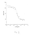
- a selective A 1 adenosine receptor agonist, R-N 6 -isopropyladenosine (R-PIA) displaces binding of the highly selective A 1 adenosine receptor antagonist radioligand, [ 3 H]-1,3-dipropyl-8-cyclopentyladenosine xanthine [ 3 H]DPCPX.
- R-PIA R-N 6 -isopropyladenosine
- CV 1808 did not displace bound [ 3 H]DPCPX.
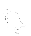
- LPS displaces the highly selective A 1 adenosine receptor antagonist ligand [ 3 H] DPCPX in a dose-dependent manner in membranes prepared from sheep and hamster brains and cultured feline PAECs (with and without hypoxia).
- the lowest sensitivity of the assay for LPS in saline and plasma is in hamster brain membranes at 100 pg/ml.
- serum pretreated with PCA had no effect on [ 3 H]DPCPX binding.
- the Kd (dissociation constant or receptor affinity) and Bmax (receptor density) of adenosine A 1 receptors for the selective A 1 adenosine receptor antagonist [ 3 H] DPCPX were determined in saturation experiments in both sheep and hamster brains and in feline PAECs with hypoxia and without hypoxia (also referred to herein as normoxia). Results from these experiments are shown in Table 1. Saturation Experiments with [ 3 H]-DPCPX in Membranes from Sheep and Hamster Brains and Feline PAECs Bmax (fmoles/mg protein) Kd (nM) Sheep Brain 2251 0.37 Hamster Brain 3733 0.32 Cell Normoxia 1064 0.33 Cell Hypoxia 1458 0.26
- coli 0111:B4 for [3H] DPCPX binding in hamster brains could be completely prevented by prior treatment with pertussis toxin (100 ng/ml).
- the EC 50 for E. coli 0111:B4 for [3H]DPCPX binding in hamster brains was increased by prior treatment with a nonhydrolyzable GTP analog [GppNHp (100 ⁇ M)](0.38 ⁇ g/ml without Gpp NHP versus 0.9 ⁇ g/ml in the presence of Gpp NHp).
- Endotoxin did not bind to dopamine (D2) receptors in competition experiments with [3H] sulpride in hamster brain membranes.
- the sensitivity of the assay of the present invention is verified in competition experiments with known concentrations of endotoxin (1 pg/ml to 1 ⁇ g/ml) from 4 different species of LPS: E. coli 055:B5; E. coli 0111:B4, Serratia marcescens , and Salmonella typhimurium in saline or plasma in the presence of [ 3 H] DPCPX (0.2-0.5 nM) or [ 3 H] CCPA (0.2 - 0.5 nM).
- the lowest sensitivity of the radioligand binding assay using [ 125 I]BW-A844U (0.2 nm) and hamster brain membranes to detect endotoxin LPS, E.
- coli 0111:B4 was 2 to 5 pg/ml.
- the lowest sensitivity of the assay with [ 3 H]DPCPX and hamster brain membranes or membranes cf Sf9 cells expressing rat brain A, adenosine receptors to detect endotoxin was 0.5 pg/ml or 0.1 to 1.0 pg/ml, respectively. Lyophilization of the Sf9 cell membranes did not affect the sensitivity of the assay.
- Radioligand binding data were analyzed by nonlinear regression analysis equipped with a statistical package. These data are then used as standard curves for the determination of endotoxin concentrations in samples.
- Sensitivity and specificity of the radicligand binding assay for endotoxin or the LAL plus chromogenic substrate endotoxin assay are determined.
- the influences of interfering substances on the standard curves for endotoxin in the radioligand binding assay or LAL plus chromogenic substrate assay are also determined.
- Substances commonly present as preservatives in pharmaceutical solutions such as propylene gylcol (5%), phenol (0.01%), thimerosal (0.01%), benzyl alcohol (1%), chlorobutanol (0.5%), methylparaben (0.2%) and benzyalkonium chloride (0.01%) were also tested using [ 1 H]DPCPX and lyophilized membranes from Sf9 cells and found to have no interfering effect as determined by assay sensitivity.
- kits for the detection of endotoxin in a sample and diagnosis of septicemia in an animal.
- kits are provided for measuring the displacement of a detectably labeled A 1 adenosine receptor agent.
- the kit comprises a source of A 1 adenosine receptors such as membranes containing A 1 adenosine receptors or purified A 1 adenosine receptors, a detectably labeled A 1 adenosine receptor agent, and endotoxin standards.
- the kits may also comprise perchloric acid, dilution buffers such as phosphate buffered saline or Tris-HCl, non-pyrogenic filters, pipettes and test tubes.
- kits are provided for measuring endotoxin by RIA or ELISA.
- the kits comprise a solid phase support coated with a first binding partner capable of binding endotoxin, a means for detecting endotoxin bound to the first binding partner, and endotoxin standards.
- Frozen sheep brain (Pel Freeze, Rodgers, Arkansas) or hamster brain (Harlan Bioproducts, Indianapolis, Indiana) was thawed and homogenized in 10 volumes of cold (4°C), 0.32 M sucrose. The homogenate was centrifuged at 1,000 x g for 10 minutes to remove the nuclear fraction. The supernatant was centrifuged at 30,000 x g for 30 minutes. The resulting pellet was resuspended in 10 ml water and left on ice for 30 minutes to obtain synaptosomal membranes.
- the suspension was then centrifuged at 48,000 x g for 10 minutes and the membranes resuspended in 50 mM Tris-HCl (pH 7.4) at a concentration of 8 mg protein/ml, frozen in liquid nitrogen, and stored at -20°C until binding assay.
- Feline PAECs were grown to confluency in 75 mm 2 culture plates.
- the confluent cells were washed twice with KSP buffer (10 mM KH 2 P0 4 , 50 mM sucrose).
- the cells were scraped and suspended in KSP buffer at a concentration of 1 x 10 6 cells/ml in two conical centrifuge tubes. The tubes were centrifuged at 1000 g x 2 minutes and the buffer discarded.
- the cells from one tube were then suspended in deoxygenated buffer (bubbled with N 2 gas for 10 minutes) and bubbled with N 2 gas for 30 seconds to insure hypoxic conditions. These cells were then incubated for 2 hours in this hypoxic buffer.
- Cells from the other tube were suspended in normal KSP buffer and incubated at 37°C for 2 hours. At the end of incubation, cells were homogenized immediately for membrane preparation.
- the cells were homogenized by sonication and centrifuged at 1000 x g for 10 minutes. The supernatant was centrifuged again at 30, 000 x g for 30 minutes. The membrane fraction was reconstituted with Tris buffer for radioligand binding studies.
- Binding of [ 3 H] DPCPX or [ 3 H] CCPA to membranes was carried out in an assay volume of 500 ⁇ l Tris-HCl, pH 7.4, MgCl 2 (10 mM) (for membranes from feline PAECs only) containing [ 3 H] DPCPX or [ 3 H] CCPA (0.01 to 4 nM).
- Nonspecific binding of [ 3 H] DPCPX or [ 3 H] CCPA was determined in the presence of R-PIA (100 ⁇ M) or theophylline 1 mM), respectively.
- R-PIA 100 ⁇ M
- theophylline 1 mM To remove endogenous adenosine, adenosine deaminase was present in all binding assays at a concentration of 0.2 U/ml.
- Incubations were performed for 2 hours at room temperature for sheep and hamster brain membranes and 400 for feline PAEC membranes. Incubations were terminated by rapid removal of the incubation buffer by filtration through Whatman GF/B filters (Whatman Inc., Clifton, NJ). The membrane bound radioactivity was measured by scintillation counting of the filter paper. To determine Kd, Bmax, and nonspecific binding in sheep and hamster brain and feline PAEC membranes, experiments are performed in duplicate.
- Radioligand binding data are analyzed with the use of Graph Pad Prism by nonlinear regression analysis equipped with a statistical package. Data are expressed as mean ⁇ SEM. Hill plots and Schild plots are determined for each species of LPS in competition experiments and are used as standard curves for the determinations of the concentration of endotoxin in samples with unknown concentrations of endotoxin.
- Saline is spiked with known concentrations of different species of LPS: E. coli 055:B5; E. coli 0111:B4, Serratia marcescens , and Salmonella typhimurium to produce a final concentration of 0, 1 pg/ml, 10 pg/ml, 100 pg/ml, 1 ng/ml, 10 ng/ml, 100 ng/ml, 1 ⁇ g/ml and 10 ⁇ g/ml LPS to construct standard curves for Radioligand Binding Endotoxin Assay, as described in Examples 2-4, or LAL plus Chromogenic Endotoxin Assay.
- Blood from humans (5 ml) and animals (2 ml) is obtained in endotoxin-free syringes and anticoagulated test tubes containing endotoxin-free heparin (4 IU/ml) (Endo Tube ET, Chromogenix AB, Molndal, Sweden) and cooled in an ice bath immediately after obtaining the sample.
- the blood is centrifuged at 150 x g for 10 minutes at 4°C and 0.5 - 1.0 ml of the plasma layer is removed in a sterile manner using disposable pipettes (Steriltips, 1000 11L, Eppendorf, Darmstadt, Germany) and stored in LPS-free, sterile polypropylene tubes (Rorchen, 115271, 12.0/75 mm, Greiner Labortechnik, Frickenhausen, Germany) at -23°C until assay.
- a sample of plasma is spiked with known concentrations of different species of LPS: E. coli 055:B5; E.
- coli 0111:B4, Serratia marcescens , and Salmonella typhimurium to produce a final concentration of 0, 1 pg/ml, 10 pg/ml, 100 pg/ml, 1 ng/ml, 10 ng/ml, 100 ng/ml, 1 ⁇ g/ml and 10 ⁇ g/ml LPS and placed in an ice bath to construct standard curves for Radioligand Binding Endotoxin Assay or LAL, Chromogenic Endotoxin Assay. Perchloric acid precipitation of plasma to eliminate factors which interfere with LAL or radioligand binding assay is carried out as described by Obayashi, T. J. Lab. Clin. Med. 1984 , 104:321-330.
- PCA perchloric acid
- Blood from humans (5 ml) and animals (2 ml) suspected of having septicemia is collected in endotoxin-free syringes and anticoagulated test tubes containing endotoxin-free heparin (4 IU/ml) (Endo Tube ET, Chromogenix AB, Molndal, Sweden) and cooled in an ice bath immediately after obtaining the sample.
- the blood is centrifuged at 150 x g for 10 minutes at 4°C and 0.5 - 1.0 ml of the plasma layer is removed in a sterile manner using disposable pipettes (Steriltips, 1000 Aisle, Eppendorf, Darmstadt, Germany) and stored in LPS-free, sterile polypropylene tubes (Rorchen, 115271, 12.0/75 mm, Greiner labortechnik, Frickenhausen, Germany) at -23°C until assayed for the presence of endotoxin according to the methods described herein.
Landscapes
- Life Sciences & Earth Sciences (AREA)
- Health & Medical Sciences (AREA)
- Engineering & Computer Science (AREA)
- Immunology (AREA)
- Molecular Biology (AREA)
- Biomedical Technology (AREA)
- Chemical & Material Sciences (AREA)
- Hematology (AREA)
- Urology & Nephrology (AREA)
- Food Science & Technology (AREA)
- General Physics & Mathematics (AREA)
- Cell Biology (AREA)
- Biotechnology (AREA)
- Medicinal Chemistry (AREA)
- Physics & Mathematics (AREA)
- Analytical Chemistry (AREA)
- Biochemistry (AREA)
- General Health & Medical Sciences (AREA)
- Microbiology (AREA)
- Pathology (AREA)
- Proteomics, Peptides & Aminoacids (AREA)
- Tropical Medicine & Parasitology (AREA)
- Virology (AREA)
- Measuring Or Testing Involving Enzymes Or Micro-Organisms (AREA)
- Investigating Or Analysing Biological Materials (AREA)
- Medicines Containing Antibodies Or Antigens For Use As Internal Diagnostic Agents (AREA)
- Peptides Or Proteins (AREA)
Claims (7)
- Verfahren zur Bestimmung von Endotoxingehalten in einer Probe, umfassend:(a) Binden eines A1-Adenosinrezeptor-Agens an A1-Adenosinrezeptoren;(b) Inkontaktbringen des gebundenen Agens und der A1-Adenosinrezeptoren mit einer Probe, so dass alles Endotoxin in der Probe das gebundene Agens durch Binden an die A1-Adenosinrezeptoren verdrängt; und(c) Messung der Menge des verdrängten Agens.
- Verfahren nach Anspruch 1, worin das A1-Adenosinrezeptor-Agens ein A1-Adenosinrezeptor-Antagonist ist.
- Verfahren nach Anspruch 1, worin das A1-Adenosinrezeptor-Agens ein A1-Adenosinrezeptor-Agonist ist.
- Verfahren zur Diagnose von Blutvergiftung in einem Tier, umfassend:(a) Binden eines A1-Adenosinrezeptor-Agens an A1-Adenosinrezeptoren;(b) Inkontaktbringen des gebundenen Agens und der A1-Adenosinrezeptoren mit einer Probe aus einem Tier, das im Verdacht steht, Blutvergiftung zu haben, so dass alles Endotoxin in der Probe das gebundene Agens durch Binden an die A1-Adenosinrezeptoren verdrängt;(c) Messung der Menge des verdrängten Agens; und(d) Bestimmung des Endotoxingehalts in der Probe, so dass eine Blutvergiftung diagnostiziert werden kann.
- Kit für den Nachweis von Endotoxin in einer Probe, umfassend:(a) eine A1-Adenosinrezeptorquelle;(b) ein nachweisbar markiertes A1-Adenosinrezeptor-Agens; und(c) Endotoxinstandards.
- Verfahren zur Bestimmung von Endotoxingehalten in einer Probe, umfassend:(a) Beschichten eines Festphasenträgers mit einem ersten Bindungspartner, der in der Lage ist, Endotoxin zu immobilisieren;(b) Inkontaktbringen des Festphasenträgers mit einer Probe, die im Verdacht steht, Endotoxin zu enthalten; und(c) Inkontaktbringen des Festphasenträgers mit einem Reagens zum Nachweis von Endotoxin, das auf dem Festphasenträger immobilisiert ist, wobei der erste Bindungspartner oder das Reagens zum Nachweis von immobilisiertem Endotoxin aus der Gruppe ausgewählt ist, die aus A1-Adenosinrezeptoren, A1-Adenosinrezeptor-Lipopolysaccharid-Komplexen oder monoklonalen Antikörpern gegen A1-Adenosinrezeptoren oder A1-Adenosinrezeptor-Lipopolysaccharid-Komplexe besteht.
- Kit für den Nachweis von Endotoxin, umfassend:worin der erste Bindungspartner oder das Reagens zum Nachweis von immobilisiertem Endotoxin aus der Gruppe ausgewählt ist, die aus A1-Adenosinrezeptoren, A1-Adenosinrezeptor-Lipopolysaccharid-Komplexen oder monoklonalen Antikörpern gegen A1-Adenosinrezeptoren oder A1-Adenosinrezeptor-Lipopolysaccharid-Komplexe besteht.(a) einen Festphasenträger, beschichtet mit einem ersten Bindungspartner, der in der Lage ist, Endotoxin zu immobilisieren;(b) ein Reagens zum Nachweis des auf dem Festphasenträger immobilisierten Endotoxins; und(c) Endotoxinstandards,
Applications Claiming Priority (3)
| Application Number | Priority Date | Filing Date | Title |
|---|---|---|---|
| US08/652,928 US5773306A (en) | 1996-05-24 | 1996-05-24 | Methods and kits for the detection of endotoxin |
| US652928 | 1996-05-24 | ||
| PCT/US1997/008754 WO1997044665A1 (en) | 1996-05-24 | 1997-05-27 | Methods and kits for the detection of endotoxin |
Publications (3)
| Publication Number | Publication Date |
|---|---|
| EP0906574A1 EP0906574A1 (de) | 1999-04-07 |
| EP0906574A4 EP0906574A4 (de) | 2001-01-03 |
| EP0906574B1 true EP0906574B1 (de) | 2004-04-14 |
Family
ID=24618782
Family Applications (1)
| Application Number | Title | Priority Date | Filing Date |
|---|---|---|---|
| EP97926695A Expired - Lifetime EP0906574B1 (de) | 1996-05-24 | 1997-05-27 | Verfahren und sets zur detektion von endotoxinen |
Country Status (9)
| Country | Link |
|---|---|
| US (2) | US5773306A (de) |
| EP (1) | EP0906574B1 (de) |
| JP (1) | JP3197280B2 (de) |
| AT (1) | ATE264508T1 (de) |
| AU (1) | AU3140497A (de) |
| CA (1) | CA2253236C (de) |
| DE (1) | DE69728665T2 (de) |
| ES (1) | ES2216154T3 (de) |
| WO (1) | WO1997044665A1 (de) |
Cited By (1)
| Publication number | Priority date | Publication date | Assignee | Title |
|---|---|---|---|---|
| DE102004025780A1 (de) * | 2004-05-26 | 2005-12-22 | Stief, Thomas, Dr.med. | Verfahren zur Anti-Limulus-Faktoren-unabhängigen Bestimmung der Lipopolysaccharid- und/oder Lipid-A- und/oder Glukan-Reaktivität, dafür geeignetes Testsystem sowie dessen Verwendung |
Families Citing this family (10)
| Publication number | Priority date | Publication date | Assignee | Title |
|---|---|---|---|---|
| US5733916A (en) * | 1995-03-24 | 1998-03-31 | The Trustees Of The University Of Pennsylvania | Prevention and treatment of ischemia-reperfusion and endotoxin-related injury using adenosine and purino receptor antagonists |
| US5773306A (en) * | 1996-05-24 | 1998-06-30 | Trustees Of The University Of Pennsylvania | Methods and kits for the detection of endotoxin |
| GB9807814D0 (en) * | 1998-04-09 | 1998-06-10 | Allied Therapeutics Limited | Solid phase test for endoxin |
| US6790661B1 (en) * | 1999-07-16 | 2004-09-14 | Verax Biomedical, Inc. | System for detecting bacteria in blood, blood products, and fluids of tissues |
| WO2002095391A1 (en) * | 2001-05-24 | 2002-11-28 | Endacea Inc. | Methods and formulations for increasing the affinity of a1 adenosine receptor ligands for the a1 adenosine receptor |
| US20040121406A1 (en) * | 2002-05-23 | 2004-06-24 | Wilson Constance Neely | Methods and formulations for increasing the affinity of a1 adenosine receptor ligands for the a1 adenosine receptor |
| DE10247430A1 (de) * | 2002-10-11 | 2004-04-29 | Fresenius Hemocare Gmbh | Verfahren zur Bestimmung des Gehalts an Endotoxinen in Flüssigkeiten |
| JP6043288B2 (ja) * | 2010-09-15 | 2016-12-14 | エンダセア, インコーポレイテッド | 時間分解蛍光ベースのアッセイによるリポ多糖の測定のための使用方法およびキット |
| US20140295477A1 (en) * | 2011-09-01 | 2014-10-02 | Fresenius Medical Care Holdings, Inc | Kit For Sampling And Detection Of Endotoxin In Aqueous Solution |
| DE102014223430B4 (de) * | 2014-11-17 | 2022-03-03 | Fraunhofer-Gesellschaft zur Förderung der angewandten Forschung e.V. | Kompetitives Immunassay-Testsystem zum Nachweis eines Pyrogens |
Family Cites Families (2)
| Publication number | Priority date | Publication date | Assignee | Title |
|---|---|---|---|---|
| DK255887D0 (da) * | 1987-05-20 | 1987-05-20 | Claus Koch | Immunoassay |
| US5773306A (en) * | 1996-05-24 | 1998-06-30 | Trustees Of The University Of Pennsylvania | Methods and kits for the detection of endotoxin |
-
1996
- 1996-05-24 US US08/652,928 patent/US5773306A/en not_active Expired - Fee Related
-
1997
- 1997-05-27 AT AT97926695T patent/ATE264508T1/de not_active IP Right Cessation
- 1997-05-27 AU AU31404/97A patent/AU3140497A/en not_active Abandoned
- 1997-05-27 CA CA002253236A patent/CA2253236C/en not_active Expired - Fee Related
- 1997-05-27 JP JP54276197A patent/JP3197280B2/ja not_active Expired - Fee Related
- 1997-05-27 ES ES97926695T patent/ES2216154T3/es not_active Expired - Lifetime
- 1997-05-27 EP EP97926695A patent/EP0906574B1/de not_active Expired - Lifetime
- 1997-05-27 DE DE69728665T patent/DE69728665T2/de not_active Expired - Fee Related
- 1997-05-27 WO PCT/US1997/008754 patent/WO1997044665A1/en not_active Ceased
-
2002
- 2002-06-12 US US10/137,004 patent/US6908742B2/en not_active Expired - Fee Related
Cited By (2)
| Publication number | Priority date | Publication date | Assignee | Title |
|---|---|---|---|---|
| DE102004025780A1 (de) * | 2004-05-26 | 2005-12-22 | Stief, Thomas, Dr.med. | Verfahren zur Anti-Limulus-Faktoren-unabhängigen Bestimmung der Lipopolysaccharid- und/oder Lipid-A- und/oder Glukan-Reaktivität, dafür geeignetes Testsystem sowie dessen Verwendung |
| DE102004025780B4 (de) * | 2004-05-26 | 2008-04-03 | Stief, Thomas, Dr.med. | Verfahren zur Anti-Limulus-Faktoren-unabhängigen Bestimmung der Lipopolysaccharid- und/oder Lipid-A- und/oder Glukan-Reaktivität, dafür geeignetes Testsystem sowie dessen Verwendung |
Also Published As
| Publication number | Publication date |
|---|---|
| EP0906574A1 (de) | 1999-04-07 |
| WO1997044665A1 (en) | 1997-11-27 |
| EP0906574A4 (de) | 2001-01-03 |
| CA2253236C (en) | 2004-05-25 |
| US6908742B2 (en) | 2005-06-21 |
| ATE264508T1 (de) | 2004-04-15 |
| DE69728665T2 (de) | 2004-09-30 |
| JP3197280B2 (ja) | 2001-08-13 |
| US20020182641A1 (en) | 2002-12-05 |
| ES2216154T3 (es) | 2004-10-16 |
| CA2253236A1 (en) | 1997-11-27 |
| US5773306A (en) | 1998-06-30 |
| JPH11510904A (ja) | 1999-09-21 |
| AU3140497A (en) | 1997-12-09 |
| DE69728665D1 (de) | 2004-05-19 |
Similar Documents
| Publication | Publication Date | Title |
|---|---|---|
| EP0615129B1 (de) | Verfahren zum selektiven Nachweis von perinuklearen anti-neutrophilen cytoplasmischen Antikörpern bei ulzerativen Kolitis oder primärer sclerotischer Cholangitis | |
| US5051356A (en) | Specific binding composition comprising a low pI protein or carbohydrate and a diagnostic test kit and method of use | |
| AU685487B2 (en) | Reagent for endotoxin-specific assay | |
| EP0279517B1 (de) | Nichtimmunochemische Bindung von Lipopolysacchariden und Sandwich-Bestimmung dafür | |
| EP0906574B1 (de) | Verfahren und sets zur detektion von endotoxinen | |
| Henshaw et al. | Elevations of neutrophil proteinase 3 in serum of patients with Wegener's granulomatosis and polyarteritis nodosa | |
| JP2944721B2 (ja) | エンドトキシンの測定剤 | |
| WO1997044665A9 (en) | Methods and kits for the detection of endotoxin | |
| NO159820B (no) | Fremgangsmaate ved bestemmelse av antigen-antistoff-reaksjoner. | |
| DK174032B1 (da) | Sæt samt fremgangsmåde til immunometrisk dosering, der kan anvendes på hele celler | |
| US7462495B2 (en) | Methods and compositions for use in diagnosing and characterizing chronic immune disease | |
| CN113777326A (zh) | 一种高特异检测肝素结合蛋白的试剂盒及其应用 | |
| US5338684A (en) | Stable aqueous FK506 standards | |
| CN113588939A (zh) | 一种肝素结合蛋白测定试剂盒、制备方法及使用方法 | |
| JP3698563B2 (ja) | グリコサミノグリカンの測定方法及び測定キット | |
| CN102292639B (zh) | 检测抗睾丸抗原自身抗体的免疫测试 | |
| CN113252909B (zh) | 一种基于量子点免疫荧光检测试剂盒制备方法 | |
| WO1990010232A1 (en) | Composition containing labeled streptococcal antibody, test kit and assay using same | |
| US8076088B2 (en) | Method of detecting human β-defensins | |
| CN113109574A (zh) | 检测抗突变型瓜氨酸化波形蛋白抗体的试剂盒以及检测方法 | |
| JP2000111553A (ja) | 正常アグリカン測定法とその応用 | |
| JP4588053B2 (ja) | 酸化アポリポタンパク質ai及びそれを含有する酸化リポタンパク質の測定法及びキット | |
| CN108414775A (zh) | 中性粒细胞明胶酶相关脂质运载蛋白检测试剂盒 | |
| JPH1194836A (ja) | 慢性関節リウマチ用マーカーとしての使用および慢性関節リウマチ診断用免疫試薬 | |
| JP2000074913A (ja) | IgA腎症の検査方法 |
Legal Events
| Date | Code | Title | Description |
|---|---|---|---|
| PUAI | Public reference made under article 153(3) epc to a published international application that has entered the european phase |
Free format text: ORIGINAL CODE: 0009012 |
|
| 17P | Request for examination filed |
Effective date: 19981123 |
|
| AK | Designated contracting states |
Kind code of ref document: A1 Designated state(s): AT BE CH DE DK ES FI FR GB GR IE IT LI LU MC NL PT SE |
|
| A4 | Supplementary search report drawn up and despatched |
Effective date: 20001117 |
|
| AK | Designated contracting states |
Kind code of ref document: A4 Designated state(s): AT BE CH DE DK ES FI FR GB GR IE IT LI LU MC NL PT SE |
|
| 17Q | First examination report despatched |
Effective date: 20011220 |
|
| GRAH | Despatch of communication of intention to grant a patent |
Free format text: ORIGINAL CODE: EPIDOS IGRA |
|
| GRAS | Grant fee paid |
Free format text: ORIGINAL CODE: EPIDOSNIGR3 |
|
| GRAA | (expected) grant |
Free format text: ORIGINAL CODE: 0009210 |
|
| AK | Designated contracting states |
Kind code of ref document: B1 Designated state(s): AT BE CH DE DK ES FI FR GB GR IE IT LI LU MC NL PT SE |
|
| PG25 | Lapsed in a contracting state [announced via postgrant information from national office to epo] |
Ref country code: NL Free format text: LAPSE BECAUSE OF FAILURE TO SUBMIT A TRANSLATION OF THE DESCRIPTION OR TO PAY THE FEE WITHIN THE PRESCRIBED TIME-LIMIT Effective date: 20040414 Ref country code: FI Free format text: LAPSE BECAUSE OF FAILURE TO SUBMIT A TRANSLATION OF THE DESCRIPTION OR TO PAY THE FEE WITHIN THE PRESCRIBED TIME-LIMIT Effective date: 20040414 Ref country code: BE Free format text: LAPSE BECAUSE OF FAILURE TO SUBMIT A TRANSLATION OF THE DESCRIPTION OR TO PAY THE FEE WITHIN THE PRESCRIBED TIME-LIMIT Effective date: 20040414 |
|
| REG | Reference to a national code |
Ref country code: GB Ref legal event code: FG4D |
|
| REG | Reference to a national code |
Ref country code: CH Ref legal event code: EP |
|
| REF | Corresponds to: |
Ref document number: 69728665 Country of ref document: DE Date of ref document: 20040519 Kind code of ref document: P |
|
| REG | Reference to a national code |
Ref country code: IE Ref legal event code: FG4D |
|
| PG25 | Lapsed in a contracting state [announced via postgrant information from national office to epo] |
Ref country code: LU Free format text: LAPSE BECAUSE OF NON-PAYMENT OF DUE FEES Effective date: 20040527 Ref country code: IE Free format text: LAPSE BECAUSE OF NON-PAYMENT OF DUE FEES Effective date: 20040527 |
|
| PG25 | Lapsed in a contracting state [announced via postgrant information from national office to epo] |
Ref country code: MC Free format text: LAPSE BECAUSE OF NON-PAYMENT OF DUE FEES Effective date: 20040531 |
|
| REG | Reference to a national code |
Ref country code: CH Ref legal event code: NV Representative=s name: R. A. EGLI & CO. PATENTANWAELTE |
|
| PG25 | Lapsed in a contracting state [announced via postgrant information from national office to epo] |
Ref country code: SE Free format text: LAPSE BECAUSE OF FAILURE TO SUBMIT A TRANSLATION OF THE DESCRIPTION OR TO PAY THE FEE WITHIN THE PRESCRIBED TIME-LIMIT Effective date: 20040714 Ref country code: GR Free format text: LAPSE BECAUSE OF FAILURE TO SUBMIT A TRANSLATION OF THE DESCRIPTION OR TO PAY THE FEE WITHIN THE PRESCRIBED TIME-LIMIT Effective date: 20040714 Ref country code: DK Free format text: LAPSE BECAUSE OF FAILURE TO SUBMIT A TRANSLATION OF THE DESCRIPTION OR TO PAY THE FEE WITHIN THE PRESCRIBED TIME-LIMIT Effective date: 20040714 |
|
| NLV1 | Nl: lapsed or annulled due to failure to fulfill the requirements of art. 29p and 29m of the patents act | ||
| REG | Reference to a national code |
Ref country code: ES Ref legal event code: FG2A Ref document number: 2216154 Country of ref document: ES Kind code of ref document: T3 |
|
| ET | Fr: translation filed | ||
| PLBE | No opposition filed within time limit |
Free format text: ORIGINAL CODE: 0009261 |
|
| STAA | Information on the status of an ep patent application or granted ep patent |
Free format text: STATUS: NO OPPOSITION FILED WITHIN TIME LIMIT |
|
| REG | Reference to a national code |
Ref country code: IE Ref legal event code: MM4A |
|
| 26N | No opposition filed |
Effective date: 20050117 |
|
| REG | Reference to a national code |
Ref country code: HK Ref legal event code: WD Ref document number: 1018811 Country of ref document: HK |
|
| PG25 | Lapsed in a contracting state [announced via postgrant information from national office to epo] |
Ref country code: PT Free format text: LAPSE BECAUSE OF NON-PAYMENT OF DUE FEES Effective date: 20040914 |
|
| PGFP | Annual fee paid to national office [announced via postgrant information from national office to epo] |
Ref country code: ES Payment date: 20080619 Year of fee payment: 12 Ref country code: DE Payment date: 20080605 Year of fee payment: 12 Ref country code: CH Payment date: 20080527 Year of fee payment: 12 |
|
| PGFP | Annual fee paid to national office [announced via postgrant information from national office to epo] |
Ref country code: AT Payment date: 20080514 Year of fee payment: 12 |
|
| PGFP | Annual fee paid to national office [announced via postgrant information from national office to epo] |
Ref country code: IT Payment date: 20080530 Year of fee payment: 12 |
|
| PGFP | Annual fee paid to national office [announced via postgrant information from national office to epo] |
Ref country code: GB Payment date: 20080528 Year of fee payment: 12 |
|
| REG | Reference to a national code |
Ref country code: CH Ref legal event code: PL |
|
| GBPC | Gb: european patent ceased through non-payment of renewal fee |
Effective date: 20090527 |
|
| PG25 | Lapsed in a contracting state [announced via postgrant information from national office to epo] |
Ref country code: LI Free format text: LAPSE BECAUSE OF NON-PAYMENT OF DUE FEES Effective date: 20090531 Ref country code: CH Free format text: LAPSE BECAUSE OF NON-PAYMENT OF DUE FEES Effective date: 20090531 Ref country code: AT Free format text: LAPSE BECAUSE OF NON-PAYMENT OF DUE FEES Effective date: 20090527 |
|
| REG | Reference to a national code |
Ref country code: FR Ref legal event code: ST Effective date: 20100129 |
|
| PG25 | Lapsed in a contracting state [announced via postgrant information from national office to epo] |
Ref country code: FR Free format text: LAPSE BECAUSE OF NON-PAYMENT OF DUE FEES Effective date: 20090602 |
|
| PGFP | Annual fee paid to national office [announced via postgrant information from national office to epo] |
Ref country code: FR Payment date: 20080514 Year of fee payment: 12 |
|
| PG25 | Lapsed in a contracting state [announced via postgrant information from national office to epo] |
Ref country code: GB Free format text: LAPSE BECAUSE OF NON-PAYMENT OF DUE FEES Effective date: 20090527 |
|
| PG25 | Lapsed in a contracting state [announced via postgrant information from national office to epo] |
Ref country code: DE Free format text: LAPSE BECAUSE OF NON-PAYMENT OF DUE FEES Effective date: 20091201 |
|
| REG | Reference to a national code |
Ref country code: ES Ref legal event code: FD2A Effective date: 20090528 |
|
| PG25 | Lapsed in a contracting state [announced via postgrant information from national office to epo] |
Ref country code: ES Free format text: LAPSE BECAUSE OF NON-PAYMENT OF DUE FEES Effective date: 20090528 |
|
| PG25 | Lapsed in a contracting state [announced via postgrant information from national office to epo] |
Ref country code: IT Free format text: LAPSE BECAUSE OF NON-PAYMENT OF DUE FEES Effective date: 20090527 |