EP0865609B1 - Verfahren zur diagnose und therapie von plattenepithelkarzinomen - Google Patents
Verfahren zur diagnose und therapie von plattenepithelkarzinomen Download PDFInfo
- Publication number
- EP0865609B1 EP0865609B1 EP96942362A EP96942362A EP0865609B1 EP 0865609 B1 EP0865609 B1 EP 0865609B1 EP 96942362 A EP96942362 A EP 96942362A EP 96942362 A EP96942362 A EP 96942362A EP 0865609 B1 EP0865609 B1 EP 0865609B1
- Authority
- EP
- European Patent Office
- Prior art keywords
- antibody
- lymph node
- node metastasis
- larynx
- tumor
- Prior art date
- Legal status (The legal status is an assumption and is not a legal conclusion. Google has not performed a legal analysis and makes no representation as to the accuracy of the status listed.)
- Expired - Lifetime
Links
Images


Classifications
-
- G—PHYSICS
- G01—MEASURING; TESTING
- G01N—INVESTIGATING OR ANALYSING MATERIALS BY DETERMINING THEIR CHEMICAL OR PHYSICAL PROPERTIES
- G01N33/00—Investigating or analysing materials by specific methods not covered by groups G01N1/00 - G01N31/00
- G01N33/48—Biological material, e.g. blood, urine; Haemocytometers
- G01N33/50—Chemical analysis of biological material, e.g. blood, urine; Testing involving biospecific ligand binding methods; Immunological testing
- G01N33/53—Immunoassay; Biospecific binding assay; Materials therefor
- G01N33/574—Immunoassay; Biospecific binding assay; Materials therefor for cancer
-
- A—HUMAN NECESSITIES
- A61—MEDICAL OR VETERINARY SCIENCE; HYGIENE
- A61K—PREPARATIONS FOR MEDICAL, DENTAL OR TOILETRY PURPOSES
- A61K51/00—Preparations containing radioactive substances for use in therapy or testing in vivo
- A61K51/02—Preparations containing radioactive substances for use in therapy or testing in vivo characterised by the carrier, i.e. characterised by the agent or material covalently linked or complexing the radioactive nucleus
- A61K51/04—Organic compounds
- A61K51/08—Peptides, e.g. proteins, carriers being peptides, polyamino acids, proteins
- A61K51/10—Antibodies or immunoglobulins; Fragments thereof, the carrier being an antibody, an immunoglobulin or a fragment thereof, e.g. a camelised human single domain antibody or the Fc fragment of an antibody
- A61K51/1027—Antibodies or immunoglobulins; Fragments thereof, the carrier being an antibody, an immunoglobulin or a fragment thereof, e.g. a camelised human single domain antibody or the Fc fragment of an antibody against receptors, cell-surface antigens or cell-surface determinants
-
- A—HUMAN NECESSITIES
- A61—MEDICAL OR VETERINARY SCIENCE; HYGIENE
- A61P—SPECIFIC THERAPEUTIC ACTIVITY OF CHEMICAL COMPOUNDS OR MEDICINAL PREPARATIONS
- A61P35/00—Antineoplastic agents
-
- C—CHEMISTRY; METALLURGY
- C07—ORGANIC CHEMISTRY
- C07K—PEPTIDES
- C07K14/00—Peptides having more than 20 amino acids; Gastrins; Somatostatins; Melanotropins; Derivatives thereof
- C07K14/435—Peptides having more than 20 amino acids; Gastrins; Somatostatins; Melanotropins; Derivatives thereof from animals; from humans
- C07K14/705—Receptors; Cell surface antigens; Cell surface determinants
- C07K14/70585—CD44
-
- C—CHEMISTRY; METALLURGY
- C07—ORGANIC CHEMISTRY
- C07K—PEPTIDES
- C07K16/00—Immunoglobulins [IGs], e.g. monoclonal or polyclonal antibodies
- C07K16/18—Immunoglobulins [IGs], e.g. monoclonal or polyclonal antibodies against material from animals or humans
- C07K16/28—Immunoglobulins [IGs], e.g. monoclonal or polyclonal antibodies against material from animals or humans against receptors, cell surface antigens or cell surface determinants
- C07K16/2884—Immunoglobulins [IGs], e.g. monoclonal or polyclonal antibodies against material from animals or humans against receptors, cell surface antigens or cell surface determinants against CD44
-
- A—HUMAN NECESSITIES
- A61—MEDICAL OR VETERINARY SCIENCE; HYGIENE
- A61K—PREPARATIONS FOR MEDICAL, DENTAL OR TOILETRY PURPOSES
- A61K2123/00—Preparations for testing in vivo
-
- C—CHEMISTRY; METALLURGY
- C07—ORGANIC CHEMISTRY
- C07K—PEPTIDES
- C07K2317/00—Immunoglobulins specific features
- C07K2317/30—Immunoglobulins specific features characterized by aspects of specificity or valency
- C07K2317/34—Identification of a linear epitope shorter than 20 amino acid residues or of a conformational epitope defined by amino acid residues
Definitions
- the invention relates to methods for diagnosis and therapy of squamous cell carcinoma, which are based on the expression of the variable exon v6 of the CD44 gene, means for such processes and their use.
- CD44 splice variants of CD44
- the CD44 isoforms are generated by alternative splicing in such a way that the sequences of 10 exons (v1-v10) are completely cut out in CD44s, but can occur in different combinations in the larger variants (Screaton et al ., 1992; Heider et al . , 1993; Hofmann et al ., 1991).
- the variants differ in that different amino acid sequences are inserted at a certain point in the extracellular part of the protein. Such variants could be detected in various human tumor cells and in human tumor tissue.
- the expression of CD44 variants in the course of colorectal carcinogenesis has recently been examined (Heider et al ., 1993).
- CD44 variants are absent in normal human colonic epithelium and only weak expression is detectable in the proliferating cells of the crypts. In later stages of tumor progression, for example in adenocarcinomas, all malignancies express variants of CD44. Furthermore, expression of CD44 splice variants in activated lymphocytes and in non-Hodgkin lymphomas has recently been shown (Koopman et al ., 1993).
- the object of the present invention was to develop new processes for Therapy of squamous cell carcinoma and the provision of funds for such procedures.
- This object could be achieved with the present invention. It relates to an object within the scope of claims 1 to 4. It relates to methods for the therapy of squamous cell carcinomas which are based on the expression of the variant exon v6 of the CD44 gene as a molecular marker or target.
- the present invention relates to methods which are based on the strong and homogeneous expression of v6 in squamous cell carcinomas, which could be found surprisingly and in contrast to the teaching as was known from the prior art.
- Antibody molecules with appropriate specificity are particularly suitable as vehicles to selectively reach squamous cell carcinomas in vivo .
- the monoclonal antibody BIWA-1 (clone VFF-18) is particularly preferred is secreted by a hybridoma cell line, which on June 7, 1994 under the accession number DSM ACC2174 at the DSM-German Collection for Microorganisms and Zellkulturen GmbH, Mascheroder Weg 1b, D-38124 Braunschweig, Germany was (WO 95/33771), or derivatives of this antibody.
- the nucleic acid and amino acid sequence of the variant exon v6 of the CD44 gene is known (Screaton et al ., 1992, Tölg et al ., 1993).
- the existence of degenerate or allelic variants is not important for the implementation of the invention; such variants are therefore expressly included.
- the sequence of exon v6 of the human CD44 gene is:
- the invention can be practiced with polyclonal or monoclonal antibodies specific for an epitope encoded by exon v6, particularly an epitope within the amino acid sequence WFGNRWHEGYR.
- Antibodies against known amino acid sequences can be produced by methods known per se (Catty, 1989). For example, a peptide of this sequence can be synthesized and used as an antigen in an immunization protocol.
- Another way is to produce a fusion protein which contains the desired amino acid sequence by integrating a nucleic acid (which can be prepared synthetically or, for example, from a suitable sample, for example by polymerase chain reaction (PCR)), which codes for this sequence, into an expression vector and the fusion protein is expressed in a host organism.
- PCR polymerase chain reaction
- the optionally purified fusion protein can then be used as an antigen in an immunization protocol and insert-specific antibodies or, in the case of monoclonal antibodies, hybridomas which express insert-specific antibodies can be selected using suitable methods.
- suitable methods are state of the art. Heider et al . (1993, 1996a) and Koopman et al . (1993) describe the production of antibodies against variant epitopes of CD44.
- antibody molecules derived from poly- or monoclonal antibodies can also be used for the method according to the invention, for example Fab or F (ab ') 2 fragments of immunoglobulins, recombinantly produced single-chain antibodies (scFv), chimeric or humanized antibodies as well as other molecules that bind specifically to epitopes encoded by exon v6.
- Faboder F (ab ') 2 fragments or other fragments can be generated from the complete immunoglobulin of the antibody BIWA-1 (VFF-18) or other antibodies (Kreitman et al ., 1993).
- VFF-18 antibody BIWA-1
- the person skilled in the art is also able to produce recombinant v6-specific antibody molecules.
- Such recombinant antibody molecules can be, for example, humanized antibodies (Shin et al ., 1989; Güssow et Seemann, 1991), bispecific or bifunctional antibodies (Weiner et al ., 1993; Goodwin, 1989, Featherstone, 1996), single - chain antibodies ( scFv , Johnson et Bird, 1991), complete or fragmentary immunoglobulins (Coloma et al ., 1992; Nesbit et al ., 1992; Barbas et al ., 1992), or antibodies generated by chain shuffling (Winter et al ., 1994) , Humanized antibodies can be produced, for example, by CDR grafting (EP 0239400).
- Framework regions can also be modified (EP 0519596; WO 9007861). Methods such as PCR (eg EP 0368684; EP 0438310; WO 9207075) or computer modeling (eg WO 9222653) can be used today for the humanization of antibodies. Fusion proteins, for example single-chain antibody / toxin fusion proteins (Chaudhary et al ., 1990; Friedman et al ., 1993) can also be produced and used. In addition to polyclonal and monoclonal antibodies, the generic terms “antibodies” and “antibody molecules” are intended to include all of the compounds discussed in this section, as well as other compounds that can be structurally derived from immunoglobulins and that can be prepared by methods known per se.
- antibody molecules preferably BIWA-1 antibody molecules, fragments thereof or recombinant antibody molecules with the same idiotype, for example with radioactive isotopes such as 125 I, 131 I, 111 In, 99m Tc or radioactive compounds (Larson et al ., 1991; Thomas et al ., 1989; Srivastava, 1988), enzymes such as peroxidase or alkaline phosphatase (Catty et Raykundalia, 1989), with fluorescent dyes (Johnson, 1989) or biotin molecules (Guesdon et al ., 1979).
- radioactive isotopes such as 125 I, 131 I, 111 In, 99m Tc or radioactive compounds (Larson et al ., 1991; Thomas et al ., 1989; Srivastava, 1988), enzymes such as peroxidase or alkaline phosphatase (Catty et Raykundalia, 1989), with fluorescent dyes (Johnson, 1989) or
- v6-specific antibody molecules preferably BIWA-1 (VFF-18) antibody molecules or VFF-18-derived antibody molecules, for example fragments thereof or recombinant antibody molecules with the same idiotype, with radioisotopes such as 90 Y, 131 I, 186 Re , 188 Re, 153 Sm, 67 Cu, 212 Bi, 213 Bi, 177 Lu (Quadri et al ., 1993; Lenhard et al ., 1985, Vriesendorp et al ., 1991; Wilbur et al ., 1989, Maraveyas et al ., 1995a, Jurcic et Scheinberg, 1994), toxins (Vitetta et al ., 1991; Vitetta et Thorpe, 1991; Kreitman et al ., 1993; Theuer et al ., 1993), cytostatics (Schrappe et al ., 1992) , Prodrugs (Wang et
- the antibody molecule can also be linked to a cytokine or another immunomodulatory polypeptide, for example to tumor necrosis factor, lymphotoxin (Reisfeld et al ., 1996) or interleukin-2 (Becker et al ., 1996).
- the antibody molecules can also be modified, for example with streptavidin or biotin, for use in a pretargeting system (Goodwin, 1995).
- the diagnostic method can advantageously be used to examine samples from patients, for example from biopsies, in whom there is suspicion of squamous cell carcinoma or who have already been diagnosed, but the tumor is to be more precisely characterized.
- Variant CD44 molecules which contain an amino acid sequence which is encoded by the variable exon v6 can be detected at the protein level by means of antibodies or at the nucleic acid level by means of specific nucleic acid probes or primers for the polymerase chain reaction (PCR).
- tissue sections can be examined immunohistochemically with antibodies using methods known per se.
- Extracts or body fluids obtained from tissue samples can also be examined by other immunological methods using antibodies, for example in Western blots, enzyme-linked immunosorbent assays (ELISA, Catty et Raykundalia, 1989), radioimmunoassays (RIA, Catty et Murphy, 1989) or related immunoassays.
- the examinations can be qualitative, semi-quantitative or quantitative.
- antibody molecules with specificity according to the invention are also suitable for in vivo diagnosis of squamous cell carcinoma. If the antibody molecule bears a detectable label, the label can be detected for diagnostic purposes, for example visualization of the tumor in vivo (imaging), or for example for radio-assisted surgery ( radioguided surgery ).
- Antibody molecules with the specificity according to the invention and optionally linked to a cytotoxic agent can advantageously be used for the therapy of squamous cell carcinoma.
- the application can be systemic or topical, for example by intravenous (as a bolus or continuous infusion), intraperitoneal, intramuscular, subcutaneous or similar injection / infusion.
- Protocols for the administration of conjugated or non-conjugated antibodies are state of the art (Mulshine et al ., 1991; Larson et al ., 1991; Vitetta et Thorpe, 1991 ; Vitetta et al ., 1991; Breitz et al ., 1992, 1995; Press et al ., 1989; Weiner et al ., 1989; Chatal et al ., 1989; Sears et al ., 1982).
- a therapeutic application can, for example, be analogous to the use of the antibody 1.1ASML (Thatr et al ., 1993).
- Unmodified monoclonal antibodies can be used directly for therapy if they have the intrinsic effector function suitable for a cytotoxic effect, for example for complement-induced or antibody-induced cellular cytotoxicity (Riethmüller et al . 1994). Suitable monoclonal antibodies for this use are mouse antibodies of the isotype IgG2a or antibodies of the human IgG1 type. Unmodified antibodies can also be applied to induce a patient's own anti-tumor response via an anti-idiotypic mechanism (Baum et al ., 1993; Khazaeli et al ., 1994).
- a preferred embodiment of a therapeutic application is a humanized v6-specific immunoglobulin or an F (ab ') 2 fragment thereof with 90 Y (Quadri et al ., 1993; Vriesendorp et al ., 1995), 131 I (Maraveyas et al ., 1995a, 1995b; Juweid et al . , 1995; Press et al ., 1995; Thomas et al ., in: Catty 1985, pp. 230-239) 186 Re (Breitz et al ., 1992, 1995) or to link another suitable radioisotope and use it for radioimmunotherapy of squamous cell carcinoma.
- This agent can then be administered to a patient with an antigen-positive tumor in a dosage of 0.1 to 1 mCi / kg body weight, preferably 0.3 to 0.5 mCi / kg body weight.
- a possible dosing scheme can be, for example, 2 x 150 mCi every 6 weeks.
- the person skilled in the art can determine the maximum possible dosages using methods known per se (Maraveyas et al ., 1995a, 1995b). With a total amount of protein to be administered of 2 to 5 mg, the administration can take the form of a rapid intravenous bolus injection. For larger amounts of protein, an infusion can be the cheaper form of administration.
- the agent may be necessary to mix the agent with an excess (eg ten times the molar excess) of the non-radioactive antibody before administration; in this case, administration is better in the form of an intravenous infusion, for example over 15 minutes.
- the application can be repeated.
- the therapy can be combined with external radiation therapy. It can also be supported by bone marrow transplantation; this is particularly necessary if the therapy reaches a dose of more than 1.6 Gy in the bone marrow.
- Antibody molecules according to the invention can also be used ex vivo for the purification of CD34-positive stem and precursor cell preparations (immunopurging). Radiation or chemotherapy for squamous cell carcinoma can be supported by autologous bone marrow transplantation. The applied hematopoietic stem and progenitor cells must be free of tumor cells. This can be achieved by incubation with antibody molecules according to the invention, for example antibody-toxin conjugates (Myklebust et al ., 1994; DE P 196 48 209.7).
- Antibody molecules according to the invention can also be introduced into the T cell receptor of T lymphocytes in the form of recombinant constructs.
- Such reprogrammed T lymphocytes selectively bind to the antigen-expressing tumor cells and exert cytotoxic effects, so that they can be used for the therapy of squamous cell carcinoma (PCT / EP9604631; Altenschmidt et al ., 1996).
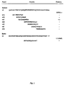
- Figure 1 Determination of the epitope specificity of BIWA-1 by binding to synthetic peptides derived from the human CD44v6 sequence. The corresponding peptide from rat CD44v6 was tested with the antibody 1.1ASML. Binding was determined in an ELISA, the peptides being immobilized on microtiter plates (cf. Heider et al ., 1996b, FIG. 2). -: no bond, +/-: weak bond, +: strong bond.
- Fig. 3 Comparison of the antigen binding of different CD44v6-specific mAbs.
- the binding of four different CD44v6-specific mAbs to human SCC A-431 cells was measured in a cell ELISA.
- MAb BIWA-1 shows a higher affinity for the tumor cells than the other mAbs.
- Fig. 4 Refined epitope mapping of the mAb BIWA-1.
- the binding of BIWA-1 to various overlapping synthetic peptides spanning amino acids 18-32 of the CD44v6-encoded region was measured in a competitive ELISA.
- the minimal binding sequence (peptide v6 (19-29)) is underlined.
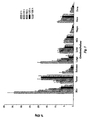
- FIG. 5 Biodistribution of 125 I-BIWA-1 in A-431 xenografted nude mice. The accumulation of the antibody is shown as% ID / g (mean ⁇ SEM) at 4, 24, 48, 120 and 168 h post injection.
- Example 1 Expression of CD44v6 in squamous cell carcinoma
- the entire variant region of the HPKII type of CD44v was amplified from human keratinocyte cDNA by polymerase chain reaction (PCR).
- PCR polymerase chain reaction
- the two PCR primers 5 ' -CAGGCTGGGAGCCAAATGAAGAAAATG- 3' , positions 25-52, and 5 '-TGATAAGGAACGATTGACATTAGAGTTGGA- 3 ', positions 1013-984 of the LCLC97-variant region, as described by Hofinann et al. contained an Eco RI recognition site which was used to clone the PCR product directly into the vector pGEX-2T (Smith et al ., 1988).
- the resulting construct (pGEX CD44v HPKII, v3-v10) codes for a fusion protein of ⁇ 70 kD consisting of glutathione-S-transferase from Schistosoma japonicum and the exons v3-v10 from human CD44 (FIG. 1; Heider et al ., 1993).
- the fusion protein was in E. coli expressed and then affinity purified via glutathione agarose (Smith et al ., 1988).
- the immunizations were carried out every 4 weeks. 14 days after the last immunization, the animals were immunized for three consecutive days with 10 ⁇ g of fusion protein in PBS. The following day, spleen cells from an animal with a high antibody titer were fused with P3.X63-Ag8.653 mouse myeloma cells using polyethylene glycol 4000. The hybridoma cells were then selected in microtiter plates in HAT medium (Köhler et Milstein, 1975; Kearney et al ., 1979).
- the determination of the antibody titer in the serum or the screening of the hybridoma supernatants was carried out using an ELISA.
- microtiter plates were first coated with fusion protein (GST-CD44v3-10) or only with glutathione-S-transferase. It was then incubated with serial dilutions of serum samples or hybridoma supernatants and the specific antibodies were detected with peroxidase-conjugated antibodies against mouse immunoglobulin. Hybridomas that only reacted with glutathione-S-transferase were discarded.
- BIWA-1 (VFF-18; production and properties see also WO 95/33771) only fusion proteins containing a domain encoded by exon v6.
- various synthetic peptides representing the parts of the v6 domain used in ELISA binding assays (Fig. 1).
- the 14 amino acid peptide v6D showed the strongest binding.
- the epitope of BIWA-1 in whole or in part within the sequence QWFGNRWHEGYRQT of the domain, which is encoded by Exon v6. This sequence is homologous to the binding epitope of the antibody 1.1ASML used in a therapeutic rat model and the is specific for rat CD44v6 (Fig. 1).
- paraffin sections (4 ⁇ m) in Rotihistol (Roth, Germany) were deparaffinized 3 times for 10 minutes each and then rehydrated in an ascending alcohol series. The cuts were briefly made with A. dest . washed and then cooked in a microwave oven (Sharp Model R-6270) 3 times for 10 minutes at 600 watts in 0.01 M Na citrate buffer. The sections were cooled for 20 minutes after each microwave incubation. After the final cooling step, the slides were washed in PBS and preincubated with normal goat serum (10% in PBS).
- the sections were incubated for 30 minutes with horseradish peroxidase, which was coupled to biotin as streptavidin-biotin-peroxidase complex (DAKO Corp.). The sections were then incubated in 3,3-amino-9-ethyl-carbazole substrate (Sigma Immunochemicals) for 5-10 minutes, the reaction was stopped with H 2 O and the sections were counterstained with hematoxylin.
- the staining was evaluated with a Zeiss axioscope light microscope and the staining intensities were quantified as follows: +++, strong expression; ++, moderate expression; +, weak expression; -, ambiguous or no expression detectable. Only tumor cells with clear membrane staining were rated positive.
- the percentage of positive tumor cells in each section was roughly estimated and two groups were formed: focal positive tumors (less than 10% of the tumor cells reacted with the antibody) and positive tumors (10 or more% of the tumor cells positive). If less than 80% of the tumor cells in the positive cells reacted with the antibody, the corresponding percentage was displayed.
- CD44v6-specific monoclonal antibody BIWA-1 analyzed.
- Expression CD44v6 Isoforms containing were observed in all but one tumor sample. The majority of the samples showed expression of the antigen on 80-100% of the tumor cells, the staining was limited to the membrane of the tumor cells. With stromal tissue, lymphocytes, No reaction was observed in muscle cells or endothelium.
- the staining reaction was very homogeneous within a given tumor section, with most tumor cells of the section having the same staining intensity. No significant differences in the CD44v6 expression pattern were observed between primary tumors and metastases. A detailed summary of the results is shown in Table 1, examples are shown in FIG. 2.
- the human SCC cell line A-431 (spontaneous epidermoid carcinoma of the vulva) was obtained from the American Type Culture Collection (Rockwell MD) and according to the Manufacturer information bred.
- the surface expression of CD44v6 containing isoforms was determined by FACS analysis using a FITC-linked mAb BIWA-1 has been.
- Antibodies in different concentrations (8-132 nM) in HBS (10 mM Hepes pH 7.4, 150 mM sodium chloride, 3.4 mM EDTA, 0.05% BIA core surfactant P20) was beyond the antigen-specific Surface injected at a flow rate of 5 ul / min. The interaction was called Change in SPR signal recorded. The dissociation of the antibody was carried out for 5 Minutes observed in the buffer flow (HBS). The surface of the chip was scanned with a single pulse regenerated from 15 ⁇ l 30 mM HCl. Analysis of the data and calculation of the kinetic Constant was with the Pharmacia Biosensor BIA Evaluation Software, version 2.1 carried out.
- A-431 cells expressing CD44v6 were cultured in 96-well plates (Falcon Microtest III, Becton Dickinson, Lincoln Park, NJ) at 5 x 10 4 per well in RPMI 1640 with 10% fetal calf serum overnight at 37 ° C , After washing with PBS / 0.05% Tween 20, the cells were fixed with ice-cold ethanol for 1 minute, followed by a washing step. Incubation with the primary antibodies (VFF4, VFF7, BIWA-1, BBA-13, 1 ng / ml to 600 ng / ml, each in assay buffer: PBS / 0.5% BSA / 0.05% Tween 20) was carried out for 1 hour at room temperature and was followed by 3 washes.
- a rabbit anti-mouse IgG-horseradish peroxidase-conjugated antibody (DAKO Corporation, Copenhagen, Denmark; dilution 1: 6000 in assay buffer) was used as secondary antibody (1 h / RT). After 3 washing steps, the color development was brought about with TMB solution (Kirkegaard + Perry, Gaithersburg, USA). The absorbance was measured using a Hewlett-Packard ELISA reader.
- Figure 3 shows the relative affinities of the antibodies as determined by BIAcore analysis were determined to be reflected in their interaction with the tumor cells, BIWA-1 clearly shows the highest binding affinity.
- the protein domain which is encoded by the CD44 exon v6, consists of 45 amino acids ( Figure 4).
- Figure 4 To more precisely define the epitope that BIWA-1 recognizes, a series of synthetic peptides was used in ELISA assays. Preliminary experiments showed binding to a centrally located 14-mer (amino acid residues 18-31; FIG. 4; see. also Figure 1), but not on peptides outside this region. A second series of Peptides were therefore synthesized and tested in competitive ELISAs (Figure 4). The Results show that peptide 19-29 (WFGNRWHEGYR) represents the minimal structure, which is required for high affinity binding. The elimination of the C-terminal arginine residues resulted in a bond more than 100 times weaker.
- mice Eight week old female BALB / c nu / nu nude mice (B&K Universal, Renton, WA) were injected subcutaneously into the left midline 5 x 10 6 cultured A-431 cells (human epidermoid carcinoma of the vulva). Xenotransplanted animals carrying A-431 tumors were used for biodistibution experiments within two weeks (tumor weights: 40-50 mg).
- Protein G-purified mAb BIWA-1 (murine IgG1) was coupled to streptavidin using the heterobifunctional crosslinker succinimidyl 4- (N-maleimido-methyl) cyclohexane-1-carboxylate. Streptavidin-lysyl residues were linked to reduced antibody-cysteinyl residues, which were generated by pretreatment of the antibody with dithiothreitol. Obtained 1: 1 Korjungate (> 90%) were further purified with ion exchange chromatography.
- BIWA-1 / SA was labeled with 125 I via primary amines of lysine using p-iodophenyl labeling reagent (PIP; NEN Dupont, Wilmington, DE) followed by the method of Willbur et al. (1989). Labeling BIWA-1 with SA or 125 I did not change the immunoreactivity or the pharmacokinetics of the antibody in mice.
- PIP p-iodophenyl labeling reagent
- mice xenografted with human A-431 tumors were injected with 5-7 ⁇ Ci 125 I to 50 ⁇ g mAb BIWA-1 (specific activity 0.1-0.14 mCi / mg) intravenously (iv) via the lateral tail vein.
- mice were weighed, bled over the retro-orbital plexus, and sacrificed by cervical dislocation.
- Nine organs and tissues were collected and weighed, blood, tail, lungs, liver, spleen, stomach, kidneys, intestine and tumor.
- Radioactivity in tissues was counted in a gamma scintillation counter (Packard Instrument Company, Meriden, CT) compared to standards of injected antibody preparation with the energy window set to 25-80 keV for 125 I.
- the percent injected dose / g of tissue was calculated (% ID / g).
- BIWA-1 did not cross-react with murine CD44v6 antigen.
- Table 4 and FIG. 5 show the uptake of radioactivity in tumors and normal tissue. Iodinated BIWA-1 showed rapid tumor uptake (7.6% injected dose / g at 4 h post-injection), which increased to 18% ID / g at 48 h and then remained constant for up to 120 h. Seven days post-injection (168 h) the tumor still contained 15.3% ID / g tissue.
- Tumor Tissue ratios were calculated for individual times and are shown in Table 4. At 24 h post-injection, the tumor: blood ratio was 0.48 and increased to 3.16 on day 7.
- Tumor Tissue ratios of 125 I-BIWA-1 in A-431 tumor-bearing nude mice at different times after injection Ratio of tumor to 4 h 24 hours 48 h 120 h 168 h blood 00:22 00:48 1.31 2.60 3.16 tail 1.18 2.62 7.70 12:28 13.6 lung 00:40 1:03 2.65 7:04 4.82 liver 0.94 1.18 2.28 3:57 3.24 spleen 1:40 1.84 4:00 4.86 4:42 stomach 3.89 7:37 19:40 25.56 33.96 kidney 0.82 1.31 2.72 2.79 2:53 intestine 3:54 6.24 11.94 19:24 27.78
- a total of 544 tumor samples were immunohistochemically analyzed with the monoclonal antibody BIWA 1 (clone VFF-18) for expression of CD44v6 examined.
- the samples were either paraffin-embedded or immediately after surgical removal frozen in liquid nitrogen and until use at -70 ° C been kept.
- the tissues were obtained by routine surgery or biopsy, normal tissues were accompanied by received the tumor samples.
- the immunohistochemical test was carried out as in Example 1 executed.
- Table 5 shows an overview of the immunohistochemical analysis of 397 different tumor samples with the mAb BIWA 1.
- Expression of CD44v6 in human tumors Type Total n Positive cases n % Basalioma primary tumor 16 10 62 Chest AC primary tumor 17 15 88 Lymph node metastasis 34 31 91 Liver metastases 4 4 100 Colon AC Lymph node metastasis 51 21 41 Liver metastases 26 13 50 Brain metastases 6 6 100 Larynx SCC Lymph node metastasis 18 18 100 Lung AC primary tumor 35 15 43 Lung SCC primary tumor 9 9 100 Esophagus SCC primary tumor 20 20 100 Prostate AC primary tumor 16 5 31 Lymph node metastasis 18 0 0 RCC primary tumor 27 5 18 SCLC primary tumor 31 7 23 Stomach AC primary tumor 22 15 68 Lymph node metastasis 43 16 37 Liver metastases 4 4 100 Total n 397
- CD44v6 containing isoforms was observed in all but three tumor samples were found (one larynx case, 2 lung cases). The majority of the samples showed expression of the antigen on 80 to 100% of the tumor cells within a single one Section, the staining mainly concentrated on the membrane of the tumor cells was. The most homogeneous staining pattern was found in carcinomas of the larynx, Esophagus and hypopharynx found, with most of the incision's tumor cells showed the same coloring intensity.
Landscapes
- Health & Medical Sciences (AREA)
- Life Sciences & Earth Sciences (AREA)
- Chemical & Material Sciences (AREA)
- Immunology (AREA)
- Organic Chemistry (AREA)
- General Health & Medical Sciences (AREA)
- Medicinal Chemistry (AREA)
- Molecular Biology (AREA)
- Proteomics, Peptides & Aminoacids (AREA)
- Biochemistry (AREA)
- Engineering & Computer Science (AREA)
- Biophysics (AREA)
- Urology & Nephrology (AREA)
- Hematology (AREA)
- Biomedical Technology (AREA)
- Pharmacology & Pharmacy (AREA)
- Animal Behavior & Ethology (AREA)
- Public Health (AREA)
- Veterinary Medicine (AREA)
- Physics & Mathematics (AREA)
- Genetics & Genomics (AREA)
- Cell Biology (AREA)
- Optics & Photonics (AREA)
- General Chemical & Material Sciences (AREA)
- Zoology (AREA)
- Gastroenterology & Hepatology (AREA)
- Hospice & Palliative Care (AREA)
- Oncology (AREA)
- Epidemiology (AREA)
- Nuclear Medicine, Radiotherapy & Molecular Imaging (AREA)
- Biotechnology (AREA)
- Toxicology (AREA)
- Microbiology (AREA)
- Chemical Kinetics & Catalysis (AREA)
- Food Science & Technology (AREA)
- Analytical Chemistry (AREA)
- General Physics & Mathematics (AREA)
- Pathology (AREA)
- Peptides Or Proteins (AREA)
- Medicines Containing Antibodies Or Antigens For Use As Internal Diagnostic Agents (AREA)
Description

| Expression von CD44v6 in Plattenepithelkarzinomen | |||||
| Probe | Tumortyp | BIWA-1 Reaktivität | |||
| 46937 | 86 | Primär | Larynx | +++* | |
| 4687 | 90 | Primär | Larynx | +++ | |
| 8372 | 90 | Primär | Larynx | +++ | |
| 17427 | 90 | Primär | Larynx | +++ | |
| 27298 | 90 | Primär | Larynx | +++ | |
| 46908 | 90 | Primär | Larynx | +++ | |
| 51334 | 90 | Primär | Larynx | +++ | |
| 51402 | 91 | Primär | Larynx | +++ | |
| 60414 | 91 | Primär | Larynx | +++ | |
| 61733 | 91 | Primär | Larynx | +++ | |
| 12280 | 92 | Primär | Larynx | +++ | |
| 23140 | 92 | Primär | Larynx | +++ | |
| 31792 | 92 | Primär | Larynx | +++ | |
| 32214 | 92 | Primär | Larynx | +++ | |
| 10209 | 95 | Primär | Larynx | +++ | |
| 2366 | 86 | Primär | Haut | +++ | |
| 2574 | 86 | Primär | Haut | +++ | |
| 9916 | 86 | Primär | Haut | ++/+++ | |
| 2696 | 87 | Primär | Haut | +++ | |
| 8906 | 87 | Primär | Haut | +++ | |
| 8191 | 88 | Primär | Haut | +++ | |
| 8354 | 88 | Primär | Haut | ++ 50% | |
| 11963 | 88 | Primär | Haut | ++ | |
| 5590 | 90 | Primär | Haut | ++/+++ | |
| 530 | 92 | Primär | Haut | +++ | |
| 2583 | 94 | Primär | Haut | +++ | |
| 11337 | 94 | Primär | Haut | +++ | |
| 10901 | 95 | Primär | Haut | +++ | |
| 11557 | 95 | Primär | Haut | +++ | |
| 11744 | 95 | Primär | Haut | +++ | |
| 11917 | 95 | Primär | Haut | +++ | |
| 4688 | 90 | I | Lymphknotenmetastase | Larynx | ++/+++ |
| 4688 | 90 | II | Lymphknotenmetastase | Larynx | - |
| 8374 | 90 | Lymphknotenmetastase | Larynx | +++ | |
| 17428 | 90 | Lymphknotenmetastase | Larynx | +++ | |
| 27300 | 90 | Lymphknotenmetastase | Larynx | +++ | |
| 36942 | 90 | Lymphknotenmetastase | Larynx | +++ | |
| 46909 | 90 | Lymphknotenmetastase | Larynx | ++ | |
| 51336 | 90 | Lymphknotenmetastase | Larynx | +++ | |
| 41108 | 91 | Lymphknotenmetastase | Larynx | +++ | |
| 51398 | 91 | Lymphknotenmetastase | Larynx | +++ | |
| 60416 | 91 | Lymphknotenmetastase | Larynx | +++ | |
| 61734 | 91 | Lymphknotenmetastase | Larynx | +++ | |
| 1318 | 92 | I | Lymphknotenmetastase | Larynx | +++ |
| 1318 | 92 | II | Lymphknotenmetastase | Larynx | +++ |
| 1318 | 92 | III | Lymphknotenmetastase | Larynx | +++ |
| 1318 | 92 | IV | Lymphknotenmetastase | Larynx | +++ |
| 2863 | 92 | I | Lymphknotenmetastase | Larynx | +++ |
| 2863 | 92 | II | Lymphknotenmetastase | Larynx | +++ |
| 5745 | 92 | I | Lymphknotenmetastase | Larynx | +++ |
| 5745 | 92 | II | Lymphknotenmetastase | Larynx | +++ |
| 8969 | 92 | I | Lymphknotenmetastase | Larynx | +++ |
| 8969 | 92 | II | Lymphknotenmetastase | Larynx | +++ |
| 8969 | 92 | III | Lymphknotenmetastase | Larynx | ++ |
| 8969 | 92 | IV | Lymphknotenmetastase | Larynx | +++ |
| 8969 | 92 | 2/I | Lymphknotenmetastase | Larynx | +++ |
| 8969 | 92 | 2/II | Lymphknotenmetastase | Larynx | +++ |
| 8969 | 92 | 2/III | Lymphknotenmetastase | Larynx | ++ |
| 8969 | 92 | 2/IV | Lymphknotenmetastase | Larynx | +/++ |
| 9366 | 92 | Lymphknotenmetastase | Larynx | +++ | |
| 9509 | 92 | Lymphknotenmetastase | Larynx | +++ | |
| 9566 | 92 | Lymphknotenmetastase | Larynx | +++ | |
| 12283 | 92 | Lymphknotenmetastase | Larynx | +++ | |
| 14046 | 92 | Lymphknotenmetastase | Larynx | +++ | |
| 31787 | 92 | Lymphknotenmetastase | Larynx | +++ | |
| 49228 | 92 | Lymphknotenmetastase | Larynx | +++ 50% | |
| 29228 | 93 | Lymphknotenmetastase | Larynx | +++ | |
| 29829 | 93 | Lymphknotenmetastase | Larynx | ++ | |
| 29804 | 95 | Lymphknotenmetastase | Larynx | ++/+++ | |
| 15293 | 91 | Lymphknotenmetastase | Lunge | + 25% | |
| 1667 | 92 | Lymphknotenmetastase | Lunge | + 20% | |
| 2757 | 92 | I | Lymphknotenmetastase | Lunge | +++ |
| 2757 | 92 | II | Lymphknotenmetastase | Lunge | +++ |
| 2757 | 92 | III | Lymphknotenmetastase | Lunge | +++ |
| 2757 | 92 | IV | Lymphknotenmetastase | Lunge | +++ |
| 4790 | 92 | Lymphknotenmetastase | Lunge | +++ | |
| 6168 | 92 | I | Lymphknotenmetastase | Lunge | ++ 50% |
| 6168 | 92 | II | Lymphknotenmetastase | Lunge | +++ |
| 6168 | 92 | III | Lymphknotenmetastase | Lunge | +++ |
| 6168 | 92 | IV | Lymphknotenmetastase | Lunge | +++ |
| 7206 | 92 | Lymphknotenmetastase | Lunge | +++ | |
| 7531 | 92 | I | Lymphknotenmetastase | Lunge | +++ |
| 7531 | 92 | II | Lymphknotenmetastase | Lunge | +++ |
| 7531 | 92 | III | Lymphknotenmetastase | Lunge | ++/+++ |
| 7531 | 92 | IV | Lymphknotenmetastase | Lunge | +++ |
| 10324 | 92 | Lymphknotenmetastase | Lunge | +++ | |
| 10519 | 92 | II | Lymphknotenmetastase | Lunge | +++ |
| 10519 | 92 | RMII | Lymphknotenmetastase | Lunge | +++ |
| 10958 | 92 | Lymphknotenmetastase | Lunge | +++ | |
| 11425 | 92 | I | Lymphknotenmetastase | Lunge | +++ |
| 11425 | 92 | II | Lymphknotenmetastase | Lunge | +++ |
| 13055 | 92 | Lymphknotenmetastase | Lunge | ++/+++ | |
| 13055 | 92 | II | Lymphknotenmetastase | Lunge | fokal +++ |
| 13055 | 92 | III | Lymphknotenmetastase | Lunge | +++ |
| 15663 | 92 | Lymphknotenmetastase | Lunge | +++ | |
| 16713 | 92 | Lymphknotenmetastase | Lunge | +++ | |
| 14980 | 91 | I | Lymphknotenmetastase | Oesophagus | +++ |
| 14980 | 91 | II | Lymphknotenmetastase | Oesophagus | +++ |
| 16641 | 91 | I | Lymphknotenmetastase | Oesophagus | +++ |
| 16641 | 91 | II | Lymphknotenmetastase | Oesophagus | +++ |
| 16641 | 91 | III | Lymphknotenmetastase | Oesophagus | +++ |
| 1059 | 92 | Lymphknotenmetastase | Oesophagus | + | |
| 1710 | 92 | I | Lymphknotenmetastase | Oesophagus | +++ |
| 1710 | 92 | II | Lymphknotenmetastase | Oesophagus | +++ |
| 1710 | 92 | III | Lymphknotenmetastase | Oesophagus | +++ |
| 11502 | 92 | I | Lymphknotenmetastase | Oesophagus | +++ |
| 11502 | 92 | II | Lymphknotenmetastase | Oesophagus | ++ |
| 202 | 92 | Lymphknotenmetastase | Mundhöhle | ++60% | |
| 6030 | 92 | Lymphknotenmetastase | Mundhöhle | +/++/+++ 25% | |
| 7335 | 92 | I | Lymphknotenmetastase | Mundhöhle | +++ |
| 7335 | 92 | II | Lymphknotenmetastase | Mundhöhle | +++ |
| 15324 | 92 | II | Lymphknotenmetastase | Mundhöhle | +++ 70% |
| 16164 | 92 | I | Lymphknotenmetastase | Mundhöhle | +++ |
| 16164 | 92 | II | Lymphknotenmetastase | Mundhöhle | +++ 50% |
| 16412 | 92 | Lymphknotenmetastase | Mundhöhle | ++/+++ | |
| 16836 | 92 | I | Lymphknotenmetastase | Mundhöhle | +++ |
| 16836 | 92 | II | Lymphknotenmetastase | Mundhöhle | +++ |
| 16836 | 92 | III | Lymphknotenmetastase | Mundhöhle | +++ |
| 6228 | 92 | I | Lymphknotenmetastase | Tonsille | +++ |
| 6228 | 92 | II | Lymphknotenmetastase | Tonsille | +++ |
| 6618 | 92 | Lymphknotenmetastase | Tonsille | +++ | |
| 11840 | 92 | Lymphknotenmetastase | Tonsille | ++ | |
| 14172 | 91 | 4 | Lebermetastase | Oesophagus | +++ |
| 14172 | 91 | 5 | Lebermetastase | Oesophagus | +++ |
| 4131 | 94 | 1 | Lebermetastase | Oesophagus | +/++ |
| 8438 | 94 | Lebermetastase | Oesophagus | fokal ++/+++ |
| Expression CD44v6 in Prostata-Adenokarzinomen, Nierenzellkarzinomen, und Lebermetastasen von kolorektalen Karzinomen | |||||
| Tumortyp | n | BIWA-1 Reaktivität | |||
| negativ | fokal pos. | positiv | |||
| Prostata-Adenokarzinom | Primär | 16 | 8 | 3 | 5 |
| Prostata-Adenokarzinom | Lymphknotenmetastasen | 19 | 15 | 2 | 2 |
| Nierenzellkarzinom | Primär | 19 | 17 | 0 | 2 |
| Kolorektales Karzinom | Lebermetastasen | 30 | 7 | 8 | 15 |
| Kinetische und Affinitätskonstanten verschiedener CD44v6-spezifischer mAbs | |||
| Antikörper | ka (M-1s-1) | kd(s-1) | Kd (M) |
| VFF4 | 1.1 x 10⁵ | 2.6 x 10⁻⁵ | 2.4 x 10⁻¹⁰ |
| VFF7 | 1.1 x 10⁵ | 1.2 x 10⁻⁴ | 1.1 x 10⁻⁹ |
| BIWA-1 | 1.3 x 10⁵ | 2.2 x 10⁻⁵ | 1.7 x 10⁻¹⁰ |
| BBA-13 | 3.7 x 10⁴ | 2.3 x 10⁻⁵ | 6.2 x 10⁻¹⁰ |
| Tumor:Gewebe-Verhältnisse von 125I-BIWA-1 in A-431-tumortragenden Nacktmäusen zu verschiedenen Zeitpunkten post Injektion | |||||
| Verhältnis von Tumor zu | 4 h | 24 h | 48 h | 120 h | 168 h |
| Blut | 0.22 | 0.48 | 1.31 | 2.60 | 3.16 |
| Schwanz | 1.18 | 2.62 | 7.70 | 12.28 | 13.06 |
| Lunge | 0.40 | 1.03 | 2.65 | 7.04 | 4.82 |
| Leber | 0.94 | 1.18 | 2.28 | 3.57 | 3.24 |
| Milz | 1.40 | 1.84 | 4.00 | 4.86 | 4.42 |
| Magen | 3.89 | 7.37 | 19.40 | 25.56 | 33.96 |
| Niere | 0.82 | 1.31 | 2.72 | 2.79 | 2.53 |
| Darm | 3.54 | 6.24 | 11.94 | 19.24 | 27.78 |
| Expression von CD44v6 in menschlichen Tumoren | ||||
| Typ | Gesamt n | Positive Fälle | ||
| n | % | |||
| Basalioma | Primärtumor | 16 | 10 | 62 |
| Brust AC | Primärtumor | 17 | 15 | 88 |
| Lymphknoten-Metastasen | 34 | 31 | 91 | |
| Leber-Metastasen | 4 | 4 | 100 | |
| Kolon AC | Lymphknoten-Metastasen | 51 | 21 | 41 |
| Leber-Metastasen | 26 | 13 | 50 | |
| Hirn-Metastasen | 6 | 6 | 100 | |
| Larynx SCC | Lymphknoten-Metastasen | 18 | 18 | 100 |
| Lunge AC | Primärtumor | 35 | 15 | 43 |
| Lunge SCC | Primärtumor | 9 | 9 | 100 |
| Ösophagus SCC | Primärtumor | 20 | 20 | 100 |
| Prostata AC | Primärtumor | 16 | 5 | 31 |
| Lymphknoten-Metastasen | 18 | 0 | 0 | |
| RCC | Primärtumor | 27 | 5 | 18 |
| SCLC | Primärtumor | 31 | 7 | 23 |
| Magen AC | Primärtumor | 22 | 15 | 68 |
| Lymphknoten-Metastasen | 43 | 16 | 37 | |
| Leber-Metastasen | 4 | 4 | 100 | |
| Gesamt n | 397 |
| Expression von CD44v6 in Plattenepithelkarzinomen | ||||||||
| Typ | Gesamt n | Negativ | Fokal pos. | Positiv | ||||
| n | % | n | % | n | % | |||
| Hypopharynx | LNM | 10 | 0 | 0 | 0 | 0 | 10 | 100 |
| Oropharynx | PT | 3 | 0 | 0 | 0 | 0 | 3 | 100 |
| LNM | 7 | 0 | 0 | 0 | 0 | 7 | 100 | |
| Larynx | PT | 15 | 0 | 0 | 0 | 0 | 15 | 100 |
| LNM | 30 | 1 | 3 | 0 | 0 | 29 | 97 | |
| Lunge | PT | 18 | 2 | 11 | 0 | 0 | 16 | 89 |
| LNM | 27 | 0 | 0 | 1 | 4 | 26 | 96 | |
| Ösophagus | PT | 20 | 0 | 0 | 1 | 5 | 19 | 95 |
| LNM | 11 | 0 | 0 | 0 | 0 | 11 | 100 | |
| LM | 3 | 0 | 0 | 0 | 0 | 3 | 100 | |
| Mundhöhle | PT | 16 | 0 | 0 | 0 | 0 | 16 | 100 |
| LM | 6 | 0 | 0 | 0 | 0 | 6 | 100 | |
| Haut | PT | 15 | 0 | 0 | 0 | 0 | 15 | 100 |
| Tonsille | LNM | 4 | 0 | 0 | 0 | 0 | 4 | 100 |
| Gesamt n | 185 | |||||||
| Fokal pos.: < 10 % der Tumorzellen positiv; LNM: Lymphknotenmetastasen; PT: Primärtumor; LM: Lebermetastasen |
- (A) NAME: Forschungszentrum Karlsruhe GmbH
- (B) STRASSE: Postfach 3640
- (C) ORT: Karlsruhe
- (E) LAND: Deutschland
- (F) POSTLEITZAHL: 76021
- (A) NAME: Heider, Karl-Heinz
- (B) STRASSE: Hervicusgasse 4/3/21
- (C) ORT: Wien
- (E) LAND: Oesterreich
- (F) POSTLEITZAHL: 1120
- (A) NAME: Adolf, Guenther
- (B) STRASSE: Stiftgasse 15-17/10
- (C) ORT: Wien
- (E) LAND: Oesterreich
- (F) POSTLEITZAHL: 1070
- (A) NAME: Ostermann, Elinborg
- (B) STRASSE: Mauerbachstr. 56/6
- (C) ORT: Wien
- (E) LAND: Oesterreich
- (F) POSTLEITZAHL: 1140
- (A) NAME: Patzelt, Erik
- (B) STRASSE: Hans-Buchmueller-Gasse 8
- (C) ORT: Purkersdorf
- (E) LAND: Oesterreich
- (F) POSTLEITZAHL: 3002
- (A) NAME: Sproll, Marlies
- (B) STRASSE: Schwenkgasse 3
- (C) ORT: Wien
- (E) LAND: Oesterreich
- (F) POSTLEITZAHL: 1120
/label= v6
/note= "GenBank data base accession No. L05411"
/citation= ([1])
Bell, MV
Jackson, DG
Cornelis, FB
Gerth, U
Bell, JI


Claims (4)
- Verwendung eines Antikörpermoleküls, das an die Aminosäuresequenz WFGNRWHEGYR bindet, zur Herstellung einer pharmazeutischen Zusammensetzung zur Therapie von Plattenepithelkarzinomen.
- Verwendung nach Anspruch 1, dadurch gekennzeichnet, daß das Antikörpermolekül der monoklonale Antikörper BIWA-1 (VFF-18), der von der Hybridomzellinie mit der Hinterlegungsnummer DSM ACC2174 gebildet wird, oder ein Derivat dieses Antikörpers ist.
- Verwendung nach Anspruch 1 oder 2, dadurch gekennzeichnet, daß das Antikörpermolekül ein monoklonaler Antikörper, ein Fab- oder F(ab')2-Fragment eines Immunglobulins, ein rekombinant hergestellter Antikörper, ein rekombinant hergestellter chimärer bzw. humanisierter Antikörper, ein bifunktioneller oder ein single-chain-Antikörper (scFv) ist.
- Verwendung nach einem der Ansprüche 1 bis 3, dadurch gekennzeichnet, daß das Antikörpermolekül mit einem radioaktiven Isotop, einer photoaktivierbaren Verbindung, einer radioaktiven Verbindung, einem Enzym, einem Fluoreszenzfarbstoff, einem Biotinmolekül, einem Toxin, einem Zytostatikum, einem Prodrug, einem Antikörpermolekül mit einer anderen Spezifität, einem Zytokin oder einem anderen immunmodulatorischen Polypeptid verknüpft ist.
Applications Claiming Priority (5)
| Application Number | Priority Date | Filing Date | Title |
|---|---|---|---|
| DE19545472 | 1995-12-06 | ||
| DE19545472A DE19545472A1 (de) | 1995-12-06 | 1995-12-06 | Verfahren zur Diagnose und Therapie von Plattenepithelkarzinomen |
| DE19615074 | 1996-04-17 | ||
| DE19615074 | 1996-04-17 | ||
| PCT/EP1996/005448 WO1997021104A1 (de) | 1995-12-06 | 1996-12-05 | Verfahren zur diagnose und therapie von plattenepithelkarzinomen |
Publications (2)
| Publication Number | Publication Date |
|---|---|
| EP0865609A1 EP0865609A1 (de) | 1998-09-23 |
| EP0865609B1 true EP0865609B1 (de) | 2003-03-19 |
Family
ID=26020988
Family Applications (1)
| Application Number | Title | Priority Date | Filing Date |
|---|---|---|---|
| EP96942362A Expired - Lifetime EP0865609B1 (de) | 1995-12-06 | 1996-12-05 | Verfahren zur diagnose und therapie von plattenepithelkarzinomen |
Country Status (26)
| Country | Link |
|---|---|
| EP (1) | EP0865609B1 (de) |
| JP (1) | JP2000502067A (de) |
| KR (1) | KR100473824B1 (de) |
| CN (1) | CN1151377C (de) |
| AR (1) | AR004360A1 (de) |
| AT (1) | ATE235056T1 (de) |
| AU (1) | AU726704B2 (de) |
| BG (1) | BG62985B1 (de) |
| BR (1) | BR9611901A (de) |
| CA (1) | CA2239709A1 (de) |
| CO (1) | CO4520233A1 (de) |
| CZ (1) | CZ174198A3 (de) |
| DE (1) | DE59610248D1 (de) |
| DK (1) | DK0865609T3 (de) |
| EE (1) | EE03783B1 (de) |
| ES (1) | ES2190484T3 (de) |
| HK (1) | HK1011560A1 (de) |
| MX (1) | MX9804459A (de) |
| NO (1) | NO319903B1 (de) |
| NZ (1) | NZ324314A (de) |
| PL (1) | PL184521B1 (de) |
| PT (1) | PT865609E (de) |
| SK (1) | SK284378B6 (de) |
| TR (1) | TR199801027T2 (de) |
| UY (1) | UY24389A1 (de) |
| WO (1) | WO1997021104A1 (de) |
Families Citing this family (12)
| Publication number | Priority date | Publication date | Assignee | Title |
|---|---|---|---|---|
| US7189397B2 (en) * | 1999-10-08 | 2007-03-13 | Arius Research Inc. | Cytotoxicity mediation of cells evidencing surface expression of CD44 |
| US20050100542A1 (en) * | 1999-10-08 | 2005-05-12 | Young David S. | Cytotoxicity mediation of cells evidencing surface expression of CD44 |
| EE200300569A (et) * | 2001-05-18 | 2004-04-15 | Boehringer Ingelheim International Gmbh | CD44v6-spetsiifilised antikehad |
| US20030103985A1 (en) | 2001-05-18 | 2003-06-05 | Boehringer Ingelheim International Gmbh | Cytotoxic CD44 antibody immunoconjugates |
| US6972324B2 (en) | 2001-05-18 | 2005-12-06 | Boehringer Ingelheim Pharmaceuticals, Inc. | Antibodies specific for CD44v6 |
| EP1258255A1 (de) * | 2001-05-18 | 2002-11-20 | Boehringer Ingelheim International GmbH | Konjugate eines Antikörpers zu CD44 und eines Maytansinoids |
| EP1391213A1 (de) * | 2002-08-21 | 2004-02-25 | Boehringer Ingelheim International GmbH | Zusammensetzungen und Verfahren zur Behandlung von Krebs mit einem Maytansinoid-CD44-Antikörper Immunokonjugat und chemotherapeutische Mittel |
| EP1417974A1 (de) * | 2002-11-08 | 2004-05-12 | Boehringer Ingelheim International GmbH | Zusammensetzungen und Verfahren zur Behandlung von Krebs mit einem zytotoxischen cd44 Antikörper-immunokonjugat und Radiotherapie |
| CA2786337A1 (en) * | 2010-02-04 | 2011-08-11 | F. Hoffmann-La Roche Ag | A monoclonal antibody to cd44 for use in the treatment of head and neck squamous cell carcinoma |
| US9218450B2 (en) * | 2012-11-29 | 2015-12-22 | Roche Molecular Systems, Inc. | Accurate and fast mapping of reads to genome |
| JP2021519608A (ja) * | 2018-02-22 | 2021-08-12 | マルティチュード・インコーポレーテッド | 治療抗体およびその使用 |
| WO2023227644A2 (en) | 2022-05-25 | 2023-11-30 | Akiram Therapeutics Ab | Binding protein |
Family Cites Families (4)
| Publication number | Priority date | Publication date | Assignee | Title |
|---|---|---|---|---|
| WO1995000658A1 (en) * | 1993-06-18 | 1995-01-05 | Sirpa Jalkanen | COMPOSITIONS AND DIAGNOSTIC METHODS USING MONOCLONAL ANTIBODIES AGAINST CD44v6 |
| US6010865A (en) * | 1993-06-22 | 2000-01-04 | Boehringer Ingelheim International Gmbh | Process for detecting a variant CD44 gene product |
| DE4326573A1 (de) * | 1993-08-07 | 1995-02-23 | Boehringer Ingelheim Int | Durch Exon v5 des CD44-Gens kodierte Polypeptide als Targets für Immuntherapie und Immunszintigraphie von Tumoren |
| UA58482C2 (uk) * | 1994-06-08 | 2003-08-15 | Бьорінгер Інгельхайм Інтернаціональ Гмбх | Моноклональне антитіло vff-18 проти сd44v6 і його фрагменти |
-
1996
- 1996-12-03 UY UY24389A patent/UY24389A1/es not_active IP Right Cessation
- 1996-12-04 AR ARP960105480A patent/AR004360A1/es unknown
- 1996-12-05 PT PT96942362T patent/PT865609E/pt unknown
- 1996-12-05 NZ NZ324314A patent/NZ324314A/en unknown
- 1996-12-05 JP JP9520993A patent/JP2000502067A/ja not_active Ceased
- 1996-12-05 CZ CZ981741A patent/CZ174198A3/cs unknown
- 1996-12-05 EE EE9800164A patent/EE03783B1/xx not_active IP Right Cessation
- 1996-12-05 ES ES96942362T patent/ES2190484T3/es not_active Expired - Lifetime
- 1996-12-05 WO PCT/EP1996/005448 patent/WO1997021104A1/de not_active Application Discontinuation
- 1996-12-05 CA CA002239709A patent/CA2239709A1/en not_active Abandoned
- 1996-12-05 AU AU11773/97A patent/AU726704B2/en not_active Ceased
- 1996-12-05 BR BR9611901A patent/BR9611901A/pt not_active Application Discontinuation
- 1996-12-05 SK SK736-98A patent/SK284378B6/sk unknown
- 1996-12-05 DE DE59610248T patent/DE59610248D1/de not_active Expired - Fee Related
- 1996-12-05 KR KR10-1998-0704242A patent/KR100473824B1/ko not_active IP Right Cessation
- 1996-12-05 EP EP96942362A patent/EP0865609B1/de not_active Expired - Lifetime
- 1996-12-05 CN CNB961992484A patent/CN1151377C/zh not_active Expired - Fee Related
- 1996-12-05 AT AT96942362T patent/ATE235056T1/de not_active IP Right Cessation
- 1996-12-05 CO CO96063967A patent/CO4520233A1/es unknown
- 1996-12-05 DK DK96942362T patent/DK0865609T3/da active
- 1996-12-05 TR TR1998/01027T patent/TR199801027T2/xx unknown
- 1996-12-05 PL PL96327066A patent/PL184521B1/pl not_active IP Right Cessation
-
1998
- 1998-06-04 MX MX9804459A patent/MX9804459A/es not_active IP Right Cessation
- 1998-06-05 BG BG102513A patent/BG62985B1/bg unknown
- 1998-06-05 NO NO19982588A patent/NO319903B1/no unknown
- 1998-12-07 HK HK98112910A patent/HK1011560A1/xx not_active IP Right Cessation
Also Published As
| Publication number | Publication date |
|---|---|
| AU1177397A (en) | 1997-06-27 |
| DE59610248D1 (de) | 2003-04-24 |
| SK284378B6 (sk) | 2005-02-04 |
| MX9804459A (es) | 1998-09-30 |
| EE03783B1 (et) | 2002-06-17 |
| NO982588D0 (no) | 1998-06-05 |
| PL327066A1 (en) | 1998-11-23 |
| JP2000502067A (ja) | 2000-02-22 |
| CO4520233A1 (es) | 1997-10-15 |
| EP0865609A1 (de) | 1998-09-23 |
| CN1151377C (zh) | 2004-05-26 |
| NO982588L (no) | 1998-08-05 |
| CZ174198A3 (cs) | 1999-02-17 |
| KR19990071952A (ko) | 1999-09-27 |
| PT865609E (pt) | 2003-08-29 |
| HK1011560A1 (en) | 1999-07-16 |
| CA2239709A1 (en) | 1997-06-12 |
| UY24389A1 (es) | 2001-10-25 |
| AU726704B2 (en) | 2000-11-16 |
| ES2190484T3 (es) | 2003-08-01 |
| CN1207811A (zh) | 1999-02-10 |
| EE9800164A (et) | 1998-12-15 |
| NZ324314A (en) | 2000-02-28 |
| KR100473824B1 (ko) | 2005-09-30 |
| WO1997021104A1 (de) | 1997-06-12 |
| BR9611901A (pt) | 1999-03-02 |
| BG102513A (en) | 1999-02-26 |
| BG62985B1 (bg) | 2000-12-29 |
| ATE235056T1 (de) | 2003-04-15 |
| AR004360A1 (es) | 1998-11-04 |
| PL184521B1 (pl) | 2002-11-29 |
| TR199801027T2 (xx) | 1998-10-21 |
| DK0865609T3 (da) | 2003-06-23 |
| SK73698A3 (en) | 1999-01-11 |
| NO319903B1 (no) | 2005-09-26 |
Similar Documents
| Publication | Publication Date | Title |
|---|---|---|
| EP0765343B1 (de) | MONOKLONALER ANTIKÖRPER GEGEN CD44v6 | |
| KR102353885B1 (ko) | Cd8에의 항원 결합 구조체들 | |
| EP1957115B1 (de) | Verfahren zur behandlung von ovarial- und nierenkarzinom mit antikörpern gegen t-zell-immunglobulin-domänen- und mucin-domänen-1-(tim-1)-antigen | |
| AU2007218962A1 (en) | Cytotoxicity mediation of cells evidencing surface expression of TROP-2 | |
| EP0865609B1 (de) | Verfahren zur diagnose und therapie von plattenepithelkarzinomen | |
| US20080063598A1 (en) | Humanized Antibody | |
| AU2007218959A1 (en) | Cancerous disease modifying antibodies | |
| DE19653607A1 (de) | Verfahren zur Diagnose und Therapie von Hodgkin-Lymphomen | |
| EP0713398A1 (de) | DURCH EXON v5 DES CD44-GENS KODIERTE POLYPEPTIDE ALS TARGETS FÜR IMMUNTHERAPIE UND IMMUNSZINTIGRAPHIE VON TUMOREN | |
| AU2007218958A1 (en) | Cancerous disease modifying antibody 141205-02 | |
| RU2193779C2 (ru) | Средство и способ лечения плоскоклеточного рака | |
| TW200936757A (en) | Cancerous disease modifying antibodies | |
| KR20070022219A (ko) | 인간화 항체 | |
| DE4431297A1 (de) | Monoklonaler Antikörper gegen CD44v6 | |
| JP2010523479A (ja) | ハイブリドーマ細胞系列ar51a630.3により産生される癌性疾患修飾性抗体010207−01 | |
| MXPA99005544A (es) | Molecula de anticuerpo y su empleo para la diagnosis y terapia de linfomas de hodgkin | |
| JP2011511767A (ja) | 細胞傷害性抗癌モノクローナル抗体 |
Legal Events
| Date | Code | Title | Description |
|---|---|---|---|
| PUAI | Public reference made under article 153(3) epc to a published international application that has entered the european phase |
Free format text: ORIGINAL CODE: 0009012 |
|
| 17P | Request for examination filed |
Effective date: 19980706 |
|
| AK | Designated contracting states |
Kind code of ref document: A1 Designated state(s): AT BE CH DE DK ES FI FR GB GR IE IT LI LU MC NL PT SE |
|
| AX | Request for extension of the european patent |
Free format text: LT PAYMENT 980706;LV PAYMENT 980706 |
|
| RAX | Requested extension states of the european patent have changed |
Free format text: LT PAYMENT 980706;LV PAYMENT 980706;RO PAYMENT 980706 |
|
| RAP1 | Party data changed (applicant data changed or rights of an application transferred) |
Owner name: FORSCHUNGSZENTRUM KARLSRUHE GMBH Owner name: BOEHRINGER INGELHEIM INTERNATIONAL GMBH |
|
| 17Q | First examination report despatched |
Effective date: 20010226 |
|
| GRAG | Despatch of communication of intention to grant |
Free format text: ORIGINAL CODE: EPIDOS AGRA |
|
| GRAG | Despatch of communication of intention to grant |
Free format text: ORIGINAL CODE: EPIDOS AGRA |
|
| GRAH | Despatch of communication of intention to grant a patent |
Free format text: ORIGINAL CODE: EPIDOS IGRA |
|
| GRAH | Despatch of communication of intention to grant a patent |
Free format text: ORIGINAL CODE: EPIDOS IGRA |
|
| GRAA | (expected) grant |
Free format text: ORIGINAL CODE: 0009210 |
|
| AK | Designated contracting states |
Designated state(s): AT BE CH DE DK ES FI FR GB GR IE IT LI LU MC NL PT SE |
|
| AX | Request for extension of the european patent |
Extension state: LT LV RO |
|
| REG | Reference to a national code |
Ref country code: GB Ref legal event code: FG4D Free format text: NOT ENGLISH |
|
| REG | Reference to a national code |
Ref country code: CH Ref legal event code: EP |
|
| REG | Reference to a national code |
Ref country code: IE Ref legal event code: FG4D Free format text: GERMAN |
|
| REF | Corresponds to: |
Ref document number: 59610248 Country of ref document: DE Date of ref document: 20030424 Kind code of ref document: P |
|
| REG | Reference to a national code |
Ref country code: SE Ref legal event code: TRGR |
|
| GBT | Gb: translation of ep patent filed (gb section 77(6)(a)/1977) |
Effective date: 20030507 |
|
| REG | Reference to a national code |
Ref country code: DK Ref legal event code: T3 |
|
| REG | Reference to a national code |
Ref country code: GR Ref legal event code: EP Ref document number: 20030402013 Country of ref document: GR |
|
| REG | Reference to a national code |
Ref country code: ES Ref legal event code: FG2A Ref document number: 2190484 Country of ref document: ES Kind code of ref document: T3 |
|
| ET | Fr: translation filed | ||
| PLBE | No opposition filed within time limit |
Free format text: ORIGINAL CODE: 0009261 |
|
| STAA | Information on the status of an ep patent application or granted ep patent |
Free format text: STATUS: NO OPPOSITION FILED WITHIN TIME LIMIT |
|
| 26N | No opposition filed |
Effective date: 20031222 |
|
| PGFP | Annual fee paid to national office [announced via postgrant information from national office to epo] |
Ref country code: PT Payment date: 20041122 Year of fee payment: 9 Ref country code: GB Payment date: 20041122 Year of fee payment: 9 |
|
| PGFP | Annual fee paid to national office [announced via postgrant information from national office to epo] |
Ref country code: FI Payment date: 20041208 Year of fee payment: 9 |
|
| PGFP | Annual fee paid to national office [announced via postgrant information from national office to epo] |
Ref country code: SE Payment date: 20041209 Year of fee payment: 9 |
|
| PGFP | Annual fee paid to national office [announced via postgrant information from national office to epo] |
Ref country code: NL Payment date: 20041210 Year of fee payment: 9 Ref country code: MC Payment date: 20041210 Year of fee payment: 9 Ref country code: LU Payment date: 20041210 Year of fee payment: 9 Ref country code: DK Payment date: 20041210 Year of fee payment: 9 Ref country code: CH Payment date: 20041210 Year of fee payment: 9 |
|
| PGFP | Annual fee paid to national office [announced via postgrant information from national office to epo] |
Ref country code: FR Payment date: 20041213 Year of fee payment: 9 Ref country code: DE Payment date: 20041213 Year of fee payment: 9 Ref country code: AT Payment date: 20041213 Year of fee payment: 9 |
|
| PGFP | Annual fee paid to national office [announced via postgrant information from national office to epo] |
Ref country code: IE Payment date: 20041215 Year of fee payment: 9 |
|
| PGFP | Annual fee paid to national office [announced via postgrant information from national office to epo] |
Ref country code: ES Payment date: 20041216 Year of fee payment: 9 |
|
| PGFP | Annual fee paid to national office [announced via postgrant information from national office to epo] |
Ref country code: GR Payment date: 20041223 Year of fee payment: 9 |
|
| PGFP | Annual fee paid to national office [announced via postgrant information from national office to epo] |
Ref country code: BE Payment date: 20050118 Year of fee payment: 9 |
|
| PG25 | Lapsed in a contracting state [announced via postgrant information from national office to epo] |
Ref country code: IT Free format text: LAPSE BECAUSE OF NON-PAYMENT OF DUE FEES Effective date: 20051205 Ref country code: IE Free format text: LAPSE BECAUSE OF NON-PAYMENT OF DUE FEES Effective date: 20051205 Ref country code: GB Free format text: LAPSE BECAUSE OF NON-PAYMENT OF DUE FEES Effective date: 20051205 Ref country code: AT Free format text: LAPSE BECAUSE OF NON-PAYMENT OF DUE FEES Effective date: 20051205 |
|
| PG25 | Lapsed in a contracting state [announced via postgrant information from national office to epo] |
Ref country code: SE Free format text: LAPSE BECAUSE OF NON-PAYMENT OF DUE FEES Effective date: 20051206 |
|
| PG25 | Lapsed in a contracting state [announced via postgrant information from national office to epo] |
Ref country code: ES Free format text: LAPSE BECAUSE OF NON-PAYMENT OF DUE FEES Effective date: 20051207 |
|
| PG25 | Lapsed in a contracting state [announced via postgrant information from national office to epo] |
Ref country code: MC Free format text: LAPSE BECAUSE OF NON-PAYMENT OF DUE FEES Effective date: 20051231 Ref country code: LU Free format text: LAPSE BECAUSE OF NON-PAYMENT OF DUE FEES Effective date: 20051231 Ref country code: LI Free format text: LAPSE BECAUSE OF NON-PAYMENT OF DUE FEES Effective date: 20051231 Ref country code: CH Free format text: LAPSE BECAUSE OF NON-PAYMENT OF DUE FEES Effective date: 20051231 Ref country code: BE Free format text: LAPSE BECAUSE OF NON-PAYMENT OF DUE FEES Effective date: 20051231 |
|
| PG25 | Lapsed in a contracting state [announced via postgrant information from national office to epo] |
Ref country code: DK Free format text: LAPSE BECAUSE OF NON-PAYMENT OF DUE FEES Effective date: 20060102 |
|
| PG25 | Lapsed in a contracting state [announced via postgrant information from national office to epo] |
Ref country code: PT Free format text: LAPSE BECAUSE OF NON-PAYMENT OF DUE FEES Effective date: 20060605 |
|
| PG25 | Lapsed in a contracting state [announced via postgrant information from national office to epo] |
Ref country code: NL Free format text: LAPSE BECAUSE OF NON-PAYMENT OF DUE FEES Effective date: 20060701 Ref country code: DE Free format text: LAPSE BECAUSE OF NON-PAYMENT OF DUE FEES Effective date: 20060701 |
|
| LTLA | Lt: lapse of european patent or patent extension |
Effective date: 20051205 |
|
| REG | Reference to a national code |
Ref country code: PT Ref legal event code: MM4A Effective date: 20060605 Ref country code: DK Ref legal event code: EBP |
|
| REG | Reference to a national code |
Ref country code: CH Ref legal event code: PL |
|
| EUG | Se: european patent has lapsed | ||
| GBPC | Gb: european patent ceased through non-payment of renewal fee |
Effective date: 20051205 |
|
| PG25 | Lapsed in a contracting state [announced via postgrant information from national office to epo] |
Ref country code: FR Free format text: LAPSE BECAUSE OF NON-PAYMENT OF DUE FEES Effective date: 20060831 |
|
| NLV4 | Nl: lapsed or anulled due to non-payment of the annual fee |
Effective date: 20060701 |
|
| REG | Reference to a national code |
Ref country code: IE Ref legal event code: MM4A |
|
| REG | Reference to a national code |
Ref country code: FR Ref legal event code: ST Effective date: 20060831 |
|
| REG | Reference to a national code |
Ref country code: ES Ref legal event code: FD2A Effective date: 20051207 |
|
| BERE | Be: lapsed |
Owner name: FORSCHUNGSZENTRUM *KARLSRUHE G.M.B.H. Effective date: 20051231 Owner name: *BOEHRINGER INGELHEIM INTERNATIONAL G.M.B.H. Effective date: 20051231 |
|
| PG25 | Lapsed in a contracting state [announced via postgrant information from national office to epo] |
Ref country code: GR Free format text: LAPSE BECAUSE OF NON-PAYMENT OF DUE FEES Effective date: 20030319 |