EP0853450B1 - Compositions pour le traitement de lesions vasculaires - Google Patents
Compositions pour le traitement de lesions vasculaires Download PDFInfo
- Publication number
- EP0853450B1 EP0853450B1 EP96936138A EP96936138A EP0853450B1 EP 0853450 B1 EP0853450 B1 EP 0853450B1 EP 96936138 A EP96936138 A EP 96936138A EP 96936138 A EP96936138 A EP 96936138A EP 0853450 B1 EP0853450 B1 EP 0853450B1
- Authority
- EP
- European Patent Office
- Prior art keywords
- nucleic acid
- growth factor
- balloon
- vegf
- blood vessel
- Prior art date
- Legal status (The legal status is an assumption and is not a legal conclusion. Google has not performed a legal analysis and makes no representation as to the accuracy of the status listed.)
- Expired - Lifetime
Links
Images


Classifications
-
- C—CHEMISTRY; METALLURGY
- C07—ORGANIC CHEMISTRY
- C07K—PEPTIDES
- C07K14/00—Peptides having more than 20 amino acids; Gastrins; Somatostatins; Melanotropins; Derivatives thereof
- C07K14/435—Peptides having more than 20 amino acids; Gastrins; Somatostatins; Melanotropins; Derivatives thereof from animals; from humans
- C07K14/52—Cytokines; Lymphokines; Interferons
-
- A—HUMAN NECESSITIES
- A61—MEDICAL OR VETERINARY SCIENCE; HYGIENE
- A61K—PREPARATIONS FOR MEDICAL, DENTAL OR TOILETRY PURPOSES
- A61K38/00—Medicinal preparations containing peptides
- A61K38/16—Peptides having more than 20 amino acids; Gastrins; Somatostatins; Melanotropins; Derivatives thereof
- A61K38/17—Peptides having more than 20 amino acids; Gastrins; Somatostatins; Melanotropins; Derivatives thereof from animals; from humans
- A61K38/18—Growth factors; Growth regulators
- A61K38/1858—Platelet-derived growth factor [PDGF]
- A61K38/1866—Vascular endothelial growth factor [VEGF]
-
- A—HUMAN NECESSITIES
- A61—MEDICAL OR VETERINARY SCIENCE; HYGIENE
- A61K—PREPARATIONS FOR MEDICAL, DENTAL OR TOILETRY PURPOSES
- A61K48/00—Medicinal preparations containing genetic material which is inserted into cells of the living body to treat genetic diseases; Gene therapy
Definitions
- the present invention relates to means for enhancing the relining of a blood vessel by use of an endothelial cell (EC) mitogen, especially in the context of inducing reendothelialization of an injured blood vessel.
- EC endothelial cell
- Atherosclerosis a common form of arteriosclerosis, results from the development of an intimal lesion and subsequent narrowing of the vessel lumen. As the lesions increase in size, they reduce the diameter of the arteries and impede blood circulation.
- balloon angioplasty a catheter equipped with an inflatable balloon is threaded intravascularly to the site of the atherosclerotic narrowing of the vessel. Inflation of the balloon compresses the plaque enlarging the vessel.
- Abrupt closure refers to the acute occlusion of a vessel immediately after or within the initial hours following a dilation procedure. Abrupt closure occurs in approximately one in twenty cases and frequently results in myocardial infarction and death if blood flow is not restored in a timely manner.
- Restenosis refers to the re-narrowing of an artery after an initially successful angioplasty. Restenosis of the blood vessel is thought to be due to injury to the endothelial cells of the blood vessel during angioplasty, or during inflation of the balloon catheter. During healing of the blood vessel after surgery, smooth muscle cells proliferate faster than endothelial cells narrowing the lumen of the blood vessel, and starting the atherosclerotic process anew. In recent years, smooth muscle cell proliferation has been recognized as a major clinical problem limiting the long-term efficacy of percutaneous transluminal coronary angioplasty.
- nucleic acid capable of expressing an endothelial cell mitogen when delivered to the site of a blood vessel injury, i.e., the denuded endothelial lining of a blood vessel wall, induces reendothelialization of the injured blood vessel and consequently reduces restenosis.
- the present invention thus relates to the following subject matter:
- the invention may be used to treat any blood vessel injury that results in denuding of the endothelial lining of the vessel wall, including, for example, those injuries resulting from balloon angioplasty and related devices (e.g., directional atherectomy, rotational atherectomy, laser angioplasty, transluminal extraction, pulse spray thrombolysis) and deployment of an endovascular stent.
- balloon angioplasty and related devices e.g., directional atherectomy, rotational atherectomy, laser angioplasty, transluminal extraction, pulse spray thrombolysis
- the injured portion of the blood vessel may be contacted with the nucleic acid by any means of administration.
- One preferred means of administration is the use of standard catheter delivery systems known in the art, for example, a double balloon catheter, a porous balloon catheter or a hydrogel polymer coated balloon.
- the blood vessel is contacted with the nucleic acid at the time of vessel injury, for example, at the time of balloon angioplasty or stent deployment.
- This can be accomplished by incorporating the nucleic acid on the surface of the balloon or stent.
- the nucleic acid can be incorporated on the balloon surface by means a hydrophilic polymer coating on the balloon surface.
- the hydrophilic polymer is selected to allow incorporation of the nucleic acid to be delivered to the site of injury and thereafter released when the hydrophilic polymer contacts the site.
- the hydrophilic polymer is a hydrogel polymer.
- Other hydrophilic polymers will work, so long as they can retain the nucleic acid, so that, on contact with site, transfer of genetic material occurs.
- the injured vessel may also be contacted with the hydrophilic polymer incorporating the nucleic acid by means of an applicator such as a catheter which is coated with the nucleic acid-bearing hydrophilic polymer.
- an applicator such as a catheter which is coated with the nucleic acid-bearing hydrophilic polymer.
- the applicator can exert some pressure against the arterial cells, to improve contact between the nucleic acid-bearing hydrophilic polymer and the arterial cells.
- the hydrophilic polymer coats at least a portion of an inflatable balloon of the balloon catheter.
- the nucleic acid may be administered by other means.
- it may be carried by a microdelivery vehicle such as cationic liposomes or target specific vectors.
- a microdelivery vehicle such as cationic liposomes or target specific vectors.
- Such vectors have been described in the art and include those comprising a target moiety and a nucleic acid moiety. Nucleic acid encoding different mitogens may be used separately or simultaneously.
- endothelial cell mitogen means any protein, polypeptide, mutein or portion that is capable of inducing endothelial cell growth.
- proteins include, for example, vascular endothelial growth factor (VEGF), acidic fibroblast growth factor (aFGF), basic fibroblast growth factor (bFGF), hepatocyte growth factor (j catter factor), and colony stimulating factor (CSF).
- VEGF vascular endothelial growth factor
- aFGF acidic fibroblast growth factor
- bFGF basic fibroblast growth factor
- j catter factor hepatocyte growth factor
- CSF colony stimulating factor
- the term "effective amount" means a sufficient amount of nucleic acid delivered to the cells at the site of injury to produce an adequate level of the endothelial cell mitogen, i.e., levels capable of inducing endothelial cell growth.
- the important aspect is the level of mitogen expressed. Accordingly, one can use multiple transcripts or one can have the gene under the control of a promoter that will result in high levels of expression. In an alternative embodiment, the gene would be under the control of a factor that results in extremely high levels of expression, e.g., tat and the corresponding tar element.
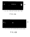
- Fig. 12B shows gene expression in organs retrieved from VEGF-transfected rabbit.
- Lane 1 molecular weight markers
- Lane 2 positive control (smooth muscle cells transfected with VEGF);
- Lane 3 negative control (no RNA);
- Lane 4 negative control (no tissue);
- Lane 5 negative control (PCR of LacZ-Tf artery);
- Lane 6 negative control (PCR of VEGF-Tf artery excluding reverse transcriptase reaction).
- the present invention provides means for inducing reendothelialization of the lining of an injured blood vessel by contacting the injured portion of the vessel with a nucleic acid encoding an endothelial cell mitogen operably linked to a promoter (nucleic acid cassette) to result in expression of the mitogen when delivered to the cells at the site of vascular injury.
- the resulting reendothelialization of the injured blood vessel inhibits smooth muscle cell proliferation and consequently reduces restenosis.
- the invention may be used to treat any blood vessel injury that results in denuding of the endothelial lining of the vessel wall, including, for example, those injuries resulting from balloon angioplasty and deployment of endovascular stents.
- the invention relates to the following subject matter :
- the nucleic acid may be any nucleic acid (DNA or RNA) including genomic DNA, cDNA and mRNA, encoding an endothelial cell mitogen which can be used to express the mitogen, i.e., a protein, polypeptide, mutein or portion that is capable of inducing endothelial cell growth.
- VEGF vascular endothelial growth factor
- aFGF acidic fibroblast growth factor
- bFGF basic fibroblast growth factor
- j catter factor hepatocyte growth factor
- CSF colony stimulating factor
- VEGF has been shown to be an endothelial cell-specific mitogen (Ferrara, et al., Biochem Biophys Res Commun., 161:851-855, (1989), Keck, et al., Science, 246:1309-1312 (1989), and Plouet, et al., Embo J., 3801-3806 (1989)).
- VEGF was purified independently as a tumor-secreted factor that included vascular permeability by the Miles assay (Keck, et al., supra, and Connolly, et al., J. Biol. Chem., 264:20017-20024 (1989)), and thus has an alternate designation, vascular permeability factor (VPF).
- VPF vascular permeability factor
- VEGF heparin-binding, angiogenic growth factors.
- the NH 2 terminus of VEGF is preceded by a typical signal sequence; therefore, unlike bFGF, VEGF can be secreted by intact cells.
- its high-affinity binding sites shown to include the tyrosine kinase receptors Flt-1 and Flt- 1/KDR are present on endothelial cells. Ferrara, et al., supra, and Conn, et al., Proc. Natl. Acad Sci. USA, 87:1323-1327 (1990). (Interaction of VEGF with lower affinity binding sites has been shown to induce mononuclear phagocyte chemotaxis).
- nucleotide sequence of numerous peptides and proteins, including endothelial cell mitogens, are readily available through a number of computer data bases, for example, GenBank, EMBL and Swiss-Prot. Using this information, a DNA or RNA segment encoding the desired may be chemically synthesized or, alternatively, such a DNA or RNA segment may be obtained using routine procedures in the art, e.g, PCR amplification.
- the nucleic acid is preferably inserted into a cassette where it is operably linked to a promoter.
- the promoter must be capable of driving expression of the mitogen in the desired target host cell.
- the selection of appropriate promoters can readily be accomplished. Preferably, one would use a high expression promoter.
- An example of a suitable promoter is the 763-base-pair cytomegalovirus (CMV) promoter.
- CMV 763-base-pair cytomegalovirus
- RSV Rous sarcoma virus
- MMT Mobility Management Function
- a cassette can then be inserted into a vector, e.g., a plasmid vector such as pUC118, pBR322, or other known plasmid vectors, that includes, for example, an E. coli origin of replication. See, Sambrook, et al., Molecular Cloning: A Laboratory Manual, Cold Spring Harbor Laboratory press, (1989).
- the plasmid vector may also include a selectable marker such as the ⁇ -lactamase gene for ampicillin resistance, provided that the marker polypeptide does not adversely effect the metabolism of the organism being treated.
- the cassette can also be bound to a nucleic acid binding moiety in a synthetic delivery system, such as the system disclosed in WO 95/22618.
- the DNA may also be used with a microdelivery vehicle such as cationic liposomes and adenoviral vectors.
- a microdelivery vehicle such as cationic liposomes and adenoviral vectors.
- Replication-defective recombinant adenoviral vectors can be produced in accordance with known techniques. See, Quantin, et al., Proc. Natl. Acad. Sci. USA, 89:2581-2584 (1992); Stratford-Perricadet, et al., J. Clin. Invest., 90:626-630 (1992); and Rosenfeld, et al., Cell, 68:143-155 (1992).
- the injured portion of the blood vessel is contacted with the DNA encoding an endothelial cell mitogen by any means familiar to the skilled artisan, including, for example, double-balloon catheters, porous-balloon catheters and hydrophilic coated balloon catheters.
- double-balloon catheters including, for example, double-balloon catheters, porous-balloon catheters and hydrophilic coated balloon catheters.
- double-balloon catheters e.g., Lancet 1:1106-1108, (1989); Wolinsky, et al., J. Am. Coll. Cardiol., 15:475-485 (1990); March, et al., Cardio Intervention, 2:11-26 (1992)); WO93/00051 and WO93/00052; U.S. Pat. No. 5,304,121.
- Dichek Textbook of Interventional Cardiology, Vol. 2, 61:989-1005.
- a hydrophilic coated balloon catheter is preferred in certain situations.
- a hydrophilic coated balloon catheter has a hydrophilic polymer on the outer surface of the balloon which permits the contact between the hydrophilic polymer bearing the nucleic acid to be transferred and the cells of the injured portion of the blood vessel to be made with some pressure, thus facilitating the transfer of the nucleic acid to the cells.
- other supports for the hydrophilic polymer are also useful, such as catheters or solid rods having a surface of hydrophilic polymer.
- the catheters or rods or other substrates which are flexible, to facilitate threading through the arteries to reach the point of intended application.
- the hydrophilic polymer is a hydrogel polymer, a cross-linked polymer material formed from the combination of a colloid and water.
- Cross-linking reduces solubility and produces a jelly-like polymer that is characterized by the ability to swell and absorb liquid, e.g., that containing the DNA.
- Suitable hydrogel polymers include, for example, those selected from the group consisting of polycarboxylic acids, cellulosic polymers, gelatin, polyvinylpyrrolidone, maleic anhydride polymers, polyamides, polyvinyl alcohols, and polyethylene oxides.
- Preferred hydrogels are polyacrylic acid polymers available as HYDROPLUS (Mansfield Boston Scientific Corp., Watertown, MA.) and described in U.S. Pat. No. 5,091,205.
- hydrophilic arterial balloon When a hydrophilic arterial balloon is used, it is not necessary to protect the balloon prior to inflation, since relatively little of the nucleic acid is lost in transit to the treatment site until the balloon is inflated and the hydrophilic polymer bearing the nucleic acid is pressed against the injured site.
- hydrophilic polymer-surfaced catheters or rods are used as the vehicle or substrate, the surface can protected, e.g. by a sheath, until the point of intended application is reached, and then the protection removed to permit the hydrophilic polymer bearing the nucleic acid to contact the injured site.
- the nucleic acid in aqueous solution is incorporated into the hydrophilic polymer to form a nucleic acid-hydrophilic polymer composition.
- the nucleic acid is incorporated without complexing or chemical reaction with the hydrophilic polymer, and is preferably relatively freely released therefrom when placed in contact with the cells at the site of injury.
- the resulting structure comprises a support, e.g. the balloon of the balloon catheter, on which is mounted the hydrogel, in or on which is incorporated the desired DNA and its associated vehicle, e.g., phage or plasmid vector.
- the hydrophilic polymer is preferably adhered to the support, so that after application of the DNA to the target cells, the hydrophilic polymer is removed with the support.
- the nucleic acid-hydrophilic composition contacts the arterial cell by means of a catheter.
- the catheter is preferably a balloon catheter constructed for insertion in a blood vessel and has a catheter shaft and an expandable dilation balloon mounted on the catheter shaft. At least a portion of the exterior surface of the expandable portion is defined by a coating of a tenaciously adhered hydrophilic polymer.
- Incorporated in the hydrophilic polymer is an aqueous solution of the DNA to be delivered to the cells of the injured portion of the blood vessel.
- the means for delivering the nucleic acid, e.g., catheter, and the nucleic acid in an aqueous solution may be included together in a kit.
- the hydrophilic polymer (preferably hydrogel) coating when dry, is preferably on the order of about 1 to 10 microns thick, with a 2 to 5 micron coating typical. Very thin hydrogel coatings, e.g., of about 0.2-0.3 microns (dry) and much thicker hydrogel coatings, e.g., more than 10 microns (dry), are also possible. Typically, hydrogel coating thickness may swell by about a factor of 2 to 10 or more when the hydrogel coating is hydrated.
- FIG. 1 is the wall of the blood vessel.
- the figure shows the catheter body 2 held in place by the inflation of a balloon 3 inflated at the site of occlusion 6.
- the balloon comprises a hydrogel coating 4 incorporating DNA 5.
- the nucleic acid e.g., DNA
- the balloon may be inflated. If necessary, the polymer may be dried with warm air and the DNA application repeated.
- the amount of DNA to be applied to the arterial surface depends on the ability of the DNA to be expressed in the arterial cells. Generally, the amount of naked DNA applied to the balloon catheter is between about 0.1 and 100 ⁇ g/mm 2 , more preferably between about 0.5 and about 20 ⁇ g/mm 2 , most preferably between about 1.5 and about 8 ⁇ g/mm 2 .
- a balloon catheter having an inflated lateral area of about 630 mm 2 e.g., a balloon catheter having an inflated diameter of about 5 mm and a length of about 40mm
- a surface having about 0.8 to about 8 ⁇ g/mm 2 of DNA when the balloon is inflated and contacts the interior of the artery.
- between 1 mg and 3 mg of DNA are applied to the polymer, providing a DNA loading of about 1.6 to about 4.8 ⁇ g/mm 2 .
- the catheter is inserted using standard percutaneous application techniques and directed to the desired location, e.g., an occlusion or stent.
- the balloon is directed towards an arterial occlusion.
- the balloon for example, a hydrogel coated balloon
- it is inflated such that the hydrogel coating of the balloon contacts the occlusion causing release of the DNA directly into the arterial tissue.
- the introduction of the DNA into the plaque and surrounding tissue occurs simultaneously with the opening of the occlusion of the dilation balloon.
- the DNA encoding the endothelial cell mitogen is simultaneously applied to the denuded endothelial lining of the blood vessel where it is expressed facilitating re-endothelialization, thus reducing restenosis.
- Preferred periods of balloon inflation range from 30 seconds to 30 minutes, more preferably 1 minute to 5 minutes.
- the DNA is expressed by the cells at the site of injury for a period of time sufficient for reendothelialization. Because the vectors containing the DNA are not normally incorporated into the genome of the cells, expression of the protein of interest takes place for only a limited time. Typically, the endothelial cell mitogen is only expressed in therapeutic levels for about two days to several weeks, preferably for about 1-2 weeks. Reapplication of the DNA can be utilized to provide additional periods of expression of the therapeutic polypeptide.
- the vehicle be it arterial balloon, catheter, flexible rod or other shaped vehicle, can be furnished with means to assist in accurate placement within the intended body cavity.
- it can be furnished with a radioactive element, or made radio-opaque, furnished with means permitting easy location using ultrasound, etc.
- the invention may in principle be used to treat other blood vessel injuries that result in denuding of the endothelial lining of the vessel wall.
- primary angioplasty is becoming widely used for the treatment of acute myocardial infarction.
- Acceleration of reendothelialization using the means of the present invention can stabilize an unstable plaque and prevent re-occlusion.
- endovascular stents are becoming widely used as an adjunct to balloon angioplasty. Stents are useful for rescuing a sub-optimal primary result as well as for diminishing restenosis.
- the liability of the endovascular prosthesis has been its susceptibility to thrombotic occlusion in approximately 3% of patients with arteries 3.3 mm or larger. If patients undergo stent deployment in arteries smaller than this the incidence of sub-acute thrombosis is even higher. Sub-acute thrombosis is currently prevented only by the aggressive use of anticoagulation. The combination of vascular intervention and intense anticoagulation creates significant risks with regard to peripheral vascular trauma at the time of the stent/angioplasty procedure.
- Reendothelialization of the portion of the vessel injured by the stent deployment can eliminate these liabilities, thereby attenuating the thrombogenicity of the stented site and also reducing smooth muscle cell proliferation, responsible for stenosis in 10 to 15% of patients who undergo stent therapy.
- the elimination of an aggressive anticoagulation regiment constitutes a significant improvement in utilizing stent technology. Multiple stents have a higher rate of subacute thrombosis and restenosis which can be reduced by use of the methods of the present invention.
- stent deployment can now be applied to patients with arteries smaller than 3.0 mm in diameter.
- Ach acetylcholine chloride
- EC endothelial cell
- LacZ gene encoding nuclear-targeted ⁇ -galactosidase
- nTf non-transfected
- rET re-endothelialization
- Tf transfected.
- the mammalial expression vector employed in these experiments contains a cytomegalovirus promoter and the cDNA for recombinant human VEGF 165 ; the latter was initially isolated from cDNA libraries prepared from HL60 leukemia cells (Leung, et al., Science, 246:1306-1309 (1989)). Expression of this plasmid in rabbit vascular smooth muscle cells was previously documents in vitro by immunoabsorbent assay for VEGF protein at 3 days post-transfection (Isner, et al., Circulation, 91:2687-2692 (1995)).
- VEGF 165 The biological activity of VEGF 165 secreted from cells transfected with this construct (phVEGF 165 ) was previously confirmed by the finding that media conditioned by transfected human 293 cells promoted the proliferation of capillary cells (Leung, et al., supra ) in vitro; and angiographic and histochemical evidence of angiogenesis following catheter administration of the vector in vivo (Isner, et al., supra).
- the plasmid pGSVLacZ (courtesy of Dr. Marie Bonnerot) containing a nuclear targeted ⁇ -galactosidase sequence coupled to the simian virus 40 early promoter (Bonnerot, et al., Proc. Natl. Acad. Sci. USA, 84:6795-6799 (1987)) was used for all control transfections as previously described (Takeshita, et al., J Clin Invest., 93:652-661 (1994)).
- Each rabbit was anesthetized with ketamine (10 mg/kg) and acepromazine (0.2 mg/kg) following premedication with xylazine (2 mg/kg). Supplemental anesthetic doses were not required. Spontaneous ventilation was maintained throughout the intervention. Access to the right common carotid artery was obtained via a small midline incision in the neck. In each rabbit, a 2.0 mm diameter, 2.0 cm long, hydrogel-coated balloon catheter (Slider with Hydroplus, Boston Scientific, Watertown, MA) was used to perform balloon angioplasty and arterial gene transfer (Riessen, et al., Hum Gene Ther., 4:749-758 (1993)).
- the angioplasty balloon was prepared ex vivo by first advancing the deflated balloon through a 5 Fr. teflon protective sheath (Boston Scientific), designed to minimize contact between blood and the balloon upon introduction of the catheter into the carotid artery.
- plasmid DNA was applied to the exterior hydrogel polymer coating the angioplasty balloon.
- the balloon was inflated to 3 atm. pressure.
- a conventional pipette was then used to apply 400 ⁇ g of phVEGF 165 to the 20 ⁇ m-thick later of hydrogel polymer coating the external surface of the inflated balloon.
- the plasmid DNA was applied at a concentration of 10 ⁇ g/ ⁇ L .
- the applied plasmid solution imparts a grossly visible white glaze to the balloon surface.
- the balloon was next deflated, withdrawn back into the protective sheath, and reinflated to 3 atm within the sheath to again minimize blood flow across the balloon surface.
- the sheath and angioplasty catheter was then introduced via the right common carotid artery, and advanced to the lower abdominal aorta using a 0.014 in. guidewire (Hi-Torque Floppy II, Advanced Cardiovascular Systems, Temecula, CA) under fluoroscopic guidance.
- the balloon catheter was then deflated and advanced into one femoral artery where it was positioned using angiographic landmarks. Balloon inflation was then performed 3 times for 1 min each at 4 atm.
- femoral artery For each rabbit, after completion of transfection of one femoral artery (with phVEGF 165 or pGSVLacZ), the contralateral femoral artery underwent balloon injury (but no transfection) using a new 2.0 mm diameter, 2.0 cm long hydrogel-coated angioplasty balloon. Balloon injury, with or without gene transfer, was performed by one person who was blind to the treatment. Following completion of the entire procedure, nitroglycerin (0.25 mg, SoloPak Laboratories, Franklin Park, IL) and heparin sodium (200 USP units, Elkins-sinn, Cherry Hill) NJ were administered intraarterially to prevent acute occlusion of the balloon-injured sites.
- nitroglycerin (0.25 mg, SoloPak Laboratories, Franklin Park, IL
- heparin sodium 200 USP units, Elkins-sinn, Cherry Hill
- Vasometer reactivity of the arterial segment subjected to balloon angioplasty and arterial gene transfer was evaluated on the day of sacrifice.
- a 3 Fr., end-hole infusion catheter (Tracker-18 TM , Target Therapeutics, San Jose, CA) was inserted into the left carotid artery and advanced to the origin of transfected iliac artery using a 0.018 in. guidewire (Hi-Torque Floppy II) under fluoroscopic guidance. This catheter was used both for infusion of vasoactive drugs and selective angiography of the femoral artery.
- Angiography was performed immediately after drug administration using 1 ml of non-ionic contrast media (lsovue-370, Squibb Diagnostics, New Brunswick, NJ).
- Serial angiographic images were recorded on 105-mm spot film at a rate of 2 films per sec. for 4 sec.
- acetylcholine chloride (Ach) and serotonin creatine sulfate (5-HT) were delivered from a constant infusion pump (1 ml/min) via the 3 Fr. catheter at doses of 0.15, 1.5, and 15 ⁇ g/kg/min, each for 2 min. Five min was allowed to elapse between each dose of agent to re-establish basal blood flow conditions. After administration of Ach and 5-HT respectively were completed, a 2-min intra-arterial infusion of sodium nitroprusside (Np)(1.5 ⁇ g/kg/min) was administered to assess endothelium-independent vasomotorreactvity. Finally, an identical protocol was employed to evaluate the contralateral injured but non-transfected femoral artery.
- Ach acetylcholine chloride
- 5-HT serotonin creatine sulfate
- the angiographic luminal diameter of the femoral artery prior to and after drug infusion was determined using an automated edge-detection system (LeFree, et al., Proc SPIE, 626:334-341 (1986) and Mancini, et al., Circulation, 75:452-460 (1987)).
- Each balloon-injured site was defined and the boundary lines were draw according to the pilot angiogram of angioplasty balloon injury.
- the angiogram selected for analysis was scanned with a high resolution video camera; the signal produced by the video camera was digitized and displayed on a video monito. Center-lines were traced manually for a 20 mm-long segment defined by the boundary lines drawn previously.
- Contours were detected automatically on the basis of the weighted sum of first and second derivative functions applied to the digitized brightness information.
- the average angiographic luminal diameter and the minimum luminal diameter were then determined for the defined 20 mm-long segment of each transfected as well as non-transfected balloon-injured artery.
- Ach, 5-HT and Np were obtained from Sigma Chemical Co., St. Louis, MO. Fresh stock solutions of each were prepared immediately before each experiment.
- the initially injured 2-cm long segment of femoral artery was then dissected free and incised longitudinally.
- the harvested arterial segment was pinned to a cork board, further fixed in 100% methanol, and photographed using a dissecting microscope (STEMI SR, Zeiss, Germany) in preparation for planimetric analysis of rET (see below).
- Tissues were further fixed by immersion in 100% methanol, embedded on longitudinal edge in paraffin, and cut in 5- ⁇ m sections onto slides coated with 3-aminopropyl-triethoxy-silane.
- Planimetric analysis was performed using the photograph of the harvested arterial segment taken through the dissecting microscope.
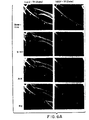
- the area of the intimal surface which was stained blue following application of Evans blue dye was interpreted to identify the portion of the arterial segment which remained endothelium-deficient; these macroscopic analyses were confirmed by immunostaining of light microscopic sections using an endothelial cell-specific marker (vide infra).
- a computerized sketching program (MacMeasure version 1.9; NIMH, Bethesda, MD) interfaced with a digitizing board (Summagraphics, Fairfield, CT) was used by one person blinded to the treatment protocol to outline the Evans blue positive and negative areas respectively. Specifically, the extent of endothelialized area was calculated as a percent of the total intimal area encompassed within the 2-cm length of artery.
- the thickness of the native media of the artery wall is variable reflecting in part the dimensions (diameter) of the individual rabbit femoral artery. Accordingly, thickness of the media was used to index the extent of neointimal thickening, and is thus stated as the ratio of intima to media area (I/M).
- PCNA proliferating cell nuclear antigen
- Bound primary antibody was detected using an avidin-biotin-immunoperoxidase method (Elite Avidin-Biotin Detection System, Signet). Sections were lightly counterstained with Bandeiraea simplicifolia I (BSI) lectin 5 ⁇ g/ml, Vector Laboratories, Burlingame, CA)(25). For lectin staining, sections pretreated with 0.3%hydrogen peroxidase were incubated with 5 ⁇ g/ml of biotinylated lectin (Vector) overnight at 4°C.
- BSI Bandeiraea simplicifolia I
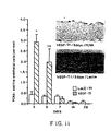
- the extent of proliferative activity for each longitudinal section was measured by one person blinded to the treatment regimen as the number of positive cells in the intima per length of the 20 mm-long longitudinal section, i.e. cells/mm.
- PCNA staining of the rabbit ileum served as the positive control for this study.
- master mixes for a number of reactions were made up and aliquoted to tubes containing RNA. Reactions were incubated at 42°C for 1 hr, then at 95°C for 5 min to terminate the reaction. Twenty ml of diethyl pyrocarbonate (DEPC) water was then added and 5 ml of the diluted reaction (1/8th) was used in the PCR analysis.
- DEPC diethyl pyrocarbonate
- the optimized reaction in a total volume of 20 ⁇ l contained 0.2 mM of each deoxynucleotide triphosphate, 3mM MgCl 2 , 2 ml PCR II buffer (Perkin-Elmer, Norwalk, CT; final concentrations, 50 mM KCl, 10 mM Tris-HCl), 5 ng/ml (13.77 pmoles) of each primer, and 0.5 units of AmpliTaq DNA polymerase (Perkin-Elmer).
- the PCR was performed on a 9600 PCR system (Perkin-Elmer) using microamp 0.2 ml thin-walled tubes.
- Amplification was performed for 35-45 cycles of 94°C for 20 sec, 55°C for 20 sec, and 72°C for 20 sec, ending with 5 min at 72°C.
- controls were includes with no RNA and no transverse transcriptase.
- a pair of oligonucleotide primers (22 mers) was designed to amplify a 258 bp sequence from the mRNA of human VEGF. To ensure specificity and avoid amplification of endogenous rabbit VEGF, each primer was selected from a region which is not conserved among different species.
- Sequences of primers used were: 5'-GAGGGCAGAATCATCACGAAGT-3' (SEQ ID NO:1)(sense); and 5'-TCCTATGTGCTGGCCTTGGTGA-3' (SEQ ID NO:2)(antisense).
- Preliminary experiments showed that cultured rabbit smooth muscle cells hybridized with this primer only when transfected with a plasmid containing the cDNA for human VEGF. Cultures of normal (non-transfected) or ⁇ -galactosidase-transfected rabbit smooth muscle cells showed no hybridization.
- RT-PCR products were analyzed by 2% agarose gel electrophoresis. DNA bands were visualized under UV illumination after staining with ethidium bromide.
- LacZ-Tf arteries were harvested at day 5, and ⁇ -galactosidase activity was determined by incubation with 5-bromo-4-chrolo-3-indolyl ⁇ -D-galactoside chromogen (X-Gal, Sigma) as previously described (Riessen, et al., supra). Following staining with X-Gal solution, tissues were paraffin-embedded, sectioned, and counterstained with nuclear fast red.
- Nuclear localized ⁇ -galactosidase expression of the plasmid pGSVLacZ could not result from endogenous ⁇ -galactosidase activity; accordingly, histochemical identification of ⁇ -galactosidase within the cell nucleus was interpreted as evidence for successful gene transfer and gene expression, Cytoplasmic or other staining was considered non-specific for the purpose of the present study.
- VEGF-Tf vascular endothelial growth factor 165
- VEGF-nTf contralateral balloon-injured, non-trasfected arteries
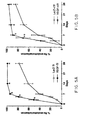
- Vasoconstriction (+) or vasodilation (-) in response to endothelium-dependant and independant agonists following phVEGF 165 gene therapy 1wk 5-HT Ach Np LacZ-T ⁇ (+)48.6 ⁇ 5.4 (+)4.2 ⁇ 2.8 (-)4.2 ⁇ 2.8 VEGF-T ⁇ (+)24.2 ⁇ 4.1* (-)6.8 ⁇ 2.2* (-)6.6 ⁇ 1.7 LacZ-nT ⁇ (+)53.6 ⁇ 5.0 (+)12.8 ⁇ 6.6 (-)5.8 ⁇ 3.8 VEGF-nTF (+)29.3 ⁇ 3.4* (-)0.5 ⁇ 3.0 ⁇ (-)8.0 ⁇ 2.2 2 wks 5-HT Ach Np LacZ-T ⁇ (+)56.3 ⁇ 8.0 (+)15.1 ⁇ 4.8 (-)0.9 ⁇ 2.9 VEGF-T ⁇ (+)24.2 ⁇ 4.5* (-)3.3 ⁇ 2.8* (-)5.4 ⁇ 2.2 LacZ-nT ⁇ (+)48.4 ⁇ 5.6 (+)10.8 ⁇ 5.0 (+)0.7 ⁇ 2.
- Selective immunostaining of adjacent sections established that proliferating cells were predominantly smooth muscle cells.
- PCNA-positive cells were identified as ECs by positive BSI lectin staining of apparently identical cells located along the luminal border of adjacent (serially cut) histologic sections. Proliferative activity of ECs was constant throughout the 4 wks following balloon injury in LacA-Tf. In contrast, EC proliferation in VEGF-Tf arteries demonstrated a prominent peak at 3d, and was then reduced by 1 wk post-transfection (Fig. 11).
- Thromboresistance constitutes an important consideration in the immediate post-angioplasty setting. This is particularly true when angioplasty is performed for acute myocardial infarction, in which case the risk of subacute reocclusion is increased.
- ph VEGF 165 -accelerated rET led to augmented thromboresistance. Specifically, thrombotic occlusion developed less frequently in animals transfected with ph VEGF 165 (2/64) than in those transfected with Lac-Z(14/64) (p ⁇ 0.001).
- RT-PCR was also performed on sections from other organs and disclosed no mRNA expression of VEGF at any sit remote from the VEGF-transfected arterial segment (Fig. 12).
- arteries transfected with phVEGF 165 disclosed recovery of near-normal endothelium-dependent response within one week.
- the physiologic response among VEGF-Tf rabbits at four weeks can be assumed to represent the response of restored endothelium in the absence of ongoing VEGF 165 secretion, since analyses using RT-PCR in the present and previous (Isner, et al., supra) studies have consistently disclosed no evidence of transgene expression beyond 3 weeks.
- VEGF-induced rET appeared to result in improved thromboresistance, a function of particular importance in the post-angioplasty setting.
- VEGF gene transfer for the purpose of stabilizing acutely ruptured plaque may have additional merit.
- the pGSVLacZ vector is described above.
- the cDNA to be used in this example encodes the 165 amino acid isoform of VEGF and has been described previously.
- the plasmid into which the VEGF cDNA has been inserted is a simple eucaryotic expression plasmid that utilizes cytomegalovirus (CMV) promoter/enhancer to drive VEGF expression.
- CMV cytomegalovirus
- This plasmid is a derivative of pCG (Tanaka, et al., Cell, 60:375-386 (1990)).
- the backbone plasmid vector is derived from pBSM13+ .
- a cassette encoding the kanamycin resistance gene has been inserted, and the SV40 origin of replication present in the original vector (phVEGF 165 ) has been largely removed.
- the CMV promoter is from positions -522 to +72 relative to the CMV cap site.
- Upstream from the phVEGF 165 SR coding sequence is the HSV thymidine kinase gene 5' leader from positions + 51 to + 114 relative to the thymidine kinase cap site.
- Downstream from the VEGF coding sequence is the rabbit b-globin gene sequence from positions + 905 to + 2080 relative to the b-globin cap site.
- the entire phVEGF 165 SR sequence (6609 bp) has been determined using 30 sequence primers.
- the structure of the double-stranded DNA was determined by the cycle sequencing method using fluorescent dideoxy terminator nucleotides with an Applied Biosystem 373A Automated sequencer. Sequences were analyzed on Macintosh Quadra computers with MacVector and Sequence Navigator software. The quality control for this sequencing analysis consists of parallel sequence analyses of Bluescript and M13 controls. This analysis revealed a 99.1 % sequence homology between our determined sequence and the predicted sequence of the 6609 bp plasmid.
- Procedures were performed using New Zealand rabbits (3.5-4.0kg). All animals received aspirin (100 mg/day) for 1 week before the procedure and the treatment was continued until sacrifice. All received heparin (1000 units) and antibiotics (2.5 mg/kg) at the time of the procedure. The protocol was designed to distinguish both the local and the systemic effect of the therapy.
- the treated side of the VEGF group had significantly better endothelial coverage at day 7 than all the other sides (p ⁇ 005).
Claims (11)
- Utilisation d'une solution aqueuse contenant un acide nucléique pour la fabrication d'un médicament destiné à l'induction de la ré-endothélialisation de la tunique interne d'un vaisseau sanguin lésé et à la réduction de la resténose par la délivrance de l'acide nucléique à la partie lésée du vaisseau sanguin, dans laquelle l'acide nucléique code pour un mitogène des cellules endothéliales, choisi dans le groupe comprenant le facteur de croissance endothéliale vasculaire (Vascular Endothelial Growth Factor; VEGF), le facteur de croissance des fibroblastes acide (acidic Fibroblast Growth Factor ; a-FGF), le facteur de croissance des fibroblastes basique (basic Fibroblast Growth Factor; b-FGF), le facteur de croissance des hépatocytes (Hepatocyte Growth Factor ; HGF) et le facteur stimulant la formation de colonies (Colony Stimulating Factor; CSF), fonctionnellement lié à un promoteur.
- Utilisation selon la revendication 1, dans laquelle la lésion est le résultat d'une angioplastie transluminale.
- Utilisation selon la revendication 1, dans laquelle la lésion est le résultat de la mise en place d'une endoprothèse vasculaire.
- Utilisation selon l'une quelconque des revendications précédentes, dans laquelle le moyen de délivrance dudit acide nucléique à la partie lésée du vaisseau sanguin est un système de délivrance par cathéter.
- Utilisation selon la revendication 4, dans laquelle le système de délivrance par cathéter est un cathéter à double ballonnet, un cathéter à ballonnet poreux ou un cathéter à ballonnet revêtu d'un polymère hydrophile.
- Utilisation selon la revendication 5, dans laquelle le polymère hydrophile du cathéter à ballonnet revêtu d'un polymère hydrophile est un polymère en hydrogel.
- Utilisation d'une solution aqueuse contenant un acide nucléique pour la fabrication d'un médicament destiné à l'induction de la ré-endothélialisation de la tunique interne d'un vaisseau sanguin lésé et à la réduction de la resténose par la délivrance de l'acide nucléique à la partie lésée du vaisseau sanguin en utilisant un cathéter à ballonnet revêtu d'un hydrogel, dans laquelle l'acide nucléique code pour un mitogène des cellules endothéliales, choisi dans le groupe comprenant le facteur de croissance endothéliale vasculaire (VEGF), le facteur de croissance des fibroblastes acide (a-FGF), le facteur de croissance des fibroblastes basique (b-FGF), le facteur de croissance des hépatocytes (HGF) et le facteur stimulant la formation de colonies (CSF), fonctionnellement lié à un promoteur.
- Utilisation d'une solution aqueuse contenant un acide nucléique pour la fabrication d'un médicament destiné à l'induction de la ré-endothélialisation de la tunique interne d'un vaisseau sanguin lésé et à la réduction de la resténose par la délivrance de l'acide nucléique à la partie lésée du vaisseau sanguin en utilisant une endoprothèse vasculaire, dans laquelle l'acide nucléique code pour un mitogène des cellules endothéliales, choisi dans le groupe comprenant le facteur de croissance endothéliale vasculaire (VEGF), le facteur de croissance des fibroblastes acide (a-FGF), le facteur de croissance des fibroblastes basique (b-FGF), le facteur de croissance des hépatocytes (HGF) et le facteur stimulant la formation de colonies (CSF), fonctionnellement lié à un promoteur.
- Utilisation d'une solution aqueuse contenant un acide nucléique pour la fabrication d'un médicament destiné à l'induction de la ré-endothélialisation de la tunique interne d'un vaisseau sanguin lésé et à la réduction de la resténose par la délivrance de l'acide nucléique à la partie lésée du vaisseau sanguin en utilisant un ballonnet ou la mise en place d'une endoprothèse, dans laquelle l'acide nucléique code pour un mitogène des cellules endothéliales, choisi dans le groupe comprenant le facteur de croissance endothéliale vasculaire (VEGF), le facteur de croissance des fibroblastes acide (a-FGF), le facteur de croissance des fibroblastes basique (b-FGF), le facteur de croissance des hépatocytes (HGF) et le facteur stimulant la formation de colonies (CSF), fonctionnellement lié à un promoteur et dans laquelle l'acide nucléique est incorporé à la surface du ballonnet ou de fendoprothèse mise en place.
- Utilisation selon la revendication 9, dans laquelle la lésion est le résultat d'une angioplastie transluminale.
- Utilisation selon la revendication 9, dans laquelle la lésion est le résultat de la mise en place d'une endoprothèse vasculaire.
Applications Claiming Priority (3)
| Application Number | Priority Date | Filing Date | Title |
|---|---|---|---|
| US538301 | 1990-06-14 | ||
| US08/538,301 US5830879A (en) | 1995-10-02 | 1995-10-02 | Treatment of vascular injury using vascular endothelial growth factor |
| PCT/US1996/015813 WO1997012519A1 (fr) | 1995-10-02 | 1996-10-02 | Traitement de lesions vasculaires |
Publications (3)
| Publication Number | Publication Date |
|---|---|
| EP0853450A1 EP0853450A1 (fr) | 1998-07-22 |
| EP0853450A4 EP0853450A4 (fr) | 2003-01-15 |
| EP0853450B1 true EP0853450B1 (fr) | 2007-05-09 |
Family
ID=24146332
Family Applications (1)
| Application Number | Title | Priority Date | Filing Date |
|---|---|---|---|
| EP96936138A Expired - Lifetime EP0853450B1 (fr) | 1995-10-02 | 1996-10-02 | Compositions pour le traitement de lesions vasculaires |
Country Status (8)
| Country | Link |
|---|---|
| US (2) | US5830879A (fr) |
| EP (1) | EP0853450B1 (fr) |
| JP (2) | JP2000505780A (fr) |
| AT (1) | ATE361669T1 (fr) |
| AU (1) | AU725499B2 (fr) |
| CA (1) | CA2233499A1 (fr) |
| DE (1) | DE69637075T2 (fr) |
| WO (1) | WO1997012519A1 (fr) |
Families Citing this family (168)
| Publication number | Priority date | Publication date | Assignee | Title |
|---|---|---|---|---|
| US6228844B1 (en) | 1991-11-12 | 2001-05-08 | Vical Incorporated | Stimulating vascular growth by administration of DNA sequences encoding VEGF |
| US6706694B1 (en) | 1990-03-21 | 2004-03-16 | Vical Incorporated | Expression of exogenous polynucleotide sequences in a vertebrate |
| US6239172B1 (en) | 1997-04-10 | 2001-05-29 | Nitrosystems, Inc. | Formulations for treating disease and methods of using same |
| FR2738842B1 (fr) * | 1995-09-15 | 1997-10-31 | Rhone Poulenc Rorer Sa | Molecule d'adn circulaire a origine de replication conditionnelle, leur procede de preparation et leur utilisation en therapie genique |
| US6090618A (en) * | 1996-10-07 | 2000-07-18 | Arch Development Corporation | DNA constructs and viral vectors comprising a smooth muscle promoter |
| US20020001574A1 (en) * | 1995-12-13 | 2002-01-03 | Jon A. Woiff | Process of delivering a polynucleotide to a muscle cell via the vascular system |
| US6321109B2 (en) | 1996-02-15 | 2001-11-20 | Biosense, Inc. | Catheter based surgery |
| US6143037A (en) * | 1996-06-12 | 2000-11-07 | The Regents Of The University Of Michigan | Compositions and methods for coating medical devices |
| US6443974B1 (en) * | 1996-07-28 | 2002-09-03 | Biosense, Inc. | Electromagnetic cardiac biostimulation |
| EP0941116B1 (fr) * | 1996-11-01 | 2005-01-19 | Ark Therapeutics Limited | Utilisation de vegf pour l'obtention d'un medicament pour le traitment ou prevention de l'hyperplasie endoveineuse et systeme d'administration |
| US20080305999A1 (en) * | 1996-11-01 | 2008-12-11 | John Francis Martin | Therapeutic use of growth factor, and delivery device, especially for the treatment of intimal hyperplasia |
| US5980887A (en) * | 1996-11-08 | 1999-11-09 | St. Elizabeth's Medical Center Of Boston | Methods for enhancing angiogenesis with endothelial progenitor cells |
| US8075880B2 (en) | 1999-01-11 | 2011-12-13 | Steward St. Elizabeth's Medical Center Of Boston, Inc. | Compositions and methods for regulating angiogenesis |
| IT1289728B1 (it) * | 1996-12-10 | 1998-10-16 | Sorin Biomedica Cardio Spa | Dispositivo di impianto e corredo che lo comprende |
| US8172897B2 (en) | 1997-04-15 | 2012-05-08 | Advanced Cardiovascular Systems, Inc. | Polymer and metal composite implantable medical devices |
| US6240616B1 (en) | 1997-04-15 | 2001-06-05 | Advanced Cardiovascular Systems, Inc. | Method of manufacturing a medicated porous metal prosthesis |
| US10028851B2 (en) | 1997-04-15 | 2018-07-24 | Advanced Cardiovascular Systems, Inc. | Coatings for controlling erosion of a substrate of an implantable medical device |
| US6306166B1 (en) * | 1997-08-13 | 2001-10-23 | Scimed Life Systems, Inc. | Loading and release of water-insoluble drugs |
| EP1044035A4 (fr) | 1997-12-31 | 2002-07-17 | Pharmasonics Inc | Procedes, systemes et kits pour l'apport intravasculaire d'acides nucleiques |
| US6794369B2 (en) | 1997-12-31 | 2004-09-21 | Pharmasonics | Methods, systems, and kits for intravascular nucleic acid delivery |
| WO1999044538A1 (fr) * | 1998-01-27 | 1999-09-10 | The Regents Of The University Of California | Serpentins biodegradables a base de polymeres/proteines pour implants endoluminaux |
| US7070607B2 (en) * | 1998-01-27 | 2006-07-04 | The Regents Of The University Of California | Bioabsorbable polymeric implants and a method of using the same to create occlusions |
| US20030129750A1 (en) * | 1998-02-05 | 2003-07-10 | Yitzhack Schwartz | Homing of donor cells to a target zone in tissue using active therapeutics or substances |
| US20030113303A1 (en) * | 1998-02-05 | 2003-06-19 | Yitzhack Schwartz | Homing of embryonic stem cells to a target zone in tissue using active therapeutics or substances |
| US6309370B1 (en) | 1998-02-05 | 2001-10-30 | Biosense, Inc. | Intracardiac drug delivery |
| US6676937B1 (en) * | 1998-03-09 | 2004-01-13 | Caritas St. Elizabeth's Medical Center Of Boston Inc. | Compositions and methods for modulating vascularization |
| US20040228834A1 (en) * | 1998-03-09 | 2004-11-18 | Jeffrey Isner | Compositions and methods for modulating vascularization |
| US20030118567A1 (en) * | 1999-03-26 | 2003-06-26 | Stewart Duncan John | Cell-based therapy for the pulmonary system |
| CA2266805C (fr) | 1998-03-27 | 2015-04-28 | An-Go-Gen Inc. | Therapie genique cellulaire dans le traitement des affections pulmonaires |
| US6482406B1 (en) | 1999-03-26 | 2002-11-19 | Duncan J. Stewart | Cell-based gene therapy for the pulmonary system |
| US9585916B2 (en) | 1999-03-26 | 2017-03-07 | Northern Therapeutics Inc. | Cell based therapy for the pulmonary system |
| US20030039694A1 (en) * | 1998-04-28 | 2003-02-27 | Martin John Francis | Periadventitial delivery device |
| EP1095132A4 (fr) | 1998-07-10 | 2003-09-24 | Brigham & Womens Hospital | Procede d'implantation de cellules |
| WO2000015285A1 (fr) * | 1998-09-14 | 2000-03-23 | Mirus Corporation | Procede d'administration d'acides nucleiques aux tissus cardiaques |
| AU6409499A (en) | 1998-10-01 | 2000-04-17 | Children's Medical Center Corporation | Method of treatment of cardiovascular injuries |
| US6440944B2 (en) * | 1998-10-16 | 2002-08-27 | Genvec, Inc. | Methods of administering adenoviral vectors |
| US6958147B1 (en) * | 1998-10-26 | 2005-10-25 | Licentia Ltd | Use of VEGF-C to prevent restenosis |
| CA2340593C (fr) * | 1998-10-26 | 2012-05-08 | Kari Alitalo | Utilisation du gene ou de la proteine vegf-c ou vegf-d pour prevenir la restenose |
| CA2319447C (fr) | 1998-12-01 | 2010-01-26 | Washington University | Dispositif d'embolisation |
| US7279568B2 (en) * | 1998-12-31 | 2007-10-09 | Viromed Limited | Highly efficient eukaryotic expression vector comprising an exogenous transcription regulatory element |
| WO2000044306A1 (fr) * | 1999-01-27 | 2000-08-03 | The Regents Of The University Of California | Spirales a base de polymere/proteine biodegradable, pour implants endoluminaux |
| CA2375617A1 (fr) * | 1999-06-07 | 2000-12-14 | Edwards Lifesciences Corporation | Angiogenese ciblee |
| US6329348B1 (en) | 1999-11-08 | 2001-12-11 | Cornell Research Foundation, Inc. | Method of inducing angiogenesis |
| US7037332B2 (en) * | 2000-03-15 | 2006-05-02 | Orbus Medical Technologies, Inc. | Medical device with coating that promotes endothelial cell adherence |
| US8109994B2 (en) | 2003-01-10 | 2012-02-07 | Abbott Cardiovascular Systems, Inc. | Biodegradable drug delivery material for stent |
| US7875283B2 (en) | 2000-04-13 | 2011-01-25 | Advanced Cardiovascular Systems, Inc. | Biodegradable polymers for use with implantable medical devices |
| US6527801B1 (en) | 2000-04-13 | 2003-03-04 | Advanced Cardiovascular Systems, Inc. | Biodegradable drug delivery material for stent |
| DE60134021D1 (de) * | 2000-06-27 | 2008-06-26 | Anges Mg Inc | Pharmazeutische zubereitungen zur angiogenese-therapie |
| AU2001280789A1 (en) * | 2000-07-25 | 2002-02-05 | The Board Of Trustees Of The Leland Stanford Junior University | Non-viral linear dna vectors and methods for using the same |
| EP1347794A2 (fr) * | 2000-11-27 | 2003-10-01 | Medtronic, Inc. | Tuteurs et procedes de preparation de tuteurs a partir de fils recouverts de couches de revetement hydrogel |
| US6913762B2 (en) * | 2001-04-25 | 2005-07-05 | Mayo Foundation For Medical Education And Research | Stent having non-woven framework containing cells |
| WO2002095038A2 (fr) * | 2001-05-23 | 2002-11-28 | Fornix Biosciences N.V. | Vecteurs d'expression amelioree de vegf pour le traitement de maladies |
| US7223740B2 (en) * | 2001-05-23 | 2007-05-29 | Fornix Biosciences N.V. | Vectors for enhanced expression of VEGF for atrial disease treatment |
| EP1262199A1 (fr) * | 2001-05-23 | 2002-12-04 | Fornix Biosciences N.V. | Vecteurs pour l'expression augmentée de VEGF pour le traítement des maladies |
| ATE462006T1 (de) * | 2001-08-01 | 2010-04-15 | Univ Bristol | Isoform des vegfs |
| US7989018B2 (en) | 2001-09-17 | 2011-08-02 | Advanced Cardiovascular Systems, Inc. | Fluid treatment of a polymeric coating on an implantable medical device |
| US7285304B1 (en) | 2003-06-25 | 2007-10-23 | Advanced Cardiovascular Systems, Inc. | Fluid treatment of a polymeric coating on an implantable medical device |
| US6863683B2 (en) | 2001-09-19 | 2005-03-08 | Abbott Laboratoris Vascular Entities Limited | Cold-molding process for loading a stent onto a stent delivery system |
| US6663880B1 (en) | 2001-11-30 | 2003-12-16 | Advanced Cardiovascular Systems, Inc. | Permeabilizing reagents to increase drug delivery and a method of local delivery |
| US7202066B2 (en) * | 2002-01-29 | 2007-04-10 | Carrington Laboratories, Inc. | Combination of a growth factor and a protease enzyme |
| AU2003217271A1 (en) * | 2002-01-29 | 2003-09-02 | A. Mark Colb | Endothelialization of vascular surfaces |
| DE60323943D1 (de) * | 2002-02-21 | 2008-11-20 | Encelle Inc | Immobilisierte bioaktive hydrogel matrizen für oberflächenbeschichtungen |
| KR100562824B1 (ko) | 2002-03-20 | 2006-03-23 | 주식회사 바이로메드 | 유전자 발현효율이 높으며 간세포 성장인자의 두 가지이형체를 동시에 발현하는 하이브리드 간세포 성장인자유전자 |
| US7488573B2 (en) | 2002-06-26 | 2009-02-10 | Children's Hospital Medical Center | In vivo angiogenesis assay |
| US7435419B2 (en) * | 2002-07-19 | 2008-10-14 | Beth Israel Deaconess Medical Center | Methods of diagnosing and treating pre-eclampsia or eclampsia |
| US7335362B2 (en) | 2002-07-19 | 2008-02-26 | Beth Israel Deaconess Medical Center | Methods of treating pre-eclampsia or eclampsia |
| ES2534926T3 (es) * | 2002-07-19 | 2015-04-30 | Beth Israel Deaconess Medical Center | Métodos para tratar la preeclampsia |
| MXPA05000798A (es) * | 2002-07-24 | 2005-04-19 | Hoffmann La Roche | Polipeptidos t1249 pegilados. |
| WO2006006948A2 (fr) | 2002-11-14 | 2006-01-19 | Dharmacon, Inc. | Methodes et compositions permettant de selectionner des arnsi presentant une fonctionnalite amelioree |
| EP2314691A3 (fr) * | 2002-11-14 | 2012-01-18 | Dharmacon, Inc. | SIRNA fonctionnel et hyperfonctionnel |
| US7470538B2 (en) | 2002-12-05 | 2008-12-30 | Case Western Reserve University | Cell-based therapies for ischemia |
| US8435550B2 (en) | 2002-12-16 | 2013-05-07 | Abbot Cardiovascular Systems Inc. | Anti-proliferative and anti-inflammatory agent combination for treatment of vascular disorders with an implantable medical device |
| US7758881B2 (en) | 2004-06-30 | 2010-07-20 | Advanced Cardiovascular Systems, Inc. | Anti-proliferative and anti-inflammatory agent combination for treatment of vascular disorders with an implantable medical device |
| US7186789B2 (en) | 2003-06-11 | 2007-03-06 | Advanced Cardiovascular Systems, Inc. | Bioabsorbable, biobeneficial polyester polymers for use in drug eluting stent coatings |
| EP1639006A1 (fr) * | 2003-06-18 | 2006-03-29 | Georg-August-Universität Göttingen | Utilisation d'un gene ou d'un produit genetique du recepteur de vegf |
| US7198675B2 (en) | 2003-09-30 | 2007-04-03 | Advanced Cardiovascular Systems | Stent mandrel fixture and method for selectively coating surfaces of a stent |
| US20060085062A1 (en) * | 2003-11-28 | 2006-04-20 | Medlogics Device Corporation | Implantable stent with endothelialization factor |
| US20050214339A1 (en) | 2004-03-29 | 2005-09-29 | Yiwen Tang | Biologically degradable compositions for medical applications |
| US8273383B2 (en) * | 2004-05-04 | 2012-09-25 | Children's Medical Center Corporation | Methods and compositions for treatment of preeclampsia |
| WO2005118016A1 (fr) * | 2004-05-27 | 2005-12-15 | Medtronic, Inc. | Dispositif medical comprenant un agent biologiquement actif |
| US8568469B1 (en) | 2004-06-28 | 2013-10-29 | Advanced Cardiovascular Systems, Inc. | Stent locking element and a method of securing a stent on a delivery system |
| US8241554B1 (en) | 2004-06-29 | 2012-08-14 | Advanced Cardiovascular Systems, Inc. | Method of forming a stent pattern on a tube |
| US8778256B1 (en) | 2004-09-30 | 2014-07-15 | Advanced Cardiovascular Systems, Inc. | Deformation of a polymer tube in the fabrication of a medical article |
| US7731890B2 (en) | 2006-06-15 | 2010-06-08 | Advanced Cardiovascular Systems, Inc. | Methods of fabricating stents with enhanced fracture toughness |
| US8747879B2 (en) | 2006-04-28 | 2014-06-10 | Advanced Cardiovascular Systems, Inc. | Method of fabricating an implantable medical device to reduce chance of late inflammatory response |
| US8747878B2 (en) | 2006-04-28 | 2014-06-10 | Advanced Cardiovascular Systems, Inc. | Method of fabricating an implantable medical device by controlling crystalline structure |
| US7971333B2 (en) | 2006-05-30 | 2011-07-05 | Advanced Cardiovascular Systems, Inc. | Manufacturing process for polymetric stents |
| US9283099B2 (en) | 2004-08-25 | 2016-03-15 | Advanced Cardiovascular Systems, Inc. | Stent-catheter assembly with a releasable connection for stent retention |
| US7229471B2 (en) | 2004-09-10 | 2007-06-12 | Advanced Cardiovascular Systems, Inc. | Compositions containing fast-leaching plasticizers for improved performance of medical devices |
| KR20130119506A (ko) | 2004-09-24 | 2013-10-31 | 베스 이스라엘 데코니스 메디칼 센터 | 임신성 합병증의 진단 및 치료 방법 |
| US7740849B2 (en) * | 2004-09-24 | 2010-06-22 | Beth Israel Deaconess Medical Center | Use of compounds that bind soluble endoglin and SFLT-1 for the treatment of pregnancy related hypertensive disorders |
| US7875233B2 (en) | 2004-09-30 | 2011-01-25 | Advanced Cardiovascular Systems, Inc. | Method of fabricating a biaxially oriented implantable medical device |
| US8043553B1 (en) | 2004-09-30 | 2011-10-25 | Advanced Cardiovascular Systems, Inc. | Controlled deformation of a polymer tube with a restraining surface in fabricating a medical article |
| US8173062B1 (en) | 2004-09-30 | 2012-05-08 | Advanced Cardiovascular Systems, Inc. | Controlled deformation of a polymer tube in fabricating a medical article |
| WO2007053161A2 (fr) * | 2004-12-15 | 2007-05-10 | Beth Israel Deaconess Medical Center | Acides nucléiques et polypeptides utiles pour diagnostiquer et traiter des complications de la grossesse |
| US20110230407A1 (en) * | 2005-03-14 | 2011-09-22 | Alexander Yuzhakov | Hepatocyte growth factor pathway activators in demyelinating diseases and central nervous system trauma |
| US7381048B2 (en) | 2005-04-12 | 2008-06-03 | Advanced Cardiovascular Systems, Inc. | Stents with profiles for gripping a balloon catheter and molds for fabricating stents |
| US20060284840A1 (en) * | 2005-06-15 | 2006-12-21 | Research In Motion Limited | Portable electronic device including pointer and related methods |
| US7658880B2 (en) | 2005-07-29 | 2010-02-09 | Advanced Cardiovascular Systems, Inc. | Polymeric stent polishing method and apparatus |
| US9248034B2 (en) | 2005-08-23 | 2016-02-02 | Advanced Cardiovascular Systems, Inc. | Controlled disintegrating implantable medical devices |
| US20070100323A1 (en) * | 2005-10-31 | 2007-05-03 | Ludwig Florian N | Facilitation of endothelialization by in situ surface modification |
| US7867547B2 (en) | 2005-12-19 | 2011-01-11 | Advanced Cardiovascular Systems, Inc. | Selectively coating luminal surfaces of stents |
| US20070156230A1 (en) | 2006-01-04 | 2007-07-05 | Dugan Stephen R | Stents with radiopaque markers |
| US7951185B1 (en) | 2006-01-06 | 2011-05-31 | Advanced Cardiovascular Systems, Inc. | Delivery of a stent at an elevated temperature |
| US7964210B2 (en) | 2006-03-31 | 2011-06-21 | Abbott Cardiovascular Systems Inc. | Degradable polymeric implantable medical devices with a continuous phase and discrete phase |
| US8003156B2 (en) | 2006-05-04 | 2011-08-23 | Advanced Cardiovascular Systems, Inc. | Rotatable support elements for stents |
| US7761968B2 (en) | 2006-05-25 | 2010-07-27 | Advanced Cardiovascular Systems, Inc. | Method of crimping a polymeric stent |
| US7951194B2 (en) | 2006-05-26 | 2011-05-31 | Abbott Cardiovascular Sysetms Inc. | Bioabsorbable stent with radiopaque coating |
| US20130325104A1 (en) | 2006-05-26 | 2013-12-05 | Abbott Cardiovascular Systems Inc. | Stents With Radiopaque Markers |
| US20070282434A1 (en) * | 2006-05-30 | 2007-12-06 | Yunbing Wang | Copolymer-bioceramic composite implantable medical devices |
| US7842737B2 (en) | 2006-09-29 | 2010-11-30 | Abbott Cardiovascular Systems Inc. | Polymer blend-bioceramic composite implantable medical devices |
| US8343530B2 (en) | 2006-05-30 | 2013-01-01 | Abbott Cardiovascular Systems Inc. | Polymer-and polymer blend-bioceramic composite implantable medical devices |
| US7959940B2 (en) | 2006-05-30 | 2011-06-14 | Advanced Cardiovascular Systems, Inc. | Polymer-bioceramic composite implantable medical devices |
| US20090286271A1 (en) * | 2006-05-31 | 2009-11-19 | Karumanchi Ananth S | Methods of Diagnosing and Treating Complications of Pregnancy |
| US8034287B2 (en) | 2006-06-01 | 2011-10-11 | Abbott Cardiovascular Systems Inc. | Radiation sterilization of medical devices |
| US8486135B2 (en) | 2006-06-01 | 2013-07-16 | Abbott Cardiovascular Systems Inc. | Implantable medical devices fabricated from branched polymers |
| US8603530B2 (en) | 2006-06-14 | 2013-12-10 | Abbott Cardiovascular Systems Inc. | Nanoshell therapy |
| US8048448B2 (en) | 2006-06-15 | 2011-11-01 | Abbott Cardiovascular Systems Inc. | Nanoshells for drug delivery |
| US8535372B1 (en) | 2006-06-16 | 2013-09-17 | Abbott Cardiovascular Systems Inc. | Bioabsorbable stent with prohealing layer |
| US8333000B2 (en) | 2006-06-19 | 2012-12-18 | Advanced Cardiovascular Systems, Inc. | Methods for improving stent retention on a balloon catheter |
| US8017237B2 (en) | 2006-06-23 | 2011-09-13 | Abbott Cardiovascular Systems, Inc. | Nanoshells on polymers |
| US9072820B2 (en) | 2006-06-26 | 2015-07-07 | Advanced Cardiovascular Systems, Inc. | Polymer composite stent with polymer particles |
| US8128688B2 (en) | 2006-06-27 | 2012-03-06 | Abbott Cardiovascular Systems Inc. | Carbon coating on an implantable device |
| US7794776B1 (en) | 2006-06-29 | 2010-09-14 | Abbott Cardiovascular Systems Inc. | Modification of polymer stents with radiation |
| US7740791B2 (en) | 2006-06-30 | 2010-06-22 | Advanced Cardiovascular Systems, Inc. | Method of fabricating a stent with features by blow molding |
| US7823263B2 (en) | 2006-07-11 | 2010-11-02 | Abbott Cardiovascular Systems Inc. | Method of removing stent islands from a stent |
| US7757543B2 (en) | 2006-07-13 | 2010-07-20 | Advanced Cardiovascular Systems, Inc. | Radio frequency identification monitoring of stents |
| US7998404B2 (en) | 2006-07-13 | 2011-08-16 | Advanced Cardiovascular Systems, Inc. | Reduced temperature sterilization of stents |
| US7794495B2 (en) | 2006-07-17 | 2010-09-14 | Advanced Cardiovascular Systems, Inc. | Controlled degradation of stents |
| US7886419B2 (en) | 2006-07-18 | 2011-02-15 | Advanced Cardiovascular Systems, Inc. | Stent crimping apparatus and method |
| US8016879B2 (en) | 2006-08-01 | 2011-09-13 | Abbott Cardiovascular Systems Inc. | Drug delivery after biodegradation of the stent scaffolding |
| US9173733B1 (en) | 2006-08-21 | 2015-11-03 | Abbott Cardiovascular Systems Inc. | Tracheobronchial implantable medical device and methods of use |
| US7923022B2 (en) | 2006-09-13 | 2011-04-12 | Advanced Cardiovascular Systems, Inc. | Degradable polymeric implantable medical devices with continuous phase and discrete phase |
| US8099849B2 (en) | 2006-12-13 | 2012-01-24 | Abbott Cardiovascular Systems Inc. | Optimizing fracture toughness of polymeric stent |
| US8262723B2 (en) | 2007-04-09 | 2012-09-11 | Abbott Cardiovascular Systems Inc. | Implantable medical devices fabricated from polymer blends with star-block copolymers |
| US7829008B2 (en) | 2007-05-30 | 2010-11-09 | Abbott Cardiovascular Systems Inc. | Fabricating a stent from a blow molded tube |
| US7959857B2 (en) | 2007-06-01 | 2011-06-14 | Abbott Cardiovascular Systems Inc. | Radiation sterilization of medical devices |
| US8293260B2 (en) | 2007-06-05 | 2012-10-23 | Abbott Cardiovascular Systems Inc. | Elastomeric copolymer coatings containing poly (tetramethyl carbonate) for implantable medical devices |
| US8202528B2 (en) | 2007-06-05 | 2012-06-19 | Abbott Cardiovascular Systems Inc. | Implantable medical devices with elastomeric block copolymer coatings |
| US8425591B1 (en) | 2007-06-11 | 2013-04-23 | Abbott Cardiovascular Systems Inc. | Methods of forming polymer-bioceramic composite medical devices with bioceramic particles |
| US8048441B2 (en) | 2007-06-25 | 2011-11-01 | Abbott Cardiovascular Systems, Inc. | Nanobead releasing medical devices |
| US7901452B2 (en) | 2007-06-27 | 2011-03-08 | Abbott Cardiovascular Systems Inc. | Method to fabricate a stent having selected morphology to reduce restenosis |
| US7955381B1 (en) | 2007-06-29 | 2011-06-07 | Advanced Cardiovascular Systems, Inc. | Polymer-bioceramic composite implantable medical device with different types of bioceramic particles |
| US20090202606A1 (en) * | 2008-01-25 | 2009-08-13 | Viromed Co., Ltd. | Treatment and Prevention of Cardiac Conditions Using Two or More Isoforms of Hepatocyte Growth Factor |
| AU2009234598C1 (en) * | 2008-04-09 | 2012-08-23 | Viromed Co., Ltd. | Lyophilized DNA formulations for enhanced expression of plasmid DNA |
| CN102149379A (zh) * | 2008-07-10 | 2011-08-10 | 安吉翁生物医药有限公司 | 调节肝细胞生长因子(分散因子)活性的方法和肝细胞生长因子(分散因子)活性的小分子调节剂组合物 |
| US8808353B2 (en) | 2010-01-30 | 2014-08-19 | Abbott Cardiovascular Systems Inc. | Crush recoverable polymer scaffolds having a low crossing profile |
| US8568471B2 (en) | 2010-01-30 | 2013-10-29 | Abbott Cardiovascular Systems Inc. | Crush recoverable polymer scaffolds |
| WO2012015775A2 (fr) | 2010-07-28 | 2012-02-02 | Dharmacon, Inc. | Siarn ciblant la vegfa et méthodes de traitement in vivo |
| EP2600901B1 (fr) | 2010-08-06 | 2019-03-27 | ModernaTX, Inc. | Compositions pharmaceutiques a base d'acides nucléiques modifiés et leur utilisation medicale |
| ES2737960T3 (es) | 2010-10-01 | 2020-01-17 | Modernatx Inc | Nucleósidos, nucleótidos y ácidos nucleicos modificados y sus usos |
| AU2012236099A1 (en) | 2011-03-31 | 2013-10-03 | Moderna Therapeutics, Inc. | Delivery and formulation of engineered nucleic acids |
| US8726483B2 (en) | 2011-07-29 | 2014-05-20 | Abbott Cardiovascular Systems Inc. | Methods for uniform crimping and deployment of a polymer scaffold |
| US9464124B2 (en) | 2011-09-12 | 2016-10-11 | Moderna Therapeutics, Inc. | Engineered nucleic acids and methods of use thereof |
| EP3682905B1 (fr) | 2011-10-03 | 2021-12-01 | ModernaTX, Inc. | Nucléosides, nucléotides et acides nucléiques modifiés et leurs utilisations |
| EP2791160B1 (fr) | 2011-12-16 | 2022-03-02 | ModernaTX, Inc. | Compositions de mrna modifiés |
| US9303079B2 (en) | 2012-04-02 | 2016-04-05 | Moderna Therapeutics, Inc. | Modified polynucleotides for the production of cytoplasmic and cytoskeletal proteins |
| US9283287B2 (en) | 2012-04-02 | 2016-03-15 | Moderna Therapeutics, Inc. | Modified polynucleotides for the production of nuclear proteins |
| AU2013243948A1 (en) | 2012-04-02 | 2014-10-30 | Moderna Therapeutics, Inc. | Modified polynucleotides for the production of proteins associated with human disease |
| US9572897B2 (en) | 2012-04-02 | 2017-02-21 | Modernatx, Inc. | Modified polynucleotides for the production of cytoplasmic and cytoskeletal proteins |
| EP4074834A1 (fr) | 2012-11-26 | 2022-10-19 | ModernaTX, Inc. | Arn à terminaison modifiée |
| US8980864B2 (en) | 2013-03-15 | 2015-03-17 | Moderna Therapeutics, Inc. | Compositions and methods of altering cholesterol levels |
| CA2926218A1 (fr) | 2013-10-03 | 2015-04-09 | Moderna Therapeutics, Inc. | Polynucleotides codant pour un recepteur de lipoproteines de faible densite |
| JP6240337B2 (ja) | 2013-10-22 | 2017-11-29 | バイロメッド カンパニー リミテッド | 肝細胞増殖因子の2つ以上のアイソフォームを利用した筋萎縮性側索硬化症の予防又は治療用組成物 |
| US9999527B2 (en) | 2015-02-11 | 2018-06-19 | Abbott Cardiovascular Systems Inc. | Scaffolds having radiopaque markers |
| US9700443B2 (en) | 2015-06-12 | 2017-07-11 | Abbott Cardiovascular Systems Inc. | Methods for attaching a radiopaque marker to a scaffold |
| EP3823677A4 (fr) | 2018-07-19 | 2022-06-01 | Helixmith Co., Ltd. | Compositions pharmaceutiques lyophilisées destinées à la thérapie génique par adn nu |
| SG11202110406SA (en) | 2019-04-11 | 2021-10-28 | Angion Biomedica Corp | Solid forms of (e)-3-[2-(2-thienyl)vinyl]-1h-pyrazole |
Family Cites Families (5)
| Publication number | Priority date | Publication date | Assignee | Title |
|---|---|---|---|---|
| US4883755A (en) | 1987-10-28 | 1989-11-28 | Thomas Jefferson University | Method of reendothelializing vascular linings |
| US5332671A (en) * | 1989-05-12 | 1994-07-26 | Genetech, Inc. | Production of vascular endothelial cell growth factor and DNA encoding same |
| US5674192A (en) * | 1990-12-28 | 1997-10-07 | Boston Scientific Corporation | Drug delivery |
| US5304121A (en) * | 1990-12-28 | 1994-04-19 | Boston Scientific Corporation | Drug delivery system making use of a hydrogel polymer coating |
| US5792453A (en) * | 1995-02-28 | 1998-08-11 | The Regents Of The University Of California | Gene transfer-mediated angiogenesis therapy |
-
1995
- 1995-10-02 US US08/538,301 patent/US5830879A/en not_active Expired - Fee Related
-
1996
- 1996-10-02 EP EP96936138A patent/EP0853450B1/fr not_active Expired - Lifetime
- 1996-10-02 AU AU73861/96A patent/AU725499B2/en not_active Ceased
- 1996-10-02 JP JP9514394A patent/JP2000505780A/ja not_active Withdrawn
- 1996-10-02 WO PCT/US1996/015813 patent/WO1997012519A1/fr active IP Right Grant
- 1996-10-02 AT AT96936138T patent/ATE361669T1/de not_active IP Right Cessation
- 1996-10-02 CA CA002233499A patent/CA2233499A1/fr not_active Abandoned
- 1996-10-02 DE DE69637075T patent/DE69637075T2/de not_active Expired - Fee Related
-
1997
- 1997-07-30 US US08/910,539 patent/US6258787B1/en not_active Expired - Fee Related
-
2007
- 2007-03-12 JP JP2007062691A patent/JP2007181722A/ja active Pending
Also Published As
| Publication number | Publication date |
|---|---|
| DE69637075T2 (de) | 2008-01-24 |
| JP2000505780A (ja) | 2000-05-16 |
| JP2007181722A (ja) | 2007-07-19 |
| ATE361669T1 (de) | 2007-06-15 |
| US6258787B1 (en) | 2001-07-10 |
| AU7386196A (en) | 1997-04-28 |
| EP0853450A1 (fr) | 1998-07-22 |
| DE69637075D1 (de) | 2007-06-21 |
| WO1997012519A1 (fr) | 1997-04-10 |
| AU725499B2 (en) | 2000-10-12 |
| CA2233499A1 (fr) | 1997-04-10 |
| EP0853450A4 (fr) | 2003-01-15 |
| US5830879A (en) | 1998-11-03 |
Similar Documents
| Publication | Publication Date | Title |
|---|---|---|
| EP0853450B1 (fr) | Compositions pour le traitement de lesions vasculaires | |
| US5652225A (en) | Methods and products for nucleic acid delivery | |
| US5851521A (en) | Viral vectors and their use for treating hyperproliferative disorders, in particular restenosis | |
| EP0883343B1 (fr) | Compositions pour le traitement des tissus ischemies | |
| Asahara et al. | Accelerated restitution of endothelial integrity and endothelium-dependent function after phVEGF165 gene transfer | |
| Nabel et al. | Direct transfer of transforming growth factor beta 1 gene into arteries stimulates fibrocellular hyperplasia. | |
| KR100695590B1 (ko) | 산화질소 또는 프로스타시클린 생산을 자극하는 약제의 치료학적 용도 및 전달 기구 | |
| CA2265252C (fr) | Facteur angiogenique et utilisation de celui-ci dans le traitement de maladies cardiovasculaires | |
| Isner et al. | Arterial Gene Transfer for Therapeutic Angiogenesis in Patients with Peripheral Artery Disease. St. Elizabeth's Medical Center, Tufts University School of Medicine, Boston, Massachusetts | |
| Flugelman et al. | Low level in vivo gene transfer into the arterial wall through a perforated balloon catheter. | |
| US5863904A (en) | Methods for treating cancers and restenosis with P21 | |
| Maillard et al. | Percutaneous delivery of the gax gene inhibits vessel stenosis in a rabbit model of balloon angioplasty | |
| Katoh et al. | Expression of vascular endothelial growth factor (VEGF) in human thyroid neoplasms | |
| JP5231587B2 (ja) | Vegf−cまたはvegf−d遺伝子またはタンパク質を用いた再狭窄の予防 | |
| JP2001510028A (ja) | p27およびその融合物で血管増殖性疾患を処置する方法 | |
| JP2007509984A (ja) | 色素上皮由来因子、その新規な生物活性及びその使用方法 | |
| US20030100889A1 (en) | Method of administration of a gene of interest to a vascular tissue | |
| Houston et al. | The transcriptional corepressor NAB2 blocks Egr-1-mediated growth factor activation and angiogenesis | |
| Uthoff et al. | VEGF isoforms and mutations in human colorectal cancer | |
| Jingjing et al. | Vascular endothelial growth factor is increased following coronary artery occlusion in the dog heart | |
| US20080305999A1 (en) | Therapeutic use of growth factor, and delivery device, especially for the treatment of intimal hyperplasia | |
| JP2002533113A (ja) | 複数の剪断応力応答配列(ssre)および目的の遺伝子を含む発現ベクター、およびその使用法 | |
| EP1319419A1 (fr) | Procédé d'administration de gênes aux tissus vasculaires | |
| Tuveson | The regulation and function of the fibroblast growth factor binding protein (FGF-BP) in colon adenocarcinoma | |
| Sharma et al. | Vascular growth in the intermittently ischemic heart: A study on growth factors expression |
Legal Events
| Date | Code | Title | Description |
|---|---|---|---|
| PUAI | Public reference made under article 153(3) epc to a published international application that has entered the european phase |
Free format text: ORIGINAL CODE: 0009012 |
|
| 17P | Request for examination filed |
Effective date: 19980423 |
|
| AK | Designated contracting states |
Kind code of ref document: A1 Designated state(s): AT BE CH DE DK ES FI FR GB GR IE IT LI LU MC NL PT SE |
|
| RIC1 | Information provided on ipc code assigned before grant |
Free format text: 7A 01N 43/04 A, 7A 61K 31/70 B, 7A 61K 48/00 B |
|
| A4 | Supplementary search report drawn up and despatched |
Effective date: 20021203 |
|
| AK | Designated contracting states |
Kind code of ref document: A4 Designated state(s): AT BE CH DE DK ES FI FR GB GR IE IT LI LU MC NL PT SE |
|
| 17Q | First examination report despatched |
Effective date: 20030930 |
|
| RTI1 | Title (correction) |
Free format text: COMPOSITIONS FOR THE TREATMENT OF VASCULAR INJURY |
|
| GRAP | Despatch of communication of intention to grant a patent |
Free format text: ORIGINAL CODE: EPIDOSNIGR1 |
|
| GRAS | Grant fee paid |
Free format text: ORIGINAL CODE: EPIDOSNIGR3 |
|
| GRAA | (expected) grant |
Free format text: ORIGINAL CODE: 0009210 |
|
| AK | Designated contracting states |
Kind code of ref document: B1 Designated state(s): AT BE CH DE DK ES FI FR GB GR IE IT LI LU MC NL PT SE |
|
| PG25 | Lapsed in a contracting state [announced via postgrant information from national office to epo] |
Ref country code: LI Free format text: LAPSE BECAUSE OF FAILURE TO SUBMIT A TRANSLATION OF THE DESCRIPTION OR TO PAY THE FEE WITHIN THE PRESCRIBED TIME-LIMIT Effective date: 20070509 Ref country code: FI Free format text: LAPSE BECAUSE OF FAILURE TO SUBMIT A TRANSLATION OF THE DESCRIPTION OR TO PAY THE FEE WITHIN THE PRESCRIBED TIME-LIMIT Effective date: 20070509 Ref country code: CH Free format text: LAPSE BECAUSE OF FAILURE TO SUBMIT A TRANSLATION OF THE DESCRIPTION OR TO PAY THE FEE WITHIN THE PRESCRIBED TIME-LIMIT Effective date: 20070509 |
|
| REG | Reference to a national code |
Ref country code: GB Ref legal event code: FG4D |
|
| REG | Reference to a national code |
Ref country code: CH Ref legal event code: EP |
|
| REG | Reference to a national code |
Ref country code: IE Ref legal event code: FG4D |
|
| REF | Corresponds to: |
Ref document number: 69637075 Country of ref document: DE Date of ref document: 20070621 Kind code of ref document: P |
|
| PG25 | Lapsed in a contracting state [announced via postgrant information from national office to epo] |
Ref country code: SE Free format text: LAPSE BECAUSE OF FAILURE TO SUBMIT A TRANSLATION OF THE DESCRIPTION OR TO PAY THE FEE WITHIN THE PRESCRIBED TIME-LIMIT Effective date: 20070809 |
|
| PG25 | Lapsed in a contracting state [announced via postgrant information from national office to epo] |
Ref country code: ES Free format text: LAPSE BECAUSE OF FAILURE TO SUBMIT A TRANSLATION OF THE DESCRIPTION OR TO PAY THE FEE WITHIN THE PRESCRIBED TIME-LIMIT Effective date: 20070820 |
|
| ET | Fr: translation filed | ||
| PGFP | Annual fee paid to national office [announced via postgrant information from national office to epo] |
Ref country code: IE Payment date: 20070926 Year of fee payment: 12 |
|
| REG | Reference to a national code |
Ref country code: CH Ref legal event code: PL |
|
| PG25 | Lapsed in a contracting state [announced via postgrant information from national office to epo] |
Ref country code: AT Free format text: LAPSE BECAUSE OF FAILURE TO SUBMIT A TRANSLATION OF THE DESCRIPTION OR TO PAY THE FEE WITHIN THE PRESCRIBED TIME-LIMIT Effective date: 20070509 |
|
| PGFP | Annual fee paid to national office [announced via postgrant information from national office to epo] |
Ref country code: GB Payment date: 20070926 Year of fee payment: 12 |
|
| PG25 | Lapsed in a contracting state [announced via postgrant information from national office to epo] |
Ref country code: PT Free format text: LAPSE BECAUSE OF FAILURE TO SUBMIT A TRANSLATION OF THE DESCRIPTION OR TO PAY THE FEE WITHIN THE PRESCRIBED TIME-LIMIT Effective date: 20071009 Ref country code: DK Free format text: LAPSE BECAUSE OF FAILURE TO SUBMIT A TRANSLATION OF THE DESCRIPTION OR TO PAY THE FEE WITHIN THE PRESCRIBED TIME-LIMIT Effective date: 20070509 |
|
| PGFP | Annual fee paid to national office [announced via postgrant information from national office to epo] |
Ref country code: NL Payment date: 20071031 Year of fee payment: 12 Ref country code: DE Payment date: 20071031 Year of fee payment: 12 |
|
| PLBI | Opposition filed |
Free format text: ORIGINAL CODE: 0009260 |
|
| 26 | Opposition filed |
Opponent name: WICHMANN, HENDRIK Effective date: 20080205 |
|
| PGFP | Annual fee paid to national office [announced via postgrant information from national office to epo] |
Ref country code: BE Payment date: 20071031 Year of fee payment: 12 |
|
| PLAX | Notice of opposition and request to file observation + time limit sent |
Free format text: ORIGINAL CODE: EPIDOSNOBS2 |
|
| PG25 | Lapsed in a contracting state [announced via postgrant information from national office to epo] |
Ref country code: IT Free format text: LAPSE BECAUSE OF FAILURE TO SUBMIT A TRANSLATION OF THE DESCRIPTION OR TO PAY THE FEE WITHIN THE PRESCRIBED TIME-LIMIT Effective date: 20070509 Ref country code: GR Free format text: LAPSE BECAUSE OF FAILURE TO SUBMIT A TRANSLATION OF THE DESCRIPTION OR TO PAY THE FEE WITHIN THE PRESCRIBED TIME-LIMIT Effective date: 20070810 |
|
| PGFP | Annual fee paid to national office [announced via postgrant information from national office to epo] |
Ref country code: FR Payment date: 20071023 Year of fee payment: 12 |
|
| NLR1 | Nl: opposition has been filed with the epo |
Opponent name: WICHMANN, HENDRIK |
|
| PG25 | Lapsed in a contracting state [announced via postgrant information from national office to epo] |
Ref country code: MC Free format text: LAPSE BECAUSE OF NON-PAYMENT OF DUE FEES Effective date: 20071031 |
|
| PLAF | Information modified related to communication of a notice of opposition and request to file observations + time limit |
Free format text: ORIGINAL CODE: EPIDOSCOBS2 |
|
| BERE | Be: lapsed |
Owner name: ST. ELIZABETH'S MEDICAL CENTER OF BOSTON, INC. Effective date: 20081031 |
|
| GBPC | Gb: european patent ceased through non-payment of renewal fee |
Effective date: 20081002 |
|
| NLV4 | Nl: lapsed or anulled due to non-payment of the annual fee |
Effective date: 20090501 |
|
| REG | Reference to a national code |
Ref country code: FR Ref legal event code: ST Effective date: 20090630 |
|
| PG25 | Lapsed in a contracting state [announced via postgrant information from national office to epo] |
Ref country code: NL Free format text: LAPSE BECAUSE OF NON-PAYMENT OF DUE FEES Effective date: 20090501 |
|
| PG25 | Lapsed in a contracting state [announced via postgrant information from national office to epo] |
Ref country code: LU Free format text: LAPSE BECAUSE OF NON-PAYMENT OF DUE FEES Effective date: 20071002 Ref country code: DE Free format text: LAPSE BECAUSE OF NON-PAYMENT OF DUE FEES Effective date: 20090501 |
|
| PG25 | Lapsed in a contracting state [announced via postgrant information from national office to epo] |
Ref country code: BE Free format text: LAPSE BECAUSE OF NON-PAYMENT OF DUE FEES Effective date: 20081031 |
|
| PG25 | Lapsed in a contracting state [announced via postgrant information from national office to epo] |
Ref country code: IE Free format text: LAPSE BECAUSE OF NON-PAYMENT OF DUE FEES Effective date: 20081002 Ref country code: FR Free format text: LAPSE BECAUSE OF NON-PAYMENT OF DUE FEES Effective date: 20081031 |
|
| PG25 | Lapsed in a contracting state [announced via postgrant information from national office to epo] |
Ref country code: GB Free format text: LAPSE BECAUSE OF NON-PAYMENT OF DUE FEES Effective date: 20081002 |
|
| PLBD | Termination of opposition procedure: decision despatched |
Free format text: ORIGINAL CODE: EPIDOSNOPC1 |
|
| PLAU | Termination of opposition procedure: information related to despatch of decision deleted |
Free format text: ORIGINAL CODE: EPIDOSDOPC1 |
|
| PLAB | Opposition data, opponent's data or that of the opponent's representative modified |
Free format text: ORIGINAL CODE: 0009299OPPO |
|
| R26 | Opposition filed (corrected) |
Opponent name: WICHMANN, HENDRIK Effective date: 20080205 |
|
| PLCK | Communication despatched that opposition was rejected |
Free format text: ORIGINAL CODE: EPIDOSNREJ1 |
|
| PLBN | Opposition rejected |
Free format text: ORIGINAL CODE: 0009273 |
|
| STAA | Information on the status of an ep patent application or granted ep patent |
Free format text: STATUS: OPPOSITION REJECTED |
|
| 27O | Opposition rejected |
Effective date: 20110522 |