EP0852491B1 - Utilisation d'heparinase pour diminuer les reactions inflammatoires - Google Patents
Utilisation d'heparinase pour diminuer les reactions inflammatoires Download PDFInfo
- Publication number
- EP0852491B1 EP0852491B1 EP96936052A EP96936052A EP0852491B1 EP 0852491 B1 EP0852491 B1 EP 0852491B1 EP 96936052 A EP96936052 A EP 96936052A EP 96936052 A EP96936052 A EP 96936052A EP 0852491 B1 EP0852491 B1 EP 0852491B1
- Authority
- EP
- European Patent Office
- Prior art keywords
- heparinase
- heparin
- iii
- endothelium
- leukocytes
- Prior art date
- Legal status (The legal status is an assumption and is not a legal conclusion. Google has not performed a legal analysis and makes no representation as to the accuracy of the status listed.)
- Expired - Lifetime
Links
- 108010022901 Heparin Lyase Proteins 0.000 title claims abstract description 195
- 230000028709 inflammatory response Effects 0.000 title claims abstract description 33
- 230000007423 decrease Effects 0.000 title claims abstract description 16
- 108010083213 heparitinsulfate lyase Proteins 0.000 claims description 72
- 208000028867 ischemia Diseases 0.000 claims description 50
- 239000003814 drug Substances 0.000 claims description 19
- 241000605114 Pedobacter heparinus Species 0.000 claims description 14
- 238000002360 preparation method Methods 0.000 claims description 14
- 206010063837 Reperfusion injury Diseases 0.000 claims description 11
- 210000000056 organ Anatomy 0.000 claims description 6
- 241000588724 Escherichia coli Species 0.000 claims description 5
- 238000001356 surgical procedure Methods 0.000 claims description 5
- 208000010125 myocardial infarction Diseases 0.000 claims description 4
- 230000035939 shock Effects 0.000 claims description 4
- 238000002054 transplantation Methods 0.000 claims description 4
- 230000002612 cardiopulmonary effect Effects 0.000 claims description 3
- 239000002773 nucleotide Substances 0.000 claims description 2
- 125000003729 nucleotide group Chemical group 0.000 claims description 2
- 229920002971 Heparan sulfate Polymers 0.000 abstract description 109
- 210000000265 leukocyte Anatomy 0.000 abstract description 89
- 229960002897 heparin Drugs 0.000 abstract description 76
- 238000011282 treatment Methods 0.000 abstract description 76
- HTTJABKRGRZYRN-UHFFFAOYSA-N Heparin Chemical compound OC1C(NC(=O)C)C(O)OC(COS(O)(=O)=O)C1OC1C(OS(O)(=O)=O)C(O)C(OC2C(C(OS(O)(=O)=O)C(OC3C(C(O)C(O)C(O3)C(O)=O)OS(O)(=O)=O)C(CO)O2)NS(O)(=O)=O)C(C(O)=O)O1 HTTJABKRGRZYRN-UHFFFAOYSA-N 0.000 abstract description 73
- 210000002889 endothelial cell Anatomy 0.000 abstract description 69
- 102000019034 Chemokines Human genes 0.000 abstract description 52
- 108010012236 Chemokines Proteins 0.000 abstract description 52
- 210000003038 endothelium Anatomy 0.000 abstract description 51
- 210000001519 tissue Anatomy 0.000 abstract description 32
- 206010015866 Extravasation Diseases 0.000 abstract description 31
- 230000036251 extravasation Effects 0.000 abstract description 31
- 210000002469 basement membrane Anatomy 0.000 abstract description 24
- 238000005096 rolling process Methods 0.000 abstract description 21
- 238000009825 accumulation Methods 0.000 abstract description 14
- 230000029087 digestion Effects 0.000 abstract description 11
- 206010061218 Inflammation Diseases 0.000 abstract description 9
- 230000004054 inflammatory process Effects 0.000 abstract description 9
- 230000002792 vascular Effects 0.000 abstract description 6
- 230000006378 damage Effects 0.000 abstract description 5
- 230000008685 targeting Effects 0.000 abstract description 4
- 108020001756 ligand binding domains Proteins 0.000 abstract 1
- 210000000440 neutrophil Anatomy 0.000 description 78
- 210000004027 cell Anatomy 0.000 description 75
- FAPWRFPIFSIZLT-UHFFFAOYSA-M Sodium chloride Chemical compound [Na+].[Cl-] FAPWRFPIFSIZLT-UHFFFAOYSA-M 0.000 description 61
- 230000010410 reperfusion Effects 0.000 description 52
- 241000700159 Rattus Species 0.000 description 45
- 239000011780 sodium chloride Substances 0.000 description 36
- 102000004890 Interleukin-8 Human genes 0.000 description 35
- 108090001007 Interleukin-8 Proteins 0.000 description 35
- 108090000623 proteins and genes Proteins 0.000 description 35
- 102000004190 Enzymes Human genes 0.000 description 34
- 108090000790 Enzymes Proteins 0.000 description 34
- 241000283973 Oryctolagus cuniculus Species 0.000 description 34
- 229940088598 enzyme Drugs 0.000 description 34
- 230000000694 effects Effects 0.000 description 31
- 239000010410 layer Substances 0.000 description 31
- 230000027455 binding Effects 0.000 description 28
- 239000008280 blood Substances 0.000 description 28
- 102000004169 proteins and genes Human genes 0.000 description 28
- 229920000669 heparin Polymers 0.000 description 25
- 238000000034 method Methods 0.000 description 24
- 210000004369 blood Anatomy 0.000 description 23
- 102000037865 fusion proteins Human genes 0.000 description 21
- 108020001507 fusion proteins Proteins 0.000 description 21
- 206010061216 Infarction Diseases 0.000 description 20
- 230000004913 activation Effects 0.000 description 20
- 230000007574 infarction Effects 0.000 description 20
- 239000002953 phosphate buffered saline Substances 0.000 description 20
- 230000001965 increasing effect Effects 0.000 description 18
- 239000012981 Hank's balanced salt solution Substances 0.000 description 17
- 239000000243 solution Substances 0.000 description 17
- QAOWNCQODCNURD-UHFFFAOYSA-L sulfate group Chemical group S(=O)(=O)([O-])[O-] QAOWNCQODCNURD-UHFFFAOYSA-L 0.000 description 17
- 210000002216 heart Anatomy 0.000 description 16
- 230000005012 migration Effects 0.000 description 16
- 238000013508 migration Methods 0.000 description 16
- 108010006406 heparinase II Proteins 0.000 description 15
- 229910019142 PO4 Inorganic materials 0.000 description 14
- 108010067787 Proteoglycans Proteins 0.000 description 14
- 102000016611 Proteoglycans Human genes 0.000 description 14
- 238000002474 experimental method Methods 0.000 description 14
- 230000010412 perfusion Effects 0.000 description 14
- NBIIXXVUZAFLBC-UHFFFAOYSA-K phosphate Chemical compound [O-]P([O-])([O-])=O NBIIXXVUZAFLBC-UHFFFAOYSA-K 0.000 description 14
- 239000010452 phosphate Substances 0.000 description 14
- 230000015572 biosynthetic process Effects 0.000 description 13
- 230000017531 blood circulation Effects 0.000 description 13
- 230000003993 interaction Effects 0.000 description 13
- 239000002609 medium Substances 0.000 description 13
- 230000007115 recruitment Effects 0.000 description 13
- 210000002966 serum Anatomy 0.000 description 13
- 210000003205 muscle Anatomy 0.000 description 12
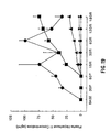
- 230000036470 plasma concentration Effects 0.000 description 12
- 239000006228 supernatant Substances 0.000 description 12
- 102000004127 Cytokines Human genes 0.000 description 11
- 108090000695 Cytokines Proteins 0.000 description 11
- 239000012979 RPMI medium Substances 0.000 description 11
- 229940079593 drug Drugs 0.000 description 11
- 238000002347 injection Methods 0.000 description 11
- 239000007924 injection Substances 0.000 description 11
- 108010044426 integrins Proteins 0.000 description 11
- 102000006495 integrins Human genes 0.000 description 11
- 238000004458 analytical method Methods 0.000 description 10
- 238000003556 assay Methods 0.000 description 10
- 230000000593 degrading effect Effects 0.000 description 10
- 239000001963 growth medium Substances 0.000 description 10
- 238000001727 in vivo Methods 0.000 description 10
- VNYSSYRCGWBHLG-AMOLWHMGSA-N leukotriene B4 Chemical compound CCCCC\C=C/C[C@@H](O)\C=C\C=C\C=C/[C@@H](O)CCCC(O)=O VNYSSYRCGWBHLG-AMOLWHMGSA-N 0.000 description 10
- 108010092694 L-Selectin Proteins 0.000 description 9
- 102000016551 L-selectin Human genes 0.000 description 9
- 230000004927 fusion Effects 0.000 description 9
- 238000001802 infusion Methods 0.000 description 9
- 238000003786 synthesis reaction Methods 0.000 description 9
- 102000003974 Fibroblast growth factor 2 Human genes 0.000 description 8
- 108090000379 Fibroblast growth factor 2 Proteins 0.000 description 8
- 239000002975 chemoattractant Substances 0.000 description 8
- 230000003511 endothelial effect Effects 0.000 description 8
- 230000001605 fetal effect Effects 0.000 description 8
- 230000000004 hemodynamic effect Effects 0.000 description 8
- 210000001616 monocyte Anatomy 0.000 description 8
- 239000000047 product Substances 0.000 description 8
- 239000000523 sample Substances 0.000 description 8
- 210000000264 venule Anatomy 0.000 description 8
- 229920002683 Glycosaminoglycan Polymers 0.000 description 7
- 108091006905 Human Serum Albumin Proteins 0.000 description 7
- 102000008100 Human Serum Albumin Human genes 0.000 description 7
- 102100025390 Integrin beta-2 Human genes 0.000 description 7
- 108010064593 Intercellular Adhesion Molecule-1 Proteins 0.000 description 7
- 102100037877 Intercellular adhesion molecule 1 Human genes 0.000 description 7
- 108060008682 Tumor Necrosis Factor Proteins 0.000 description 7
- 230000000747 cardiac effect Effects 0.000 description 7
- 230000003247 decreasing effect Effects 0.000 description 7
- 239000000463 material Substances 0.000 description 7
- 230000009467 reduction Effects 0.000 description 7
- 230000028327 secretion Effects 0.000 description 7
- 239000000725 suspension Substances 0.000 description 7
- 230000002861 ventricular Effects 0.000 description 7
- XLYOFNOQVPJJNP-UHFFFAOYSA-N water Chemical compound O XLYOFNOQVPJJNP-UHFFFAOYSA-N 0.000 description 7
- 241000283690 Bos taurus Species 0.000 description 6
- NTYJJOPFIAHURM-UHFFFAOYSA-N Histamine Chemical compound NCCC1=CN=CN1 NTYJJOPFIAHURM-UHFFFAOYSA-N 0.000 description 6
- 108010008212 Integrin alpha4beta1 Proteins 0.000 description 6
- 102100036154 Platelet basic protein Human genes 0.000 description 6
- 102100040247 Tumor necrosis factor Human genes 0.000 description 6
- 108010000134 Vascular Cell Adhesion Molecule-1 Proteins 0.000 description 6
- 102100023543 Vascular cell adhesion protein 1 Human genes 0.000 description 6
- 239000011575 calcium Substances 0.000 description 6
- 229910052791 calcium Inorganic materials 0.000 description 6
- 239000010432 diamond Substances 0.000 description 6
- 210000003989 endothelium vascular Anatomy 0.000 description 6
- 210000002744 extracellular matrix Anatomy 0.000 description 6
- 230000000302 ischemic effect Effects 0.000 description 6
- 239000011777 magnesium Substances 0.000 description 6
- 239000011159 matrix material Substances 0.000 description 6
- 239000004005 microsphere Substances 0.000 description 6
- 239000013612 plasmid Substances 0.000 description 6
- 229920000642 polymer Polymers 0.000 description 6
- 238000000746 purification Methods 0.000 description 6
- 239000011347 resin Substances 0.000 description 6
- 229920005989 resin Polymers 0.000 description 6
- UCSJYZPVAKXKNQ-HZYVHMACSA-N streptomycin Chemical compound CN[C@H]1[C@H](O)[C@@H](O)[C@H](CO)O[C@H]1O[C@@H]1[C@](C=O)(O)[C@H](C)O[C@H]1O[C@@H]1[C@@H](NC(N)=N)[C@H](O)[C@@H](NC(N)=N)[C@H](O)[C@H]1O UCSJYZPVAKXKNQ-HZYVHMACSA-N 0.000 description 6
- 210000005166 vasculature Anatomy 0.000 description 6
- 239000003981 vehicle Substances 0.000 description 6
- 108091003079 Bovine Serum Albumin Proteins 0.000 description 5
- 208000001778 Coronary Occlusion Diseases 0.000 description 5
- 206010011086 Coronary artery occlusion Diseases 0.000 description 5
- 238000002965 ELISA Methods 0.000 description 5
- 101000935040 Homo sapiens Integrin beta-2 Proteins 0.000 description 5
- MHAJPDPJQMAIIY-UHFFFAOYSA-N Hydrogen peroxide Chemical compound OO MHAJPDPJQMAIIY-UHFFFAOYSA-N 0.000 description 5
- 108010035766 P-Selectin Proteins 0.000 description 5
- 102100023472 P-selectin Human genes 0.000 description 5
- 102100033237 Pro-epidermal growth factor Human genes 0.000 description 5
- 210000001744 T-lymphocyte Anatomy 0.000 description 5
- 239000012190 activator Substances 0.000 description 5
- 150000001720 carbohydrates Chemical group 0.000 description 5
- 230000008859 change Effects 0.000 description 5
- 239000003795 chemical substances by application Substances 0.000 description 5
- 239000002158 endotoxin Substances 0.000 description 5
- 239000000284 extract Substances 0.000 description 5
- 239000003102 growth factor Substances 0.000 description 5
- 230000002757 inflammatory effect Effects 0.000 description 5
- 230000005764 inhibitory process Effects 0.000 description 5
- 238000002955 isolation Methods 0.000 description 5
- 239000007787 solid Substances 0.000 description 5
- 238000005406 washing Methods 0.000 description 5
- YBJHBAHKTGYVGT-ZKWXMUAHSA-N (+)-Biotin Chemical compound N1C(=O)N[C@@H]2[C@H](CCCCC(=O)O)SC[C@@H]21 YBJHBAHKTGYVGT-ZKWXMUAHSA-N 0.000 description 4
- 102100021943 C-C motif chemokine 2 Human genes 0.000 description 4
- 101710155857 C-C motif chemokine 2 Proteins 0.000 description 4
- 101100289995 Caenorhabditis elegans mac-1 gene Proteins 0.000 description 4
- 108010024212 E-Selectin Proteins 0.000 description 4
- 102100023471 E-selectin Human genes 0.000 description 4
- 108010037362 Extracellular Matrix Proteins Proteins 0.000 description 4
- 102000010834 Extracellular Matrix Proteins Human genes 0.000 description 4
- 241001465754 Metazoa Species 0.000 description 4
- 206010028851 Necrosis Diseases 0.000 description 4
- 102000003800 Selectins Human genes 0.000 description 4
- 108090000184 Selectins Proteins 0.000 description 4
- 230000001588 bifunctional effect Effects 0.000 description 4
- 244000309466 calf Species 0.000 description 4
- 230000001413 cellular effect Effects 0.000 description 4
- 238000005119 centrifugation Methods 0.000 description 4
- 238000013270 controlled release Methods 0.000 description 4
- 239000003431 cross linking reagent Substances 0.000 description 4
- 238000010790 dilution Methods 0.000 description 4
- 239000012895 dilution Substances 0.000 description 4
- 238000000338 in vitro Methods 0.000 description 4
- 238000011534 incubation Methods 0.000 description 4
- 230000002401 inhibitory effect Effects 0.000 description 4
- 210000004731 jugular vein Anatomy 0.000 description 4
- 229920006008 lipopolysaccharide Polymers 0.000 description 4
- 238000005259 measurement Methods 0.000 description 4
- 230000007246 mechanism Effects 0.000 description 4
- 230000002107 myocardial effect Effects 0.000 description 4
- 230000017074 necrotic cell death Effects 0.000 description 4
- 239000008188 pellet Substances 0.000 description 4
- 229920001606 poly(lactic acid-co-glycolic acid) Polymers 0.000 description 4
- 230000008569 process Effects 0.000 description 4
- 230000004044 response Effects 0.000 description 4
- NLXLAEXVIDQMFP-UHFFFAOYSA-N Ammonia chloride Chemical compound [NH4+].[Cl-] NLXLAEXVIDQMFP-UHFFFAOYSA-N 0.000 description 3
- 101800003265 Beta-thromboglobulin Proteins 0.000 description 3
- 102000008186 Collagen Human genes 0.000 description 3
- 108010035532 Collagen Proteins 0.000 description 3
- 229920002307 Dextran Polymers 0.000 description 3
- YMWUJEATGCHHMB-UHFFFAOYSA-N Dichloromethane Chemical compound ClCCl YMWUJEATGCHHMB-UHFFFAOYSA-N 0.000 description 3
- 239000006144 Dulbecco’s modified Eagle's medium Substances 0.000 description 3
- 101800003838 Epidermal growth factor Proteins 0.000 description 3
- XEKOWRVHYACXOJ-UHFFFAOYSA-N Ethyl acetate Chemical compound CCOC(C)=O XEKOWRVHYACXOJ-UHFFFAOYSA-N 0.000 description 3
- 241000589565 Flavobacterium Species 0.000 description 3
- 102000008055 Heparan Sulfate Proteoglycans Human genes 0.000 description 3
- 102100024025 Heparanase Human genes 0.000 description 3
- 101000973997 Homo sapiens Nucleosome assembly protein 1-like 4 Proteins 0.000 description 3
- 101000947178 Homo sapiens Platelet basic protein Proteins 0.000 description 3
- 101710175625 Maltose/maltodextrin-binding periplasmic protein Proteins 0.000 description 3
- 108090000235 Myeloperoxidases Proteins 0.000 description 3
- 102000003896 Myeloperoxidases Human genes 0.000 description 3
- 229930182555 Penicillin Natural products 0.000 description 3
- JGSARLDLIJGVTE-MBNYWOFBSA-N Penicillin G Chemical compound N([C@H]1[C@H]2SC([C@@H](N2C1=O)C(O)=O)(C)C)C(=O)CC1=CC=CC=C1 JGSARLDLIJGVTE-MBNYWOFBSA-N 0.000 description 3
- 101000702488 Rattus norvegicus High affinity cationic amino acid transporter 1 Proteins 0.000 description 3
- 239000006146 Roswell Park Memorial Institute medium Substances 0.000 description 3
- 108090000054 Syndecan-2 Proteins 0.000 description 3
- 239000007983 Tris buffer Substances 0.000 description 3
- 238000002835 absorbance Methods 0.000 description 3
- 239000000427 antigen Substances 0.000 description 3
- 102000036639 antigens Human genes 0.000 description 3
- 108091007433 antigens Proteins 0.000 description 3
- 210000001715 carotid artery Anatomy 0.000 description 3
- 230000020411 cell activation Effects 0.000 description 3
- 230000005779 cell damage Effects 0.000 description 3
- 230000010261 cell growth Effects 0.000 description 3
- 239000013553 cell monolayer Substances 0.000 description 3
- 229920001436 collagen Polymers 0.000 description 3
- 210000000399 corneal endothelial cell Anatomy 0.000 description 3
- 238000007405 data analysis Methods 0.000 description 3
- 230000001419 dependent effect Effects 0.000 description 3
- 230000003292 diminished effect Effects 0.000 description 3
- 230000002255 enzymatic effect Effects 0.000 description 3
- 210000003979 eosinophil Anatomy 0.000 description 3
- 229940116977 epidermal growth factor Drugs 0.000 description 3
- 238000000855 fermentation Methods 0.000 description 3
- 230000004151 fermentation Effects 0.000 description 3
- 239000012091 fetal bovine serum Substances 0.000 description 3
- 239000012634 fragment Substances 0.000 description 3
- 230000012010 growth Effects 0.000 description 3
- 108010037536 heparanase Proteins 0.000 description 3
- 229960001340 histamine Drugs 0.000 description 3
- 230000002209 hydrophobic effect Effects 0.000 description 3
- 229910052588 hydroxylapatite Inorganic materials 0.000 description 3
- 210000004969 inflammatory cell Anatomy 0.000 description 3
- BPHPUYQFMNQIOC-NXRLNHOXSA-N isopropyl beta-D-thiogalactopyranoside Chemical compound CC(C)S[C@@H]1O[C@H](CO)[C@H](O)[C@H](O)[C@H]1O BPHPUYQFMNQIOC-NXRLNHOXSA-N 0.000 description 3
- 230000023404 leukocyte cell-cell adhesion Effects 0.000 description 3
- 230000007774 longterm Effects 0.000 description 3
- 210000004962 mammalian cell Anatomy 0.000 description 3
- 230000001404 mediated effect Effects 0.000 description 3
- 239000003094 microcapsule Substances 0.000 description 3
- 239000000203 mixture Substances 0.000 description 3
- 210000000107 myocyte Anatomy 0.000 description 3
- 229940049954 penicillin Drugs 0.000 description 3
- XYJRXVWERLGGKC-UHFFFAOYSA-D pentacalcium;hydroxide;triphosphate Chemical compound [OH-].[Ca+2].[Ca+2].[Ca+2].[Ca+2].[Ca+2].[O-]P([O-])([O-])=O.[O-]P([O-])([O-])=O.[O-]P([O-])([O-])=O XYJRXVWERLGGKC-UHFFFAOYSA-D 0.000 description 3
- 210000002027 skeletal muscle Anatomy 0.000 description 3
- 239000001488 sodium phosphate Substances 0.000 description 3
- 229910000162 sodium phosphate Inorganic materials 0.000 description 3
- 238000007619 statistical method Methods 0.000 description 3
- 229960005322 streptomycin Drugs 0.000 description 3
- 239000013589 supplement Substances 0.000 description 3
- 230000036964 tight binding Effects 0.000 description 3
- LENZDBCJOHFCAS-UHFFFAOYSA-N tris Chemical compound OCC(N)(CO)CO LENZDBCJOHFCAS-UHFFFAOYSA-N 0.000 description 3
- RYFMWSXOAZQYPI-UHFFFAOYSA-K trisodium phosphate Chemical compound [Na+].[Na+].[Na+].[O-]P([O-])([O-])=O RYFMWSXOAZQYPI-UHFFFAOYSA-K 0.000 description 3
- VBEQCZHXXJYVRD-GACYYNSASA-N uroanthelone Chemical compound C([C@@H](C(=O)N[C@H](C(=O)N[C@@H](CS)C(=O)N[C@@H](CC(N)=O)C(=O)N[C@@H](CS)C(=O)N[C@H](C(=O)N[C@@H]([C@@H](C)CC)C(=O)NCC(=O)N[C@@H](CC=1C=CC(O)=CC=1)C(=O)N[C@@H](CO)C(=O)NCC(=O)N[C@@H](CC(O)=O)C(=O)N[C@@H](CCCNC(N)=N)C(=O)N[C@@H](CS)C(=O)N[C@@H](CCC(N)=O)C(=O)N[C@@H]([C@@H](C)O)C(=O)N[C@@H](CCCNC(N)=N)C(=O)N[C@@H](CC(O)=O)C(=O)N[C@@H](CC(C)C)C(=O)N[C@@H](CCCNC(N)=N)C(=O)N[C@@H](CC=1C2=CC=CC=C2NC=1)C(=O)N[C@@H](CC=1C2=CC=CC=C2NC=1)C(=O)N[C@@H](CCC(O)=O)C(=O)N[C@@H](CC(C)C)C(=O)N[C@@H](CCCNC(N)=N)C(O)=O)C(C)C)[C@@H](C)O)NC(=O)[C@H](CO)NC(=O)[C@H](CC(O)=O)NC(=O)[C@H](CC(C)C)NC(=O)[C@H](CO)NC(=O)[C@H](CCC(O)=O)NC(=O)[C@@H](NC(=O)[C@H](CC=1NC=NC=1)NC(=O)[C@H](CCSC)NC(=O)[C@H](CS)NC(=O)[C@@H](NC(=O)CNC(=O)CNC(=O)[C@H](CC(N)=O)NC(=O)[C@H](CC(C)C)NC(=O)[C@H](CS)NC(=O)[C@H](CC=1C=CC(O)=CC=1)NC(=O)CNC(=O)[C@H](CC(O)=O)NC(=O)[C@H](CC=1C=CC(O)=CC=1)NC(=O)[C@H](CO)NC(=O)[C@H](CO)NC(=O)[C@H]1N(CCC1)C(=O)[C@H](CS)NC(=O)CNC(=O)[C@H]1N(CCC1)C(=O)[C@H](CC=1C=CC(O)=CC=1)NC(=O)[C@H](CO)NC(=O)[C@@H](N)CC(N)=O)C(C)C)[C@@H](C)CC)C1=CC=C(O)C=C1 VBEQCZHXXJYVRD-GACYYNSASA-N 0.000 description 3
- OWEGMIWEEQEYGQ-UHFFFAOYSA-N 100676-05-9 Natural products OC1C(O)C(O)C(CO)OC1OCC1C(O)C(O)C(O)C(OC2C(OC(O)C(O)C2O)CO)O1 OWEGMIWEEQEYGQ-UHFFFAOYSA-N 0.000 description 2
- JKMHFZQWWAIEOD-UHFFFAOYSA-N 2-[4-(2-hydroxyethyl)piperazin-1-yl]ethanesulfonic acid Chemical compound OCC[NH+]1CCN(CCS([O-])(=O)=O)CC1 JKMHFZQWWAIEOD-UHFFFAOYSA-N 0.000 description 2
- QTBSBXVTEAMEQO-UHFFFAOYSA-M Acetate Chemical compound CC([O-])=O QTBSBXVTEAMEQO-UHFFFAOYSA-M 0.000 description 2
- 229920000856 Amylose Polymers 0.000 description 2
- 241000894006 Bacteria Species 0.000 description 2
- 241000606125 Bacteroides Species 0.000 description 2
- 102100032367 C-C motif chemokine 5 Human genes 0.000 description 2
- UXVMQQNJUSDDNG-UHFFFAOYSA-L Calcium chloride Chemical compound [Cl-].[Cl-].[Ca+2] UXVMQQNJUSDDNG-UHFFFAOYSA-L 0.000 description 2
- 102100025580 Calmodulin-1 Human genes 0.000 description 2
- 108010055166 Chemokine CCL5 Proteins 0.000 description 2
- 241000343673 Cytophagia Species 0.000 description 2
- WSFSSNUMVMOOMR-UHFFFAOYSA-N Formaldehyde Chemical compound O=C WSFSSNUMVMOOMR-UHFFFAOYSA-N 0.000 description 2
- WQZGKKKJIJFFOK-GASJEMHNSA-N Glucose Natural products OC[C@H]1OC(O)[C@H](O)[C@@H](O)[C@@H]1O WQZGKKKJIJFFOK-GASJEMHNSA-N 0.000 description 2
- 239000007995 HEPES buffer Substances 0.000 description 2
- 101001027128 Homo sapiens Fibronectin Proteins 0.000 description 2
- 101001091610 Homo sapiens Krev interaction trapped protein 1 Proteins 0.000 description 2
- 101000582950 Homo sapiens Platelet factor 4 Proteins 0.000 description 2
- 201000001779 Leukocyte adhesion deficiency Diseases 0.000 description 2
- 108010064548 Lymphocyte Function-Associated Antigen-1 Proteins 0.000 description 2
- TWRXJAOTZQYOKJ-UHFFFAOYSA-L Magnesium chloride Chemical compound [Mg+2].[Cl-].[Cl-] TWRXJAOTZQYOKJ-UHFFFAOYSA-L 0.000 description 2
- GUBGYTABKSRVRQ-PICCSMPSSA-N Maltose Natural products O[C@@H]1[C@@H](O)[C@H](O)[C@@H](CO)O[C@@H]1O[C@@H]1[C@@H](CO)OC(O)[C@H](O)[C@H]1O GUBGYTABKSRVRQ-PICCSMPSSA-N 0.000 description 2
- 102100028793 Mucosal addressin cell adhesion molecule 1 Human genes 0.000 description 2
- 101710139349 Mucosal addressin cell adhesion molecule 1 Proteins 0.000 description 2
- 102100030304 Platelet factor 4 Human genes 0.000 description 2
- 239000002202 Polyethylene glycol Substances 0.000 description 2
- 239000004372 Polyvinyl alcohol Substances 0.000 description 2
- 102100028688 Putative glycosylation-dependent cell adhesion molecule 1 Human genes 0.000 description 2
- 108010090804 Streptavidin Proteins 0.000 description 2
- 108090000190 Thrombin Proteins 0.000 description 2
- XSQUKJJJFZCRTK-UHFFFAOYSA-N Urea Chemical compound NC(N)=O XSQUKJJJFZCRTK-UHFFFAOYSA-N 0.000 description 2
- NTECHUXHORNEGZ-UHFFFAOYSA-N acetyloxymethyl 3',6'-bis(acetyloxymethoxy)-2',7'-bis[3-(acetyloxymethoxy)-3-oxopropyl]-3-oxospiro[2-benzofuran-1,9'-xanthene]-5-carboxylate Chemical compound O1C(=O)C2=CC(C(=O)OCOC(C)=O)=CC=C2C21C1=CC(CCC(=O)OCOC(C)=O)=C(OCOC(C)=O)C=C1OC1=C2C=C(CCC(=O)OCOC(=O)C)C(OCOC(C)=O)=C1 NTECHUXHORNEGZ-UHFFFAOYSA-N 0.000 description 2
- 238000013019 agitation Methods 0.000 description 2
- BFNBIHQBYMNNAN-UHFFFAOYSA-N ammonium sulfate Chemical compound N.N.OS(O)(=O)=O BFNBIHQBYMNNAN-UHFFFAOYSA-N 0.000 description 2
- 229910052921 ammonium sulfate Inorganic materials 0.000 description 2
- 235000011130 ammonium sulphate Nutrition 0.000 description 2
- 238000000540 analysis of variance Methods 0.000 description 2
- 210000000709 aorta Anatomy 0.000 description 2
- 229960002685 biotin Drugs 0.000 description 2
- 235000020958 biotin Nutrition 0.000 description 2
- 239000011616 biotin Substances 0.000 description 2
- 230000036772 blood pressure Effects 0.000 description 2
- 210000004204 blood vessel Anatomy 0.000 description 2
- 230000037396 body weight Effects 0.000 description 2
- 239000001110 calcium chloride Substances 0.000 description 2
- 229910001628 calcium chloride Inorganic materials 0.000 description 2
- 230000008614 cellular interaction Effects 0.000 description 2
- 238000010276 construction Methods 0.000 description 2
- 230000001627 detrimental effect Effects 0.000 description 2
- LOKCTEFSRHRXRJ-UHFFFAOYSA-I dipotassium trisodium dihydrogen phosphate hydrogen phosphate dichloride Chemical compound P(=O)(O)(O)[O-].[K+].P(=O)(O)([O-])[O-].[Na+].[Na+].[Cl-].[K+].[Cl-].[Na+] LOKCTEFSRHRXRJ-UHFFFAOYSA-I 0.000 description 2
- 238000009826 distribution Methods 0.000 description 2
- 238000005538 encapsulation Methods 0.000 description 2
- 210000003743 erythrocyte Anatomy 0.000 description 2
- 239000012894 fetal calf serum Substances 0.000 description 2
- 239000012530 fluid Substances 0.000 description 2
- 239000000499 gel Substances 0.000 description 2
- 238000002523 gelfiltration Methods 0.000 description 2
- 239000008103 glucose Substances 0.000 description 2
- 230000009931 harmful effect Effects 0.000 description 2
- RBTKNAXYKSUFRK-UHFFFAOYSA-N heliogen blue Chemical compound [Cu].[N-]1C2=C(C=CC=C3)C3=C1N=C([N-]1)C3=CC=CC=C3C1=NC([N-]1)=C(C=CC=C3)C3=C1N=C([N-]1)C3=CC=CC=C3C1=N2 RBTKNAXYKSUFRK-UHFFFAOYSA-N 0.000 description 2
- 102000022382 heparin binding proteins Human genes 0.000 description 2
- 108091012216 heparin binding proteins Proteins 0.000 description 2
- 230000006698 induction Effects 0.000 description 2
- 230000002452 interceptive effect Effects 0.000 description 2
- 210000005246 left atrium Anatomy 0.000 description 2
- 150000002632 lipids Chemical class 0.000 description 2
- 238000011068 loading method Methods 0.000 description 2
- 210000003141 lower extremity Anatomy 0.000 description 2
- 238000004519 manufacturing process Methods 0.000 description 2
- 210000004379 membrane Anatomy 0.000 description 2
- 239000012528 membrane Substances 0.000 description 2
- 208000031225 myocardial ischemia Diseases 0.000 description 2
- 210000004165 myocardium Anatomy 0.000 description 2
- 239000013642 negative control Substances 0.000 description 2
- 238000001543 one-way ANOVA Methods 0.000 description 2
- 102000013415 peroxidase activity proteins Human genes 0.000 description 2
- 108040007629 peroxidase activity proteins Proteins 0.000 description 2
- 229920003023 plastic Polymers 0.000 description 2
- 239000004033 plastic Substances 0.000 description 2
- 229920000515 polycarbonate Polymers 0.000 description 2
- 239000004417 polycarbonate Substances 0.000 description 2
- 229920001223 polyethylene glycol Polymers 0.000 description 2
- 229920002451 polyvinyl alcohol Polymers 0.000 description 2
- 229920000036 polyvinylpyrrolidone Polymers 0.000 description 2
- 239000001267 polyvinylpyrrolidone Substances 0.000 description 2
- 235000013855 polyvinylpyrrolidone Nutrition 0.000 description 2
- 238000001556 precipitation Methods 0.000 description 2
- 238000002203 pretreatment Methods 0.000 description 2
- AAEVYOVXGOFMJO-UHFFFAOYSA-N prometryn Chemical compound CSC1=NC(NC(C)C)=NC(NC(C)C)=N1 AAEVYOVXGOFMJO-UHFFFAOYSA-N 0.000 description 2
- 230000004224 protection Effects 0.000 description 2
- 238000001243 protein synthesis Methods 0.000 description 2
- 102000005962 receptors Human genes 0.000 description 2
- 108020003175 receptors Proteins 0.000 description 2
- 238000011084 recovery Methods 0.000 description 2
- 230000002829 reductive effect Effects 0.000 description 2
- 230000001105 regulatory effect Effects 0.000 description 2
- 238000000926 separation method Methods 0.000 description 2
- 239000011734 sodium Substances 0.000 description 2
- 239000002904 solvent Substances 0.000 description 2
- 238000000638 solvent extraction Methods 0.000 description 2
- 238000000527 sonication Methods 0.000 description 2
- 239000000758 substrate Substances 0.000 description 2
- 108010012704 sulfated glycoprotein p50 Proteins 0.000 description 2
- 230000009885 systemic effect Effects 0.000 description 2
- 238000012360 testing method Methods 0.000 description 2
- 229960004072 thrombin Drugs 0.000 description 2
- 230000014616 translation Effects 0.000 description 2
- LWIHDJKSTIGBAC-UHFFFAOYSA-K tripotassium phosphate Chemical compound [K+].[K+].[K+].[O-]P([O-])([O-])=O LWIHDJKSTIGBAC-UHFFFAOYSA-K 0.000 description 2
- GPRLSGONYQIRFK-MNYXATJNSA-N triton Chemical compound [3H+] GPRLSGONYQIRFK-MNYXATJNSA-N 0.000 description 2
- 208000003663 ventricular fibrillation Diseases 0.000 description 2
- 238000010865 video microscopy Methods 0.000 description 2
- LAQPKDLYOBZWBT-NYLDSJSYSA-N (2s,4s,5r,6r)-5-acetamido-2-{[(2s,3r,4s,5s,6r)-2-{[(2r,3r,4r,5r)-5-acetamido-1,2-dihydroxy-6-oxo-4-{[(2s,3s,4r,5s,6s)-3,4,5-trihydroxy-6-methyloxan-2-yl]oxy}hexan-3-yl]oxy}-3,5-dihydroxy-6-(hydroxymethyl)oxan-4-yl]oxy}-4-hydroxy-6-[(1r,2r)-1,2,3-trihydrox Chemical group O[C@H]1[C@H](O)[C@H](O)[C@H](C)O[C@H]1O[C@H]([C@@H](NC(C)=O)C=O)[C@@H]([C@H](O)CO)O[C@H]1[C@H](O)[C@@H](O[C@]2(O[C@H]([C@H](NC(C)=O)[C@@H](O)C2)[C@H](O)[C@H](O)CO)C(O)=O)[C@@H](O)[C@@H](CO)O1 LAQPKDLYOBZWBT-NYLDSJSYSA-N 0.000 description 1
- NWUYHJFMYQTDRP-UHFFFAOYSA-N 1,2-bis(ethenyl)benzene;1-ethenyl-2-ethylbenzene;styrene Chemical compound C=CC1=CC=CC=C1.CCC1=CC=CC=C1C=C.C=CC1=CC=CC=C1C=C NWUYHJFMYQTDRP-UHFFFAOYSA-N 0.000 description 1
- PKDBCJSWQUOKDO-UHFFFAOYSA-M 2,3,5-triphenyltetrazolium chloride Chemical compound [Cl-].C1=CC=CC=C1C(N=[N+]1C=2C=CC=CC=2)=NN1C1=CC=CC=C1 PKDBCJSWQUOKDO-UHFFFAOYSA-M 0.000 description 1
- HVAUUPRFYPCOCA-AREMUKBSSA-N 2-O-acetyl-1-O-hexadecyl-sn-glycero-3-phosphocholine Chemical compound CCCCCCCCCCCCCCCCOC[C@@H](OC(C)=O)COP([O-])(=O)OCC[N+](C)(C)C HVAUUPRFYPCOCA-AREMUKBSSA-N 0.000 description 1
- QKNYBSVHEMOAJP-UHFFFAOYSA-N 2-amino-2-(hydroxymethyl)propane-1,3-diol;hydron;chloride Chemical compound Cl.OCC(N)(CO)CO QKNYBSVHEMOAJP-UHFFFAOYSA-N 0.000 description 1
- UAIUNKRWKOVEES-UHFFFAOYSA-N 3,3',5,5'-tetramethylbenzidine Chemical compound CC1=C(N)C(C)=CC(C=2C=C(C)C(N)=C(C)C=2)=C1 UAIUNKRWKOVEES-UHFFFAOYSA-N 0.000 description 1
- FQRHOOHLUYHMGG-BTJKTKAUSA-N 3-(2-acetylphenothiazin-10-yl)propyl-dimethylazanium;(z)-4-hydroxy-4-oxobut-2-enoate Chemical compound OC(=O)\C=C/C(O)=O.C1=C(C(C)=O)C=C2N(CCCN(C)C)C3=CC=CC=C3SC2=C1 FQRHOOHLUYHMGG-BTJKTKAUSA-N 0.000 description 1
- QHRTZMDBPOUHPU-UHFFFAOYSA-N 4-(4-amino-3-methoxyphenyl)-2-methoxyaniline;hydrochloride Chemical compound Cl.C1=C(N)C(OC)=CC(C=2C=C(OC)C(N)=CC=2)=C1 QHRTZMDBPOUHPU-UHFFFAOYSA-N 0.000 description 1
- ODHCTXKNWHHXJC-VKHMYHEASA-N 5-oxo-L-proline Chemical group OC(=O)[C@@H]1CCC(=O)N1 ODHCTXKNWHHXJC-VKHMYHEASA-N 0.000 description 1
- VHUUQVKOLVNVRT-UHFFFAOYSA-N Ammonium hydroxide Chemical compound [NH4+].[OH-] VHUUQVKOLVNVRT-UHFFFAOYSA-N 0.000 description 1
- 102100030009 Azurocidin Human genes 0.000 description 1
- 101710154607 Azurocidin Proteins 0.000 description 1
- 102100032912 CD44 antigen Human genes 0.000 description 1
- 210000001266 CD8-positive T-lymphocyte Anatomy 0.000 description 1
- 102000014914 Carrier Proteins Human genes 0.000 description 1
- LZZYPRNAOMGNLH-UHFFFAOYSA-M Cetrimonium bromide Chemical compound [Br-].CCCCCCCCCCCCCCCC[N+](C)(C)C LZZYPRNAOMGNLH-UHFFFAOYSA-M 0.000 description 1
- 102000009410 Chemokine receptor Human genes 0.000 description 1
- 108050000299 Chemokine receptor Proteins 0.000 description 1
- 108020004705 Codon Proteins 0.000 description 1
- 102400000498 Connective tissue-activating peptide III Human genes 0.000 description 1
- 206010011703 Cyanosis Diseases 0.000 description 1
- 241000255925 Diptera Species 0.000 description 1
- 239000012591 Dulbecco’s Phosphate Buffered Saline Substances 0.000 description 1
- KCXVZYZYPLLWCC-UHFFFAOYSA-N EDTA Chemical compound OC(=O)CN(CC(O)=O)CCN(CC(O)=O)CC(O)=O KCXVZYZYPLLWCC-UHFFFAOYSA-N 0.000 description 1
- 241001131785 Escherichia coli HB101 Species 0.000 description 1
- 108010010803 Gelatin Proteins 0.000 description 1
- 206010018364 Glomerulonephritis Diseases 0.000 description 1
- SXRSQZLOMIGNAQ-UHFFFAOYSA-N Glutaraldehyde Chemical compound O=CCCCC=O SXRSQZLOMIGNAQ-UHFFFAOYSA-N 0.000 description 1
- 108010017213 Granulocyte-Macrophage Colony-Stimulating Factor Proteins 0.000 description 1
- 102100039620 Granulocyte-macrophage colony-stimulating factor Human genes 0.000 description 1
- 102100031573 Hematopoietic progenitor cell antigen CD34 Human genes 0.000 description 1
- 101000868273 Homo sapiens CD44 antigen Proteins 0.000 description 1
- 101000777663 Homo sapiens Hematopoietic progenitor cell antigen CD34 Proteins 0.000 description 1
- 206010020565 Hyperaemia Diseases 0.000 description 1
- 102000001706 Immunoglobulin Fab Fragments Human genes 0.000 description 1
- 108010054477 Immunoglobulin Fab Fragments Proteins 0.000 description 1
- 102100037872 Intercellular adhesion molecule 2 Human genes 0.000 description 1
- 101710148794 Intercellular adhesion molecule 2 Proteins 0.000 description 1
- 102000000589 Interleukin-1 Human genes 0.000 description 1
- 108010002352 Interleukin-1 Proteins 0.000 description 1
- 102000015696 Interleukins Human genes 0.000 description 1
- 108010063738 Interleukins Proteins 0.000 description 1
- 108010076118 L-selectin counter-receptors Proteins 0.000 description 1
- -1 LT C4 & LT D4) Chemical compound 0.000 description 1
- 101710116435 Outer membrane protein Proteins 0.000 description 1
- QGMRQYFBGABWDR-UHFFFAOYSA-M Pentobarbital sodium Chemical compound [Na+].CCCC(C)C1(CC)C(=O)NC(=O)[N-]C1=O QGMRQYFBGABWDR-UHFFFAOYSA-M 0.000 description 1
- 108091005804 Peptidases Proteins 0.000 description 1
- 102000035195 Peptidases Human genes 0.000 description 1
- 206010057249 Phagocytosis Diseases 0.000 description 1
- 108010003541 Platelet Activating Factor Proteins 0.000 description 1
- 239000004952 Polyamide Substances 0.000 description 1
- 229920001710 Polyorthoester Polymers 0.000 description 1
- 229920001213 Polysorbate 20 Polymers 0.000 description 1
- 102000017033 Porins Human genes 0.000 description 1
- 108010013381 Porins Proteins 0.000 description 1
- WCUXLLCKKVVCTQ-UHFFFAOYSA-M Potassium chloride Chemical class [Cl-].[K+] WCUXLLCKKVVCTQ-UHFFFAOYSA-M 0.000 description 1
- 241000700157 Rattus norvegicus Species 0.000 description 1
- 208000004756 Respiratory Insufficiency Diseases 0.000 description 1
- 206010039085 Rhinitis allergic Diseases 0.000 description 1
- 238000010165 Scheffé test Methods 0.000 description 1
- 206010040047 Sepsis Diseases 0.000 description 1
- 238000000692 Student's t-test Methods 0.000 description 1
- 238000010161 Student-Newman-Keuls test Methods 0.000 description 1
- QAOWNCQODCNURD-UHFFFAOYSA-N Sulfuric acid Chemical compound OS(O)(=O)=O QAOWNCQODCNURD-UHFFFAOYSA-N 0.000 description 1
- OUUQCZGPVNCOIJ-UHFFFAOYSA-M Superoxide Chemical class [O-][O] OUUQCZGPVNCOIJ-UHFFFAOYSA-M 0.000 description 1
- GLNADSQYFUSGOU-GPTZEZBUSA-J Trypan blue Chemical compound [Na+].[Na+].[Na+].[Na+].C1=C(S([O-])(=O)=O)C=C2C=C(S([O-])(=O)=O)C(/N=N/C3=CC=C(C=C3C)C=3C=C(C(=CC=3)\N=N\C=3C(=CC4=CC(=CC(N)=C4C=3O)S([O-])(=O)=O)S([O-])(=O)=O)C)=C(O)C2=C1N GLNADSQYFUSGOU-GPTZEZBUSA-J 0.000 description 1
- 108090000631 Trypsin Proteins 0.000 description 1
- 102000004142 Trypsin Human genes 0.000 description 1
- 208000027418 Wounds and injury Diseases 0.000 description 1
- 229960001946 acepromazine maleate Drugs 0.000 description 1
- 230000009471 action Effects 0.000 description 1
- 230000002411 adverse Effects 0.000 description 1
- 238000001042 affinity chromatography Methods 0.000 description 1
- 230000002776 aggregation Effects 0.000 description 1
- 238000004220 aggregation Methods 0.000 description 1
- 235000010443 alginic acid Nutrition 0.000 description 1
- 229920000615 alginic acid Polymers 0.000 description 1
- 201000010105 allergic rhinitis Diseases 0.000 description 1
- 238000012870 ammonium sulfate precipitation Methods 0.000 description 1
- 230000003444 anaesthetic effect Effects 0.000 description 1
- 238000010171 animal model Methods 0.000 description 1
- 230000000845 anti-microbial effect Effects 0.000 description 1
- 239000012062 aqueous buffer Substances 0.000 description 1
- 239000007864 aqueous solution Substances 0.000 description 1
- 208000006673 asthma Diseases 0.000 description 1
- QVGXLLKOCUKJST-UHFFFAOYSA-N atomic oxygen Chemical compound [O] QVGXLLKOCUKJST-UHFFFAOYSA-N 0.000 description 1
- 210000003719 b-lymphocyte Anatomy 0.000 description 1
- 230000001580 bacterial effect Effects 0.000 description 1
- 230000004888 barrier function Effects 0.000 description 1
- 230000009286 beneficial effect Effects 0.000 description 1
- 108091008324 binding proteins Proteins 0.000 description 1
- 229920000229 biodegradable polyester Polymers 0.000 description 1
- 239000004622 biodegradable polyester Substances 0.000 description 1
- 230000002051 biphasic effect Effects 0.000 description 1
- 230000000903 blocking effect Effects 0.000 description 1
- 239000001045 blue dye Substances 0.000 description 1
- 230000036760 body temperature Effects 0.000 description 1
- 210000000988 bone and bone Anatomy 0.000 description 1
- 239000000872 buffer Substances 0.000 description 1
- 210000004899 c-terminal region Anatomy 0.000 description 1
- 239000004202 carbamide Substances 0.000 description 1
- 230000005961 cardioprotection Effects 0.000 description 1
- 230000003293 cardioprotective effect Effects 0.000 description 1
- 230000015556 catabolic process Effects 0.000 description 1
- 230000003197 catalytic effect Effects 0.000 description 1
- 238000005341 cation exchange Methods 0.000 description 1
- 239000003729 cation exchange resin Substances 0.000 description 1
- 230000021164 cell adhesion Effects 0.000 description 1
- 239000006285 cell suspension Substances 0.000 description 1
- 210000002421 cell wall Anatomy 0.000 description 1
- 238000012512 characterization method Methods 0.000 description 1
- 238000006243 chemical reaction Methods 0.000 description 1
- 210000000038 chest Anatomy 0.000 description 1
- HGAZMNJKRQFZKS-UHFFFAOYSA-N chloroethene;ethenyl acetate Chemical compound ClC=C.CC(=O)OC=C HGAZMNJKRQFZKS-UHFFFAOYSA-N 0.000 description 1
- 238000004587 chromatography analysis Methods 0.000 description 1
- 230000004087 circulation Effects 0.000 description 1
- 238000000975 co-precipitation Methods 0.000 description 1
- 230000000536 complexating effect Effects 0.000 description 1
- 239000012141 concentrate Substances 0.000 description 1
- 210000002808 connective tissue Anatomy 0.000 description 1
- 108010035886 connective tissue-activating peptide Proteins 0.000 description 1
- 230000008094 contradictory effect Effects 0.000 description 1
- 210000004351 coronary vessel Anatomy 0.000 description 1
- 238000012937 correction Methods 0.000 description 1
- 230000001086 cytosolic effect Effects 0.000 description 1
- 210000001151 cytotoxic T lymphocyte Anatomy 0.000 description 1
- 230000000254 damaging effect Effects 0.000 description 1
- 230000003413 degradative effect Effects 0.000 description 1
- 230000018044 dehydration Effects 0.000 description 1
- 238000006297 dehydration reaction Methods 0.000 description 1
- 230000003111 delayed effect Effects 0.000 description 1
- 238000012217 deletion Methods 0.000 description 1
- 230000037430 deletion Effects 0.000 description 1
- 239000003599 detergent Substances 0.000 description 1
- 238000000502 dialysis Methods 0.000 description 1
- 230000035487 diastolic blood pressure Effects 0.000 description 1
- 230000003205 diastolic effect Effects 0.000 description 1
- IJKVHSBPTUYDLN-UHFFFAOYSA-N dihydroxy(oxo)silane Chemical compound O[Si](O)=O IJKVHSBPTUYDLN-UHFFFAOYSA-N 0.000 description 1
- 230000010339 dilation Effects 0.000 description 1
- 238000007865 diluting Methods 0.000 description 1
- 230000003467 diminishing effect Effects 0.000 description 1
- 150000002016 disaccharides Chemical class 0.000 description 1
- 239000012153 distilled water Substances 0.000 description 1
- 231100000673 dose–response relationship Toxicity 0.000 description 1
- 239000003937 drug carrier Substances 0.000 description 1
- 238000001035 drying Methods 0.000 description 1
- 239000000975 dye Substances 0.000 description 1
- 238000013195 electrical cardioversion Methods 0.000 description 1
- 230000008030 elimination Effects 0.000 description 1
- 238000003379 elimination reaction Methods 0.000 description 1
- 238000010828 elution Methods 0.000 description 1
- 239000008393 encapsulating agent Substances 0.000 description 1
- 238000011156 evaluation Methods 0.000 description 1
- 230000005284 excitation Effects 0.000 description 1
- 230000007717 exclusion Effects 0.000 description 1
- 239000012997 ficoll-paque Substances 0.000 description 1
- 238000009472 formulation Methods 0.000 description 1
- 238000005194 fractionation Methods 0.000 description 1
- 229920000159 gelatin Polymers 0.000 description 1
- 239000008273 gelatin Substances 0.000 description 1
- 235000019322 gelatine Nutrition 0.000 description 1
- 235000011852 gelatine desserts Nutrition 0.000 description 1
- 238000010353 genetic engineering Methods 0.000 description 1
- 239000011521 glass Substances 0.000 description 1
- 150000002327 glycerophospholipids Chemical class 0.000 description 1
- 125000003827 glycol group Chemical group 0.000 description 1
- PCHJSUWPFVWCPO-UHFFFAOYSA-N gold Chemical compound [Au] PCHJSUWPFVWCPO-UHFFFAOYSA-N 0.000 description 1
- 239000010931 gold Substances 0.000 description 1
- 229910052737 gold Inorganic materials 0.000 description 1
- 210000000224 granular leucocyte Anatomy 0.000 description 1
- 229960004198 guanidine Drugs 0.000 description 1
- PJJJBBJSCAKJQF-UHFFFAOYSA-N guanidinium chloride Chemical compound [Cl-].NC(N)=[NH2+] PJJJBBJSCAKJQF-UHFFFAOYSA-N 0.000 description 1
- BCQZXOMGPXTTIC-UHFFFAOYSA-N halothane Chemical compound FC(F)(F)C(Cl)Br BCQZXOMGPXTTIC-UHFFFAOYSA-N 0.000 description 1
- 229960003132 halothane Drugs 0.000 description 1
- 238000003306 harvesting Methods 0.000 description 1
- 210000002837 heart atrium Anatomy 0.000 description 1
- 239000002874 hemostatic agent Substances 0.000 description 1
- 238000004128 high performance liquid chromatography Methods 0.000 description 1
- 238000009775 high-speed stirring Methods 0.000 description 1
- 238000000265 homogenisation Methods 0.000 description 1
- 210000005260 human cell Anatomy 0.000 description 1
- 238000007654 immersion Methods 0.000 description 1
- 230000002163 immunogen Effects 0.000 description 1
- 238000010348 incorporation Methods 0.000 description 1
- 239000000411 inducer Substances 0.000 description 1
- 230000001939 inductive effect Effects 0.000 description 1
- 230000008595 infiltration Effects 0.000 description 1
- 238000001764 infiltration Methods 0.000 description 1
- 230000000977 initiatory effect Effects 0.000 description 1
- 208000014674 injury Diseases 0.000 description 1
- 229940047122 interleukins Drugs 0.000 description 1
- 238000007918 intramuscular administration Methods 0.000 description 1
- 238000010253 intravenous injection Methods 0.000 description 1
- 238000012771 intravital microscopy Methods 0.000 description 1
- 208000012947 ischemia reperfusion injury Diseases 0.000 description 1
- 238000002372 labelling Methods 0.000 description 1
- 210000005240 left ventricle Anatomy 0.000 description 1
- 150000002617 leukotrienes Chemical class 0.000 description 1
- 230000000670 limiting effect Effects 0.000 description 1
- 239000002502 liposome Substances 0.000 description 1
- 239000007788 liquid Substances 0.000 description 1
- 238000004811 liquid chromatography Methods 0.000 description 1
- 210000001165 lymph node Anatomy 0.000 description 1
- 210000004698 lymphocyte Anatomy 0.000 description 1
- 210000003563 lymphoid tissue Anatomy 0.000 description 1
- 210000002540 macrophage Anatomy 0.000 description 1
- 229910001629 magnesium chloride Inorganic materials 0.000 description 1
- 238000012423 maintenance Methods 0.000 description 1
- 230000002503 metabolic effect Effects 0.000 description 1
- 229920000609 methyl cellulose Polymers 0.000 description 1
- 239000001923 methylcellulose Substances 0.000 description 1
- 235000010981 methylcellulose Nutrition 0.000 description 1
- 244000005700 microbiome Species 0.000 description 1
- 239000011859 microparticle Substances 0.000 description 1
- 238000002156 mixing Methods 0.000 description 1
- 230000004048 modification Effects 0.000 description 1
- 238000012986 modification Methods 0.000 description 1
- 238000001823 molecular biology technique Methods 0.000 description 1
- 239000003068 molecular probe Substances 0.000 description 1
- 210000000865 mononuclear phagocyte system Anatomy 0.000 description 1
- 238000011587 new zealand white rabbit Methods 0.000 description 1
- 230000001473 noxious effect Effects 0.000 description 1
- 238000010899 nucleation Methods 0.000 description 1
- 235000015097 nutrients Nutrition 0.000 description 1
- 229920001542 oligosaccharide Polymers 0.000 description 1
- 150000002482 oligosaccharides Chemical class 0.000 description 1
- 239000003960 organic solvent Substances 0.000 description 1
- 230000003204 osmotic effect Effects 0.000 description 1
- 239000001301 oxygen Substances 0.000 description 1
- 229910052760 oxygen Inorganic materials 0.000 description 1
- 230000036961 partial effect Effects 0.000 description 1
- 230000007310 pathophysiology Effects 0.000 description 1
- 229960002275 pentobarbital sodium Drugs 0.000 description 1
- 239000000137 peptide hydrolase inhibitor Substances 0.000 description 1
- 210000001322 periplasm Anatomy 0.000 description 1
- 230000008782 phagocytosis Effects 0.000 description 1
- 239000008194 pharmaceutical composition Substances 0.000 description 1
- 230000000144 pharmacologic effect Effects 0.000 description 1
- 238000005191 phase separation Methods 0.000 description 1
- 239000008363 phosphate buffer Substances 0.000 description 1
- 150000003904 phospholipids Chemical class 0.000 description 1
- 229920002401 polyacrylamide Polymers 0.000 description 1
- 229920002647 polyamide Polymers 0.000 description 1
- 229920000728 polyester Polymers 0.000 description 1
- 238000003752 polymerase chain reaction Methods 0.000 description 1
- 239000000256 polyoxyethylene sorbitan monolaurate Substances 0.000 description 1
- 235000010486 polyoxyethylene sorbitan monolaurate Nutrition 0.000 description 1
- 229920002635 polyurethane Polymers 0.000 description 1
- 239000004814 polyurethane Substances 0.000 description 1
- 239000011148 porous material Substances 0.000 description 1
- 239000011736 potassium bicarbonate Substances 0.000 description 1
- 229910000028 potassium bicarbonate Inorganic materials 0.000 description 1
- TYJJADVDDVDEDZ-UHFFFAOYSA-M potassium hydrogencarbonate Chemical compound [K+].OC([O-])=O TYJJADVDDVDEDZ-UHFFFAOYSA-M 0.000 description 1
- 229910000160 potassium phosphate Inorganic materials 0.000 description 1
- 235000011009 potassium phosphates Nutrition 0.000 description 1
- 230000002265 prevention Effects 0.000 description 1
- 125000002924 primary amino group Chemical group [H]N([H])* 0.000 description 1
- 102000004196 processed proteins & peptides Human genes 0.000 description 1
- 108090000765 processed proteins & peptides Proteins 0.000 description 1
- 230000002035 prolonged effect Effects 0.000 description 1
- 230000004850 protein–protein interaction Effects 0.000 description 1
- 239000003725 proteoliposome Substances 0.000 description 1
- 230000017854 proteolysis Effects 0.000 description 1
- 229940024999 proteolytic enzymes for treatment of wounds and ulcers Drugs 0.000 description 1
- 229940043131 pyroglutamate Drugs 0.000 description 1
- 230000002285 radioactive effect Effects 0.000 description 1
- 239000012857 radioactive material Substances 0.000 description 1
- 238000003259 recombinant expression Methods 0.000 description 1
- 238000011160 research Methods 0.000 description 1
- 201000004193 respiratory failure Diseases 0.000 description 1
- 230000029058 respiratory gaseous exchange Effects 0.000 description 1
- 108091008146 restriction endonucleases Proteins 0.000 description 1
- 238000004007 reversed phase HPLC Methods 0.000 description 1
- 238000012552 review Methods 0.000 description 1
- 206010039073 rheumatoid arthritis Diseases 0.000 description 1
- 230000033764 rhythmic process Effects 0.000 description 1
- 210000005241 right ventricle Anatomy 0.000 description 1
- 102220047090 rs6152 Human genes 0.000 description 1
- 239000012723 sample buffer Substances 0.000 description 1
- 229920006298 saran Polymers 0.000 description 1
- 239000002356 single layer Substances 0.000 description 1
- 239000001509 sodium citrate Substances 0.000 description 1
- NLJMYIDDQXHKNR-UHFFFAOYSA-K sodium citrate Chemical compound O.O.[Na+].[Na+].[Na+].[O-]C(=O)CC(O)(CC([O-])=O)C([O-])=O NLJMYIDDQXHKNR-UHFFFAOYSA-K 0.000 description 1
- 238000002415 sodium dodecyl sulfate polyacrylamide gel electrophoresis Methods 0.000 description 1
- 238000005063 solubilization Methods 0.000 description 1
- 230000007928 solubilization Effects 0.000 description 1
- 230000009870 specific binding Effects 0.000 description 1
- 238000001228 spectrum Methods 0.000 description 1
- 238000001694 spray drying Methods 0.000 description 1
- 238000010186 staining Methods 0.000 description 1
- 230000000638 stimulation Effects 0.000 description 1
- 239000000126 substance Substances 0.000 description 1
- 208000011580 syndromic disease Diseases 0.000 description 1
- 229920001059 synthetic polymer Polymers 0.000 description 1
- 230000035488 systolic blood pressure Effects 0.000 description 1
- 230000002123 temporal effect Effects 0.000 description 1
- 210000002435 tendon Anatomy 0.000 description 1
- 150000004044 tetrasaccharides Chemical class 0.000 description 1
- 125000003831 tetrazolyl group Chemical group 0.000 description 1
- 238000011287 therapeutic dose Methods 0.000 description 1
- 239000003848 thrombocyte activating factor antagonist Substances 0.000 description 1
- 230000000451 tissue damage Effects 0.000 description 1
- 231100000827 tissue damage Toxicity 0.000 description 1
- 208000037816 tissue injury Diseases 0.000 description 1
- 230000001988 toxicity Effects 0.000 description 1
- 231100000419 toxicity Toxicity 0.000 description 1
- 231100000583 toxicological profile Toxicity 0.000 description 1
- 210000003437 trachea Anatomy 0.000 description 1
- 230000001052 transient effect Effects 0.000 description 1
- 102000035160 transmembrane proteins Human genes 0.000 description 1
- 108091005703 transmembrane proteins Proteins 0.000 description 1
- 239000012588 trypsin Substances 0.000 description 1
- 102000003390 tumor necrosis factor Human genes 0.000 description 1
- 230000003827 upregulation Effects 0.000 description 1
- 239000007762 w/o emulsion Substances 0.000 description 1
- 238000012784 weak cation exchange Methods 0.000 description 1
- 238000005303 weighing Methods 0.000 description 1
- 238000001262 western blot Methods 0.000 description 1
Images


Classifications
-
- A—HUMAN NECESSITIES
- A61—MEDICAL OR VETERINARY SCIENCE; HYGIENE
- A61K—PREPARATIONS FOR MEDICAL, DENTAL OR TOILETRY PURPOSES
- A61K38/00—Medicinal preparations containing peptides
- A61K38/16—Peptides having more than 20 amino acids; Gastrins; Somatostatins; Melanotropins; Derivatives thereof
- A61K38/43—Enzymes; Proenzymes; Derivatives thereof
- A61K38/51—Lyases (4)
-
- A—HUMAN NECESSITIES
- A61—MEDICAL OR VETERINARY SCIENCE; HYGIENE
- A61K—PREPARATIONS FOR MEDICAL, DENTAL OR TOILETRY PURPOSES
- A61K47/00—Medicinal preparations characterised by the non-active ingredients used, e.g. carriers or inert additives; Targeting or modifying agents chemically bound to the active ingredient
- A61K47/50—Medicinal preparations characterised by the non-active ingredients used, e.g. carriers or inert additives; Targeting or modifying agents chemically bound to the active ingredient the non-active ingredient being chemically bound to the active ingredient, e.g. polymer-drug conjugates
-
- A—HUMAN NECESSITIES
- A61—MEDICAL OR VETERINARY SCIENCE; HYGIENE
- A61P—SPECIFIC THERAPEUTIC ACTIVITY OF CHEMICAL COMPOUNDS OR MEDICINAL PREPARATIONS
- A61P29/00—Non-central analgesic, antipyretic or antiinflammatory agents, e.g. antirheumatic agents; Non-steroidal antiinflammatory drugs [NSAID]
-
- A—HUMAN NECESSITIES
- A61—MEDICAL OR VETERINARY SCIENCE; HYGIENE
- A61P—SPECIFIC THERAPEUTIC ACTIVITY OF CHEMICAL COMPOUNDS OR MEDICINAL PREPARATIONS
- A61P43/00—Drugs for specific purposes, not provided for in groups A61P1/00-A61P41/00
-
- A—HUMAN NECESSITIES
- A61—MEDICAL OR VETERINARY SCIENCE; HYGIENE
- A61P—SPECIFIC THERAPEUTIC ACTIVITY OF CHEMICAL COMPOUNDS OR MEDICINAL PREPARATIONS
- A61P9/00—Drugs for disorders of the cardiovascular system
- A61P9/10—Drugs for disorders of the cardiovascular system for treating ischaemic or atherosclerotic diseases, e.g. antianginal drugs, coronary vasodilators, drugs for myocardial infarction, retinopathy, cerebrovascula insufficiency, renal arteriosclerosis
-
- C—CHEMISTRY; METALLURGY
- C12—BIOCHEMISTRY; BEER; SPIRITS; WINE; VINEGAR; MICROBIOLOGY; ENZYMOLOGY; MUTATION OR GENETIC ENGINEERING
- C12N—MICROORGANISMS OR ENZYMES; COMPOSITIONS THEREOF; PROPAGATING, PRESERVING, OR MAINTAINING MICROORGANISMS; MUTATION OR GENETIC ENGINEERING; CULTURE MEDIA
- C12N9/00—Enzymes; Proenzymes; Compositions thereof; Processes for preparing, activating, inhibiting, separating or purifying enzymes
- C12N9/88—Lyases (4.)
-
- C—CHEMISTRY; METALLURGY
- C07—ORGANIC CHEMISTRY
- C07K—PEPTIDES
- C07K2319/00—Fusion polypeptide
-
- Y—GENERAL TAGGING OF NEW TECHNOLOGICAL DEVELOPMENTS; GENERAL TAGGING OF CROSS-SECTIONAL TECHNOLOGIES SPANNING OVER SEVERAL SECTIONS OF THE IPC; TECHNICAL SUBJECTS COVERED BY FORMER USPC CROSS-REFERENCE ART COLLECTIONS [XRACs] AND DIGESTS
- Y10—TECHNICAL SUBJECTS COVERED BY FORMER USPC
- Y10S—TECHNICAL SUBJECTS COVERED BY FORMER USPC CROSS-REFERENCE ART COLLECTIONS [XRACs] AND DIGESTS
- Y10S424/00—Drug, bio-affecting and body treating compositions
- Y10S424/81—Drug, bio-affecting and body treating compositions involving autoimmunity, allergy, immediate hypersensitivity, delayed hypersensitivity, immunosuppression, immunotolerance, or anergy
Definitions
- This invention is directed to the use of a heparinase enzyme in the preparation of a medicament for intravascular administration to decrease inflammatory responses arising from an ischaemia/reperfusion injury in a patient's tissue.
- An inflammatory response is local response to cellular injury that is marked by capillary dilation, leukocytic infiltration, redness, heat, and pain and serves as a mechanism initiating the elimination of noxious agents and of damaged tissue.
- a generalized inflammatory response within a tissue occurs by the recruitment of leukocytes to the tissue. Destruction of bacteria, foreign materials and/or damaged cells occurs through phagocytosis and/or extracellular degranulation (secretion of degradative enzymes, antimicrobial proteins and myeloperoxidase, which forms superoxides from secreted H 2 O 2 ). While most localized inflammatory responses are beneficial, harmful inflammatory responses can occur. Many harmful inflammatory responses also involve accumulation of leukocytes within a tissue. This accumulation results in the destruction of viable cells and tissue. In addition to damaging tissue, these responses are detrimental to, or debilitating for, the afflicted individual.
- Examples of detrimental inflammatory responses can include the following; ischemia/reperfusion injury following myocardial infarction, shock, stroke, organ transplantation, cardiopulmonary bypass surgery, allograft rejection, rheumatoid arthritis, antigen-induced asthma, allergic rhinitis, and glomerulonephritis (see the review in; Harlan, et al., Immunol. Rev., 114:5-12, 1990; Carlos and Harlan, Blood, 84:2068-2101,1994.)
- Leukocyte recruitment involves a cascade of cellular events, beginning with activation of vascular endothelium by damaged or infected tissue adjacent to the endothelium. Activation of the endothelium results in enhanced adhesion of leukocytes to the endothelial cells, and transendothelial migration (extravasation) by bound leukocytes into the damaged tissue. Endothelial activation is manifested by a short-term, rapid, and/or a long-term stimulation of the endothelial cells.
- Activators such as thrombin, chemoattractant leukotrienes, B 4 , C 4 and D 4 (LTB 4 , LT C 4 & LT D 4 ), and histamine cause rapid, transient ( ⁇ 30 minutes) endothelial cell activation, independent of protein synthesis, and can increase endothelial cell surface levels of the chemoattractants such as platelet activating factor (PAF; a glycerophospholipid) and LTB 4 (shown for histamine), and adhesion molecules; ICAM-1 (shown for thrombin) and P-selectin (Zimmerman, et al., J .
- PAF platelet activating factor
- ICAM-1 shown for thrombin
- P-selectin Zimmerman, et al., J .
- IL-1b and TNF-a Long-term (hours in duration) protein synthesis dependent, endothelial cells activation is produced by cytokines, such as IL-1b and TNF-a, and by lipopolysaccharide (LPS) and results in maintenance of increased surface levels of adhesion molecules: E-selectin, P-selectin, ICAM-1 and VCAM-1 (reviewed by Carlos, and Harlan, Blood, 84:2068-2101, 1994).
- IL-1b and TNF-a also increase the synthesis of PAF by endothelial cells (Kuijpers, et al., J . Cell Biol., 117:565-574, 1992).
- IL-1b endothelial cell activation by IL-1b, TNF-a, LPS and histamine has been shown to increase the synthesis and secretion of the chemokine, IL-8 (Stricter, et al., Science, 243:1467-1469,1989; Jeannin, et al., Blood, 84:2229-2233, 1994).
- Chemokines, IL-8 and MCP-1 have been shown to be produced by and to be present on the endothelial cells surface (Huber, et al., Science, 254:99-102, 1991; Springer, Nature, 346:425-434, 1990).
- the chemokine, MIP-1b has been shown to be present on lymph node endothelium, in vivo (Taub, et al., Science, 260:355-359, 1993; Tanaka, et al., Nature, 361:79-82, 1993).
- chemokines are all heparin binding proteins, which after being secreted, bind to cell surface and extracellular matrix proteoglycans possessing heparin and heparan sulfate moieties (reviewed by Miller, et al., Crit. Rev. Immunol., 12:17-46, 1992).
- Heparin and heparan sulfate are similar glycosaminoglycan moieties found interspersed on the same unbranched carbohydrate chains. They are covalently attached to the protein backbones of proteoglycans. Despite what these two names imply, heparin is more highly sulfated than is heparan sulfate. Proteoglycans are present on cell surfaces and in extracellular matrices (e.g. in the basement membrane of endothelium).
- chemokines Because of difficulty in distinguishing regions of heparin and heparan sulfate on the same carbohydrate chain, little data exists on the binding preference of chemokines for either heparin or heparan sulfate moieties. There is some indication that chemokines IL-8 and GRO bind with greater affinity to heparan sulfate than heparin, and that PF4 and NAP-2 bind with greater affinity to heparin moieties (Witt, and Lander, Curr. Biol., 4:394-400, 1994). Generally, chemokines are referred to as heparin binding proteins.
- C-terminal regions of the chemokines IL-8, PF4, MCP-1 and NAP-2 have been shown to form an a-helix, and to bind to heparin/heparan sulfate (Webb, et al., Proc. Natl. Acad. Sci. USA, 90:7158-7162, 1993; Zucker, et al., Proc. Natl. Acad. Sci. USA, 86:7571-7574, 1989; Matsushima, et al., in Interleukins: Molec. Biol. Immunol., ed. Kistimoto, Karger, Basel, 236-265, 1992). This is likely to be a structure, common to all of the chemokines.
- Adhesion of leukocytes to endothelium is thought to be a two step process (reviewed by Carlos, and Harlan, Blood, 84:2068-2101, 1994). Initially, leukocytes roll along the surface of blood vessels. Increased rolling is initially mediated on vascular endothelium (within the first 30 minutes) by interactions between Sialyl Lewisx structures on the leukocyte surface and P-selectin and E-selectin, which are increased on activated endothelial cells (Ley, et al., Blood, 85:3727-3735, 1995).
- Increased rolling is also mediated (after 40 minutes) by interactions between L-selectin on leukocyte cellular membranes and heparin-like molecules on the vascular endothelium, which are cytokine-inducable (Karin. et al., Science, 261:480-483,1993), or between L-selectin on lymphocytes and vascular addressins; GlyCAM-1, CD34 and MAdCAM-1 on high endothelial venules (HEVs) in lymphoid tissue.
- the second step, firm adhesion of leukocytes to endothelial cells is based on interactions between leukocyte integrins (e.g.
- LFA-1, Mac-1, VLA-4) and endothelial cellular adhesion molecules CAMs; e.g. ICAM-1, ICAM-2, VCAM-1, MAdCAM-1).
- Leukocytes flatten on the endothelial surface, and shed L-selectin, concomitant with, firm adhesion (Kishimoto, et al., Science, 245:1238-1242, 1989; Jutila, et al., J. Immunol., 143:3318-3324, 1989; Smith, et al., Clin. Invest., 87:609-618, 1991).
- Selectin and CAM levels increase on the endothelium surface in response to many cytokines and chemoattractants. These increases are dependent on synthesis and/or secretion of additional selectin and CAM molecules onto the cell surface.
- activation of leukocytes for firm adhesion has been shown to occur within seconds (Bargatze, et al., J . Exp. Med., 178:367-373, 1993), through increased secretion of integrins, and more importantly, through induction of conformational changes in cell surface integrins (integrin activation), which permits tight binding of the integrins to CAMs (reviewed in Zimmerman, Immunol. Today, 13:93-99, 1992).
- PAF and E-selectin can activate integrins for endothelial cell adhesion (Lorant et al., J. Biol. Chem., 115:223-234,1991; Lo, J . Exp. Med., 173:1493-1500,1991).
- the presence of MIP-1b, immobilized by binding to CD44 (possesses heparin/heparan sulfate moieties), or a heparin-BSA conjugate, has been shown to be required for CD8+ T-cell binding to immobilized VCAM-1 molecules.
- IL-8 activates neutrophils, eosinophils and T cells.
- RANTES activates monocytes, eosinophils and T cells.
- MCP-1 activates monocytes.
- MIP-1a activates CD4+ T cells, monocytes and B cells, while MIP-1b activates monocytes and CD8+ T cells (reviewed in Lasky, Current Biol., 3:366-378, 1993).
- selectins, integrins, CAMs and chemokines are thought to select for the adhesion and migration of the leukocyte subtypes observed in different inflamed tissues (Butcher, Cell, 67:1033-1039, 1991).
- VLA-4 is not expressed by neutrophils (Winn and Harlan, J . Clin. Invest., 92:1168-1174, 1993).
- chemokines are important for activation and increased surface levels of integrins VLA-4 and CD18 on leukocytes.
- IL-8 immobilized on a polycarbonate filter has been shown to be adequate for directing migration of neutrophils through the filter (Rot, Immunol. Today , 13:291-294). Huber, et al.
- LPS has been shown to directly increase monocyte IL-1b expression (Porat, et al., FASEB J ., 6:2482-2489, 1992).
- IL-8, IL-1b and TNF-a are produced by neutrophils activated with GM-CSF, another cytokine produced by activated macrophages, endothelium and T-cells (McCain, et al., Am. J . Respir. Cell Molec.
- IL-1b and TNF-a have been shown to stimulate monocytes, thereby increasing the expression of the chemokines, IL-8 and MIP-1a (Lukacs, et al., Blood, 82:3668-3674, 1993).
- Activated neutrophils and monocytes have been shown to be a major source of LTB 4 production (Samuelsson, et al., Science, 237:1171-1176, 1987; Brach, et al., Eur.
- LTB 4 is not directly involved in further recruitment of leukocytes, but because neutrophils stimulated with LTB 4 produce IL-8, the LTB 4 -stimulated neutrophils could promote further neutrophil recruitment, indirectly, through formation of an IL-8 gradient (McCain, et al., Am. J . Respir. Cell Molec. Biol., 10:651-657 , 1994).
- the continued recruitment of leukocytes by these leukocyte-derived activators would require using the vascular endothelium as an intermediate.
- Endothelial cells and basement membranes would bind and display neutrophil-derived chemokines, forming a gradient, or leukocyte-derived cytokines would activate the endothelium, which would also cause the creation of a chemokine gradient.
- soluble activation factors cytokines, chemokines and chemoattractants
- cytokines cytokines, chemokines and chemoattractants
- a systemic activation of leukocytes could occur (Finn, et al., J. Thorac. J. Surg., 105:234-241, 1993).
- the activated leukocytes would then bind transiently to unactivated endothelium and/or degranulate, causing sepsis (Sawyer, et al., Rev. Infect. Dis., 11:S1532-1544, 1989).
- the activated leukocytes would produce and secrete additional cytokines, chemokines; and LTB 4 into the blood. This increase in activator concentration could up-regulate unactivated cells and amplify the systemic response.
- This invention is directed to the discovery that heparinase degrading enzymes, either separately or in combination, can be used to decrease localized inflammatory response.
- the present invention provides the use of a heparinase enzyme in the preparation of a medicament for intravascular administration to decrease localised inflammatory responses arising from an ischaemia/reperfusion injury in a patient's tissue.
- the heparinases useful in this invention can be from a variety of sources: heparinases I, II, and III from the Gram negative bacterium Flavobacterium heparinum, heparinase from Bacteroides strains, heparinase from Flavobacterium Hp206, heparinase from Cytophagia species, and heparanases from mammalian cells. These enzymes, either singly or in combination, are referred to herein as heparinase or heparinase enzyme.
- the heparinase may be overexpressed from a recombinant nucleotide sequence in Flavobacterium heparinum or Escherichia coli.
- the heparinase may be heparinase III.
- the ischaemia/reperfusion injury may be myocardial infarction, stroke, organ transplantation shock or cardiopulmonary bypass surgery.
- Heparin and heparan sulfate moieties are degraded on the surface of endothelial cells and from basement membranes by administration of heparinase. Removal of heparin and heparan sulfate moieties from up-regulated proteoglycans on activated endothelial cells prevents L-selectin, found on leukocytes, from interacting with the proteoglycans. By decreasing L-selectin-proteoglycan interactions, leukocyte rolling on activated endothelium can be inhibited.
- chemokines which are bound to the heparin and heparan sulfate, are released from the cell surfaces and basement membrane.
- the loss of bound chemokines decreases the localized concentration of chemokines and disrupts the chemokine gradient produced by activated endothelium, thereby inhibiting chemokine activation of rolling leukocytes, which is required for firm adhesion, and preventing extravasation of leukocytes along the chemokine gradient.
- decreased leukocyte rolling, activation and extravasation can inhibit localized tissue inflammatory responses by interfering with fundamental mechanisms of leukocyte recruitment.
- Heparinase enzyme can be targeted to specific cell types, tissues or organs by the selected method of administration, which deliver localized high concentrations of the enzymes or physically limit the dispersal of the enzymes. Additionally, according to this invention heparinase can be targeted by fusion of the enzymes to binding domains from antibodies; growth factors or adhesion molecules.
- the fusion proteins are produced by construction and expression of gene fusions in recombinant organisms.
- the binding domains can recognize cell surface molecules on activated endothelium (e.g., ICAM-1, VCAM-1, P-selectin, E-selectin), or on endothelial cell subtypes (e.g., GlyCAM-1).
- Targeted fusion enzymes can increase the number and specificity of indications, while decreasing the amounts of enzyme required for efficacy and possible side-effects resulting from treatments.
- Interactions between leukocytes and endothelium are critical to the progress of localized inflammatory responses. These critical interactions include functional contacts between endothelium bound chemokines and leukocyte chemokine receptors, and between leukocyte L-selectin and heparin/heparan sulfate proteoglycans on endothelium.
- This invention is based on the discovery and is directed to the use of heparinase enzyme and heparinase fusion protein to decrease leukocyte-chemokine and leukocyte-endothelial cells proteoglycan interactions, and thereby inhibiting localized inflammation.
- Heparin and heparan sulfate are glycosaminoglycan moieties of proteoglycans located on the surface of many different cell types and also found in the extracellular matrices produced by many cells. Endothelial cells produce extracellular matrix, primarily on their abluminal side, referred to as basement membrane. Endothelial cells, activated by certain cytokines or by other inflammatory response stimulators, increase their surface levels of heparin and heparan sulfate proteoglycans (excluding high endothelial venules), which act as inflammatory adhesion molecules, and interact with L-selectin on rolling leukocytes.
- chemokines This interaction increases contacts of leukocytes with the endothelium (increased rolling of leukocytes), which are necessary for subsequent steps in leukocyte recruitment.
- Activated endothelium also increases it's synthesis and secretion of chemokines.
- the secretion of chemokines in turn increases the localized concentration of the same chemokines, because the chemokines bind to the heparin and heparan sulfate moieties of proteoglycans on the endothelial cells surfaces and in the basement membranes. This localized concentration gradient is required for activation of leukocytes for firm adhesion and extravasation, and results in leukocyte recruitment to inflammatory sites.
- Heparinase enzymes have been found in microorganisms including Flavobacterium heparinum (Lohse and Linhardt, J. Biol. Chem . 267:2437-24355, 1992), Bacteroides strains (Saylers, et al., Appl. Environ. Microbiol. 33:319-322, 1977; Nakamura, et al., J . Clin. Microbiol. 26:1070-1071, 1988), Flavobacterium Hp206 (Yoshida, et al., 10th Annual Symposium of Glycoconjugates, Jerusalem 1989) and Cytophagia species (Bohn, et al., Drug Res. 41(I), Nr.
- Heparanases from mammalian cells have also been described (Fuks, et al., US patent number 5,362,641, 1994; Hoogewerf et al., J . Biol. Chem. 270:3268-3277, 1995).
- the heparinases from Flavobacterium heparinum, heparinase I (EC 4.2.2.7), and heparinase II degrade heparin, while heparinase II also degrades heparan sulfate, as does heparinase III (EC 4.2.2.8).
- the products of complete digestion by these enzymes are mainly disaccharides, though small quantities of tetrasaccharides and oligosaccharides may persist.
- These enzymes can be used to remove cell surface and basement membrane glycosaminoglycans, heparin and heparan sulfate.
- glycosaminoglycans which are critical for leukocyte recruitment. Loss of endothelial cells bound chemokines decreases activation of leukocyte integrins and inhibits firm adhesion by the leukocytes. It also inhibits extravasation of leukocytes, because the leukocytes require the presence of a bound gradient of chemokine for transmigration. It is believed, without being limited, that unbound chemoattractants are depleted from the endothelium layer by blood flow, preventing formation of a significant soluble chemoattractant gradient.
- heparinase 50% of the digested cell surface and basement membrane heparin and heparan sulfate are replaced within 2 to 4 hours, and it is completely replaced within 12 to 16 hours. Longer treatment times (3 and 5 hours) greatly extended the time needed to replace the same amount of heparin/heparan sulfate. Inflammatory responses would be significantly diminished by a slow rate of replacement of cell surface heparin/heparan sulfate. Appropriate administration of heparinase could extend the duration of diminished inflammatory response.
- Heparinases or a combination thereof, that may be used in this invention can be prepared from a variety of sources.
- Heparinase may be prepared by isolation from bacterial or mammalian cells, either those which naturally produce the enzymes or have been genetically engineered to produce the enzymes as described in by known methods.
- mammalian heparanases from human cells may be isolated according to procedures for purification described by Fuks, et al. (U.S. 5,362,641, 1994).
- Heparinase enzymes can be purified from cultures of Flavobacterium heparinum as follows. F. heparinum is cultured in 15 L computer controlled fermenters, in a variation of the defined nutrient medium described by Galliher et al., Appl. Environ. Microbiol. 41(2):360-365, 1981. For fermentations designed to produce heparin lyases, semi-purified heparin (Celsus Laboratories) is included in the media at a concentration of 1.0 g/L as the inducer of heparinase synthesis. The cells are harvested by centrifugation and the desired enzymes released from the periplasmic space by a variation of the osmotic shock procedure described by U.S. Patent No. 5,169,772 to Zimmermann, et al. (1992).
- Proteins from the crude osmolate are adsorbed onto cation exchange resin (CBX, J.T. Baker) at a conductivity of between one and seven mhos. Unbound proteins from the extract are discarded and the resin packed into a chromatography column (5.0 cm i.d. x 100 cm). The bound proteins elute at a linear flow rate of 3.75 cm•min-1 with step gradients of 0.01 M phosphate, 0.01 M phosphate/0.1 M sodium chloride, 0.01 M phosphate/0.25 M sodium chloride and 0.01 M phosphate/ 1.0 M. sodium chloride, all at pH, 7.0 ⁇ 0.1.
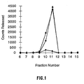
- Heparinase II elutes in the 0.1 M NaCl fraction while heparinases, I and III, elute in the 0.25 M fraction.
- the 0.1 M sodium chloride step is eliminated and the three heparinases co- eluted with 0.25 M sodium chloride.
- the heparinase fractions are loaded directly onto a column containing cellufine sulfate (5.0 cm i.d. x 30 cm, Amicon) and eluted at a linear flow rate of 2.50 cm•min-1 with step gradients of 0.01 M phosphate, 0.01 M phosphate/0.2 M sodium chloride, 0.01 M phosphate/0.4 M sodium chloride and 0.01 M phosphate/ 1.0 M.
- Heparinase I is further purified by loading material from the cellufine sulfate column, diluted to a conductivity less than 5 mhos, onto a hydroxyapatite column (2.6 cm i.d. x 20 cm) and eluting the bound protein at a linear flow rate of 1.0 cm•min-1 with a linear gradient of phosphate (0.01 to 0.25 M) and sodium chloride (0.0 to 0.5 M). Heparinase I elutes in a single protein peak approximately mid-way through the gradient.
- the heparinase enzymes obtained by this method are greater than 98.5% pure as estimated by reverse phase HPLC analysis (BioCad, POROS II). Purification results for the heparinase enzymes are shown in Table A. Purification of heparinase enzymes from Flavobacterium heparinum fermentations sample activity (IU) specific activity (IU/mg) yield (%) fermentation heparin degrading 94,500 100 heparan sulfate degrading 75,400 ND 100 osmolate heparin degrading 52,100 55 heparan sulfate degrading 42,000 ND 56 cation exchange heparin degrading 22,600 24 heparan sulfate degrading 27,540 ND 37 cellufine sulfate heparin degrading 19,200 20 heparan sulfate degrading 9,328 30.8 12 hydroxylapatite heparinase 1 16,300 115.3 17 heparin
- Glycosaminoglycan degrading enzymes also can be isolated from recombinant expression systems such as the heparinase I expression system described by Sasisekharan, et al., Proc. Natl. Acad. Sci. USA 90:8660-8664, 1993; the heparinase, II and III, expression systems described in WO 95/34635.
- the F . heparinum genes are isolated and doned into plasmids downstream from an inducable promoter.
- the plasmids are introduced into E . coli and the expression of the desired enzyme directed by a suitable induction method such as temperature shift or addition of IPTG to the medium.
- the enzymes can be recovered in a purified form by a modification of the methods described herein.
- Cell disruption is achieved by homogenization, sonication or enzymatic treatment to break the cell wall and release cytoplasmic components. If enzyme synthesis results in aggregation, the aggregate can then be dissolved by a denaturing agent, 3 to 6 M guanidine HCl or 4 to 8 M urea and the protein refolded by removal of the denaturing agent through dialysis or dilution.
- the refolded enzyme can be further purified using the liquid chromatographic methods described above.
- Fusion proteins incorporating heparinase enzyme fused to proteins with specific binding properties can be created by recombinant molecular biology techniques. By choosing an appropriate binding protein, heparinase activity can be targeted to specific sites, in vivo. ICAM-1 has been shown to be preferentially expressed on the surface of activated endothelial cells (Dustin, et al., J. Immunol., 137:245-254, 1986).
- fusion proteins an antibody, Fab fragment or variable region, specific for ICAM-1, VCAM-1 or P-selectin, when fused to heparinase enzyme or an active portion thereof, localizes heparinase activity near the luminal and abluminal surfaces of activated endothelium. Heparin and heparan sulfate moieties are removed in this area, causing breakdown of the chemokine gradient produced by the endothelium.
- fusion of heparinase enzyme, or an active portion thereof, to the I-domain of LFA-1 or Mac-1 targets activated endothelium for removal of heparin and heparan sulfate, inhibiting leukocyte rolling and chemokine gradient formation.
- Receptors for cytokines such as IL-1b are up-regulated on activated endothelium and provide another target for binding of fusion proteins.
- IL-lb, or the receptor binding domain of IL-1b to heparinase targeting can also be achieved.
- the fusion proteins can decrease inflammatory responses at lower blood concentrations than is required for comparable decreases using unfused heparinase.
- other cells in the vascular system will be less affected by the enzyme activity of the fusion protein, reducing possible side effects of treatments.
- Heparinase fusion proteins created by genetic engineering retain the binding and catalytic properties of heparinase and of the protein to which it is fused.
- Three heparinases have been purified to homogeneity from Flavobacterium heparinum, and have been produced in a recombinant form in Escherichia coli.
- Fusion proteins consisting of heparinase enzyme combined with binding domains from antibodies or adhesion molecules can be produced with a gene fusion in a recombinant host, while retaining the functionalities of binding and the enzymatic activity of the separate proteins.
- These molecules can also be purified to homogeneity by procedures normally used for purification of the individual parts of the fusion protein (e.g.
- the recombinant enzyme may not contain amino-terminal pyroglutamate or carbohydrate moieties. All recombinant heparinase may contain deletions, additional and/or altered amino adds, which modify the enzymatic activity of the natural enzyme or the functioning of the binding domains. Heparinase and fusion heparinase can be stabilized for in vivo use, by complexing with polyethylene glycol, cross linking agents, and by microencapsulation.
- the gene for heparinase I was isolated from F. heparinum as described by Sasisekharan, et al., Proc. Natl. Acad. Sci. 90:3660- 3664, 1993, and an EcoR I restriction site was inserted 5' to the codon encoding the glutamine-21 residue by polymerase chain reaction.
- a fragment containing the heparinase I gene was prepared by digestion with restriction endonucleases; Eco R I and Bam H I, and ligated to the EcoR I/ Bam H I cleaved pMALc2 plasmid (New England Biolabs).
- the resulting plasmid contained a hybrid gene encoding a 82,000 - 85,000 Dalton protein incorporating the maltose binding protein (MalB) fused 5' to the heparinase I gene.
- This plasmid was inserted into Escherichia coli HB101 cells using the calcium chloride mediated method described by Cohen et al., Proc. Natl. Acad. Sci. 69:2110-211. These cells exhibited heparinase activity under the control of the tac promoter, allowing synthesis of the fusion protein by addition of 0.1 mM of the inducing agent IPTG to the growth medium.
- the HB101(pMALc2-HEP1Q21) cells were grown to a cell density of 1.0 g/L dry cell weight in 500 ml, M9 medium containing 0.1 mM IPTG at 37 ⁇ C and concentrated by centrifugation, 10,000 g x 10 minutes.
- the cell pellet was suspended in 10 ml 0.025 M Tris, pH 7.7, and the cells disrupted by sonication using a Heat Systems Model XL2020, 4.5 minutes, power level 3, 30 second on 30 second off cycles. Cell debris was removed by centrifugation, 10,000 g x 10 minutes, and the supernatant applied to an amylose affinity resin column (1.0 i.d. x 2 cm, New England Biolabs).
- the bound protein was eluted with a step gradient of 0.025 M Tris containing 0.01 M maltose at pH 7.5.
- the fusion protein eluted in a protein peak which displayed a heparinase specific activity of 23.77 IU/mg.
- the heparinase-maltose binding fusion protein also can be purified by standard protein separation techniques based on heparinase properties.
- Cell sonicates were fractionated by ammonium sulfate precipitation.
- Non-specific proteins were removed with a precipitation step at 1.7 M ammonium sulfate and the supernatant precipitated by raising the ammonium sulfate concentration to 3.2 M.
- the precipitated material contained the fusion protein and was resuspended in 0.025 M sodium phosphate, pH 6.5. The material was applied to a weak cation exchange column (1.6 i.d. x 10 cm, CBX, J.T.
- a BamH I/ Sal I restriction fragment from pGBH3, which contains the heparinase III gene from Flavobacterium heparinum was inserted into pMALc2 to form a gene for fusion of a maltose binding protein with heparinase III.
- Extracts of the E . coli strain DH5a containing the fusion gene plasmid were produced as described in the last example, and these extracts contained 18.7 IU/ml/O.D. of heparinase III activity.
- the extract was also combined with amylose affinity resin and the resin was then separated from the extract by centrifugation.
- the resin was washed once with 0.025M Tris (pH 7.5) solution and proteins bound to the resin were resuspended in SDS-PAGE sample buffer and separated according to size on a 7.5% SDS-polyacrylamide gel.
- Western blot analysis of the gel with anti-heparinase III specific antibody identified a 116,000 Da. protein, which corresponds to the expected size of the fusion protein. This analysis indicates that the fusion protein has a functional maltose binding domain.
- This example demonstrates that the heparinase III protein can also be fused to a binding domain to produce a bifunctional fusion enzyme.
- Glutaraldehyde is one type of bifunctional cross-linking agent.
- PCT WO95/00171, by Novo Nordisk A/S contains a listing of other useful bifunctional cross-linking agents, and teaches the use of these, which is incorporated herein.
- Heparinase enzyme is administered locally. Local administration provides greater control. Heparinase is mixed with an appropriate pharmaceutical or veterinary carrier, then administered in an effective amount to produce the desired effect on the treated cells using methods known to those skilled in the art, for example, for local application, use of perfusion, injection or a catheter.
- Targeting and effective concentration dosages can be achieved by preparation of targeted enzymes as described above, or by the use of targeting vehicles, such as a catheter or localized injection, to achieve controlled site specific delivery of enzyme.
- Heparinase enzyme can be formulated in a carrier for administration by injection, for example, in saline or an aqueous buffer, using standard methodology, or encapsulated in a polymeric matrix.
- Encapsulation of heparinase in controlled release formulations is well known; materials include but not limited to liposomes, lipospheres, biodegradable polymeric matrices, and vesicles. These encapsulants are typically microparticles having a diameter from 60 nm to 100 microns, but preferably less than ten microns, and more preferably one micron or less in diameter.
- Proteosomes are prepared from outer membrane proteins of the Meningococcal bacteria and been reported to bind proteins containing hydrophobic anchors by Lowell, et al., Science, 240:800 (1988). Proteosome proteins are highly hydrophobic, reflecting their role as transmembrane proteins and porins. When isolated, their hydrophobic protein-protein interactions cause them to form naturally multimolecular, membranous 60 to 1000 nm vesicles or membrane vesicle fragments, depending on the strength of the detergent used in their isolation. Heparinase can also be encapsulated within a proteoliposome as described by Miller et al., J. Exp. Med.
- heparinase can be encapsulated in lipid vesicles such as NovasomeTM lipid vesicles (Micro Vescular Systems, Inc., Nashua, NH).
- lipid vesicles such as NovasomeTM lipid vesicles (Micro Vescular Systems, Inc., Nashua, NH).
- NovasomeTM lipid vesicles Micro Vescular Systems, Inc., Nashua, NH
- Another carrier is described in WO 91/07760 by Nova Pharmaceuticals, the teachings of which are incorporated herein, which is referred to as a liposphere, having a solid core and an outer shell layer formed of phospholipid.
- the carrier may also be a polymeric delayed release system.
- Biodegradable synthetic polymers are particularly useful to effect the controlled release of heparinase. Microencapsulation has been applied to the injection of microencapsulated pharmaceuticals to give a controlled release. A number of factors contribute to the selection of a particular polymer for microencapsulation. The reproducibility of polymer synthesis and the microencapsulation process, the cost of the microencapsulation materials and process, the toxicological profile, the requirements for variable release kinetics and the physicochemical compatibility of the polymer and the antigens are all factors that must be considered. Examples of useful polymers are polycarbonates, polyesters, polyurethanes, polyorthoesters and polyamides, particularly those that are biodegradable.
- PLGA poly (d,l-lactide-co-glycolide)
- PLGA poly (d,l-lactide-co-glycolide)
- This is a biodegradable polyester that has a long history of medical use in erodible sutures, bone plates and other temporary prostheses, where it has not exhibited any toxicity.
- a wide variety of pharmaceuticals including peptides and antigens have been formulated into PLGA microcapsules.
- the PLGA microencapsulation process uses a phase separation of a water-in-oil emulsion. Heparinase is prepared as an aqueous solution and the PLGA is dissolved in a suitable organic solvents such as methylene chloride and ethyl acetate.
- Heparinase enzyme can be administered by injection, infusion or perfusion. Typically, injection is performed using either a syringe or catheter. Either a syringe or a catheter can be used to apply heparinase locally to areas of blood vessels, tissues or organs. Patients diagnosed with localized inflammations can be treated by introduction of heparinase into their vascular system by these means. Heparinase can also be administered before or simultaneously with surgery, to reduce resulting inflammatory responses. In addition, preceding transplant surgery, the donor organ can be perfused with a heparinase preparation to reduce inflammation upon reperfusion after transplantation.
- Example 1 Treatment of Endothelial Cells with Heparinase to Release Heparin/Heparan Sulfate.
- Heparinase alters the cell surface and basement membrane by cleaving the heparin and heparan sulfate moieties from the cell surface and extracellular matrix proteoglycans. Removal of these glycosaminoglycans will decrease leukocyte-endothelium interactions (leukocyte rolling) by decreasing binding of L-selectin on leukocytes to endothelium proteoglycans. In addition, the removal of heparin and heparan sulfate will decrease binding of chemokines to the endothelium, which will reduce leukocyte activation, sticking and extravasation.
- 35 S-sulfate containing endothelial cell layers were produced by seeding 24 well dishes with primary bovine corneal endothelial cells. These were grown until 1 day prior to confluence in DMEM with 10% fetal calf serum and 5% calf serum. One day prior to confluence the cells were diluted 10- fold into Fisher medium supplemented with 10% fetal calf serum, 5% calf serum, 4% dextran, and 25 mCi/ml Na 2 35SO 4 and cultured for 3 days with the addition of 0.5 ng/ml per day bFGF. Incorporation of label by near-confluent cells localizes the label in and on the cell and minimizes the 35 S- label incorporated into the basement membrane.
- Endothelial cell layers containing 35 S- sulfate were treated with 600 ul phosphate buffered saline or heparinases III, in phosphate buffered saline, at a concentration of 1.0 IU/ml, in duplicate wells.
- the digestions were allowed to proceed for 5, 30 or 60 minutes at 37 ⁇ C.
- 400 ul of digestion solution was removed from each well and fractionated on a Bio-sil SEC 125-5 gel filtration column controlled by a Beckman System Gold HPLC, equipped with an autosampler. The flow rate was 1 ml/minute and 1 ml fractions were collected.
- the amount of 35S- sulfate present in each fraction was determined by measuring an aliquot of each fraction on a Packard 1600 TR liquid scintillation counter.
- the labeled, untreated control solutions were fractionated and measured in the same manner, and the quantity of radioactive material in each fraction (background) was subtracted from the amount present in fractions from the heparinase digested samples.
- the amount of cell surface 35 S-labeled heparin/heparan sulfate in each fraction released by heparinase III treatment is shown in figure 1.
- Example 2 Determination of the Extent of Removal and the Rate of Replacement of Heparin/Heparan Sulfate Moieties on Endothelial Cells and in Basement Membranes Treated with Heparinase.
- the growth factor basic Fibroblast Growth Factor (bFGF)
- bFGF basic Fibroblast Growth Factor
- bFGF basic Fibroblast Growth Factor
- Binding of 125 I- labeled bFGF to cell surface and basement membrane proteoglycans was used to access the amount of heparin/heparan sulfate removed from unactivated and IL-1b activated endothelial cell layers and their basement membranes by heparinases I, II or III.
- heparinase 70 to 90%; figures 2, 3 and 4. This demonstrates that the endothelium can be almost completely depleted of heparin/heparan sulfate moieties by use of heparinase. With a 1 hour heparinase treatment the rate of replacement of the heparin/heparan sulfate is generally biphasic in nature. Replacement of 40 to 50% of the digested heparin/heparan sulfate occurs within a few hours.
- a human endothelial cell line was grown to confluency in 48 well dishes in 0.25 ml/well of RPMI medium, containing; 1% penicillin /streptomycin, and 20% fetal serum. Cells in half of the wells were activated for 4 hours with 10 ul of 50 ng/ml IL-1b.
- the wells were washed 3 times with HBSS and incubated with RPMI medium, containing; 1% penicillin / streptomycin and 20% fetal serum, ⁇ IL-1b, at 37°C, in 5% CO 2 , for the times indicated in Figure 2.
- the wells were again washed 3 times with HBSS and 0.1 ml of Incubation Medium was added to each well, and the plates were cooled on ice for 5 min. To all of the wells was added 20 ul of 125 ng/ml 125 I-bFGF, and 20 ul of 20 ug/ml unlabeled bFGF.
- Example 3 Treatment of Activated Endothelial Cell Layers and Basement Membranes with Heparinase to Release Heparin/Heparan Sulfate Bound Chemokine, IL-8.
- IL-8 is produced by endothelium activated by IL-1b and other cytokines and chemoattractants, which are secreted by inflamed tissues. If IL-8 bound to endothelium can be solubilized by treatment with heparinase, then it would be removed from the area of inflammation by blood flow and the localized inflammatory response would be inhibited.
- the in vitro removal and solubilization of 0.5 to 3 fold more endogenous, immobilized IL-8 (vs. secreted IL-8) from activated endothelium by heparinases I, II or III, or by heparinases I and III demonstrates that the bound chemokine gradient can be destroyed by heparinase treatment.
- the cells were grown until confluent, at 37°C, in 5% CO 2 .
- the culture medium was changed every other day during the growth period.
- the day before the chemokine assay the medium was exchanged for 1 ml of RPMI medium without heparin.
- IL-1b human recombinant IL-1b
- RPMI medium minus fetal serum, epidermal growth factor, endothelial cell growth supplement and heparin
- BSA Hanks Balanced Salt Solution
- 0.5 ml of RPMI medium (minus fetal serum, epidermal growth factor, endothelial cell growth supplement and heparin) and 2% BSA was added to the wells for the times indicated in Figure 3. After the indicated times, the wells were emptied and washed once with 1 ml of HBSS. 0.5 ml of HBSS with or without 1 IU/ml of heparinase I, II or III; or heparinases I and III were added to the wells. The plates were incubated at 37°C on a heat block for 15 minutes with occasional agitation. After 15 minutes, the supernatants were collected and assayed for IL-8.
- ELISA enzyme-linked immunosorbent assay
- Example 4 Treatment of Endothelial Cell Layers with Heparinase to Inhibit Neutrophil Adhesion.
- endothelial cell surface molecules activate rolling neutrophils for tight binding to the walls of postcapillary venules.
- Chemokines bound to heparin/heparan sulfate have been identified as important signal molecules for activation of neutrophils for tight binding in the microcapillaries.
- the in vitro neutrophil adhesion assay system described below is commonly used to analyze conditions affecting neutrophil adhesion to endothelial cells.
- Treatment of the activated human endothelial cell layers used in this assay with either heparinase I, II or III resulted in significant reductions in the level of neutrophil adhesion to the endothelium.
- 25 ml of venous blood was drawn from a healthy donor into 1/10 volume of 0.1M sodium citrate, pH 7.4, and was diluted with 25 ml of Dulbecco's phosphate-buffered saline containing calcium chloride and magnesium chloride (D-PBS). 10 ml aliquots of diluted blood were layered on 10 ml of Ficoll-Paque in 50 ml tubes. The tubes were centrifuged at 400 x g for 30 minutes at 20°C, and were allowed to stop, without braking.
- D-PBS Dulbecco's phosphate-buffered saline containing calcium chloride and magnesium chloride
- the upper layers were removed and the pellets were resuspended in 3 volumes of a solution of 155 mM NH 4 Cl, 10 mM KHCO 3 and 0.1 mM EDTA, pH 7.4 at 4°C, to lyse the erythrocytes.
- the tubes were inverted after a few minutes and the contents turn black after 7 to 8 minutes. After 10 minutes, the tubes were centrifuged again at 400 x g for 5 minutes at 4°C. The supernatants were aspirated and the pellets were resuspended in the NH 4 Cl solution containing 0.5% human albumin. The suspensions were pooled and the volume was brought to 50 ml.
- the cell suspension was incubated on ice for 15 minutes and centrifuged at 400 x g for 5 minutes at 4°C. The supernatant was removed and if the pellet was still red, it was washed again with NH 4 Cl solution containing 0.5% human albumin. Finally, the cells were resuspended in D-PBS with 5 mg/ml human albumin and refrigerated, until needed. A 10 ⁇ l aliquot of suspension was diluted with 10 ⁇ l of trypan blue and the cells were counted on a hemacytometer to determine the number of viable cells per volume of suspension.
- a suspension of 10 x 106 neutrophils was made in 2 ml of PBS (no Ca, no Mg) with 5 mg/ml human albumin.
- BCECF-AM (Molecular Probes) was added to the suspension for a final concentration of 46 uM.
- the neutrophils were incubated in a water bath at a set temperature of 37°C for 30 minutes after which they were centrifuged and rinsed twice with PBS (no Ca, no Mg) with 5 mg/ml human albumin. They were finally resuspended in 10 ml of RPMI + 20 % bovine fetal serum at 37°C.
- HUVEC at passage number 3 were trypsinized and counted.
- the cells were plated in RPMI + 20 % bovine fetal serum + 95 ug/ml heparin + ECGS + EGF at 5 x 10⁴ cells per well in 96 well plates. They were grown at 37 °C, 5% CO2 for 18 hours. At that point the growth medium was replaced with RPMI + 20 % bovine fetal serum ( 2 ng/ml IL-1b for 4 hours.
- the cells were then rinsed with HBSS and treated with heparinase I, II or III at 0.1 IU/ml in HBSS for 1 hour at 37 °C, 5% CO2.
- HUVEC were rinsed once with HBSS.
- 200 ul of the neutrophil suspension (which corresponds to 200,000 neutrophils) was added to each well of treated or control HUVEC.
- the plate was put at 37 °C, 5% CO2 for 30 minutes.
- the adhesion period was stopped by centrifuging the plate upside down at 250 x g for 5 minutes.
- 200 ul of PBS (no Ca, no Mg) + 5 mg/ml human albumin was added to each well and the plate was read with a Fluorolite 1000 fluorescence plate reader at a voltage of 2.5 V.
- the emission filter was at 535 nm ⁇ 35 and the excitation filter at 485 ⁇ 22.
- a standard curve was generated by putting known amounts of BCECF-AM-stained neutrophils, resuspended in PBS (no Ca, no Mg) + 5 mg/ml human albumin in wells of another 96 well plate which contained confluent HUVEC layers. The fluorescence units were plotted against the quantity of neutrophils and a slope was calculated. The standard curve was used to determine the number of bound neutrophils in the control and heparinase treated wells. The percent differences between IL-1b activated HUVEC layers and comparable HUVEC layers treated with heparinases I, II and III are shown in figure 6.
- Example 5 Treatment of Endothelial Cell Layers and Basement Membranes with Heparinase to Inhibit Neutrophil Extravasation.
- Leukocytes from the blood accumulate in inflamed tissues by transmigration across the adjacent endothelium (extravasation).
- the endothelial cell layer is activated by the inflamed tissue (via cytokines and chemoattractants), and the affected endothelium directs and localizes the accumulation of leukocytes in the inflamed tissue.
- the activated endothelium forms an immobilized chemokine gradient on its cell surface and in its basement membrane.
- the in vitro neutrophil transmigration assay system described below is commonly used to analyze conditions affecting neutrophil extravasation.
- Neutrophils were isolated as described in example 4. Human fibronectin was dissolved at 0.4 mg/ml in RPMI medium without serum. Filter inserts (6.25 mm) of pore size, 3 ⁇ m or 8 ⁇ m, were coated with 4 ⁇ g/cm2 of human fibronectin for one hour, at room temperature, and were rinsed with distilled water. Wells of a 24 well plate were filled with 0.3 ml of RPMI medium with 20 % fetal bovine serum, 95 ⁇ g/ml heparin, 200 ⁇ g/ml ECGS and 10 ng/ml EGF.
- the coated filter inserts were seated in the wells, and 8 x 10⁴ human umbilical venous endothelial cells (HUVEC; used at passages 1 to 7) in 0.3 ml of complete RPMI medium, were plated on the coated filter inserts.
- the cells were allowed to grow for 48 to 65 hours, at 37°C, in 5% CO 2 .
- the culture medium from the filter inserts and the wells was changed once during the growth period for RPMI medium lacking heparin. After the growth period, the culture medium underneath all inserts, except negative control inserts, was removed and replaced with fresh culture medium lacking heparin and growth factors, but containing 2 ng/ml of human recombinant IL-1b.
- the culture medium under negative control inserts was replaced with fresh culture medium.
- the multiwell plates were incubated at 37°C, in 5% CO 2 , for 4 hours.
- the medium was removed from the inserts and wells and the cells were rinsed once with Hank's Balanced Salt Solution (HBSS).
- HBSS Hank's Balanced Salt Solution
- the filters and wells were filled with 0.3 ml of a solution of HBSS; treated inserts received HBSS containing heparinase I, II or III instead of HBSS. This treatment was performed for the times indicated in Figure 1, at 37°C, in 5% CO 2 .
- the solution was removed and the cells were rinsed once with HBSS.
- Standards containing between 1 x 10⁵ and 1 x 10⁶ neutrophils in a 1 ml volume were produced by diluting an aliquot of the neutrophil suspension in D-PBS. 4 ml of 50 mM potassium phosphate, pH 6.7, containing 0.5 % hexadecyltrimethylammonium bromide and 0.5 % triton was added to each standard and thawed sample. 0.1 ml of each sample or standard was added to a plastic cuvette. 2.9 ml of 50 mM phosphate buffer, pH 6.0 containing 0.167 mg/ml o-dianisidine hydrochloride and 0.0005% hydrogen peroxide were added to the cuvettes. The change in absorbance at 460 nm was monitored every 30 seconds for 3 minutes by use of a spectrophotometer.
- the rate changes obtained from the standards were used to produce a curve of the rate of increase in absorbance versus numbers of neutrophils. This curve was used to quantitate the number of neutrophils in each sample, which had migrated through, either a treated, or an untreated endothelial layer. The number of migrating neutrophils was divided by 1.5 x 10⁶ to determine the percentage of neutrophil migration.
- the effectiveness of the heparinase treatment in this assay depended on the extent to which the HUVEC were covering the filter surface. The extent of coverage was based on dye exclusion analysis performed after an extravasation experiment, and it varied somewhat from filter to filter in a single experiment. Generally, if the filter was densely covered with a tightly packed HUVEC layer, the percentage of extravasating neutrophils was low ( ⁇ 10%), and the differences between treated and untreated wells were not statistically significant (large well to well variability).
- Example 6 Treatment of Endothelial Cell Layers and Basement Membranes with Human Heparinase (b-thromboglobulin) to Inhibit Neutrophil Extravasation.
- b-thromboglobulin are a mixture of the chemokines CTAP-III and NAP-2. At non-physiological pH (pH 5.8-6), these chemokines have heparinase activity, while at pH 7, they bind heparin and act as chemotactic cytokines for leukocytes.
- the heparinase activity of commercial b-thromboglobulin preparations were analyzed at pH 5.8 and pH 7, by digestion of radioactive 35SO 4 -labeled ECM. These preparations showed significant human heparinase activity only at pH 5.8.
- Bovine corneal endothelial cells at passage 1 to 8 were split 1:10 form a confluent plate and seeded in 4 well plates in DMEM low glucose with 10 % fetal bovine serum, 5% calf serum and 4% Dextran added. The dishes received 1 ng/ml bFGF 3 times a week prior to reaching confluency.
- b-thromboglobulin from Wellmark (product #41705) or Calbiochem (product #605165) were used for digestion of the labeled ECM.
- 100 ug of enzyme was dissolved in 1 ml of water (Wellmark) or PBS (Calbiochem). A further dilution was done in PBS at pH 5.8 or 7.
- the matrices were covered with 250 ul of PBS alone or PBS with 1 or 5 ug of ⁇ -thromboglobulin.
- the matrices were incubated in a CO 2 incubator for 3 hours. Aliquots of 100 ul were taken from every well and counted. The amount of radioactivity released from each enzyme-treated matrix was compared to an untreated matrix, and the results are displayed in figure 8.
- HUVEC were grown on filter inserts and activated as described in example 5.
- Five ug of ⁇ -thromboglobulin in PBS at pH 5.8 or PBS alone were applied to the HUVEC layer for one hour.
- Neutrophils were added above the filters and the number of extravasating neutrophils were quantitated as described in example 5.
- the effect of the human heparinase treatments on neutrophil extravasation are shown in figure 9.
- Example 7 Treatment of Rats with Heparinase Inhibits Leukocyte-Endothelial Cell Interactions Following Ischemia /Reperfusion.
- This example illustrates the effect of heparinase III on leukocyte behavior in vivo.
- This example demonstrates that heparinase treatment of vasculature would inhibit neutrophil accumulation in microcapillaries and in the surrounding tissues. In vivo heparinase treatment would result in decreased inflammatory responses.
- heparinase treatment significantly increased microvascular perfusion within reperfused muscle following ischemia.
- increased microvascular perfusion would positively affect the recovery of muscle tissue and positively affect the outcome of ischemia/reperfusion (i.e. inflammatory) events.
- mice Male Wistar rats weighing 225 to 250 gm were anesthetized by inhalation of halothane (1% - 1.5%) and the carotid artery and jugular vein cannulated to monitor blood pressure and permit infusion of fluids, respectively.
- the extensor digitorum longus (EDL) muscle in the rat hind limb was prepared for intravital microscopy. Briefly, with the anaesthetized rat lying on the stage of the microscope, the EDL muscle was exposed by reflection of the overlying skin and separation of the tibialis anterior and gastrocnemius muscles. A suture was tied around the distal tendon of the muscle allowing reflection of the muscle into a saline bath positioned on the microscope stage.
- EDL extensor digitorum longus
- Plasma levels of heparinase III are measured using a heparinase III ELISA.
- the heparinase III ELISA is a quantitative two-antibody sandwich assay. Affinity purified anti-heparinase III rabbit antibodies are coated onto a microtiter plate. Wells are washed and incubated for 2 hours at 37(C with blocking buffer (TBS, 1% BSA +1% Tween 20). After 3 washes, standards and samples are added to the wells and any heparinase III present is bound by the immobilized antibody. Any unbound substance is then washed away and biotin labeled anti- heparinase III rabbit antibodies are added to the wells.
- a series of rats were infused with heparinase III via the venous catheter at a rate of 3 ml/hr for 5 hr. to maintain a heparinase level of 1.0 IU/ml in the blood. Through such pilot studies it was determined that 0.33 IU/gm body weight/hr was adequate for this purpose.
- Microvascular blood flow and leukocyte behavior was recorded every 15 min for a total of 90 to 105 min of reperfusion. Blood samples were taken before the administration of heparinase and during reperfusion to ensure correct plasma concentrations of heparinase.
- a plasma concentration of approximately 1 IU/ml ( adjusted for activity) was achieved during infusion, and in spite of a trend toward increased plasma levels during the last 2 hours of infusion, the plasma concentration remained constant (figure 10). Prolonged infusion of heparinase III appeared to have no adverse side-effects at least in terms of blood pressure, or rate of respiration.
- Le the number of extravasated leukocytes (Le) did not change during reperfusion after 3 hr of ischemia (figure 13).
- Le for sham and naive rats are not available for the 3 hr ischemia protocol.
- the Le for the heparinase III treated rats was higher than for the naive rats, but was significantly lower than the Le in the sham treated animals. This data is displayed in figure 14, as the percent difference in the Le for heparinase and sham treated rats vs. naive rats.
- Ischemia produces a significant degree of damage at the level of myocytes and endothelial cells within the coronary vascular bed; this can lead to extravasation of plasma and other blood and cellular components into the interstitial space.
- Polymorphonuclear leukocytes can migrate through the endothelial cell layer; this migration of neutrophils across the connective tissue barrier is dependent on the actions of neutrophil-derived proteolytic enzymes even in the presence of plasma antiproteases.
- Restoration of blood flow allows rapid access of inflammatory cells to jeopardized myocardium.
- Neutrophil adhesion to endothelial cells is stimulated by endotoxin IL-1b (activates vascular endothelium to produce adherence molecules for leukocytes) or tumor necrosis factor.
- Heparinase III was found to attenuate the extent of tissue necrosis when administered at a target dose of 25 ug/ml either before the onset of coronary occlusion or at the onset of reperfusion. Lower dosages of heparinase III did not provide cardioprotection in this animal preparation of ischemia-reperfusion injury; however, at a target dose of 5 ug/ml there was a trend toward reduced infarct size. Protection was obtained without significant changes in cardiac hemodynamics or transmural blood flow distribution.
- this example demonstrates that heparinase treatment before or at the time of the inflammatory event can reduce tissue injury.
- this example indicates that tissue damage resulting from leukocyte accumulation during any inflammatory event can be reduced by heparinase treatment or pretreatment.
- mice Male New Zealand White rabbits (2.2-3.0 Kg body weight) were used for these studies. Rabbits were cared for in accordance with the Guide to the Care and Use of Experimental Animals (vol. 1 and 2) of the Canadian Council on Animal Care. They were premedicated with intramuscular acepromazine maleate (5 mg/Kg; Austin Laboratories) and anesthetized with pentobarbital sodium (25 mg/Kg; i.v.; MTC Pharmaceuticals). Additional anesthetic was administered hourly. The trachea was cannulated and rabbits were mechanically ventilated with room air.
- the right jugular vein was cannulated for administration of drugs (Heparinase III or vehicle); the left jugular vein was cannulated to permit withdrawal of blood for determinations of plasma heparinase levels.
- a cannula PE-90 was placed in the left carotid artery for withdrawal of reference arterial blood during injection of radiolabeled microspheres.
- the heart was exposed via a left thoracotomy and a snare (4-0 silk) placed around the first anterolateral branch of the left circumflex coronary artery midway between the atrioventricular groove and the apex.
- the silk suture was passed through a length of Tygon tubing to provide a snare for coronary occlusion.
- Left ventricular chamber pressure was obtained with a fluid-filled catheter positioned via the apex. Cardiac hemodynamics were allowed to stabilize for 20 minutes.
- Rabbits were assigned to seven different treatment groups; Group 1 rabbits were given saline (i.v.) for 60 minutes prior to onset of ischemia; Group 2 rabbits were given saline (i.v.) at the onset of coronary reperfusion; Group 3 rabbits were given heparinase III (25 ug/ml target level, i.v.) for 60 minutes prior to onset of ischemia; Group 4 rabbits were given heparinase III (25 ug/ml target level, i.v.) at the onset of coronary reperfusion; Groups 5, 6 and 7 rabbits were given target levels of either 0.25, 1.25, or 5.0 (g/ml heparinase III, respectively at the onset of coronary reperfusion.
- Drug or saline was infused intravenously (4.0 ml/hr) for 60 minutes prior to onset of myocardial ischemia in two treatment groups (Groups 1 and 3) and then continuously for 2 hours using a Harvard infusion pump (Ealing Scientific, Montreal, Canada). In the remaining experimental groups vehicle or drug infusion was initiated at the time of reperfusion and continued for 3 hours during reperfusion. Rabbits were assigned to a particular group by rotating drug treatment on succeeding experiments through the seven treatment protocols. All rabbits were subjected to 45 minutes of regional coronary occlusion followed by 180 minutes of reperfusion.
- Blood samples were obtained from the right jugular vein at baseline, 30 and 60 minutes after vehicle or drug infusion in Group 1 and 3 rabbits; blood was also obtained at 15, 30, 60, 120 and 180 minutes of coronary reperfusion. In the remaining experimental groups blood was obtained at baseline and 15, 30, 60, 120 and 180 minutes of coronary reperfusion. Blood samples were centrifuged for 15 minutes at 1500 rpm at 4°C; plasma was frozen and stored at -20 °C for later analysis. Heparinase III plasma levels were determined as described in example
- Blood flow to ischemic and non-ischemic vascular beds was measured using radiolabeled microspheres ( ⁇ 15 um; NEN, Boston, MA) using the reference withdrawal technique.
- 0.4-0.6 x 10⁶ microspheres labeled with either 113Sn, 46Sc, or 85Sr agitated mechanically with a vortex mixer immediately before use
- injection of microspheres into the left atrium ensures adequate mixing in the LV chamber and prevents streaming artifacts which occur with direct injections of microspheres into the coronary circulation).
- Reference arterial blood samples from the carotid artery were collected beginning 10 seconds before the injection of microspheres and continuing for 2 minutes thereafter at a rate of 2.6 ml/min.
- Myocardial blood flow distribution was assessed at; 1- baseline, 2- 30 minutes after onset of coronary reperfusion and 3- 180 minutes coronary reperfusion.
- Tissue and reference blood radioactivity is measured using a multichannel pulse-height analyzer (Cobra II, Canberra Packard) with correction for overlap of isotope spectra.
- infarct size normalized to anatomic risk zone size
- Hearts were sectioned into 2 mm slices and the outline of the LV slices and the tetrazolium-negative (i.e., infarct) areas were traced onto clear acetate sheets.
- Anatomic risk zone was delineated by the absence of Monastral blue dye and traced onto clear acetate sheets. Infarcts were normalized to anatomic risk zone size for each heart.
- Total LV cross-sectional area, risk area, infarct area were determined from enlarged tracings (1.5X) by computerized planimetry (Sigma Scan; Jandel Scientific Inc., Calif.) using a Summagraphics Summasketch Plus Bitpad connected to an IBM PS/2 computer. Risk volume, infarct volume, and LV volume for each slice was calculated as the sum of the area obtained by computerized planimetry and the thickness of each ventricular slice. The values from the sequential slices were summed to provide the total volume of the LV, risk zone and infarct zone.
- Heart rate-blood pressure product was used as an index for cardiac metabolic demand.
- Infarct volume, risk volume, infarct size, and LV volume were compared by a one-way ANOVA. Where overall group differences were detected, Dunnett's multiple comparison test was used for comparison to controls. All statistical comparisons were made with Statistical Analysis Systems Programs (SAS Institute, Cary, NC) for the personal computer. A probability ( p ) level of less than 0.05 was considered statistically significant.
- SAS Institute SAS Institute, Cary, NC
- Heart rate, left ventricular systolic and diastolic pressures and heart rate-blood pressure product (indicator of myocardial oxygen demand) before the onset of coronary occlusion were comparable for all of the treatment groups.
- Heart rate-blood pressure product (figure 16) appeared to be higher at 60 minutes reperfusion in rabbits treated with heparinase III (25 ug/ml target level) at the time of reperfusion (Group 4); however, cardiac hemodynamics were similar for all animals at the end of the study.
- Heparinase III injectate levels which were administered to the respective treatment groups are shown in figure 18.
- the initial heparinase III target dose i.e., therapeutic dose
- the respective drug dilutions 1:5, 1:20 and 1:100; dilutions were made with saline.
- a time-course of actual plasma heparinase III concentrations is shown in figure 19; pre-treatment with heparinase III and treatment at the time of reperfusion provided similar plasma drug concentrations in Groups 3 and 4.
- Plasma heparinase III concentrations were considerably lower after 3 hours coronary reperfusion in rabbits which were initially pre-treated (drug only administered during first 3 hours of experimental protocol); most importantly, the drug was on board at the time of coronary reperfusion. This may account for the similar results obtained in Groups 3 and 4 with respect to infarct size.
- compositions containing heparinase for diminishing localized inflammatory responses.
Claims (4)
- Utilisation d'une enzyme héparinase dans la préparation d'un médicament pour une administration intra-vasculaire pour diminuer les réactions inflammatoires localisées provenant d'une blessure par reperfusion/ischémie dans le tissu d'un patient.
- Utilisation selon la revendication 1, caractérisée en ce que ladite enzyme héparinase est surexprimée à partir d'une séquence de nucléotides recombinée, dans Flavobacterium heparinum ou Escherichia coli.
- Utilisation selon la revendication 1 ou la revendication 2, caractérisée en ce que ladite enzyme héparinase est l'héparinase III.
- Utilisation selon l'une quelconque de revendications 1 à 3, caractérisée en ce que la blessure par reperfusion/ischémie est l'infractus du myocarde, l'ictus, la transplantation d'un organe, le collapsus ou la chirurgie à circulation extracorporelle.
Priority Applications (2)
| Application Number | Priority Date | Filing Date | Title |
|---|---|---|---|
| DK96936052T DK0852491T3 (da) | 1995-09-29 | 1996-09-27 | Anvendelse af heparinaser til at mindske inflammatoriske responser |
| EP04076600A EP1552846A3 (fr) | 1995-09-29 | 1996-09-27 | Utilisation d'heparinase pour diminuer les réactions inflammatoires |
Applications Claiming Priority (4)
| Application Number | Priority Date | Filing Date | Title |
|---|---|---|---|
| US462295P | 1995-09-29 | 1995-09-29 | |
| US604622 | 1995-09-29 | ||
| US08/722,659 US20010006635A1 (en) | 1995-09-29 | 1996-09-27 | Use of heparinase to decrease inflammatory responses |
| PCT/US1996/015593 WO1997011684A1 (fr) | 1995-09-29 | 1996-09-27 | Utilisation d'heparinase pour diminuer les reactions inflammatoires |
Related Child Applications (1)
| Application Number | Title | Priority Date | Filing Date |
|---|---|---|---|
| EP04076600A Division EP1552846A3 (fr) | 1995-09-29 | 1996-09-27 | Utilisation d'heparinase pour diminuer les réactions inflammatoires |
Publications (3)
| Publication Number | Publication Date |
|---|---|
| EP0852491A1 EP0852491A1 (fr) | 1998-07-15 |
| EP0852491A4 EP0852491A4 (fr) | 2002-08-07 |
| EP0852491B1 true EP0852491B1 (fr) | 2004-08-11 |
Family
ID=26673252
Family Applications (2)
| Application Number | Title | Priority Date | Filing Date |
|---|---|---|---|
| EP04076600A Withdrawn EP1552846A3 (fr) | 1995-09-29 | 1996-09-27 | Utilisation d'heparinase pour diminuer les réactions inflammatoires |
| EP96936052A Expired - Lifetime EP0852491B1 (fr) | 1995-09-29 | 1996-09-27 | Utilisation d'heparinase pour diminuer les reactions inflammatoires |
Family Applications Before (1)
| Application Number | Title | Priority Date | Filing Date |
|---|---|---|---|
| EP04076600A Withdrawn EP1552846A3 (fr) | 1995-09-29 | 1996-09-27 | Utilisation d'heparinase pour diminuer les réactions inflammatoires |
Country Status (11)
| Country | Link |
|---|---|
| US (3) | US20010006635A1 (fr) |
| EP (2) | EP1552846A3 (fr) |
| JP (1) | JP3713276B2 (fr) |
| AT (1) | ATE273020T1 (fr) |
| AU (1) | AU703394B2 (fr) |
| CA (1) | CA2233343A1 (fr) |
| DE (1) | DE69633127T2 (fr) |
| DK (1) | DK0852491T3 (fr) |
| ES (1) | ES2227611T3 (fr) |
| PT (1) | PT852491E (fr) |
| WO (1) | WO1997011684A1 (fr) |
Families Citing this family (29)
| Publication number | Priority date | Publication date | Assignee | Title |
|---|---|---|---|---|
| US6153187A (en) * | 1997-09-02 | 2000-11-28 | Insight Strategy & Marketing Ltd. | Use of glycosaminoglycans degrading enzymes for management of airway associated diseases |
| CA2242693C (fr) * | 1997-09-04 | 2002-09-17 | Becton, Dickinson And Company | Formulation pour additif et methode d'utilisation |
| JP2002504376A (ja) | 1998-02-24 | 2002-02-12 | ファルマシア・アンド・アップジョン・カンパニー | ヒト血小板ヘパラナーゼポリペプチド、それをコードするポリヌクレオチド分子、およびヘパラナーゼ活性を改変する化合物の同定方法 |
| US7056504B1 (en) | 1998-08-27 | 2006-06-06 | Massachusetts Institute Of Technology | Rationally designed heparinases derived from heparinase I and II |
| EP1010762A1 (fr) * | 1998-12-02 | 2000-06-21 | Aventis Behring Gesellschaft mit beschränkter Haftung | Constructions d'ADN comprenant des facteurs de coagulation sanguine et la P-sélectine |
| EP1026250A1 (fr) * | 1998-12-02 | 2000-08-09 | Aventis Behring GmbH | Constructions d'ADN comprenant des facteurs de coagulation sanguine et la P-sélectine |
| CA2393855A1 (fr) | 1999-12-22 | 2001-06-28 | Oxford Glycosciences (Uk) Ltd. | Substances |
| ES2527853T3 (es) * | 2000-03-08 | 2015-01-30 | Massachusetts Institute Of Technology | Heparinasa III y sus usos |
| CA2423469A1 (fr) | 2000-10-18 | 2002-04-25 | Massachusetts Institute Of Technology | Procedes et produits associes a l'administration pulmonaires de polysaccharides |
| US7629309B2 (en) * | 2002-05-29 | 2009-12-08 | Zystor Therapeutics, Inc. | Targeted therapeutic proteins |
| US7560424B2 (en) * | 2001-04-30 | 2009-07-14 | Zystor Therapeutics, Inc. | Targeted therapeutic proteins |
| US20040005309A1 (en) * | 2002-05-29 | 2004-01-08 | Symbiontics, Inc. | Targeted therapeutic proteins |
| JP4641705B2 (ja) * | 2001-04-30 | 2011-03-02 | ザイストール セラピューティクス, インコーポレイテッド | 治療的タンパク質の亜細胞標的化 |
| US20030072761A1 (en) * | 2001-10-16 | 2003-04-17 | Lebowitz Jonathan | Methods and compositions for targeting proteins across the blood brain barrier |
| WO2005074655A2 (fr) * | 2004-01-30 | 2005-08-18 | Emory University | Materiaux et procede favorisant la regeneration nerveuse |
| DE602005020745D1 (de) * | 2004-02-10 | 2010-06-02 | Zystor Therapeutics Inc | Saure alpha-glukosidase und fragmente davon |
| CN1312183C (zh) * | 2004-05-19 | 2007-04-25 | 清华大学 | 一种肝素酶i融合蛋白及其编码基因与表达方法 |
| CN100344769C (zh) * | 2005-08-04 | 2007-10-24 | 清华大学 | 一种制备低分子量肝素的方法 |
| US7691612B2 (en) | 2005-11-03 | 2010-04-06 | Momenta Pharmaceuticals, Inc. | Heparan sulfate glycosaminoglycan lyase and uses thereof |
| US7767420B2 (en) * | 2005-11-03 | 2010-08-03 | Momenta Pharmaceuticals, Inc. | Heparan sulfate glycosaminoglycan lyase and uses thereof |
| US7691613B2 (en) * | 2006-11-03 | 2010-04-06 | Momenta Pharmaceuticals, Inc. | Glycosaminoglycan lyase IV and uses thereof |
| CA2669347A1 (fr) * | 2006-11-13 | 2008-05-29 | Zystor Therapeutics, Inc. | Methodes de traitement de la maladie de pompe |
| PL2279210T3 (pl) | 2008-05-07 | 2017-10-31 | Biomarin Pharm Inc | Lizosomalne peptydy kierujące i ich zastosowania |
| EP3075386B1 (fr) | 2009-06-17 | 2019-10-16 | BioMarin Pharmaceutical Inc. | Formulations pour enzymes lysosomales |
| WO2014131470A1 (fr) * | 2013-02-28 | 2014-09-04 | Kao Corporation | Composition et procédé pour le lissage semi-permanent des cheveux |
| JP7330893B2 (ja) | 2017-03-10 | 2023-08-22 | ザ ユニバーシティ オブ ノース カロライナ アット チャペル ヒル | 短時間作用型ヘパリンベースの抗凝集剤化合物及び方法 |
| US11865137B2 (en) | 2017-11-03 | 2024-01-09 | The University Of North Carolina At Chapel Hill | Sulfated oligosaccharides having anti-inflammatory activity |
| JP2021528421A (ja) | 2018-06-20 | 2021-10-21 | ザ ユニバーシティ オブ ノース カロライナ アット チャペル ヒルThe University Of North Carolina At Chapel Hill | 細胞保護の方法と組成物 |
| WO2021097345A1 (fr) * | 2019-11-13 | 2021-05-20 | The University Of North Carolina At Chapel Hill | Effet d'oligosaccharides d'héparane sulfate (hs) dans une lésion de reperfusion ischémique du foie |
Family Cites Families (12)
| Publication number | Priority date | Publication date | Assignee | Title |
|---|---|---|---|---|
| GB941664A (en) * | 1959-02-19 | 1963-11-13 | Henry Thompson Stanton Jr | Buccal or sublingual tablet containing carbohydra e enzyme for controlling inflammation |
| US4179337A (en) * | 1973-07-20 | 1979-12-18 | Davis Frank F | Non-immunogenic polypeptides |
| IL85145A (en) * | 1987-01-23 | 1994-08-26 | Univ Australian | Anti-metastatic pharmacological or veterinary preparations containing modified herpin with reduced anticoagulant activity |
| US5169772A (en) * | 1988-06-06 | 1992-12-08 | Massachusetts Institute Of Technology | Large scale method for purification of high purity heparinase from flavobacterium heparinum |
| KR927003090A (ko) * | 1989-08-23 | 1992-12-17 | 하다사 메디칼 오르가니제이션 | 헤파린효소를 포함하는 상처 치유 약제 |
| US5099855A (en) | 1989-11-09 | 1992-03-31 | State Of Oregon, Acting By And Through The Oregon State Board Of Higher Education, Acting For And On Behalf Of The Oregon Health Sciences University | Methods of and apparatus for monitoring respiration and conductive gel used therewith |
| EP0546077A1 (fr) * | 1990-08-27 | 1993-06-16 | Chiron Corporation | Medicaments peptidiques a base de cd18 utilises dans le traitement d'affections |
| US5270197A (en) * | 1990-12-20 | 1993-12-14 | The Children's Medical Center Corporation | Cells expressing a substantial number of surface high affinity HBGF receptors but relatively few low affinity HBGF binding sites and system for assaying binding to HBGF receptor |
| US5714376A (en) * | 1991-10-23 | 1998-02-03 | Massachusetts Institute Of Technology | Heparinase gene from flavobacterium heparinum |
| DK75593D0 (fr) | 1993-06-25 | 1993-06-25 | Novo Nordisk As | |
| EP0726773A1 (fr) * | 1993-11-17 | 1996-08-21 | Massachusetts Institute Of Technology | Procede d'inhibition de l'angiogenese a l'aide d'heparinase |
| US5997863A (en) * | 1994-07-08 | 1999-12-07 | Ibex Technologies R And D, Inc. | Attenuation of wound healing processes |
-
1996
- 1996-09-27 JP JP51372397A patent/JP3713276B2/ja not_active Expired - Fee Related
- 1996-09-27 PT PT96936052T patent/PT852491E/pt unknown
- 1996-09-27 AU AU73791/96A patent/AU703394B2/en not_active Ceased
- 1996-09-27 US US08/722,659 patent/US20010006635A1/en not_active Abandoned
- 1996-09-27 EP EP04076600A patent/EP1552846A3/fr not_active Withdrawn
- 1996-09-27 WO PCT/US1996/015593 patent/WO1997011684A1/fr active IP Right Grant
- 1996-09-27 ES ES96936052T patent/ES2227611T3/es not_active Expired - Lifetime
- 1996-09-27 EP EP96936052A patent/EP0852491B1/fr not_active Expired - Lifetime
- 1996-09-27 DE DE69633127T patent/DE69633127T2/de not_active Expired - Fee Related
- 1996-09-27 AT AT96936052T patent/ATE273020T1/de not_active IP Right Cessation
- 1996-09-27 CA CA002233343A patent/CA2233343A1/fr not_active Abandoned
- 1996-09-27 DK DK96936052T patent/DK0852491T3/da active
-
2005
- 2005-05-02 US US11/118,477 patent/US20050191288A1/en not_active Abandoned
-
2006
- 2006-02-22 US US11/357,967 patent/US7264799B2/en not_active Expired - Fee Related
Also Published As
| Publication number | Publication date |
|---|---|
| AU703394B2 (en) | 1999-03-25 |
| PT852491E (pt) | 2004-11-30 |
| US20010006635A1 (en) | 2001-07-05 |
| ATE273020T1 (de) | 2004-08-15 |
| DK0852491T3 (da) | 2004-12-20 |
| DE69633127T2 (de) | 2005-08-04 |
| EP0852491A4 (fr) | 2002-08-07 |
| CA2233343A1 (fr) | 1997-04-03 |
| JPH11512721A (ja) | 1999-11-02 |
| DE69633127D1 (de) | 2004-09-16 |
| EP0852491A1 (fr) | 1998-07-15 |
| US20050191288A1 (en) | 2005-09-01 |
| ES2227611T3 (es) | 2005-04-01 |
| JP3713276B2 (ja) | 2005-11-09 |
| AU7379196A (en) | 1997-04-17 |
| WO1997011684A1 (fr) | 1997-04-03 |
| US7264799B2 (en) | 2007-09-04 |
| US20060140928A1 (en) | 2006-06-29 |
| EP1552846A2 (fr) | 2005-07-13 |
| EP1552846A3 (fr) | 2008-05-07 |
Similar Documents
| Publication | Publication Date | Title |
|---|---|---|
| US7264799B2 (en) | Use of heparinases to decrease inflammatory response | |
| EP0769961B1 (fr) | Utilisation d'enzymes bacteriel p.ex. heparinases, chondroitinases pour moduler des processus de cicatrisation | |
| Murphy-Ullrich et al. | Interactions of thrombospondin with endothelial cells: receptor-mediated binding and degradation. | |
| AU697488B2 (en) | Functionally active selectin-derived peptides and ligand for GMP-140 | |
| Handagama et al. | Kistrin, an integrin antagonist, blocks endocytosis of fibrinogen into guinea pig megakaryocyte and platelet alpha-granules. | |
| US6022854A (en) | Platelet specific therapeutic compound and method of treating | |
| Smedsrød et al. | Studies in vivo and in vitro on the uptake and degradation of soluble collagen α1 (I) chains in rat liver endothelial and Kupffer cells | |
| US7727535B2 (en) | Platelet glycoprotein Ib alpha variant fusion polypeptides and methods of use thereof | |
| AU659432B2 (en) | A method for the inhibition or prevention of tumor cell metastasis with hirudin | |
| AU702976B2 (en) | Prodrug compositions and drug delivery methods using synthetic receptors | |
| US7674466B2 (en) | Targeting and prolonging association of drugs to the luminal surface of the pulmonary vascular endothelial cells using antibodies that bind to ICAM-1 | |
| VELKY et al. | Plasma fibronectin response to sepsis: mobilization or synthesis? | |
| AU754117B2 (en) | Targeting and prolonging association of drugs to the luminal surface of the pulmonary vascular endothelial cells | |
| Liebman et al. | Human Growth Factor/Immunoglobulin Complexes for Treatment of Myocardial Ischemia-Reperfusion Injury | |
| Yan et al. | The role of selectins and CD18 in leukotriene B4-mediated white blood cell emigration in human skin grafts transplanted on SCID mice | |
| Muzykantov et al. | Streptavidin-mediated coupling of therapeutic proteins to carrier erythrocytes | |
| Crawford et al. | Targeting Platelets Containing Electro-encapsulated lloprost to Balloon Injured Aorta in Rats |
Legal Events
| Date | Code | Title | Description |
|---|---|---|---|
| PUAI | Public reference made under article 153(3) epc to a published international application that has entered the european phase |
Free format text: ORIGINAL CODE: 0009012 |
|
| 17P | Request for examination filed |
Effective date: 19980416 |
|
| AK | Designated contracting states |
Kind code of ref document: A1 Designated state(s): AT BE CH DE DK ES FI FR GB GR IE IT LI LU MC NL PT SE |
|
| RIC1 | Information provided on ipc code assigned before grant |
Free format text: 7A 61K 38/47 A, 7A 61P 29/00 B |
|
| A4 | Supplementary search report drawn up and despatched |
Effective date: 20020620 |
|
| AK | Designated contracting states |
Kind code of ref document: A4 Designated state(s): AT BE CH DE DK ES FI FR GB GR IE IT LI LU MC NL PT SE |
|
| 17Q | First examination report despatched |
Effective date: 20021118 |
|
| GRAP | Despatch of communication of intention to grant a patent |
Free format text: ORIGINAL CODE: EPIDOSNIGR1 |
|
| RAP1 | Party data changed (applicant data changed or rights of an application transferred) |
Owner name: BIOMARIN PHARMACEUTICAL, INC. |
|
| RAP1 | Party data changed (applicant data changed or rights of an application transferred) |
Owner name: BIOMARIN PHARMACEUTICAL, INC. |
|
| RAP1 | Party data changed (applicant data changed or rights of an application transferred) |
Owner name: BIOMARIN PHARMACEUTICAL INC |
|
| GRAS | Grant fee paid |
Free format text: ORIGINAL CODE: EPIDOSNIGR3 |
|
| GRAA | (expected) grant |
Free format text: ORIGINAL CODE: 0009210 |
|
| AK | Designated contracting states |
Kind code of ref document: B1 Designated state(s): AT BE CH DE DK ES FI FR GB GR IE IT LI LU MC NL PT SE |
|
| REG | Reference to a national code |
Ref country code: GB Ref legal event code: FG4D |
|
| REG | Reference to a national code |
Ref country code: CH Ref legal event code: EP |
|
| REG | Reference to a national code |
Ref country code: IE Ref legal event code: FG4D |
|
| REF | Corresponds to: |
Ref document number: 69633127 Country of ref document: DE Date of ref document: 20040916 Kind code of ref document: P |
|
| REG | Reference to a national code |
Ref country code: GR Ref legal event code: EP Ref document number: 20040403099 Country of ref document: GR |
|
| REG | Reference to a national code |
Ref country code: CH Ref legal event code: NV Representative=s name: KIRKER & CIE SA |
|
| REG | Reference to a national code |
Ref country code: PT Ref legal event code: SC4A Free format text: AVAILABILITY OF NATIONAL TRANSLATION Effective date: 20041007 |
|
| REG | Reference to a national code |
Ref country code: SE Ref legal event code: TRGR |
|
| REG | Reference to a national code |
Ref country code: DK Ref legal event code: T3 |
|
| REG | Reference to a national code |
Ref country code: ES Ref legal event code: FG2A Ref document number: 2227611 Country of ref document: ES Kind code of ref document: T3 |
|
| ET | Fr: translation filed | ||
| PLBE | No opposition filed within time limit |
Free format text: ORIGINAL CODE: 0009261 |
|
| STAA | Information on the status of an ep patent application or granted ep patent |
Free format text: STATUS: NO OPPOSITION FILED WITHIN TIME LIMIT |
|
| 26N | No opposition filed |
Effective date: 20050512 |
|
| PGFP | Annual fee paid to national office [announced via postgrant information from national office to epo] |
Ref country code: PT Payment date: 20080908 Year of fee payment: 13 Ref country code: ES Payment date: 20080926 Year of fee payment: 13 Ref country code: CH Payment date: 20080930 Year of fee payment: 13 |
|
| PGFP | Annual fee paid to national office [announced via postgrant information from national office to epo] |
Ref country code: NL Payment date: 20080924 Year of fee payment: 13 Ref country code: MC Payment date: 20080902 Year of fee payment: 13 Ref country code: IT Payment date: 20080926 Year of fee payment: 13 Ref country code: IE Payment date: 20080925 Year of fee payment: 13 Ref country code: FR Payment date: 20080917 Year of fee payment: 13 Ref country code: FI Payment date: 20080930 Year of fee payment: 13 Ref country code: AT Payment date: 20080902 Year of fee payment: 13 |
|
| PGFP | Annual fee paid to national office [announced via postgrant information from national office to epo] |
Ref country code: GB Payment date: 20080929 Year of fee payment: 13 |
|
| PGFP | Annual fee paid to national office [announced via postgrant information from national office to epo] |
Ref country code: LU Payment date: 20081006 Year of fee payment: 13 Ref country code: DK Payment date: 20080929 Year of fee payment: 13 Ref country code: DE Payment date: 20081031 Year of fee payment: 13 |
|
| PGFP | Annual fee paid to national office [announced via postgrant information from national office to epo] |
Ref country code: SE Payment date: 20080929 Year of fee payment: 13 Ref country code: BE Payment date: 20081009 Year of fee payment: 13 |
|
| PGFP | Annual fee paid to national office [announced via postgrant information from national office to epo] |
Ref country code: GR Payment date: 20080930 Year of fee payment: 13 |
|
| BERE | Be: lapsed |
Owner name: *BIOMARIN PHARMACEUTICAL INC. Effective date: 20090930 |
|
| REG | Reference to a national code |
Ref country code: PT Ref legal event code: MM4A Free format text: LAPSE DUE TO NON-PAYMENT OF FEES Effective date: 20100329 |
|
| REG | Reference to a national code |
Ref country code: NL Ref legal event code: V1 Effective date: 20100401 |
|
| PG25 | Lapsed in a contracting state [announced via postgrant information from national office to epo] |
Ref country code: PT Free format text: LAPSE BECAUSE OF NON-PAYMENT OF DUE FEES Effective date: 20100329 Ref country code: MC Free format text: LAPSE BECAUSE OF NON-PAYMENT OF DUE FEES Effective date: 20090930 Ref country code: FI Free format text: LAPSE BECAUSE OF NON-PAYMENT OF DUE FEES Effective date: 20090927 |
|
| REG | Reference to a national code |
Ref country code: CH Ref legal event code: PL |
|
| EUG | Se: european patent has lapsed | ||
| REG | Reference to a national code |
Ref country code: DK Ref legal event code: EBP |
|
| GBPC | Gb: european patent ceased through non-payment of renewal fee |
Effective date: 20090927 |
|
| REG | Reference to a national code |
Ref country code: IE Ref legal event code: MM4A |
|
| REG | Reference to a national code |
Ref country code: FR Ref legal event code: ST Effective date: 20100531 |
|
| PG25 | Lapsed in a contracting state [announced via postgrant information from national office to epo] |
Ref country code: AT Free format text: LAPSE BECAUSE OF NON-PAYMENT OF DUE FEES Effective date: 20090927 |
|
| PG25 | Lapsed in a contracting state [announced via postgrant information from national office to epo] |
Ref country code: NL Free format text: LAPSE BECAUSE OF NON-PAYMENT OF DUE FEES Effective date: 20100401 Ref country code: IE Free format text: LAPSE BECAUSE OF NON-PAYMENT OF DUE FEES Effective date: 20090928 Ref country code: FR Free format text: LAPSE BECAUSE OF NON-PAYMENT OF DUE FEES Effective date: 20090930 Ref country code: DE Free format text: LAPSE BECAUSE OF NON-PAYMENT OF DUE FEES Effective date: 20100401 |
|
| PG25 | Lapsed in a contracting state [announced via postgrant information from national office to epo] |
Ref country code: BE Free format text: LAPSE BECAUSE OF NON-PAYMENT OF DUE FEES Effective date: 20090930 |
|
| PG25 | Lapsed in a contracting state [announced via postgrant information from national office to epo] |
Ref country code: LI Free format text: LAPSE BECAUSE OF NON-PAYMENT OF DUE FEES Effective date: 20090930 Ref country code: GR Free format text: LAPSE BECAUSE OF NON-PAYMENT OF DUE FEES Effective date: 20100406 Ref country code: CH Free format text: LAPSE BECAUSE OF NON-PAYMENT OF DUE FEES Effective date: 20090930 |
|
| PG25 | Lapsed in a contracting state [announced via postgrant information from national office to epo] |
Ref country code: GB Free format text: LAPSE BECAUSE OF NON-PAYMENT OF DUE FEES Effective date: 20090927 |
|
| PG25 | Lapsed in a contracting state [announced via postgrant information from national office to epo] |
Ref country code: DK Free format text: LAPSE BECAUSE OF NON-PAYMENT OF DUE FEES Effective date: 20090930 |
|
| PG25 | Lapsed in a contracting state [announced via postgrant information from national office to epo] |
Ref country code: IT Free format text: LAPSE BECAUSE OF NON-PAYMENT OF DUE FEES Effective date: 20090927 |
|
| PG25 | Lapsed in a contracting state [announced via postgrant information from national office to epo] |
Ref country code: LU Free format text: LAPSE BECAUSE OF NON-PAYMENT OF DUE FEES Effective date: 20090927 |
|
| PG25 | Lapsed in a contracting state [announced via postgrant information from national office to epo] |
Ref country code: SE Free format text: LAPSE BECAUSE OF NON-PAYMENT OF DUE FEES Effective date: 20090928 |
|
| REG | Reference to a national code |
Ref country code: ES Ref legal event code: FD2A Effective date: 20111116 |
|
| PG25 | Lapsed in a contracting state [announced via postgrant information from national office to epo] |
Ref country code: ES Free format text: LAPSE BECAUSE OF NON-PAYMENT OF DUE FEES Effective date: 20090928 |