EP0807124B1 - Monoclonal antibody against flt4 receptor tyrosine kinase and its use in diagnosis and therapy - Google Patents
Monoclonal antibody against flt4 receptor tyrosine kinase and its use in diagnosis and therapy Download PDFInfo
- Publication number
- EP0807124B1 EP0807124B1 EP95922533A EP95922533A EP0807124B1 EP 0807124 B1 EP0807124 B1 EP 0807124B1 EP 95922533 A EP95922533 A EP 95922533A EP 95922533 A EP95922533 A EP 95922533A EP 0807124 B1 EP0807124 B1 EP 0807124B1
- Authority
- EP
- European Patent Office
- Prior art keywords
- flt4
- lymphatic
- monoclonal antibody
- tissue
- vessels
- Prior art date
- Legal status (The legal status is an assumption and is not a legal conclusion. Google has not performed a legal analysis and makes no representation as to the accuracy of the status listed.)
- Expired - Lifetime
Links
Images


Classifications
-
- C—CHEMISTRY; METALLURGY
- C07—ORGANIC CHEMISTRY
- C07K—PEPTIDES
- C07K14/00—Peptides having more than 20 amino acids; Gastrins; Somatostatins; Melanotropins; Derivatives thereof
- C07K14/435—Peptides having more than 20 amino acids; Gastrins; Somatostatins; Melanotropins; Derivatives thereof from animals; from humans
- C07K14/705—Receptors; Cell surface antigens; Cell surface determinants
- C07K14/71—Receptors; Cell surface antigens; Cell surface determinants for growth factors; for growth regulators
-
- A—HUMAN NECESSITIES
- A61—MEDICAL OR VETERINARY SCIENCE; HYGIENE
- A61P—SPECIFIC THERAPEUTIC ACTIVITY OF CHEMICAL COMPOUNDS OR MEDICINAL PREPARATIONS
- A61P29/00—Non-central analgesic, antipyretic or antiinflammatory agents, e.g. antirheumatic agents; Non-steroidal antiinflammatory drugs [NSAID]
-
- A—HUMAN NECESSITIES
- A61—MEDICAL OR VETERINARY SCIENCE; HYGIENE
- A61P—SPECIFIC THERAPEUTIC ACTIVITY OF CHEMICAL COMPOUNDS OR MEDICINAL PREPARATIONS
- A61P31/00—Antiinfectives, i.e. antibiotics, antiseptics, chemotherapeutics
-
- A—HUMAN NECESSITIES
- A61—MEDICAL OR VETERINARY SCIENCE; HYGIENE
- A61P—SPECIFIC THERAPEUTIC ACTIVITY OF CHEMICAL COMPOUNDS OR MEDICINAL PREPARATIONS
- A61P31/00—Antiinfectives, i.e. antibiotics, antiseptics, chemotherapeutics
- A61P31/04—Antibacterial agents
-
- A—HUMAN NECESSITIES
- A61—MEDICAL OR VETERINARY SCIENCE; HYGIENE
- A61P—SPECIFIC THERAPEUTIC ACTIVITY OF CHEMICAL COMPOUNDS OR MEDICINAL PREPARATIONS
- A61P35/00—Antineoplastic agents
-
- A—HUMAN NECESSITIES
- A61—MEDICAL OR VETERINARY SCIENCE; HYGIENE
- A61P—SPECIFIC THERAPEUTIC ACTIVITY OF CHEMICAL COMPOUNDS OR MEDICINAL PREPARATIONS
- A61P35/00—Antineoplastic agents
- A61P35/02—Antineoplastic agents specific for leukemia
-
- A—HUMAN NECESSITIES
- A61—MEDICAL OR VETERINARY SCIENCE; HYGIENE
- A61P—SPECIFIC THERAPEUTIC ACTIVITY OF CHEMICAL COMPOUNDS OR MEDICINAL PREPARATIONS
- A61P35/00—Antineoplastic agents
- A61P35/04—Antineoplastic agents specific for metastasis
-
- A—HUMAN NECESSITIES
- A61—MEDICAL OR VETERINARY SCIENCE; HYGIENE
- A61P—SPECIFIC THERAPEUTIC ACTIVITY OF CHEMICAL COMPOUNDS OR MEDICINAL PREPARATIONS
- A61P37/00—Drugs for immunological or allergic disorders
-
- A—HUMAN NECESSITIES
- A61—MEDICAL OR VETERINARY SCIENCE; HYGIENE
- A61P—SPECIFIC THERAPEUTIC ACTIVITY OF CHEMICAL COMPOUNDS OR MEDICINAL PREPARATIONS
- A61P43/00—Drugs for specific purposes, not provided for in groups A61P1/00-A61P41/00
-
- A—HUMAN NECESSITIES
- A61—MEDICAL OR VETERINARY SCIENCE; HYGIENE
- A61P—SPECIFIC THERAPEUTIC ACTIVITY OF CHEMICAL COMPOUNDS OR MEDICINAL PREPARATIONS
- A61P9/00—Drugs for disorders of the cardiovascular system
-
- C—CHEMISTRY; METALLURGY
- C07—ORGANIC CHEMISTRY
- C07K—PEPTIDES
- C07K16/00—Immunoglobulins [IGs], e.g. monoclonal or polyclonal antibodies
- C07K16/18—Immunoglobulins [IGs], e.g. monoclonal or polyclonal antibodies against material from animals or humans
- C07K16/28—Immunoglobulins [IGs], e.g. monoclonal or polyclonal antibodies against material from animals or humans against receptors, cell surface antigens or cell surface determinants
- C07K16/2863—Immunoglobulins [IGs], e.g. monoclonal or polyclonal antibodies against material from animals or humans against receptors, cell surface antigens or cell surface determinants against receptors for growth factors, growth regulators
-
- A—HUMAN NECESSITIES
- A61—MEDICAL OR VETERINARY SCIENCE; HYGIENE
- A61K—PREPARATIONS FOR MEDICAL, DENTAL OR TOILETRY PURPOSES
- A61K39/00—Medicinal preparations containing antigens or antibodies
- A61K2039/505—Medicinal preparations containing antigens or antibodies comprising antibodies
-
- A—HUMAN NECESSITIES
- A61—MEDICAL OR VETERINARY SCIENCE; HYGIENE
- A61K—PREPARATIONS FOR MEDICAL, DENTAL OR TOILETRY PURPOSES
- A61K38/00—Medicinal preparations containing peptides
Definitions
- the present invention relates generally to receptor tyrosine kinases, nucleic acid probes and antibodies specifically recognizing such receptors, and the use of such probes and antibodies for identifying lymphatic vessels and high endothelial venules (HEV) in animal and human tissues and lymphatic endothelial cells in culture. More specifically the present invention is directed to antibodies specific to FLT4, a receptor tyrosine kinase, and to methods for identifying FLT4 expression in lymphatic vessels and ultimately diagnosing and treating disease states in animals and humans involving changes in lymphatic tissue, such as inflammatory, infectious and immunological diseases, metastatic lymph nodes and lymphangiomas.
- HEV high endothelial venules
- the development of the vascular tree occurs through angiogenesis and, according to some theories, the formation of the lymphatic system starts shortly after arterial and venous development by sprouting from veins (1, 2) .
- endothelial cells proliferate very slowly, except during angiogenesis associated with neovascularization.
- Growth factors stimulating angiogenesis excert their effects via specific endothelial cell surface receptor tyrosine kinases.
- the protein product of the FLT4 receptor tyrosine kinase cDNA, cloned from a human erythroleukemia cell line is N-glycosylated and contains seven immunoglobulin-like loops in its extracellular domain.
- the cytoplasmic tyrosine kinase domain of FLT4 is about 80 % identical at the amino acid level with the corresponding domains of FLT1 and KDR and about 60 % identical with the receptors for platelet-derived growth factor, colony stimulating factor-1, stem cell factor and the FLT3 receptor (3) .
- FLT4 Although the biological function of FLT4 are as yet unknown, its restricted expression pattern indicated that its functions may involve the vascular endothelium.
- a comparison of FLT4, FLT1, and KDR/FLK-1 receptor mRNA signals showed overlapping, but distinct expression patterns in the tissues studied (4) .
- These data suggested that the receptor tyrosine kinases encoded by this gene family may have distinct functions in the regulation of the growth and/or differentiation of blood vessels.
- lymphatic system A major function of the lymphatic system is to provide fluid return from tissues and transport many extravascular substances back to the blood.
- lymphocytes leave the blood, migrate through lymphoid organs and other tissues, and enter the lymphatic vessels, and return to the blood through the thoracic duct.
- Specialized venules, high endothelial venules, (HEVs) bind lymphocytes again and cause their extravasation into tissues.
- the lymphatic vessels and especially the lymph nodes thus play an important role in immunology and they are also sites of development of metastasis of different tumors.
- lymphatic vessels have been difficult to identify, because there are no specific markers available for them.
- Lymphatic vessels are most commonly studied with the aid of lymphography.
- lymphography X-ray contrast medium is injected directly into a lymphatic vessel. That contrast medium is distributed along the efferent drainage vessels of the lymphatic system. The contrast medium is collected in lymph nodes, where it stays for up to half a year, during which time X-ray analyses allow the follow-up of lymph node size and architecture. This diagnostics is especially important in cancer patients with metastases in the lymph nodes and in lymphatic malignancies, such as lymphoma
- the present application is directed to FLT4 peptides and other constructs and to the use of FLT4 as a specific marker for lymphatic endothelial cells.
- the invention is directed to antibodies specifically recognizing FLT4, especially to monoclonal antibodies, and compostions containing such antibodies. Further disclosed in the present application is the use of such monoclonal antibodies for diagnostic purposes for detecting and measuring the amount of FLT4 receptors in tissues, especially in lymphatic tissues and in lymphatic endothelial cells.
- the invention provides monoclonal antibodies specifically recognizing the FLT4 receptor. More specifically this invention provides a monoclonal antibody designated 9D9F9.
- the hybridoma cell line which produces monoclonal antibody 9D9F9 is deposited with the Deutsche Sammlung von Mikroorganismen und Zellkulturen GmbH (DSM) under the provisions of the Budapest Treaty (DSM accession number ACC2210).
- Monoclonal antibodies labelled with a detectable marker are also provided.
- the term detectable marker encompasses any detectable marker known to those skilled in the art.
- the detectable marker is selected from the group consisting of radioisotopes, florochromes, dyes, enzymes and biotin.
- suitable radioisotopes include, but are not limited to 125 I and 131 I.
- Monoclonal antibodies of the present invention may also be used in a method for detecting the presence of FLT4-receptors in a cell sample, comprising the steps of exposing a cell sample to a monoclonal antibody of the present invention and detecting the binding of said monoclonal antibody to FLT4-receptors.
- Another aspect of the present invention thus relates to a method of determining the presence of FLT4-receptors in a cell sample, comprising the steps of:
- the exposure of a cell mixture to monoclonal antibodies of the invention can be in solution, as is the case for fluorescence-activated cell sorting, or it can be on solid tissue specimens, such as biopsy material, or it can be with the monoclonal antibody immobilized on a solid support, as is the case with column chromatography or direct immune adherence.
- the mixture of cells that is to be exposed to the monoclonal antibody can be any solution of blood cells or tissue cells.
- the cell mixture is from normal or pathological tissue containing or suspected to contain lymphatic endothelial cells.
- those cells with FLT4 -receptors will bind to the monoclonal antibody to form an antibody-FLT4 -receptor complex.
- the presence of the antibody-FLT4 -receptor complex, and therefore FLT4 receptors can be detected by methods known in the art. These methods include immunohistochemical methods standard in the art, such as immunofluorescence, FACS analysis, ELISA, IRMA (a sandwich type of immunochemistry assay), immunohisto-chemistry, RIA using 125 I-label and autoradiography.
- the present invention also provides monoclonal antibodies conjugated to an imageable agent.
- imageable agent includes, but is not limited to, radioisotopes.
- a preferred radioisotope is 99m-technetium.
- the invention is directed to a method for monitoring lymphatic vessels and their endothelial cells in tissue samples and in organisms.
- the present invention further provides clinical detection methods describing the state of lymphatic tissue, ans especially lymphatic vessels (inflammation, infection, traumas, growth, neoplasia etc.) and methods for detecting lymphatic vessels and thus lymphatic vascularization in an organism.
- the present invention provides a method for detecting and identifying lymphatic changes charactherized by FLT4 expression in connection to metastatic cancers, inflammatory, infectious and immunological conditions, which method comprises the steps of
- a tissue which may be detected by this method is any normal, precancerous or cancerous solid tumor tissue with FLT4-containing lymphatic cells or cells which express the FLT4-receptor.
- the monoclonal antibody is labelled with a detectable marker as described herein. Methods of the invention are useful for detecting and differentiating various forms of cancer, especially metastases in the lymph nodes and other lymphatic malignancies, such as lymphomas, as well as lymphangiomas.
- a method of imaging the presence of lymphatic vessels, high endothelial venules or lymph nodes in human patients is also provided by this invention.
- This method comprises administration of labelled antibodies and detection by imaging at sites where FLT4 expressing cells are present, in lymphatic vessels or lymph nodes.
- the invention is further directed to a method of stimulating or antagonizing the function of FTL4 in lymphatic vascularization and in inflammatory, infectious and immunological conditions, said method comprising inhibiting the FLT4-mediated lymphatic vascularization by providing amounts of a FLT4-binding compound sufficient to block the FLT4 endothelial cell sites participating is such reaction, especially where FLT4 function is associated with a disease such as metastatic cancers, lymphomas, inflammation (chronic or acute), infections and immunological diseases.
- a disease such as metastatic cancers, lymphomas, inflammation (chronic or acute), infections and immunological diseases.
- FLT4 is a specific marker that detects lymphatic vessel endothelium and therefore useful as a marker for lymphatic changes in pathological states in man.
- the present inventors have earlier shown that the expression pattern of FLT4 in comparison to FLT1 and KDR differs greatly in tissues of 18-week-old human fetuses (4) .
- the inventors cloned partial cDNAs for mouse FLT4. Using these probes in in situ hybridization, FLT4 mRNA expression during mouse development was analysed and it was found that FLT4 is expressed during vasculogenesis and angiogenesis of the lymphatic system.
- mouse FLT4 cDNA fragments showed that their deduced amino acid sequence is almost identical with the corresponding human sequence (amino acid identity about 96 % in both segments studied). Further evidence for the identity of the mouse FLT4 cDNA was obtained from Northern hybridization where probes from both species yielded the typical 5.8 kb mRNA signal from mouse tissues. Analysis of RNA isolated from various tissues of adult mice showed FLT4 expression in the liver, lung, heart, spleen and kidney, with no or very little hybridization in the brain and testes. This pattern is similar to the pattern reported earlier by Galland et al. (5) . The results of RNase protection suggested that the FLT4 gene is needed during mouse development, starting from 8.5 day p.c. embryos, and the relative expression levels appeared quite stable.
- FLT4 mRNA became restricted to vascular plexuses devoid of blood cells, representing developing lymphatic vessels. Only the lymphatic endothelium and some high endothelial venules expressed FLT4 mRNA in adult human tissues. Increased expression occurred in lymphatic sinuses and high endothelial venules in metastatic lymph nodes and in lymphangioma.
- FLT4 is a novel marker for lymphatic vessels and some high endothelial venules in human adult tissues. They also support the theory on the venous origin of lymphatic vessels. FLT4, as a growth factor receptor, may be involved in the differentiation and functions of these vessels.
- plaques from a IFIX® II genomic library from 129SV mice was screened with the S2.5 human FLT4 receptor cDNA fragment covering the extracellular domain (3) .
- a 2.5 kb Bam HI fragment was subcloned from a positive plaque and sequenced from both ends. From this subclone, polymerase chain reaction was used to amplify and clone into the pBluescript KSII+/- vector (Stratagene) an exon fragment covering nucleotides 1745-2049 of the mouse FLT4 cDNA sequence (9) .
- a second fragment covering nucleotides 1-192 was similarly cloned.
- RNA probe was generated from the linearized FLT4 plasmid obtained according to Example 1 using [ 32 P]-UTP and T7 polymerase for the antisense orientation.
- the ⁇ -actin probe used corresponds to nucleotides 1188-1279 of the published mouse ⁇ -actin sequence (12) .
- the labelled transcripts were hybridzed to 30 ⁇ g of total RNA overnight at 52 °C.
- Unhybridized RNA was digested with RNase A (10 U/ml) and T1 (1 ⁇ g/ml) at 37 °C, pH 7.5 for 1 h. The RNases were inactivated by proteinase K digestion at 37 °C for 15 min and the samples were analysed in a 6% polyacrylamide/7M urea gel.
- the pattern of expression of FLT4 analysed in this experiment showed that very weak mRNA signals were obtained from lung, liver, heart, kidney, skeletal muscle and spleen, whereas testis and brain were apparently without specific signal (Fig. 1A).
- Analysis of a series of RNAs collected during different phases of mouse development by RNase protection assay showed that the FLT4 mRNA was expressed throughout embryogenesis from day 8 p.c. to newborn mice without great variations in signal intensity (Fig. 1B).
- mice sections of 7.5 and 8.5 day p.c. mouse embryos were hybridized with labelled FLT4 RNAs.
- Mouse embryos were derived from matings of CBA and NMRI mice. Pregnant mice were killed by cervical dislocation and the embryos were either immediately frozen or transferred via phosphate buffered saline into 4% paraformaldehyde. The embryos and isolated mouse organs were fixed for 18 h at 4°C, dehydrated, embedded in paraffin and cut into 6 ⁇ m sections.
- RNA probes (antisense and sense) of 192 and 349 nucleotides (see Example 1) were generated from linearized plasmids using [ 35 S]-UTP.
- In situ hybridization of sections was performed according to Wilkinson et al. (13, 14) , with the following modifications: 1) instead of toluene, xylene was used before embedding in paraffin wax, 2) 6 ⁇ m sections were cut, placed on a layer of diethyl pyrocarbonate-treated water on the surface of glass slides pretreated with 2% 3-triethoxysilyipropylamine, 3) alkaline hydrolysis of the probes was omitted, and 4) the high stringency wash was for 80 min at 65°C in a solution containing 30 mM DTT and 1 x SSC.
- the sections were covered with NTB-2 emulsion (Kodak) and stored at 4°C. The slides were exposed for 14 days, developed and stained with hematoxylin. Control hybridizations with sense strand and RNase A-treated sections did not give a specific signal above background.
- FLT4 mRNA was not expressed in 7.5 day p.c. mouse embryos, but bright signals were detected in the posterior cardinal vein (cv) on day 8.5 of development.
- the developing heart (data not shown) and dorsal aorta (da) were FLT4-negative.
- FLT4 was prominently expressed in the allantois (al in panel B), whereas developing blood islands of the yolk sac were negative (data not shown).
- angioblasts (ab) of the head mesenchyme were strongly FLT4 positive (C).
- the developing placenta FLT4 signal was first seen in peripheral sinusoidal veins (data not shown).
- placenta the endothelium of venous lacunae (vl in D) and the giant cells partially fused to the Reichert's membrane (data not shown) expressed FLT4 mRNA.
- FLT4 expression was very prominent in the earliest endothelial cell precursors, the angioblasts, it appeared to be restricted only to certain vessels of 8.5 day p.c. embryos.
- the Tie receptor is known to be expressed in all endothelial cells of developing mouse embryos and thus provides a marker for these cells.
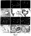
- the FLT4 probe hybridized very weakly if at all with arterial endothelia of 11.5 day p.c. embryos, e.g. with the endothelium of the developing dorsal aorta (da in Fig. 2 E,F) or the carotic arteries (data not shown). Instead, FLT4 signal was much more prominent in the developing veins. For example, FLT4 signal was detected veins surrounding the developing metanephros (v, sv in E), while the Tie probe predominantly recognized capillaries (c) within the metanephros (F).
- FLT4 mRNA is distributed in several regions of a 12.5 day p.c. mouse embryo, being particularly prominent in the dilated vessel of the axillar region (ax).
- a similar FLT4 positive vessel structure was seen in the mid-sagittal section in the jugular area (data not shown).
- a plexus-like pattern of FLT4 expressing vessels appeared in the periorbital region (po) and surrounding the developing vertebrae (vb). Also, just beneath the developing skin, a FLT4-positive vascular network was evident (sc).
- Weaker capillary signals were obtained from several regions, including the developing brain (b).
- FLT4 mRNA could also be detected in small vessels of the neck region, of the developing snout and at the base of the developing tongue as well as in the tail region (data not shown). Besides, the liver (li) was strongly positive for FLT4 mRNA in a spotlike pattern.
- Photographs of a transverse section of the upper thorax of a 16.5 day p.c. embryo hybridized with the FLT4 probe are shown in panels C and D of Figure 4.
- the section shown in C has been stained with hematoxylin-eosin to visualize the different types of vessels in this area. These include the carotic and brachiochepalic arteries (ca, ba), the vena cava (vc) and the thoracic duct, which is smaller in size and lacks surrounding muscular and connective tissue (arrow).
- a magnification of the region of thoracic duct is shown in panel D, where the FLT4 autoradiographic grains can be seen. Endothelial cells of the thoracic duct as well as a small vessel (v) in the vicinity hybridize with the FLT4 probe.
- Example 3 The in situ hybridization results described in Example 3, showed that FLT4 is expressed in venous endothelial cells and later in lymphatic vessels and some venous endothelial cells, but not in arterial endothelia. In order to see if such regulation was maintained in vitro , we studied cultured endothelial cells using Northern blotting and hybridization analysis.
- Endothelial cells from human aorta, femoral vein, umbilical vein, and from foreskin microvessels were isolated, cultured and characterized as previously described by Van Hinsberg, (15,16) . They were used at confluent density after five to eight passages (split ratio 1:3) for the isolation of polyadenylated RNA.
- endothelial cell lines EA ⁇ hy926, BCE and LEII did not express FLT4 (data not shown).
- cultured human microvascular, venous and umbilical vein endothelial cells were positive for the FLT4-specific 5.8 and 4.5 kb mRNAs, whereas the aortic endothelial cells were negative (Fig. 5).
- another endothelial receptor tyrosine kinase gene, Tie was expressed as a 4.4 kb mRNA in all endothelial cell types studied.
- Example 3 The results obtained in Example 3 indicated that the FLT4 mRNA becomes largely confined to the endothelium of lymphatic vessels during development. Because of the potential significance of this finding in humans, we also studied FLT4 in adult human tissues using a human FLT4 probe.
- the human FLT4 probe used was an EcoRI-SphI fragment covering base pairs 1-595 of the cDNA (3) .
- the von Willebrand factor probe was an EcoRI-Hindlll fragment covering base pairs 1-2334 (17) .
- Normal lung tissue was obtained from a resection of the left inferior lung lobe affected by epidermoid cancer.
- Mesenterium and mesenterial lymph nodes were obtained from a patient having a colonic adenocarcinoma.
- a normal lymph node adjacent to the salivary gland was enucleated because of its abnormal size.
- the tonsils from two patients and the two appendixes had no diagnostic changes.
- Two lymphangiomyomas and three cystic lymphangiomas were studied with similar results.
- FLT4 is expressed in the lymphatic sinuses (Is) and afferent and efferent lymphatic vessels (data not shown). The same pattern is seen in a lymph node containing adenocarcinoma metastases (C, D). Some HEVs in both normal and metastatic lymph node were also positive.
- FLT4 expression is shown in a cystic lymphangioma (compare with the hematoxylin-eosin stained section in F). Notably, the specificity of FLT4 to lymphatic endothelia is evident from the comparison with the in situ signals for von Willebrandt factor in all blood vessels (F).
- Tissues from 17 and 20-week-old human fetuses were obtained from legal abortions induced with prostaglandins. The study was approved by the Ethical Committee of the Helsinki University Central Hospital. The gestational age was estimated from the fetal foot length. The fetal tissues were embedded in Tissue-Tek (Miles), frozen immediately and stored at -70 °C.
- Anti-FLT4 antiserum was cross-absorbed to a GST-Sepharose column to remove anti-GST-antibodies and then purified by GST-FLT4 affinity chromatography.
- Several 6 ⁇ m-thick cryostat sections of the tissues were fixed with acetone and treated with 0.3% H 2 O 2 in methanol for 30 min to block endogenous peroxidase activity. After washing, the sections were incubated with 5% normal swine serum.
- Sections were then incubated with antibodies against FLT4, washed and bound antibodies were detected with peroxidase-conjugated swine anti-rabbit IgG followed by staining for peroxidase activity using 0.2% 3,3-diaminobenzidine (Amersham) as a substrate.
- the sections were counterstained in Meyer's hematoxylin.
- Anti-FLT4 immunoperoxidase staining of human fetal mesenterium showed FLT4 protein in the endothelium of several vessels (Fig. 4A), while control stainings with antigen-blocked anti-FLT4 antibodies (B) and preimmune sera (C) were negative.
- Figure 4D shows results of staining using an antiserum against the factor VIII-related antigen, which is specific for vascular endothelial cells.
- mice Four months old Balb/c male mice were immunized by intraperitoneal injection of the recombinantly produced FLT4 protein (see Example 7) in concentrated medium (150 ⁇ g/mouse), emulsified with Freund's complete adjuvant. Booster injections of 150 ⁇ g were given at three to four week intervals and a final booster (10 ⁇ g FLT4 in PBS administered intraperitoneally) was given after another three-week interval. Four days after the final booster dose, the mice were sacrified and mouse splenic lymphoid cells were fused with SP 2/0 plasmacytoma cells at a 2:1 ratio, respectively.
- the fused cells were harvested in 96-well culture plates (NUNC) in Ex-Cell 320 medium (SERALAB) containing 20% fetal calf serum and HAT supplement (hypoxanthine-aminopterin-thymidine; GIBCO, 043-01060H; diluted 50-fold). Cells were cultured at +37°C, ina 5% CO 2 atmosphere. After 10 days, HAT-supplemented medium was changed to HT-supplemented cell culture medium (GIBCO; 043-01065H, diluted 50-fold). HT medium is identical to HAT medium, but lacks aminopterin.
- the subclass of monoclonal antibody was determined using rat monoclonal antibody to mouse subclass as biotin conjugate (SEROTEC) in IFMA.
- Balb/c mice were used to produce monoclonal antibodies in ascites fluid.
- the hybridomas described above were intraperitoneally injected into mice after pretreatment of the animals with pristane (2,6,10,14-tetramethylpentadecan 98%, ALDRICH-CHEMIE D7924 Steinheim, Cat.No. T 2,280-2).
- pristane (2,6,10,14-tetramethylpentadecan 98%, ALDRICH-CHEMIE D7924 Steinheim, Cat.No. T 2,280-2).
- 0.5 ml of pristane (i.v.) was injected about two weeks prior to the hybridoma cells.
- the amount of cells injected were approximately 7.5 to 9 x 10 6 per mouse.
- Ascites was collected 10 to 14 days after injection of the hybridomas.
- mice Two months old Balb/c mice (female) were immunized by intraperitoneal injection of the recombinantly produced FLT-4 protein (see Example 7) (20 ⁇ g/mouse), emulsified with Freund's complete adjuvant. Booster injections of 20 ⁇ g were given at three to four week intervals and a final booster (10 ⁇ g FLT-4 in PBS administered i.v.) was given after another three-week interval. Four days after the final booster dose, the mice were sacrified and mouse splenic lymphoid cells were fused with SP 2/0 plasmacytoma cells at a 2:1 ratio, respectively.
- the fused cells were harvested in 96-well culture plates (FALCON) in OptiMEM 1 (with Glutamax 1, 51985-026, GIBCO BRL) medium containing 20 % fetal calf serum and HAT supplement (hypoxanthine-aminopterin-thymidine; GIBCO BRL 21060-017; diluted 1:50 fold). Cells were cultured at +37 °C, in a 5% CO2 atmosphere. After 10 days, HAT-supplemented medium was changed to HT-supplemented cell culture medium (GIBCO BRL; 41065-012, diluted 1:50-fold). HT-medium is identical to HAT-medium,but lacks aminopterin.
- IFMA ImmunoFluoroMetric Assay
- bovine serum albumin (BSA) was added to the labelled FLT4 and the label was stored at +4°C.
- the number of europium ions incorporated per FLT4 molecule was 1.9, as determined by measuring the fluorescence in a ratio to that of known EuCl 3 standards (Hemmilä et al., Anal.Biochem ., 137:335-343, 1984).
- the antibodies produced in Example 8 were screened using a Sandwich type immunofluorometric assay using microtitration strip wells (NUNC, polysorb) coated with rabbid anti-mouse Ig (Z 259, DAKOPATTS). The precoated wells were washed once by Platewash 1296-024 (WALLAC) with DELFIA wash solution.
- the DELFIA assay buffer was used as a dilution buffer for cell culture supernatants and for serum of the spleenectomized mouse (at dilutions between 1:1000 to 1: 100 000) used as positive control in the preliminary screening assay.
- the europium-labelled FLT4 was added at a dilution of 1:500 in 100 ⁇ l of the assay buffer. After 5 min on a Plateshake shaker and one hour incubation at RT the strips were washed as described above.
- Enhancement solution was added at 200 ⁇ l/well. The plates were then shaken for 5 min an a Plateshake shaker and the intensity of fluorescence was measured by ARCUS-1230 (WALLAC) for 10-15 min.
- WALLAC ARCUS-1230
- anti-FLT4 9D9F9 One clone, designated anti-FLT4 9D9F9 was found to stably secrete monoclonal antibody which was determined to be of immunoglobulin class IgG1 by IFMA.
- Hybridoma 9d9f9 was deposited with the German Collection of Microorganisms and Cell Cultures, Department of Human and Animal Cell Cultures and Viruses, Mascheroder Weg 1b, 3300 Braunschweig, Germany, March 23, 1995, and given accession No. ACC2210.
- PD-10 PARMACIA
- bovine serum albumin (BSA) was added to the labelled FLT-4 and the label was stored at +4°C.
- the number of europium ions incorporated per FLT-4 molecule was 1.9, as determined by measuring the fluorescence in a ratio to that of known EuCl3 standards (Hemmil et al., Anal.Biochem., 137: 335-343, 1984).
- Example 8 The antibodies produced in Example 8, were screened using a FLT-4 specific IFMA using microtitration wells (Nunc, Polysorb) coated with rabbit antimouse Ig (Z 259, DAKO). The precoated wells were washed once with wash solution (Wallac) by using DELFIA Plate wash.
- the DELFIA assay buffer was used as dilution buffer for cell culture supernatants (dilution 1:2 in preliminary screening) and for serum of the splenectomized mouse (dilutions 1:1 000 to 1:100 000) which was used as positive control.
- the purified antiFLT-4 9D9F9 (mouse subclass IgG1) was used at concentrations between 1.0 ng/ml and 250 ng/ml. Samples were first shaken at room temperature for five minutes on Plate shake (Wallac) and then incubated approx. 18 hours at +4°C.
- the frames were first washed four times, then the Eu-labelled FLT-4 (1:2000, in 100 ⁇ l assay buffer) was added and finally the frames were incubated for one hour at room temperature. After washing as described the enhancement solution (200 ⁇ l/well, Wallac) was added and the frames were shaken for 5 minutes on Plate shake. The intensity of fluorescence was measured by ARCUS-1230 (Wallac).
- a standard curve for quantitation of antiFLT-4 antibodies was made by using affinity purified antiFLT-4 9D9F9.
- the linear range reached from 1.0 ng/ml to 250 ng/ml.
- antibodies according to the present invention are useful in the diagnosis and identification of lymphatic vessels, lymphatic endothelial cells, high endothelial venules, lymphangiomas, metastatic lymph nodes and other disease states of the lymphatic system, the detection and monitoring of metastatic spread, in the stimulation and inhibition of endothelial cells of lymphatic vessels and high endothelial venules, in the introduction of molecules selectively into endothelial cells and in the imaging of lymphatic vessels and their disease states.
- Other uses of the presently-claimed subject matter are apparent to the skilled artisan.
Landscapes
- Health & Medical Sciences (AREA)
- Chemical & Material Sciences (AREA)
- Life Sciences & Earth Sciences (AREA)
- Organic Chemistry (AREA)
- Medicinal Chemistry (AREA)
- General Health & Medical Sciences (AREA)
- Pharmacology & Pharmacy (AREA)
- Public Health (AREA)
- Nuclear Medicine, Radiotherapy & Molecular Imaging (AREA)
- Chemical Kinetics & Catalysis (AREA)
- Veterinary Medicine (AREA)
- General Chemical & Material Sciences (AREA)
- Animal Behavior & Ethology (AREA)
- Immunology (AREA)
- Oncology (AREA)
- Bioinformatics & Cheminformatics (AREA)
- Engineering & Computer Science (AREA)
- Biophysics (AREA)
- Proteomics, Peptides & Aminoacids (AREA)
- Communicable Diseases (AREA)
- Molecular Biology (AREA)
- Genetics & Genomics (AREA)
- Biochemistry (AREA)
- Heart & Thoracic Surgery (AREA)
- Zoology (AREA)
- Pain & Pain Management (AREA)
- Cardiology (AREA)
- Hematology (AREA)
- Cell Biology (AREA)
- Toxicology (AREA)
- Rheumatology (AREA)
- Gastroenterology & Hepatology (AREA)
- Peptides Or Proteins (AREA)
- Preparation Of Compounds By Using Micro-Organisms (AREA)
- Measuring Or Testing Involving Enzymes Or Micro-Organisms (AREA)
- Medicines Containing Antibodies Or Antigens For Use As Internal Diagnostic Agents (AREA)
- Micro-Organisms Or Cultivation Processes Thereof (AREA)
Applications Claiming Priority (3)
| Application Number | Priority Date | Filing Date | Title |
|---|---|---|---|
| US25775494A | 1994-06-09 | 1994-06-09 | |
| US257754 | 1994-06-09 | ||
| PCT/FI1995/000337 WO1995033772A1 (en) | 1994-06-09 | 1995-06-09 | Flt4 receptor tyrosine kinase and its use in diagnosis and therapy |
Publications (2)
| Publication Number | Publication Date |
|---|---|
| EP0807124A1 EP0807124A1 (en) | 1997-11-19 |
| EP0807124B1 true EP0807124B1 (en) | 2006-05-17 |
Family
ID=22977612
Family Applications (1)
| Application Number | Title | Priority Date | Filing Date |
|---|---|---|---|
| EP95922533A Expired - Lifetime EP0807124B1 (en) | 1994-06-09 | 1995-06-09 | Monoclonal antibody against flt4 receptor tyrosine kinase and its use in diagnosis and therapy |
Country Status (14)
| Country | Link |
|---|---|
| EP (1) | EP0807124B1 (es) |
| JP (4) | JP3723210B2 (es) |
| AT (1) | ATE326484T1 (es) |
| AU (1) | AU694524B2 (es) |
| CA (1) | CA2192235C (es) |
| DE (1) | DE69534996T2 (es) |
| DK (1) | DK0807124T3 (es) |
| ES (1) | ES2263152T3 (es) |
| FI (1) | FI964862A (es) |
| MX (1) | MX9606218A (es) |
| NO (1) | NO319383B1 (es) |
| NZ (1) | NZ288235A (es) |
| PT (1) | PT807124E (es) |
| WO (1) | WO1995033772A1 (es) |
Families Citing this family (25)
| Publication number | Priority date | Publication date | Assignee | Title |
|---|---|---|---|---|
| US6824777B1 (en) | 1992-10-09 | 2004-11-30 | Licentia Ltd. | Flt4 (VEGFR-3) as a target for tumor imaging and anti-tumor therapy |
| US6107046A (en) * | 1992-10-09 | 2000-08-22 | Orion Corporation | Antibodies to Flt4, a receptor tyrosine kinase and uses thereof |
| US6130071A (en) * | 1997-02-05 | 2000-10-10 | Helsinki University Licensing, Ltd. | Vascular endothelial growth factor C (VEGF-C) ΔCys156 protein and gene, and uses thereof |
| US6645933B1 (en) * | 1995-08-01 | 2003-11-11 | Helsinki University Licensing Ltd. Oy | Receptor ligand VEGF-C |
| WO2002060950A2 (en) | 1994-11-14 | 2002-08-08 | Ludwig Institute For Cancer Research | Flt4(vegfr-3) as a target for tumor imaging and anti-tumor therapy |
| US6245530B1 (en) | 1995-08-01 | 2001-06-12 | Ludwig Institute For Cancer Research | Receptor ligand |
| US6221839B1 (en) | 1994-11-14 | 2001-04-24 | Helsinki University Licensing Ltd. Oy | FIt4 ligand and methods of use |
| US6403088B1 (en) | 1995-08-01 | 2002-06-11 | Helsinki University Licensing, Ltd. | Antibodies reactive with VEGF-C, a ligand for the Flt4 receptor tyrosine kinase (VEGFR-3) |
| US6818220B1 (en) | 1994-11-14 | 2004-11-16 | Licentia Ltd. | Vascular endothelial growth factor C (VEGF-C) protein and gene mutants thereof, and uses thereof |
| US7423125B2 (en) | 1995-08-01 | 2008-09-09 | Vegenics Limited | Antibodies to VEGF-C |
| US6361946B1 (en) | 1997-02-05 | 2002-03-26 | Licentia Ltd | Vascular endothelial growth factor C (VEGF-C) protein and gene, mutants thereof, and uses thereof |
| EP0848755B2 (en) | 1995-09-08 | 2011-02-09 | Genentech, Inc. | Vegf-related protein |
| US6100071A (en) | 1996-05-07 | 2000-08-08 | Genentech, Inc. | Receptors as novel inhibitors of vascular endothelial growth factor activity and processes for their production |
| US7125714B2 (en) | 1997-02-05 | 2006-10-24 | Licentia Ltd. | Progenitor cell materials and methods |
| FR2783325A1 (fr) * | 1998-09-11 | 2000-03-17 | Inst Nat Sante Rech Med | Moyens pour le controle de la regulation negative d'une activation transmise par un rtk |
| ATE489108T1 (de) * | 1998-10-09 | 2010-12-15 | Vegenics Ltd | Flt4 (vegfr-3) als ziel für tumornachweis (imaging) und antitumortherapie |
| WO2000062063A1 (en) * | 1999-04-13 | 2000-10-19 | Northwest Biotherapeutics, Inc. | Methods for the diagnosis and treatment of metastatic prostate tumors |
| EP1553414A1 (en) * | 1999-04-13 | 2005-07-13 | Medarex, Inc. | Methods for the diagnosis and treatment of metastatic prostate tumors |
| US6927203B1 (en) | 1999-08-17 | 2005-08-09 | Purdue Research Foundation | Treatment of metastatic disease |
| US6599717B1 (en) * | 1999-10-01 | 2003-07-29 | Exelixis, Inc. | Invertebrate vascular endothelial growth factor receptor |
| EP2025686A4 (en) * | 2006-05-29 | 2009-12-23 | Consejo Superior Investigacion | ANTI-CHOLINE KINASE ALPHA MONOCLONAL ANTIBODIES AND THEIR APPLICATION IN CANCER DIAGNOSTIC ANALYSIS, ANALYSIS AND DRUG PREPARATION TECHNIQUES |
| WO2008093246A2 (en) | 2007-02-02 | 2008-08-07 | Vegenics Limited | Vegf receptor antagonist for treating organ transplant alloimmunity and arteriosclerosis |
| AU2014218318B2 (en) | 2013-02-18 | 2018-02-15 | Vegenics Pty Limited | Ligand binding molecules and uses thereof |
| EP2994758B1 (en) | 2013-05-08 | 2017-12-20 | Opthea Limited | Biomarkers for age-related macular degeneration (amd) |
| CN114262683B (zh) * | 2022-03-01 | 2022-06-17 | 中国科学院动物研究所 | 一种表达vegfr3 d2多肽的细菌制剂及其构建方法和应用 |
Family Cites Families (2)
| Publication number | Priority date | Publication date | Assignee | Title |
|---|---|---|---|---|
| KR940703857A (ko) * | 1992-01-09 | 1994-12-12 | 티모 에스코 | 타이, 새로운 내피 세포 수용체 티로신 키나제(tie, a novel endothelial cell receptor tyrosine kinase) |
| DK0666868T4 (da) * | 1992-10-28 | 2006-09-18 | Genentech Inc | Anvendelse af anti-VEGF-antistoffer til behandling af cancer |
-
1995
- 1995-06-09 EP EP95922533A patent/EP0807124B1/en not_active Expired - Lifetime
- 1995-06-09 CA CA2192235A patent/CA2192235C/en not_active Expired - Lifetime
- 1995-06-09 WO PCT/FI1995/000337 patent/WO1995033772A1/en active IP Right Grant
- 1995-06-09 AU AU27388/95A patent/AU694524B2/en not_active Expired
- 1995-06-09 AT AT95922533T patent/ATE326484T1/de not_active IP Right Cessation
- 1995-06-09 DK DK95922533T patent/DK0807124T3/da active
- 1995-06-09 ES ES95922533T patent/ES2263152T3/es not_active Expired - Lifetime
- 1995-06-09 JP JP50041096A patent/JP3723210B2/ja not_active Expired - Fee Related
- 1995-06-09 MX MX9606218A patent/MX9606218A/es unknown
- 1995-06-09 NZ NZ288235A patent/NZ288235A/xx unknown
- 1995-06-09 DE DE69534996T patent/DE69534996T2/de not_active Expired - Lifetime
- 1995-06-09 PT PT95922533T patent/PT807124E/pt unknown
-
1996
- 1996-12-04 FI FI964862A patent/FI964862A/fi unknown
- 1996-12-06 NO NO19965231A patent/NO319383B1/no unknown
-
2005
- 2005-01-06 JP JP2005001867A patent/JP2005139199A/ja active Pending
-
2009
- 2009-02-04 JP JP2009023515A patent/JP2009102415A/ja active Pending
-
2012
- 2012-06-20 JP JP2012138421A patent/JP2012193191A/ja active Pending
Non-Patent Citations (9)
| Title |
|---|
| B. PYTOWSKI ET AL.: "Complete and specific inhibition of adult lymphatic regeneration by a novel VEGFR-3 Neutralizing Antibody", JOURNAL OF THE NATIONAL CANCER INSTITUTE, vol. 97, no. 1, 5 January 2005 (2005-01-05), pages 14 - 21 * |
| C. CURSIEFEN ET AL.: "Spontaneous corneal hem- and lymphangiogenesis in mice with destrin-mutation depend on VEGFR3 signaling", AMERICAN JOURNAL OF PATHOLOGY, vol. 166, no. 5, May 2005 (2005-05-01), pages 1367 - 1377 * |
| FERRERA N. ET AL: "The biology of VEGF", ENDOCRINE REVIEWS, vol. 18, no. 1, 1997, pages 4 - 25 * |
| H. KUBO ET AL.: "Blockade of vascular endothelial growth factor receptor-3 signaling inhibits fibroblast growth factor-2-induced lymphangiogenesis in mouse cornea", PNAS, vol. 99, no. 13, 25 June 2002 (2002-06-25), pages 8868 - 8873 * |
| K. SHIMIZU ET AL.: "Suppression of VEGFR-3 signaling inhibits lymph node metastasis in gastric cancer", CANCER SCIENCE, vol. 95, no. 4, April 2004 (2004-04-01), pages 328 - 333 * |
| L.CHEN ET AL.: "Vascular endothelial growth factor receptor-3 mediates induction of corneal alloimmunty", NATURE MEDICINE, vol. 10, no. 8, August 2004 (2004-08-01), pages 813 - 815 * |
| M. CHOY ET AL.: "VEGF-C signaling through Flt-4 (VEGFR3) medaites leukemic cell proliferation and survival", BLOOD, vol. 96, no. 11, 16 November 2000 (2000-11-16), pages 502A - 503A * |
| P. SAHARINEN ET AL.: "Lymphatic vasculature: development, molecular regulation and role in tumor metastasis and inflammation", TRENDS IN IMMUNOLOGY, vol. 25, no. 7, July 2004 (2004-07-01), pages 387 - 395 * |
| X. ZHANG ET AL.: "The Vegfr-3 receptor participates in Kaposi's sarcoma associated virus (KSHV)/human herpes virus 8 (HHV-8) infection of endothelial cells", BLOOD, vol. 102, no. 11, 16 November 2003 (2003-11-16), pages 279A * |
Also Published As
| Publication number | Publication date |
|---|---|
| DK0807124T3 (da) | 2006-08-28 |
| FI964862A0 (fi) | 1996-12-04 |
| WO1995033772A1 (en) | 1995-12-14 |
| CA2192235A1 (en) | 1995-12-14 |
| NO965231L (no) | 1996-12-06 |
| ES2263152T3 (es) | 2006-12-01 |
| NO319383B1 (no) | 2005-07-25 |
| ATE326484T1 (de) | 2006-06-15 |
| NO965231D0 (no) | 1996-12-06 |
| JPH10500858A (ja) | 1998-01-27 |
| MX9606218A (es) | 1998-02-28 |
| DE69534996D1 (de) | 2006-06-22 |
| JP2009102415A (ja) | 2009-05-14 |
| PT807124E (pt) | 2006-08-31 |
| FI964862A (fi) | 1997-02-10 |
| JP2012193191A (ja) | 2012-10-11 |
| JP2005139199A (ja) | 2005-06-02 |
| DE69534996T2 (de) | 2007-05-10 |
| AU694524B2 (en) | 1998-07-23 |
| JP3723210B2 (ja) | 2005-12-07 |
| NZ288235A (en) | 1999-05-28 |
| AU2738895A (en) | 1996-01-04 |
| CA2192235C (en) | 2011-02-15 |
| EP0807124A1 (en) | 1997-11-19 |
Similar Documents
| Publication | Publication Date | Title |
|---|---|---|
| EP0807124B1 (en) | Monoclonal antibody against flt4 receptor tyrosine kinase and its use in diagnosis and therapy | |
| MXPA96006218A (es) | Cinasa de tirosina receptora de flt4 y su uso endiagnostico y terapia | |
| Scott et al. | A truncated intracellular HER2/neu receptor produced by alternative RNA processing affects growth of human carcinoma cells | |
| Carnemolla et al. | A tumor-associated fibronectin isoform generated by alternative splicing of messenger RNA precursors. | |
| US6107046A (en) | Antibodies to Flt4, a receptor tyrosine kinase and uses thereof | |
| US7829091B2 (en) | Therapy targeting FLT4 (VEGER-3) expressed in blood vessels | |
| Barnhill et al. | Expression of platelet‐derived growth factor (PDGF)‐A, PDGF‐B and the PDGF‐alpha receptor, but not the PDGF‐beta receptor, in human malignant melanoma in vivo | |
| US20160184405A1 (en) | Flt4 (vegfr-3) as a target for tumor imaging and anti-tumor therapy | |
| JP2001517075A (ja) | 組換え血管内皮細胞増殖因子d(vegf―d) | |
| JPH09509570A (ja) | Mts−1遺伝子により転移性癌の診断 | |
| Naldini et al. | Phosphotyrosine antibodies identify the p210c-abl tyrosine kinase and proteins phosphorylated on tyrosine in human chronic myelogenous leukemia cells | |
| JPH10511840A (ja) | 形態形成タンパク質特異細胞表面レセプターおよびその使用 | |
| KR101560843B1 (ko) | 페리오스틴의 Exon-17 부위에 의해 코드되는 펩티드에 대한 항체를 함유하는 암 치료제 | |
| AU2005219322B2 (en) | Pharmaceutical Composition Comprising CXCR3 Inhibitor | |
| Tucker et al. | Novel tenascin variants with a distinctive pattern of expression in the avian embryo | |
| JPH07501441A (ja) | リンパ抗原cd30 | |
| CA2123582A1 (en) | Recombinant mammalian calcitonin receptors and uses thereof | |
| Hewitt et al. | Basement membrane collagen‐IV synthesis in colorectal tumours | |
| RU2186783C2 (ru) | Кроличья антисыворотка, ингибирующая транспорт катионных аминокислот, и содержащая ее фармацевтическая композиция | |
| Maher et al. | Partial cloning of the M subunit of laminin from adult rat lipocytes: expression of the M subunit by cells isolated from normal and injured liver | |
| JP4469180B2 (ja) | テネイシンw組成物およびその使用 | |
| CN100541198C (zh) | 诊断和治疗癌症的方法和用于其中的组合物 | |
| Adachi et al. | Potential role of SC1, a cell adhesion molecule, in mammary gland tumors | |
| LeMosy | Biochemical analysis of a human glioma laminin: evidence for a novel glial A chain | |
| Durham | Characterization of laminin subunit chains during lung development |
Legal Events
| Date | Code | Title | Description |
|---|---|---|---|
| PUAI | Public reference made under article 153(3) epc to a published international application that has entered the european phase |
Free format text: ORIGINAL CODE: 0009012 |
|
| 17P | Request for examination filed |
Effective date: 19970107 |
|
| AK | Designated contracting states |
Kind code of ref document: A1 Designated state(s): AT BE CH DE DK ES FR GB GR IE IT LI LU MC NL PT SE |
|
| RAP1 | Party data changed (applicant data changed or rights of an application transferred) |
Owner name: ORION CORPORATION Owner name: HELSINKI UNIVERSITY LICENSING LTD. OY |
|
| RIN1 | Information on inventor provided before grant (corrected) |
Inventor name: KARNANI, PAEIVI Inventor name: PAJUSOLA, KATRI Inventor name: MATIKAINEN, MARJA-TERTTU Inventor name: MUSTONEN, TUIJA Inventor name: KORHONEN, JAANA Inventor name: KAIPAINEN, ARJA Inventor name: ALITALO, KARI |
|
| 17Q | First examination report despatched |
Effective date: 19990514 |
|
| RTI1 | Title (correction) |
Free format text: MONOCLONAL ANTIBODY AGAINST FLT4 RECEPTOR TYROSINE KINASE AND ITS USE IN DIAGNOSIS AND THERAPY |
|
| GRAP | Despatch of communication of intention to grant a patent |
Free format text: ORIGINAL CODE: EPIDOSNIGR1 |
|
| RTI1 | Title (correction) |
Free format text: MONOCLONAL ANTIBODY AGAINST FLT4 RECEPTOR TYROSINE KINASE AND ITS USE IN DIAGNOSIS AND THERAPY |
|
| GRAS | Grant fee paid |
Free format text: ORIGINAL CODE: EPIDOSNIGR3 |
|
| RIN1 | Information on inventor provided before grant (corrected) |
Inventor name: KARNANI, PAEIVI Inventor name: PAJUSOLA, KATRI Inventor name: MATIKAINEN, MARJA-TERTTU Inventor name: MUSTONEN, TUIJA Inventor name: KORHONEN, JAANA Inventor name: KAIPAINEN, ARJA Inventor name: ALITALO, KARI |
|
| GRAA | (expected) grant |
Free format text: ORIGINAL CODE: 0009210 |
|
| RAP1 | Party data changed (applicant data changed or rights of an application transferred) |
Owner name: ORION CORPORATION Owner name: LICENSING OY |
|
| RIN1 | Information on inventor provided before grant (corrected) |
Inventor name: KARNANI, PAEIVI, C/O MOLECULAR/CANCER BIOLOGY LAB. Inventor name: PAJUSOLA, KATRI Inventor name: MATIKAINEN, MARJA-TERTTU Inventor name: MUSTONEN, TUIJA Inventor name: KORHONEN, JAANA Inventor name: KAIPAINEN, ARJA Inventor name: ALITALO, KARI, C/O MOLECULAR/CANCER BIOLOGY LAB. |
|
| AK | Designated contracting states |
Kind code of ref document: B1 Designated state(s): AT BE CH DE DK ES FR GB GR IE IT LI LU MC NL PT SE |
|
| REG | Reference to a national code |
Ref country code: GB Ref legal event code: FG4D |
|
| REG | Reference to a national code |
Ref country code: CH Ref legal event code: EP |
|
| REG | Reference to a national code |
Ref country code: IE Ref legal event code: FG4D |
|
| REF | Corresponds to: |
Ref document number: 69534996 Country of ref document: DE Date of ref document: 20060622 Kind code of ref document: P |
|
| RAP2 | Party data changed (patent owner data changed or rights of a patent transferred) |
Owner name: LUDWIG INSTITUTE FOR CANCER RESEARCH Owner name: LICENTIA OY |
|
| REG | Reference to a national code |
Ref country code: GB Ref legal event code: 732E |
|
| REG | Reference to a national code |
Ref country code: SE Ref legal event code: TRGR |
|
| REG | Reference to a national code |
Ref country code: DK Ref legal event code: T3 |
|
| REG | Reference to a national code |
Ref country code: PT Ref legal event code: SC4A Effective date: 20060630 |
|
| NLT2 | Nl: modifications (of names), taken from the european patent patent bulletin |
Owner name: LICENTIA OY EN LUDWIG INSTITUTE FOR CANCER RESEARC Effective date: 20060628 |
|
| REG | Reference to a national code |
Ref country code: CH Ref legal event code: NV Representative=s name: VOSSIUS & PARTNER |
|
| REG | Reference to a national code |
Ref country code: GR Ref legal event code: EP Ref document number: 20060402882 Country of ref document: GR |
|
| REG | Reference to a national code |
Ref country code: FR Ref legal event code: RM |
|
| REG | Reference to a national code |
Ref country code: ES Ref legal event code: FG2A Ref document number: 2263152 Country of ref document: ES Kind code of ref document: T3 |
|
| ET | Fr: translation filed | ||
| PLBE | No opposition filed within time limit |
Free format text: ORIGINAL CODE: 0009261 |
|
| STAA | Information on the status of an ep patent application or granted ep patent |
Free format text: STATUS: NO OPPOSITION FILED WITHIN TIME LIMIT |
|
| 26N | No opposition filed |
Effective date: 20070220 |
|
| PGFP | Annual fee paid to national office [announced via postgrant information from national office to epo] |
Ref country code: PT Payment date: 20070516 Year of fee payment: 13 |
|
| PGFP | Annual fee paid to national office [announced via postgrant information from national office to epo] |
Ref country code: SE Payment date: 20070618 Year of fee payment: 13 |
|
| PGFP | Annual fee paid to national office [announced via postgrant information from national office to epo] |
Ref country code: MC Payment date: 20070626 Year of fee payment: 13 Ref country code: LU Payment date: 20070626 Year of fee payment: 13 Ref country code: AT Payment date: 20070626 Year of fee payment: 13 |
|
| PGFP | Annual fee paid to national office [announced via postgrant information from national office to epo] |
Ref country code: NL Payment date: 20070627 Year of fee payment: 13 Ref country code: DK Payment date: 20070627 Year of fee payment: 13 Ref country code: BE Payment date: 20070627 Year of fee payment: 13 |
|
| REG | Reference to a national code |
Ref country code: CH Ref legal event code: PCAR Free format text: VOSSIUS & PARTNER;NADELBERG 3;4051 BASEL (CH) |
|
| PGFP | Annual fee paid to national office [announced via postgrant information from national office to epo] |
Ref country code: GR Payment date: 20070628 Year of fee payment: 13 |
|
| REG | Reference to a national code |
Ref country code: PT Ref legal event code: MM4A Free format text: LAPSE DUE TO NON-PAYMENT OF FEES Effective date: 20081209 |
|
| BERE | Be: lapsed |
Owner name: *LUDWIG INSTITUTE FOR CANCER RESEARCH Effective date: 20080630 Owner name: *LICENTIA OY Effective date: 20080630 |
|
| PG25 | Lapsed in a contracting state [announced via postgrant information from national office to epo] |
Ref country code: PT Free format text: LAPSE BECAUSE OF NON-PAYMENT OF DUE FEES Effective date: 20081209 Ref country code: MC Free format text: LAPSE BECAUSE OF NON-PAYMENT OF DUE FEES Effective date: 20080630 |
|
| REG | Reference to a national code |
Ref country code: DK Ref legal event code: EBP |
|
| EUG | Se: european patent has lapsed | ||
| NLV4 | Nl: lapsed or anulled due to non-payment of the annual fee |
Effective date: 20090101 |
|
| REG | Reference to a national code |
Ref country code: GB Ref legal event code: 732E Free format text: REGISTERED BETWEEN 20090212 AND 20090218 |
|
| PG25 | Lapsed in a contracting state [announced via postgrant information from national office to epo] |
Ref country code: BE Free format text: LAPSE BECAUSE OF NON-PAYMENT OF DUE FEES Effective date: 20080630 |
|
| PG25 | Lapsed in a contracting state [announced via postgrant information from national office to epo] |
Ref country code: AT Free format text: LAPSE BECAUSE OF NON-PAYMENT OF DUE FEES Effective date: 20080609 |
|
| PG25 | Lapsed in a contracting state [announced via postgrant information from national office to epo] |
Ref country code: NL Free format text: LAPSE BECAUSE OF NON-PAYMENT OF DUE FEES Effective date: 20090101 |
|
| PG25 | Lapsed in a contracting state [announced via postgrant information from national office to epo] |
Ref country code: GR Free format text: LAPSE BECAUSE OF NON-PAYMENT OF DUE FEES Effective date: 20090107 |
|
| REG | Reference to a national code |
Ref country code: CH Ref legal event code: PUE Owner name: VEGENICS LIMITED Free format text: LUDWIG INSTITUTE FOR CANCER RESEARCH#STADELHOFERSTRASSE 22#8001 ZURICH (CH) $ LICENTIA OY#EROTTAJANKATU 19 B 5#00130 HELSINKI (FI) -TRANSFER TO- VEGENICS LIMITED#LEVEL 1, 10 WALLACE AVE#TOORAK, VICTORIA 3142 (AU) |
|
| PG25 | Lapsed in a contracting state [announced via postgrant information from national office to epo] |
Ref country code: DK Free format text: LAPSE BECAUSE OF NON-PAYMENT OF DUE FEES Effective date: 20090106 |
|
| PG25 | Lapsed in a contracting state [announced via postgrant information from national office to epo] |
Ref country code: DK Free format text: LAPSE BECAUSE OF NON-PAYMENT OF DUE FEES Effective date: 20080630 |
|
| PG25 | Lapsed in a contracting state [announced via postgrant information from national office to epo] |
Ref country code: LU Free format text: LAPSE BECAUSE OF NON-PAYMENT OF DUE FEES Effective date: 20080609 |
|
| PG25 | Lapsed in a contracting state [announced via postgrant information from national office to epo] |
Ref country code: SE Free format text: LAPSE BECAUSE OF NON-PAYMENT OF DUE FEES Effective date: 20080610 |
|
| PGFP | Annual fee paid to national office [announced via postgrant information from national office to epo] |
Ref country code: CH Payment date: 20110614 Year of fee payment: 17 |
|
| REG | Reference to a national code |
Ref country code: CH Ref legal event code: PL |
|
| REG | Reference to a national code |
Ref country code: CH Ref legal event code: PL |
|
| PG25 | Lapsed in a contracting state [announced via postgrant information from national office to epo] |
Ref country code: LI Free format text: LAPSE BECAUSE OF NON-PAYMENT OF DUE FEES Effective date: 20120630 Ref country code: CH Free format text: LAPSE BECAUSE OF NON-PAYMENT OF DUE FEES Effective date: 20120630 |
|
| PGFP | Annual fee paid to national office [announced via postgrant information from national office to epo] |
Ref country code: GB Payment date: 20140620 Year of fee payment: 20 Ref country code: IE Payment date: 20140617 Year of fee payment: 20 |
|
| PGFP | Annual fee paid to national office [announced via postgrant information from national office to epo] |
Ref country code: ES Payment date: 20140618 Year of fee payment: 20 Ref country code: DE Payment date: 20140625 Year of fee payment: 20 |
|
| PGFP | Annual fee paid to national office [announced via postgrant information from national office to epo] |
Ref country code: FR Payment date: 20140617 Year of fee payment: 20 |
|
| PGFP | Annual fee paid to national office [announced via postgrant information from national office to epo] |
Ref country code: IT Payment date: 20140630 Year of fee payment: 20 |
|
| REG | Reference to a national code |
Ref country code: DE Ref legal event code: R071 Ref document number: 69534996 Country of ref document: DE |
|
| REG | Reference to a national code |
Ref country code: GB Ref legal event code: PE20 Expiry date: 20150608 |
|
| REG | Reference to a national code |
Ref country code: IE Ref legal event code: MK9A |
|
| PG25 | Lapsed in a contracting state [announced via postgrant information from national office to epo] |
Ref country code: GB Free format text: LAPSE BECAUSE OF EXPIRATION OF PROTECTION Effective date: 20150608 |
|
| REG | Reference to a national code |
Ref country code: ES Ref legal event code: FD2A Effective date: 20150925 |
|
| PG25 | Lapsed in a contracting state [announced via postgrant information from national office to epo] |
Ref country code: ES Free format text: LAPSE BECAUSE OF EXPIRATION OF PROTECTION Effective date: 20150610 Ref country code: IE Free format text: LAPSE BECAUSE OF EXPIRATION OF PROTECTION Effective date: 20150609 |