EP0641223B1 - Biotherapeutic cell-coated microspheres - Google Patents
Biotherapeutic cell-coated microspheres Download PDFInfo
- Publication number
- EP0641223B1 EP0641223B1 EP93911706A EP93911706A EP0641223B1 EP 0641223 B1 EP0641223 B1 EP 0641223B1 EP 93911706 A EP93911706 A EP 93911706A EP 93911706 A EP93911706 A EP 93911706A EP 0641223 B1 EP0641223 B1 EP 0641223B1
- Authority
- EP
- European Patent Office
- Prior art keywords
- microspheres
- skin
- cells
- composition according
- replacement composition
- Prior art date
- Legal status (The legal status is an assumption and is not a legal conclusion. Google has not performed a legal analysis and makes no representation as to the accuracy of the status listed.)
- Expired - Lifetime
Links
Images

Classifications
-
- A—HUMAN NECESSITIES
- A61—MEDICAL OR VETERINARY SCIENCE; HYGIENE
- A61L—METHODS OR APPARATUS FOR STERILISING MATERIALS OR OBJECTS IN GENERAL; DISINFECTION, STERILISATION OR DEODORISATION OF AIR; CHEMICAL ASPECTS OF BANDAGES, DRESSINGS, ABSORBENT PADS OR SURGICAL ARTICLES; MATERIALS FOR BANDAGES, DRESSINGS, ABSORBENT PADS OR SURGICAL ARTICLES
- A61L27/00—Materials for grafts or prostheses or for coating grafts or prostheses
-
- A—HUMAN NECESSITIES
- A61—MEDICAL OR VETERINARY SCIENCE; HYGIENE
- A61K—PREPARATIONS FOR MEDICAL, DENTAL OR TOILETRY PURPOSES
- A61K8/00—Cosmetics or similar toiletry preparations
- A61K8/02—Cosmetics or similar toiletry preparations characterised by special physical form
- A61K8/11—Encapsulated compositions
-
- A—HUMAN NECESSITIES
- A61—MEDICAL OR VETERINARY SCIENCE; HYGIENE
- A61K—PREPARATIONS FOR MEDICAL, DENTAL OR TOILETRY PURPOSES
- A61K8/00—Cosmetics or similar toiletry preparations
- A61K8/18—Cosmetics or similar toiletry preparations characterised by the composition
- A61K8/96—Cosmetics or similar toiletry preparations characterised by the composition containing materials, or derivatives thereof of undetermined constitution
- A61K8/98—Cosmetics or similar toiletry preparations characterised by the composition containing materials, or derivatives thereof of undetermined constitution of animal origin
- A61K8/981—Cosmetics or similar toiletry preparations characterised by the composition containing materials, or derivatives thereof of undetermined constitution of animal origin of mammals or bird
- A61K8/985—Skin or skin outgrowth, e.g. hair, nails
-
- A—HUMAN NECESSITIES
- A61—MEDICAL OR VETERINARY SCIENCE; HYGIENE
- A61L—METHODS OR APPARATUS FOR STERILISING MATERIALS OR OBJECTS IN GENERAL; DISINFECTION, STERILISATION OR DEODORISATION OF AIR; CHEMICAL ASPECTS OF BANDAGES, DRESSINGS, ABSORBENT PADS OR SURGICAL ARTICLES; MATERIALS FOR BANDAGES, DRESSINGS, ABSORBENT PADS OR SURGICAL ARTICLES
- A61L27/00—Materials for grafts or prostheses or for coating grafts or prostheses
- A61L27/50—Materials characterised by their function or physical properties, e.g. injectable or lubricating compositions, shape-memory materials, surface modified materials
- A61L27/60—Materials for use in artificial skin
-
- A—HUMAN NECESSITIES
- A61—MEDICAL OR VETERINARY SCIENCE; HYGIENE
- A61P—SPECIFIC THERAPEUTIC ACTIVITY OF CHEMICAL COMPOUNDS OR MEDICINAL PREPARATIONS
- A61P17/00—Drugs for dermatological disorders
-
- A—HUMAN NECESSITIES
- A61—MEDICAL OR VETERINARY SCIENCE; HYGIENE
- A61Q—SPECIFIC USE OF COSMETICS OR SIMILAR TOILETRY PREPARATIONS
- A61Q19/00—Preparations for care of the skin
-
- C—CHEMISTRY; METALLURGY
- C12—BIOCHEMISTRY; BEER; SPIRITS; WINE; VINEGAR; MICROBIOLOGY; ENZYMOLOGY; MUTATION OR GENETIC ENGINEERING
- C12N—MICROORGANISMS OR ENZYMES; COMPOSITIONS THEREOF; PROPAGATING, PRESERVING, OR MAINTAINING MICROORGANISMS; MUTATION OR GENETIC ENGINEERING; CULTURE MEDIA
- C12N5/00—Undifferentiated human, animal or plant cells, e.g. cell lines; Tissues; Cultivation or maintenance thereof; Culture media therefor
- C12N5/06—Animal cells or tissues; Human cells or tissues
- C12N5/0697—Artificial constructs associating cells of different lineages, e.g. tissue equivalents
- C12N5/0698—Skin equivalents
-
- A—HUMAN NECESSITIES
- A61—MEDICAL OR VETERINARY SCIENCE; HYGIENE
- A61K—PREPARATIONS FOR MEDICAL, DENTAL OR TOILETRY PURPOSES
- A61K2800/00—Properties of cosmetic compositions or active ingredients thereof or formulation aids used therein and process related aspects
- A61K2800/40—Chemical, physico-chemical or functional or structural properties of particular ingredients
- A61K2800/41—Particular ingredients further characterized by their size
- A61K2800/412—Microsized, i.e. having sizes between 0.1 and 100 microns
-
- C—CHEMISTRY; METALLURGY
- C12—BIOCHEMISTRY; BEER; SPIRITS; WINE; VINEGAR; MICROBIOLOGY; ENZYMOLOGY; MUTATION OR GENETIC ENGINEERING
- C12N—MICROORGANISMS OR ENZYMES; COMPOSITIONS THEREOF; PROPAGATING, PRESERVING, OR MAINTAINING MICROORGANISMS; MUTATION OR GENETIC ENGINEERING; CULTURE MEDIA
- C12N2502/00—Coculture with; Conditioned medium produced by
- C12N2502/09—Coculture with; Conditioned medium produced by epidermal cells, skin cells, oral mucosa cells
- C12N2502/094—Coculture with; Conditioned medium produced by epidermal cells, skin cells, oral mucosa cells keratinocytes
-
- C—CHEMISTRY; METALLURGY
- C12—BIOCHEMISTRY; BEER; SPIRITS; WINE; VINEGAR; MICROBIOLOGY; ENZYMOLOGY; MUTATION OR GENETIC ENGINEERING
- C12N—MICROORGANISMS OR ENZYMES; COMPOSITIONS THEREOF; PROPAGATING, PRESERVING, OR MAINTAINING MICROORGANISMS; MUTATION OR GENETIC ENGINEERING; CULTURE MEDIA
- C12N2502/00—Coculture with; Conditioned medium produced by
- C12N2502/13—Coculture with; Conditioned medium produced by connective tissue cells; generic mesenchyme cells, e.g. so-called "embryonic fibroblasts"
- C12N2502/1323—Adult fibroblasts
-
- C—CHEMISTRY; METALLURGY
- C12—BIOCHEMISTRY; BEER; SPIRITS; WINE; VINEGAR; MICROBIOLOGY; ENZYMOLOGY; MUTATION OR GENETIC ENGINEERING
- C12N—MICROORGANISMS OR ENZYMES; COMPOSITIONS THEREOF; PROPAGATING, PRESERVING, OR MAINTAINING MICROORGANISMS; MUTATION OR GENETIC ENGINEERING; CULTURE MEDIA
- C12N2531/00—Microcarriers
-
- C—CHEMISTRY; METALLURGY
- C12—BIOCHEMISTRY; BEER; SPIRITS; WINE; VINEGAR; MICROBIOLOGY; ENZYMOLOGY; MUTATION OR GENETIC ENGINEERING
- C12N—MICROORGANISMS OR ENZYMES; COMPOSITIONS THEREOF; PROPAGATING, PRESERVING, OR MAINTAINING MICROORGANISMS; MUTATION OR GENETIC ENGINEERING; CULTURE MEDIA
- C12N2533/00—Supports or coatings for cell culture, characterised by material
- C12N2533/30—Synthetic polymers
- C12N2533/40—Polyhydroxyacids, e.g. polymers of glycolic or lactic acid (PGA, PLA, PLGA); Bioresorbable polymers
Definitions
- the present invention relates to the field of tissue implants and more particularly to the application of skin implants for the treatment of full- and partial-thickness skin injuries, such as burns and other wounds.
- Full-thickness and partial-thickness skin injuries represent a significant cost to health care systems. For example, about 2 million people in North America suffer from burns each year. Of these about 200,000 people are hospitalized, 15,000 of which die of burn-related causes. The overall hospital cost for treating these patients is in the order of $1000/percentage burned area ($U.S., 1992) so that the average burn patient with burns to 20 to 30% of their body generates initial hospital care costs of about $25,000, not including the cost of further treatment and potential loss of productivity and income.
- McMillan et al J Burn Care Rehab 6:444-446; 1985 have demonstrated that operating room expenses increase logarithmically with the percent of body surface area burned. Clearly, there is a requirement for advances in technology to mitigate these costs and to reduce the suffering of the patients.
- Skin consists of a dermal layer which underlies an epidermal layer.
- the dermal layer consists mostly of fibroblasts and is about five times the thickness of the epidermal layer.
- the epidermal layer of intact skin consisting mainly of keratinocytes and immune cells such as dendritic Langerhans cells, normally prevents water loss and microbial invasion, so that full- and partial-thickness skin injuries can be life-threatening.
- the rate of wound closure to prevent the escape of essential body fluids and the invasion of bacteria is therefore a vital factor in the recovery of the patient. Accordingly a wide array of wound coverings has been developed to expedite wound closure.
- An existing treatment of burns and wounds includes the use of the patient's own skin, or cadaver- or porcine-derived tissue for grafting onto the wound of the patient.
- Traditional patient-derived skin graft (autograft) techniques are generally very painful to the patient who is already suffering from the burn or other wound.
- An autograft is comprised of a substantial thickness of both the epidermal and dermal layers of the skin taken from another site on the body.
- the autograft is treated to form a lattice pattern across the skin injury.
- the lattice pattern in the dermis layer of the autograft are subsequently filled with permanent scar tissue in vivo. These scars are often very large and can be severely disfiguring or, depending on their location, can cause dysfunction.
- the patient may not have enough non-burned area in order to salvage a large enough graft for transplant to another location of the body.
- Skin grafts derived from cadavers (allograft) or porcine (xenograft) sources have been used in an effort to reduce the suffering of the patient and to encourage wound healing.
- a major drawback of the allograft is the possibility of disease transmission (for example, HIV, hepatitis B).
- the epidermal layer shows marked antigenicity so that grafts, including allografts, not derived directly from the patient are usually rejected within two weeks of implant.
- the xenograft provides a graft when there is a shortage of human donor tissue, it is rejected even more rapidly than the allograft and must be removed on the third day after application before drying and sloughing and before strong adhesion to the wound necessitates surgical excision.
- the mesh matrix skin patches are typically supplied in dimensions of about 10 cm wide by 10 cm long and of only a few cell layers thick. It will be appreciated by those skilled in the art that burn sites are often larger and rarely of uniform thickness or of planar structure. The patches do not adequately account for contour variations in the skin so that the problem of disfigurement still exists to a greater or lesser degree in most cases. While they are more stable than grafts, the thin patches are still very fragile and in many instances the thin sheets are strengthened with petroleum jelly impregnated gauze for surgical procedures. Additionally, the small pieces of skin must still be sutured and/or stapled to each other and onto the body resulting in a prolongation of the surgical procedure on an already weak and compromised patient.
- the applied skin patches can have very poor gas/mass transfer characteristics leading to the potential for tissue necrosis due to lack of nutrients reaching the cells. This, in turn, can lead to blistering of the patch. Gas and mass transfer can be further adversely affected by a residual layer of petroleum jelly on the site once the gauze is removed following the surgical procedure. The residual layer of petroleum jelly can even lead to partitioning of various factors, such as growth factors, into this layer where they would not be available for subsequent action on the cells.
- a culture system comprised of a gas permeable bag with a recirculating pump has been developed (Marrow-Tech Inc., U.S.A.) to move media across the surface of the growing culture on a mesh fabric contained within.
- the cells are typically seeded randomly on the mesh thereby creating "multi-nuclei" of cell growth.
- any cells that are not firmly anchored to the surface will likely circulate through the pump and experience shear and other disruptive effects.
- the system is still labour intensive and cumbersome leading to an increased chance of contamination due to excessive handling requirements.
- the system is not static, the environment is not totally homogeneous and the cells anchored to the mesh are not of a uniform growth phase due to the semi-static culture conditions and the random seeding of cells on the mesh.
- the effective area of viable cells can be significantly reduced by suturing and/or stapling at the edges of the patch which can destroy or disrupt the proliferation of cells.
- Demetriou, A.A. ("Replacement of liver function in rats by transplantation of microcarrier-attached hepatocytes" Science 233:1190-1192; September 12, 1986) describes attachment of hepatocytes to collagen-coated cross-linked dextran microcarriers for subsequent implantation in the peritoneal cavity of rats.
- the microcarriers are used to provide a surface for attachment so that the hepatocytes survive and function in vivo.
- the microcarriers are not intended to resorb or degrade once implanted.
- PCT/US90/02257 (Vacanti et al, published November 1, 1990, WO 90/12604) relates to an implant of large volumes of cells on polymeric matrices.
- the matrix is a fibrous biocompatible degradable or non-degradable sheet material having an interstitial spacing of 100-200 ⁇ m.
- Vacanti et al describe the attachment and growth of hepatocytes to the matrix for subsequent implant in the mesentery of the small intestine.
- European Patent Application No. 242305 relates to a human type I or III collagen that is acid- and pepsin-treated.
- the collagen gel can be formed into films, sheets or beads.
- EP 242305 contemplates the use of collagen formed into films or sheets as a base for a skin graft and collagen formed into beads as a substrate for suspension cell culture.
- WO89/03228 describes an epidermal graft system wherein epidermal cells are layered on a sheet of collagen-coated, pliable material such as a synthetic surgical dressing.
- the surgical dressing is precut into a shape to fit a designated wound.
- Cells are cultured in a tissue culture flask and subsequently trypsinized for layering on the surgical dressing.
- the graft is then inverted on the site of a skin injury, cell-side down. Eventually, the dressing sloughs off partially and then is completely removed.
- EP 333328 relates to clinical applications of amniotic cells which may be used directly, or after application to an intermediate material, for example a prosthesis, or a biologically acceptable carrier, such as Sepharose beads, for the treatment of aging skin.
- An object of the present invention is to provide a living skin replacement for the treatment of full- and partial-thickness skin injuries, such as burns and other wounds, which can accommodate contour variations. It is a further object of the present invention to provide an implant having a dermal layer and a functional epidermis which does not require the use of stapling, suturing, or other attachment methods.
- a living skin replacement composition for full-thickness and partial-thickness skin injuries which adapts to the shape of the injuries, characterized in that the composition comprises a slurry of a biologically acceptable liquid carrier, microspheres, the microspheres comprising a material which is biocompatible and resorbable in vivo, the microspheres having a macroporosity of 30 to 80%, and proliferative skin cells selected from the group consisting of keratinocytes, dermal fibroblasts, melanocytes and combinations thereof coating the microspheres.
- cells are attached to microspheres for subsequent implant in vivo.
- a slurry of skin cell-coated microspheres is used as a living skin replacement for full- and partial-thickness skin injuries.
- the slurry of cell-coated microspheres is applied directly to the skin injury in much the same way as a salve or paste.
- the skin cells can be derived from a relatively small tissue explant from a patient.
- dermal fibroblasts which exhibit low allergenicity in transplants, can be derived from a donor, including a cadaver.
- the epidermal layer shows marked antigenicity so that implants of this layer normally need to be derived directly from the patient.
- the Langerhans cells are largely responsible for the antigenicity of the epidermal layer (Bagot, M. et al, Clin Exp Immunol 71:138; 1988). It is therefore possible that a pure culture of keratinocytes could be derived from a donor and that rejection thereof would be minimal or absent.
- a small tissue explant typically 4 cm 2
- the explant could include the epidermal layer alone or a combination of the epidermal and dermal layers.
- the tissue is then dissociated using conventional enzyme and processing techniques and seeded either into tissue culture flasks for subsequent cell expansion of the fibroblasts and/or the keratinocytes.
- the cells of the dermal and epidermal layers can be separated with enzymes such as dispase or by soaking in culture medium or phosphate buffered saline or by other well established techniques. It will be appreciated by those skilled in the art that a "bank" of donor-derived, including cadaver-derived, skin cells could be established for prompt treatment of skin injuries.
- the microspheres have a diameter in the range of about 50 to 500 ⁇ m, and preferably in the range of about 80 to 250 ⁇ m.
- the microspheres can be made of a variety of materials as will be discussed in more detail hereinafter.
- the important consideration in the choice of microsphere is that the material thereof must be biocompatible and should be capable of being resorbed into the body without the formation of toxic by-products.
- One suitable material is polyhydroxybutyrate (PHB) which is conventionally used for resorbable surgical staples and suture materials. PHB is resorbed in vivo with the ultimate end products being carbon dioxide and water.
- Other suitable materials are lactide-glycolide polymers which are commercially available for use in drug delivery systems (Medisorb Technologies International, USA). The polymers are absorbed by random hydrolysis of the ester linkages and are broken down into lactic and glycolic acids, which are normal metabolic by-products.
- dermal fibroblasts isolated from a patient- or donor-derived tissue explant are cultured in a tissue culture flask until a sufficient number of the cells is produced.
- a sufficient number of cells to provide a viable cell density in the bioreactor of about or at least 10 4 to 10 5 cells/ml.
- this cell density corresponds to approximately 5 cells/microsphere.
- the cells are detached from the bottom of a tissue culture flask using, for example, trypsin. It will be appreciated by those skilled in the art that a number of passages may be required in different sized tissue culture flasks to achieve the desired number of cells.
- the cells are then seeded in a bioreactor containing a suitable cell culture medium and microspheres at a density of 1 to 25 g/L with an optimal level of between 2 and 5 g/L.
- the microspheres can be added to the culture medium immediately prior to cell-seeding or the microspheres may be pretreated by soaking in the medium for a length of time prior to cell-seeding.
- the cells are then allowed to attach to the microspheres under static or semi-static conditions for a prescribed length of time, for example 3 to 6 hours.
- the attachment step can be performed in a reduced volume of medium with intermittent agitation for a few minutes every half hour for approximately 3 to 6 hours.
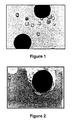
- Figure 1 is a photomicrograph showing neo-natal foreskin derived dermal fibroblasts attached to PHB-PHV (polyhydroxybutyrate-polyhydroxyvalerate copolymer) microspheres at 1 hour post cell-seeding. The dermal fibroblasts are starting to attach to the microspheres.
- PHB-PHV polyhydroxybutyrate-polyhydroxyvalerate copolymer
- FIG. 2 is a photomicrograph showing the dermal fibroblasts attached to PHB-PHV microspheres of Figure 1 at 22 hours post cell-seeding. The majority of the dermal fibroblasts that have attached to the microsphere have become flattened and are no longer spherical.
- the dissociated cells from the tissue explant can be seeded directly into a bioreactor.
- a 4 cm 2 tissue explant yields about 2-5 x 10 7 cells which is sufficient to provide a seeding density of between 3 and 5 cells/microsphere in 1 L of medium.
- the dermal fibroblast cells, attached to the microspheres, are then grown in suspension culture in a bioreactor, which may conveniently have a working volume of 250 ml to several litres. It is possible to provide a substantial skin implant with the cells cultured in a 1 L bioreactor. Based on an average surface area of 5000 cm 2 /g dry weight microspheres, a microsphere density of between 3 and 5 mg/ml provides an effective surface area of about 2 to 2.5 m 2 in a 1 L working volume vessel. The cells eventually cover this surface area creating a comparable layer of skin for further migration and proliferation in vivo.
- microcarriers which are small beads having a diameter in the range of 100 to 200 ⁇ m with a surface area of about 5000 cm 2 /g, and are typically made of cross-linked dextran. Certain cell lines have been grown on microcarriers to improve the unit productivity for cell growth and/or product formation over that achievable in static culture in tissue culture flasks (van Wezel, A.L. "Growth of Cell-Strains and Primary Cells in Microcarriers in Homogeneous Culture” Nature 216:64-65; October 7, 1967).
- Static cultures for the production of skin grafts on support mesh sheets, in tissue culture containers have an effective surface/volume ratio (S/V) of about 2-5 cm -1 compared to an S/V of about 150 cm -1 for microspheres suspended at a concentration of 25 g/L.
- suspension culture is less labour-intensive and the culture is homogeneous so that problems of gas transfer, depletion of nutrients, and accumulation of nutrients are not as restrictive.
- tissue culture flasks would be required, based on a typical available surface area of 80 cm 2 per flask.
- the dermal fibroblast cells are grown in the bioreactor until they reach a state of confluence or near confluence, at which point the cells substantially coat the microsphere. Confluence is typically achieved in about 7 days. However it is possible to use the cell-coated microspheres therapeutically before this time at a point of near confluence when the cell population is still in a highly migratory and proliferative state. The cells are then concentrated by removal of excess culture medium and washed in situ with an appropriate buffer or solution to form a microsphere/cell slurry.
- the dermal fibroblast cells are not removed from the microspheres.
- the microsphere/cell slurry is then applied directly on the wound in much the same way as a salve or paste would be applied.
- each microsphere has a similar number of cells attached thereto resulting in a homogeneous population for subsequent application on the skin injury.
- the present invention reduces the required number of manipulations of the cells. This, in turn, reduces the potential for damage to the cells themselves and minimizes the chances of contamination leading to wound infection.
- Cells applied to a wound in accordance with the present invention can be easily delivered to the entire surface of the wound. This is a very important advantage since cells grown on a mesh matrix, and even patient-derived skin grafts, will often have many non-contact areas where the skin may not fully establish. The consistency of the slurry allows for correction of contour variations that may be present in a wound. This is a great advantage over the planar implants of the prior art because, firstly, a wound is rarely of uniform depth with a smooth flat base and, secondly, the wound may extend across a significant area. For example, it will be appreciated by those skilled in the art that, in the treatment of a burn extending the length of the arm on the underside thereof, there are many natural curves along its length.
- the present invention provides an implant that can fill even the deepest wound for effective healing thereof and more natural, tridimensional tissue regeneration at the injury site.
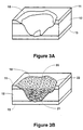
- FIG 3A An example of an irregular full-thickness skin injury 10 is shown in Figure 3A.
- the skin injury 10 extends through the epidermis 11 and the dermis 12 to the underlying muscle 13. It will be appreciated by those skilled in the art that a planar skin graft would not provide an adequate skin implant for this type of injury.
- Figure 3B is a schematic representation of the same skin injury 10 implanted with a living skin replacement 20 of the present invention.
- the living skin replacement 20 provides an implant of dermal fibroblasts 21 and keratinocytes 22.
- Subsequent layers of dermal fibroblasts may be applied to the wound over the first layer of dermal fibroblasts, for example, to correct any further contour variations. It is also possible that a donor-derived dermal fibroblast/microsphere slurry may be applied to the wound followed by an application of a patient-derived dermal fibroblast/microsphere slurry.
- the application of the dermal layer allows for rapid production of the structural protein collagen and other growth and attachment factors.
- Early application of dermal fibroblasts can minimize or prevent contraction of the skin which is the major cause of scarring and excess fluid loss.
- Dermal fibroblasts can differentiate and align in an axial fashion in a wound to effectively hold the wound together especially if the skin injury is a longitudinal cut.
- the early application of microspheres coated with fibroblasts leads to better wound healing and reduces the suffering of the patient.
- a gas-permeable wound dressing may be used to cover the site.
- the dermal fibroblasts of the microsphere/cell slurry then migrate and grow off the microspheres into the surrounding tissue to produce a continuous surface in place of the skin injury. With the passage of time and as the cells grow, the microspheres start to resorb in vivo.
- the skin implant also includes the regeneration of the epidermal layer. While the dermal fibroblast layer is being established in vivo, cells from the epidermal layer of the patient can then be cultured in a tissue culture flask. When a sufficient number of cells has been produced, the cells are removed from the bottom of the tissue culture flask using, for example, trypsin. The cells are then seeded in a bioreactor containing a suitable cell culture medium and microspheres. As previously discussed, another option is to use epidermal cells from a patient-derived tissue explant to inoculate the microspheres directly. The cells are allowed to attach to the microspheres and cultured in suspension in a bioreactor in the same manner as described earlier for the dermal fibroblasts. It will be appreciated by those skilled in the art that the use of a broad spectrum antibiotic in the initial phase of cell proliferation may be required owing to the likelihood of wound site contamination from the normal skin flora and that imparted from other sources.
- Figure 4 is a photomicrograph of neo-natal foreskin derived keratinocytes attached to PHB-PHV microspheres at 24 hours post cell-seeding. The majority of the keratinocytes that have attached to the microspheres are flattened and no longer spherical.
- the microspheres can be added to the culture medium immediately prior to cell-seeding or the microspheres may be pretreated by soaking in the medium for a length of time prior to cell-seeding.
- Figure 5 is a photomicrograph of neo-natal foreskin derived keratinocytes attached to PHB-PHV microspheres at 24 hours post cell-seeding. The microspheres were incubated in culture medium for 24 hours prior to cell-seeding. The majority of the keratinocytes that have attached to the microspheres are flattened.
- the epidermal cells including keratinocytes
- some or all of the culture medium is removed, resulting in a slurry of cell-coated microspheres.
- the wound dressing if any, is removed from the wound site and the slurry is applied over the established or establishing layer of dermal fibroblasts to regenerate the outer skin layer thereby establishing a protective barrier of intact skin.
- a gas-permeable wound dressing may be used to cover the skin injury.
- the epidermal cells then migrate and grow off the microspheres into the surrounding tissue to produce a continuous surface in replacement of the skin injury. As the cells grow, in vivo resorption of the microspheres commences.
- a certain percentage of the epidermal cells may be treated either in vivo or in vitro with specific agents such as calcium and/or cAMP to accelerate production of a stratum corneum, the uppermost layer of dead, highly keratinized cells.
- the stratum corneum helps to regulate the amount of water lost from the body and also to prevent microbial invasion into the wound site. By controlled acceleration of the differentiation of this layer, the repaired wound site will function more like uninjured, intact skin.
- cells from both the dermal and the epidermal layers are co-cultured in a tissue culture flask.
- This embodiment can provide for enhanced paracrine and autocrine function development.
- the cells are then removed from the tissue culture flask and seeded in a bioreactor containing cell culture medium and microspheres.
- the cells are allowed to attach to the microspheres under static or semi-static conditions for subsequent co-culture in suspension in a bioreactor.
- An individual microsphere may then have cells from both the dermal and epidermal layers attached thereto.
- the resulting microsphere/cell slurry is applied directly on the wound in much the same way as a salve or paste would be applied for regeneration of the dermal and epidermal layers simultaneously.
- a gas-permeable wound dressing may then be used to cover the skin injury site until the wound is effectively healed by the normal reformation of the dermal and epidermal components.
- dermal fibroblasts and cells derived from the epidermal layer are cultured independently in separate tissue culture flasks.
- the dermal fibroblasts are removed from its tissue culture flask using, for example, trypsin and seeded in a vessel containing an appropriate cell culture medium and microspheres.
- the dermal fibroblasts are then allowed to attach to the microspheres under static or semi-static conditions.
- the epidermal cells including keratinocytes, are removed from its tissue culture flask using, for example, trypsin and seeded in another vessel containing an appropriate cell culture medium and microspheres. The epidermal cells are then allowed to attach to the microspheres under static or semi-static conditions.
- microspheres coated with dermal fibroblasts and the microspheres coated with epidermal cells are then introduced into a single bioreactor for subsequent co-culture thereof.
- the resulting slurry of cell-coated microspheres is then applied to the wound for simultaneous regeneration of the dermal and epidermal layers.
- a gas-permeable wound dressing may then be applied to cover the wound.
- dermal fibroblasts are attached to microspheres and allowed to proliferate in a bioreactor. After a period of time, cells from the epidermal layer are seeded into the bioreactor for subsequent attachment to the microspheres already coated to some degree with dermal fibroblasts. The keratinocytes can thereby benefit from the paracrine effects of the fibroblasts.
- the present invention can also be used for implants in vivo of melanocytes for imparting natural pigmentation to the skin and providing UV protection.
- the epidermal layer and optionally a small percentage of the underlying dermal layer can be surgically removed using a dermatome or other suitable surgical instrument. Subsequently, a slurry of cell-coated microspheres is applied directly to the pretreated area in one or more layers to correct contour variations.
- microspheres used in the present invention can be made of a variety of materials which are biocompatible and capable of being readily resorbed into the body by natural in vivo enzyme action without the formation of toxic by-products.
- Suitable materials for the microspheres include natural and synthetically-derived bioresorbable materials such as polyhydroxybutyrate (PHB), PHB-polyhydroxyvalerate (PHB-PHV) copolymers, PHB having polyester bonds, lactide-glycolide polymers, lipids, phospholipids, polylactones, polyesters, polylactides, polyglycolides, polyanhydrides, collagen, gelatin and other resorbable materials not having an adverse effect on tissues during healing (i.e. not toxic to the cells as presented initially or through the end products of resorption). These materials can be used in pure form or as a blend of materials to enhance physiochemical properties or to control degradation rates thereof.
- PHB polyhydroxybutyrate
- PHB-PHV PHB-polyhydroxyvalerate copolymers
- the microspheres have a density of between 1.01 to 1.04 g/ml in order to facilitate mixing and suspension in culture media.
- the microspheres have a macroporosity of between 30 and 80% and a range of porosity of 30 to 80 ⁇ m.
- the microspheres have a relatively high surface area, for example, about 5000 cm 2 /g dry weight, compared with the mesh matrices of the prior art planar technologies, which generally have surface areas in the range of 200-700 cm 2 /g.
- a greater amount of material is required to impart adequate strength to the mesh matrix. Accordingly, significantly more material would be required to provide an implant for the same surface area and, likewise, more material would have to be resorbed in vivo with the mesh matrix.
- the microspheres can also be formed with a core of one material and an outer layer or coating of another material to improve the microsphere resorbability and functionality; including charge density, attachment of other chemicals or compounds, and enhancement of cell attachment and spreading.
- a coating of phospholipid allows for the generation of a polar surface with the functional phosphate head group.
- the acyl group of the phospholipid provides a more hydrophobic surface for the microsphere.
- Various chemicals or biomolecules can then be attached to these portions of the phospholipids.
- certain stratum corneum lipids including phospholipids and sphingosines can be coated onto microspheres to impart antimicrobial activity. Sphingosines are particularly good at inhibiting microbial growth at ⁇ g levels ("Antimicrobial activity of sphingosines" J Invest Dermatol 98:269-273; 1992).
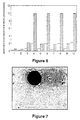
- Figure 6 is a histogram of data produced by a cytofluorometer for measuring cell viability.
- Cell viability is measured with a fluorochrome specific to viable cells and a fluorochrome specific to dead cells. These measurements are expressed as "Arbitrary Fluorescence Units" and are plotted on the histogram with cross-hatched bars representing viable cells and blank bars representing dead cells.
- Interpretation of the data is corrected by data sets 1 and 2 for phosphate buffer solution and the control, respectively.
- Keratinocytes from the same cell inoculum were seeded on PHB and on PHB coated with a phospholipid and, in each case, incubated for 24 hours.
- Cell viability of the attached cells was measured by introducing the above-mentioned fluorochromes to the appropriate sample. Samples without cells attached thereto were also treated with the fluorochromes to provide further correction for the specific resorbable material.
- the cell-free samples are presented in data sets 3, 5, 7 and 9 which provide correction factors for data sets 4, 6, 8 and 10, respectively.
- Data sets 4 and 8 represent the viability of keratinocytes attached to PHB, while data sets 6 and 10 indicate PHB coated with a phospholipid.
- the resorbable materials of data sets 3, 4, 5 and 6 were autoclaved prior to seeding with cells while the resorbable materials of data sets 7, 8, 9 and 10 were sterilized by a non-thermal ethylene oxide process.
- the data indicates that the cells are highly viable on these resorbable materials.
- the method of pre-sterilization does not affect the attachment efficiency or viability of the cell populations.
- the microsphere is formed by combining the polysaccharide and the resorbable material so that the resorbable material is randomly distributed through the core.
- the polysaccharide such as, dextran or starch
- the polysaccharide can be eroded or chemically or enzymatically digested away before the stage of coating of the microsphere with skin cells, leaving an open structure for enhanced hydrolysis of the resorbable material once in vivo.
- This type of microsphere reduces diffusional limitations of nutrients or metabolic by-products, thereby further facilitating growth of cells. Additionally, various growth or other factors incorporated in or on the microsphere are released more rapidly, thereby allowing for "access" to these factors at the appropriate time during the healing process.
- the microsphere can be used with the polysaccharide component intact for resorption during cell proliferation in vitro and in vivo.
- the microspheres resorb at a rate comparable to cell population expansion. In this way, as the microspheres are resorbed, the cells grow into the small voids left by the partially resorbed microsphere thereby minimizing scarring and contour variations.
- One approach to the choice of materials and/or factors or coatings is to choose one combination for the dermal layer and another for the epidermal layer.
- the microspheres in the epidermal layer resorb much more rapidly than the underlying material for the dermal layer. This allows the keratinocytes to migrate and spread on the underlying dermal layer in a more natural manner.
- the coatings are different for the different types of cells although one type of coating may be suitable.
- Microspheres can have other features imparted to them by incorporating additives in or on the microspheres, for example by immobilization, encapsulation, covalent linking, or by simple adsorption. These additives are used to enhance cell proliferation, to improve the local environment in vivo following implantation, and/or to control release of certain agents. Such additives can be incorporated in or on substantially all microspheres of an implant or onto only a proportion thereof.
- the controlled release of antimicrobial agents is particularly important as the majority of these compounds are cytotoxic to both dermal fibroblasts and keratinocytes at the therapeutic levels currently used clinically, resulting in a profound negative effect on wound healing.
- Better controlled release of antimicrobial agent can be achieved by prior treatment of the microspheres with the antimicrobial agent, resulting in less detriment to the healing of the wound while asepsis is maintained.
- the surface of the microspheres can be pre-treated to enhance attachment of cells thereto, for example, with an attachment factor such as arginine-glycine-aspartic acid tripeptide (RGD), or poly-L-lysine.
- an attachment factor such as arginine-glycine-aspartic acid tripeptide (RGD), or poly-L-lysine.
- Figure 7 is a photomicrograph showing neo-natal foreskin derived dermal fibroblasts attached to PHB-PHV microspheres treated with RGD attachment factor at 1 hour post cell-seeding.
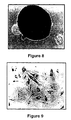
- Figure 8 is a photomicrograph showing the dermal fibroblasts attached to PHB-PHV microspheres treated with RGD of Figure 7 at 22 hours post cell-seeding. The majority of the cells that have attached to the microsphere are flattened and no longer spherical.
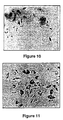
- Figure 9 is a photomicrograph of neo-natal foreskin derived dermal fibroblasts attached to a glass surface (control) and incubated for 26 hours.
- Figure 10 is a photomicrograph of neo-natal foreskin derived dermal fibroblasts attached to a planar surface of PHB-PHV and incubated for 26 hours.
- Figure 11 is a photomicrograph of neo-natal foreskin derived dermal fibroblasts attached to a planar surface of PHB-PHV treated with RGD and incubated for 26 hours.
- the viable cells in Figures 9, 10 and 11 are stained with Neutral Red. The dye is actively taken up by viable cells to demonstrate, in Figures 10 and 11, that the cells are highly viable on the bioresorbable materials used.
- growth factors such as epidermal growth factor (EGF), transforming growth factor-alpha (TGF-A), transforming growth factor-beta (TGF-beta), keratinocyte growth factor (KGF), basic fibroblast growth factor (bFGF), and insulin-like growth factor-I (IGF-I).
- EGF epidermal growth factor
- TGF-A transforming growth factor-alpha
- TGF-beta transforming growth factor-beta
- KGF keratinocyte growth factor
- bFGF basic fibroblast growth factor
- IGF-I insulin-like growth factor-I
- a controlled delivery of these factors can be obtained by the use of microspheres in order to obtain better control of wound healing, since if delivery of mitogenic (growth) or angiogenic (inducing recapillarization of the wound) factors is too rapid, scarring of the wound site can occur.
- Growth factors can also be immobilized onto the surface of the microspheres with, for example, a permanent covalent linkage so that the
- Monoclonal antibodies can be incorporated onto the microspheres to control the local in vivo levels of TGF-beta, for example, in order to avoid an over-abundant supply of this factor at a particular point in the wound healing process.
- TGF-beta if available in excess amounts, can lead to a disproportionate excess of collagen production by fibroblasts and macrophage cells.
- Resorption of the microspheres can be enhanced by incorporating enzymes such as lipase, depolymerase, and dehydrogenase into the microspheres. These enzymes enhance the resorption by other endogenous enzymes in vivo or free radicals generated by the body (e.g. macrophage cells) which can attack the structural linkages leading to depolymerization of the bioresorbable material.
- enzymes such as lipase, depolymerase, and dehydrogenase
- PDGF platelet derived growth factor
- additives include polyamines to mitigate hypertrophic scarring, materials which impart biostatic, microbicidal or anti-rejection properties, and proteinoids, for enhanced activity and/or stability.
- microspheres growth, migration, angiogenesis and other factors can be incorporated in or on a further supply of microspheres that are not intended to be cell-coated.
- ultra small microspheres as these are not required in large quantities thereby reducing the amount of material for resorption.
- These ultra small microspheres with diameters ranging from 10 to 50 microns can be interspersed with the larger cell-coated microspheres into the wound site or added independently at a suitable time.
- the surrounding tissues may be pretreated with chemotactic, angiogenic, and mitogenic factors coated on the surface of such cell-free microspheres in order to improve the environment for cell migration, nutrient delivery, migration and growth.
- serum-free media are suitable for the cultivation of dermal fibroblasts and keratinocytes. Numerous serum-free media are commercially available including, for example, Clonetics Corporation KGM, FGM and GIBCO KGM.
- keratinocytes require lower levels of calcium in the medium to encourage rapid growth rates and prevent terminal differentiation. Under higher levels of calcium (>0.1 mM), the keratinocytes tend to grow slowly and to undergo terminal differentiation leading to apoptosis (programmed cell death).
- Dermal fibroblasts are usually grown in media with calcium levels in the range of 1 to 3mM to allow for normal growth and function. Accordingly, in one embodiment the dermal fibroblasts are first cultured in a medium containing calcium in a concentration greater than 0.1mM. After a period of time, the cells are washed with a phosphate buffered saline (PBS) solution in situ to remove the growth medium.
- PBS phosphate buffered saline
- Keratinocytes and a different medium with a reduced calcium concentration are then added to the tissue culture flask containing the cultured dermal fibroblasts.
- the net result in further culturing is a slowing down of the fibroblast growth while allowing for good keratinocyte growth. This is an ideal situation as it allows for a layer of keratinocytes to grow in the presence of the fibroblasts without the fibroblasts overgrowing the keratinocytes.
- the fibroblasts while not proliferating, can still produce factors required by the keratinocytes for optimal growth and organization on the dermal surface.
- Factors produced by fibroblasts that can enable optimal keratinocyte growth/organization include fibronectin and TGF-beta.
- the present invention provides potential for geographical centralization of wound or burn treatment tacilities for the actual cell manipulation.
- Application of the dermal fibroblasts to full- and partial-thickness skin injuries can be expedited with the maintenance of a local "bank" of donor-derived, including cadaver-derived, dermal fibroblasts which exhibit very low allergenicity in transplants.
- a tissue explant derived from the patient can then be shipped in an appropriate transport medium similar to that used for transporting organs for transplant (such as, ViaSpan (trade-mark) UW Solution (Dupont), or other maintenance medium) to a central cell processing facility for culture of the keratinocytes while the patient is already being treated with a dermal fibroblast/microsphere slurry.
- an appropriate transport medium similar to that used for transporting organs for transplant (such as, ViaSpan (trade-mark) UW Solution (Dupont), or other maintenance medium) to a central cell processing facility for culture of the keratinocytes while the patient is already being treated with a dermal fibroblast/microsphere slurry.
- a 4 cm 2 tissue explant could be rapidly transported and expanded to treat an equivalent tissue area of several square metres within 1 to 3 weeks if required. The growth of sufficient cells for treatment is markedly faster and more effective than the conventional approaches outlined.
- the cell-coated microspheres can be harvested, concentrated and put into a maintenance medium for shipment to the remote treatment centre.
- Large numbers of cells in the order of 10 7 cells/ml, can be transported on the microspheres in a relatively small volume. This is an advantage over the planar technology which requires a larger number of containers for the equivalent amount of cells.
- the transport of the mesh matrices is hampered by the fragile nature of the tissue preparation.
- the microsphere/cell slurry of the present invention is less fragile and can still be satisfactorily shipped under less than ideal conditions.
- Cell-coated microspheres can be transported in medium similar to that used for transport of transplant organs such as heart. Moreover, the medium can be adapted to include perfluorocarbons for enhanced oxygen transfer.
- the microsphere/cell slurry requires little or no further treatment before application to the skin injury.
- Known technologies such as tangential- or cross-flow membranes or hollow fibre membranes can also be used in transport thereby retaining the cells/microspheres while manipulating the environment in which they reside.
- cryo-preserving medium may include dimethylsulfoxide (DMSO) which requires elaborate techniques including a sterile environment and numerous manipulations, requiring a tissue culture laboratory at the receiving end for the removal thereof before implant. This type of facility may not always be available on site when or where treatment is required.
- DMSO dimethylsulfoxide
- the present invention allows for fast and effective treatment of full- and partial-thickness skin injuries, such as burns and other wounds. Unlike conventional methods, the present invention does not require the use of stapling, suturing, or other attachment methods for application to the patient, nor is it constrained by problems associated with body profiling or curvature. Furthermore, the application of the microsphere/cell slurry can be conducted under both ideal and less than ideal conditions.
- the living skin replacement of the present invention can also be used as a "skin model" in risk and safety assessment assays for pharmaceutical, cosmetic and household compounds. This is a growing area owing to the demand to circumvent animal testing.
- the present invention also finds utility in the cosmetic sector for correcting skin defects.
Landscapes
- Health & Medical Sciences (AREA)
- Life Sciences & Earth Sciences (AREA)
- General Health & Medical Sciences (AREA)
- Engineering & Computer Science (AREA)
- Animal Behavior & Ethology (AREA)
- Public Health (AREA)
- Veterinary Medicine (AREA)
- Chemical & Material Sciences (AREA)
- Dermatology (AREA)
- Biomedical Technology (AREA)
- Epidemiology (AREA)
- Zoology (AREA)
- Organic Chemistry (AREA)
- Bioinformatics & Cheminformatics (AREA)
- Wood Science & Technology (AREA)
- Medicinal Chemistry (AREA)
- Biotechnology (AREA)
- Birds (AREA)
- Genetics & Genomics (AREA)
- Oral & Maxillofacial Surgery (AREA)
- Transplantation (AREA)
- General Engineering & Computer Science (AREA)
- Biochemistry (AREA)
- Microbiology (AREA)
- Cell Biology (AREA)
- Pharmacology & Pharmacy (AREA)
- General Chemical & Material Sciences (AREA)
- Nuclear Medicine, Radiotherapy & Molecular Imaging (AREA)
- Chemical Kinetics & Catalysis (AREA)
- Materials For Medical Uses (AREA)
- Immobilizing And Processing Of Enzymes And Microorganisms (AREA)
- Medicines Containing Material From Animals Or Micro-Organisms (AREA)
- Micro-Organisms Or Cultivation Processes Thereof (AREA)
- Manufacturing Of Micro-Capsules (AREA)
- Medicinal Preparation (AREA)
Applications Claiming Priority (3)
| Application Number | Priority Date | Filing Date | Title |
|---|---|---|---|
| GB9210574 | 1992-05-18 | ||
| GB929210574A GB9210574D0 (en) | 1992-05-18 | 1992-05-18 | Biotherapeutic cell-coated microspheres for wound/burn and prothesis implant applications |
| PCT/CA1993/000187 WO1993023088A1 (en) | 1992-05-18 | 1993-04-23 | Biotherapeutic cell-coated microspheres |
Publications (2)
| Publication Number | Publication Date |
|---|---|
| EP0641223A1 EP0641223A1 (en) | 1995-03-08 |
| EP0641223B1 true EP0641223B1 (en) | 2001-08-08 |
Family
ID=10715656
Family Applications (1)
| Application Number | Title | Priority Date | Filing Date |
|---|---|---|---|
| EP93911706A Expired - Lifetime EP0641223B1 (en) | 1992-05-18 | 1993-04-23 | Biotherapeutic cell-coated microspheres |
Country Status (16)
| Country | Link |
|---|---|
| US (1) | US5830507A (es) |
| EP (1) | EP0641223B1 (es) |
| JP (1) | JP2820796B2 (es) |
| KR (1) | KR950701533A (es) |
| AT (1) | ATE203916T1 (es) |
| BR (1) | BR9306375A (es) |
| CA (1) | CA2135999C (es) |
| DE (1) | DE69330562T2 (es) |
| DK (1) | DK0641223T3 (es) |
| ES (1) | ES2161229T3 (es) |
| GB (1) | GB9210574D0 (es) |
| GR (1) | GR3036741T3 (es) |
| NZ (1) | NZ252661A (es) |
| PT (1) | PT641223E (es) |
| RU (1) | RU2104039C1 (es) |
| WO (1) | WO1993023088A1 (es) |
Families Citing this family (46)
| Publication number | Priority date | Publication date | Assignee | Title |
|---|---|---|---|---|
| US5654267A (en) * | 1988-12-20 | 1997-08-05 | La Jolla Cancer Research Center | Cooperative combinations of ligands contained within a matrix |
| IL115728A0 (en) * | 1994-10-25 | 1996-01-19 | Boehringer Mannheim Gmbh | Biomaterial containing epithelial cells and use thereof as a transplant |
| IL118376A0 (en) * | 1996-05-22 | 1996-09-12 | Univ Ben Gurion | Polysaccharide sponges for cell culture and transplantation |
| US5972332A (en) | 1997-04-16 | 1999-10-26 | The Regents Of The University Of Michigan | Wound treatment with keratinocytes on a solid support enclosed in a porous material |
| US20020037566A1 (en) * | 1997-04-16 | 2002-03-28 | Rees Riley S. | Cell-coated supports |
| US5861149A (en) * | 1997-06-04 | 1999-01-19 | Polyheal Ltd. | Methods for wound treatment |
| WO1999021963A1 (en) * | 1997-10-25 | 1999-05-06 | Roche Diagnostics Gmbh | Autodegradable microcarriers and their use |
| US6378527B1 (en) * | 1998-04-08 | 2002-04-30 | Chondros, Inc. | Cell-culture and polymer constructs |
| CA2329010A1 (en) * | 1998-04-17 | 1999-10-28 | Angiogenix Incorporated | Therapeutic angiogenic factors and methods for their use |
| JP4653887B2 (ja) * | 1998-10-23 | 2011-03-16 | ポリヒール リミテッド | 創傷治療用のミクロスフェアを含む組成物 |
| DE19937102A1 (de) * | 1999-08-06 | 2001-02-15 | Universitaetsklinikum Freiburg | Gewebeersatz und Verfahren zu seiner Herstellung |
| US7056503B2 (en) * | 1999-08-19 | 2006-06-06 | Regents Of The University Of Michigan | Enclosures housing cell-coated supports for treating tumors |
| DE19949290A1 (de) * | 1999-10-12 | 2001-04-26 | Albrecht Bettermann | Partikuläres Konstrukt zur Verwendung in der Transplantationsmedizin |
| KR20010110945A (ko) | 2000-06-09 | 2001-12-15 | 김정용 | 음경 확대 방법 |
| US6719970B1 (en) * | 2000-07-10 | 2004-04-13 | Alkermes Controlled Therapeutics, Inc. | Method of generating cartilage |
| US20040037828A1 (en) | 2002-07-09 | 2004-02-26 | Bar-Ilan University | Methods and pharmaceutical compositions for healing wounds |
| US20100129332A1 (en) * | 2000-07-31 | 2010-05-27 | Tamar Tennenbaum | Methods and pharmaceutical compositions for healing wounds |
| US20030148924A1 (en) * | 2002-07-09 | 2003-08-07 | Tamar Tennenbaum | Methods and pharmaceutical compositions of healing wounds |
| US20060258562A1 (en) * | 2000-07-31 | 2006-11-16 | Healor Ltd. | Methods and pharmaceutical compositions for healing wounds |
| JP2002281964A (ja) * | 2000-12-26 | 2002-10-02 | Sanyo Chem Ind Ltd | 細胞生産方法 |
| AUPR289601A0 (en) * | 2001-02-05 | 2001-03-01 | Commonwealth Scientific And Industrial Research Organisation | Method of tissue repair |
| US20050129776A1 (en) * | 2002-05-03 | 2005-06-16 | Inserm | Microparticles supporting cells and active substances |
| FR2839260B1 (fr) * | 2002-05-03 | 2005-02-25 | Inst Nat Sante Rech Med | Microparticules a base d'un materiau bicompatible et biodegradable, supportant des cellules et des substances biologiquement actives |
| US7402323B2 (en) * | 2002-08-21 | 2008-07-22 | Akpharma, Inc. | Compositions and methods for treating skin conditions |
| EP1896092A1 (de) * | 2003-05-24 | 2008-03-12 | JUVENA (International) AG | Kosmetische oder dermatologische Zubereitung umfassend eine Nährmedienphase |
| EP1636350B1 (de) * | 2003-06-23 | 2007-09-12 | Fraunhofer-Gesellschaft zur Förderung der angewandten Forschung e.V. | Verfahren zur differenzierung von stammzellen in zellen, die ein pankreatisches hormon produzieren |
| ZA200601089B (en) * | 2003-08-07 | 2007-05-30 | Hearlor Ltd | Pharmaceutical compositions and methods for accelerating wound healing |
| AU2003289475A1 (en) * | 2003-10-20 | 2005-05-05 | Jms Co., Ltd. | Cell handling device, human tissue regeneration composition, and human tissue regeneration method |
| AU2006286152B2 (en) * | 2005-08-29 | 2012-05-31 | Healor Ltd. | Methods and compositions for prevention and treatment of diabetic and aged skin |
| WO2008073614A2 (en) * | 2006-11-02 | 2008-06-19 | Akpharma Inc. | Composition and method for enhancing skin cell growth, proliferation and repair |
| KR100805208B1 (ko) | 2007-03-27 | 2008-02-21 | 주식회사 펩트론 | 엑센딘 함유 서방성 제제 조성물, 엑센딘 함유 서방성미립구 및 이의 제조 방법 |
| US20100255070A1 (en) * | 2007-05-22 | 2010-10-07 | Prelief Inc. | Compositions and methods for preventing, minimizing and healing skin irritation and trauma |
| RU2502520C2 (ru) * | 2007-07-30 | 2013-12-27 | Хилор Лтд. | Фармацевтическая композиция и относящиеся способы |
| WO2009043052A1 (en) * | 2007-09-27 | 2009-04-02 | Columbia University | Methods and systems for forming biocompatible materials |
| WO2009086535A2 (en) * | 2007-12-27 | 2009-07-09 | The Trustees Of Columbia University In The City Of New York | Systems and methods for forming patterned extracellular matrix materials |
| CN104491845A (zh) | 2008-04-18 | 2015-04-08 | 科尔普兰特有限公司 | 产生和使用原胶原的方法 |
| AU2009258745B2 (en) | 2008-06-13 | 2011-11-17 | Fuji Kasei Co., Ltd. | Skin-substitutive membrane, mold, and method of evaluating external preparation for skin |
| EA014044B1 (ru) * | 2008-07-09 | 2010-08-30 | Общество С Ограниченной Ответственностью "Аква-Альянс" | Наносомальная лекарственная форма препарата пролонгированного действия для лечения гепатита с (варианты) |
| LT2506859T (lt) * | 2009-12-04 | 2016-09-26 | Magle Ab | Hidrolizuoto krakmolo mikrosferos su endogeniniais įkrautais ligandais |
| CA2789972A1 (en) | 2010-01-11 | 2011-07-14 | Healor Ltd. | Method for treatment of inflammatory disease and disorder |
| JP5241764B2 (ja) * | 2010-03-31 | 2013-07-17 | テルモ株式会社 | 生体適合性材料の評価方法 |
| RU2616866C1 (ru) * | 2015-12-31 | 2017-04-18 | Федеральное государственное бюджетное образовательное учреждение высшего образования "Московский государственный университет имени М.В. Ломоносова" (МГУ) | Биорезорбируемый микроноситель для доставки клеток в область заживления и регенерации ран |
| RU2658707C1 (ru) * | 2016-12-28 | 2018-06-22 | Федеральное государственное бюджетное образовательное учреждение высшего образования "Московский государственный университет имени М.В. Ломоносова" (МГУ) | Способ восстановления кожного покрова |
| RU2644633C1 (ru) * | 2016-12-28 | 2018-02-13 | Федеральное государственное бюджетное образовательное учреждение высшего образования "Московский государственный университет имени М.В. Ломоносова" (МГУ) | Способ восстановления кожного покрова |
| CN110960513A (zh) * | 2019-11-29 | 2020-04-07 | 苏州瑞微生物科技有限公司 | 一种微球阵列及其制备方法和用途 |
| CN113201481B (zh) * | 2021-04-19 | 2023-10-13 | 清华大学深圳国际研究生院 | 皮肤微球及其制备方法和应用 |
Family Cites Families (12)
| Publication number | Priority date | Publication date | Assignee | Title |
|---|---|---|---|---|
| FR2461002A1 (fr) * | 1979-07-13 | 1981-01-30 | Inst Nat Sante Rech Med | Procede pour stimuler la croissance des cellules d'epiderme humain et produits faisant application dudit procede |
| CA1215922A (en) * | 1984-05-25 | 1986-12-30 | Connaught Laboratories Limited | Microencapsulation of living tissue and cells |
| EP0215274B1 (en) * | 1985-08-14 | 1993-03-31 | Memorial Hospital for Cancer and Allied Diseases | Epidermal cell extracts and method to enhance wound healing and regenerate epidermis |
| FR2597499A1 (fr) * | 1986-04-18 | 1987-10-23 | Merieux Inst | Procede de culture microbiologique sur supports de collagene, supports pour ce procede et produits obtenus |
| NZ222413A (en) * | 1986-11-05 | 1991-06-25 | Ethicon Inc | Compositions containing a polypeptide growth factor and a water-soluble cellulose polymer stabiliser |
| US5015584A (en) * | 1987-10-14 | 1991-05-14 | Board Of Regents, The University Of Texas System | Epidermal graft system |
| GB8803697D0 (en) * | 1988-02-17 | 1988-03-16 | Deltanine Research Ltd | Clinical developments using amniotic membrane cells |
| CA1307225C (en) * | 1988-07-19 | 1992-09-08 | David W. Armstrong | Cell culture bioreactor |
| US5135915A (en) * | 1988-10-14 | 1992-08-04 | Genentech, Inc. | Method for the treatment of grafts prior to transplantation using TGF-.beta. |
| EP0422209B1 (en) * | 1989-04-25 | 1995-03-15 | Children's Medical Center Corporation | Implants for large volumes of cells on polymeric matrices |
| US4996154A (en) * | 1989-05-04 | 1991-02-26 | Millipore Corporation | Method for growing cellular tissue |
| US5618531A (en) * | 1990-10-19 | 1997-04-08 | New York University | Method for increasing the viability of cells which are administered to the brain or spinal cord |
-
1992
- 1992-05-18 GB GB929210574A patent/GB9210574D0/en active Pending
-
1993
- 1993-04-23 US US08/338,596 patent/US5830507A/en not_active Expired - Fee Related
- 1993-04-23 WO PCT/CA1993/000187 patent/WO1993023088A1/en active IP Right Grant
- 1993-04-23 DK DK93911706T patent/DK0641223T3/da active
- 1993-04-23 ES ES93911706T patent/ES2161229T3/es not_active Expired - Lifetime
- 1993-04-23 NZ NZ252661A patent/NZ252661A/en not_active IP Right Cessation
- 1993-04-23 RU RU94046192A patent/RU2104039C1/ru not_active IP Right Cessation
- 1993-04-23 KR KR1019940704108A patent/KR950701533A/ko not_active IP Right Cessation
- 1993-04-23 AT AT93911706T patent/ATE203916T1/de not_active IP Right Cessation
- 1993-04-23 DE DE69330562T patent/DE69330562T2/de not_active Expired - Fee Related
- 1993-04-23 CA CA002135999A patent/CA2135999C/en not_active Expired - Fee Related
- 1993-04-23 JP JP5519724A patent/JP2820796B2/ja not_active Expired - Fee Related
- 1993-04-23 BR BR9306375A patent/BR9306375A/pt not_active Application Discontinuation
- 1993-04-23 PT PT93911706T patent/PT641223E/pt unknown
- 1993-04-23 EP EP93911706A patent/EP0641223B1/en not_active Expired - Lifetime
-
2001
- 2001-09-27 GR GR20010401598T patent/GR3036741T3/el not_active IP Right Cessation
Also Published As
| Publication number | Publication date |
|---|---|
| PT641223E (pt) | 2001-12-28 |
| EP0641223A1 (en) | 1995-03-08 |
| GB9210574D0 (en) | 1992-07-01 |
| DE69330562D1 (de) | 2001-09-13 |
| ES2161229T3 (es) | 2001-12-01 |
| RU94046192A (ru) | 1996-10-10 |
| NZ252661A (en) | 1996-09-25 |
| JPH07507063A (ja) | 1995-08-03 |
| AU668959B2 (en) | 1996-05-23 |
| CA2135999A1 (en) | 1993-11-25 |
| JP2820796B2 (ja) | 1998-11-05 |
| ATE203916T1 (de) | 2001-08-15 |
| AU4257993A (en) | 1993-12-13 |
| KR100381207B1 (es) | 2003-10-23 |
| KR950701533A (ko) | 1995-04-28 |
| CA2135999C (en) | 1999-07-13 |
| BR9306375A (pt) | 1997-03-04 |
| RU2104039C1 (ru) | 1998-02-10 |
| US5830507A (en) | 1998-11-03 |
| WO1993023088A1 (en) | 1993-11-25 |
| DK0641223T3 (da) | 2001-10-01 |
| GR3036741T3 (en) | 2001-12-31 |
| DE69330562T2 (de) | 2002-05-29 |
Similar Documents
| Publication | Publication Date | Title |
|---|---|---|
| EP0641223B1 (en) | Biotherapeutic cell-coated microspheres | |
| Freed et al. | Biodegradable polymer scaffolds for tissue engineering | |
| US5830708A (en) | Methods for production of a naturally secreted extracellular matrix | |
| EP0422209B1 (en) | Implants for large volumes of cells on polymeric matrices | |
| US5716404A (en) | Breast tissue engineering | |
| AU2003234317B2 (en) | Vascularization enhanced graft constructs | |
| US20050129730A1 (en) | Tissue composites and uses thereof | |
| CA2330104A1 (en) | Creation of three-dimensional tissues | |
| US20040180431A1 (en) | Compositions and methods for production and use of an injectable naturally secreted extracellular matrix | |
| JPH11503946A (ja) | ヒアルロン酸誘導体を基本とするバイオ適合性物質を支持体として含む人工皮膚 | |
| JP2005537845A (ja) | フィブリン細胞支持体およびその使用方法 | |
| WO2001003750A1 (en) | Human naturally secreted extracellular matrix-coated device | |
| Khor et al. | Preliminary study of a polycaprolactone membrane utilized as epidermal substrate | |
| Lafrance et al. | Novel living skin replacement biotherapy approach for wounded skin tissues | |
| Huang et al. | Multifunctional implantable particles for skin tissue regeneration: preparation, characterization, in vitro and in vivo studies | |
| US9259445B2 (en) | Integrated implant system (IIS) biocompatible, biodegradable and bioactive, comprising a biocompatible sterile porous polymeric matrix and a gel, integrating in situ the tridimensional matrix structure | |
| AU759066B2 (en) | 3D matrix for producing cell transplants | |
| AU668959C (en) | Biotherapeutic cell-coated microspheres | |
| EP2040766A2 (en) | Integrated implant system (iis) biocompatible, biodegradable and bioactive, comprising a biocompatible sterile porous polymeric matrix and a gel, integrating in situ the tridimensional matrix structure |
Legal Events
| Date | Code | Title | Description |
|---|---|---|---|
| PUAI | Public reference made under article 153(3) epc to a published international application that has entered the european phase |
Free format text: ORIGINAL CODE: 0009012 |
|
| 17P | Request for examination filed |
Effective date: 19941111 |
|
| AK | Designated contracting states |
Kind code of ref document: A1 Designated state(s): AT BE CH DE DK ES FR GB GR IE IT LI LU MC NL PT SE |
|
| 17Q | First examination report despatched |
Effective date: 19960129 |
|
| GRAG | Despatch of communication of intention to grant |
Free format text: ORIGINAL CODE: EPIDOS AGRA |
|
| GRAG | Despatch of communication of intention to grant |
Free format text: ORIGINAL CODE: EPIDOS AGRA |
|
| GRAH | Despatch of communication of intention to grant a patent |
Free format text: ORIGINAL CODE: EPIDOS IGRA |
|
| GRAG | Despatch of communication of intention to grant |
Free format text: ORIGINAL CODE: EPIDOS AGRA |
|
| GRAH | Despatch of communication of intention to grant a patent |
Free format text: ORIGINAL CODE: EPIDOS IGRA |
|
| GRAH | Despatch of communication of intention to grant a patent |
Free format text: ORIGINAL CODE: EPIDOS IGRA |
|
| GRAA | (expected) grant |
Free format text: ORIGINAL CODE: 0009210 |
|
| AK | Designated contracting states |
Kind code of ref document: B1 Designated state(s): AT BE CH DE DK ES FR GB GR IE IT LI LU MC NL PT SE |
|
| REF | Corresponds to: |
Ref document number: 203916 Country of ref document: AT Date of ref document: 20010815 Kind code of ref document: T |
|
| REG | Reference to a national code |
Ref country code: CH Ref legal event code: EP |
|
| REG | Reference to a national code |
Ref country code: IE Ref legal event code: FG4D |
|
| REF | Corresponds to: |
Ref document number: 69330562 Country of ref document: DE Date of ref document: 20010913 |
|
| REG | Reference to a national code |
Ref country code: CH Ref legal event code: NV Representative=s name: E. BLUM & CO. PATENTANWAELTE |
|
| REG | Reference to a national code |
Ref country code: DK Ref legal event code: T3 |
|
| ET | Fr: translation filed | ||
| REG | Reference to a national code |
Ref country code: ES Ref legal event code: FG2A Ref document number: 2161229 Country of ref document: ES Kind code of ref document: T3 |
|
| REG | Reference to a national code |
Ref country code: PT Ref legal event code: SC4A Free format text: AVAILABILITY OF NATIONAL TRANSLATION Effective date: 20010920 |
|
| REG | Reference to a national code |
Ref country code: GB Ref legal event code: IF02 |
|
| PG25 | Lapsed in a contracting state [announced via postgrant information from national office to epo] |
Ref country code: MC Free format text: LAPSE BECAUSE OF NON-PAYMENT OF DUE FEES Effective date: 20020423 Ref country code: LU Free format text: LAPSE BECAUSE OF NON-PAYMENT OF DUE FEES Effective date: 20020423 |
|
| PLBE | No opposition filed within time limit |
Free format text: ORIGINAL CODE: 0009261 |
|
| STAA | Information on the status of an ep patent application or granted ep patent |
Free format text: STATUS: NO OPPOSITION FILED WITHIN TIME LIMIT |
|
| 26N | No opposition filed | ||
| PG25 | Lapsed in a contracting state [announced via postgrant information from national office to epo] |
Ref country code: IT Free format text: LAPSE BECAUSE OF NON-PAYMENT OF DUE FEES Effective date: 20050423 |
|
| PGFP | Annual fee paid to national office [announced via postgrant information from national office to epo] |
Ref country code: BE Payment date: 20060324 Year of fee payment: 14 |
|
| PGFP | Annual fee paid to national office [announced via postgrant information from national office to epo] |
Ref country code: PT Payment date: 20070420 Year of fee payment: 15 |
|
| PGFP | Annual fee paid to national office [announced via postgrant information from national office to epo] |
Ref country code: ES Payment date: 20070423 Year of fee payment: 15 |
|
| PGFP | Annual fee paid to national office [announced via postgrant information from national office to epo] |
Ref country code: IE Payment date: 20070426 Year of fee payment: 15 Ref country code: DK Payment date: 20070426 Year of fee payment: 15 Ref country code: DE Payment date: 20070426 Year of fee payment: 15 Ref country code: CH Payment date: 20070426 Year of fee payment: 15 |
|
| PGFP | Annual fee paid to national office [announced via postgrant information from national office to epo] |
Ref country code: SE Payment date: 20070427 Year of fee payment: 15 Ref country code: AT Payment date: 20070427 Year of fee payment: 15 |
|
| PGFP | Annual fee paid to national office [announced via postgrant information from national office to epo] |
Ref country code: NL Payment date: 20070430 Year of fee payment: 15 |
|
| REG | Reference to a national code |
Ref country code: CH Ref legal event code: PFA Owner name: NATIONAL RESEARCH COUNCIL OF CANADA Free format text: NATIONAL RESEARCH COUNCIL OF CANADA# #OTTAWA ONTARIO K1A OR6 (CA) -TRANSFER TO- NATIONAL RESEARCH COUNCIL OF CANADA# #OTTAWA ONTARIO K1A OR6 (CA) |
|
| PGFP | Annual fee paid to national office [announced via postgrant information from national office to epo] |
Ref country code: GB Payment date: 20070427 Year of fee payment: 15 |
|
| BERE | Be: lapsed |
Owner name: *NATIONAL RESEARCH COUNCIL OF CANADA Effective date: 20070430 |
|
| PG25 | Lapsed in a contracting state [announced via postgrant information from national office to epo] |
Ref country code: BE Free format text: LAPSE BECAUSE OF NON-PAYMENT OF DUE FEES Effective date: 20070430 |
|
| PGFP | Annual fee paid to national office [announced via postgrant information from national office to epo] |
Ref country code: FR Payment date: 20070425 Year of fee payment: 15 |
|
| PGFP | Annual fee paid to national office [announced via postgrant information from national office to epo] |
Ref country code: GR Payment date: 20070430 Year of fee payment: 15 |
|
| REG | Reference to a national code |
Ref country code: PT Ref legal event code: MM4A Free format text: LAPSE DUE TO NON-PAYMENT OF FEES Effective date: 20081023 |
|
| REG | Reference to a national code |
Ref country code: CH Ref legal event code: PL |
|
| REG | Reference to a national code |
Ref country code: DK Ref legal event code: EBP |
|
| EUG | Se: european patent has lapsed | ||
| GBPC | Gb: european patent ceased through non-payment of renewal fee |
Effective date: 20080423 |
|
| NLV4 | Nl: lapsed or anulled due to non-payment of the annual fee |
Effective date: 20081101 |
|
| REG | Reference to a national code |
Ref country code: IE Ref legal event code: MM4A |
|
| PG25 | Lapsed in a contracting state [announced via postgrant information from national office to epo] |
Ref country code: PT Free format text: LAPSE BECAUSE OF NON-PAYMENT OF DUE FEES Effective date: 20081023 Ref country code: NL Free format text: LAPSE BECAUSE OF NON-PAYMENT OF DUE FEES Effective date: 20081101 Ref country code: LI Free format text: LAPSE BECAUSE OF NON-PAYMENT OF DUE FEES Effective date: 20080430 Ref country code: DE Free format text: LAPSE BECAUSE OF NON-PAYMENT OF DUE FEES Effective date: 20081101 Ref country code: CH Free format text: LAPSE BECAUSE OF NON-PAYMENT OF DUE FEES Effective date: 20080430 |
|
| REG | Reference to a national code |
Ref country code: FR Ref legal event code: ST Effective date: 20081231 |
|
| PG25 | Lapsed in a contracting state [announced via postgrant information from national office to epo] |
Ref country code: AT Free format text: LAPSE BECAUSE OF NON-PAYMENT OF DUE FEES Effective date: 20080423 |
|
| PG25 | Lapsed in a contracting state [announced via postgrant information from national office to epo] |
Ref country code: IE Free format text: LAPSE BECAUSE OF NON-PAYMENT OF DUE FEES Effective date: 20080423 Ref country code: FR Free format text: LAPSE BECAUSE OF NON-PAYMENT OF DUE FEES Effective date: 20080430 Ref country code: DK Free format text: LAPSE BECAUSE OF NON-PAYMENT OF DUE FEES Effective date: 20080430 |
|
| REG | Reference to a national code |
Ref country code: ES Ref legal event code: FD2A Effective date: 20080424 |
|
| PG25 | Lapsed in a contracting state [announced via postgrant information from national office to epo] |
Ref country code: GR Free format text: LAPSE BECAUSE OF NON-PAYMENT OF DUE FEES Effective date: 20081104 Ref country code: GB Free format text: LAPSE BECAUSE OF NON-PAYMENT OF DUE FEES Effective date: 20080423 |
|
| PG25 | Lapsed in a contracting state [announced via postgrant information from national office to epo] |
Ref country code: ES Free format text: LAPSE BECAUSE OF NON-PAYMENT OF DUE FEES Effective date: 20080424 |
|
| PG25 | Lapsed in a contracting state [announced via postgrant information from national office to epo] |
Ref country code: SE Free format text: LAPSE BECAUSE OF NON-PAYMENT OF DUE FEES Effective date: 20080424 |
|
| PGFP | Annual fee paid to national office [announced via postgrant information from national office to epo] |
Ref country code: IT Payment date: 20070627 Year of fee payment: 15 |
|
| PGRI | Patent reinstated in contracting state [announced from national office to epo] |
Ref country code: IT Effective date: 20110616 |
|
| PGRI | Patent reinstated in contracting state [announced from national office to epo] |
Ref country code: IT Effective date: 20110616 |