EP0577458B1 - Nukleotid- und Peptidsequenzen des Immunschwächevirus der Katze, Isolat WO und Anwendungen der Sequenzen in der Diagnostik und zur Verhinderung der Infektion - Google Patents
Nukleotid- und Peptidsequenzen des Immunschwächevirus der Katze, Isolat WO und Anwendungen der Sequenzen in der Diagnostik und zur Verhinderung der Infektion Download PDFInfo
- Publication number
- EP0577458B1 EP0577458B1 EP93401538A EP93401538A EP0577458B1 EP 0577458 B1 EP0577458 B1 EP 0577458B1 EP 93401538 A EP93401538 A EP 93401538A EP 93401538 A EP93401538 A EP 93401538A EP 0577458 B1 EP0577458 B1 EP 0577458B1
- Authority
- EP
- European Patent Office
- Prior art keywords
- peptide
- vector
- fiv
- vif
- fragment
- Prior art date
- Legal status (The legal status is an assumption and is not a legal conclusion. Google has not performed a legal analysis and makes no representation as to the accuracy of the status listed.)
- Expired - Lifetime
Links
Images


Classifications
-
- C—CHEMISTRY; METALLURGY
- C07—ORGANIC CHEMISTRY
- C07K—PEPTIDES
- C07K14/00—Peptides having more than 20 amino acids; Gastrins; Somatostatins; Melanotropins; Derivatives thereof
- C07K14/005—Peptides having more than 20 amino acids; Gastrins; Somatostatins; Melanotropins; Derivatives thereof from viruses
-
- C—CHEMISTRY; METALLURGY
- C07—ORGANIC CHEMISTRY
- C07K—PEPTIDES
- C07K16/00—Immunoglobulins [IG], e.g. monoclonal or polyclonal antibodies
- C07K16/08—Immunoglobulins [IG], e.g. monoclonal or polyclonal antibodies against material from viruses
- C07K16/10—RNA viruses
- C07K16/112—Retroviridae (F), e.g. leukemia viruses
- C07K16/114—Lentivirus (G), e.g. human immunodeficiency virus [HIV], feline immunodeficiency virus [FIV] or simian immunodeficiency virus [SIV]
-
- A—HUMAN NECESSITIES
- A61—MEDICAL OR VETERINARY SCIENCE; HYGIENE
- A61K—PREPARATIONS FOR MEDICAL, DENTAL OR TOILETRY PURPOSES
- A61K39/00—Medicinal preparations containing antigens or antibodies
-
- C—CHEMISTRY; METALLURGY
- C12—BIOCHEMISTRY; BEER; SPIRITS; WINE; VINEGAR; MICROBIOLOGY; ENZYMOLOGY; MUTATION OR GENETIC ENGINEERING
- C12N—MICROORGANISMS OR ENZYMES; COMPOSITIONS THEREOF; PROPAGATING, PRESERVING, OR MAINTAINING MICROORGANISMS; MUTATION OR GENETIC ENGINEERING; CULTURE MEDIA
- C12N2740/00—Reverse transcribing RNA viruses
- C12N2740/00011—Details
- C12N2740/10011—Retroviridae
- C12N2740/15011—Lentivirus, not HIV, e.g. FIV, SIV
- C12N2740/15022—New viral proteins or individual genes, new structural or functional aspects of known viral proteins or genes
Definitions
- the present invention relates to peptides and their fragments, specific for the virus of feline immunodeficiency (VIF) and use said fragments as diagnostic reagent and as agent inducing an immune response (cellular and / or production of antibodies), in particular for the prevention of feline immunodeficiency.
- VIP feline immunodeficiency
- the present invention also relates to the nucleotide sequences coding for said peptides.
- Feline immunodeficiency is caused by a lentivirus, feline immunodeficiency virus (FIV), which has a genetic structure similar to that of primate lentivirus (HIV and VIS).
- FV feline immunodeficiency virus
- Feline immunodeficiency poses a significant problem veterinary health, to the extent that a number significant number of cats infected with VIF has been detected in United States, Japan and Europe (5 to 25% of animals).
- feline immunodeficiency is essentially diagnosed at the onset clinical signs (generalized lymphadenopathy and occurrence of opportunistic infections), while a diagnosis very early would have significant benefits so much in treatment than in prevention of this disease.
- VIF envelope proteins are considered as being at the center of the host-virus relationship; their study is therefore essential to understand how the virus interacts with the immune system (neutralization epitopes, B and T epitopes) and with target cells for infection, and develop both high-performance diagnostic reagents and effective vaccines, consisting of viral proteins immunogenic and protective action, vis-à-vis the whole strains of VIF.
- the VIF env protein can provide, after cleavage, two glycoprotein fragments, called SU (surface glycoprotein) and TM (transmembrane glycoprotein).
- SU surface glycoprotein
- TM transmembrane glycoprotein
- the present invention is therefore intended to provide, on the one hand, sequences from VIF and their selected fragments in particular for their strain specificity, and, on the other hand, to a specific immunogenic and / or protective composition feline immunodeficiency virus, obtained in expressing fragments selected for their capacity neutralizing and / or the presence of recognition epitopes group and / or type; obtaining by genius genetics or chemical synthesis of such specific peptides has the advantage of solving the production problem virus and directly obtain the desired peptides.
- Nucleotide sequences from RNA feline immunodeficiency virus (VIF) genomics include at least one fragment of a gene with hypervariable zones, chosen from the group consisting of the gene encoding the env protein of the Wo strain of VIF and the gene coding for the gag protein of the strain Wo from VIF.
- this comprises the nucleotide sequence and the deduced amino acid sequence of formula I below, which corresponds to the env gene of VIF, strain Wo:
- Such a sequence corresponds to the cDNA of the mRNA of the env protein of the VIF strain Wo.
- Said peptide is especially recognized by antibodies to at least one different strain from that from which said peptide and / or by the antibodies produced by naturally infected cats and what it has a response response capacity immune system and / or production of protective antibodies vis-à-vis at least one strain of VIF, in uninfected cats.
- the fragment including a segment of 45 amino acids, called TM3, corresponds to positions 744-788 of the sequence of formula II below, which segment contains an epitope including the sequence Gln 764 -Leu-Gln-Glu-Trp-Glu -Asp-Trp-Val-Gly-Trp-Ile-Gly-Asn-Ile 778 or the sequence Gln 763 -Gln-Leu-Gln-Glu-Trp-Glu-Asp 770 (peptide called P241) or the sequence Val 772 - Gly-Trp-Ile-Gly-Asn-Ile-Pro 779 (peptide called P242).
- the peptide which corresponds to the Env protein of the VIF, strain Wo has the following sequence:
- epitope within the meaning of this invention, the epitopes common to all strains of VIF (conserved areas), recognized by directed antibodies against all strains of VIF (recognition of group); moreover, all these fragments induce a production antibody and / or cellular response.
- peptides are coded by a sequence or a nucleotide sequence fragment as defined in the sequence of formula I above.
- peptide fragment includes all fragments including a peptide segment in accordance with the invention as well as homologous peptides; in general, we mean by homologous peptide, the peptide fragments whose position and function are equivalent, within the VIF (same location as that defined above, on the VIF genome); more specifically TM3 fragments, also understood by homologous peptides, peptides that do not lose the properties of recognition of the antibodies directed against the peptide of reference.
- All of said peptides can advantageously be obtained either by cloning or by synthesis, in particular by synthesis of Merrifield.
- the present invention also relates to a recombinant cloning and / or expression vector, characterized in that it comprises a nucleotide sequence encoding one of the TM3 peptides defined above.
- recombinant vector either a plasmid, a cosmid, than a phage.
- said vector consists of a plasmid comprising a origin of replication, at least one selection marker and a nucleotide sequence encoding one of the TM3 peptides defined above, which vector is a cloning vector.
- said cloning vector consists of a plasmid capable of replicating in E. coli , into which is inserted a nucleotide sequence coding for one of the TM3 peptides defined above.
- said vector consists of a recombinant vector comprising an origin of replication in a microorganism suitable host, including bacteria or eukaryotic cell, at least one gene whose expression allows the selection of either bacteria or cells eukaryotes having received said vector, a promoter allowing the expression of genes in said bacteria or eukaryotic cells, and into which is inserted a nucleotide sequence encoding one of the TM3 peptides defined above, which vector is a vector expression of a TM3 peptide as defined above.
- said expression vector consists of a phage ⁇ gt11 into which is inserted, in phase at its site, EcoRI, fragments of digestion with DNAse of the plasmid pKSe1.
- the present invention also relates to an immunogenic and / or protective composition, characterized in that it comprises at least one peptide TM3 as defined above, possibly associated with minus another immunogenic or immunostimulating substance and / or a pharmaceutically acceptable vehicle.
- the present invention has, moreover, an object a method for detecting anti-FIV antibodies, characterized in that it consists in detecting anti-VIF antibodies possibly present in a biological sample to using a TM3 peptide as defined above, optionally fixed on an appropriate solid support, bringing said biological sample into contact with said peptide (s) to which the antibodies bind anti-FIV if such antibodies are present in the sample to be analyzed, the reading of the result being revealed by an appropriate means, in particular EIA, RIA, fluorescence.
- This process notably makes it possible to verify the seroconversion of vaccinated animals or to carry out epidemiological serological surveys.
- TM3 peptide said method allows determine a VIF infection, regardless of the strain (universal reagent as specified above).
- the present invention further relates to a kit, ready to use, for implementing the process anti-VIF antibody detection, characterized in that it includes, in addition to useful quantities of buffers suitable for carrying out said detection, appropriate doses of at least one TM3 peptide such as defined above.
- the present invention also relates to a reagent for diagnosing feline immunodeficiency, characterized in that it is chosen from the group consisting by one of the TM3 peptides as defined above, and in that it constitutes a universal diagnostic reagent of VIF strain.
- PBMC Peripheral blood mononuclear cells
- peripheral blood mononuclear cells obtained from cats said negative control (SPF cats)
- SPF cats peripheral blood mononuclear cells
- MORAILLON A. et al., 1992, Vet. Mic., 31, 41- 45 In vitro properties and experimental pathogenic effect of three feline immunodeficiency viruses isolated from cats with terminal diseases ).
- the Mg ++-dependent reverse transcriptase (RT) activity is more particularly monitored. After 15 days, the total cellular DNA is extracted with phenol (SAMBROOK et al., 1989, Molecular cloning, a laboratory manual), from RT positive cultures and used as starting material for the amplification of the VIF genes. This makes it possible to avoid multiple passages in vitro and adaptation to CD4 negative cell lines.
- the env and gag genes of the Wo strain of the feline immunodeficiency virus are respectively amplified in three and two overlapping fragments, using synthetic primers corresponding to regions conserved in the sequences of VIF, Petaluma isolate (TALBOTT et al, 1989 ) and PPR isolate (PHILIPPS et al., 1990).
- a vector M13mp8 is digested with the enzyme SmAI, so as to be able to insert into it the PCR fragments obtained above [for example, for the env gene fragments of the sequence of formula I, E 1 (nucleotides 1-977), E 2 (978-2016) and E 3 (2017-2562)].
- the recombinant vector obtained is used to transform competent E. coli JM101.
- the recombinant clones are detected by in situ hybridization with the corresponding PCR fragments, labeled with phosphorus 32.
- Single strand DNA sequencing from recombinant phages is carried out by the enzymatic method by dideoxynucleotides, with a sequencing kit sequenase® 2.0 DNA, using, as primers, oligonucleotides complementary to the vector (17 seas, universal primer) or the VIF insert.
- the three fragments (E 1 , E 2 and E 3 ) are prepared from the recombinant vector M13mp8 as defined above, using the restriction sites present in the multisite binding sequence of M13mp8 or in the amplification primers: E 1 , HindIII-BclI; E 2 , BclI-SpeI; E 3 , SpeI-EcoRI.
- the three fragments are ligated into a KS + Bluescript® vector (Stratagene), digested with the enzymes HindIII and EcoRI, downstream of the T3 promoter. The construction obtained is called pKSe.
- Said fragments can also be ligated into a SK + Bluescript® vector (Stratagene), digested with the enzymes HindIII and EcoRI, downstream of the T7 promoter.
- the latter construction called pKSe1
- pKSe1 allows transcription of the env gene in mammalian cells expressing the phage T7 RNA polymerase gene
- pKSe allows transcription of the env gene in mammalian cells expressing the gene for phage T3 RNA polymerase.
- variable sequences in accordance with the invention selected are summarized in the Table below; advantageously, the selection of these fragments makes it possible to define, by deduction, areas conserved in all of the VIFs, and makes it possible to properly highlight the variable areas and the areas conserved; which makes it possible to define universal VIF detection reagents, selective reagents and to provide animals with the best possible protection:
- RNA For the in vitro synthesis of RNA, the two constructions pKSe and pKSe1 are used, the transcription being carried out in the first case, by T3 polymerase and in the second case, by T7 polymerase, as specified above.
- 1 ⁇ g of DNA of the plasmids pKSe1 and pKSe is linearized, using the EcoRI restriction site, present in the multisite binding sequence of the vector, downstream of the inserted gene.
- the linearized and proteinase K treated plasmid is transcribed, using T3 RNA polymerase for pKSe and T7 RNA polymerase for pKSe1, according to the protocol provided by the manufacturer (Stratagene). After treatment with DNase and extraction with phenol, the transcript is precipitated with ethanol, in the presence of 5 ⁇ g of E. coli tRNA, resuspended in 15 ⁇ l of water treated with DEPC and stored at -70 ° C. The size of the transcripts is evaluated by electrophoresis on 1% agarose gel in 10 mM phosphate buffer, pH 7, after denaturation with glyoxal / dimethylsulfoxide.
- RNA transcribed in vitro is translated for 30 minutes at 30 ° C, in a final volume of 12.5 ⁇ g containing 6 ⁇ g of rabbit reticulocyte lysate (RRL) (Promega), 0.25 ⁇ l d '' a mixture of amino acids devoid of methionine (Promega) and 1.75 ⁇ l of methionine labeled with sulfur 35 in the absence (-M) or in the presence (+ M) of microsomal membranes (1 or 2 ⁇ l), to allow protein glycosylation (Figure 2A).
- the 1 or 2 ⁇ l of dog pancreatic microsomal membranes (Promega) are added per 12.5 ⁇ l of reaction mixture.
- 3 ⁇ l of aliquots are taken at different times (15, 30, 60 and 90 minutes, 4 hours). The samples are stored at -70 ° C.
- Inhibition studies in the presence of castanospermine are performed by incubating the translation mixture containing microsomal membranes (2 ⁇ l for 12.5 ⁇ l mixture) with 5 mM castanospermine (final concentration) (Sigma), prepared immediately in water treated with DEPC, at 30 ° C for 15 minutes, before addition of RNA.
- Translation products are denatured by boil in Laemmli buffer and evaluated by electrophoresis (SDS-PAGE, gradient 5-10%). Markers of the molecular weights are also used (BIORAD H.M.W. or Rainbow markers). After electrophoresis, the gels are fixed, processed and autoradiographed on a film Kodak XAR-5.
- RNA transcribed in vitro from the plasmid pKSe migrates, in an agarose gel under denaturing conditions, in a single band of approximately 2.5 kb, corresponding to the expected dimension.
- RNA transcribed in vitro from the plasmid pKSe migrates, in an agarose gel under denaturing conditions, in a single band of approximately 2.5 kb, corresponding to the expected dimension.
- RTL rabbit reticulocyte lysate
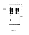
- the product obtained in greater quantity has an apparent molecular weight of 90 kDa (FIG. 2A), compatible with the calculated molecular weight of the encoded polypeptide, deduced from the sequence (98.079 kDa).
- RNA in the presence of microsomes pancreatic dog which allows N-glycosylation of the polypeptide chain obtained, provides two high molecular weight products, respectively 150 and 130 kDa ( Figure 2A).
- Figure 2A To analyze the relationship between the two glycosylated products obtained, experiments spread over time; we realize also, as specified above, the translation in the presence castanospermine, an ⁇ -glucosidase inhibitor I of the rough endoplasmic reticulum, so that determine if the lowest molecular weight product could be derived from 150 kDa glycoprotein per cut sugars during glycosylation.
- VIF anti-sera are obtained from two SPF cats (cats lacking specific pathogens), inoculated with Wo or Petaluma isolates and also from two cats naturally infected, but seronegative towards the virus feline leukemia. Control sera are obtained from the two SPF cats. For the pre-absorption stage, a stock of SPF cat sera is used. Polyclonal rabbit antibodies directed against synthetic peptides as described in French Patent Application No. 2,669,338 are also used, at concentrations of 20 ⁇ g / ml.
- the first of these peptides (P100), 21 amino acids in length, is located at the C-terminus of the VIF surface glycoprotein and includes the Cleavage site between the surface glycoprotein (SU) and the transmembrane glycoprotein (TM); the second peptide (P102), with a length of 25 amino acids, corresponds to the C-terminus of the VIF glycoprotein TM.
- Pre-immunized rabbit sera are used for the pre-absorption step and as a negative control in immunoprecipitation assays.
- the supernatants are then incubated overnight with 4 ⁇ l of serum infected cats, SPF cat serum or polyclonal antibodies rabbit synthetic anti-peptide as defined above. 50 ⁇ l of PrA are added and incubated for 1 hour. After 4 washes in RIPA buffer, the immunocomplex-PrA pellets are eluted by boiling for 5 minutes in 50 ⁇ l of Laemmli buffer. The samples are analyzed on an SDS-PAGE 5-10 gradient %.
- FL-4 cells derived from peripheral blood mononuclear cells (PBMC) infected with a Petaluma strain and spontaneously producing VIF (Yamamoto, 1991), are washed in cold PBS and lysed in RIPA buffer. The cell lysates are clarified by centrifugation at 17000 g for 30 min.
- PBMC peripheral blood mononuclear cells
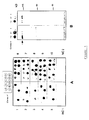
- the mature env translation products obtained in vitro are subjected to immunoprecipitation (figure 3) with the various sera from the abovementioned animals (sera SPF, experimentally inoculated with Wo and Petaluma (bands 1 and 4), sera from naturally infected cats (bands 2 and 5), sera from uninfected SPF cats, as negative control (strip 3)).
- This figure 3 illustrates the results obtained; the two glycosylated products (150 kDa and 130 kDa) are immunoprecipitated by sera from infected cats, the 130 kDa product being the major product.
- the 90 kDa band probably represents residual non-glycosylated material.
- the product of lowest molecular weight can be considered a transcription or partial translation product. No band is detected in the sera of SPF cats and not infected.
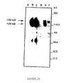
- FIG. 3b illustrates, by comparison, the immunoprecipitation of metabolically labeled VIF proteins obtained from FL-4 cells.
- Lanes a, b and c represent the result obtained with sera from uninfected SPF cats, sera from cats infected with a Petaluma strain and sera from cats infected with VIF Wo.
- the bands d, e and f correspond to a pre-immune rabbit serum, polyclonal rabbit sera directed against the C-terminal peptide of the SU protein (from amino acid 6 to the TM portion (P100)) and rabbit sera polyclonal cells directed against a C-terminal peptide of the TM protein (P102).
- Lanes b and c allow the detection of two proteins (150 kDa and 115-125 kDa), which correspond to the envelope precursor protein and to the SU glycoprotein, respectively, as confirmed by immunoprecipitation with the two rabbit antibodies. raised against peptides including the SU-TM cleavage site or corresponding to the C-terminal part of the env precursor (bands e and f).
- nucleic acid fragments isolated from a test sample and transferred to a nitrocellulose filter are hybridized in situ with a nucleic acid fragment according to the invention (probe labeled with dATP 32 P), 58 ° C overnight, after blocking the non-specific bonds, with a Denhardt solution.
- V 1 -V 9 variable fragments
- Overlapping fragments of the plasmid pKSe are generated by DNase I digestion in the presence of manganese ions.
- the DNA fragments obtained, after an optimal incubation time with DNase (time giving fragments of length of about 200 bp) are treated with the Klenow fragment of DNA polymerase from E. coli , so as to obtain blunt-ended ends, for efficient ligation with the binding sequence.
- the labeling with phosphorus 32 of the DNA fragments allows the control of the subsequent steps.
- bp 10 base pair binding sequences (bp), having an EcoRI site (Pharmacia), are used for ligation with the pKSe fragments obtained.
- the ligature products are then digested with the enzyme EcoRI restriction and separated by electrophoresis in 2% LMP NUSieve agarose gels to select a set of fragments comprising between 100 and 200 bp and eliminating free linkage sequences.
- the selected fragments are extracted with phenol from agarose and ligated with a vector ⁇ gt11 digested with the restriction enzyme EcoRI (Promega).
- the ligation products are packaged using a Packagene kit (Promega) and spread on a strain of E. coli Y1090.
- the recombinant phages are selected by detection of a color in the presence of 25 ⁇ l of IPTG 1 M and 25 ⁇ l of Xgal 80 mg / ml.
- the spreading of the ⁇ gt11 bank provides 10 5 independent clones.
- the phage DNA obtained from the non-stained plates, is analyzed by PCR using primers ⁇ gt11 1218 and 1222, complementary to the ⁇ -galactosidase fragment of the matrix of ⁇ gt11: 8 of the 10 clones contain inserts of average size d 'around 160 bp.
- the bank is then amplified to obtain a titer of 3 ⁇ 10 9 per ml.
- the protein SU glycoprotein outer surface envelope
- the TM protein transmembrane envelope viral glycoprotein
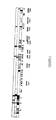
- Table I and Figure 4 specify the nucleotide sequences of the different inserts as well as the corresponding peptide sequences.
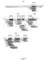
- the overlapping sequences highlight the presence of 8 immunogenic domains (epitopes), 5 of which are located in the extramembrane glycoprotein (gp) SU (SU1, amino acids 253-289; SU2, amino acids 388-424; SU3, amino acids 467-492; SU4, amino acids 508-528 and SU5, amino acids 572-606) and 3 in TM transmembrane gp (TM2, amino acids 681-711; TM3, amino acids 744-788 and TM4, amino acids 826-854) (Figure 1).
- gp extramembrane glycoprotein
- the peptide sequence 597-646 which is defined by clones 20 and 124, overlaps the 5th SU epitope (574-606) and undoubtedly represents a TM NH 2 -terminal (TM1) epitope. This hypothesis is confirmed by the immunological screening with cat sera, which shows the different reactivities between clone 51 (epitope SU5) and clone 124 (epitope TM1).
- FIG. 4 illustrates the localization of the epitopic regions of the envelope glycoproteins (gp) of the VIF, with SU: gp100; TM: gp40.
- the cleavage sites are represented by vertical arrows; the main clones are represented by thick lines and the various rectangles indicate the epitopes corresponding to the minimal overlapping sequences (dark colored rectangle Table II, below, illustrates the number of clones having the immunogenic regions as specified in Table I below above.
- Immunogenic regions Number of clones 1st 7 2nd 11 3rd 22 4th 10 5th 31 6th 4
- the recombinant clones ⁇ gt11 54, 133, 73, 154, 44, 51, 124, 157, 9 and 27 are expressed as lysogens in E. coli Y1089, as described by HUYNH et al. (1985).
- Crude lysate, containing the fusion proteins, is prepared from lysogenic culture induced by IPTG by making freeze-thaw cycles and sonication of the cell pellet, resuspended in 200 ⁇ l of TEP buffer (100 mM Tris-HCl). , pH 7.4, 10 mM EDTA, 1 mM PMSF).
- the extracts are brought to the boil in a Laemmli buffer and resuspended in 5-10% polyacrylamide SDS gels (40 ⁇ g / well) using a Mini PII device (BIO-RAD). After transferring the proteins to a nitrocellulose filter and blocking the non-specific sites by incubation with 3% defatted milk in 10 mM Tris, 150 mM NaCl, 0.1% Tween 20 (TNT buffer), strips of nitrocellulose are incubated with cat serum (diluted to 300th) for 2 hours at 37 ° C.
- the binding antibodies are revealed by incubation with goat IgG coupled to anti-cat peroxidase (dilution to 1000th) followed by a revelation with 4-chloro-1-naphthol (Merck).
- the controls consist of an E. coli Y1089 lysate infected with ⁇ gt11 and a pool of normal sera.
- hypervariability successively concerns the segments 361-377, which extends on either side of a cysteine preserved in position 367, 4 short fragments 452-455, 466-471, 479- 485 and 494-496, the dipeptide 541-542 and the segment 553-570, which comprises a cysteine stored in position 566.
- Two hypervariable zones have also been located in the TM protein: residues 710-718 and 832-842. Variable regions are found in the N-terminal rev-like domain, in the C-terminal portion of the SU and in the C-terminal portion of the hydrophobic transmembrane domain of the TM (between positions 220 and 286) and exhibit variability below the 10% threshold.
- such peptides advantageously include the 9 variable regions as defined above (hypervariable regions + regions adjacent variables), namely: V1 (residues 26-72), V2 (residues 96-174, 23% divergence), V3 (residues 361-422, including the hypervariable region 361-391 and the variable region 392-422, 12% divergence), V4 (residues 452-497, 13% divergence), V5 (residues 541-570, 30% divergence), V6 (residues 586-612, 10% divergence), V7 (residues 710-718, 26% of divergence), V8 (residues 765-778, 12% divergence) and V9 (residues 832-842, 21% divergence), as shown Figure 6 which illustrates the variable domains and the conserved domains: conserved domains are represented in white, the variable regions in gray, and the
- Peptides corresponding to immunodominant epitopes are absorbed in the well of an ELISA plate.
- test serum is incubated at a dilution optimal (predetermined during the development of the E-LISA, using a series of sera from infected cats and SPF cats).
- a dilution optimal predetermined during the development of the E-LISA, using a series of sera from infected cats and SPF cats.
- the presence of anti-VIF antibodies is revealed by anti-cat Ig antibodies coupled to horseradish peroxidase, in the presence of a chromogenic substrate.
- Peptides are used in concentrations between 1 and 10 ⁇ g / ml; to perform the coating of the microtitration plates with the peptides, 0.1 M sodium carbonate, pH 9.6 PBS or poly-L-lysine microplates are used.
- Free sites are saturated with different concentrations of defatted milk or bovine serum albumin.
- the different sera are diluted from 1: 1 to 1: 200.
- the test is carried out as follows: 0.25 ⁇ g of peptide in 50 ⁇ g of 0.1 M sodium carbonate, pH 9.6 are adsorbed on the wells of a microtiter plate (DYNATECH IMMULON® 2) overnight at 4 ° C. The wells are then washed three times with PBS. The residual adsorption sites on the microplates are saturated by incubation with 100 ⁇ l of PBS containing milk (1%) and Tween 20 (0.1%) (ELISA, EB buffer), during 2 hours. 50 ⁇ l of diluted cat serum are incubated in the wells for 2 hours at room temperature.
- peroxidase conjugated anti-cat immunoglobulins (0.5 ⁇ g / ml) (KIRKEGAARD & PERRY LABORATORIES, INC) are added for one hour at room temperature. After 5 washes with PBS, the conjugate to the adsorbed peroxidase is visualized with 2,2'-azino-bis (3-ethylbenz-thiazoline-6 sulfonic acid) (ABTS) (Sigma) 0.2 mg / ml in acetic acid at 0.6%, pH 4.7 and at a final concentration (w / v) of H 2 O 2 of 0.1%.
- ABTS 2,2'-azino-bis (3-ethylbenz-thiazoline-6 sulfonic acid)
- FIG. 7 shows in particular an ELISA test whose reagent is the peptide P237 is highly specific (no false positive) and sensitive (all sera from infected cats of different origins are reactive).
- the use of such a synthetic peptide provides a pure reagent and inexpensive for routine diagnosis of infection at VIF.
- Figure 8 illustrates these results ( ⁇ : SPF cats; •: experimentally infected cats; ⁇ : naturally infected cats).
- cats Source (s) Tested samples SPF France 56 Swiss 5 naturally infected with VIF France 89 Francec 9 Swiss 7 Scotland 5 experimentally inoculated with VIF isolates: wo France 22 Me France 11 Be France 8 The France 7 Br France 3 Gi France 2 Villefranche France 1 Envnip France 1 Petaluma France 4 Aebli Swiss 1 Gasser Swiss 1
Landscapes
- Chemical & Material Sciences (AREA)
- Life Sciences & Earth Sciences (AREA)
- Organic Chemistry (AREA)
- Health & Medical Sciences (AREA)
- Virology (AREA)
- Biophysics (AREA)
- General Health & Medical Sciences (AREA)
- Genetics & Genomics (AREA)
- Medicinal Chemistry (AREA)
- Molecular Biology (AREA)
- Proteomics, Peptides & Aminoacids (AREA)
- Biochemistry (AREA)
- Immunology (AREA)
- Gastroenterology & Hepatology (AREA)
- Peptides Or Proteins (AREA)
- Medicines Containing Antibodies Or Antigens For Use As Internal Diagnostic Agents (AREA)
- Measuring Or Testing Involving Enzymes Or Micro-Organisms (AREA)
Claims (10)
- Peptid, das vom Env-Protein des VIF des Stamms Wo stammt, das ein konserviertes Epitop der Proteine des VIF bildet, dadurch gekennzeichnet, dass es aus der Gruppe, bestehend ausdem Fragment aus 45 Aminosäuren, das TM3 genannt wird, und den Positionen 744 bis 788 der Sequenz der Formel I entspricht,dem Fragment der Sequenz Gln763-Gln-Leu-Gln-Glu-Trp-Glu-Asp770 (Peptid, das P241 genannt wird), das den Positionen 763-770 der Sequenz der Formel I entspricht, unddem Fragment der Sequenz Val772-Gly-Trp-Ile-Gly-Asn-Ile-Pro779 (Peptid, das P242 genannt wird), das den Positionen 772-779 der Sequenz der Formel I entspricht, ausgewählt ist.
- Rekombinanter Klonierungs- und/oder Expressionsvektor, dadurch gekennzeichnet, dass er eine Nukleotidsequenz umfasst, die für das Peptid nach Anspruch 1 kodiert.
- Vektor nach Anspruch 2, dadurch gekennzeichnet, dass er aus einem Plasmid, das einen Replikationsursprung, mindestens einen Selektionsmarker und eine Nukleotidsequenz, die für das Peptid nach Anspruch 1 kodiert, gebildet wird, wobei der Vektor ein Klonierungsvektor ist.
- Vektor nach Anspruch 3, dadurch gekennzeichnet, dass der Klonierungsvektor durch ein Plasmid gebildet wird, das fähig ist, sich in E. coli zu replizieren, in den eine Nukleotidsequenz, die für das Peptid nach Anspruch 1 kodiert, insertiert ist.
- Vektor nach Anspruch 4, dadurch gekennzeichnet, dass er durch einen rekombinanten Vektor in einem geeigneten Wirtsmikroorganismus, insbesondere einem Bakterium oder einer Eukaryontenzelle, gebildet wird, umfassend einen Replikationsursprung, mindestens ein Gen, dessen Expression die Selektion der Bakterien oder der Eukaryontenzellen, die den Vektor aufgenommen haben, erlaubt, einen Promotor, der die Expression der Gene in den Bakterien oder Eukaryontenzellen erlaubt, und in den eine Nukleotidsequenz insertiert ist, die für das Peptid nach Anspruch 1 kodiert.
- Vektor nach Anspruch 5, dadurch gekennzeichnet, dass er durch einen Phagen λgt11 gebildet wird, in den Abbaufragmente des Plasmids pKSe1 im Leserahmen auf dem Niveau seiner EcoRI-Schnittstelle insertiert sind.
- Zusammensetzung mit immunogener Wirkung und/oder Schutzwirkung, dadurch gekennzeichnet, dass sie mindestens ein Peptid nach Anspruch 1, gegebenenfalls in Verbindung mit mindestens einer anderen immunogenen oder immunstimulierenden Substanz und/oder ein pharmazeutisch annehmbares Vehikel umfasst.
- Verfahren zum Nachweis von Antikörpern gegen VIF, dadurch gekennzeichnet, dass es darin besteht, Antikörper gegen VIF, die gegebenenfalls in einer biologischen Probe vorliegen, mit Hilfe eines Peptids gemäß Anspruch 1, das gegebenenfalls an einem geeigneten festen Träger fixiert ist, nachzuweisen, wobei die biologische Probe mit dem Peptid/den Peptiden, an das/an die sich die Antikörper gegen VIF binden, wenn solche Antikörper in der zu analysierenden Probe vorliegen, in Kontakt gebracht wird und das Ablesen des Resultats durch ein geeignetes Mittel, insbesondere DIA, RIA, Fluoreszenz, sichtbar gemacht wird.
- Kit, der zur Verwendung im Verfahren zum Nachweisen von Antikörpern gegen VIF nach Anspruch 8 gebrauchsfertig ist, dadurch gekennzeichnet, dass er außer nützlichen Mengen an Puffern, die zur Durchführung des genannten Nachweises geeignet sind, geeignete Dosen mindestens eines Peptids nach Anspruch 1 umfasst.
- Reagens zur Diagnose von Katzen-Immundefizienz, dadurch gekennzeichnet, dass es aus der Gruppe, bestehend aus einem der Peptide gemäß Anspruch 1 ausgewählt ist, und dass es ein universelles Diagnosereagens für einen VIF-Stamm ist.
Applications Claiming Priority (6)
| Application Number | Priority Date | Filing Date | Title |
|---|---|---|---|
| FR9207258A FR2692269B1 (fr) | 1992-06-16 | 1992-06-16 | Peptides immunogènes et/ou neutralisants de vif de souche WO et leurs applications au diagnostic et à la prévention de l'immunodéficience féline. |
| FR9207257 | 1992-06-16 | ||
| FR9207257A FR2692279B1 (fr) | 1992-06-16 | 1992-06-16 | Séquences nucléotidiques issues de la souche WO du vif et leurs fragments, applications desdites séquences à l'expression de peptides immunogènes et au diagnostic de l'immunodéficience féline. |
| FR9207258 | 1992-06-16 | ||
| FR9214026A FR2692270A1 (fr) | 1992-06-16 | 1992-11-23 | Peptides immunogènes et/ou neutralisants de vif de souche WO et leurs applications au diagnostic et à la prévention de l'immunodéficience féline. |
| FR9214026 | 1992-11-23 |
Publications (2)
| Publication Number | Publication Date |
|---|---|
| EP0577458A1 EP0577458A1 (de) | 1994-01-05 |
| EP0577458B1 true EP0577458B1 (de) | 2004-09-29 |
Family
ID=27252623
Family Applications (1)
| Application Number | Title | Priority Date | Filing Date |
|---|---|---|---|
| EP93401538A Expired - Lifetime EP0577458B1 (de) | 1992-06-16 | 1993-06-16 | Nukleotid- und Peptidsequenzen des Immunschwächevirus der Katze, Isolat WO und Anwendungen der Sequenzen in der Diagnostik und zur Verhinderung der Infektion |
Country Status (3)
| Country | Link |
|---|---|
| EP (1) | EP0577458B1 (de) |
| AT (1) | ATE278018T1 (de) |
| DE (1) | DE69333636T2 (de) |
Cited By (1)
| Publication number | Priority date | Publication date | Assignee | Title |
|---|---|---|---|---|
| US7776546B2 (en) | 2003-09-11 | 2010-08-17 | Idexx Laboratories, Inc. | Method and device for detecting feline immunodeficiency virus |
Families Citing this family (10)
| Publication number | Priority date | Publication date | Assignee | Title |
|---|---|---|---|---|
| WO1994020622A1 (en) * | 1993-03-11 | 1994-09-15 | Akzo Nobel N.V. | Polypeptide fragment capable of inducing neutralising antibodies against feline immuno-deficiency virus |
| FR2721031B1 (fr) | 1994-06-09 | 1996-07-26 | Centre Nat Rech Scient | Fragment peptidique spécifique du virus de l'immunodéficience féline (VIF) et son utilisation comme réactif de diagnostic. |
| FR2732346B1 (fr) * | 1995-03-27 | 1997-05-30 | Centre Nat Rech Scient | Proteines mutees, codees par un gene env mute de lentivirus, fragments peptidiques inclus dans lesdites proteines, vecteurs d'expression exprimant lesdites proteines mutees et leurs applications |
| US5820869A (en) * | 1995-06-07 | 1998-10-13 | American Home Products Corporation | Recombinant raccoon pox viruses and their use as an effective vaccine against feline immunodeficiency virus infection |
| FR2769916B1 (fr) | 1997-10-17 | 2000-12-01 | Centre Nat Rech Scient | Peptides issus du gene env du virus de l'immunodeficience feline et leurs applications immunoprotectrices et vaccinales |
| FR2828686B1 (fr) * | 2001-08-17 | 2003-12-12 | Hippocampe | Nouveaux peptides et leur utilisation comme medicaments contre l'infection par le fiv chez le chat |
| FR2828687B1 (fr) * | 2001-08-17 | 2003-12-12 | Hippocampe | Nouveaux peptides et leur utilisation comme medicaments contre l'infection par le fiv chez le chat |
| FR2833598A1 (fr) * | 2001-12-17 | 2003-06-20 | Hippocampe | Oligopeptides et leur utilisation comme medicaments contre l'infection par le fiv chez le chat |
| WO2005080939A2 (en) * | 2004-02-19 | 2005-09-01 | Idexx Laboratories, Inc. | Method and device for detecting feline immunodeficiency virus |
| JP4866848B2 (ja) * | 2004-06-30 | 2012-02-01 | アイデックス ラボラトリーズ インコーポレイテッド | ネコ免疫不全ウイルス(fiv)のenvタンパク質のv3領域に由来するペプチドの使用を含むfivを検出するための方法および装置 |
Family Cites Families (1)
| Publication number | Priority date | Publication date | Assignee | Title |
|---|---|---|---|---|
| FR2669338A1 (fr) * | 1990-11-21 | 1992-05-22 | Centre Nat Rech Scient | Fragments peptidiques issus de la proteine externe du vif, anticorps antifragments, application de ceux-ci au diagnostic et/ou au traitement de l'immunodeficience feline. |
-
1993
- 1993-06-16 AT AT93401538T patent/ATE278018T1/de not_active IP Right Cessation
- 1993-06-16 EP EP93401538A patent/EP0577458B1/de not_active Expired - Lifetime
- 1993-06-16 DE DE69333636T patent/DE69333636T2/de not_active Expired - Lifetime
Cited By (1)
| Publication number | Priority date | Publication date | Assignee | Title |
|---|---|---|---|---|
| US7776546B2 (en) | 2003-09-11 | 2010-08-17 | Idexx Laboratories, Inc. | Method and device for detecting feline immunodeficiency virus |
Also Published As
| Publication number | Publication date |
|---|---|
| DE69333636T2 (de) | 2005-10-06 |
| EP0577458A1 (de) | 1994-01-05 |
| DE69333636D1 (de) | 2004-11-04 |
| ATE278018T1 (de) | 2004-10-15 |
Similar Documents
| Publication | Publication Date | Title |
|---|---|---|
| BE1005485A5 (fr) | Agent viral. | |
| AU625970B2 (en) | Hiv-3 retrovirus and its use | |
| EP0787191B1 (de) | Nukleotiden-sequenzen aus retroviren-antigenen hiv-i gruppe (oder untergruppe) o | |
| EP1916304B1 (de) | Antigene Polypeptide, die mit Multipler Sklerose assoziiert sind, und Verwendungszwecke | |
| WO2000075665A1 (fr) | Detection precoce des flavivirus en utilisant la clycoproteine ns1 | |
| EP0577458B1 (de) | Nukleotid- und Peptidsequenzen des Immunschwächevirus der Katze, Isolat WO und Anwendungen der Sequenzen in der Diagnostik und zur Verhinderung der Infektion | |
| KR100204258B1 (ko) | 재조합 고양이 코로나바이러스 에스 단백질 | |
| LU86972A1 (fr) | Anticorps monoclonaux,peptides et compositions les contenant,destinees au diagnostic et au traitement des infections par le virus hiv | |
| US5736378A (en) | Molecular cloning and characterization of the feline immunodeficiency virus isolate PPR | |
| EP0550691A1 (de) | Peptide, welche hiv-retroviren-hemmende antikörper induzieren, sowie gegen diese peptide gerichtete antikörper. | |
| FR2687404A1 (fr) | Peptides immunologiquement apparentes aux proteines d'un agent viral et leurs applications biologiques. | |
| US20030215793A1 (en) | Complete genome sequence of a simian immunodeficiency virus from a wild chimpanzee | |
| WO1998030699A1 (fr) | Polynucleotide codant pour un polypeptide de 27 kd de mycobacteries appartenant au complexe de mycobacterium tuberculosis, application au diagnostic et a la prevention de la tuberculose | |
| EP0542845B1 (de) | Schützende antikörper erregende plasmodium falciparum antigene und deren verwendung in impfstoffen | |
| EP0769058A1 (de) | Nukleinsäurefragmenten und entsprechende peptide aus den ziegen arthritis und enkephalitis virus (caev) genom und verwendungen davon | |
| FR2804685A1 (fr) | Utilisation d'une proteine de leptospire pour la prevention et/ou le diagnostic et/ou le traitement de la leptospirose animale et/ou humaine | |
| FR2692269A1 (fr) | Peptides immunogènes et/ou neutralisants de vif de souche WO et leurs applications au diagnostic et à la prévention de l'immunodéficience féline. | |
| FR2692270A1 (fr) | Peptides immunogènes et/ou neutralisants de vif de souche WO et leurs applications au diagnostic et à la prévention de l'immunodéficience féline. | |
| FR2645173A1 (fr) | Clonage et expression de genes codant pour des peptides et/ou des fragments de peptides du virus mokola, application a la preparation d'un v | |
| CN117083290A (zh) | 戊型肝炎病毒样颗粒及其用途 | |
| FR2692279A1 (fr) | Séquences nucléotidiques issues de la souche WO du vif et leurs fragments, applications desdites séquences à l'expression de peptides immunogènes et au diagnostic de l'immunodéficience féline. | |
| HK40096253A (zh) | 戊型肝炎病毒样颗粒及其用途 | |
| FR2994193A1 (fr) | Souches mutantes de neospora et leurs utilisations | |
| FR2593519A1 (fr) | Vaccin contre le syndrome immunodeficitaire acquis et intermediaires pour la preparation de ce vaccin | |
| EP0431156A1 (de) | Impfstoff gegen rinderleukämievirus |
Legal Events
| Date | Code | Title | Description |
|---|---|---|---|
| PUAI | Public reference made under article 153(3) epc to a published international application that has entered the european phase |
Free format text: ORIGINAL CODE: 0009012 |
|
| AK | Designated contracting states |
Kind code of ref document: A1 Designated state(s): AT BE CH DE DK ES FR GB GR IE IT LI LU MC NL PT SE |
|
| 17P | Request for examination filed |
Effective date: 19940613 |
|
| RAP1 | Party data changed (applicant data changed or rights of an application transferred) |
Owner name: CENTRE NATIONAL DE LA RECHERCHE SCIENTIFIQUE (CNRS |
|
| 17Q | First examination report despatched |
Effective date: 19990712 |
|
| GRAP | Despatch of communication of intention to grant a patent |
Free format text: ORIGINAL CODE: EPIDOSNIGR1 |
|
| GRAS | Grant fee paid |
Free format text: ORIGINAL CODE: EPIDOSNIGR3 |
|
| GRAA | (expected) grant |
Free format text: ORIGINAL CODE: 0009210 |
|
| RIC1 | Information provided on ipc code assigned before grant |
Ipc: 7G 01N 33/68 B Ipc: 7G 01N 33/53 B Ipc: 7A 61K 39/21 B Ipc: 7C 12Q 1/68 B Ipc: 7C 12N 15/70 B Ipc: 7C 12N 15/49 A |
|
| AK | Designated contracting states |
Kind code of ref document: B1 Designated state(s): AT BE CH DE DK ES FR GB GR IE IT LI LU MC NL PT SE |
|
| PG25 | Lapsed in a contracting state [announced via postgrant information from national office to epo] |
Ref country code: NL Free format text: LAPSE BECAUSE OF FAILURE TO SUBMIT A TRANSLATION OF THE DESCRIPTION OR TO PAY THE FEE WITHIN THE PRESCRIBED TIME-LIMIT Effective date: 20040929 Ref country code: IE Free format text: LAPSE BECAUSE OF FAILURE TO SUBMIT A TRANSLATION OF THE DESCRIPTION OR TO PAY THE FEE WITHIN THE PRESCRIBED TIME-LIMIT Effective date: 20040929 Ref country code: AT Free format text: LAPSE BECAUSE OF FAILURE TO SUBMIT A TRANSLATION OF THE DESCRIPTION OR TO PAY THE FEE WITHIN THE PRESCRIBED TIME-LIMIT Effective date: 20040929 |
|
| REG | Reference to a national code |
Ref country code: GB Ref legal event code: FG4D Free format text: NOT ENGLISH |
|
| REG | Reference to a national code |
Ref country code: CH Ref legal event code: EP |
|
| REG | Reference to a national code |
Ref country code: IE Ref legal event code: FG4D Free format text: FRENCH |
|
| REF | Corresponds to: |
Ref document number: 69333636 Country of ref document: DE Date of ref document: 20041104 Kind code of ref document: P |
|
| PG25 | Lapsed in a contracting state [announced via postgrant information from national office to epo] |
Ref country code: SE Free format text: LAPSE BECAUSE OF FAILURE TO SUBMIT A TRANSLATION OF THE DESCRIPTION OR TO PAY THE FEE WITHIN THE PRESCRIBED TIME-LIMIT Effective date: 20041229 Ref country code: GR Free format text: LAPSE BECAUSE OF FAILURE TO SUBMIT A TRANSLATION OF THE DESCRIPTION OR TO PAY THE FEE WITHIN THE PRESCRIBED TIME-LIMIT Effective date: 20041229 Ref country code: DK Free format text: LAPSE BECAUSE OF FAILURE TO SUBMIT A TRANSLATION OF THE DESCRIPTION OR TO PAY THE FEE WITHIN THE PRESCRIBED TIME-LIMIT Effective date: 20041229 |
|
| PG25 | Lapsed in a contracting state [announced via postgrant information from national office to epo] |
Ref country code: ES Free format text: LAPSE BECAUSE OF FAILURE TO SUBMIT A TRANSLATION OF THE DESCRIPTION OR TO PAY THE FEE WITHIN THE PRESCRIBED TIME-LIMIT Effective date: 20050109 |
|
| GBT | Gb: translation of ep patent filed (gb section 77(6)(a)/1977) |
Effective date: 20050131 |
|
| NLV1 | Nl: lapsed or annulled due to failure to fulfill the requirements of art. 29p and 29m of the patents act | ||
| REG | Reference to a national code |
Ref country code: IE Ref legal event code: FD4D |
|
| PG25 | Lapsed in a contracting state [announced via postgrant information from national office to epo] |
Ref country code: LU Free format text: LAPSE BECAUSE OF NON-PAYMENT OF DUE FEES Effective date: 20050616 |
|
| PG25 | Lapsed in a contracting state [announced via postgrant information from national office to epo] |
Ref country code: MC Free format text: LAPSE BECAUSE OF NON-PAYMENT OF DUE FEES Effective date: 20050630 Ref country code: LI Free format text: LAPSE BECAUSE OF NON-PAYMENT OF DUE FEES Effective date: 20050630 Ref country code: CH Free format text: LAPSE BECAUSE OF NON-PAYMENT OF DUE FEES Effective date: 20050630 Ref country code: BE Free format text: LAPSE BECAUSE OF NON-PAYMENT OF DUE FEES Effective date: 20050630 |
|
| PLBE | No opposition filed within time limit |
Free format text: ORIGINAL CODE: 0009261 |
|
| STAA | Information on the status of an ep patent application or granted ep patent |
Free format text: STATUS: NO OPPOSITION FILED WITHIN TIME LIMIT |
|
| 26N | No opposition filed |
Effective date: 20050630 |
|
| REG | Reference to a national code |
Ref country code: CH Ref legal event code: PL |
|
| BERE | Be: lapsed |
Owner name: CENTRE NATIONAL DE LA RECHERCHE *SCIENTIFIQUE Effective date: 20050630 |
|
| PG25 | Lapsed in a contracting state [announced via postgrant information from national office to epo] |
Ref country code: PT Free format text: LAPSE BECAUSE OF NON-PAYMENT OF DUE FEES Effective date: 20050228 |
|
| PGFP | Annual fee paid to national office [announced via postgrant information from national office to epo] |
Ref country code: DE Payment date: 20120524 Year of fee payment: 20 |
|
| PGFP | Annual fee paid to national office [announced via postgrant information from national office to epo] |
Ref country code: FR Payment date: 20120705 Year of fee payment: 20 Ref country code: GB Payment date: 20120525 Year of fee payment: 20 |
|
| PGFP | Annual fee paid to national office [announced via postgrant information from national office to epo] |
Ref country code: IT Payment date: 20120526 Year of fee payment: 20 |
|
| REG | Reference to a national code |
Ref country code: DE Ref legal event code: R071 Ref document number: 69333636 Country of ref document: DE |
|
| REG | Reference to a national code |
Ref country code: DE Ref legal event code: R071 Ref document number: 69333636 Country of ref document: DE |
|
| REG | Reference to a national code |
Ref country code: GB Ref legal event code: PE20 Expiry date: 20130615 |
|
| PG25 | Lapsed in a contracting state [announced via postgrant information from national office to epo] |
Ref country code: GB Free format text: LAPSE BECAUSE OF EXPIRATION OF PROTECTION Effective date: 20130615 Ref country code: DE Free format text: LAPSE BECAUSE OF EXPIRATION OF PROTECTION Effective date: 20130618 |