EP0577458B1 - Nucleotide and Peptide sequences of the FIV isolate WO and their uses in diagnostic and prevention of the feline immunodeficiency virus infection - Google Patents
Nucleotide and Peptide sequences of the FIV isolate WO and their uses in diagnostic and prevention of the feline immunodeficiency virus infection Download PDFInfo
- Publication number
- EP0577458B1 EP0577458B1 EP93401538A EP93401538A EP0577458B1 EP 0577458 B1 EP0577458 B1 EP 0577458B1 EP 93401538 A EP93401538 A EP 93401538A EP 93401538 A EP93401538 A EP 93401538A EP 0577458 B1 EP0577458 B1 EP 0577458B1
- Authority
- EP
- European Patent Office
- Prior art keywords
- peptide
- vector
- fiv
- vif
- fragment
- Prior art date
- Legal status (The legal status is an assumption and is not a legal conclusion. Google has not performed a legal analysis and makes no representation as to the accuracy of the status listed.)
- Expired - Lifetime
Links
Images


Classifications
-
- C—CHEMISTRY; METALLURGY
- C07—ORGANIC CHEMISTRY
- C07K—PEPTIDES
- C07K14/00—Peptides having more than 20 amino acids; Gastrins; Somatostatins; Melanotropins; Derivatives thereof
- C07K14/005—Peptides having more than 20 amino acids; Gastrins; Somatostatins; Melanotropins; Derivatives thereof from viruses
-
- C—CHEMISTRY; METALLURGY
- C07—ORGANIC CHEMISTRY
- C07K—PEPTIDES
- C07K16/00—Immunoglobulins [IG], e.g. monoclonal or polyclonal antibodies
- C07K16/08—Immunoglobulins [IG], e.g. monoclonal or polyclonal antibodies against material from viruses
- C07K16/10—RNA viruses
- C07K16/112—Retroviridae (F), e.g. leukemia viruses
- C07K16/114—Lentivirus (G), e.g. human immunodeficiency virus [HIV], feline immunodeficiency virus [FIV] or simian immunodeficiency virus [SIV]
-
- A—HUMAN NECESSITIES
- A61—MEDICAL OR VETERINARY SCIENCE; HYGIENE
- A61K—PREPARATIONS FOR MEDICAL, DENTAL OR TOILETRY PURPOSES
- A61K39/00—Medicinal preparations containing antigens or antibodies
-
- C—CHEMISTRY; METALLURGY
- C12—BIOCHEMISTRY; BEER; SPIRITS; WINE; VINEGAR; MICROBIOLOGY; ENZYMOLOGY; MUTATION OR GENETIC ENGINEERING
- C12N—MICROORGANISMS OR ENZYMES; COMPOSITIONS THEREOF; PROPAGATING, PRESERVING, OR MAINTAINING MICROORGANISMS; MUTATION OR GENETIC ENGINEERING; CULTURE MEDIA
- C12N2740/00—Reverse transcribing RNA viruses
- C12N2740/00011—Details
- C12N2740/10011—Retroviridae
- C12N2740/15011—Lentivirus, not HIV, e.g. FIV, SIV
- C12N2740/15022—New viral proteins or individual genes, new structural or functional aspects of known viral proteins or genes
Definitions
- the present invention relates to peptides and their fragments, specific for the virus of feline immunodeficiency (VIF) and use said fragments as diagnostic reagent and as agent inducing an immune response (cellular and / or production of antibodies), in particular for the prevention of feline immunodeficiency.
- VIP feline immunodeficiency
- the present invention also relates to the nucleotide sequences coding for said peptides.
- Feline immunodeficiency is caused by a lentivirus, feline immunodeficiency virus (FIV), which has a genetic structure similar to that of primate lentivirus (HIV and VIS).
- FV feline immunodeficiency virus
- Feline immunodeficiency poses a significant problem veterinary health, to the extent that a number significant number of cats infected with VIF has been detected in United States, Japan and Europe (5 to 25% of animals).
- feline immunodeficiency is essentially diagnosed at the onset clinical signs (generalized lymphadenopathy and occurrence of opportunistic infections), while a diagnosis very early would have significant benefits so much in treatment than in prevention of this disease.
- VIF envelope proteins are considered as being at the center of the host-virus relationship; their study is therefore essential to understand how the virus interacts with the immune system (neutralization epitopes, B and T epitopes) and with target cells for infection, and develop both high-performance diagnostic reagents and effective vaccines, consisting of viral proteins immunogenic and protective action, vis-à-vis the whole strains of VIF.
- the VIF env protein can provide, after cleavage, two glycoprotein fragments, called SU (surface glycoprotein) and TM (transmembrane glycoprotein).
- SU surface glycoprotein
- TM transmembrane glycoprotein
- the present invention is therefore intended to provide, on the one hand, sequences from VIF and their selected fragments in particular for their strain specificity, and, on the other hand, to a specific immunogenic and / or protective composition feline immunodeficiency virus, obtained in expressing fragments selected for their capacity neutralizing and / or the presence of recognition epitopes group and / or type; obtaining by genius genetics or chemical synthesis of such specific peptides has the advantage of solving the production problem virus and directly obtain the desired peptides.
- Nucleotide sequences from RNA feline immunodeficiency virus (VIF) genomics include at least one fragment of a gene with hypervariable zones, chosen from the group consisting of the gene encoding the env protein of the Wo strain of VIF and the gene coding for the gag protein of the strain Wo from VIF.
- this comprises the nucleotide sequence and the deduced amino acid sequence of formula I below, which corresponds to the env gene of VIF, strain Wo:
- Such a sequence corresponds to the cDNA of the mRNA of the env protein of the VIF strain Wo.
- Said peptide is especially recognized by antibodies to at least one different strain from that from which said peptide and / or by the antibodies produced by naturally infected cats and what it has a response response capacity immune system and / or production of protective antibodies vis-à-vis at least one strain of VIF, in uninfected cats.
- the fragment including a segment of 45 amino acids, called TM3, corresponds to positions 744-788 of the sequence of formula II below, which segment contains an epitope including the sequence Gln 764 -Leu-Gln-Glu-Trp-Glu -Asp-Trp-Val-Gly-Trp-Ile-Gly-Asn-Ile 778 or the sequence Gln 763 -Gln-Leu-Gln-Glu-Trp-Glu-Asp 770 (peptide called P241) or the sequence Val 772 - Gly-Trp-Ile-Gly-Asn-Ile-Pro 779 (peptide called P242).
- the peptide which corresponds to the Env protein of the VIF, strain Wo has the following sequence:
- epitope within the meaning of this invention, the epitopes common to all strains of VIF (conserved areas), recognized by directed antibodies against all strains of VIF (recognition of group); moreover, all these fragments induce a production antibody and / or cellular response.
- peptides are coded by a sequence or a nucleotide sequence fragment as defined in the sequence of formula I above.
- peptide fragment includes all fragments including a peptide segment in accordance with the invention as well as homologous peptides; in general, we mean by homologous peptide, the peptide fragments whose position and function are equivalent, within the VIF (same location as that defined above, on the VIF genome); more specifically TM3 fragments, also understood by homologous peptides, peptides that do not lose the properties of recognition of the antibodies directed against the peptide of reference.
- All of said peptides can advantageously be obtained either by cloning or by synthesis, in particular by synthesis of Merrifield.
- the present invention also relates to a recombinant cloning and / or expression vector, characterized in that it comprises a nucleotide sequence encoding one of the TM3 peptides defined above.
- recombinant vector either a plasmid, a cosmid, than a phage.
- said vector consists of a plasmid comprising a origin of replication, at least one selection marker and a nucleotide sequence encoding one of the TM3 peptides defined above, which vector is a cloning vector.
- said cloning vector consists of a plasmid capable of replicating in E. coli , into which is inserted a nucleotide sequence coding for one of the TM3 peptides defined above.
- said vector consists of a recombinant vector comprising an origin of replication in a microorganism suitable host, including bacteria or eukaryotic cell, at least one gene whose expression allows the selection of either bacteria or cells eukaryotes having received said vector, a promoter allowing the expression of genes in said bacteria or eukaryotic cells, and into which is inserted a nucleotide sequence encoding one of the TM3 peptides defined above, which vector is a vector expression of a TM3 peptide as defined above.
- said expression vector consists of a phage ⁇ gt11 into which is inserted, in phase at its site, EcoRI, fragments of digestion with DNAse of the plasmid pKSe1.
- the present invention also relates to an immunogenic and / or protective composition, characterized in that it comprises at least one peptide TM3 as defined above, possibly associated with minus another immunogenic or immunostimulating substance and / or a pharmaceutically acceptable vehicle.
- the present invention has, moreover, an object a method for detecting anti-FIV antibodies, characterized in that it consists in detecting anti-VIF antibodies possibly present in a biological sample to using a TM3 peptide as defined above, optionally fixed on an appropriate solid support, bringing said biological sample into contact with said peptide (s) to which the antibodies bind anti-FIV if such antibodies are present in the sample to be analyzed, the reading of the result being revealed by an appropriate means, in particular EIA, RIA, fluorescence.
- This process notably makes it possible to verify the seroconversion of vaccinated animals or to carry out epidemiological serological surveys.
- TM3 peptide said method allows determine a VIF infection, regardless of the strain (universal reagent as specified above).
- the present invention further relates to a kit, ready to use, for implementing the process anti-VIF antibody detection, characterized in that it includes, in addition to useful quantities of buffers suitable for carrying out said detection, appropriate doses of at least one TM3 peptide such as defined above.
- the present invention also relates to a reagent for diagnosing feline immunodeficiency, characterized in that it is chosen from the group consisting by one of the TM3 peptides as defined above, and in that it constitutes a universal diagnostic reagent of VIF strain.
- PBMC Peripheral blood mononuclear cells
- peripheral blood mononuclear cells obtained from cats said negative control (SPF cats)
- SPF cats peripheral blood mononuclear cells
- MORAILLON A. et al., 1992, Vet. Mic., 31, 41- 45 In vitro properties and experimental pathogenic effect of three feline immunodeficiency viruses isolated from cats with terminal diseases ).
- the Mg ++-dependent reverse transcriptase (RT) activity is more particularly monitored. After 15 days, the total cellular DNA is extracted with phenol (SAMBROOK et al., 1989, Molecular cloning, a laboratory manual), from RT positive cultures and used as starting material for the amplification of the VIF genes. This makes it possible to avoid multiple passages in vitro and adaptation to CD4 negative cell lines.
- the env and gag genes of the Wo strain of the feline immunodeficiency virus are respectively amplified in three and two overlapping fragments, using synthetic primers corresponding to regions conserved in the sequences of VIF, Petaluma isolate (TALBOTT et al, 1989 ) and PPR isolate (PHILIPPS et al., 1990).
- a vector M13mp8 is digested with the enzyme SmAI, so as to be able to insert into it the PCR fragments obtained above [for example, for the env gene fragments of the sequence of formula I, E 1 (nucleotides 1-977), E 2 (978-2016) and E 3 (2017-2562)].
- the recombinant vector obtained is used to transform competent E. coli JM101.
- the recombinant clones are detected by in situ hybridization with the corresponding PCR fragments, labeled with phosphorus 32.
- Single strand DNA sequencing from recombinant phages is carried out by the enzymatic method by dideoxynucleotides, with a sequencing kit sequenase® 2.0 DNA, using, as primers, oligonucleotides complementary to the vector (17 seas, universal primer) or the VIF insert.
- the three fragments (E 1 , E 2 and E 3 ) are prepared from the recombinant vector M13mp8 as defined above, using the restriction sites present in the multisite binding sequence of M13mp8 or in the amplification primers: E 1 , HindIII-BclI; E 2 , BclI-SpeI; E 3 , SpeI-EcoRI.
- the three fragments are ligated into a KS + Bluescript® vector (Stratagene), digested with the enzymes HindIII and EcoRI, downstream of the T3 promoter. The construction obtained is called pKSe.
- Said fragments can also be ligated into a SK + Bluescript® vector (Stratagene), digested with the enzymes HindIII and EcoRI, downstream of the T7 promoter.
- the latter construction called pKSe1
- pKSe1 allows transcription of the env gene in mammalian cells expressing the phage T7 RNA polymerase gene
- pKSe allows transcription of the env gene in mammalian cells expressing the gene for phage T3 RNA polymerase.
- variable sequences in accordance with the invention selected are summarized in the Table below; advantageously, the selection of these fragments makes it possible to define, by deduction, areas conserved in all of the VIFs, and makes it possible to properly highlight the variable areas and the areas conserved; which makes it possible to define universal VIF detection reagents, selective reagents and to provide animals with the best possible protection:
- RNA For the in vitro synthesis of RNA, the two constructions pKSe and pKSe1 are used, the transcription being carried out in the first case, by T3 polymerase and in the second case, by T7 polymerase, as specified above.
- 1 ⁇ g of DNA of the plasmids pKSe1 and pKSe is linearized, using the EcoRI restriction site, present in the multisite binding sequence of the vector, downstream of the inserted gene.
- the linearized and proteinase K treated plasmid is transcribed, using T3 RNA polymerase for pKSe and T7 RNA polymerase for pKSe1, according to the protocol provided by the manufacturer (Stratagene). After treatment with DNase and extraction with phenol, the transcript is precipitated with ethanol, in the presence of 5 ⁇ g of E. coli tRNA, resuspended in 15 ⁇ l of water treated with DEPC and stored at -70 ° C. The size of the transcripts is evaluated by electrophoresis on 1% agarose gel in 10 mM phosphate buffer, pH 7, after denaturation with glyoxal / dimethylsulfoxide.
- RNA transcribed in vitro is translated for 30 minutes at 30 ° C, in a final volume of 12.5 ⁇ g containing 6 ⁇ g of rabbit reticulocyte lysate (RRL) (Promega), 0.25 ⁇ l d '' a mixture of amino acids devoid of methionine (Promega) and 1.75 ⁇ l of methionine labeled with sulfur 35 in the absence (-M) or in the presence (+ M) of microsomal membranes (1 or 2 ⁇ l), to allow protein glycosylation (Figure 2A).
- the 1 or 2 ⁇ l of dog pancreatic microsomal membranes (Promega) are added per 12.5 ⁇ l of reaction mixture.
- 3 ⁇ l of aliquots are taken at different times (15, 30, 60 and 90 minutes, 4 hours). The samples are stored at -70 ° C.
- Inhibition studies in the presence of castanospermine are performed by incubating the translation mixture containing microsomal membranes (2 ⁇ l for 12.5 ⁇ l mixture) with 5 mM castanospermine (final concentration) (Sigma), prepared immediately in water treated with DEPC, at 30 ° C for 15 minutes, before addition of RNA.
- Translation products are denatured by boil in Laemmli buffer and evaluated by electrophoresis (SDS-PAGE, gradient 5-10%). Markers of the molecular weights are also used (BIORAD H.M.W. or Rainbow markers). After electrophoresis, the gels are fixed, processed and autoradiographed on a film Kodak XAR-5.
- RNA transcribed in vitro from the plasmid pKSe migrates, in an agarose gel under denaturing conditions, in a single band of approximately 2.5 kb, corresponding to the expected dimension.
- RNA transcribed in vitro from the plasmid pKSe migrates, in an agarose gel under denaturing conditions, in a single band of approximately 2.5 kb, corresponding to the expected dimension.
- RTL rabbit reticulocyte lysate
- the product obtained in greater quantity has an apparent molecular weight of 90 kDa (FIG. 2A), compatible with the calculated molecular weight of the encoded polypeptide, deduced from the sequence (98.079 kDa).
- RNA in the presence of microsomes pancreatic dog which allows N-glycosylation of the polypeptide chain obtained, provides two high molecular weight products, respectively 150 and 130 kDa ( Figure 2A).
- Figure 2A To analyze the relationship between the two glycosylated products obtained, experiments spread over time; we realize also, as specified above, the translation in the presence castanospermine, an ⁇ -glucosidase inhibitor I of the rough endoplasmic reticulum, so that determine if the lowest molecular weight product could be derived from 150 kDa glycoprotein per cut sugars during glycosylation.
- VIF anti-sera are obtained from two SPF cats (cats lacking specific pathogens), inoculated with Wo or Petaluma isolates and also from two cats naturally infected, but seronegative towards the virus feline leukemia. Control sera are obtained from the two SPF cats. For the pre-absorption stage, a stock of SPF cat sera is used. Polyclonal rabbit antibodies directed against synthetic peptides as described in French Patent Application No. 2,669,338 are also used, at concentrations of 20 ⁇ g / ml.
- the first of these peptides (P100), 21 amino acids in length, is located at the C-terminus of the VIF surface glycoprotein and includes the Cleavage site between the surface glycoprotein (SU) and the transmembrane glycoprotein (TM); the second peptide (P102), with a length of 25 amino acids, corresponds to the C-terminus of the VIF glycoprotein TM.
- Pre-immunized rabbit sera are used for the pre-absorption step and as a negative control in immunoprecipitation assays.
- the supernatants are then incubated overnight with 4 ⁇ l of serum infected cats, SPF cat serum or polyclonal antibodies rabbit synthetic anti-peptide as defined above. 50 ⁇ l of PrA are added and incubated for 1 hour. After 4 washes in RIPA buffer, the immunocomplex-PrA pellets are eluted by boiling for 5 minutes in 50 ⁇ l of Laemmli buffer. The samples are analyzed on an SDS-PAGE 5-10 gradient %.
- FL-4 cells derived from peripheral blood mononuclear cells (PBMC) infected with a Petaluma strain and spontaneously producing VIF (Yamamoto, 1991), are washed in cold PBS and lysed in RIPA buffer. The cell lysates are clarified by centrifugation at 17000 g for 30 min.
- PBMC peripheral blood mononuclear cells
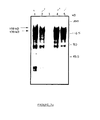
- the mature env translation products obtained in vitro are subjected to immunoprecipitation (figure 3) with the various sera from the abovementioned animals (sera SPF, experimentally inoculated with Wo and Petaluma (bands 1 and 4), sera from naturally infected cats (bands 2 and 5), sera from uninfected SPF cats, as negative control (strip 3)).
- This figure 3 illustrates the results obtained; the two glycosylated products (150 kDa and 130 kDa) are immunoprecipitated by sera from infected cats, the 130 kDa product being the major product.
- the 90 kDa band probably represents residual non-glycosylated material.
- the product of lowest molecular weight can be considered a transcription or partial translation product. No band is detected in the sera of SPF cats and not infected.
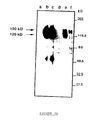
- FIG. 3b illustrates, by comparison, the immunoprecipitation of metabolically labeled VIF proteins obtained from FL-4 cells.
- Lanes a, b and c represent the result obtained with sera from uninfected SPF cats, sera from cats infected with a Petaluma strain and sera from cats infected with VIF Wo.
- the bands d, e and f correspond to a pre-immune rabbit serum, polyclonal rabbit sera directed against the C-terminal peptide of the SU protein (from amino acid 6 to the TM portion (P100)) and rabbit sera polyclonal cells directed against a C-terminal peptide of the TM protein (P102).
- Lanes b and c allow the detection of two proteins (150 kDa and 115-125 kDa), which correspond to the envelope precursor protein and to the SU glycoprotein, respectively, as confirmed by immunoprecipitation with the two rabbit antibodies. raised against peptides including the SU-TM cleavage site or corresponding to the C-terminal part of the env precursor (bands e and f).
- nucleic acid fragments isolated from a test sample and transferred to a nitrocellulose filter are hybridized in situ with a nucleic acid fragment according to the invention (probe labeled with dATP 32 P), 58 ° C overnight, after blocking the non-specific bonds, with a Denhardt solution.
- V 1 -V 9 variable fragments
- Overlapping fragments of the plasmid pKSe are generated by DNase I digestion in the presence of manganese ions.
- the DNA fragments obtained, after an optimal incubation time with DNase (time giving fragments of length of about 200 bp) are treated with the Klenow fragment of DNA polymerase from E. coli , so as to obtain blunt-ended ends, for efficient ligation with the binding sequence.
- the labeling with phosphorus 32 of the DNA fragments allows the control of the subsequent steps.
- bp 10 base pair binding sequences (bp), having an EcoRI site (Pharmacia), are used for ligation with the pKSe fragments obtained.
- the ligature products are then digested with the enzyme EcoRI restriction and separated by electrophoresis in 2% LMP NUSieve agarose gels to select a set of fragments comprising between 100 and 200 bp and eliminating free linkage sequences.
- the selected fragments are extracted with phenol from agarose and ligated with a vector ⁇ gt11 digested with the restriction enzyme EcoRI (Promega).
- the ligation products are packaged using a Packagene kit (Promega) and spread on a strain of E. coli Y1090.
- the recombinant phages are selected by detection of a color in the presence of 25 ⁇ l of IPTG 1 M and 25 ⁇ l of Xgal 80 mg / ml.
- the spreading of the ⁇ gt11 bank provides 10 5 independent clones.
- the phage DNA obtained from the non-stained plates, is analyzed by PCR using primers ⁇ gt11 1218 and 1222, complementary to the ⁇ -galactosidase fragment of the matrix of ⁇ gt11: 8 of the 10 clones contain inserts of average size d 'around 160 bp.
- the bank is then amplified to obtain a titer of 3 ⁇ 10 9 per ml.
- the protein SU glycoprotein outer surface envelope
- the TM protein transmembrane envelope viral glycoprotein
- Table I and Figure 4 specify the nucleotide sequences of the different inserts as well as the corresponding peptide sequences.
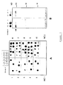
- the overlapping sequences highlight the presence of 8 immunogenic domains (epitopes), 5 of which are located in the extramembrane glycoprotein (gp) SU (SU1, amino acids 253-289; SU2, amino acids 388-424; SU3, amino acids 467-492; SU4, amino acids 508-528 and SU5, amino acids 572-606) and 3 in TM transmembrane gp (TM2, amino acids 681-711; TM3, amino acids 744-788 and TM4, amino acids 826-854) (Figure 1).
- gp extramembrane glycoprotein
- the peptide sequence 597-646 which is defined by clones 20 and 124, overlaps the 5th SU epitope (574-606) and undoubtedly represents a TM NH 2 -terminal (TM1) epitope. This hypothesis is confirmed by the immunological screening with cat sera, which shows the different reactivities between clone 51 (epitope SU5) and clone 124 (epitope TM1).
- FIG. 4 illustrates the localization of the epitopic regions of the envelope glycoproteins (gp) of the VIF, with SU: gp100; TM: gp40.
- the cleavage sites are represented by vertical arrows; the main clones are represented by thick lines and the various rectangles indicate the epitopes corresponding to the minimal overlapping sequences (dark colored rectangle Table II, below, illustrates the number of clones having the immunogenic regions as specified in Table I below above.
- Immunogenic regions Number of clones 1st 7 2nd 11 3rd 22 4th 10 5th 31 6th 4
- the recombinant clones ⁇ gt11 54, 133, 73, 154, 44, 51, 124, 157, 9 and 27 are expressed as lysogens in E. coli Y1089, as described by HUYNH et al. (1985).
- Crude lysate, containing the fusion proteins, is prepared from lysogenic culture induced by IPTG by making freeze-thaw cycles and sonication of the cell pellet, resuspended in 200 ⁇ l of TEP buffer (100 mM Tris-HCl). , pH 7.4, 10 mM EDTA, 1 mM PMSF).
- the extracts are brought to the boil in a Laemmli buffer and resuspended in 5-10% polyacrylamide SDS gels (40 ⁇ g / well) using a Mini PII device (BIO-RAD). After transferring the proteins to a nitrocellulose filter and blocking the non-specific sites by incubation with 3% defatted milk in 10 mM Tris, 150 mM NaCl, 0.1% Tween 20 (TNT buffer), strips of nitrocellulose are incubated with cat serum (diluted to 300th) for 2 hours at 37 ° C.
- the binding antibodies are revealed by incubation with goat IgG coupled to anti-cat peroxidase (dilution to 1000th) followed by a revelation with 4-chloro-1-naphthol (Merck).
- the controls consist of an E. coli Y1089 lysate infected with ⁇ gt11 and a pool of normal sera.
- hypervariability successively concerns the segments 361-377, which extends on either side of a cysteine preserved in position 367, 4 short fragments 452-455, 466-471, 479- 485 and 494-496, the dipeptide 541-542 and the segment 553-570, which comprises a cysteine stored in position 566.
- Two hypervariable zones have also been located in the TM protein: residues 710-718 and 832-842. Variable regions are found in the N-terminal rev-like domain, in the C-terminal portion of the SU and in the C-terminal portion of the hydrophobic transmembrane domain of the TM (between positions 220 and 286) and exhibit variability below the 10% threshold.
- such peptides advantageously include the 9 variable regions as defined above (hypervariable regions + regions adjacent variables), namely: V1 (residues 26-72), V2 (residues 96-174, 23% divergence), V3 (residues 361-422, including the hypervariable region 361-391 and the variable region 392-422, 12% divergence), V4 (residues 452-497, 13% divergence), V5 (residues 541-570, 30% divergence), V6 (residues 586-612, 10% divergence), V7 (residues 710-718, 26% of divergence), V8 (residues 765-778, 12% divergence) and V9 (residues 832-842, 21% divergence), as shown Figure 6 which illustrates the variable domains and the conserved domains: conserved domains are represented in white, the variable regions in gray, and the
- Peptides corresponding to immunodominant epitopes are absorbed in the well of an ELISA plate.
- test serum is incubated at a dilution optimal (predetermined during the development of the E-LISA, using a series of sera from infected cats and SPF cats).
- a dilution optimal predetermined during the development of the E-LISA, using a series of sera from infected cats and SPF cats.
- the presence of anti-VIF antibodies is revealed by anti-cat Ig antibodies coupled to horseradish peroxidase, in the presence of a chromogenic substrate.
- Peptides are used in concentrations between 1 and 10 ⁇ g / ml; to perform the coating of the microtitration plates with the peptides, 0.1 M sodium carbonate, pH 9.6 PBS or poly-L-lysine microplates are used.
- Free sites are saturated with different concentrations of defatted milk or bovine serum albumin.
- the different sera are diluted from 1: 1 to 1: 200.
- the test is carried out as follows: 0.25 ⁇ g of peptide in 50 ⁇ g of 0.1 M sodium carbonate, pH 9.6 are adsorbed on the wells of a microtiter plate (DYNATECH IMMULON® 2) overnight at 4 ° C. The wells are then washed three times with PBS. The residual adsorption sites on the microplates are saturated by incubation with 100 ⁇ l of PBS containing milk (1%) and Tween 20 (0.1%) (ELISA, EB buffer), during 2 hours. 50 ⁇ l of diluted cat serum are incubated in the wells for 2 hours at room temperature.
- peroxidase conjugated anti-cat immunoglobulins (0.5 ⁇ g / ml) (KIRKEGAARD & PERRY LABORATORIES, INC) are added for one hour at room temperature. After 5 washes with PBS, the conjugate to the adsorbed peroxidase is visualized with 2,2'-azino-bis (3-ethylbenz-thiazoline-6 sulfonic acid) (ABTS) (Sigma) 0.2 mg / ml in acetic acid at 0.6%, pH 4.7 and at a final concentration (w / v) of H 2 O 2 of 0.1%.
- ABTS 2,2'-azino-bis (3-ethylbenz-thiazoline-6 sulfonic acid)
- FIG. 7 shows in particular an ELISA test whose reagent is the peptide P237 is highly specific (no false positive) and sensitive (all sera from infected cats of different origins are reactive).
- the use of such a synthetic peptide provides a pure reagent and inexpensive for routine diagnosis of infection at VIF.
- Figure 8 illustrates these results ( ⁇ : SPF cats; •: experimentally infected cats; ⁇ : naturally infected cats).
- cats Source (s) Tested samples SPF France 56 Swiss 5 naturally infected with VIF France 89 Francec 9 Swiss 7 Scotland 5 experimentally inoculated with VIF isolates: wo France 22 Me France 11 Be France 8 The France 7 Br France 3 Gi France 2 Villefranche France 1 Envnip France 1 Petaluma France 4 Aebli Swiss 1 Gasser Swiss 1
Landscapes
- Chemical & Material Sciences (AREA)
- Life Sciences & Earth Sciences (AREA)
- Organic Chemistry (AREA)
- Health & Medical Sciences (AREA)
- Virology (AREA)
- Biophysics (AREA)
- General Health & Medical Sciences (AREA)
- Genetics & Genomics (AREA)
- Medicinal Chemistry (AREA)
- Molecular Biology (AREA)
- Proteomics, Peptides & Aminoacids (AREA)
- Biochemistry (AREA)
- Immunology (AREA)
- Gastroenterology & Hepatology (AREA)
- Peptides Or Proteins (AREA)
- Medicines Containing Antibodies Or Antigens For Use As Internal Diagnostic Agents (AREA)
- Measuring Or Testing Involving Enzymes Or Micro-Organisms (AREA)
Abstract
Description
La présente invention est relative à des peptides et à leurs fragments, spécifiques du virus de l'immunodéficience féline (VIF) ainsi qu'à l'utilisation desdits fragments comme réactif de diagnostic et comme agent induisant une réponse immune (cellulaire et/ou production d'anticorps), notamment pour la prévention de l'immunodéficience féline.The present invention relates to peptides and their fragments, specific for the virus of feline immunodeficiency (VIF) and use said fragments as diagnostic reagent and as agent inducing an immune response (cellular and / or production of antibodies), in particular for the prevention of feline immunodeficiency.
La présente invention est également relative aux séquences nucléotidiques codant pour lesdits peptides.The present invention also relates to the nucleotide sequences coding for said peptides.
L'immunodéficience féline est due à un lentivirus, le virus de l'immunodéficience féline (VIF), qui présente une structure génétique similaire à celle des lentivirus des primates (VIH et VIS).Feline immunodeficiency is caused by a lentivirus, feline immunodeficiency virus (FIV), which has a genetic structure similar to that of primate lentivirus (HIV and VIS).
L'immunodéficience féline pose un problème important de santé vétérinaire, dans la mesure où un nombre important de chats infectés par le VIF a été décelé aux Etats-Unis, au Japon et en Europe (5 à 25 % des animaux).Feline immunodeficiency poses a significant problem veterinary health, to the extent that a number significant number of cats infected with VIF has been detected in United States, Japan and Europe (5 to 25% of animals).
A l'heure actuelle, l'immunodéficience féline est essentiellement diagnostiquée lors de l'apparition des signes cliniques (lymphadénopathie généralisée et survenue d'infections opportunistes), alors qu'un diagnostic très précoce aurait des avantages importants tant dans le traitement que dans la prévention de cette maladie.Currently, feline immunodeficiency is essentially diagnosed at the onset clinical signs (generalized lymphadenopathy and occurrence of opportunistic infections), while a diagnosis very early would have significant benefits so much in treatment than in prevention of this disease.
Plusieurs isolats viraux indépendants ont été mis en évidence à travers le monde et un certain nombre de travaux pour mettre en évidence la structure des souches isolées ont été réalisés, notamment en ce qui concerne la souche américaine Petaluma [R.L. TALBOTT et al. (Natl. Acad. Sci. USA, 1989, 86, 5743-5747) ; T.R. PHILIPPS et al. (J. Virol., 1990, 64, 10, 4605-4613)], les souches japonaises (souches TM1 et TM2) [T. MIYAZAWA et al. (Arch. Virol., 1989, 108, 59-68)] ou les isolats suisses (FIVZ1 et FIVZ2) [S. MORIKAWA et al., (Virus Research, 1991, 21, 53-63)].Several independent viral isolates have been demonstrated around the world and a certain number of studies to demonstrate the structure of the isolated strains have been carried out, in particular with regard to the American strain Petaluma [RL TALBOTT et al. (Natl. Acad. Sci. USA, 1989, 86 , 5743-5747); TR PHILIPPS et al. (J. Virol., 1990, 64 , 10, 4605-4613)], the Japanese strains (strains TM1 and TM2) [T. MIYAZAWA et al. (Arch. Virol., 1989, 108, 59-68)] or the Swiss isolates (FIVZ1 and FIVZ2) [S. MORIKAWA et al., (Virus Research, 1991, 21 , 53-63)].
Les séquences nucléotidiques de trois clones proviraux, dérivés des isolats de VIF américains (souche Petaluma) ont été décrites (clones FIV34TF10, FIV14 et isolat PPR) [R.A. OLMSTED et al., (Proc. Natl. Acad. Sci. USA, 1989, 86, 2448-2452) ; T.R. PHILIPPS et al., 1990 ; R.L. TALBOTT et al., (Froc. Natl. Acad. Sci. USA, 1989, 86, 5743-5747) et comparées aux deux isolats suisses (S. MORIKAWA et al.). Cette comparaison a conduit S. MORIKAWA et al. à préciser la présence de certaines régions conservées et de certaines régions variables dans le gène env du VIF.The nucleotide sequences of three proviral clones, derived from American VIF isolates (strain Petaluma ) have been described (clones FIV34TF10, FIV14 and isolate PPR) [RA OLMSTED et al., (Proc. Natl. Acad. Sci. USA, 1989, 86 , 2448-2452); TR PHILIPPS et al., 1990; RL TALBOTT et al., (Froc. Natl. Acad. Sci. USA, 1989, 86 , 5743-5747) and compared with the two Swiss isolates (S. MORIKAWA et al.). This comparison led S. MORIKAWA et al. to specify the presence of certain conserved regions and certain variable regions in the VIF env gene.
Des souches françaises ont également été isolées (souches Wo et Me) [MORAILLON A. et al., 1992, Vet. Mic. , 31, 41-45, In vitro properties and experimental pathogenic effect of three feline immunodeficiency viruses isolated from cats with terminal diseases].French strains have also been isolated (Wo and Me strains) [MORAILLON A. et al., 1992, Vet. Mic. , 31, 41-45, In vitro properties and experimental pathogenic effect of three feline immunodeficiency viruses isolated from cats with terminal diseases ].
Les protéines d'enveloppe du VIF sont considérées comme étant au centre de la relation hôte-virus ; leur étude est donc essentielle pour comprendre l'interaction du virus avec le système immunitaire (épitopes de neutralisations, épitopes B et T) et avec les cellules-cibles de l'infection, et mettre au point aussi bien des réactifs de diagnostic performants que des vaccins efficaces, constitués de protéines virales à action immunogène et protectrice, vis-à-vis de l'ensemble des souches de VIF.VIF envelope proteins are considered as being at the center of the host-virus relationship; their study is therefore essential to understand how the virus interacts with the immune system (neutralization epitopes, B and T epitopes) and with target cells for infection, and develop both high-performance diagnostic reagents and effective vaccines, consisting of viral proteins immunogenic and protective action, vis-à-vis the whole strains of VIF.
La protéine env du VIF peut fournir, après clivage, deux fragments glycoprotéiques, dénommés SU (glycoprotéine de surface) et TM (glycoprotéine transmembranaire).The VIF env protein can provide, after cleavage, two glycoprotein fragments, called SU (surface glycoprotein) and TM (transmembrane glycoprotein).
Il a été montré que des fragments peptidiques issus d'une protéine env du VIF contiennent des déterminants majeurs de la protection (Demande de Brevet français n° 2 669 338 ou Demande Internationale PCT WO 92/09632 au nom de la Demanderesse).Peptide fragments have been shown from a VIF env protein contain determinants of protection (Patent Application French No. 2,669,338 or PCT International Application WO 92/09632 in the name of the Applicant).
Poursuivant ses travaux, la Demanderesse a trouvé :
- d'une part, qu'en vue de mettre au point des outils de diagnostic plus performants et des moyens de production de vaccins spécifiquement protecteurs, il était important de sélectionner, à partir de ces souches, des séquences aptes à être performantes dans le diagnostic différentiel précoce de l'immunodéficience féline et aptes à produire des produits dérivés par génie génétique, à fin de vaccination, ou à choisir une immunothérapie adaptée à la souche infectante,
- et d'autre part, qu'il était nécessaire, pour optimiser aussi bien le diagnostic que la prévention (vaccins sous-unités), de sélectionner des peptides viraux de petites tailles, particulièrement adaptés au diagnostic et à la prévention et permettant de former un pool de réactifs pour la détection de groupe (ensemble des souches de VIF) et/ou la détection de type (détection sélective de certaines souches).
- on the one hand, that in order to develop more efficient diagnostic tools and means of producing specifically protective vaccines, it was important to select, from these strains, sequences capable of being effective in diagnosis early feline immunodeficiency differential and able to produce products derived by genetic engineering, for vaccination purposes, or to choose an immunotherapy adapted to the infecting strain,
- and secondly, that it was necessary, in order to optimize both the diagnosis and the prevention (subunit vaccines), to select viral peptides of small sizes, particularly suitable for diagnosis and prevention and making it possible to form a reagent pool for group detection (all strains of VIF) and / or type detection (selective detection of certain strains).
La présente invention s'est en conséquence donné pour but de pourvoir, d'une part, à des séquences issues du VIF et à leurs fragments sélectionnés notamment pour leur spécificité de souche, et, d'autre part, à une composition à pouvoir immunogène et/ou protecteur, spécifique du virus de l'immunodéficience féline, obtenue en exprimant des fragments sélectionnés pour leur capacité neutralisante et/ou la présence d'épitopes de reconnaissance de groupe et/ou de type ; l'obtention par génie génétique ou synthèse chimique de tels peptides spécifiques a pour avantage de résoudre le problème de production de virus et d'obtenir directement les peptides désirés.The present invention is therefore intended to provide, on the one hand, sequences from VIF and their selected fragments in particular for their strain specificity, and, on the other hand, to a specific immunogenic and / or protective composition feline immunodeficiency virus, obtained in expressing fragments selected for their capacity neutralizing and / or the presence of recognition epitopes group and / or type; obtaining by genius genetics or chemical synthesis of such specific peptides has the advantage of solving the production problem virus and directly obtain the desired peptides.
Des séquences nucléotidiques issues de l'ARN génomique du virus de l'immunodéficience féline (VIF), comprennent au moins un fragment d'un gène présentant des zones hypervariables, choisi dans le groupe constitué par le gène codant pour la protéine env de la souche Wo du VIF et le gène codant pour la protéine gag de la souche Wo du VIF.Nucleotide sequences from RNA feline immunodeficiency virus (VIF) genomics, include at least one fragment of a gene with hypervariable zones, chosen from the group consisting of the gene encoding the env protein of the Wo strain of VIF and the gene coding for the gag protein of the strain Wo from VIF.
Conformément à ladite séquence, celle-ci
comprend la séquence en nucléotides et la séquence
déduite en amino-acides de formule I ci-après, qui
correspond au gène env du VIF, souche Wo :


Une telle séquence correspond à l'ADNc de l'ARNm de la protéine env de la souche Wo du VIF.Such a sequence corresponds to the cDNA of the mRNA of the env protein of the VIF strain Wo.
La présente invention a pour objet un peptide issu de la protéine Env du VIF de souche Wo, qui constitue un épitope conservé des protéines du VIF, caractérisé en ce qu'il est choisi dans le groupe constitué :
- du fragment de 45 aminoacides, dénommé TM3, et correspondant aux positions 744-788 de la séquence de formule I ;
- du fragment de séquence Gln763-Gln-Leu-Gln-Glu-Trp-Glu-Asp770 (peptide dénommé P241), correspondant aux positions 763-770 de la séquence de formule I ; et
- du fragment de séquence Val772-Gly-Trp-Ile-Gly-Asn-Ile-Pro779 (peptide dénommé P242), correspondant aux positions 772-779 de la séquence de formule I.
- the fragment of 45 amino acids, called TM3, and corresponding to positions 744-788 of the sequence of formula I;
- of the sequence fragment Gln 763 -Gln-Leu-Gln-Glu-Trp-Glu-Asp 770 (peptide called P241), corresponding to positions 763-770 of the sequence of formula I; and
- of the fragment of sequence Val 772 -Gly-Trp-Ile-Gly-Asn-Ile-Pro 779 (peptide called P242), corresponding to positions 772-779 of the sequence of formula I.
Ledit peptide est notamment reconnu par les anticorps dirigés contre au moins une souche différente de celle dont provient ledit peptide et/ou par les anticorps produits par des chats infectés naturellement et en ce qu'il présente une capacité d'induction d'une réponse immune cellulaire et/ou de production d'anticorps protecteurs vis-à-vis d'au moins une souche de VIF, chez des chats non infectés.Said peptide is especially recognized by antibodies to at least one different strain from that from which said peptide and / or by the antibodies produced by naturally infected cats and what it has a response response capacity immune system and / or production of protective antibodies vis-à-vis at least one strain of VIF, in uninfected cats.
Le fragment incluant un segment de 45 amino-acides, dénommé TM3, correspond aux positions 744-788 de la séquence de formule II ci-après, lequel segment contient un épitope incluant la séquence Gln764-Leu-Gln-Glu-Trp-Glu-Asp-Trp-Val-Gly-Trp-Ile-Gly-Asn-Ile778 ou la séquence Gln763-Gln-Leu-Gln-Glu-Trp-Glu-Asp770 (peptide dénommé P241) ou la séquence Val772-Gly-Trp-Ile-Gly-Asn-Ile-Pro779 (peptide dénommé P242).The fragment including a segment of 45 amino acids, called TM3, corresponds to positions 744-788 of the sequence of formula II below, which segment contains an epitope including the sequence Gln 764 -Leu-Gln-Glu-Trp-Glu -Asp-Trp-Val-Gly-Trp-Ile-Gly-Asn-Ile 778 or the sequence Gln 763 -Gln-Leu-Gln-Glu-Trp-Glu-Asp 770 (peptide called P241) or the sequence Val 772 - Gly-Trp-Ile-Gly-Asn-Ile-Pro 779 (peptide called P242).
Le peptide qui correspond à la protéine Env du
VIF, souche Wo, présente la séquence suivante :
On entend par épitope, au sens de la présente invention, les épitopes communs à toutes les souches de VIF (zones conservées), reconnus par les anticorps dirigés contre toutes les souches de VIF (reconnaissance de groupe) ; de plus, tous ces fragments induisent une production d'anticorps et/ou une réponse cellulaire.By epitope, within the meaning of this invention, the epitopes common to all strains of VIF (conserved areas), recognized by directed antibodies against all strains of VIF (recognition of group); moreover, all these fragments induce a production antibody and / or cellular response.
Ces fragments ont l'avantage de présenter une sélectivité et/ou une spécificité importante vis-à-vis du VIF, tout en étant de petite taille (prix de revient nettement diminué).These fragments have the advantage of presenting a selectivity and / or significant specificity with respect to VIF, while being small (cost price markedly decreased).
Ces peptides sont codés par une séquence ou un fragment de séquence nucléotidique telle que définie dans la séquence de formule I ci-dessus.These peptides are coded by a sequence or a nucleotide sequence fragment as defined in the sequence of formula I above.
Ces peptides TM3 permettent :
- d'un point de vue diagnostic :
en tant que fragments universels, de reconnaítre les anticorps produits par un VIF, s'il est présent, quelle que soit la souche. - d'un point de vue protecteur (vaccin en particulier)
:
les fragments TM3 induisent plus Spécifiquement la production d'anticorps, et éventuellement une réponse cellulaire ; en conséquence le pool de peptides conforme à l'invention assure soit une protection spécifique vis-à-vis de la souche d'origine, soit une protection plus large (plusieurs souches de VIF).
- from a diagnostic point of view:
as universal fragments, to recognize the antibodies produced by a VIF, if it is present, whatever the strain. - from a protective point of view (vaccine in particular):
the TM3 fragments more specifically induce the production of antibodies, and possibly a cellular response; consequently, the peptide pool according to the invention provides either specific protection with respect to the original strain, or broader protection (several strains of VIF).
Au sens de la présente invention le terme fragment peptidique comprend tous les fragments incluant un segment de peptide conforme à l'invention ainsi que les peptides homologues ; de manière générale, on entend par peptide homologue, les fragments peptidiques dont la position et la fonction sont équivalents, au sein du VIF (même localisation que celle définie ci-dessus, sur le génome du VIF) ; en ce qui concerne plus particulièrement les fragments TM3, on entend également par peptides homologues, les peptides qui ne perdent pas les propriétés de reconnaissance des anticorps dirigés contre le peptide de référence.Within the meaning of the present invention the term peptide fragment includes all fragments including a peptide segment in accordance with the invention as well as homologous peptides; in general, we mean by homologous peptide, the peptide fragments whose position and function are equivalent, within the VIF (same location as that defined above, on the VIF genome); more specifically TM3 fragments, also understood by homologous peptides, peptides that do not lose the properties of recognition of the antibodies directed against the peptide of reference.
L'ensemble desdits peptides peut avantageusement être obtenus soit par clonage, soit par synthèse, notamment par synthèse de Merrifield.All of said peptides can advantageously be obtained either by cloning or by synthesis, in particular by synthesis of Merrifield.
La présente invention a également pour objet un vecteur recombinant de clonage et/ou d'expression, caractérisé en ce qu'il comprend une séquence nucléotidique codant pour l'un des peptides TM3 définis ci-dessus.The present invention also relates to a recombinant cloning and / or expression vector, characterized in that it comprises a nucleotide sequence encoding one of the TM3 peptides defined above.
On entend, au sens de la présente invention, par vecteur recombinant, aussi bien un plasmide, un cosmide, qu'un phage.For the purposes of the present invention, by recombinant vector, either a plasmid, a cosmid, than a phage.
Selon un mode de réalisation avantageux dudit vecteur, il est constitué par un plasmide comprenant une origine de réplication, au moins un marqueur de sélection et une séquence nucléotidique codant pour l'un des peptides TM3 définis ci-dessus, lequel vecteur est un vecteur de clonage.According to an advantageous embodiment of said vector, it consists of a plasmid comprising a origin of replication, at least one selection marker and a nucleotide sequence encoding one of the TM3 peptides defined above, which vector is a cloning vector.
Selon une disposition avantageuse de ce mode de réalisation, ledit vecteur de clonage est constitué par un plasmide capable de se répliquer dans E.coli, dans lequel est insérée une séquence nucléotidique codant pour l'un des peptides TM3 définis ci-dessus. According to an advantageous arrangement of this embodiment, said cloning vector consists of a plasmid capable of replicating in E. coli , into which is inserted a nucleotide sequence coding for one of the TM3 peptides defined above.
Dans le cas où l'on introduit la séquence de formule I dans un vecteur Bluescript® SK+, ledit vecteur est dénommé pKSe1.In the case where the sequence of formula I is introduced into a Bluescript® SK + vector, said vector is called pKSe1.
Ledit vecteur a été déposé auprès de la Collection Nationale de Microorganismes tenue par l'Institut Pasteur, le 12 juin 1992 sous le n° I-1220.Said vector has been deposited with the Collection National of Microorganisms held by the Institute Pasteur, June 12, 1992 under No. I-1220.
Lorsque ladite séquence est introduite dans un vecteur Bluescript® KS+, ledit vecteur est dénommé pKSe.When said sequence is introduced into a Bluescript® KS + vector, said vector is called pKSe.
Selon un autre mode de réalisation dudit vecteur, il est constitué par un vecteur recombinant comprenant une origine de réplication dans un microorganisme hôte approprié, notamment une bactérie ou une cellule eucaryote, au moins un gène dont l'expression permet la sélection soit des bactéries, soit des cellules eucaryotes ayant reçu ledit vecteur, un promoteur permettant l'expression des gènes dans lesdites bactéries ou cellules eucaryotes, et dans lequel est insérée une séquence nucléotidique codant pour l'un des peptides TM3 définis ci-dessus, lequel vecteur est un vecteur d'expression d'un peptide TM3 tel que défini ci-dessus.According to another embodiment of said vector, it consists of a recombinant vector comprising an origin of replication in a microorganism suitable host, including bacteria or eukaryotic cell, at least one gene whose expression allows the selection of either bacteria or cells eukaryotes having received said vector, a promoter allowing the expression of genes in said bacteria or eukaryotic cells, and into which is inserted a nucleotide sequence encoding one of the TM3 peptides defined above, which vector is a vector expression of a TM3 peptide as defined above.
Avantageusement, ledit vecteur d'expression est constitué par un phage λgt11 dans lequel est inséré, en phase au niveau de son site, EcoRI, des fragments de digestion à la DNAse du plasmide pKSe1.Advantageously, said expression vector consists of a phage λgt11 into which is inserted, in phase at its site, EcoRI, fragments of digestion with DNAse of the plasmid pKSe1.
La présente invention a également pour objet une composition à pouvoir immunogène et/ou protecteur, caractérisée en ce qu'elle comprend au moins un peptide TM3 tel que défini ci-dessus, éventuellement associé à au moins une autre substance immunogène ou immunostimulante et/ou un véhicule pharmaceutiquement acceptable.The present invention also relates to an immunogenic and / or protective composition, characterized in that it comprises at least one peptide TM3 as defined above, possibly associated with minus another immunogenic or immunostimulating substance and / or a pharmaceutically acceptable vehicle.
La présente invention, a, de plus, pour objet un procédé de détection d'anticorps anti-VIF, caractérisé en ce qu'il consiste à détecter les anticorps anti-VIF éventuellement présents dans un échantillon biologique à l'aide d'un peptide TM3 tel que défini ci-dessus, éventuellement fixé sur un support solide approprié, en mettant en présence ledit échantillon biologique avec ledit/lesdits peptides, auxquels se lient les anticorps anti-VIF si de tels anticorps sont présents dans l'échantillon à analyser, la lecture du résultat étant révélée par un moyen approprié, notamment EIA, RIA, fluorescence.The present invention has, moreover, an object a method for detecting anti-FIV antibodies, characterized in that it consists in detecting anti-VIF antibodies possibly present in a biological sample to using a TM3 peptide as defined above, optionally fixed on an appropriate solid support, bringing said biological sample into contact with said peptide (s) to which the antibodies bind anti-FIV if such antibodies are present in the sample to be analyzed, the reading of the result being revealed by an appropriate means, in particular EIA, RIA, fluorescence.
Ce procédé permet notamment de vérifier la séroconversion des animaux vaccinés ou de procéder à des enquêtes sérologiques à visée épidémiologique.This process notably makes it possible to verify the seroconversion of vaccinated animals or to carry out epidemiological serological surveys.
A l'aide d'un peptide TM3, ledit procédé permet de déterminer une infection à VIF, quelle que soit la souche (réactif universel tel que précisé ci-dessus).Using a TM3 peptide, said method allows determine a VIF infection, regardless of the strain (universal reagent as specified above).
La présente invention a, en outre, pour objet un kit, prêt à l'emploi, pour la mise en oeuvre du procédé de détection d'anticorps anti-VIF, caractérisé en ce qu'il comprend, outre des quantités utiles de tampons appropriés pour la mise en oeuvre de ladite détection, des doses appropriées d'au moins un peptide TM3 tel que défini ci-dessus.The present invention further relates to a kit, ready to use, for implementing the process anti-VIF antibody detection, characterized in that it includes, in addition to useful quantities of buffers suitable for carrying out said detection, appropriate doses of at least one TM3 peptide such as defined above.
La présente invention a également pour objet un réactif de diagnostic de l'immunodéficience féline, caractérisé en ce qu'il est choisi dans le groupe constitué par l'un des peptides TM3 tels que définis ci-dessus, et en ce qu'il constitue un réactif de diagnostic universel de souche de VIF.The present invention also relates to a reagent for diagnosing feline immunodeficiency, characterized in that it is chosen from the group consisting by one of the TM3 peptides as defined above, and in that it constitutes a universal diagnostic reagent of VIF strain.
Des cellules mononucléaires de sang périphérique (PBMC), obtenues à partir de chats infectés naturellement par l'isolat français Wo, présentant un syndrome d'immunodéficience acquise féline, sont co-cultivées avec des cellules mononucléaires de sang périphérique, obtenues à partir de chats dits contrôle négatif (chats SPF), en présence d'interleukine-2 humaine recombinante (Genzyme, Boston, USA) et de concanavaline A (Sigma) (MORAILLON A. et al., 1992, Vet. Mic., 31, 41-45, In vitro properties and experimental pathogenic effect of three feline immunodeficiency viruses isolated from cats with terminal diseases).Peripheral blood mononuclear cells (PBMC), obtained from cats naturally infected with the French isolate Wo, presenting a feline acquired immunodeficiency syndrome, are co-cultivated with peripheral blood mononuclear cells, obtained from cats said negative control (SPF cats), in the presence of recombinant human interleukin-2 (Genzyme, Boston, USA) and concanavalin A (Sigma) (MORAILLON A. et al., 1992, Vet. Mic., 31, 41- 45, In vitro properties and experimental pathogenic effect of three feline immunodeficiency viruses isolated from cats with terminal diseases ).
L'activité réverse transcriptase (RT) dépendante de Mg++ est plus particulièrement surveillée. Après 15 jours, l'ADN cellulaire total est extrait au phénol (SAMBROOK et al., 1989, Molecular cloning, a laboratory manual), à partir de cultures RT positives et utilisé comme matériel de départ pour l'amplification des gènes du VIF. Ceci permet d'éviter les passages multiples in vitro et l'adaptation à des lignées cellulaires CD4 négatives.The Mg ++- dependent reverse transcriptase (RT) activity is more particularly monitored. After 15 days, the total cellular DNA is extracted with phenol (SAMBROOK et al., 1989, Molecular cloning, a laboratory manual), from RT positive cultures and used as starting material for the amplification of the VIF genes. This makes it possible to avoid multiple passages in vitro and adaptation to CD4 negative cell lines.
Les gènes env et gag de la souche Wo du virus de l'immunodéficience féline sont respectivement amplifiés en trois et deux fragments chevauchants, en utilisant des amorces synthétiques correspondant à des régions conservées dans les séquences de VIF, isolat Petaluma (TALBOTT et al, 1989) et isolat PPR (PHILIPPS et al., 1990).The env and gag genes of the Wo strain of the feline immunodeficiency virus are respectively amplified in three and two overlapping fragments, using synthetic primers corresponding to regions conserved in the sequences of VIF, Petaluma isolate (TALBOTT et al, 1989 ) and PPR isolate (PHILIPPS et al., 1990).
Les séquences des amorces et les positions (conformément à la numérotation utilisée pour le VIF 34TF10), sont comme suit :
- env :
- pour le premier fragment env, dénommé E1, on utilise comme amorce 5', le fragment 5'-GCAACAATAAGAATGG-CAGAAGC-3' (6251-6274) et comme amorce 3', le fragment 5'-GTTTAGGGTGACTAGTTAATATGTAAACC-3' (7236-7263) ;
- pour le second fragment env, dénommé E2, on utilise comme amorce 5', le fragment 5'-CAAATCCCACTGAT-CAATTATACATTTGG-3' (7236-7263), et comme amorce 3', le fragment 5'-CGTAGTTCATGATCATTATCCTAATTTTC-3' (8269-8298) ;
- en ce qui concerne le fragment dénommé E3, on utilise comme amorce 5', le fragment 5'-GCATCAAGTAC-TAGTAATAGGATTAAAAG-3' (8269-8298), et comme amorce 3', le fragment 5'-CTTTTTCTCCTCCTTACTACTTC-3' (8809-8833) ;
- gag :
- pour le premier fragment gag, dénommé G1, on utilise comme amorce 5', le fragment 5'-GGAGAGATTCTACAG-CAACATGGGG-3' (608-632) et comme amorce 3', le fragment 5'-GCCAATTTTCCTAATGCCTCGAGATACCATGC-3' (1398-1429) ;
- pour le second fragment gag, dénommé G2, on utilise comme amorce 5', le fragment 5'-GCATGGTATCTCGAGG-CATTAGGAAAATTGGC63' (1398-1429) et comme amorce 3', le fragment 5'-AATTTACAAATCCAATAGTTTCTCCTCC-3' (1954-2123).
Tris 10 mM, du KCl 50 mM et du MgCl2 2 mM.
- approx :
- for the first env fragment, designated E 1 , the 5'-primer is used, the 5'-GCAACAATAAGAATGG-CAGAAGC-3 'fragment (6251-6274) and the 5'-GTTTAGGGTGACTAGTTAATATGTAAACC-3' fragment (7236) -7263);
- for the second env fragment, designated E 2 , the 5 'primer is used, the 5'-CAAATCCCACTGAT-CAATTATACATTTGG-3' fragment (7236-7263), and as the 3 'primer, the 5'-CGTAGTTCATGATCATTATCCTAATTTTC-3' fragment ( 8269-8298);
- as regards the fragment designated E 3 , the 5'-primer is used, the 5'-GCATCAAGTAC-TAGTAATAGGATTAAAAG-3 'fragment (8269-8298), and as the 3' primer, the 5'-CTTTTTCTCCTCCTTACTACTTC-3 'fragment ( 8809-8833);
- gag :
- for the first gag fragment, called G 1 , the 5'-primer, the 5'-GGAGAGATTCTACAG-CAACATGGGG-3 'fragment (608-632) is used and the 5'-GCCAATTTTCCTAATGCCTCGAGATACCATGC-3' fragment (1398) -1429);
- for the second gag fragment, called G 2 , the 5'-primer, the 5'-GCATGGTATCTCGAGG-CATTAGGAAAATTGGC63 'fragment (1398-1429) is used and the 5'-AATTTACAAATCCAATAGTTTCTCCTCC-3' fragment (1954-2123) ).
Un vecteur M13mp8 est digéré par l'enzyme SmAI, de manière à pouvoir y insérer les fragments PCR obtenus ci-dessus [par exemple, pour le gène env fragments de la séquence de formule I, E1 (nucléotides 1-977), E2 (978-2016) et E3 (2017-2562)]. Le vecteur recombinant obtenu est utilisé pour transformer des E. coli JM101 compétents. A vector M13mp8 is digested with the enzyme SmAI, so as to be able to insert into it the PCR fragments obtained above [for example, for the env gene fragments of the sequence of formula I, E 1 (nucleotides 1-977), E 2 (978-2016) and E 3 (2017-2562)]. The recombinant vector obtained is used to transform competent E. coli JM101.
Les clones recombinants sont détectés par une hybridation in situ avec les fragments PCR correspondants, marqués au phosphore 32.The recombinant clones are detected by in situ hybridization with the corresponding PCR fragments, labeled with phosphorus 32.
Le séquençage de l'ADN simple brin à partir de phages recombinants est réalisé par la méthode enzymatique par les didésoxynucléotides, avec un kit de séquençage séquenase® 2.0 DNA, utilisant, comme amorces, des oligonucléotides complémentaires au vecteur (17 mers, amorce universelle) ou l'insert VIF.Single strand DNA sequencing from recombinant phages is carried out by the enzymatic method by dideoxynucleotides, with a sequencing kit sequenase® 2.0 DNA, using, as primers, oligonucleotides complementary to the vector (17 seas, universal primer) or the VIF insert.
Pour reconstituer le gène env complet, les trois fragments (E1, E2 et E3) sont préparés à partir du vecteur recombinant M13mp8 tel que défini ci-dessus, en utilisant les sites de restriction présents dans la séquence de liaison multisites du M13mp8 ou dans les amorces d'amplification : E1, HindIII-BclI ; E2, BclI-SpeI ; E3, SpeI-EcoRI. Les trois fragments sont ligatures dans un vecteur KS+ Bluescript® (Stratagène), digéré par les enzymes HindIII et EcoRI, en aval du promoteur T3. La construction obtenue est dénommée pKSe.To reconstitute the complete env gene, the three fragments (E 1 , E 2 and E 3 ) are prepared from the recombinant vector M13mp8 as defined above, using the restriction sites present in the multisite binding sequence of M13mp8 or in the amplification primers: E 1 , HindIII-BclI; E 2 , BclI-SpeI; E 3 , SpeI-EcoRI. The three fragments are ligated into a KS + Bluescript® vector (Stratagene), digested with the enzymes HindIII and EcoRI, downstream of the T3 promoter. The construction obtained is called pKSe.
Lesdits fragments peuvent également être ligaturés dans un vecteur SK+ Bluescript® (Stratagène), digéré par les enzymes HindIII et EcoRI, en aval du promoteur T7. Cette dernière construction, dénommée pKSe1, permet la transcription du gène env dans des cellules de mammifère exprimant le gène de l'ARN polymérase du phage T7, alors que la construction pKSe permet la transcription du gène env dans des cellules de mammifère exprimant le gène de l'ARN polymérase du phage T3.Said fragments can also be ligated into a SK + Bluescript® vector (Stratagene), digested with the enzymes HindIII and EcoRI, downstream of the T7 promoter. The latter construction, called pKSe1, allows transcription of the env gene in mammalian cells expressing the phage T7 RNA polymerase gene, while the construction pKSe allows transcription of the env gene in mammalian cells expressing the gene for phage T3 RNA polymerase.
L'analyse des séquences est réalisée sur ordinateur avec le programme "Salsa".Sequence analysis is performed on computer with the "Salsa" program.
Les multiples alignements comparant la séquence VIF Wo avec d'autres séquences de VIF et notamment VIF34TF10 (USA) (TALBOTT et al., 1989), VIF 14 (USA) (OLMSTED et al., 1989), VIF PPR (USA) (PHILLIPS et al., 1990), VIF TM2/GVEPX (JAPON) (KIYOMASU et al., 1991 ; MIYAZAWA et al.), VIF Z1 et VIF Z2 (SUISSE) (MORIKAWA et al., 1991), VIF 19K1 et VIF 1.9K2 (Pays Bas) (SIEBELINK K.M.J. et al., J. Virol., 1992, 66, 1091-1097) obtenus à partir de la banque de gènes "GenBank", sont illustrés à la figure 1a et 1b, dans lesquelles il apparaít que les variations sont, pour la plupart, situées sur le gène env et sont, en référence auxdites séquences en aminoacides :
- séquence FIV Wo/séquence FIVZ2 : 8,9 %
- séquence FIV Wo/séquence isolat Petaluma : 10,2-10,9 %
- séquence FIV Wo/ séquence FIV19 : 10,8 %
- séquence FIV Wo/séquence FIVZ1 : 11 %
- séquence FIV Wo/séquence FIVPPR : 13,6 %
- séquence FIV Wo/séquence FIVGVEPX : 19 %.
- IVF Wo sequence / FIVZ2 sequence: 8.9%
- IVF Wo sequence / Petaluma isolate sequence: 10.2-10.9%
- IVF Wo sequence / IVF19 sequence: 10.8%
- IVF Wo sequence / FIVZ1 sequence: 11%
- IVF Wo sequence / IVFPR sequence: 13.6%
- FIV Wo sequence / FIVGVEPX sequence: 19%.
Les séquences variables conformes à l'invention
sélectionnées (V1 à V9, en référence à leurs séquences
en aminoacides) sont récapitulées dans le Tableau
ci-après ; de manière avantageuse, la sélection de ces
fragments permet de définir, par déduction, des zones
conservées chez l'ensemble des VIF, et permet de bien
mettre en valeur les zones variables et les zones conservées
; ce qui permet de définir des réactifs universels
de détection du VIF, des réactifs sélectifs et de faire
bénéficier les animaux de la meilleure protection possible
:

Pour la synthèse in vitro de l'ARN, les deux constructions pKSe et pKSe1 sont utilisées, la transcription étant effectuée dans le premier cas, par la polymérase T3 et dans le deuxième cas, par la polymérase T7, comme précisé ci-dessus.For the in vitro synthesis of RNA, the two constructions pKSe and pKSe1 are used, the transcription being carried out in the first case, by T3 polymerase and in the second case, by T7 polymerase, as specified above.
1 µg d'ADN des plasmides pKSe1 et pKSe est
linéarisé, en utilisant le site de restriction EcoRI,
présent dans la séquence de liaison multisites du vecteur,
en aval du gène inséré. Le plasmide linéarisé et
traité à la protéinase K est transcrit, en utilisant
l'ARN polymérase T3 pour pKSe et l'ARN polymérase T7 pour
pKSe1, conformément au protocole fourni par le fabricant
(Stratagène). Après traitement à la DNase et extraction
au phénol, le produit de transcription est précipité à
l'éthanol, en présence de 5 µg d'ARNt d'E. coli, remis en
suspension dans 15 µl d'eau traitée au DEPC et stocké à
-70°C. La taille des transcrits est évaluée par électrophorèse
sur gel d'agarose à 1 % dans un tampon phosphate
10 mM, pH 7, après une dénaturation au
glyoxal/diméthylsulfoxide.1 μg of DNA of the plasmids pKSe1 and pKSe is linearized, using the EcoRI restriction site, present in the multisite binding sequence of the vector, downstream of the inserted gene. The linearized and proteinase K treated plasmid is transcribed, using T3 RNA polymerase for pKSe and T7 RNA polymerase for pKSe1, according to the protocol provided by the manufacturer (Stratagene). After treatment with DNase and extraction with phenol, the transcript is precipitated with ethanol, in the presence of 5 μg of E. coli tRNA, resuspended in 15 μl of water treated with DEPC and stored at -70 ° C. The size of the transcripts is evaluated by electrophoresis on 1% agarose gel in 10 mM phosphate buffer,
Environ 10 ng de l'ARN transcrit in vitro est
traduit pendant 30 minutes à 30°C, dans un volume final
de 12,5 µg contenant 6 µg de lysat de réticulocytes de
lapin (RRL) (Promega), 0,25 µl d'un mélange d'aminoacides
dépourvus de méthionine (Promega) et 1,75 µl de méthionine
marquée au soufre 35 en l'absence (-M) ou en présence
(+M) de membranes microsomales (1 ou 2 µl), pour
permettre la glycosylation des protéines (figure 2A). Les
1 ou 2 µl de membranes microsomales pancréatiques de
chien (Promega) sont ajoutés par 12,5 µl de mélange de
réaction. Pour les essais de cinétique, 3 µl d'aliquots
sont prélevés à des moments différents (15, 30, 60 et
90 minutes, 4 heures). Les échantillons sont stockés à
-70°C.About 10 ng of the RNA transcribed in vitro is translated for 30 minutes at 30 ° C, in a final volume of 12.5 µg containing 6 µg of rabbit reticulocyte lysate (RRL) (Promega), 0.25 µl d '' a mixture of amino acids devoid of methionine (Promega) and 1.75 µl of methionine labeled with
Les études d'inhibition en présence de castanospermine, sont réalisées en incubant le mélange de traduction contenant les membranes microsomales (2 µl pour 12,5 µl de mélange) avec de la castanospermine 5 mM (concentration finale) (Sigma), préparée extemporanément dans de l'eau traitée au DEPC, à 30°C pendant 15 minutes, avant l'addition d'ARN.Inhibition studies in the presence of castanospermine, are performed by incubating the translation mixture containing microsomal membranes (2 µl for 12.5 µl mixture) with 5 mM castanospermine (final concentration) (Sigma), prepared immediately in water treated with DEPC, at 30 ° C for 15 minutes, before addition of RNA.
Les produits de traduction sont dénaturés par ébullition dans un tampon de Laemmli et évalués par électrophorèse (SDS-PAGE, gradient 5-10 %). Des marqueurs du poids moléculaire sont également utilisés (BIORAD H.M.W. ou marqueurs Rainbow). Après l'électrophorèse, les gels sont fixés, traités et autoradiographiés sur un film Kodak XAR-5.Translation products are denatured by boil in Laemmli buffer and evaluated by electrophoresis (SDS-PAGE, gradient 5-10%). Markers of the molecular weights are also used (BIORAD H.M.W. or Rainbow markers). After electrophoresis, the gels are fixed, processed and autoradiographed on a film Kodak XAR-5.
Dans ces conditions, l'ARN transcrit in vitro à partir du plasmide pKSe (contenant le gène env de VIF Wo complet) migre, dans un gel d'agarose dans des conditions dénaturantes, en une seule bande d'approximativement 2,5 kb, correspondant à la dimension attendue. Après traduction de ce transcrit dans un lysat de réticulocytes de lapin (RRL), le produit obtenu en plus grande quantité, a un poids moléculaire apparent de 90 kDa (figure 2A), compatible avec le poids moléculaire calculé du polypeptide codé, déduit de la séquence (98,079 kDa).Under these conditions, the RNA transcribed in vitro from the plasmid pKSe (containing the complete VIF Wo env gene) migrates, in an agarose gel under denaturing conditions, in a single band of approximately 2.5 kb, corresponding to the expected dimension. After translating this transcript into a rabbit reticulocyte lysate (RRL), the product obtained in greater quantity has an apparent molecular weight of 90 kDa (FIG. 2A), compatible with the calculated molecular weight of the encoded polypeptide, deduced from the sequence (98.079 kDa).
La traduction de l'ARN en présence de microsomes pancréatiques de chien, qui permet une N-glycosylation de la chaíne polypeptidique obtenue, fournit deux produits de poids moléculaires élevés, respectivement de 150 et 130 kDa (figure 2A). Pour analyser la relation entre les deux produits glycosylés obtenus, on réalise des expérimentations étalées dans le temps ; on réalise également, comme précisé ci-dessus, la traduction en présence de castanospermine, un inhibiteur de l'α-glucosidase I du réticulum endoplasmique rugueux, de manière à déterminer si le produit de poids moléculaire le plus bas pourrait dériver de la glycoprotéine de 150 kDa par coupure des sucres pendant la glycosylation. La cinétique de la maturation de l'enveloppe protéinique montre l'apparition, au bout de 15 minutes, des deux produits glycosylés, qui augmente quantitativement jusqu'à 90 minutes d'incubation (figure 2B). En présence de castanospermine, les deux molécules migrent à un poids moléculaire plus élevé, respectivement de 160 et 145 kDa, et restent non modifiées pendant les expérimentations étalées dans le temps (figure 2B).Translation of RNA in the presence of microsomes pancreatic dog, which allows N-glycosylation of the polypeptide chain obtained, provides two high molecular weight products, respectively 150 and 130 kDa (Figure 2A). To analyze the relationship between the two glycosylated products obtained, experiments spread over time; we realize also, as specified above, the translation in the presence castanospermine, an α-glucosidase inhibitor I of the rough endoplasmic reticulum, so that determine if the lowest molecular weight product could be derived from 150 kDa glycoprotein per cut sugars during glycosylation. The kinetics of the maturation of the protein envelope shows the appearance, after 15 minutes, of the two glycosylated products, which increases quantitatively up to 90 minutes incubation (Figure 2B). In the presence of castanospermine, both molecules migrate at a higher molecular weight high, respectively 160 and 145 kDa, and remain un modified during the experiments spread out in the time (Figure 2B).
Ces expériences montrent qu'en l'absence de
membranes microsomales qui permettent la glycosylation,
on observe un produit de poids moléculaire apparent de
90 kDa, correspondant ainsi au précurseur d'enveloppe non
glycosylé. Après addition de membranes microsomales, on
observe deux espèces glycosylées de poids moléculaires
respectifs 150 kDa et 130 kDa, confirmant la glycosylation
importante de ce peptide. La séquence env de Wo
contient 21 sites N-glycosylation potentiels, qui sont
probablement utilisés complètement comme le suggère les
poids moléculaires des produits de traduction, en présence
de membranes microsomales.These experiments show that in the absence of
microsomal membranes which allow glycosylation,
we observe a product of apparent molecular weight of
90 kDa, thus corresponding to the envelope precursor no
glycosylated. After addition of microsomal membranes,
observes two glycosylated
Des anti-sérums de VIF sont obtenus à partir de deux chats SPF (chats dépourvus de pathogènes spécifiques), inoculés avec des isolats Wo ou Petaluma et à partir également de deux chats infectés naturellement, mais séronégatifs vis-à-vis du virus de la leucémie féline. Des sérums de contrôle sont obtenus à partir des deux chats SPF. Pour l'étape de la préabsorption, un stock de sérums de chats SPF est utilisé. Des anticorps polyclonaux de lapin dirigés contre des peptides synthétiques tels que décrits dans la Demande de Brevet français n° 2 669 338 sont également utilisés, à des concentrations de 20 µg/ml. Le premier de ces peptides (P100), d'une longueur de 21 aminoacides, est situé à l'extrémité C terminale de la glycoprotéine de surface du VIF et inclut le site de Coupure entre la glycoprotéine de surface (SU) et la glycoprotéines transmembranaire (TM) ; le deuxième peptide (P102), d'une longueur de 25 aminoacides, correspond à l'extrémité C terminale de la glycoprotéine TM du VIF. Des sérums de lapin préimmunisés sont utilisés pour l'étape de préabsorption et comme contrôle négatif dans les essais d'immunoprécipitation. VIF anti-sera are obtained from two SPF cats (cats lacking specific pathogens), inoculated with Wo or Petaluma isolates and also from two cats naturally infected, but seronegative towards the virus feline leukemia. Control sera are obtained from the two SPF cats. For the pre-absorption stage, a stock of SPF cat sera is used. Polyclonal rabbit antibodies directed against synthetic peptides as described in French Patent Application No. 2,669,338 are also used, at concentrations of 20 μg / ml. The first of these peptides (P100), 21 amino acids in length, is located at the C-terminus of the VIF surface glycoprotein and includes the Cleavage site between the surface glycoprotein (SU) and the transmembrane glycoprotein (TM); the second peptide (P102), with a length of 25 amino acids, corresponds to the C-terminus of the VIF glycoprotein TM. Pre-immunized rabbit sera are used for the pre-absorption step and as a negative control in immunoprecipitation assays.
Des aliquots de produits de traduction in vitro (approximativement 100 000 cpm, déterminés après précipitation à l'acide trichloroacétique) sont dilués dans 200 µl d'un tampon RIPA (Triton X-100 1 %, sodium désoxycholate 0,5 %, NaCl 150 mM, EDTA 2 mM dans Tris-HCl pH 7,5, 50 mM). Les réactions sont réalisées à 4°C. Les échantillons sont préabsorbés pendant 30 minutes avec 4 µl d'un pool de sérums de chats SPF ou avec un sérum de lapin préimmunisé et précipités avec 50 µl de protéine A-Sépharose CL-4B (PrA, Pharmacia). Les surnageants sont alors incubés pendant une nuit avec 4 µl de sérum de chats infectés, de sérum de chats SPF ou d'anticorps polyclonaux de lapin anti-peptide synthétique tel que défini ci-dessus. 50 µl de PrA sont ajoutés et incubés pendant 1 heure. Après 4 lavages dans un tampon RIPA, les culots des immunocomplexes-PrA sont élués par ébullition pendant 5 minutes dans 50 µl de tampon Laemmli. Les échantillons sont analysés sur un gradient SDS-PAGE 5-10 %.Aliquots of in vitro translation products (approximately 100,000 cpm, determined after precipitation with trichloroacetic acid) are diluted in 200 µl of RIPA buffer (1% Triton X-100, sodium deoxycholate 0.5%, 150 mM NaCl, 2 mM EDTA in Tris-HCl pH 7.5, 50 mM). The reactions are carried out at 4 ° C. The samples are pre-absorbed for 30 minutes with 4 µl of a pool of SPF cat sera or with a serum of pre-immunized rabbit and precipitated with 50 µl of protein A-Sepharose CL-4B (PrA, Pharmacia). The supernatants are then incubated overnight with 4 µl of serum infected cats, SPF cat serum or polyclonal antibodies rabbit synthetic anti-peptide as defined above. 50 µl of PrA are added and incubated for 1 hour. After 4 washes in RIPA buffer, the immunocomplex-PrA pellets are eluted by boiling for 5 minutes in 50 µl of Laemmli buffer. The samples are analyzed on an SDS-PAGE 5-10 gradient %.
Des cellules FL-4, dérivées des cellules mononucléaires de sang périphériques (PBMC) infectées par une souche Petaluma et produisant spontanément le VIF (Yamamoto,1991), sont lavées dans du PBS froid et lysées dans un tampon RIPA. Les lysats cellulaires sont clarifiés par centrifugation à 17000g pendant 30 min.FL-4 cells, derived from peripheral blood mononuclear cells (PBMC) infected with a Petaluma strain and spontaneously producing VIF (Yamamoto, 1991), are washed in cold PBS and lysed in RIPA buffer. The cell lysates are clarified by centrifugation at 17000 g for 30 min.
Les produits de traduction env mature obtenus
in vitro, sont soumis à une immunoprécipitation (figure
3) avec les différents sérums d'animaux précités (sérums
SPF, inoculés expérimentalement avec des isolats Wo et
Petaluma (bandes 1 et 4), sérums de chats infectés naturellement
(bandes 2 et 5), sérums de chats SPF non infectés,
comme contrôle négatif (bande 3)). Cette figure 3
illustre les résultats obtenus ; les deux produits glycosylés
(150 kDa et 130 kDa) sont immunoprécipités par
les sérums de chats infectés, le produit de 130 kDa étant
le produit majeur. La bande de 90 kDa représente probablement
un matériel non glycosylé résiduel. Le produit de
poids moléculaire le plus bas, peut être considéré comme
un produit de transcription ou de traduction partielle.
Aucune bande n'est détectée dans les sérums de chats SPF
et non infectés.The mature env translation products obtained
in vitro, are subjected to immunoprecipitation (figure
3) with the various sera from the abovementioned animals (sera
SPF, experimentally inoculated with Wo and
Petaluma (
La figure 3b illustre, en comparaison, l'immunoprécipitation
de protéines VIF marquées métaboliquement,
obtenues à partir de cellules FL-4. Les bandes a, b
et c représentent le résultat obtenu avec des sérums de
chats SPF non infectés, des sérums de chats infectés avec
une souche Petaluma et des sérums de chats infectés par
le VIF Wo. Les bandes d, e et f correspondent à un sérum
de lapin préimmune, des sérums de lapin polyclonaux dirigés
contre le peptide C-terminal de la protéine SU (de
l'aminoacide 6 à la portion TM(P100)) et des sérums de
lapin polyclonaux dirigés contre un peptide C-terminal de
la protéine TM (P102). Les bandes b et c permettent la
détection de deux protéines (150 kDa et 115-125 kDa), qui
correspondent à la protéine précurseur d'enveloppe et à
la glycoprotéine SU, respectivement, comme le confirme
l'immunoprécipitation avec les deux anticorps de lapin
dirigés contre les peptides incluant le site de clivage
SU-TM ou correspondant à la partie C-terminale du précurseur
env (bandes e et f).FIG. 3b illustrates, by comparison, the immunoprecipitation of metabolically labeled VIF proteins obtained from FL-4 cells. Lanes a, b and c represent the result obtained with sera from uninfected SPF cats, sera from cats infected with a Petaluma strain and sera from cats infected with VIF Wo. The bands d, e and f correspond to a pre-immune rabbit serum, polyclonal rabbit sera directed against the C-terminal peptide of the SU protein (from
On hybride in situ les fragments d'acide nucléique isolés à partir d'un échantillon à tester et transféré sur un filtre de nitrocellulose, avec un fragment d'acide nucléique conforme à l'invention (sonde marquée avec du dATP 32P), à 58°C pendant une nuit, après blocage des liaisons non spécifiques, avec une solution de Denhardt.The nucleic acid fragments isolated from a test sample and transferred to a nitrocellulose filter are hybridized in situ with a nucleic acid fragment according to the invention (probe labeled with dATP 32 P), 58 ° C overnight, after blocking the non-specific bonds, with a Denhardt solution.
Un tel procédé, mis en oeuvre avec un pool de fragments dits variables (V1-V9) permet une différenciation spécifique de souche de VIF. Such a method, implemented with a pool of so-called variable fragments (V 1 -V 9 ) allows a specific differentiation of VIF strain.
Le clonage du gène env du VIF Wo dans un vecteur Bluescript® KS+ fournissant la construction pKSe ou dans un vecteur Bluescript® SK+ fournissant la construction pKSe1 ont été décrits à l'exemple 1.The cloning of the VIF Wo env gene into a Bluescript® KS + vector providing the construction pKSe or into a Bluescript® SK + vector providing the construction pKSe1 were described in Example 1.
Des fragments chevauchants du plasmide pKSe sont générés par digestion à la DNase I en présence d'ions manganèse. Les fragments d'ADN obtenus, après un temps optimal d'incubation avec la DNase (temps donnant des fragments de longueur d'environ 200 pb) sont traités avec le fragment de Klenow de la DNA polymérase d'E. coli, de manière à obtenir des extrémités à bouts francs, pour une ligature efficace avec la séquence de liaison. Le marquage au phosphore 32 des fragments d'ADN permet le contrôle des étapes ultérieures.Overlapping fragments of the plasmid pKSe are generated by DNase I digestion in the presence of manganese ions. The DNA fragments obtained, after an optimal incubation time with DNase (time giving fragments of length of about 200 bp) are treated with the Klenow fragment of DNA polymerase from E. coli , so as to obtain blunt-ended ends, for efficient ligation with the binding sequence. The labeling with phosphorus 32 of the DNA fragments allows the control of the subsequent steps.
Des séquences de liaison de 10 paires de base (pb), ayant un site EcoRI (Pharmacia), sont utilisées pour la ligature avec les fragments pKSe obtenus. Les produits de la ligature sont alors digérés avec l'enzyme de restriction EcoRI et séparés par électrophorèse dans des gels d'agarose LMP NUSieve 2 %, de manière à sélectionner un ensemble de fragments comprenant entre 100 et 200 pb et à éliminer les séquences de liaison libres.10 base pair binding sequences (bp), having an EcoRI site (Pharmacia), are used for ligation with the pKSe fragments obtained. The ligature products are then digested with the enzyme EcoRI restriction and separated by electrophoresis in 2% LMP NUSieve agarose gels to select a set of fragments comprising between 100 and 200 bp and eliminating free linkage sequences.
Les fragments sélectionnés sont extraits au
phénol à partir de l'agarose et ligaturés avec un vecteur
λgt11 digéré à l'enzyme de restriction EcoRI (Promega).
Les produits de la ligature sont encapsidés en utilisant
un kit Packagene (Promega) et étalés sur une souche
d'E. coli Y1090. Les phages recombinants sont sélectionnés
par détection d'une couleur en présence de 25 µl
d'IPTG 1 M et de 25 µl de Xgal 80 mg/ml. L'étalement de
la banque λgt11 fournit 105 clones indépendants. L'ADN de
phage, obtenu à partir des plaques non colorées, est analysé
par PCR en utilisant des amorces λgt11 1218 et 1222,
complémentaires du fragment β-galactosidase de la matrice
de λgt11 : 8 des 10 clones contiennent des inserts de
dimension moyenne d'environ 160 pb. La banque est alors
amplifiée pour obtenir un titre de 3 x 10⁹ par ml.The selected fragments are extracted with phenol from agarose and ligated with a vector λgt11 digested with the restriction enzyme EcoRI (Promega). The ligation products are packaged using a Packagene kit (Promega) and spread on a strain of E. coli Y1090. The recombinant phages are selected by detection of a color in the presence of 25 μl of IPTG 1 M and 25 μl of
Une hybridation in situ de 37 phages avec des sondes d'ADN marquées au phosphore 32, représentant une amplification PCR des 3 régions E1, E2 et E3 du gène env Wo (fragments 1-977 ; 978-2016 ; 2017-2562), indiquent que la banque est représentative du gène env entier.An in situ hybridization of 37 phages with DNA probes labeled with phosphorus 32, representing a PCR amplification of the 3 regions E 1 , E 2 and E 3 of the env gene Wo (fragments 1-977; 978-2016; 2017-2562 ), indicate that the library is representative of the entire env gene.
-
La banque env λgt11 est criblée en utilisant
des sérums de deux chats expérimentalement infectés avec
l'isolat VIF Wo. La banque est étalée à une concentration
approximative de 3x10⁴ pfu sur E. coli Y1090 et les
plaques sont incubées à 42°C pendant 3 à 4 heures. Les
plaques sont alors recouvertes de filtres de nitrocellulose
saturés d'IPTG 10 mM et incubés pendant 3 heures à
37°C. Les filtres sont traités pour un criblage immunologique
avec des sérums félins, dénommés CTF 1915 et CTF
1916, dilués au 300ème en utilisant des méthodes standards
[procédure au lait dégraissé (MANIATIS)]. Des IgG
de chèvre anti-chat (H+L) couplées à la peroxydase (KPL)
et diluées au 1000ème, sont utilisées comme deuxième
anticorps. Une réaction positive est révélée par du 4-chloro-1-naphtol
(Merck) (bruit de fond peu important).
Des fragments d'agar, contenant les particules
de phages obtenues à partir de régions correspondant à
des signaux positifs sur le filtre, sont prélevés et
incubés avec un 1 ml de SM qui comprend :
NaCl 2,9 g MgSO4 2 g Tris 1M pH 7,5 2,5 ml Gélatine 2 % 2,5 ml H2O 500 ml NaCl 2.9 g MgSO 4 2 g Tris 1M pH 7.5 2.5 ml 2% gelatin 2.5 ml H 2 O 500 ml -
Le criblage immunologique proprement dit de
la banque est réalisé avec les deux sérums cités ci-dessus.
170 plaques positives sont détectées au premier
criblage, 108 de ces plaques sont isolées après le troisième
criblage ; 60 clones sur les 108 sont séquencés et
les autres sont analysés par une hybridation in situ avec
des sondes radioactives correspondant aux régions immunogéniques
majeures déjà séquencées, comme spécifié ci-dessus.
Pour l'hybridation in situ, l'ADN de bactériophage
est transféré sur un filtre de nitrocellulose et
hybridé avec des sondes marquées au phosphore 32 à 58°C,
pendant une nuit, après le blocage des liaisons non spécifiques
avec une solution de Denhardt. Comme sondes, on
utilise des produits de l'amplification des clones de VIF
1, 2, 9, 35, 44, 90, 133, 147 et 170 marqués avec du dATP
[32]P, en utilisant la méthode de translation de coupure.The actual immunological screening of the library is carried out with the two sera mentioned above. 170 positive plates are detected at the first screening, 108 of these plates are isolated after the third screening; 60 of the 108 clones are sequenced and the others are analyzed by in situ hybridization with radioactive probes corresponding to the major immunogenic regions already sequenced, as specified above. For in situ hybridization, the bacteriophage DNA is transferred to a nitrocellulose filter and hybridized with phosphorus-labeled probes 32 at 58 ° C., overnight, after blocking the non-specific bonds with a Denhardt solution. As probes, amplification products of the
VIF clones
En utilisant cette double stratégie, 85
inserts ont été sélectionnés comme correspondant à 6 régions
immunologiques de l'enveloppe comme visible dans
les Tableaux I et II ci-après, dans lesquels le Tableau I
présente les séquences des peptides antigéniques exprimés
par les clones de bactériophages recombinants et le
Tableau II précise la distribution des clones positifs,
dans les régions immunogènes.
Il faut noter que la protéine SU (glycoprotéine d'enveloppe externe de surface) présente un poids moléculaire de 100 à 120 et que la protéine TM (glycoprotéine virale d'enveloppe transmembranaire) présente un poids moléculaire compris entre 35 et 45.It should be noted that the protein SU (glycoprotein outer surface envelope) has a molecular weight from 100 to 120 and that the TM protein (transmembrane envelope viral glycoprotein) present a molecular weight between 35 and 45.
Le Tableau I et la figure 4 précisent les séquences nucléotidiques des différents inserts ainsi que les séquences peptidiques correspondantes.Table I and Figure 4 specify the nucleotide sequences of the different inserts as well as the corresponding peptide sequences.
Les séquences chevauchantes mettent en valeur
la présence de 8 domaines immunogènes (épitopes), dont 5
sont situées dans la glycoprotéine extramembranaire (gp)
SU (SU1, aminoacides 253-289 ; SU2, aminoacides 388-424 ;
SU3, aminoacides 467-492 ; SU4, aminoacides 508-528 et
SU5, aminoacides 572-606) et 3 dans la gp transmembranaire
TM (TM2, aminoacides 681-711 ; TM3, aminoacides
744-788 et TM4, aminoacides 826-854) (figure 1). La
séquence peptidique 597-646, qui est définie par les
clones 20 et 124, chevauche le 5ème épitope SU (574-606)
et représente, sans doute, un épitope TM NH2-terminal
(TM1). Cette hypothèse est confirmée par le criblage
immunologique avec les sérums de chats, qui montre les
différentes réactivités entre le clone 51 (épitope SU5)
et le clone 124 (épitope TM1).The overlapping sequences highlight the presence of 8 immunogenic domains (epitopes), 5 of which are located in the extramembrane glycoprotein (gp) SU (SU1, amino acids 253-289; SU2, amino acids 388-424; SU3, amino acids 467-492; SU4, amino acids 508-528 and SU5, amino acids 572-606) and 3 in TM transmembrane gp (TM2, amino acids 681-711; TM3, amino acids 744-788 and TM4, amino acids 826-854) (Figure 1). The peptide sequence 597-646, which is defined by
Cette figure 4 illustre la localisation des
régions épitopiques des glycoprotéines (gp) d'enveloppe
du VIF, avec SU:gp100 ; TM:gp40. Dans cette figure, les
sites de clivages sont représentés par des flèches verticales
; les clones principaux sont représentés par des
lignes épaisses et les différents rectangles indiquent
les épitopes correspondants aux séquences chevauchantes
minimales (rectangle de couleur sombreLe Tableau II, ci-après,
illustre le nombre de clones présentant les
régions immunogéniques telles que précisées au Tableau I
ci-dessus.
Les phages présentant la même séquence sont comptés une fois et les phages contenant un ou plusieurs inserts ou qui hybrident avec plus d'une sonde sont éliminés.
- Le criblage sérologique des épitopes identifiés
est réalisé à partir de sérums de 24 chats infectés
expérimentalement avec différents isolats de VIF et à
partir de 24 chats naturellement infectés (2 de ceux-ci
étant également co-infectés par le virus de la leucémie
féline) (Tableau III). Tous les sérums ont été considérés
comme réactifs avec les protéines structurales virales
lorsqu'ils sont testés en Western blot. 8 sérums de chats
SPF sont utilisés comme contrôle. Les clones de bactériophages
54, 133, 154, 44, 51, 124, 157, 9 et 27, du
Tableau I, contenant les inserts correspondant aux
domaines épitopiques identifiés, ont été également criblés
en utilisant un essai dot-phage, (figures 5A et 5B)
comme suit :
Approximativement 100 pfu de chaque phage dans
1 µl de SM sont étalés sur une couche de E. coli Y1090 et
mis en culture jusqu'à ce que des plaques apparaissent.Pour le criblage immunologique, un filtre de
nitrocellulose imprégné d'IPTG est placé sur la couche et
on réalise les étapes comme décrit précédemment pour le
criblage de la banque.Les résultats sont illustrés sur le Tableau
III ci-après et sur les figures 5.A la figure 5A, chaque filtre de nitrocellulose
correspond à un test sérique.Les tests sont réalisés comme précisés ci-dessus
et la révélation de la réaction se fait avec du 4-chloro-1-naphtol
jusqu'à ce que le bruit de fond des
échantillons contrôle devienne visible (filtre λ : phage
non recombinant utilisé comme contrôle négatif).Les numéros de clones sont indiqués en haut de
la figure (54, 51, 44, 27, 9 et λ, 157, 154, 133, 124).Deux tests sont plus particulièrement représentés
à cette figure (1-5 et 6-10), NC1 et NC2 étant des
contrôles négatifs (chats SPF) ; 1, 7 et 10 : chats inoculés
par du VIF Villefranche ; 2 et 3 : chats infectés
naturellement ; 4 et 9 : chats inoculés avec du VIF
ENVNIP ; 5 : chats inoculés par du VIF Petaluma ; 6 :
chats inoculés par un VIF Me ; 8 : chat inoculé par un
VIF Wo.
Le Tableau III, dans lequel PET = Petaluma ; Villefr = Villefranche ; NAT. INF. 1st = chats naturellement infectés provenant de l'école vétérinaire de Nanties ; NAT. INF. 2nd = chats naturellement infectés provenant de l'école vétérinaire de Maisons-Alfort ; IV = inoculation intraveineuse ; IC = inoculation intra-cérébrale, montre la réactivité des régions épitopiques avec des sérums de chats SPF et infectés : le clone 133 (épitope SU2) est réactif avec tous les sérums de chats infectés expérimentalement et avec 23/24 sérums de chats naturellement infectés. Le clone 157 (épitope TM2) est reconnu par la totalité des sérums de chats infectés. Pour ces deux clones, l'intensité de réaction est très importante. Le clone 9 (épitope TM3) est réactif avec 100 % des sérums de chats infectés expérimentalement et avec 75 % des sérums de chats infectés naturellement ; le clone 27 (TM4) réagit avec 29 % des sérums de chats infectés expérimentalement (1 Wo
et 4 Me) mais seulement avec 12,5 % de chats infectés naturellement. Le clone 44 (SU4) réagit avec 25 % des sérums de chats infectés expérimentalement et avec 12,5 % des chats infectés naturellement.Les autres épitopes montrent une réactivité variable sans différence significative entre les infections naturelles et expérimentales : 29 % SU1, 39,5 % SU5 et 27 % TM1. SU3, qui est détecté par les deux sérums de chats infectés Wo utilisés pour le criblage de la banque, réagit seulement avec un seul des deux autres sérums testés (des chats infectés par Wo) et ne réagit du tout avec les sérums d'autre origine. Aucune réactivité n'est détectée avec les sérums normaux. - La réactivité en dot-phage des sérums de chats infectés avec les peptides exprimés par les clones sus-dits est confirmée par le Western blot (figure 5B) réalisé avec des lysats d'E. coli, contenant les antigènes recombinants exprimés par les phages lysogènes. La figure 5B illustre cette réactivité. Les sérums utilisés sont les mêmes que ceux de la figure 5A n° 6 et n° 8. Les numéros de clones sont indiqués en haut de la figure.
- The serological screening of the identified epitopes is carried out from sera of 24 cats infected experimentally with different VIF isolates and from 24 cats naturally infected (2 of these also being co-infected with the feline leukemia virus) ( Table III). All sera were considered reactive with viral structural proteins when tested in Western blot. 8 sera of SPF cats are used as a control. The
bacteriophage clones Table III, in which PET = Petaluma ; Villefr = Villefranche; NAT. INF. 1st = naturally infected cats from the Nanties veterinary school; NAT. INF. 2nd = naturally infected cats from the Maisons-Alfort veterinary school; IV = intravenous inoculation; IC = intra-cerebral inoculation, shows the reactivity of the epitopic regions with sera from SPF and infected cats: clone 133 (epitope SU2) is reactive with all sera from experimentally infected cats and with 23/24 sera from naturally infected cats. Clone 157 (TM2 epitope) is recognized by all of the sera from infected cats. For these two clones, the reaction intensity is very important. Clone 9 (TM3 epitope) is reactive with 100% of sera from experimentally infected cats and with 75% of sera from naturally infected cats; clone 27 (TM4) reacts with 29% of the sera of experimentally infected cats (1 Wo and 4 Me) but only with 12.5% of cats naturally infected. Clone 44 (SU4) reacts with 25% of the sera of experimentally infected cats and with 12.5% of naturally infected cats. The other epitopes show variable reactivity with no significant difference between natural and experimental infections: 29% SU1, 39, 5% SU5 and 27% TM1. SU3, which is detected by the two sera from Wo infected cats used for screening the bank, reacts only with one of the two other sera tested (cats infected with Wo) and does not react at all with sera from other origin . No reactivity is detected with normal sera. - The dot-phage reactivity of sera from cats infected with the peptides expressed by the above-mentioned clones is confirmed by the Western blot (FIG. 5B) produced with lysates of E. coli , containing the recombinant antigens expressed by the lysogenic phages . FIG. 5B illustrates this reactivity. The sera used are the same as those in FIG. 5A no. 6 and no. 8. The clone numbers are indicated at the top of the figure.
De manière plus précise, les clones recombinant λgt11 54, 133, 73, 154, 44, 51, 124, 157, 9 et 27 sont exprimés en tant que lysogènes dans E. coli Y1089, comme décrit par HUYNH et al. (1985). Du lysat brut, contenant les protéines de fusion, est préparé à partir de culture lysogénique induite par IPTG en faisant des cycles congélation-décongélation et sonication du culot cellulaire, remis en suspension dans 200 µl d'un tampon TEP (Tris-HCl 100 mM, pH 7,4, EDTA 10 mM, PMSF 1 mM).More specifically, the recombinant clones λgt11 54, 133, 73, 154, 44, 51, 124, 157, 9 and 27 are expressed as lysogens in E. coli Y1089, as described by HUYNH et al. (1985). Crude lysate, containing the fusion proteins, is prepared from lysogenic culture induced by IPTG by making freeze-thaw cycles and sonication of the cell pellet, resuspended in 200 μl of TEP buffer (100 mM Tris-HCl). , pH 7.4, 10 mM EDTA, 1 mM PMSF).
Les extraits sont portés à ébullition dans un tampon Laemmli et remis en suspension dans des gels de SDS polyacrylamide 5-10 % (40 µg/puits) en utilisant un appareil Mini PII (BIO-RAD). Après le transfert des protéines sur un filtre de nitrocellulose et le blocage des sites non spécifiques par incubation avec du lait délipidé à 3 % dans du Tris 10 mM, NaCl 150 mM, Tween 20 0,1 % (tampon TNT), des bandes de nitrocellulose sont incubées avec le sérum de chats (dilué au 300ème) pendant 2 heures à 37°C. Les anticorps liants sont révélés par incubation avec des IgG de chèvre couplés à de la peroxydase anti-chat (dilution au 1 000ème) suivie par une révélation avec du 4-chloro-1-naphtol (Merck). Les contrôles sont constitués par un lysat de E. coli Y1089 infecté par du λgt11 et par un pool de sérums normaux.The extracts are brought to the boil in a Laemmli buffer and resuspended in 5-10% polyacrylamide SDS gels (40 μg / well) using a Mini PII device (BIO-RAD). After transferring the proteins to a nitrocellulose filter and blocking the non-specific sites by incubation with 3% defatted milk in 10 mM Tris, 150 mM NaCl, 0.1% Tween 20 (TNT buffer), strips of nitrocellulose are incubated with cat serum (diluted to 300th) for 2 hours at 37 ° C. The binding antibodies are revealed by incubation with goat IgG coupled to anti-cat peroxidase (dilution to 1000th) followed by a revelation with 4-chloro-1-naphthol (Merck). The controls consist of an E. coli Y1089 lysate infected with λgt11 and a pool of normal sera.
L'analyse des séquences est réalisée sur ordinateur avec le programme "Salsa" (voir exemple 1, f) pour les pourcentages comparatifs entre différentes souches et figures 1a et 1b).Sequence analysis is performed on computer with the "Salsa" program (see example 1, f) for the comparative percentages between different strains and Figures 1a and 1b).
Dans la glycoprotéine SU de env, l'hypervariabilité
concerne successivement les segments 361-377, qui
s'étend de part et d'autre d'une cystéine conservée en
position 367, 4 fragments courts 452-455, 466-471, 479-485
et 494-496, le dipeptide 541-542 et le segment 553-570,
qui comprend une cystéine conservée en position 566.
Deux zones hypervariables ont également été localisées
dans la protéine TM : résidus 710-718 et 832-842. Des
régions variables se trouvent dans le domaine N-terminal
rev-like, dans la portion C-terminale de la SU et dans la
portion C-terminale du domaine transmembranaire hydrophobe
de la TM (entre les positions 220 et 286) et
présentent une variabilité en dessous du seuil de 10 %.In the SU glycoprotein of env , hypervariability successively concerns the segments 361-377, which extends on either side of a cysteine preserved in
De manière plus précise, de tels peptides comprennent avantageusement, les 9 régions variables telles que définies ci-dessus (régions hypervariables+régions variables adjacentes), à savoir : V1 (résidus 26-72), V2 (résidus 96-174, 23 % de divergence), V3 (résidus 361-422, comprenant la région hypervariable 361-391 et la région variable 392-422, 12 % de divergence), V4 (résidus 452-497, 13 % de divergence), V5 (résidus 541-570, 30 % de divergence), V6 (résidus 586-612, 10 % de divergence), V7 (résidus 710-718, 26 % de divergence), V8 (résidus 765-778, 12 % de divergence) et V9 (résidus 832-842, 21 % de divergence), comme le montre la figure 6 qui illustre les domaines variables et les domaines conservés : les domaines conservés sont représentés en blanc, les régions variables en gris, et les régions hypervariables en noir.More specifically, such peptides advantageously include the 9 variable regions as defined above (hypervariable regions + regions adjacent variables), namely: V1 (residues 26-72), V2 (residues 96-174, 23% divergence), V3 (residues 361-422, including the hypervariable region 361-391 and the variable region 392-422, 12% divergence), V4 (residues 452-497, 13% divergence), V5 (residues 541-570, 30% divergence), V6 (residues 586-612, 10% divergence), V7 (residues 710-718, 26% of divergence), V8 (residues 765-778, 12% divergence) and V9 (residues 832-842, 21% divergence), as shown Figure 6 which illustrates the variable domains and the conserved domains: conserved domains are represented in white, the variable regions in gray, and the hypervariable regions in black.
Des peptides correspondants aux épitopes immunodominants (SU2, TM2 ou TM3) sont absorbés dans les puits d'une plaque ELISA.Peptides corresponding to immunodominant epitopes (SU2, TM2 or TM3) are absorbed in the well of an ELISA plate.
Le sérum à tester est incubé à une dilution optimale (prédéterminée lors de la mise au point de l'E-LISA, en utilisant une série de sérums de chats infectés et de chats SPF). La présence d'anticorps anti-VIF est révélée par des anticorps anti-Ig de chat couplés à de la peroxydase de raifort, en présence d'un substrat chromogène. The test serum is incubated at a dilution optimal (predetermined during the development of the E-LISA, using a series of sera from infected cats and SPF cats). The presence of anti-VIF antibodies is revealed by anti-cat Ig antibodies coupled to horseradish peroxidase, in the presence of a chromogenic substrate.
Les peptides sélectionnés sont les peptides P237, P240, P241 et P242 ; ils correspondent respectivement, comme précisé ci-dessus, à :
- P237, fragment inclus dans TM2, correspondant aux positions 697-705 de la séquence de formule II,
- P240, fragment inclus dans SU2, correspondant aux positions 398-408 de la séquence de formule II,
- fragments P241 et P242, inclus dans TM3, et correspondant respectivement aux positions 763-770 et 772-779 de la séquence de formule II.
- P237, fragment included in TM2, corresponding to positions 697-705 of the sequence of formula II,
- P240, fragment included in SU2, corresponding to positions 398-408 of the sequence of formula II,
- P241 and P242 fragments, included in TM3, and corresponding respectively to positions 763-770 and 772-779 of the sequence of formula II.
Ces différents peptides sont testés séparément, vis-à-vis de 17 sérums issus de chats infectés naturellement, vis-à-vis de 16 sérums de chats infectés expérimentalement avec différentes souches de VIF et vis-à-vis de sérums de 11 chats négatifs (chats SPF).These different peptides are tested separately, against 17 sera from infected cats naturally, against 16 sera from infected cats experimentally with different strains of VIF and vis-à-vis sera from 11 negative cats (SPF cats).
Les peptides sont utilisés à des concentrations comprises entre 1 et 10 µg/ml ; pour effectuer le revêtement des plaques de microtitration avec les peptides, du carbonate de sodium 0,1 M, pH 9,6 PBS ou des microplaques préactivées à la poly-L-lysine sont utilisés.Peptides are used in concentrations between 1 and 10 µg / ml; to perform the coating of the microtitration plates with the peptides, 0.1 M sodium carbonate, pH 9.6 PBS or poly-L-lysine microplates are used.
La saturation des sites libres est réalisée avec des concentrations différentes de lait dégraissé ou de sérum albumine bovine.Free sites are saturated with different concentrations of defatted milk or bovine serum albumin.
Les différents sérums sont dilués de 1:1 à 1:200.The different sera are diluted from 1: 1 to 1: 200.
Le test se déroule comme suit : 0,25 µg de peptide dans 50 µg de carbonate de sodium 0,1 M, pH 9,6 sont adsorbés sur les puits d'une plaque de microtitration (DYNATECH IMMULON® 2) pendant une nuit à 4°C. Les puits sont ensuite lavés trois fois avec du PBS. Les sites d'adsorption résiduels sur les microplaques sont saturés par incubation avec 100 µl de PBS contenant du lait (1 %) et du Tween 20 (0,1 %) (Tampon ELISA, EB), pendant 2 heures. 50 µl de sérum de chat dilué sont incubés dans les puits pendant 2 heures à température ambiante.The test is carried out as follows: 0.25 μg of peptide in 50 µg of 0.1 M sodium carbonate, pH 9.6 are adsorbed on the wells of a microtiter plate (DYNATECH IMMULON® 2) overnight at 4 ° C. The wells are then washed three times with PBS. The residual adsorption sites on the microplates are saturated by incubation with 100 µl of PBS containing milk (1%) and Tween 20 (0.1%) (ELISA, EB buffer), during 2 hours. 50 µl of diluted cat serum are incubated in the wells for 2 hours at room temperature.
Après lavage trois fois avec du tampon EB, des immunoglobulines-anti-chat conjuguées à la peroxydase (0,5 µg/ml) (KIRKEGAARD & PERRY LABORATORIES, INC) sont ajoutées pendant une heure à température ambiante. Après 5 lavages au PBS, le conjugué à la peroxydase adsorbé est visualisé avec de l'acide 2,2'-azino-bis (3-éthylbenz-thiazoline-6 sulfonique) (ABTS) (Sigma) 0,2 mg/ml dans de l'acide acétique à 0,6 %, pH 4,7 et à une concentration finale (p/v) de H2O2 de 0,1 %.After washing three times with EB buffer, peroxidase conjugated anti-cat immunoglobulins (0.5 μg / ml) (KIRKEGAARD & PERRY LABORATORIES, INC) are added for one hour at room temperature. After 5 washes with PBS, the conjugate to the adsorbed peroxidase is visualized with 2,2'-azino-bis (3-ethylbenz-thiazoline-6 sulfonic acid) (ABTS) (Sigma) 0.2 mg / ml in acetic acid at 0.6%, pH 4.7 and at a final concentration (w / v) of H 2 O 2 of 0.1%.
Les essais sont réalisés en double.The tests are carried out in duplicate.
Pour discriminer les réactions positives des négatives, une valeur limite est calculée comme suit : P = mNC + 3 x Sd (absorbance moyenne pour des sérums SPF négatifs contrôles (NC) + 3 déviations standard). Les valeurs d'absorbance supérieures à P sont considérées comme indiquant la présence d'anticorps vis-à-vis desdits peptides.To discriminate between positive and negative reactions, a limit value is calculated as follows: P = m NC + 3 x Sd (average absorbance for control negative SPF sera (NC) + 3 standard deviations). Absorbance values greater than P are considered to indicate the presence of antibodies to said peptides.
Le test ELISA (figures 7a et 7b, □), utilisant le fragment P240, détecte 100 % des sérums de chats naturellement infectés et 87,5 % des sérums de chats infectés expérimentalement (P = 0,156 (0,084 + 3 x 0,024)).The ELISA test (Figures 7a and 7b, □), using the P240 fragment, detects 100% of cat sera naturally infected and 87.5% of sera from infected cats experimentally (P = 0.156 (0.084 + 3 x 0.024)).
Le fragment P237 (figures 7a et 7b, ▪) est reconnu par 100 % des sérums de chats infectés naturellement et expérimentalement (P = 0,294 (0,138 + 3 x 0,052)).The P237 fragment (Figures 7a and 7b, ▪) is recognized by 100% of sera from naturally infected cats and experimentally (P = 0.294 (0.138 + 3 x 0.052)).
Le fragment P241 (figures 7a et 7b, ![]()
![]()
Le fragment P242 (figures 7a et 7b,![]()
![]()
Tous les sérums de chats SPF gardent une valeur de P inférieure à celle de chacun des peptides précités.All SPF cat serums keep a value P lower than that of each of the above peptides.
Ces résultats, illustrés à la figure 7 (figure 7a : sérums de chats infectés expérimentalement avec du VIF ; figure 7b : sérums de chats infectés naturellement par du VIF), comme précisé ci-dessus, montrent en particulier qu'un test ELISA dont le réactif est le peptide P237 est hautement spécifique (pas de faux positif) et sensible (tous les sérums de chats infectés de différentes origines sont réactifs). De plus, l'utilisation d'un tel peptide synthétique fournit un réactif pur et peu coûteux pour le diagnostic en routine de l'infection à VIF.These results, illustrated in figure 7 (figure 7a: sera from cats experimentally infected with VIF; Figure 7b: sera from naturally infected cats by VIF), as explained above, show in particular an ELISA test whose reagent is the peptide P237 is highly specific (no false positive) and sensitive (all sera from infected cats of different origins are reactive). In addition, the use of such a synthetic peptide provides a pure reagent and inexpensive for routine diagnosis of infection at VIF.
En outre, l'absorbance moyenne M des sérums positifs est supérieure pour P237 par rapport aux autres peptides (M237 = 1,042 ± 0,110 ; M240 = 0,434 ± 0,219 ; M241 = 0,660 ± 0,161 ; M242 = 0,589 ± 0,136).In addition, the mean absorbance M of positive sera is higher for P237 compared to the other peptides (M 237 = 1.042 ± 0.110; M 240 = 0.434 ± 0.219; M 241 = 0.660 ± 0.161; M 242 = 0.589 ± 0.136).
Les résultats ci-après montrent également l'intérêt du peptide P237 en tant que réactif dans un test immunodiagnostic :The results below also show the advantage of the peptide P237 as a reagent in a immunodiagnostic test:
110 sérums de chats infectés naturellement, 61 sérums de chats infectés expérimentalement avec différents isolats de VIF (Tableau IV) et 61 sérums de chats SPF sont testés par ELISA.110 sera from naturally infected cats, 61 sera from cats experimentally infected with different VIF isolates (Table IV) and 61 cat sera SPF are tested by ELISA.
La valeur limite de cet essai est 0,346 (P = 0,088 + 3 x 0,086).The limit value for this test is 0.346 (P = 0.088 + 3 x 0.086).
Les 171 sérums de chats infectés par le VIF montrent une réaction positive, alors qu'aucun des chats SPF ne présente de réaction positive.The 171 sera from cats infected with VIF show a positive reaction, whereas none of the cats SPF shows no positive reaction.
La figure 8 illustre ces résultats (▴ : chats
SPF ; • : chats infectés expérimentalement ; ◆ : chats
infectés naturellement).
Ainsi que cela ressort de ce qui précède, l'invention ne se limite nullement à ceux de ses modes de mise en oeuvre, de réalisation et d'application qui viennent d'être décrits de façon plus explicite ; elle en embrasse au contraire toutes les variantes qui peuvent venir à l'esprit du technicien en la matière, sans s'écarter du cadre, ni de la portée de la présente invention.As is clear from the above, the invention is in no way limited to those of its modes of implementation, realization and application that come to be described more explicitly; she embraces it on the contrary all the variants that can come in the mind of the technician in the matter, without departing of the scope and scope of the present invention.
Claims (10)
- Peptide derived from the Env protein of FIV of strain Wo, which constitutes a conserved epitope of the FIV proteins, characterised in that it is selected from the group constituted:by the 45 amino acid fragment, called TM3, corresponding to positions 744-788 of the sequence of formula I;by the fragment having the sequence Gln763-Gln-Leu-Gln-Glu-Trp-Glu-Asp770 (peptide called P241), corresponding to positions 763-770 of the sequence of formula I; andby the fragment having the sequence Val772-Gly-Trp-Ile-Gly-Asn-Ile-Pro779 (peptide called P242), corresponding to positions 772-779 of the sequence of formula I.
- Recombinant cloning and/or expression vector, characterised in that it comprises a nucleotide sequence encoding the peptide according to claim 1.
- Vector according to claim 2, characterised in that it is constituted by a plasmid comprising an origin of replication, at least one selectable marker and a nucleotide sequence encoding the peptide according to claim 1, which vector is a cloning vector.
- Vector according to claim 3, characterised in that the cloning vector is constituted by a plasmid which is capable of replicating itself in E. coli and in which a nucleotide sequence encoding the peptide according to claim 1 is inserted.
- Vector according to claim 4, characterised in that it is constituted by a recombinant vector comprising an origin of replication in a suitable host micro-organism, especially a bacterium or a eukaryotic cell, at least one gene whose expression permits the selection either of the bacteria or of the eukaryotic cells that have received the vector, a promoter permitting the expression of the genes in the bacteria or eukaryotic cells, and in which vector a nucleotide sequence encoding the peptide according to claim 1 is inserted.
- Vector according to claim 5, characterised in that it is constituted by a λgt11 phage in which is inserted, in frame at its EcoRI site, fragments for the DNase digestion of the plasmid pKSe1.
- Composition having an immunogenic and/or protective power, characterised in that it comprises at least one peptide according to claim 1, optionally associated with at least one other immunogenic or immunostimulant substance and/or a pharmaceutically acceptable vehicle.
- Process for detecting anti-FIV antibodies, characterised in that it consists in detecting any anti-FIV antibodies present in a biological sample using a peptide according to claim 1, optionally fixed on a suitable solid support, by bringing together the biological sample and the peptide(s) to which the anti-FIV antibodies bind if such antibodies are present in the sample to be analysed, the reading of the result being revealed by a suitable means, especially EIA, RIA, fluorescence.
- Ready-for-use kit for implementing the process for detecting anti-FIV antibodies according to claim 8, characterised in that it comprises, in addition to useful amounts of suitable buffers for implementing the detection, suitable doses of at least one peptide according to claim 1.
- Diagnostic reagent for feline immunodeficiency, characterised in that it is selected from the group constituted by one of the peptides according to claim 1, and in that it constitutes a universal diagnostic reagent for FIV strains.
Applications Claiming Priority (6)
| Application Number | Priority Date | Filing Date | Title |
|---|---|---|---|
| FR9207258A FR2692269B1 (en) | 1992-06-16 | 1992-06-16 | Immunogenic and / or neutralizing peptides of sharp WO strain and their applications to the diagnosis and prevention of feline immunodeficiency. |
| FR9207257 | 1992-06-16 | ||
| FR9207257A FR2692279B1 (en) | 1992-06-16 | 1992-06-16 | Nucleotide sequences derived from the live WO strain and their fragments, applications of said sequences to the expression of immunogenic peptides and to the diagnosis of feline immunodeficiency. |
| FR9207258 | 1992-06-16 | ||
| FR9214026A FR2692270A1 (en) | 1992-06-16 | 1992-11-23 | Nucleic acid sequences of GAG and ENV genes |
| FR9214026 | 1992-11-23 |
Publications (2)
| Publication Number | Publication Date |
|---|---|
| EP0577458A1 EP0577458A1 (en) | 1994-01-05 |
| EP0577458B1 true EP0577458B1 (en) | 2004-09-29 |
Family
ID=27252623
Family Applications (1)
| Application Number | Title | Priority Date | Filing Date |
|---|---|---|---|
| EP93401538A Expired - Lifetime EP0577458B1 (en) | 1992-06-16 | 1993-06-16 | Nucleotide and Peptide sequences of the FIV isolate WO and their uses in diagnostic and prevention of the feline immunodeficiency virus infection |
Country Status (3)
| Country | Link |
|---|---|
| EP (1) | EP0577458B1 (en) |
| AT (1) | ATE278018T1 (en) |
| DE (1) | DE69333636T2 (en) |
Cited By (1)
| Publication number | Priority date | Publication date | Assignee | Title |
|---|---|---|---|---|
| US7776546B2 (en) | 2003-09-11 | 2010-08-17 | Idexx Laboratories, Inc. | Method and device for detecting feline immunodeficiency virus |
Families Citing this family (10)
| Publication number | Priority date | Publication date | Assignee | Title |
|---|---|---|---|---|
| WO1994020622A1 (en) * | 1993-03-11 | 1994-09-15 | Akzo Nobel N.V. | Polypeptide fragment capable of inducing neutralising antibodies against feline immuno-deficiency virus |
| FR2721031B1 (en) | 1994-06-09 | 1996-07-26 | Centre Nat Rech Scient | Feline immunodeficiency virus (VIF) specific peptide fragment and its use as a diagnostic reagent. |
| FR2732346B1 (en) * | 1995-03-27 | 1997-05-30 | Centre Nat Rech Scient | MUTE PROTEINS, ENCODED BY LENTIVIRUS MUTE GENES, PEPTIDE FRAGMENTS INCLUDED IN SAID PROTEINS, EXPRESSION VECTORS EXPRESSING MUTE PROTEINS AND THEIR APPLICATIONS |
| US5820869A (en) * | 1995-06-07 | 1998-10-13 | American Home Products Corporation | Recombinant raccoon pox viruses and their use as an effective vaccine against feline immunodeficiency virus infection |
| FR2769916B1 (en) | 1997-10-17 | 2000-12-01 | Centre Nat Rech Scient | PEPTIDES FROM THE ENV GENE OF THE FELINE IMMUNODEFICIENCY VIRUS AND THEIR IMMUNOPROTECTIVE AND VACCINE APPLICATIONS |
| FR2828686B1 (en) * | 2001-08-17 | 2003-12-12 | Hippocampe | NOVEL PEPTIDES AND THEIR USE AS MEDICAMENTS AGAINST IVF INFECTION IN CATS |
| FR2828687B1 (en) * | 2001-08-17 | 2003-12-12 | Hippocampe | NOVEL PEPTIDES AND THEIR USE AS MEDICAMENTS AGAINST IVF INFECTION IN CATS |
| FR2833598A1 (en) * | 2001-12-17 | 2003-06-20 | Hippocampe | New peptides, useful for treating feline immunodeficiency virus (FIV) infections in cats, comprise short fragments of the viral gp36 envelope protein |
| WO2005080939A2 (en) * | 2004-02-19 | 2005-09-01 | Idexx Laboratories, Inc. | Method and device for detecting feline immunodeficiency virus |
| JP4866848B2 (en) * | 2004-06-30 | 2012-02-01 | アイデックス ラボラトリーズ インコーポレイテッド | Methods and apparatus for detecting FIV comprising the use of peptides derived from the V3 region of the ENV protein of feline immunodeficiency virus (FIV) |
Family Cites Families (1)
| Publication number | Priority date | Publication date | Assignee | Title |
|---|---|---|---|---|
| FR2669338A1 (en) * | 1990-11-21 | 1992-05-22 | Centre Nat Rech Scient | PEPTIDE FRAGMENTS FROM THE EXTERNAL PROTEIN OF FIV, ANTIFORING ANTIBODIES, APPLICATION THEREOF TO THE DIAGNOSIS AND / OR TREATMENT OF FELINE IMMUNODEFICIENCY |
-
1993
- 1993-06-16 AT AT93401538T patent/ATE278018T1/en not_active IP Right Cessation
- 1993-06-16 EP EP93401538A patent/EP0577458B1/en not_active Expired - Lifetime
- 1993-06-16 DE DE69333636T patent/DE69333636T2/en not_active Expired - Lifetime
Cited By (1)
| Publication number | Priority date | Publication date | Assignee | Title |
|---|---|---|---|---|
| US7776546B2 (en) | 2003-09-11 | 2010-08-17 | Idexx Laboratories, Inc. | Method and device for detecting feline immunodeficiency virus |
Also Published As
| Publication number | Publication date |
|---|---|
| DE69333636T2 (en) | 2005-10-06 |
| EP0577458A1 (en) | 1994-01-05 |
| DE69333636D1 (en) | 2004-11-04 |
| ATE278018T1 (en) | 2004-10-15 |
Similar Documents
| Publication | Publication Date | Title |
|---|---|---|
| BE1005485A5 (en) | Viral agent. | |
| AU625970B2 (en) | Hiv-3 retrovirus and its use | |
| EP0787191B1 (en) | Nucleotide sequences of hiv-1 type (or subtype) o retrovirus antigens | |
| EP1916304B1 (en) | Antigenic polypeptides associated with multiple sclerosis and uses | |
| WO2000075665A1 (en) | Early detection of flaviviruses using the ns1 glycoprotein | |
| EP0577458B1 (en) | Nucleotide and Peptide sequences of the FIV isolate WO and their uses in diagnostic and prevention of the feline immunodeficiency virus infection | |
| KR100204258B1 (en) | Recombinant Cat Coronavirus S Protein | |
| LU86972A1 (en) | MONOCLONAL ANTIBODIES, PEPTIDES AND COMPOSITIONS CONTAINING THEM, FOR DIAGNOSIS AND TREATMENT OF HIV VIRUS INFECTIONS | |
| US5736378A (en) | Molecular cloning and characterization of the feline immunodeficiency virus isolate PPR | |
| EP0550691A1 (en) | ANTIBODY INDUCING PEPTIDES INHIBITING HIV-TYPE RETROVIRUSES AND ANTIBODIES DIRECTED AGAINST THESE PEPTIDES. | |
| FR2687404A1 (en) | IMMUNOLOGICALLY RELATED PEPTIDES TO PROTEINS OF A VIRAL AGENT AND THEIR BIOLOGICAL APPLICATIONS. | |
| US20030215793A1 (en) | Complete genome sequence of a simian immunodeficiency virus from a wild chimpanzee | |
| WO1998030699A1 (en) | POLYNUCLEOTIDE CODING FOR A 27 kD POLYPEPTIDE OF MYCOBACTERIA BELONGING TO THE TUBERCULOSIS MYCOBACTERIUM COMPLEX, APPLICATION TO THE DIAGNOSIS AND TO THE PREVENTION OF TUBERCULOSIS | |
| EP0542845B1 (en) | Plasmodium falciparum antigen for eliciting protective antibodies for use in vaccines | |
| EP0769058A1 (en) | Nucleic acid fragments and corresponding peptide fragments from the caprine arthritis-encephalitis virus (caev) genome, and uses thereof | |
| FR2804685A1 (en) | USE OF A LEPTOSPIRE PROTEIN FOR THE PREVENTION AND / OR DIAGNOSIS AND / OR TREATMENT OF ANIMAL AND / OR HUMAN LEPTOSPIROSIS | |
| FR2692269A1 (en) | Nucleic acid sequences of GAG and ENV genes | |
| FR2692270A1 (en) | Nucleic acid sequences of GAG and ENV genes | |
| FR2645173A1 (en) | CLONING AND EXPRESSION OF GENES ENCODING PEPTIDES AND / OR FRAGMENTS OF PEPTIDES OF MOKOLA VIRUS, APPLICATION TO THE PREPARATION OF A V | |
| CN117083290A (en) | Hepatitis E virus-like particles and their uses | |
| FR2692279A1 (en) | Nucleic acid sequences of GAG and ENV genes | |
| HK40096253A (en) | Hepatitis e virus-like particles and uses thereof | |
| FR2994193A1 (en) | NEOSPORA MUTANT STRAINS AND USES THEREOF | |
| FR2593519A1 (en) | Vaccine against the acquired immunodeficiency syndrome and intermediates for preparing this vaccine | |
| EP0431156A1 (en) | Vaccine against bovine leukemia virus |
Legal Events
| Date | Code | Title | Description |
|---|---|---|---|
| PUAI | Public reference made under article 153(3) epc to a published international application that has entered the european phase |
Free format text: ORIGINAL CODE: 0009012 |
|
| AK | Designated contracting states |
Kind code of ref document: A1 Designated state(s): AT BE CH DE DK ES FR GB GR IE IT LI LU MC NL PT SE |
|
| 17P | Request for examination filed |
Effective date: 19940613 |
|
| RAP1 | Party data changed (applicant data changed or rights of an application transferred) |
Owner name: CENTRE NATIONAL DE LA RECHERCHE SCIENTIFIQUE (CNRS |
|
| 17Q | First examination report despatched |
Effective date: 19990712 |
|
| GRAP | Despatch of communication of intention to grant a patent |
Free format text: ORIGINAL CODE: EPIDOSNIGR1 |
|
| GRAS | Grant fee paid |
Free format text: ORIGINAL CODE: EPIDOSNIGR3 |
|
| GRAA | (expected) grant |
Free format text: ORIGINAL CODE: 0009210 |
|
| RIC1 | Information provided on ipc code assigned before grant |
Ipc: 7G 01N 33/68 B Ipc: 7G 01N 33/53 B Ipc: 7A 61K 39/21 B Ipc: 7C 12Q 1/68 B Ipc: 7C 12N 15/70 B Ipc: 7C 12N 15/49 A |
|
| AK | Designated contracting states |
Kind code of ref document: B1 Designated state(s): AT BE CH DE DK ES FR GB GR IE IT LI LU MC NL PT SE |
|
| PG25 | Lapsed in a contracting state [announced via postgrant information from national office to epo] |
Ref country code: NL Free format text: LAPSE BECAUSE OF FAILURE TO SUBMIT A TRANSLATION OF THE DESCRIPTION OR TO PAY THE FEE WITHIN THE PRESCRIBED TIME-LIMIT Effective date: 20040929 Ref country code: IE Free format text: LAPSE BECAUSE OF FAILURE TO SUBMIT A TRANSLATION OF THE DESCRIPTION OR TO PAY THE FEE WITHIN THE PRESCRIBED TIME-LIMIT Effective date: 20040929 Ref country code: AT Free format text: LAPSE BECAUSE OF FAILURE TO SUBMIT A TRANSLATION OF THE DESCRIPTION OR TO PAY THE FEE WITHIN THE PRESCRIBED TIME-LIMIT Effective date: 20040929 |
|
| REG | Reference to a national code |
Ref country code: GB Ref legal event code: FG4D Free format text: NOT ENGLISH |
|
| REG | Reference to a national code |
Ref country code: CH Ref legal event code: EP |
|
| REG | Reference to a national code |
Ref country code: IE Ref legal event code: FG4D Free format text: FRENCH |
|
| REF | Corresponds to: |
Ref document number: 69333636 Country of ref document: DE Date of ref document: 20041104 Kind code of ref document: P |
|
| PG25 | Lapsed in a contracting state [announced via postgrant information from national office to epo] |
Ref country code: SE Free format text: LAPSE BECAUSE OF FAILURE TO SUBMIT A TRANSLATION OF THE DESCRIPTION OR TO PAY THE FEE WITHIN THE PRESCRIBED TIME-LIMIT Effective date: 20041229 Ref country code: GR Free format text: LAPSE BECAUSE OF FAILURE TO SUBMIT A TRANSLATION OF THE DESCRIPTION OR TO PAY THE FEE WITHIN THE PRESCRIBED TIME-LIMIT Effective date: 20041229 Ref country code: DK Free format text: LAPSE BECAUSE OF FAILURE TO SUBMIT A TRANSLATION OF THE DESCRIPTION OR TO PAY THE FEE WITHIN THE PRESCRIBED TIME-LIMIT Effective date: 20041229 |
|
| PG25 | Lapsed in a contracting state [announced via postgrant information from national office to epo] |
Ref country code: ES Free format text: LAPSE BECAUSE OF FAILURE TO SUBMIT A TRANSLATION OF THE DESCRIPTION OR TO PAY THE FEE WITHIN THE PRESCRIBED TIME-LIMIT Effective date: 20050109 |
|
| GBT | Gb: translation of ep patent filed (gb section 77(6)(a)/1977) |
Effective date: 20050131 |
|
| NLV1 | Nl: lapsed or annulled due to failure to fulfill the requirements of art. 29p and 29m of the patents act | ||
| REG | Reference to a national code |
Ref country code: IE Ref legal event code: FD4D |
|
| PG25 | Lapsed in a contracting state [announced via postgrant information from national office to epo] |
Ref country code: LU Free format text: LAPSE BECAUSE OF NON-PAYMENT OF DUE FEES Effective date: 20050616 |
|
| PG25 | Lapsed in a contracting state [announced via postgrant information from national office to epo] |
Ref country code: MC Free format text: LAPSE BECAUSE OF NON-PAYMENT OF DUE FEES Effective date: 20050630 Ref country code: LI Free format text: LAPSE BECAUSE OF NON-PAYMENT OF DUE FEES Effective date: 20050630 Ref country code: CH Free format text: LAPSE BECAUSE OF NON-PAYMENT OF DUE FEES Effective date: 20050630 Ref country code: BE Free format text: LAPSE BECAUSE OF NON-PAYMENT OF DUE FEES Effective date: 20050630 |
|
| PLBE | No opposition filed within time limit |
Free format text: ORIGINAL CODE: 0009261 |
|
| STAA | Information on the status of an ep patent application or granted ep patent |
Free format text: STATUS: NO OPPOSITION FILED WITHIN TIME LIMIT |
|
| 26N | No opposition filed |
Effective date: 20050630 |
|
| REG | Reference to a national code |
Ref country code: CH Ref legal event code: PL |
|
| BERE | Be: lapsed |
Owner name: CENTRE NATIONAL DE LA RECHERCHE *SCIENTIFIQUE Effective date: 20050630 |
|
| PG25 | Lapsed in a contracting state [announced via postgrant information from national office to epo] |
Ref country code: PT Free format text: LAPSE BECAUSE OF NON-PAYMENT OF DUE FEES Effective date: 20050228 |
|
| PGFP | Annual fee paid to national office [announced via postgrant information from national office to epo] |
Ref country code: DE Payment date: 20120524 Year of fee payment: 20 |
|
| PGFP | Annual fee paid to national office [announced via postgrant information from national office to epo] |
Ref country code: FR Payment date: 20120705 Year of fee payment: 20 Ref country code: GB Payment date: 20120525 Year of fee payment: 20 |
|
| PGFP | Annual fee paid to national office [announced via postgrant information from national office to epo] |
Ref country code: IT Payment date: 20120526 Year of fee payment: 20 |
|
| REG | Reference to a national code |
Ref country code: DE Ref legal event code: R071 Ref document number: 69333636 Country of ref document: DE |
|
| REG | Reference to a national code |
Ref country code: DE Ref legal event code: R071 Ref document number: 69333636 Country of ref document: DE |
|
| REG | Reference to a national code |
Ref country code: GB Ref legal event code: PE20 Expiry date: 20130615 |
|
| PG25 | Lapsed in a contracting state [announced via postgrant information from national office to epo] |
Ref country code: GB Free format text: LAPSE BECAUSE OF EXPIRATION OF PROTECTION Effective date: 20130615 Ref country code: DE Free format text: LAPSE BECAUSE OF EXPIRATION OF PROTECTION Effective date: 20130618 |