EP0464155B1 - Facteur de croissance cellulaire endotheliale, son isolation et son expression - Google Patents
Facteur de croissance cellulaire endotheliale, son isolation et son expression Download PDFInfo
- Publication number
- EP0464155B1 EP0464155B1 EP90907823A EP90907823A EP0464155B1 EP 0464155 B1 EP0464155 B1 EP 0464155B1 EP 90907823 A EP90907823 A EP 90907823A EP 90907823 A EP90907823 A EP 90907823A EP 0464155 B1 EP0464155 B1 EP 0464155B1
- Authority
- EP
- European Patent Office
- Prior art keywords
- growth factor
- cells
- cell growth
- endothelial cell
- glu
- Prior art date
- Legal status (The legal status is an assumption and is not a legal conclusion. Google has not performed a legal analysis and makes no representation as to the accuracy of the status listed.)
- Expired - Lifetime
Links
Images


Classifications
-
- C—CHEMISTRY; METALLURGY
- C07—ORGANIC CHEMISTRY
- C07K—PEPTIDES
- C07K14/00—Peptides having more than 20 amino acids; Gastrins; Somatostatins; Melanotropins; Derivatives thereof
- C07K14/435—Peptides having more than 20 amino acids; Gastrins; Somatostatins; Melanotropins; Derivatives thereof from animals; from humans
- C07K14/52—Cytokines; Lymphokines; Interferons
-
- A—HUMAN NECESSITIES
- A61—MEDICAL OR VETERINARY SCIENCE; HYGIENE
- A61P—SPECIFIC THERAPEUTIC ACTIVITY OF CHEMICAL COMPOUNDS OR MEDICINAL PREPARATIONS
- A61P17/00—Drugs for dermatological disorders
- A61P17/02—Drugs for dermatological disorders for treating wounds, ulcers, burns, scars, keloids, or the like
-
- A—HUMAN NECESSITIES
- A61—MEDICAL OR VETERINARY SCIENCE; HYGIENE
- A61P—SPECIFIC THERAPEUTIC ACTIVITY OF CHEMICAL COMPOUNDS OR MEDICINAL PREPARATIONS
- A61P43/00—Drugs for specific purposes, not provided for in groups A61P1/00-A61P41/00
-
- A—HUMAN NECESSITIES
- A61—MEDICAL OR VETERINARY SCIENCE; HYGIENE
- A61K—PREPARATIONS FOR MEDICAL, DENTAL OR TOILETRY PURPOSES
- A61K38/00—Medicinal preparations containing peptides
Definitions
- the present invention relates to a novel growth factor for vascular endothelial cells identified in media condition of cultured bovine pituitary follicular cells and of murine tumor cells.
- the invention also relates to isolation, purification and cloning of the growth factor.
- Angiogenesis is a multi-step phenomenon which involves capillary endothelial cell proliferation, migration and tissue infiltration (1). It plays a central role in a variety of physiological and pathological processes such as embryonic development, wound healing, atherosclerosis and tumor growth (1,2).
- Several factors induce angiogenesis have recently been isolated and characterized. Among these are the acidic and basic form of fibroblast growth factor (FGF), both capable of stimulating capillary endothelial cell growth in vitro as well as being chemotactic for that cell type (2).
- FGF fibroblast growth factor
- both acidic and basic FGF stimulate collagenase activity and plasminogen activator production while blocking the activity of plasminogen inhibitor (3,4).
- TGFa tumor necrosis factor alpha
- TGF ⁇ transforming growth factor beta
- TGFa transforming growth factor alpha
- EGF epidermal growth factor
- a number of growth factors such as acidic and basic FGF, PDGF and EGF, are broadly mitogenic for a number of cell types. This broad mitogenicity is desirable in many types of wound healing applications. There are, however, specific types of wound healing applications in which it would be more desirable to employ growth factors having more cell-specific mitogenic activity. For example, following vascular graft surgery or balloon angioplasty, it would be highly desirable to employ a wound healing agent incorporating mitogenic factor having mitogenic activity that is highly specific for vascular endothelial cells. At present, no highly suitable mitogenic factor exists for this type of application.
- bFGF bFGF
- aFGF acidic fibroblast growth factor
- bFGF bFGF
- the gene for bFGF does not code for a conventional signal peptide, required for the extracellular transport of proteins according to classic secretory pathways (44). Accordingly, the growth factor is not appreciably secreted in the medium (15,45) and responsive cell types are dependent on exogenous bFGF for optimal proliferation in culture, even though they may contain significant intracellular concentrations of mitogen (46,47,48).
- bovine pituitary FC is mitogenic for adrenal-cortex-derived capillary endothelial cells.
- these cells are responsive either to bFGF or aFGF but are not stimulated to proliferate by EGF, TGF alfa, TGF beta, PDGF, insulin or TNF (2).
- the present invention describes the purification and biological characterizations of such a novel growth factor. Its unique N-terminal amino acid sequence, as well as its specificity for vascular endothelial cells, distinguishes it from any previously described growth factor.
- a novel growth factor in isolated form that is, unaccompanied by impurities which normally accompany the native molecule when it is produced in vivo.
- the growth factor of the invention shall be referred to herein as "folliculo stellate-derived endothelial cell growth factor” (FSdGF), since it was originally isolated from bovine folliculo stellate cells, or as “vascular endothelial growth factor” (VEGF). It is to be understood, however, that these terms are intended herein to encompass the protein, regardless of its source or manner of production.
- FSdGF in isolated form provides the means for isolating cloned DNA sequences encoding the protein, so that it can be produced in commercial quantities using the known techniques of recombinant DNA technology.
- FSdGF herein is intended to encompass the corresponding proteins produced by other than bovine species, e.g. the human protein, even though it is known that minor variations in amino acid sequence from species to species may occur which do not significantly affect the useful activities of a protein.
- bovine species e.g. the human protein
- FSdGF biologically active fragments thereof, as well as N-terminally and/or C-terminally extended versions thereof, which retain qualitatively the biological activities of the FSdGF described herein. While the form of FSdGF which was isolated using procedures described herein is apparently glycosylated, it is known that production of proteins by recombinant means in certain procaryotic hosts such as E. coli generally does not result in glycosylated forms of the protein, but that the resulting unglycosylated forms are often quite useful. Accordingly, the term “FSdGF” encompasses glycosylate and unglycosylated forms of the molecule, provided that they retain qualitatively the biological activities described herein.
- FSdGF can be obtained in isolated form from conditioned cell culture media containing FSdGF by a process which includes the steps of ammonium sulfate precipitation; heparin sepharose affinity chromatography; exclusion gel chromatography; cation exchange chromatography; and optionally reverse phase high pressure liquid chromatography
- Bio Gel P-60 (Registered Trade Mark), Bio Rad protein assay kit, silver nitrate stain kit and low molecular weight standards for Na Dod S0 4 /PAGE were from Bio Rad (Richmond, CA).
- Heparin Sepharose (Registered Trade Mark), Concanavalin A Sepharose, and Mono S (Registered Trade Mark) column HR5/5 were obtained from Pharmacia (Piscataway, NJ).
- the Vydac C 4 reverse phase column was purchased from Separation Group (Hesperia, CA).
- Dulbecco's Modified Eagle's Medium (DMEM) was obtained from Grand Island Biological Co. (Grand Island, NY).
- STV serum containing 0.05% Trypsin, 0.01 M sodium phosphate pH 7.3 and 0.02% EDTA
- CS Calf serum
- FCS fetal calf serum
- Tissue culture dishes were purchased from Falcon Plastics (Oxnard, CA), except for large scale Nunc culture plates (600 cm 2 ) which were from Applied Scientific (San Francisco, CA).
- Gentamicin was obtained from Schering Co. (Kenilworth, NJ), and Fungizone was purchased from E.R. Squibb and Sons (Princeton, NJ).
- Leupeptin, gelatin, transferring and insulin were Sigma (St. Louis, MO).
- Pituitary derived basic FGF and neutralizing rabbit polyclonal antibodies directed against basic FGF were prepared as previously described (18,19).
- FS cell cultures were prepared and characterized as previously described (17,20). Confluent cultures, which consisted of homogeneous dome-forming cell monolayers, were dissociated by exposure to STV supplemented with Na 2 EDTA to a final concentration of 0.3% (4-5 min, 24°C). The cells were then seeded at a split ratio of 1:10 into large-scale culture plates and grown in the presence of DMEM supplemented with 5% CS, 5% FSC, 50 ⁇ g/ml gentamicin and 2.5 ⁇ g/ml Fungizone (18). Upon reach confluency, cultures were further passaged or exposed to serum free medium (see below).
- Conditioned medium collected from the confluent monolayers was centrifuged (10,000g, 15 min) in order to remove floating cells and cell debris. The pH of the supernatant was then adjusted to 5.6 with 6N HCI. Ammonium sulfate (NH 4 ) 2 SO 4 (520 g/liter) was added, and the suspension was set for 6 hr 4°C, the precipitate was then collected by centrifugation (10,000g, 30 min), redissolved in PBS, and stored at -70°C.
- the precipitates from 3 collections were thawed, pooled and then dialyzed overnight at 4°C against 10m mM Tris-HCI pH 7.3, 50 mM NaCl.
- the insoluble material was removed by centrifugation (10,000 g, 30 min) and the supernatant was loaded onto a heparin Sepharose resin (20ml) that had been equilibrated in 10 mM Tris-HCI pH 7.3, 50 mM NaCl.
- the resin was washed extensively with the equilibration buffer until the absorbance had returned to baseline, and was then eluted stepwise with increasing NaCI concentrations (0.15 M, 0.45 M, 1 M and 3M NaCI). Aliquots were removed from the fractions for cell proliferation assays, and fractions with the highest bioactivity were pooled and concentrated to 1 ml with an Amicon ultrafiltration cell (Model 12) equipped with a Diaflo YM 10 ultrafiltration membrane.
- the concentrated sample was loaded onto a Bio Gel P-60 column (100-200 mesh 1 x 95 cm) equilibrated at 4°C in PBS and was eluted with PBS.
- the Bio Gel P-60 column may be replaced with a Sephadex G-100 which appears to be more efficient. Aliquots of each fraction were taken for cell proliferation assay and the bioactive fractions were pooled, and diluted two fold with 20 mM HEPES pH 8.3.
- the sample was then applied with a Super loop onto a Mono S column linked to a FPLC system (Pharmacia, Piscataway, NJ). Elution was achieved with a multilineal gradient (20 mM HEPES pH 8.3 to 20 mM HEPES pH 8.3, 1 M NaCI).
- the active fractions were pooled and loaded onto a Vydac C 4 HPLC column that had been equilibrated in 0.1% trifluoroacetic acid (TFA), 20% acetonitrile.
- TFA trifluoroacetic acid
- the column was eluted with a linear gradient of 20 to 45% aqueous acetonitrile. Aliquots for the bioassay were then taken, and the column fractions were stored frozen at -70°C.
- the mitogenic activity of the column fractions and purified samples was determined by using as target cells adrenal cortex-derived capillary endothelial cells (ACE cells) (22).
- ACE cells adrenal cortex-derived capillary endothelial cells
- Stock cultures, maintained in the presence of the DMEM supplemented with 10% CS, 50 ⁇ m/ml gentamicin, and 0.25 pm/ml Fungizone were passaged weekly on gelatinized tissue culture dishes at a split ratio of 1:10.
- cells were seeded in 12 well cluster plates at a density of 5 x 10 3 cells per well in 1 ml DMEM supplemented with 10% calf serum and antibiotics, as described previously (19).
- a set of triplicate wells was trypsinized, and cells were counted to determine the plating efficiency.
- Ten microliter aliquots of the appropriate dilution of each sample, as indicated in the figure legend detailed descriptions below, were then added in triplicate to wells in the dishes on days 0 and 2. After 4 days in culture, the plates were trysinized, and cell densities were determined with a Coulter counter (Coulter Electronics, Hialeah, FL).
- the mitogenic activity of the final purified material was also tested on human umbilical endothelial cells, bovine granulosa cells, adrenal cortex cells, corneal endothelial cells, BHK-21 cells and BALB/MK mouse epidermal keratinocytes.
- human umbilical endothelial cells bovine granulosa cells
- adrenal cortex cells a cell that stimulates corneal endothelial cells
- corneal endothelial cells fibroblasts
- BHK-21 cells boLB/MK mouse epidermal keratinocytes.
- assays were conducted as described for bovine vascular endothelial cells.
- the dose response curves for the growth factor depicted in Figs. 2B and 4 illustrate that as little as about 25 pg/ml stimulates ACE proliferation. Saturation was observed at about 500 pg/ml with an ED 50 of 65 pg/ml (Fig. 4B). These values compared favorably with the range of concentrations where bFGF promotes the proliferation of ACE cells (minimal effect at 10 pg/ml, saturation 200 pg/ml, and ED 50 at 50 pg/ml, ref. 22 and Fig. 4B). However, the final density of the culture grown in presence of the FS derived growth factor was half that of cultures exposed to optimal concentrations of bFGF.
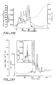
- Fig. 1 Purification of FSdGF by HSAC. gel exclusion chromatography and Mono S ion exchange chromatography
- Fig. 1 B After concentrating the 1 M NaCl HSAC bioactive fractions 126 to 133 to 1 ml in an Amicon YM10 concentrator, the ultrafiltration retentate was applied on a Bio Gel P-60 column (100-200 mesh, 1 x 95 cm) equilibrated and run at 4°C in PBS. The flow rate for development of the column was 6 ml/hr, and 1.45 ml fractions were collected. Absorbancy was monitored at 280 nm. The elution positions of molecular mass markers (in kDa) were as indicated by the arrows.
- Fig. 1 C The bioactive fractions 26 and 29 eluted from the Bio Gel P-60 column were pooled and diluted three fold with 20 mM HEPES pH 8.3. Using a 50 ml Super loop, the sample was then applied on a Mono S HR 5/5 column equilibrated in the 20 mM HEPES pH 8.3 room temperature. The column was eluted with a multilinear gradient of NaCI (0 M to 1 M) as follows: 0 M NaCI for 5 min, 0 M NaCI to 0.45 M NaCI in 45 min, 0.45 M NaCl to 1 M NaCI in 15 min, 1 M NaCl for 5 min. Absorbancy was monitored at 280 nm.
- Flow rate was 1 ml per min and 1 ml fractions were collected. Aliquots of each fraction were diluted 1 to 100 in 0.2% gelatin in PBS, and 10 ⁇ l aliquots were bioassayed on ACE cells in 12 well dishes as described in the Materials and Methods section. The histograms show the distribution of the biological activity with most of the biological activity eluting in fractions 37 to 40 (0.33 M NaCI). Fractions indicated by the asterisks were pooled and further examined by RP-HPLC using C 4 column.
- Fig. 2 Reverse phase HPLC of the Mono S purified and bioactive fractions and comparison of the ability of FS cell conditioned medium at various stages of purification to stimulate the proliferation of low density ACE cell cultures.
- Fig. 2A The active Mono S fractions (fraction 38 to 40; Fig. 3) were loaded onto a Vydac C 4 column (25 x 0.46 cm, 5 ⁇ m particle size, 300 A pore size) equilibrated in 0.1% (v/v) TFA, 20% acetonitrile. The arrows shown the times of injection. Protein was eluted with a 115 min linear gradient of 20-45% acetonitrile in 0.1% TFA at a flow rate of 0.6 ml/min, at room temperature. Fractions of 1.5 ml were collected except in the region where the bioactivity was expected to elute; in this region fraction volumes were limited manually to the size of the individual peak fractions.
- Fig. 2B Low density ACE cell cultures (5 x 10 3 cells/well) were seeded and their proliferation was measured as described in Material and Methods. Samples tested were (NH 4 ) 2 S0 4 precipitate [ ⁇ ]; pool of the HSAC 1 M NaCl fractions [ ⁇ ]; pool of the bioactive Bio Gel P-60 fractions [0]; pool of the bioactive Mono S fractions [ ⁇ ]; bioactive C 4 fraction [0]. Individual points are the mean of triplicate determinations, and standard deviations were less than 10% of the mean. Control cultures exposed to DMEM supplemented with 10% CS had a final cell density of 1.5 x 10 4 cell/well.
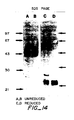
- the gel was then stained using the silver nitrate stain kit of BioRad.
- the samples were: lanes 3 and 6, 0.23 M pool; lanes 4 and 7, 0.28M pool; lanes 5 and 8, 0.33M pool.
- the molecular weight markers runs in lanes 1, 2 and 9 were: bovine serum albumin (MW 66,000), ovalbumin (MW 42,700), carbonic anhydrase (MW 31,000), soybean trypsin inhibitor (MW 21,500), and lysozyme (MW 14,500).
- Fig. 4 Comparison of the ability of pituitary derived bFGF versus FSdGF to stimulate the growth of HUE cells (A), ACE cells (B), and BHK-21 cells (C).
- Fig. 4A Low density cultures of HUE cells (5 x 10 3 cells per 1 cm diameter gelatinized well) were exposed to HEPES (25 mM) buffered medium 199 supplemented with 100 ⁇ g.ml heparin, 10- 8 M selenium, 20% FCS and increasing concentrations of either pituitary derived bFGF (0) or FSdGF(•). Heparin was added only once at the time of seeding while both bFGF and FSdGF were added every other day. After 6 days in culture, triplicate wells were trypsinized and cell counted. The final desnity of cultures exposed to 20% FCS alone was 1.5 x 10 4 cells per well. Standard deviation was less than 10% of the mean.
- Fig. 4B Low density cultures of ACE cells (5 x 10 3 cells per 1 cm diameter well) were exposed to DMEM supplemented with 10% CS and increasing concentrations of either pituitary derived bFGF (0) or FSdGF(•) added every other day. After 5 days in culture, triplicate wells were trypsinized and cell counted. The final density of cultures exposed to 10% CS alone was 1.3 x 10 4 cells per well. Standard deviation was less than 10% of the mean.
- Fig. 5 Comparison of the ability of bFGF versus FSdGF to stimulate the proliferation of BCE cells, granulosa cells, Adrenal cortex cells and BALB/MK cells.
- the reduced protein was alkylated by the addition of solid iodoacetamide to give a final concentration of 25mM and the reaction allowed to proceed for 30 minutes at room temperature.
- lysines were modified with succinic anhydride.
- Four aliquots of ⁇ l each freshly prepared 100mg/ml succinic anhydride in acetonitrile were added at five minute intervals.
- Peptide 24 gave a single sequence H-Ser-Phe-Cys-Arg-Pro-lle-Glu-Thr-Leu-Val-Asp-lle-Phe-Gln-Glu-Tyr-Pro-Asp-Glu-lle-.
- Peptide 15 gave a single sequence H-Ser-Phe-Cys-Arg-Pro-lle-Glu-Thr-Leu-Val-Asp-lle-Phe-Gln-Glu-Tyr-Pro-Asp/lle-Glu.
- Ser 1 of the peptides 24 and 25 corresponds to Ser 23 of the amino terminal sequence shown above so that these sequences can be merged to give the sequence of the first 42 aminio acids of the protein.
- FSdGF can, therefore, be used in the treatment of full-thickness wounds such as dermal ulcers, including the categories of pressure sores, venous stasis ulcers and diabetic ulcers.
- FSdGF can be used in the treatment of full-thickness burns and injuries site for a skin graft. In this case, the FSdGF is either applied directly to the site or is used to soak the skin that is being transplanted prior to grafting. In a similar fashion, FSdGF will be used in plastic surgery when the reconconstruction is required following a burn, other trauma or for the cosmetic purposes.
- FSdGF In the cases where FSdGF is being used for topical wound healing, as described above, it may be administered by any of the routes described below for the reendothelialization of vascular tissue, or more preferably by a topical means. In these cases, it will be administered as either a solution, gel, cream, ointment or as a dry powder directly to the site of injury. Slow release devices direction FSdGF to the injured site will also be used. In topical applications, FSdGF will be applied at concentration ranging from 50 to 1,000 ⁇ g/ml either in a single application, or in dosing regimens that are daily or every few days for a period of one to several weeks.
- FSdGF can be used as a post-operative wound healing agent in balloon angioplasty a procedure in which vascular endothelial cells are removed, together with atherosclerotic plaques.
- FSdGF can be applied to vasuclar endothelial surfaces by systemic or local intravenous application either as intravenous bolus injection or infusions. If desired, the FSdGF can be administered over time using a micrometering pump.
- Suitable compositions for intravenous administration comprise FSdGF in an amount effective to promote endothelial cell growth and a parenteral carrier material.
- FSdGF may also be used to promote endothelialization in vascular graft surgery.
- FSdGF can be applied to the surfaces of the graft and/or at the junctions of the graft and the existing vasculature in order to promote the growth of vascular endothelial cells.
- the FSdGF may be applied intravenously as described above for balloon angioplasty or it may be applied directly to the surfaces of the graft and/or the existing vasculature either before or during surgery.
- Suitable carrier materials include, for example, 1-5% carbopol.
- the FSdGF may be present in the carrier over a wide range of concentrations, for example, from about 50 ⁇ g/mg to about 1000 ⁇ g/mg.
- FSdGF may also be employed to repair vascular damage following myocardial infarction.
- the FSdGF is administered intravenously for this purpose, either in individual injections or by micrometering pump over a period of time as described above.
- FSdGF may also be used as a growth factor for the in vitro culturing of endothelial cells.
- FSdGF can be added to the cell culture medium at a concentration from about 10 ng/ml to about 10 ⁇ g/ml.
- the amino acid sequence of FSdGF will be used to design synthetic oligonucleotide probes for the retrieval of the FSdGF gene. These probes will either be of a mixed sequence based on all possible genetic code choices, or will be of single sequence based on codon choice preferences and other factors. In the first instances, probes based on the amino acid sequence of bovine FSdGF will be used to screen either bovine cDNA libraries made from folliculostellate cells, or bovine genomic libraries. Bovine DNA clones encoding FSdGF thus isolated will be sequenced to determine the complete coding and hence amino acid sequence of bovine FSdGF.
- bovine FSdGF clones will then be used as probes to isolate human FSdGF sequences from either cDNA libraries generated from tissues shown to express the factor, or from human genomic libraries. In this way, the complete nucleotide and hence amino acid sequence of human FSdGF can be established.
- VEGF heparin-binding endothelial cell growth factor
- the media conditioned by FC was found to stimulate the proliferation rate of low-density microvascular endothelial cells.
- Table 2 summarizes the steps for the purification of the growth promoting activity and the corresponding yield in bioactivity.
- the mitogenic activity was precipitated by 50% ammonium sulfate and resuspended to a volume suitable for subsequent purification.
- the H-S step provided an efficient way of further concentrating such activity and also provided a ten fold purification.
- Approximately 90% of the biological activity was eluted in the presence of 0.9 M NaCI (Fig. 8). The bioactivity was not affected heating the fractions at 65° C for 5 min and was decreased 25-30% following the exposure to 0.1% TFA (pH 2) for two hours.
- the most bioactive HS fractions was applied to a semi preparative C4 reversed phase HPLC column, a method suitable for rapid purification of proteins and peptides.
- the bioactivity was eluted as a single peak in the presence of about 29% acetonitrile (Fig. 9A).
- a silver-stained (56) SDS/PAGE gel on the most bioactive fractions revealed the presence of three or four bands.
- These fractions were further purified by a second reversed phase HPLC step, using an analytical C4 column which was eluted with a gradient of 2-propanol, instead of acetonitrile.
- a single peak of bioactivity corresponding to a distinct peak in the absorption profile was obtained. (Fig. 9B).
- the peak fractions from the second reversed phase step displayed a single band on a silver stained SDS/PAGE, with an apparent M r of about 23 kDa under reducing conditions (Fig. 10).
- the intensity of staining of the band was highly correlated to the mitogenic activity across the bioactivity profile. Because previous experiments, using a molecular sieve with a TSK G 3000 SW column, suggested a M r in the range of 40-43 kDa, the possibility that the growth factor in native conditions is a dimer was considered. This was strongly suggested by the finding that the purified material had an apparent M r of about 45 kDa in a silver stained SDS-PAGE under non-reducing conditions (see Fig. 10).
- the dose response curve for the purified growth factor revealed a half maximal effect on adrenal cortex-derived capillary endothelial cells proliferation at 150-200 pg/ml and a maximal effect at 1-1.5 ng/ml. These values were derived from protein sequencing and were found to be in good agreement with those obtained by comparing the relative intensitieis of bands with standards in silver stained SDS/PAGE.
- the growth factor was heat and acid stable and its p. I., as estimated by chromatofocusing on a Mono P column is about 8.5.
- the purified growth factor was able to stimulate the proliferation of vascular endothelial cells at concentrations between 25 pg and 1-1.5 ng/ml. These values, assuming a M r of 45 kDa, correspond respectively to 0.55 pM and 22-33 pM. Such values are in the same range as those obtained with bFGF (2,56).
- the novel growth factor did not induce any appreciable mitogenic effect on corneal endothelial cells, lens epithelial cells, BHK-21 fibroblasts, adrenal cortex cells, or keratynocytes.
- bFGF and aFGF are both potent mitogens for all of these cell types (2,56).
- VEGF vascular endothelial growth factor
- VEGF vascular endothelial growth factor
- VEGF vascular endothelial growth factor
- a soluble endothelial cell growth factor such as VEGF may play a more dynamic role in the physiological regulation of the vascular endothelial cells proliferation, either in the cyclical growth of blood vessels which takes place in organs such as the corpus luteum (62) or in the tonic maintenace of the differentiated stage of the endothelium in the vascular tree.
- Fiaure 11 Dose-responsive growth of adrenal cortex derived capillary endothelial cells in the presence of purified VEGF. Cells were seeded at the density of 1 x 10 4 /well in 12 well plates. The indicated amounts of VEGF were added a few hours after plating in 5 ⁇ l/ml aliquots. After five days, cells were counted in a Coulter counter. The results shown represent mean values of three separate experiments conducted in duplicate. Duplicates in each experiment varied less than 10%.
- Angiogenesis is a multi-step phenomenon which involves capillary endothelial cell profileration, migration and tissue infiltration(1). It plays a central role in a variety of physiological and pathological process such as embryonic development, wound healing, atherosclerosis and tumor growth (1,2). Several factors that induce angiogenesis have recently been isolated and characterized. Of these, only the basic and acidic forms of FGF have been shown to directly control all steps of angiogenesis including vascular endothelial cell proliferation, migration and increased expression of plasminogen activator and collagenase activity(2).
- FGF angiogenic
- two puzzling questions point to the existence of other angiogenic factors which could complement the action of FGF.
- FGF lacks the hydrophobic signal sequences that govern secretion(12,13), yet for acceptance as an angiogenic factor any putative mediator should be shown to be a diffusible substance which induces new capillary formation from a microcirculatory bed.
- FGF is produced by endothelial cells themselves(14, 15). If FGF is present in and around endothelial cells yet the cells are quiescent, other factors must come into play to trigger angiogenesis.
- the present invention also relates to the isolation and characterization of a new endothelial cell mitogen produced and secreted by AtT20 cells.
- This mouse cell line is available from The America Type Culture Collection, 12301 Park- lawn Drive, Rockville, Maryland.
- the factor produced by AtT20 cells has a unique target cell specificity, since it stimulates only vascular endothelial cells to proliferate and does not affect other cell types sensitive to FGF.
- the endothelial cell mitogen present in the conditioned medium was then purified by a combination of steps including heparin sepharose affinity chromatography (HSAC), exclusion gel chromatography on Sephadex G 100, cation exchange on chromatography on Mono S resin, and finally, by PR-HPLC with a C4 Vydac column (see Table 3). Fractions were assayed for bioactivity using adrenal cortex derived capillary endothelial cells. When the collected material was applied on HS under the conditions described in Fig. 13A the activity eluted in a broad peak in the range of 0.5 to 0.6M. In order to concentrate it, elution with 0.8 M NaCI was carried.
- a small amount of contaminant with a MW of 27 kDa whose migration was not affected by reduction was also present. This contaminant did not amount to more than 5% of the total.
- a single peak of protein with a small shoulder was obtained (Fig. 13D).
- Microsequencing of this material reveals a unique terminal amino acid sequence. Approximately 2 ⁇ g (80 pmole) of protein was sequenced using an Applied Biosystems 477A gas phase protein sequenator. A single amino acid was identified in each of the first 24 cycles consistent with a homogeneous protein. The yield of the amino terminal residue was 30 pmole. Unambiguous assignment modes for cycles 1 to 5 were as follows: Ala-Pro-Met-Ala-Glu. A longer amino acid sequence for the growth factor from AtT-20 was later determined and is described below.
- the dose response curve for the growth factor depicted in Fig. 15B illustrates that as little as 50 pg/ml stimulates ACE proliferation. Saturation was observed at 1 ng/ml with an ED 500 f 150 pg/ml. These values compared favorably with the range of concentrations where bFGF promotes the proliferation of ACE cells (minimal effect at about 10 pg/ml saturation at about 200 pg/ml and ED 50 at 50 pg/ml,(22) and Fig. 15).
- growth factor MW 45kDa, basic pl, affinity for HS
- biological properties mitogenic for vascular endothelial cells
- EGF EGF, TGFa, PDGF, TGFB or the recently reported keratinocyte growth factor(21).
- EGF EGF, TGFa, PDGF, TGFB
- keratinocyte growth factor(21) EGF, TGFa, PDGF, TGFB
- PDECGF a single chain polypeptide while the AtT20 growth factor has a dimeric structure.
- AtT20 derived growth factor represents a previously unknown growth factor.
- this novel growth factor is mitogenic for capillary endothelial cells, it is not yet known whether it can stimulate other events linked to angiogenesis. These include chemotaxis of capillary endothelial cells and activation of the synthesis of cellular enzymes such as collagenase and plasminogen activator which are involved in the breakdown of capillary basement membrane(3).
- the name of vasculotropin is suggested for this novel growth factor.
- An advantage of culturing the bovine cells to collect the growth factor is that the overall cell structure has good cohesive integrity That is to say, the cell layer usually remains intact for collecting the conditioning medium for many days, and even over a month or more of successive collection of the media samples.
- the dome structure it is possible to collect samples from above the dome and also from within or below the dome. Normally, the growth factor is in a higher concentration within the dome.
- a disadvantage observed during the culturing of the bovine cells is that these cells produce a large amount of different proteins, which are secreted from the cell. Thus the purification steps need to be able to remove more of the undesired protein.
- AtT-20 cell line is that it is commercially available.
- An advantage of the culturing of the murine cells, AtT-20, is that these cells produce the novel growth factor without producing a large amount of other proteins which might interfere with the subsequent isolation steps.
- a disadvantage of culturing the murine cells AtT-20 is that the structural integrity of the cell culture layer is not high. Thus during the culturing and collection, small portions of the cell layer will break away float in the conditioning media and then die. Often the culturing of the murine cells is only possible up to about 7 days or slightly longer.
- novel growth factor from bovine sources is often of sufficient purity after the mono S step and a Sephadex G-100 step that the RP HPCL-C4 purification is not necessary RP HPLC can be sued to determine the purity of the FsdGF.
- the N-terminal sequence was determined for the growth factor protein derived from mouse AtT20 cells. Two N-terminal sequencing runs were carried out on the protein in an Applied Biosystems gas-phase protein sequencer. The second run, the sequencer was loaded with approximately 2.5 times as much of the protein as the first run.
- Tissue culture media and reagents were obtained from Gibco (Grand Island, N.Y). Acetonitrile and 2-propanol were purchased from Fisher Sci. (Fair Lawn, NJ). Heparin-sepharose (H-S) was obtained from Pharmacia (Piscataway, N.J.). Vydac HPLC columns were from The Separation Group (Hesperia, CA). Molecular weight markers for PAGE and protein determination kit were from Bio Rad Labs (Richmond, CA). Tissue culture plates were purchased from Costar, except for large scale Nunc plates (24.5 x 24.5 cm), which were Applied Sci. (San Francisco, CA). All other reagents were from Sigma Chemical Co. (St. Louis, MO) or Applied Biosystems (Foster City, CA).
- follicular cells were incubated for three days in a serum-free medium consisting of low glucose Dulbecco's modified Eagle's medium supplemented with transferrin (10 ⁇ l/ml), insulin (5 ⁇ g/ml), 2 mM glutamine and antibiotics.
- the conditioned medium (CM) 150 ml was then collected centrifuged (10000 xg, 15 min. 4°C) in order to remove cell debris, and then applied to a Heparin-Sepharose column which had been preequilibrated with 10 mM Tris/Cl, pH 7.0. The column was then sequentially eluted with 10 mM Tris/Cl, pH 7.0 containing 0.6, 1 and 3 M NaCI.
- the flow rate was 21 ml/h. Fractions of 700 pi were collected and aliquots were tested for bioactivity on adrenal cortex-derived microvascular endothelial cells. The majority of the bioactivity was eluted in the present of 0.6 M NaCI. This chromatographic behavior is different from that of aFGF or bFGF, which are known to elute, respectively, in the presence of 0.9-1.1 M NaCI and 1.8-2.2 M NaCI.
- the most bioactive 0.6 NaCI fractions were pooled and further examined for determination of the molecular weight of growth factor activity
- a standard 12.5% polyacrilamide SDS slab gel was prepared. Ten percent glycerol and 2% SDS were added to the pooled fractions. Fifty per cent of the sample was treated with 2.5% 2-mercaptoethanol. The remainder 50% was not exposed to 2-mercaptoethanol or other reducing agents.
- the samples and prestained molecular weight markers were then incubated for 3 min at 37°C and electrophoresed overnight at a current of 10 mA. When the electrophoresis was completed, the gel was briefly rinsed in PBS and the distance of the molecular weight markers from the top of the gel was immediately meaured.
- bovine pituitary FC Primary cultures of bovine pituitary FC were established as previously described (20,41). In one embodiment in the culturing the 20% fetal bovine serum in reference 20 was reduced to 10%. Concentrations of 5 to 20% should be effective. Also no DNAase is used. All other components are the same. At confluency, cells were passaged into large scale tissue culture plates in the presence of low glucose Dulbecco's modified Eagle's medium (DMEM) supplemented with 10% fetal bovine serum, 2 mM glutamine and antibiotics. Shortly after reaching confluency the cultures were extensively washed with PBS in order to remove serum components.
- DMEM low glucose Dulbecco's modified Eagle's medium
- CM conditioned medium
- Ammonium sulfate 500 g/L was added under constant stirring, until the salt was completely in solution. After 8-12 hours in the cold room, the material was centrifuged (20,000 xg, 45 min at 4°C). The supernatant was discarded and the pellet was resuspended with 10 mM Tris/CI, pH 7.2, 50 mM NaCI and dialyzed at 4°C against the same buffer for 8-12 h. The final volume was 50-60 fold less than the original.
- the CM is concentrated using ultrafiltration using an Amecon stir cell (2.5 liter unit) using a membrane having a molecular weight cut off of 10,000 daltons with similar results.
- the concentrated CM was applied to a H-S column (14) (10 ml) preequilibrated with 10 mM Tris/Cl, pH 7.2, 50 mM NaCI. The column was then washed with the same buffer until the absorbance at 280 nm was negligible and then eluted stepwise with 10mM Tris/Cl pH 7.2 containing 0.15, 0.9 nd 3 M NaCI. The flow rate was 1.5 ml/min. Fractions of 1.5 ml were collected and aliquots, diluted with 0.2 % gelatin in PBS, were tested for mitogenic activity on endothelial cells.
- Bovine adrenal cortex or brain-derived capillary endothelial cells, adult or fetal bovine aortic endothelial cells, human umbilical vein endothelial cells, bovine corneal endothelial cells, adrenal cortex cells, lens epithelial cells, BHK-21 fibroblasts and human keratynocytes were cultured and maintained as previously described (17,47,48,50,51,52,26,53).
- cells were seeded in the presence of their respective growth media at the density of 2 x 10 4 /35 mm dish or 1 x 10 4 /well in 12 multiwell plates. Fractions were added to cells in 5 ⁇ l/ml aliquots. After 4 or 5 days, cells were dissociated by exposure to trypsin and counted in a Coulter counter.
- the growth factor described herein may be either the dimer (43,000 to 46,000 kDa) or the monomer (about 23 kDa).
Landscapes
- Health & Medical Sciences (AREA)
- Chemical & Material Sciences (AREA)
- Life Sciences & Earth Sciences (AREA)
- Organic Chemistry (AREA)
- Medicinal Chemistry (AREA)
- General Health & Medical Sciences (AREA)
- Veterinary Medicine (AREA)
- Nuclear Medicine, Radiotherapy & Molecular Imaging (AREA)
- General Chemical & Material Sciences (AREA)
- Pharmacology & Pharmacy (AREA)
- Chemical Kinetics & Catalysis (AREA)
- Animal Behavior & Ethology (AREA)
- Bioinformatics & Cheminformatics (AREA)
- Public Health (AREA)
- Engineering & Computer Science (AREA)
- Gastroenterology & Hepatology (AREA)
- Proteomics, Peptides & Aminoacids (AREA)
- Toxicology (AREA)
- Biochemistry (AREA)
- Biophysics (AREA)
- Genetics & Genomics (AREA)
- Molecular Biology (AREA)
- Zoology (AREA)
- Dermatology (AREA)
- Medicines That Contain Protein Lipid Enzymes And Other Medicines (AREA)
- Peptides Or Proteins (AREA)
- Preparation Of Compounds By Using Micro-Organisms (AREA)
- Micro-Organisms Or Cultivation Processes Thereof (AREA)
- Materials For Medical Uses (AREA)
Claims (42)
Applications Claiming Priority (7)
| Application Number | Priority Date | Filing Date | Title |
|---|---|---|---|
| US32818189A | 1989-03-24 | 1989-03-24 | |
| US34616589A | 1989-05-02 | 1989-05-02 | |
| US346165 | 1989-05-02 | ||
| US36023589A | 1989-06-01 | 1989-06-01 | |
| US360235 | 1989-06-01 | ||
| PCT/US1990/001568 WO1990011084A1 (fr) | 1989-03-24 | 1990-03-22 | Facteur de croissance cellulaire endotheliale, son isolation et son expression |
| US328181 | 1994-10-24 |
Publications (3)
| Publication Number | Publication Date |
|---|---|
| EP0464155A1 EP0464155A1 (fr) | 1992-01-08 |
| EP0464155A4 EP0464155A4 (en) | 1992-05-06 |
| EP0464155B1 true EP0464155B1 (fr) | 1997-05-07 |
Family
ID=27406587
Family Applications (1)
| Application Number | Title | Priority Date | Filing Date |
|---|---|---|---|
| EP90907823A Expired - Lifetime EP0464155B1 (fr) | 1989-03-24 | 1990-03-22 | Facteur de croissance cellulaire endotheliale, son isolation et son expression |
Country Status (12)
| Country | Link |
|---|---|
| EP (1) | EP0464155B1 (fr) |
| JP (1) | JP3549202B2 (fr) |
| AT (1) | ATE152626T1 (fr) |
| AU (1) | AU647737B2 (fr) |
| CA (1) | CA2050318C (fr) |
| DE (1) | DE69030660T2 (fr) |
| DK (1) | DK0464155T3 (fr) |
| ES (1) | ES2103740T3 (fr) |
| IL (1) | IL93846A0 (fr) |
| NZ (1) | NZ233067A (fr) |
| PT (1) | PT93560B (fr) |
| WO (1) | WO1990011084A1 (fr) |
Cited By (1)
| Publication number | Priority date | Publication date | Assignee | Title |
|---|---|---|---|---|
| US7030083B2 (en) | 1998-09-09 | 2006-04-18 | University Of Washington | Treatment of eclampsia and preeclampsia |
Families Citing this family (9)
| Publication number | Priority date | Publication date | Assignee | Title |
|---|---|---|---|---|
| US5332671A (en) * | 1989-05-12 | 1994-07-26 | Genetech, Inc. | Production of vascular endothelial cell growth factor and DNA encoding same |
| US7300791B2 (en) | 1989-05-12 | 2007-11-27 | Genentech, Inc. | Production of vascular endothelial cell growth factor and DNA encoding same |
| US5194596A (en) * | 1989-07-27 | 1993-03-16 | California Biotechnology Inc. | Production of vascular endothelial cell growth factor |
| DK0666868T4 (da) * | 1992-10-28 | 2006-09-18 | Genentech Inc | Anvendelse af anti-VEGF-antistoffer til behandling af cancer |
| CN1173991C (zh) * | 1992-11-13 | 2004-11-03 | 马克斯普朗克科学促进协会 | 作为血管内皮生长因子受体的f1k-1 |
| DK1112083T3 (da) | 1998-09-09 | 2004-03-08 | Univ Washington | Behandling af mikrovaskulære angiopatier |
| US6783953B1 (en) | 1998-12-22 | 2004-08-31 | Janssen Pharmaceutica N.V. | Vascular endothelial growth factor-X |
| US7067317B2 (en) | 2000-12-07 | 2006-06-27 | Sangamo Biosciences, Inc. | Regulation of angiogenesis with zinc finger proteins |
| AU2002228841C1 (en) | 2000-12-07 | 2006-11-23 | Sangamo Biosciences, Inc | Regulation of angiogenesis with zinc finger proteins |
Family Cites Families (6)
| Publication number | Priority date | Publication date | Assignee | Title |
|---|---|---|---|---|
| US4456550A (en) * | 1982-11-22 | 1984-06-26 | President And Fellows Of Harvard College | Vascular permeability factor |
| US5008196A (en) * | 1987-08-21 | 1991-04-16 | Monsanto Company | Stimulation of endothelial cell growth |
| US5240848A (en) * | 1988-11-21 | 1993-08-31 | Monsanto Company | Dna sequences encoding human vascular permeability factor having 189 amino acids |
| US5332671A (en) * | 1989-05-12 | 1994-07-26 | Genetech, Inc. | Production of vascular endothelial cell growth factor and DNA encoding same |
| EP0399816B1 (fr) * | 1989-05-24 | 1995-12-20 | Merck & Co. Inc. | Purification et caractérisation d'un facteur de croissance dérivé du rétinoblastane |
| US5194596A (en) * | 1989-07-27 | 1993-03-16 | California Biotechnology Inc. | Production of vascular endothelial cell growth factor |
-
1990
- 1990-03-22 AU AU55659/90A patent/AU647737B2/en not_active Expired
- 1990-03-22 DE DE69030660T patent/DE69030660T2/de not_active Expired - Lifetime
- 1990-03-22 DK DK90907823.0T patent/DK0464155T3/da active
- 1990-03-22 WO PCT/US1990/001568 patent/WO1990011084A1/fr active IP Right Grant
- 1990-03-22 AT AT90907823T patent/ATE152626T1/de not_active IP Right Cessation
- 1990-03-22 IL IL93846A patent/IL93846A0/xx not_active IP Right Cessation
- 1990-03-22 CA CA2050318A patent/CA2050318C/fr not_active Expired - Lifetime
- 1990-03-22 JP JP50705690A patent/JP3549202B2/ja not_active Expired - Lifetime
- 1990-03-22 EP EP90907823A patent/EP0464155B1/fr not_active Expired - Lifetime
- 1990-03-22 ES ES90907823T patent/ES2103740T3/es not_active Expired - Lifetime
- 1990-03-23 PT PT93560A patent/PT93560B/pt not_active IP Right Cessation
- 1990-03-23 NZ NZ233067A patent/NZ233067A/xx unknown
Cited By (1)
| Publication number | Priority date | Publication date | Assignee | Title |
|---|---|---|---|---|
| US7030083B2 (en) | 1998-09-09 | 2006-04-18 | University Of Washington | Treatment of eclampsia and preeclampsia |
Also Published As
| Publication number | Publication date |
|---|---|
| EP0464155A1 (fr) | 1992-01-08 |
| AU647737B2 (en) | 1994-03-31 |
| ATE152626T1 (de) | 1997-05-15 |
| DE69030660T2 (de) | 1997-09-25 |
| JP3549202B2 (ja) | 2004-08-04 |
| CA2050318A1 (fr) | 1990-09-25 |
| JPH04504262A (ja) | 1992-07-30 |
| ES2103740T3 (es) | 1997-10-01 |
| PT93560B (pt) | 1996-08-30 |
| DE69030660D1 (de) | 1997-06-12 |
| EP0464155A4 (en) | 1992-05-06 |
| AU5565990A (en) | 1990-10-22 |
| CA2050318C (fr) | 2011-04-12 |
| NZ233067A (en) | 1993-05-26 |
| PT93560A (pt) | 1990-11-07 |
| WO1990011084A1 (fr) | 1990-10-04 |
| IL93846A0 (en) | 1990-12-23 |
| DK0464155T3 (da) | 1997-11-17 |
Similar Documents
| Publication | Publication Date | Title |
|---|---|---|
| US20060025577A1 (en) | Endothelial cell growth factor methods of isolation and expression | |
| Ferrara et al. | Pituitary follicular cells secrete a novel heparin-binding growth factor specific for vascular endothelial cells | |
| Böhlen et al. | Acidic fibroblast growth factor (FGF) from bovine brain: amino‐terminal sequence and comparison with basic FGF. | |
| Vlodavsky et al. | Aortic endothelial cells synthesize basic fibroblast growth factor which remains cell associated and platelet‐derived growth factor‐like protein which is secreted | |
| Gospodarowicz et al. | Isolation and characterization of a vascular endothelial cell mitogen produced by pituitary-derived folliculo stellate cells. | |
| US6689580B1 (en) | Growth factor | |
| JP3117992B2 (ja) | 血管内皮細胞成長因子の生産およびそれをコードするdna | |
| US5633147A (en) | Transforming growth factor αH1 | |
| US5811393A (en) | Heparin binding mitogen with homology to epidermal growth factor (EGF) | |
| Muramatsu et al. | Localization of heparin-binding, neurite outgrowth and antigenic regions in midkine molecule | |
| Favard et al. | Purification and biological properties of vasculotropin, a new angiogenic cytokine | |
| NL8900178A (nl) | Groei regulerende clycoproteinen. | |
| JP3159216B2 (ja) | 白血球由来の成長因子 | |
| EP0464155B1 (fr) | Facteur de croissance cellulaire endotheliale, son isolation et son expression | |
| EP0461560B1 (fr) | Facteur recombinant humain de croissance d'hépatocyte et son procédé de préparation | |
| US5227302A (en) | DNA encoding platelet derived endothelial cell growth factor (PD-ECGF) | |
| WO1996018730A1 (fr) | Facteur de croissance prostatique | |
| Miyazono et al. | Platelet-derived endothelial cell growth factor | |
| EP0377855B1 (fr) | Facteur de croissance des cellules endothéliales | |
| Ueno et al. | Purification and partial characterization of a mitogenic factor from bovine liver: structural homology with basic fibroblast growth factor | |
| US5756686A (en) | Peptides derived from endothelial cell growth factor | |
| EP2275437A1 (fr) | Polypeptide et composition pharmaceutique contenant le polypeptide | |
| US6235884B1 (en) | Heparin binding mitogen with homology to epidermal growth factor (EGF) | |
| JP3375997B2 (ja) | 血管内皮細胞増殖促進剤 | |
| AU656453B2 (en) | Thrombin-binding substance and process for preparing the same |
Legal Events
| Date | Code | Title | Description |
|---|---|---|---|
| PUAI | Public reference made under article 153(3) epc to a published international application that has entered the european phase |
Free format text: ORIGINAL CODE: 0009012 |
|
| 17P | Request for examination filed |
Effective date: 19911015 |
|
| AK | Designated contracting states |
Kind code of ref document: A1 Designated state(s): AT BE CH DE DK ES FR GB IT LI LU NL SE |
|
| A4 | Supplementary search report drawn up and despatched |
Effective date: 19920318 |
|
| AK | Designated contracting states |
Kind code of ref document: A4 Designated state(s): AT BE CH DE DK ES FR GB IT LI LU NL SE |
|
| 17Q | First examination report despatched |
Effective date: 19940811 |
|
| GRAG | Despatch of communication of intention to grant |
Free format text: ORIGINAL CODE: EPIDOS AGRA |
|
| GRAH | Despatch of communication of intention to grant a patent |
Free format text: ORIGINAL CODE: EPIDOS IGRA |
|
| GRAH | Despatch of communication of intention to grant a patent |
Free format text: ORIGINAL CODE: EPIDOS IGRA |
|
| GRAA | (expected) grant |
Free format text: ORIGINAL CODE: 0009210 |
|
| AK | Designated contracting states |
Kind code of ref document: B1 Designated state(s): AT BE CH DE DK ES FR GB IT LI LU NL SE |
|
| REF | Corresponds to: |
Ref document number: 152626 Country of ref document: AT Date of ref document: 19970515 Kind code of ref document: T |
|
| REG | Reference to a national code |
Ref country code: CH Ref legal event code: EP |
|
| REF | Corresponds to: |
Ref document number: 69030660 Country of ref document: DE Date of ref document: 19970612 |
|
| ET | Fr: translation filed | ||
| REG | Reference to a national code |
Ref country code: ES Ref legal event code: FG2A Ref document number: 2103740 Country of ref document: ES Kind code of ref document: T3 |
|
| REG | Reference to a national code |
Ref country code: DK Ref legal event code: T3 |
|
| PLBE | No opposition filed within time limit |
Free format text: ORIGINAL CODE: 0009261 |
|
| STAA | Information on the status of an ep patent application or granted ep patent |
Free format text: STATUS: NO OPPOSITION FILED WITHIN TIME LIMIT |
|
| 26N | No opposition filed | ||
| REG | Reference to a national code |
Ref country code: GB Ref legal event code: IF02 |
|
| PGFP | Annual fee paid to national office [announced via postgrant information from national office to epo] |
Ref country code: AT Payment date: 20090304 Year of fee payment: 20 Ref country code: ES Payment date: 20090326 Year of fee payment: 20 Ref country code: DK Payment date: 20090325 Year of fee payment: 20 |
|
| PGFP | Annual fee paid to national office [announced via postgrant information from national office to epo] |
Ref country code: NL Payment date: 20090324 Year of fee payment: 20 |
|
| PGFP | Annual fee paid to national office [announced via postgrant information from national office to epo] |
Ref country code: CH Payment date: 20090325 Year of fee payment: 20 |
|
| PGFP | Annual fee paid to national office [announced via postgrant information from national office to epo] |
Ref country code: DE Payment date: 20090327 Year of fee payment: 20 Ref country code: SE Payment date: 20090327 Year of fee payment: 20 Ref country code: LU Payment date: 20090401 Year of fee payment: 20 Ref country code: IT Payment date: 20090331 Year of fee payment: 20 |
|
| PGFP | Annual fee paid to national office [announced via postgrant information from national office to epo] |
Ref country code: BE Payment date: 20090430 Year of fee payment: 20 |
|
| PGFP | Annual fee paid to national office [announced via postgrant information from national office to epo] |
Ref country code: FR Payment date: 20090317 Year of fee payment: 20 |
|
| PGFP | Annual fee paid to national office [announced via postgrant information from national office to epo] |
Ref country code: GB Payment date: 20090403 Year of fee payment: 20 |
|
| BE20 | Be: patent expired |
Owner name: THE *REGENTS OF THE UNIVERSITY OF CALIFORNIA Effective date: 20100322 |
|
| REG | Reference to a national code |
Ref country code: CH Ref legal event code: PL Ref country code: NL Ref legal event code: V4 Effective date: 20100322 |
|
| REG | Reference to a national code |
Ref country code: DK Ref legal event code: EUP |
|
| REG | Reference to a national code |
Ref country code: GB Ref legal event code: PE20 Expiry date: 20100321 |
|
| EUG | Se: european patent has lapsed | ||
| REG | Reference to a national code |
Ref country code: ES Ref legal event code: FD2A Effective date: 20100323 |
|
| PG25 | Lapsed in a contracting state [announced via postgrant information from national office to epo] |
Ref country code: GB Free format text: LAPSE BECAUSE OF EXPIRATION OF PROTECTION Effective date: 20100321 |
|
| PG25 | Lapsed in a contracting state [announced via postgrant information from national office to epo] |
Ref country code: NL Free format text: LAPSE BECAUSE OF EXPIRATION OF PROTECTION Effective date: 20100322 Ref country code: ES Free format text: LAPSE BECAUSE OF EXPIRATION OF PROTECTION Effective date: 20100323 |
|
| PG25 | Lapsed in a contracting state [announced via postgrant information from national office to epo] |
Ref country code: DE Free format text: LAPSE BECAUSE OF EXPIRATION OF PROTECTION Effective date: 20100322 |