EP0369176A2 - Essai immunologique utilisant de spectroscopie photo-acoustique - Google Patents
Essai immunologique utilisant de spectroscopie photo-acoustique Download PDFInfo
- Publication number
- EP0369176A2 EP0369176A2 EP89119250A EP89119250A EP0369176A2 EP 0369176 A2 EP0369176 A2 EP 0369176A2 EP 89119250 A EP89119250 A EP 89119250A EP 89119250 A EP89119250 A EP 89119250A EP 0369176 A2 EP0369176 A2 EP 0369176A2
- Authority
- EP
- European Patent Office
- Prior art keywords
- solid phase
- antibody
- antigen
- photoacoustic
- labeled
- Prior art date
- Legal status (The legal status is an assumption and is not a legal conclusion. Google has not performed a legal analysis and makes no representation as to the accuracy of the status listed.)
- Withdrawn
Links
Images

Classifications
-
- G—PHYSICS
- G01—MEASURING; TESTING
- G01N—INVESTIGATING OR ANALYSING MATERIALS BY DETERMINING THEIR CHEMICAL OR PHYSICAL PROPERTIES
- G01N21/00—Investigating or analysing materials by the use of optical means, i.e. using sub-millimetre waves, infrared, visible or ultraviolet light
- G01N21/17—Systems in which incident light is modified in accordance with the properties of the material investigated
- G01N21/1702—Systems in which incident light is modified in accordance with the properties of the material investigated with opto-acoustic detection, e.g. for gases or analysing solids
-
- G—PHYSICS
- G01—MEASURING; TESTING
- G01N—INVESTIGATING OR ANALYSING MATERIALS BY DETERMINING THEIR CHEMICAL OR PHYSICAL PROPERTIES
- G01N33/00—Investigating or analysing materials by specific methods not covered by groups G01N1/00 - G01N31/00
- G01N33/48—Biological material, e.g. blood, urine; Haemocytometers
- G01N33/50—Chemical analysis of biological material, e.g. blood, urine; Testing involving biospecific ligand binding methods; Immunological testing
- G01N33/53—Immunoassay; Biospecific binding assay; Materials therefor
- G01N33/543—Immunoassay; Biospecific binding assay; Materials therefor with an insoluble carrier for immobilising immunochemicals
- G01N33/54366—Apparatus specially adapted for solid-phase testing
- G01N33/54373—Apparatus specially adapted for solid-phase testing involving physiochemical end-point determination, e.g. wave-guides, FETS, gratings
Definitions
- the present invention relates to an immunoassay method and more particularly, to a method for immunoassay which is suitable for analyzing and determining the immune component in a sample utilizing a photoacoustic spectroscopy.
- the sensitivity of a photoacoustic spectrometer is rendered highly sensitive to the particulate substance to be analyzed, using an exited light having a wavelength either identical with or similar to the size of the particulate substance to be analyzed, whereby the particulate substance is selectively detected and quantitatively determined.
- An invention of U.S. Serial No. 283,814 filed December 13, 1988 also relates to immunoassay using photoacoustic spectroscopy.
- an immune reaction is caused within a reactor to form an immune complex labeled with fine particles on the solid phase.
- the fine particles are separated from the solid phase in the reactor and the dispersion of the fine particles is introduced into a measuring cell for photoacoustic spectroscopy. The fine particles in the dispersion are measured.
- the particulate antigen-antibody complex can be detected in such a state that influence by other particles is minimized.
- concentration of rheumatoid factor, cancer specific antigen, etc. is extremely low and it is thus desired to develop a method for measurement with much higher sensitivity.
- the prior invention described above is also desired to achieve measurement with much higher sensitivity.
- An object of the present invention is to provide a method for immunoassay which permits to measure an antigen or antibody contained even in a trace amount in body fluids with high sensitivity, utilizing a photoacoustic spectroscopy.
- the antigen-antibody complex is labeled with a color material and photoacoustic properties of the label are determined by photoacoustic spectroscopy.
- a color material a fluorescent substance, a dye or colored particles can be used.
- a pressure medium for photoacoustic determination is a gas.
- a substance absorbs light.
- the substance takes in light energy, and atoms or molecules constituting the substance are excited.
- the atoms or the molecules release heat energy when they return to the ground state.
- an acoustic wave is generated by the heat energy. According to the photoacoustic spectroscopy, thus generated acoustic wave is detected.
- a photoacoustic spectrometer comprises as main constitutional elements a laser light source, a chopper for intermitting a light, a photoacoustic cell, an acoustic sensor, an amplifier and a signal processor system. After light from the light source has passed through a photometer, the light is converted to intermittent light by means of the chopper to be casted on the photoacoustic cell. In the cell, a sample absorbs the intermittent light, whereby a photoacoustic signal is generated. The photoacoustic signal is detected by the acoustic sensor such as a microphone.
- a solid phase to be determined is placed in the photoacoustic cell which is sealed in such a state that gas is present, and a leak into the gas of heat generated as a result of the light absorption by a sample is detected as a periodic pressure change by means of a highly sensitive microphone.
- a pressure change (photoacoustic signal) generated when a sample is exposed to an incident light is converted into an electric signal (generation of voltage) by means of a microphone or a piezoelectric element.
- the antigen-antibody reaction can be quantitatively analyzed by a photoacoustic spectroscopy, whereby the desired antigen or antibody can be quantitatively determined.
- trace components which could only be determined from a practical viewpoint by radioimmunoassay (RIA) heretofore can be quantitatively determined rapidly with high sensitivity in a simple manner.
- RIA radioimmunoassay
- a plurality of analyses can also be determined concurrently.
- a first antibody immobilized to the solid phase is reacted with a labeled second antibody capable of specifically reacting with the first antibody.
- the immune complex labeled with a color material is formed on the surface of the solid phase together with the antibody-bound antigen contained in the sample.
- the solid phase is separated from the liquid, introduced into a measuring cell and measured by a photoacoustic spectroscopy.
- the second antibody labeled with a label is not required to be specifically reactive with the antigen to be analyzed but is sufficient to specifically react with the first antibody.
- sheep anti-rabbit IgG antibody can be used as the second antibody. For this reason, it can be avoided to use expensive antibody in large quantities. In view of reagent costs, this embodiment is thus excellent.
- AFP ⁇ -feto-protein
- the first antibody is rabbit-anti-AFP
- sheep anti-rabbit IgG antibody can be used as the second antibody. For this reason, it can be avoided to use expensive antibody in large quantities. In view of reagent costs, this embodiment is thus excellent.
- a dye, a fluorescence emitting substance, etc. which can be bound to the analyte or antibody capable of specifically reacting with the analyte.
- a dye or a fluorescent substance included in or bound or adsorbed to microspheres such as liposome; a dye or a fluorescence bound or adsorbed to a carrier such as latex particles; colorless particles or carriers which are labeled with colored latex, etc. may also be used as the label.
- These substances are often referred to as color materials hereinafter.
- the solid phase preferably takes a portable structure formed into a film or plate having a surface capable of holding liquid therein, like a microplate which becomes a little bit hollow; a film made of synthetic resin such as nylon, polyester, etc.; a container packed with a filler such as agarose, Sepharose, silica gel, etc.
- a filler such as agarose, Sepharose, silica gel, etc.
- glass pieces or paper which can be immersed in liquid, and the like may also be used.
- TLC thin layer chromatography
- a substance present around the surface can be measured with relatively high sensitivity. This is because, when a photoacoustic signal is detected by means of a microphone using as a pressure medium, e.g., gas, a substance such as an adsorbed matter, etc. present around the surface of the solid phase can be detected with high sensitivity, since only the region around the surface of the solid sample contributes to the signal. Where solid phase itself which is a substrate substantially has no absorption and has a larger reflectivity, this tendency is more remarkable. In TLC, silica gel, alumina or the like is often generally used as the adsorbent.
- the adsorbed matter i.e., the label is colored so that S/N can be greatly improved.
- the present invention permits to measure with sensitivity as high as two figures or more in such a state that the label is adsorbed on the reacted solid phase.
- a ratio of signal to background noises (S/N) for photoacoustic measurement is improved.
- the solid phase on which the antigen-antibody complex labeled with the color material is adsorbed is moved to the position of a spectrometer for exposure to light.
- the color material present on the surface of the solid phase absorbs the light and releases heat energy in response to the light absorbed.
- the greater the quantity of light absorbed the greater the photoacoustic signal obtained.
- a color of the label it is chosen to have the absorption maximum substantially in compliance with the central wavelength of the excited monochromatic light. For example, when an argon laser having a wavelength of 488 nm is used as the excited light, Food Yellow 3 having the absorption maximum wavelength of 482 nm is used as a dye for the label; in this case, good results are obtained.
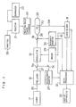
- FIG. 1 An outlined construction of an analytical apparatus for practicing one embodiment of the present invention is shown in Fig. 1.
- Light source for excitation 17 is a light source for argon laser having an output of 10W.
- Laser light 25 from argon laser light source has oscillation rays in several wavelengths at 488 nm, 514.5 nm and over the ultraviolet to visible regions.
- an appropriate dye in dye laser 18 is subjected to pumping.
- the wavelength of the excited light 25 from the argon laser light source 17 is set at 514.5 nm and rhodamine is used as the dye for the dye laser 18.
- the laser light 27, its wavelength being modified in response to the size of the analyte, is in part divided by half mirror 5a.
- the wavelength of incident light via reflection mirror 5c is confirmed by wavelength monitor 28.
- the remaining laser light is modulated to a periodic rectangular wave by a modulator 19 comprising light chopper of rotary blade type and is converted into the excited light 26.
- the excited light 26 is cast on a measuring cell 23. In the cell, the sample absorbs the light to give a photoacoustic signal.
- the photoacoustic signal 34 generated in the measuring cell 23 is detected by means of a microphone or piezoelectric element 70 and amplified by a lock-in amplifier 31, by referring to reference signal 35 synchronized with light intensity modulation output from a driver 29 of the modulator 19.
- a part of the excited light 26 is divided by the half mirror 5b and its intensity is monitored by a light intensity monitor 30.
- Information on the intensity of the photoacoustic signal through line 72 and the phase of the photoacoustic signal through line 73 is input to a data processor 32; further information on the wavelength, modulated frequency and intensity of the excited light from a wavelength monitor 28, the modulation driver 29 and the light intensity monitor 30 is also input to the data processor 32 through line 71.
- the data processor 32 displays parameters on conditions for the measurement, for example, information on wavelength of the excited light, modulation frequency, etc. on a display apparatus 33 or performs data processing of the measurement results and displays a calibration curve of the reaction product or quantitative results on a sample having unknown concentration, and the like on the display apparatus 33.
- Body fluids such as plasma are placed with a sample pipetter 36 on a disk-like apparatus within a reactor 21.
- Reagent solution for forming the antigen-antibody complex is added through a reagent dispenser 22.
- the reacted solid phase on which the immune complex is formed in the reactor 21 is inserted into the photoacoustic cell 23 by means of a reacted solid phase transfer 20.
- the cell is sealed in such a state that the air is present.
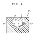
- the measuring cell 23 consists of a glass container equipped with a light incident window, inside of which a microphone is placed. An open-close cover provided in the cell is closed and sealed when the reacted solid phase is encased in the cell.
- the cell 23 has a cylindrical room, the upper part of which can be opened by a reactor forming material 1.
- the room 2 is sealable with a quartz glass-made cover 3.
- the microphone 70 is embedded in the side wall of the room 2.
- a stand 4 of transparent glass is mounted to the bottom of the room.
- the disk-like solid phase 5 can be placed at the center of the stand 4.
- the solid phase has a size of 10 mm in diameter and 2 mm in high.
- the solid phase can be changed to various sizes.
- the volume of the space is 0.5 to 1.0 ml.
- the excited light is cast on the solid phase 5 through the cover 3. Therefore, the cover 3 functions as a light incident window.
- Fig. 5 shows a pretreatment equipment within the analytical apparatus in Fig. 1.
- the pretreatment equipment includes the reactor 21, the reagent dispenser 22 and the sample pipetter 36 in Fig. 1.
- a pipetter 40 in Fig. 5 corresponds to the pipetter 36 in Fig. 1.
- Distributor 37 in Fig. 5 corresponds to the dispenser 22 in Fig. 1.
- a sample table 10 on which a plurality of the standard substances having different concentrations are placed for respective items for measurement is provided.
- a plurality of standard substances can be placed on the sample table 10 continuously for every item for measurement.
- a reaction table 121 has reactors 122, on the circumference of which the reacted solid phase 5 having bound thereto a plurality of antibodies for analyses of a plurality of items is placed.
- the reaction table is constructed to be freely rotatable. Transfer of the standard material and sample is performed by means of a sampling probe 41. Dispense of the reagent is effected by a moving distributor 37.
- the reactors 122 in which a plurality of the solid phases in the kind and number are encased are constructed to make continuous line on the reaction table 121 for every item.
- a necessary solid phase is supplied to the corresponding reactor through a reacted solid phase supplier 42.
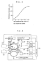
- a discharge apparatus 129 and a washing apparatus 124 are placed on the reactor line. Details of the photoacoustic signal measuring apparatus 49 are shown in Fig. 1.
- a sealed type photoacoustic cell 23 is so constructed that a pressure change generated as the result of the exposure of the solid phase placed within the cell to the light source 17 can be detected by means of a microphone.
- a controlling apparatus is equipped with a multiplexer 53, an A/D converter 54, a read only memory (hereafter referred to as ROM), a random access memory (hereafter referred to as RAM), a printer 55, an operation panel 52 and a driving circuit for mechanism 135.
- the A/D converter 54 is further connected to a central processor 51 via an interface 50.
- the central processor 51 functions to control the whole apparatus including the mechanism, prepare a calibration curve and perform data processing such as operation of the concentration, etc.
- a microcomputer is used for the central processor.
- the reaction table 121 goes counterclockwise by 1 pitch of the reactor and stops at the position.
- a time from the rotation to the stop of the reaction table is, for example, 20 seconds
- the above operation is repeated as one cycle for 20 seconds.
- the specific sample to be measured goes counterclockwise by one pitch of the reactor at the position where the reaction table 121 is in a standstill state.
- Discharge of the reagent from the moving distributor 37 is made in such a state that the reactor 122 stops at the discharge position 47 on the reaction table 121.
- the distributor 37 selects a necessary reagent solution from a line of reagent containers 39 to discharge into the reactor. Taking as an example a specific sample, a first reaction is initiated between the sample added at the discharge position 45 and the solid phase in the reactor and a second reaction is initiated with the reagent at the discharge position 47.
- the reaction table 121 rotates and stops at the stop position 46, only the reacted solid phase in the reactor is taken into the photoacoustic cell in which the photoacoustic signal is detected by means of a photoacoustic apparatus 49.
- a signal of measurement wavelength currently required is chosen by the multiplexer 53, incorporated into the central processor 51 through the A/D converter 54, and memorized on RAM.
- the central processor operates following the program of ROM, extracts the measurement data in RAM and performs data processing.
- the reaction table 121 is placed in a thermostat 123, whereby the reaction in the reactor can be proceeded at a controlled temperature.
- a removable rotary holder was used instead of the reaction table 121 shown in Fig. 5.
- a plurality of solid phase disks composed of a porous material capable of filtering liquid therethrough were set on the holder. After the cellulose membrane having bound thereto anti-human HCG (human chorionic gonadotropin) antibody was adsorbed to the porous reacted solid phase disks, the holder on which the disks were placed was put on the reaction apparatus 21. Through the sample pipetter 36, 100 ⁇ l of the serum sample was dispensed on the reacted solid phase disks.
- the reacted solid phase in the reaction apparatus 21 was transferred to the photoacoustic cell 23 by means of the reacted solid phase transfer 20.
- the photoacoustic cell was covered with a cover for sealing.
- a laser light from the light source 17 was then cast into the cell 23 to measure the photoacoustic signal based on the antigen-antibody complex formed on the cellulose membrane surface.
- HCG in the sample was measured; an example of calibration curve in this case is shown in Fig. 2.
- Filter paper (Toyo No. 51) was cut into a size of 1 x 2 cm and made a raw material for the solid phase. About 1 g of the paper solid phase was suspended in 20 ml of 5 M potassium phosphate buffer (pH 12.1) and 10 ml of water was added to the suspension. While stirring with a stirrer, 10 ml of BrCN solution (0.1 g/l) was gradually added to the mixture over 2 minutes followed by reacting for further 6 minutes. The reaction was carried out at 4°C.
- reaction mixture was washed with a large quantity of 0.005 M ice-cooled NaHCO3 solution, and 5 mg of purified anti-human AFP antibody and purified anti-human CEA antibody were reacted at room temperature for 8 hours. After further reacting with 1 M ethanolamine (pH 8.0) at 4°C overnight, the reaction mixture was sequentially washed with 0.5 M NaHCO3 solution, 0.1 M acetate buffer solution (pH 4.0) and 0.015 M PBS (pH 7.2), and then freeze dried in 1.5 ml of PBS (pH 7.2).
- 1 M ethanolamine pH 8.0
- a first reagent dispersion of yellow latex particles having bound thereto anti-human AFP antibody and a second reagent dispersion of blue latex particles having bound thereto anti-human CEA antibody were prepared as the reagents.
- To the solid phase were added 250 ⁇ l each of the first and second reagent dispersions. The mixture was reacted at room temperature for 10 minutes. The reaction mixture was then washed twice with 1 ml of 0.9% NaCl. After the washing liquid was removed, the solid phase was withdrawn from the reaction apparatus 21 and placed in the photoacoustic cell 23. After the cell 23 was sealed, the solid phase was exposed to 2 kinds of laser light having different wavelengths, whereby the photoacoustic signal was measured.
- AFP and CEA can be quantitatively determined concurrently on one reacted solid phase disk.
- An example of the calibration curve for simultaneous quantitative determination of AFP and CEA used in this measurement method is shown in Fig. 3. In this case, AFP and CEA are measured using the wavelength corresponding thereto.
- the solid phase As the solid phase, a disk-like glass piece was used. Anti-TSH (thyroid stimulating hormone) antibody was previously bound to the glass wall surface on the solid phase.
- the reactor in which the solid phase was encased was placed on the reaction apparatus 21. Through the sample pipetter 36, 20 ⁇ l of the serum sample containing TSH was dispensed in the reactor followed by reacting with the anti-TSH antibody on the solid phase at 37°C for 5 minutes.
Applications Claiming Priority (2)
| Application Number | Priority Date | Filing Date | Title |
|---|---|---|---|
| JP261539/88 | 1988-10-19 | ||
| JP26153988A JPH02108968A (ja) | 1988-10-19 | 1988-10-19 | 免疫分析方法 |
Publications (2)
| Publication Number | Publication Date |
|---|---|
| EP0369176A2 true EP0369176A2 (fr) | 1990-05-23 |
| EP0369176A3 EP0369176A3 (fr) | 1991-04-24 |
Family
ID=17363304
Family Applications (1)
| Application Number | Title | Priority Date | Filing Date |
|---|---|---|---|
| EP19890119250 Withdrawn EP0369176A3 (fr) | 1988-10-19 | 1989-10-17 | Essai immunologique utilisant de spectroscopie photo-acoustique |
Country Status (3)
| Country | Link |
|---|---|
| EP (1) | EP0369176A3 (fr) |
| JP (1) | JPH02108968A (fr) |
| CA (1) | CA2000817A1 (fr) |
Cited By (8)
| Publication number | Priority date | Publication date | Assignee | Title |
|---|---|---|---|---|
| DE4231892A1 (de) * | 1992-09-21 | 1994-03-24 | Inst Bioprozess Analysenmesst | Verfahren zur Analyse von Stoffen und Meßzelle zur Durchführung des Verfahrens |
| WO2003042669A1 (fr) * | 2001-11-13 | 2003-05-22 | Battelle Memorial Institute | Spectroscopie photo-acoustique effectuee avec un reseau |
| GB2393246A (en) * | 2002-09-21 | 2004-03-24 | Sonoptix Ltd | Transducer sensor |
| US6873415B2 (en) | 2001-11-13 | 2005-03-29 | Battelle Memorial Institute | Photoacoustic spectroscopy sample array vessel and photoacoustic spectroscopy method for using the same |
| US6999174B2 (en) | 2001-11-13 | 2006-02-14 | Battelle Memorial Institute | Photoacoustic spectroscopy sample array vessels and photoacoustic spectroscopy methods for using the same |
| WO2008008402A2 (fr) | 2006-07-11 | 2008-01-17 | The Curators Of The University Of Missouri | Dispositif et procédé de détection photo-acoustique |
| US8848191B2 (en) | 2012-03-14 | 2014-09-30 | Honeywell International Inc. | Photoacoustic sensor with mirror |
| CN112067556A (zh) * | 2020-09-29 | 2020-12-11 | 湖北鑫英泰系统技术股份有限公司 | 一种油浸式设备油气检测方法及装置 |
Citations (3)
| Publication number | Priority date | Publication date | Assignee | Title |
|---|---|---|---|---|
| EP0142481A2 (fr) * | 1983-11-14 | 1985-05-22 | Ab Varilab | Procédé d'analyse d'un échantillon d'une substance par des moyens de spectroscopie photo-acoustique ou optothermique et porte-échantillons pour la réalisation du procédé |
| EP0256474A2 (fr) * | 1986-08-11 | 1988-02-24 | Hitachi, Ltd. | Méthode et appareil pour détecter une substance particulaire déterminée |
| EP0266461A1 (fr) * | 1986-11-03 | 1988-05-11 | The Center For Immunological Studies | Procédés pour la détection et le décèlement quantitatif de substances immunologiques |
-
1988
- 1988-10-19 JP JP26153988A patent/JPH02108968A/ja active Pending
-
1989
- 1989-10-16 CA CA 2000817 patent/CA2000817A1/fr not_active Abandoned
- 1989-10-17 EP EP19890119250 patent/EP0369176A3/fr not_active Withdrawn
Patent Citations (3)
| Publication number | Priority date | Publication date | Assignee | Title |
|---|---|---|---|---|
| EP0142481A2 (fr) * | 1983-11-14 | 1985-05-22 | Ab Varilab | Procédé d'analyse d'un échantillon d'une substance par des moyens de spectroscopie photo-acoustique ou optothermique et porte-échantillons pour la réalisation du procédé |
| EP0256474A2 (fr) * | 1986-08-11 | 1988-02-24 | Hitachi, Ltd. | Méthode et appareil pour détecter une substance particulaire déterminée |
| EP0266461A1 (fr) * | 1986-11-03 | 1988-05-11 | The Center For Immunological Studies | Procédés pour la détection et le décèlement quantitatif de substances immunologiques |
Non-Patent Citations (2)
| Title |
|---|
| EMBO JOURNAL vol. 3, no. 2, 1984, OXFORD GB pages 263 - 266; C.W.Achwal et al.: "Estimation of the amount of 5-methyl-cytosine in Drosophila melanogaster DNA by amplified ELISA and photoacoustic spectroscopy" * |
| JOURNAL OF PHOTOACOUSTICS vol. 1, no. 3, 1983, NEW YORK US pages 347 - 354; T.Masujima et al.: "Application of laser-induced photoacoustic detection to immunoassay" * |
Cited By (13)
| Publication number | Priority date | Publication date | Assignee | Title |
|---|---|---|---|---|
| DE4231892A1 (de) * | 1992-09-21 | 1994-03-24 | Inst Bioprozess Analysenmesst | Verfahren zur Analyse von Stoffen und Meßzelle zur Durchführung des Verfahrens |
| US6873415B2 (en) | 2001-11-13 | 2005-03-29 | Battelle Memorial Institute | Photoacoustic spectroscopy sample array vessel and photoacoustic spectroscopy method for using the same |
| WO2003042669A1 (fr) * | 2001-11-13 | 2003-05-22 | Battelle Memorial Institute | Spectroscopie photo-acoustique effectuee avec un reseau |
| US6999174B2 (en) | 2001-11-13 | 2006-02-14 | Battelle Memorial Institute | Photoacoustic spectroscopy sample array vessels and photoacoustic spectroscopy methods for using the same |
| US6870626B2 (en) | 2001-11-13 | 2005-03-22 | Battelle Memorial Institute | Array-based photoacoustic spectroscopy |
| WO2004027396A1 (fr) * | 2002-09-21 | 2004-04-01 | Sonoptix (Uk) Limited | Capteur biochimique avec detecteurs thermoelastiques |
| GB2393246A (en) * | 2002-09-21 | 2004-03-24 | Sonoptix Ltd | Transducer sensor |
| US7740803B2 (en) | 2002-09-21 | 2010-06-22 | Sonoptix (Uk) Limited | Biochemical sensor with thermoelastic probes |
| WO2008008402A2 (fr) | 2006-07-11 | 2008-01-17 | The Curators Of The University Of Missouri | Dispositif et procédé de détection photo-acoustique |
| EP2047250A2 (fr) * | 2006-07-11 | 2009-04-15 | The Curators Of The University Of Missouri | Dispositif et procédé de détection photo-acoustique |
| EP2047250A4 (fr) * | 2006-07-11 | 2014-04-02 | Univ Missouri | Dispositif et procédé de détection photo-acoustique |
| US8848191B2 (en) | 2012-03-14 | 2014-09-30 | Honeywell International Inc. | Photoacoustic sensor with mirror |
| CN112067556A (zh) * | 2020-09-29 | 2020-12-11 | 湖北鑫英泰系统技术股份有限公司 | 一种油浸式设备油气检测方法及装置 |
Also Published As
| Publication number | Publication date |
|---|---|
| JPH02108968A (ja) | 1990-04-20 |
| CA2000817A1 (fr) | 1990-04-19 |
| EP0369176A3 (fr) | 1991-04-24 |
Similar Documents
| Publication | Publication Date | Title |
|---|---|---|
| JP2656564B2 (ja) | 免疫分析方法 | |
| US4918025A (en) | Self contained immunoassay element | |
| US4791461A (en) | Portable analyzer | |
| WO1990010875A1 (fr) | Analyseur d'echantillon de liquide et procede d'analyse d'echantillon de liquide utilisant ledit analyseur | |
| EP0198513A2 (fr) | Procédé analytique et dispositif pour la détermination de fluorescence ou phosphorescence | |
| GB2040441A (en) | Apparatus and method for qualitative and quantitative determination of immunological reactions | |
| HU176294B (en) | Process for determining antigenes and antibodies | |
| EP0204807B1 (fr) | Procede, appareil et systeme pour effectuer une analyse d'affinite biospecifique a l'aide d'une colonne munie d'une partie temoin | |
| WO1987007386A1 (fr) | Procede immunoanalytique en phase solide | |
| WO1988007202A1 (fr) | Technique amelioree d'analyse et appareil prevu a cet effet | |
| EP0521421A2 (fr) | Procédé et dispositif pour l'analyse d'un échantillon liquide | |
| US5776784A (en) | Apparatus and method for reagent separation in a chemical analyzer | |
| EP0369176A2 (fr) | Essai immunologique utilisant de spectroscopie photo-acoustique | |
| EP0256474A2 (fr) | Méthode et appareil pour détecter une substance particulaire déterminée | |
| JP5749010B2 (ja) | センサ | |
| JP3670383B2 (ja) | 分析装置および分析方法 | |
| EP0190019B1 (fr) | Procédé et dispositif de manipulation de fluides | |
| JPS60211365A (ja) | 免疫学的測定法 | |
| JPH0743381B2 (ja) | 光音響免疫分析方法及び装置 | |
| JP2002365211A (ja) | 測定装置 | |
| WO2005090998A1 (fr) | Procede d'agitation, cellule, equipement de mesure utilisant la cellule, et procede de mesure | |
| JPH06186163A (ja) | 化学分析方法 | |
| GB1600069A (en) | Method for the measurement of antigens and antibodies | |
| JPH01214761A (ja) | 光音響分光法による免疫分析方法 | |
| JPH05288753A (ja) | 免疫測定方法および装置 |
Legal Events
| Date | Code | Title | Description |
|---|---|---|---|
| PUAI | Public reference made under article 153(3) epc to a published international application that has entered the european phase |
Free format text: ORIGINAL CODE: 0009012 |
|
| 17P | Request for examination filed |
Effective date: 19891017 |
|
| AK | Designated contracting states |
Kind code of ref document: A2 Designated state(s): CH DE FR GB IT LI |
|
| PUAL | Search report despatched |
Free format text: ORIGINAL CODE: 0009013 |
|
| AK | Designated contracting states |
Kind code of ref document: A3 Designated state(s): CH DE FR GB IT LI |
|
| STAA | Information on the status of an ep patent application or granted ep patent |
Free format text: STATUS: THE APPLICATION HAS BEEN WITHDRAWN |
|
| 18W | Application withdrawn |
Withdrawal date: 19930126 |