EP0338436A2 - Monoklonale Antikörper gegen das Neuropeptid Y, Verfahren zu ihrer Herstellung sowie ihre Verwendung - Google Patents
Monoklonale Antikörper gegen das Neuropeptid Y, Verfahren zu ihrer Herstellung sowie ihre Verwendung Download PDFInfo
- Publication number
- EP0338436A2 EP0338436A2 EP89106626A EP89106626A EP0338436A2 EP 0338436 A2 EP0338436 A2 EP 0338436A2 EP 89106626 A EP89106626 A EP 89106626A EP 89106626 A EP89106626 A EP 89106626A EP 0338436 A2 EP0338436 A2 EP 0338436A2
- Authority
- EP
- European Patent Office
- Prior art keywords
- npy
- mak
- hnpy
- monoclonal antibodies
- monoclonal antibody
- Prior art date
- Legal status (The legal status is an assumption and is not a legal conclusion. Google has not performed a legal analysis and makes no representation as to the accuracy of the status listed.)
- Withdrawn
Links
Images
Classifications
-
- C—CHEMISTRY; METALLURGY
- C07—ORGANIC CHEMISTRY
- C07K—PEPTIDES
- C07K16/00—Immunoglobulins [IG], e.g. monoclonal or polyclonal antibodies
- C07K16/18—Immunoglobulins [IG], e.g. monoclonal or polyclonal antibodies against material from animals or humans
- C07K16/28—Immunoglobulins [IG], e.g. monoclonal or polyclonal antibodies against material from animals or humans against receptors, cell surface antigens or cell surface determinants
- C07K16/30—Immunoglobulins [IG], e.g. monoclonal or polyclonal antibodies against material from animals or humans against receptors, cell surface antigens or cell surface determinants from tumour cells
-
- A—HUMAN NECESSITIES
- A61—MEDICAL OR VETERINARY SCIENCE; HYGIENE
- A61K—PREPARATIONS FOR MEDICAL, DENTAL OR TOILETRY PURPOSES
- A61K38/00—Medicinal preparations containing peptides
Definitions
- Human neuropeptide Y is a 36 amino acid peptide (Tatemoto, K. Neuropeptide Y: The complete amino acid sequence of the brain peptide, Proc. Natl. Acad. Sci. USA 79: 5485-5489, 1982), which in Tissue and serum from phaeochromocytoma and ganglioneuroblastoma patients was detected. NPY has an antagonistic effect and is held responsible for the release of noradrenaline. NPY (hNPY) appears interesting as a tumor marker for steroid hormone-producing tumors, thyroid carcinomas and neuroendocrine tumors (neuroblastoma).
- NPY shows 50% and 70% sequential analogy to PP (pancreatic polypeptide) and PYY (peptide YY).
- IRMA immunoradiometric assay
- the present invention now relates to monoclonal antibodies which are directed against the following regions of the NPY:
- monoclonal antibodies can be used as diagnostics, active substances or active substance carriers.
- Four hybridoma cell clones were obtained, the secreted MAKs (NPY 02 - NPY 05) were characterized.
- the identification of the monoclonal antibodies was carried out in an ELISA method using rabbit antibodies which were directed against the various immunoglobulin subclasses (Nordic).
- the affinity constants were measured by incubating the MAKs with radioactively labeled NPY in the presence of increasing concentrations of the unlabeled total peptide using a rapid RIA method.
- the RIA binding data thus obtained were used to determine the affinity constants (K a ) after a Scatchard plot analysis.
- a fast RIA was used to test the anti-hNPY antibodies in the various steps of MAK production (mouse serum, hybridoma cells, cell supernatants of the cloned cells, ascites fluids). 10 to 100 ⁇ l of medium were incubated for 2 hours at 20 ° C. with 500 ⁇ l of phosphate buffer (50 mmol, pH 7.5), the phosphate buffer being 20 mmol of EDTA, 0.3% BSA (bovine serum albumin), 0.005% aprotinin and NPY labeled with J125 (3.103 cpm; specific activity: 73 TBq / mmol;).
- the epitopes which are recognized by the different MAKs, were localized in a competitive one Inhibition assay with synthetically produced hNPY fragments and with the NPY-related peptides hPP (human pancreatic polypeptide), bPP (bovine pancreatic polypeptide) and pPYY (porcine peptide YY). These binding data were also measured in an RIA.
- hNPY The preparation of hNPY and its partial sequences (1-12, 11-18, 11-24, 23-36, 27-36 and 27-36 (free of C-terminus)) was carried out by solid-phase Merrifield synthesis (Merrifield, RB, Solid phase peptide synthesis, I, The synthesis of a tetrapeptide, J. Am. Chem. Soc. 85, 2149, (1963)).
- the syntheses were carried out using an "automatic synthesizer" (Applied Biosystems, Model 430A, Foster City, CA).
- the peptides are split off from the carrier polymer using hydrofluoric acid.
- the peptides were then purified by gel filtration over Bio-Gel P4 (Bio-Rad, Richmond, CA).
- the purity was checked by means of reversed phase LC and the amino acid composition was determined on an LKB Alpha Analyzer.
- the related peptides hPP, bPP and pPYY are commercially available (for example Sigma Chemicals Co., St. Louis, Mo).
- hNPY itself (for the MAKs NPY 02 - NPY 04) or the partial amino acid sequence 27-36 (for the MAK NPY 05), each coupled to KLH (keyhole limpet haemocyanin, SIGMA), was used as the immunogen with glutaraldehyde as Coupling agent used.
- KLH keyhole limpet haemocyanin, SIGMA
- the 6-week-old mice were immunized for the first time by subcutaneous injection of 25 ⁇ g of the immunogen described above in complete Freund's adjuvant. The one on it following injections were carried out monthly with the same concentration of the immunogen in complete Freund's adjuvant.
- the antibody content in the mouse serum was determined using the rapid RIA method described above. If the result was positive (ie 60% binding or more with the mouse serum diluted to 50%), two doses of immunogen were administered; once 9 days (ip) and once 3 days (iv) before the fusion.
- Cell fusion, hybridoma selection, cloning and ascites production were carried out according to the method described by Bidart et al. (Identification of epitopes associated with hCG and hCG carboxyl terminus by monoclonal antibodies produced against a synthetic peptide, J. Immunol. 134, 457 (1985)).
- mice spleen cells were fused with NS 1 myeloma cells by incubation in the presence of 40% (w / v) polyethylene glycol (molecular weight: 1000). After a cultivation period of two weeks, the cell supernatants were transferred to the screening RIA (method as described above) for the production of anti-hNPY antibodies.
- the monoclonal antibodies were then purified either by Protein A Sepharose Affinity chromatography (described by EY et al., Isolation of pure IgG1, IgG2a and IgG2b immunoglobulines from mouse serum using protein A sepharose, Immunochemistry, 15, 429, (1978)) (in the case of the igG2 antibody NPY 05) or by DEAS-Sephacel chromatography with subsequent Protein A procedure (in the case of antibodies NPY 02-04).
- the purified monoclonal antibodies were then treated according to the iodogen method (Fraker, P.SJ, Speck, JC, Protein and cell membrane iodination using a sparingly soluble chloroamide, Biochem.
- Table 1 summarizes the essential characteristics of the monoclonal antibodies NPY 02-NPY 05.
- the epitopes on hNPY recognized by the monoclonal antibodies NPY 02-NPY 05 were determined in an inhibition test with various hNPY partial sequences and hNPY-related peptides.
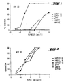
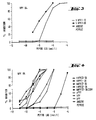
- Figures 1 to 4 show the percentage inhibition of the four monoclonal antibodies, plotted against the concentration of different hNPY partial sequences or hNPY-related peptides.
- the monoclonal antibody NPY 05 shows a high affinity for pPYY, hPP, bPP and for the C-terminal amid region.
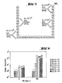
- Figure 5 shows hNPY with the four bound monoclonal antibodies NPY 02-NPY 05.
- the monoclonal antibodies NPY 02 to NPY 05 were each bound once to the solid phase of a polystyrene test tube and the other radioactively labeled with J125.
- the signal-to-noise ratio of different antibody pairs as a function of two different NPY concentrations (25 pmol / l and 500 pmol / l) is shown in FIG. 6.
- the monoclonal antibodies NPY 03 and NPY 04 seem to be interesting for the determination of precursors of hNPY, especially those with a longer amino acid sequence.
- the two monoclonal antibodies NPY 02 and NPY 05 were chosen, not least because of their good affinity constants and because of the position of the binding sites on hNPY, for the development of an IRMA for hNPY.
- the sensitivity of this assay was 0.5 fmol / ml hNPY.
- Cross reactions of the monoclonal antibodies used with PP or PYY could not be determined up to a PP or PYY concentration of 10 nmol / ml.
- the hNPY concentration was determined using the sandwich assay described above. 95% of the samples showed hNPY concentrations below 5 fmol / ml. This concentration was taken as the upper physiological limit. Of the remaining samples with an hNPY concentration above 5 fmol / ml, 6 were still below 10 fmol / ml, the rest had hNPY concentrations of at most 30 fmol / ml. In patients with neuroendocrine tumors (56 plasma samples were measured), the hNPY concentration was significantly higher (for phaeochromocytoma and neuroblastoma). The highest plasma hNPY values were observed in the patient with malignant phaeocromocytoma.
Landscapes
- Health & Medical Sciences (AREA)
- Chemical & Material Sciences (AREA)
- Organic Chemistry (AREA)
- Immunology (AREA)
- Life Sciences & Earth Sciences (AREA)
- Biophysics (AREA)
- Biochemistry (AREA)
- General Health & Medical Sciences (AREA)
- Genetics & Genomics (AREA)
- Medicinal Chemistry (AREA)
- Molecular Biology (AREA)
- Proteomics, Peptides & Aminoacids (AREA)
- Cell Biology (AREA)
- Preparation Of Compounds By Using Micro-Organisms (AREA)
- Medicines Containing Antibodies Or Antigens For Use As Internal Diagnostic Agents (AREA)
- Peptides Or Proteins (AREA)
- Micro-Organisms Or Cultivation Processes Thereof (AREA)
- Medicines That Contain Protein Lipid Enzymes And Other Medicines (AREA)
Abstract
Description
- Humanes Neuropeptid Y (hNPY) ist ein Peptid mit 36 Aminosäuren (Tatemoto, K. Neuropeptide Y: The complete amino acid sequence of the brain peptide, Proc. Natl. Acad. Sci. USA 79: 5485-5489, 1982), das im Gewebe und Serum von Phaeochromocytom- und Ganglioneuroblastom-Patienten nachgewiesen wurde. NPY wirkt antagonistisch und wird für die Freisetzung von Noradrenalin verantwortlich gemacht. Interessant erscheint NPY (hNPY) als Tumormarker für steroidhormon-produzierende Tumore, Schilddrüsenkarzinome und neuroendocrine Tumore (Neuroblastom).
- NPY zeigt 50 % bzw. 70 % sequenzielle Analogie zu PP (pancreatic polypeptide) und zum PYY (peptide YY).
- Ein immunoradiometrischer Assay (IRMA) zur Bestimmung von NPY in Plasma ist bereits beschrieben (Corder, R., Lowry, P.J., Peptides 6 (6), 1195 - 1200, 1985). Dabei werden polyklonale Antikörper bzw. Antikörperseren, die durch Immunisierung von Schafen bzw. Kaninchen mit NPY bzw. NPY-Teilsequenzen erhalten wurden, verwendet. Der markierte Sandwichkomplex wird mit einem festphasengebundenen Antikörper, der gegen die Fc-Regionen eines im Assay verwendeten Antikörpers gerichtet ist, abgetrennt.
- Die vorliegende Erfindung betrifft nun monoklonale Antikörper, die gegen folgende Regionen des NPY gerichtet sind:

- Diese monoklonalen Antikörper können als Diagnostikum, Wirkstoff oder Wirkstoffträger verwendet werden. Vier Hybridomazellklone konnten gewonnen werden, deren sezernierte MAKs (NPY 02 - NPY 05) charakterisiert wurden.
- Die Identifikation der monoklonalen Antikörper wurde in einem ELISA-Verfahren durchgeführt, wobei Kaninchen-Antikörper benutzt wurden, die gegen die verschiedenen Immunglobulin-Subklassen gerichtet waren (Nordic). Die Messung der Affinitätskonstanten erfolgte durch Inkubation der MAKs mit radioaktiv markiertem NPY in Gegenwart von steigenden Konzentrationen des unmarkierten Gesamtpeptids nach einem schnellen RIA-Verfahren. Die so erhaltenen RIA-Bindungsdaten wurden zur Ermittlung der Affinitätskonstanten (Ka) nach einer Scatchard-Plot-Analyse herangezogen.
- Zum Test der Anti-hNPY-Antikörper in den verschiedenen Schritten der MAK-Herstellung (Mausserum, Hybridomazellen, Zellüberstände der klonierten Zellen, Aszitesflüssigkeiten) wurde ein schneller RIA benutzt.
Dabei wurden 10 bis 100 µl Medium 2 Stunden bei 20°C mit 500 µl Phosphatpuffer (50 mmol, pH 7,5) inkubiert, wobei der Phosphatpuffer 20 mmol EDTA, 0,3 % BSA (bovine serum albumin), 0,005 % Aprotinin und mit J¹²⁵ markiertes NPY (3.10³ cpm; specific activity: 73 TBq/mmol;) enthielt. Durch Hinzugabe von 500 µl eines Phosphatpuffers, der 0,02 % Gelatine, 1,6 % Aktivkohle und 0,16 % Dextran enthielt, wurde die Inkubation gestoppt. Nach Schütteln (10 sec) und Zentrifugation wurde die radioaktive Strahlung sowohl in den Überständen (B = gebundenes NPY) als auch im Niederschlag (F = freies NPY) mit einem Zählrohr gemessen. Der Quotient aus gebundenem und gebundenem plus freiem Anteil ergab die prozentuale Bindung von NPY an die MAKs. - Die Lokalisation der Epitope, die von den verschiedenen MAKs erkannt werden, erfolgte in einem kompetitiven Inhibitionsassay mit synthetisch hergestellten hNPY-Fragmenten und mit den NPY-verwandten Peptiden hPP (human pancreatic polypeptide), bPP (bovine pancreatic polypeptide) und pPYY (porcine peptide YY). Diese Bindungsdaten wurden ebenfalls in einem RIA gemessen.
- Im folgenden ist die weitere Ausgestaltung der Erfindung beschrieben.
- Die Herstellung von hNPY und seiner Teilsequenzen (1-12, 11-18, 11-24, 23-36, 27-36 und 27-36 (C-terminusfrei)) erfolgte durch Festphasen-Merrifield-Synthese (Merrifield, R.B., Solid phase peptide synthesis, I, The synthesis of a tetrapeptide, J. Am. Chem. Soc. 85, 2149, (1963)). Die Synthesen wurden mit einem "automatic synthesizer" (Applied Biosystems, Model 430A, Foster City, CA) durchgeführt. Die Abspaltung der Peptide von dem Trägerpolymer erfolgt mit Fluorwasserstoffsäure. Anschließend wurden die Peptide durch Gelfiltration über Bio-Gel P4 (Bio-Rad, Richmond, CA) gereinigt. Die Reinheit wurde mittels Reversed-Phase-LC überprüft und die Aminosäurezusammensetzung an einem LKB Alpha Analyser bestimmt.
Die verwandten Peptide hPP, bPP und pPYY sind käuflich erhältlich (beispielsweise Sigma Chemicals Co., St. Louis, Mo). - Zur Herstellung der monoklonalen Antikörper wurde als Immunogen entweder hNPY selbst (für die MAKs NPY 02 - NPY 04) oder die Teilaminosäuresequenz 27-36 (für den MAK NPY 05), jeweils gekoppelt an KLH (keyhole limpet haemocyanin, SIGMA), mit Glutaraldehyd als Kopplungsagenz, verwendet.
Die erste Immunisierung der 6 Wochen alten Mäuse erfolgte durch Subcutaninjektion von 25 µg des oben beschriebenen Immunogens in komplettem Freund'schen Adjuvans. Die darauf folgenden Injektionen wurden monatlich mit der gleichen Konzentration des Immunogens in komplettem Freund'schen Adjuvans durchgeführt. Jeweils vor diesen Folgeimmunisierungen wurde der Antikörpergehalt im Mausserum nach der oben beschrieben schnellen RIA-Methode bestimmt. Bei einem positiven Befund (d.h. 60 % Bindung oder mehr mit dem auf 50 % verdünnten Mausserum) wurden noch zwei Immunogen-Dosen verabreicht; einmal 9 Tage (i.p.) und einmal 3 Tage (i.v.) vor der Fusion.
Die Zellfusion, Hybridoma Selektion, Klonierung und Aszites Produktion wurden nach der Methode, die von Bidart et al. (Identification of epitopes associated with hCG and hCG carboxyl terminus by monoclonal antibodies produced against a synthetic peptide, J. Immunol. 134, 457 (1985)) beschrieben wurde, durchgeführt. Dabei wurden die Mäusemilzzellen mit NS 1 Myelom-Zellen durch Inkubation in Anwesenheit von 40 % (w/v) Polyethylenglykol (Molekulargew.: 1000) fusioniert. Nach einer Kultivierungszeit von zwei Wochen wurden die Zellüberstände in den Screening-RIA (Verfahren wie oben beschrieben) zwecks Gewinnung von Anti-hNPY-Antikörper überführt. Zellen aus 75 (71 aus den Immunisierungen mit vollständigem hNPY, 4 aus den Immunisierungen mit dem 27-36-Subpeptid) RIA-positiven Wells (Vertiefungen einer Mikrotiterplatte) wurden kloniert und jeweils 2·10⁶ Zellen von ausgewählten 4 Hybridomas (3 resultierend aus hNPY, 1 resultierend aus dem 27-36-Subpeptid) wurden dann nackten Mäusen i.p. injiziert. Zwei Wochen nach der Injektion wurde die Aszitesflüssigkeit entnommen.
Die so erhaltenen 4 monoklonalen Antikörper NPY 02-NPY 05 (MAK NPY 05 resultierend aus der Immunisierung mit dem 27-36-Subpeptid) wurden durch Präzipitation mit 50 %iger Ammoniumsulfat-Lösung von der Maus-Aszitesflüssigkeit getrennt. Anschließend erfolgte die Reinigung der monoklonalen Antikörper entweder durch Protein-A-Sepharose- Affinitäts-Chromatographie (beschrieben von EY et al., Isolation of pure IgG1, IgG2a und IgG2b immunoglobulines from mouse serum using protein A sepharose, Immunochemistry, 15, 429, (1978)) (im Fall des igG2-Antikörpers NPY 05) oder durch DEAS-Sephacel-Chromatographie mit anschließender Protein A Prozedur (im Fall der Antikörper NPY 02-04). Die gereinigten monoklonalen Antikörper wurden dann nach der Iodogenmethode (Fraker, P.SJ, Speck, J.C., Protein and cell membrane iodination using a sparingly soluble chloroamide, Biochem. Biophys. Res. Commun., 80, 849, (1977)) mit J¹²⁵ markiert (spezifische Aktivität: 450-600 KBq/µg Immunoglobulin). Vor dem Einsatz wurden die markierten monoklonalen Antikörper mit PBS, welches 20 mmol EDTA, 10 mmol Barbital, 1 % BSA und 50 UI/1 Aprotinin enthielt, versetzt. - In Tabelle 1 sind die wesentlichen Charakteristika der monoklonalen Antikörper NPY 02-NPY 05 zusammengefaßt.

- Die von den monoklonalen Antikörpern NPY 02-NPY 05 erkannten Epitope auf hNPY wurden in einem Inhibitionstest mit verschiedenen hNPY-Teilsequenzen und hNPY-verwandten Peptiden ermittelt. Die Figuren 1 bis 4 zeigen die prozentuale Inhibition der vier monoklonalen Antikörper, aufgetragen gegen die Konzentration von verschiedenen hNPY-Teilsequenzen bzw. hNPY-verwandten Peptiden. Die IC 50-Werte (= notwendige Konzentration für eine 50%ige Inhibition) der monoklonalen Antikörper NPY 02-NPY 05 sind in Tabelle 2 angegeben.

- Von dem monoklonalen Antikörper NPY 02 wird allein das 11-24-Fragment von hNPY und pPYY erkannt. Bindungen zu anderen Fragmenten oder Peptiden treten nicht auf. Diese Resultate legen nahe, daß die antigene Determinante zwischen den Aminosäuren 8 und 24 lokalisiert ist, da pPYY und hNPY in diesem Bereich nur die Sequenz 8-12 gemeinsam haben. Da die 18-24-Teilsequenz und die 1-12-Teilsequenz nicht an den monoklonalen Antikörper NPY 02 bindet, verkürzt sich die antigene Determinante auf die Aminosäuresequenz 8-18, wahrscheinlich 10-15.
Der monoklonale Antikörper NPY 05 zeigt eine hohe Affinität für pPYY, hPP, bPP und für die C-terminale amidische Region. Die 32-36-Teilsequenz mit freier COOH-Gruppe wird nur schwach erkannt (IC 50 = 2·10⁻⁵ mol). Da zum einen der monoklonale Antikörper NPY 05 durch Immunisierung mit der 27-36-Teilsequenz erhalten wurde und andererseits die Bindung von J¹²⁵-NPY an den Antikörper sowohl durch das 32-36-Subpeptid, als auch durch das gesamte hNPY inhibiert wird, liegt das von NPY 05 erkannte Epitop sicherlich auf den letzten fünf Aminosäuren von hNPY. Die starke Abnahme des IC 50-Wertes im Fall der Erkennung des 32-36-COOH-Fragments (IC 50 = 2·10⁻⁵ mol) zeigt die Bedeutung der Amidgruppe in dieser Epitop-Bindungsregion an. Figure 5 zeigt hNPY mit den vier gebundenen monoklonalen Antikörpern NPY 02-NPY 05. - Für die Entwicklung eines IRMA ("Sandwich Assay") zur Bestimmung der hNPY-Konzentration in Seren wurden die monoklonalen Antikörper NPY 02 bis NPY 05 jeweils einmal an die Festphase eines Polystyrol-Teströhrchens gebunden und zum anderen mit J¹²⁵ radioaktiv markiert. Das Signal-Rausch-Verhältnis von verschiedenen Antikörperpaaren in Abhängigkeit von zwei verschiedenen NPY-Konzentrationen (25 pmol/l und 500 pmol/l) ist in Figur 6 gezeigt. Die Kombination MAK NPY 02 auf der Festphase und MAK NPY 05 radioaktiv markiert (S/R bei 500 pmol/l NPY = 118; siehe Figur 6) hat sich dabei als günstig erwiesen.
Die monoklonalen Antikörper NPY 03 und NPY 04 scheinen interessant für die Bestimmung von Precursoren von hNPY, insbesondere solchen, mit längerer Aminosäuresequenz. - Die beiden monoklonalen Antikörper NPY 02 und NPY 05 wurden, nicht zuletzt aufgrund ihrer guten Affinitätskonstanten und aufgrund der Lage der Bindungsstellen auf hNPY, für die Entwicklung eines IRMA für hNPY ausgewählt. Für den "Sandwich-Assay" wurde der MAK NPY 02 an der Festphase von Polystyrol-Teströhrchen gebunden und der MAK NPY 05 radioaktiv mit J¹²⁵ markiert (150·10³ cpm, spec. activity = 450-600 KBq/µg). 300 µl eines NPY-Standards (0-100 fmol/ml) bzw. eines Patienten-Testserums wurde in die beschichteten Teströhrchen gegeben. Anschließend wurden 50 µl des markierten MAKs hinzugegeben (Inkubationsdauer: 16 Stunden bei 4°C unter leichtem Schütteln). Nach einem Waschschritt (3 mal 1 ml entionisiertes Wasser, welches 0,3 % Tween 20 enthielt) wurde die an die Festphase gebundene Radioaktivität gemessen. Durch Mittelwertbildung von 2 Proben mit gleicher Standard-NPY-Konzentration wurde das Signal-Rausch-Verhältnis bestimmt. Die Mittelwerte wurden zur Erstellung einer Eichkurve herangezogen.
- Die Empfindlichkeit dieses Assays lag bei 0,5 fmol/ml hNPY. Kreuzreaktionen der verwendeten monoklonalen Antikörper mit PP bzw. PYY konnten bis zu einer PP- bzw. PYY-Konzentration von 10 nmol/ml nicht festgestellt werden.
- In 217 Plasmaproben von gesunden Patienten zwischen 18 und 55 Jahren wurde die hNPY-Konzentration mit dem oben beschriebenen Sandwich-Assay bestimmt. 95 % der Proben zeigten hNPY-Konzentrationen unterhalb 5 fmol/ml. Diese Konzentration wurde als obere physiologische Grenze angenommen. Von den restlichen Proben mit einer hNPY-Konzentration oberhalb 5 fmol/ml befanden sich 6 noch unterhalb 10 fmol/ml, die übrigen besaßen hNPY-Konzentrationen von maximal 30 fmol/ml. Bei Patienten mit neuroendocrinen Tumoren (56 Plasmaproben wurden gemessen) lag die hNPY-Konzentration signifikant höher (bei Phaeochromocytom und Neuroblastom). Die höchsten Plasma-hNPY-Werte wurden beim Patienten mit malignem Phaeocromocytom beobachtet.
Claims (10)
- einen festphasengebundenen MAK, ausgewählt aus NPY 02, NPY 03, NPY 04, NPY 05 und
- einem radioaktiv markierten MAK, ausgewählt aus NPY 02, NPY 03, NPY 04, NPY 05,
wobei der festphasengebundene MAK nicht mit dem radioaktiv markierten MAK identisch ist.
Applications Claiming Priority (2)
| Application Number | Priority Date | Filing Date | Title |
|---|---|---|---|
| DE3813592 | 1988-04-22 | ||
| DE19883813592 DE3813592A1 (de) | 1988-04-22 | 1988-04-22 | Monoklonale antikoerper gegen das neuropeptid y, verfahren zu ihrer herstellung sowie ihre verwendung |
Publications (2)
| Publication Number | Publication Date |
|---|---|
| EP0338436A2 true EP0338436A2 (de) | 1989-10-25 |
| EP0338436A3 EP0338436A3 (de) | 1990-04-18 |
Family
ID=6352653
Family Applications (1)
| Application Number | Title | Priority Date | Filing Date |
|---|---|---|---|
| EP89106626A Withdrawn EP0338436A3 (de) | 1988-04-22 | 1989-04-13 | Monoklonale Antikörper gegen das Neuropeptid Y, Verfahren zu ihrer Herstellung sowie ihre Verwendung |
Country Status (3)
| Country | Link |
|---|---|
| EP (1) | EP0338436A3 (de) |
| JP (1) | JPH02242672A (de) |
| DE (1) | DE3813592A1 (de) |
Cited By (3)
| Publication number | Priority date | Publication date | Assignee | Title |
|---|---|---|---|---|
| US5670482A (en) * | 1992-06-20 | 1997-09-23 | Glaxo Wellcome Inc. | Neuropeptide Y antagonists |
| EP2785738A4 (de) * | 2011-12-02 | 2015-07-22 | Garvan Inst Med Res | Antikörper gegen npy und pyy sowie verwendungen davon |
| CN115636874A (zh) * | 2021-07-19 | 2023-01-24 | 武汉市朗典精医生物科技有限公司 | 制备神经肽y(npy)抗体及建立检测npy方法 |
Families Citing this family (1)
| Publication number | Priority date | Publication date | Assignee | Title |
|---|---|---|---|---|
| JP2001242390A (ja) * | 2000-02-29 | 2001-09-07 | Asahi Optical Co Ltd | 接眼変倍光学系 |
-
1988
- 1988-04-22 DE DE19883813592 patent/DE3813592A1/de not_active Withdrawn
-
1989
- 1989-04-13 EP EP89106626A patent/EP0338436A3/de not_active Withdrawn
- 1989-04-21 JP JP1100344A patent/JPH02242672A/ja active Pending
Non-Patent Citations (3)
| Title |
|---|
| 17th Annual Meeting of the Society for Neuroscience, New Orleans, 16-21.11.1987., Soc. Neurosci Abstr. Band 13, Nr. 2, Seite 1278; C.-H. LEE et al.: "Monoclonal antibodies to an endogenous neuropeptide with putative morphinemodulating activity" * |
| Journal of Clinical Endocrinology and Metabolism, Band 68, Nr. 4, 1989, Seiten 808-813, The Endocrine Society, US; E. GROUZMANN et al.: "Plasma neuropeptide Y concentrations in patients with neuroendocrine tumors" * |
| Proc. Natl. Acad. Sci. USA, Band 79, September 1982, Seiten 5485-5489; K. TATEMOTO: "Neuropeptide Y: Complete amino acid sequence of the brain peptide" * |
Cited By (4)
| Publication number | Priority date | Publication date | Assignee | Title |
|---|---|---|---|---|
| US5670482A (en) * | 1992-06-20 | 1997-09-23 | Glaxo Wellcome Inc. | Neuropeptide Y antagonists |
| EP2785738A4 (de) * | 2011-12-02 | 2015-07-22 | Garvan Inst Med Res | Antikörper gegen npy und pyy sowie verwendungen davon |
| US20150307599A1 (en) * | 2011-12-02 | 2015-10-29 | Garvan Institute Of Medical Research | Anti-npy and pyy antibodies and uses thereof |
| CN115636874A (zh) * | 2021-07-19 | 2023-01-24 | 武汉市朗典精医生物科技有限公司 | 制备神经肽y(npy)抗体及建立检测npy方法 |
Also Published As
| Publication number | Publication date |
|---|---|
| EP0338436A3 (de) | 1990-04-18 |
| JPH02242672A (ja) | 1990-09-27 |
| DE3813592A1 (de) | 1989-11-02 |
Similar Documents
| Publication | Publication Date | Title |
|---|---|---|
| DE69321914T2 (de) | Antikörper gegen Plasmodium falciparum | |
| DE3888224T2 (de) | Bestimmung vom Tumornekrosefaktor; monoklonaler Antikörper und Zusammensetzung. | |
| DE69321955T2 (de) | Bnp antikörper und immunologischer nachweis der ihn benutzt | |
| DE69432629T3 (de) | Antikörper gegen beta-amyloid oder derivative davon und seine verwendung | |
| DE3887675T2 (de) | Antikörper. | |
| DE69228436T2 (de) | Monoklonaler Antikörper, der den C-Terminus von hBNP erkennt | |
| DE69119656T2 (de) | Satz zur spezifischen Bestimmung von Angiotensin-II | |
| DE69132864T2 (de) | Antikörper gegen hypophysäre adenylatzyklase aktivierende peptid-pacap, hybridome und bestimmung von pacap | |
| DE69821246T2 (de) | Testverfahren für collagenpeptid | |
| DE69132122T2 (de) | Reinigung von cytokeratinfragmenten | |
| DE60027542T2 (de) | Methoden zur herstellung immunologischer reagentien positionsspezifisch für phosphorylierungen | |
| DE3789781T2 (de) | Antigene, Antikörper und Verfahren zur Identifizierung humaner metastatischer Tumoren und Zellinien zur Herstellung dieser Antikörper. | |
| DE69504620T2 (de) | Monoklonaler Antikörper gegen menschliches Glicentin, Hybridoma zur Herstellung dieses Antikörpers und Testverfahren für menschliches Glicentin unter Verwendung dieses Antikörpers | |
| DE60219645T2 (de) | Antikörper gegen metastin und dessen verwendung zur diagnose von schwangerschaft | |
| DE68925923T2 (de) | Antigene der einzigen Spaltungsstelle von Fibrinogen durch Elastase | |
| DE69629976T2 (de) | Typ I Prokollagen aminoterminales Propeptid, dessen Antikörper und Testverfahren unter Verwendung davon | |
| DE69733957T2 (de) | Verfahren zur bestimmung der gegenwart des gehirnspezifischen proteins s-100 beta | |
| EP0124779A2 (de) | Radioimmunverfahren für Thymosin beta 4 | |
| DE3485965T2 (de) | Monoklonaler antikoerper mit spezifitaet gegen nur einen typ eines isozyms der schweren kette des menschlichen herzmyosins. | |
| DE68911713T2 (de) | Monoklonale Antikörper, die atriales natriuretisches Polypeptide erkennen, ihre Herstellung und Verwendung. | |
| DE69637411T2 (de) | Antikörper gegen den menschlichen gastrinfreisetzenden Peptidvorläufer und deren Verwendung | |
| EP0338436A2 (de) | Monoklonale Antikörper gegen das Neuropeptid Y, Verfahren zu ihrer Herstellung sowie ihre Verwendung | |
| DE69316388T2 (de) | CRF Bindungsproteine | |
| DE69509626T2 (de) | Sandwich-Immunotestverfahren für N-Peptide | |
| DE3885975T2 (de) | Monoklonale Antikörper gegen menschliche alpha-Atrialnatriüretic-Polypeptide und entsprechende Hybridome, deren Herstellung und Verwendung. |
Legal Events
| Date | Code | Title | Description |
|---|---|---|---|
| PUAI | Public reference made under article 153(3) epc to a published international application that has entered the european phase |
Free format text: ORIGINAL CODE: 0009012 |
|
| AK | Designated contracting states |
Kind code of ref document: A2 Designated state(s): AT BE CH DE ES FR GB GR IT LI LU NL SE |
|
| PUAL | Search report despatched |
Free format text: ORIGINAL CODE: 0009013 |
|
| AK | Designated contracting states |
Kind code of ref document: A3 Designated state(s): AT BE CH DE ES FR GB GR IT LI LU NL SE |
|
| 17P | Request for examination filed |
Effective date: 19900623 |
|
| STAA | Information on the status of an ep patent application or granted ep patent |
Free format text: STATUS: THE APPLICATION HAS BEEN WITHDRAWN |
|
| 18W | Application withdrawn |
Withdrawal date: 19910808 |
|
| R18W | Application withdrawn (corrected) |
Effective date: 19910808 |