EP0316695B1 - Rekombinante HIV-2 Polypeptide - Google Patents
Rekombinante HIV-2 Polypeptide Download PDFInfo
- Publication number
- EP0316695B1 EP0316695B1 EP88118488A EP88118488A EP0316695B1 EP 0316695 B1 EP0316695 B1 EP 0316695B1 EP 88118488 A EP88118488 A EP 88118488A EP 88118488 A EP88118488 A EP 88118488A EP 0316695 B1 EP0316695 B1 EP 0316695B1
- Authority
- EP
- European Patent Office
- Prior art keywords
- polypeptide
- hiv
- accordance
- env
- antibodies
- Prior art date
- Legal status (The legal status is an assumption and is not a legal conclusion. Google has not performed a legal analysis and makes no representation as to the accuracy of the status listed.)
- Expired - Lifetime
Links
- 108090000765 processed proteins & peptides Proteins 0.000 title claims abstract description 127
- 102000004196 processed proteins & peptides Human genes 0.000 title claims abstract description 122
- 229920001184 polypeptide Polymers 0.000 title claims abstract description 118
- 238000000034 method Methods 0.000 claims abstract description 45
- 241000725303 Human immunodeficiency virus Species 0.000 claims abstract description 34
- 108091028043 Nucleic acid sequence Proteins 0.000 claims abstract description 22
- 239000013604 expression vector Substances 0.000 claims abstract description 18
- 230000000890 antigenic effect Effects 0.000 claims abstract description 12
- 230000002163 immunogen Effects 0.000 claims abstract description 12
- 101710091045 Envelope protein Proteins 0.000 claims abstract description 6
- 101710188315 Protein X Proteins 0.000 claims abstract description 6
- 239000013060 biological fluid Substances 0.000 claims abstract description 5
- 230000008569 process Effects 0.000 claims abstract description 5
- 102100021696 Syncytin-1 Human genes 0.000 claims abstract 2
- 239000013612 plasmid Substances 0.000 claims description 146
- 239000012634 fragment Substances 0.000 claims description 75
- 241000713772 Human immunodeficiency virus 1 Species 0.000 claims description 46
- 241000588724 Escherichia coli Species 0.000 claims description 28
- 238000001514 detection method Methods 0.000 claims description 18
- 102000004190 Enzymes Human genes 0.000 claims description 15
- 108090000790 Enzymes Proteins 0.000 claims description 15
- 239000002773 nucleotide Substances 0.000 claims description 15
- 125000003729 nucleotide group Chemical group 0.000 claims description 15
- 210000002966 serum Anatomy 0.000 claims description 14
- 238000004519 manufacturing process Methods 0.000 claims description 11
- 241001596967 Escherichia coli M15 Species 0.000 claims description 10
- 238000012360 testing method Methods 0.000 claims description 7
- 210000001124 body fluid Anatomy 0.000 claims description 5
- 239000010839 body fluid Substances 0.000 claims description 5
- 238000003018 immunoassay Methods 0.000 claims description 5
- 239000011541 reaction mixture Substances 0.000 claims description 5
- 241000894006 Bacteria Species 0.000 claims description 4
- 241001465754 Metazoa Species 0.000 claims description 4
- 239000000427 antigen Substances 0.000 claims description 4
- 108091007433 antigens Proteins 0.000 claims description 4
- 102000036639 antigens Human genes 0.000 claims description 4
- 239000007787 solid Substances 0.000 claims description 4
- 239000012876 carrier material Substances 0.000 claims description 3
- 101710132601 Capsid protein Proteins 0.000 claims description 2
- 241000124008 Mammalia Species 0.000 claims description 2
- 101000582398 Staphylococcus aureus Replication initiation protein Proteins 0.000 claims description 2
- 125000001360 methionine group Chemical group N[C@@H](CCSC)C(=O)* 0.000 claims description 2
- 238000005406 washing Methods 0.000 claims description 2
- 125000003275 alpha amino acid group Chemical group 0.000 claims 2
- 108010058683 Immobilized Proteins Proteins 0.000 claims 1
- 230000003100 immobilizing effect Effects 0.000 claims 1
- 238000002372 labelling Methods 0.000 claims 1
- 125000002924 primary amino group Chemical group [H]N([H])* 0.000 claims 1
- 230000001131 transforming effect Effects 0.000 claims 1
- 241000713340 Human immunodeficiency virus 2 Species 0.000 abstract description 60
- 238000002360 preparation method Methods 0.000 abstract description 14
- 108020004511 Recombinant DNA Proteins 0.000 abstract description 4
- 238000005516 engineering process Methods 0.000 abstract description 4
- 108020004414 DNA Proteins 0.000 description 59
- 108090000623 proteins and genes Proteins 0.000 description 49
- 208000031886 HIV Infections Diseases 0.000 description 44
- LFQSCWFLJHTTHZ-UHFFFAOYSA-N Ethanol Chemical compound CCO LFQSCWFLJHTTHZ-UHFFFAOYSA-N 0.000 description 39
- 210000004027 cell Anatomy 0.000 description 32
- 108091008146 restriction endonucleases Proteins 0.000 description 25
- 238000010276 construction Methods 0.000 description 21
- 150000001413 amino acids Chemical group 0.000 description 20
- ISWSIDIOOBJBQZ-UHFFFAOYSA-N Phenol Chemical compound OC1=CC=CC=C1 ISWSIDIOOBJBQZ-UHFFFAOYSA-N 0.000 description 16
- HEDRZPFGACZZDS-UHFFFAOYSA-N Chloroform Chemical compound ClC(Cl)Cl HEDRZPFGACZZDS-UHFFFAOYSA-N 0.000 description 14
- 108091034117 Oligonucleotide Proteins 0.000 description 14
- 229940088598 enzyme Drugs 0.000 description 13
- 235000014304 histidine Nutrition 0.000 description 12
- 210000003705 ribosome Anatomy 0.000 description 12
- 229960000723 ampicillin Drugs 0.000 description 11
- AVKUERGKIZMTKX-NJBDSQKTSA-N ampicillin Chemical compound C1([C@@H](N)C(=O)N[C@H]2[C@H]3SC([C@@H](N3C2=O)C(O)=O)(C)C)=CC=CC=C1 AVKUERGKIZMTKX-NJBDSQKTSA-N 0.000 description 11
- 229930027917 kanamycin Natural products 0.000 description 11
- 229960000318 kanamycin Drugs 0.000 description 11
- SBUJHOSQTJFQJX-NOAMYHISSA-N kanamycin Chemical compound O[C@@H]1[C@@H](O)[C@H](O)[C@@H](CN)O[C@@H]1O[C@H]1[C@H](O)[C@@H](O[C@@H]2[C@@H]([C@@H](N)[C@H](O)[C@@H](CO)O2)O)[C@H](N)C[C@@H]1N SBUJHOSQTJFQJX-NOAMYHISSA-N 0.000 description 11
- 229930182823 kanamycin A Natural products 0.000 description 11
- 125000000487 histidyl group Chemical group [H]N([H])C(C(=O)O*)C([H])([H])C1=C([H])N([H])C([H])=N1 0.000 description 10
- 239000000523 sample Substances 0.000 description 10
- 102000012410 DNA Ligases Human genes 0.000 description 9
- 108010061982 DNA Ligases Proteins 0.000 description 9
- 108090000204 Dipeptidase 1 Proteins 0.000 description 9
- 102000006635 beta-lactamase Human genes 0.000 description 9
- 239000000872 buffer Substances 0.000 description 9
- 239000000203 mixture Substances 0.000 description 9
- 239000013598 vector Substances 0.000 description 9
- 108010054278 Lac Repressors Proteins 0.000 description 8
- 230000036436 anti-hiv Effects 0.000 description 8
- 239000002609 medium Substances 0.000 description 8
- 238000010561 standard procedure Methods 0.000 description 8
- 102000003960 Ligases Human genes 0.000 description 7
- 108090000364 Ligases Proteins 0.000 description 7
- JLCPHMBAVCMARE-UHFFFAOYSA-N [3-[[3-[[3-[[3-[[3-[[3-[[3-[[3-[[3-[[3-[[3-[[5-(2-amino-6-oxo-1H-purin-9-yl)-3-[[3-[[3-[[3-[[3-[[3-[[5-(2-amino-6-oxo-1H-purin-9-yl)-3-[[5-(2-amino-6-oxo-1H-purin-9-yl)-3-hydroxyoxolan-2-yl]methoxy-hydroxyphosphoryl]oxyoxolan-2-yl]methoxy-hydroxyphosphoryl]oxy-5-(5-methyl-2,4-dioxopyrimidin-1-yl)oxolan-2-yl]methoxy-hydroxyphosphoryl]oxy-5-(6-aminopurin-9-yl)oxolan-2-yl]methoxy-hydroxyphosphoryl]oxy-5-(6-aminopurin-9-yl)oxolan-2-yl]methoxy-hydroxyphosphoryl]oxy-5-(6-aminopurin-9-yl)oxolan-2-yl]methoxy-hydroxyphosphoryl]oxy-5-(6-aminopurin-9-yl)oxolan-2-yl]methoxy-hydroxyphosphoryl]oxyoxolan-2-yl]methoxy-hydroxyphosphoryl]oxy-5-(5-methyl-2,4-dioxopyrimidin-1-yl)oxolan-2-yl]methoxy-hydroxyphosphoryl]oxy-5-(4-amino-2-oxopyrimidin-1-yl)oxolan-2-yl]methoxy-hydroxyphosphoryl]oxy-5-(5-methyl-2,4-dioxopyrimidin-1-yl)oxolan-2-yl]methoxy-hydroxyphosphoryl]oxy-5-(5-methyl-2,4-dioxopyrimidin-1-yl)oxolan-2-yl]methoxy-hydroxyphosphoryl]oxy-5-(6-aminopurin-9-yl)oxolan-2-yl]methoxy-hydroxyphosphoryl]oxy-5-(6-aminopurin-9-yl)oxolan-2-yl]methoxy-hydroxyphosphoryl]oxy-5-(4-amino-2-oxopyrimidin-1-yl)oxolan-2-yl]methoxy-hydroxyphosphoryl]oxy-5-(4-amino-2-oxopyrimidin-1-yl)oxolan-2-yl]methoxy-hydroxyphosphoryl]oxy-5-(4-amino-2-oxopyrimidin-1-yl)oxolan-2-yl]methoxy-hydroxyphosphoryl]oxy-5-(6-aminopurin-9-yl)oxolan-2-yl]methoxy-hydroxyphosphoryl]oxy-5-(4-amino-2-oxopyrimidin-1-yl)oxolan-2-yl]methyl [5-(6-aminopurin-9-yl)-2-(hydroxymethyl)oxolan-3-yl] hydrogen phosphate Polymers Cc1cn(C2CC(OP(O)(=O)OCC3OC(CC3OP(O)(=O)OCC3OC(CC3O)n3cnc4c3nc(N)[nH]c4=O)n3cnc4c3nc(N)[nH]c4=O)C(COP(O)(=O)OC3CC(OC3COP(O)(=O)OC3CC(OC3COP(O)(=O)OC3CC(OC3COP(O)(=O)OC3CC(OC3COP(O)(=O)OC3CC(OC3COP(O)(=O)OC3CC(OC3COP(O)(=O)OC3CC(OC3COP(O)(=O)OC3CC(OC3COP(O)(=O)OC3CC(OC3COP(O)(=O)OC3CC(OC3COP(O)(=O)OC3CC(OC3COP(O)(=O)OC3CC(OC3COP(O)(=O)OC3CC(OC3COP(O)(=O)OC3CC(OC3COP(O)(=O)OC3CC(OC3COP(O)(=O)OC3CC(OC3COP(O)(=O)OC3CC(OC3CO)n3cnc4c(N)ncnc34)n3ccc(N)nc3=O)n3cnc4c(N)ncnc34)n3ccc(N)nc3=O)n3ccc(N)nc3=O)n3ccc(N)nc3=O)n3cnc4c(N)ncnc34)n3cnc4c(N)ncnc34)n3cc(C)c(=O)[nH]c3=O)n3cc(C)c(=O)[nH]c3=O)n3ccc(N)nc3=O)n3cc(C)c(=O)[nH]c3=O)n3cnc4c3nc(N)[nH]c4=O)n3cnc4c(N)ncnc34)n3cnc4c(N)ncnc34)n3cnc4c(N)ncnc34)n3cnc4c(N)ncnc34)O2)c(=O)[nH]c1=O JLCPHMBAVCMARE-UHFFFAOYSA-N 0.000 description 7
- 239000006142 Luria-Bertani Agar Substances 0.000 description 6
- TWRXJAOTZQYOKJ-UHFFFAOYSA-L Magnesium chloride Chemical compound [Mg+2].[Cl-].[Cl-] TWRXJAOTZQYOKJ-UHFFFAOYSA-L 0.000 description 6
- 239000000020 Nitrocellulose Substances 0.000 description 6
- XSQUKJJJFZCRTK-UHFFFAOYSA-N Urea Chemical compound NC(N)=O XSQUKJJJFZCRTK-UHFFFAOYSA-N 0.000 description 6
- 230000002779 inactivation Effects 0.000 description 6
- 101150109249 lacI gene Proteins 0.000 description 6
- 239000012528 membrane Substances 0.000 description 6
- 229920001220 nitrocellulos Polymers 0.000 description 6
- 230000009257 reactivity Effects 0.000 description 6
- 230000010076 replication Effects 0.000 description 6
- 239000007858 starting material Substances 0.000 description 6
- 208000030507 AIDS Diseases 0.000 description 5
- 241001302584 Escherichia coli str. K-12 substr. W3110 Species 0.000 description 5
- 125000003412 L-alanyl group Chemical group [H]N([H])[C@@](C([H])([H])[H])(C(=O)[*])[H] 0.000 description 5
- 238000012300 Sequence Analysis Methods 0.000 description 5
- 239000011543 agarose gel Substances 0.000 description 5
- 108700004026 gag Genes Proteins 0.000 description 5
- 101150098622 gag gene Proteins 0.000 description 5
- 238000011534 incubation Methods 0.000 description 5
- BPHPUYQFMNQIOC-NXRLNHOXSA-N isopropyl beta-D-thiogalactopyranoside Chemical compound CC(C)S[C@@H]1O[C@H](CO)[C@H](O)[C@H](O)[C@H]1O BPHPUYQFMNQIOC-NXRLNHOXSA-N 0.000 description 5
- 239000000463 material Substances 0.000 description 5
- QKNYBSVHEMOAJP-UHFFFAOYSA-N 2-amino-2-(hydroxymethyl)propane-1,3-diol;hydron;chloride Chemical compound Cl.OCC(N)(CO)CO QKNYBSVHEMOAJP-UHFFFAOYSA-N 0.000 description 4
- DHMQDGOQFOQNFH-UHFFFAOYSA-N Glycine Chemical compound NCC(O)=O DHMQDGOQFOQNFH-UHFFFAOYSA-N 0.000 description 4
- ZRALSGWEFCBTJO-UHFFFAOYSA-N Guanidine Chemical compound NC(N)=N ZRALSGWEFCBTJO-UHFFFAOYSA-N 0.000 description 4
- 102100034349 Integrase Human genes 0.000 description 4
- KFZMGEQAYNKOFK-UHFFFAOYSA-N Isopropanol Chemical compound CC(C)O KFZMGEQAYNKOFK-UHFFFAOYSA-N 0.000 description 4
- 108010025815 Kanamycin Kinase Proteins 0.000 description 4
- FAPWRFPIFSIZLT-UHFFFAOYSA-M Sodium chloride Chemical compound [Na+].[Cl-] FAPWRFPIFSIZLT-UHFFFAOYSA-M 0.000 description 4
- DBMJMQXJHONAFJ-UHFFFAOYSA-M Sodium laurylsulphate Chemical compound [Na+].CCCCCCCCCCCCOS([O-])(=O)=O DBMJMQXJHONAFJ-UHFFFAOYSA-M 0.000 description 4
- 101710172711 Structural protein Proteins 0.000 description 4
- 239000007984 Tris EDTA buffer Substances 0.000 description 4
- ZKHQWZAMYRWXGA-KNYAHOBESA-N [[(2r,3s,4r,5r)-5-(6-aminopurin-9-yl)-3,4-dihydroxyoxolan-2-yl]methoxy-hydroxyphosphoryl] dihydroxyphosphoryl hydrogen phosphate Chemical compound C1=NC=2C(N)=NC=NC=2N1[C@@H]1O[C@H](COP(O)(=O)OP(O)(=O)O[32P](O)(O)=O)[C@@H](O)[C@H]1O ZKHQWZAMYRWXGA-KNYAHOBESA-N 0.000 description 4
- 238000007792 addition Methods 0.000 description 4
- 238000001042 affinity chromatography Methods 0.000 description 4
- 238000000246 agarose gel electrophoresis Methods 0.000 description 4
- 238000004458 analytical method Methods 0.000 description 4
- 230000015572 biosynthetic process Effects 0.000 description 4
- 238000005119 centrifugation Methods 0.000 description 4
- ZMMJGEGLRURXTF-UHFFFAOYSA-N ethidium bromide Chemical compound [Br-].C12=CC(N)=CC=C2C2=CC=C(N)C=C2[N+](CC)=C1C1=CC=CC=C1 ZMMJGEGLRURXTF-UHFFFAOYSA-N 0.000 description 4
- 229960005542 ethidium bromide Drugs 0.000 description 4
- 239000000499 gel Substances 0.000 description 4
- MGFYIUFZLHCRTH-UHFFFAOYSA-N nitrilotriacetic acid Chemical compound OC(=O)CN(CC(O)=O)CC(O)=O MGFYIUFZLHCRTH-UHFFFAOYSA-N 0.000 description 4
- 239000002245 particle Substances 0.000 description 4
- 238000002264 polyacrylamide gel electrophoresis Methods 0.000 description 4
- 102000004169 proteins and genes Human genes 0.000 description 4
- 238000000746 purification Methods 0.000 description 4
- 239000012723 sample buffer Substances 0.000 description 4
- 239000013049 sediment Substances 0.000 description 4
- 239000012064 sodium phosphate buffer Substances 0.000 description 4
- 238000010186 staining Methods 0.000 description 4
- 238000003786 synthesis reaction Methods 0.000 description 4
- 108010035563 Chloramphenicol O-acetyltransferase Proteins 0.000 description 3
- PEDCQBHIVMGVHV-UHFFFAOYSA-N Glycerine Chemical compound OCC(O)CO PEDCQBHIVMGVHV-UHFFFAOYSA-N 0.000 description 3
- 102100034353 Integrase Human genes 0.000 description 3
- OKKJLVBELUTLKV-UHFFFAOYSA-N Methanol Chemical compound OC OKKJLVBELUTLKV-UHFFFAOYSA-N 0.000 description 3
- 241001045988 Neogene Species 0.000 description 3
- 102000004160 Phosphoric Monoester Hydrolases Human genes 0.000 description 3
- 108090000608 Phosphoric Monoester Hydrolases Proteins 0.000 description 3
- 108010021757 Polynucleotide 5'-Hydroxyl-Kinase Proteins 0.000 description 3
- 102000008422 Polynucleotide 5'-hydroxyl-kinase Human genes 0.000 description 3
- 229920002684 Sepharose Polymers 0.000 description 3
- 241000700605 Viruses Species 0.000 description 3
- 239000002671 adjuvant Substances 0.000 description 3
- 239000004202 carbamide Substances 0.000 description 3
- 238000006243 chemical reaction Methods 0.000 description 3
- 239000003153 chemical reaction reagent Substances 0.000 description 3
- 230000000694 effects Effects 0.000 description 3
- 108010078428 env Gene Products Proteins 0.000 description 3
- HNDVDQJCIGZPNO-UHFFFAOYSA-N histidine Natural products OC(=O)C(N)CC1=CN=CN1 HNDVDQJCIGZPNO-UHFFFAOYSA-N 0.000 description 3
- 238000002955 isolation Methods 0.000 description 3
- 239000007788 liquid Substances 0.000 description 3
- XIXADJRWDQXREU-UHFFFAOYSA-M lithium acetate Chemical compound [Li+].CC([O-])=O XIXADJRWDQXREU-UHFFFAOYSA-M 0.000 description 3
- 229910001629 magnesium chloride Inorganic materials 0.000 description 3
- MYWUZJCMWCOHBA-VIFPVBQESA-N methamphetamine Chemical compound CN[C@@H](C)CC1=CC=CC=C1 MYWUZJCMWCOHBA-VIFPVBQESA-N 0.000 description 3
- 244000005700 microbiome Species 0.000 description 3
- 239000008188 pellet Substances 0.000 description 3
- 229920002401 polyacrylamide Polymers 0.000 description 3
- 238000003127 radioimmunoassay Methods 0.000 description 3
- 239000011347 resin Substances 0.000 description 3
- 229920005989 resin Polymers 0.000 description 3
- 235000020183 skimmed milk Nutrition 0.000 description 3
- 239000000243 solution Substances 0.000 description 3
- 239000000126 substance Substances 0.000 description 3
- 238000006467 substitution reaction Methods 0.000 description 3
- 238000012546 transfer Methods 0.000 description 3
- 238000013519 translation Methods 0.000 description 3
- 238000002255 vaccination Methods 0.000 description 3
- 108700028369 Alleles Proteins 0.000 description 2
- 241000283707 Capra Species 0.000 description 2
- 108091026890 Coding region Proteins 0.000 description 2
- 238000002965 ELISA Methods 0.000 description 2
- MHAJPDPJQMAIIY-UHFFFAOYSA-N Hydrogen peroxide Chemical compound OO MHAJPDPJQMAIIY-UHFFFAOYSA-N 0.000 description 2
- 101150017040 I gene Proteins 0.000 description 2
- 125000000570 L-alpha-aspartyl group Chemical group [H]OC(=O)C([H])([H])[C@]([H])(N([H])[H])C(*)=O 0.000 description 2
- 125000003440 L-leucyl group Chemical group O=C([*])[C@](N([H])[H])([H])C([H])([H])C(C([H])([H])[H])([H])C([H])([H])[H] 0.000 description 2
- 125000002842 L-seryl group Chemical group O=C([*])[C@](N([H])[H])([H])C([H])([H])O[H] 0.000 description 2
- CHJJGSNFBQVOTG-UHFFFAOYSA-N N-methyl-guanidine Natural products CNC(N)=N CHJJGSNFBQVOTG-UHFFFAOYSA-N 0.000 description 2
- 102000003992 Peroxidases Human genes 0.000 description 2
- 229920001213 Polysorbate 20 Polymers 0.000 description 2
- 108010022394 Threonine synthase Proteins 0.000 description 2
- 239000007983 Tris buffer Substances 0.000 description 2
- 235000001014 amino acid Nutrition 0.000 description 2
- 244000309466 calf Species 0.000 description 2
- 101150055766 cat gene Proteins 0.000 description 2
- 238000004113 cell culture Methods 0.000 description 2
- 230000008859 change Effects 0.000 description 2
- 230000002759 chromosomal effect Effects 0.000 description 2
- 238000003776 cleavage reaction Methods 0.000 description 2
- 239000003599 detergent Substances 0.000 description 2
- 238000002405 diagnostic procedure Methods 0.000 description 2
- 102000004419 dihydrofolate reductase Human genes 0.000 description 2
- SWSQBOPZIKWTGO-UHFFFAOYSA-N dimethylaminoamidine Natural products CN(C)C(N)=N SWSQBOPZIKWTGO-UHFFFAOYSA-N 0.000 description 2
- 230000002068 genetic effect Effects 0.000 description 2
- 238000010438 heat treatment Methods 0.000 description 2
- 238000003119 immunoblot Methods 0.000 description 2
- 230000000968 intestinal effect Effects 0.000 description 2
- 239000002502 liposome Substances 0.000 description 2
- 239000006166 lysate Substances 0.000 description 2
- 239000011159 matrix material Substances 0.000 description 2
- 229930182817 methionine Natural products 0.000 description 2
- 238000010369 molecular cloning Methods 0.000 description 2
- 101150091879 neo gene Proteins 0.000 description 2
- 230000003287 optical effect Effects 0.000 description 2
- 108040007629 peroxidase activity proteins Proteins 0.000 description 2
- 229920000642 polymer Polymers 0.000 description 2
- 239000000256 polyoxyethylene sorbitan monolaurate Substances 0.000 description 2
- 235000010486 polyoxyethylene sorbitan monolaurate Nutrition 0.000 description 2
- 230000001566 pro-viral effect Effects 0.000 description 2
- 235000018102 proteins Nutrition 0.000 description 2
- 230000007017 scission Effects 0.000 description 2
- 239000011780 sodium chloride Substances 0.000 description 2
- 125000006850 spacer group Chemical group 0.000 description 2
- LENZDBCJOHFCAS-UHFFFAOYSA-N tris Chemical compound OCC(N)(CO)CO LENZDBCJOHFCAS-UHFFFAOYSA-N 0.000 description 2
- FMYBFLOWKQRBST-UHFFFAOYSA-N 2-[bis(carboxymethyl)amino]acetic acid;nickel Chemical compound [Ni].OC(=O)CN(CC(O)=O)CC(O)=O FMYBFLOWKQRBST-UHFFFAOYSA-N 0.000 description 1
- LVSPDZAGCBEQAV-UHFFFAOYSA-N 4-chloronaphthalen-1-ol Chemical compound C1=CC=C2C(O)=CC=C(Cl)C2=C1 LVSPDZAGCBEQAV-UHFFFAOYSA-N 0.000 description 1
- ZCYVEMRRCGMTRW-UHFFFAOYSA-N 7553-56-2 Chemical compound [I] ZCYVEMRRCGMTRW-UHFFFAOYSA-N 0.000 description 1
- 229920000936 Agarose Polymers 0.000 description 1
- 102000002260 Alkaline Phosphatase Human genes 0.000 description 1
- 108020004774 Alkaline Phosphatase Proteins 0.000 description 1
- 244000153158 Ammi visnaga Species 0.000 description 1
- 235000010585 Ammi visnaga Nutrition 0.000 description 1
- 235000014469 Bacillus subtilis Nutrition 0.000 description 1
- 102100026189 Beta-galactosidase Human genes 0.000 description 1
- 101710094648 Coat protein Proteins 0.000 description 1
- 108020004705 Codon Proteins 0.000 description 1
- 238000011537 Coomassie blue staining Methods 0.000 description 1
- 101150074155 DHFR gene Proteins 0.000 description 1
- 102000053602 DNA Human genes 0.000 description 1
- 230000006820 DNA synthesis Effects 0.000 description 1
- 102000016928 DNA-directed DNA polymerase Human genes 0.000 description 1
- 108010014303 DNA-directed DNA polymerase Proteins 0.000 description 1
- 229920002307 Dextran Polymers 0.000 description 1
- KCXVZYZYPLLWCC-UHFFFAOYSA-N EDTA Chemical compound OC(=O)CN(CC(O)=O)CCN(CC(O)=O)CC(O)=O KCXVZYZYPLLWCC-UHFFFAOYSA-N 0.000 description 1
- 241000701959 Escherichia virus Lambda Species 0.000 description 1
- 241000701988 Escherichia virus T5 Species 0.000 description 1
- 241000282326 Felis catus Species 0.000 description 1
- 241000192125 Firmicutes Species 0.000 description 1
- 239000004366 Glucose oxidase Substances 0.000 description 1
- 108010015776 Glucose oxidase Proteins 0.000 description 1
- 239000004471 Glycine Substances 0.000 description 1
- 102100021181 Golgi phosphoprotein 3 Human genes 0.000 description 1
- 241000282412 Homo Species 0.000 description 1
- 108010001336 Horseradish Peroxidase Proteins 0.000 description 1
- 239000007836 KH2PO4 Substances 0.000 description 1
- 125000001176 L-lysyl group Chemical group [H]N([H])[C@]([H])(C(=O)[*])C([H])([H])C([H])([H])C([H])([H])C(N([H])[H])([H])[H] 0.000 description 1
- FFEARJCKVFRZRR-BYPYZUCNSA-N L-methionine Chemical compound CSCC[C@H](N)C(O)=O FFEARJCKVFRZRR-BYPYZUCNSA-N 0.000 description 1
- 125000000769 L-threonyl group Chemical group [H]N([H])[C@]([H])(C(=O)[*])[C@](O[H])(C([H])([H])[H])[H] 0.000 description 1
- 125000003798 L-tyrosyl group Chemical group [H]N([H])[C@]([H])(C(=O)[*])C([H])([H])C1=C([H])C([H])=C(O[H])C([H])=C1[H] 0.000 description 1
- 125000003580 L-valyl group Chemical group [H]N([H])[C@]([H])(C(=O)[*])C(C([H])([H])[H])(C([H])([H])[H])[H] 0.000 description 1
- 101800001509 Large capsid protein Proteins 0.000 description 1
- 229910013594 LiOAc Inorganic materials 0.000 description 1
- 101710125418 Major capsid protein Proteins 0.000 description 1
- 102000016943 Muramidase Human genes 0.000 description 1
- 108010014251 Muramidase Proteins 0.000 description 1
- 108010062010 N-Acetylmuramoyl-L-alanine Amidase Proteins 0.000 description 1
- 125000001429 N-terminal alpha-amino-acid group Chemical group 0.000 description 1
- 206010029719 Nonspecific reaction Diseases 0.000 description 1
- 101710141454 Nucleoprotein Proteins 0.000 description 1
- 102000004316 Oxidoreductases Human genes 0.000 description 1
- 108090000854 Oxidoreductases Proteins 0.000 description 1
- 101800005149 Peptide B Proteins 0.000 description 1
- 108091000080 Phosphotransferase Proteins 0.000 description 1
- 239000004793 Polystyrene Substances 0.000 description 1
- 101710083689 Probable capsid protein Proteins 0.000 description 1
- 108010034634 Repressor Proteins Proteins 0.000 description 1
- 102000009661 Repressor Proteins Human genes 0.000 description 1
- 239000012506 Sephacryl® Substances 0.000 description 1
- 229920005654 Sephadex Polymers 0.000 description 1
- 239000012507 Sephadex™ Substances 0.000 description 1
- 108020005038 Terminator Codon Proteins 0.000 description 1
- 108010003533 Viral Envelope Proteins Proteins 0.000 description 1
- 230000004520 agglutination Effects 0.000 description 1
- BFNBIHQBYMNNAN-UHFFFAOYSA-N ammonium sulfate Chemical compound N.N.OS(O)(=O)=O BFNBIHQBYMNNAN-UHFFFAOYSA-N 0.000 description 1
- 229910052921 ammonium sulfate Inorganic materials 0.000 description 1
- 235000011130 ammonium sulphate Nutrition 0.000 description 1
- 238000003149 assay kit Methods 0.000 description 1
- 108010005774 beta-Galactosidase Proteins 0.000 description 1
- 230000004071 biological effect Effects 0.000 description 1
- 230000005540 biological transmission Effects 0.000 description 1
- 101150049515 bla gene Proteins 0.000 description 1
- 210000004369 blood Anatomy 0.000 description 1
- 239000008280 blood Substances 0.000 description 1
- 210000003756 cervix mucus Anatomy 0.000 description 1
- 238000012512 characterization method Methods 0.000 description 1
- 239000013522 chelant Substances 0.000 description 1
- 238000004587 chromatography analysis Methods 0.000 description 1
- 230000000295 complement effect Effects 0.000 description 1
- 238000007796 conventional method Methods 0.000 description 1
- 230000008878 coupling Effects 0.000 description 1
- 238000010168 coupling process Methods 0.000 description 1
- 238000005859 coupling reaction Methods 0.000 description 1
- 238000005520 cutting process Methods 0.000 description 1
- 230000000120 cytopathologic effect Effects 0.000 description 1
- 230000007423 decrease Effects 0.000 description 1
- 238000012217 deletion Methods 0.000 description 1
- 230000037430 deletion Effects 0.000 description 1
- 238000011033 desalting Methods 0.000 description 1
- 238000003745 diagnosis Methods 0.000 description 1
- 238000000502 dialysis Methods 0.000 description 1
- 239000000539 dimer Substances 0.000 description 1
- BNIILDVGGAEEIG-UHFFFAOYSA-L disodium hydrogen phosphate Chemical compound [Na+].[Na+].OP([O-])([O-])=O BNIILDVGGAEEIG-UHFFFAOYSA-L 0.000 description 1
- 229910000397 disodium phosphate Inorganic materials 0.000 description 1
- 235000019800 disodium phosphate Nutrition 0.000 description 1
- 238000001962 electrophoresis Methods 0.000 description 1
- 238000010828 elution Methods 0.000 description 1
- 230000002255 enzymatic effect Effects 0.000 description 1
- 210000003743 erythrocyte Anatomy 0.000 description 1
- 238000000605 extraction Methods 0.000 description 1
- 239000012530 fluid Substances 0.000 description 1
- 230000004927 fusion Effects 0.000 description 1
- 108020001507 fusion proteins Proteins 0.000 description 1
- 102000037865 fusion proteins Human genes 0.000 description 1
- 238000001502 gel electrophoresis Methods 0.000 description 1
- 238000005227 gel permeation chromatography Methods 0.000 description 1
- 238000002523 gelfiltration Methods 0.000 description 1
- 229940116332 glucose oxidase Drugs 0.000 description 1
- 235000019420 glucose oxidase Nutrition 0.000 description 1
- 150000004676 glycans Chemical class 0.000 description 1
- 239000001963 growth medium Substances 0.000 description 1
- PJJJBBJSCAKJQF-UHFFFAOYSA-N guanidinium chloride Chemical compound [Cl-].NC(N)=[NH2+] PJJJBBJSCAKJQF-UHFFFAOYSA-N 0.000 description 1
- 238000004128 high performance liquid chromatography Methods 0.000 description 1
- 229910052739 hydrogen Inorganic materials 0.000 description 1
- 230000037189 immune system physiology Effects 0.000 description 1
- 238000000338 in vitro Methods 0.000 description 1
- 238000010348 incorporation Methods 0.000 description 1
- 230000006698 induction Effects 0.000 description 1
- 230000001939 inductive effect Effects 0.000 description 1
- 238000002347 injection Methods 0.000 description 1
- 239000007924 injection Substances 0.000 description 1
- 238000003780 insertion Methods 0.000 description 1
- 230000037431 insertion Effects 0.000 description 1
- 230000003993 interaction Effects 0.000 description 1
- 229910052740 iodine Inorganic materials 0.000 description 1
- 239000011630 iodine Substances 0.000 description 1
- 238000004255 ion exchange chromatography Methods 0.000 description 1
- 239000004816 latex Substances 0.000 description 1
- 229920000126 latex Polymers 0.000 description 1
- 229960000274 lysozyme Drugs 0.000 description 1
- 239000004325 lysozyme Substances 0.000 description 1
- 235000010335 lysozyme Nutrition 0.000 description 1
- 239000003550 marker Substances 0.000 description 1
- 108020004999 messenger RNA Proteins 0.000 description 1
- 230000000813 microbial effect Effects 0.000 description 1
- 230000004048 modification Effects 0.000 description 1
- 238000012986 modification Methods 0.000 description 1
- 125000000896 monocarboxylic acid group Chemical group 0.000 description 1
- 229910000402 monopotassium phosphate Inorganic materials 0.000 description 1
- 235000019796 monopotassium phosphate Nutrition 0.000 description 1
- 230000035772 mutation Effects 0.000 description 1
- 108020004707 nucleic acids Proteins 0.000 description 1
- 102000039446 nucleic acids Human genes 0.000 description 1
- 150000007523 nucleic acids Chemical class 0.000 description 1
- 125000003835 nucleoside group Chemical group 0.000 description 1
- 230000005257 nucleotidylation Effects 0.000 description 1
- 238000010647 peptide synthesis reaction Methods 0.000 description 1
- 150000004713 phosphodiesters Chemical class 0.000 description 1
- 230000026731 phosphorylation Effects 0.000 description 1
- 238000006366 phosphorylation reaction Methods 0.000 description 1
- 102000020233 phosphotransferase Human genes 0.000 description 1
- 238000007747 plating Methods 0.000 description 1
- 229920001282 polysaccharide Polymers 0.000 description 1
- 239000005017 polysaccharide Substances 0.000 description 1
- 229920002223 polystyrene Polymers 0.000 description 1
- GNSKLFRGEWLPPA-UHFFFAOYSA-M potassium dihydrogen phosphate Chemical compound [K+].OP(O)([O-])=O GNSKLFRGEWLPPA-UHFFFAOYSA-M 0.000 description 1
- 238000001556 precipitation Methods 0.000 description 1
- 230000000644 propagated effect Effects 0.000 description 1
- 239000003531 protein hydrolysate Substances 0.000 description 1
- 230000002285 radioactive effect Effects 0.000 description 1
- 238000004366 reverse phase liquid chromatography Methods 0.000 description 1
- 239000012146 running buffer Substances 0.000 description 1
- 210000003296 saliva Anatomy 0.000 description 1
- 210000000582 semen Anatomy 0.000 description 1
- 238000002415 sodium dodecyl sulfate polyacrylamide gel electrophoresis Methods 0.000 description 1
- 239000007790 solid phase Substances 0.000 description 1
- 239000000758 substrate Substances 0.000 description 1
- 230000001988 toxicity Effects 0.000 description 1
- 231100000419 toxicity Toxicity 0.000 description 1
- 230000009466 transformation Effects 0.000 description 1
- 239000013638 trimer Substances 0.000 description 1
- 239000001226 triphosphate Substances 0.000 description 1
- 235000011178 triphosphate Nutrition 0.000 description 1
- 125000002264 triphosphate group Chemical class [H]OP(=O)(O[H])OP(=O)(O[H])OP(=O)(O[H])O* 0.000 description 1
- 230000003612 virological effect Effects 0.000 description 1
- XLYOFNOQVPJJNP-UHFFFAOYSA-N water Substances O XLYOFNOQVPJJNP-UHFFFAOYSA-N 0.000 description 1
- DGVVWUTYPXICAM-UHFFFAOYSA-N β‐Mercaptoethanol Chemical compound OCCS DGVVWUTYPXICAM-UHFFFAOYSA-N 0.000 description 1
Images


Classifications
-
- C—CHEMISTRY; METALLURGY
- C07—ORGANIC CHEMISTRY
- C07K—PEPTIDES
- C07K14/00—Peptides having more than 20 amino acids; Gastrins; Somatostatins; Melanotropins; Derivatives thereof
- C07K14/005—Peptides having more than 20 amino acids; Gastrins; Somatostatins; Melanotropins; Derivatives thereof from viruses
-
- A—HUMAN NECESSITIES
- A61—MEDICAL OR VETERINARY SCIENCE; HYGIENE
- A61K—PREPARATIONS FOR MEDICAL, DENTAL OR TOILETRY PURPOSES
- A61K38/00—Medicinal preparations containing peptides
-
- A—HUMAN NECESSITIES
- A61—MEDICAL OR VETERINARY SCIENCE; HYGIENE
- A61K—PREPARATIONS FOR MEDICAL, DENTAL OR TOILETRY PURPOSES
- A61K39/00—Medicinal preparations containing antigens or antibodies
-
- C—CHEMISTRY; METALLURGY
- C12—BIOCHEMISTRY; BEER; SPIRITS; WINE; VINEGAR; MICROBIOLOGY; ENZYMOLOGY; MUTATION OR GENETIC ENGINEERING
- C12N—MICROORGANISMS OR ENZYMES; COMPOSITIONS THEREOF; PROPAGATING, PRESERVING, OR MAINTAINING MICROORGANISMS; MUTATION OR GENETIC ENGINEERING; CULTURE MEDIA
- C12N2740/00—Reverse transcribing RNA viruses
- C12N2740/00011—Details
- C12N2740/10011—Retroviridae
- C12N2740/16011—Human Immunodeficiency Virus, HIV
- C12N2740/16111—Human Immunodeficiency Virus, HIV concerning HIV env
- C12N2740/16122—New viral proteins or individual genes, new structural or functional aspects of known viral proteins or genes
Definitions
- the present invention relates to new polypeptides which include at least one antigenic and / or immunogenic determinant group of the envelope protein (env) of the HIV-2 virus, DNA sequences which encode these polypeptides, recombinant vectors which contain these DNA sequences with them recombinant vectors transformed microorganisms and methods for their production by means of recombinant DNA technology.
- the invention further relates to methods for the detection of HIV antibodies (HIV-2 antibodies or HIV-1 and HIV-2 antibodies) or HIV viruses (HIV-2 viruses or HIV-1 and HIV-2 viruses) in human sera or others biological body fluids.
- HIV-2 In 1986 a new virus called HIV-2 was isolated from AIDS patients in West Africa [Clavel et al., Science 233 , 343-346 (1986); Clavel et al., Nature 324 , 691-695 (1986)]. This virus has been linked to the AIDS-causing HIV-1 virus due to its morphology, lymphotropism and cytophatic in vitro effects on T4-positive cells. Despite these similar properties, however, a genetic comparison of HIV-1 and HIV-2 viruses shows only a limited sequence homology [Guyader et al., Nature 326 , 662-669 (1987) 1.
- HIV-2 viruses More than 20 different HIV-2 viruses have been isolated from AIDS patients in West Africa but also from AIDS patients in Europe [Guyader et al., Supra; Brun-Vezinet et al., Lancet 1, 128-132 (1987)]. The sera of these AIDS patients were all negative in the HIV-1 ELISA [Brun-Vezinet et al., Supra]. Therefore, there is a great need for accurate and rapid procedures for the diagnosis of HIV-2 viruses in human blood and other body fluids.
- EP-A-284 383 (publication date September 28, 1988) describes short peptides from the envelope protein (env) and structural protein (gag) of the LAV-2, which are suitable for the detection of antibodies against this virus.
- the PCT application WO87 / 04459 (publication date July 30, 1987) describes the isolation and characterization of HIV-2, in particular the large coat protein (env) and certain fragments thereof.
- EP-A-283 327 (publication date September 21, 1988) describes short peptides from the env proteins of HIV-1, HIV-2 and SIV-1 with homologous sequences.
- new polypeptides with an amino acid sequence that corresponds to at least one antigenic and / or immunogenic determinant group of the envelope protein (env) of the HIV-2 virus were therefore produced. These polypeptides allow detection of HIV-2 antibodies or HIV-2 viruses or fragments thereof in human sera or other biological body fluids.
- the present invention therefore relates to polypeptides with an amino acid sequence which matches at least one antigenic and / or immunogenic determinant group of the HIV-2 env protein.
- the present invention relates to a polypeptide having the amino acid sequence or fragments or functional equivalents thereof, covalently linked to an affinity peptide and a polypeptide, the amino acid sequence of which corresponds to at least one antigenic and / or immunogenic determinant group of the HIV-1 envelope protein (env) and / or the HIV-1 structural protein (gag). Since such fusion proteins have both antigenic and / or immunogenic determinant groups of HIV-1 and HIV-2 viruses, these fusion polypeptides provide an effective diagnostic tool for the simultaneous detection of HIV-1 and HIV-2 antibodies or HIV-1 and HIV-2 Viruses or fragments thereof in human sera or other biological body fluids.
- a particularly preferred polypeptide according to the invention has the formula
- amino acid sequence of a polypeptide has no effect on the biological activity of a polypeptide.
- amino acid substitutions are Ala / Ser, Val / Ile, Asp / Glu, Thr / Ser, Ala / Gly, Ala / Thr, Ser / Asn, Ala / Val, Ser / Gly, Tyr / Phe, Ala / Pro, Lys / Arg, Asp / Asn, Leu / Ile, Leu / Val, Ala / Glu and vice versa (see Doolittle, in "The Proteins", ed. Neurath, H, and Hill, RL, Academic Press, New York [1979] ).
- An affinity peptide is understood to mean peptides which contain an amino acid sequence which preferably binds to an affinity chromatography support material.
- affinity peptides are peptides which contain at least two histidine residues (see also European patent application, publ. No. 282 042).
- Such affinity peptides bind selectively to nitrilotriacetic acid nickel chelate resins [Hochuli and Döbeli, Biol. Chem. Hoppe-Seyler 368 , 748 (1987); European Patent Application, Publ, No. 253 303].
- Polypeptides containing such an affinity peptide can thus be selectively separated from the other polypeptides.
- the affinity peptide can be linked to either the C-terminus or the N-terminus of the polypeptides with the amino acid sequence of formula I or fragments or functional equivalents thereof.
- the polypeptides according to the invention can contain methionine (encoded for the ATG) as the first N-terminal amino acid.
- the microbial host can partially or fully process the translation product, thereby cleaving the N-terminal methionine.
- polypeptides according to the invention can also be in the form of multimers, e.g. in the form of dimers, trimers, tetramers etc.
- polypeptides according to the invention can also be covalently linked to polypeptides whose amino acid sequence matches at least one antigenic and / or immunogenic determinant of the HIV-2 structural protein (gag).
- the invention further provides DNA sequences which encode the polypeptides according to the invention, recombinant vectors which contain these DNA sequences, unicellular organisms for the production of the polypeptides according to the invention and methods for the production of such DNA sequences, recombinant vectors and unicellular organisms. Methods for expression, isolation and purification of the polypeptides according to the invention are also described.
- inventive polypeptides thus produced can be used with the methods of this invention for a number of important immunological processes.
- the polypeptides according to the invention can be used as a diagnostic reagent for the detection of antibodies against HIV-2 viruses or for the simultaneous detection of antibodies against HIV-1 and HIV-2 viruses (hereinafter collectively referred to as antibodies against HIV viruses) in human sera. Since they can be made in a homogeneous form, problems with non-specific reactions can be based on the use of diagnostic reagents relatively crude viral HIV protein lysates have been eliminated in the past.
- the polypeptides according to the invention can be used in animals to produce antibodies which are directed against the antigenic determinant groups contained in these polypeptides.
- Such antibodies in turn can be used in conjunction with the polypeptides according to the invention which have been appropriately labeled in a radioimmunoassay (RIA) or in an enzyme immunoassay (ELISA) to detect the presence of HIV-2 viruses or HIV-1 and HIV-2 viruses ( hereinafter collectively referred to as HIV viruses) or particles thereof in human sera or in other biological liquids ⁇ such as in tear fluid, semen, vaginal secretions and saliva.
- RIA radioimmunoassay
- ELISA enzyme immunoassay
- HIV viruses HIV-2 viruses or HIV-1 and HIV-2 viruses
- the particles (or fragments) of HIV viruses that can be detected using these methods include natural parts of the HIV viral envelope proteins (env).
- polypeptides according to the invention can be prepared by means of conventional methods of peptide synthesis, in liquid or, preferably on a solid phase, such as the method by Merrifield (J. Am. Chem. Soc. 85 , 2149-2154 [1963]), or by means of other equivalent methods of the prior art of technology.
- the polypeptides according to the invention can also be used using the methods of recombinant DNA technology [Maniatis et al. in Molecular Cloning - A Laboratory Manual, Cold Spring Harbor Laboratory (1982)].
- the DNA sequences which code for the polypeptides according to the invention can be synthesized using conventional chemical methods, for example using the phosphotriester method [Narang et al., In Meth. Enzymol. 68 , 90-108 (1979)] or by means of the phosphodiester method [Brown et al., Meth. Enzymol. 68 , 109-151 (1979)].
- the nucleotide sequences of the DNA fragments can be identical to those nucleotide sequences which encode the natural HIV-2 or HIV-1 and HIV-2 polypeptides.
- the genetic code is degenerate, there is the possibility that partially or completely different nucleotide sequences encode the same polypeptides. If necessary, such codons can be selected for the nucleotide sequences which are also preferably used by the host organism which is used for the expression of the polypeptide [Grosjean et al., Gene 18 , 199-209 (1982)].
- care must be taken to ensure that the DNA sequences thus obtained do not contain any partial sequences which make the construction of expression vectors more difficult, for example by introducing an undesired restriction enzyme interface.
- DNA sequences which code for the polypeptides according to the invention can also be produced by isolating a DNA fragment which codes for the amino acid sequence of the formula I from isolated proviral HIV-2 DNA or from genomic DNA from cells, into which the proviral HIV-2 genome was integrated, isolated and then incorporated into a suitable vector which encodes the partial sequences A and C of the general formulas ABC, ACB and CBA.
- Suitable vectors can be composed of segments of chromosomal, non-chromosomal and synthetic DNA sequences, such as various known plasmids and phage DNAs.
- plasmids of the pDS family Bujard et al., Methods in Enzymology, ed. Wu and Grossmann, Academic Press, Inc., vol. 155, 416-433 (1987)].
- a synthetic BamH1 fragment which encodes the amino acid sequence of the formula I (env (60) gene
- BamH1 or BglII digested DNA of the plasmid pDS78 / RBSII, 6xHis and with BglII digested DNA of the plasmid pRBSII-env (80) -gag (419) -6xHis linked to the expression vectors penv (60) -DHFR, pDHFR-env (60) and pRBSII-env (80) -gag (419) -env (60) -6xHis to isolate which encode the synthesis of the particularly preferred polypeptides according to the invention of the formulas II-IV.
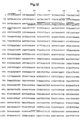
- Example 1 The synthesis of the synthetic env (60) gene is described in Example 1. Its nucleotide sequence and the amino acid sequence derived therefrom (ENV (60)) is shown in FIG. 1.
- the constructions of the plasmids pDS78 / RBSII, 6xHis, penv (60) -DHFR, pDHFR-env (60), pRBSII-env (80) -gag (419) -6xHis and pRBSII-env (80) -gag (419) - env (60) -6xHis are described in detail in Examples 2-4, 6 and 7.
- the expression vectors which contain the DNA sequences which code for the polypeptides according to the invention operatively linked to an expression control sequence can be introduced into any suitable host organism in a manner known per se.
- the selection of a suitable host organism is determined by various factors known to the person skilled in the art. For example, compatibility with the selected vector, toxicity of the expression product, expression characteristics, necessary biological safety precautions and costs play a role and a balance must be found between all of these factors.
- Suitable host organisms are gram-negative and gram-positive bacteria, for example E. coli and B. subtilis strains. Particularly preferred host organisms of the present invention are the E. coli strain M15 (described by Villarejo et al. in J. Bacteriol. 120 , 466-474 [1974] as OZ 291) and E. coli W3110 (ATCC No. 273325). In addition to the E. coli strains mentioned above, other generally available E. coli strains, for example E. coli 294 (ATCC No. 31446) and E. coli RR1 (ATCC No. 31343) can also be used.
- E. coli 294 ATCC No. 31446
- E. coli RR1 ATCC No. 31343
- the manner in which the polypeptides according to the invention are expressed depends on the expression vector / host cell system selected.
- the host organisms which contain a desired expression vector are usually propagated under conditions which are optimal for the growth of the host organisms.
- Induction can be accomplished by adding an inductor or a de-pressor to the growth medium or by changing a physical parameter, e.g. a change in temperature.
- expression is controlled by the lac repressor.
- IPTG isopropyl- ⁇ -D-thiogalactopyranoside
- the host organisms can be digested using a detergent, for example sodium dodecyl sulfate (SDS).
- a detergent for example sodium dodecyl sulfate (SDS).
- SDS sodium dodecyl sulfate
- the host organisms can be isolated by mechanical [Charm et al., Meth. Enzymol. 22 , 476-556 (1971)], enzymatic (lysozyme treatment) or chemical (detergent treatment, urea or guanidinium chloride treatment, etc.) means, or by a combination of these means.
- polypeptides according to the invention After the polypeptides according to the invention have been dissolved out of the host organisms, they can be prepared using known methods, for example by centrifugation at different speeds, by precipitation with ammonium sulfate, by dialysis (at atmospheric pressure or under reduced pressure), by preparative isoelectrofocusing, by preparative gel electrophoresis or by means of various chromatographic methods Methods such as gel filtration, high-performance liquid chromatography (HPLC), ion exchange chromatography, reverse phase chromatography and affinity chromatography (for example on Sepharose Blue C1-6B or on carrier-bound monoclonal antibodies directed against the polypeptides according to the invention) are purified.
- HPLC high-performance liquid chromatography
- ion exchange chromatography ion exchange chromatography
- reverse phase chromatography affinity chromatography
- polypeptides according to the invention are preferably purified on nitrilotriacetic acid (NTA) resins of the general formula carrier matrix spacer-NH- (CH2) x -CH (COOH-N (CH2COO ⁇ ) 2Ni2+, where x is 2, 3 or 4 ⁇ ⁇ .
- NTA nitrilotriacetic acid
- Materials which are used in affinity and gel chromatography for example crosslinked dextrans, agarose (in particular in the form known under the trademark Sepharose®) or polyacrylamides, are suitable as the carrier matrix, and the spacers which are already known from affinity chromatography are suitable as spacers -Groups in question ⁇ the groups -O-CH2-CH (OH) -CH2- and -O-CO- are preferred.
- a particularly preferred NTA resin for the purification of the polypeptides according to the invention is that of the formula [Sepharose®CL 6B] -O-CH2-CH (OH) -CH2-NH- (CH2) 4CH (COOH) -N (CH2COO ⁇ ) 2Ni2+.
- polypeptides according to the invention obtainable by the methods described above can, as already mentioned above can be used as diagnostic tools for the detection of antibodies against HIV viruses in human sera.
- the polypeptides according to the invention can be used for this purpose in numerous known detection methods.
- the polypeptides according to the invention can be labeled using known methods and these labeled polypeptides can then be mixed with a human serum sample, which is suspected to contain antibodies against HIV viruses, to form polypeptide / antibody complexes. The complexes formed can then be detected in a manner known per se.
- the polypeptides according to the invention can also be coupled to a solid support and then brought into contact with a human serum sample in order to give a further example.
- Antibodies to HIV viruses in the sample bind to this coupled polypeptide and the complexes so formed, after unbound polypeptides and antibodies have been removed by washing, can be reacted with a reagent such as Staphylococcus aureus Protein A (e.g.
- anti-HIV antibodies antibodies against the polypeptides according to the invention
- various diagnostic tests for the detection of HIV viruses or fragments thereof in human sera or in other biological fluids can be developed.
- Such antibodies can be produced by injecting an immunogenic composition containing a polypeptide according to the invention and a physiologically compatible carrier material into a mammal or bird.
- the one for the injection the necessary amount of protein is known to the person skilled in the art or can be determined by known methods.
- carrier material used in the context of the present invention refers either to known compositions suitable for human administration or to known adjuvants used in animal vaccination. Suitable adjuvants for use in humans and animals are known to the person skilled in the art [WHO Techn. Rep.
- polypeptides according to the invention can also be administered after incorporation into liposomes or other microcarrier materials or after coupling to polysaccharides, other polypeptides or other polymers.
- one or more additional vaccinations follow a few weeks after the first vaccination, which results in a high content of anti-HIV antibodies. These can then be isolated in a manner known per se.
- monoclonal antibodies can also be used for the above tests.
- the methods for producing such antibodies are known to the person skilled in the art [Köhler et al., Nature 256 , 495-497 (1975)].
- the anti-HIV antibodies obtainable by the methods described above can be used for various diagnostic tests for the detection of HIV viruses or fragments thereof. Such tests can be performed in the form of radioimmunoassays, either in solution or on a solid support. However, enzyme immunoassays can also be carried out. These tests can be performed either directly or indirectly using a second antibody directed against the anti-HIV antibodies. Numerous enzyme activities can are coupled to the antibodies, for example peroxidase, glucose oxidase, ⁇ -galactosidase and alkaline phosphatase, which produce a color after adding a substrate solution.
- the principle underlying many of these tests is based on reacting human serum or other biological liquids suspected of containing HIV viruses or fragments thereof with a known amount of anti-HIV antibodies so that antigen / Antibody complexes can arise and these complexes can be detected in a manner known per se.
- anti-HIV antisera there are many other methods of detection using anti-HIV antisera, such as: B. various agglutination tests.
- the interaction between antibodies and HIV viruses or fragments thereof is detected by means of arrangements in which particles are coated with anti-HIV antibodies.
- particles are, for example, latex spheres, liposomes, erythrocytes, polyacrylamide spheres or any suitable polymer.
- test kits consisting of a vessel which contains a polypeptide according to the invention or anti-HIV antibodies of the present invention.
- the synthetic HIV-2 env (60) gene was composed of 14 oligonucleotide fragments, the length of which varied between 17 and 31 nucleotides (see FIG. 1).
- Oligonucleotide fragments 1-14 were simultaneous solid support material was synthesized by the method described by Bannwarth and Iaiza in DNA 5 , 413-419 (1986).
- the polynucleotide kinase was heat-inactivated for 90 minutes (2 min., 95 ° C.) and the phosphorylated oligonucleotide fragments were precipitated after addition of ⁇ l of 5M lithium acetate (LiOAc) and 100 ⁇ l of isopropanol for 30 minutes at -78 ° C.
- LiOAc 5M lithium acetate
- the precipitated oligonucleotide fragments were then added Washed and dried ethanol, the phosphorylated oligonucleotide fragments 2, 3, 8, 9, 10 and the non-phosphorylated fragment 1 or the phosphorylated oligonucleotide fragments 4, 5, 6, 7, 11, 12, 13 and that not phosphorylated e fragment 14 were hybridized according to known methods described in the literature [Maniatis et al., supra] and then ligated in 100 ⁇ l ligase buffer [Maniatis et al., supra] containing 31 units of T4 ligase (37 ° C., 1.5 Hours). The two subfragments obtained were then precipitated as described above, then washed with ethanol and dried.
- the two subfragments were subsequently separated using 6% polyacrylamide gel electrophoresis, eluted according to standard methods [Maniatis et al., Supra], desalted using a Sephadex G-50 column and, using T4 ligase, as described above, to give the desired HIV-2 env (60) -Gen linked together.
- polyacrylamide gel electrophoresis, elution and desalting After purification of the HIV-2 env (60) gene using the methods described above (polyacrylamide gel electrophoresis, elution and desalting), it became as described above, phosphorylated.
- the plasmid pDS78 / RBSII was used to construct the plasmid pDS78 / RBSII, 6xHis.
- E. coli cells transformed with this plasmid and the plasmid pDMI, 1 were deposited with the German Collection of Microorganisms in Göttingen on September 3, 1987 according to the Budapest Treaty [E. coli M15 (pDS78 / RBSII, pDMI, 1), DSM No. 4232].
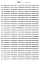
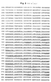
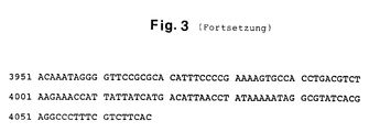
- pDS78 / RBSII The portion of pDS78 / RBSII (FIGS. 2 and 3) which lies between the restriction sites for XbaI and XhoI and which contains the replication region and the gene for ⁇ -lactamase which confers ampicillin resistance to the cells originally comes from the plasmid pBR322 [Bolivar et al., Gene 2: 95-113 (1977); Sutcliffe, Cold Spring Harbor Symp. Quant. Biol. 43: 77-90 (1979)].
- the gene for the ⁇ -lactamase is modified in that the interfaces for the restriction enzymes HincII and PstI are eliminated.
- these changes in the DNA sequence do not affect the amino acid sequence of the ⁇ -lactamase.
- the remaining part of the plasmid carries the regulatable promoter / operator element N25OPSN25OP29 and the ribosomal binding site RBSII.
- This ribosomal binding site was derived from the ribosomal binding site of the promoter P G25 of the E. coli phage T5 [R. Gentz, dissertation, Heidelberg University, FRG (1984)] and obtained as an EcoRI / BamHI fragment via DNA synthesis.
- the gene of the dihydrofolate reductase of the mouse cell line AT-3000 follows [Chang et al., Nature 275 , 617-624 (1978); Masters et al., Gene 21 , 59-63 (1983)], which has been modified to provide an interface for translation just before the termination codon for translation the restriction enzyme BglII was introduced.
- the plasmid pDS78 / RBSII also contains the terminator t o of the E.
- pDS78 / RBSII contains the regulatable promoter / operator element N25OPSN25OP29 and the ribosomal binding site RBSII. Due to the high efficiency of these expression signals, the plasmid pDS78 / RBSII and its derivatives such as the plasmid pDS78 / RBSII, 6xHis can only be kept stable in E. coli cells if the promoter / operator element is attached by binding a lac repressor the operator is repressed. The lac repressor is encoded in the lacI gene. N25OPSN25OP29 can only be repressed efficiently if there is a sufficient number of repressor molecules in the cells.
- the lacI q allele was used which contains a mutant promoter which leads to an increased expression of the repressor gene.
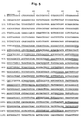
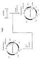
- This lacI q allele is contained in the plasmid pDMI, 1 (FIGS. 4 and 5).
- this plasmid carries the neo gene, which gives the bacteria kanamycin resistance, which is used as a selection marker.
- pDMI, 1 is compatible with the plasmids mentioned above.
- E. coli cells transformed with such expression vectors must contain pDMI, 1 to ensure that the expression vector is kept stable in the cells. This system is induced by adding IPTG to the medium at the desired cell density.
- the plasmid pDMI, 1 (FIGS. 4 and 5) carries the neo gene of neomycin phosphotransferase from the transposon Tn5 [Beck et al., Gene 19 , 327-336 (1982)], which confers kanamycin resistance to E. coli cells and the like lac I gene [Farabough, Nature 274 , 765-769 (1978)] with the promoter mutation I q [Calos, Nature 274 , 762-765 (1978)], which encodes the lac repressor.
- the plasmid pDMI, 1 also contains a region of the plasmid pACYC184 [Chang et al., J. Bacteriol. 134 1141-1156 (1978)], which contains all the information necessary for replication and stable transmission to the daughter cells.
- two complementary oligonucleotides the nucleotide sequence of which is shown in FIG. 6 as a double-stranded DNA sequence, were first synthesized, as described above (Example 1, section B).
- the lyophilized oligonucleotides were taken up in water and dissolved at 4 ° C. for 1 hour.
- the DNA concentration was 100 nmol / ml.
- the DNA of the plasmid pDS78 / RBSII was prepared for ligation with the two phosphorylated oligonucleotides by first cutting 2 pmol of the plasmid DNA with the restriction enzyme BamHI. The sample was extracted with phenol, etherified and the DNA precipitated using lithium acetate / isopropanol as described above. The sediment was dried and taken up in 20 ul TE buffer.
- the said XhoI / BamHI fragment was integrated into the plasmid pDS78 / RBSII, the original XhoI-BamHI fragment of this plasmid being replaced (FIG. 7).
- 1 pmol DNA of the plasmid pDS78 / RBSII was cut with the restriction enzymes XhoI and BamHI before the larger DNA fragment was isolated by means of agarose gel electrophoresis.
- E.coli M15 cells containing plasmid pDMI, 1 were prepared according to the Morrison method [Methods Enzymol. 68 , 326-331 (1979)] prepared for the transformation.
- the ligation mixture was added to 200 ⁇ l of competent cells after heating at 65 ° C for 7 minutes. The mixture was kept in ice for 30 minutes, then incubated at 42 ° C for 2 minutes, and incubated at 37 ° C for 90 minutes after the addition of 0.5 ml LB medium.
- the cells were then plated on LB agar plates containing 100 ⁇ g / ml ampicillin and 25 ⁇ l / ml kanamycin and incubated overnight at 37 ° C. Individual colonies were picked with a sterile toothpick, transferred to a tube containing 10 ml LB medium with 100 ⁇ l / ml ampicillin and 25 ⁇ g / ml kanamycin, and kept in the shaker incubator for 12 hours. The cells were then sedimented and the plasmid DNA was isolated according to the method of Birnboim and Doly [Nucleic Acids Res. 7 , 1515-1523 (1979)].
- Plasmids are included. Plasmids with such a fragment were given the designation pDS78 / RBSII, 6xHis (FIG. 7).
- the double-stranded circular plasmid DNA was sequenced using a primer sequence labeled with ⁇ - [32 P] -ATP.
- This starter sequence contains the nucleotides from position 89-108 of the plasmid pDS78 / RBSII.
- 0.3 pmol of the isolated plasmid DNA was precipitated with alcohol, the sediment was washed once with 80% ethanol, dried and finally dissolved in 8 ⁇ l 1/4 Te buffer. The sample was incubated for 5 minutes at 95 ° C., cooled to 0 ° C. and centrifuged (Eppendorf table centrifuge, 2 minutes, 12,000 rpm).
- the plasmid penv (60) -DHFR was linked to the synthetically produced HIV-2 env (60) gene (Fig. 8).
- the plasmid linearized with the restriction enzyme BglII was used to construct the plasmid pDHFR-env (60) pDS78 / RBSII, 6xHis linked to the synthetically produced HIV-2 env (60) gene (Fig. 9).
- the polypeptides ENV (60) -DHFR and DHFR-ENV (60) in E. coli expressed then transferred to nitrocellulose filters and incubated with suitable sera.
- the HIV-1 ENV (80) -DHFR polypeptide [Certa et al., EMBO J. 5 , 3051-3056 (1986)] served as a control.
- the Nitrocellulose membranes were first incubated for 4 hours at room temperature in PBS [Maniatis et al., Supra] containing 5% dried skim milk. Then the one nitrocellulose membrane in PBS containing 5% dried skim milk and 1: 1000 diluted HIV-1 positive serum and the other membrane in PBS containing 5% dried skim milk and 1: 1000 diluted HIV-2 positive serum for 4 hours incubated at room temperature. The nitrocellulose membranes were then washed three times with PBS containing 0.3% Tween-20 and then for 1 hour at room temperature in PBS containing 0.3% Tween-20, 5% goat serum and 1: 2000 diluted goat anti Human IgG peroxidase conjugate (Nordic), incubated.
- PBS Maniatis et al., Supra
- the ENV (60) -DHFR and DHFR-ENV (60) polypeptides are only recognized by antibodies in the serum of HIV-2 infected patients.
- the ENV (80) -DHFR polypeptide is only recognized by antibodies in the serum of HIV-1 infected patients.
- the plasmid pDS56 / RBSII was used to construct the plasmid pRBSII-6xHis.
- E. coli cells transformed with this plasmid and the plasmid pDMI, 1 were deposited with the German Microorganism Collection in Braunschweig on December 23, 1987 in accordance with the Budapest Treaty [E. coli M15 (pDS56 / BSII; pDMI, 1), DSM-No .: 4330].
- the remaining part of the plasmid carries the regulatable promoter / operator element N250PSN250P29 followed by the ribosome binding site RBSII, which is part of an EcoRI / BamHI fragment. This is followed by the terminator t o of the E. coli phyagen Lambda (Schwarz et al., Supra), the promoter-free gene of chloramphenicol acetyltransferase (Marcoli et al., Supra) and the terminator T1 of the E. coli rrnB operon (Brosius et al., supra).
- the plasmid pDS56 / RBSII and its derivatives can only be kept stable in E. coli cells if the promoter / operator element is bound by binding lac repressor is repressed to the operator (see example 2, section A).
- the HIV-2 gene env (60) and the HIV-1 genes env (80) and gag (419) with the expression vector were used pRBSII-6xHis linked.
- T4 DNA ligase After adding one unit of T4 DNA ligase, the mixture was incubated at 14 ° C. overnight. The mixture was then incubated at 65 ° C. for 10 minutes and brought to a volume of 100 ⁇ l with restriction enzyme buffer. 100 units of BamHI and 10 units of HindIII were added and the mixture was then incubated at 37 ° C. for 3 hours. The DNA was then extracted with phenol and chloroform and precipitated with ethanol. The DNA pellet was dissolved in sample buffer and separated on a 6% polyacrylamide gel. After staining with ethidium bromide, a 250 base pair band was cut out and isolated by electroeluation.
- the DNA was extracted with phenol: chloroform (1: 1), precipitated with ethanol, in 50 ⁇ l 50 mM Tris-HCl, pH7.6, containing 10mM MgCl2, 10mM DTT, 0.5mM ATP, 100 pmol phosphorylated 12er BglII linker ( 5′-GGAAGATCTTCC-3 ′) and 1 unit of T4 DNA ligase ⁇ incubated overnight at 14 ° C. The reaction mixture was then heated to 65 ° C. and brought to a volume of 100 ⁇ l with restriction enzyme buffer. 100 units of BglII were added and then incubated for 3 hours at 37 ° C.
- HIV-1 env (80) gene was prepared as described by Certa et al., [EMBOJ.5, 3051-3056 (1986)].
- plasmid pRBSII-gag (419) -6xHis 0.1 pmol of the vector pRBSII-6xHis (see B) linearized with BamHI and BglII (see B) with 0.3 pmol of the gag (419) gene by incubation with 1 unit of T4 DNA ligase at 14 ° C linked overnight. After heat inactivation of the enzyme, the DNA was transformed into E. coli strain W3110, which contained the plasmid pDMI, 1. The cells were plated on LB agar plates containing 100 ⁇ g / ml ampicillin and 25 ⁇ g / ml kanamycin. The plates were incubated overnight at 37 ° C. Individual colonies were grown overnight at 37 ° C. and the plasmid DNA was then isolated by standard methods [Maniatis et al., Supra] (FIG. 18).
- plasmid pRBSII-env (80) -gag (419) -6xHis 0.1 pmol of the plasmid pRBSII-gag (419) -6xHis was cut with BamHI and treated with CIP. The DNA was extracted with phenol: chloroform (1: 1) and precipitated with 2 volumes of ethanol. BamHI linearized pRBSII-gag (419) -6xHis plasmid DNA was incubated with 0.3 pmol of the HIV-1 env (80) gene and 1 unit of T4 DNA ligase at 14 ° C. overnight. After heat inactivation of the enzyme, the DNA was transformed into the E.
- coli strain W3110 which contained the plasmid pDMI, 1.
- the cells were plated on LB agar plates containing 100 ⁇ g / ml ampicillin and 25 ⁇ g / ml kanamycin. The plates were incubated overnight at 37 ° C. Individual colonies were grown overnight at 37 ° C. and the plasmid DNA was then isolated by standard methods [Maniatis et al., Supra] (FIG. 19).
- pRBSII-env (80) -gag (419) -env (60) -6xHis 0.1pmol of the plasmid pRBSII-env (80) -gag (419) -6xHis BglII was cut and treated with CIP. The DNA was then extracted with phenol: chloroform (1: 1) and precipitated with 2 volumes of ethanol. The pRBSII-env (80) -gag (419) -6xHis plasmid DNA linearized with BglII was then incubated with 0.3 pmol of the HIV-2 env (60) gene and 1 unit of T4-DNA ligase at 14 ° C.
- the DNA was transformed into the E. coli strain W3110, which contained the plasmid pDMI, 1.
- the cells were plated on LB agar plates containing 100 ⁇ g / ml ampicillin and 25 ⁇ g / ml kanamycin. The plates were incubated overnight at 37 ° C. Individual colonies were grown overnight at 37 ° C. and the plasmid DNA was then isolated by standard methods [Maniatis et al., Supra] (FIG. 20).
- ENV (80) -GAG (419) -ENV (60) polypeptide reacts with sera from HIV-1 and HIV-2 infected individuals
- ENV (80) -GAG (418) -ENV (60 ) Polypeptide expressed in E. coli then purified and tested with suitable sera in the enzyme immunoassay (EIA).
- E. coli W3110 cells containing the plasmid pDMI, 1, were transformed with the plasmid pRBSII-env (80) -gag (419) -env (60) -6xHis and grown in LB medium [Maniatis et al., supra] containing 100 ⁇ g / ml ampicillin and 25 ⁇ g / ml kanamycin .
- LB medium Gibco-d et al., supra
- the culture was induced with IPTG (2 mM final conc.) And allowed to grow for a further 2 hours. The cells were then harvested by centrifugation.
- the harvested cells were resuspended in PBS buffer (0.2 g / l KCl, 8.0 g / l NaCl, 0.2 g / l KH2PO4, 1.144 g / l Na2HPO4, pH 7.0) and broken up using a high-pressure homogenizer.
- the cell homogenate obtained was centrifuged and the pellet was extracted twice with 0.1M sodium phosphate buffer, pH 8.0, containing 6M guanidine ⁇ HCl.
- the ENV (80) -GAG (419) -ENV (60) eluate obtained using an NTA column was then subjected twice to a Sephacryl S-200 column (Pharmacia, running buffer: 50mM Tris.HCl, pH 7.0, containing 5mM EDTA and 1% or 0.1% SDS) chromatographed. Using SDS polyacrylamide gel electrophoresis and amino acid analysis, a purity of 89% of the ENV (80) -GAG (419) -ENV (60) polypeptide obtained was detected.
- the purified ENV (80) -GAG (419) -ENV (60) polypeptide was, as in the European patent application, publication no. 270 114 described, bound to polystyrene balls tested for its reactivity with HIV-1 and HIV-2 positive sera in the enzyme immunoassay (EIA).
- EIA enzyme immunoassay
- ENV (80-GAG (419) -ENV (60) polypeptide recognizes all positive HIV-1 and HIV-2 sera.
Description
- Die vorliegende Erfindung betrifft neue Polypeptide, welche mindestens eine antigene und/oder immunogene determinante Gruppe des Hüllproteins (env) des HIV-2 Virus einschliessen, DNA-Sequenzen, die diese Polypeptide kodieren, rekombinante Vektoren, die diese DNA-Sequenzen enthalten, mit diesen rekombinanten Vektoren transformierte Mikroorganismen sowie Verfahren zu deren Herstellung mittels rekombinanter DNA Technologie. Die Erfindung betrifft ferner Verfahren zum Nachweis von HIV-Antikörpern (HIV-2 Antikörper oder HIV-1 und HIV-2 Antikörper) oder HIV-Viren (HIV-2 Viren oder HIV-1 und HIV-2 Viren) in menschlichen Seren oder anderen biologischen Körperflüssigkeiten.
- 1986 wurde ein neues Virus mit der Bezeichnung HIV-2 aus westafrikanischen AIDS-Patienten isoliert [Clavel et al., Science 233, 343-346 (1986); Clavel et al., Nature 324, 691-695 (1986)]. Dieses Virus wurde auf Grund seiner Morphologie, seines Lymphotropismus und seiner cytophatischen in vitro Wirkung auf T₄-positive Zellen mit den AIDS verursachenden HIV-1 Viren in Verbindung gebracht. Trotz dieser ähnlichen Eigenschaften zeigt ein genetischer Vergleich von HIV-1 und HIV-2 Viren jedoch nur eine begrenzte Sequenzhomologie [Guyader et al., Nature 326, 662-669 (1987)1.
- Mehr als 20 verschiedene HIV-2 Viren wurden aus westafrikanischen AIDS-Patienten aber auch aus europäischen AIDS-Patienten isoliert [Guyader et al., supra; Brun-Vezinet et al., Lancet 1, 128-132 (1987)]. Die Seren dieser AIDS-Patienten waren im HIV-1 ELISA alle negativ [Brun-Vezinet et al., supra]. Deshalb ist das Bedürfnis nach genauen und schnellen Verfahren für die Diagnose von HIV-2 Viren in menschlichem Blut und in anderen Körperflüssigkeiten sehr gross.
- EP-A-284 383 (Publikationsdatum 28.9.1988) beschreibt kurze Peptide aus dem Hüllprotein (env) und Strukturprotein (gag) des LAV-2, die zur Detektion von Antikörpern gegen dieses Virus geeignet sind. In der PCT-Anmeldung WO87/04459 (Publikationsdatum 30.7.1987) wird die Isolierung und Charakterisierung von HIV-2, insbesondere des grossen Hüllproteins (env) sowie bestimmter Fragmente davon, beschrieben. EP-A-283 327 (Publikationsdatum 21.9.1988) beschreibt kurze Peptide aus den env Proteinen von HIV-1, HIV-2 und SIV-1 mit homologen Sequenzen.
- Durch die Anwendung rekombinanter DNA-Techniken wurden deshalb neue Polypeptide mit einer Aminosäuresequenz, die mit mindestens einer antigenen und/oder immunogenen determinanten Gruppe des Hüllproteins (env) des HIV-2 Virus übereinstimmt, hergestellt. Diese Polypeptide gestatten den Nachweis von HIV-2 Antikörpern oder HIV-2 Viren oder Fragmenten davon in menschlichen Seren oder anderen biologischen Körperflüssigkeiten.
- Die vorliegende Erfindung betrifft deshalb Polypeptide mit einer Aminosäuresequenz, die mit mindestens einer antigenen und/oder immunogenen determinanten Gruppe des HIV-2 env-Proteins übereinstimmt.
- Genauer gesagt, betrifft die vorliegende Erfindung ein Polypeptid mit der Aminosäuresequenz

oder Fragmente oder funktionelle Aequivalente davon, kovalent verknüpft mit einem Affinitätspeptid und einem Polypeptid, dessen Aminosäuresequenz mit mindestens einer antigenen und/oder immunogenen determinanten Gruppe des HIV-1 Hüllproteins (env) und/oder des HIV-1 Strukturproteins (gag) übereinstimmt. Da solche Fusionsproteine sowohl antigene und/oder immunogene determinanten Gruppe von HIV-1 als auch HIV-2 Viren aufweisen, stellen diese Fusionspolypeptide ein wirksames diagnostisches Werkzeug zum gleichzeitigen Nachweis von HIV-1 und HIV-2 Antikörpen oder HIV-1 und HIV-2 Viren oder Fragmenten davon in menschlichen Seren oder anderen biologischen Körperflüssigkeiten dar. - Die bevorzugten Polypeptide gemäss der vorliegenden Erfindung sind durch die allgemeinen Formeln
A - B - C
A - C - B
und
C - B - A
definiert, worin - A
- ein Affinitätspeptid ist,
- B
- ein Polypeptid mit der Aminosäuresequenz der Formel I ist, und
- C
- ein Polypeptid, dessen Aminosäuresequenz mit mindestens einer antigenen und/oder immunogenen determinanten Gruppe HIV-1 Hüllproteins (env) und/oder des HIV-1 Strukturproteins (gag) übereinstimmt, ist.
- Ein besonders bevorzugt erfindungsgemäss Polypeptid hat der Formel

- Der Ausdruck "funktionelle Aequivalente" der im Zusammenhang mit den erfindungsgemässen Polypeptiden verwendet wird, bezieht sich auf Polypeptide, deren Aminosäuresequenzen durch Nukleotidsubstitutionen, Nukleotid-Deletionen, Nukleotid-Insertionen oder Nukleotid-Additionen aus den oben gezeigten Aminosäuresequenzen hervorgegangen sind und mit mindestens einer antigenen und/oder immunogenen determinanten Gruppe des HIV-2 env-Proteins übereinstimmen.
- Gewisse Substitutionen in der Aminosäuresequenz eines Polypeptids haben keinen Einfluss auf die biologische Aktivität eines Polypeptids. Beispiele solcher Aminosäuresubstitutionen sind Ala/Ser, Val/Ile, Asp/Glu, Thr/Ser, Ala/Gly, Ala/Thr, Ser/Asn, Ala/Val, Ser/Gly, Tyr/Phe, Ala/Pro, Lys/Arg, Asp/Asn, Leu/Ile, Leu/Val, Ala/Glu und vice versa (vgl. Doolittle, in "The Proteins", ed. Neurath, H, und Hill, R.L., Academic Press, New York [1979]).
- Unter einem Affinitätspeptid werden Peptide verstanden, die eine Aminosäuresequenz enthalten, die bevorzugt an ein Affinitätschromatographieträgermaterial bindet. Beispiele für solche Affinitätspeptide sind Peptide, die mindestens zwei Histidinreste enthalten (siehe hierzu europäische Patentanmeldung, Publ. Nr. 282 042). Solche Affinitätspeptide binden selektiv an Nitrilotriessigsäure-Nickelchelatharze [Hochuli and Döbeli, Biol. Chem. Hoppe-Seyler 368, 748 (1987); europäische Patentanmeldung, Publ, Nr. 253 303]. Polypeptide, die ein solches Affinitätspeptid enthalten, können damit selektiv von den übrigen Polypeptiden abgetrennt werden. Das Affinitätspeptid kann entweder mit dem C-Terminus oder dem N-Terminus der Polypeptide mit der Aminosäuresequenz der Formel I oder Fragmenten oder funktionellen Aequivalenten davon verknüpft sein.
- Die erfindungsgemässen Polypeptide können aufgrund ihrer Herstellungsverfahren Methionin (für das ATG kodiert) als erste N-terminale Aminosäure enthalten. Andererseits kann der mikrobielle Wirt das Translationsprodukt teilweise oder vollständig prozessieren, wodurch das N-terminale Methionin abgespalten wird.
- Die erfindungsgemässen Polypeptide können auch in Form von Multimeren, z.B. in Form von Dimeren, Trimeren, Tetrameren etc., vorliegen. Natürlich können die erfindungsgemässen Polypeptide auch mit Polypeptiden, deren Aminosäuresequenz mit mindestens einer antigenen und/oder immunogenen Determinante des HIV-2 Strukturproteins (gag) übereinstimmt, kovalent verknüpft sein.
- Die Erfindung liefert ferner DNA-Sequenzen, die die erfindungsgemässen Polypeptide kodieren, rekombinante Vektoren die diese DNA-Sequenzen enthalten, einzellige Organismen zur Herstellung der erfindungsgemässen Polypeptide sowie Verfahren zur Herstellung solcher DNA-Sequenzen, rekombinanter Vektoren und einzelligen Organismen. Es werden auch Methoden zur Expression, Isolierung und Reinigung der erfindungsgemässen Polypeptide beschrieben. Die so hergestellten erfindungsgemässen Polypeptide können mit den Methoden dieser Erfindung für eine Anzahl wichtiger immunologischer Prozesse verwendet werden.
- Die erfindungsgemässen Polypeptide können als diagnostisches Reagens zum Nachweis von Antikörpern gegen HIV-2 Viren oder zum gleichzeitigen Nachweis von Antikörpern gegen HIV-1 und HIV-2 Viren (im folgenden kollektiv als Antikörper gegen HIV-Viren bezeichnet) in menschlichen Seren verwendet werden. Da sie in homogener Form hergestellt werden können, können Probleme mit unspezifischen Reaktionen, die die Verwendung von diagnostischen Reagenzien auf der Basis relativ roher viraler HIV Proteinlysate in der Vergangenheit eingeschränkt haben, eliminiert werden.
- Als Immunogen verwendet, können die erfindungsgemässen Polypeptide zur Produktion von Antikörpern, die gegen die in diesen Polypeptiden enthaltenen antigenen determinanten Gruppen gerichtet sind, in Tieren verwendet werden. Solche Antikörper wiederum können in Verbindung mit den erfindungsgemässen Polypeptiden, die entsprechend markiert wurden, in einem Radioimmunassay (RIA) oder in einem Enzymimmunassay (ELISA) verwendet werden, um die Gegenwart von HIV-2 Viren oder HIV-1 und HIV-2 Viren (im folgenden kollektiv als HIV-Viren bezeichnet) oder Partikeln davon in menschlichen Seren oder in anderen biologischen Flüssigkeiten¸ wie z.B. in Tränenflüssigkeit, Samen, Vaginalsekret und Speichel, festzustellen. Die Partikel (oder Fragmente) von HIV-Viren, die mit diesen Methoden nachgewiesen werden können, umfassen natürliche Teile der viralen HIV-Hüllproteine (env).
- Die erfindungsgemässen Polypeptide können mittels konventioneller Methoden der Peptidsynthese, in flüssiger oder, vorzugsweise an fester Phase, wie der Methode von Merrifield (J. Am. Chem. Soc. 85, 2149-2154 [1963]), oder mittels anderer gleichwertiger Methoden des Standes der Technik, hergestellt werden.
- Andererseits können die erfindungsgemässen Polypeptide auch unter Verwendung der Methoden der DNA-Rekombinationstechnik [Maniatis et al. in "Molecular Cloning - A Laboratory Manual", Cold Spring Harbor Laboratory (1982)] hergestellt werden. Beispielsweise können die DNA-Sequenzen, die für die erfindungsgemässen Polypeptide codieren, mittels konventioneller chemischer Methoden synthetisiert werden, z.B. mittels der Phosphotriestermethode [Narang et al., In Meth. Enzymol. 68, 90-108 (1979)] oder mittels der Phosphodiestermethode [Brown et al., Meth. Enzymol. 68, 109-151 (1979)]. Bei beiden Methoden werden in der Regel längere Oligonukleotide synthetisiert, die in vorgegebener Art und Weise aneinander gehängt werden. Die Nukleotidsequenzen der DNA-Fragmente können mit denjenigen Nukleotidsequenzen, die die natürlichen HIV-2 oder HIV-1 und HIV-2 Polypeptide codieren, identisch sein. Da der genetische Code degeneriert ist, besteht andererseits die Möglichkeit, dass teilweise oder vollständig unterschiedliche Nukleotidsequenzen die gleichen Polypeptide codieren. Gegebenenfalls können für die Nukleotidsequenzen solche Codons gewählt werden, die auch vom Wirtsorganismus, der zur Expression des Polypeptids verwendet wird, bevorzugt verwendet werden [Grosjean et al., Gene 18, 199-209 (1982)]. Dabei muss aber darauf geachtet werden, dass die so erhaltenen DNA-Sequenzen keine Teilsequenzen enthalten, die die Konstruktion von Expressionsvektoren, z.B. durch Einführung einer unerwünschten Restriktionsenzymschnittstelle, erschweren.
- Im weiteren können die DNA-Sequenzen, die für die erfindungsgemässen Polypeptide kodieren, auch hergestellt werden, in dem man ein DNA-Fragment, das für die Aminosäuresequenz der Formel I kodiert, aus isolierter proviraler HIV-2 DNA oder aus genomischer DNA von Zellen, in welche das provirale HIV-2 Genom integriert wurde, isoliert und anschliessend in einen geeigneten Vektor, der die Teilsequenzen A und C der allgemeinen Formeln A-B-C, A-C-B und C-B-A kodiert, einbaut.
- Nach der Herstellung der DNA-Sequenzen, die für die erfindungsgemässen Polypeptide kodieren, können diese nach bekannten Methoden in jeden geeigneten Expressionsvektor, der die notwendigen Expressionssignale liefert, eingebaut werden. Geeignete Vektoren können aus Segmenten von chromosomalen, nicht-chromosomalen und synthetischen DNA-Sequenzen zusammengesetzt sein, wie z.B. verschiedene bekannte Plasmide und Phagen DNA's. Hierzu kann auf das vorstehend genannte Lehrbuch von Maniatis et al. verwiesen werden. Besonders geeignete Vektoren sind Plasmide der pDS-Familie [Bujard et al., Methods in Enzymology, Hrsg. Wu und Grossmann, Academic Press, Inc., Vol. 155, 416-433 (1987)].
- In den bevorzugten Ausführungsformen der vorliegenden Erfindung wurde ein synthetisches BamH1 Fragment, welches die Aminosäuresequenz der Formel I kodiert (env(60)-Gen), mit BamH1 bzw. BglII gespaltener DNA des Plasmids pDS78/RBSII,6xHis und mit BglII gespaltener DNA des Plasmids pRBSII-env(80)-gag(419)-6xHis verbunden, um die Expressionsvektoren penv(60)-DHFR, pDHFR-env(60) und pRBSII-env(80)-gag(419)-env(60)-6xHis zu isolieren, welche die Synthese der besonders bevorzugten erfindungsgemässen Polypeptide der Formeln II-IV kodieren. Die Synthese des synthetischen env(60)-Gens ist in Beispiel 1 beschrieben. Seine Nukleotidsequenz und die davon abgeleitete Aminosäuresequenz (ENV(60)) ist in Figur 1 gezeigt. Die Konstruktionen der Plasmide pDS78/RBSII, 6xHis, penv(60)-DHFR, pDHFR-env(60), pRBSII-env(80)-gag(419)-6xHis und pRBSII-env(80)-gag(419)-env(60)-6xHis werden im Detail in den Beispielen 2-4, 6 und 7 beschrieben.
- Die Expressionsvektoren, welche die DNA-Sequenzen, die für die erfindungsgemässen Polypeptide kodieren, operativ mit einer Expressionskontrollsequenz verknüpft enthalten, können in an sich bekannter Weise in jeden geeigneten Wirtsorganismus eingeführt werden. Die Auswahl eines geeigneten Wirtsorganismus wird von verschiedenen dem Fachmann bekannten Faktoren bestimmt. So spielen beispielsweise Kompatibilität mit dem ausgewählten Vektor, Toxizität des Expressionsproduktes, Expressionscharakteristika, notwendige biologische Sicherheitsvorkehrungen und Kosten eine Rolle und es muss ein Mittelweg zwischen allen diesen Faktoren gefunden werden.
- Als geeignete Wirtsorganismen kommen gram-negative und gram-positive Bakterien in Frage, beispielsweise E. coli und B. subtilis Stämme. Besonders bevorzugte Wirtsorganismen der vorliegenden Erfindung sind der E. coli Stamm M15 (von Villarejo et al. in J. Bacteriol. 120, 466-474 [1974] als OZ 291 beschrieben) und E. coli W3110 (ATCC Nr. 273325). Ausser den oben genannten E. coli Stämmen können aber auch andere allgemein zugängliche E. coli Stämme, beispielsweise E. coli 294 (ATCC Nr. 31446) und E. coli RR1 (ATCC Nr. 31343) verwendet werden.
- Die Art und Weise wie die Expression der erfindungsgemässen Polypeptide erfolgt ist abhängig vom gewählten Expressionsvektor/Wirtszellsystem. Ueberlicherweise werden die Wirtsorganismen, die einen gewünschten Expressionsvektor enthalten, unter Bedingungen, die für das Wachstum der Wirtsorganismen optimal sind, vermehrt. Gegen Ende des exponentiellen Wachstums, wenn die Zunahme der Zellzahl pro Zeiteinheit abnimmt, wird die Expression des gewünschten Polypeptids induziert, d.h. die das gewünschte Polypeptid kodierende DNA wird transkribiert und die transkribierte mRNA wird translatiert. Die Induktion kann durch Zugabe eines Induktors oder eines Derepressors zum Wachstummedium oder durch Veränderung eines physikalischen Parameters, z.B. einer Temperaturänderung, erfolgen. In den in den bevorzugten Ausführungsformen der vorliegenden Erfindung verwendeten Expressionsvektoren, wird die Expression durch den lac-Repressor kontrolliert. Durch Zugabe von Isopropyl-β-D-thiogalactopyranosid (IPTG) wird die Expressionskontrollsequenz dereprimiert und dadurch die Synthese des gewünschten Polypeptids induziert.
- Zur Isolierung von geringen Mengen an erfindungsgemässen Polypeptiden für analytische Zwecke, z.B. für Polyacrylamidgelektrophoresen, können die Wirtsorganismen mittels eines Detergenz, z.B. Natriumdodecylsulfat (SDS) aufgeschlossen werden. Zur Isolierung von grossen Mengen an erfindungsgemässen Polypeptiden können die Wirtsorganismen durch mechanische [Charm et al., Meth. Enzymol. 22, 476-556 (1971)], enzymatische (Lysozymbehandlung) oder chemische (Detergenzbehandlung, Harnstoff- oder Guanidiniumchlorid-Behandlung, etc.) Mittel, oder durch eine Kombination dieser Mittel, aufgeschlossen werden.
- Nachdem die erfindungsgemässen Polypeptide aus den Wirtsorganismen herausgelöst sind, können sie mittels bekannter Methoden, z.B. mittels Zentrifugation bei unterschiedlichen Geschwindigkeiten, mittels Präzipitation mit Ammoniumsulfat, mittels Dialyse (bei Normaldruck oder bei reduziertem Druck), mittels präparativer Isoelektrofokussierung, mittels präparativer Gelelektrophorese oder mittels verschiedener chromatographischer Methoden wie Gelfiltration, Hochleistungs-Flüssigkeitschromatographie (HPLC), Ionenaustauscherchromatographie, Umkehrphasenchromatographie und Affinitätschromatographie (z.B. an Sepharose Blau C1-6B oder an trägergebundenen, gegen die erfindungsgemässen Polypeptide gerichteten monoklonalen Antikörpern) gereinigt werden. Vorzugsweise jedoch werden die erfindungsgemässen Polypeptide an Nitrilotriessigsäure (NTA)-Harzen der allgemeinen Formel Trägermatrix-Spacer-NH-(CH₂)x -CH(COOH-N(CH₂COO⁻)₂Ni²⁺, worin x 2, 3 oder 4 bedeutet¸ gereinigt. Als Trägermatrix kommen Materialien in Frage, wie sie in der Affinitäts- und Gelchromatographie verwendet werden, beispielsweise vernetzte Dextrane, Agarose (insbesondere in der unter dem Warenzeichen Sepharose® bekannten Form) oder Polyacrylamide. Als Spacer kommen die aus der Affinitäts-Chromatographie bereits bekannten Spacer-Gruppen in Frage¸ wobei die Gruppen -O-CH₂-CH(OH) -CH₂- und -O-CO- bevorzugt sind.
- Ein besonders bevorzugtes NTA-Harz für die Reinigung der erfindungsgemässen Polypeptide ist das der Formel
[Sepharose®CL 6B]-O-CH₂-CH(OH)-CH₂-NH-(CH₂)₄CH(COOH)-N(CH₂COO⁻)₂Ni²⁺.
- Die nach den vorstehend beschriebenen Methoden erhältlichen erfindungsgemässen Polypeptide können, wie bereits vorstehend erwähnt, als diagnostische Werkzeuge zum Nachweis von Antikörpern gegen HIV-Viren in menschlichen Seren verwendet werden. Die erfindungsgemässen Polypeptide können zu diesem Zweck in zahlreichen, bekannten Nachweisverfahren verwendet werden.
- Beispielsweise können die erfindungsgemässen Polypeptide nach bekannten Methoden markiert werden und diese markierten Polypeptide dann zur Ausbildung von Polypeptid/Antikörper-Komplexen mit einer menschlichen Serumsprobe, die im Verdacht steht Antikörper gegen HIV-Viren zu enthalten, vermischt werden. Die gebildeten Komplexe können dann in an sich bekannter Weise nachgewiesen werden. Die erfindungsgemässen Polypeptide können aber auch an einen festen Träger gekoppelt und dann mit einer menschlichen Serumsprobe in Kontakt gebracht werden, um ein weiteres Beispiel zu geben. Antikörper gegen HIV-Viren in der Probe binden an dieses gekoppelte Polypeptid und die so gebildeten Komplexe können, nachdem nicht-gebundene Polypeptide und Antikörper durch Waschung entfernt wurden, mittels eines Reagenz, wie beispielsweise Staphylococcus aureus Protein A (beispielsweise mit ¹²⁵Jod markiert) oder einem zweiten anti-Ig-Antikörper (beispielsweise mit einem Radioisotop oder mit Meerrettichperoxidase markiert) nachgewiesen werden. Viele Modifikationen und Variationen dieser vorstehend genannten Nachweismethoden liegen für den Fachmann im Bereich des möglichen, von denen einige im folgenden vorgeschlagen werden.
- Durch Verwendung von Antikörpern gegen die erfindungsgemässen Polypeptide (im folgenden anti-HIV-Antikörper) können verschiedene diagnostische Tests zur Erkennung von HIV-Viren oder Fragmenten davon in menschlichen Seren oder in anderen biologischen Flüssigkeiten entwickelt werden. Solche Antikörper können durch Injektion einer immunogenen Zusammensetzung, enthaltend ein erfindungsgemässes Polypeptid und ein physiologisch verträgliches Trägermaterial, in einen Säuger oder Vogel hergestellt werden. Die für die Injektion notwendige Menge Protein ist dem Fachmann bekannt oder kann nach bekannten Methoden bestimmt werden. Der im Zusammenhang mit der vorliegenden Erfindung verwendete Ausdruck "Tragermaterial" bezieht sich entweder auf bekannte Zusammensetzungen, die für menschliche Verabreichung geeignet sind, oder auf bekannte, bei der Tierimpfung verwendete Adjuvantien. Geeignete Adjuvantien für die Anwendung an Mensch und Tier sind dem Fachmann bekannt [WHO Techn. Rep. Series 595, 1-40 (1976); Jollis et al., "Chemical and Biological Basis of Adjuvants", in Molecular Biochemistry and Biophysics Vol. 13, 1-148 (1973) Springer Verlag Berlin]. Die erfindungsgemässen Polypeptide können aber auch nach Einbau in Liposomen oder andere Mikro-Trägermaterialien oder nach Kopplung an Polysaccharide, andere Polypeptide oder andere Polymere verabreicht werden.
- In einem besonders typischen Beispiel folgen einige Wochen nach der ersten Impfung eine oder mehrere Zusatzimpfungen, was einen hohen Gehalt an anti-HIV-Antikörpern zur Folge hat. Diese können dann in an sich bekannter Weise isoliert werden.
- Natürlich können auch monoklonale Antikörper für die vorstehend genannten Tests verwendet werden. Die Methoden zur Herstellung solcher Antikörper sind dem Fachmann bekannt [Köhler et al., Nature 256, 495-497 (1975)].
- Die nach den vorstehend beschriebenen Methoden erhältlichen anti-HIV-Antikörper können, wie bereits vorstehend erläutert, für verschiedene diagnostische Tests zum Nachweis von HIV-Viren oder Fragmenten davon verwendet werden. Solche Tests können in Form von Radioimmunoassays, entweder in Lösung oder an festem Träger, durchgeführt werden. Es können aber auch Enzymimmunoassays durchgeführt werden. Diese Test konnen entweder direkt oder indirekt mittels eines zweiten Antikörpers, der gegen die anti-HIV-Antikörper gerichtet ist, durchgeführt werden. Zahlreiche Enzymaktivitäten können an die Antikörper gekoppelt werden, z.B. Peroxidase, Glucoseoxidase, β-Galactosidase und Alkalische Phosphatase, die nach Zugabe einer Substratlösung eine Färbung erzeugen.
- Das Prinzip das vielen dieser Tests zugrunde liegt beruht darauf, dass man menschliches Serum oder andere biologische Flüssigkeiten, die im Verdacht stehen HIV-Viren oder Fragmente davon zu enthalten, mit einer bekannten Menge an anti-HIV-Antikörpern reagieren lässt, so dass Antigen/Antikörper-Komplexe entstehen können und diese Komplexe in an sich bekannter Weise nachweist.
- Der Fachmann wird auch erkennen, dass es viele andere Nachweisverfahren gibt, bei denen anti-HIV-Antiseren zum Einsatz kommen, wie z. B. verschiedene Agglutinationstests. Hierbei wird die Wechselwirkung zwischen Antikörpern und HIV-Viren oder Fragmenten davon mittels Anordnungen, in denen Partikel mit anti-HIV-Antikörpern überzogen sind, nachgewiesen. Solche Partikel sind beispielsweise Latex-Kugeln, Liposomen, Erythrozyten, Polyacrylamidkugeln oder irgendein geeignetes Polymer.
- Die vorstehend beschriebenen Methoden zum Nachweis von HIV-Viren oder von Antikörpern gegen HIV-Viren können in geeigneten Test-Kits bestehend aus einem Gefäss, welches ein erfindungsgemässes Polypeptid oder anti-HIV-Antikörper der vorliegenden Erfindung enthält, durchgeführt werden.
- Die folgenden Figuren und die nachfolgenden Beispiele tragen zum besseren Verständnis der vorliegenden Erfindung bei, ohne sie zu beschränken.
- In den Figuren treten die folgenden Abkürzungen und Symbole auf:
B, Bg, E, H, P, Sa, X und Xb bezeichnen Schnittstellen für die Restriktionsenzyme BamHI, BglII, EcoRI, HindIII, PstI, SalI, XhoI bzw. XbaI.
repräsentiert die Promotoren der Gene bla, lacI und neo;
repräsentiert die ribosomalen Bindungsstellen der Gene bla, cat, neo und lacI;
repräsentiert die Terminatoren t₀ und T1;
repräsentiert das regulierbare Promotor/Operator Element N25OPSN25OP29;
repräsentiert die ribosomale Bindungstelle RBSII;→ repräsentiert die kodierende Region unter Kontrolle dieser ribosomalen Bindungsstelle;
repräsentiert die Regionen, die für die sechs Histidine kodiert;
repräsentiert die für die Replikation benötigte Region (repl.);
repräsentiert kodierende Regionen für Dihydrofolatreduktase (dhfr), Chloramphenicolacetyltransferase, lac-Repressor (lacI), β-Lactamase (bla) und Neomycinphosphotransferase (neo);
repräsentiert das synthetische env(60)-Gen;
repräsentiert HIV-1 gag-Genfragmente. - Fig. 1
- Darstellung der Nukleotidsequenz des synthetischen HIV-2 env(60)-Gens und die davon abgeleitete Aminosäuresequenz des HIV-2 ENV(60) Polypeptide. Die individuellen Oligonukleotidfragmente, aus denen das synthetische HIV-2T env(60)-Gen zusammengesetzt ist, sind durch die Zahlen 1-14 gekennzeichnet.
- Fig. 2
- Schematische Darstellung des Plasmids pDS78/RBSII.
- Fig. 3
- Nukleotidsequenz des Plasmid pDS78/RBSII. In der Sequenz sind die Erkennungsstellen für die in Fig. 2 aufgeführten Restriktionsenzyme überstrichen, während die für β-Lactamase bzw. Dihydrodfolatreduktase kodierenden Regionen unterstrichen sind.
- Fig. 4
- Schematische Darstellung des Plasmids pDMI,1
- Fig. 5
- Nukleotidsequenz des Plasmids pDMI,1. In der Sequenz sind die Erkennungsstellen für die in Fig. 4 aufgeführten Restriktionsenzyme überstrichen, während die für Neomycinphosphotransferase bzw. lac-Repressor kodierenden Regionen unterstrichen sind.
- Fig. 6
- Schematische Darstellung der Herstellung des XhoI/BamHI-Fragments mit dem regulierbaren Promotor/Operator-Element N250PSN250P29, der ribosomalen Bindungsstelle RBSII sowie der für sechs Histidine kodierenden Region.
- Fig. 7
- Schematische Darstellung der Konstruktion des Plasmids pDS78/RBSII, 6xHis unter Verwendung des Plasmids pDS78/RBSII und des XhoI/BamHI-Fragments mit dem regulierbaren Promotor/Operator-Element N250PSN250P29, der ribosomalen Bindungsstelle RBSII sowie der für sechs Histidine kodierenden Region.
- Fig. 8
- Schematische Darstellung der Konstruktion des Plasmides penv(60)-DHFR.
- Fig. 9
- Schematische Darstellung der Konstruktion des Plasmids pDHFR-env(80).
- Fig. 10
- Reaktivität der ENV(60)-DHFR und DFHR-ENV(60) Polypeptide mit HIV positiven Seren.
Der obere Teil zeigt die elektrophoretische Analyse von E. coli M15 Lysaten enthaltend die Plasmide pDS78/RBSII, 6xHis (Spur a), penv(60)-DHFR (Spur b) bzw. pDHFR-env(60) (Spur c). Spur d enthält gereinigtes HIV-1 ENV(80)-DHFR. Der untere Teil zeigt die entsprechenden Immunoblots, die mit HIV-2 (links) bzw. HIV-1 (rechts) positivem Serum entwickelt wurden. Die M Spuren enthalten vorgefärbte Molekulargewichtsstandards, deren Grösse in Kilodalton angegeben ist. - Fig. 11
- Schematische Darstellung des Plasmids pDS56/RBSII.
- Fig. 12
- Nukleosidsequenz des Plasmids pDS56/RBSII. Die in Abbildung 11 aufgeführten Schnittstellen für Restriktionsenzyme sind überstrichen während die unter Kontrolle der RBSII stehende Region unterstrichen ist.
- Fig. 13
- Schematische Darstellung der Konstruktion und Isolierung des BamHI/XbaI-Fragments mit der für 6 Histidine kodierenden Region dem Terminator to, dem cat-Gen und dem Terminator T1, welches bei der Konstruktion des Plasmids pRBSII-6xHis eingesetzt wurde.
- Fig. 14
- Schematische Darstellung der Konstruktion des Plasmids pRBSII-6xHis durch Verknüpfen des in Abbildung 13 gezeigten BamHI/XbaI-Fragments mit dem die Replikationsregion enthaltenden XbaI/BamHI-Fragment des Plasmids pDS56/RBSII.
- Fig. 15
- Schematische Darstellung der Herstellung des HIV-1 BamHI/BglII-gag(419)-Genfragments.
- Fig. 16
- Darstellung der Nukleotidsequenz des im Plasmid pU-GAG enthaltenen HIV-1 gag-Genfragments.
- Fig. 17
- Darstellung der Nukleotidsequenz des im Plasmid p2-3U/HindIII 10 enthaltenen HIV-1 gag-Genfragments.
- Fig. 18
- Schematische Darstellung der Konstruktion des Plasmids pRBSII-gag(419)-6xHis.
- Fig. 19
- Schematische Darstellung der Konstruktion des Plasmids pRBSII-env(80)-gag(419)-6xHis.
- Fig. 20
- Schematische Darstellung der Konstruktion des Plasmids pRBSII-env(80)-gag(419)-env(60)-6xHis.
- Das synthetische HIV-2 env(60)-Gen wurde aus 14 Oligonukleotidfragmenten, deren Länge zwischen 17 und 31 Nukleotiden variierte, zusammengesetzt (siehe Figur 1).
- Die Oligonukleotidfragmente 1-14 wurden simultan, an festem Trägermaterial nach dem von Bannwarth und Iaiza in DNA 5, 413-419 (1986) beschriebenen Verfahren synthetisiert.
- Je 100 pmol der Oligonukleotidfragmente 2, 3, 8, 9, 10 bzw. 4, 5, 6, 7, 11, 12, 13 wurden in 100 µl Kinasepuffer [Maniatis et al., "Molecular Cloning", Cold Spring Harbor Laboratory (1982)] enthaltend 100 Einheiten Polynukleotidkinase und 100 µCi γ-³² P-ATP (5000 Ci/mmol, bei 37°C während 15 Minuten phosphoryliert. Danach wurde ein 10-facher Ueberschuss an kaltem ATP zu den Reaktionsmischungen hinzugefügt. Nach einer weiteren Inkubationszeit von 90 Minuten wurde die Polynukleotidkinase hitzeinaktiviert (2 Min., 95°C) und die phosphorylierten Oligonukleotidfragmente wurden nach Zugabe von µl 5M Lithiumacetat (LiOAc) und 100 µl Isopropanol während 30 Minuten bei -78°C präzipitiert. Die präzipitierten Oligonukleotidfragmente wurden dann mit Aethanol gewaschen und getrocknet. Die phosphorylierten Oligonukleotidfragmente 2, 3, 8, 9, 10 und das nicht phosphorylierte Fragment 1 bzw. die phosphorylierten Oligonukleotidfragmente 4, 5, 6, 7, 11, 12, 13 und das nicht phosphorylierte Fragment 14 wurden nach bekannten, in der Literatur beschriebenen Methoden [Maniatis et al., supra] hybridisiert und dann in 100 µl Ligasepuffer [Maniatis et al., supra] enthaltend 31 Einheiten T4-Ligase ligiert (37°C, 1,5 Stunden). Anschliessend wurden die beiden erhaltenen Subfragmente, wie vorstehend beschrieben, präzipiert, dann mit Aethanol gewaschen und getrocknet. Die beiden Subfragmente wurden nachfolgend mittels 6% Polyacrylamidgelelektrophorese aufgetrennt, nach Standardmethoden eluiert [Maniatis et al., supra], mittels einer Sephadex G-50 Säule entsalzt und mittels T4-Ligase, wie vorstehend beschrieben, zum gewünschten HIV-2 env(60)-Gen miteinander verknüpft. Nach der Reinigung des HIV-2 env(60)-Gens mittels den vorstehend beschriebenen Verfahren (Polyacrylamidgelelektrophorese, Elution und Entsalzen) wurde es, wie vorstehend beschrieben, phosphoryliert.
- Zur Konstruktion des Plasmids pDS78/RBSII,6xHis wurde das Plasmid pDS78/RBSII verwendet. Mit diesem Plasmid sowie dem Plasmid pDMI,1 transformierte E. coli Zellen wurden bei der Deutschen Sammlung von Mikroorganismen in Göttingen am 3. September 1987 nach dem Budapester Vertrag hinterlegt [E. coli M15 (pDS78/RBSII, pDMI,1), DSM-Nr: 4232].
- Der Anteil von pDS78/RBSII (Fig. 2 und 3), der zwischen den Restriktionsschnittstellen für XbaI und XhoI liegt und die Replikationsregion sowie das Gen für β-Lactamase, das den Zellen Ampicillinresistenz verleiht, enthält, stammt ursprünglich aus dem Plasmid pBR322 [Bolivar et al., Gene 2, 95-113 (1977); Sutcliffe, Cold Spring Harbor Symp. Quant. Biol. 43, 77-90 (1979)]. Jedoch ist das Gen für die β-Lactamase dahingegen modifiziert, dass die Schnittstellen für die Restriktionsenzyme HincII und PstI eliminiert sind. Diese Veränderungen in der DNA-Sequenz wirken sich aber nicht auf die Aminosäuresequenz der β-Lactamase aus. Der verbleibende Teil des Plasmids trägt das regulierbare Promotor/Operator-Element N25OPSN25OP29 und die ribosomale Bindungsstelle RBSII. Diese ribosomale Bindungsstelle wurde von der ribosomalen Bindungsstelle des Promotors PG25 des E.coli-Phagen T5 [R. Gentz, Dissertation, Universität Heidelberg, BRD (1984)] abgeleitet und als EcoRI/BamHI-Fragment über DNA-Synthese erhalten. Es folgt das Gen der Dihydrofolatreduktase der Maus-Zellinie AT-3000 [Chang et al., Nature 275, 617-624 (1978); Masters et al., Gene 21, 59-63 (1983)], das dahingehend verändert wurde, dass direkt vor dem Terminationscodon für die Translation eine Schnittstelle für das Restriktionsenzym BglII eingeführt wurde. Weiterhin enthält das Plasmid pDS78/RBSII den Terminator to des E. coli-Phagen Lambda [Schwarz et al., Nature 272, 410-414 (1978)], das Promotor-freie Gen der Chloramphenicolacetyltransferase [Marcoli et al., FEBS Letters, 110, 11-14 (1980)] und den Terminator T1 des E. coli rrnB Operons [Brosius et al., J. Mol. Biol., 148, 107-127 (1981)].
- pDS78/RBSII enthält das regulierbare Promotor/Operator-Element N25OPSN25OP29 sowie die ribosomale Bindungsstelle RBSII. Auf Grund der hohen Effizienz dieser Expressionssignale können das Plasmid pDS78/RBSII und seine Derivate wie das Plasmid pDS78/RBSII,6xHis nur dann stabil in E.coli-Zellen gehalten werden, wenn das Promotor/Operator Element durch das Binden eines lac-Repressors an den Operator reprimiert wird. Der lac-Repressor ist im lacI-Gen kodiert. N25OPSN25OP29 kann nur dann effizient reprimiert werden, wenn eine ausreichende Zahl von Repressormolekülen in den Zellen vorhanden ist. Daher wurde das lacIq-Allel benutzt, das eine Promotormutante enthält, die zu einer erhöhten Expression des Repressorgens führt. Dieses lacIq-Allel ist im Plasmid pDMI,1 (Fig. 4 und 5) enthalten. Dieses Plasmid trägt zusätzlich zum lac-I-Gen das neo-Gen, das den Bakterien Kanamycinresistenz verleiht, welche als Selektionsmarker benutzt wird. pDMI,1 ist mit den oben erwähnten Plasmiden kompatibel. E.coli-Zellen, die mit solchen Expressionsvektoren transformiert werden, müssen pDMI,1 enthalten, um sicherzustellen, dass der Expressionsvektor stabil in den Zellen gehalten wird. Eine Induktion dieses Systems wird durch Zugabe von IPTG ins Medium bei der gewünschten Zelldichte erreicht.
- Das Plasmid pDMI,1 (Fig. 4 und 5) trägt das neo-Gen der Neomycinphosphotransferase aus dem Transposon Tn5 [Beck et al., Gene 19, 327-336 (1982)], das E.coli-Zellen Kanamycinresistenz verleiht und das lac I-Gen [Farabough, Nature 274, 765-769 (1978)] mit der Promotormutation Iq [Calos, Nature 274, 762-765 (1978)], das den lac-Repressor kodiert. Ausserdem enthält das Plasmid pDMI,1 eine Region des Plasmids pACYC184 [Chang et al., J. Bacteriol. 134 1141-1156 (1978)], die alle Informationen enthält, die für die Replikation und stabile Weitergabe an die Tochterzellen notwendig sind.
- Zur Konstruktion des Plasmids pDS78/RBSII,6xHis (Fig. 6 und 7) wurde das EcoRI/BamHI-Fragment von pDS78/RBSII mit der ribosomalen Bindungsstelle RBSII um eine Region verlängert, die für sechs Histidine kodiert.
- Dazu wurden zunächst zwei komplementäre Oligonukleotide, deren Nukleotidsequenz in Fig. 6 als doppelsträngige DNA-Sequenz dargestellt ist, wie vorstehend beschrieben (Bsp. 1, Abschnitt B), synthetisiert. Die lyophilisierten Oligonukleotide wurden in Wasser aufgenommen und 1 Stunde bei 4°C gelöst. Die DNA-Konzentration betrug 100 nmol/ml. Zur Phosphorylierung wurden jeweils 150 pmol der beiden Oligonukleotide in 20 µl 50 mM Tris/HCl [pH 8,5) und 10 mM MgCl₂ mit 2 pmol γ-[³²P]-ATP (5000 Ci/mmol) und 1 Einheit (E) Polynukleotidkinase während 20 Minuten bei 37°C inkubiert. Anschliessend wurden 5 nmol ATP zugegeben und nach weiteren 20 Minuten bei 37°C wurden die Reaktionen durch Erhitzen auf 65°C beendet.
- Die DNA des Plasmids pDS78/RBSII wurde zur Ligierung mit den beiden phosphorylierten Oligonukleotiden vorbereitet, indem zunächst 2 pmol der Plasmid DNA mit dem Restriktionsenzym BamHI geschnitten wurden. Die Probe wurde mit Phenol extrahiert, ausgeäthert und die DNA mittels Lithiumacetat/Isopropanol, wie vorstehend beschrieben, ausgefällt. Das Sediment wurde getrocknet und in 20 µl TE-Puffer aufgenommen. Zur Ligierung mit den phosphorylierten Oligonukleotiden wurden nun 1,5 pmol der mit BamHI geschnittenen Plasmid-DNA mit je 60 pmol der phosphorylierten Oligonukleotide in Ligasepuffer mit 2 Einheiten T4-DNA Ligase während 2 Stunden bei 15°C inkubiert. Nach einer Inkubation bei 65°C während 5 Minuten wurde die ligierte DNA mit den Restriktionsenzymen XhoI und BamHI geschnitten. Danach wurde das XhoI/BamHI-Fragment mit dem regulierbaren Promotor N25OPSN25OP29, der ribosomalen Bindungsstelle RBSII sowie der für 6 Histidine kodierenden Region (Fig. 6) mittels Agarose Gelelektrophorese isoliert wurde.
- Zur Konstruktion des Plasmids pDS78/RBSII,6xHis wurde das genannte XhoI/BamHI-Fragment in das Plasmid pDS78/RBSII integriert, wobei das ursprüngliche XhoI-BamHI-Fragment dieses Plasmids ersetzt wurde (Fig. 7). Dazu wurden zunächst 1 pmol DNA des Plasmids pDS78/RBSII mit den Restriktionsenzymen XhoI und BamHI geschnitten, bevor das grössere DNA Fragment mittels Agarose Gelelektrophorese isoliert wurde. 0,05 pmol dieses Fragments wurden nun mit 0,1 pmol des isolierten XhoI/BamHI-Fragments in Ligierungspuffer mit 2 Einheiten T4-DNA Ligase während 2 Stunden bei 15°C inkubiert. E.coli M15 Zellen enthaltend Plasmid pDMI,1 wurden nach der Methode von Morrison [Methods Enzymol. 68, 326-331 (1979)] für die Transformation vorbereitet. Die Ligierungsmischung wurde nach 7-minütigem Erhitzen auf 65°C zu 200 µl kompetenter Zellen hinzugefügt. Die Mischung wurde 30 Minuten in Eis gehalten, dann 2 Minuten bei 42°C inkubiert, und nach der Zugabe von 0,5 ml LB-Medium während 90 Minuten bei 37°C inkubiert. Die Zellen wurden dann auf LB-Agarplatten, die 100 µg/ml Ampicillin und 25 µl/ml Kanamycin enthielten, ausplattiert und über Nacht bei 37°C inkubiert. Einzelne Kolonien wurden mit einem sterilen Zahnstocher gepickt, in ein Röhrchen überführt, das 10 ml LB-Medium mit 100 µl/ml Ampicillin und 25 µg/ml Kanamycin enthielt, und 12 Stunden im Schüttelinkubator gehalten. Danach wurden die Zellen sedimentiert und die Plasmid-DNA nach der Methode von Methode von Birnboim und Doly [Nucleic Acids Res. 7, 1515-1523 (1979)] isoliert.
- Je 0,2 µg der isolierten Plasmide wurden mit den Restriktionsenzymen XhoI und BamHI geschnitten, um nachzuprüfen, ob ein Fragment, welches das regulierbare Operator/Repressor-Element N25OPSN25OP29, die ribosomale Bindungsstelle RBSII sowie die für die sechs Histidine kodierende Region enthält, in diesen Plasmiden enthalten ist. Plasmide mit einem solchen Fragment erhielten die Bezeichnung pDS78/RBSII,6xHis (Fig. 7).
- Um nachzuweisen, dass die richtige Sequenz in pDS78/RBSII,6xHis enthalten ist, wurde die doppelsträngige zirkuläre Plasmid-DNA sequenziert, wobei eine mit γ-[³²P]-ATP markierte Startersequenz (primer) benutzt wurde. Diese Startersequenz enthält die Nukleotide von Position 89-108 des Plasmids pDS78/RBSII. 0,3 pmol der isolierten Plasmid-DNA wurden mit Alkohol gefällt, das Sediment einmal mit 80% Aethanol gewaschen, getrocknet und schliesslich in 8 µl 1/4 Te-Puffer gelöst. Die Probe wurde für 5 Minuten bei 95°C inkubiert, auf 0°C abgekühlt und zentrifugiert (Eppendorf-Tischzentrifuge, 2 Minuten, 12 000 Upm). 1,5 pmol Startersequenz in einem Volumen von 2 µl wurden zugegeben bevor die Probe zunächst für 2 Minuten bei 95°C und dann für 5 Minuten bei 42°C inkubiert wurde. Dann wurde die DNA nach der Methode von Sanger et al. [Proc. Natl. Acad. Sci. USA 74, 5463-6567 (1977) sequenziert. Da ein radioaktiver markierter "Primer" verwendet wurde, wurden alle Reaktionen mit unmarkierten Deoxynukleotidtriphosphaten durchgeführt. Die DNA-Sequenzanalyse ergab, dass pDS78/RBSII,6xHis die in Fig. 6 angegebene Sequenz enthält.
- Zur Konstruktion des Plasmids penv(60)-DHFR wurde das mit dem Restriktionsenzym BamHI linearisierte Plasmid pDS78/RBSII,6xHis mit dem synthetisch hergestellten HIV-2 env(60)Gen, verknüpft (Abb. 8).
- 4 pmol des Plasmids pDS78/RBSII wurden mit dem Restriktionsenzym BamHI geschnitten. Anschliessend wurde die DNA mit CIP [calf intestinal phosphatase] behandelt. Die Enzyme wurden dann hitzeaktiviert und nach Zugabe von Probenpuffer wurde die DNA in einem 1% low melt Agarose Gel aufgetrennt. Die entsprechende DNA Bande wurde nach Anfärbung mit Ethidiumbromid unter UV-Licht (300 nm) ausgeschnitten und die DNA nach Standardmethode extrahiert [Maniatis et al., supra].
- Die Herstellung dieses Gens ist in Beispiel 1 beschrieben.
- 0,05 pmol des Fragments 1 und 0,1 pmol des Fragments 2 wurden mit 1E T4-Ligase inkubiert (15°C, 2h). Nach Hitzeinaktivierung des Enzyms wurde die DNA in den E. coli-Stamm M15, der das Plasmid pDMI,1 enthielt, transformiert. Die Zellen wurden auf LB-Agarplatten, enthaltend 100 µg/ml Ampicillin und 25 µg/ml Kanamycin, ausplattiert. Die Platten wurden für 15 Stunden bei 37°C inkubiert. Die Ligierung ergab etwa 500 Kolonien. Einzelne Kolonien wurden in je 10 ml LB Medium überführt, über Nacht bei 37°C wachsen gelassen und anschliessend wurde die Plasmid-DNA nach Standard-Methoden [Maniatis et al., supra] isoliert.
- Um nachzuweisen, dass die richtige HIV-2 env(60)-Gensequenz in der richtigen Orientierung in der BamHI Stelle des Plasmids pDS78/RBSII,6xHis enthalten ist, wurde die doppelsträngige zirkuläre Plasmid-DNA sequenziert, wobei ein mit [γ-³²P]-ATP markierte Startersequenz (primer) benutzt wurde.
- 0,3 pmol der isolierten Plasmid-DNA wurden mit Alkohol gefällt, das Sediment einmal mit 80% Aethanol gewaschen, getrocknet und schliesslich in 8 µl 1/4 TE-Puffer gelöst. Nach Zugabe von 2 pmol der radioaktiv markierten Startersequenz wurde die Probe zunächst für 2 Minuten bei 95° C und für 5 Minuten bei 42° inkubiert. Danach wurde die DNA nach der Methode von Sanger et al. [Proc. Natl. Acad. Sci. USA 74, 5463-6567 (1977)] sequenziert. Die Sequenzanalyse ergab, dass die korrekte HIV-2 env(60)-Gensequenz in der BamHI Restriktionsstelle des Plasmids pDS78/RBSII,6xHis integriert worden war.
- Zur Konstruktion des Plasmids pDHFR-env(60) wurden das mit dem Restriktionsenzym BglII linearisierte Plasmid pDS78/RBSII,6xHis mit dem synthetisch hergestellten HIV-2 env(60)-Gen verknüpft (Abb. 9).
- 4 pmol des Plasmids pDS78/RBSII,6xHis wurden mit dem Restriktionsenzym BGlII geschnitten. Anschliessend wurde die DNA mit CIP [calf intestinal phosphatase] behandelt. Die Enzyme wurden hitzeinaktiviert und nach Zugabe von Probenpuffer wurde die DNA in einem 1% low melt Agarose Gel aufgetrennt. Die entsprechende DNA-Bande wurde nach Anfärbung mit Ethidiumbromid unter UV-Licht (300 nm) ausgeschnitten und die DNA nach Standardmethode extrahiert [Maniatis et al., supra].
- Die Herstellung dieses Gens ist in Beispiel 1 beschrieben.
- 0,05 pmol des Fragments 1 und 0,1 pmol des Fragments 2 wurden mit 1E T4-Ligase inkubiert (15°C, 2h). Nach Hitzeinaktivierung des Enzyms wurde die DNA in den E. coli-Stamm M15, der das Plasmid pDMI,1 enthielt, transformiert. Die Zellen wurden auf LB-Agarplatten ausplattiert, die 100 µg/ml Ampicillin und 25 µg/ml Kanamycin enthielten. Die Platten wurden für 15 Stunden bei 37°C inkubiert. Die Ligierung ergab etwa 500 Kolonien. Einzelne Kolonien wurden in je 10 ml LB Medium über bei 37°C wachsen gelassen und anschliessend wurde die Plasmid-DNA nach Standard-Methoden [Maniatis et al., supra] isoliert.
- Um nachzuweisen, dass die richtige HIV-2 env(60)-Gensequenz in der richtigen Orientierung in der BglII Stelle des Plasmids pDS78/RBSII,6xHis enthalten ist, wurde die doppelsträngige zirkuläre Plasmid-DNA sequenziert, wobei ein mit [γ-³²P]-ATP markierte Startersequenz (primer) benutzt wurde.
- 0,3 pmol der isolierten Plasmid-DNA wurden mit Alkohol gefällt, das Sediment einmal mit 80% Aethanol gewaschen, getrocknet und schliesslich in 8 µl 1/4 TE-Puffer gelöst. Nach Zugabe von 2 pmol der radioaktiv markierten Startersequenz wurde die Probe zunächst für 2 Minuten bei 95° C und dann für 5 Minuten bei 42° inkubiert. Dann wurde die DNA nach der Methode von Sanger et al. [Proc. Natl. Acad. Sci. USA 74, 5463-6567 (1977)] sequenziert. Die Sequenzanalyse ergab, dass die korrekte HIV-2 env(60)-Gensequenz in der BglII Restriktionsstelle des Plasmids pDS78/RBSII,6xHis integriert worden war.
- Um nachzuweisen, dass das env(60)-Gen eine antigene determinante Gruppe kodiert, die von Antikörpern in Seren von HIV-2 infizierten Personen erkannt wird, wurden die Polypeptide ENV(60)-DHFR und DHFR-ENV(60) in E. coli exprimiert, danach auf Nitrozellulosefilter transferiert und mit geeigneten Seren inkubiert. Das HIV-1 ENV(80)-DHFR Polypeptid [Certa et al., EMBO J. 5, 3051-3056 (1986)] diente als Kontrolle.
- E. coli M15 Zellen, enthaltend das Plasmid pDMI,1, wurden mit dem Plasmid penv(60)-DHFR bzw. pDHFR-env(60) transformiert und in LB-Medium [Maniatis et al., supra], enthaltend 100 µg/ml Ampicillin und 20 µg/ml Kanamycin, wachsen gelassen. Bei einer optischen Dichte von OD₆₀₀ = 0,7 wurden die Kulturen mit IPTG (2 mM Endkonz.) induziert und für weitere 4 Stunden wachsen gelassen. Danach wurden die Zellen mittels Zentrifugation geerntet.
- 50 µl der Zellkulturen wurden in 125 mM Tris-HCl, pH 6,8, enthaltend 3% SDS, 3% β-Mercaptoethanol und 20% Glycerin resuspendiert. Die Proben wurden während 5 Minuten gekocht und anschliessend mittels 12,5% Polyacrylamidgelelektrophorese aufgetrennt [U.K. Laemmli, Nature 227, 680-685 (1970)]. Die Proteinbanden wurden mittels Coomassie-Blau-Färbung sichtbar gemacht (Fig. 10, oberer Teil).
- Proben der induzierten Zellkulturen (siehe Abschnitt B), ein E. coli Kontroll-Lysat und gereinigtes HIV-1 ENV(80)-DHFR Polypeptid (1 µg pro Spur) wurden, wie in Abschnitt C beschrieben, elektrophoretisch aufgetrennt (2 Gele). Die ungefärbten Gele wurden jeweils mit einer Nitrocellulosemembran bedeckt. Die belegten Gele wurden dann mit jeweils 2 Blatt Filterpapier bedeckt und in eine Transfer-Kammer überführt. Diese wurde mit Transferpuffer (25 mM Tris, pH 8,3, enthaltend 192 mM Glycin und 20% Methanol) gefüllt und der Transfer der Polypeptide erfolgte während 12 Stunden bei 4°C, und 100 mA Stromstärke. Danach wurden die Nitrocellulosemembranen zuerst während 4 Stunden bei Raumtemperatur in PBS [Maniatis et al., supra], enthaltend 5% getrocknete Magermilch, inkubiert. Dann wurde die eine Nitrocellulosemembran in PBS, enthaltend 5% getrocknete Magermilch und 1:1000 verdünntes HIV-1 positives Serum, und die andere Membran in PBS, enthaltend 5% getrocknete Magermilch und 1:1000 verdünntes HIV-2 positives Serum, während 4 Stunden bei Raumtemperatur inkubiert. Danach wurden die Nitrocellulosemembranen zunächst 3x mit PBS, enthaltend 0,3% Tween-20, gewaschen und danach während 1 Stunde bei Raumtemperatur in PBS, enthaltend 0,3% Tween-20, 5% Ziegenserum und 1:2000 verdünntes Ziegen-Anti-Human-IgG-Peroxidase-Konjugat (Nordic), inkubiert. Nach drei Waschungen mit PBS wurden die Proteinbanden auf den Nitrozellulosemembranen, welche mit Antikörpern reagiert hatten, mittels Inkubation in 25 ml PBS enthaltend 5 µg 4-Chloro-1-naphtol und 5 µl 30% Wasserstoffperoxid, sichtbar gemacht. Die Reaktion wurde durch Zugabe von PBS gestoppt.
- Wie in Figur 10 zu sehen ist, werden die ENV(60)-DHFR und DHFR-ENV(60) Polypeptide nur von Antikörpern im Serum von HIV-2 infizierten Patienten erkannt. Andererseits wird das ENV(80)-DHFR Polypeptid nur von Antikörpern im Serum von HIV-1 infizierten Patienten erkannt.
- Zur Konstruktion des Plasmids pRBSII-6xHis wurde das Plasmid pDS56/RBSII verwendet. Mit diesem Plasmid sowie dem Plasmid pDMI,1 transformierte E. coli Zellen wurden bei der Deutschen Sammlung von Mikroorganism in Braunschweig am 23. Dezember 1987 nach dem Budapester Vertrg hinterlegt [E. coli M15 (pDS56/BSII; pDMI,1), DSM-Nr.: 4330].
- Der Anteil von pDS56/RBSII (Fig. 11 und 12), der zwischen den Schnittstellen für die Restriktionsenzyme XbaI und XhoI liegt und die Replikationsregion sowie das Gen für die β-Lactamase, das den Zellen Ampicillinresistenz verleiht, enthält, stammt ursprünglich aus dem Plasmid pRB322 (Bolivar et al., supra; Sutcliffe, supra). Jedoch ist das Gen für die β-Lactamase dahingegen modifiziert, dass die Schnittstellen für die Restriktionsenzyme HincII und PstI eliminiert sind. Diese Veränderung in der DNA-Sequenz wirken sich aber nicht auf die Aminosäuresequenz der β-Lactamase aus. Der verbleibende Teil des Plasmids trägt das regulierbare Promotor/Operator-Element N250PSN250P29 gefolgt von der Ribosomenbindungsstelle RBSII, die Teil eines EcoRI/BamHI-Fragments ist. Es folgen der Terminator to des E. coli-Phyagen Lambda (Schwarz et al., supra), das Promotor-freie Gen der Chloramphenicolacetyltransferase (Marcoli et al., supra) und der Terminator T1 des E. coli rrnB Operons (Brosius et al., supra).
- Auf Grund der hohen Effizienz der Expressionssignale N250PSN250P29 und RBSII kann das Plasmid pDS56/RBSII und seine Derivate, wie das Plasmid pRBSII-6xHis, auch nur dann stabil in E. coli-Zellen gehalten werden, wenn das Promotor/Operator Element durch das Binden eines lac-Repressors an den Operator reprimiert ist (siehe hierzu Beispiel 2, Abschnitt A).
-
- (1) 2 pmol des Plasmids pDS56/RBSII wurden mit dem Restriktionsenzym HindIII geschnitten. Nach Aufarbeitung der Probe wurden dem geschnittenen Plasmid 50 pmol eines phosphorylierten Adaptors (der für 6 Histidine kodiert) zugegeben und die Probe mit T4-DNA-Ligase, wie beschrieben, inkubiert. Nach Aufarbeitung des Ligierungsansatzes wurde die DNA mit den Restriktionsenzymen BamHI und XbaI geschnitten und das BamHI-XbaI-Fragment mit der für 6 Histidine kodierenden Region, dem Terminator to, dem cat-Gen und dem Terminator T1 mittels Agarose-Gelelektrophorese isoliert (Abb. 13).
- (2) 2 pmol des Plasmids pDS56/RBSII wurden mit den Restriktionsenzymen XbaI und BamHI geschnitten und das XbaI/BamHI-Fragment mit der Replikationsregion, dem bla-Gen dem Promotor N25OPSN25OP29 und der Ribosomenbindungsstelle RBSII mittels Agarose-Gelelektrophorese isoliert (Abb. 14).
- (3) Je 0,1 pmol der isolierten Fragmente wurden, wie beschrieben (Beispiel 2), ligiert und anschliessend in den E. coli-Stamm M15 (pDMI,1) transformiert. Nach Plattierung und Inkubation (Beispiel 2.B) wurden einzelnen Kolonien in 10 ml Medium, wie beschrieben, aufgewachsen und die Plasmide nach der Methode von Birnboim und Doly (supra) isoliert. Eine Restriktionsanalyse mit den Enzymen BamHI und XbaI ergab, dass die Plasmide die 2 gewünschten Fragmente enthielten. Diese Plasmide erhielten die Bezeichnung pBSII-6xHis (Abb. 14).
- Zur Konstruktion des Plasmids pRBSII-env(80)-gag(419)-env(60)-6xHis wurde das HIV-2 Gen env(60) sowie die HIV-1 Gene env(80) und gag(419) mit dem Expressionsvektor pRBSII-6xHis verknüpft.
- 2 pmol des Plasmids pRBSII-6xHis wurden mit je 10 Einheiten der Restriktionsenzyme BamHI und BglII geschnitten. Nach Zugabe von Probenpuffer wurde die DNA auf einem 1%-Agarosegel aufgetrennt. Die der Plasmid-DNA entsprechende Bande wurde nach Anfärbung mit Ethidiumbromid unter UV-Licht (300nm) ausgeschnitten und durch Elektroeluation gereinigt (Maniatis et al., supra). Die Enden dies gereinigten Fragmentes wurden dann mit Phosphatase dephosphoryliert. Anschliessend wurde die DNA mit Phenol: Chloroform (1:1) extrahiert, mit Ethanol gefällt und in 1/4 TE-Puffer gelöst.
- 2 pmol des Plasmids pU-GAG, welches ein SstI/BglII Fragment des HIV-1 gag-Gens enthält (dessen Nukleotidsequenz in Figur 16 gezeigt ist), wurden mit 12 Einheiten des Restriktionsenzyms XmnI verdaut. Anschliessend wurde die DNA mit Phenol: Chloroform (1:1) extrahiert und mit 2 Volumen Ethanol bei -20°C ausgefällt. Das NDA-Pellet wurde in 50µl 50mM Tris-HCl, pH 7.6, enthaltend 10mM MgCl₂, 10mM DTT, 0,5mM ATP, 100pmol phosphorylierte 10er BamHI-linker (5′-CCGGATCCGG-3′), aufgenommen. Nach Zugabe von einer Einheit T4-DNA Ligase wurde über Nacht bei 14°C inkubiert. Dann wurde die Mischung während 10 Minuten bei 65°C inkubiert und mit Restriktionsenzympuffer auf ein Volumen von 100µl gebracht. 100 Einheiten BamHI und 10 Einheiten HindIII wurden zugegeben und die Mischung danach während 3 Stunden bei 37°C inkubiert. Anschliessend wurde die DNA mit Phenol und Chloroform extrahiert und mit Ethanol gefällt. Das DNA-Pellet wurde in Probenpuffer gelöst und auf einem 6%-Polyacrylamidgel aufgetrennt. Nach dem Anfärben mit Ethidiumbromid wurde eine Bande mit 250 Basenpaaren ausgeschnitten und durch Elektroeluation isoliert.
- 2 pmol des Plasmids p2-3U/HindIII10, welches ein HindIII Fragment des HIV-1 gag-Gens enthält (dessen Sequenz in Fig. 17 gezeigt ist), wurden mit 11 Einheiten BglII während einer Stunde bei 37°C verdaut, 10 Minuten auf 65°C erhitzt, auf Raumtemperatur abgekühlt und 45 Minuten mit dem Klenow Fragment der E.coli DNA polymerase inkubiert. Danach wurde die DNA mit Phenol: Chloroform (1:1) extrahiert, mit Ethanol gefällt, in 50µl 50 mM Tris-HCl, pH7.6, enthalend 10mM MgCl₂, 10mM DTT, 0.5mM ATP, 100 pmol phosphorylierter 12er BglII-linker (5′-GGAAGATCTTCC-3′) und 1 Einheit T4 DNA-Ligase¸ aufgenommen über Nacht bei 14°C inkubiert. Das Reaktionsgemisch wurde dann auf 65°C erhitzt und mit Restriktionsenzympuffer auf ein Volumen von 100µl gebracht. 100 Einheiten BglII wurden zugegeben und danach wurde für 3 Stunden bei 37°C inkubiert. Nach Zugabe von NaCl (50mM Endkonzentration) wurden 10 Einheiten HindIII zugegeben und es wurde 1 Stunde bei 37°C inkubiert. Nach der Extraktion mit Phenol: Chloroform (1:1) wurde die DNA mit Ethanol gefällt und anschliessend auf ein 1% Agarosegel aufgetragen. Ein Fragment mit 1020 Basenpaaren wurde ausgeschnitten und durch Elektroeluation isoliert und gereinigt.
- Je 1 pmol des BamHI-HindIII Fragments und des HindIII-BglII Fragments wurden mit 1 Einheit T4-DNA Ligase miteinander verknüpft. Nach Hitzeinaktivierung der Ligase wurden die ligierten Fragmente mit BamHI und BglII nachgeschnitten und auf ein 1% Agarosegel aufgetragen. Das dem HIV-1 gag(419)-Gen entsprechende Fragment wurde durch Elektroeluation isoliert und gereinigt. (Fig. 15)
- Die Präparation des HIV-1 env(80)-Gens wurde, wie von Certa et al., [EMBOJ.5, 3051-3056 (1986)] beschrieben, hergestellt.
- Die Herstellung des HIV-2 env(60)-Gens ist in Beispiel 1 beschrieben.
- Zur Konstruktion des Plasmids pRBSII-gag(419)-6xHis wurden 0.1 pmol des mit BamHI und BglII linearisierten Vektors pRBSII-6xHis (siehe B) mit 0.3 pmol des gag(419) -Gens durch Inkubation mit 1 Einheit T4-DNA Ligase bei 14°C über Nacht verknüpft. Nach Hitzeinaktivierung des Enzyms wurde die DNA in den E.coli-Stamm W3110, der das Plasmid pDMI,1 enthielt, transformiert. Die Zellen wurden auf LB-Agarplatten ausplattiert, die 100µg/ml Ampicillin und 25µg/ml Kanamycin enthielten. Die Platten wurden über Nacht bei 37°C inkubiert. Einzelne Kolonien wurden über Nacht bei 37°C wachsen gelassen und anschliessend wurde die Plasmid-DNA nach Standardmethoden [Maniatis et al., supra] isoliert (Fig. 18).
- Zur Konstruktion des Plasmids pRBSII-env(80)-gag(419)-6xHis wurden 0.1 pmol des Plasmids pRBSII-gag(419)-6xHis mit BamHI geschnitten unt mit CIP behandelt. Die DNA wurde mit Phenol: Chloroform (1:1) extrahiert und mit 2 Volumen Ethanol gefällt. Die mit BamHI linearisierte pRBSII-gag(419)-6xHis Plasmid-DNA wurde mit 0.3 pmol des HIV-1 env(80)-Gens und 1 Einheit T4-DNA Ligase über Nacht bei 14°C inkubiert. Nach Hitzeinaktivierung des Enzyms wurde die DNA in den E. coli-Stamm W3110, der das Plasmid pDMI,1 enthielt, transformiert. Die Zellen wurden auf LB-Agarplatten ausplattiert, die 100µg/ml Ampicillin und 25µg/ml Kanamycin enthielten. Die Platten wurden über Nacht bei 37°C inkubiert. Einzelne Kolonien wurden über Nacht bei 37°C wachsen gelassen und anschliessend wurde die Plasmid-DNA nach Standardmethoden [Maniatis et al., supra] isoliert (Fig. 19).
- Zur Konstruktion des Plasmids pRBSII-env(80)-gag(419)-env(60)-6xHis wurden 0.1pmol des Plasmids pRBSII-env(80)-gag(419)-6xHis BglII geschnitten und mit CIP behandelt. Anschliessend wurde die DNA mit Phenol: Chloroform (1:1) extrahiert und mit 2 Volumen Ethanol gefällt. Die mit BglII linearisierte pRBSII-env(80)-gag(419)-6xHis Plasmid-DNA wurde dann mit 0.3 pmol des HIV-2 env(60)-Gens und 1 Einheit T4-DNA Ligase über Nacht bei 14°C inkubiert. Nach Hitzeinaktivierung des Enzyms wurde die DNA in den E. coli-Stamm W3110, der das Plasmid pDMI,1 enthielt, transformiert. Die Zellen wurden auf LB-Agarplatten ausplattiert, die 100µg/ml Ampicillin und 25µg/ml Kanamycin enthielten. Die Platten wurden über Nacht bei 37°C inkubiert. Einzelne Kolonien wurden über Nacht bei 37°C wachsen gelassen und anschliessend wurde die Plasmid-DNA nach Standardmethoden [Maniatis et al., supra] isoliert (Fig. 20).
- Um nachzuweisen, dass das ENV(80)-GAG(419)-ENV(60)-Polypeptid mit Seren von HIV-1 und HIV-2 infizierten Personen reagiert, wurde das ENV(80)-GAG(418)-ENV(60)-Polypeptid in E. coli exprimiert, danach gereinigt und mit geeigneten Seren im Enzymimmunoassay (EIA) getestet.
- E. coli W3110 Zellen, enthaltend das Plasmid pDMI,1, wurden mit dem Plasmid pRBSII-env(80)-gag(419)-env(60)-6xHis transformiert und in LB-Medium [Maniatis et al., supra], enthaltend 100µg/ml Ampicillin und 25µg/ml Kanamycin, wachsen gelassen. Bei einer optischen Dichte von OD₆₀₀ = 1,0 wurde die Kultur mit IPTG (2 mM Endkonz.) induziert und für weitere 2 Stunden wachsen gelassen. Danach wurden die Zellen mittels Zentrifugation geerntet.
- Die geernteten Zellen wurden in PBS-Puffer (0,2 g/l KCl, 8,0 g/l NaCl, 0,2 g/l KH₂PO₄, 1,144 g/l Na₂HPO₄, pH 7,0) resuspendiert und mittels Hochdruckhomogenisator aufgebrochen. Das erhaltene Zellhomogenisat wurde zentrifugiert und das Pellet wurde zweimal mit 0,1M Natriumphosphatpuffer, pH 8,0, enthaltend 6M Guanidin·HCl, extrahiert.
- Nichtgelöstes Material wurde mittels Zentrifugation entfernt und falls notwendig filtriert. Die geklärte Lösung wurde auf eine NTA-Säule (deren Struktur und Herstellung in der europäischen Patentanmeldung, Publikations-Nr. 253 303 beschrieben ist) aufgetragen. Die Säule wurde anschliessend zuerst mit 0,1M Natriumphosphatpuffer, pH 8,0 enthaltend 6M Guanidin·HCl, und dann mit 0,1M Natriumphosphatpuffer, pH 6,5, enthaltend 8M Harnstoff, gewaschen. Die Eluation des ENV(80)-GAG(419)-ENV(60)-Polypeptids erfolgte mit 0,1M Natriumphosphatpuffer, pH 4,0, enthaltend 8M Harnstoff.
- Das mittels NTA-Säule erhaltene ENV(80)-GAG(419)-ENV(60)-Eluat wurde anschliessend zweimal an einer Sephacryl S-200-Säule (Pharmacia, Laufpuffer: 50mM Tris·HCl, pH 7,0, enthaltend 5mM EDTA und 1% bzw. 0,1% SDS) chromatographiert. Mittels SDS-Polyacrylamidgelelektrophorese und Aminosäureanalyse konnte eine Reinheit von 89% des erhaltenen ENV(80)-GAG(419)-ENV(60)-Polypeptids nachgewiesen werden.
- Das gereinigte ENV(80)-GAG(419)-ENV(60)-Polypeptid wurde, wie in der europäischen Patentanmeldung, Publikations-Nr. 270 114 beschrieben, an Polystyrolkugeln gebunden auf seine Reaktivität mit HIV-1 und HIV-2 positiven Seren im Enzymimmunoassay (EIA) getestet. Die mit gereinigtem ENV(80)-GAG(419)-ENV(60)-Polypeptid und gereinigtem DHFR-ENV(60)-Polypeptid (positive HIV-2 Kontrolle) erhaltenen EIA-Werte ergaben folgendes Bild:

- Wie zu sehen ist, werden vom ENV(80-GAG(419)-ENV(60)-Polypeptid alle positiven HIV-1 und HIV-2 Seren erkannt.
Claims (27)
- Ein Polypeptid mit der Aminosäuresequenzoder Fragmente oder funktionelle Aequivalente davon, kovalent verknüpft mit einem Affinitätspeptid und einem Polypeptid, dessen Aminosäuresequenz mit mindestens einer antigenen und/oder immunogenen determinanten Gruppe des HIV-1 Hüllproteins (env) und/oder des HIV-1 Strukturproteins (gag) übereinstimmt.

- Ein Polypeptid gemäss Anspruch 1, mit der folgenden Formel:


- Ein Polypeptid gemäss Anspruch 1 oder 2 mit einem zusätzlichen Methioninrest am Aminoende.
- Ein Polypeptid gemäss einem der Ansprüche 1-3, welches in Bakterien hergestellt wurde.
- Ein Polypeptid gemäss einem der Ansprüche 1-3, welches in E. coli hergestellt wurde.
- Eine DNA-Sequenz, welche ein Polypeptid gemäss einem der Ansprüche 1-3 kodiert.
- Eine DNA-Sequenz gemäss Anspruch 6, welche die Nukleotidsequenzoder Teilsequenzen davon oder funktionelle Aequivalente dieser Nukleotidsequenz oder Teilsequenzen einschliesst.

- Ein Expressionsvektor, worin eine DNA-Sequenz gemäss Anspruch 6 oder 7 operativ an eine Expressionskontrollsequenz gebunden ist.
- Ein Expressionsvektor gemäss Anspruchs 8, der in E. coli replizieren kann.
- Ein Expressionsvektor gemäss Anspruch 9, der das Plasmid pRBSII-env(80)-gag(419)-env(60)-6xHis ist.
- Ein mit einem Expressionsvektor gemäss einem der Ansprüche 8-10 transformiertes Bakterium.
- Ein mit einem Expressionsvektor gemäss einem dem Ansprüche 8-10 transformierter E. coli Stamm.
- Ein mit einem Expressionsvektor gemäss einem der Ansprüche 8-13 transformierter E. coli M15 Stamm.
- Ein Polypeptid gemäss einem der Ansprüche 1-5 als Antigen.
- Verfahren zur Herstellung eines Polypeptids gemäss einem der Ansprüche 1-5, dadurch gekennzeichnet, dass man ein Bakterium mit einem Expressionsvektor gemäss einem der Ansprüche 8-10 transformiert, dann unter geeigneten Wachstumsbedingungen kultiviert und das besagte Polypeptid isoliert und, falls gewünscht, reinigt.
- Verfahren zur Herstellung von HIV-Antikörpern, dadurch gekennzeichnet, dass man einem Säuger oder Vogel eine ausreichende Menge eines Polypeptids gemäss einem der Ansprüche 1-5 injiziert und die gebildeten Antikörper aus dem Serum dieser Tiere isoliert.
- Verfahren zum Nachweis von Antikörpern gegen HIV-Viren in menschlichen Seren, dadurch gekennzeichnet, dass man(a) ein Polypeptid gemäss einem der Ansprüche 1-5 markiert;(b) dieses markierte Polypeptid mit einer menschlichen Serumprobe reagieren lässt; und(c) die in der Reaktionsmischung entstandenen markierten Polypeptid/Antikörper-Komplexe nachweist.
- Verfahren zum Nachweis von Antikörpern gegen HIV-Viren in menschlichen Seren, dadurch gekennzeichnet, dass man(a) ein Polypeptid gemäss einem der Ansprüche 1-5 an festem Trägermaterial immobilisiert;(b) eine menschliche Serumprobe mit diesem immobilisierten Protein in Kontakt bringt;(c) nicht-gebundene Polypeptide und Antikörper durch Waschung entfernt; und(d) die gewaschenen immobilisierten Polypeptid/Antikörper-Komplexe durch Zugabe von markiertem Staphylococcus aureus Protein A oder durch Zugabe von markierten menschlichen anti-Ig-Antikörpern nachweist.
- Verfahren zum Nachweis von HIV-Viren oder Fragmenten davon in menschlichen Seren oder anderen biologischen Flüssigkeiten, dadurch gekennzeichnet, dass man(a) eine menschliche Serumprobe oder eine andere Körperflüssigkeitsprobe mit einer bekannten Menge von Antikörpern gegen ein Polypeptid gemäss einem der Ansprüche 1-5 reagieren lässt; und(b) die in der Reaktionsmischung entstandenen Antigen/Antikörper-Komplexe nachweist.
- Verfahren gemäss Anspruch 19, bei dem die Antikörper mit einem Enzym markiert sind und die in der Reaktionsmischung entstandenen Antigen/Antikörper-Komplexe mittels Enzymimmunoassay nachgewiesen werden.
- Antikörper gegen ein Polypeptid gemäss einem der Ansprüche 1-5.
- Antikörper gemäss Anspruch 21, die monoklonale Antikörper sind.
- Die Verwendung eines Polypeptids gemäss einem der Ansprüche 1-5 zur Herstellung von HIV-Antikörpern.
- Die Verwendung eines Polypeptids gemäss einem der Ansprüche 1-5 zum Nachweis von HIV-Antikörpern in menschlichen Seren oder anderen biologischen Flüssigkeiten.
- Die Verwendung eines Polypeptids gemäss einem der Ansprüche 1-5 zum Nachweis von HIV-Viren oder Fragmenten davon in menschlichen Seren oder anderen biologischen Flüssigkeiten.
- Test-Kit zur Bestimmung von HIV-Viren oder Fragmenten davon bestehend aus einem Gefäss, welches HIV-Antikörper gegen ein Polypeptid gemäss einem der Ansprüche 1-5 enthält
- Test-Kit zur Bestimmung von HIV-Antikörpern bestehend aus einem Gefäss, welches ein Polypeptid gemäss einem der Ansprüche 1-5 enthält.
Priority Applications (1)
| Application Number | Priority Date | Filing Date | Title |
|---|---|---|---|
| AT88118488T ATE87487T1 (de) | 1987-11-16 | 1988-11-05 | Rekombinante hiv-2 polypeptide. |
Applications Claiming Priority (2)
| Application Number | Priority Date | Filing Date | Title |
|---|---|---|---|
| CH445487 | 1987-11-16 | ||
| CH4454/87 | 1987-11-16 |
Publications (2)
| Publication Number | Publication Date |
|---|---|
| EP0316695A1 EP0316695A1 (de) | 1989-05-24 |
| EP0316695B1 true EP0316695B1 (de) | 1993-03-31 |
Family
ID=4276428
Family Applications (1)
| Application Number | Title | Priority Date | Filing Date |
|---|---|---|---|
| EP88118488A Expired - Lifetime EP0316695B1 (de) | 1987-11-16 | 1988-11-05 | Rekombinante HIV-2 Polypeptide |
Country Status (11)
| Country | Link |
|---|---|
| US (1) | US5874533A (de) |
| EP (1) | EP0316695B1 (de) |
| JP (1) | JP2887238B2 (de) |
| AT (1) | ATE87487T1 (de) |
| AU (1) | AU630861B2 (de) |
| DE (1) | DE3879881D1 (de) |
| DK (1) | DK637988A (de) |
| ES (1) | ES2054769T3 (de) |
| FI (1) | FI885296A (de) |
| NO (1) | NO175153C (de) |
| NZ (1) | NZ226904A (de) |
Cited By (10)
| Publication number | Priority date | Publication date | Assignee | Title |
|---|---|---|---|---|
| EP0362927A2 (de) * | 1988-10-06 | 1990-04-11 | Akzo N.V. | Mit HIV-Antikörpern immunochemisch reaktive synthetische Polypeptide |
| WO1991013909A1 (en) * | 1990-03-15 | 1991-09-19 | Proteus Molecular Design Limited | Synthetic polypeptides |
| EP0497585A2 (de) * | 1991-01-30 | 1992-08-05 | Leslie Deriemer Anderson | Verfahren zur Immobilisierung und Vernetzung von Proteinen und sonstigen Molekülen, und dessen Verwendung |
| US5306614A (en) * | 1986-01-22 | 1994-04-26 | Institut Pasteur | Methods and kits for diagnosing human immunodeficiency virus type 2(HIV-2) |
| US5578715A (en) * | 1986-01-22 | 1996-11-26 | Institut Pasteur | Methods, kits, and probes for diagnosing HIV-2 |
| US5580739A (en) * | 1986-01-22 | 1996-12-03 | Institut Pasteur | Peptides of human immunodeficiency virus type 2 (HIV-2) and in vitro diagnostic methods and kits employing the peptides for the detection of HIV-2 |
| US5830641A (en) * | 1986-01-22 | 1998-11-03 | Institut Pasteur | In vitro diagnostic assays for the detection of HIV-1 or HIV-2 employing viral-specific antigens and antibodies |
| US5976785A (en) * | 1986-01-22 | 1999-11-02 | Institut Pasteur | Competitive assays for determining the effectiveness of a human immunodeficiency virus type 2 (HIV-2) antiviral agent, employing peptides and proteins of HIV-2 |
| US6054565A (en) * | 1986-03-03 | 2000-04-25 | Institut Pasteur | Nucleic Acids of HIV-2, Diagnostic Test Kit and Method using Nucleic Acid Probes of HIV-2 |
| US6514691B1 (en) | 1986-01-22 | 2003-02-04 | Institut Pasteur | Peptides of human immunodeficiency virus type 2 (HIV-2), antibodies against peptides of HIV-2, and methods and kits for detecting HIV-2 |
Families Citing this family (3)
| Publication number | Priority date | Publication date | Assignee | Title |
|---|---|---|---|---|
| GB9200117D0 (en) * | 1992-01-06 | 1992-02-26 | Connaught Lab | Production of recombinant chimeric proteins for vaccine use |
| US9150605B2 (en) | 2002-11-05 | 2015-10-06 | Isis Pharmaceuticals, Inc. | Compositions comprising alternating 2′-modified nucleosides for use in gene modulation |
| US20060160783A1 (en) * | 2004-12-30 | 2006-07-20 | Transform Pharmaceuticals, Inc. | Novel omeprazole forms and related methods |
Citations (2)
| Publication number | Priority date | Publication date | Assignee | Title |
|---|---|---|---|---|
| EP0283327A2 (de) * | 1987-01-16 | 1988-09-21 | Institut Pasteur | Peptide mit den immunologischen Eigenschaften von HIV-2 |
| EP0292454A2 (de) * | 1987-05-18 | 1988-11-23 | Syntello Ab | Synthetisches Peptid-Antigen für Nachweis von HIV-2-Infektionen |
Family Cites Families (6)
| Publication number | Priority date | Publication date | Assignee | Title |
|---|---|---|---|---|
| US4569794A (en) * | 1984-12-05 | 1986-02-11 | Eli Lilly And Company | Process for purifying proteins and compounds useful in such process |
| ATE47725T1 (de) * | 1986-01-22 | 1989-11-15 | Pasteur Institut | Zum hervorrufen von aids geeigneter retrovirus vom typ hiv-2, sowie seine antigen- und nukleinsaeure-bestandteile. |
| US4925784A (en) * | 1986-04-04 | 1990-05-15 | Hoffmann-La Roche Inc. | Expression and purification of an HTLV-III gag/env gene protein |
| ATE108457T1 (de) * | 1987-03-25 | 1994-07-15 | Genetic Systems Corp | Synthetische antigene zur bestimmung der aids- bezogenen, von lav-2 verursachten krankheiten. |
| FR2620030B1 (fr) * | 1987-09-07 | 1990-03-23 | Transgene Sa | Vecteur d'expression des proteines du virus hiv-2, un agent causal du sida, culture cellulaire infectee ou transformee par ce vecteur, proteines obtenues, vaccin et anticorps obtenus |
| DE58905660D1 (de) * | 1988-04-25 | 1993-10-28 | Hoffmann La Roche | Diagnose-Hilfsmittel. |
-
1988
- 1988-11-05 ES ES88118488T patent/ES2054769T3/es not_active Expired - Lifetime
- 1988-11-05 EP EP88118488A patent/EP0316695B1/de not_active Expired - Lifetime
- 1988-11-05 DE DE8888118488T patent/DE3879881D1/de not_active Expired - Fee Related
- 1988-11-05 AT AT88118488T patent/ATE87487T1/de not_active IP Right Cessation
- 1988-11-09 NZ NZ226904A patent/NZ226904A/xx unknown
- 1988-11-11 AU AU25076/88A patent/AU630861B2/en not_active Ceased
- 1988-11-15 JP JP63286929A patent/JP2887238B2/ja not_active Expired - Lifetime
- 1988-11-15 NO NO885090A patent/NO175153C/no unknown
- 1988-11-15 DK DK637988A patent/DK637988A/da not_active Application Discontinuation
- 1988-11-16 FI FI885296A patent/FI885296A/fi not_active Application Discontinuation
-
1994
- 1994-03-15 US US08/213,416 patent/US5874533A/en not_active Expired - Fee Related
Patent Citations (2)
| Publication number | Priority date | Publication date | Assignee | Title |
|---|---|---|---|---|
| EP0283327A2 (de) * | 1987-01-16 | 1988-09-21 | Institut Pasteur | Peptide mit den immunologischen Eigenschaften von HIV-2 |
| EP0292454A2 (de) * | 1987-05-18 | 1988-11-23 | Syntello Ab | Synthetisches Peptid-Antigen für Nachweis von HIV-2-Infektionen |
Cited By (21)
| Publication number | Priority date | Publication date | Assignee | Title |
|---|---|---|---|---|
| US5830641A (en) * | 1986-01-22 | 1998-11-03 | Institut Pasteur | In vitro diagnostic assays for the detection of HIV-1 or HIV-2 employing viral-specific antigens and antibodies |
| US5770703A (en) * | 1986-01-22 | 1998-06-23 | Institut Pasteur | Nucleic acids encoding peptides of the envelope region of HIV-2 and peptides, polypeptides, and methods for producing the peptides and polypeptides of the HIV-2 envelope gene |
| US6355789B1 (en) | 1986-01-22 | 2002-03-12 | Institut Pasteur | Cloned dna sequences related to the entire genomic rna of human immunodeficiency virus ii (hiv-2), polypeptides encoded by these dna sequences and use of these dna clones and polypeptides in diagnostic kits |
| US5976785A (en) * | 1986-01-22 | 1999-11-02 | Institut Pasteur | Competitive assays for determining the effectiveness of a human immunodeficiency virus type 2 (HIV-2) antiviral agent, employing peptides and proteins of HIV-2 |
| US5866319A (en) * | 1986-01-22 | 1999-02-02 | Institut Pasteur | Methods, kits, and probes for diagnosing HIV-2 |
| US5306614A (en) * | 1986-01-22 | 1994-04-26 | Institut Pasteur | Methods and kits for diagnosing human immunodeficiency virus type 2(HIV-2) |
| US6162439A (en) * | 1986-01-22 | 2000-12-19 | Institut Pasteur | Human immunodeficiency virus type 2 (HIV-2) polypeptides and methods of producing them |
| US5578715A (en) * | 1986-01-22 | 1996-11-26 | Institut Pasteur | Methods, kits, and probes for diagnosing HIV-2 |
| US5580739A (en) * | 1986-01-22 | 1996-12-03 | Institut Pasteur | Peptides of human immunodeficiency virus type 2 (HIV-2) and in vitro diagnostic methods and kits employing the peptides for the detection of HIV-2 |
| US6514691B1 (en) | 1986-01-22 | 2003-02-04 | Institut Pasteur | Peptides of human immunodeficiency virus type 2 (HIV-2), antibodies against peptides of HIV-2, and methods and kits for detecting HIV-2 |
| US6048685A (en) * | 1986-01-22 | 2000-04-11 | Institut Pasteur | In vitro diagnostic assay employing HIV-2 antigens for the detection of HIV-2 specific antibodies |
| US6979535B2 (en) | 1986-01-22 | 2005-12-27 | Institut Pasteur | Human immunodeficiency virus type 2 (HIV-2) env polypeptide and diagnostic assays |
| US6518015B1 (en) | 1986-01-22 | 2003-02-11 | Institut Pasteur | Peptide comprising the sequence CAFRQVC and methods using it |
| US6054565A (en) * | 1986-03-03 | 2000-04-25 | Institut Pasteur | Nucleic Acids of HIV-2, Diagnostic Test Kit and Method using Nucleic Acid Probes of HIV-2 |
| US6261762B1 (en) | 1986-03-03 | 2001-07-17 | Institut Pasteur | Cloned DNA sequences related to the entire genomic RNA of human immunodeficiency virus II (HIV-2), polypeptides encoded by these DNA sequences and the use of these DNA clones polypeptides in diagnostic kits |
| EP0362927A3 (de) * | 1988-10-06 | 1990-11-14 | Akzo N.V. | Mit HIV-Antikörpern immunochemisch reaktive synthetische Polypeptide |
| EP0362927A2 (de) * | 1988-10-06 | 1990-04-11 | Akzo N.V. | Mit HIV-Antikörpern immunochemisch reaktive synthetische Polypeptide |
| WO1991013909A1 (en) * | 1990-03-15 | 1991-09-19 | Proteus Molecular Design Limited | Synthetic polypeptides |
| US5439829A (en) * | 1991-01-30 | 1995-08-08 | Eli Lilly And Company | Immobilization of biologically active molecules by changing the Oxidation state of a chelated transition metal ion |
| EP0497585A2 (de) * | 1991-01-30 | 1992-08-05 | Leslie Deriemer Anderson | Verfahren zur Immobilisierung und Vernetzung von Proteinen und sonstigen Molekülen, und dessen Verwendung |
| EP0497585A3 (en) * | 1991-01-30 | 1993-05-05 | Leslie Deriemer Anderson | Method of immobilizing and crosslinking proteins and other molecules and uses thereof |
Also Published As
| Publication number | Publication date |
|---|---|
| AU2507688A (en) | 1989-05-18 |
| NO175153C (no) | 1994-09-07 |
| ATE87487T1 (de) | 1993-04-15 |
| ES2054769T3 (es) | 1994-08-16 |
| NO175153B (no) | 1994-05-30 |
| DK637988A (da) | 1989-05-17 |
| EP0316695A1 (de) | 1989-05-24 |
| JP2887238B2 (ja) | 1999-04-26 |
| FI885296A (fi) | 1989-05-17 |
| DE3879881D1 (de) | 1993-05-06 |
| JPH01160998A (ja) | 1989-06-23 |
| FI885296A0 (fi) | 1988-11-16 |
| AU630861B2 (en) | 1992-11-12 |
| DK637988D0 (da) | 1988-11-15 |
| US5874533A (en) | 1999-02-23 |
| NO885090L (no) | 1989-05-18 |
| NO885090D0 (no) | 1988-11-15 |
| NZ226904A (en) | 1991-08-27 |
Similar Documents
| Publication | Publication Date | Title |
|---|---|---|
| EP0282042B1 (de) | Neue Fusionsproteine und deren Reinigung | |
| DE69333036T2 (de) | DNA-Fragmente, die für die Untereinheiten von dem Neisseria Meningitidis Rezeptor kodieren | |
| DE3711016C2 (de) | ||
| DE69333110T2 (de) | Mit dem cytotoxin von helicobacter pylori assoziiertes, immunodominantes antigen verwendbar in impfstoffen und zur diagnose | |
| DE69835756T2 (de) | Hiv-1 tat und/oder nef enthaltende fusionsproteine | |
| DE69334002T2 (de) | Membran-assoziierte immunogene proteine des mycobakterien | |
| DE69534822T2 (de) | Verfahren zur bakteriellen herstellung von heterologen polypeptiden mit disulfidbrücke | |
| US5965400A (en) | DNA encoding a truncated pneumogoccal surface protein (PspA) | |
| DE69233667T2 (de) | Immunogene, nichtgiftige Mutanten des Choleratoxins und des Toxins-LT, ihre Herstellung und ihre Verwendung zur Herstellung von Impfstoffen | |
| DE69233008T2 (de) | Fusionierung von Peptiden und Proteinen mit Thioredoxin und thioredoxin-ähnlichen Molekülen | |
| EP0316695B1 (de) | Rekombinante HIV-2 Polypeptide | |
| US6676945B2 (en) | Mycobacterial proteins, microorganisms producing them and their use for vaccines and for the detection of tuberculosis | |
| DD210304A5 (de) | Verfahren zur herstellung eines replikablen vektors | |
| JPH06504446A (ja) | 肺炎球菌タンパクの構造遺伝子 | |
| US5702918A (en) | HIV-2 envelope polypeptides | |
| Jans et al. | Mutations in the uncE gene affecting assembly of the c-subunit of the adenosine triphosphatase of Escherichia coli | |
| EP0297291B1 (de) | Äusseres Membranprotein F von Pseudomonas aeruginosa | |
| JPH04501807A (ja) | 尿酸オキシダーゼ活性蛋白質、それをコードした組換え遺伝子、発現ベクター、微生物および形質転換細胞 | |
| EP0519218A2 (de) | Hybrid-Plasmid für 38 kDa Antigen von M. Tuberculosis, E. coli als Wirt, 38 kDa Antigen und etwa 33 kDa Protein | |
| DE69534747T2 (de) | Haemophilus influenzae (nt hi) lkp tip adhesin protein und nukleinsäure | |
| EP0440207B1 (de) | Expression von HIV1- und 2- Polypeptiden und deren Verwendung | |
| EP0882129B1 (de) | Rekombinante expression von s-layer-proteinen | |
| DE69535705T2 (de) | Funktionelles aufspüren und/oder expressionsvektoren bei mycobakterien | |
| EP0564532B1 (de) | Von proteinen des hepatitis c-virus abgeleitete polypeptide und deren verwendung | |
| EP0309746A1 (de) | Antimalaria-Vakzine |
Legal Events
| Date | Code | Title | Description |
|---|---|---|---|
| PUAI | Public reference made under article 153(3) epc to a published international application that has entered the european phase |
Free format text: ORIGINAL CODE: 0009012 |
|
| 17P | Request for examination filed |
Effective date: 19881105 |
|
| AK | Designated contracting states |
Kind code of ref document: A1 Designated state(s): AT BE CH DE ES FR GB IT LI NL SE |
|
| RAP1 | Party data changed (applicant data changed or rights of an application transferred) |
Owner name: F. HOFFMANN-LA ROCHE AG |
|
| 17Q | First examination report despatched |
Effective date: 19911002 |
|
| GRAA | (expected) grant |
Free format text: ORIGINAL CODE: 0009210 |
|
| AK | Designated contracting states |
Kind code of ref document: B1 Designated state(s): AT BE CH DE ES FR GB IT LI NL SE |
|
| REF | Corresponds to: |
Ref document number: 87487 Country of ref document: AT Date of ref document: 19930415 Kind code of ref document: T |
|
| ITF | It: translation for a ep patent filed |
Owner name: BARZANO' E ZANARDO MILANO S.P.A. |
|
| REF | Corresponds to: |
Ref document number: 3879881 Country of ref document: DE Date of ref document: 19930506 |
|
| ET | Fr: translation filed | ||
| GBT | Gb: translation of ep patent filed (gb section 77(6)(a)/1977) |
Effective date: 19930621 |
|
| PLBE | No opposition filed within time limit |
Free format text: ORIGINAL CODE: 0009261 |
|
| STAA | Information on the status of an ep patent application or granted ep patent |
Free format text: STATUS: NO OPPOSITION FILED WITHIN TIME LIMIT |
|
| 26N | No opposition filed | ||
| REG | Reference to a national code |
Ref country code: ES Ref legal event code: FG2A Ref document number: 2054769 Country of ref document: ES Kind code of ref document: T3 |
|
| PGFP | Annual fee paid to national office [announced via postgrant information from national office to epo] |
Ref country code: SE Payment date: 19941103 Year of fee payment: 7 |
|
| EAL | Se: european patent in force in sweden |
Ref document number: 88118488.1 |
|
| PG25 | Lapsed in a contracting state [announced via postgrant information from national office to epo] |
Ref country code: SE Effective date: 19951106 |
|
| EUG | Se: european patent has lapsed |
Ref document number: 88118488.1 |
|
| PGFP | Annual fee paid to national office [announced via postgrant information from national office to epo] |
Ref country code: GB Payment date: 19991103 Year of fee payment: 12 |
|
| PGFP | Annual fee paid to national office [announced via postgrant information from national office to epo] |
Ref country code: DE Payment date: 19991108 Year of fee payment: 12 |
|
| PGFP | Annual fee paid to national office [announced via postgrant information from national office to epo] |
Ref country code: FR Payment date: 19991109 Year of fee payment: 12 |
|
| PGFP | Annual fee paid to national office [announced via postgrant information from national office to epo] |
Ref country code: CH Payment date: 19991111 Year of fee payment: 12 |
|
| PGFP | Annual fee paid to national office [announced via postgrant information from national office to epo] |
Ref country code: AT Payment date: 19991112 Year of fee payment: 12 |
|
| PGFP | Annual fee paid to national office [announced via postgrant information from national office to epo] |
Ref country code: NL Payment date: 19991130 Year of fee payment: 12 Ref country code: ES Payment date: 19991130 Year of fee payment: 12 |
|
| PGFP | Annual fee paid to national office [announced via postgrant information from national office to epo] |
Ref country code: BE Payment date: 20000121 Year of fee payment: 12 |
|
| PG25 | Lapsed in a contracting state [announced via postgrant information from national office to epo] |
Ref country code: GB Free format text: LAPSE BECAUSE OF NON-PAYMENT OF DUE FEES Effective date: 20001105 Ref country code: AT Free format text: LAPSE BECAUSE OF NON-PAYMENT OF DUE FEES Effective date: 20001105 |
|
| PG25 | Lapsed in a contracting state [announced via postgrant information from national office to epo] |
Ref country code: ES Free format text: LAPSE BECAUSE OF NON-PAYMENT OF DUE FEES Effective date: 20001106 |
|
| PG25 | Lapsed in a contracting state [announced via postgrant information from national office to epo] |
Ref country code: LI Free format text: LAPSE BECAUSE OF NON-PAYMENT OF DUE FEES Effective date: 20001130 Ref country code: CH Free format text: LAPSE BECAUSE OF NON-PAYMENT OF DUE FEES Effective date: 20001130 Ref country code: BE Free format text: LAPSE BECAUSE OF NON-PAYMENT OF DUE FEES Effective date: 20001130 |
|
| BERE | Be: lapsed |
Owner name: F. HOFFMANN-LA ROCHE A.G. Effective date: 20001130 |
|
| PG25 | Lapsed in a contracting state [announced via postgrant information from national office to epo] |
Ref country code: NL Free format text: LAPSE BECAUSE OF NON-PAYMENT OF DUE FEES Effective date: 20010601 |
|
| GBPC | Gb: european patent ceased through non-payment of renewal fee |
Effective date: 20001105 |
|
| REG | Reference to a national code |
Ref country code: CH Ref legal event code: PL |
|
| PG25 | Lapsed in a contracting state [announced via postgrant information from national office to epo] |
Ref country code: FR Free format text: LAPSE BECAUSE OF NON-PAYMENT OF DUE FEES Effective date: 20010731 |
|
| NLV4 | Nl: lapsed or anulled due to non-payment of the annual fee |
Effective date: 20010601 |
|
| PG25 | Lapsed in a contracting state [announced via postgrant information from national office to epo] |
Ref country code: DE Free format text: LAPSE BECAUSE OF NON-PAYMENT OF DUE FEES Effective date: 20010801 |
|
| REG | Reference to a national code |
Ref country code: FR Ref legal event code: ST |
|
| REG | Reference to a national code |
Ref country code: ES Ref legal event code: FD2A Effective date: 20011214 |
|
| PG25 | Lapsed in a contracting state [announced via postgrant information from national office to epo] |
Ref country code: IT Free format text: LAPSE BECAUSE OF NON-PAYMENT OF DUE FEES;WARNING: LAPSES OF ITALIAN PATENTS WITH EFFECTIVE DATE BEFORE 2007 MAY HAVE OCCURRED AT ANY TIME BEFORE 2007. THE CORRECT EFFECTIVE DATE MAY BE DIFFERENT FROM THE ONE RECORDED. Effective date: 20051105 |