EP0243818B1 - Anheftende Biomaterialien für die Adhäsion von Zellen und Geweben - Google Patents
Anheftende Biomaterialien für die Adhäsion von Zellen und Geweben Download PDFInfo
- Publication number
- EP0243818B1 EP0243818B1 EP87105672A EP87105672A EP0243818B1 EP 0243818 B1 EP0243818 B1 EP 0243818B1 EP 87105672 A EP87105672 A EP 87105672A EP 87105672 A EP87105672 A EP 87105672A EP 0243818 B1 EP0243818 B1 EP 0243818B1
- Authority
- EP
- European Patent Office
- Prior art keywords
- substrate
- cells
- polyphenolic protein
- group
- coated
- Prior art date
- Legal status (The legal status is an assumption and is not a legal conclusion. Google has not performed a legal analysis and makes no representation as to the accuracy of the status listed.)
- Expired - Lifetime
Links
Images


Classifications
-
- C—CHEMISTRY; METALLURGY
- C12—BIOCHEMISTRY; BEER; SPIRITS; WINE; VINEGAR; MICROBIOLOGY; ENZYMOLOGY; MUTATION OR GENETIC ENGINEERING
- C12N—MICROORGANISMS OR ENZYMES; COMPOSITIONS THEREOF; PROPAGATING, PRESERVING, OR MAINTAINING MICROORGANISMS; MUTATION OR GENETIC ENGINEERING; CULTURE MEDIA
- C12N5/00—Undifferentiated human, animal or plant cells, e.g. cell lines; Tissues; Cultivation or maintenance thereof; Culture media therefor
- C12N5/0068—General culture methods using substrates
-
- A—HUMAN NECESSITIES
- A61—MEDICAL OR VETERINARY SCIENCE; HYGIENE
- A61L—METHODS OR APPARATUS FOR STERILISING MATERIALS OR OBJECTS IN GENERAL; DISINFECTION, STERILISATION OR DEODORISATION OF AIR; CHEMICAL ASPECTS OF BANDAGES, DRESSINGS, ABSORBENT PADS OR SURGICAL ARTICLES; MATERIALS FOR BANDAGES, DRESSINGS, ABSORBENT PADS OR SURGICAL ARTICLES
- A61L27/00—Materials for grafts or prostheses or for coating grafts or prostheses
- A61L27/36—Materials for grafts or prostheses or for coating grafts or prostheses containing ingredients of undetermined constitution or reaction products thereof, e.g. transplant tissue, natural bone, extracellular matrix
- A61L27/38—Materials for grafts or prostheses or for coating grafts or prostheses containing ingredients of undetermined constitution or reaction products thereof, e.g. transplant tissue, natural bone, extracellular matrix containing added animal cells
-
- C—CHEMISTRY; METALLURGY
- C12—BIOCHEMISTRY; BEER; SPIRITS; WINE; VINEGAR; MICROBIOLOGY; ENZYMOLOGY; MUTATION OR GENETIC ENGINEERING
- C12N—MICROORGANISMS OR ENZYMES; COMPOSITIONS THEREOF; PROPAGATING, PRESERVING, OR MAINTAINING MICROORGANISMS; MUTATION OR GENETIC ENGINEERING; CULTURE MEDIA
- C12N2533/00—Supports or coatings for cell culture, characterised by material
- C12N2533/50—Proteins
Definitions
- Bioadhesive polyphenolic proteins refers to an adhesive that is compatible with the metabolism, growth or function of living tissues, cells, and other biologically active moieties in vitro or in vivo .
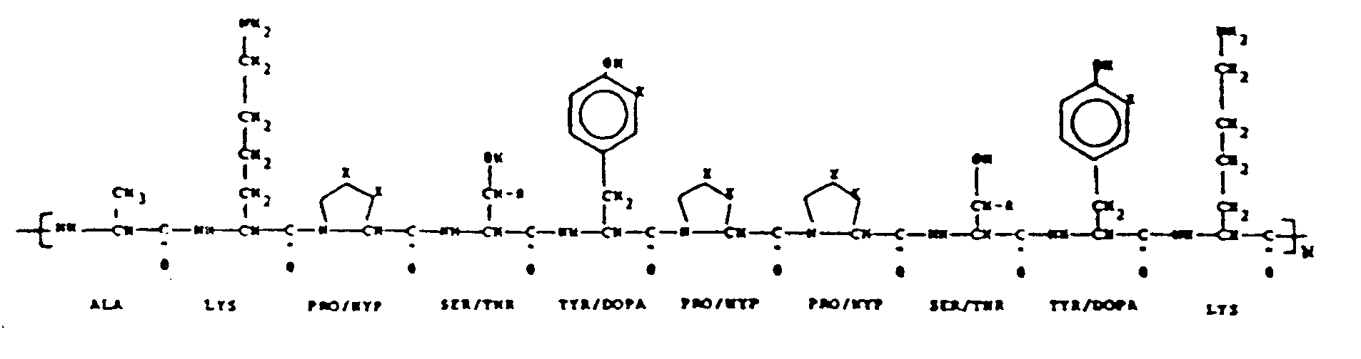
- Bioadhesive polyphenolic proteins are based on the sequence of repeating decapeptides having the formula: as described in United States Patent No. 4,585,585 entitled "Decapeptides Produced From Bioadhesive Polyphenolic Proteins”.
- Bioadhesives produced from the repeating decapeptides are ideal because they now have been found to enable cells and other biologically active moieties, such as proteins, DNA, hormones and antibiotics to attach to virtually any substrate.
- Applications in vitro include research diagnostics, cell product harvest, and cell metabolism research.
- In vivo applications include the production of confluent cell monolayers on the surface of prostheses, especially cardiovascular prostheses.
- tissue culture is the technique or process of propagating and/or supporting the metabolism of tissues or cells derived from organisms (plant or animal) in a formulated nutritive environment. Once isolated by gentle tissue dissociation, cells are incubated in nutritive media capable of supporting life functions. With few exceptions, cells require attachment to a substratum in order to perform normal metabolic functions, grow and divide. In tissue, the substratum which provides the matrix for cell growth consists of collagen, laminin, and fibronectin. In vitro, this substratum is most often plastic, although glass and microporous cellulosic filters are sometimes used as substitutes.
- Examples of cell uses produced via tissue culture include: (1) the study of the metabolism of the cell, the metabolism of parasites (i.e., viruses, bacteria, etc.) within the cell, the interactive metabolism of different cell types (i.e., epithelial cells, fibroblasts, immuno-competent cells, thymocytes, platelets, etc.), the effect of exogenous factors on cellular metabolism, the genetic composition of cells ( in vitro diagnostics); (2) the production of specific compounds, i.e., genes, proteins or other cellular components; and (3) the re-implantation of cells as for skin, corneal grafts, brain, vascular grafts, and in vitro fertilization.
- parasites i.e., viruses, bacteria, etc.
- the interactive metabolism of different cell types i.e., epithelial cells, fibroblasts, immuno-competent cells, thymocytes, platelets, etc.
- the effect of exogenous factors on cellular metabolism i.e., the genetic composition of
- adhesion promoters have assisted with attachment problems to a significant degree, certain inadequacies are still noteworthy.
- confluent cell monolayers are desired on prostheses, especially cardiovascular prostheses.
- Endothelial cells normally line the lumen of such vessels and actively prevent thrombosis, which is a major problem in the management of patients with cardiovascular disease. These cells also produce basement membrane material, the matrix for further wound healing.
- these prostheses are made of Teflon®, a substrate which does not promote cell attachment. No other known attachment factor mediates cell attachment to Teflon®.
- bioadhesive polyphenolic proteins obviates these problems. It attaches well to a variety of substrates in the presence of water and does not fail in a continuously humid environment. Being a true adhesive, bioadhesive polyphenolic protein rapidly attaches to both substrates and a variety of cells, tissues and other biologically active moieties. It can be stored at 4 o C for at least 10 months and at room temperature for at least 1 month without degradation or loss of function. Further, it can be synthesized by solid phase peptide synthesis, or via a genetic engineering approach, thereby permitting greater standardization of large quantities.
- bioadhesive polyphenolic proteins derived from the marine mussel is described in United States Patent No. 4,585,585, "Decapeptides Produced from Bioadhesive Polyphenolic Proteins".
- Formulations of bioadhesive polyphenolic proteins and methods for production of bioadhesive polyphenolic proteins are the subject of co-pending patent applications. Methods for the preparation of bioadhesive polyphenolic proteins are known in the art (Waite & Tanzer, 1981, Science 212, 1038).
- tissue culture or non-tissue culture materials and substrates including plastic, glass, metals, microporous filters (cellulosic, nylon, glass fiber, polyester, polycarbonate, polyethylene terephthalate and other synthetic and nonsynthetic materials including other synthetic polymeric materials and products resulting from modifications made to the aforementioned synthetic polymeric materials) and synthetic or alloplastic materials that may be used in tissue or prosthetic graft procedures (e.g., mechanical heart and polytetrafluoroethylene and related vascular grafting materials).
- tissue or prosthetic graft procedures e.g., mechanical heart and polytetrafluoroethylene and related vascular grafting materials.
- a second object of the present invention is to provide preparations useful as adhesive factors to promote or augment attachment efficiency, rate and/or strength of adhesion of other biologically active moieties such as proteins, DNA, hormones and antibiotics to a variety of substrates, some of which are mentioned above.
- a third object of the present invention is to provide the preparation or layering of substrates with bioadhesive polyphenolic proteins and the assays employed to investigate the effectiveness of such layers on the above-mentioned parameters.
- the present invention provides a method of affixing viable cells, tissues and other biologically active moieties such as proteins, DNA, hormones and antibiotics, to a substrate comprising:
- cell culture utilizes plastic substrates and, to a lesser degree, glass and microporous filters for cell attachment and propagation. More recently, physiological substrates (collagen, laminin, fibronectin, poly-D- and poly-L-lysine) have been utilized for these purposes in lieu of plastic to avoid problems inherent in cell culture at low seeding densities, using freshly isolated cells or on substrates less suitable for attachment (e.g., Teflon®).
- physiological substrates collagen, laminin, fibronectin, poly-D- and poly-L-lysine
- Bioadhesive polyphenolic proteins provide a suitable alternative because of their high binding affinity for both cells and a variety of substrates, biological and inert.
- Bioadhesive polyphenolic protein formulations have been evaluated for their efficiency in binding cells in vitro for cell culture.
- the formulations tested have included (1) 95% pure bioadhesive polyphenolic protein prepared from natural sources ("Formulation 1") and (2) 45% pure bioadhesive polyphenolic protein prepared from natural sources ("Formulation 2").
- bioadhesive polyphenolic proteins After preparing bioadhesive polyphenolic proteins according to procedures described in United States Patent No. 4,585,585, these formulations were thoroughly characterized biochemically using high performance liquid chromatography, assays for the quantitation of L-dopa, amino acid analysis and polyacrylamide gel electrophoresis.
- the composition of key amino acids in the bioadhesive polyphenolic protein formulations is given in Table 1.
- collagen comprises the majority of the remaining 55%.
- the basic unit of the bioadhesive polyphenolic protein is a decapeptide (chain of 10 amino acids) which is repeated through covalent bonds to similar decapeptides as many as 75-85 times.
- These formulations, based on bioadhesive polyphenolic protein, are stable, based on adhesive functionality, at 4 o C in 5% (v/v) acetic acid, pH 2.8 for greater than 10 months.
- Extracted preparations containing 40 to 50% collagen are stable at room temperature in 5% (v/v) acetic acid, pH 2.8 or following drying onto plastic substrates for at least 2 months.
- bioadhesive polyphenolic protein to strongly attach to a variety of substrates permits the attachment, maintenance and growth of cells to surfaces that heretofore posed problems either because of their composition, their application, or the type of cell requiring attachment.
- Substrates that could be used include plastic, glass, and microporous filters (e.g., cellulosic, nylon, glass fiber, polyester, polycarbonate) for conventional cell culture research and/or cell product harvesting from bio-reactors used in batch cell culture or in genetic engineering; hollow fiber tubes for cell product harvesting; and prosthetic vascular graft materials such as polytetrafluoroethylene (Teflon®) and related materials.
- plastic, glass, and microporous filters e.g., cellulosic, nylon, glass fiber, polyester, polycarbonate
- hollow fiber tubes for cell product harvesting
- prosthetic vascular graft materials such as polytetrafluoroethylene (Teflon®) and related materials.
- Bioadhesive polyphenolic protein would increase attachment efficiency, attachment rate and strength of attachment. This latter parameter is critical in applications involving cell product harvesting procedures or re-implantation of cells on vascular grafts which involve the passage of fluids over cell monolayers.
- cells that attach poorly following isolation from tissue or due to cell type, and cells that do not normally attach such as blood cells and suspension tissue culture cells (histiocytic lymphomas, platelets, white and red blood cells, etc.) could also be attached to substrates through this intermediate.
- bioadhesive polyphenolic protein to strongly attach to a variety of substrates permits the attachment of many other biologically active moieties, such as DNA, proteins, hormones and antibiotics.
- the coating of substrates with bioadhesive polyphenolic protein formulations and attachment to substrates is generally performed as follows. Depending on the final concentration per square cm desired, about 1 to 2 ⁇ l of sterile bioadhesive polyphenolic protein ranging from 10 to 60 ⁇ g per ⁇ l are evenly applied per cm2 of substrate.
- the resultant film is dried rapidly by placing the substrate within a laminar flow hood. Once dried, the film is treated with 35-100% ethanol or isopropanol for rinsing and fixation and then with sterile tissue culture medium for removal of residual alcohol and non-adsorbed extraneous moieties.
- the substrate may be used immediately or dried for storage.
- Cells or other biologically active moieties to be attached to the film are adjusted to desired concentrations and added to the substrate in serum-free or serum-containing medium.
- the cells are evaluated for attachment, growth, or function, or treated according to prescribed objectives of experiments requiring the attached cells in tissues culture.
- the bioadhesive polyphenolic protein can be affixed to the biologically active moieties and then,the resultant biologically active moieties can be affixed to the substrate.
- Formulations containing either 95% bioadhesive polyphenolic protein (Formulation 1) or 45% bioadhesive polyphenolic protein (Formulation 2) were evenly layered on 35 mm tissues (9 cm2) culture plastic petri dishes at 50 ⁇ g per dish in 5% (v/v) acetic acid, dried rapidly, "fixed”, and sterilized by rinsing with 100% ethanol. Dishes were then rinsed with sterile triple distilled water.
- Cells were prepared for the attachment assay as follows. Bovine corneal endothelial cells were treated with trypsin, a protease which digests cell attachment proteins, following growth in subculture in 5% CO2 in air at 37 o C in a humidified incubator. Cell monolayers were rinsed with serum-free medium to remove excess serum and medium that might interfere with trypsinization and incubated with 0.05% trypsin - 0.02% ethylene diamine tetraacetic acid (EDTA) for 10 minutes. Cells detached by the action of trypsin were transferred by pipette and gently centrifuged at 250 x g. Resultant pellets were re-suspended in serum-free minimal essential medium (Earle's salts) to remove any remaining serum proteins and trypsin from cell surfaces and again centrifuged.
- trypsin a protease which digests cell attachment proteins
- Viable cell counts were obtained using a dye exclusion test, where representative aliquots of cells were then re-suspended to a final concentration of 2 x 10⁵ cells per ml in minimal essential medium containing 15% fetal bovine serum.
- Cells were seeded in untreated plastic tissue culture petri dishes (control) and in tissue culture dishes layered with bioadhesive polyphenolic protein. At 1, 2.5, 5, 12.5, and about 20 min., triplicate experimental and control plates were chosen at random for quantitation of unattached cells. Unattached cells were removed from plates by rinsing and counted on a hemacytometer; replicate aliquots of cells that had been used, but that had not been added to dishes, were also counted in triplicate. Data were calculated as percent of cells attached by subtracting the number of unattached cells harvested from each dish from the total number of cells plated.
- Formulation 2 is very stable upon long-term storage. When tested by amino acid analysis, the L-dopa to protein ratios remained stable for Formulation 2 after 4 months at 4 o and -20 o C; whereas, a decline of up to 25% is found under similar conditions with Formulation 1 (Table 3). TABLE 3 Percent of Bioadhesive Polyphenolic Protein Remaining With Time at 4 o C and -20 o C Storage (Determined by Amino Acid Analysis) Formulation 1 Formulation 2 3 months 4 months 3 months 4 months 4 o 97% 76% 100% 98% -20 o 97% 82% 104% 100%
- Formulation 2 is preferred for purposes of enhancing cell attachment efficiency.
- bovine serum is the major protein constituent found in PBS. Strength of attachment was indirectly evaluated by the ability or inability to remove attached cells by trypsin from substrates to which they were attached. The concentration of bovine serum albumin employed was equivalent to that found in 0.5% to 1% FBS.
- the coating of tissue culture petri dishes with Formulation 2 of bioadhesive polyphenolic protein was accomplished as in Example 1.
- the cell line U937 is a human histiocytic lymphoma that was established from malignant cells isolated from a pleural effusion. These cells grow in suspension continuously in RPMI 1640 tissue culture medium, supplemented with 10% fetal bovine serum. U937 cells attach poorly to plastic in the presence of serum.
- Tissue culture petri dishes 35 mm dishes were coated with bioadhesive polyphenolic protein according to procedures outlined in Example 1.
- U937 cells were transferred to centrifuge tubes and prepared in the manner described in Example 1.
- Cells were seeded on plastic tissue culture dishes and on dishes coated with 100 ⁇ g of bioadhesive polyphenolic protein (Formulation 2), and evaluated in triplicate for attachment efficiency, (see Example 1) at 5, 12.5, and 20 minutes.
- the results in Table 5 (which are shown graphically in Figure 3) clearly demonstrate the effect of bioadhesive polyphenolic protein on attachment of U937 cells.
- bovine corneal endothelial cells were seeded at a density of 250 cells per tissue culture petri dish (35 mm diameter, 9.65 cm2) on either plastic, bioadhesive polyphenolic protein, collagen, laminin, poly-D-lysine, or fibronectin. The cells were allowed to grow for 5 days, at which time colony sizes (number of cells per colony) and numbers of colonies per plate were evaluated for each of the variables by staining the cells with crystal violet. Data obtained were used to determine the effect of each of these factors on attachment (number of colonies) and growth (size of colonies).
- Collagen - Collagen-coated plates were prepared by diluting 1 part cold (4 o C) collagen dispersion into 6 parts of cold 50% methanol. This mixture was mixed vigorously for several minutes and pipetted onto a petri dish so that only the bottom of the dish was covered. Within 20 seconds, the collagen was removed by aspiration and the dish was tilted upside down at 30 o against a lid to dry. Following 1 hour of drying undisturbed in a laminar flow hood, the dishes were ready for use.
- Laminin - Laminin is supplied in 1 mg quantities in 1 ml of 50 mM tris(hydroxymethyl)aminomethane in physiological saline. Following a slow thaw of laminin solution at 0 to 4 o C from -20 o C, 10 to 15 ⁇ g of laminin solution was pipetted into petri dishes in 0.5 ml of 0.01 M sodium phosphate buffer, pH 7.4. The dishes were dried at 37 o C. Immediately upon drying, the dishes are prepared for use.
- Fibronectin - Fibronectin is supplied in 1 mg quantities as a lyophilized powder. Prior to use, fibronectin is allowed to equilibrate to room temperature after storage at 4 o C. The powder is reconstituted with 1 ml sterile distilled water and allowed to stand for 30 minutes for solubilization. Ten to 20 ⁇ g of fibronectin solution is added to each dish in 0.5 ml and allowed to air dry. At this time, the dish is ready for cell seeding.
- Poly-D-lysine - Poly-D-lysine is supplied in quantities of 5 mg of lyophilized powder. Prior to use, this powder is allowed to equilibrate to room temperature following storage at 4 o C. Dishes are coated with 50 mg in 1 ml of sterile distilled water and allowed to stand at room temperature for 5 minutes. At that time, the solution is aspirated and the dishes are rinsed two times with 1.5 ml sterile distilled water. Following each rinse, liquid is aspirated completely. The dishes are dried and used immediately.
- the plating efficiency of the cells seeded on each of the factors is evaluated following crystal violet staining of the cells. This is achieved by first rinsing the dishes containing the colonies with serum-free medium to remove excess proteins and fixing the cells with 10% neutral buffered formalin for 10 minutes. The formalin is then removed from the plates by aspiration and 0.1% crystal violet in tapwater is then added to the plates for a period of 7 minutes. Immediately following staining, crystal violet is poured off and the cells are rinsed in a beaker of running tapwater to remove excess stain. Following complete drying of the plates, colonies on duplicates representing each of the variables are counted; and cells in ten randomly selected colonies per plate are counted.
- bioadhesive polyphenolic protein Although no significant differences were found between bioadhesive polyphenolic protein and poly-D-lysine (possibly due to the high level of lysine found in each of these molecules), the use of bioadhesive polyphenolic protein as an attachment factor is nonetheless more advantageous as a substrate based on its ability to (1) displace water, (2) to attach to materials including metal and Teflon® (for example, prosthetic devices), (3) to be used in vivo and in vitro , and (4) to form high strength bonds based on L-dopa, hydroxylated and lysine amino acid residues.
- Teflon® for example, prosthetic devices
- PTFE Polytetrafluoroethylene
- the major problem with the use of this material is that the seeding of vascular cells on PTFE is very difficult due to its high hydrophobicity. For many implants, a confluent cell monolayer on its surface would prevent clot formation.
- the vascular implant material was coated with 200 ⁇ g per CM2 of bioadhesive polyphenolic protein (Formulation 2). Five hundred thousand endothelial cells were then allowed to attach to the bioadhesive polyphenolic protein.
- the cells were also seeded onto Teflon® without coating with bioadhesive polyphenolic protein; and Teflon®coated with bioadhesive polyphenolic protein without seeding of cells acted as a control. Following 15 minutes of attachment, excess cells were rinsed from the vascular implant material and the vascular implants were fixed with formalin, stained with crystal violet, and dried as described in Example 4.
- the pellet is re-suspended in 900 mls of 5% acetic acid using the blender on high speed. Bioadhesive polyphenolic protein remains in the supernatant during centrifugation at 10 K rpm for 45 minutes. The approximately 1000 mls of supernatant is put into an ice bath with continual stirring. 5 mls of 2M sodium borate plus 95 mls of 5 M sodium chloride are added to the stirring supernatant. This mixture is centrifuged at 10K rpm for 15 minutes. The new supernatant is treated identically as above with the addition of four times as much 2M sodium borate and 5 M sodium chloride. Once again, the mixture is centrifuged at 10K for 15 minutes.
- the pellet is re-suspended in the following mixture: 7.5 mls of 2M sodium borate, 50 mls of 5M sodium chloride, 50 mls of distilled water, 37.5 mls of 8M urea in 5% acetic acid, and 5.6 mls of concentrated acetic acid.
- the mixture is slowly stirred for approximately 16 hours.
- the suspension is centrifuged at 10 K rpm for 15 minutes.
- the supernatant is saved and dialyzed (8-12K molecular weight cut-off membranes) against 5% acetic acid for approximately 16 hours.
- Amino acid analysis establishes that the extract contains 45% pure bioadhesive polyphenolic protein.
- the purity of the extract is governed by the number of extractions effected.
- the yield of pure bioadhesive polyphenolic protein decreases as the number of extractions increases. All procedures described herein were conducted at 4 o C.
- Bioadhesive polyphenolic protein 45% pure (Formulation 2), was used to immobilize heparin, a mucopolysaccharide having specific anticoagulant properties, and peroxidase, a protein enzyme which oxidizes peroxide. This was done to show that other substances could be efficiently bound to plasticware via a bioadhesive polyphenolic protein intermediate.
- bioadhesive polyphenolic protein was dried onto tissue culture plasticware dishes of 2 cm2 area for a final concentration of 3.5 ⁇ g/cm2.
- the protein was washed with 100% ethanol and then twice with water as described in Example 1.
- Peroxidase was immobilized in a similar manner: 5 different concentrations of peroxidase, 1, 0.5, 0.1, 0.05, 0.025 ⁇ g/dish, were added to both uncoated plastic dishes and plastic dishes coated with bioadhesive polyphenolic protein (in duplicate). As with heparin, a 0.1 M phosphate buffer wash was used to remove loosely bound enzyme.
- the assay for peroxidase involves adding a substrate mixture, peroxide plus 0-phenylene diamine (OPD) in phosphate buffered saline.
- the substrate mix, per ml contains 100 ⁇ l of peroxide (40 ⁇ l of 30% peroxide in 50 ml water) plus 100 ⁇ l OPD (10.7 mg in 8.56 ml water) and 800 ⁇ l phosphate buffered saline. 1 ml is added to each dish. After 5 minutes incubation at 23 o C, 100 ⁇ l of 4 N sulfuric acid is added to stop the reaction. It is a colorimetric assay with a wavelength optimum at 490 nm. The data in duplicate is presented as absorbance units at 490 nm in Table 8.
- bioadhesive polyphenolic protein As with heparin, at the higher concentrations no enhancement is seen by employing bioadhesive polyphenolic protein, sufficient enzyme binds to plastic. At the lower concentrations, significant enhancement or recovery is seen by employing bioadhesive polyphenolic protein.
- Bioadhesive polyphenolic protein has been found to successfully serve as a substrate for tissues and cells in histology and cytology.
- 45% bioadhesive polyphenolic protein (Formulation 2) was used to affix bovine Descemet's membrane with endothelial cell preparations to glass slides.
- Whole cornea were removed from freshly killed cows and placed either in physiological saline or in 10% neutral buffered formalin. Descemet's membrane was then removed from the posterior side of the cornea by gentle peeling. The tissue was transferred to slides (pre-cleaned with 5% acetic acid) and coated with 50 ⁇ g of bioadhesive polyphenolic protein.
- Tissue preparations were then dried onto the bioadhesive polyphenolic protein at room temperature or on 55 o C warming plates for twenty minutes.
- formalin fixed tissue was used, the tissue was rinsed with saline for removal of excess formalin prior to attachment to the bioadhesive polyphenolic protein.
- tissue-slide preparations were treated with formalin to fix tissues to the bioadhesive polyphenolic protein for five minutes. Tissues treated in this manner were retained on the bioadhesive polyphenolic protein for weeks in aqueous solutions.
- saline dilute and 100% ethanol, and xylene the tissues still remained intact. In the absence of bioadhesive polyphenolic protein, adherence of tissues to slides did not even survive the first formalin treatment.
Landscapes
- Health & Medical Sciences (AREA)
- Life Sciences & Earth Sciences (AREA)
- Engineering & Computer Science (AREA)
- Chemical & Material Sciences (AREA)
- Biomedical Technology (AREA)
- Zoology (AREA)
- Cell Biology (AREA)
- General Health & Medical Sciences (AREA)
- Organic Chemistry (AREA)
- Wood Science & Technology (AREA)
- Genetics & Genomics (AREA)
- Bioinformatics & Cheminformatics (AREA)
- Biotechnology (AREA)
- Public Health (AREA)
- Chemical Kinetics & Catalysis (AREA)
- Epidemiology (AREA)
- Transplantation (AREA)
- Veterinary Medicine (AREA)
- Oral & Maxillofacial Surgery (AREA)
- Medicinal Chemistry (AREA)
- Dermatology (AREA)
- Microbiology (AREA)
- Animal Behavior & Ethology (AREA)
- Biochemistry (AREA)
- General Engineering & Computer Science (AREA)
- Botany (AREA)
- Apparatus Associated With Microorganisms And Enzymes (AREA)
- Micro-Organisms Or Cultivation Processes Thereof (AREA)
- Peptides Or Proteins (AREA)
- Immobilizing And Processing Of Enzymes And Microorganisms (AREA)
- Agricultural Chemicals And Associated Chemicals (AREA)
- Materials For Medical Uses (AREA)
Claims (31)
- Verfahren zur Befestigung biologisch aktiver Komponenten an einem Substrat, bei dem:(1)ein Substrat mit einer sterilen Zubereitung beschichtet wird, die polyphenolisches Protein enthält, das etwa 35 bis 100 Gewichtsprozent reines bioadhäsives polyphenolisches Protein enhält mit der sich wiederholenden Dekapeptideinheit:
 (2) die Beschichtung auf dem Substrat getrocknet wird;(3)die Beschichtung auf dem Substrat fixiert wird;(4) das beschichtete Substrat gespült wird, um nicht fest an dem Substrat anhaftendes fremdes Material zu entfernen; und(5) biologisch aktive Komponenten auf das beschichtete Substrat aufgetragen werden, wodurch die biologisch aktiven Komponenten an dem beschichteten Substrat befestigt werden.
 - Verfahren nach Anspruch 1, bei dem das Substrat mit 0,1 bis 2 µl der Zubereitung pro cm² Substrat beschichtet wird, die etwa 10 bis 60 µg/µl bioadhäsives phenolisches Protein enthält.
- Verfahren nach Anspruch 1, bei dem die Beschichtung auf dem Substrat fixiert wird, indem das Substrat mit einem wasserfreien biologisch kompatiblen sterilisierenden Medium gespült wird.
- Verfahren nach Anspruch 3, bei dem das sterilisierende Medium 30 bis 100% Ethanol ist.
- Verfahren nach Anspruch 3, bei dem das sterilisierende Medium 30 bis 100% Isopropanol ist.
- Verfahren nach Anspruch 1, bei dem das beschichtete Substrat zum Entfernen fremden Materials mit Wasser oder Pufferlösung gespült wird.
- Verfahren nach Anspruch 1, bei dem das Substrat aus der Gruppe bestehend aus synthetischen polymeren Materialien, Glas, Metallen und mikroporösen Filtern ausgewählt wird.
- Verfahren nach Anspruch 7, bei dem das Substrat Polytetrafluorethylen ist.
- Verfahren zur Befestigung lebender Zellen an einem Substrat, bei dem:(1) ein Substrat mit einer sterilen Zubereitung beschichtet wird, die polyphenolisches Protein enthält, das etwa 35 bis 100 Gewichtsprozent reines bioadhäsives polyphenolisches Protein enthält mit der sich wiederholenden Dekapeptideinheit:
 (2) bei dem die Beschichtung auf dem Substrat getrocknet wird;(3) bei dem die Beschichtung auf dem Substrat fixiert wird;(4) bei dem das beschichtete Substrats gespült wird, um nicht fest an dem Substrat anhaftendes fremdes Material zu entfernen; und(5) bei dem lebende Zellen auf das beschichtete Substrat aufgetragen werden, wodurch die Zellen an dem beschichteten Substrat befestigt werden und in Anwesenheit einer nährstoffhaltigen Umgebung normale Zellstoffwechselfunktionen durchführen.
 - Verfahren nach Anspruch 9, bei dem das Substrat mit 0,1 bis 2 µl der Zubereitung pro cm² Substrat beschichtet wird, die etwa 10 bis 60 µg/µl bioadhäsives polyphenolisches Protein enthält.
- Verfahren nach Anspruch 9, bei dem die Beschichtung auf dem Substrat fixiert wird, indem das Substrat mit einem wasserfreien, biologisch kompatiblen, sterilisierenden Medium gespült wird.
- Verfahren nach Anspruch 11, bei dem das sterilisierende Medium 30 bis 100% Ethanol ist.
- Verfahren nach Anspruch 11, bei dem das sterilisierende Medium 30 bis 100% Isopropanol ist.
- Verfahren nach Anspruch 9, bei dem das beschichtete Substrat zum Entfernen fremden Materials mit sterilem, wässrigem, serumfreiem Gewebekulturmedium gespült wird.
- Verfahren nach Anspruch 9, bei dem das beschichtete Substrat zum Entfernen fremden Materials mit Wasser oder Pufferlösung gespült wird.
- Verfahren nach Anspruch 9, bei dem die lebenden Zellen in einem serumhaltigen Medium auf das beschichtete Substrat aufgetragen werden.
- Verfahren nach Anspruch 9, bei dem die lebenden Zellen in einem serumfreien Medium auf das beschichtete Substrat aufgetragen werden.
- Verfahren nach Anspruch 9, bei dem das Substrat ein in vitro Substrat ist.
- Verfahren nach Anspruch 18, bei dem das Substrat aus der Gruppe, bestehend aus synthetischen polymeren Materialien, Glas, Metallen und mikroporösen Filtern ausgewählt ist.
- Verfahren nach Anspruch 19, bei dem das Substrat Polytetrafluorethylen ist.
- Verfahren nach Anspruch 9, bei dem das Substrat ein in vivo Substrat ist
- Verfahren nach Anspruch 21, bei den, das Substrat aus der Gruppe, bestehend aus Kollagen, Laminin, Fibronektin, prothetischen Transplantations-oder Implantationsmaterialien und Polylysin ausgewählt ist.
- Verfahren nach Anspruch 22, bei dem das Substrat Polytetrafluorethylen ist.
- Zellkultursystem, umfassend: ein Substrat; eine Beschichtung darauf aus einer sterilen Zubereitung, die polyphenolisches Protein enhält, das etwa 35 bis 100 Gewichtsprozent reines bioadhäsives polyphenolisches Protein enthält mit der sich wiederholenden Dekapeptideinheit:

- Zellkultursystem nach Anspruch 24, bei dem das Substrat ein in vitro Substrat ist.
- Zellkultursystem nach Anspruch 25, bei dem das Substrat aus der Gruppe, bestehend aus synthetischem polymerem Material, Glas, Metallen und mikroporösen Filtern ausgewählt wird.
- Zellkultursystem nach Anspruch 26, bei dem das Substrat Polytetrafluorethylen ist.
- Zellkultursystem nach Anspruch 24, bei dem das Substrat ein in vivo Substrat ist.
- Zellkultursystem nach Anspruch 28, bei dem das Substrat aus der Gruppe, bestehend aus Kollagen, Laminin, Fibronektin, prothetischen Transplantations-oder Implantationsmaterialien und Polylysin ausgewählt ist.
- Zellkultursystem nach Anspruch 29, bei dem das Substrat Polytetrafluorethylen ist
- Verfahren zur Befestigung biologisch aktiver Komponenten an einem Substrat . bei dem:(1) die biologisch aktiven Komponenten in einer serumfreien Lösung dispergiert (feinst verteilt)werden;(2)bei dem eine sterile Zubereitung, die polyphenolisches Protein enthält, das etwa 35 bis 100 Gewichtsprozent reines bioadhäsives polyphenolisches Protein enthält mit der sich wiederholenden Dekapeptideinheit:
 (3) bei dem die entstehenden biologisch aktiven Komponenten zurückgewonnen werden; und(4) bei dem die zurückgewonnenen biologisch aktiven Komponenten an dem Substrat befestigt werden.
Priority Applications (1)
| Application Number | Priority Date | Filing Date | Title |
|---|---|---|---|
| AT87105672T ATE84820T1 (de) | 1986-04-25 | 1987-04-16 | Anheftende biomaterialien fuer die adhaesion von zellen und geweben. |
Applications Claiming Priority (4)
| Application Number | Priority Date | Filing Date | Title |
|---|---|---|---|
| US34801 | 1979-04-30 | ||
| US85668786A | 1986-04-25 | 1986-04-25 | |
| US856687 | 1986-04-25 | ||
| US07/034,801 US5108923A (en) | 1986-04-25 | 1987-04-03 | Bioadhesives for cell and tissue adhesion |
Publications (3)
| Publication Number | Publication Date |
|---|---|
| EP0243818A2 EP0243818A2 (de) | 1987-11-04 |
| EP0243818A3 EP0243818A3 (en) | 1989-03-08 |
| EP0243818B1 true EP0243818B1 (de) | 1993-01-20 |
Family
ID=26711386
Family Applications (1)
| Application Number | Title | Priority Date | Filing Date |
|---|---|---|---|
| EP87105672A Expired - Lifetime EP0243818B1 (de) | 1986-04-25 | 1987-04-16 | Anheftende Biomaterialien für die Adhäsion von Zellen und Geweben |
Country Status (9)
| Country | Link |
|---|---|
| US (1) | US5108923A (de) |
| EP (1) | EP0243818B1 (de) |
| JP (1) | JPH0779695B2 (de) |
| AU (1) | AU597647B2 (de) |
| CA (1) | CA1328237C (de) |
| DE (1) | DE3783643T2 (de) |
| DK (1) | DK171996B1 (de) |
| FI (1) | FI91037C (de) |
| NO (1) | NO174675C (de) |
Families Citing this family (48)
| Publication number | Priority date | Publication date | Assignee | Title |
|---|---|---|---|---|
| US5202256A (en) * | 1984-09-13 | 1993-04-13 | Enzon Labs, Inc. | Bioadhesive precursor protein expression vectors |
| US5049504A (en) * | 1986-11-24 | 1991-09-17 | Genex Corporation | Bioadhesive coding sequences |
| US5202236A (en) * | 1984-09-13 | 1993-04-13 | Enzon Labs Inc. | Method of producing bioadhesive protein |
| AU592670B2 (en) * | 1986-08-15 | 1990-01-18 | Commonwealth Scientific And Industrial Research Organisation | Promoting cell adhesion and growth on a substrate |
| US5759774A (en) * | 1988-05-18 | 1998-06-02 | Cobe Laboratories, Inc. | Method of detecting circulating antibody types using dried or lyophilized cells |
| US5122470A (en) * | 1988-07-05 | 1992-06-16 | Banes Albert J | Floating cell culture device and method |
| ATE102649T1 (de) * | 1988-07-13 | 1994-03-15 | Collaborative Biomed Prod | Gewebe-immobilisierungs- und zellkultursystem und verfahren zum anbringen biologisch wirksamer teile an einem substrat. |
| US5192663A (en) * | 1988-11-04 | 1993-03-09 | Immucor, Inc. | Article having an organic dye and a monolayer of dried mammalian cells and a method for utilizing the article |
| US5030560A (en) * | 1988-11-04 | 1991-07-09 | Immucor, Inc. | Method for drying mammalian cells for use in solid phase immunoassays and articles incorporating same |
| US5574134A (en) * | 1989-07-11 | 1996-11-12 | University Of Delaware | Polypeptide monomers, linearly extended and/or crosslinked forms thereof, and applications thereof |
| NZ237832A (en) * | 1990-04-17 | 1994-05-26 | Curative Tech Inc | Coating a prosthetic surface with mammalian cells |
| US5197973A (en) * | 1990-12-14 | 1993-03-30 | Creative Biomolecules, Inc. | Synthetic bioadhesive |
| GB9211176D0 (en) * | 1992-05-27 | 1992-07-08 | Central Blood Lab Authority | Assay |
| US5310669A (en) * | 1992-06-22 | 1994-05-10 | The Trustees Of Dartmouth College | Fullerene coated surfaces and uses thereof |
| US5817470A (en) * | 1995-03-10 | 1998-10-06 | Sociedad Biotecnologica Collico Limitada | Immobilization of antigens to solid support by the mussel adhesive polyphenolic protein and the method for use therein |
| US5955353A (en) * | 1997-05-22 | 1999-09-21 | Excorp Medical, Inc. | Hollow fiber bioreactor with an extrafilament flow plug |
| US6221425B1 (en) | 1998-01-30 | 2001-04-24 | Advanced Cardiovascular Systems, Inc. | Lubricious hydrophilic coating for an intracorporeal medical device |
| US20020022588A1 (en) * | 1998-06-23 | 2002-02-21 | James Wilkie | Methods and compositions for sealing tissue leaks |
| US6214054B1 (en) * | 1998-09-21 | 2001-04-10 | Edwards Lifesciences Corporation | Method for fixation of biological tissues having mitigated propensity for post-implantation calcification and thrombosis and bioprosthetic devices prepared thereby |
| DK1334116T3 (da) * | 1998-09-28 | 2007-09-17 | Bio Polymer Products Of Sweden | En fremgangsmåde til fremstilling af polyphenoliske adhæsive proteiner |
| US7858679B2 (en) * | 2001-07-20 | 2010-12-28 | Northwestern University | Polymeric compositions and related methods of use |
| US7618937B2 (en) * | 2001-07-20 | 2009-11-17 | Northwestern University | Peptidomimetic polymers for antifouling surfaces |
| US8815793B2 (en) * | 2001-07-20 | 2014-08-26 | Northwestern University | Polymeric compositions and related methods of use |
| US6878168B2 (en) | 2002-01-03 | 2005-04-12 | Edwards Lifesciences Corporation | Treatment of bioprosthetic tissues to mitigate post implantation calcification |
| US8911831B2 (en) * | 2002-07-19 | 2014-12-16 | Northwestern University | Surface independent, surface-modifying, multifunctional coatings and applications thereof |
| US20080085554A1 (en) * | 2004-02-13 | 2008-04-10 | Norio Nakatsuji | Culture Medium for Culturing Feeder Cells for Embryonic Stem Cells Culture and the Prepared Feeder Cells |
| DE102004031258A1 (de) * | 2004-06-29 | 2006-02-09 | Jennissen, Herbert P., Prof. Dr. | Proteinhybride mit polyhydroxyaromatischen Aminosäure-Epitopen |
| US7732539B2 (en) * | 2006-02-16 | 2010-06-08 | National Science Foundation | Modified acrylic block copolymers for hydrogels and pressure sensitive wet adhesives |
| WO2007126127A1 (ja) * | 2006-04-28 | 2007-11-08 | Kuraray Co., Ltd. | 細胞培養容器およびその製造方法 |
| US7622533B2 (en) | 2006-08-04 | 2009-11-24 | Nerites Corporation | Biomimetic compounds and synthetic methods therefor |
| US8563117B2 (en) * | 2006-08-04 | 2013-10-22 | Phillip B. Messersmith | Biomimetic modular adhesive complex: materials, methods and applications therefore |
| EP2077718B2 (de) * | 2006-10-27 | 2022-03-09 | Edwards Lifesciences Corporation | Biologisches gewebe für chirurgische implantation |
| WO2008089032A1 (en) * | 2007-01-11 | 2008-07-24 | Northwestern University | Fouling resistant coatings and methods of making same |
| US8673286B2 (en) | 2007-04-09 | 2014-03-18 | Northwestern University | DOPA-functionalized, branched, poly(aklylene oxide) adhesives |
| US8383092B2 (en) * | 2007-02-16 | 2013-02-26 | Knc Ner Acquisition Sub, Inc. | Bioadhesive constructs |
| US9101691B2 (en) * | 2007-06-11 | 2015-08-11 | Edwards Lifesciences Corporation | Methods for pre-stressing and capping bioprosthetic tissue |
| US8357387B2 (en) | 2007-12-21 | 2013-01-22 | Edwards Lifesciences Corporation | Capping bioprosthetic tissue to reduce calcification |
| US20110130465A1 (en) * | 2009-12-01 | 2011-06-02 | Nerites Corporation | Coatings for prevention of biofilms |
| US8846390B2 (en) | 2010-03-23 | 2014-09-30 | Edwards Lifesciences Corporation | Methods of conditioning sheet bioprosthetic tissue |
| CN102946916B (zh) | 2010-06-17 | 2015-03-18 | 爱德华兹生命科学公司 | 通过化学修饰抗原性碳水化合物稳定生物假体组织的方法 |
| US8906601B2 (en) | 2010-06-17 | 2014-12-09 | Edwardss Lifesciences Corporation | Methods for stabilizing a bioprosthetic tissue by chemical modification of antigenic carbohydrates |
| CA2817215C (en) | 2010-11-09 | 2017-05-09 | Knc Ner Acquisition Sub, Inc. | Adhesive compounds for use in hernia repair |
| US9351829B2 (en) | 2010-11-17 | 2016-05-31 | Edwards Lifesciences Corporation | Double cross-linkage process to enhance post-implantation bioprosthetic tissue durability |
| US10238771B2 (en) | 2012-11-08 | 2019-03-26 | Edwards Lifesciences Corporation | Methods for treating bioprosthetic tissue using a nucleophile/electrophile in a catalytic system |
| JPWO2014098182A1 (ja) * | 2012-12-19 | 2017-01-12 | 国立大学法人 東京医科歯科大学 | 心筋毒性検査および心筋細胞評価のための方法および装置 |
| WO2015009431A1 (en) * | 2013-07-19 | 2015-01-22 | Rarecyte, Inc. | Solution and method for adhering suspension components to a substrate |
| US10101247B2 (en) * | 2013-07-19 | 2018-10-16 | Rarecyte, Inc. | Solution and method for adhering suspension components to a substrate |
| US10101248B1 (en) * | 2015-12-02 | 2018-10-16 | Rarecyte, Inc. | Solution and method for adhering suspension components to a substrate |
Family Cites Families (12)
| Publication number | Priority date | Publication date | Assignee | Title |
|---|---|---|---|---|
| US3910819A (en) * | 1974-02-19 | 1975-10-07 | California Inst Of Techn | Treatment of surfaces to stimulate biological cell adhesion and growth |
| DE2603319C3 (de) * | 1976-01-29 | 1979-08-23 | Boehringer Mannheim Gmbh, 6800 Mannheim | Verfahren zur Fixierung von biologisch aktiven Proteinen an Trägern |
| FR2447275A1 (fr) * | 1979-01-25 | 1980-08-22 | Charbonnages Ste Chimique | Materiaux stratifies a base de resine phenolique et procede pour leur preparation |
| US4266032A (en) * | 1979-08-10 | 1981-05-05 | Monsanto Company | Method of culturing microcarrier supported cells |
| US4501815A (en) * | 1979-10-29 | 1985-02-26 | Albert Einstein College Of Medicine Of Yeshiva University | Article for culturing differentiated cells |
| US4352887A (en) * | 1979-10-29 | 1982-10-05 | Albert Einstein College Of Medicine Of Yeshiva University | Method and article for culturing differentiated cells |
| US4578079A (en) * | 1982-08-04 | 1986-03-25 | La Jolla Cancer Research Foundation | Tetrapeptide |
| JPS5928472A (ja) * | 1982-08-09 | 1984-02-15 | Koken:Kk | 細胞培養用基質およびこの基質を用いた細胞培養・分離法 |
| ZA839115B (en) * | 1982-12-14 | 1984-07-25 | Cpc International Inc | Continuous enzymatic process for producing maltose from starch and starch hydrolysates |
| US4585585A (en) * | 1984-03-07 | 1986-04-29 | University Of Connecticut Research & Development Corporation | Decapeptides produced from bioadhesive polyphenolic proteins |
| US4496397A (en) * | 1984-03-07 | 1985-01-29 | University Of Connecticut | Process for purifying and stabilizing catechol-containing proteins and materials obtained thereby |
| US4553974A (en) * | 1984-08-14 | 1985-11-19 | Mayo Foundation | Treatment of collagenous tissue with glutaraldehyde and aminodiphosphonate calcification inhibitor |
-
1987
- 1987-04-03 US US07/034,801 patent/US5108923A/en not_active Expired - Lifetime
- 1987-04-16 DE DE8787105672T patent/DE3783643T2/de not_active Expired - Fee Related
- 1987-04-16 EP EP87105672A patent/EP0243818B1/de not_active Expired - Lifetime
- 1987-04-21 FI FI871728A patent/FI91037C/fi not_active IP Right Cessation
- 1987-04-22 CA CA000535245A patent/CA1328237C/en not_active Expired - Fee Related
- 1987-04-22 DK DK205287A patent/DK171996B1/da not_active IP Right Cessation
- 1987-04-22 NO NO871665A patent/NO174675C/no unknown
- 1987-04-23 AU AU71885/87A patent/AU597647B2/en not_active Ceased
- 1987-04-25 JP JP62102971A patent/JPH0779695B2/ja not_active Expired - Lifetime
Also Published As
| Publication number | Publication date |
|---|---|
| FI91037C (fi) | 1994-05-10 |
| JPH0779695B2 (ja) | 1995-08-30 |
| DK205287D0 (da) | 1987-04-22 |
| EP0243818A3 (en) | 1989-03-08 |
| DK205287A (da) | 1987-10-26 |
| EP0243818A2 (de) | 1987-11-04 |
| FI91037B (fi) | 1994-01-31 |
| NO871665L (no) | 1987-10-26 |
| JPS6339583A (ja) | 1988-02-20 |
| DE3783643T2 (de) | 1993-05-13 |
| NO871665D0 (no) | 1987-04-22 |
| US5108923A (en) | 1992-04-28 |
| AU597647B2 (en) | 1990-06-07 |
| NO174675C (no) | 1994-06-22 |
| AU7188587A (en) | 1987-10-29 |
| DE3783643D1 (de) | 1993-03-04 |
| FI871728A7 (fi) | 1987-10-26 |
| CA1328237C (en) | 1994-04-05 |
| FI871728A0 (fi) | 1987-04-21 |
| NO174675B (no) | 1994-03-07 |
| DK171996B1 (da) | 1997-09-08 |
Similar Documents
| Publication | Publication Date | Title |
|---|---|---|
| EP0243818B1 (de) | Anheftende Biomaterialien für die Adhäsion von Zellen und Geweben | |
| US4645669A (en) | Culturing and emplacement of differentiated cells in vivo | |
| US7815686B2 (en) | Vascularization enhanced graft constructs | |
| WO2002014480A9 (en) | Decellularized tissue engineered constructs and tissues | |
| CN104436305A (zh) | 以脱细胞生物膜为载体的心肌补片及制备方法与应用 | |
| US5932207A (en) | Complex active ingredient for the production of biological parts, especially organs for living organisms: method for the production of the same and its use | |
| JPS6062979A (ja) | 筋細胞の培養の方法 | |
| EP0905231B1 (de) | Verfahren zur Verbesserung der Stabilität und/oder Haltbarkeit von verschiedenen Substraten | |
| US5980888A (en) | Keratinocytes attached to microcarriers for treatment of skin wounds | |
| JPH09511673A (ja) | 上皮細胞を含む生物材料及び移植片としてのその利用 | |
| CA2467581A1 (en) | Milk protein biofilm and uses thereof | |
| US4656130A (en) | Collagen coated cell growth plates | |
| JP4874514B2 (ja) | 上皮系細胞の重層化培養方法、それから得られる重層化上皮系細胞シート及びその利用方法 | |
| LEE et al. | Endothelial cell seeding onto the extracellular matrix of fibroblasts for the development of a small diameter polyurethane vessel | |
| EP0636184A1 (de) | Medien für die isolierung und stabilisierung von zellen | |
| EP0350714B1 (de) | Gewebe-Immobilisierungs- und Zellkultursystem und Verfahren zum Anbringen biologisch wirksamer Teile an einem Substrat | |
| BERNHARD et al. | Development of a nonthrombogenic collagenous blood-prosthetic interface | |
| RU2817370C1 (ru) | Матрикс адгезионный биоактивный для работы с культурами клеток и способ его применения | |
| KR100231279B1 (ko) | 치주조직을 복원하기 위한 골아세포 이식용 조직성장인자를 함유한 생분해성 폴리사카라이드 스폰지제제 | |
| JPH07135961A (ja) | 血管内皮細胞培養用基材及びその製造方法 | |
| KR870001417B1 (ko) | Pta-210 세포주를 이용한 인간 성장호르몬의 생산방법 |
Legal Events
| Date | Code | Title | Description |
|---|---|---|---|
| PUAI | Public reference made under article 153(3) epc to a published international application that has entered the european phase |
Free format text: ORIGINAL CODE: 0009012 |
|
| AK | Designated contracting states |
Kind code of ref document: A2 Designated state(s): AT BE CH DE ES FR GB GR IT LI LU NL SE |
|
| PUAL | Search report despatched |
Free format text: ORIGINAL CODE: 0009013 |
|
| AK | Designated contracting states |
Kind code of ref document: A3 Designated state(s): AT BE CH DE ES FR GB GR IT LI LU NL SE |
|
| 17P | Request for examination filed |
Effective date: 19890825 |
|
| 17Q | First examination report despatched |
Effective date: 19911007 |
|
| GRAA | (expected) grant |
Free format text: ORIGINAL CODE: 0009210 |
|
| RAP1 | Party data changed (applicant data changed or rights of an application transferred) |
Owner name: COLLABORATIVE BIOMEDICAL PRODUCTS INC. |
|
| AK | Designated contracting states |
Kind code of ref document: B1 Designated state(s): AT BE CH DE ES FR GB GR IT LI LU NL SE |
|
| PG25 | Lapsed in a contracting state [announced via postgrant information from national office to epo] |
Ref country code: GR Free format text: LAPSE BECAUSE OF FAILURE TO SUBMIT A TRANSLATION OF THE DESCRIPTION OR TO PAY THE FEE WITHIN THE PRESCRIBED TIME-LIMIT Effective date: 19930120 |
|
| REF | Corresponds to: |
Ref document number: 84820 Country of ref document: AT Date of ref document: 19930215 Kind code of ref document: T |
|
| ITF | It: translation for a ep patent filed | ||
| REF | Corresponds to: |
Ref document number: 3783643 Country of ref document: DE Date of ref document: 19930304 |
|
| PG25 | Lapsed in a contracting state [announced via postgrant information from national office to epo] |
Ref country code: ES Free format text: LAPSE BECAUSE OF FAILURE TO SUBMIT A TRANSLATION OF THE DESCRIPTION OR TO PAY THE FEE WITHIN THE PRESCRIBED TIME-LIMIT Effective date: 19930501 |
|
| ET | Fr: translation filed | ||
| PLBE | No opposition filed within time limit |
Free format text: ORIGINAL CODE: 0009261 |
|
| STAA | Information on the status of an ep patent application or granted ep patent |
Free format text: STATUS: NO OPPOSITION FILED WITHIN TIME LIMIT |
|
| 26N | No opposition filed | ||
| PGFP | Annual fee paid to national office [announced via postgrant information from national office to epo] |
Ref country code: ES Payment date: 19940428 Year of fee payment: 8 |
|
| EPTA | Lu: last paid annual fee | ||
| EAL | Se: european patent in force in sweden |
Ref document number: 87105672.7 |
|
| PGFP | Annual fee paid to national office [announced via postgrant information from national office to epo] |
Ref country code: AT Payment date: 19980415 Year of fee payment: 12 |
|
| PGFP | Annual fee paid to national office [announced via postgrant information from national office to epo] |
Ref country code: SE Payment date: 19980416 Year of fee payment: 12 |
|
| PGFP | Annual fee paid to national office [announced via postgrant information from national office to epo] |
Ref country code: NL Payment date: 19980428 Year of fee payment: 12 |
|
| PGFP | Annual fee paid to national office [announced via postgrant information from national office to epo] |
Ref country code: LU Payment date: 19980504 Year of fee payment: 12 |
|
| PGFP | Annual fee paid to national office [announced via postgrant information from national office to epo] |
Ref country code: CH Payment date: 19980505 Year of fee payment: 12 |
|
| PGFP | Annual fee paid to national office [announced via postgrant information from national office to epo] |
Ref country code: BE Payment date: 19980609 Year of fee payment: 12 |
|
| PG25 | Lapsed in a contracting state [announced via postgrant information from national office to epo] |
Ref country code: LU Free format text: LAPSE BECAUSE OF NON-PAYMENT OF DUE FEES Effective date: 19990416 Ref country code: AT Free format text: LAPSE BECAUSE OF NON-PAYMENT OF DUE FEES Effective date: 19990416 |
|
| PG25 | Lapsed in a contracting state [announced via postgrant information from national office to epo] |
Ref country code: SE Free format text: LAPSE BECAUSE OF NON-PAYMENT OF DUE FEES Effective date: 19990417 |
|
| PG25 | Lapsed in a contracting state [announced via postgrant information from national office to epo] |
Ref country code: LI Free format text: LAPSE BECAUSE OF NON-PAYMENT OF DUE FEES Effective date: 19990430 Ref country code: CH Free format text: LAPSE BECAUSE OF NON-PAYMENT OF DUE FEES Effective date: 19990430 Ref country code: BE Free format text: LAPSE BECAUSE OF NON-PAYMENT OF DUE FEES Effective date: 19990430 |
|
| BERE | Be: lapsed |
Owner name: COLLABORATIVE BIOMEDICAL PRODUCTS INC. Effective date: 19990430 |
|
| PG25 | Lapsed in a contracting state [announced via postgrant information from national office to epo] |
Ref country code: NL Free format text: LAPSE BECAUSE OF NON-PAYMENT OF DUE FEES Effective date: 19991101 |
|
| REG | Reference to a national code |
Ref country code: CH Ref legal event code: PL |
|
| NLV4 | Nl: lapsed or anulled due to non-payment of the annual fee |
Effective date: 19991101 |
|
| EUG | Se: european patent has lapsed |
Ref document number: 87105672.7 |
|
| PGFP | Annual fee paid to national office [announced via postgrant information from national office to epo] |
Ref country code: FR Payment date: 20010330 Year of fee payment: 15 |
|
| PGFP | Annual fee paid to national office [announced via postgrant information from national office to epo] |
Ref country code: DE Payment date: 20010402 Year of fee payment: 15 |
|
| PGFP | Annual fee paid to national office [announced via postgrant information from national office to epo] |
Ref country code: GB Payment date: 20010403 Year of fee payment: 15 |
|
| REG | Reference to a national code |
Ref country code: GB Ref legal event code: IF02 |
|
| PG25 | Lapsed in a contracting state [announced via postgrant information from national office to epo] |
Ref country code: GB Free format text: LAPSE BECAUSE OF NON-PAYMENT OF DUE FEES Effective date: 20020416 |
|
| PG25 | Lapsed in a contracting state [announced via postgrant information from national office to epo] |
Ref country code: DE Free format text: LAPSE BECAUSE OF NON-PAYMENT OF DUE FEES Effective date: 20021101 |
|
| GBPC | Gb: european patent ceased through non-payment of renewal fee |
Effective date: 20020416 |
|
| PG25 | Lapsed in a contracting state [announced via postgrant information from national office to epo] |
Ref country code: FR Free format text: LAPSE BECAUSE OF NON-PAYMENT OF DUE FEES Effective date: 20021231 |
|
| REG | Reference to a national code |
Ref country code: FR Ref legal event code: ST |
|
| PG25 | Lapsed in a contracting state [announced via postgrant information from national office to epo] |
Ref country code: IT Free format text: LAPSE BECAUSE OF NON-PAYMENT OF DUE FEES;WARNING: LAPSES OF ITALIAN PATENTS WITH EFFECTIVE DATE BEFORE 2007 MAY HAVE OCCURRED AT ANY TIME BEFORE 2007. THE CORRECT EFFECTIVE DATE MAY BE DIFFERENT FROM THE ONE RECORDED. Effective date: 20050416 |