EP0167690A2 - Création de gouttelettes - Google Patents
Création de gouttelettes Download PDFInfo
- Publication number
- EP0167690A2 EP0167690A2 EP84305399A EP84305399A EP0167690A2 EP 0167690 A2 EP0167690 A2 EP 0167690A2 EP 84305399 A EP84305399 A EP 84305399A EP 84305399 A EP84305399 A EP 84305399A EP 0167690 A2 EP0167690 A2 EP 0167690A2
- Authority
- EP
- European Patent Office
- Prior art keywords
- microcapsules
- droplet
- voltage
- needle
- electroconductive
- Prior art date
- Legal status (The legal status is an assumption and is not a legal conclusion. Google has not performed a legal analysis and makes no representation as to the accuracy of the status listed.)
- Granted
Links
- 239000003094 microcapsule Substances 0.000 claims abstract description 76
- 230000003068 static effect Effects 0.000 claims abstract description 7
- 238000001125 extrusion Methods 0.000 claims abstract 8
- 239000012528 membrane Substances 0.000 claims description 31
- 238000000034 method Methods 0.000 claims description 29
- 239000000463 material Substances 0.000 claims description 24
- IXPNQXFRVYWDDI-UHFFFAOYSA-N 1-methyl-2,4-dioxo-1,3-diazinane-5-carboximidamide Chemical compound CN1CC(C(N)=N)C(=O)NC1=O IXPNQXFRVYWDDI-UHFFFAOYSA-N 0.000 claims description 22
- 239000000661 sodium alginate Substances 0.000 claims description 22
- 235000010413 sodium alginate Nutrition 0.000 claims description 22
- 229940005550 sodium alginate Drugs 0.000 claims description 22
- 239000000243 solution Substances 0.000 claims description 22
- 230000015572 biosynthetic process Effects 0.000 claims description 19
- 239000002609 medium Substances 0.000 claims description 16
- 239000007788 liquid Substances 0.000 claims description 15
- 210000004153 islets of langerhan Anatomy 0.000 claims description 12
- 241001465754 Metazoa Species 0.000 claims description 9
- 239000007924 injection Substances 0.000 claims description 9
- 238000002347 injection Methods 0.000 claims description 9
- 239000000648 calcium alginate Substances 0.000 claims description 8
- 235000010410 calcium alginate Nutrition 0.000 claims description 8
- 229960002681 calcium alginate Drugs 0.000 claims description 8
- OKHHGHGGPDJQHR-YMOPUZKJSA-L calcium;(2s,3s,4s,5s,6r)-6-[(2r,3s,4r,5s,6r)-2-carboxy-6-[(2r,3s,4r,5s,6r)-2-carboxylato-4,5,6-trihydroxyoxan-3-yl]oxy-4,5-dihydroxyoxan-3-yl]oxy-3,4,5-trihydroxyoxane-2-carboxylate Chemical compound [Ca+2].O[C@@H]1[C@H](O)[C@H](O)O[C@@H](C([O-])=O)[C@H]1O[C@H]1[C@@H](O)[C@@H](O)[C@H](O[C@H]2[C@H]([C@@H](O)[C@H](O)[C@H](O2)C([O-])=O)O)[C@H](C(O)=O)O1 OKHHGHGGPDJQHR-YMOPUZKJSA-L 0.000 claims description 8
- 230000004060 metabolic process Effects 0.000 claims description 6
- 230000002526 effect on cardiovascular system Effects 0.000 claims description 5
- 235000015097 nutrients Nutrition 0.000 claims description 5
- 230000000694 effects Effects 0.000 claims description 4
- 210000000987 immune system Anatomy 0.000 claims description 3
- 239000012736 aqueous medium Substances 0.000 claims description 2
- 230000002503 metabolic effect Effects 0.000 claims description 2
- 230000002441 reversible effect Effects 0.000 claims 2
- 238000010924 continuous production Methods 0.000 claims 1
- 102000004169 proteins and genes Human genes 0.000 claims 1
- 108090000623 proteins and genes Proteins 0.000 claims 1
- 210000001519 tissue Anatomy 0.000 description 25
- 239000002775 capsule Substances 0.000 description 24
- 210000004027 cell Anatomy 0.000 description 23
- 239000000499 gel Substances 0.000 description 22
- 239000011162 core material Substances 0.000 description 15
- 229920000656 polylysine Polymers 0.000 description 15
- 108010039918 Polylysine Proteins 0.000 description 14
- UXVMQQNJUSDDNG-UHFFFAOYSA-L Calcium chloride Chemical compound [Cl-].[Cl-].[Ca+2] UXVMQQNJUSDDNG-UHFFFAOYSA-L 0.000 description 11
- 210000004369 blood Anatomy 0.000 description 10
- 239000008280 blood Substances 0.000 description 10
- 239000011248 coating agent Substances 0.000 description 9
- 238000000576 coating method Methods 0.000 description 9
- 229940072056 alginate Drugs 0.000 description 8
- 229920000615 alginic acid Polymers 0.000 description 8
- 238000001727 in vivo Methods 0.000 description 8
- 239000002253 acid Substances 0.000 description 7
- 235000010443 alginic acid Nutrition 0.000 description 7
- 206010012601 diabetes mellitus Diseases 0.000 description 7
- 229920000642 polymer Polymers 0.000 description 7
- FHVDTGUDJYJELY-UHFFFAOYSA-N 6-{[2-carboxy-4,5-dihydroxy-6-(phosphanyloxy)oxan-3-yl]oxy}-4,5-dihydroxy-3-phosphanyloxane-2-carboxylic acid Chemical compound O1C(C(O)=O)C(P)C(O)C(O)C1OC1C(C(O)=O)OC(OP)C(O)C1O FHVDTGUDJYJELY-UHFFFAOYSA-N 0.000 description 6
- 125000003277 amino group Chemical group 0.000 description 6
- 239000000017 hydrogel Substances 0.000 description 6
- 230000003247 decreasing effect Effects 0.000 description 5
- 230000007774 longterm Effects 0.000 description 5
- 230000035899 viability Effects 0.000 description 5
- 239000007864 aqueous solution Substances 0.000 description 4
- 238000006243 chemical reaction Methods 0.000 description 4
- 210000003494 hepatocyte Anatomy 0.000 description 4
- 210000000056 organ Anatomy 0.000 description 4
- MKWKNSIESPFAQN-UHFFFAOYSA-N N-cyclohexyl-2-aminoethanesulfonic acid Chemical compound OS(=O)(=O)CCNC1CCCCC1 MKWKNSIESPFAQN-UHFFFAOYSA-N 0.000 description 3
- FAPWRFPIFSIZLT-UHFFFAOYSA-M Sodium chloride Chemical compound [Na+].[Cl-] FAPWRFPIFSIZLT-UHFFFAOYSA-M 0.000 description 3
- GLNADSQYFUSGOU-GPTZEZBUSA-J Trypan blue Chemical compound [Na+].[Na+].[Na+].[Na+].C1=C(S([O-])(=O)=O)C=C2C=C(S([O-])(=O)=O)C(/N=N/C3=CC=C(C=C3C)C=3C=C(C(=CC=3)\N=N\C=3C(=CC4=CC(=CC(N)=C4C=3O)S([O-])(=O)=O)S([O-])(=O)=O)C)=C(O)C2=C1N GLNADSQYFUSGOU-GPTZEZBUSA-J 0.000 description 3
- 229920000249 biocompatible polymer Polymers 0.000 description 3
- 238000005538 encapsulation Methods 0.000 description 3
- 238000002513 implantation Methods 0.000 description 3
- 230000003993 interaction Effects 0.000 description 3
- 238000005342 ion exchange Methods 0.000 description 3
- 239000010410 layer Substances 0.000 description 3
- 210000005229 liver cell Anatomy 0.000 description 3
- 229910052751 metal Inorganic materials 0.000 description 3
- 239000002184 metal Substances 0.000 description 3
- 239000002504 physiological saline solution Substances 0.000 description 3
- 239000000047 product Substances 0.000 description 3
- 230000035484 reaction time Effects 0.000 description 3
- 230000004044 response Effects 0.000 description 3
- 239000011780 sodium chloride Substances 0.000 description 3
- 238000010186 staining Methods 0.000 description 3
- 229910001220 stainless steel Inorganic materials 0.000 description 3
- 239000010935 stainless steel Substances 0.000 description 3
- 239000006228 supernatant Substances 0.000 description 3
- 239000000725 suspension Substances 0.000 description 3
- XLYOFNOQVPJJNP-UHFFFAOYSA-N water Substances O XLYOFNOQVPJJNP-UHFFFAOYSA-N 0.000 description 3
- WHBMMWSBFZVSSR-UHFFFAOYSA-N 3-hydroxybutyric acid Chemical compound CC(O)CC(O)=O WHBMMWSBFZVSSR-UHFFFAOYSA-N 0.000 description 2
- 239000008000 CHES buffer Substances 0.000 description 2
- 229910052783 alkali metal Inorganic materials 0.000 description 2
- -1 alkali metal alginate Chemical class 0.000 description 2
- 230000008901 benefit Effects 0.000 description 2
- 239000000560 biocompatible material Substances 0.000 description 2
- 239000001110 calcium chloride Substances 0.000 description 2
- 229910001628 calcium chloride Inorganic materials 0.000 description 2
- 210000000748 cardiovascular system Anatomy 0.000 description 2
- 230000010261 cell growth Effects 0.000 description 2
- 230000008859 change Effects 0.000 description 2
- 238000004132 cross linking Methods 0.000 description 2
- 230000007423 decrease Effects 0.000 description 2
- 201000010099 disease Diseases 0.000 description 2
- 208000037265 diseases, disorders, signs and symptoms Diseases 0.000 description 2
- 230000005611 electricity Effects 0.000 description 2
- 238000002474 experimental method Methods 0.000 description 2
- 230000028709 inflammatory response Effects 0.000 description 2
- 238000007912 intraperitoneal administration Methods 0.000 description 2
- 231100000252 nontoxic Toxicity 0.000 description 2
- 230000003000 nontoxic effect Effects 0.000 description 2
- 230000035699 permeability Effects 0.000 description 2
- 229920001282 polysaccharide Polymers 0.000 description 2
- 239000005017 polysaccharide Substances 0.000 description 2
- 150000004804 polysaccharides Chemical class 0.000 description 2
- 230000002035 prolonged effect Effects 0.000 description 2
- 239000000126 substance Substances 0.000 description 2
- 239000002344 surface layer Substances 0.000 description 2
- 230000001360 synchronised effect Effects 0.000 description 2
- 238000002054 transplantation Methods 0.000 description 2
- OYPRJOBELJOOCE-UHFFFAOYSA-N Calcium Chemical compound [Ca] OYPRJOBELJOOCE-UHFFFAOYSA-N 0.000 description 1
- BHPQYMZQTOCNFJ-UHFFFAOYSA-N Calcium cation Chemical compound [Ca+2] BHPQYMZQTOCNFJ-UHFFFAOYSA-N 0.000 description 1
- 241000282472 Canis lupus familiaris Species 0.000 description 1
- SXRSQZLOMIGNAQ-UHFFFAOYSA-N Glutaraldehyde Chemical compound O=CCCCC=O SXRSQZLOMIGNAQ-UHFFFAOYSA-N 0.000 description 1
- HTTJABKRGRZYRN-UHFFFAOYSA-N Heparin Chemical compound OC1C(NC(=O)C)C(O)OC(COS(O)(=O)=O)C1OC1C(OS(O)(=O)=O)C(O)C(OC2C(C(OS(O)(=O)=O)C(OC3C(C(O)C(O)C(O3)C(O)=O)OS(O)(=O)=O)C(CO)O2)NS(O)(=O)=O)C(C(O)=O)O1 HTTJABKRGRZYRN-UHFFFAOYSA-N 0.000 description 1
- 206010061218 Inflammation Diseases 0.000 description 1
- 229920002873 Polyethylenimine Polymers 0.000 description 1
- 239000004372 Polyvinyl alcohol Substances 0.000 description 1
- 229920001284 acidic polysaccharide Polymers 0.000 description 1
- 150000004805 acidic polysaccharides Chemical class 0.000 description 1
- 239000011149 active material Substances 0.000 description 1
- 230000002411 adverse Effects 0.000 description 1
- 150000001340 alkali metals Chemical group 0.000 description 1
- 125000000129 anionic group Chemical group 0.000 description 1
- QVGXLLKOCUKJST-UHFFFAOYSA-N atomic oxygen Chemical compound [O] QVGXLLKOCUKJST-UHFFFAOYSA-N 0.000 description 1
- 230000009286 beneficial effect Effects 0.000 description 1
- 210000004204 blood vessel Anatomy 0.000 description 1
- 230000037396 body weight Effects 0.000 description 1
- 229910052791 calcium Inorganic materials 0.000 description 1
- 239000011575 calcium Substances 0.000 description 1
- 229910001424 calcium ion Inorganic materials 0.000 description 1
- 125000003178 carboxy group Chemical group [H]OC(*)=O 0.000 description 1
- 230000015556 catabolic process Effects 0.000 description 1
- 125000002091 cationic group Chemical group 0.000 description 1
- 150000001768 cations Chemical class 0.000 description 1
- 239000002801 charged material Substances 0.000 description 1
- 239000007979 citrate buffer Substances 0.000 description 1
- 239000000470 constituent Substances 0.000 description 1
- 230000006378 damage Effects 0.000 description 1
- 238000006731 degradation reaction Methods 0.000 description 1
- 238000013461 design Methods 0.000 description 1
- 238000010586 diagram Methods 0.000 description 1
- 238000009792 diffusion process Methods 0.000 description 1
- LOKCTEFSRHRXRJ-UHFFFAOYSA-I dipotassium trisodium dihydrogen phosphate hydrogen phosphate dichloride Chemical compound P(=O)(O)(O)[O-].[K+].P(=O)(O)([O-])[O-].[Na+].[Na+].[Cl-].[K+].[Cl-].[Na+] LOKCTEFSRHRXRJ-UHFFFAOYSA-I 0.000 description 1
- 230000009881 electrostatic interaction Effects 0.000 description 1
- 210000003743 erythrocyte Anatomy 0.000 description 1
- 230000005284 excitation Effects 0.000 description 1
- 230000001605 fetal effect Effects 0.000 description 1
- 229920000669 heparin Polymers 0.000 description 1
- 229960002897 heparin Drugs 0.000 description 1
- 125000002887 hydroxy group Chemical group [H]O* 0.000 description 1
- 150000002466 imines Chemical class 0.000 description 1
- 238000007654 immersion Methods 0.000 description 1
- 239000007943 implant Substances 0.000 description 1
- 230000006872 improvement Effects 0.000 description 1
- 238000000338 in vitro Methods 0.000 description 1
- 230000004054 inflammatory process Effects 0.000 description 1
- 230000001788 irregular Effects 0.000 description 1
- 230000007794 irritation Effects 0.000 description 1
- 230000000670 limiting effect Effects 0.000 description 1
- 210000004185 liver Anatomy 0.000 description 1
- 208000019423 liver disease Diseases 0.000 description 1
- 230000004048 modification Effects 0.000 description 1
- 238000012986 modification Methods 0.000 description 1
- 239000012811 non-conductive material Substances 0.000 description 1
- 229910052760 oxygen Inorganic materials 0.000 description 1
- 239000001301 oxygen Substances 0.000 description 1
- 239000003973 paint Substances 0.000 description 1
- 210000000496 pancreas Anatomy 0.000 description 1
- 210000004923 pancreatic tissue Anatomy 0.000 description 1
- 239000002953 phosphate buffered saline Substances 0.000 description 1
- 239000007981 phosphate-citrate buffer Substances 0.000 description 1
- 230000035790 physiological processes and functions Effects 0.000 description 1
- 229920001308 poly(aminoacid) Polymers 0.000 description 1
- 229920002451 polyvinyl alcohol Polymers 0.000 description 1
- 230000008569 process Effects 0.000 description 1
- 230000001681 protective effect Effects 0.000 description 1
- 230000006916 protein interaction Effects 0.000 description 1
- 230000000717 retained effect Effects 0.000 description 1
- 239000001509 sodium citrate Substances 0.000 description 1
- NLJMYIDDQXHKNR-UHFFFAOYSA-K sodium citrate Chemical compound O.O.[Na+].[Na+].[Na+].[O-]C(=O)CC(O)(CC([O-])=O)C([O-])=O NLJMYIDDQXHKNR-UHFFFAOYSA-K 0.000 description 1
- 210000000952 spleen Anatomy 0.000 description 1
- 239000007921 spray Substances 0.000 description 1
- 238000007592 spray painting technique Methods 0.000 description 1
- 238000001356 surgical procedure Methods 0.000 description 1
- 230000004083 survival effect Effects 0.000 description 1
- 238000012360 testing method Methods 0.000 description 1
Images

Classifications
-
- A—HUMAN NECESSITIES
- A61—MEDICAL OR VETERINARY SCIENCE; HYGIENE
- A61K—PREPARATIONS FOR MEDICAL, DENTAL OR TOILETRY PURPOSES
- A61K9/00—Medicinal preparations characterised by special physical form
- A61K9/14—Particulate form, e.g. powders, Processes for size reducing of pure drugs or the resulting products, Pure drug nanoparticles
- A61K9/16—Agglomerates; Granulates; Microbeadlets ; Microspheres; Pellets; Solid products obtained by spray drying, spray freeze drying, spray congealing,(multiple) emulsion solvent evaporation or extraction
- A61K9/1682—Processes
- A61K9/1694—Processes resulting in granules or microspheres of the matrix type containing more than 5% of excipient
-
- A—HUMAN NECESSITIES
- A61—MEDICAL OR VETERINARY SCIENCE; HYGIENE
- A61K—PREPARATIONS FOR MEDICAL, DENTAL OR TOILETRY PURPOSES
- A61K9/00—Medicinal preparations characterised by special physical form
- A61K9/14—Particulate form, e.g. powders, Processes for size reducing of pure drugs or the resulting products, Pure drug nanoparticles
- A61K9/16—Agglomerates; Granulates; Microbeadlets ; Microspheres; Pellets; Solid products obtained by spray drying, spray freeze drying, spray congealing,(multiple) emulsion solvent evaporation or extraction
- A61K9/1605—Excipients; Inactive ingredients
- A61K9/1629—Organic macromolecular compounds
- A61K9/1652—Polysaccharides, e.g. alginate, cellulose derivatives; Cyclodextrin
-
- A—HUMAN NECESSITIES
- A61—MEDICAL OR VETERINARY SCIENCE; HYGIENE
- A61K—PREPARATIONS FOR MEDICAL, DENTAL OR TOILETRY PURPOSES
- A61K9/00—Medicinal preparations characterised by special physical form
- A61K9/48—Preparations in capsules, e.g. of gelatin, of chocolate
- A61K9/50—Microcapsules having a gas, liquid or semi-solid filling; Solid microparticles or pellets surrounded by a distinct coating layer, e.g. coated microspheres, coated drug crystals
- A61K9/5073—Microcapsules having a gas, liquid or semi-solid filling; Solid microparticles or pellets surrounded by a distinct coating layer, e.g. coated microspheres, coated drug crystals having two or more different coatings optionally including drug-containing subcoatings
-
- B—PERFORMING OPERATIONS; TRANSPORTING
- B01—PHYSICAL OR CHEMICAL PROCESSES OR APPARATUS IN GENERAL
- B01J—CHEMICAL OR PHYSICAL PROCESSES, e.g. CATALYSIS OR COLLOID CHEMISTRY; THEIR RELEVANT APPARATUS
- B01J13/00—Colloid chemistry, e.g. the production of colloidal materials or their solutions, not otherwise provided for; Making microcapsules or microballoons
- B01J13/02—Making microcapsules or microballoons
- B01J13/04—Making microcapsules or microballoons by physical processes, e.g. drying, spraying
-
- B—PERFORMING OPERATIONS; TRANSPORTING
- B01—PHYSICAL OR CHEMICAL PROCESSES OR APPARATUS IN GENERAL
- B01J—CHEMICAL OR PHYSICAL PROCESSES, e.g. CATALYSIS OR COLLOID CHEMISTRY; THEIR RELEVANT APPARATUS
- B01J13/00—Colloid chemistry, e.g. the production of colloidal materials or their solutions, not otherwise provided for; Making microcapsules or microballoons
- B01J13/02—Making microcapsules or microballoons
- B01J13/06—Making microcapsules or microballoons by phase separation
- B01J13/08—Simple coacervation, i.e. addition of highly hydrophilic material
-
- B—PERFORMING OPERATIONS; TRANSPORTING
- B01—PHYSICAL OR CHEMICAL PROCESSES OR APPARATUS IN GENERAL
- B01J—CHEMICAL OR PHYSICAL PROCESSES, e.g. CATALYSIS OR COLLOID CHEMISTRY; THEIR RELEVANT APPARATUS
- B01J2/00—Processes or devices for granulating materials, e.g. fertilisers in general; Rendering particulate materials free flowing in general, e.g. making them hydrophobic
- B01J2/02—Processes or devices for granulating materials, e.g. fertilisers in general; Rendering particulate materials free flowing in general, e.g. making them hydrophobic by dividing the liquid material into drops, e.g. by spraying, and solidifying the drops
- B01J2/06—Processes or devices for granulating materials, e.g. fertilisers in general; Rendering particulate materials free flowing in general, e.g. making them hydrophobic by dividing the liquid material into drops, e.g. by spraying, and solidifying the drops in a liquid medium
- B01J2/08—Gelation of a colloidal solution
-
- G—PHYSICS
- G01—MEASURING; TESTING
- G01N—INVESTIGATING OR ANALYSING MATERIALS BY DETERMINING THEIR CHEMICAL OR PHYSICAL PROPERTIES
- G01N15/00—Investigating characteristics of particles; Investigating permeability, pore-volume or surface-area of porous materials
- G01N15/10—Investigating individual particles
- G01N15/14—Optical investigation techniques, e.g. flow cytometry
- G01N15/149—Optical investigation techniques, e.g. flow cytometry specially adapted for sorting particles, e.g. by their size or optical properties
Definitions
- the present invention is concerned with droplet generation, particularly with respect to droplet generation in the encapsulation of living cells or individual cells in microcapsules.
- living tissue or individual cells are suspended in an aqueous solution of a reversibly- g ellable material, typically sodium alginate, and droplets of this suspension are allowed to drop into a hardening solution, typically calcium chloride.
- a hardening solution typically calcium chloride.
- the temporary capsules so formed are then treated with polylysine and polyethylene imine to form an outer semi-permeable coating.
- the core material is reliquified by ion-exchange of the calcium ions.
- microcapsules produced in the aforesaid pending application represent a significant advance in the treatment of diseases requiring organ transplantation, there is one drawback which inhibits more ideal utilization of the microcapsules and this drawback arises from the relatively large size of the individual microcapsules, which have a diameter from 700 to 1000 ⁇ m.
- Microcapsules produced according to the procedure of the Lim patents also had relatively large diameters of about 1000 to 2000 ⁇ m. Microcapsules having these diameters cannot be injected directly into the cardiovascular system, since they would occlude the blood vessel. Accordingly, the microcapsules must be implanted into large body cavities, such as the intraperitoneal cavity.
- the relatively large size of the microcapsules compared to the microencapsulated tissue or cells results in a high diffusional resistance for molecules passing through the microcapsule core.
- microcapsules having a diameter of less than 700 um, preferably about 150 to about 500 pm. Such microcapsules constitute one aspect of the present invention.
- novel microcapsules are formed of biocompatible material and contain living tissue or cells as a core material.
- a preferred core material is islets of Langerhans, so as to effect long term control of blood sugar levels in diabetic animals, including humans, by cardiovascular injection of biocompatible microencapsulated islets of Langerhans.
- microcapsules in accordance with this invention, for example 200 to 300 jum, permits direct injection of the microcapsules into the blood stream, so that they may eventually lodge inside body organs, such as the liver or spleen, where they are continuously washed with fresh blood.
- the direct contact between the microcapsule and the blood significantly decreases the response time of the encapsulated tissue or cells to any biochemical change and thereby increases its efficiency.
- the smaller microcapsules result in a lower diffusional resistance for molecules passing through the microcapsule core, further increasing the efficiency of the cells.
- fewer of the smaller diameter microencapsulated islets of Langerhans need to be transplanted per kilogram of recipient body weight to achieve prolonged control of blood sugar levels in diabetic patients.
- the living tissue-containing microcapsules also may be injected or implanted into any other convenient location in the body to be treated thereby, although this manner of administration is less preferred for the reasons noted above.
- the small diameter microcapsules provided in accordance with this invention are formed by employing an electrostatic droplet generator in the initial g el-droplet-forming step.
- a droplet suspended from a source, is charged with a high static voltage and a second location, for example, a collecting vessel, is charged with opposite polarity, so as to attract the droplet.
- a threshold of voltage difference between the locations is passed, the droplet moves from the source towards the second location and, thereby, to the collecting vessel.
- An adjustable high voltage pulse is generated and applied to the droplet formed on the end of a needle by a syringe pump.
- the height of the voltage pulse, the pulse frequency and the pulse length are synchronized with the amount of material dispensed, so that known sizes of droplets can be repeatedly generated and collected.
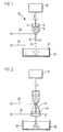
- Figure 1 shows a droplet-forming apparatus 10 constructed in accordance with one embodiment of the invention.
- a syringe 12 of non-conducting, usually polymeric, material contains a gel droplet-forming liquid 14 which contains living cells.
- a plunger 16 is driven by a syringe pump 18 to expel droplets 20 from the lower end of a stainless steel syringe needle 22 communicating with the lower end of the syringe reservoir 12, towards a collecting vessel 24 containing a hardening solution 26, which may be aqueous calcium chloride solution in the case of an aqueous droplet-forming liquid containing sodium alginate.
- the positive lead 28 of an electrical pulse generator (see Figure 3) is connected to the needle 22 while the negative lead of the pulse generator is connected to the hardening solution.
- the needle 22 may be bevelled at its outlet tip 27, if desired.
- the tip 27 is located at a specific distance from the top of the recipient medium 26 in the collecting vessel 24 consistent with the voltage pulse to be applied therebetween to effect droplet formation.
- the size of the droplets 18 may be varied by varying the distance between the needle tip 27 and the liquid in the collecting vessel 24, with shorter distances leading to smaller droplets, by varying the voltage applied by the leads 28 and 30 with increased voltage leading to smaller droplets, by varying the pulse length of applied electricity with decreasing pulse length leading to smaller droplets, or by varying the speed-of the pump 18 with decreasing pump speed leading to smaller droplets.
- Figure 2 illustrates an alternative arrangement wherein the positive wire 28 from the pulse generator is detached from the needle 22 and instead is attached to a stainless steel ring 32 which is mounted to the lower end of a conical support 34 of non-conductive material which surrounds and extends below the needle 22.
- the negative lead 30 is attached to the needle 22 rather than to the recipient medium 26. In this arrangement, the distance between the tip 27 of the needle 22 and the top of the recipient medium 26 does not affect the gel droplet size.
- the static voltage which is applied by lead wires 28 and 30 during droplet formation in Figures 1 and 2 results in gel droplets having a diameter less than about 700 L im, preferably about 150 to about 500 pm. These droplets then may be coated with a thin coating of a semi-permeable biocompatible membrane. The resulting microcapsules are small enough to be injected into an animal body using an 18 gauge needle fitted to a syringe.
- the voltage applied during droplet formation is a static one, the viability of encapsulated living tissue, such as islets of Langerhans or liver cells, is not destroyed, and hence the microencapsulated living tissue is capable of on-going metabolism.
- an electrostatic pulse generator 110 suitable for formation of electrostatic pulses to be applied during droplet formation by the apparatus of Figure 1 or 2 is illustrated in Figure 3.
- the pulse generator 110 includes an isolated power supply 112, which may be connected to any desired source of electric power, logic circuitry 114, console panel 116 having adjusting knobs for pulse frequency 118, pulse width 120 and high voltage output 122, a pulse amplifier 124, and a high voltage transformer and rectifier 126 which outputs to the electrical lead wires 28 and 30.
- the electrical pulse voltage, pulse frequency and pulse length which pass to the droplet forming apparatus 10 by the lead wires 28 and 30 may each vary widely, depending on the size of droplets desired.
- the pulse voltage which determines the strength of the force pulling the droplets from the end of the needle 22, usually varies from about 1 to about 25 KV.
- the pulse frequency which determines how many pulses are applied to the droplet, usually varies from about 10 to about 100 sec.
- the pulse length which determines the length of time for which the droplet-forming force is applied, usually varies from about 1 to about 6 m. sec.
- the interaction of the various time periods and their meaning is further illustrated in Figure 4. These values are synchronized with the amount of material dispensed from the needle to obtain uniformly-sized droplets.
- the droplet generator of this invention is capable of producing very small, spherical droplets ccntaining living cells with each step of the droplet fcrmation being under the direct control of the operator.
- electrostatic droplet forming method and apparatus described herein has application in other fields and, for example, may be used in spray painting and ink printing.
- living tissue or individual cells are encapsulated in a biocompatible semi-permeable membrane, in the form of a hydrogel.
- the material to be encapsulated is suspended in a physiologically-compatible medium containing a water soluble substance which can be reversibly gelled to provide a temporary protective environment for the tissue.
- the medium is formed into droplets containing the tissue, using the droplet generation procedure cf the invention, and gelled, for example, by changing conditions of temperature, pH or ionic environment, to form temporary capsules, of substantially perfect spherical shape. Thereafter, the temporary capsules which result are treated to form a membrane of controlled permeability and negatively-charged outer surface about the shape-retaining temporary capsules.
- the semi-permeable nature of the membrane permits nutrients and oxygen to flow to the core material and metabolic products to flow therefrom while retaining the core material within the microcapsule.
- the biocompatible nature of the semi-permeable membrane allows the passage of such materials to and from the core to occur without inflammation or other adverse body response while the outer negatively-charged surface inhibits surficial cell growth, so that the membrane remains semi-permeable and effective for extended periods of time, typically from three to six months or longer.
- the temporary capsules may be formed from any non-toxic water-soluble substance that can be gelled to form a shape retaining mass by a chance of conditions in the medium in which it is placed, and also comprises plural groups that are readily ionized to form anionic or cationic groups. The presence of such groups enables surface layers of the capsule to cross-link to produce a permanent membrane when exposed to polymers containing multiple functionalities of the opposite charge.
- the temporary capsules are formed from a polysaccharide gum, which may be natural or synthetic, of a type that can be gelled to form a shape retaining mass by exposure to a change in conditions and can be permanently cross-linked or hardened by polymers containing reactive groups, such as amino groups, which can react with the acidic polysaccharide constituents.
- a polysaccharide gum which may be natural or synthetic, of a type that can be gelled to form a shape retaining mass by exposure to a change in conditions and can be permanently cross-linked or hardened by polymers containing reactive groups, such as amino groups, which can react with the acidic polysaccharide constituents.
- the gum is alkali metal alginate, specifically sodium alginate, although other water-soluble gums may be used.
- the temporary capsules may be formed from sodium alginate by extruding droplets of aqueous sodium alginate solution into an aqueous calcium chloride solution. It is preferred that the temporary capsules be substantially spherical so that perfectly spherical microcapsules can be formed for cardiovascular injection. Substantially perfectly spherical temporary capsules are formed by using an aqueous sodium alginate solution having a viscosity of at least about 30 centipoise. At viscosities below this critical lower limit, the temporary capsules have an irregular shape.
- Formation of the permanent semi-permeable membrane about the temporary capsules preferably is effected by ionic reaction between free acid groups in the surface layer of the gelled gum and biocompatible polymers containing acid-reactive groups, such as, amino groups, typically in a dilute aqueous solution of the selected polymer.
- the cross-linking biocompatible polymers which may be used include polyamino acids, preferably polylysine. It is noted that pclyethyleneimine and other imine-containing polymers are unsuitable for membrane formation in view of their non-biocompatible nature.
- the molecular weight of the preferred polylysine polymer should be controlled within a narrow range of about 10,000 to about 30,000, preferably about 17,000, to achieve the required membrane porosity.
- the use of polylysine or other polyamino acid results in microcapsules having a positively-charged surface, which would be unsuitable for long term viability, although the microcapsules are biocompatible.
- polylysine or other polyamino acid it is important for long term in vivo life for the polylysine or other polyamino acid to be reacted for a period of time sufficient to develop a substantial thickness of membrane, sc as to provide a substantial number of surface groups for post-reaction, as discussed below, sufficient structural strength to permit in vivo injection and sufficient quantity of biocompatible polymer to permit in vivo structural integrity to be retained.
- a reaction time of at least six minutes is required to achieve these results, generally up to about 9 minutes. These reaction times result in a polylysine layer thickness of about 5 microns.
- the actual strength of the aqueous solution of polylysine used to react with the temporary capsules does not affect the capsule wall thickness, at concentration levels in excess of about 0.05 wt.%.
- the semi-permeable membrane formed about the temporary capsules by the reaction with the polyareino acid next is treated with a non-toxic biocompatible water-soluble polymeric material which is capable of ionic reaction with free amino groups to form an outer negatively-charged coating about the membrane, typically by suspension of the microcapsules in an aqueous solution of the polymeric material.
- the material used tc form the outer coating preferably is the same material as is used to form the temporary capsules, preferably a polysaccharide gum, more preferably an alkali metal alginate, such as, sodium alginate.
- Other biocompatible polymeric materials containing base-reactive groups such as, polyvinyl alcohol and poly beta-hydroxy butyric acid, may be used to form the outer coating to the microcapsules. Molecular weights of such polymeric materials typically vary from about 10 4 to about 10 6 .
- the biocompatible water-soluble polymeric material containing amino-reactive groups reacts with the outer amino-groups of the semi-permeable membrane to form an outer coating.
- This outer coating permanently shrouds the polyamino acid layer, although leaving intact the porosity of the semi-permeable membrane, and provides a negatively-charged surface.
- the outer negatively-charged polymer coating resists degradation and removal, in vivo, so that the positively charged surfaces are not exposed to the body environment.
- the treatment of the polyamino microcapsules with the biocompatible base-reactive material retains the overall biocompatible nature of the semi-permeable membrane and results in a negatively-charged outer surface which inhibits cell growth and, therefore, permits the semi-permeable membrane to retain its permeability and hence effectiveness over an extended period of time.
- reliquification of the suspending medium for the core material may be effected by re-establishing the conditions under which the material is liquid. This may be achieved by ion exchange to remove multivalent cation, for example, by immersion in phosphate buffered saline or citrate buffer.
- the reliquification step though beneficial in decreasing diffusion resistance, is not essential for the provision of an effective product and may be omitted, since it has been shown that transplanted islets (rat to mouse) in microcapsules whose interiors have not been reliquified, are also effective in normalizing blood sugar levels of diabetic animals.
- the calcium alginate gel core does not reliquify inside the body, since intact gel cores have been found in microcapsules recovered from diabetic animals up to one year after implantation.
- the process of the invention may be used to encapsulate living tissue, multicellular fractions thereof or individual cells, for example, islets of Langerhans, liver cells and red blood cells, and other biologically-active material.
- the microcapsules which result may be implanted into an appropriate site within a mammalian body for the purpose of providing the body with the specialized physiological function of the tissue while the tissue remains viable.
- the implantation may be achieved by simple injection, so that surgical procedures are not required.
- cardiovascular injection may be effected, in view of the smaller diameter microcapsules which result from the electrostatic droplet generation procedure.
- the core of the microcapsules contains the living tissue cells and an aqueous medium of nutrients sufficient to maintain the tissue and allow its normal metabolism.
- the cells are viable, physiologically active and capable of ongoing metabolism.
- the biocompatible semi-permeable membrane encapsulating the core material consists of interpenetrating layers of ionically-interacted biocompatible materials.
- the overall wall thickness of the semi-permeable membrane usually varies from about 4 to about 6 nm.
- the microcapsules themselves have a diameter in the range of less than about 700 ⁇ m , preferably in the range of about 150 to about 500 pm for microcapsules containing islets of Langerhans as the core material.
- the biocompatible semi-permeable membrane is in the form of a hydrogel and hence has an overall water content within the membrane structure of at least about 20 wt%, which may vary up to about 95 wt%, depending on the molecular weight of the polyamino acid.
- living-cells are microencapsulated within a pclylysine-alginate semi-permeable hydrogel.
- the cells are initially suspended uniformly in a sodium alginate solution in physiological saline.
- the living cells take the form of islets of Langerhans from an animal pancreas.
- Spherical droplets containing the cells are produced from an aqueous sodium alginate solution by the electrostatic droplet generation procedure of the invention and are collected as gelled spheres in a hardening solution, such as, calcium chloride.
- the gelled spheres are coated with polylysine followed by an outer coating of sodium alginate.
- the microcapsules may then be suspended in isotonic sodium citrate or other convenient ion exchange medium to reliquify the alginate gel inside the microcapsule to restore the cells to a mobile state. As noted earlier, this step may be omitted, if desired.
- the outer biochemically inert but biocompatible alginate surface is a negatively-charged hydrogel containing up to about 95% water.
- the low interfacial tension between the swollen gel surface and the aqueous biological environment minimizes protein interaction, otherwise a strong protein-polymer interaction may cause a severe inflammatory response.
- the biocompatibility of the hydrogel membrane leads to long term viability of the capsules when implanted.
- Polyethyleneimine-surfaced microcapsules do not appear to possess this property, since they produce a strong inflammatory response and hence are rejected by the body, which severely limits the useful in vivo life of the microcapsules.
- the soft rubbery consistency of most hydrogels may also contribute to their biocompatibility by decreasing frictional irritation to surrounding tissues.
- the strength of the microcapsules may be increased by additional cross-linking, for example, using glutaraldehyde, prior to reliquification of the g el, if effected.
- the biocompatible outer surface be composed of sodium alginate, but it is essential that the cuter surface be biocompatible and negatively-charged. Binding occurs between the negatively-charged groups, usually hydroxyl or carboxyl groups, of the biocompatible outer surface material, and the positively-charged amino groups on polylysine.
- biocompatible microcapsules capable of long term in vivo life and having a diameter which render them suitable for injection of living tissue into the blood stream, so that the microcapsules may lodge inside body organs for ongoing metabolism therein. While the primary benefit of the smaller diameter microcapsules of the invention is in in-vivo uses, the living tissue-containing microcapsules may also be put to a variety of in-vitro uses.
- microcapsules containing living tissue or cells may be used to form microcapsules containing a variety of other core materials, depending on the intended end use of the microcapsules.
- This Example illustrates the formation of small diameter gel droplets using an electrostatic droplet generator.
- FIG. 1 An apparatus as illustrated in Figure 1 was set up.
- a 1.5% w/v sodium alginate solution (14) was placed in a 10 cc syringe (12) to which is attached a 22 gauge stainless steel needle (22) having a 90° bevel outlet.
- the positive polarity wire (28) was attached to the metal leur lock section of the needle and the needle-syringe combination was attached to the syringe pump (18).
- a 1.1% calcium chloride solution (26) was poured into a 4" x 1" petri dish (24) to which was attached the negative polarity wire (30).
- the petri dish (24) was positioned so that the liquid surface therein was 10 mm from the tip of the needle (22).
- the pulse voltage dial (122) on the adjustment panel (116) was set at 12 KV, the pulse frequency dial (118) at 20 sec -1 , the pulse length dial (120) at 2 m.. sec, and the syringe pump speed at 4 ml/hr.
- the syringe pump (18) and droplet generator were both turned on so that sodium alginate liquid droplets (20) were drawn from the tip (27) of the needle (22) and, upon entering the calcium ride solution in the petri dish (24), calcium alginate gel droplets were formed and were collected therein.
- the resultant calcium alginate gel droplets were found to be perfectly smooth and spherical and with a mean diameter of 300 ( ⁇ 50 SD) ⁇ m.
- the syringe needle (22) used in this Example was of the same diameter as was previously used in an air jet syringe wherein a rapidly flowing air stream was used to remove sodium alginate liquid droplets from the tip (27) of the needle (22) using the air jet syringe, the smallest diameter calcium alginate gel droplets attainable had a diameter of 700 ⁇ m.
- the electrostatic procedure described in this Example therefore, was able to decrease the g el droplet diameter to approximately half this value.
- This Example illustrates the formation of small diameter gel droplets using an alternative form of droplet generation.
- Example 1 The procedure of Example 1 was repeated, except that the apparatus of Figure 2 was utilized, i.e. the negative polarity wire (30) was attached to the needle (22) and the positive polarity wire (28) is attached to the metal ring device (32) which is spaced downwardly from the tip of the needle (22).
- the centre of the metal ring (32) was positioned 7 mm downwardly from the tip (27) of the needle (22) and an uncharged petri dish (24) was positioned about 5 cm downwardly from the ring assembly.
- the calcium alginate gel droplets produced by this procedure and collected in the petri dish were observed to be perfectly smooth and spherical and to have a mean diameter of 450 ( ⁇ 65 SD) ⁇ m.
- SD standard deviation
- This Example illustrates the viability of living tissue after passage through the electrostatic droplet generator.
- Example 1 The procedure of Example 1 was repeated except that islets of Langerhans extracted from the pancreatic tissue of dogs were added to the sodium alginate solution in the syringe in a concentration of 500 islets/2 ml and the calcium chloride solution was replaced by saline, so that gel droplet formation did not occur in this experiment. After passage through the electrcstatic droplet generator, 100% of the islets were shown to be viable using Trypan blue staining. All the islets appeared white when viewed under the microscope, there being no evidence of the blue appearance characteristic of dead islets.
- This Example illustrates the formation of small semi-permeable microcapsules containing islets of Langerhans.
- the gelled droplets were incubated for 6 minutes in 0.05% (w/w) solution of polylysine having a molecular weight of 17,000. The supernatant was decanted and the polylysine capsules were washed with dilute CHES, 1.1% calcium chloride solution and physiological saline.
- the washed polylysine capsules were incubated for 4 minutes in 30 ml of 0.03% sodium alginate to permit the formation of an outer alginate membrane on the initial polylysine membrane, by ionic interaction between the negatively-charged alginate and the positively-charged polylysine.
- microcapsules were washed with saline, 0.05M citrate buffer for 6 minutes to reliquify the inner calcium alginate, and a final saline wash.
- the microcapsules were found to be perfectly spherical and each to contain from 1 to 2 viable islets.
- the microcapsules had a mean diameter of 300 ( ⁇ 50 SD) microns and wall thicknesses of 5 ⁇ m.
- the microcapsules were suspended in nutrient medium at 37°C.
- the viability of the islets was demonstrated by Trypan Blue staining after the capsule walls were dissociated with heparin.
- This Example illustrates the formation of small semi-permeable microcapsules containing hepatocytes (liver cells).
- Example 4 The procedure of Example 4 was repeated except that fetal mouse or adult rat hepatocytes were added to the sodium alginate solution in amounts of 10" hepatocytes/ml of alginate solution and the distance from the tip of the needle to the surface of the calcium chloride solution was decreased to 7 mm.
- the resulting microcapsules were spherical in appearance and had a diameter of 250 ⁇ m ( ⁇ 50 SD).
- the presence of viable hepatocytes was demonstrated by Trypan Blue staining and histology, even after more than 4 weeks in culture at 37°C.
- This Example illustrates the effect of needle parameters on gel droplet size.
- Example 2 The procedure of Example 1 was repeated, except that a 26 gauge needle having a 22-degree bevel was used in place of the 22 gauge needle having the 90-degree bevel.
- the resultant gel droplets had a diameter of 170 um ( ⁇ 30 SD), demonstrating the smaller diameter gel droplets and consequently microcapsules can be formed by using a smaller diameter needle.
- the present invention provides a novel droplet generation procedure using electrostatic forces which is particularly useful in the microencapsulation of living tissue or cells to form small diameter microcapsules suitable for cardiovascular injection. Modifications are possible within the scope of the invention.
Landscapes
- Chemical & Material Sciences (AREA)
- Health & Medical Sciences (AREA)
- Engineering & Computer Science (AREA)
- Bioinformatics & Cheminformatics (AREA)
- Organic Chemistry (AREA)
- Chemical Kinetics & Catalysis (AREA)
- Epidemiology (AREA)
- Animal Behavior & Ethology (AREA)
- General Health & Medical Sciences (AREA)
- Public Health (AREA)
- Veterinary Medicine (AREA)
- Pharmacology & Pharmacy (AREA)
- Medicinal Chemistry (AREA)
- Dispersion Chemistry (AREA)
- Life Sciences & Earth Sciences (AREA)
- Medicinal Preparation (AREA)
- Micro-Organisms Or Cultivation Processes Thereof (AREA)
- Medicines Containing Material From Animals Or Micro-Organisms (AREA)
- Medicines That Contain Protein Lipid Enzymes And Other Medicines (AREA)
- Prostheses (AREA)
- Apparatus Associated With Microorganisms And Enzymes (AREA)
- Transition And Organic Metals Composition Catalysts For Addition Polymerization (AREA)
- Preparation Of Compounds By Using Micro-Organisms (AREA)
- Manufacturing Of Micro-Capsules (AREA)
Priority Applications (1)
| Application Number | Priority Date | Filing Date | Title |
|---|---|---|---|
| AT84305399T ATE60506T1 (de) | 1984-07-11 | 1984-08-08 | Erzeugung von troepfchen. |
Applications Claiming Priority (3)
| Application Number | Priority Date | Filing Date | Title |
|---|---|---|---|
| CA000458605A CA1241598A (fr) | 1984-07-11 | 1984-07-11 | Generation de gouttelettes |
| CA458605 | 1984-07-11 | ||
| CN85105964.3A CN1004257B (zh) | 1984-07-11 | 1985-08-06 | 小滴生成 |
Publications (3)
| Publication Number | Publication Date |
|---|---|
| EP0167690A2 true EP0167690A2 (fr) | 1986-01-15 |
| EP0167690A3 EP0167690A3 (en) | 1987-09-30 |
| EP0167690B1 EP0167690B1 (fr) | 1991-01-30 |
Family
ID=76224745
Family Applications (1)
| Application Number | Title | Priority Date | Filing Date |
|---|---|---|---|
| EP84305399A Expired - Lifetime EP0167690B1 (fr) | 1984-07-11 | 1984-08-08 | Création de gouttelettes |
Country Status (6)
| Country | Link |
|---|---|
| EP (1) | EP0167690B1 (fr) |
| JP (1) | JPS6144823A (fr) |
| CN (1) | CN1004257B (fr) |
| AT (1) | ATE60506T1 (fr) |
| CA (2) | CA1241598A (fr) |
| DE (1) | DE3484067D1 (fr) |
Cited By (13)
| Publication number | Priority date | Publication date | Assignee | Title |
|---|---|---|---|---|
| EP0290984A2 (fr) * | 1987-05-15 | 1988-11-17 | Henkel Kommanditgesellschaft auf Aktien | Procédé pour l'agglomération en granules de particules solides |
| US4920090A (en) * | 1987-05-15 | 1990-04-24 | Henkel Kommanditgesellschaft Auf Aktien | Process for the formation of shaped agglomerates from particulate solids |
| US4933122A (en) * | 1987-02-13 | 1990-06-12 | Kirin Beer Kabushiki Kaisha | Process and apparatus for producing beads |
| US5015423A (en) * | 1986-10-29 | 1991-05-14 | Kanegafuchi Kagaku Kogyo Kabushiki Kaisha | Method of making uniform polymer particles |
| EP0503713A1 (fr) * | 1991-03-11 | 1992-09-16 | SOLVAY (Société Anonyme) | Particules microsphéroidales et leur procédé d'obtention. |
| DE4312970A1 (de) * | 1993-04-21 | 1994-10-27 | Juergen Dr Schrezenmeir | Mikrokapsel sowie Verfahren und Vorrichtung zu ihrer Herstellung |
| DE4426396A1 (de) * | 1994-07-26 | 1996-02-01 | Ulrich Prof Dr Zimmermann | Verfahren zur Herstellung konzentrierter Lösungen von mikroverkapselten Zellen oder von suspendierten Wirkstoffen in mikroverkapselter Form |
| EP0746265A1 (fr) * | 1994-03-08 | 1996-12-11 | The Regents Of The University Of California | Dissolution in situ de couches d'alginate de greffes de tissus biologiques |
| WO2009017549A1 (fr) * | 2007-08-01 | 2009-02-05 | Abbott Cardiovascular Systems Inc. | Procédé d'électropulvérisation permettant la fabrication de particules enrobées en vue de l'administration d'agents de traitement |
| US8206622B2 (en) | 2004-02-23 | 2012-06-26 | Eyesense Ag | Process for production of ionically crosslinked polysaccharide microspheres |
| WO2020117159A1 (fr) * | 2018-12-02 | 2020-06-11 | Bursa Teknik Universitesi | Mécanisme d'égouttement pour réduire la taille des particules dans des procédés de microencapsulation |
| CN112972664A (zh) * | 2021-02-08 | 2021-06-18 | 武汉大学 | 基于微流控芯片从血液中制备凝胶液滴单核细胞疫苗的装置及方法 |
| CN114921343A (zh) * | 2022-06-28 | 2022-08-19 | 中国科学院苏州生物医学工程技术研究所 | 一种基于高压脉冲电场的细胞凝胶微球生成装置 |
Families Citing this family (7)
| Publication number | Priority date | Publication date | Assignee | Title |
|---|---|---|---|---|
| CA3066594A1 (fr) | 2004-10-08 | 2006-04-20 | Georgia Tech Research Corporation | Microencapsulation de cellules dans des hydrogels a l'aide de potentiels electrostatiques |
| CN100488618C (zh) * | 2006-09-25 | 2009-05-20 | 上海理工大学 | 用静电喷雾制备液芯微胶囊的方法 |
| JP2010184913A (ja) * | 2009-02-13 | 2010-08-26 | Freunt Ind Co Ltd | 微生物または生物由来物質含有微細粒子およびその製造方法 |
| CN104785164B (zh) * | 2015-04-15 | 2017-01-25 | 西北大学 | 一种静电喷雾制备胶体颗粒装置及其控制方法 |
| CN108673707A (zh) * | 2018-05-10 | 2018-10-19 | 河源帝诺新材料有限公司 | 一种氧化锆微珠高压静电吸引滴定成型设备 |
| CN109007943A (zh) * | 2018-07-12 | 2018-12-18 | 王兰玺 | 一种爆珠自动挤压滴料装置 |
| CN110564689A (zh) * | 2019-07-29 | 2019-12-13 | 嘉兴市桔猫生物技术有限公司 | 个性化肺癌pdo模型及其制备方法与检测试剂盒 |
Citations (4)
| Publication number | Priority date | Publication date | Assignee | Title |
|---|---|---|---|---|
| GB2094833A (en) * | 1981-03-13 | 1982-09-22 | Damon Corp | Process and system for producing biological materials from encapsulated cells |
| US4352883A (en) * | 1979-03-28 | 1982-10-05 | Damon Corporation | Encapsulation of biological material |
| US4391909A (en) * | 1979-03-28 | 1983-07-05 | Damon Corporation | Microcapsules containing viable tissue cells |
| GB2119734A (en) * | 1979-03-28 | 1983-11-23 | Damon Corp | Encapsulated living tissue |
-
1984
- 1984-07-11 CA CA000458605A patent/CA1241598A/fr not_active Expired
- 1984-08-08 EP EP84305399A patent/EP0167690B1/fr not_active Expired - Lifetime
- 1984-08-08 AT AT84305399T patent/ATE60506T1/de not_active IP Right Cessation
- 1984-08-08 DE DE8484305399T patent/DE3484067D1/de not_active Expired - Fee Related
-
1985
- 1985-07-11 JP JP60151470A patent/JPS6144823A/ja active Granted
- 1985-08-06 CN CN85105964.3A patent/CN1004257B/zh not_active Expired
-
1987
- 1987-02-17 CA CA000529952A patent/CA1240815A/fr not_active Expired
Patent Citations (4)
| Publication number | Priority date | Publication date | Assignee | Title |
|---|---|---|---|---|
| US4352883A (en) * | 1979-03-28 | 1982-10-05 | Damon Corporation | Encapsulation of biological material |
| US4391909A (en) * | 1979-03-28 | 1983-07-05 | Damon Corporation | Microcapsules containing viable tissue cells |
| GB2119734A (en) * | 1979-03-28 | 1983-11-23 | Damon Corp | Encapsulated living tissue |
| GB2094833A (en) * | 1981-03-13 | 1982-09-22 | Damon Corp | Process and system for producing biological materials from encapsulated cells |
Cited By (24)
| Publication number | Priority date | Publication date | Assignee | Title |
|---|---|---|---|---|
| US5015423A (en) * | 1986-10-29 | 1991-05-14 | Kanegafuchi Kagaku Kogyo Kabushiki Kaisha | Method of making uniform polymer particles |
| US4933122A (en) * | 1987-02-13 | 1990-06-12 | Kirin Beer Kabushiki Kaisha | Process and apparatus for producing beads |
| EP0290984A2 (fr) * | 1987-05-15 | 1988-11-17 | Henkel Kommanditgesellschaft auf Aktien | Procédé pour l'agglomération en granules de particules solides |
| EP0290984A3 (en) * | 1987-05-15 | 1989-02-15 | Henkel Kommanditgesellschaft Auf Aktien | Process for agglomerating solid particles to form granules |
| US4866023A (en) * | 1987-05-15 | 1989-09-12 | Henkel Kommanditgesellschaft Auf Aktien | Process for the shaping agglomeration of particulate solids and solids produced by the process |
| US4920090A (en) * | 1987-05-15 | 1990-04-24 | Henkel Kommanditgesellschaft Auf Aktien | Process for the formation of shaped agglomerates from particulate solids |
| EP0680780A3 (fr) * | 1991-03-11 | 1995-12-27 | Solvay | Procédé d'obtention de particules microsphéroidales homodisperses. |
| BE1004675A3 (fr) * | 1991-03-11 | 1993-01-12 | Solvay | Procede d'obtention de particules microspheroidales homodisperses, particules microspheroidales de silice a surface specifique elevee, catalyseurs supportes sur ces particules et procede de polymerisation des alpha-olefines en presence de ces catalyseurs. |
| US5232883A (en) * | 1991-03-11 | 1993-08-03 | Solvay (Societe Anonyme) | Process for obtaining monodisperse microspheroidal particles, microspheroidal silica particles of high specific surface, and catalysts supported on these particles |
| US5296564A (en) * | 1991-03-11 | 1994-03-22 | Solvay (Societe Anonyme) | Process for the polymerization of alpha-olefins |
| EP0680780A2 (fr) * | 1991-03-11 | 1995-11-08 | SOLVAY (Société Anonyme) | Procédé d'obtention de particules microsphéroidales homodisperses |
| EP0503713A1 (fr) * | 1991-03-11 | 1992-09-16 | SOLVAY (Société Anonyme) | Particules microsphéroidales et leur procédé d'obtention. |
| DE4312970A1 (de) * | 1993-04-21 | 1994-10-27 | Juergen Dr Schrezenmeir | Mikrokapsel sowie Verfahren und Vorrichtung zu ihrer Herstellung |
| EP0746265A1 (fr) * | 1994-03-08 | 1996-12-11 | The Regents Of The University Of California | Dissolution in situ de couches d'alginate de greffes de tissus biologiques |
| EP0746265A4 (fr) * | 1994-03-08 | 1998-05-06 | Univ California | Dissolution in situ de couches d'alginate de greffes de tissus biologiques |
| WO1996003205A1 (fr) * | 1994-07-26 | 1996-02-08 | Ulrich Zimmermann | Preparation de cellules ou de principes actifs disperses sous forme de microcapsules, et microcapsules ainsi obtenues |
| DE4426396A1 (de) * | 1994-07-26 | 1996-02-01 | Ulrich Prof Dr Zimmermann | Verfahren zur Herstellung konzentrierter Lösungen von mikroverkapselten Zellen oder von suspendierten Wirkstoffen in mikroverkapselter Form |
| US8206622B2 (en) | 2004-02-23 | 2012-06-26 | Eyesense Ag | Process for production of ionically crosslinked polysaccharide microspheres |
| WO2009017549A1 (fr) * | 2007-08-01 | 2009-02-05 | Abbott Cardiovascular Systems Inc. | Procédé d'électropulvérisation permettant la fabrication de particules enrobées en vue de l'administration d'agents de traitement |
| WO2020117159A1 (fr) * | 2018-12-02 | 2020-06-11 | Bursa Teknik Universitesi | Mécanisme d'égouttement pour réduire la taille des particules dans des procédés de microencapsulation |
| CN112972664A (zh) * | 2021-02-08 | 2021-06-18 | 武汉大学 | 基于微流控芯片从血液中制备凝胶液滴单核细胞疫苗的装置及方法 |
| CN112972664B (zh) * | 2021-02-08 | 2022-07-05 | 武汉大学 | 基于微流控芯片从血液中制备凝胶液滴单核细胞疫苗的装置及方法 |
| CN114921343A (zh) * | 2022-06-28 | 2022-08-19 | 中国科学院苏州生物医学工程技术研究所 | 一种基于高压脉冲电场的细胞凝胶微球生成装置 |
| CN114921343B (zh) * | 2022-06-28 | 2023-09-05 | 中国科学院苏州生物医学工程技术研究所 | 一种基于高压脉冲电场的细胞凝胶微球生成装置 |
Also Published As
| Publication number | Publication date |
|---|---|
| CN85105964A (zh) | 1987-02-25 |
| CN1004257B (zh) | 1989-05-24 |
| JPS6238326B2 (fr) | 1987-08-17 |
| CA1240815A (fr) | 1988-08-23 |
| EP0167690A3 (en) | 1987-09-30 |
| CA1241598A (fr) | 1988-09-06 |
| JPS6144823A (ja) | 1986-03-04 |
| EP0167690B1 (fr) | 1991-01-30 |
| ATE60506T1 (de) | 1991-02-15 |
| DE3484067D1 (de) | 1991-03-07 |
Similar Documents
| Publication | Publication Date | Title |
|---|---|---|
| US4789550A (en) | Microcapsule composition suitable for cardiovascular injection | |
| US4956128A (en) | Droplet generation | |
| EP0167690B1 (fr) | Création de gouttelettes | |
| US4673566A (en) | Microencapsulation of living tissue and cells | |
| US5656469A (en) | Method of encapsulating biological substances in microspheres | |
| US4806355A (en) | Microencapsulation of living tissue and cells | |
| US4689293A (en) | Microencapsulation of living tissue and cells | |
| US5656468A (en) | Cells or tissue coated with non-fibrogenic alginate less than 200 μm thick | |
| CA1258429A (fr) | Microencapsulation de cellules vivantes | |
| Van Harreveld et al. | Light-and electron-microscopic changes in central nervous tissue after electrophoretic injection of glutamate | |
| US20180346873A1 (en) | Artificial micro-gland | |
| US5429821A (en) | Non-fibrogenic high mannuronate alginate coated transplants, processes for their manufacture, and methods for their use | |
| JP3007144B2 (ja) | 細胞カプセル押し出し成形システム | |
| US5182111A (en) | In vivo delivery of active factors by co-cultured cell implants | |
| US6649384B2 (en) | System and method for encapsulating biological material by applying electrostatic charge to capsules | |
| EP0127989A2 (fr) | Micro-encapsulation de tissu vivant et de cellules | |
| WO2008121447A1 (fr) | Membranes à assemblage automatique, et leurs procédés pertinents | |
| CN110184263B (zh) | 一种监测肌细胞力学性质和收缩频率的核壳结构微球及其应用 | |
| Lahooti et al. | Microencapsulation of normal and transfected L929 fibroblasts in a HEMA-MMA copolymer | |
| Lewińska et al. | Electrostatic microencapsulation of living cells | |
| JPH07507810A (ja) | 内植可能なカプセル | |
| JP4705097B2 (ja) | 細胞をカプセル化するための装置 | |
| CN114957759B (zh) | 一种核壳结构微载体及其制备方法 | |
| EP3878512A1 (fr) | Greffon et utilisation correspondante | |
| Goosen et al. | Immobilization of cells using electrostatic droplet generation |
Legal Events
| Date | Code | Title | Description |
|---|---|---|---|
| PUAI | Public reference made under article 153(3) epc to a published international application that has entered the european phase |
Free format text: ORIGINAL CODE: 0009012 |
|
| AK | Designated contracting states |
Designated state(s): AT BE CH DE FR GB IT LI LU NL SE |
|
| RIN1 | Information on inventor provided before grant (corrected) |
Inventor name: GOOSEN, MATTHEUS FLORENTIUS ALBERTUS Inventor name: SUN, ANTHONY MEIN-FANG Inventor name: HOMMEL, MARTIN |
|
| PUAL | Search report despatched |
Free format text: ORIGINAL CODE: 0009013 |
|
| AK | Designated contracting states |
Kind code of ref document: A3 Designated state(s): AT BE CH DE FR GB IT LI LU NL SE |
|
| 17P | Request for examination filed |
Effective date: 19880310 |
|
| 17Q | First examination report despatched |
Effective date: 19891117 |
|
| GRAA | (expected) grant |
Free format text: ORIGINAL CODE: 0009210 |
|
| AK | Designated contracting states |
Kind code of ref document: B1 Designated state(s): AT BE CH DE FR GB IT LI LU NL SE |
|
| REF | Corresponds to: |
Ref document number: 60506 Country of ref document: AT Date of ref document: 19910215 Kind code of ref document: T |
|
| ITF | It: translation for a ep patent filed | ||
| REF | Corresponds to: |
Ref document number: 3484067 Country of ref document: DE Date of ref document: 19910307 |
|
| ET | Fr: translation filed | ||
| PLBE | No opposition filed within time limit |
Free format text: ORIGINAL CODE: 0009261 |
|
| STAA | Information on the status of an ep patent application or granted ep patent |
Free format text: STATUS: NO OPPOSITION FILED WITHIN TIME LIMIT |
|
| 26N | No opposition filed | ||
| EPTA | Lu: last paid annual fee | ||
| EAL | Se: european patent in force in sweden |
Ref document number: 84305399.2 |
|
| PGFP | Annual fee paid to national office [announced via postgrant information from national office to epo] |
Ref country code: FR Payment date: 19960619 Year of fee payment: 13 |
|
| PGFP | Annual fee paid to national office [announced via postgrant information from national office to epo] |
Ref country code: LU Payment date: 19960701 Year of fee payment: 13 |
|
| PGFP | Annual fee paid to national office [announced via postgrant information from national office to epo] |
Ref country code: SE Payment date: 19960704 Year of fee payment: 13 |
|
| PGFP | Annual fee paid to national office [announced via postgrant information from national office to epo] |
Ref country code: BE Payment date: 19960711 Year of fee payment: 13 |
|
| PGFP | Annual fee paid to national office [announced via postgrant information from national office to epo] |
Ref country code: GB Payment date: 19960806 Year of fee payment: 13 |
|
| PGFP | Annual fee paid to national office [announced via postgrant information from national office to epo] |
Ref country code: DE Payment date: 19960827 Year of fee payment: 13 |
|
| PGFP | Annual fee paid to national office [announced via postgrant information from national office to epo] |
Ref country code: NL Payment date: 19960830 Year of fee payment: 13 Ref country code: AT Payment date: 19960830 Year of fee payment: 13 |
|
| PGFP | Annual fee paid to national office [announced via postgrant information from national office to epo] |
Ref country code: CH Payment date: 19960910 Year of fee payment: 13 |
|
| PG25 | Lapsed in a contracting state [announced via postgrant information from national office to epo] |
Ref country code: LU Free format text: LAPSE BECAUSE OF NON-PAYMENT OF DUE FEES Effective date: 19970808 Ref country code: GB Free format text: LAPSE BECAUSE OF NON-PAYMENT OF DUE FEES Effective date: 19970808 Ref country code: AT Free format text: LAPSE BECAUSE OF NON-PAYMENT OF DUE FEES Effective date: 19970808 |
|
| PG25 | Lapsed in a contracting state [announced via postgrant information from national office to epo] |
Ref country code: SE Free format text: LAPSE BECAUSE OF NON-PAYMENT OF DUE FEES Effective date: 19970809 |
|
| PG25 | Lapsed in a contracting state [announced via postgrant information from national office to epo] |
Ref country code: LI Free format text: LAPSE BECAUSE OF NON-PAYMENT OF DUE FEES Effective date: 19970831 Ref country code: CH Free format text: LAPSE BECAUSE OF NON-PAYMENT OF DUE FEES Effective date: 19970831 Ref country code: BE Free format text: LAPSE BECAUSE OF NON-PAYMENT OF DUE FEES Effective date: 19970831 |
|
| BERE | Be: lapsed |
Owner name: CONNAUGHT LABORATORIES LTD Effective date: 19970831 |
|
| PG25 | Lapsed in a contracting state [announced via postgrant information from national office to epo] |
Ref country code: NL Free format text: LAPSE BECAUSE OF NON-PAYMENT OF DUE FEES Effective date: 19980301 |
|
| GBPC | Gb: european patent ceased through non-payment of renewal fee |
Effective date: 19970808 |
|
| REG | Reference to a national code |
Ref country code: CH Ref legal event code: PL |
|
| PG25 | Lapsed in a contracting state [announced via postgrant information from national office to epo] |
Ref country code: FR Free format text: LAPSE BECAUSE OF NON-PAYMENT OF DUE FEES Effective date: 19980430 |
|
| PG25 | Lapsed in a contracting state [announced via postgrant information from national office to epo] |
Ref country code: DE Free format text: LAPSE BECAUSE OF NON-PAYMENT OF DUE FEES Effective date: 19980501 |
|
| EUG | Se: european patent has lapsed |
Ref document number: 84305399.2 |
|
| NLV4 | Nl: lapsed or anulled due to non-payment of the annual fee |
Effective date: 19980301 |
|
| REG | Reference to a national code |
Ref country code: FR Ref legal event code: ST |