EP0163041A1 - Réactif diagnostique pour les maladies de coeur et son utilisation - Google Patents
Réactif diagnostique pour les maladies de coeur et son utilisation Download PDFInfo
- Publication number
- EP0163041A1 EP0163041A1 EP85103514A EP85103514A EP0163041A1 EP 0163041 A1 EP0163041 A1 EP 0163041A1 EP 85103514 A EP85103514 A EP 85103514A EP 85103514 A EP85103514 A EP 85103514A EP 0163041 A1 EP0163041 A1 EP 0163041A1
- Authority
- EP
- European Patent Office
- Prior art keywords
- monoclonal antibody
- heavy chain
- antibody
- myosin heavy
- cardiac myosin
- Prior art date
- Legal status (The legal status is an assumption and is not a legal conclusion. Google has not performed a legal analysis and makes no representation as to the accuracy of the status listed.)
- Granted
Links
Images

Classifications
-
- A—HUMAN NECESSITIES
- A61—MEDICAL OR VETERINARY SCIENCE; HYGIENE
- A61K—PREPARATIONS FOR MEDICAL, DENTAL OR TOILETRY PURPOSES
- A61K51/00—Preparations containing radioactive substances for use in therapy or testing in vivo
-
- G—PHYSICS
- G01—MEASURING; TESTING
- G01N—INVESTIGATING OR ANALYSING MATERIALS BY DETERMINING THEIR CHEMICAL OR PHYSICAL PROPERTIES
- G01N33/00—Investigating or analysing materials by specific methods not covered by groups G01N1/00 - G01N31/00
- G01N33/48—Biological material, e.g. blood, urine; Haemocytometers
- G01N33/50—Chemical analysis of biological material, e.g. blood, urine; Testing involving biospecific ligand binding methods; Immunological testing
- G01N33/58—Chemical analysis of biological material, e.g. blood, urine; Testing involving biospecific ligand binding methods; Immunological testing involving labelled substances
- G01N33/60—Chemical analysis of biological material, e.g. blood, urine; Testing involving biospecific ligand binding methods; Immunological testing involving labelled substances involving radioactive labelled substances
-
- A—HUMAN NECESSITIES
- A61—MEDICAL OR VETERINARY SCIENCE; HYGIENE
- A61K—PREPARATIONS FOR MEDICAL, DENTAL OR TOILETRY PURPOSES
- A61K39/00—Medicinal preparations containing antigens or antibodies
- A61K39/395—Antibodies; Immunoglobulins; Immune serum, e.g. antilymphocytic serum
-
- A—HUMAN NECESSITIES
- A61—MEDICAL OR VETERINARY SCIENCE; HYGIENE
- A61K—PREPARATIONS FOR MEDICAL, DENTAL OR TOILETRY PURPOSES
- A61K51/00—Preparations containing radioactive substances for use in therapy or testing in vivo
- A61K51/02—Preparations containing radioactive substances for use in therapy or testing in vivo characterised by the carrier, i.e. characterised by the agent or material covalently linked or complexing the radioactive nucleus
- A61K51/04—Organic compounds
- A61K51/08—Peptides, e.g. proteins, carriers being peptides, polyamino acids, proteins
- A61K51/10—Antibodies or immunoglobulins; Fragments thereof, the carrier being an antibody, an immunoglobulin or a fragment thereof, e.g. a camelised human single domain antibody or the Fc fragment of an antibody
- A61K51/1018—Antibodies or immunoglobulins; Fragments thereof, the carrier being an antibody, an immunoglobulin or a fragment thereof, e.g. a camelised human single domain antibody or the Fc fragment of an antibody against material from animals or humans
-
- G—PHYSICS
- G01—MEASURING; TESTING
- G01N—INVESTIGATING OR ANALYSING MATERIALS BY DETERMINING THEIR CHEMICAL OR PHYSICAL PROPERTIES
- G01N33/00—Investigating or analysing materials by specific methods not covered by groups G01N1/00 - G01N31/00
- G01N33/48—Biological material, e.g. blood, urine; Haemocytometers
- G01N33/50—Chemical analysis of biological material, e.g. blood, urine; Testing involving biospecific ligand binding methods; Immunological testing
- G01N33/68—Chemical analysis of biological material, e.g. blood, urine; Testing involving biospecific ligand binding methods; Immunological testing involving proteins, peptides or amino acids
- G01N33/6887—Chemical analysis of biological material, e.g. blood, urine; Testing involving biospecific ligand binding methods; Immunological testing involving proteins, peptides or amino acids from muscle, cartilage or connective tissue
-
- A—HUMAN NECESSITIES
- A61—MEDICAL OR VETERINARY SCIENCE; HYGIENE
- A61K—PREPARATIONS FOR MEDICAL, DENTAL OR TOILETRY PURPOSES
- A61K2123/00—Preparations for testing in vivo
-
- Y—GENERAL TAGGING OF NEW TECHNOLOGICAL DEVELOPMENTS; GENERAL TAGGING OF CROSS-SECTIONAL TECHNOLOGIES SPANNING OVER SEVERAL SECTIONS OF THE IPC; TECHNICAL SUBJECTS COVERED BY FORMER USPC CROSS-REFERENCE ART COLLECTIONS [XRACs] AND DIGESTS
- Y10—TECHNICAL SUBJECTS COVERED BY FORMER USPC
- Y10S—TECHNICAL SUBJECTS COVERED BY FORMER USPC CROSS-REFERENCE ART COLLECTIONS [XRACs] AND DIGESTS
- Y10S530/00—Chemistry: natural resins or derivatives; peptides or proteins; lignins or reaction products thereof
- Y10S530/866—Chemistry: natural resins or derivatives; peptides or proteins; lignins or reaction products thereof involving immunoglobulin or antibody fragment, e.g. fab', fab, fv, fc, heavy chain or light chain
Definitions
- the present invention relates to a diagnostic agent useful for diagnosis of heart disease such as myocardial infarction and myocardial disease by imaging, and a method for the diagnosis of heart disease with use of the diagnostic agent.
- diagnosis by imaging which involves administering a radiolabeled tracers into a body, detecting y-rays emitted by the radioisotope to convert the same into an image, processing the image with a computer to obtain a two- or three-dimensional image, and diagnosing the site or size of the myocardial infarction on the basis of the image thus obtained has made rapid progress.
- tracers used heretofore in the diagnosis by imaging in cardiac nuclear medicine have not always been able to depict specifically the site of myocardial infarction.
- Tl scintigraphy of myocardium using thallium-201 applies the mechanism wherein the Tl behaves in vivo similarly as potassium ion and is taken into cells of the heart liver, kidneys, endocrine organs, tumors and the like where turnover rate is relatively fast, whereby normal cardiac muscle is depicted while the Tl is not ingested into necrotized or ischemic cardiac muscle at an infarction site, which site is depicted as a defect. Accordingly, the Tl does not always depict cardiac muscle specifically, and it has also been difficult to determine by this method whether the infarction occurred recently or in the past.
- Pyrophosphate scintigraphy of myocardium utilizes the phenomenon of technetium -99m( 99m Tc)-labeled pyrophosphate accumulating at an infraction site, which site is depicted as a positive scintigram.
- This tracer deposits also in the peripheral region of the infarction site and thus is liable to overestimate the infarction area.
- a myosin molecule has a sub-unit structure comprising two heavy chains and four light chains (two species in the case of human cardiac muscle), so that the anti-serum obtained by immunization with the myosin, even if purified by means of affinity chromatography, also contains antibody molecules specific for light chains.
- myocardial infarction myocardial cell membranes in a necrotized region are destroyed whereby cardiac myosin light chains are released into the blood together with polypeptides such as creatine phospho- kinase (CPK) and lactate dehydrogenase.
- CPK creatine phospho- kinase
- the antibody having specificity to myosin light chain and its active fragment form an immune complex with the light chain released into the blood and are consumed on the route to the infarction site. Meanwhile, there is the possibility of the immune complex inducing an allergic reaction and various other biological reactions. A further problem is that the anti-serum can be prepared only in a limited quantity and therefore cannot be supplied in a large quantity for clinical purposes.
- the present invention provides a diagnostic agent comprising a radiolabeled monoclonal antibody having specificity to human cardiac myosin heavy chain or its active fragment and useful for diagnosis of heart disease such as myocardial infarction, myocardial disease and the like.
- the present invention in one aspect thereof, further provides a diagnostic agent for heart disease comprising a radiolabeled monoclonal antibody having specificity to an isozyme of human cardiac myosin heavy chain or its active fragment.
- a monoclonal antibody having specificity to human cardiac myosin heavy chain a type or S type is employed.
- the present invention in another aspect thereof, provides a method for the diagnosis of heart disease with use of the diagnostic agent.
- cardiac myosin light chains are released into the blood while large molecules such as cardiac myosin heavy chains stay in dead cells.
- an antibody having specificity to cardiac myosin heavy chain as an imaging agent, the problem concerning the consumption of an antibody • in the blood on the route to the infarction site can be dissolved, whereby the radioisotope can be accumulated specifically at the infarction site in a high yield. Therefore, it has become possible to obtain a sharp image of the infarction site at a lower dose level.
- this antibody has the advantageous feature of less undesirable side effects such as the induction of an allergic reaction and various other biological reactions due to the formation of an immune complex with the cardiac myosin light chain in the blood.
- monoclonal antibodies specific for an isozyme of cardiac myosin heavy chain have the advantageous feature, in addition to those described above, of being usable for localization of myocardial infarction. More specifically, application of monoclonal antibodies having specificity to cardiac myosin heavy chain a type has facilitated the diagnosis of atrial myocardial infarction which has heretofore been extremely difficult. In contrast, application of monoclonal antibodies having specificity to cardiac myosin heavy chain S type makes possible topographic diagnosis of ventricular myocardial infarction by imaging.
- monoclonal antibodies can be supplied stably in great quantities by known methods as useful and highly specific antibodies.
- the diagnostic agent of the present invention is obtained by labeling with radioisotopes monoclonal antibodies having specificity to human cardiac myosin heavy chain or human cardiac myosin heavy chain a type or S type (hereinafter, where necessary, referred to generically as "monoclonal antibodies") or the active fragments thereof.
- monoclonal antibodies or the active fragments thereof are not particularly limited in preparation method and procedure for labeling with radioisotopes, nor is the diagnostic agent of the present invention limited in form of preparation, which can be suitably selected according to the purpose..
- the monoclonal antibody and its active fragment used in the present invention can be prepared respectively by applying the generally practiced cell fusion method (cf. G. Kohler, C. Milstein, Eur. J. Immunol. 6 511-519 (1976) and M. Shulman et al., Nature 276 269-270 (1978)) to obtain a hybridoma producing the antibody and deriving a monoclonal antibody from the hybridoma and by subjecting the monoclonal antibody thus derived to hydrolysis to obtain its active fragment.
- the generally practiced cell fusion method cf. G. Kohler, C. Milstein, Eur. J. Immunol. 6 511-519 (1976) and M. Shulman et al., Nature 276 269-270 (1978)
- Preparation of antibody-producing cells is carried out by immunizing an xenogenic animal such as mouse, rat, rabbit, sheep, horse, bovine, etc., with a human cardiac myosin heavy chain, human atrial myosin (a type), human ventricular myosin ( ⁇ type) or a cardiac myosin equivalent immunochemically to the human cardiac myosin heavy chain or human cardiac myosin a type or ⁇ type prepared from bovine, horse or hog, and taking antibody-producing cells from spleen cells, thymocytes, lymphnode cells and/or peripheral blood lymphocytes.
- an xenogenic animal such as mouse, rat, rabbit, sheep, horse, bovine, etc.
- myeloma cells originated from various animals such as mice, rats, rabbits, and humans, can be used.
- the cell line to be used should preferably be drug resistant, not viable in a selective medium but viable after fusion.
- the cell line most commonly used is a 8-azaguanine resistant cell line, which is defective in hypoxanthine phosphoribosyl transferase and cannot be grown in hypoxanthine- aminopterine-thymidine (HAT) medium.
- HAT hypoxanthine- aminopterine-thymidine
- the cell line is also preferably of the "non secretor" type.
- Typical examples of such cell lines are P 3 /x63-Ag 8(Nature 256, 495-497 (1975)), P 3/ x63-Ag 8 U 1 (P 3 U 1 ) (ATCC CRL-1597) (Current Topics in Microbiology and Immunology, 81, 1-7 (1978)), P 3 /x63-Ag 8 ⁇ 6 ⁇ 5 ⁇ 3 (x63 ⁇ 6 ⁇ 5 ⁇ 3) (ATCC CRL-1580) (J. Immunology, 123, 1548-1550 (1979)), P 3 /NSI- l-Ag 4 - 1 (NS-1) (European J.
- Sp210-Ag 14 (SP2) (ATCC CRL-1581) (Nature, 276, 269-270 (1978)) derived from mouse myeloma MOPC-21 cell line.
- Rat myeloma 210 RCY 3 Ag 1-2-3 (Y3 Ag 1 ⁇ 2 ⁇ 3) (Nature 277, 131-133 (1979)), and human myeloma U-266-AR 1 (Proc. Natl. Acad. Sci. U.S.A., 77, 1158 (1980)), and GM 1500 (Nature, 228, 448 (1980)) are also available.
- Some of the cell lines listed above are commercially available.
- Cell fusion is carried out by mixing 1 0 7 to 10 8 myeloma cells with antibody producing cells at a mixing ratio of from 1:4 to 1:10 in a medium for culturing animal cells such as Eagle's minimum essential medium (MEM) and RPMI 1640.
- a medium for culturing animal cells such as Eagle's minimum essential medium (MEM) and RPMI 1640.
- MEM Eagle's minimum essential medium
- RPMI 1640 a medium for culturing animal cells
- a fusing aid a polyethylene glycol (PEG) having an average molecular weight of 1,000 to 6,000, a polyvinyl alcohol, a virus, or the like is used.
- Selection of hybridoma from the cells after cell fusion process is conducted by selective growth in a selective medium.
- the cells are diluted appropriately with, for example, RPMI 1640 medium containing 15% fetal calf serum, placed on a microtiter plate to about 10 5 - 10 6 cells/well, and a selective medium (e.g., HAT medium) is added to each well, which step is followed by appropriate exchange of the selective medium.
- a selective medium e.g., HAT medium
- HAT medium e.g., 8- azaguanine resistant cell line
- unfused myeloma cells will die on about the 10th day after cultivation, and the antibody producing cells which are normal cells cannot be grown in vitro for a long term. Accordingly, the cells grown on the 10th to 14th day are all hybridomas.
- a screening for hybridomas producing anti-cardiac myosin heavy chain antibody, anti-cardiac myosin heavy chain a antibody or anti-cardiac myosin heavy chain antibody can be carried out according to the Enzyme Linked Immunosorbent Assay, which will be hereinafter called "ELISA”.
- a cardiac myosin heavy chain a type such as bovine atrial myosin or a cardiac myosin heavy chain ⁇ type such as human ventricular myosin is dissolved previously in a buffer such as phosphate buffered saline (PBS) or sodium hydrogen carbonate (pH 8.0) to 10 - 100 ug/ml; aliquots each of 50 ⁇ l are added to a soft plate (96 wells) such as polyvinyl chloride (PVC) plate for ELISA; and the plate is left to stand at 4°C overnight. Then, the antigen is discarded, and, after washing with PBS, PBS containing 1 % bovine serum albumin (BSA) is added.
- PBS phosphate buffered saline
- BSA bovine serum albumin
- the mixture is then left to stand at room temperature for one hour to block with BSA the sites to which no antigen is bound. Aliquots of 50 pI from the supernatant of each well are added, left to stand at room temperature for one hour, and washed three times with PBS. Then, biotinyl anti-mouse immunoglobulin antiserum (second antibody) is added, and the mixture is left to stand at room temperature for one hour. After washing three times with PBS, avidin D-enzyme complex is added, and the mixture is left to stand at room temperature for 15 minutes. After washing four times with PBS, the optical density is measured with addition of the substrate for the enzyme.
- the well which contains a monoclonal antibody specific for the antigen can be easily judged according to the procedure as described above, whereby screening.for hybridoma can be carried out.
- the most pure monoclonal antibody can be obtained by culturing the hybridoma producing that monoclonal antibody in a medium for culturing animal cells such as RPMI 1640 medium containing 10 to 15% fetal calf serum or serum free medium and obtaining the antibody from the supernatant.
- a medium for culturing animal cells such as RPMI 1640 medium containing 10 to 15% fetal calf serum or serum free medium.
- those conventionally used in animal cell culturing method may be suitably applied.
- a method to produce antibodies in a larger amount it is possible to employ a method in which, after a mineral oil such as pristan (2,6,10,14-tetramethylpentadecane) has been administered intraperitoneally into syngeneic animals from which the parental myeloma of hybridoma has originated, the hybridoma is injected intraperitoneally to be proliferated in a large amount therein. Hybridomas will grow as ascitic tumors within 10 - 18 days to produce antibodies in high concentrations (about 1 to 20 mg/ml) in serum and ascitic fluid.
- pristan 2,6,10,14-tetramethylpentadecane
- purification can be carried out after ammonium sulfate fractionation by a method such as DEAE cellulose ion exchange column chromatography, affinity column chromatography using Sepharose 4B having cardiac myosin bound thereto or the like, or gel filtration column chromatography.
- any of (Fab') 2 fragment, (Fab') fragment, Fab fragment, and like fragments that retain the immunological properties of the monoclonal antibody used in the present invention can be employed.
- These active fragments may be prepared from purified monoclonal antibodies in accordance with known procedures such as treatments with papain, pepsine and trypsin. (c.f. "Medicochemical Experiment Method Series Vol. 4 Immunochemistry", Kabushiki Kaisha Nakayama Shoten (March 20, 1972), pp 91-119, “Methods in Immunology and Immunochemistry Vol. 1", pp 422-423, Academic Press, 1967)
- the monoclonal antibodies and the fragments thereof thus prepared can also be labeled with radioisotopes by various known methods.
- nuclides of radioisotopes are iodine-125, iodine-123, iodine-131, indium-111, technetium-99m, gallium-67, lead-203, ruthenium-97, mercury-197, thallium-201, and bismuth-212.
- a method of labeling with these radioisotopes can be selected according to the species thereof. With respect to radioactive iodine, for instance, the chloramine T method, the iodine chloride method or the lactoperoxidase method may be employed. (cf. "Radioisotope Drug Metabolism Experimental Method", Maruzen K.K.
- bifunctional chelating agents are l-amino-6,17-dihydroxy-7,10,28,21-tetraoxo-27-(N-acetylhydroxyimino)-6,11,17,22-tetraazaheptaeicosane (desferrioxamine), 8-hydroxyquinoline, ethylenediaminetetraacetic acid, diethylenetriaminepentaacetic acid (DTPA), diaminocyclohexyltetraacetic acid, 3-aminomethylene-2,4-pentanedionebis (thiosemicarbazone) and the N-alkyl or N-phenyl derivatives thereof, acetylacetone, and citric acid.
- Coupling of an antibody or its active fragment with a bifunctional chelating agent may be carried out in accordance with a conventional method such as the carbodiimido method, the acid anhydride method or the glutaraldehyde method.
- bifunctional chelating agents are desferrioxamine and dithiosemicarbazone derivatives for technetium-99m and gallium-67 labeling while DTPA is preferred for indium-111 and bismuth-212 labeling.
- a pertechnetiumate is contacted with a reducing agent (e.g., stannous chloride, stannous iodide, or stannous fluoride) to reduce technetium to a tri-, tetra- or pentoxide thereof.
- a reducing agent e.g., stannous chloride, stannous iodide, or stannous fluoride
- the diagnostic agent of the present invention further encompasses as one embodiment thereof a "kit” comprising a coupled compound of an antibody or its fragment with a bifunctional chelating agent and a radioisotope solution.
- This kit may be provided with a column for chromatography for purifying the nuclide.
- the diagnostic agent of the present invention is administered intravenously into a human body.
- This diagnostic agent is provided in a form suitable for administration by injection, and may be prepared, for example, by using a solution of sodium chloride or glucose as a carrier.
- the dose although varying with the particular radionuclide used for labeling, is ordinarily in the range of from 100 pCi to 30 mCi, preferably from 500 pCi to 3 mCi.
- the patient's heart region is scanned with a scintilation scanner or camera to detect radioactivity originated from the diagnostic agent to obtain an image, whereby diagnosis by imaging will be possible.
- Bovine atrial myosin (1 mg/ml) or human ventricular myosin (1 mg/ml) was dissolved in a physiological sodium chloride solution and mixed with complete Freund's adjuvant in a ratio of 1:1 to prepare an emulsion.
- the emulsion was administered intraperitoneally into a BALB/C mouse (female, 6 weeks old) several times every two weeks (50 ⁇ g/head), and finally 30 pg of bovine atrial myosin or human ventricular myosin was administered intravenously.
- Mouse myeloma P 3 U 1 was washed with MEM and mixed with the spleen cells in a ratio of 10:1.
- 1 ml of 50% PEG 1000 MEM solution was gradually added to a pellet or cake thus obtained to carry out cell fusion. Further, the MEM solution was gradually added to obtain a final quantity of 10 ml.
- centrifugation was conducted, and the pellet was suspended in RPMI 1640 medium containing 15% fetal calf serum to 1 x 10 5 cells/0.1 ml as P 3 U l and plating on a 96-well microplate in 0.1 ml/well.
- the supernatant containing a monoclonal antibody for cardiac myosin heavy chain which reacts with both atrial and ventricular myosins the supernatant which reacts with bovine atrial myosin but does not react with human ventricular myosin (monoclonal antibody having specificity to cardiac myosin heavy chain a type is contained in this supernatant), and the supernatant which reacts with human ventricular myosin but does not react with bovine atrial myosin (monoclonal antibody having specificity to cardiac myosin heavy chain a type is contained in this supernatant), and the supernatant which reacts with human ventricular myosin but does not react with bovine atrial myosin (monoclon
- hybridomas CMA-25 cell line and CMA-34 cell line producing an antibody having specificity to cardiac myosin heavy chain; CMA-19 cell line producing an antibody having specificity to cardiac myosin heavy chain a type; and HMC-14 cell line, HMC-48 cell line and HMC-50 cell line producing an antibody having specificity to cardiac myosin heavy chain ⁇ type were obtained.
- hybridomas listed above was cultured in an RPMI 1640 medium containing 15% fetal calf serum in a 96-well microplate, then with scale-up to 25 cm 2 flask and 75 cm 2 flask, and the culture supernatants were collected.
- Titers of the monoclonal antibodies in these supernatants were determined by the ELISA method.
- the titer is expressed as dilution magnitude of the antibody sample from the original solution which gives 50% of the absorbance, taken as 100%, which is obtained by the ELISA method for the sample in which a sufficient amount of antibody exists relative to the coated antigen.
- each of the antibodies was determined by means of a MONOABID EIA KIT (supplied by ZYMED Co.).
- mice 5 x 10 6 cells/head of hybridoma HMC-48 cell line were administered into mice which had been previously administered with pristan to induce ascitic tumors.
- the ascitic fluids obtained from the mice 10 to 20 days after administration were pooled to obtain a 50% saturated ammonium sulfate fraction. This fraction was then subjected to DE52 column chromatography to elute therefrom a purified monoclonal antibody (HMC-48).
- HMC-48 the purified monoclonal antibody produced in Example 1 were dialyzed against a sodium acetate-hydrochloride buffer (pH 4.5), and 2.6 ml of an antibody solution (10 mg/ml) was formed with the buffer. To this solution was added 0.5 mg of pepsine (2948 U/mg, supplied by Millipore Co.), and the mixture was caused to react for 18 hours at 37°C.
- the fragment thus obtained was concentrated to 5 mg/ml through Amicon B-15; the concentrate gradually mixed with a carboxycarbonic acid anhydride mixture of diethylenetriaminepentaacetic acid (DTPA) by the Krejcarek et al. method (Biochem. Biophys. Res. Commun., Vol. 77, pp.581-587 (1977)); and the mixture was caused to react overnight at 4°C.
- DTPA diethylenetriaminepentaacetic acid
- reaction solution was then dialyzed against 0.1 M acetate buffer (pH 5.0), and (Fab') 2 -DTPA fractions were collected through Sephadex G-25, which fractions were dialyzed against O.lM glycine-hydrochloride buffer (pH 3.5).
- the HMC-48 (Fab') -DTPA thus obtained was mixed with indium chloride 111 In in the buffer, and the mixture was caused to react for 30 minutes. As a result, 1.5 mCi/mg protein of HMC-48(Fab') 2 -DTPA- Ill In was obtained.
- a monoclonal antibody (CMA-34) was treated similarly as in Example 2 to obtain a (Fab') 2 fragment.
- a monoclonal antibody (CMA-19) was purified similarly as in Example 1 and freeze-dried.
- reaction solution was subjected to affinity column chromatography using a column packed with Protein A Sepharose CL-4B (supplied by Pharmacia Fine Chemicals) equilibrated previously with a phosphate buffer (pH 7.4) for adsorption of Fc fragments and unfragmented antibodies. Unadsorbed fractions were collected and concentrated to 5 mg/ml through Amicon B-15. To the Fab fragment thus obtained was bound DTPA by the procedure of Example 2 to produce CMA-19Fab-DTPA- 111 In having a specific radioactivity of 1.3 mCi/mg protein.
- Example 5 Production of 111 In-Iabeled anti-human ventricular myosin heavy chain ⁇ type monoclonal antibody Fab fragment
- the reaction solution was applied to Protein A-Sepharose CL-4B column equilibrated with 0.1 M Tris-hydrochloride buffer (pH 8.0).
- the pass-through fraction was concentrated, and was further applied to Ultrogel AcA 54 (supplied by LKB Co.) column equilibrated with PBS (pH 7.0) to obtain a Fab fragment having a molecular weight of 50,000.
- Example 2 To the Fab fragment obtained in the manner described above was bound DTPA by the procedure of Example 2, and the HMC-48Fab-DTPA thus obtained was mixed with indium chloride 111 In in 0.1 M glycine-hydrochloride buffer (pH 3.5). The mixture was caused to react for 30 minutes to obtain 1.5 mCi/mg protein of 111 In-labeled HMC-48Fab-DTPA.
- Example 2 2 m C i of the 1-monoclonal antibody (HMC-48) obtained in Example 1 was administered intravenously into a dog with myocardial infarction artificially induced by ligating the coronary artery. 36 hours after administration, a planar image and a single photon emission computed tomography (SPECT) were obtained by a gamma scintillation camera (Maxicamera 400 AT, supplied by General Electric Co.).
- SPECT single photon emission computed tomography
- the 131 I-monoclonal antibody accumulated at the infarction site in the canine ventricular muscle. This antibody was also found to accumulate slightly in the thyroid gland only, but was not found to accumulate in the bones or anywhere else as opposed to 99m Tc-pyrophosphate.
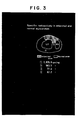
- Example 5 1 mg of the 111 In-labeled HMC-48Fab-DTPA (1.5 mCi/mg) obtained in Example 5 was administered intravenously into a dog with myocardial infarction artificially induced by ligating the coronary artery. 48 hours after administration, the heart was excised out of the dog. From the excised heart, specimens each of a size of about 3 x 3 mm were prepared, and the radioactivity at the infarction site and that at the non-infarction site were respectively measured.
- the radioactivity was 2879.8 cpm/mg at the infarction site,-563.5 cpm/mg on the periphery thereof, and 77.2 cpm/mg and 67.2 cpm/mg in the normal regions, about 40-fold radioactivity accumulating at the infarction site in comparison with the normal regions.
Applications Claiming Priority (2)
| Application Number | Priority Date | Filing Date | Title |
|---|---|---|---|
| JP59058767A JPS60201260A (ja) | 1984-03-27 | 1984-03-27 | 心疾患診断薬 |
| JP58767/84 | 1984-03-27 |
Publications (2)
| Publication Number | Publication Date |
|---|---|
| EP0163041A1 true EP0163041A1 (fr) | 1985-12-04 |
| EP0163041B1 EP0163041B1 (fr) | 1989-10-11 |
Family
ID=13093693
Family Applications (1)
| Application Number | Title | Priority Date | Filing Date |
|---|---|---|---|
| EP85103514A Expired EP0163041B1 (fr) | 1984-03-27 | 1985-03-25 | Réactif diagnostique pour les maladies de coeur et son utilisation |
Country Status (7)
| Country | Link |
|---|---|
| US (1) | US4943427A (fr) |
| EP (1) | EP0163041B1 (fr) |
| JP (1) | JPS60201260A (fr) |
| KR (1) | KR920002166B1 (fr) |
| AU (1) | AU566358B2 (fr) |
| CA (1) | CA1252714A (fr) |
| DE (1) | DE3573661D1 (fr) |
Cited By (8)
| Publication number | Priority date | Publication date | Assignee | Title |
|---|---|---|---|---|
| US4877599A (en) * | 1986-11-10 | 1989-10-31 | New England Deaconess Hospital Corporation | Detection of vascular disease with labelled antibodies |
| WO1989012467A1 (fr) * | 1988-06-13 | 1989-12-28 | Centocor, Inc. | Procede d'estimation de risque d'infarctus du myocarde |
| EP0389376A2 (fr) * | 1989-03-23 | 1990-09-26 | Biotech Cardio-Vision | Production et caractéristiques d'un anticorps monoclonal anti-myosinique de la souris |
| EP0583390A1 (fr) * | 1991-05-06 | 1994-02-23 | Immunomedics Inc | Depistage des lesions cardiovasculaires. |
| US5510466A (en) * | 1988-11-15 | 1996-04-23 | Massachusetts Institute Of Technology | Scavenger receptor protein and antibody thereto |
| US5726153A (en) * | 1988-05-02 | 1998-03-10 | New England Deaconess Hospital Corporation | Synthetic peptides for arterial imaging |
| US5908757A (en) * | 1994-10-25 | 1999-06-01 | Yamasa Corporation | Antibody reagent for detecting dissecting aortic aneurysm and uses thereof |
| US5955055A (en) * | 1988-05-02 | 1999-09-21 | New England Deaconess Hospital Corporation | Synthetic peptides for arterial imaging at vascular imaging sites |
Families Citing this family (7)
| Publication number | Priority date | Publication date | Assignee | Title |
|---|---|---|---|---|
| US5626830A (en) * | 1989-03-23 | 1997-05-06 | Biotech Cardio-Vision, Societe En Commandite Enregistree | Anti-myosin mouse monoclonal antibody and method of use for diagnosis of myocardial infarction |
| CA2050941C (fr) * | 1989-03-28 | 1999-02-23 | Ryozo Nagai | Anticorps contre les chaines lourdes de la myosine des muscles lisses |
| JP2829755B2 (ja) * | 1989-12-27 | 1998-12-02 | 株式会社第一ラジオアイソトープ研究所 | 抗ヒト心筋cプロテインモノクローナル抗体及び該抗体を産生するハイブリドーマ |
| US5326778A (en) * | 1992-03-03 | 1994-07-05 | Research Corporation Technologies, Inc. | Conjugates of biotin and deferoxamine for radioimmunoimaging and radioimmunotherapy |
| JP4035167B2 (ja) | 1996-02-19 | 2008-01-16 | ヤマサ醤油株式会社 | 血管障害性疾患用診断薬 |
| JP4054380B2 (ja) * | 1996-02-19 | 2008-02-27 | ヤマサ醤油株式会社 | 消化器疾患用診断薬 |
| US6248869B1 (en) | 1997-05-29 | 2001-06-19 | Medical Analysis Systems, Inc. | Troponin I forms and use of the same |
Citations (3)
| Publication number | Priority date | Publication date | Assignee | Title |
|---|---|---|---|---|
| US4036945A (en) * | 1976-05-03 | 1977-07-19 | The Massachusetts General Hospital | Composition and method for determining the size and location of myocardial infarcts |
| EP0035265A2 (fr) * | 1980-03-03 | 1981-09-09 | Milton David Goldenberg | Localisation de tumeurs et traitement à l'aide d'anticorps et fragments d'anticorps marqués |
| FR2515046A1 (fr) * | 1981-10-27 | 1983-04-29 | Hybritech Inc | Anticorps monoclonaux marques par un radionucleide et leur application a la visualisation d'une tumeur |
Family Cites Families (3)
| Publication number | Priority date | Publication date | Assignee | Title |
|---|---|---|---|---|
| US4331647A (en) * | 1980-03-03 | 1982-05-25 | Goldenberg Milton David | Tumor localization and therapy with labeled antibody fragments specific to tumor-associated markers |
| US4421735A (en) * | 1980-04-17 | 1983-12-20 | The Massachusetts General Hospital | Radiolabeled diagnostic compositions and method for making the same |
| JPS6028998A (ja) * | 1983-07-06 | 1985-02-14 | Yamasa Shoyu Co Ltd | 心筋ミオシン重鎖に対する単一クロ−ン抗体 |
-
1984
- 1984-03-27 JP JP59058767A patent/JPS60201260A/ja active Granted
-
1985
- 1985-03-25 DE DE8585103514T patent/DE3573661D1/de not_active Expired
- 1985-03-25 EP EP85103514A patent/EP0163041B1/fr not_active Expired
- 1985-03-25 US US06/715,815 patent/US4943427A/en not_active Expired - Fee Related
- 1985-03-25 KR KR1019850001946A patent/KR920002166B1/ko not_active IP Right Cessation
- 1985-03-26 AU AU40377/85A patent/AU566358B2/en not_active Ceased
- 1985-03-27 CA CA000477682A patent/CA1252714A/fr not_active Expired
Patent Citations (3)
| Publication number | Priority date | Publication date | Assignee | Title |
|---|---|---|---|---|
| US4036945A (en) * | 1976-05-03 | 1977-07-19 | The Massachusetts General Hospital | Composition and method for determining the size and location of myocardial infarcts |
| EP0035265A2 (fr) * | 1980-03-03 | 1981-09-09 | Milton David Goldenberg | Localisation de tumeurs et traitement à l'aide d'anticorps et fragments d'anticorps marqués |
| FR2515046A1 (fr) * | 1981-10-27 | 1983-04-29 | Hybritech Inc | Anticorps monoclonaux marques par un radionucleide et leur application a la visualisation d'une tumeur |
Cited By (11)
| Publication number | Priority date | Publication date | Assignee | Title |
|---|---|---|---|---|
| US4877599A (en) * | 1986-11-10 | 1989-10-31 | New England Deaconess Hospital Corporation | Detection of vascular disease with labelled antibodies |
| US5726153A (en) * | 1988-05-02 | 1998-03-10 | New England Deaconess Hospital Corporation | Synthetic peptides for arterial imaging |
| US5955055A (en) * | 1988-05-02 | 1999-09-21 | New England Deaconess Hospital Corporation | Synthetic peptides for arterial imaging at vascular imaging sites |
| WO1989012467A1 (fr) * | 1988-06-13 | 1989-12-28 | Centocor, Inc. | Procede d'estimation de risque d'infarctus du myocarde |
| US5046499A (en) * | 1988-06-13 | 1991-09-10 | Centocor, Inc. | Method for myocardial infarct risk assessment |
| US5510466A (en) * | 1988-11-15 | 1996-04-23 | Massachusetts Institute Of Technology | Scavenger receptor protein and antibody thereto |
| EP0389376A2 (fr) * | 1989-03-23 | 1990-09-26 | Biotech Cardio-Vision | Production et caractéristiques d'un anticorps monoclonal anti-myosinique de la souris |
| EP0389376A3 (en) * | 1989-03-23 | 1990-12-19 | Rougier Inc. | Production and characteristics of anti-myosin mouse monoclonal antibody |
| EP0583390A1 (fr) * | 1991-05-06 | 1994-02-23 | Immunomedics Inc | Depistage des lesions cardiovasculaires. |
| EP0583390A4 (fr) * | 1991-05-06 | 1994-04-27 | Immunomedics, Inc. | |
| US5908757A (en) * | 1994-10-25 | 1999-06-01 | Yamasa Corporation | Antibody reagent for detecting dissecting aortic aneurysm and uses thereof |
Also Published As
| Publication number | Publication date |
|---|---|
| US4943427A (en) | 1990-07-24 |
| EP0163041B1 (fr) | 1989-10-11 |
| KR850006139A (ko) | 1985-10-02 |
| CA1252714A (fr) | 1989-04-18 |
| AU566358B2 (en) | 1987-10-15 |
| DE3573661D1 (en) | 1989-11-16 |
| AU4037785A (en) | 1985-10-03 |
| JPS60201260A (ja) | 1985-10-11 |
| JPH0477733B2 (fr) | 1992-12-09 |
| KR920002166B1 (ko) | 1992-03-19 |
Similar Documents
| Publication | Publication Date | Title |
|---|---|---|
| US5648471A (en) | One vial method for labeling antibodies with Technetium-99m | |
| Khaw et al. | Monoclonal antibody to cardiac myosin: imaging of experimental myocardial infarction | |
| EP0163041B1 (fr) | Réactif diagnostique pour les maladies de coeur et son utilisation | |
| EP0354923B1 (fr) | Procede de marquage d'anticorps a l'aide d'un ion metallique | |
| EP0035265B1 (fr) | Localisation de tumeurs et traitement à l'aide d'anticorps et fragments d'anticorps marqués | |
| EP0210970A2 (fr) | Détection, visualisation et thérapie de carcinomes de cellules rénales utilisant des anticorps monoclonaux | |
| Milenic et al. | Comparison of methods for the generation of immunoreactive fragments of a monoclonal antibody (B72. 3) reactive with human carcinomas | |
| Pauwels et al. | The labeling of proteins and LDL with 99mTc: a new direct method employing KBH4 and stannous chloride | |
| US4917878A (en) | Novel use of a radiolabelled antibody against stage specific embryonic antigen for the detection of occult abscesses in mammals | |
| EP0131834B1 (fr) | Anticorps monoclonal ayant une spécificité pour un seul type d'isozyme de la chaîne lourde de la myosine cardiaque humaine | |
| JP2829755B2 (ja) | 抗ヒト心筋cプロテインモノクローナル抗体及び該抗体を産生するハイブリドーマ | |
| EP0569531B1 (fr) | Fragments d'anticorps stabilises | |
| JPH01197445A (ja) | 乳癌診断剤 | |
| Qiu et al. | 99mTc-labeled HAb18 McAb Fab fragment for radioimmunoimaging in nude mice bearing human hepatocellular carcinoma | |
| De Castiglia et al. | 99mTc direct labeling of anti-CEA monoclonal antibodies: Quality control and preclinical studies | |
| Haber | Antibodies in vivo | |
| JP2950928B2 (ja) | 標識化抗体 | |
| JP4035167B2 (ja) | 血管障害性疾患用診断薬 | |
| EP0592549A1 (fr) | Agents d'imagerie specifiques aux tissus comprenant des anticorps anti-idiotypiques permettant de produire des images internes | |
| JPS6296500A (ja) | 抗−ヒト乳癌モノクロ−ナル抗体 |
Legal Events
| Date | Code | Title | Description |
|---|---|---|---|
| PUAI | Public reference made under article 153(3) epc to a published international application that has entered the european phase |
Free format text: ORIGINAL CODE: 0009012 |
|
| AK | Designated contracting states |
Designated state(s): BE CH DE FR GB IT LI NL SE |
|
| 17P | Request for examination filed |
Effective date: 19860421 |
|
| 17Q | First examination report despatched |
Effective date: 19880324 |
|
| GRAA | (expected) grant |
Free format text: ORIGINAL CODE: 0009210 |
|
| AK | Designated contracting states |
Kind code of ref document: B1 Designated state(s): BE CH DE FR GB IT LI NL SE |
|
| REF | Corresponds to: |
Ref document number: 3573661 Country of ref document: DE Date of ref document: 19891116 |
|
| ITF | It: translation for a ep patent filed |
Owner name: SOCIETA' ITALIANA BREVETTI S.P.A. |
|
| ET | Fr: translation filed | ||
| PLBE | No opposition filed within time limit |
Free format text: ORIGINAL CODE: 0009261 |
|
| STAA | Information on the status of an ep patent application or granted ep patent |
Free format text: STATUS: NO OPPOSITION FILED WITHIN TIME LIMIT |
|
| 26N | No opposition filed | ||
| ITTA | It: last paid annual fee | ||
| EAL | Se: european patent in force in sweden |
Ref document number: 85103514.7 |
|
| PGFP | Annual fee paid to national office [announced via postgrant information from national office to epo] |
Ref country code: SE Payment date: 20000307 Year of fee payment: 16 |
|
| PGFP | Annual fee paid to national office [announced via postgrant information from national office to epo] |
Ref country code: FR Payment date: 20000310 Year of fee payment: 16 |
|
| PGFP | Annual fee paid to national office [announced via postgrant information from national office to epo] |
Ref country code: DE Payment date: 20000318 Year of fee payment: 16 |
|
| PGFP | Annual fee paid to national office [announced via postgrant information from national office to epo] |
Ref country code: GB Payment date: 20000322 Year of fee payment: 16 |
|
| PGFP | Annual fee paid to national office [announced via postgrant information from national office to epo] |
Ref country code: CH Payment date: 20000328 Year of fee payment: 16 |
|
| PGFP | Annual fee paid to national office [announced via postgrant information from national office to epo] |
Ref country code: NL Payment date: 20000330 Year of fee payment: 16 |
|
| PGFP | Annual fee paid to national office [announced via postgrant information from national office to epo] |
Ref country code: BE Payment date: 20000518 Year of fee payment: 16 |
|
| PG25 | Lapsed in a contracting state [announced via postgrant information from national office to epo] |
Ref country code: GB Free format text: LAPSE BECAUSE OF NON-PAYMENT OF DUE FEES Effective date: 20010325 |
|
| PG25 | Lapsed in a contracting state [announced via postgrant information from national office to epo] |
Ref country code: SE Free format text: LAPSE BECAUSE OF NON-PAYMENT OF DUE FEES Effective date: 20010326 |
|
| PG25 | Lapsed in a contracting state [announced via postgrant information from national office to epo] |
Ref country code: LI Free format text: LAPSE BECAUSE OF NON-PAYMENT OF DUE FEES Effective date: 20010331 Ref country code: CH Free format text: LAPSE BECAUSE OF NON-PAYMENT OF DUE FEES Effective date: 20010331 Ref country code: BE Free format text: LAPSE BECAUSE OF NON-PAYMENT OF DUE FEES Effective date: 20010331 |
|
| BERE | Be: lapsed |
Owner name: YAMASA SHOYU K.K. Effective date: 20010331 |
|
| PG25 | Lapsed in a contracting state [announced via postgrant information from national office to epo] |
Ref country code: NL Free format text: LAPSE BECAUSE OF NON-PAYMENT OF DUE FEES Effective date: 20011001 |
|
| EUG | Se: european patent has lapsed |
Ref document number: 85103514.7 |
|
| GBPC | Gb: european patent ceased through non-payment of renewal fee |
Effective date: 20010325 |
|
| REG | Reference to a national code |
Ref country code: CH Ref legal event code: PL |
|
| PG25 | Lapsed in a contracting state [announced via postgrant information from national office to epo] |
Ref country code: FR Free format text: LAPSE BECAUSE OF NON-PAYMENT OF DUE FEES Effective date: 20011130 |
|
| NLV4 | Nl: lapsed or anulled due to non-payment of the annual fee |
Effective date: 20011001 |
|
| REG | Reference to a national code |
Ref country code: FR Ref legal event code: ST |
|
| PG25 | Lapsed in a contracting state [announced via postgrant information from national office to epo] |
Ref country code: DE Free format text: LAPSE BECAUSE OF NON-PAYMENT OF DUE FEES Effective date: 20020101 |