EP0148155A2 - Knockenbildungsfaktoren - Google Patents
Knockenbildungsfaktoren Download PDFInfo
- Publication number
- EP0148155A2 EP0148155A2 EP85100047A EP85100047A EP0148155A2 EP 0148155 A2 EP0148155 A2 EP 0148155A2 EP 85100047 A EP85100047 A EP 85100047A EP 85100047 A EP85100047 A EP 85100047A EP 0148155 A2 EP0148155 A2 EP 0148155A2
- Authority
- EP
- European Patent Office
- Prior art keywords
- protein
- bone
- proteins
- osteogenic
- calf
- Prior art date
- Legal status (The legal status is an assumption and is not a legal conclusion. Google has not performed a legal analysis and makes no representation as to the accuracy of the status listed.)
- Granted
Links
Images


Classifications
-
- C—CHEMISTRY; METALLURGY
- C07—ORGANIC CHEMISTRY
- C07K—PEPTIDES
- C07K14/00—Peptides having more than 20 amino acids; Gastrins; Somatostatins; Melanotropins; Derivatives thereof
- C07K14/435—Peptides having more than 20 amino acids; Gastrins; Somatostatins; Melanotropins; Derivatives thereof from animals; from humans
- C07K14/475—Growth factors; Growth regulators
- C07K14/51—Bone morphogenetic factor; Osteogenins; Osteogenic factor; Bone-inducing factor
-
- A—HUMAN NECESSITIES
- A61—MEDICAL OR VETERINARY SCIENCE; HYGIENE
- A61K—PREPARATIONS FOR MEDICAL, DENTAL OR TOILETRY PURPOSES
- A61K38/00—Medicinal preparations containing peptides
Definitions
- Bone is a highly specialized connective tissue with unique mechanical properties derived from its extensive matrix structure. A network of fibrous bundles composed of the protein collagen is presumed to provide the tension-resistant behavior of bone. In addition other materials including proteoglycans, noncollagenous proteins,-lipids and acidic proteins associated with a mineral phase consisting primarily of poorly crystallized hydroxyapatite are deposited in the extensive matrix architecture of bone. Bone tissue is continuously renewed throughout the life of mammals. This physiologic process might serve to maintain the properties of a young tissue.
- the processes of bone formation and renewal are carried out by specialized cells. Osteogenesis vis-a-vis morphogenesis and growth of bone is presumably carried out by the "osteoblasts" (bone - forming cells). Remodeling of bone is apparently brought about by an interplay between the activities of the bone-resorbing cells called “osteoclasts" and the bone-forming osteoblasts.
- the bony skeleton is thus not only an architectural structure with a mechanical function but also is a living tissue capable of growth, modeling, remodeling and repair. Since these processes are carried out by specialized living cells, chemical (pharmaceutical- /hormonal), physical and physicochemical alterations can affect the quality, quantity and shaping of bone tissue.
- a variety of pathological disorders as well as physical stress necessitate active formation of bone tissue at rates that are significantly higher than that which can be supported by the normal milieu of the body. It is thus of value to identify physiologically acceptable chemical agents (hormones/pharmaceuticals/growth factors) that can induce the formation of bone at a predetermined site. Such agents could either provide a permissive matrix structure for the deposition of bone-forming cells or cause growth stimulation of bone-forming cells or induce the differentiation of appropriate progenitors of bone-forming cells.
- Urist et al. have been able to provide evidence that bone matrix-associated noncollagenous proteins can be isolated by dissociative treatment of demineralized bone powder and that this mixture of extracted materials as well as partially fractionated materials obtained therefrom contain bone morphogenetic activity, see Urist, M. R., et al., Proc. Natl. Acad. Sci. USA, Vol. 76, No. 4, pp. 1828-1832 (1979); Urist, M. R., et al., Proceedings of The Society of Experimental Biology and Medicine, Vol. 162, pp. 48-53 (1979); Hanamura, H., et al., Clinical Orthopaedics, Vol. 148, pp. 281-290 (1980); Urist, M.
- the 18,500 dalton protein induced bone formation when implanted alone or with various combinations of other bone derived proteins. Only samples containing the 18,500 dalton protein induced bone formation. It was further indicated that other bovine bone derived proteins having molecular weights of 14,000, 17,000, 17,500, 22,000 or 34,000 daltons, respectively, when implanted alone or in various combinations failed to induce bone formation. A bovine bone derived protein of 24,000 daltons was also mentioned and there was no indication that the 24,000 dalton protein induced bone formation.
- U. S. Patent Nos. 4,294,753, 4,455,256 and 4,434,094 all concern processes for fractionation of crude protein mixtures obtained by dissociative extractions of demineralized bone matrix.
- the teachings of U. S. Patent Nos. 4,294,753, 4,455,256 and 4,434,094 at best yield a mixture of different proteins and not an essentially homogeneous protein species identified as having osteogenic activity.
- the present invention concerns a family of immunologically related primary osteogenic factors (also referred to herein as "P3 proteins”), each of which in its substantially pure state induces bone formation at a predetermined site in a mammal when applied alone or in admixture with a suitable pharmaceutically acceptable carrier.
- P3 proteins immunologically related primary osteogenic factors
- proteins such proteins are further described herein and have been given identifying names such as Pl, P2, P3, P4 and the like based on their migration in polyacrylamide gels under dissociating, reducing conditions
- proteins which are so obtained, there is a protein referred to herein as the "primary osteogenic factor” (also referred to herein by its identifying name as the "P3 protein") which induces bone formation at a predetermined site in a mammal when administered alone or in admixture with a suitable pharmaceutically acceptable carrier.
- a particular primary osteogenic factor can be obtained from the bone of a particular mammalian species.
- the primary osteogenic factor obtained from bovine bone corresponds to and is immunologically related to the primary osteogenic factor obtained from human bone, and corresponds to and is immunologically related to the respective primary osteogenic factors which can be isolated from the bone of other mammalian species.
- the term "primary osteogenic factor” or "P3 protein” refers to a particular P3 protein endogenous to the bone of a particular mammalian species.
- this family of immunologically related proteins is comprised of the P3 proteins obtained from the bone of respective mammalian species, each P3 protein being a molecular species variant of the other members of the family.
- the osteogenic substances of the present invention include the osteogenic factors selected from the group consisting of: (a) the proteins of the P3 family of immunologically related proteins (that is, the P3 proteins); (b) the osteogenically active polypeptides derived from said P3 proteins; (c) polypeptides which are or can be converted to osteogenically active entities which are immunologically related to one or more of the P3 proteins; and (d) mixtures of osteogenic factors selected from (a), (b) and (c).
- the osteogenic factors selected from the group consisting of: (a) the proteins of the P3 family of immunologically related proteins (that is, the P3 proteins); (b) the osteogenically active polypeptides derived from said P3 proteins; (c) polypeptides which are or can be converted to osteogenically active entities which are immunologically related to one or more of the P3 proteins; and (d) mixtures of osteogenic factors selected from (a), (b) and (c).
- the preferred P3 proteins are: the P3 protein which can be obtained from bovine bone (such as calf bone), and referred to herein either as “bovine P3 protein” or "calf P3 protein”; the P3 protein which can be obtained from human bone, referred to herein as “human P3 protein”; and the P3 protein which can be obtained from porcine bone, referred to herein as "porcine P3 protein".
- the P3 proteins of the present invention exhibit the ability to promote or stimulate osteogenesis at desired locations in mammals.
- it is preferred to use an osteogenic factor obtained from a particular mammalian species for administration to that mammalian species for example, the P3 protein obtained from human bone is preferred for administration to humans; however, the use of P3 proteins obtained from the bone of mammalian species which are different from the mammalian species to be treated is within the scope of the present invention.
- Osteogenically active entities include any portion of the proteins or polypeptides which are the subject of the present invention having osteogenic activity and functional derivatives thereof having osteogenic activity and includes any osteogenically active entities that can be produced by conventional procedures such as chemical synthesis, enzymatic modification or recombinant DNA techniques.
- Derivatives of such active polypeptides can include, for example, chemically or enzymatically modified polypeptides; fusion proteins; or polypeptides bound to a suitable carrier substance such as a polymer, etc.
- the present invention also concerns a method for isolating, purifying and characterizing the P3 proteins and to a method of using one or more of the P3 proteins and/or osteogenically active polypeptides and/or immunologically related (that is, immunologically related to one or more of the P3 proteins) osteogenically active entities as pharmaceutical agents for the stimulation of bone growth in mammals.
- Pharmaceutically acceptable compositions comprised of one or more of the P3 proteins and/or osteogenically active polypeptides and/or immunologically related osteogenically active entities in combination with a pharmaceutically acceptable carrier are also disclosed herein. Such compositions can optionally contain other bioactive materials or other ingredients which aid in the administration of the composition or add to the effectiveness of the composition.
- osteogenesis means formation of new bone or induction of growth of pre-existing bones at specific sites in response to local administration (for example, implantation) of an osteogenically active preparation in a pharmaceutically acceptable manner.
- osteoogenic amount refers to an amount of the osteogenic P3 protein and/or osteogenically active polypeptide and/or immunologically related osteogenically active entity sufficient to provide the desired effect.
- osteoogenically active or “osteogenic” means that the preparation has the capability to promote or induce osteogenesis.
- the primary osteogenic factor and several other proteins can each be purified to an essentially homogeneous state starting from the crude extract of the demineralized bone powder of a particular mammalian species.
- a substantially pure preparation of the primary osteogenic factor can be obtained in addition to substantially pure preparations of several other proteins which do not promote bone formation in the absence of the primary osteogenic factor.
- proteins are representative members of the family of-immunologically related P3 proteins; the P3 protein purified to an essentially homogeneous state obtained from calf bone according to the procedures essentially as described herein, has an apparent molecular weight of 22,000 to 24,000 daltons, and has an amino terminal sequence and an amino acid composition as described later herein. Similarly, the P3 protein isolated from human bone and purified to an essentially homogeneous state according to the procedure essentially as described herein is immunologically related to the calf P3 protein and porcine P3 protein, has an apparent molecular weight of 22,000 to 24,000 daltons and an amino acid composition as described later herein.
- the P3 protein isolated from porcine bone and purified to an essentially homogeneous state according to the procedures essentially as described herein is immunologically related to the calf P3 protein and human P3 protein, has an apparent molecular weight of 22,000 to 24,000 daltons and an amino acid composition as described later herein.
- Each of the P3 proteins irrespective of bone tissue source, exhibits osteogenic activity.
- P2 and P4 protein preparations, designated herein as P2 and P4, and unrelated to the P3 protein, have also been isolated from bone of each of several different mammalian species.
- a typical P2 protein isolated from calf bone has an apparent molecular weight of 30,000 to 33,000 daltons, is incapable of inducing osteogenesis in the absence of a representative P3 protein, and has an amino terminal sequence as described later herein.
- Immunologically related members of this P2 protein family have been isolated according to the procedure essentially as described herein from human bone and porcine bone.
- P4 proteins have been isolated according to the procedures described herein.
- the P4 preparation consists of two major components which are incapable of inducing osteogenesis in the absence of P3 protein, both having an apparent molecular weight of about 16,000 to 18,000 daltons and are characterized by amino terminal amino acid sequences as described later herein.
- Immunologically related members of this P4 protein family also incapable of inducing osteogenesis in the absence of P3 protein, have been isolated from human bone and porcine bone according to the procedure essentially as described herein.
- the invention also concerns molecular entities detected using antibodies prepared against any of the P3 proteins described herein; other proteins with molecular weights which are substantially different than the molecular weights of the P3 proteins have been detected in other mammalian tissues (that is, in tissues other than bone) by the ability of these other proteins to be recognized by the antibodies to P3 proteins.
- proteins having an apparent molecular weight of about 40,000 to about 45,000 daltons have been identified by this technique in tissues such as rat, human or bovine brains; calf or rat dental pulp; bovine, human or porcine cartilage of the trachea or joints; and cultured cells derived from rat bone tumors.
- immunologically related proteins of apparent molecular weights substantially higher than a biologically active entity frequently represent biosynthetic precursors or covalently modified forms (such as glycosylated and/or acylated forms, etc.) of the active entity.
- Such precursors may or may not show biological activity but can often be processed by standard chemical or enzymatic means to the active entity.
- the 40,000 to 45,000 dalton proteins obtainable and identified by means described herein may comprise a distinct family of immunologically related proteins which may be osteogenetically active or activable.
- proteins having apparent molecular weights of about 14,000 to about 16,000 daltons have been detected in several mammalian tissues by their ability to bind to antibodies generated against a P3 protein. These proteins might be derived from the P3 proteins or their precursors by specific or nonspecific cleavage by proteases. Some of these proteins may have osteogenic activity.
- the specific antibodies generated against a P3 protein have also detected the presence of proteins having molecular weights of from about 22,000 to 26,000 daltons in mammalian tissues other than bone and in cultured cells derived from non-bone tissues; these proteins may be osteogenically active.
- essentially homogeneous is meant to describe a protein which is homogeneous by one or more purity or homogeneity characteristics normally used by those of skill in the art of protein chemistry.
- an essentially homogeneous protein will show constant and reproducible characteristics within standard experimental deviations for parameters such as the following: amino acid analysis, amino- or carboxyl-terminal sequence, band pattern on conventional polyacrylamide gel electrophoresis (PAGE) or other chromatographic techniques, molecular weight, isoelectric point, immunological properties and other such parameters.
- PAGE polyacrylamide gel electrophoresis
- the present invention includes mixtures of two or more essentially homogeneous proteins, for example, mixtures of P3 and P4; of P3 and P2; of P2, P3 and P4; of P3 with neutral matrix protein(s); of P3 with other yet to be discovered osteogenic proteins; of P3's from two or more mammalian sources, and the like.
- the terms are also not meant to exclude the presence of minor impurities which do not interfere with the biological activity of the protein, and which may be present, for example, due to incomplete purification.
- the application of the osteogenic substances of the present invention can be conveniently accomplished by administering, such as by implanting, a lyophilized preparation or suspension of one or more of the osteogenic P3 proteins and/or one or more osteogenically active polypeptides and/or one or more immunologically related osteogenically active entities in sufficient quantity to promote osteogenesis at the desired site.
- pharmaceutically acceptable compositions can be used which are comprised of one or more of the osteogenic P3 proteins and/or one or more of the osteogenically active polypeptides and/or one or more of the immunologically related osteogenically active entities described herein and a pharmaceutically acceptable matrix such as collagenous proteins or matrix material derived from powdered bone extracted with strong denaturing agents, or other pharmaceutically acceptable carriers.
- a convenient method of processing and demineralizing bone is as follows:
- the bone powder is sieved to obtain particles having a size range of about 75 to 500 p in diameter.
- Demineralization that is, the removal of calcium phosphate from the bone matrix
- HC1 solution for example, by stirring bone powder for one hour with about 10 to 15 milliliters (ml) of 0.5 normal (N) HC1 per gram (g) dry weight of bone powder, decanting the liquid and then repeating this process three or four times.
- the demineralized bone powder is then washed extensively with deionized distilled water until the pH approaches neutrality.
- the water is removed from the demineralized bone powder by washing with ethanol, then ether, and then drying.
- the demineralized bone powder can be stored at low temperatures (for example, -20° to -80°C). Demineralization of the bone powder can also be accomplished using other well known procedures, for example, using a chelator such as ethylenediaminetetraacetic acid.
- a chelator such as ethylenediaminetetraacetic acid.
- the water-rinsed powder is tested for mineral content [(that is, calcium content), for example, by the silver nitrate staining method of von Kossa, see J. von Kossa, Ziegler's Beitr. 29, 163 (1901)].
- the von Kossa stain is negative the treated bone powder is sufficiently demineralized to be ready for the extraction of proteins.
- Such demineralized bone powder when implanted in test animals induces the formation of new bone at the site of implantation, that is, contains putative, but unidentified, osteogenic factors.
- Demineralized bone powder prepared as described above, is extracted by constant stirring with an aqueous solution of about 2 to 8 molar (M) guanidinium-hydrochloride (GuHCl) in a buffer such as Trizma-hydrochloride (Tris ⁇ HCl) at or near pH 7.0 for a time sufficient to extract the desired proteins.
- M guanidinium-hydrochloride
- Tris ⁇ HCl Trizma-hydrochloride
- the extraction is performed by stirring the demineralized bone powder with 4M GuHCl-0.01M Tris ⁇ HCl buffer (pH 7.0) in the presence of a proteolytic enzyme inhibitor such as phenylmethylsulfonylfluoride for 8 to 12 hours (hrs) between about 4° to 20°C.
- the proteins from demineralized bone powder can be extracted by contacting the demineralized bone powder with an appropriate GuHCl-Tris ⁇ HCl buffer for a time sufficient to obtain substantial q uan- tities of the desired proteins.
- an appropriate GuHCl-Tris ⁇ HCl buffer for a time sufficient to obtain substantial q uan- tities of the desired proteins.
- approximately 1500 milligrams (mg) of total proteins are extracted in a three day extraction period with 4M GuHCl-0.01M Tris ⁇ HCl buffer (pH 7.0).
- the extract is filtered, for example, over Whatman paper, and the filtrate concentrated by conventional procedures; in typical experiments, an Amicon ultrafiltration apparatus (Amicon Corporation, Lexington, Massachusetts) with a membrane filter or a hollow fiber filtration cartridge with molecular cut-off size of approximately 5,000 daltons is used for the concentration step (that is, the membrane or hollow fiber cartridge retains molecules having a molecular weight greater than approximately 5,000 daltons, for example, an appropriate Diaflo @ ultrafiltration membrane such as YM-5 or a hollow fiber cartridge such as HlP5-20 from Amicon Corporation).
- an Amicon ultrafiltration apparatus Amicon Corporation, Lexington, Massachusetts
- a membrane filter or a hollow fiber filtration cartridge with molecular cut-off size of approximately 5,000 daltons is used for the concentration step (that is, the membrane or hollow fiber cartridge retains molecules having a molecular weight greater than approximately 5,000 daltons, for example, an appropriate Diaflo @ ultrafiltration membrane such as YM-5 or a hollow fiber cartridge such as H
- the various buffers for example, the 4M GuHCl-0.01M Tris ⁇ HCl buffer, and solutions, for example, the 0.5N HC1 solution, described herein are aqueous buffers or solutions in which the indicated materials are present in water at the indicated concentration.
- the protein components of the concentrated protein solution were fractionated using various conventional chromatographic techniques including high performance liquid chromatography (HPLC) as follows:
- pool C which consisted of pooled fractions V, VI and VII, was concentrated using conventional procedures.
- pool C obtained from the elution of the total proteins on the Sepharose CL-6B column represents about 40% of the total proteins obtained in an 8 to 12 hr extraction of demineralized calf bone powder with 4M GuHCl-0.01M Tris ⁇ HCl buffer (pH 7.0).
- the alpha pool contained minor protein components of molecular weight higher than 50,000 daltons; the beta pool contained a major species at 38,000 to 40,000 daltons, some minor higher molecular weight contaminants, and small quantities of lower molecular weight protein species migrating between 14,000 and 30,000 daltons; the gamma I and gamma II pools contained four major size class species migrating at 31,000 to 35,000 daltons, at 22,000 to 25,000 daltons, at 16,000 to 18,000 daltons, and at 12,000 to 14,000 daltons; the delta pool contained mostly proteins in the 12,000 to 14,000 dalton range.
- the final purification was accomplished by reverse phase HPLC of the partially purified protein preparations, obtained from Sephacryl S-200 column chromatography, using a Beckman Altex HPLC controlled by a Model 421 microprocessor unit. Two approaches have been used.
- a characteristic feature of some of the proteins, especially the P3 protein family described herein is the lack of solubility in the absence of a strong dissociating agent such as GuHCl.
- a strong dissociating agent such as GuHCl.
- the removal of GuHCl resulted in a coprecipitation of other proteins along with P3.
- a method was, therefore, developed where narrow pools consisting of only one or two major proteins were obtained from the Sephacryl S-200 column and used as the starting material for further purification by HPLC.
- pools such as the ones described above were dialyzed directly against an aqueous solvent containing 0.1% trifluoroacetic acid (TFA) supplemented with acetonitrile (ACN) at concentrations of between 10% to 15% by volume.
- TFA trifluoroacetic acid
- ACN acetonitrile
- a conventional dialysis membrane tubing with molecular weight cut-off size of 3,500 daltons or lower is conveniently used in this procedure.
- Proteins soluble in the TFA:ACN solvent could then be conveniently obtained by removal of the insoluble material from each dialyzed pool by centrifugation.
- the soluble proteins at this point could be chromatographed on a reverse phase HPLC column such as the Protesil 300 octyl column described herein.
- TFA:ACN soluble proteins obtained from the peak fractions in this manner were applied to a 0.46 cm x 25.0 cm Protesil 300 octyl column (Whatman) of 10 micron particle size equilibrated with 0.1% TFA:10% ACN. Proteins bound to the column under these conditions were eluted at a flow rate of 60 ml/hr using a linear 10% to 80% ACN gradient developed over 45 minutes.
- P2 and Pl proteins were sequentially recovered with increasing ACN concentrations (depicted by the dashed line) from the gamma I peak.
- P1 protein can be obtained from the beta peak while P5a and P5b are obtained from the delta peak.
- the P3 protein elutes between the gamma I and gamma II regions of the Sephacryl S-200 column.
- the P3 protein is found in both the soluble and the insoluble materials obtained by dialysis of appropriate fractions against TFA:ACN. The lack of solubility of the P3 protein thus yields essentially homogeneous P3 protein in the insoluble material.
- the P3 protein retained in solution in the TFA:ACN solvent can be further purified by reverse phase HPLC essentially as described above.
- the second procedure to purify proteins to an essentially homogeneous state was designed to take advantage of the high degree of insolubility of certain proteins in the 35,000 to 14,000 dalton molecular weight range, especially when they are present together at high concentrations (for example, approximately 10 mg/ml).
- proteins eluting in the gamma I and gamma II pools from the Sephacryl S-200 column chromatography that is, the pools where the bone inducing activity is found) were concentrated to approximately 10 mg/ml.
- the material was rapidly dialyzed [for example, six changes each of 4 liters every 2 to 3 hrs, (using dialysis tubing with a molecular cut-off size of 2,000 daltons)] against deionized distilled water at 15° to 23°C. Precipitated proteins were collected by centrifugation and washed several times with deionized distilled water keeping the concentration of protein at higher than 10 mg/ml of washing water. The principal constituents of this precipitated material were found to be P2, P3, P4 and P5a; small amounts of Pl protein was found in variable quantities in some cases.
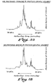
- the final pellet was dissolved in 0.1% TFA with 15% ACN and the solubilized material was applied to a Protesil 300 octyl column. Increasing ACN concentration eluted the P2, P3, P4 and P5a proteins as shown in Figure 3B, a typical elution profile.
- each of the major protein species described in Table 1 was further purifed by rechromatographing on the Protesil 300 octyl column. Pools of fractions obtained as indicated in Figure 3 were concentrated by lyophilization and redissolved in 0.1% TFA and about 10 to 20% ACN depending upon the particular lyophilized material and reapplied to the Protesil 300 octyl column. The proteins were eluted from the column using a linear 10% to 80% ACN gradient at a flow rate of 60 ml/hr under conditions as previously described herein except that the proteins were eluted over a longer period thus resulting in numerous individual fractions. The purity of each of the protein fractions was determined using conventional discontinous PAGE. Those fractions which showed only one major species were used for further chemical and biological characterizations. Typically these fractions were lyophilized and stored as lyophilized powders.
- Figure 4 depicts the results of a typical discontinuous gel electrophoretic analysis on sodium dodecyl sulfate-polyacrylamide gels.
- the analysis was performed on a discontinuous polyacrylamide gel system in the presence of sodium dodecyl sulfate and a reducing agent where the resolving gel was 13% in acrylamide and 0.35% in bis-acrylamide crosslinker at a pH of 8.8.
- the gel was run at 50 volts for 30 minutes followed by 7 hrs at 100 volts. Protein bands were visualized by staining with coomassie brilliant blue R.
- Columns 1 and 8 depict gels with the following standard molecular weight markers: 95,000 (phosphorylase A), 68,000 (bovine serum albumin), 43,000 (ovalbumin), 31,000 (carbonic anhydrase), 21,000 (soybean trypsin inhibitor), and 14,000 (ribonuclease); Columns 2, 3, 4, 5 and 6 show, respectively, the Pl, P2, P3, P4 and P5 proteins (CP1 thru CP5) from demineralized calf bone powder; and Column 7 the P3 protein (HP3) from demineralized human bone powder. Portions shaded with oblique lines are bands of low concentration.
- calf P3 protein is also referred to herein as "bovine P3 protein"
- the partial amino terminal sequence of the calf P3 protein was determined to be H 2 N-Phe-Pro-Val--Tyr-Asp-Tyr-Ser-Pro-Ala-Arg-Leu-Lys-Glu-Ala.
- the partial amino terminal sequence of the calf P2 protein was determined to be H 2 N-Trp-?- p ro-Tyr-?-Trp.
- a protein was obtained from demineralized human bone powder having a molecular weight of approximately 23,000 daltons and designated as human P3 protein.
- Human P3 protein purified to an essentially homogeneous state induced the formation of bone when implanted.
- the human P3 protein obtained from human bone is related to the calf P3 protein.
- a protein was obtained from demineralized porcine bone powder having a molecular weight of approximately 23,000 daltons and designated as porcine P3 protein.
- Porcine P3 protein obtained from porcine bone is related to the calf P3 and human P3 proteins.
- the respective calf, human and porcine P3 proteins show the following similarities:
- Amino acid compositions for the calf, the porcine and the human P3 proteins were determined from acid hydrolysates prepared with redistilled 6N hydrochloric acid (110°C, 24 hrs). The tubes were evacuated prior to sealing to eliminate oxygen. Following removal of the 6N hydrochloric acid by evaporation and reconstitution in citrate buffer, the hydrolysates were analyzed, on a Beckman Model 6300 automatic amino acid analyzer. Standard operating procedures were used as described by Benson, J. R., et al., "Amino Acid Analysis of Peptides” in Peptides: Analysis, Synthesis, Biology (E. Gross and J. Meienhofer, eds.) Academic Press, New York, Vol. 4, pp. 217-260 (1981). The quantitative data produced was converted to residues/mole of protein based upon the molecular weights estimated by conventional PAGE.
- Table 2 presents amino acid composition data obtained for the human P3, the porcine P3 and the calf P3 proteins.
- the data represents the average values obtained from six runs per protein (triplicate analyses on each of two independently purified preparations) on a Beckman Model 6300 amino acid analyzer.
- determinations of amino acid compositions are subject to errors introduced by (i) the extent of protein hydrolysis where the yields of certain amino acids vary because of variations in stability and/or hydrolyzability of different amino acid residues, (ii) minor impurities whose exact amounts vary from one preparation to another, (iii) computerized data analysis derived from the peak-areas corresponding to the positions of different amino acids, and (iv) errors inherent in the estimation of the exact molecular weights of proteins by conventional techniques such as SDS-PAGE or column chromatography.
- the numbers represent approximate number of residues (to the nearest whole number) of the indicated amino acids per mole of the respective proteins whose molecular weights were estimated by independent methods conventionally used, such as polyacrylamide gel electrophoresis.
- the numbers for the P3 proteins of this invention are within experimental errors inherent in the art of amino acid composition analysis. Values in parentheses were determined based upon absorbance at specific wavelengths.
- the amino acid composition data indicates the estimated number of residues of each amino acid per molecule of the respective P3 proteins. As appreciated by one skilled in the art, the accuracy of amino acid composition data is dependent upon the number of test runs and the extent of protein purification.
- the amino acid composition data indicates that the compositions of human, porcine and calf P3 proteins are similar to each other, for example, each has high contents of aspartic acid (+ asparagine) and glutamic acid (+ glutamine) residues, as well as substantial numbers of tyrosine residues.
- Table 3 contains the amino acid compositions of several proteins previously described in the art, namely, a 17,000 to 18,000 dalton bone morphogenetic protein from calf bone described by Urist, M. R., et al., Science, Vol. 220, pp. 680-686 (1983), see “Calf lU in Table 3; a 17,000 to 18,000 dalton bone morphogenetic protein from human bone, Urist, M. R., et al., Proceedings of The Society of Experimental Biology and Medicine, Vol. 173, pp. 194-199 (1983), see “Human 2 " in Table 3; a 23,000 dalton bone morphogenetic protein from rabbit dentin, Conover, M. A., and Urist, M.
- the amino acid composition data presented herein demonstrates that the P3 family of osteogenic factors are distinct from all other biologically active proteins that are claimed to be involved in osteogenesis and disclosed in the prior art.
- Proteases enzymes that digest protein molecules into smaller polypeptide fragments, cleave peptide bonds with a certain degree of specificity.
- the enzyme trypsin cleaves peptide bonds after arginine and lysine residues while the V8 protease from Staphylococcus aureus cleaves peptide bonds after aspartic acid and glutamic acid residues.
- the fragments generated from one protein will have homologous counterparts in the other protein when each protein is digested with the same protease.
- the proteolytic cleavage patterns of the P3 proteins were studied using V8 protease from Staphylococcus aureus.
- porcine P3 and the calf P3 proteins were treated in parallel with Staphylococcus aureus V8 protease, and the digestion product subjected to a strong reducing treatment in order to release fragments that might be held together by disulfide bonds.
- the peptide fragments thus generated were subjected to reverse phase HPLC on a Synchropak C8 column.
- the peptide fragments were eluted using a linear gradient, from 20% to 70% by volume of acetonitrile in 0.1% trifluoroacetic acid in water. The elution of the peptides was monitored by measuring the absorbance of the effluent at 229 nm.
- porcine P3 and human P3 proteins were each first treated with a reducing agent, and the free sulfhydryl groups were chemically derivatized by S-carboxymethylation using techniques well known in the art.
- the S-carboxymethylated protein preparations were each digested with Staphylococcus aureus V8 protease.
- the peptide fragments which resulted from the V8 protease digestion were subjected to reverse phase HPLC on a Whatman C18 column.
- the peptides were eluted using a linear acetonitrile gradient, from 20% to 70% by volume in 0.1% TFA in water.
- the intact P3 proteins from human and porcine bone did not yield any amino acid sequence information, most likely indicating that the amino terminal amino acid residue in each of these protein preparations was blocked; however, it was possible to obtain amino acid sequence information when the calf P3 protein was analyzed to determine its amino terminal amino acid sequence (see the aminc terminal amino acid sequence for calf P3 protein previously provided herein).
- modifications which block the amino terminal amino acid residue may be physiological, i.e., take place during the biosynthesis of the proteins in the tissues or may sometimes occur during the process of purification.
- P3 proteins obtained from bone of different mammalian species have been used to immunize laboratory animals, such as rabbits, in order to generate monospecific antibodies capable of binding to the respective P3 proteins.
- approximately 100 micrograms of the particular P3 protein to which antibodies are to be raised is admixed with Freund's complete adjuvant and inoculated into the footpads and at subcutaneous sites in rabbits; ten to fourteen days later a comparable amount of protein admixed with incomplete Freund's adjuvant is inoculated subcutaneously; after an additional ten to fourteen days the animal is inoculated with an additional 50 to 100 ⁇ g of protein mixed in a ten percent solution of aluminum hydroxide.-
- the animal is subsequently immunized at four week intervals with 50 to 100 ⁇ g of protein.
- the specific antibody titers developed against the respective P3 proteins have been measured by (a) an enzyme linked immunosorbent assay (ELISA) and by (b) the ability of the antibody to immunoprecipitate radiolabelled P3 protein molecules.
- ELISA enzyme linked immunosorbent assay
- 10 to 20 nanograms of the test antigen that is, the particular P3 protein being tested
- a plastic surface by air-drying of a protein solution within the wells of microtiter dishes (Falcon Products or Bellco Products).
- Serial dilutions of test antisera are incubated within the wells, unbound antibodies are removed by washing and the bound antibodies are then incubated with an enzyme conjugated second antibody preparation directed against the immunoglobulins of the species in which the test antiserum was generated.
- the amount of enzyme bound in each well is then quantitated by an appropriate color assay. In such testing, sera with high titers of antibody against the test antigen can be diluted several thousand fold and will still show significant color development.
- the test antigen is labelled with a radioisotope such as 125 1.
- a radioisotope such as 125 1.
- a fixed quantity of the radiolabelled antigen is then incubated with serial dilutions of the test antisera.
- the immunecomplexed antigen is precipitated either using a second antibody directed against the immunoglobulins of the species in which the test serum was generated, or using a fixed Staphylococcus aureus bacterial suspension, or using Staphylococcus aureus protein A immobilized onto beads.
- serum with high titers of specific antibody can be diluted several thousand fold and still precipitate significant amounts of the radiolabelled antigen.
- the specific titers of antibodies in test sera samples is determined by subtracting the values (color or precipitated radioactivity) obtained with serum from nonimmunized animals or from animals immunized with a protein which is unrelated to the test protein.
- the specificity of the antisera as well as the immunological relatedness of different proteins can be estimated in either assay by examining the relative effects of serum dilutions on the extent of binding of the antigen.
- the extent of immunological relatedness between a protein and the test antigen is determined by the slope of displacement of radiolabelled test antigen bound to the antibody when increasing amounts of the unlabelled competing protein are added to the incubation mixture.
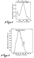
- the human P3 protein does not compete as effectively as the calf P3 protein in displacing radiolabelled calf P3 protein from binding to antibodies raised against the calf P3 protein ( Figure 7A).
- both the human P3 and the porcine P3 proteins efficiently competed with the binding of radiolabelled porcine P3 protein to antibodies raised against porcine P3 protein; the calf P3 protein competed with a much lower efficiency ( Figure 7B).
- porcine P3 protein was more effective than calf P3 protein in displacing radiolabelled human P3 protein from binding to antibodies raised against human P3 protein (Figure 7C). Since antibodies to porcine P3 protein immunoprecipitated the iodinated human P3 protein, the P3 proteins were compared for their ability to compete in a broad cross-species assay ( Figure 7D). Calf P3 protein again showed the least efficient competition in this assay. The fact that the respective P3 proteins compete with each other in the binding of specific antibodies raised against a particular P3 protein demonstrates that the respective P3 proteins are immunologically related.
- the immunological relatedness between the P3 proteins was also tested by the ability of antibodies directed against a particular P3 protein to bind to "denatured" or "denatured and reduced” forms of P3 proteins of other species using the technique of immunoblotting.
- proteins are denatured by heating in sodium dodecyl sulfate in the presence or absence of a reducing agent.
- the denatured or denatured and reduced proteins are then subjected to denaturing polyacrylamide gel electrophoresis in the presence of sodium dodecyl sulfate (SDS-PAGE).
- SDS-PAGE sodium dodecyl sulfate
- the protein molecules are then transferred to a nitrocellulose membrane filter. This replica of the electrophoretically resolved protein bands is then incubated with antibodies directed against the respective proteins.
- the binding of antibody to a protein band is monitored by the ability of radiolabelled or enzyme conjugated protein A from Staphylococcus aureus to bind to the antibodies attached to the proteins on the nitrocellulose membrane.
- the location of the appropriate protein bands recognized by the antibody is then visualized by autoradiography. This technique allows one to identify the molecular species which is recognized by a specific antibody preparation.
- the immunological relatedness between calf, porcine and human P3 proteins was also examined by the immunoblotting technique.
- Antibodies raised against either the calf P3 protein or the porcine P3 protein bound to P3 proteins obtained from calf, porcine and human bone.
- many fragments (generated by proteolytic digestions) of one P3 protein were recognized by antibodies raised against the P3 protein of another species.
- Protein factors that induce growth/regeneration of mammalian tissues are frequently biosynthesized at sites other than the target sites.
- biosynthesis of these factors may proceed through a larger or modified form that can be converted to a biologically active species in vivo or in vitro.
- the production of biologically active peptides often proceeds through a number of intermediates, for example, prepro-proteins or pro-proteins, having varying degrees of biological activity or no biological activity.
- the identification of the P3 protein family of mammalian osteogenic factors permits one skilled in the art to detect, typically by immunological means, other related molecular entities in cultured cells and other tissues.
- a convenient method for identifying other proteinaceous materials which may possess osteogenic activity employs the immunoblotting technology previously described herein; however, other technologies such as the use of radioimmunoassays may also be used.
- a candidate tissue or cultured cell mass is extracted with strong dissociative agents, such as guanidine hydrochloride or guanidine isothiocyanate or sodium dodecyl sulfate, in the presence or absence of a reducing agent.
- the total extract or partially fractionated extracts are then subjected to SDS-PAGE and the electrophoretically separated protein species transferred onto nitrocellulose membrane filters.
- the presence of molecular species related to the P3 protein osteogenic factors are detected by their ability to bind antibodies directed against the P3 proteins.
- an immunologically related entity with an apparent molecular weight of about 40,000 to 45,000 daltons, was detected using antibodies raised against calf P3 protein in certain fractions containing higher molecular weight entities; in fractions that contained the lower molecular weight proteins extracted from bovine brain a mixture of lower molecular weight entities immunologically related to calf P3 protein, having molecular weights in the range of about 14,000 to 25,000 daltons, were found.
- osteogenically active or activable proteins or polypeptides detected by immunological techniques described herein and other osteogenically active or activable proteins or polypeptides present in other mammalian tissues detected by antibodies raised against a P3 protein osteogenic factor from a mammalian bone tissue source are considered to be within the scope of this invention.
- Bone matrix powder (75 to 500 micron size) is demineralized as described herein and then extracted sequentially three times, each with 15 to 20 ml of 4M GuHC1 per gram of demineralized bone powder. The extracted matrix is extensively washed with water, followed by ethanol and ether and then the powder is dried. This powder, when implanted in a test animal, such as a rat, does not induce osteogenesis and is called inactive bone matrix (IBM).
- IBM inactive bone matrix
- the IBM powder is mixed with an aqueous solution or suspension of the protein and the water removed by lyophilization.
- the reconstituted matrix is then packed in gelatin capsules and implanted subcutaneously near the thigh muscles of young (one to two months old) rats. Varying amounts of protein preparations are used together with a constant amount of IBM in each capsule to determine the efficacy of the different protein preparations.
- osteogenic activity induced by an implant is estimated by the examination of the excised implant (also referred to herein as an explant) by two approaches, (a) measuring the level of the enzyme alkaline phosphatase in the tissues developed at the implantation site, such tissue being excised at 17 to 20 days following implantation of a preparation for which osteogenic activity is to be determined and (b) performing a histologic examination of a 5 to 7 micron thick section of the tissue developed at the implant site following staining of paraffin-fixed sections of this tissue with toluidine blue (stains cartilage matrix), hematoxylin-eosin (resolves fibrous, cartilaginous and bone tissues) and von Kossa silver stain (for calcified matrix of bone tissue).
- toluidine blue stains cartilage matrix
- hematoxylin-eosin resolves fibrous, cartilaginous and bone tissues
- von Kossa silver stain for calcified matrix of bone tissue.
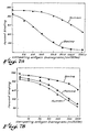
- the level of alkaline phosphatase is measured since active bone formation is characteristically preceded by a significant surge of this enzyme and continued formation of bone is accompanied by a stable elevated level of alkaline phosphatase activity compared to that found in non-bone fibrous tissue surrounding the implants.
- An approximate quantitation of the levels of bone inducing activity in a protein preparation has been obtained by quantitating the level of alkaline phosphatase per unit weight of explant tissue.
- the explant tissue is homogenized in an appropriate buffer such as Tris-saline, dissociated with a nonionic detergent and the solubilized enzymes that are released from the tissue are obtained by removing the debris using centrifugation.
- the levels of alkaline phosphatase are quantitated by measuring the conversion of paranitrophenylphosphate to paranitrophenol catalyzed by dilutions of the test extract and calculating from a standard curve of known enzyme activity.
- partially purified protein pools i.e., alpha, beta, gamma I, gamma II, and delta as previously described herein
- partially purified protein pools i.e., alpha, beta, gamma I, gamma II, and delta as previously described herein
- These respective protein pools were reconstituted with IBM and implanted subcutaneously in rat thighs.
- the bioassay studies indicated the presence of maximum osteogenic activity in proteins in pools gamma I and gamma II.
- the three major components of the gamma fractions that is, the P2, the P3 and the P4 proteins were purified to an essentially homogeneous state using reverse phase HPLC as described herein.
- the purified proteins either singly or in a complete mixture, were reconstituted with inactive bone matrix and a bone induction assay performed. The results are shown in Table 6.
- the data in Table 6 indicates that the calf P3 protein induced the formation of bone.
- Implants containing the calf P3 preparation developed into tissues that contained high levels of alkaline phosphatase enzyme activity.
- implants prepared by reconstituting with either the P2 or the P4 preparation failed to produce detectable bone.
- all three proteins were used in combination, significant bone formation was observed and high levels of alkaline phosphatase enzyme were obtained with one-third the amount of P3 protein (as compared to the P3 protein implant alone). It thus appears that at low concentrations of P3 protein, the presence of the P2 and/or the P4 protein provides enhancement of osteogenesis induced by the P3 protein.
- P3 proteins obtained from human bone or porcine bone were reconstituted with IBM and implanted. Approximately three weeks later explants of tissue surrounding the implantation site were examined for alkaline phosphatase activity and histological characteristics. In each case, 750 ⁇ g of either P3 protein induced the formation of bone.
- an osteogenic amount of one or more of the P3 proteins and/or osteogenically active polypeptides and/or immunologically related osteogenically active entities is administered at or in the proximity of the site in the mammal at which bone induction is desired.
- Administration will depend on the age, condition, sex and other characteristics of the subject to be treated.
- Preferred administration is by implantation, local injection or time controlled delivery using microcapsules, or other devices. Dosages will depend on the site and configuration of the area to be healed, such as, for example, a fracture zone.
- a 5 cubic millimeter bone chip can be obtained with about 100 to 200 micrograms (pg) of P3 protein administered or implanted locally in the form of an implant in about 100 mg of IBM.
- Active preparations can include other suitable bioactive materials such as growth factors, cell attachment factors, chemotactic agents, steroids, antibiotics, antiinflammatory agents and the like.
Landscapes
- Health & Medical Sciences (AREA)
- Chemical & Material Sciences (AREA)
- Organic Chemistry (AREA)
- Life Sciences & Earth Sciences (AREA)
- Genetics & Genomics (AREA)
- Proteomics, Peptides & Aminoacids (AREA)
- Zoology (AREA)
- Biochemistry (AREA)
- Biophysics (AREA)
- General Health & Medical Sciences (AREA)
- Toxicology (AREA)
- Medicinal Chemistry (AREA)
- Molecular Biology (AREA)
- Gastroenterology & Hepatology (AREA)
- Orthopedic Medicine & Surgery (AREA)
- Medicines That Contain Protein Lipid Enzymes And Other Medicines (AREA)
- Peptides Or Proteins (AREA)
- Materials For Medical Uses (AREA)
- Saccharide Compounds (AREA)
- Magnetic Heads (AREA)
- Medicines Containing Material From Animals Or Micro-Organisms (AREA)
Priority Applications (1)
| Application Number | Priority Date | Filing Date | Title |
|---|---|---|---|
| AT85100047T ATE42309T1 (de) | 1984-01-04 | 1985-01-02 | Knockenbildungsfaktoren. |
Applications Claiming Priority (2)
| Application Number | Priority Date | Filing Date | Title |
|---|---|---|---|
| US56816784A | 1984-01-04 | 1984-01-04 | |
| US568167 | 1984-01-04 |
Publications (3)
| Publication Number | Publication Date |
|---|---|
| EP0148155A2 true EP0148155A2 (de) | 1985-07-10 |
| EP0148155A3 EP0148155A3 (en) | 1986-08-13 |
| EP0148155B1 EP0148155B1 (de) | 1989-04-19 |
Family
ID=24270176
Family Applications (1)
| Application Number | Title | Priority Date | Filing Date |
|---|---|---|---|
| EP85100047A Expired EP0148155B1 (de) | 1984-01-04 | 1985-01-02 | Knockenbildungsfaktoren |
Country Status (11)
| Country | Link |
|---|---|
| EP (1) | EP0148155B1 (de) |
| JP (1) | JPS60226814A (de) |
| AT (1) | ATE42309T1 (de) |
| AU (1) | AU580975B2 (de) |
| CA (1) | CA1241641A (de) |
| DE (1) | DE3569542D1 (de) |
| DK (1) | DK161524C (de) |
| GR (1) | GR850006B (de) |
| IE (1) | IE57966B1 (de) |
| NZ (1) | NZ210699A (de) |
| ZA (1) | ZA8547B (de) |
Cited By (52)
| Publication number | Priority date | Publication date | Assignee | Title |
|---|---|---|---|---|
| EP0242466A1 (de) * | 1984-07-16 | 1987-10-28 | Celtrix Laboratories, Inc. | Teilweise gereinigter knocheninduzierender Faktor zur Anwendung in der Orteogenese |
| EP0271668A1 (de) * | 1986-10-22 | 1988-06-22 | Gesellschaft für Biotechnologische Forschung mbH (GBF) | Wachstumstimulierendes Material, Herstellungsverfahren und therapeutische Zusammensetzung |
| EP0212474A3 (de) * | 1985-08-07 | 1988-11-17 | The Regents Of The University Of California | Morphogenetische Knochenpeptide |
| US4877864A (en) * | 1987-03-26 | 1989-10-31 | Genetics Institute, Inc. | Osteoinductive factors |
| EP0255565A3 (en) * | 1986-08-05 | 1990-01-31 | Robapharm Ag | Composition for stimulating chondrocytes and osteoblasts )ossein hydroxyapatite compound), method for its production and pharmaceutical products containing said composition) |
| WO1989009787A3 (en) * | 1988-04-08 | 1990-02-08 | Creative Biomolecules Inc | Osteogenic devices |
| WO1990010018A1 (en) * | 1989-02-23 | 1990-09-07 | Creative Biomolecules, Inc. | Bone collagen matrix for implants |
| EP0313578A4 (en) * | 1986-07-01 | 1990-10-03 | Genetics Institute, Inc. | Novel osteoinductive compositions |
| US4968590A (en) * | 1988-04-08 | 1990-11-06 | Stryker Corporation | Osteogenic proteins and polypeptides |
| EP0409472A1 (de) * | 1989-07-19 | 1991-01-23 | Chiron Corporation | Morphogenetisches Knochenprotein |
| EP0336760A3 (de) * | 1988-04-06 | 1991-03-13 | Celtrix Pharmaceuticals, Inc. | Knochenwachstum induzierendes Protein |
| WO1991005802A1 (en) * | 1989-10-17 | 1991-05-02 | Creative Biomolecules, Inc. | Osteogenic devices |
| EP0401055A3 (de) * | 1989-06-02 | 1991-08-07 | Chiron Corporation | Knochenkalzifizierungsfaktor |
| EP0394418A4 (en) * | 1988-10-11 | 1992-01-22 | International Genetic Engineering, Inc. (Ingene) | Osteogenic factors |
| US5108753A (en) * | 1988-04-08 | 1992-04-28 | Creative Biomolecules | Osteogenic devices |
| US5162114A (en) * | 1989-02-23 | 1992-11-10 | Stryker Corporation | Bone collagen matrix for xenogenic implants |
| US5250302A (en) * | 1988-04-08 | 1993-10-05 | Stryker Corporation | Osteogenic devices |
| US5258494A (en) * | 1988-04-08 | 1993-11-02 | Stryker Corporation | Osteogenic proteins |
| US5266683A (en) * | 1988-04-08 | 1993-11-30 | Stryker Corporation | Osteogenic proteins |
| US5324819A (en) * | 1988-04-08 | 1994-06-28 | Stryker Corporation | Osteogenic proteins |
| US5354557A (en) * | 1988-04-08 | 1994-10-11 | Stryker Corporation | Osteogenic devices |
| US5645591A (en) * | 1990-05-29 | 1997-07-08 | Stryker Corporation | Synthetic bone matrix |
| US5652118A (en) * | 1991-03-11 | 1997-07-29 | Creative Biomolecules, Inc. | Nucleic acid encoding a novel morphogenic protein, OP-3 |
| US5656593A (en) * | 1991-03-11 | 1997-08-12 | Creative Biomolecules, Inc. | Morphogen induced periodontal tissue regeneration |
| US5670336A (en) * | 1988-04-08 | 1997-09-23 | Stryker Corporation | Method for recombinant production of osteogenic protein |
| US5674844A (en) * | 1991-03-11 | 1997-10-07 | Creative Biomolecules, Inc. | Treatment to prevent loss of and/or increase bone mass in metabolic bone diseases |
| US5693615A (en) * | 1991-06-05 | 1997-12-02 | The Procter & Gamble Company | Therapeutic compositions for osteoinduction |
| US5739107A (en) * | 1991-03-11 | 1998-04-14 | Creative Biomolecules, Inc. | Morphogen treatment of gastrointestinal ulcers |
| US5849686A (en) * | 1991-03-11 | 1998-12-15 | Creative Biomolecules, Inc. | Morphogen-induced liver regeneration |
| US5854071A (en) * | 1991-03-11 | 1998-12-29 | Creative Biomolecules, Inc. | OP-3- induced morphongenesis |
| US5928940A (en) * | 1996-09-24 | 1999-07-27 | Creative Biomolecules, Inc. | Morphogen-responsive signal transducer and methods of use thereof |
| US5972884A (en) * | 1991-03-11 | 1999-10-26 | Creative Biomolecules, Inc. | Morphogen treatment of gastrointestinal ulcers |
| US6022853A (en) * | 1991-08-30 | 2000-02-08 | Creative Biomolecules, Inc. | Morphogen-enriched dietary composition |
| US6077823A (en) * | 1991-03-11 | 2000-06-20 | Creative Biomolecules, Inc. | Method for reducing tissue damage associated with ischemia-reperfusion or hypoxia injury |
| US6090776A (en) * | 1991-03-11 | 2000-07-18 | Creative Bio Molecules, Inc. | Morphogen treatment of organ implants |
| US6177406B1 (en) | 1986-07-01 | 2001-01-23 | Genetics Institute, Inc. | BMP-3 products |
| US6245889B1 (en) | 1986-07-01 | 2001-06-12 | Genetics Institute, Inc. | BMP-4 products |
| US6495513B1 (en) | 1991-03-11 | 2002-12-17 | Curis, Inc. | Morphogen-enhanced survival and repair of neural cells |
| US6531445B1 (en) | 1991-03-11 | 2003-03-11 | Curis, Inc. | Protein-induced morphogenesis in liver tissue |
| US6586388B2 (en) | 1988-04-08 | 2003-07-01 | Stryker Corporation | Method of using recombinant osteogenic protein to repair bone or cartilage defects |
| WO2004004630A2 (en) | 2002-06-20 | 2004-01-15 | Nicolaas Duneas | Osteoinductive biomaterials |
| US6800603B2 (en) | 1991-03-11 | 2004-10-05 | Curis, Inc. | Morphogen-induced neural cell adhesion |
| US6919308B2 (en) | 1988-04-08 | 2005-07-19 | Stryker Corporation | Osteogenic devices |
| US6949505B1 (en) | 1991-03-11 | 2005-09-27 | Curis, Inc. | Morphogen-induced dendritic growth |
| US7056882B2 (en) | 1991-03-11 | 2006-06-06 | Curis, Inc. | Treatment to prevent loss of and/or increase bone mass in metabolic bone diseases |
| WO2008097541A2 (en) | 2007-02-02 | 2008-08-14 | Acceleron Pharma Inc. | Variants derived from actriib and uses therefor |
| US7737116B2 (en) | 2001-02-08 | 2010-06-15 | Wyeth | Modified and stabilized GDF propeptides and uses thereof |
| EP2272959A1 (de) | 1993-05-12 | 2011-01-12 | Genetics Institute, LLC | BMP-11 Zusammenstellungen |
| EP2314617A2 (de) | 2004-07-23 | 2011-04-27 | Acceleron Pharma Inc. | ActRII-Rezeptor-Polypeptide |
| WO2011063018A1 (en) | 2009-11-17 | 2011-05-26 | Acceleron Pharma Inc. | Actriib proteins and variants and uses therefore relating to utrophin induction for muscular dystrophy therapy |
| US9717822B2 (en) | 2007-06-15 | 2017-08-01 | Warsaw Orthopedic, Inc. | Bone matrix compositions and methods |
| WO2018067874A1 (en) | 2016-10-05 | 2018-04-12 | Acceleron Pharma Inc. | Variant actriib proteins and uses thereof |
Families Citing this family (17)
| Publication number | Priority date | Publication date | Assignee | Title |
|---|---|---|---|---|
| JPH0723322B2 (ja) * | 1985-12-07 | 1995-03-15 | 克之 藤井 | 液状骨形成剤からなる注射液 |
| IL90683A0 (en) * | 1988-06-27 | 1990-01-18 | Yissum Res Dev Co | Osteogenic growth factors derived from regenerating bone marrow |
| EP0612348B1 (de) | 1991-11-04 | 2003-04-23 | Genetics Institute, LLC | Rekombinante knochenmorphogenetische protein heterodimere, zusammensetzungen und verfahren zur verwendung |
| US6291206B1 (en) | 1993-09-17 | 2001-09-18 | Genetics Institute, Inc. | BMP receptor proteins |
| EP0733109B9 (de) | 1993-12-07 | 2006-07-05 | Genetics Institute, LLC | Bmp-12, bmp-13 und diese enthaltende sehne-induzierende zusammensetzungen |
| US6727224B1 (en) | 1999-02-01 | 2004-04-27 | Genetics Institute, Llc. | Methods and compositions for healing and repair of articular cartilage |
| DE19906096A1 (de) | 1999-02-13 | 2000-08-17 | Walter Sebald | Protein mit einem Heparin-bindenden Epitop |
| US7189392B1 (en) | 1999-10-15 | 2007-03-13 | Genetics Institute, Llc | Injectable carrier formulations of hyaluronic acid derivatives for delivery of osteogenic proteins |
| ATE357244T1 (de) | 2003-09-12 | 2007-04-15 | Wyeth Corp | Injizierbare feste calciumphosphat-stäbe zur abgabe von osteogenen proteinen |
| US8328876B2 (en) | 2003-12-31 | 2012-12-11 | Warsaw Orthopedic, Inc. | Bone matrix compositions and methods |
| ES2393756T3 (es) | 2005-11-01 | 2012-12-27 | Warsaw Orthopedic, Inc. | Métodos para producir composiciones de matriz ósea |
| US9554920B2 (en) | 2007-06-15 | 2017-01-31 | Warsaw Orthopedic, Inc. | Bone matrix compositions having nanoscale textured surfaces |
| WO2008157492A2 (en) | 2007-06-15 | 2008-12-24 | Osteotech, Inc. | Osteoinductive demineralized cancellous bone |
| EP2167148B1 (de) | 2007-06-15 | 2017-12-27 | Warsaw Orthopedic, Inc. | Verfahren zur behandlung von gewebe |
| WO2009009688A1 (en) | 2007-07-10 | 2009-01-15 | Osteotech, Inc. | Delivery system |
| ES2446544T3 (es) | 2007-10-19 | 2014-03-10 | Warsaw Orthopedic, Inc. | Composiciones de matrices óseas desmineralizadas y métodos |
| WO2010093959A2 (en) | 2009-02-12 | 2010-08-19 | Osteotech, Inc. | Delivery systems, tools, and methods of use |
Family Cites Families (2)
| Publication number | Priority date | Publication date | Assignee | Title |
|---|---|---|---|---|
| US4294753A (en) * | 1980-08-04 | 1981-10-13 | The Regents Of The University Of California | Bone morphogenetic protein process |
| US4608199A (en) * | 1984-03-20 | 1986-08-26 | Arnold Caplan | Bone protein purification process |
-
1984
- 1984-12-21 NZ NZ210699A patent/NZ210699A/xx unknown
- 1984-12-28 AU AU37221/84A patent/AU580975B2/en not_active Expired
-
1985
- 1985-01-02 GR GR850006A patent/GR850006B/el unknown
- 1985-01-02 AT AT85100047T patent/ATE42309T1/de not_active IP Right Cessation
- 1985-01-02 DK DK002285A patent/DK161524C/da not_active IP Right Cessation
- 1985-01-02 EP EP85100047A patent/EP0148155B1/de not_active Expired
- 1985-01-02 DE DE8585100047T patent/DE3569542D1/de not_active Expired
- 1985-01-03 ZA ZA8547A patent/ZA8547B/xx unknown
- 1985-01-03 CA CA000471370A patent/CA1241641A/en not_active Expired
- 1985-01-03 IE IE10/85A patent/IE57966B1/en not_active IP Right Cessation
- 1985-01-04 JP JP60000078A patent/JPS60226814A/ja active Pending
Cited By (97)
| Publication number | Priority date | Publication date | Assignee | Title |
|---|---|---|---|---|
| EP0242466A1 (de) * | 1984-07-16 | 1987-10-28 | Celtrix Laboratories, Inc. | Teilweise gereinigter knocheninduzierender Faktor zur Anwendung in der Orteogenese |
| EP0212474A3 (de) * | 1985-08-07 | 1988-11-17 | The Regents Of The University Of California | Morphogenetische Knochenpeptide |
| EP1254956A3 (de) * | 1986-07-01 | 2002-11-13 | Genetics Institute, LLC | Osteoinduktive Zusammensetzungen |
| US6245889B1 (en) | 1986-07-01 | 2001-06-12 | Genetics Institute, Inc. | BMP-4 products |
| EP0313578A4 (en) * | 1986-07-01 | 1990-10-03 | Genetics Institute, Inc. | Novel osteoinductive compositions |
| EP1254956A2 (de) | 1986-07-01 | 2002-11-06 | Genetics Institute, LLC | Osteoinduktive Zusammensetzungen |
| US6177406B1 (en) | 1986-07-01 | 2001-01-23 | Genetics Institute, Inc. | BMP-3 products |
| EP0688869A1 (de) | 1986-07-01 | 1995-12-27 | Genetics Institute, Inc. | Osteoinduktive Zusammensetzungen |
| EP0255565A3 (en) * | 1986-08-05 | 1990-01-31 | Robapharm Ag | Composition for stimulating chondrocytes and osteoblasts )ossein hydroxyapatite compound), method for its production and pharmaceutical products containing said composition) |
| EP0271668A1 (de) * | 1986-10-22 | 1988-06-22 | Gesellschaft für Biotechnologische Forschung mbH (GBF) | Wachstumstimulierendes Material, Herstellungsverfahren und therapeutische Zusammensetzung |
| US5154931A (en) * | 1986-10-22 | 1992-10-13 | Gesellschaft Fur Biotechnologische Forschung Mbh (Gbf) | Growth-stimulating material derived from porcine bone therefor and a manufacturing process |
| WO1993012803A1 (de) * | 1986-10-22 | 1993-07-08 | Krueger Wolfgang | Wachstumstimulierendes material, herstellungsverfahren und therapeutische zusammensetzung |
| US4877864A (en) * | 1987-03-26 | 1989-10-31 | Genetics Institute, Inc. | Osteoinductive factors |
| EP0336760A3 (de) * | 1988-04-06 | 1991-03-13 | Celtrix Pharmaceuticals, Inc. | Knochenwachstum induzierendes Protein |
| US6551995B1 (en) | 1988-04-08 | 2003-04-22 | Stryker Corporation | Osteogenic devices |
| US7176284B2 (en) | 1988-04-08 | 2007-02-13 | Stryker Corporation | Osteogenic proteins |
| WO1989009787A3 (en) * | 1988-04-08 | 1990-02-08 | Creative Biomolecules Inc | Osteogenic devices |
| US6261835B1 (en) | 1988-04-08 | 2001-07-17 | Stryker Corporation | Nucleotide sequences encoding osteogenic proteins |
| US6297213B1 (en) | 1988-04-08 | 2001-10-02 | Stryker Corporation | Osteogenic devices |
| US5958441A (en) * | 1988-04-08 | 1999-09-28 | Stryker Biotech Corporation | Devices comprising chondrogenic protein and methods of inducing endochondral bone formation therewith |
| US5250302A (en) * | 1988-04-08 | 1993-10-05 | Stryker Corporation | Osteogenic devices |
| US5258494A (en) * | 1988-04-08 | 1993-11-02 | Stryker Corporation | Osteogenic proteins |
| US5266683A (en) * | 1988-04-08 | 1993-11-30 | Stryker Corporation | Osteogenic proteins |
| US4968590A (en) * | 1988-04-08 | 1990-11-06 | Stryker Corporation | Osteogenic proteins and polypeptides |
| US5324819A (en) * | 1988-04-08 | 1994-06-28 | Stryker Corporation | Osteogenic proteins |
| US5354557A (en) * | 1988-04-08 | 1994-10-11 | Stryker Corporation | Osteogenic devices |
| US5468845A (en) * | 1988-04-08 | 1995-11-21 | Stryker Corporation | Antibodies to osteogenic proteins |
| US5863758A (en) * | 1988-04-08 | 1999-01-26 | Stryker Corporation | Nucleic acids encoding osteogenic proteins |
| US5496552A (en) * | 1988-04-08 | 1996-03-05 | Stryker Corporation | Osteogenic devices |
| EP0714665A2 (de) | 1988-04-08 | 1996-06-05 | Stryker Corporation | Osteogene Vorrichtungen |
| EP1225225A3 (de) * | 1988-04-08 | 2003-01-08 | Stryker Corporation | Osteogene Vorrichtungen |
| US5840325A (en) * | 1988-04-08 | 1998-11-24 | Stryker Corporation | Osteogenic devices |
| US5814604A (en) * | 1988-04-08 | 1998-09-29 | Stryker Corporation | Methods for inducing endochondral bone formation comprising administering CBMP-2A, CBMP-2B, and/or virants thereof |
| US5108753A (en) * | 1988-04-08 | 1992-04-28 | Creative Biomolecules | Osteogenic devices |
| US7078221B2 (en) | 1988-04-08 | 2006-07-18 | Stryker Biotech | Nucleic acid molecules encoding osteogenic proteins |
| US5670336A (en) * | 1988-04-08 | 1997-09-23 | Stryker Corporation | Method for recombinant production of osteogenic protein |
| US6919308B2 (en) | 1988-04-08 | 2005-07-19 | Stryker Corporation | Osteogenic devices |
| US5750651A (en) * | 1988-04-08 | 1998-05-12 | Stryker Corporation | Cartilage and bone-inducing proteins |
| US6586388B2 (en) | 1988-04-08 | 2003-07-01 | Stryker Corporation | Method of using recombinant osteogenic protein to repair bone or cartilage defects |
| EP0394418A4 (en) * | 1988-10-11 | 1992-01-22 | International Genetic Engineering, Inc. (Ingene) | Osteogenic factors |
| US4975526A (en) * | 1989-02-23 | 1990-12-04 | Creative Biomolecules, Inc. | Bone collagen matrix for zenogenic implants |
| US5162114A (en) * | 1989-02-23 | 1992-11-10 | Stryker Corporation | Bone collagen matrix for xenogenic implants |
| US5171574A (en) * | 1989-02-23 | 1992-12-15 | Stryker Corporation | Bone collagen matrix for implants |
| WO1990010018A1 (en) * | 1989-02-23 | 1990-09-07 | Creative Biomolecules, Inc. | Bone collagen matrix for implants |
| US5635374A (en) * | 1989-06-02 | 1997-06-03 | Chiron Corporation | Bone calcification factor and recombinant production of the factor nucleic acid encoding |
| EP0401055A3 (de) * | 1989-06-02 | 1991-08-07 | Chiron Corporation | Knochenkalzifizierungsfaktor |
| US5620867A (en) * | 1989-07-19 | 1997-04-15 | Chiron Corporation | Bone morphogenetic protein expression and DNA |
| EP0409472A1 (de) * | 1989-07-19 | 1991-01-23 | Chiron Corporation | Morphogenetisches Knochenprotein |
| WO1991005802A1 (en) * | 1989-10-17 | 1991-05-02 | Creative Biomolecules, Inc. | Osteogenic devices |
| AU648997B2 (en) * | 1989-10-17 | 1994-05-12 | Stryker Corporation | Osteogenic devices |
| US6468308B1 (en) | 1990-05-29 | 2002-10-22 | Stryker Corporation | Synthetic bone matrix |
| US5645591A (en) * | 1990-05-29 | 1997-07-08 | Stryker Corporation | Synthetic bone matrix |
| US6605117B2 (en) | 1990-05-29 | 2003-08-12 | Stryker Corporation | Synthetic bone matrix |
| US5972884A (en) * | 1991-03-11 | 1999-10-26 | Creative Biomolecules, Inc. | Morphogen treatment of gastrointestinal ulcers |
| US5656593A (en) * | 1991-03-11 | 1997-08-12 | Creative Biomolecules, Inc. | Morphogen induced periodontal tissue regeneration |
| US6090776A (en) * | 1991-03-11 | 2000-07-18 | Creative Bio Molecules, Inc. | Morphogen treatment of organ implants |
| US6077823A (en) * | 1991-03-11 | 2000-06-20 | Creative Biomolecules, Inc. | Method for reducing tissue damage associated with ischemia-reperfusion or hypoxia injury |
| US6399569B1 (en) | 1991-03-11 | 2002-06-04 | Curis, Inc. | Morphogen treatments for limiting proliferation of epithelial cells |
| US7196056B2 (en) | 1991-03-11 | 2007-03-27 | Curis, Inc. | Protein-induced morphogenesis of kidney tissue |
| US5652118A (en) * | 1991-03-11 | 1997-07-29 | Creative Biomolecules, Inc. | Nucleic acid encoding a novel morphogenic protein, OP-3 |
| US5854071A (en) * | 1991-03-11 | 1998-12-29 | Creative Biomolecules, Inc. | OP-3- induced morphongenesis |
| US6495513B1 (en) | 1991-03-11 | 2002-12-17 | Curis, Inc. | Morphogen-enhanced survival and repair of neural cells |
| US5849686A (en) * | 1991-03-11 | 1998-12-15 | Creative Biomolecules, Inc. | Morphogen-induced liver regeneration |
| US6531445B1 (en) | 1991-03-11 | 2003-03-11 | Curis, Inc. | Protein-induced morphogenesis in liver tissue |
| US5739107A (en) * | 1991-03-11 | 1998-04-14 | Creative Biomolecules, Inc. | Morphogen treatment of gastrointestinal ulcers |
| US6565843B1 (en) | 1991-03-11 | 2003-05-20 | Curis, Inc. | Protein-induced tissue morphogenesis |
| US5733878A (en) * | 1991-03-11 | 1998-03-31 | Creative Biomolecules, Inc. | Morphogen-induced periodontal tissue regeneration |
| US6153583A (en) * | 1991-03-11 | 2000-11-28 | Stryker Corporation | OP-3 induced morphogenesis |
| US7060680B2 (en) | 1991-03-11 | 2006-06-13 | Curis, Inc. | Morphogen treatments for limiting proliferation of epithelial cells |
| US6800603B2 (en) | 1991-03-11 | 2004-10-05 | Curis, Inc. | Morphogen-induced neural cell adhesion |
| US5674844A (en) * | 1991-03-11 | 1997-10-07 | Creative Biomolecules, Inc. | Treatment to prevent loss of and/or increase bone mass in metabolic bone diseases |
| US6949505B1 (en) | 1991-03-11 | 2005-09-27 | Curis, Inc. | Morphogen-induced dendritic growth |
| US7056882B2 (en) | 1991-03-11 | 2006-06-06 | Curis, Inc. | Treatment to prevent loss of and/or increase bone mass in metabolic bone diseases |
| US5693615A (en) * | 1991-06-05 | 1997-12-02 | The Procter & Gamble Company | Therapeutic compositions for osteoinduction |
| US6022853A (en) * | 1991-08-30 | 2000-02-08 | Creative Biomolecules, Inc. | Morphogen-enriched dietary composition |
| EP2272959A1 (de) | 1993-05-12 | 2011-01-12 | Genetics Institute, LLC | BMP-11 Zusammenstellungen |
| US5928940A (en) * | 1996-09-24 | 1999-07-27 | Creative Biomolecules, Inc. | Morphogen-responsive signal transducer and methods of use thereof |
| US7737116B2 (en) | 2001-02-08 | 2010-06-15 | Wyeth | Modified and stabilized GDF propeptides and uses thereof |
| US8710025B2 (en) | 2001-02-08 | 2014-04-29 | Wyeth Llc | Modified and stabilized GDF propeptides and uses thereof |
| US8222384B2 (en) | 2001-02-08 | 2012-07-17 | Wyeth Llc | Modified and stabilized GDF propeptides and uses thereof |
| US7728116B2 (en) | 2002-06-20 | 2010-06-01 | Altis Biologics (Proprietary) Limited | Method of preparing an osteogenic protein fraction |
| WO2004004630A2 (en) | 2002-06-20 | 2004-01-15 | Nicolaas Duneas | Osteoinductive biomaterials |
| EP3489257A1 (de) | 2004-07-23 | 2019-05-29 | Acceleron Pharma Inc. | Actrii-rezeptor-polypeptide, verfahren und zusammensetzungen |
| EP2314617A2 (de) | 2004-07-23 | 2011-04-27 | Acceleron Pharma Inc. | ActRII-Rezeptor-Polypeptide |
| EP3059245A1 (de) | 2004-07-23 | 2016-08-24 | Acceleron Pharma Inc. | Actrii-rezeptorpolypeptide, verfahren und zusammensetzungen |
| EP2332977A2 (de) | 2004-07-23 | 2011-06-15 | Acceleron Pharma Inc. | AvtRII-Rezeptor-Polypeptide |
| EP2607379A1 (de) | 2007-02-02 | 2013-06-26 | Acceleron Pharma, Inc. | Aus ActRIIB abgeleitete Varianten und Anwendungen davon |
| EP2805967A1 (de) | 2007-02-02 | 2014-11-26 | Acceleron Pharma, Inc. | Aus ActRIIB abgeleitete Varianten und Anwendungen davon |
| EP3053933A1 (de) | 2007-02-02 | 2016-08-10 | Acceleron Pharma, Inc. | Aus actriib abgeleitete varianten und anwendungen davon |
| EP3293198A1 (de) | 2007-02-02 | 2018-03-14 | Acceleron Pharma Inc. | Aus actriib abgeleitete varianten und anwendungen davon |
| WO2008097541A2 (en) | 2007-02-02 | 2008-08-14 | Acceleron Pharma Inc. | Variants derived from actriib and uses therefor |
| EP3708578A1 (de) | 2007-02-02 | 2020-09-16 | Acceleron Pharma Inc. | Aus actriib abgeleitete varianten und anwendungen davon |
| US9717822B2 (en) | 2007-06-15 | 2017-08-01 | Warsaw Orthopedic, Inc. | Bone matrix compositions and methods |
| US10357511B2 (en) | 2007-06-15 | 2019-07-23 | Warsaw Orthopedic, Inc. | Bone matrix compositions and methods |
| WO2011063018A1 (en) | 2009-11-17 | 2011-05-26 | Acceleron Pharma Inc. | Actriib proteins and variants and uses therefore relating to utrophin induction for muscular dystrophy therapy |
| EP3332796A1 (de) | 2009-11-17 | 2018-06-13 | Acceleron Pharma Inc. | Actriib-proteine und varianten davon sowie verwendungen davon zur utrophin-induktion für therapien gegen muskuläre dystrophie |
| WO2018067874A1 (en) | 2016-10-05 | 2018-04-12 | Acceleron Pharma Inc. | Variant actriib proteins and uses thereof |
Also Published As
| Publication number | Publication date |
|---|---|
| JPS60226814A (ja) | 1985-11-12 |
| DK2285A (da) | 1985-07-05 |
| EP0148155A3 (en) | 1986-08-13 |
| EP0148155B1 (de) | 1989-04-19 |
| ZA8547B (en) | 1986-09-24 |
| DK2285D0 (da) | 1985-01-02 |
| DE3569542D1 (en) | 1989-05-24 |
| IE850010L (en) | 1985-07-04 |
| DK161524B (da) | 1991-07-15 |
| ATE42309T1 (de) | 1989-05-15 |
| AU580975B2 (en) | 1989-02-09 |
| DK161524C (da) | 1991-12-23 |
| GR850006B (de) | 1985-05-03 |
| NZ210699A (en) | 1989-06-28 |
| IE57966B1 (en) | 1993-06-02 |
| CA1241641A (en) | 1988-09-06 |
| AU3722184A (en) | 1985-07-11 |
Similar Documents
| Publication | Publication Date | Title |
|---|---|---|
| EP0148155B1 (de) | Knockenbildungsfaktoren | |
| US4804744A (en) | Osteogenic factors | |
| AU615810B2 (en) | Osteogenic factors | |
| US4795804A (en) | Bone morphogenetic agents | |
| CA2116559C (en) | Treatment to prevent loss of and/or increase bone mass in metabolic bone diseases | |
| Miller | Isolation and characterization of the cyanogen bromide peptides from the. alpha. 1 (II) chain of chick cartilage collagen | |
| Seyedin et al. | Purification and characterization of two cartilage-inducing factors from bovine demineralized bone. | |
| CA1341030C (en) | Protein active in humoral hypercalcemia of malignancy-pthrp | |
| EP0336760A2 (de) | Knochenwachstum induzierendes Protein | |
| EP0128041A2 (de) | Polypeptide mit einer Skelettwachstumsfaktor-Wirksamkeit | |
| WO1984004924A1 (en) | Purified transforming growth factor-beta derived from human platelets and placentas | |
| EP0220241A1 (de) | Gereinigtes protein mit angiogenischer wirkung und dessen herstellung. | |
| Rosenstreich et al. | A human urine-derived interleukin 1 inhibitor. Homology with deoxyribonuclease I. | |
| Neame et al. | Pleiotrophin is an abundant protein in dissociative extracts of bovine fetal epiphyseal cartilage and nasal cartilage from newborns | |
| IL104954A (en) | Use of osteogenic oligopeptides in the preparation of pharmaceutical compositions for the treatment of bone diseases and some such novel oligopeptides, pharmaceutical compositions containing them and their preparation | |
| US4935497A (en) | Dentin chondrogenic inductive agent | |
| EP0241136A2 (de) | Humaner, Klasse 1 Heparin bindender, Wachstumsfaktor | |
| Low et al. | Thymosins: Isolation, structural studies, and biological activities | |
| JP2000109500A (ja) | ヒト尿由来細胞接着糖蛋白質、その製造方法およびそれを 含有する医薬品 | |
| EP0413794A1 (de) | Periodont- und knochenregenerierungsfaktor, materialien und verfahren | |
| Block et al. | A chemical relationship between the protein fractions obtained from fowl serum by cellulose ion-exchange chromatography. Evidence for an amino acid “anlage” | |
| CA2191321A1 (en) | Gene coding for modified bone morphogenic protein receptor | |
| DE69813620T2 (de) | Verwendung von citrullin enthaltenden peptiden, die von filaggrin abstammen, zur behandlung von autoimmunkrankheiten | |
| JPH0551400A (ja) | 短鎖コラーゲン | |
| WO1993016715A1 (en) | Fsf-1 and the early detection of fibrosis |
Legal Events
| Date | Code | Title | Description |
|---|---|---|---|
| PUAI | Public reference made under article 153(3) epc to a published international application that has entered the european phase |
Free format text: ORIGINAL CODE: 0009012 |
|
| AK | Designated contracting states |
Designated state(s): AT BE CH DE FR GB IT LI LU NL SE |
|
| PUAL | Search report despatched |
Free format text: ORIGINAL CODE: 0009013 |
|
| AK | Designated contracting states |
Kind code of ref document: A3 Designated state(s): AT BE CH DE FR GB IT LI LU NL SE |
|
| RAP1 | Party data changed (applicant data changed or rights of an application transferred) |
Owner name: INTERNATIONAL GENETIC ENGINEERING, INC. |
|
| 17P | Request for examination filed |
Effective date: 19860911 |
|
| 17Q | First examination report despatched |
Effective date: 19870415 |
|
| GRAA | (expected) grant |
Free format text: ORIGINAL CODE: 0009210 |
|
| AK | Designated contracting states |
Kind code of ref document: B1 Designated state(s): AT BE CH DE FR GB IT LI LU NL SE |
|
| REF | Corresponds to: |
Ref document number: 42309 Country of ref document: AT Date of ref document: 19890515 Kind code of ref document: T |
|
| ITF | It: translation for a ep patent filed | ||
| REF | Corresponds to: |
Ref document number: 3569542 Country of ref document: DE Date of ref document: 19890524 |
|
| ET | Fr: translation filed | ||
| PLBE | No opposition filed within time limit |
Free format text: ORIGINAL CODE: 0009261 |
|
| STAA | Information on the status of an ep patent application or granted ep patent |
Free format text: STATUS: NO OPPOSITION FILED WITHIN TIME LIMIT |
|
| 26N | No opposition filed | ||
| ITTA | It: last paid annual fee | ||
| EPTA | Lu: last paid annual fee | ||
| PGFP | Annual fee paid to national office [announced via postgrant information from national office to epo] |
Ref country code: CH Payment date: 19941213 Year of fee payment: 11 |
|
| PGFP | Annual fee paid to national office [announced via postgrant information from national office to epo] |
Ref country code: AT Payment date: 19941227 Year of fee payment: 11 |
|
| PGFP | Annual fee paid to national office [announced via postgrant information from national office to epo] |
Ref country code: LU Payment date: 19950101 Year of fee payment: 11 |
|
| PGFP | Annual fee paid to national office [announced via postgrant information from national office to epo] |
Ref country code: SE Payment date: 19950112 Year of fee payment: 11 |
|
| PGFP | Annual fee paid to national office [announced via postgrant information from national office to epo] |
Ref country code: BE Payment date: 19950130 Year of fee payment: 11 |
|
| EAL | Se: european patent in force in sweden |
Ref document number: 85100047.1 |
|
| PGFP | Annual fee paid to national office [announced via postgrant information from national office to epo] |
Ref country code: NL Payment date: 19950131 Year of fee payment: 11 |
|
| PG25 | Lapsed in a contracting state [announced via postgrant information from national office to epo] |
Ref country code: LU Free format text: LAPSE BECAUSE OF NON-PAYMENT OF DUE FEES Effective date: 19960102 Ref country code: AT Effective date: 19960102 |
|
| PG25 | Lapsed in a contracting state [announced via postgrant information from national office to epo] |
Ref country code: SE Effective date: 19960103 |
|
| PG25 | Lapsed in a contracting state [announced via postgrant information from national office to epo] |
Ref country code: LI Effective date: 19960131 Ref country code: CH Effective date: 19960131 Ref country code: BE Effective date: 19960131 |
|
| BERE | Be: lapsed |
Owner name: INTERNATIONAL GENETIC ENGINEERING INC. Effective date: 19960131 |
|
| PG25 | Lapsed in a contracting state [announced via postgrant information from national office to epo] |
Ref country code: NL Effective date: 19960801 |
|
| REG | Reference to a national code |
Ref country code: CH Ref legal event code: PL |
|
| NLV4 | Nl: lapsed or anulled due to non-payment of the annual fee |
Effective date: 19960801 |
|
| EUG | Se: european patent has lapsed |
Ref document number: 85100047.1 |
|
| REG | Reference to a national code |
Ref country code: GB Ref legal event code: IF02 |
|
| PGFP | Annual fee paid to national office [announced via postgrant information from national office to epo] |
Ref country code: GB Payment date: 20031231 Year of fee payment: 20 |
|
| PGFP | Annual fee paid to national office [announced via postgrant information from national office to epo] |
Ref country code: FR Payment date: 20040108 Year of fee payment: 20 |
|
| PGFP | Annual fee paid to national office [announced via postgrant information from national office to epo] |
Ref country code: DE Payment date: 20040115 Year of fee payment: 20 |
|
| PG25 | Lapsed in a contracting state [announced via postgrant information from national office to epo] |
Ref country code: GB Free format text: LAPSE BECAUSE OF EXPIRATION OF PROTECTION Effective date: 20050101 |
|
| REG | Reference to a national code |
Ref country code: GB Ref legal event code: PE20 |