EP0124193B1 - Method and device for bacterial testing - Google Patents
Method and device for bacterial testing Download PDFInfo
- Publication number
- EP0124193B1 EP0124193B1 EP84300500A EP84300500A EP0124193B1 EP 0124193 B1 EP0124193 B1 EP 0124193B1 EP 84300500 A EP84300500 A EP 84300500A EP 84300500 A EP84300500 A EP 84300500A EP 0124193 B1 EP0124193 B1 EP 0124193B1
- Authority
- EP
- European Patent Office
- Prior art keywords
- container
- culture
- liquid
- growth
- medium
- Prior art date
- Legal status (The legal status is an assumption and is not a legal conclusion. Google has not performed a legal analysis and makes no representation as to the accuracy of the status listed.)
- Expired
Links
Images


Classifications
-
- C—CHEMISTRY; METALLURGY
- C12—BIOCHEMISTRY; BEER; SPIRITS; WINE; VINEGAR; MICROBIOLOGY; ENZYMOLOGY; MUTATION OR GENETIC ENGINEERING
- C12M—APPARATUS FOR ENZYMOLOGY OR MICROBIOLOGY; APPARATUS FOR CULTURING MICROORGANISMS FOR PRODUCING BIOMASS, FOR GROWING CELLS OR FOR OBTAINING FERMENTATION OR METABOLIC PRODUCTS, i.e. BIOREACTORS OR FERMENTERS
- C12M41/00—Means for regulation, monitoring, measurement or control, e.g. flow regulation
- C12M41/30—Means for regulation, monitoring, measurement or control, e.g. flow regulation of concentration
- C12M41/36—Means for regulation, monitoring, measurement or control, e.g. flow regulation of concentration of biomass, e.g. colony counters or by turbidity measurements
-
- C—CHEMISTRY; METALLURGY
- C12—BIOCHEMISTRY; BEER; SPIRITS; WINE; VINEGAR; MICROBIOLOGY; ENZYMOLOGY; MUTATION OR GENETIC ENGINEERING
- C12M—APPARATUS FOR ENZYMOLOGY OR MICROBIOLOGY; APPARATUS FOR CULTURING MICROORGANISMS FOR PRODUCING BIOMASS, FOR GROWING CELLS OR FOR OBTAINING FERMENTATION OR METABOLIC PRODUCTS, i.e. BIOREACTORS OR FERMENTERS
- C12M41/00—Means for regulation, monitoring, measurement or control, e.g. flow regulation
- C12M41/30—Means for regulation, monitoring, measurement or control, e.g. flow regulation of concentration
- C12M41/34—Means for regulation, monitoring, measurement or control, e.g. flow regulation of concentration of gas
-
- C—CHEMISTRY; METALLURGY
- C12—BIOCHEMISTRY; BEER; SPIRITS; WINE; VINEGAR; MICROBIOLOGY; ENZYMOLOGY; MUTATION OR GENETIC ENGINEERING
- C12Q—MEASURING OR TESTING PROCESSES INVOLVING ENZYMES, NUCLEIC ACIDS OR MICROORGANISMS; COMPOSITIONS OR TEST PAPERS THEREFOR; PROCESSES OF PREPARING SUCH COMPOSITIONS; CONDITION-RESPONSIVE CONTROL IN MICROBIOLOGICAL OR ENZYMOLOGICAL PROCESSES
- C12Q1/00—Measuring or testing processes involving enzymes, nucleic acids or microorganisms; Compositions therefor; Processes of preparing such compositions
- C12Q1/02—Measuring or testing processes involving enzymes, nucleic acids or microorganisms; Compositions therefor; Processes of preparing such compositions involving viable microorganisms
- C12Q1/04—Determining presence or kind of microorganism; Use of selective media for testing antibiotics or bacteriocides; Compositions containing a chemical indicator therefor
-
- Y—GENERAL TAGGING OF NEW TECHNOLOGICAL DEVELOPMENTS; GENERAL TAGGING OF CROSS-SECTIONAL TECHNOLOGIES SPANNING OVER SEVERAL SECTIONS OF THE IPC; TECHNICAL SUBJECTS COVERED BY FORMER USPC CROSS-REFERENCE ART COLLECTIONS [XRACs] AND DIGESTS
- Y10—TECHNICAL SUBJECTS COVERED BY FORMER USPC
- Y10S—TECHNICAL SUBJECTS COVERED BY FORMER USPC CROSS-REFERENCE ART COLLECTIONS [XRACs] AND DIGESTS
- Y10S435/00—Chemistry: molecular biology and microbiology
- Y10S435/807—Gas detection apparatus
Definitions
- a common technique is to look for the presence of bacteria in a patient's body fluids, particularly blood.
- a bottle of broth (a "blood culture bottle") is set up and a small quantity of blood injected through the enclosing rubber stopper.
- the bottle is incubated at 37°C, and examined each day for bacterial growth - defined here as slight turbidity in the solution. This can take anything from 3-20 days depending on the organism, and as most samples taken eventually turn out to be negative it is a most unrewarding task. Bottles in which growth has taken place are noted as soon as possible, a small quantity of the broth is removed and an identification of the organism made by conventional (and interesting) methods. If it were possible to devise a way of selecting out the positive bottles much faster, a great step forward would have been made.
- a problem underlying the invention is to provide means for detection of bacterial growth which is simple and reliable to use on a routine large scale.
- the approach taken is to detect change (increase) in gas pressure due to growth in a particular manner as described below. This approach requires the formulation of culture media in which bacterial growth does result in a pressure change.
- a culture test process for culturing bacteria and indicating the production of gas during growth of said culture, said process comprising the steps of
- a bacterial culture test device for culturing bacteria and indicating the production of gas during growth of said culture, said device comprising a first container containing sterile liquid growth and gasifying medium; said first container having a gaseous headspace above said liquid medium and a closure for said first container which is located above said liquid medium and which is gas-tight; characterised in that:
- the method and device have various applications, including:
- the nature of the closed vessel is not critical. It may conveniently be a plastics or glass bottle or vial closed with a pierceable autoclavable closure.
- the size of the vessel should be such that, after introduction of the sample, the headspace is small enough for any pressure change therein (resulting from bacterial growth) to be readily detected.
- Incubation may be under aerobic or anaerobic conditions. It will often be necessary to incubate portions of the same sample under both conditions to be sure of detecting bacteria present therein.
- the bacterial growth medium is therefore formulated with the object that bacterial growth be accompanied by pressure change.
- bacterial growth is arranged to be by a fermentative, rather than an oxidative, pathway with evolution of gas and consequent pressure increase. While it is probably not possible to formulate a growth medium that satisfies these criteria for a// bacteria, it is possible to do so for all commonly encountered pathogenic bacteria. Where necessary, different growth media can be formulated to detect different genera of bacteria suspected to be present in samples.
- a growth medium can be formulated to give a pressure increase in the presence of the bacteria that are commonly isolated from blood samples within 24-72 hours of incubation. These bacteria may be aerobic, anaerobic or facultative anaerobes. Such a medium could contain, in addition to the usual basic components (peptides, amino acids, carbohydrates, general growth factors)
- Any kind of pressure-indicating device can in principle be used to detect a pressure increase in the vessel resulting from growth of bacteria.
- pressure changes can be detected visually by using a coiled lay-flat tube connected to the hypodermic needle, whereby a pressure increase causes the tube to uncoil.
- Another pressure-indicating device is a syringe connected to a hypodermic needle inserted through the pierceable closure of the bottle.
- a pressure increase causes the piston to rise up the cylinder.
- the volume in the cylinder below the piston may become filled with gas from the bottle.
- the hypodermic needle is caused-to extend below the surface of the liquid in the bottle, whereby a pressure increase forces liquid up into the cylinder, thus automatically providing a sample in the syringe which can be removed for analysis.
- a glass bottle 10 is closed with a pierceable autoclavable closure 12 and contains 50 ml of bacterial growth medium 14 into which has been introduced a sample of blood possibly containing bacteria.
- a hypodermic syringe comprises a cylinder 16, a piston 18 and a hypodermic needle 20.
- the cylinder has a small vent 22 on one side fitted with a bacterial filter (not shown).
- the piston carries a shaft 24 on the other end of which is a marker 26 which fits in the open end 28 of the cylinder when the syringe is not in use.
- the marker is coloured red so as to be easily visible.
- a locating device may be provided on the closure 12 to ensure that the hypodermic syringe is located centrally on the top of the bottle.
- the hypodermic needle 20 has been pushed through the closure 12 and inserted into the bottle 10 until its tip is below the surface 30 of the liquid therein. Bacterial growth has taken place in the bottle, resulting in generation of gas in the headspace 32, and this has forced liquid up the needle 20 and into the syringe at 34. This in turn has forced the piston 18 to rise, so that the marker 26 has become visible above the top of the cylinder 16. If more gas is generated in the bottle, the vent 22 will act to prevent the piston from being pushed right out of the cylinder. Venting will also prevent any toxic effects due to a build-up of volatile by-products being produced.
- the bottle illustrated is being incubated with a large number of others. During routine inspection, laboratory staff will observe by the marker 26 that bacteria are present in this bottle. The syringe can then be removed and the same 34 already therein used for analysis.
- sample 34 will remain viable in the syringe for up to a week or in some cases longer, it is preferred that bottles should be inspected at least daily, and that samples should be removed for analysis as soon as possible.
- the device shown in Figure 2 is similar to that of Figure 1, except that the closed container to hold the liquid 34 forced out of the bottle by pressure increase is formed, not by a piston slidable within a cylinder, but by a flexible diaphragm within an open-top vessel.
- the arrangement of Figure 2 has the advantage of avoiding possible loss of sterility.
- Like parts are designated by like numbers in both figures.
- a pressure indicating device comprises a vessel 36, open at its upper and lower ends and divided into two halves by a horizontally extending flexible diaphragm 38.
- the flexible diaphragm 38 carries a disc 40 supporting a shaft 24 whose upper end is a loose fit in the opening 44 at the top of the vessel 36.
- the shaft 24 carries a one-way lock 46, which enables the shaft to pass easily upwards through the opening 44, but not to pass accidentally downwards through the opening.
- the top end of the shaft 24 is coloured red so as to be easily visible.
- Mounted on the opening at the lower end of the vessel 36 is a hypodermic needle 20.
- the hypodermic needle 20 has been pushed through the closure 12 and inserted into the bottle 10 until its tip is below the surface 30 of the liquid therein.
- the disc 40 is at or near its lowest extremity, and the shaft 24 does not extend significantly above the opening 44.
- the pressure indicating device has been removed from the bottle, and pressure applied to the upper end of the shaft 24 so as to force liquid 34 down through the hypodermic needle 20 and into a dish 48 for further study.
- Manual pressure on the shaft 24 overrides the one-way lock 46.
- a glass bottle 50 is closed by means of a pierceable autoclavable closure 52 held in place by a clamping ring 54.
- the bottlee has an internal capacity of 59 cc and contains a mixture 55 of 38 ml of culture broth and 5 ml of blood (or other sample to be tested).
- the means for detecting pressure change comprise a generally cylindrical vessel 56, closed at its upper end except for a small axial hole 58, and closed at its lower end except for an axial luer connection 60 and hypodermic needle 62.
- the vessel carries an external thread 64 at its upper end, by means of which a cap 66 has been secured in place.
- This cap includes a hydrophobic microbial filter 68 which overlies the hole 58, and a small axial vent 70.
- the vessel At its lower end, the vessel has a cylindrical skirt 72 which terminates at an inwardly facing rib 74.
- the hypodermic needle 62 has been pushed through the closure 52, and the rib 74 is releasably retained below the clamping ring 54, so that the vessel 56 is held in fixed relationship with the bottle 50.
- the vessel 56 is of plastics material, clear or translucent above the line 76 and opaque below that line.
- the vessel 56 has a capacity of 20 ml.
- the length of the needle 62 is chosen such that after about 12-15 ml of liquid has passed up through it from the bottle 50 to the vessel 56, its lower end is clear of the liquid surface in the bottle. Thereafter only gas can pass to the vessel 56. This avoids any danger of overfilling and flooding the vessel.
- the bottle 50 containing the nutrient broth is taken to the patient, and 5 ml of blood introduced.
- This can be done in one of two ways. Either a sterile air-vent needle is inserted through the closure 52 prior to introduction via another needle of the blood. Or the interior of the bottle is maintained under a controlled reduced pressure so that, when the interior is connected to a syringe containing, say, 20 ml of blood, or indeed when a connection is made direct to the patient's vein, 5 ml of the blood is drawn in thus leaving the interior of the bottle at atmospheric pressure.
- the bottle is then taken to the laboratory, where the closure 52 is swabbed with disinfectant and the needle 62 inserted.
- the device is then incubated, preferably with agitation, at 37°C.
- the initial expansion resulting from this temperature rise causes a small amount of liquid to pass up through the needle 62 into the vessel 56, but this liquid is masked by the opaque part of the vessel below the line 76.
- the device is examined periodically. When the liquid surface in the vessel 56 rises above the line 76, indicating the presence of bacteria in the sample, the cap 66 is removed and the liquid sampled through the hole 58 by means of a loop or sterile swab. Microscope examination, direct antibiotic susceptibility tests and subcultures can be made of the sample.
- a series of bottles were prepared by providing in each 50 ml of a bacterial growth medium formulated along the lines described above, sealing each with a pierceable closure and then sterilising. Samples containing different bacteria were then introduced into different bottles, and the pressure in each measured by means of a pressure transducer fitted with a hypodermic needle thrust through the closure. The bottles were then incubated for 18 hours, and the pressures measured again. The following results were obtained.
- a series of bottles were prepared by providing in each 45 ml of a bacterial growth medium formulated along the lines described above, sealing each with a pierceable closure and then sterilising. 5 ml samples of blood containing known small amounts of different organisms were then introduced into different bottles. Control bottles were also set up to which similar quantities of sterile blood were added.
- a pressure indicating device as illustrated in Figure 1 with the bar 18 above the venting hole 22 was inserted into each bottle through the pierceable closure. The bottles were then placed in an incubator and after a period of equilibration (30 minutes) the pressure device was sealed by pushing the bar 18 to the base of the device. The bottles were examined at intervals and the times at which the coloured plungers became visible were noted. The following results were obtained.
Landscapes
- Chemical & Material Sciences (AREA)
- Life Sciences & Earth Sciences (AREA)
- Organic Chemistry (AREA)
- Health & Medical Sciences (AREA)
- Zoology (AREA)
- Engineering & Computer Science (AREA)
- Wood Science & Technology (AREA)
- Bioinformatics & Cheminformatics (AREA)
- Microbiology (AREA)
- Biotechnology (AREA)
- Analytical Chemistry (AREA)
- Genetics & Genomics (AREA)
- Biochemistry (AREA)
- General Engineering & Computer Science (AREA)
- General Health & Medical Sciences (AREA)
- Biomedical Technology (AREA)
- Sustainable Development (AREA)
- Proteomics, Peptides & Aminoacids (AREA)
- Toxicology (AREA)
- Physics & Mathematics (AREA)
- Biophysics (AREA)
- Immunology (AREA)
- Molecular Biology (AREA)
- Measuring Or Testing Involving Enzymes Or Micro-Organisms (AREA)
- Apparatus Associated With Microorganisms And Enzymes (AREA)
- Sampling And Sample Adjustment (AREA)
Description
- In hospital microbiology laboratories, a common technique is to look for the presence of bacteria in a patient's body fluids, particularly blood. Traditionally a bottle of broth (a "blood culture bottle") is set up and a small quantity of blood injected through the enclosing rubber stopper. The bottle is incubated at 37°C, and examined each day for bacterial growth - defined here as slight turbidity in the solution. This can take anything from 3-20 days depending on the organism, and as most samples taken eventually turn out to be negative it is a most unrewarding task. Bottles in which growth has taken place are noted as soon as possible, a small quantity of the broth is removed and an identification of the organism made by conventional (and interesting) methods. If it were possible to devise a way of selecting out the positive bottles much faster, a great step forward would have been made.
- Attempts have been made to meet this need by mechanising and automating the job of testing for bacterial growth. Equipment is commercially available for doing this by monitoring changes in a variety of physical parameters which occur during bacterial growth. Examples of such parameters are changes in conductivity and impedance. Other systems try to measure metabolic products on a micro scale - one technique measures the amount of 14C released from labelled glucose and amino acids. These very small changes need complicated electronic systems for amplification of signals. The equipment required is expensive, and there is general concern that the "black box" approach to microbiology is not necessarily a good thing.
- Amongst prior art references to methods of gas analysis in connection with microbial and enzyme activity are for example US 4 170 520 (Weaver), US 4 314 029 (Ohtake), US 4 152 213 (Ahnell) and DE 2 041 253 (VEB .. Ilmenau). All of these concern gas-sensing devices of some complexity.
- A problem underlying the invention is to provide means for detection of bacterial growth which is simple and reliable to use on a routine large scale. The approach taken is to detect change (increase) in gas pressure due to growth in a particular manner as described below. This approach requires the formulation of culture media in which bacterial growth does result in a pressure change.
- According to one aspect of the invention, there is provided a culture test process for culturing bacteria and indicating the production of gas during growth of said culture, said process comprising the steps of
- (a) introducing a sample possibly containing bacteria into a sterile liquid growth and gasifying medium in a first container of a culture test device, and
- (b) incubating said liquid medium to culture said sample; characterised in that:
- Also provided by the invention is a bacterial culture test device for culturing bacteria and indicating the production of gas during growth of said culture, said device comprising a first container containing sterile liquid growth and gasifying medium; said first container having a gaseous headspace above said liquid medium and a closure for said first container which is located above said liquid medium and which is gas-tight; characterised in that:
- said device also comprises a further upper container which is located above said first container, has a vent or diaphragm or flexible wall, and communicates with a narrow tube extending downwards from said upper container and opening into said liquid medium below the surface thereof, so that culture liquid driven up from said first container can pass through said tube and into said upper container, which is such as to enable visual detection of the presence of culture liquid therein;
- whereby in use and in the event of gas production during culture growth in said liquid medium, pressure develops in said gaseous headspace and drives part of said liquid medium upwards into said upper liquid container, thereby to provide visual indication of said gas production in said culture.
- These test processes and devices enable the problem underlying the invention to be solved in a convenient way, especially in particular embodiments wherein for example:
- (a) numbers of similar culture test devices are inculated together, and in that culture liquid which has been driven into an upper container by gas pressure due to culture growth in the first container below, is removed for analysis;
- (b) said upper container has a transparent wall, thereby to enable visual detection of liquid driven thereinto; and/or
- (c) said upper container is masked to a certain level to obscure the view of liquid contained therein below a defined line, to allow distinction between pressure change due to culture growth and pressure change which is due to temperature change.
- The method and device have various applications, including:
- (a) testing for presence of bacteria in a sample, as discussed above. The sample may be blood or other body fluids such as urine. Or the sample may be milk or some other beverage. A pressure change will indicate the presence of bacteria in the sample;
- (b) determining whether an organism is sensitive to an antimicrobial agent. In this case, both the organism and the antimicrobial agent will be introduced into the closed vessel, and a pressure change will indicate that the organism is not sensitive;
- (c) testing for the presence of an antimicrobial agent in a sample. In this case, a pressure change will indicate the absence of the antimicrobial agent;
- (d) identifying a particular organism or a particular antimicrobial agent.
- The remainder of this specification is mainly directed to application a).
- The nature of the closed vessel is not critical. It may conveniently be a plastics or glass bottle or vial closed with a pierceable autoclavable closure. The size of the vessel should be such that, after introduction of the sample, the headspace is small enough for any pressure change therein (resulting from bacterial growth) to be readily detected.
- Incubation may be under aerobic or anaerobic conditions. It will often be necessary to incubate portions of the same sample under both conditions to be sure of detecting bacteria present therein.
- If bacterial growth takes place without evolution or absorption of gas, there will be no pressure change and the method and device of this invention will not work. The bacterial growth medium is therefore formulated with the object that bacterial growth be accompanied by pressure change. Preferably bacterial growth is arranged to be by a fermentative, rather than an oxidative, pathway with evolution of gas and consequent pressure increase. While it is probably not possible to formulate a growth medium that satisfies these criteria for a// bacteria, it is possible to do so for all commonly encountered pathogenic bacteria. Where necessary, different growth media can be formulated to detect different genera of bacteria suspected to be present in samples.
- Using the principles set out below, a growth medium can be formulated to give a pressure increase in the presence of the bacteria that are commonly isolated from blood samples within 24-72 hours of incubation. These bacteria may be aerobic, anaerobic or facultative anaerobes. Such a medium could contain, in addition to the usual basic components (peptides, amino acids, carbohydrates, general growth factors)
- a) sodium pyruvate which will be an additional energy source and takes part in catalase reactions to break down any superoxide products.
- b) menadione and sodium succinate which are essential for growth of Bacteroides spp (important pathogens in anaerobic infections). These are examples of specific metabolites to enable certain organisms to grow quickly; there would be others
- c) potassium nitrate which will be used by organisms under anaerobic conditions to produce nitrogen and related gases that result in an overall positive pressure change. This is particularly important with Pseudomonas spp which will grow well aerobically but use up the oxygen in headspace resulting in a net negative pressure.
- d) gelatin to counter any toxic effects of anti-complementary agents
- e) sodium thioglycollate and di-thiothreitol to reduce the redox potential Eh of the solution to low levels (providing exacting reducing conditions essential for the growth of obligate anaerobes)
- f) sodium bicarbonate which can supply carbon dioxide as a growth stimulant within the medium.
- An example of an all purpose formulation is (in grams per litre) -
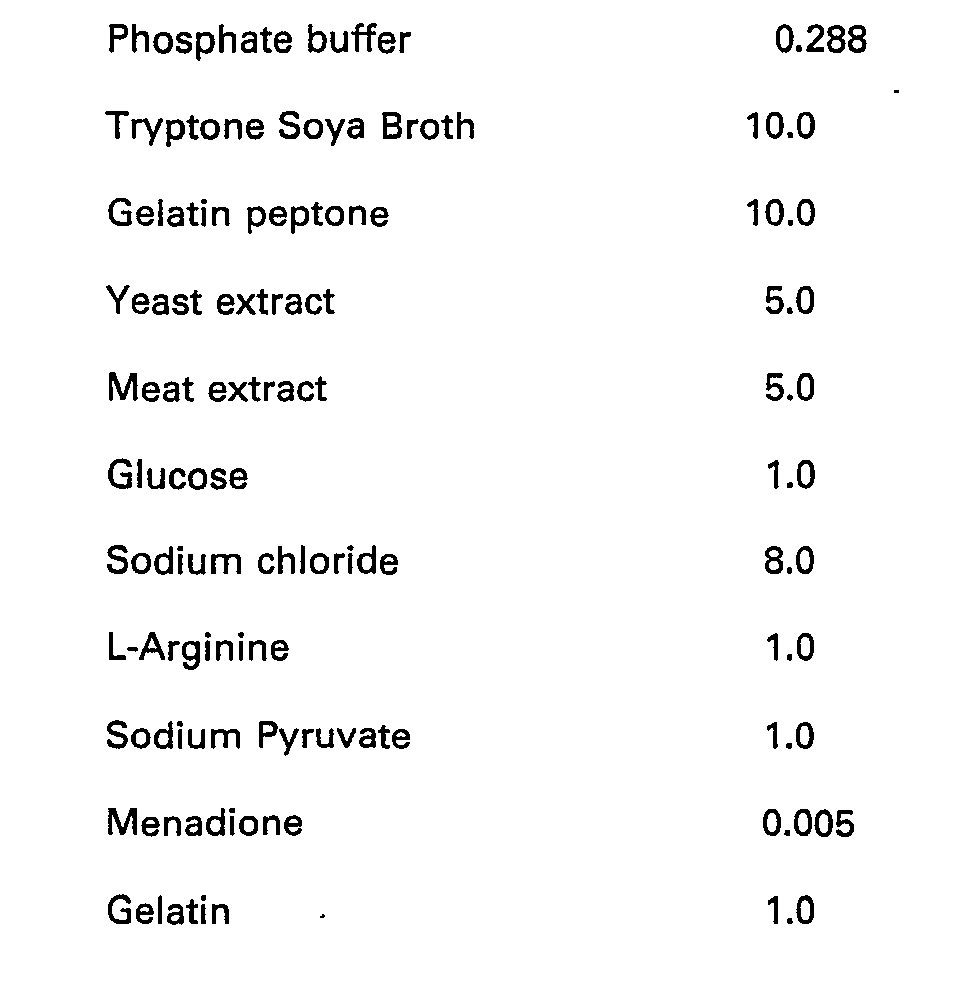
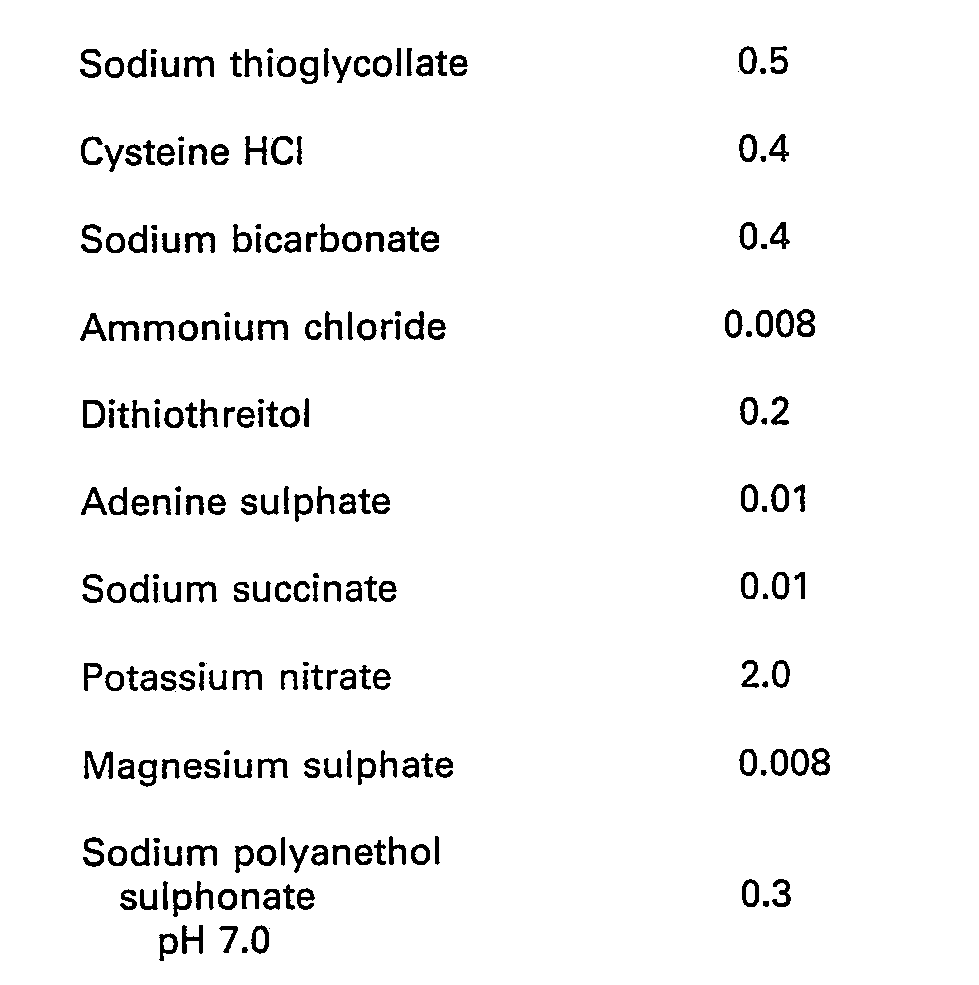
- Any kind of pressure-indicating device can in principle be used to detect a pressure increase in the vessel resulting from growth of bacteria. For example, it is possible to measure pressure changes electronically using a pressure transducer connected to a hypodermic needle inserted through the pierceable closure of the bottle.
- Alternatively, pressure changes can be detected visually by using a coiled lay-flat tube connected to the hypodermic needle, whereby a pressure increase causes the tube to uncoil.
- Another pressure-indicating device is a syringe connected to a hypodermic needle inserted through the pierceable closure of the bottle. A pressure increase causes the piston to rise up the cylinder. When this happens, the volume in the cylinder below the piston may become filled with gas from the bottle. But in a preferred embodiment, the hypodermic needle is caused-to extend below the surface of the liquid in the bottle, whereby a pressure increase forces liquid up into the cylinder, thus automatically providing a sample in the syringe which can be removed for analysis. In this case, it is possible to use an indicator which rises by flotation on the top of the liquid of the syringe, instead of a piston sliding in the cylinder.
- In the accompanying drawings:-
- Figure 1 is a sectional side elevation of a bacterial device according to the invention, and
- Figures 2a, 2b and 2c are sectional side elevations of another such device.
- Figure 3 is a sectional side elevation of yet another such device.
- Referring to Figure 1, a
glass bottle 10 is closed with a pierceableautoclavable closure 12 and contains 50 ml ofbacterial growth medium 14 into which has been introduced a sample of blood possibly containing bacteria. A hypodermic syringe comprises acylinder 16, apiston 18 and ahypodermic needle 20. The cylinder has asmall vent 22 on one side fitted with a bacterial filter (not shown). The piston carries ashaft 24 on the other end of which is amarker 26 which fits in theopen end 28 of the cylinder when the syringe is not in use. The marker is coloured red so as to be easily visible. If desired, a locating device (not shown) may be provided on theclosure 12 to ensure that the hypodermic syringe is located centrally on the top of the bottle. Thehypodermic needle 20 has been pushed through theclosure 12 and inserted into thebottle 10 until its tip is below thesurface 30 of the liquid therein. Bacterial growth has taken place in the bottle, resulting in generation of gas in theheadspace 32, and this has forced liquid up theneedle 20 and into the syringe at 34. This in turn has forced thepiston 18 to rise, so that themarker 26 has become visible above the top of thecylinder 16. If more gas is generated in the bottle, thevent 22 will act to prevent the piston from being pushed right out of the cylinder. Venting will also prevent any toxic effects due to a build-up of volatile by-products being produced. - The bottle illustrated is being incubated with a large number of others. During routine inspection, laboratory staff will observe by the
marker 26 that bacteria are present in this bottle. The syringe can then be removed and the same 34 already therein used for analysis. - Although the
sample 34 will remain viable in the syringe for up to a week or in some cases longer, it is preferred that bottles should be inspected at least daily, and that samples should be removed for analysis as soon as possible. - The device shown in Figure 2 is similar to that of Figure 1, except that the closed container to hold the liquid 34 forced out of the bottle by pressure increase is formed, not by a piston slidable within a cylinder, but by a flexible diaphragm within an open-top vessel. The arrangement of Figure 2 has the advantage of avoiding possible loss of sterility. Like parts are designated by like numbers in both figures.
- Referring to Figure 2, a
glass bottle 10 is closed with a pierceableautoclavable closure 12 and contains 50 ml ofbacterial growth medium 14 into which has been introduced a sample of blood possibly containing bacteria. A pressure indicating device comprises avessel 36, open at its upper and lower ends and divided into two halves by a horizontally extendingflexible diaphragm 38. Theflexible diaphragm 38 carries adisc 40 supporting ashaft 24 whose upper end is a loose fit in theopening 44 at the top of thevessel 36. Theshaft 24 carries a one-way lock 46, which enables the shaft to pass easily upwards through theopening 44, but not to pass accidentally downwards through the opening. The top end of theshaft 24 is coloured red so as to be easily visible. Mounted on the opening at the lower end of thevessel 36 is ahypodermic needle 20. - As shown in Figure 2a, the
hypodermic needle 20 has been pushed through theclosure 12 and inserted into thebottle 10 until its tip is below thesurface 30 of the liquid therein. Thedisc 40 is at or near its lowest extremity, and theshaft 24 does not extend significantly above theopening 44. - As shown in Figure 2b, bacterial growth has taken place in the bottle, resulting ir) generation of gas in the
headspace 32, and this has forced liquid 34 up the needle and into the lower part of thevessel 36. This in turn has flexed thediaphragm 38 causing thedisc 40 to rise so that the upper end of theshaft 24 has become visible above theopening 44. - As shown in Figure 2c, the pressure indicating device has been removed from the bottle, and pressure applied to the upper end of the
shaft 24 so as to force liquid 34 down through thehypodermic needle 20 and into adish 48 for further study. Manual pressure on theshaft 24 overrides the one-way lock 46. - Referring now to Figure 3, a
glass bottle 50 is closed by means of a pierceableautoclavable closure 52 held in place by a clampingring 54. The bottlee has an internal capacity of 59 cc and contains amixture 55 of 38 ml of culture broth and 5 ml of blood (or other sample to be tested). - The means for detecting pressure change comprise a generally
cylindrical vessel 56, closed at its upper end except for a smallaxial hole 58, and closed at its lower end except for anaxial luer connection 60 andhypodermic needle 62. The vessel carries anexternal thread 64 at its upper end, by means of which a cap 66 has been secured in place. This cap includes a hydrophobicmicrobial filter 68 which overlies thehole 58, and a smallaxial vent 70. - At its lower end, the vessel has a
cylindrical skirt 72 which terminates at an inwardly facingrib 74. As shown in the drawing, thehypodermic needle 62 has been pushed through theclosure 52, and therib 74 is releasably retained below the clampingring 54, so that thevessel 56 is held in fixed relationship with thebottle 50. Thevessel 56 is of plastics material, clear or translucent above theline 76 and opaque below that line. - The
vessel 56 has a capacity of 20 ml. The length of theneedle 62 is chosen such that after about 12-15 ml of liquid has passed up through it from thebottle 50 to thevessel 56, its lower end is clear of the liquid surface in the bottle. Thereafter only gas can pass to thevessel 56. This avoids any danger of overfilling and flooding the vessel. - In use as a blood culture device, the
bottle 50 containing the nutrient broth is taken to the patient, and 5 ml of blood introduced. This can be done in one of two ways. Either a sterile air-vent needle is inserted through theclosure 52 prior to introduction via another needle of the blood. Or the interior of the bottle is maintained under a controlled reduced pressure so that, when the interior is connected to a syringe containing, say, 20 ml of blood, or indeed when a connection is made direct to the patient's vein, 5 ml of the blood is drawn in thus leaving the interior of the bottle at atmospheric pressure. - The bottle is then taken to the laboratory, where the
closure 52 is swabbed with disinfectant and theneedle 62 inserted. The device is then incubated, preferably with agitation, at 37°C. The initial expansion resulting from this temperature rise causes a small amount of liquid to pass up through theneedle 62 into thevessel 56, but this liquid is masked by the opaque part of the vessel below theline 76. Thereafter, the device is examined periodically. When the liquid surface in thevessel 56 rises above theline 76, indicating the presence of bacteria in the sample, the cap 66 is removed and the liquid sampled through thehole 58 by means of a loop or sterile swab. Microscope examination, direct antibiotic susceptibility tests and subcultures can be made of the sample. - The following Examples illustrate the invention.
- A series of bottles were prepared by providing in each 50 ml of a bacterial growth medium formulated along the lines described above, sealing each with a pierceable closure and then sterilising. Samples containing different bacteria were then introduced into different bottles, and the pressure in each measured by means of a pressure transducer fitted with a hypodermic needle thrust through the closure. The bottles were then incubated for 18 hours, and the pressures measured again. The following results were obtained.

- A series of bottles were prepared by providing in each 45 ml of a bacterial growth medium formulated along the lines described above, sealing each with a pierceable closure and then sterilising. 5 ml samples of blood containing known small amounts of different organisms were then introduced into different bottles. Control bottles were also set up to which similar quantities of sterile blood were added. A pressure indicating device as illustrated in Figure 1 with the
bar 18 above the ventinghole 22 was inserted into each bottle through the pierceable closure. The bottles were then placed in an incubator and after a period of equilibration (30 minutes) the pressure device was sealed by pushing thebar 18 to the base of the device. The bottles were examined at intervals and the times at which the coloured plungers became visible were noted. The following results were obtained.
said sample is cultured in said medium in a first container which has a closed gaseous headspace above said liquid medium closed by a gas-tight closure for said first container located above said liquid medium; and in that there is also used as part of said device a further, upper liquid container located above said first container, and a narrow tube extending downwards from said upper container and opening below the surface of said liquid medium in said first container, said upper liquid container being such as to enable visual detection of the presence of liquid therein; whereby in the event of gas production during growth of the culture, part of the culture liquid is driven by pressure increase in said gaseous headspace upwards through said tube into said upper container, thereby to provide visual indication of said gas production in said culture.
Claims (12)
said sample is cultured in said medium in a first container (10, 50) which has a closed gaseous headspace (32) above said liquid medium (14, 55), closed by a gas-tight closure (12, 52) for said first container (10, 50) located above said liquid medium; (14, 55); and in that there is also used as part of said device a further, upper liquid container (16, 56) located above said first container (10, 50), and a narrow tube (20, 62) extending downwards from said upper container (10, 50) and opening below the surface of said liquid medium (14, 55) in said first container (10, 50), said upper liquid container (16, 56) being such as to enable visual detection of the presence of liquid (34) therein; whereby in the event of gas production during growth of the culture, part of the culture liquid (14, 55) is driven by pressure increase in said gaseous headspace upwards (34) through said tube (20, 62) into said upper container (16, 56), thereby to provide visual indication of said gas production in said culture.
Applications Claiming Priority (2)
| Application Number | Priority Date | Filing Date | Title |
|---|---|---|---|
| GB838303096A GB8303096D0 (en) | 1983-02-04 | 1983-02-04 | Bacterial testing |
| GB8303096 | 1983-02-04 |
Publications (2)
| Publication Number | Publication Date |
|---|---|
| EP0124193A1 EP0124193A1 (en) | 1984-11-07 |
| EP0124193B1 true EP0124193B1 (en) | 1987-08-12 |
Family
ID=10537460
Family Applications (1)
| Application Number | Title | Priority Date | Filing Date |
|---|---|---|---|
| EP84300500A Expired EP0124193B1 (en) | 1983-02-04 | 1984-01-26 | Method and device for bacterial testing |
Country Status (9)
| Country | Link |
|---|---|
| US (1) | US5047331A (en) |
| EP (1) | EP0124193B1 (en) |
| JP (1) | JPS59146599A (en) |
| AU (2) | AU581841B2 (en) |
| CA (1) | CA1220702A (en) |
| DE (1) | DE3465336D1 (en) |
| ES (1) | ES529421A0 (en) |
| FI (1) | FI840406A7 (en) |
| GB (1) | GB8303096D0 (en) |
Families Citing this family (35)
| Publication number | Priority date | Publication date | Assignee | Title |
|---|---|---|---|---|
| GB8619311D0 (en) * | 1986-08-07 | 1986-09-17 | Venurecare Ltd | Hygiene testing apparatus |
| US5164796A (en) * | 1988-03-15 | 1992-11-17 | Akzo N.V. | Apparatus and method for detection of microorganisms |
| US5162229A (en) * | 1988-03-15 | 1992-11-10 | Akzo N.V. | Device and method for enhanced recovery and detection of microbial growth in the presence of antimicrobial substances |
| US5518895A (en) * | 1990-02-15 | 1996-05-21 | Akzo N.V. | Device for detecting microorganisms using piezoelectric means |
| US5217876A (en) * | 1988-03-15 | 1993-06-08 | Akzo N.V. | Method for detecting microorganisms |
| US5094955A (en) * | 1988-03-15 | 1992-03-10 | Akzo N.V. | Device and method for detecting microorganisms |
| US4952498A (en) * | 1988-05-06 | 1990-08-28 | Waters John R | Method and apparatus for detecting microorganism activity |
| JPH0257198A (en) * | 1988-08-22 | 1990-02-26 | Tokyo Metropolis | Measurement of number of coliform group |
| US5372784A (en) * | 1988-08-31 | 1994-12-13 | Baxter Diagnostics Inc. | Measurement of bacterial CO2 production in an isolated fluorophore by monitoring an absorbance regulated change of fluorescence |
| US5173434A (en) * | 1990-11-05 | 1992-12-22 | Baxter Diagnostics Inc. | Measurement of color reactions by monitoring a change of fluorescence |
| US5858769A (en) * | 1989-05-15 | 1999-01-12 | Akzo Nobel N.V. | Device for detecting microorganisms |
| US5232839A (en) * | 1990-06-14 | 1993-08-03 | Difco Laboratories Incorporated | Detecting microbiological growth |
| WO1992019764A1 (en) * | 1991-05-08 | 1992-11-12 | Baxter Diagnostics Inc. | Method and apparatus to detect bacterial contamination of transfusable blood |
| JPH07501205A (en) * | 1991-08-02 | 1995-02-09 | ユニリーバー・ナームローゼ・ベンノートシャープ | Microbial growth monitoring method and device |
| ATE154219T1 (en) * | 1993-02-09 | 1997-06-15 | Josef Zech | DEVICE FOR OBTAINING SPERM CELLS FROM SEMINAL FLUID |
| US5583044A (en) * | 1993-06-11 | 1996-12-10 | Ruksenas; M. A. | Fluid sampler and testing unit |
| US5508174A (en) * | 1993-06-18 | 1996-04-16 | The United States Of America As Represented By The Secretary Of The Air Force | Method and micro roller bottle for in vitro exposure of cells to volatile chemicals |
| US5443987A (en) * | 1993-09-02 | 1995-08-22 | Decicco; Benedict T. | Detection system for microbial contamination in health-care products |
| DE69530577T2 (en) * | 1994-02-04 | 2004-02-26 | Difco Laboratories, Ann Arbor | MICROBIOLOGICAL CULTURE BOTTLES, THEIR PRODUCTION AND USE |
| GB9411515D0 (en) * | 1994-06-09 | 1994-08-03 | Aromascan Plc | Detecting bacteria |
| WO1997008337A1 (en) * | 1995-08-25 | 1997-03-06 | Unipath Limited | Methods and apparatus for detecting microorganisms |
| US6376232B1 (en) | 1997-03-06 | 2002-04-23 | Osmetech Plc | Microorganism analysis means |
| FR2786782B1 (en) * | 1998-12-04 | 2001-02-23 | Biomerieux Sa | METHOD FOR DETECTION OF MICROORGANISMS AND MEDIUM FOR USE IN SUCH A METHOD |
| GB0206275D0 (en) * | 2002-03-16 | 2002-05-01 | Bactest Ltd | Method and apparatus for the non-invasive monitoring of gas exchange by biological material |
| DE202006010454U1 (en) * | 2006-07-04 | 2007-11-15 | Rugen, Hermann, Dipl.-Ing. | Sensor for determining the gas formation rate of a substrate |
| US8633016B2 (en) * | 2008-09-30 | 2014-01-21 | Covidien Lp | Microbial detection assembly |
| US9327804B2 (en) * | 2009-02-06 | 2016-05-03 | The United States Of America, As Represented By The Secretary Of The Navy | Bacteria-based gas generator |
| US9663194B2 (en) | 2009-02-06 | 2017-05-30 | The United States Of America, As Represented By The Secretary Of The Navy | Bouyancy control device |
| WO2010126856A1 (en) | 2009-04-27 | 2010-11-04 | The Charles Stark Draper Laboratory, Inc. | Rapid detection of volatile organic compounds for identification of mycobacterium tuberculosis in a sample |
| EP2893029B1 (en) * | 2012-09-04 | 2017-09-27 | Becton, Dickinson and Company | Bacterial pre-concentration and detection technique |
| CN105505752A (en) * | 2016-02-18 | 2016-04-20 | 允汇科技(天津)有限公司 | Body fluid culture bottle |
| KR101988233B1 (en) * | 2018-01-22 | 2019-06-12 | 강릉원주대학교산학협력단 | Device for detecting cell and method for cell detection using thereof |
| CN108277152B (en) * | 2018-04-04 | 2023-10-13 | 博品(上海)生物医药科技有限公司 | Sterile liquid exchange device for cell resuscitation and method for carrying out cell resuscitation liquid exchange by using device |
| CN111088150A (en) * | 2019-12-30 | 2020-05-01 | 江苏益玛生物科技有限公司 | Culture medium nitrogen filling method |
| FR3147813A1 (en) * | 2023-04-17 | 2024-10-18 | Synsym Biosciences Sas | Method of producing microorganisms and installation for implementation |
Family Cites Families (14)
| Publication number | Priority date | Publication date | Assignee | Title |
|---|---|---|---|---|
| US2019950A (en) * | 1930-05-31 | 1935-11-05 | Herbert H Bunzell | Determination of rope infection |
| DE869845C (en) * | 1951-08-02 | 1953-03-09 | Michael Dr Med Allert | Device for determining the sedimentation rate of the blood cells |
| GB733893A (en) * | 1952-05-23 | 1955-07-20 | Asta Werke Ag Chem Fab | Apparatus for use in preparing fresh pure cultures of living micro-organisms at the place of application |
| DE1945279U (en) * | 1966-07-02 | 1966-09-01 | Gruenenthal Chemie | AUXILIARY DEVICE FOR THE PRODUCTION OF STERILE SOLUTIONS. |
| DE2041253C3 (en) * | 1969-08-25 | 1979-02-15 | Veb Kombinat Technisches Glas Ilmenau, Ddr 6300 Ilmenau | Arrangement for the continuous detection of the gas exchange of small biological objects |
| BE791340A (en) * | 1972-01-06 | 1973-03-01 | Becton Dickinson Co | NEW METHOD AND APPARATUS FOR TAKING A CULTURE AND IDENTIFYING MICRO-ORGANISMS OF MOODS |
| US3907646A (en) * | 1974-07-24 | 1975-09-23 | Nasa | Measurement of gas production of microorganisms |
| US4152213A (en) * | 1977-03-10 | 1979-05-01 | Johnston Laboratories, Inc. | Vacuum detection of bacteria |
| US4170520A (en) * | 1977-09-16 | 1979-10-09 | Massachusetts Institute Of Technology | Apparatus for measuring reactant concentrations and quantities |
| US4197369A (en) * | 1977-09-16 | 1980-04-08 | Massachusetts Institute Of Technology | Method for measuring reactant concentrations and quantities |
| JPS55104757A (en) * | 1979-02-07 | 1980-08-11 | Ootake Seisakusho:Kk | Device for automatically measuring extinction of gas |
| AU530665B2 (en) * | 1979-06-04 | 1983-07-21 | Terumo Corp. | Microorganism-culturing device |
| US4270381A (en) * | 1979-10-01 | 1981-06-02 | Demaray David E | Recording instrument to measure microbal gas production |
| DD206683A3 (en) * | 1981-07-16 | 1984-02-01 | Werner Schade | METHOD AND DEVICE FOR DETECTING CONTAMINANTS |
-
1983
- 1983-02-04 GB GB838303096A patent/GB8303096D0/en active Pending
-
1984
- 1984-01-26 DE DE8484300500T patent/DE3465336D1/en not_active Expired
- 1984-01-26 EP EP84300500A patent/EP0124193B1/en not_active Expired
- 1984-01-27 CA CA000446207A patent/CA1220702A/en not_active Expired
- 1984-02-01 JP JP59018159A patent/JPS59146599A/en active Granted
- 1984-02-01 FI FI840406A patent/FI840406A7/en not_active Application Discontinuation
- 1984-02-02 ES ES529421A patent/ES529421A0/en active Granted
- 1984-02-03 AU AU24042/84A patent/AU581841B2/en not_active Ceased
-
1987
- 1987-10-19 US US07/110,587 patent/US5047331A/en not_active Expired - Fee Related
-
1988
- 1988-12-13 AU AU26822/88A patent/AU618771B2/en not_active Ceased
Also Published As
| Publication number | Publication date |
|---|---|
| EP0124193A1 (en) | 1984-11-07 |
| ES8600387A1 (en) | 1985-10-01 |
| FI840406A0 (en) | 1984-02-01 |
| US5047331A (en) | 1991-09-10 |
| FI840406A7 (en) | 1984-08-05 |
| ES529421A0 (en) | 1985-10-01 |
| AU2682288A (en) | 1989-07-06 |
| DE3465336D1 (en) | 1987-09-17 |
| JPS59146599A (en) | 1984-08-22 |
| JPH0558718B2 (en) | 1993-08-27 |
| AU581841B2 (en) | 1989-03-09 |
| GB8303096D0 (en) | 1983-03-09 |
| AU2404284A (en) | 1984-08-09 |
| AU618771B2 (en) | 1992-01-09 |
| CA1220702A (en) | 1987-04-21 |
Similar Documents
| Publication | Publication Date | Title |
|---|---|---|
| US5047331A (en) | Method and device for bacterial testing | |
| AU705576B2 (en) | Device and method for detecting microorganisms | |
| US5232839A (en) | Detecting microbiological growth | |
| US4945060A (en) | Device for detecting microorganisms | |
| US5217876A (en) | Method for detecting microorganisms | |
| EP0446972A2 (en) | Device and methods for detecting microorganisms | |
| US4152213A (en) | Vacuum detection of bacteria | |
| US4248830A (en) | Device for microbiological testing | |
| US3939044A (en) | Anaerobic microbe transport assembly and method of using | |
| US5672484A (en) | Microbiological culture bottle, and method of making and using same | |
| JP2008136440A (en) | Syringe type microorganism culture device | |
| GB2102947A (en) | Process and apparatus for indicating the presence of contaminating microorganisms | |
| US3171793A (en) | Fermentative analytical procedure and apparatus therefor | |
| CN2689223Y (en) | Blood culturing bottles | |
| FI70045C (en) | METHOD ANORDNING OCH NAERINGSMEDIUM FOER ATT PAOVISA BAKTEREMI | |
| McLaughlin et al. | A rapid method for detecting bacterial contamination in the presence of Penicillium and Streptomyces antibiotic fermentations | |
| HK1014372B (en) | Microbiological culture bottle, and method of making and using same | |
| Mallard | A comparative study of hydrogen utilization and methane generation by pure cultures of methane producing bacteria. | |
| JPH11253190A (en) | Bacterial culture bottle and bacterial detection method | |
| WO2000009649A1 (en) | Device and method for detecting microorganisms | |
| JPS5910791B2 (en) | Saikindo Teiyousouchi | |
| JPS5928495A (en) | Method and apparatus for detecting pollutants |
Legal Events
| Date | Code | Title | Description |
|---|---|---|---|
| PUAI | Public reference made under article 153(3) epc to a published international application that has entered the european phase |
Free format text: ORIGINAL CODE: 0009012 |
|
| AK | Designated contracting states |
Designated state(s): CH DE FR GB IT LI SE |
|
| 17P | Request for examination filed |
Effective date: 19850424 |
|
| RAP1 | Party data changed (applicant data changed or rights of an application transferred) |
Owner name: UNILEVER NV Owner name: UNILEVER PLC |
|
| GRAA | (expected) grant |
Free format text: ORIGINAL CODE: 0009210 |
|
| AK | Designated contracting states |
Kind code of ref document: B1 Designated state(s): CH DE FR GB IT LI SE |
|
| REF | Corresponds to: |
Ref document number: 3465336 Country of ref document: DE Date of ref document: 19870917 |
|
| ITF | It: translation for a ep patent filed | ||
| ET | Fr: translation filed | ||
| PLBE | No opposition filed within time limit |
Free format text: ORIGINAL CODE: 0009261 |
|
| STAA | Information on the status of an ep patent application or granted ep patent |
Free format text: STATUS: NO OPPOSITION FILED WITHIN TIME LIMIT |
|
| 26N | No opposition filed | ||
| ITPR | It: changes in ownership of a european patent |
Owner name: ASSUNZIONE O VARIAZIONE MANDATO;MODIANO & ASSOCIAT |
|
| ITTA | It: last paid annual fee | ||
| EAL | Se: european patent in force in sweden |
Ref document number: 84300500.0 |
|
| REG | Reference to a national code |
Ref country code: CH Ref legal event code: PUE Owner name: UNILEVER N.V. TRANSFER- PEWTER (NO.2) LIMITED Ref country code: CH Ref legal event code: PFA Free format text: PEWTER (NO.2) LIMITED TRANSFER- OXOID LIMITED |
|
| REG | Reference to a national code |
Ref country code: FR Ref legal event code: TP |
|
| REG | Reference to a national code |
Ref country code: GB Ref legal event code: 732E |
|
| PGFP | Annual fee paid to national office [announced via postgrant information from national office to epo] |
Ref country code: GB Payment date: 20001117 Year of fee payment: 18 |
|
| PGFP | Annual fee paid to national office [announced via postgrant information from national office to epo] |
Ref country code: FR Payment date: 20001211 Year of fee payment: 18 |
|
| PGFP | Annual fee paid to national office [announced via postgrant information from national office to epo] |
Ref country code: SE Payment date: 20001218 Year of fee payment: 18 |
|
| PGFP | Annual fee paid to national office [announced via postgrant information from national office to epo] |
Ref country code: DE Payment date: 20001219 Year of fee payment: 18 |
|
| PGFP | Annual fee paid to national office [announced via postgrant information from national office to epo] |
Ref country code: CH Payment date: 20001220 Year of fee payment: 18 |
|
| REG | Reference to a national code |
Ref country code: GB Ref legal event code: IF02 |
|
| PG25 | Lapsed in a contracting state [announced via postgrant information from national office to epo] |
Ref country code: GB Free format text: LAPSE BECAUSE OF NON-PAYMENT OF DUE FEES Effective date: 20020126 |
|
| PG25 | Lapsed in a contracting state [announced via postgrant information from national office to epo] |
Ref country code: SE Free format text: LAPSE BECAUSE OF NON-PAYMENT OF DUE FEES Effective date: 20020127 |
|
| PG25 | Lapsed in a contracting state [announced via postgrant information from national office to epo] |
Ref country code: LI Free format text: LAPSE BECAUSE OF NON-PAYMENT OF DUE FEES Effective date: 20020131 Ref country code: CH Free format text: LAPSE BECAUSE OF NON-PAYMENT OF DUE FEES Effective date: 20020131 |
|
| PG25 | Lapsed in a contracting state [announced via postgrant information from national office to epo] |
Ref country code: DE Free format text: LAPSE BECAUSE OF NON-PAYMENT OF DUE FEES Effective date: 20020801 |
|
| EUG | Se: european patent has lapsed |
Ref document number: 84300500.0 |
|
| REG | Reference to a national code |
Ref country code: CH Ref legal event code: PL |
|
| GBPC | Gb: european patent ceased through non-payment of renewal fee |
Effective date: 20020126 |
|
| PG25 | Lapsed in a contracting state [announced via postgrant information from national office to epo] |
Ref country code: FR Free format text: LAPSE BECAUSE OF NON-PAYMENT OF DUE FEES Effective date: 20020930 |
|
| REG | Reference to a national code |
Ref country code: FR Ref legal event code: ST |