WO2024242069A1 - 神経堤細胞の製造方法 - Google Patents
神経堤細胞の製造方法 Download PDFInfo
- Publication number
- WO2024242069A1 WO2024242069A1 PCT/JP2024/018417 JP2024018417W WO2024242069A1 WO 2024242069 A1 WO2024242069 A1 WO 2024242069A1 JP 2024018417 W JP2024018417 W JP 2024018417W WO 2024242069 A1 WO2024242069 A1 WO 2024242069A1
- Authority
- WO
- WIPO (PCT)
- Prior art keywords
- cells
- cell
- ncc
- induction
- culture
- Prior art date
- Legal status (The legal status is an assumption and is not a legal conclusion. Google has not performed a legal analysis and makes no representation as to the accuracy of the status listed.)
- Ceased
Links
Images























Classifications
-
- C—CHEMISTRY; METALLURGY
- C12—BIOCHEMISTRY; BEER; SPIRITS; WINE; VINEGAR; MICROBIOLOGY; ENZYMOLOGY; MUTATION OR GENETIC ENGINEERING
- C12N—MICROORGANISMS OR ENZYMES; COMPOSITIONS THEREOF; PROPAGATING, PRESERVING, OR MAINTAINING MICROORGANISMS; MUTATION OR GENETIC ENGINEERING; CULTURE MEDIA
- C12N5/00—Undifferentiated human, animal or plant cells, e.g. cell lines; Tissues; Cultivation or maintenance thereof; Culture media therefor
- C12N5/06—Animal cells or tissues; Human cells or tissues
- C12N5/0602—Vertebrate cells
- C12N5/0618—Cells of the nervous system
-
- A—HUMAN NECESSITIES
- A61—MEDICAL OR VETERINARY SCIENCE; HYGIENE
- A61K—PREPARATIONS FOR MEDICAL, DENTAL OR TOILETRY PURPOSES
- A61K35/00—Medicinal preparations containing materials or reaction products thereof with undetermined constitution
- A61K35/12—Materials from mammals; Compositions comprising non-specified tissues or cells; Compositions comprising non-embryonic stem cells; Genetically modified cells
- A61K35/48—Reproductive organs
- A61K35/54—Ovaries; Ova; Ovules; Embryos; Foetal cells; Germ cells
- A61K35/545—Embryonic stem cells; Pluripotent stem cells; Induced pluripotent stem cells; Uncharacterised stem cells
-
- C—CHEMISTRY; METALLURGY
- C07—ORGANIC CHEMISTRY
- C07K—PEPTIDES
- C07K14/00—Peptides having more than 20 amino acids; Gastrins; Somatostatins; Melanotropins; Derivatives thereof
- C07K14/435—Peptides having more than 20 amino acids; Gastrins; Somatostatins; Melanotropins; Derivatives thereof from animals; from humans
-
- C—CHEMISTRY; METALLURGY
- C12—BIOCHEMISTRY; BEER; SPIRITS; WINE; VINEGAR; MICROBIOLOGY; ENZYMOLOGY; MUTATION OR GENETIC ENGINEERING
- C12N—MICROORGANISMS OR ENZYMES; COMPOSITIONS THEREOF; PROPAGATING, PRESERVING, OR MAINTAINING MICROORGANISMS; MUTATION OR GENETIC ENGINEERING; CULTURE MEDIA
- C12N5/00—Undifferentiated human, animal or plant cells, e.g. cell lines; Tissues; Cultivation or maintenance thereof; Culture media therefor
- C12N5/06—Animal cells or tissues; Human cells or tissues
- C12N5/0602—Vertebrate cells
- C12N5/0652—Cells of skeletal and connective tissues; Mesenchyme
- C12N5/0654—Osteocytes, Osteoblasts, Odontocytes; Bones, Teeth
-
- C—CHEMISTRY; METALLURGY
- C12—BIOCHEMISTRY; BEER; SPIRITS; WINE; VINEGAR; MICROBIOLOGY; ENZYMOLOGY; MUTATION OR GENETIC ENGINEERING
- C12N—MICROORGANISMS OR ENZYMES; COMPOSITIONS THEREOF; PROPAGATING, PRESERVING, OR MAINTAINING MICROORGANISMS; MUTATION OR GENETIC ENGINEERING; CULTURE MEDIA
- C12N5/00—Undifferentiated human, animal or plant cells, e.g. cell lines; Tissues; Cultivation or maintenance thereof; Culture media therefor
- C12N5/06—Animal cells or tissues; Human cells or tissues
- C12N5/0602—Vertebrate cells
- C12N5/0652—Cells of skeletal and connective tissues; Mesenchyme
- C12N5/0661—Smooth muscle cells
-
- C—CHEMISTRY; METALLURGY
- C12—BIOCHEMISTRY; BEER; SPIRITS; WINE; VINEGAR; MICROBIOLOGY; ENZYMOLOGY; MUTATION OR GENETIC ENGINEERING
- C12N—MICROORGANISMS OR ENZYMES; COMPOSITIONS THEREOF; PROPAGATING, PRESERVING, OR MAINTAINING MICROORGANISMS; MUTATION OR GENETIC ENGINEERING; CULTURE MEDIA
- C12N5/00—Undifferentiated human, animal or plant cells, e.g. cell lines; Tissues; Cultivation or maintenance thereof; Culture media therefor
- C12N5/06—Animal cells or tissues; Human cells or tissues
- C12N5/0602—Vertebrate cells
- C12N5/0652—Cells of skeletal and connective tissues; Mesenchyme
- C12N5/0662—Stem cells
- C12N5/0668—Mesenchymal stem cells from other natural sources
-
- C—CHEMISTRY; METALLURGY
- C12—BIOCHEMISTRY; BEER; SPIRITS; WINE; VINEGAR; MICROBIOLOGY; ENZYMOLOGY; MUTATION OR GENETIC ENGINEERING
- C12N—MICROORGANISMS OR ENZYMES; COMPOSITIONS THEREOF; PROPAGATING, PRESERVING, OR MAINTAINING MICROORGANISMS; MUTATION OR GENETIC ENGINEERING; CULTURE MEDIA
- C12N5/00—Undifferentiated human, animal or plant cells, e.g. cell lines; Tissues; Cultivation or maintenance thereof; Culture media therefor
- C12N5/06—Animal cells or tissues; Human cells or tissues
- C12N5/0697—Artificial constructs associating cells of different lineages, e.g. tissue equivalents
-
- C—CHEMISTRY; METALLURGY
- C12—BIOCHEMISTRY; BEER; SPIRITS; WINE; VINEGAR; MICROBIOLOGY; ENZYMOLOGY; MUTATION OR GENETIC ENGINEERING
- C12Q—MEASURING OR TESTING PROCESSES INVOLVING ENZYMES, NUCLEIC ACIDS OR MICROORGANISMS; COMPOSITIONS OR TEST PAPERS THEREFOR; PROCESSES OF PREPARING SUCH COMPOSITIONS; CONDITION-RESPONSIVE CONTROL IN MICROBIOLOGICAL OR ENZYMOLOGICAL PROCESSES
- C12Q1/00—Measuring or testing processes involving enzymes, nucleic acids or microorganisms; Compositions therefor; Processes of preparing such compositions
- C12Q1/02—Measuring or testing processes involving enzymes, nucleic acids or microorganisms; Compositions therefor; Processes of preparing such compositions involving viable microorganisms
- C12Q1/04—Determining presence or kind of microorganism; Use of selective media for testing antibiotics or bacteriocides; Compositions containing a chemical indicator therefor
-
- G—PHYSICS
- G01—MEASURING; TESTING
- G01N—INVESTIGATING OR ANALYSING MATERIALS BY DETERMINING THEIR CHEMICAL OR PHYSICAL PROPERTIES
- G01N33/00—Investigating or analysing materials by specific methods not covered by groups G01N1/00 - G01N31/00
- G01N33/48—Biological material, e.g. blood, urine; Haemocytometers
-
- C—CHEMISTRY; METALLURGY
- C12—BIOCHEMISTRY; BEER; SPIRITS; WINE; VINEGAR; MICROBIOLOGY; ENZYMOLOGY; MUTATION OR GENETIC ENGINEERING
- C12N—MICROORGANISMS OR ENZYMES; COMPOSITIONS THEREOF; PROPAGATING, PRESERVING, OR MAINTAINING MICROORGANISMS; MUTATION OR GENETIC ENGINEERING; CULTURE MEDIA
- C12N2501/00—Active agents used in cell culture processes, e.g. differentation
- C12N2501/10—Growth factors
- C12N2501/11—Epidermal growth factor [EGF]
-
- C—CHEMISTRY; METALLURGY
- C12—BIOCHEMISTRY; BEER; SPIRITS; WINE; VINEGAR; MICROBIOLOGY; ENZYMOLOGY; MUTATION OR GENETIC ENGINEERING
- C12N—MICROORGANISMS OR ENZYMES; COMPOSITIONS THEREOF; PROPAGATING, PRESERVING, OR MAINTAINING MICROORGANISMS; MUTATION OR GENETIC ENGINEERING; CULTURE MEDIA
- C12N2501/00—Active agents used in cell culture processes, e.g. differentation
- C12N2501/10—Growth factors
- C12N2501/115—Basic fibroblast growth factor (bFGF, FGF-2)
-
- C—CHEMISTRY; METALLURGY
- C12—BIOCHEMISTRY; BEER; SPIRITS; WINE; VINEGAR; MICROBIOLOGY; ENZYMOLOGY; MUTATION OR GENETIC ENGINEERING
- C12N—MICROORGANISMS OR ENZYMES; COMPOSITIONS THEREOF; PROPAGATING, PRESERVING, OR MAINTAINING MICROORGANISMS; MUTATION OR GENETIC ENGINEERING; CULTURE MEDIA
- C12N2501/00—Active agents used in cell culture processes, e.g. differentation
- C12N2501/10—Growth factors
- C12N2501/119—Other fibroblast growth factors, e.g. FGF-4, FGF-8, FGF-10
-
- C—CHEMISTRY; METALLURGY
- C12—BIOCHEMISTRY; BEER; SPIRITS; WINE; VINEGAR; MICROBIOLOGY; ENZYMOLOGY; MUTATION OR GENETIC ENGINEERING
- C12N—MICROORGANISMS OR ENZYMES; COMPOSITIONS THEREOF; PROPAGATING, PRESERVING, OR MAINTAINING MICROORGANISMS; MUTATION OR GENETIC ENGINEERING; CULTURE MEDIA
- C12N2501/00—Active agents used in cell culture processes, e.g. differentation
- C12N2501/10—Growth factors
- C12N2501/13—Nerve growth factor [NGF]; Brain-derived neurotrophic factor [BDNF]; Cilliary neurotrophic factor [CNTF]; Glial-derived neurotrophic factor [GDNF]; Neurotrophins [NT]; Neuregulins
-
- C—CHEMISTRY; METALLURGY
- C12—BIOCHEMISTRY; BEER; SPIRITS; WINE; VINEGAR; MICROBIOLOGY; ENZYMOLOGY; MUTATION OR GENETIC ENGINEERING
- C12N—MICROORGANISMS OR ENZYMES; COMPOSITIONS THEREOF; PROPAGATING, PRESERVING, OR MAINTAINING MICROORGANISMS; MUTATION OR GENETIC ENGINEERING; CULTURE MEDIA
- C12N2501/00—Active agents used in cell culture processes, e.g. differentation
- C12N2501/10—Growth factors
- C12N2501/15—Transforming growth factor beta (TGF-β)
-
- C—CHEMISTRY; METALLURGY
- C12—BIOCHEMISTRY; BEER; SPIRITS; WINE; VINEGAR; MICROBIOLOGY; ENZYMOLOGY; MUTATION OR GENETIC ENGINEERING
- C12N—MICROORGANISMS OR ENZYMES; COMPOSITIONS THEREOF; PROPAGATING, PRESERVING, OR MAINTAINING MICROORGANISMS; MUTATION OR GENETIC ENGINEERING; CULTURE MEDIA
- C12N2501/00—Active agents used in cell culture processes, e.g. differentation
- C12N2501/10—Growth factors
- C12N2501/155—Bone morphogenic proteins [BMP]; Osteogenins; Osteogenic factor; Bone inducing factor
-
- C—CHEMISTRY; METALLURGY
- C12—BIOCHEMISTRY; BEER; SPIRITS; WINE; VINEGAR; MICROBIOLOGY; ENZYMOLOGY; MUTATION OR GENETIC ENGINEERING
- C12N—MICROORGANISMS OR ENZYMES; COMPOSITIONS THEREOF; PROPAGATING, PRESERVING, OR MAINTAINING MICROORGANISMS; MUTATION OR GENETIC ENGINEERING; CULTURE MEDIA
- C12N2501/00—Active agents used in cell culture processes, e.g. differentation
- C12N2501/10—Growth factors
- C12N2501/16—Activin; Inhibin; Mullerian inhibiting substance
-
- C—CHEMISTRY; METALLURGY
- C12—BIOCHEMISTRY; BEER; SPIRITS; WINE; VINEGAR; MICROBIOLOGY; ENZYMOLOGY; MUTATION OR GENETIC ENGINEERING
- C12N—MICROORGANISMS OR ENZYMES; COMPOSITIONS THEREOF; PROPAGATING, PRESERVING, OR MAINTAINING MICROORGANISMS; MUTATION OR GENETIC ENGINEERING; CULTURE MEDIA
- C12N2501/00—Active agents used in cell culture processes, e.g. differentation
- C12N2501/30—Hormones
- C12N2501/365—Endothelin
-
- C—CHEMISTRY; METALLURGY
- C12—BIOCHEMISTRY; BEER; SPIRITS; WINE; VINEGAR; MICROBIOLOGY; ENZYMOLOGY; MUTATION OR GENETIC ENGINEERING
- C12N—MICROORGANISMS OR ENZYMES; COMPOSITIONS THEREOF; PROPAGATING, PRESERVING, OR MAINTAINING MICROORGANISMS; MUTATION OR GENETIC ENGINEERING; CULTURE MEDIA
- C12N2501/00—Active agents used in cell culture processes, e.g. differentation
- C12N2501/30—Hormones
- C12N2501/38—Hormones with nuclear receptors
- C12N2501/385—Hormones with nuclear receptors of the family of the retinoic acid recptor, e.g. RAR, RXR; Peroxisome proliferator-activated receptor [PPAR]
-
- C—CHEMISTRY; METALLURGY
- C12—BIOCHEMISTRY; BEER; SPIRITS; WINE; VINEGAR; MICROBIOLOGY; ENZYMOLOGY; MUTATION OR GENETIC ENGINEERING
- C12N—MICROORGANISMS OR ENZYMES; COMPOSITIONS THEREOF; PROPAGATING, PRESERVING, OR MAINTAINING MICROORGANISMS; MUTATION OR GENETIC ENGINEERING; CULTURE MEDIA
- C12N2501/00—Active agents used in cell culture processes, e.g. differentation
- C12N2501/40—Regulators of development
- C12N2501/41—Hedgehog proteins; Cyclopamine (inhibitor)
-
- C—CHEMISTRY; METALLURGY
- C12—BIOCHEMISTRY; BEER; SPIRITS; WINE; VINEGAR; MICROBIOLOGY; ENZYMOLOGY; MUTATION OR GENETIC ENGINEERING
- C12N—MICROORGANISMS OR ENZYMES; COMPOSITIONS THEREOF; PROPAGATING, PRESERVING, OR MAINTAINING MICROORGANISMS; MUTATION OR GENETIC ENGINEERING; CULTURE MEDIA
- C12N2501/00—Active agents used in cell culture processes, e.g. differentation
- C12N2501/40—Regulators of development
- C12N2501/415—Wnt; Frizzeled
-
- C—CHEMISTRY; METALLURGY
- C12—BIOCHEMISTRY; BEER; SPIRITS; WINE; VINEGAR; MICROBIOLOGY; ENZYMOLOGY; MUTATION OR GENETIC ENGINEERING
- C12N—MICROORGANISMS OR ENZYMES; COMPOSITIONS THEREOF; PROPAGATING, PRESERVING, OR MAINTAINING MICROORGANISMS; MUTATION OR GENETIC ENGINEERING; CULTURE MEDIA
- C12N2501/00—Active agents used in cell culture processes, e.g. differentation
- C12N2501/70—Enzymes
- C12N2501/72—Transferases [EC 2.]
- C12N2501/727—Kinases (EC 2.7.)
-
- C—CHEMISTRY; METALLURGY
- C12—BIOCHEMISTRY; BEER; SPIRITS; WINE; VINEGAR; MICROBIOLOGY; ENZYMOLOGY; MUTATION OR GENETIC ENGINEERING
- C12N—MICROORGANISMS OR ENZYMES; COMPOSITIONS THEREOF; PROPAGATING, PRESERVING, OR MAINTAINING MICROORGANISMS; MUTATION OR GENETIC ENGINEERING; CULTURE MEDIA
- C12N2506/00—Differentiation of animal cells from one lineage to another; Differentiation of pluripotent cells
- C12N2506/45—Differentiation of animal cells from one lineage to another; Differentiation of pluripotent cells from artificially induced pluripotent stem cells
-
- C—CHEMISTRY; METALLURGY
- C12—BIOCHEMISTRY; BEER; SPIRITS; WINE; VINEGAR; MICROBIOLOGY; ENZYMOLOGY; MUTATION OR GENETIC ENGINEERING
- C12N—MICROORGANISMS OR ENZYMES; COMPOSITIONS THEREOF; PROPAGATING, PRESERVING, OR MAINTAINING MICROORGANISMS; MUTATION OR GENETIC ENGINEERING; CULTURE MEDIA
- C12N2513/00—3D culture
-
- C—CHEMISTRY; METALLURGY
- C12—BIOCHEMISTRY; BEER; SPIRITS; WINE; VINEGAR; MICROBIOLOGY; ENZYMOLOGY; MUTATION OR GENETIC ENGINEERING
- C12N—MICROORGANISMS OR ENZYMES; COMPOSITIONS THEREOF; PROPAGATING, PRESERVING, OR MAINTAINING MICROORGANISMS; MUTATION OR GENETIC ENGINEERING; CULTURE MEDIA
- C12N2533/00—Supports or coatings for cell culture, characterised by material
- C12N2533/50—Proteins
- C12N2533/52—Fibronectin; Laminin
Definitions
- the present invention relates to a method for producing neural crest cells. More specifically, the present invention relates to a method for producing neural crest cells, which includes a step of performing suspension culture of pluripotent stem cells under specific conditions, and uses of neural crest cells obtained by the method.
- Neural crest cells are cells that arise between the neuroectoderm and epidermal ectoderm when the neural tube is formed from the neural plate in early development. They have the multipotency and ability to self-proliferate, allowing them to differentiate into many types of cells, such as neurons, glial cells, mesenchymal stromal cells, bone cells, chondrocytes, corneal cells, and pigment cells. Such multipotency and ability to self-proliferate demonstrate the usefulness of NCCs as a cellular medicine for regenerative medicine, and there is a demand for technology to efficiently induce NCCs.
- Non-Patent Document 1 describes that human pluripotent stem cells seeded on Matrigel are subjected to neural priming for three days, followed by a neural crest (NC) induction process for five days, thereby inducing NCCs.
- Non-Patent Document 2 describes that human pluripotent stem cells are subjected to adhesion culture for seven days in a medium containing BMP4 and SB431542, thereby inducing NCCs, and that the NCCs induced in this manner are cultured for 23 days in a medium containing CHIR99021, SB431542, SAG, EGF, FGF2, and DMH1, thereby inducing ectodermal chondrogenic cells (ECCs).
- ECCs ectodermal chondrogenic cells
- Non-Patent Document 3 a method of inducing NCCs that have lost the ability to differentiate into neural cells by culturing pluripotent stem cells in a medium containing SB431542 and CHIR99021 under xeno-free conditions for 10 days to obtain NCCs, and then culturing the NCCs in a medium containing SB431542, EGF, and bFGF to obtain adherent cells.
- the present inventors have also succeeded in expanding NCCs that have retained the ability to differentiate into neural cells by culturing NCCs induced to differentiate from pluripotent stem cells in a medium containing a GSK-3 ⁇ inhibitor at a concentration that shows an effect equivalent to that of CHIR99021 at a concentration of more than 1 ⁇ M (Patent Document 1).
- an object of the present invention is to provide a method for inducing neural crest cells from pluripotent stem cells in a shorter period of time, preferably under xeno-free conditions.
- the inventors focused on suspension culture while researching methods for inducing bone organoids from pluripotent stem cells. Therefore, in the first step of the induction process from pluripotent stem cells to neural crest cells (NCCs), they searched for a method that could induce NCCs from pluripotent stem cells by suspension culture, rather than the conventional adhesion culture. As a result of intensive research, they found that NCCs could be stably and efficiently induced under xeno-free conditions by short-term suspension culture of pluripotent stem cells in a medium containing an ALK inhibitor and bone morphogenetic protein to form circular cell clusters, and then culturing in a medium containing an ALK inhibitor and a GSK-3 ⁇ inhibitor.
- the present inventors have verified the differentiation ability of NCC obtained by the above-mentioned suspension culture. As a result, they have found that, like conventional NCC, they can be induced to differentiate into peripheral nerve cells, melanoblasts, and mesenchymal stem cells, and that the efficiency of these differentiation inductions is very high. Furthermore, NCC obtained by suspension culture can be induced to differentiate into first pharyngeal arch ectodermal mesenchymal (PA1-EM) cells, but there have been no reports of successful induction of differentiation of NCC into PA1-EM cells in vitro, which was very surprising. It is also possible to induce bone organoids from PA1-EM cells, and the bone organoids can be transplanted into a living body to efficiently repair bone defects. The present inventors have conducted further research based on these findings, and have completed the present invention.
- PA1-EM first pharyngeal arch ectodermal mesenchymal
- a method for producing neural crest cells from pluripotent stem cells comprising: (1) a step of suspension culturing pluripotent stem cells in a medium containing an ALK inhibitor and a bone morphogenetic protein; and (2) a step of suspension culturing pluripotent stem cells in a medium containing an ALK inhibitor and a GSK-3 ⁇ inhibitor.
- a method comprising: [2-1] The method according to [1], wherein the step (2) includes a step of performing shaking culture and/or stirring culture.
- the step (1) includes a step of performing shaking culture and/or stirring culture.
- [3-1] The method according to any one of [1] to [2-2], wherein the culture period in the step (1) is 6 hours to 5 days.
- [3-2] The method according to any one of [1] to [3-1], wherein the culture period in the step (1) is 6 hours to 3 days.
- [3-3] The method according to any one of [1] to [3-2], wherein the culture period in the step (1) is 1 to 3 days.
- [4-1] The method according to any one of [1] to [3-3], wherein the culture period in the step (2) is 1 day to 6 days.
- [4-2] The method according to any one of [1] to [4-1], wherein the culture period in the step (2) is 1 to 4 days.
- [4-3] The method according to any one of [1] to [4-2], wherein the culture period in the step (2) is 1 to 3 days.
- [4-4] The method according to any one of [1] to [4-3], wherein the culture period in the steps (1) and (2) is 2 to 5 days.
- [4-5] The method according to any one of [1] to [4-4], wherein the culture period in the step (1) is 1 day, and the culture period in the step (2) is 3 days.
- [5] The method according to any one of [1] to [4-5], comprising, prior to the step (1), a step of suspension-culturing pluripotent stem cells in a medium containing a ROCK inhibitor.
- [6-1] The method according to any one of [1] to [5], wherein at least one of the bone morphogenetic proteins used in the step (1) is selected from the group consisting of BMP4, BMP2 and BMP7.
- [6-2] The method according to any one of [1] to [6-1], wherein at least one of the ALK inhibitors used in the step (2) is SB431542 or A-83-01.
- [6-3] The method according to any one of [1] to [6-2], wherein the pluripotent stem cells are derived from a patient with a disease (e.g., osteogenesis imperfecta).
- a disease e.g., osteogenesis imperfecta
- [7] The method according to any one of [1] to [6-3], wherein at least one of the steps (1) and (2) is a step of culturing cells under xeno-free conditions and/or feeder-free conditions.
- [8-1] The method according to any one of [1] to [7], wherein the pluripotent stem cells are induced pluripotent stem cells.
- [8-2] The method according to any one of [1] to [8-1], wherein the pluripotent stem cells are of human origin.
- [8-3] The method according to [8-2], wherein the pluripotent stem cells are derived from a patient with a disease (e.g., osteogenesis imperfecta).
- a neural crest cell obtained by the method according to any one of [1] to [8-3].
- a neural crest cell having all of the following characteristics (A) to (C): (A) Derived from a pluripotent stem cell; (B) Expressing one or more genes selected from CD271, SOX10, TFAP2A and PAX3; (C) Having the ability to differentiate into first pharyngeal arch ectodermal mesenchymal cells. [11-1] A neural crest cell described in [10], having at least any of the following characteristics (D) to (F): (D) having the ability to differentiate into melanoblasts; (E) having the ability to differentiate into neural cells; and (F) having the ability to differentiate into mesenchymal stem cells.
- a method for producing a differentiated cell or an organoid, comprising a step of inducing differentiation of a neural crest cell according to any one of [9] to [11-4].
- the differentiated cells are selected from the group consisting of mesenchymal cells, neural cells, and melanoblasts.
- the mesenchymal cells are selected from the group consisting of mesenchymal stem cells, first pharyngeal arch ectodermal mesenchymal cells, bone precursor cells, osteoblasts, osteocytes, chondrocytes, myoblasts, odontogenic mesenchymal cells, and smooth muscle cells.
- the organoid is a bone organoid.
- [16-1] The method according to [15], wherein the bone organoid is a jawbone organoid.
- the differentiation-inducing step is a step of culturing neural crest cells in a medium containing endothelin and fibroblast growth factor, and the differentiated cells obtained by the culture are first pharyngeal arch ectodermal mesenchymal cells.
- [17-3] The method according to [17-1] or [17-2], wherein at least one of the fibroblast growth factors is FGF-8.
- [18-1] The method according to any one of [17-1] to [17-3], wherein the medium further contains a bone morphogenetic protein.
- [18-2] The method according to [18-1], wherein at least one of the bone morphogenetic proteins is BMP4.
- a differentiated cell or organoid obtained by the method according to any one of [12] to [18-2].
- [20-1] A first pharyngeal arch ectodermal mesenchymal cell having all of the following characteristics (a) to (c): (a) derived from neural crest cells that do not express HOX; (b) expressing DLX5, PRRX1 and TWIST1; and (c) responding to epithelial signals and capable of inducing patterning.
- [20-2] A cell described in [20-1], in which the neural crest cells that do not express HOX are derived from pluripotent stem cells.
- [20-4] The cell according to any one of [20-1] to [20-3], wherein the pluripotent stem cell is of human origin.
- [20-5] The cell according to [20-4], wherein the pluripotent stem cell is derived from a patient with a disease (e.g., osteogenesis imperfecta).
- a transplantation therapy agent comprising the differentiated cell or organoid according to any one of [19] to [20-5].
- a method for screening a therapeutic or preventive agent for a disease comprising culturing the differentiated cell or organoid according to any one of [19] to [20-5] in the presence or absence of a test substance.
- a method for treating or preventing tissue damage or disease comprising administering or transplanting an effective amount of the differentiated cell or organoid according to any one of [19] to [20-5] to a subject.
- the differentiated cell or organoid according to any one of [19] to [20-5] for use in treating or preventing tissue damage or disease.
- neural crest cells can be produced from pluripotent stem cells in a short period of time with high efficiency. Because NCC can be produced with high efficiency, cell sorting is no longer necessary, which contributes to reducing development costs and ultimately the cost of clinical application. Furthermore, not only can differentiated cells such as nerve cells, melanoblasts, and mesenchymal stem cells be produced with high efficiency from the NCC obtained by the present invention, but it is also possible to produce first pharyngeal arch ectodermal mesenchymal cells, which have not been reported previously, making a significant contribution to medicine, particularly regenerative medicine in the field of oral surgery.
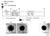
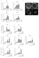
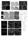
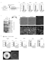
- FIG. 1 Schematic diagram of the method for inducing NCC clusters from iPS cells and micrographs of cell clusters on days 1 (D1), 2 (D2), and 5 (D5) after the start of culture.
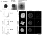
- D1 Micrograph of cell clumps induced under 3D conditions from the 1231A3 iPS cell line. Cell clumps with uniform morphology and diameters of approximately 500 ⁇ m were induced.
- B The cell clumps were dispersed into single cells using Accutase, and the rate of strongly CD271 positive cells was analyzed by flow cytometry. Strongly CD271 positive cells could be induced with high efficiency.
- C Histological image of fluorescent immunostaining of the cell clumps.
- the majority of the cells within the cell clumps were composed of CD271 and SOX10 positive NCCs. Changes in CD271 and SOX10 expression over time from seeding of iPS cells to obtaining NCC clusters. From day 4 of induction (day 2 from the start of culture in a medium containing SB431542 (hereinafter sometimes abbreviated as "SB”) and CHIR99021 (hereinafter sometimes abbreviated as "CHIR”)), SOX10- and CD271-positive NCCs began to appear.
- SB SB
- CHIR99021 hereinafter sometimes abbreviated as "CHIR”
- Non-Patent Document 3 Comparison of NCC-related gene expression in NCC induced under conventional 2D (adherent culture) conditions (Non-Patent Document 3) (outline of induction method is shown at the top of the figure) and 3D-induced NCC clusters (outline of induction method is shown in the center of the figure) using reverse transcription quantitative PCR (RT-qPCR). All NCC-related genes (NGFR, SOX10, TFAP2A, PAX3) were expressed to the same extent, and the pluripotent cell marker POU5F1 (Oct4) disappeared.
- NCC-related genes TFAP2A and PAX3
- neural plate border marker genes DLX5 and MSX2
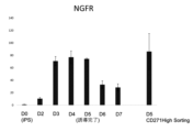
- RT-qPCR RT-qPCR in cell clumps on each day from day 0 (D0) to day 5 (D5) after the start of cell culture, and in cell clumps (D5 271H and 2D 271H) in which strongly CD271 positive cells were selected by flow cytometry. It was shown that neural plate border-related markers were elevated at D3, and that D5 cells expressed NCC-related genes to approximately the same extent as D5 271H cells and 2D 271H cells.
- NCC-related genes NGFR, SOX10
- Oct4 pluripotent cell marker Oct4
- RT-qPCR cell clumps on each day from day 0 (D0) to day 5 (D5) after the start of cell culture, and in cell clumps (D5 271H and 2D 271H) in which strongly CD271 positive cells were selected by flow cytometry. It was shown that D5 cells expressed similar levels of NCC-related genes to D5 271H cells, and also to 2D 271H cells.
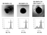
- SB and BMP action step the duration of the action step (hereinafter “SB and BMP action step") was varied, and the rate of strongly CD271 positive cells was measured by flow cytometry (the period of shaking culture was fixed).
- A When the action time of SB and BMP4 was set to 1 day, NCCs were induced with high efficiency.
- SB and BMP4 When SB and BMP4 were not applied, a clear decrease in the rate of strongly CD271 positive cells was observed. When the action time of SB and BMP4 was extended, a decrease in the rate of strongly CD271 positive cells was observed.
- Shaking culture was performed from the beginning of the NCC induction process.
- the rate of strongly CD271 positive cells was measured by flow cytometry under each condition in Figure 12.
- SB BMP4 6h When examined by flow cytometry on the 5th day after the action of SB and BMP4 for 6 hours (SB BMP4 6h) or 12 hours (SB BMP4 12h), it was shown that the efficiency of NCC induction was reduced compared to when SB and BMP4 were administered for 1 day (SB BMP4 D1).
- SB BMP4 D1 The results of measuring the rate of strongly CD271 positive cells by flow cytometry for each condition in Figure 12.
- the percentage of strongly CD271 positive cells was measured by flow cytometry when the duration of SB and CHIR treatment was extended.
- the percentage of strongly CD271 positive cells decreased in a time-dependent manner as the overall culture period was extended.
- the rate of strongly CD271 positive cells was measured by flow cytometry for each condition in Figure 12.
- NCCs were able to be induced on the 5th day when SB and BMP4 were applied for 6 hours (SB BMP4 6h) and 12 hours (SB BMP4 12h). However, when applied for 12 hours, induction of NCCs was as efficient as when SB and BMP4 were applied for 1 day (SB BMP4 D1).
- CD271 NCC marker
- E-Cadherin epithelial cell marker
- FIG. 1 shows an outline of a method that is preferable in terms of NCC induction efficiency when shaking culture is performed from the start of the SB and BMP4 action steps.
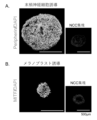
- Peripheral nerve cells and melanoblasts were induced from NCC clusters, and differentiation was confirmed by fluorescent immunostaining.
- A Peripheral nerve cells were induced from NCC clusters. Peripherin-positive peripheral nerve cells were induced with high efficiency.
- B Melanoblasts were induced from NCC clusters. MITF-positive melanoblasts were induced with high efficiency.
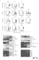
- HOXA2, HOXA3 Expression of HOX genes (HOXA2, HOXA3) was confirmed by RT-qPCR. RA-stimulated NCC clusters highly expressed HOX genes, whereas no HOX gene expression was observed in unstimulated NCC clusters.
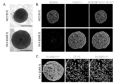
- A Microscopic images of NCC clusters and induced PA1-EM cell clusters. The size of the clusters increased due to proliferation of internal cells.
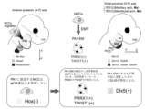
- FIG. 36 Schematic diagram of induction method to verify the best combination of Fgf8, Edn-1, and BMP4 for PA1-EM cell induction.
- the expression levels of DLX, PRRX1, and TWIST1 were measured by RT-qPCR under each condition in Figure 36.
- Edn-1 is essential for DLX5 expression
- the expression levels of DLX5, PRRX1, and TWIST1 indicate that adding Fgf8, Edn-1, and BMP4 to the medium is the optimal condition for PA1-EM cell induction.
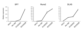
- FIG. 41 Schematic diagram of the method for inducing jaw bone-like tissue from distal-abral oral cells. Osteoprogenitor cells were induced by inducing osteogenic differentiation in PA1-EM cell clusters induced to distal-abdominal cells.
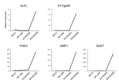
- A Increased expression of osteoprogenitor cell markers SP7, RUNX2, and DLX5 was confirmed.
- B At this induction stage, no increase in expression was confirmed for the early osteoblast marker ALPL and the immature osteocyte marker E11/gp38. Following the induction shown in Figure 41, two-stage osteogenic differentiation induction was performed. Further increases in the expression of osteoprogenitor cell markers SP7, RUNX2, and DLX5 were confirmed.
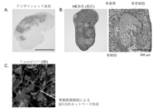
- the osteocyte-like cells formed a three-dimensional network with surrounding cells within the bone matrix.
- the human jawbone-like tissue in Figure 44 was transplanted into a 1.6 mm bone defect created in the mandibular ramus of the right mandibular angle of the lower jaw of an immunodeficient NSG mouse, and micro-CT images were taken immediately after transplantation, and at 1, 2, 3, and 4 weeks to examine the regenerative effect of the jawbone-like tissue.
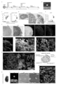
- the transplanted jawbone-like tissue sealing of the defect due to mineral deposition was confirmed as early as 3 weeks after transplantation. Efficient induction of HOX-negative NCCs from 3D-cultured iPSCs.
- NCCs head neural crest cells
- PA1-3 pharyngeal arches 1-3.
- NCC markers SOX10, NGFR, TFAP2A, RHOB, PAX3
- POU5F1 pluripotent stem cell marker
- RT-qPCR real-time quantitative PCR
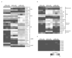
- b Flow cytometry analysis of NCC marker (CD271) in day 5 aggregates. Results are expressed as mean ⁇ standard error (six biologically independent experiments). Stepwise differentiation of iPSCs to NCCs via ectodermal lineages. a; RNA-seq analysis of 1231A3 iPSCs (d0), day 1-5 aggregates, CD271 high+ NCCs sorted from day 5 aggregates (d5 271H), and CD271 high+ NCCs sorted from a conventional two-dimensional (2D) protocol (2D 271H) (Non-Patent Document 3).
- 2D two-dimensional
- Heatmaps show gene expression levels of pluripotent stem cell (PSC) markers, ectodermal lineage (neurectoderm (NE), neural plate border (NPB), neural crest cells (NCC), and epidermal ectoderm (SE)) markers, primitive streak (PS) markers, presomitic mesoderm (PSM) markers, lateral plate mesoderm (LPM) markers, and endoderm (END) markers.
- PSC pluripotent stem cell
- NE neuropotent stem cell
- NPB neural plate border
- SE epidermal ectoderm
- SE epidermal ectoderm
- PS primitive streak
- PSM presomitic mesoderm
- LPM lateral plate mesoderm
- END endoderm
- SOST mature osteocyte markers
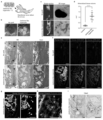
- b Safranin O/Fast Green staining and immunofluorescent staining for COLX in mdEM and clusters at day 38. Nuclei were stained with DAPI. Aggregates after chondrogenic induction served as positive controls for Safranin O and COLX expression.
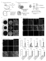
- HE staining and tricolor immunofluorescence staining for DLX2, DLX5, and HAND2 in mdEM and jawbone-like organoids generated from Ff-1 14s04 iPSCs (a), HLA-KO Ff-I 14s04 iPSCs (b), and Ff-I 01s04 (c) iPSCs. Nuclei were stained with DAPI. n 3 from three biologically independent experiments. Dashed boxes are enlarged to the right. Representative data are shown. Scale bar, 100 ⁇ m. Jaw-like organoids form bone tissue composed of human bone matrix proteins.
- HE staining e
- Azan staining e
- triple-color immunofluorescence staining for human COLI, OCN, OPN (f), and human vimentin (g) in serial sections (decalcified specimens) of transplanted tissues 4 weeks after transplantation.
- Nuclei were stained with DAPI (f, g).
- n 8. Dashed boxes are enlarged to the right (e, g).
- Black arrows indicate the edge of the defect (e).
- White and light grey arrows indicate donor human and host mouse cells, respectively (g).
- Scale bars 200 ⁇ m (e, f), 100 ⁇ m (g). h; Immunofluorescent staining for human vimentin in the transplanted tissue (decalcified specimen).
- OI-iPSC-derived mdEM and bone tissue formed under the kidney capsule of NSG mice Characterization of OI-iPSC-derived mdEM and bone tissue formed under the kidney capsule of NSG mice.
- a Tricolor immunofluorescence staining for DLX2, DLX5, and HAND2 in OI-iPSC- and resOI-iPSC-derived mdEM (d9). Scale bar, 100 ⁇ m.
- b Von Kossa and Safranin O/Fast Green staining, and immunofluorescence staining for human COLI and human OPN in OI-iPSC- and resOI-iPSC-derived bone tissue formed under the kidney capsule of NSG mice. Nuclei were stained with DAPI.
- n 3. Scale bar, 200 ⁇ m.
- b Immunostaining results for smooth muscle markers. Nuclei were stained with DAPI. High expression of Calponin and TAGLN was confirmed 10 days after smooth muscle cell induction.
- c Gene expression analysis of smooth muscle markers by RT-qPCR. High expression of smooth muscle markers (ACTA2, CNN1, TAGLN) and mature smooth muscle marker (MYH11) was confirmed 10 days after smooth muscle cell induction.
- pluripotent stem cells refer to stem cells that can differentiate into tissues and cells with various different morphologies and functions in the body, and have the ability to differentiate into cells of any of the three germ layers (endoderm, mesoderm, and ectoderm).
- pluripotent stem cells used in the present invention include induced pluripotent stem cells (iPS cells), embryonic stem cells (ES cells), embryonic stem cells derived from cloned embryos obtained by nuclear transfer (ntES cells), multipotent germline stem cells (mGS cells), and embryonic germline stem cells (EG cells), with iPS cells (more preferably human iPS cells) being preferred.
- the pluripotent stem cells are ES cells or any cells derived from a human embryo, the cells may be cells produced by destroying an embryo or cells produced without destroying an embryo, but from an ethical point of view, cells produced without destroying an embryo are preferable.
- nt ES cells are ES cells derived from cloned embryos produced by nuclear transfer technology and have almost the same properties as ES cells derived from fertilized eggs (Wakayama T. et al. (2001), Science, 292:740-743; S. Wakayama et al. (2005), Biol. Reprod., 72:932-936; Byrne J. et al. (2007), Nature, 450:497-502).
- nt ES (nuclear transfer ES) cells are ES cells established from the inner cell mass of blastocysts derived from cloned embryos obtained by replacing the nucleus of an unfertilized egg with the nucleus of a somatic cell.
- Nanog-iPSCs established by selecting using the expression of Nanog as an indicator after introducing the above four factors (Okita, K., Ichisaka, T., and Yamanaka, S. (2007). Nature 448, 313-317.); and iPSCs created by a method that does not include c-Myc (Nakagawa M, Yamanaka S., et al. Nature Biotechnology, (2008) 26, 101-106), iPSCs established by introducing six factors using a virus-free method (Okita K et al. Nat. Methods 2011 May;8(5):409-12, Okita K et al. Stem Cells. 31(3):458-66.), etc.
- induced pluripotent stem cells established by introducing four factors, OCT3/4, SOX2, NANOG, and LIN28, created by Thomson et al. (Yu J., Thomson JA. et al., Science (2007) 318: 1917-1920.), induced pluripotent stem cells created by Daley et al. (Park IH, Daley GQ. et al., Nature (2007) 451: 141-146), and induced pluripotent stem cells created by Sakurada et al. (JP Patent Publication No. 2008-307007) can also be used.
- the induced pluripotent stem cells used in the method for producing NCC of the present invention may be cells derived from patients with a genetic disease (e.g., osteogenesis imperfecta).
- a genetic disease e.g., osteogenesis imperfecta
- Cells induced to differentiate from pluripotent stem cells derived from patients with a genetic disease can serve as disease models that reflect the pathology of the disease, and are therefore suitable for screening therapeutic or preventive drugs for the disease.
- pluripotent stem cells derived from patients with a genetic disease can be differentiated into the desired cells or organoids after gene repair by genome editing using the CRISPR-Cas system, etc., and the cells can be used as a therapeutic drug for the disease.
- mGS cells are pluripotent stem cells derived from the testis and are the source of spermatogenesis. Like ES cells, these cells can be induced to differentiate into cells of various lineages, and have properties such as the ability to produce chimeric mice when transplanted into mouse blastocysts (Kanatsu-Shinohara M. et al. (2003) Biol. Reprod., 69:612-616; Shinohara K. et al. (2004), Cell, 119:1001-1012).
- GDNF glial cell line-derived neurotrophic factor
- EG cells are established from primordial germ cells during the fetal period and have pluripotency similar to that of ES cells. They can be established by culturing primordial germ cells in the presence of substances such as LIF, bFGF, and stem cell factor (Matsui Y. et al. (1992), Cell, 70:841-847; J.L. Resnick et al. (1992), Nature, 359:550-551).
- the species from which the pluripotent stem cells are derived is not particularly limited, and may be, for example, cells from rodents such as rats, mice, hamsters, and guinea pigs; lagomorphs such as rabbits; ungulates such as pigs, cows, goats, and sheep; felines such as dogs and cats; and primates such as humans, monkeys, rhesus monkeys, marmosets, orangutans, and chimpanzees.
- the preferred species is human.
- cell clump refers to a structure in which cells aggregate together to form a cluster, and is also called a cell aggregate.
- a neural crest cell (NCC) cluster refers to a cell clump that contains neural crest cells, but typically refers to a cell clump that contains mainly neural crest cells (e.g., at a ratio of 60% or more, 70% or more, or 80% or more).
- PA1-EM first pharyngeal arch ectodermal mesenchyme
- Bone morphogenetic proteins are secreted signaling molecules belonging to the TGF- ⁇ superfamily.
- Examples of bone morphogenetic proteins used in step (1) of the NCC production method of the present invention include BMP2, BMP3, BMP3b, BMP4, BMP5, BMP6, BMP7, BMP8, BMP9, BMP10, BMP11, BMP12, BMP13, BMP14, BMP15, BMP16, BMP17, and BMP18, with BMP4, BMP2, and BMP7 being preferred. Two or more types of BMPs may be used in combination.
- the concentration of BMP in the medium is adjusted appropriately depending on the type of BMP added, but is typically 1 to 50 ng/mL, preferably 2 to 30 ng/mL, and more preferably 2.5 to 10 ng/mL.
- the GSK-3 ⁇ inhibitors used in step (2) of the NCC production method of the present invention include, for example, CHIR98014 (2-[[2-[(5-nitro-6-aminopyridin-2-yl)amino]ethyl]amino]-4-(2,4-dichlorophenyl)-5-(1H-imidazol-1-yl)pyrimidine), CHIR99021 (6-[[2-[[4-(2,4-dichlorophenyl)-5-(4-methyl-1H-imidazol-2-yl)-2-pyrimidinyl]amino]ethyl]amino]nicotinonitrile), CP21R7 ( 3-(3-amino-phenyl)-4-(1-methyl-1H-indol-3-yl)-pyrrole-2,5-dione), LY2090314 (3-[9-Fluoro-1,2,3,4-tetrahydro-2-(1-piperid
- antisense oligonucleotides and siRNAs against GSK-3 ⁇ mRNA antibodies that bind to GSK-3 ⁇ , dominant negative GSK-3 ⁇ mutants, etc. can also be used as GSK-3 ⁇ inhibitors, and these are commercially available or can be synthesized according to known methods.
- the concentration of the GSK-3 ⁇ inhibitor in the medium is appropriately adjusted depending on the type of GSK-3 ⁇ inhibitor added, and is, for example, 0.01 to 20 ⁇ M, preferably 0.1 to 10 ⁇ M.
- the concentration is typically 0.1 to 10 ⁇ M, preferably 0.5 to 2 ⁇ M, more preferably 1 ⁇ M.
- the culture period can be shortened (e.g., FIG. 20), and by including a shaking culture step in step (2), the efficiency of induction into NCC can be improved (FIG. 9). Therefore, it is preferable that at least one of steps (1) and (2) includes a shaking culture step. Shaking culture may be performed throughout the entire period of each step, or only for a portion of the period.
- stirred culture may be performed instead of or in addition to shaking culture.
- Stirred culture can be performed in a culture device equipped with a stirrer or stirring blades (e.g., a bioreactor, etc.). The rotation speed of the stirrer or stirring blades when performing stirred culture is typically 10 to 100 rpm.
- the culture period in step (1) of the method for producing NCC of the present invention is typically 3 hours to 7 days, and from the viewpoint of the efficiency of induction into NCC, 6 hours to 6 days is preferable, 6 hours to 5 days is more preferable, 12 hours to 4 days is even more preferable, and 1 day to 3 days is even more preferable.
- the period of step (1) can be shortened, and for example, the culture period can be 6 hours to 3 days, 6 hours to 2 days, 12 hours to 1 day, etc.
- the culture period in step (2) of the method for producing NCC of the present invention is typically 12 hours to 5 days, and from the viewpoint of the efficiency of induction into NCC, 1 day to 6 days is preferable, 1 day to 4 days is more preferable, 2 days to 3 days is even more preferable, and 1 day to 3 days is even more preferable.
- the period can be appropriately changed depending on the period of step (1) of the method for producing NCC of the present invention, the presence or absence of shaking culture and/or agitation culture, etc.
- the culture period for steps (1) and (2) of the method for producing NCC of the present invention is typically 1 to 6 days, preferably 2 to 5 days, and more preferably 3 to 4 days.
- the period can be appropriately changed depending on the period of step (1) of the method for producing NCC of the present invention, the presence or absence of shaking culture and/or agitation culture, etc.
- a step of suspension culture of pluripotent stem cells in a medium containing a ROCK inhibitor may be carried out. This allows cell clumps of pluripotent stem cells to be formed.
- a step of preparing cell clumps of pluripotent stem cells may be carried out.
- the period of this step is not particularly limited, but is typically 12 hours to 3 days, and preferably 1 day. Therefore, when the method for producing NCC of the present invention includes a step of forming cell clumps of pluripotent stem cells, the period of the method for producing NCC of the present invention is typically 2 days to 7 days, preferably 3 days to 6 days, and more preferably 4 days to 5 days.
- ROCK inhibitors used in the present invention are not particularly limited as long as they are capable of suppressing the function of Rho-kinase (ROCK).
- examples of such inhibitors include Y-27632 (see, for example, Ishizaki et al., Mol. Pharmacol. 57, 976-983 (2000); Narumiya et al., Methods Enzymol. 325, 273-284 (2000)), fasudil/HA1077 (see, for example, Uenata et al., Nature 389: 990-994 (1997)), SR3677 (see, for example, Feng Y et al., J Med Chem.
- ROCK inhibitors include antisense nucleic acids against ROCK, RNA interference-inducing nucleic acids (e.g., siRNAs), dominant-negative mutants, and expression vectors thereof.
- RNA interference-inducing nucleic acids e.g., siRNAs
- other known low molecular weight compounds and their salts or derivatives can also be used as ROCK inhibitors (see, for example, U.S. Patent Application Publication Nos. 2005/0209261, 2005/0192304, 2004/0014755, 2004/0002508, 2004/0002507, 2003/0125344, 2003/0087919, and International Publication Nos. 2003/062227, 2003/059913, 2003/062225, 2002/076976, and 2004/039796).
- Y-27632 is preferred.
- the concentration of the ROCK inhibitor in the medium can be appropriately selected by those skilled in the art depending on the ROCK inhibitor used, but for example, when Y-27632 or a salt thereof (e.g., Y-27632 dihydrochloride, etc.) is used, the concentration is typically 0.1 ⁇ M to 100 ⁇ M, preferably 1 ⁇ M to 50 ⁇ M, more preferably 5 ⁇ M to 20 ⁇ M, and even more preferably 10 ⁇ M.
- NCCs may be selected (or isolated, concentrated, or purified) from the NCC clusters obtained in step (2) of the NCC production method of the present invention using the expression of NCC markers (e.g., high expression of CD271, etc.) as an indicator.
- NCCs can be selected using any known method, such as labeling cells with an antibody against an NCC marker such as CD271 and selecting them using flow cytometry or mass cytometry, magnetic cell separation, or an affinity column with an immobilized desired antigen.
- cells may be cultured under feeder-free and/or xeno-free conditions.
- all steps may be performed under feeder-free and xeno-free conditions.
- cells may be cultured under feeder-free and/or xeno-free conditions.
- feeder-free refers to a medium or culture condition that does not contain other cell types (i.e., feeder cells) that play a supporting role and are used to prepare the culture conditions for the cells to be cultured.
- xeno-free refers to a medium or culture condition that does not contain components derived from organisms different from the organism species of the cells to be cultured.
- RPMI-1640 medium EagleMEM (EMEM), Dulbecco's modified MEM, Glasgow's MEM (GMEM), ⁇ -MEM, 199 medium, IMDM, DMEM, Hybridoma Serum free medium, KnockOut TM DMEM, Advanced TM medium (e.g., Advanced MEM, Advanced RPMI, Advanced DMEM/F-12), Ham's Medium F-12, Ham's Medium F-10, Ham's Medium F12K, DMEM/F-12, ATCC-CRCM30, DM-160, DM-201, BME, Fischer, McCoy's 5A, Leibovitz's L-15, RITC80-7, MCDB105, MCDB107, MCDB131, MCDB153, MCDB201, NCTC109, NCTC135, Waymouth's Medium (e.g., Waymouth's Examples of suitable medium include, but are not limited
- the medium may contain one or more serum substitutes, such as, for example, Knockout Serum Replacement (KSR), N2 supplement (Invitrogen), B27 supplement (Invitrogen), albumin, transferrin, apotransferrin, fatty acids, insulin, collagen precursors, trace elements, 2-mercaptoethanol, 3'-thiolglycerol, and may also contain one or more substances, such as lipids, amino acids, L-glutamine, Glutamax (Invitrogen), non-essential amino acids, vitamins, growth factors, small molecules, antibiotics, antioxidants, pyruvate, buffers, inorganic salts, selenate, progesterone, and putrescine.
- KSR Knockout Serum Replacement
- N2 supplement Invitrogen
- B27 supplement Invitrogen
- albumin transferrin
- apotransferrin fatty acids
- insulin insulin
- collagen precursors insulin
- trace elements 2-mercaptoethanol
- 2-mercaptoethanol 3'-thiolgly
- step (2) of the method for producing an NCC of the present invention an NCC derived from a pluripotent stem cell is obtained.
- an NCC obtained (or obtained) by the method for producing an NCC of the present invention (hereinafter, sometimes referred to as "NCC of the present invention") is also provided.
- the NCC of the present invention typically has all of the following characteristics (A) to (C).
- C having the ability to differentiate into PA1-EM cells (at least mandibular PA1-EM cells).
- the present invention also provides an NCC having all of the above characteristics (A) to (C), and preferably further having at least one (preferably all) of the above characteristics (D) to (F).
- an NCC may be obtained by the method for producing an NCC of the present invention, or may be obtained by another method.
- the above NCC may also be referred to as the "NCC of the present invention.”
- the terms "expressing” or “positive” a gene are used to mean at least "production of mRNA encoded by the gene," but preferably also “production of protein encoded by the mRNA.” Therefore, if the production of mRNA encoded by the gene is detected at least by the method described in the Examples below (RT-qPCR), the gene can be said to be expressed. On the other hand, if the production of mRNA encoded by the gene is not detected (i.e., below the detection limit) by the method described in the Examples below (RT-qPCR), is at background levels, or is 1/100th or less of the positive control, the gene can be said to be not expressed or to be negative.
- High expression includes high expression (also referred to as “strong positive”).
- High expression may be expressed as High or Bright.
- CD271 highly expressing cells may be expressed as CD271 High+ cells or CD271 Bright+ cells.
- High expression can be determined based on a chart obtained by flow cytometry. The position on the chart may vary depending on the voltage setting of the instrument, the sensitivity setting, the antibody clone used, the staining conditions, the dye used, etc., but a person skilled in the art can appropriately draw a line so as not to separate a cell population recognized as a group in the obtained chart.
- PA1-EM cells are divided into maxillary PA1-EM (mxEM) and mandibular PA1-EM (mdEX), and unless otherwise specified in this specification, PA-EM includes both maxillary PA1-EM and mandibular PA-EM, although mandibular PA1-EM in particular is often intended.
- the cells obtained by the differentiation induction method of the present invention may be undifferentiated cells such as stem cells or precursor cells, or may be terminally differentiated cells.
- undifferentiated cells may be used as a term that includes both undifferentiated cells and terminally differentiated cells.
- undifferentiated cells refers to cells that have not reached terminal differentiation in the cell lineage, and examples of undifferentiated cells include stem cells and precursor cells excluding pluripotent stem cells.
- stem cells or precursor cells include neural stem cells, glial precursor cells, melanocyte stem cells, melanoblasts, mesenchymal stem cells, first pharyngeal arch ectodermal mesenchymal cells, cardiac stem cells, vascular endothelial precursor cells, vascular pericytes, skeletal muscle stem cells, adipose stem cells, bone precursor cells, osteoblasts, chondroblasts, etc.
- the bone organoid may exhibit a phenotype of a bone disease (e.g., osteogenesis imperfecta).
- bone organoids have a bone matrix, osteocytes contained in the bone matrix, and a layer of osteoblasts surrounding the bone matrix.
- Bone organoids typically have a three-dimensional network formed by bone cells connecting to each other through proteins related to the cytoskeleton, such as F-actin, and have the ability to repair the defect when transplanted into a bone defect.
- the "bone matrix” is an accumulation of substances produced by bone cells and secreted outside the cells, such as calcium phosphate, which contains proteins such as osteocalcin, osteopontin, osteonectin, and collagen.
- NCCs may be expanded. Expansion can be carried out, for example, by the method described in Patent Document 1. Specifically, the expansion can be carried out by floating culture of neural crest cells in a medium containing a GSK-3 ⁇ inhibitor and basic fibroblast growth factor (bFGF).
- the GSK-3 ⁇ inhibitor used for the expansion of NCCs can be the same as that used in the method for producing NCCs of the present invention, and is preferably SB431542.
- the bFGF (also called FGF-2) used for the expansion of NCCs can be a commercially available product from Fujifilm Wako Pure Chemical Industries, Ltd., etc.
- the medium used for the expansion may contain an ALK inhibitor or EGF (Epidermal Growth Factor).
- the ALK inhibitor can be the same as that used in the method for producing NCCs of the present invention.
- MSCs can also be obtained by culturing neural crest cells in a mesenchymal stem cell proliferation medium for about 14 days by adhesion culture.
- a mesenchymal stem cell proliferation medium for about 14 days by adhesion culture.
- a basal medium containing bovine serum e.g., ⁇ MEM medium, etc.
- a commercially available xeno-free mesenchymal stem cell proliferation medium is preferred.
- chondrocyte differentiation induction medium examples include a basal medium (e.g., DMEM/F12) containing 1% (v/v) ITS+premix, 0.17 mM AA2P, 0.35 mM proline, 0.1 mM dexamethasone, 0.15% (v/v) glucose, 1 mM sodium pyruvate, 2 mM GlutaMAX, 40 ng/ml PDGF-BB, 100 ng/mL TGF- ⁇ 3, 10 ng/ml BMP4, and 1% (v/v) FBS, and a commercially available xeno-free chondrocyte differentiation induction medium (e.g., MSCgo TM Chondrogenic XF (Sartorius)).
- a basal medium e.g., DMEM/F12
- ITS+premix 0.17 mM AA2P
- 0.35 mM proline 0.35 mM proline
- differentiation method into chondrocytes include a method in which 5 ⁇ l of mesenchymal stem cell suspension is spotted on a fibronectin-coated plate and cultured for 1 hour, and 1 ml of the above differentiation induction medium is added after 1 hour, and the cells are cultured for 14 days.
- the differentiation of chondrocytes may be confirmed by Alcian blue staining.
- Neural progenitor cells and neurons can also be obtained by seeding NCCs into a low cell adhesion culture vessel (e.g., 96-well V-bottom ultra-low adhesion culture plate (manufactured by Sumitomo Bakelite Co., Ltd.)) and culturing them for approximately 14 days in a basal medium (e.g., Neurobasal medium, etc.) supplemented with B27 Supplement, N-2 supplement, L-glutamine, BDNF, GDNF, NT-3, and NGF, or by culturing NCCs by adhesion culture in a basal medium (e.g., DMEM/F12, etc.) supplemented with N-2 Supplement, BDNF, GDNF, NT-3, and NGF for approximately 14 days.
- a basal medium e.g., Neurobasal medium, etc.
- a basal medium e.g., Neurobasal medium, etc.
- N-2 e.g., L-glutamine
- PA1-EM cell induction mandibular PA1-EM cells from the NCCs of the present invention
- a medium e.g., StemFit Basic03 medium
- the endothelin used for PA1-EM cell induction may be any of endothelin-1 (Edn-1), endothelin-2 (Edn-2), and endothelin-3 (Edn-3), with Edn-1 being preferred. These endothelins may also be used in combination.
- the concentration of endothelin in the medium can be appropriately selected by those skilled in the art depending on the endothelin used, but for example, when Edn-1 is used, it is typically 1 to 500 nM, preferably 10 to 300 nM, and more preferably 20 to 100 nM (e.g., 50 nM).
- maxillary PA1-EM cells can also be induced by using an Edn-1 inhibitor instead of endothelin in the PA1-EM cell induction method, or by culturing NCCs without including any of Edn-1, Edn-2, and Edn-3 in the medium (in the absence of these substances).
- Fibroblast growth factors (FGFs) used for PA1-EM cell induction can be commercially available products from Fujifilm Wako Pure Chemical Industries, Ltd., etc. Twenty-two types of FGFs are known in humans, and any can be used as long as they function as a cell growth factor, but in the case of the induction of PA1-EM cells from NCCs of the present invention, FGF-8 is preferred. In humans, FFG-8a, FGF-8b, FGF-8e, and FGF-8F are known as isoforms that have the function of FGF-8, and any of them can be used, but FGF-8b is preferred. Two or more types of FGFs may be used in combination.
- the concentration of FGF in the culture medium can be appropriately selected by those skilled in the art depending on the FGF used, but for example, when FGF-8 is used, it is typically 1 to 500 ng/ml, preferably 10 to 300 ng/ml, and more preferably 50 to 200 ng/ml (e.g., 100 ng/ml).
- the medium used during PA1-EM cell induction contains BMP.
- BMP include BMP2, BMP3, BMP3b, BMP4, BMP5, BMP6, BMP7, BMP8, BMP9, BMP10, BMP11, BMP12, BMP13, BMP14, BMP15, BMP16, BMP17, and BMP18, with BMP4, BMP2, and BMP7 being preferred, and BMP4 being particularly preferred.
- BMP2 BMP3, BMP3b, BMP4, BMP5, BMP6, BMP7, BMP8, BMP9, BMP10, BMP11, BMP12, BMP13, BMP14, BMP15, BMP16, BMP17, and BMP18, with BMP4, BMP2, and BMP7 being preferred, and BMP4 being particularly preferred.
- BMP4 is particularly preferred.
- Two or more types of BMP may be used in combination.
- a PA1-EM cell (hereinafter also referred to as "the PA1-EM cell of the present invention").
- the PA1-EM cell is a mandibular PA-EM cell, and typically has all of the following characteristics (a) to (c): (a) derived from neural crest cells that do not express HOX; (b) expressing DLX5, PRRX1 and TWIST1; (c) responding to epithelial signals and capable of inducing patterning.
- the PA1-EM cells may be obtained by the PA1-EM cell induction method described above, or may be obtained by another method.
- the PA1-EM cells of the present invention may further have the characteristic (d) of having actin filaments (also called "F-actin” or "actin stress fibers”).
- PA1-EM cells having the above characteristics (a) and (c), as well as the characteristic (b') of expressing POU3F3 and CYP26A1 (and may further have the above characteristic (d)) can be said to be maxillary PA-EM cells.
- the PA1-EM cells of the present invention are typically in the form of a cell cluster.
- the PA1-EM cell cluster has a surface region (surface zone), a central region (central zone), and an intermediate region (intermediate zone) located between the surface zone and the central zone.
- the central zone may contain DLX2 single positive cells
- the intermediate zone may contain DLX2 + DLX5 + cells
- the surface zone may contain DLX2 + DLX5 + HAND2 + cells.
- a cell cluster (mandibular PA1-EM cell cluster) which includes a central zone containing DLX2 single positive cells, an intermediate zone containing DLX2 + DLX5 + cells, and a surface zone containing DLX2 + DLX5 + HAND2 + cells.
- not expressing HOX means that at least HOXA2 and HOXA3 are not expressed. Cells that do not express HOX can typically be produced by producing the NCC of the present invention.
- epithelial signal means an epithelial signal expressed in the first pharyngeal arch that develops temporarily during the fetal stage of vertebrates.
- major epithelial signals include a combination of Fgf8 and Sonic hedgehog (Shh), which is a proximal-oral signal, a combination of Shh and BMP4, a combination of Fgf8 and Shh, and a combination of Edn-1 and BMP4, which are distal-oral signals, a combination of Edn-1 and BMP4, which is a distal-aboral signal, and End-1, which is a proximal-aboral signal (see also Figure 39A).
- Shh Sonic hedgehog
- BMP4 a combination of Fgf8 and Shh
- Edn-1 and BMP4 which are distal-oral signals
- Edn-1 and BMP4 which is a distal-aboral signal
- End-1 which is a proximal-aboral signal
- actin filaments are filamentous polymers formed by the polymerization of multiple globular actins (G-actins), and the presence of actin filaments can be confirmed by phalloidin staining as described below.
- the NCC used in the production of the PA1-EM cells of the present invention is not particularly limited.
- a method for producing PA1-EM cells which includes a step of inducing differentiation of NCC (in one embodiment, NCC cultured under two-dimensional conditions) into PA1-EM cells in the presence of an actin filament formation inhibitor.
- actin filament formation inhibitors used in the present invention include Y-27632, cytochalasin A, cytochalasin B, cytochalasin C, cytochalasin D, cytochalasin E, latrunculin A, latrunculin B, wiscostatin, BDM, latrunculin, and chaetoglobosin A.
- concentration of the actin filament formation inhibitor in the medium can be appropriately selected by those skilled in the art depending on the actin filament formation inhibitor used.
- the concentration is typically 0.5 ⁇ M to 500 ⁇ M, preferably 2 ⁇ M to 200 ⁇ M, and more preferably 10 ⁇ M to 100 ⁇ M (50 ⁇ M in one embodiment).
- Two or more types of actin filament formation inhibitors may be used in combination.
- PA1-EM cells for example, by culturing PA1-EM cells for about 3 days in a basal medium (e.g., StemFit Basic03 medium, etc.) supplemented with Fgf8b, SAG, BQ-123, and LDN193189, if the cells express PAX9 and LHX8, it can be evaluated that the PA1-EM cells respond to epithelial signals on the proximal-oral side and that patterning can be induced.
- a basal medium e.g., StemFit Basic03 medium, etc.
- PA1-EM cells for example, by culturing PA1-EM cells for about 3 days in a basal medium (e.g., StemFit Basic03 medium, etc.) supplemented with Fgf2, Endothelin-1, and BMP4, if the cells express GSC and MSX2, it can be evaluated that the PA1-EM cells respond to epithelial signals on the distal-abral side and that patterning can be induced.
- a basal medium e.g., StemFit Basic03 medium, etc.
- Fgf2 Fgf2, Endothelin-1, and BMP4
- proximal-oral side cells induced in this manner that express PAX9 and LHX8, as well as the distal-contra-oral side cells that express GSC and MSX2 can be further induced to differentiate.
- odontogenic mesenchymal cells can be induced by culturing the proximal-oral side cells for about 4 days in a basal medium (e.g., Basic03, etc.) supplemented with FGF8b, SAG, CHIR99021, and activin A (R&D). Since odontogenic mesenchymal cells can be the starting cells for dentin, which constitutes the main part of teeth, the PA1-EM cells and odontogenic mesenchymal cells of the present invention may also be useful in tooth regeneration.
- the above step (III) is carried out by subjecting the bone precursor cells to suspension culture in a bone cell differentiation induction medium (e.g., MSCgo TM Osteogenic XF Medium, etc.) for 20 to 30 days.
- a bone cell differentiation induction medium e.g., MSCgo TM Osteogenic XF Medium, etc.
- the medium may be replaced with another bone cell differentiation-inducing medium (e.g., StemFit Basic03 medium containing dexamethasone (usually used at 1/10 of the concentration used in osteoblast induction), ⁇ -glycerophosphate, and ascorbic acid) during the process (e.g., somewhere between 10th and 20th days from the start of the step (III), somewhere between 6th and 10th days, etc.).
- another bone cell differentiation-inducing medium e.g., StemFit Basic03 medium containing dexamethasone (usually used at 1/10 of the concentration used in osteoblast induction), ⁇ -glycerophosphate, and
- the first half of the process is aimed at inducing differentiation mainly into osteoblasts and osteocytes
- the second half of the process is aimed at maturation of osteocytes.
- This can suppress the occurrence of dead cells in the maturation of osteocytes.
- the medium when the medium is replaced with another bone cell differentiation-inducing medium during the process (III), the medium may be replaced with another medium (e.g., the bone cell differentiation-inducing medium used at the beginning of the step (III)) several days after the replacement (e.g., 6th to 10th days). Specific methods are outlined in FIG. 40 and FIG. 59, but the present invention is not limited to these methods.
- the bone organoids produced by the above method may be further matured.
- the bone organoids may be transplanted into a living body such as a mammal (e.g., transplanted under the renal capsule).
- transplanting the bone organoids into a living body allows the formation of calcifications and dense bone tissue, and the bone tissue can mature into bone organoids in which a dense network of donor bone cells is formed inside the bone tissue.
- Bone organoids can also be produced using MSCs by the method described in WO 2020/226043. Specifically, bone organoids can be produced by growing MSCs in adhesion culture to form a sheet (cell sheet), detaching the cell sheet from the culture vessel, and then performing suspension culture on the cell sheet to obtain a three-dimensional mesenchymal stem cell cluster, and culturing the cell cluster embedded in a gel using a bone differentiation induction medium.
- the gel is preferably a gel whose main component is polysaccharide, and for example, Vitrogel (The Well Bioscience) can be used.
- Neural organoids can be produced by culturing cell clumps containing neural precursor cells induced from NCCs in a suspension culture medium for inducing neural differentiation. By continuing to culture the cell clumps under the suspension culture conditions, neural organoids can be induced in a self-organizing manner.
- Examples of such neuronal differentiation-inducing media include basal media (e.g., N2 medium, etc.) containing a Wnt signal inhibitor and an ALK inhibitor.
- Wnt signal inhibitors examples include DKK1 protein, sclerostin, IWR-1e, IWP-2, IWP-3, IWP-4, IWP-L6, C59, ICG-001, FH535, WIKI4, KYO2111, PNU-74654, XAV939, and derivatives thereof.
- ALK inhibitors include those similar to those mentioned above.
- Culture vessels used for adhesion culture include those whose surfaces have been artificially treated to improve adhesion to cells (e.g., coating with basement membrane preparations, fibronectin, laminin or fragments thereof, entactin, collagen, gelatin, synthemax, vitronectin, or other extracellular matrices, or with polymers such as polylysine and polyornithine, or surface treatments such as positive charge treatment).
- culture vessels coated with fibronectin are preferred.
- the differentiation induction method of the present invention may involve culturing under feeder-free conditions and/or xeno-free conditions for all or part of the period. From the perspective of clinical use, it is preferable that the differentiation induction method of the present invention is carried out under feeder-free and xeno-free conditions for the entire period.
- the differentiation induction method of the present invention may include a step of recovering the obtained target cells or organoids.
- the recovered cells may be cryopreserved using a cell cryopreservation solution.
- the cells recovered in the container may be counted using a cell counter, or may be labeled with an antibody against a cell surface marker and selected by flow cytometry, mass cytometry, magnetic cell separation, or the like.
- a cell or organoid obtained by the differentiation induction method of the present invention (hereinafter, also referred to as the “differentiated cell or organoid of the present invention") is provided.
- the culture conditions and methods such as suspension culture, shaking culture, and agitation culture, the method for selecting the target cells, additives to the medium, specific examples and definitions of the culture vessels, etc., in the differentiation induction method of the present invention are all those described in "1. Method for producing neural crest cells" above.
- the differentiated cells or organoids of the present invention can be suitably used in regenerative medicine, and therefore, in another embodiment, a transplantation therapy agent (hereinafter, sometimes referred to as the "transplantation therapy agent of the present invention") containing the differentiated cells or organoids of the present invention is provided.
- a transplantation therapy agent hereinafter, sometimes referred to as the "transplantation therapy agent of the present invention"
- the present invention also includes a method for treating or preventing damage (including defects) or disease of tissues (e.g., bone tissue, cartilage tissue, adipose tissue, muscle tissue (e.g., skeletal muscle tissue, etc.), in which an effective amount of the differentiated cells or organoids of the present invention is administered or transplanted into a mammalian subject of treatment (e.g., human, mouse, rat, monkey, cow, horse, pig, dog, etc.).
- a mammalian subject of treatment e.g., human, mouse, rat, monkey, cow, horse, pig, dog, etc.
- treatment of tissue damage also includes regeneration of damaged tissue. Regeneration of damaged tissue includes not only complete tissue regeneration, but also cases in which a tissue-like structure is formed. For example, in the case of bone tissue, it is not necessary to determine whether the regenerated site is bone tissue in a CT (Computed Tomography) examination.
- CT Computer Tomography
- the transplantation therapy agent of the present invention can be administered or transplanted into a living body of a subject in need thereof.
- the transplantation is preferably performed in a region of the living body where the differentiated cells or organoids of the present invention can be fixed at a certain position, for example, subcutaneously, intraperitoneally, peritoneal epithelium, bone tissue (e.g., jaw bone tissue, etc.), cartilage tissue, omentum, adipose tissue, muscle tissue, or under the capsule of each organ such as the pancreas or kidney.
- the cells or organoids to be transplanted only need to be administered in a therapeutically effective amount, which may vary depending on factors such as the age, weight, size of the transplant site, and severity of the disease of the transplant subject, and are not particularly limited, but can be, for example, about 10 x 10 4 cells to 10 x 10 11 cells.
- the purpose of transplanting the differentiated cells or organoids of the present invention into a living body may be to directly regenerate damaged tissue, or to obtain an indirect effect (e.g., paracrine effect, etc.) due to factors secreted by the differentiated cells or organoids of the present invention.
- Mesenchymal stem cells can be effective in treating diseases such as acute myocardial infarction, stroke, multiple system atrophy (MSA), graft-versus-host disease, Crohn's disease, ischemic cardiomyopathy, and spinal cord injury.
- Melanoblasts and melanocytes can be effective in treating diseases such as oculocutaneous albinism and pigmentation disorders such as vitiligo vulgaris.
- Bone cells and bone organoids can be effective in treating diseases such as intractable fractures, periodontitis, or extensive bone defects after osteotomy.
- Neural progenitor cells, neural cells, or neural organoids can be effective in treating diseases such as cerebrovascular disorders, brain injuries, spinal cord injuries, and neurodegenerative diseases.
- the differentiated cells or organoids of the present invention are used as transplantation therapy agents, it is desirable to use cells or organoids derived from iPS cells established from somatic cells with the same or substantially the same HLA genotype of the recipient individual, from the viewpoint of preventing rejection reactions.
- substantially the same means that the HLA genotype matches the transplanted cells to such an extent that immune reactions can be suppressed with immunosuppressants, for example, somatic cells with HLA types that match the three loci HLA-A, HLA-B, and HLA-DR, or four loci including HLA-C. If sufficient cells cannot be obtained due to age, constitution, etc., they can also be transplanted in a state where they are embedded in capsules such as polyethylene glycol or silicone, or in porous containers, to avoid rejection reactions.
- the differentiated cells or organoids of the present invention are manufactured as parenteral preparations such as injections, suspensions, or drops by mixing with a medicamentically acceptable carrier according to conventional methods.
- a method for manufacturing a transplantation therapy agent which includes a step of formulating the differentiated cells or organoids of the present invention.
- Such a method may include a step of preparing the differentiated cells or organoids of the present invention.
- it may also include a step of preserving the differentiated cells or organoids of the present invention.
- pharma- ceutically acceptable carriers examples include aqueous solutions for injection, such as physiological saline, isotonic solutions containing glucose and other adjuvants (e.g., D-sorbitol, D-mannitol, sodium chloride, etc.).
- aqueous solutions for injection such as physiological saline, isotonic solutions containing glucose and other adjuvants (e.g., D-sorbitol, D-mannitol, sodium chloride, etc.).
- the cells of the present invention may be mixed with, for example, buffers (e.g., phosphate buffer, sodium acetate buffer), soothing agents (e.g., benzalkonium chloride, procaine hydrochloride, etc.), stabilizers (e.g., human serum albumin, polyethylene glycol, etc.), preservatives, antioxidants, etc.
- buffers e.g., phosphate buffer, sodium acetate buffer
- soothing agents e.g., benzalkon
- the transplantation therapy agent of the present invention is provided in a frozen state under conditions normally used for cryopreservation of cells, and can be thawed when needed.
- it may further contain serum or a substitute thereof, an organic solvent (e.g., DMSO), etc.
- an organic solvent e.g., DMSO
- the concentration of serum or a substitute thereof is not particularly limited, but may be about 1 to about 30% (v/v), preferably about 5 to about 20% (v/v).
- the concentration of the organic solvent is not particularly limited, but may be 0 to about 50% (v/v), preferably about 5 to about 20% (v/v).
- the differentiated cells or organoids of the present invention can also be used in a method for screening candidate drugs that are useful for treating or preventing tissue damage or disease.
- a method for screening therapeutic or preventive drugs for disease comprising a step of culturing the differentiated cells or organoids of the present invention in the presence or absence of a test substance.
- tissue damage or disease include those similar to the above-mentioned tissue damage or disease that is the therapeutic target of the transplantation therapy agent of the present invention.
- the differentiated cells or organoids used for screening may also exhibit a phenotype of a disease, for example, a bone disease such as osteogenesis imperfecta. In such a case, candidate drugs can also be screened based on the change in the phenotype.
- Test substances used in screening methods include, for example, cell extracts, cell culture supernatants, microbial fermentation products, extracts derived from marine organisms, plant extracts, purified or crude proteins, peptides, non-peptide compounds, synthetic low molecular weight compounds, and natural compounds.
- test substances can also be obtained using any of the many approaches in combinatorial library methods known in the art, including (1) biological libraries, (2) synthetic library methods using deconvolution, (3) "one-bead one-compound” library methods, and (4) synthetic library methods using affinity chromatography selection. While the biological library method using affinity chromatography selection is limited to peptide libraries, the other four approaches are applicable to small molecule libraries of peptides, non-peptide oligomers, or compounds (Lam (1997) Anticancer Drug Des. 12:145-67). Examples of methods for the synthesis of molecular libraries can be found in the art (DeWitt et al. (1993) Proc. Natl. Acad. Sci. USA 90:6909-13; Erb et al.
- Example 1 Induction of NCC clusters ⁇ Maintenance culture of human iPS cells> Human iPS cells (1231A3 line, Ff-I 14s04 line, Ff-I 14s04 HLA-KO line, iPS cell line derived from an osteogenesis imperfecta patient) generated using episomal vectors were cultured on culture plates coated with iMatrix-511 (Nippi) using StemFit AK03N medium (Ajinomoto). Medium was changed every 2 days.
- FIG. 1 An outline of the method for inducing iPS cells into NCC clusters (three-dimensional (3D) NCC induction method) is shown in Figure 1. Specifically, the procedure was as follows. Maintained human iPS cells (1231A3 line (xeno-free cell line for research)) were dispersed into single cells using Accutase (Innovative Cell Technologies), then suspended in a culture medium containing StemFit Basic03 (AK03N minus bFGF) (Ajinomoto) and 10 ⁇ M Y-27632 (FUJIFILM Wako), and seeded at 1.0 ⁇ 10 4 cells/100 ⁇ l/well on a 96-well V-bottom ultra-low attachment culture plate (Sumitomo Bakelite).
- AK03N minus bFGF StemFit Basic03
- FUJIFILM Wako 10 ⁇ M Y-27632
- the plate was centrifuged at 1200 rpm for 3 min to allow the cells to aggregate on the bottom of the plate. After culturing for one day, the cells aggregated and formed cell clusters. Next, the cell clumps were cultured for 1 day in a culture medium containing StemFit Basic03 medium supplemented with 10 ⁇ M SB431542 (Selleck) and 10 ng/mL BMP4 (R&D Systems) (this corresponds to steps D1 to D2 in Figure 1, and may be referred to below as the "SB and BMP action step").
- the cell clumps were transferred one by one to a 48-well low-attachment culture plate (Iwaki), and cultured at 120 rpm in a culture medium containing StemFit Basic03 medium supplemented with 10 ⁇ M SB431542 and 1 ⁇ M CHIR99021 (Axon Medchem) for 3 days using a shaking culture vessel (CS-LR (TAITEC)) (corresponding to steps D2 to D3 in Figure 1, hereinafter sometimes referred to as the "NCC induction step").
- CS-LR shaking culture vessel
- the percentage of CD271 positive cells in the obtained cell clumps was measured by flow cytometry.
- the cell clumps were dispersed into single cells using Accutase (Innovative Cell Technologies), and then CD271 antibody (BD Biosciences, 560326) was allowed to act in an antibody dilution solution diluted 200-fold with FACS buffer (PBS containing 0.1% human serum) at 4°C for 60 minutes. An isotype control was provided to remove non-specific background signals.
- the cells after antibody action were filtered through a 35 ⁇ m filter (Falcon) and analyzed by flow cytometry using FACS AriaII (BD Biosciences) to measure the percentage of strongly CD271 positive cells.
- FCM data was visualized using FlowJo software (BD Biosciences). Flow cytometry showed that the cell clumps contained a very high percentage of strongly CD271 positive cells, as high as 94.6% ( Figure 2B).
- the expression of CD271, SOX10, and PAX6 was confirmed for the cell clusters by immunostaining.
- the cell clusters were fixed with 4% paraformaldehyde-phosphate buffer and embedded in Tissue-Tek OCT compound (Sakura Finetech Japan). They were sliced thinly using a cryostat (Leica) to prepare 12 ⁇ m-thick frozen sections. After washing the sections with PBS, they were permeabilized with 3% TritonX-100 (Sigma) at room temperature for 15 minutes. They were blocked with Blocking one Histo (Nacalai Tesque) at room temperature for 1 hour.
- CD271 antibody Advanced Targeting System, AB-N07
- SOX10 antibody R&D, AF2864
- PAX6 antibody abcam, ab195945
- AB-N07 Advanced Targeting System
- SOX10 antibody R&D, AF2864
- PAX6 antibody abcam, ab195945
- Primary antibodies were allowed to react for 3 hours at room temperature in an antibody dilution solution diluted 200-fold with Can Get Signal TM immunostain Solution B.
- Nuclear staining was performed with 4',6-diamidino-2-phenylindole (DAPI) (Dojindo Laboratories) for 10 minutes at room temperature.
- DAPI 4',6-diamidino-2-phenylindole
- VECTASHIELD (registered trademark) The samples were mounted in Antifade Mounting Medium (VECTOR LABORATORIES). The fluorescent signals of the samples were observed using a confocal laser microscope Olympus FV3000 (Olympus). Immunostaining showed that the inside of the cell clusters was mainly composed of NCCs that were CD271 and SOX10 positive and PAX6 negative ( Figure 2C). Furthermore, the expression of CD271, SOX10, and PAX6 was confirmed by immunostaining for the cell clusters on each day from day 1 (D1) to day 5 (D5) after the start of cell culture. From D4 (2 days after the start of the NCC induction process), SOX10 and CD271 positive NCCs appeared, and the proportion of NCCs increased at D5 ( Figure 3).
- NCCs produced by conventional method top of Figure 4
- adherent culture two-dimensional (2D) NCC induction method
- 2D NCC induction method iPS cells were cultured for 10 days in a culture medium containing 10 ⁇ M SB431542 and 1 ⁇ M CHIR99021 using 6 wells coated with iMatrix-511, and NCCs were isolated from the resulting NCC clusters by selecting strongly CD271 positive cells using flow cytometry (the resulting NCCs are abbreviated as "2D iNCC" or "2D 271H”).
- NCCs were also isolated from the D5 NCC clusters by selecting strongly CD271 positive cells using flow cytometry (the resulting NCCs are abbreviated as "3D iNCC” or "D5 271H”) (center of Figure 4).
- NCC-related genes NGFR, SOX10, TFAP2A, and PAX3
- POU5F1 pluripotent cell marker POU5F1
- RT-qPCR RT-qPCR for each NCC.
- total RNA from the cell clusters was collected using RNeasy Mini Kit (Qiagen), and genomic DNA was removed using DNase-one Kit (Qiagen).
- Reverse transcription was performed using PrimeScript RT Master Mix (Takara) to synthesize single-stranded cDNA from 500 ng total RNA.
- the cDNA was combined with Thunderbird SYBR qPCR Mix (TOYOBO), and RT-qPCR was performed using StepOnePlus TM Real-Time PCR System (Applied Biosystems).
- NCC-related genes was observed to be comparable in both 2D iNCC and 3D iNCC, and it was shown that the expression of the pluripotent cell marker POU5F1 disappeared in both NCCs ( Figure 4, bottom).
- the primers used in RT-qPCR were the same as those listed in Supplementary Table 2 of Non-Patent Document 3.
- NCC-related genes NGFR, SOX10, TFAP2A, and PAX3
- neural plate border marker genes DLX5 and MSX2
- the pluripotent cell marker Oct4 were measured by RT-qPCR for cell aggregates (no selection of CD271-high expressing cells) on each day from day 0 (D0) to day 5 (D5) after the start of cell culture, as well as D5 271H and 2D 271H.
- Figures 5 shows that neural plate border-related markers increased one day after switching from the SB and BMP treatment process to the NCC induction process (D3).
- Figures 5 and 6 show that D5 cells express NCC-related genes to approximately the same extent as D5 271H cells, and that they also express NCC-related genes to approximately the same extent as 2D 271H cells.
- the primers used in RT-qPCR the base sequences of the primers other than those listed in Supplementary Table 2 of Non-Patent Document 3 are shown in Table 1.
- Table 1 also lists the base sequences of the primers used in examples other than this one (in the table, Fw indicates the forward primer and Rev indicates the reverse primer).
- NCC clusters were also obtained (Figure 8).
- the number of cells seeded and the concentration of BMP4 were adjusted for each cell line. Specifically, the Ff-I 14s04 line and the Ff-I 14s04 HLA-KO line were seeded at 3.0 ⁇ 10 3 cells/100 ⁇ l/well, and the BMP4 concentration was set to 2.5 ng/mL.
- the iPS cell line derived from a patient with osteogenesis imperfecta was seeded at 2.0 ⁇ 10 3 cells/100 ⁇ l/well, and the BMP4 concentration was set to 2.5 ng/mL.
- the culture was performed under the same conditions as the 3D NCC induction method. Specifically, the following procedure was performed. iPS cells were cultured in a culture medium containing 10 ⁇ M SB431542 and 1 ⁇ M CHIR99021 for 4 days (SB CHIR D4 in Figure 9) using 6-well plates coated with iMatrix-511, or the cells were cultured in a culture medium containing 10 ⁇ M SB431542 and 10 ng/mL BMP4 for 1 day, and then in a culture medium containing 10 ⁇ M SB431542 and 1 ⁇ M CHIR99021 for 3 days (SB BMP4 D1 SB CHIR D3 in Figure 9) to obtain cell clumps.
- Example 2 Verification of culture conditions for induction of NCC clusters ⁇ Verification of SB and BMP action process 1>
- the duration of SB431542 and BMP4 action was varied to examine the variation in the rate of strongly CD271 positive cells by flow cytometry.
- the expression patterns of the genes CD271 (NCC marker), PAX6 (neurectoderm marker), and E-Cadherin (epithelial cell marker) were examined by immunostaining. In this case, the period of shaking culture was fixed. The results are shown in Figures 10 and 11.
- FIG. 13 shows that when the SB and BMP application process was performed for 6 hours (SB BMP4 6h) or 12 hours (SB BMP4 12h), and then verified by flow cytometry on the 5th day, the NCC induction efficiency was lower than when the SB and BMP application process was performed for 1 day (SB BMP4 D1). This is presumably because not only NCC but also neuroectoderm was induced simultaneously due to the short duration of the SB and BMP application process.
- FIG. 14 shows that the NCC induction effect was high when the SB and BMP action steps were performed for 2 days (SB BMP4 D2) or 3 days (SB BMP4 D3) and then verified by flow cytometry on the 5th day.
- FIG. 15 shows that when the culture period was extended to 7 days, CD271 expression in the entire cell population decreased regardless of the duration of the SB and BMP action steps. This is presumably because differentiation of NCC into other cells progresses between the 5th and 7th days of induction.
- the results of RT-qPCR also showed that the expression level of NGFR (CD271) decreased after the 5th day of culture (FIG. 16).
- Figure 17 shows an example of a method pattern that is preferable in terms of NCC induction efficiency for the SB and BMP action steps.
- Example 3 Verification of differentiation potential of NCC The differentiation potential of the NCC obtained in Example 1 (the NCC induction method of the present invention) was verified. An overview is shown in Figure 24. Specifically, the differentiation potential of the NCC induced in Example 1 into peripheral nerve cells, melanoblasts, mesenchymal stem cells, and first pharyngeal arch ectodermal mesenchymal cells was verified.
- FIG. 25A An outline of this induction method is shown in FIG. 25A. Specifically, the method was carried out as follows.
- the NCC clumps obtained in Example 1 were transferred to a 96-well U-bottom ultra-low adhesion culture plate (manufactured by Sumitomo Bakelite Co., Ltd.) and cultured for 14 days in an induction medium containing Neurobasal medium (manufactured by Thermo Fisher Scientific Co., Ltd.) supplemented with B27 supplement (manufactured by Thermo Fisher Scientific Co., Ltd.), N-2 supplement (manufactured by Thermo Fisher Scientific Co., Ltd.), 2 mM L-glutamine (FUJIFILM Wako Co., Ltd.), 10 ng/mL BDNF (FUJIFILM Wako Co., Ltd.), GDNF (FUJIFILM Wako Co., Ltd.), NT-3 (FUJIFILM Wako Co.
- the NCC clusters obtained in Example 1 were transferred to a 96-well U-bottom ultra-low adhesion culture plate (Sumitomo Bakelite Co., Ltd.) and cultured for 6 days in induction medium containing StemFit Basic03 medium, 1 ⁇ M CHIR99021, 25 ng/mL BMP4, and 100 nM Endothelin-3 (TOCRIS). The medium was replaced on the third day of induction. Fluorescent immunostaining of MITF, a melanoblast marker, showed that the cells were differentiated into melanoblasts (Figure 26B).
- the second passage NCC was dispersed into single cells using Accutase, suspended in a culture medium containing 10 ⁇ M SB431542, 20 ng/mL Egf, and Fgf2 in StemFit Basic03 medium, and seeded on a fibronectin-coated culture plate at a cell concentration of 1 ⁇ 10 4 cells/cm 2.
- the medium was replaced with PRIME-XV MSC Expansion XSFM medium (Irvine Scientific) to start MSC induction.
- the cells were passaged every 4 days using Accutase, seeded on a culture plate at a density of 1 ⁇ 10 4 cells/cm 2 , and cultured.
- the expression of human MSC markers (CD105, CD90, CD73, CD44) was confirmed using flow cytometry, and it was shown that they had differentiated into MSCs ( Figure 28).
- first pharyngeal arch ectomesenchymal cells from NCC clusters detach from the dorsal protuberance of the neural tube and then migrate and differentiate into various organs in the developing embryo during neurogenesis, such as the first pharyngeal arch ectomesenchyme (PA1), second pharyngeal arch ectomesenchyme (PA2), third pharyngeal arch (PA3), frontonasal process (FNP), etc.
- PA1-EM first pharyngeal arch ectomesenchyme
- the characteristics of PA1-EM are outlined in Figure 29.
- HOX gene expression in the NCC clusters must be negative. Therefore, HOX gene expression was confirmed by RT-qPCR for the NCC clusters obtained in Example 1.
- NCC clusters in which HOX gene expression was induced by stimulation with retinoic acid (RA) were prepared. An overview of each method is shown in Figure 30A. HOX gene-positive NCC clusters were induced by culturing the cells in a medium containing 1.0 ⁇ M RA from the fourth day of induction. Specifically, the following procedure was followed.
- NCC-related genes NGFR, SOX10
- RT-qPCR The expression levels of NCC-related genes were the same even after RA stimulation ( Figure 30B).
- HOX genes HOXA2, HOXA3
- Fig. 8 shows the factors necessary for cell survival and proliferation, Edn-1 (Endothelin-1) as a factor necessary for mandibular selection, and BMP4 as a factor necessary for distal induction.
- Edn-1 Endothelin-1
- BMP4 BMP4 as a factor necessary for distal induction.
- NCC clusters were cultured in an induction medium consisting of StemFit Basic03 medium supplemented with 50 nM Endothelin-1 (Sigma), 100 ng/mL Fgf8b (FUJIFILM Wako), and 2.5 ng/mL BMP4 for 4 days in a shaking culture vessel.
- Fluorescent immunostaining images for DLX5 also showed that the induced ectomesenchymal cells highly expressed DLX5, reproducing the genetic characteristics of PA1-EM cells.
- PA1-EM cells were also successfully induced from iPS cell lines from patients with osteogenesis imperfecta ( Figure 33).
- NCCs were HOX negative for PA1-EM cell induction.
- PA1-EM cell induction was performed using NCCs induced without RA treatment (HOX(+) NCCs) and NCCs induced by RA treatment to express the HOX gene (HOX(-) NCCs).
- HOX(+) NCCs NCCs induced without RA treatment
- HOX(-) NCCs NCCs induced by RA treatment to express the HOX gene
- Example 4 Verification of the ability of PA1-EM cell clusters to induce patterning in the first pharyngeal arch ⁇ Induction into cells on the proximal-oral side of the first pharyngeal arch> It is known that patterning of PA1-EM cells is induced in response to epithelial signals (FIG. 39A). Therefore, we verified whether the PA1-EM cells obtained by the above-mentioned method have the ability to induce patterning in the first pharyngeal arch. The PA1-EM cell clusters were induced to become cells on the proximal-oral side (molar tooth forming region) of the first pharyngeal arch. An outline of the induction method is shown in FIG. 38. Specifically, the following procedure was performed.
- the PA1-EM cell clusters obtained in Example 3 were transferred to a 96-well U-bottom ultra-low attachment culture plate. To induce cells on the proximal-oral side of the first pharyngeal arch, the PA1-EM cell clusters were cultured for 3 days in an induction medium containing 100 ng/mL Fgf8b, 400 nM SAG (Selleck), 1.0 ⁇ M BQ-123 (Selleck), and 0.5 ⁇ M LDN193189 (TOCRIS) in StemFit Basic03 medium.
- an induction medium containing 100 ng/mL Fgf8b, 400 nM SAG (Selleck), 1.0 ⁇ M BQ-123 (Selleck), and 0.5 ⁇ M LDN193189 (TOCRIS) in StemFit Basic03 medium.
- RT-qPCR was performed for the proximal-oral markers PAX9 and LHX8 of the first pharyngeal arch, and the induction of the proximal-oral cells of the first pharyngeal arch was confirmed ( Figure 39B).
- the base sequences of the primers used for RT-qPCR are shown in Table 1.
- the PA1-EM cell clusters were induced to become cells on the distal-abdominal side of the first pharyngeal arch (the jaw bone ossification origin region).
- the outline of the induction method is shown in the lower part of FIG. 38.
- the PA1-EM cell clusters obtained in Example 3 were cultured for 3 days in an induction medium containing StemFit Basic03 medium, 20 ng/mL Fgf2, 50 nM Endothelin-1, and 10 ng/mL BMP4.
- RT-qPCR was performed for GSC and MSX2, which are markers for the distal-abdominal side of the first pharyngeal arch, and induction of cells on the distal-abdominal side of the first pharyngeal arch was confirmed (FIG. 39C).
- Example 5 Induction into jawbone-like tissue and transplantation of said tissue into mice ⁇ Induction of jawbone-like tissue from jawbone ossification origin cells>
- the PA1-EM cell cluster obtained in Example 3 was subjected to stepwise osteogenic differentiation induction to induce jawbone-like tissue.
- the outline of the induction method is shown in FIG. 40. Specifically, the PA1-EM cell cluster was first cultured for 3 days in an induction medium containing 20 ng/mL Egf, Fgf2, 50 ng/mL BMP2, 3 ⁇ M CHIR99021, and 400 nM SAG in StemFit Basic03 medium, and induced into osteoprogenitor cells.
- RT-qPCR for osteoprogenitor cell markers SP7, RUNX2, and DLX5 confirmed the induction of osteoprogenitor cells (FIG. 41A).
- RT-qPCR for the early osteoblast marker ALPL and the immature osteocyte marker E11/gp38 showed that differentiation had not progressed to osteoblasts or osteocytes (FIG. 41B).
- the cell aggregates were cultured in MSCgo Osteogenic SF,XF (Sartorius) for 16 days, and then cultured for 8 days in osteogenic differentiation-inducing medium consisting of StemFit Basic03 medium supplemented with 10 nM dexamethasone (Sigma), 10 mM ⁇ -glycerophosphate (Sigma), and 50 ⁇ g/ml ascorbic acid (Sigma).
- osteogenic differentiation-inducing medium consisting of StemFit Basic03 medium supplemented with 10 nM dexamethasone (Sigma), 10 mM ⁇ -glycerophosphate (Sigma), and 50 ⁇ g/ml ascorbic acid (Sigma).
- RT-qPCR was performed for the osteoblast marker ALPL and the osteocyte markers E11/gp38, DMP1, PHEX, and SOST, confirming the induction of bone differentiation (Figure 43).
- the resulting jawbone-like tissue was histologically observed by Alizarin Red staining ( Figure 44A) and HE staining ( Figure 44B).
- fluorescent immunostaining for F-actin was performed and the formation of a bone cell network inside the jaw bone-like tissue was confirmed (Figure 44C).
- iPSCs Human induced pluripotent stem cells reprogrammed with episomal vectors were maintained under feeder-free and xeno-free conditions as previously reported (Nakagawa, M. et al. Nakagawa, M. et al. A novel efficient feeder-Free culture system for the derivation of human induced pluripotent stem cells. Sci. Rep. 4, 1-7 (2014)).
- iPSCs were cultured on cell culture plates coated with iMatrix-511 (human laminin 511 E8 fragment; Nippi, Tokyo, Japan) in StemFit AK03N medium (Ajinomoto, Tokyo, Japan).
- the cells When they reached approximately 80% confluence, usually every 4–5 days, the cells were detached and dissociated into single cells using Accutase (Innovative Cell Technologies, CA, USA). After centrifugation, the cells were resuspended in StemFit AK03N supplemented with 10 ⁇ M Y-27632 (ROCK inhibitor; Fujifilm Wako, Tokyo, Japan) and seeded onto culture plates coated with iMatrix-511. The next day, the medium was replaced with fresh StemFit AK03N without Y-27632. Thereafter, the medium was replaced every 2 days until the next passage.
- ROCK inhibitor Fujifilm Wako, Tokyo, Japan
- HLA-I 14s04 HLA homozygous iPSCs
- Ff-I 01s04 HLA homozygous iPSCs
- Ff-14s04 with knockout of human leukocyte antigen (HLA)-A, B and CIITA genes
- HLA-KO Ff-I 14s04 clone Ff-I 14s04-AB II-KO-13
- iPSCs derived from an osteogenesis imperfecta (OI) patient OI-iPSC: clone OI08-12, showing a point mutation in the splicing donor site of intron 32 of COLIA1 (c.2235+1G>A) and classified as silence type III
- OI-iPSCs with rescued mutations resOI-iPSCs
- iPSCs were detached from the culture dish using Accutase and dissociated into single cells. These cells were rapidly reaggregated in 100 ⁇ l of StemFit Basic03 (equivalent to AK03N without bFGF, Ajinomoto) supplemented with 10 ⁇ M Y-27632 in 96-well V-bottom ultra-low cell attachment plates (Sumitomo Bakelite Co., Ltd., Tokyo, Japan).
- the cell concentrations employed for aggregation culture were as follows: 1231A3 (1.0 ⁇ 104 cells/well), Ff-1 14s04, HLA-KO Ff-1 14s04, Ff-1 014s04, OI-iPSC, and resOI-iPSC (3.0 ⁇ 103 cells/well). Cell numbers were measured using a Countess II FL (Thermo Fisher Scientific, Tokyo, Japan). Aggregation was promoted by centrifugation at 1200 rpm for 3 min (day 0). For induction of neural crest cells (NCCs), these cell aggregates were maintained in a 37°C incubator with 5% CO2 .
- NCCs neural crest cells
- the culture medium was replaced with 150 ⁇ l of StemFit Basic03 containing 10 ⁇ M SB431542 (TGF- ⁇ inhibitor; Fujifilm Wako) and BMP4 (R&D Systems, MN, USA) (1231A3, Ff-I 14s04, HLA-KO Ff-I 14s04 (10 ng/ml); Ff-I 01s04, OI-iPSCs, resOI-iPSCs (2.5 ng/ml)).
- TGF- ⁇ inhibitor Fujifilm Wako
- BMP4 R&D Systems, MN, USA
- HOX-negative NCC induction aggregates were cultured from day 4 to day 5 without changing the medium.
- day 4 aggregates were cultured from day 4 to day 5 in 250 ⁇ l of StemFit Basic03 containing 10 ⁇ M SB431542, 1 ⁇ M CHIR99021, and 10 nM all-trans retinoic acid (RA) (Fujifilm Wako).
- 1231A3 iPSCs were aggregated in medium containing 10 ⁇ M Y-27632 in 96-well V-bottom ultra-low cell attachment plates (10,000 cells/well). After 24 h (day 1), the culture medium was replaced with StemFit Basic03 containing 10 ⁇ M SB431542 and 1 ⁇ M CHIR99021 with or without BMP4 to evaluate whether BMP4 treatment affects NCC induction efficiency. After 24 h, the aggregates were transferred to 48-well plates and cultured in medium containing 10 ⁇ M SB and 1 ⁇ M CHIR on an orbital shaker at 120 rpm from days 2 to 5.
- day 1 aggregates were cultured in medium containing 10 ⁇ M SB and 10 ng/ml BMP4 for 2 days and then transferred to 48-well plates and cultured in medium containing 10 ⁇ M SB and 1 ⁇ M CHIR on an orbital shaker at 120 rpm from days 3 to 5.
- Non-Patent Document 3 In 2D conditions, NCCs were induced from 1231A3 iPSCs according to a previously established protocol (Non-Patent Document 3). Briefly, iPSCs were seeded on iMatrix-511-coated culture plates at a density of 3.6 ⁇ 103 cells/ cm2 in StemFit AK03N medium and maintained in the medium for 4 days. For NCC induction, cells were cultured in StemFit Basic03 supplemented with 10 ⁇ M SB431542 and 1 ⁇ M CHIR99021 for 10 days, with medium changes every 2 days.
- NCC clusters Day 5 NCC clumps were transferred to 96-well U-bottom ultra-low cell attachment plates (Sumitomo Bakelite).
- NCC clusters were cultured in Neurobasal medium (Thermo Fisher Scientific) supplemented with B27 supplement (Thermo Fisher Scientific), N-2 supplement (Thermo Fisher Scientific), 2 mM L-glutamine (FUJIFILM Wako), and 10 ng/ml each of BDNF, GDNF, NT-3 and NGF (Fujifilm Wako) for 14 days, with medium changes every 3 days. Differentiation was confirmed by immunostaining with anti-peripherin and anti-class III ⁇ -tubulin antibodies.
- NCC clusters were cultured in Stemfit Basic03 supplemented with 1 ⁇ M CHIR99021, 25 ng/ml BMP4, and 100 nM endothelin-3 (TOCRIS, Bristol, UK) for 6 days with medium changes every 3 days. Differentiation was confirmed by immunostaining with anti-MITF antibody.
- NCC clumps were first dissociated into single cells with Accutase and seeded on fibronectin-coated plates (Millipore, Darmstadt, Germany) at a density of 1.0 ⁇ 104 cells/ cm2 .
- Cells were cultured in StemFit Basic03 supplemented with 10 ⁇ M SB431542, 20 ng/ml each of EGF (Fujifilm Wako) and FGF2 (Fujifilm Wako). Medium was changed every 3 days and cells were passaged before reaching subconfluence.
- Expanded cells (passage 2 (PN2)) were seeded on fibronectin-coated plates at a density of 1.0 ⁇ 104 cells/ cm2 in StemFit Basic03 supplemented with 10 ⁇ M SB431542, 20 ng/ml each of EGF and FGF2. After 24 hours, the medium was replaced with PRIME-XV MSC Expansion XSFM medium (FUJIFILM Irvine Scientific). Passage was performed every 3 or 4 days. At PN2, MSC differentiation was confirmed by FACS analysis using human MSC markers (positive markers: CD105, CD90, CD73, CD44, CD29; negative markers: CD45, CD34, HLA-DR).
- Non-Patent Document 3 To confirm the differentiation potential of the induced MSC-like cells, osteogenic induction, chondrogenic induction, and adipogenic induction were performed, and the cells were stained with alizarin red, alcian blue, and oil red O, respectively, as previously reported (Non-Patent Document 3).
- HOX-negative NCC clumps were cultured in 350 ⁇ l of StemFit Basic03 supplemented with 100 ng/ml FGF8b (Fujifilm Wako), 50 nM endothelin-1 (PEPTIDE INSTITUTE, Osaka, Japan), and 2.5 ng/ml BMP4 on an orbital shaker at 120 rpm from day 5 to day 9.
- NCC clusters were cultured in 350 ⁇ l StemFit Basic03 supplemented with 100 ng/ml FGF8b and 10 ⁇ M BQ-123 (endothelin A receptor antagonist; Selleck, Tokyo, Japan) on an orbital shaker at 120 rpm from days 5 to 12, with medium replaced on day 9.
- BQ-123 endothelin A receptor antagonist
- RA-treated NCCs were cultured in 350 ⁇ l StemFit Basic03 supplemented with 100 ng/ml FGF8b, 50 nM endothelin-1, and 2.5 ng/ml BMP4 on an orbital shaker at 120 rpm from day 5 to day 9.
- CD271 high+ sorted NCCs were seeded on fibronectin-coated plates at a density of 1.0 ⁇ 104 cells/ cm2 and cultured in StemFit Basic03 supplemented with 100 ng/ml FGF8b, 50 nM endothelin-1, and 2.5 ng/ml BMP4 for 4 days.
- CD271 high+ sorted NCCs were rapidly aggregated in 100 ⁇ l StemFit Basic03 supplemented with 100 ng/ml FGF8b, 50 nM endothelin-1, and 2.5 ng/ml BMP4 at a density of 1.0 ⁇ 104 cells/well in 96-well V-bottom ultra-low cell attachment plates and cultured for 4 days.
- ⁇ Induction of mdEM derivatives The mdEM aggregates (day 9) were transferred to 96-well U-bottom ultra-low cell attachment plates (Sumitomo Bakelite).
- mdEM aggregates were cultured in 200 ⁇ l StemFit Basic03 supplemented with 50 nM endothelin-1, 20 ng/ml FGF2, and 25 ng/ml BMP4 from days 9 to 13, with medium replaced on day 11.
- proximal oral domain cells were cultured in 200 ⁇ l StemFit Basic03 supplemented with 100 ng/ml FGF8b, 400 nM SAG (Smoothened Agonist, Selleck), 10 ⁇ M BQ-123, and 500 nM LDN193189 (ALK2/3 inhibitor, TOCRIS) from days 9 to 13, with the medium replaced on day 11. Then, for dental mesenchymal induction, the proximal oral domain induction medium was replaced with 200 ⁇ l StemFit Basic03 supplemented with 100 ng/ml FGF8b, 400 nM SAG, 3 ⁇ M CHIR99021, and 30 ng/ml Activin A (R&D). Dental mesenchymal induction was carried out from day 13 to day 17, and the medium was changed on day 15.
- StemFit Basic03 supplemented with 100 ng/ml FGF8b, 400 nM SAG (Smoothened Agonist, Selleck), 10 ⁇ M BQ-123,
- mdEM aggregates (day 9) were transferred to 96-well U-bottom ultra-low cell attachment plates.
- mdEM were cultured for 3 days in 200 ⁇ l StemFit Basic03 supplemented with 20 ng/ml EGF and FGF2, 50 ng/ml BMP2 (R&D), 3 ⁇ M CHIR99021, and 400 nM SAG.
- differentiation medium was replaced with 200 ⁇ l MSCgo Osteogenic Differentiation Medium (SARTORIUS, Gottingen, Germany) and clusters were cultured for 7 days.
- the differentiation medium was replaced with 200 ⁇ l of StemFit Basic03 supplemented with 10 mM ⁇ -glycerophosphate disodium salt hydrate (Sigma, MO, USA), 250 ⁇ M L-ascorbic acid 2-phosphate sesquimagnesium salt hydrate (Sigma), 100 nM dexamethasone (Sigma), 50 ng/ml BMP2 (R&D), 3 ⁇ M CHIR99021, and 400 nM SAG, and the clusters were cultured for 7 days. From day 26 to day 38, the clusters were cultured in MSCgo Osteogenic Differentiation Medium, and the medium was changed every 2 days.
- ⁇ Chondogenesis induction in mdEM> To induce chondrogenic differentiation in 3D conditions, mdEM aggregates (day 9) were transferred to 96-well U-bottom ultra-low cell attachment plates and cultured for 21 days in MSCgo Chondrogenic XF (SARTORIUS). Chondrogenic differentiation was confirmed by Safranin O/Fast Green staining and immunofluorescence analysis using anti-COLX antibody.
- FIG. 69 An outline of this induction method is shown in FIG. 69. Specifically, the method was carried out in the following procedure.
- the NCC clumps obtained in Example 1 were dispersed into single cells using Accutase, and strongly CD271 positive cells were selected by flow cytometry.
- the selected cells were suspended in a culture solution containing 10 ⁇ M SB431542, 20 ng/mL Egf (FUJIFILM Wako), and Fgf2 (FUJIFILM Wako) in StemFit Basic03 medium, and then seeded on a fibronectin-coated culture plate at a cell concentration of 1 ⁇ 10 4 cells/cm 2 , and the cells were cultured for 5 days.
- the NCCs were dispersed into single cells using Accutase, suspended in a culture solution containing 2 ng/ml TGF- ⁇ 1 and 50 nM Endothelin-1 in StemFit Basic03 medium, and seeded on a fibronectin-coated culture plate at a cell concentration of 1 ⁇ 10 4 cells/cm 2 , and induction of differentiation into smooth muscle cells was started.
- the cells were passaged every 4 days using Accutase, seeded on a culture plate at 1 ⁇ 10 4 cells/cm 2 , and cultured. On day 10 of smooth muscle cell induction culture, the expression of smooth muscle markers was confirmed by immunostaining and RT-qPCR.
- Flow cytometry analysis and cell sorting was performed using BD FACSAria II_v1.87 (BD Biosciences, NJ, USA) according to the manufacturer's instructions.
- Cell aggregates or 2D cultured cells were dissociated using Accutase, washed with FACS buffer (PBS with 1% human serum albumin) and filtered through a 35 ⁇ m filter (Corning, NY, USA). Cells were then pelleted by centrifugation (1200 rpm, 3 min, 4 °C) and resuspended in FACS buffer containing the appropriate antibody, followed by incubation at 4 °C for 60 min. Isotype controls were included in all experiments to eliminate nonspecific background signals.
- RT-qPCR Real-time quantitative PCR
- QuantStudio 12K Flex Real-Time PCR System and StepOnePlus Real-Time PCR System_v2.3 Applied Biosystems, MA, USA
- THUNDERBIRD Next SYBR qPCR Mix TOYOBO, Osaka, Japan
- SYBR signal detection deaturation at 95°C, 3 s; annealing at 60°C, 30 s; extension at 72°C, 30 s.
- a final step to generate a dissociation curve was performed at the end of each qPCR run.
- RNA-seq analysis To perform RNA-seq analysis, total RNA was extracted using RNeasy Micro Kit and treated with RNase-Free DNase Set to remove genomic DNA contamination. 10 ng of total RNA was then reverse transcribed to generate single-stranded cDNA using SuperScript VILO cDNA Synthesis Kit (Thermo Fisher Scientific). For Ion AmpliSeq Transcriptome-targeted NGS library construction, we utilized the Ion AmpliSeq Transcriptome Human Gene Expression Panel, Chef-Ready Kit (Thermo Fisher Scientific), and Ion Chef (Thermo Fisher Scientific) instrument according to the manufacturer's protocol. Briefly, a reverse transcription master mix was prepared by combining 5X VILO Reaction Mix and 10X SuperScript III Enzyme Mix.
- the mixture was then aliquoted into reaction plates, and 10 ng of total RNA was added to each.
- the plate was loaded into a thermal cycler and incubated at 42°C for 30 min, followed by 85°C for 5 min, followed by a 10°C hold.
- Ion AmpliSeq Chef Solutions DL8 cartridge Ion AmpliSeq Chef Reagents DL8 cartridge
- Ion AmpliSeq Tip Cartridge L8 cartridge Ion AmpliSeq Tip Cartridge L8, PCR Frame Seal, IonCode 96 Well PCR Plate, PCR Plate Frame, Empty Tip Cartridge L8, and Enrichment Cartridge.
- the IonCode 96 Well PCR Plate used for the reverse transcription reaction contained an IonCode barcode adapter.
- a tube containing the Ion AmpliSeq Transcriptome Human Gene Expression Panel was inserted into the Ion AmpliSeq Chef Reagents DL8 cartridge.
- Multiplex PCR was performed in an Ion Chef thermal cycler with 13 cycles of 16-min annealing/extension reactions. Subsequent steps included primer digestion with FUPA reagent, ligation of IonCode barcode adapters, library purification using magnetic beads, homogenization of library concentrations, and mixing in a single tube.
- GSEA Gene set enrichment analysis based on Gene Ontology (GO) terms was performed using the integrated Differential Expression and Pathway analysis (iDEP) software (Ge, S. X., Son, E. W. & Yao, R. BMC Bioinformatics 19, 1-24 (2016)) (version 9.6).
- F-CHP 5-FAM-conjugated collagen hybridizing peptide
- ⁇ Cell viability> To assess cell viability, cell aggregates were assessed using the Cyto3D Live-Dead Assay Kit (TheWell BIOSCIENCE, NJ, USA) according to the manufacturer's instructions. Briefly, 2 ⁇ L of Cyto3D reagent was added to each well, and then the aggregates were incubated at 37°C for 15 min. Fluorescent signals were observed using a fluorescent microscope. To identify apoptotic cells, the sectioned samples were subjected to Terminal deoxynucleotidyl transferase dUTP nick end labelling (TUNEL) staining (DeadEnd Fluorometric TUNEL System; Promega, WI, USA) according to the manufacturer's instructions. Nuclei were counterstained with DAPI, and fluorescent signals were visualized using a confocal microscope. TUNEL-positive cells were quantified using ImageJ_v1.54f software (NIH, MD, USA).
- mice were euthanized with CO2 , and the transplanted tissues were retrieved after micro-computed tomography (microCT) scanning.
- microCT micro-computed tomography
- ⁇ Micro-CT analysis> Before euthanasia, the kidney region and jaw bone were scanned using a micro-CT system (inspeXio SMX-100CT; Shimadzu Corporation, Kyoto, Japan) with the following parameters: voxel size 30 ⁇ m, tube voltage 60 kV, tube current 0.2 mA, and exposure time 480 ms. The resolution of the CT slices was 1024 ⁇ 1024 pixels. Bone morphometrics were analyzed using 3D imaging software (TRI/3D-BON-FCS; Ratoc Systems Engineering, Osaka, Japan) according to the manufacturer's instructions. The CT values of each tissue were converted to tissue mineral density (TMD) using a calibration curve based on a phantom of known density scanned under the same conditions.
- TMD tissue mineral density
- mice Four weeks after implantation of the jawbone-like organoids, the mice were euthanized after micro-CT image scanning, and both the kidneys and jawbone were harvested. The specimens were fixed in 4% PFA at 4°C for 24 hours, followed by a 7-day decalcification period (except for von Kossa staining). The specimens were subsequently dehydrated using graded ethanol, cleared using Histo-clear, and embedded in paraffin. The specimens were sectioned into serial slices of 8 ⁇ m thickness, and the jawbone samples were cut in the coronal plane.
- HE staining, Azan staining, and TRAP staining were performed according to standard protocols and observed under an optical microscope. Quantitative evaluation of bone cells and Azan red stained areas was performed using ImageJ software.
- deparaffinized sections were permeabilized with 0.3% Triton-X-100 in PBS for 20 min at room temperature and then treated with LAB solution (Polyscience, IL, USA) for 15 min at room temperature for antigen activation. Sections were then blocked with blocking solution 2 for 1 h at room temperature and incubated overnight at 4°C with primary antibodies diluted in antibody working solution.
- LAB solution Polyscience, IL, USA
- sections were blocked using blocking solution 2 for 1 h at room temperature and incubated overnight at 4°C with primary antibodies diluted in antibody working solution.
- MOM Mouse on Mouse
- Sections were then incubated with secondary antibodies diluted in antibody working solution for 4 h at room temperature.
- Phalloidin staining was performed on frozen sections to visualize F-actin filaments. Fixed and decalcified specimens were embedded in Tissue-Tek O.C.T compound and 20 ⁇ m thick sections were cut. Sections were washed with PBS, permeabilized with 0.3% Triton-X-100 in PBS for 20 min at room temperature, blocked with blocking solution 2 for 1 h at room temperature and then incubated overnight at 4°C with Alexa Fluor 594 phalloidin (Thermo Fisher Scientific) and primary antibodies diluted in antibody working solution. After incubation, sections were exposed to secondary antibodies diluted in Alexa Fluor 594 phalloidin and antibody working solution for 4 h at room temperature. Nuclei were counterstained with DAPI.
- Example 6 Induction of human iPSCs to HOX-negative NCCs under 3D conditions
- a 3D culture system conducive to targeted induction of HOX-negative NCCs
- Fig. 46a We attempted to adapt the induction protocol in our previous study to 3D cultured iPSCs.
- dissociated human 1231A3 iPSCs feeder-free and xeno-free iPSC line
- were aggregated on 96-well V-bottom ultra-low cell attachment plates in medium containing ROCK inhibitor Y-27632 Fig.
- RNA sequencing (RNA-seq) analysis revealed a gradual transition of the cell populations from iPSC (day 0) to NCC (day 5) through the neural plate border cell state (Fig. 49a). Consistent with these findings, SOX10 and CD271 protein were detected after 4 days of induction (Fig. 49b).
- NCC clusters treated with retinoic acid (RA) to induce posterior fates as a positive control (Mica, Y., Lee, G., Chambers, S. M., Tomishima, M. J. & Studer, L. Cell Rep. 3, 1140-1152 (2013); Fattahi, F. et al. Nature 531, 105-109 (2016)) (Fig. 51a-c) and performed qPCR analysis.
- Regular NCC clusters showed minimal expression of HOX genes (HOXA1, HOXA2, HOXB2, HOXA3) (Rothstein, M., Bhattacharya, D. & Simoes-Costa, M. Dev. Biol.
- Example 7 Generation of mdEM from HOX-negative NCC clusters.
- the intra-arch polarity of PA1 ectodermal mesenchyme along the proximal-distal axis is characterized by Dlx and Hand genes (Minoux, M. & Rijli, F. M. Development vol. 137 2605-2621 (2010); Depew, M. J., Lufkin, T. & Rubenstein, J. L. R. Science (80-. ). 298, 381-385 (2002); Selleri, L. & Rijli, F. M. Nature Reviews Genetics vol. 24 610-626 (2023)) ( Figure 52a).
- Dlx2 is expressed in both maxillary ectodermal mesenchyme (mxEM) and mdEM, whereas Dlx5 expression is restricted to mdEM and Hand2 is further restricted towards the distal end.
- mxEM maxillary ectodermal mesenchyme
- Hand2 is further restricted towards the distal end.
- Fgf8 which is important for survival and proliferation of PA1 ectodermal mesenchyme, is detected throughout the nascent PA1 ectoderm (Creuzet, S., Schuler, B., Couly, G. & Le Douarin, N. M. Proc. Natl. Acad. Sci. U. S. A.
- Endothelin-1 (Edn1) is secreted exclusively from the mandibular process and drives PA1 ectomesenchyme toward an mdEM fate through activation of Dlx5 and subsequent Hand2 (Sato, T. et al. Proc. Natl. Acad. Sci. U. S. A. 105, 18806-18811 (2008)).
- Bmp4 promotes distal mandibular development (Zuniga, E., Rippen, M., Alexander, C., Schilling, T. F. & Crump, J. G. Development 138, 5147-5156 (2011); Alexander, C. et al. Development 138, 5135-5146 (2011); Tucker, A. S., Khamis, A. Al & Sharpe, P. T. Dev. Dyn. 212, 533-539 (1998)).
- FGF8 and EDN1 were tested. We observed that FGF8 was significant by downregulating the NCC marker SOX10 and upregulating the early mesenchymal markers TWIST1 and PRRX1 (Soo, K. et al. Dev.
- the aggregates contained DLX2 single positive cells in the central zone, DLX2 + DLX5 + cells in the intermediate zone, and DLX2 + DLX5 + HAND2 + cells in the superficial zone, suggesting that FEDB generated mdEMs that recapitulated the proximal-distal patterning of embryonic mdEMs (Fig. 52f, g).
- NCC clusters were treated with a combination of FGF8 and BQ-123 (also called FQ), an antagonist of endothelin A receptor (EDNRA) (Minoux, M. & Rijli, F. M. Development vol. 137 2605-2621 (2010)) (Fig. 52h). Immunostaining revealed differentiation into ectomesenchyme, albeit relatively less than FEDB (Fig. 52i). EDN1 inhibition significantly increased the expression of maxillary ectomesenchyme markers (POU3F3, CYP26A1) (Jeong, J. et al. Development 135, 2905-2916 (2008)), but not mdEM markers (Fig. 52j, k).
- RA-treated NCCs differentiated into ectodermal mesenchyme, but showed minimal expression of DLX5 and a significant increase in HOX genes (HOXB2, HOXB4) (Fig. 52m, n), confirming that early positional information in NCCs influences patterning pathways.
- Example 8 Summary of regional patterning at later stages using mdEM To confirm developmentally accurate derivation of mdEM, we investigated whether mdEM exhibits detailed regional patterning characteristic of later stages of development. At later stages, epithelial domains are further subdivided along proximal, distal, ventral, and oral axes, characterized by expression of Fgf8, Bmp4, Edn-1, and Shh, respectively (Xu, J. et al. Elife 8, 1-26 (2019); Vincentz, J. W. et al. Proc. Natl. Acad. Sci. U. S. A. 113, 7563-7568 (2016); Neubuser, A., Peters, H., Balling, R. & Martin, G. R.
- mdEM aggregates were transferred to 96-well U-bottom ultra-low cell attachment plates.
- mdEM aggregates were cultured in various induction media containing BMP4 (Figure 56a).
- HAND1 key distal cap marker
- HAND2, GATA3, DKK1 distal markers
- BMP4 concentrations 25 ng/ml.
- FGF8 was replaced with FGF2, and conditions termed BMP4 high , EDN1, FGF2 low were employed for distal cap induction (Figure 55c).
- mdEM aggregates were cultured in various induction media containing FGF8 and the Hedgehog signaling agonist SAG (Fig. 56c).
- This combination effectively induced proximal oral-related genes (FOXF1, GLI1, PITX1, GBX2, BARX1, PAX9, LHX6).
- these genes were upregulated when the opposing regional signals EDN1 and BMP4 were inhibited by BQ-123 and LDN193189 (LDN). Therefore, the condition of FGF8, SAG, BQ-123, and LDN was selected for induction of the proximal oral domain (Fig. 55c).
- mdEM Treatment of the proximal oral domain mdEM with FGF8, SHH, CHIR, and ACTIVIN-A (also called FSCA) resulted in the expression of dental mesenchymal markers represented by TFAP2B, TFAP2A, LHX6, and PAX9 (Jing, J. et al. Nat. Commun. 13, (2022)) (Fig. 57b, 58a).
- Gene Ontology (GO) analysis revealed a significant enrichment of genes related to "Odontogenesis” and "Odontogenesis of dentin-containing tooth” in induction from NCC (Fig. 58b).
- mdEM successfully exhibited regional patterning similar to fetal mandible, indicating developmentally faithful mdEM induction.
- Example 9 Creation of jawbone-like organoids from mdEM Unlike mesodermally derived bones, which form primarily by endochondral ossification, the mandible undergoes primarily intramembranous processes (Salhotra, A., Shah, H. N., Levi, B. & Longaker, M. T. Nat. Rev. Mol. Cell Biol. 21, 696-711 (2020); Parada, C. & Chai, Y. Curr. Top. Dev. Biol. 115, 31-58 (2015)). In this process, mdEM condense and directly differentiate into osteoblasts (Salhotra, A., Shah, H. N., Levi, B. & Longaker, M. T. Nat.
- SOST a mature osteocyte marker
- Fig. 59e a mature osteocyte marker
- Fig. 60b RNA-seq analysis
- Fig. 60c chondrogenic markers
- HOX gene expression remained negligible
- Example 10 Transplantation of jawbone-like organoids induces bone regeneration in an immunodeficient mouse jaw defect model
- three clusters of these organoids were directly transplanted into a 2.0 mm diameter mandibular defect in NSG mice (Fig. 64a, b).
- micro-CT analysis revealed a significant increase in the amount of mineralized tissue in the transplanted group compared to the group without the graft (Fig. 64c, d, Fig. 65). Histological examination using HE staining and Azan staining demonstrated that although new bone formation was observed in some of the groups without the graft, the bone tissue in the transplanted group closely resembled the mature structure of the host's original bone (Fig.
- OI In Vitro Disease Modeling Using Jaw Bone-Like Organoids Derived from OI-iPSCs
- OI is an inherited bone disease characterized by bone matrix fragility, usually due to defects in COLI synthesis and processing (Van Dijk, F. S. & Sillence, D. O. Am. J. Med. Genet. Part A 164, 1470-1481 (2014)).
- diagnosis of OI focuses mainly on long bones of mesodermal origin, with minimal attention paid to the jawbone.
- the severity of bone fragility may involve bone cell-bone matrix interactions, there is limited understanding of the role of bone cells in the development of OI (Pathak, J. L., Bravenboer, N. & Klein-Nulend, J. Front. Endocrinol. (Lausanne). 11, 1-14 (2020)). Therefore, we investigated the utility of jawbone-like organoids in the histological analysis of OI.
- mdEM were derived from OI-iPSCs obtained from a patient with an autosomal dominant mutation in COLIA1 (Fig. 66a) along with resOI-iPSCs (Kawai, S. et al. Nat. Biomed. Eng. 3, 558-570 (2019)).
- COLIA1 COLIA1
- resOI-iPSCs Kawai, S. et al. Nat. Biomed. Eng. 3, 558-570 (2019)
- Fig. 67a three days after osteogenic induction, early COLI production was observed in both OI-iPSC and resOI-iPSC-derived aggregates (Fig. 67a).
- collagen secreted from OI-iPSC-derived aggregates stained positive for collagen hybridizing peptide (CHP), indicating the generation of misfolded triple-helical strands.
- CHP collagen hybridizing peptide
- OI-organoids and resOI-organoids were then transplanted under the renal capsule of NSG mice.
- Micro-CT analysis after 4 weeks revealed that OI-organoids produced less and lower mineralized tissue compared to resOI-organoids (Fig. 67d, f).
- Histological analysis demonstrated that OI-organoids formed immature bone tissue stained with Azan blue, with numerous osteocytes exhibiting features similar to the typical features of OI bone (Mahr, M. et al. Int. J. Mol. Sci. 22, (2021)) (Fig. 67e, g; Fig. 66b). These osteocytes were partially apoptotic (Fig. 67h, j).
- Example 12 Induction of NCC clusters into smooth muscle cells
- scaffold stiffness may affect the direction of NCC differentiation. Indeed, mechanical forces have been identified as important for NCC migration and differentiation (Canales Coutino, B. & Mayor, R. Dev. Cell 57, 1792-1801 (2022); Zhu, Y. et al. J. Cell. Physiol. 234, 7569-7578 (2019)).
- mandibular processes requires 3D mesenchymal intercalation guided by actomyosin polarity (Tao, H. et al. Nat. Commun. 10, 1-18 (2019)).
- the induced cell clusters highly expressed the smooth muscle markers Calponin, TAGLN, ACTA2, CNN1, and TAGLN, as well as the mature smooth muscle marker MYH11. Therefore, it was demonstrated that the NCC clusters of the present invention have the ability to differentiate into smooth muscle cells.
- neural crest cells can be produced efficiently in a short period of time, making cell sorting unnecessary, which contributes to reducing development costs and ultimately costs in clinical application.
- the neural crest cells produced by the present invention are extremely useful as starting cells for producing differentiated cells such as mesenchymal stem cells and melanocytes, and organoids such as bone organoids.
- the differentiated cells and organoids obtained by the present invention can be used for the regeneration of damaged tissues, and are particularly useful in regenerative medicine.
Landscapes
- Health & Medical Sciences (AREA)
- Life Sciences & Earth Sciences (AREA)
- Engineering & Computer Science (AREA)
- Chemical & Material Sciences (AREA)
- Biomedical Technology (AREA)
- Organic Chemistry (AREA)
- Zoology (AREA)
- Wood Science & Technology (AREA)
- Genetics & Genomics (AREA)
- Biotechnology (AREA)
- Bioinformatics & Cheminformatics (AREA)
- General Health & Medical Sciences (AREA)
- Cell Biology (AREA)
- Biochemistry (AREA)
- General Engineering & Computer Science (AREA)
- Microbiology (AREA)
- Developmental Biology & Embryology (AREA)
- Immunology (AREA)
- Medicinal Chemistry (AREA)
- Proteomics, Peptides & Aminoacids (AREA)
- Molecular Biology (AREA)
- Rheumatology (AREA)
- Biophysics (AREA)
- Reproductive Health (AREA)
- Physics & Mathematics (AREA)
- Analytical Chemistry (AREA)
- Toxicology (AREA)
- Veterinary Medicine (AREA)
- Pathology (AREA)
- Hematology (AREA)
- Orthopedic Medicine & Surgery (AREA)
- Urology & Nephrology (AREA)
- Food Science & Technology (AREA)
- Virology (AREA)
- Public Health (AREA)
- General Physics & Mathematics (AREA)
- Gynecology & Obstetrics (AREA)
- Neurology (AREA)
- Neurosurgery (AREA)
- Animal Behavior & Ethology (AREA)
Priority Applications (3)
| Application Number | Priority Date | Filing Date | Title |
|---|---|---|---|
| CN202480047444.2A CN121605186A (zh) | 2023-05-19 | 2024-05-17 | 神经嵴细胞的制造方法 |
| EP24811076.9A EP4715043A1 (en) | 2023-05-19 | 2024-05-17 | Method for producing neural crest cells |
| JP2025522392A JPWO2024242069A1 (https=) | 2023-05-19 | 2024-05-17 |
Applications Claiming Priority (2)
| Application Number | Priority Date | Filing Date | Title |
|---|---|---|---|
| JP2023-082964 | 2023-05-19 | ||
| JP2023082964 | 2023-05-19 |
Publications (1)
| Publication Number | Publication Date |
|---|---|
| WO2024242069A1 true WO2024242069A1 (ja) | 2024-11-28 |
Family
ID=93589896
Family Applications (1)
| Application Number | Title | Priority Date | Filing Date |
|---|---|---|---|
| PCT/JP2024/018417 Ceased WO2024242069A1 (ja) | 2023-05-19 | 2024-05-17 | 神経堤細胞の製造方法 |
Country Status (4)
| Country | Link |
|---|---|
| EP (1) | EP4715043A1 (https=) |
| JP (1) | JPWO2024242069A1 (https=) |
| CN (1) | CN121605186A (https=) |
| WO (1) | WO2024242069A1 (https=) |
Citations (23)
| Publication number | Priority date | Publication date | Assignee | Title |
|---|---|---|---|---|
| US5223409A (en) | 1988-09-02 | 1993-06-29 | Protein Engineering Corp. | Directed evolution of novel binding proteins |
| US20020103360A1 (en) | 1998-09-01 | 2002-08-01 | Yang Pan | Novel protein related to melanoma-inhibiting protein and uses thereof |
| WO2002076976A2 (en) | 2001-03-23 | 2002-10-03 | Bayer Corporation | Rho-kinase inhibitors |
| US20030087919A1 (en) | 2001-03-23 | 2003-05-08 | Bayer Corporation | Rho-kinase inhibitors |
| WO2003059913A1 (en) | 2002-01-10 | 2003-07-24 | Bayer Healthcare Ag | Roh-kinase inhibitors |
| WO2003062225A1 (en) | 2002-01-23 | 2003-07-31 | Bayer Pharmaceuticals Corporation | Pyrimidine derivatives as rho-kinase inhibitors |
| WO2003062227A1 (en) | 2002-01-23 | 2003-07-31 | Bayer Pharmaceuticals Corporation | Rho-kinase inhibitors |
| WO2004039796A1 (de) | 2002-10-28 | 2004-05-13 | Bayer Healthcare Ag | Heteroaryloxy-substituierte phenylaminopyrimidine als rho-kinaseinhibitoren |
| WO2005037197A2 (en) | 2003-10-06 | 2005-04-28 | Glaxo Group Limited | Preperation of 1,6-disubstituted azabenzimidazoles as kinase inhibitors |
| WO2007069666A1 (ja) | 2005-12-13 | 2007-06-21 | Kyoto University | 核初期化因子 |
| WO2008118220A2 (en) | 2006-11-28 | 2008-10-02 | Veritainer Corporation | Radiation detection unit for mounting a radiation sensor to a container crane |
| US20080236610A1 (en) | 2004-04-10 | 2008-10-02 | Holger Bartels | Hair Rollers |
| WO2008124133A1 (en) | 2007-04-07 | 2008-10-16 | Whitehead Institute For Biomedical Research | Reprogramming of somatic cells |
| WO2008151058A2 (en) | 2007-05-30 | 2008-12-11 | The General Hospital Corporation | Methods of generating pluripotent cells from somatic cells |
| JP2008307007A (ja) | 2007-06-15 | 2008-12-25 | Bayer Schering Pharma Ag | 出生後のヒト組織由来未分化幹細胞から誘導したヒト多能性幹細胞 |
| US20090047263A1 (en) | 2005-12-13 | 2009-02-19 | Kyoto University | Nuclear reprogramming factor and induced pluripotent stem cells |
| WO2010140698A1 (ja) * | 2009-06-03 | 2010-12-09 | 国立大学法人東北大学 | 多能性幹細胞からの神経堤細胞群の分化誘導方法 |
| WO2016039317A1 (ja) * | 2014-09-08 | 2016-03-17 | 国立研究開発法人理化学研究所 | 小脳前駆組織の製造方法 |
| WO2016086052A1 (en) * | 2014-11-25 | 2016-06-02 | International Stem Cell Corporation | Derivation of nueral crest stem cells and uses thereof |
| WO2018199142A1 (ja) * | 2017-04-27 | 2018-11-01 | 国立大学法人京都大学 | 神経堤細胞および交感神経細胞の製造方法 |
| WO2019107485A1 (ja) | 2017-11-30 | 2019-06-06 | 国立大学法人京都大学 | 細胞の培養方法 |
| WO2020226043A1 (ja) | 2019-05-09 | 2020-11-12 | 国立大学法人広島大学 | 骨様組織及びその製造方法 |
| JP2023082964A (ja) | 2021-12-03 | 2023-06-15 | シンフォニアテクノロジー株式会社 | 単相電力変換装置及びそれを備えた電磁振動機 |
-
2024
- 2024-05-17 JP JP2025522392A patent/JPWO2024242069A1/ja active Pending
- 2024-05-17 WO PCT/JP2024/018417 patent/WO2024242069A1/ja not_active Ceased
- 2024-05-17 CN CN202480047444.2A patent/CN121605186A/zh active Pending
- 2024-05-17 EP EP24811076.9A patent/EP4715043A1/en active Pending
Patent Citations (35)
| Publication number | Priority date | Publication date | Assignee | Title |
|---|---|---|---|---|
| US5403484A (en) | 1988-09-02 | 1995-04-04 | Protein Engineering Corporation | Viruses expressing chimeric binding proteins |
| US5571698A (en) | 1988-09-02 | 1996-11-05 | Protein Engineering Corporation | Directed evolution of novel binding proteins |
| US5223409A (en) | 1988-09-02 | 1993-06-29 | Protein Engineering Corp. | Directed evolution of novel binding proteins |
| US20020103360A1 (en) | 1998-09-01 | 2002-08-01 | Yang Pan | Novel protein related to melanoma-inhibiting protein and uses thereof |
| WO2002076976A2 (en) | 2001-03-23 | 2002-10-03 | Bayer Corporation | Rho-kinase inhibitors |
| US20030087919A1 (en) | 2001-03-23 | 2003-05-08 | Bayer Corporation | Rho-kinase inhibitors |
| US20030125344A1 (en) | 2001-03-23 | 2003-07-03 | Bayer Corporation | Rho-kinase inhibitors |
| US20040014755A1 (en) | 2002-01-10 | 2004-01-22 | Dhanapalan Nagarathnam | Rho-kinase inhibitors |
| WO2003059913A1 (en) | 2002-01-10 | 2003-07-24 | Bayer Healthcare Ag | Roh-kinase inhibitors |
| US20050209261A1 (en) | 2002-01-23 | 2005-09-22 | Dhanapalan Nagarathnam | Rho-kinase inhibitors |
| US20050192304A1 (en) | 2002-01-23 | 2005-09-01 | Dhanapalan Nagarathnam | Rho-kinase inhibitors |
| US20040002507A1 (en) | 2002-01-23 | 2004-01-01 | Bayer Corporation | Rho-kinase inhibitors |
| WO2003062227A1 (en) | 2002-01-23 | 2003-07-31 | Bayer Pharmaceuticals Corporation | Rho-kinase inhibitors |
| US20040002508A1 (en) | 2002-01-23 | 2004-01-01 | Bayer Corporation | Rho-kinase inhibitors |
| WO2003062225A1 (en) | 2002-01-23 | 2003-07-31 | Bayer Pharmaceuticals Corporation | Pyrimidine derivatives as rho-kinase inhibitors |
| WO2004039796A1 (de) | 2002-10-28 | 2004-05-13 | Bayer Healthcare Ag | Heteroaryloxy-substituierte phenylaminopyrimidine als rho-kinaseinhibitoren |
| WO2005037197A2 (en) | 2003-10-06 | 2005-04-28 | Glaxo Group Limited | Preperation of 1,6-disubstituted azabenzimidazoles as kinase inhibitors |
| US20080236610A1 (en) | 2004-04-10 | 2008-10-02 | Holger Bartels | Hair Rollers |
| WO2007069666A1 (ja) | 2005-12-13 | 2007-06-21 | Kyoto University | 核初期化因子 |
| JP2008283972A (ja) | 2005-12-13 | 2008-11-27 | Kyoto Univ | 誘導多能性幹細胞の製造方法 |
| US20090047263A1 (en) | 2005-12-13 | 2009-02-19 | Kyoto University | Nuclear reprogramming factor and induced pluripotent stem cells |
| WO2008118220A2 (en) | 2006-11-28 | 2008-10-02 | Veritainer Corporation | Radiation detection unit for mounting a radiation sensor to a container crane |
| WO2008124133A1 (en) | 2007-04-07 | 2008-10-16 | Whitehead Institute For Biomedical Research | Reprogramming of somatic cells |
| WO2008151058A2 (en) | 2007-05-30 | 2008-12-11 | The General Hospital Corporation | Methods of generating pluripotent cells from somatic cells |
| WO2009006997A1 (en) | 2007-06-15 | 2009-01-15 | Izumi Bio, Inc. | Human pluripotent stem cells and their medical use |
| WO2009007852A2 (en) | 2007-06-15 | 2009-01-15 | Izumi Bio, Inc | Multipotent/pluripotent cells and methods |
| WO2009006930A1 (en) | 2007-06-15 | 2009-01-15 | Izumi Bio, Inc. | Human pluripotent stem cells induced from undifferentiated stem cells derived from a human postnatal tissue |
| JP2008307007A (ja) | 2007-06-15 | 2008-12-25 | Bayer Schering Pharma Ag | 出生後のヒト組織由来未分化幹細胞から誘導したヒト多能性幹細胞 |
| WO2010140698A1 (ja) * | 2009-06-03 | 2010-12-09 | 国立大学法人東北大学 | 多能性幹細胞からの神経堤細胞群の分化誘導方法 |
| WO2016039317A1 (ja) * | 2014-09-08 | 2016-03-17 | 国立研究開発法人理化学研究所 | 小脳前駆組織の製造方法 |
| WO2016086052A1 (en) * | 2014-11-25 | 2016-06-02 | International Stem Cell Corporation | Derivation of nueral crest stem cells and uses thereof |
| WO2018199142A1 (ja) * | 2017-04-27 | 2018-11-01 | 国立大学法人京都大学 | 神経堤細胞および交感神経細胞の製造方法 |
| WO2019107485A1 (ja) | 2017-11-30 | 2019-06-06 | 国立大学法人京都大学 | 細胞の培養方法 |
| WO2020226043A1 (ja) | 2019-05-09 | 2020-11-12 | 国立大学法人広島大学 | 骨様組織及びその製造方法 |
| JP2023082964A (ja) | 2021-12-03 | 2023-06-15 | シンフォニアテクノロジー株式会社 | 単相電力変換装置及びそれを備えた電磁振動機 |
Non-Patent Citations (86)
| Title |
|---|
| CALLAHAN ET AL., J. VIS EXP, vol. 109, 2016, pages e53806 |
| CANALES COUTINO, B.MAYOR, R, DEV. CELL, vol. 57, 2022, pages 1792 - 1801 |
| CARELL ET AL., ANGEW. CHEM. INT. ED. ENGL., vol. 33, 1994, pages 2061 |
| CHO ET AL., SCIENCE, vol. 261, 1993, pages 1303 - 5 |
| CIBELLI J.B. ET AL., NATURE BIOTECHNOL., vol. 16, 1998, pages 642 - 646 |
| COBOURNE, M. T.SHARPE, P. T, ARCH. ORAL BIOL., vol. 48, 2003, pages 1 - 14 |
| CREUZET, S.SCHULER, B.COULY, G.LE DOUARIN, N. M., PROC. NATL. ACAD. SCI. U. S. A., vol. 101, 2004, pages 4843 - 4847 |
| CULL ET AL., PROC. NATL. ACAD. SCI. USA, vol. 89, 1992, pages 1865 - 9 |
| CWIRLA ET AL., PROC. NATL. ACAD. SCI. USA, vol. 87, 1990, pages 6378 - 82 |
| DEPEW, M. J.LUFKIN, T.RUBENSTEIN, J. L. R., SCIENCE, vol. 298, 2002, pages 381 - 385 |
| DEWITT ET AL., PROC. NATL. ACAD. SCI. USA, vol. 90, 1993, pages 6909 - 13 |
| DOUFEXI, A. ELMINA, M., DEV. DYN., vol. 237, 2008, pages 3115 - 3127 |
| ERB ET AL., PROC. NATL. ACAD. SCI. USA, vol. 91, 1994, pages 11422 - 6 |
| FATTAHI, F. ET AL., NATURE, vol. 531, 2016, pages 105 - 109 |
| FELICI, J. MOL. BIOL., vol. 222, 1991, pages 301 - 10 |
| FENG Y ET AL., J MED CHEM., vol. 51, 2008, pages 6642 - 6645 |
| FODOR, NATURE, vol. 364, 1993, pages 555 - 6 |
| FUKUTA M. ET AL., PLOS ONE, vol. 9, no. 12, 2014, pages e112291 |
| GE, S. X.SON, E. W.YAO, R, BMC BIOINFORMATICS, vol. 19, 2018, pages 1 - 24 |
| HORIKIRI T. ET AL., PLOS ONE, vol. 12, no. 1, 2017, pages e0170342 |
| HOUGHTEN, BIO/TECHNIQUES, vol. 13, 1992, pages 412 - 21 |
| HUANGFU D.MELTON, DA. ET AL., NATURE BIOTECHNOLOGY, vol. 26, no. 7, 2008, pages 795 - 797 |
| ISHIZAKI ET AL., MOL. PHARMACOL., vol. 57, 2000, pages 976 - 983 |
| J. L. RESNICK ET AL., NATURE, vol. 359, 1992, pages 550 - 551 |
| J.A. THOMSON ET AL., BIOL. REPROD., vol. 55, 1996, pages 254 - 259 |
| J.A. THOMSON ET AL., PROC. NATL. ACAD. SCI. USA, vol. 92, 1995, pages 7844 - 7848 |
| J.A. THOMSON ET AL., SCIENCE, vol. 282, 1998, pages 1145 - 1147 |
| J.A. THOMSONVS. MARSHALL, CURR. TOP. DEV. BIOL., vol. 38, 1998, pages 133 - 165 |
| JEONG, J. ET AL., DEVELOPMENT, vol. 135, 2008, pages 2905 - 2916 |
| JING, J. ET AL., NAT. COMMUN., vol. 13, 2022 |
| KAMIYA D. ET AL., NPJ REGEN MED, vol. 7, no. 1, 2022, pages 47 |
| KAMIYA DAISUKE, TAKENAKA-NINAGAWA NANA, MOTOIKE SOUTA, KAJIYA MIKIHITO, AKABOSHI TEPPEI, ZHAO CHENGZHU, SHIBATA MITSUAKI, SENDA SH: "Induction of functional xeno-free MSCs from human iPSCs via a neural crest cell lineage", NPJ REGENERATIVE MEDICINE, vol. 7, no. 1, XP093242222, ISSN: 2057-3995, DOI: 10.1038/s41536-022-00241-8 * |
| KANATSU-SHINOHARA M. ET AL., BIOL. REPROD., vol. 69, 2003, pages 612 - 616 |
| KAWAI, S. ET AL., NAT. BIOMED. ENG., vol. 3, 2019, pages 558 - 570 |
| KIM JB.SCHOLER HR. ET AL., NATURE, vol. 454, 2008, pages 646 - 650 |
| KREITZER F.R. ET AL., AM J STEM CELLS, vol. 2, no. 2, 2013, pages 119 - 131 |
| LAM, ANTICANCER DRUG DES, vol. 12, 1997, pages 145 - 67 |
| LAM, NATURE, vol. 354, 1991, pages 82 - 4 |
| LAMOUILLE, S.XU, J.DERYNCK, R., NAT. REV. MOL. CELL BIOL., vol. 15, 2014, pages 178 - 196 |
| LOVE, M. I.HUBER, W.ANDERS, S, GENOME BIOL, vol. 15, 2014, pages 1 - 21 |
| M.J. EVANSM.H. KAUFMAN, NATURE, vol. 292, 1981, pages 154 - 156 |
| MAHR, M. ET AL., INT. J. MOL. SCI., vol. 22, 2021 |
| MARCHANT, L.LINKER, C.RUIZ, P.GUERRERO, N.MAYOR, R, DEV. BIOL., vol. 198, 1998, pages 319 - 329 |
| MATSUI Y., CELL, vol. 70, 1992, pages 841 - 847 |
| MICA ET AL., CELL REPORTS, vol. 3, no. 4, 2013, pages 1140 - 1152 |
| MICA, YLEE, G.CHAMBERS, S. M.TOMISHIMA, M. J.STUDER, L, CELL REP., vol. 3, 2013, pages 1140 - 1152 |
| MINOUX, M.RIJLI, F. M, DEVELOPMENT, vol. 137, 2010, pages 2605 - 2621 |
| NAKAGAWA, M. ET AL.: "A novel efficient feeder-Free culture system for the derivation of human induced pluripotent stem cells", SCI. REP., vol. 4, 2014, pages 1 - 7 |
| NAKAJIMA ET AL., CANCER CHEMOTHER PHARMACOL., vol. 52, no. 4, 2003, pages 319 - 324 |
| NARUMIYA ET AL., METHODS ENZYMOL., vol. 325, 2000, pages 273 - 284 |
| NEUBUSER, A.PETERS, H.BALLING, R.MARTIN, G. R., CELL, vol. 90, 1997, pages 247 - 255 |
| OKITA K ET AL., NAT. METHODS, vol. 8, no. 5, May 2011 (2011-05-01), pages 409 - 12 |
| OKITA K ET AL., STEM CELLS, vol. 31, no. 3, pages 458 - 66 |
| OKITA, K.ICHISAKA, T.YAMANAKA, S., NATURE, vol. 451, 2007, pages 141 - 146 |
| PARADA, C.CHAI, Y, CURR. TOP. DEV. BIOL., vol. 115, 2015, pages 31 - 58 |
| PATHAK, J. L.BRAVENBOER, N.KLEIN-NULEND, J, FRONT. ENDOCRINOL., vol. 11, 2020, pages 1 - 14 |
| PROCHAZKA, J. ET AL., DEV. CELL, vol. 35, 2015, pages 713 - 724 |
| S. WAKAYAMA ET AL., BIOL. REPROD., vol. 72, 2005, pages 932 - 936 |
| SALHOTRA, A.SHAH, H. N.LEVI, B.LONGAKER, M. T., NAT. REV. MOL. CELL BIOL., vol. 21, 2020, pages 696 - 711 |
| SASAKI ET AL., PHARMACOL. THER., vol. 93, 2002, pages 225 - 232 |
| SATO, T. ET AL., PROC. NATL. ACAD. SCI. U. S. A., vol. 105, 2008, pages 18806 - 18811 |
| SCOTTSMITH, SCIENCE, vol. 249, 1990, pages 404 - 90 |
| SELLERI, L.RIJLI, F. M., NATURE REVIEWS GENETICS, vol. 24, 2023, pages 610 - 626 |
| SHEN PEI, CHEN LU, ZHANG DAHE, XIA SIMO, LV ZHUMAN, ZOU DUOHONG, ZHANG ZHIYUAN, YANG CHI, LI WENLIN: "Rapid induction and long-term self-renewal of neural crest-derived ectodermal chondrogenic cells from hPSCs", NPJ REGENERATIVE MEDICINE, vol. 7, no. 1, XP093242220, ISSN: 2057-3995, DOI: 10.1038/s41536-022-00265-0 * |
| SHI YDING S. ET AL., CELL STEM CELL, vol. 3, 2008, pages 568 - 574 |
| SHINOHARA K. ET AL., CELL, vol. 119, 2004, pages 1001 - 1012 |
| SOO, K. ET AL., DEV. BIOL., vol. 247, 2002, pages 251 - 270 |
| STAVENGER RA ET AL., J MED CHEM., vol. 50, 2007, pages 2 - 5 |
| TAKAHASHI KYAMANAKA S. ET AL., CELL, vol. 131, 2007, pages 861 - 872 |
| TAO, H. ET AL., NAT. COMMUN., vol. 10, 2019, pages 1 - 18 |
| TRUMPP, A.DEPEW, M. J.RUBENSTEIN, J. L. R.BISHOP, J. M.MARTIN, G. R, GENES DEV, vol. 13, 1999, pages 3136 - 3148 |
| TUCKER, A. S.KHAMIS, A. ALSHARPE, P. T, DEV. DYN., vol. 212, 1998, pages 533 - 539 |
| UENATA ET AL., NATURE, vol. 389, 1997, pages 990 - 994 |
| VAN DIJK, F. S.SILLENCE, D. O, AM. J. MED. GENET. PART A, vol. 164, 2014, pages 1470 - 1481 |
| VESELA, B.SVANDOVA, E.BOBEK, J.LESOT, H.MATALOVA, E, FRONT. PHYSIOL., vol. 10, 2019, pages 1 - 8 |
| VINCENTZ, J. W. ET AL., PROC. NATL. ACAD. SCI. U. S. A., vol. 113, 2016, pages 7563 - 7568 |
| WAKAYAMA T. ET AL., SCIENCE, vol. 292, 2001, pages 740 - 743 |
| WAKAYAMA, S., JIKKEN IGAKU, vol. 26, no. 5, 2008, pages 47 - 52 |
| XU, J. ET AL., ELIFE, vol. 8, 2019, pages 1 - 26 |
| YU J.THOMSON JA ET AL., SCIENCE, vol. 318, 2007, pages 1917 - 1920 |
| YU, T.KLEIN, O. D., DEV, 2020, pages 147 |
| ZHANG X. ET AL., STEM CELLS, vol. 24, 2006, pages 2669 - 2676 |
| ZHANG, Y. D.CHEN, Z.SONG, Y Q.LIU, C.CHEN, Y P., CELL RES., vol. 15, 2005, pages 301 - 316 |
| ZHU, Y. ET AL., J. CELL. PHYSIOL., vol. 234, 2019, pages 7569 - 7578 |
| ZUCKERMANN ET AL., J. MED. CHEM., vol. 37, 1994, pages 1233 - 51 |
| ZUNIGA, E.RIPPEN, M.ALEXANDER, C.SCHILLING, T. F.CRUMP, J. G, DEVELOPMENT, vol. 138, 2011, pages 2249 - 2259 |
Also Published As
| Publication number | Publication date |
|---|---|
| CN121605186A (zh) | 2026-03-03 |
| EP4715043A1 (en) | 2026-03-25 |
| JPWO2024242069A1 (https=) | 2024-11-28 |
Similar Documents
| Publication | Publication Date | Title |
|---|---|---|
| US20240076630A1 (en) | Methods for generating neural tissue and uses thereof | |
| US12077778B2 (en) | Methods of generating human inner ear sensory epithelia and sensory neurons | |
| US20250250540A1 (en) | Method for differentiation of ocular cells and use thereof | |
| JP2022088579A (ja) | 神経組織の製造方法 | |
| CN108884441B (zh) | 集落形成培养基及其用途 | |
| JP2017504311A (ja) | 網膜色素上皮細胞を生成する方法 | |
| JPWO2020100481A1 (ja) | 脳オルガノイドの製造方法 | |
| JP2020517283A (ja) | 二分化または多分化オルガノイド | |
| KR102368751B1 (ko) | 모양체 주연부 간세포의 제조 방법 | |
| CN113474451B (zh) | 来自人多能干细胞的背源性少突胶质祖细胞 | |
| JP2018531031A6 (ja) | ヒト内耳感覚上皮および感覚ニューロンを生成する方法 | |
| JP2008099662A (ja) | 幹細胞の培養方法 | |
| WO2017183732A1 (ja) | 網膜組織の製造法 | |
| US20230174929A1 (en) | Method for generating mesoderm and/or endothelial colony forming cell-like cells having in vivo blood vessel forming capacity | |
| RS63157B1 (sr) | Metode in vitro diferencijacije dopaminskih (mda) neurona srednjeg mozga | |
| AU2017357072A1 (en) | Stem cell derived schwann cells | |
| JPWO2019054515A1 (ja) | 背側化シグナル伝達物質又は腹側化シグナル伝達物質による錐体視細胞又は桿体視細胞の増加方法 | |
| JP7376760B2 (ja) | 多能性幹細胞から各種細胞への段階的製造方法 | |
| Xiong et al. | Recapitulation of endochondral ossification by hPSC-derived SOX9+ sclerotomal progenitors | |
| Motoike et al. | Jawbone-like organoids generated from human pluripotent stem cells | |
| US12460183B2 (en) | Mature cardiomyocyte compositions | |
| JP7421049B2 (ja) | 糸球体ポドサイトの誘導方法、及び該誘導方法を用いた多能性幹細胞からのポドサイトの製造方法 | |
| US20250032551A1 (en) | Method for producing neural crest cells specialized for differentiation into mesenchymal lineage | |
| CN116121189A (zh) | 少突胶质细胞祖细胞组合物 | |
| JP7737672B2 (ja) | 下垂体ホルモン産生細胞及びその前駆細胞の分離法 |
Legal Events
| Date | Code | Title | Description |
|---|---|---|---|
| 121 | Ep: the epo has been informed by wipo that ep was designated in this application |
Ref document number: 24811076 Country of ref document: EP Kind code of ref document: A1 |
|
| ENP | Entry into the national phase |
Ref document number: 2025522392 Country of ref document: JP Kind code of ref document: A |
|
| WWE | Wipo information: entry into national phase |
Ref document number: 2024811076 Country of ref document: EP |
|
| NENP | Non-entry into the national phase |
Ref country code: DE |
|
| ENP | Entry into the national phase |
Ref document number: 2024811076 Country of ref document: EP Effective date: 20251219 |
|
| ENP | Entry into the national phase |
Ref document number: 2024811076 Country of ref document: EP Effective date: 20251219 |
|
| ENP | Entry into the national phase |
Ref document number: 2024811076 Country of ref document: EP Effective date: 20251219 |
|
| ENP | Entry into the national phase |
Ref document number: 2024811076 Country of ref document: EP Effective date: 20251219 |
|
| ENP | Entry into the national phase |
Ref document number: 2024811076 Country of ref document: EP Effective date: 20251219 |
|
| WWP | Wipo information: published in national office |
Ref document number: 2024811076 Country of ref document: EP |