WO2024106955A1 - 고리형 단백질의 제조방법 및 이의 응용 - Google Patents
고리형 단백질의 제조방법 및 이의 응용 Download PDFInfo
- Publication number
- WO2024106955A1 WO2024106955A1 PCT/KR2023/018396 KR2023018396W WO2024106955A1 WO 2024106955 A1 WO2024106955 A1 WO 2024106955A1 KR 2023018396 W KR2023018396 W KR 2023018396W WO 2024106955 A1 WO2024106955 A1 WO 2024106955A1
- Authority
- WO
- WIPO (PCT)
- Prior art keywords
- tag
- protein
- cyclic
- egfp
- proteins
- Prior art date
- Legal status (The legal status is an assumption and is not a legal conclusion. Google has not performed a legal analysis and makes no representation as to the accuracy of the status listed.)
- Ceased
Links
Images








Classifications
-
- C—CHEMISTRY; METALLURGY
- C07—ORGANIC CHEMISTRY
- C07K—PEPTIDES
- C07K14/00—Peptides having more than 20 amino acids; Gastrins; Somatostatins; Melanotropins; Derivatives thereof
-
- A—HUMAN NECESSITIES
- A61—MEDICAL OR VETERINARY SCIENCE; HYGIENE
- A61K—PREPARATIONS FOR MEDICAL, DENTAL OR TOILETRY PURPOSES
- A61K38/00—Medicinal preparations containing peptides
-
- A—HUMAN NECESSITIES
- A61—MEDICAL OR VETERINARY SCIENCE; HYGIENE
- A61K—PREPARATIONS FOR MEDICAL, DENTAL OR TOILETRY PURPOSES
- A61K9/00—Medicinal preparations characterised by special physical form
- A61K9/10—Dispersions; Emulsions
- A61K9/127—Synthetic bilayered vehicles, e.g. liposomes or liposomes with cholesterol as the only non-phosphatidyl surfactant
-
- A—HUMAN NECESSITIES
- A61—MEDICAL OR VETERINARY SCIENCE; HYGIENE
- A61K—PREPARATIONS FOR MEDICAL, DENTAL OR TOILETRY PURPOSES
- A61K9/00—Medicinal preparations characterised by special physical form
- A61K9/48—Preparations in capsules, e.g. of gelatin, of chocolate
- A61K9/50—Microcapsules having a gas, liquid or semi-solid filling; Solid microparticles or pellets surrounded by a distinct coating layer, e.g. coated microspheres, coated drug crystals
-
- C—CHEMISTRY; METALLURGY
- C12—BIOCHEMISTRY; BEER; SPIRITS; WINE; VINEGAR; MICROBIOLOGY; ENZYMOLOGY; MUTATION OR GENETIC ENGINEERING
- C12N—MICROORGANISMS OR ENZYMES; COMPOSITIONS THEREOF; PROPAGATING, PRESERVING, OR MAINTAINING MICROORGANISMS; MUTATION OR GENETIC ENGINEERING; CULTURE MEDIA
- C12N15/00—Mutation or genetic engineering; DNA or RNA concerning genetic engineering, vectors, e.g. plasmids, or their isolation, preparation or purification; Use of hosts therefor
- C12N15/09—Recombinant DNA-technology
- C12N15/63—Introduction of foreign genetic material using vectors; Vectors; Use of hosts therefor; Regulation of expression
- C12N15/70—Vectors or expression systems specially adapted for E. coli
-
- C—CHEMISTRY; METALLURGY
- C07—ORGANIC CHEMISTRY
- C07K—PEPTIDES
- C07K2319/00—Fusion polypeptide
- C07K2319/01—Fusion polypeptide containing a localisation/targetting motif
- C07K2319/04—Fusion polypeptide containing a localisation/targetting motif containing an ER retention signal such as a C-terminal HDEL motif
-
- C—CHEMISTRY; METALLURGY
- C07—ORGANIC CHEMISTRY
- C07K—PEPTIDES
- C07K2319/00—Fusion polypeptide
- C07K2319/01—Fusion polypeptide containing a localisation/targetting motif
- C07K2319/10—Fusion polypeptide containing a localisation/targetting motif containing a tag for extracellular membrane crossing, e.g. TAT or VP22
-
- C—CHEMISTRY; METALLURGY
- C07—ORGANIC CHEMISTRY
- C07K—PEPTIDES
- C07K2319/00—Fusion polypeptide
- C07K2319/35—Fusion polypeptide containing a fusion for enhanced stability/folding during expression, e.g. fusions with chaperones or thioredoxin
-
- C—CHEMISTRY; METALLURGY
- C07—ORGANIC CHEMISTRY
- C07K—PEPTIDES
- C07K2319/00—Fusion polypeptide
- C07K2319/90—Fusion polypeptide containing a motif for post-translational modification
- C07K2319/92—Fusion polypeptide containing a motif for post-translational modification containing an intein ("protein splicing")domain
Definitions
- the present invention relates to a method for producing a cyclic protein and its application, and to a protein with excellent membrane permeability and membrane adhesion in which the N-terminus and C-terminus of the target protein are cyclized.
- This invention was made under project number 2022R1A2B5B02002324 under the support of the Ministry of Science and ICT of the Republic of Korea.
- the research management agency for the project is "National Research Foundation of Korea” and the research project name is "Basic Research Project in Science and Engineering/Mid-career Researcher Support Project/Medium-career Research Foundation”
- the research project title is “Development of a photodynamic biosensor and intracellular delivery platform using cyclic light-sensitive protein,” and the research period is “2022.03.01.-2023.02.28.” am.
- Drug delivery and targeting carriers include extracellular vesicles (EV) and cell-derived vesicles (CDV).
- EV extracellular vesicles
- CDV cell-derived vesicles
- EV extracellular vesicles
- CDV cell-derived vesicles
- the first is the “Pre-loading (Endocellular loading)” method, which involves directly expressing biomolecules (DNA/RNA/protein, etc.) in cells and then purifying the endoplasmic reticulum.
- the second is to first purify the endoplasmic reticulum and then purify the endoplasmic reticulum. This is a “post-loading, extracellular loading” method of encapsulating drugs.
- the pre-loading method In the case of the pre-loading method, its use is limited depending on its characteristics, and compared to the method of loading the drug after separating the cell-derived vesicles (CDV), it is difficult to control the amount of drug loaded into the cell-derived vesicles (CDV). As the amount of cell-derived endoplasmic reticulum used in treatment increases, there is a problem in that the risk of side effects increases depending on the characteristics of the derived cells. On the other hand, in the “post-loading” method, it is difficult to load substances into isolated cell-derived endoplasmic reticulum (CDV), so cholesterol or additives to amplify cell membrane permeation must be added, or physical stimulation such as electroporation must be provided. However, this method can damage the cell-derived endoplasmic reticulum (CDV) membrane and cause drug aggregation. These technical problems require a new drug loading mechanism.
- Cell-penetrating peptide is a type of signal peptide, consisting of about 7 to 30 amino acid sequences, and is used for intracellular and intratissue delivery of the b-galactosidase (120 kDa) protein of the 11 amino acid sequence present in the TAT protein derived from the HIV virus.
- b-galactosidase 120 kDa
- Cell-penetrating peptides have a limited range of common applicability to various cell lines, and there is a problem in that the intracellular delivery directions may be different, which may exist in various cell organelles. Additionally, a technology for loading proteins into the endoplasmic reticulum through light control recently developed by Ilias was reported, but there is still the inconvenience of having to use external stimuli. Above all, in order to continuously maintain the efficacy of protein therapeutics, it is necessary to secure the stability of the protein, so it is necessary to establish effective protein loading technology in cells and endoplasmic reticulum and simultaneously establish high stability of the protein.
- Protein attachment technology to the surface of cells and endoplasmic reticulum has been related to protein labeling, and has been mainly implemented by introducing various ligands (peptides, phospholipids, cholesterol) to proteins through chemical binding methods.
- various ligands peptides, phospholipids, cholesterol
- adhesion efficiency varies depending on the type of cell and endoplasmic reticulum, and there are problems such as damage to cells and endoplasmic reticulum during the binding process and limited control of protein orientation.
- the present inventors made extensive research efforts to implement protein loading and attachment technology in cells and endoplasmic reticulum by introducing and cyclizing various peptide sequences.
- a recombinant protein is constructed by introducing a specific peptide sequence into the protein, and the permeability and stability of the protein within cells and endoplasmic reticulum are effectively increased by cyclizing the N-terminus and C-terminus. Depending on the peptide sequence at the end of the protein, the permeability and stability of the protein are effectively increased.
- the present invention was completed by confirming that membrane permeability and membrane adhesion can be easily adjusted.
- the object of the present invention is a sequence encoding an N-terminal intein; a sequence encoding a target protein to be cyclized; A sequence encoding a C-terminal extein; and a sequence encoding a C-terminal intein; to provide a cyclic protein expression platform comprising a.
- Another object of the present invention is a) introducing a cyclic protein expression platform into a host cell; and b) inducing expression of the protein in the host cell.
- Another object of the present invention is to provide a use for drug delivery using a cyclic protein as a membrane permeation or membrane attachment protein.
- Technologies that can deliver proteins to cells and endoplasmic reticulum include methods for directly expressing biomolecules in cells and technologies for attaching cell-penetrating peptides.
- the method of directly expressing biomolecules in cells is difficult to control the amount of drug loaded, and there is a risk of side effects depending on the characteristics of the derived cells.
- the cell-penetrating peptide attachment technology has the problem of making it difficult to control the amount bound to the surface and showing different attachment efficiencies depending on the type of cell or endoplasmic reticulum.
- the present inventors conducted intensive research to find a method that effectively increases the permeability and protein content in cells and endoplasmic reticulum and can easily control membrane permeability and membrane adhesion according to the peptide sequence at the end of the protein.
- the present inventors have shown that when the distance between the N-terminus and C-terminus of a protein is close, the function of the protein can be maintained even after cyclization and high stability can be maintained after being transmitted with high efficiency into cells or endoplasmic reticulum. It was confirmed that membrane permeability and membrane adhesion can be easily adjusted.
- Example 2 the stability of linear and cyclic EGFP against proteolysis was evaluated, and the cyclic EGFP protein showed stability under proteinase K, at high temperatures above 80°C and within cells, and that cyclic EGFP protein was more stable than linear proteins within cells. It was confirmed that proteins were decomposed more slowly.
- Example 3 when linear and cyclic proteins with various tags were permeated into HeLa, HEK293T, SK-BR3, and U87MG cell lines, it was confirmed that the cell penetration ability varied depending on the type and amount of integrin present in the cell membrane for each cell line. . In addition, it was confirmed that when the cyclic protein was cut into a linear protein, the intracellular penetration ability decreased.
- Example 4 HeLa cells treated with EGFP protein were analyzed by flow cytometry, and it was confirmed that more cyclic proteins penetrated into the cells than linear proteins.
- Example 5 it was confirmed that much more protein loaded into EVs was delivered into the cell than when the protein was treated alone. In addition, it was confirmed that EVs loaded with Cyc (R 4 -EGFP-H 6 ) transported a significantly larger amount of protein into the cells compared to other conditions.
- Example 6 three types of recombinant proteins were loaded and purified into PCDA-DC liposomes, and then when the average EGFP FL was compared with or without treatment with proteinase K, the cyclic protein was loaded. It was confirmed that it was most abundant in PCDA-DC liposomes.
- Example 7 by evaluating the attachment of the cyclic protein to the endoplasmic reticulum membrane, it was confirmed that the cyclic recombinant protein was loaded into LpEV in greater quantities than each linear recombinant protein. And when treated with proteinase K, all the protein was removed from LpEV, confirming that the protein was loaded on the outside of LpEV. Additionally, it was confirmed that the loading distribution of cyclic recombinant protein varies depending on the type of endoplasmic reticulum.
- Example 8 the membrane attachment of the cyclic SpyCater protein was evaluated, and when Spytag fluorescent protein was attached, it was confirmed through FACS and pellet images that about 10 times more spytag fluorescent protein was attached to the cyclic protein.
- the present inventors found that by cyclizing the N-terminus and C-terminus of the protein, permeability and protein adhesion within cells and endoplasmic reticulum can be adjusted, and protein stability can be improved.
- the cyclic protein of the present invention is expected to be able to be used as a virus vaccine or anti-cancer vaccine through EV binding to recombinant protein-based therapeutics or various protein preparations with immunogenicity by securing intracellular protein delivery technology.
- One aspect of the present invention includes a sequence encoding an N-terminal intein; A sequence encoding the target protein to be cyclized; A sequence encoding a C-terminal extein; and a sequence encoding a C-terminal intein.
- intein is also called a protein intron, and is found in eukaryotic cells, prokaryotic cells, and archaea, and more than 500 species have been discovered until recently. Inteins are a class of autocatalytic enzymes that contain both protease and ligase activities.
- a “split intein” refers to an intein comprising two complementary half inteins, referred to as N-intein and C-intein, which associate selectively and very tightly to form an active intein enzyme.
- exein refers to a protein corresponding to an exon of eukaryotic RNA, and may be a short sequence inserted into the C-terminal portion of Int C.
- protein expression platform refers to a platform used to express proteins and includes “expression vectors.”
- the expression vector is not particularly limited to this, but includes bacterial cells (e.g., E. coli, etc.), mammalian cells (e.g., human, monkey, rabbit, rat, hamster, mouse cells, etc.), plant cells, and yeast. It may be a vector capable of replicating and/or expressing the polynucleotide in prokaryotic or eukaryotic cells, including cells or insect cells.
- the polynucleotide may be operably linked to an appropriate promoter so that the polynucleotide can be expressed in a host cell, and may be a vector containing at least one selection marker.
- the polynucleotide may be introduced into a phagemid, plasmid, cosmid, mini-chromosome, virus, or retroviral vector.
- an expression vector is a linear or circular DNA molecule composed of a fragment encoding a protein of interest (target) operably linked to an additional fragment provided for transcription of the expression vector.
- additional fragments include promoter and terminator sequences.
- the expression vector also includes one or more origins of replication, one or more selection markers, etc.
- Expression vectors are generally derived from plasmid or viral DNA or contain elements of both.
- the expression vector can be transported into host cells/transformants using a transport method widely known in the art.
- a transport method widely known in the art.
- the delivery method may be used, such as the CaCl 2 method or electroporation method, and if the host cell is a eukaryotic cell, microinjection method, calcium phosphate precipitation method, electroporation method, or liposome-mediated transfection may be used. Laws and genetic bombardments can be used, but are not limited to these.
- sequence encoding the target protein to be cyclized may additionally include tag coding sequences at both ends, but is not limited thereto.
- the tag may be a membrane-attached peptide or a membrane-permeable peptide, but is not limited thereto.
- the membrane-attached peptide is H 6 -tag, R 4 -tag, RGD tag, biotin/streptavidin tag, V 5 -tag, Myc-tag, HA-tag, NE-tag, His-tag, Flag tag, Halo-tag, Snap-tag, Fc-tag, Nus-tag, BCCP, thioredoxin, SnooprTag, SpyTag ), Isopeptag, SBP-tag, S-tag, AviTag, TAT tag, NP2 tag, Xentry tag, spycatcher-spytag -spytag), snooptag-snoopcatcher (snooptag-snoopcatcher), azide-akyne (azide-akyne), and LPXT-GGGX. It may be one or more selected from the group consisting of, but is not limited to this.
- the membrane permeable peptide is H 6 -tag, R 4 -tag, R 5 -tag, R 6 -tag, R 7 -tag, RGD tag, NP2 tag, Xentry tag, Snap tag (Snap-tag) tag), SnooprTag, SpyTag, His-tag, TAT tag, Penetratin, thioredoxin (Trx) tag, B6 peptide, ABP tag, Transportan , VP-22, Amphipathic peptides, Pep-1, MAP, SAP, PPTG1, Cationic peptides, Oligoarginine, hCT(9-32) (human calcitonin (hCT) -derived cell-penetrating peptide) and Pvec (Cadherin-5), but is not limited thereto.
- the target proteins to be cyclized include antibodies, cell receptors, enzymes, structural proteins, serum, antigen proteins, cell proteins, CRISPR (Clustered Regularly Interspaced Short Palindromic Repeats, CRISPR), transcription factors, hormone proteins and signals. It may be one or more types selected from the group consisting of proteins, but is not limited thereto.
- the target protein to be cyclized has 10 to 1,500 amino acids, 10 to 1,400 amino acids, 10 to 1,300 amino acids, 10 to 1,200 amino acids, 10 to 1,100 amino acids, 10 to 1,000 amino acids, and 10 amino acids.
- Another aspect of the present invention is a polynucleotide encoding a cyclic protein.
- Another aspect of the present invention is a host cell into which a protein expression platform has been introduced.
- the term “host cell” preferably refers to a host cell that has high DNA introduction efficiency and high expression efficiency of the introduced DNA, and all microorganisms, including prokaryotic and eukaryotic, can be used.
- the host cell may be E.coli , but is not limited thereto.
- target protein refers to a protein to be cyclized and can be expressed in a host cell by inserting a polynucleotide encoding the protein into a recombinant expression platform.
- Another aspect of the present invention is a method for producing a cyclic protein comprising the following steps:
- the method for producing a cyclic protein may further include the step of isolating the expressed protein c) after step b), but is not limited thereto.
- Another aspect of the present invention is a cyclic protein produced by a cyclic protein production method.
- Another aspect of the present invention is a composition for an EV tag (Universal EV-tag) containing a cyclic protein containing a membrane-attached peptide tag.
- Universal EV-tag Universal EV-tag
- the membrane-attached peptide is H 6 -tag, R 4 -tag, RGD tag, biotin/streptavidin tag, V 5 -tag, Myc-tag, HA-tag, NE-tag, His-tag, Flag tag, Halo-tag, Snap-tag, Fc-tag, Nus-tag, BCCP, thioredoxin, SnooprTag, SpyTag ), Isopeptag, SBP-tag, S-tag, AviTag, TAT tag, NP2 tag, Xentry tag, spycatcher-spytag -spytag), snooptag-snoopcatcher, azide-akyne, and LPXT-GGGX, but is not limited to this.
- the present invention relates to a recombinant protein obtained by constructing a recombinant protein by introducing a specific peptide sequence and then cyclizing the N-terminus and C-terminus, and a method for producing the same.
- the recombinant protein of the present invention is produced in cells and endoplasmic reticulum according to the sequence of the peptide. It can control the membrane permeability and membrane adhesion of proteins and is effective in improving the stability of proteins.
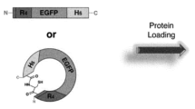
- Figure 1 is a schematic diagram showing the composition of a peptide-tagged cyclic protein (Int C and Int N ) using a split intein according to an embodiment of the present invention.
- Figure 2 is a diagram showing a method for expressing and purifying a cyclic protein according to an embodiment of the present invention.
- Figure 3 is a diagram showing the results of an experiment to confirm the cyclization characteristics of a protein according to an embodiment of the present invention.
- Figure 4 is a diagram showing the results of evaluating the stability of the EGFP protein against proteolysis by Protease K, proteolysis by temperature, and proteolysis inside cells according to an embodiment of the present invention.
- Figure 5 shows the intracellular degradation mechanism of a cyclic protein, the intracellular EGFP fluorescence amount of linear protein and cyclic protein over time, and the colocalization of LysoTracker and EGFP according to an embodiment of the present invention. This is a graph showing the confirmed results.
- Figure 6 is a diagram showing the results of evaluating the cell penetration ability of a cyclic protein according to an embodiment of the present invention.
- Figure 7 is a diagram showing the results of evaluating the cell penetration ability of a cyclic protein according to an embodiment of the present invention.
- Figure 8 is a diagram showing the results of evaluating the cell penetration ability of a cyclic protein according to an embodiment of the present invention.
- Figure 9 is a diagram showing the results of evaluating the cell permeability of a cyclic protein according to an embodiment of the present invention.
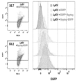
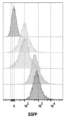
- Figure 10 is a diagram showing the results of flow cytometry analysis of HeLa cells treated with three types of EGFP proteins according to an embodiment of the present invention.
- Figure 11 is a diagram showing the results of confirming the cell penetration mechanism of a cyclic protein according to an embodiment of the present invention.
- Figure 12 is a diagram and graph showing the endoplasmic reticulum permeability of a cyclic protein according to an embodiment of the present invention.
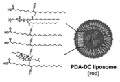
- Figure 13 is a diagram showing the production and purification process of PCDA-DC liposomes loaded with linear and cyclic proteins according to an embodiment of the present invention.
- Figure 14 is a graph showing the evaluation of the size and ion distribution of PCDA-DC liposomes loaded with linear and cyclic proteins according to an embodiment of the present invention.
- Figure 15 is a diagram and graph showing liposome permeability of a cyclic protein according to an embodiment of the present invention.
- Figure 16 is a graph and diagram showing the results of confirming the function of the protein loaded into the PCDA-DC liposome according to an embodiment of the present invention.
- Figure 17 is a schematic diagram explaining membrane attachment of a cyclic protein according to an embodiment of the present invention.
- Figure 18 is a diagram and a graph showing the results of evaluating the attachment of a cyclic protein to the endoplasmic reticulum membrane according to an embodiment of the present invention.
- Figure 19 is a graph showing the results of evaluating the membrane permeability and membrane adhesion of a cyclic protein in various endoplasmic reticulum according to an embodiment of the present invention.
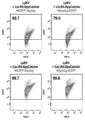
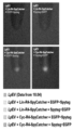
- Figure 20 is a diagram and a graph showing the results of evaluating membrane attachment of the cyclic SpyCater protein according to an embodiment of the present invention.
- the gene encoding enhanced green fluorescent protein (EGFP) containing an H 6 -tag at the C-terminus is pRSET B -EGFP-H 6 (for linear proteins, the H 6 -tag is moved from the N-terminus to the C-terminus) or pBAD-EGFP- H6 (for cyclic protein) was constructed by plasmid vector.
- EGFP enhanced green fluorescent protein
- the gene encoding linear R 4 -EGFP was amplified with primers of SEQ ID NO. 1 and 2, and cloned into the pRSET B -EGFP-H 6 plasmid vector using Nde I/ Hind III to generate pRSET B -R. 4 -EGFP-H 6 plasmid vector was generated.
- the gene encoding linear RGD (Arginylglycylaspartic acid)-EGFP was amplified using primers of SEQ ID NOs: 3 and 4, and the pRSET B -RGD-EGFP-H 6 plasmid vector was generated using Nde I/ Hind III.
- the gene encoding linear TCS (thrombin cleavage site)-R 4 -EGFP was amplified with primers of SEQ ID NOs: 5 to 7, and Nde I/ Hind III was used to generate the pRSET B -TCS-R 4 -EGFP-H 6 plasmid vector. was created.
- the gene encoding linear RGD-EGFP was amplified with primers of SEQ ID NOs: 8 and 9, and the pRSET B -RGD-EGFP-H 6 plasmid vector was generated using Nde I/ Hind III.
- the gene encoding linear R 4 -MS (monomeric photosensitizing fluorescent protein, miniSOG) was amplified using primers of SEQ ID NOs: 10 and 11, and the pRSET B -R 4 -MS-H 6 plasmid vector was generated using Nde I/ Hind III. created.
- the Npu dnaE intein from Nostoc functionform strain PCC 73102 was amplified in pSKDuet16 (#41684; Addgene, Watertown, USA).
- the gene encoding the Npu DnaE intein C-terminus (Int C ) was amplified using primers of SEQ ID NOs: 12 and 13, and cloned into the pBAD-EGFP-H 6 plasmid vector using Nco I/ Hind III to produce pBAD-Int C.
- -EGFP-H 6 plasmid vector was created.
- the short extein sequence encodes CFNG, four amino acids inserted between the C-termini of Intc, increasing the efficiency of self-splicing.
- the gene encoding the Npu DnaE intein N-terminus (Int N ) was amplified with primers of SEQ ID NOs: 14 and 15, and cloned into the pBAD-Int C -EGFP-H 6 plasmid vector using EcoR I/ Sal I.
- the pBAD-Int C -EGFP-H 6 -Int N plasmid vector was generated.
- the gene encoding the linear or split intein fusion R 4 -EGFP was used in the literature (Collagen-Immobilized Extracellular FRET Reporter for Visualizing Protease Activity Secreted by Living Cells, H. Lee, SJ Kim, H. Shin) , YP Kim, ACS Sensors 5 (3) (2020) 655-664), pCMV-R 4 -EGFP or pCMV-Int C -R 4 without affinity tag was isolated from pCMV-proMMP2-3 ⁇ FLAG. -EGFP-Int N was generated.
- Recombinant proteins (linear EGFP, cyclic EGFP, linear R 4 -EGFP, cyclic R 4 -EGFP, linear RGD-EGFP, cyclic RGD-EGFP, linear MS, linear R 4 -MS, cyclic R 4 -MS and
- E. coli carrying various plasmid vectors were inoculated into 670 mL of Luria medium supplemented with 50 ⁇ g/mL ampicillin. The culture was grown at 37°C until the optical density at 600 nm (OD600) reached 0.6 to 0.8.
- Cells were then harvested by centrifugation at 1000 ⁇ g for 10 min and resuspended in 40 mL of lysis buffer (20 mM Tris-HCl, 300 mM NaCl, 10 mM imidazole, pH 8.0).
- the resuspended pellet was dissolved by sonication in the presence of 80 mg lysozyme, centrifuged at 7100 ⁇ g for 20 minutes at 4 °C, and the supernatant was passed through a syringe filter with a 0.45 ⁇ m pore size.
- His 6 -tagged expressed proteins were separated from the supernatant using HisPur Ni-NTA resin on a disposable column balanced with buffer A. The column was washed three times with 5 mL wash buffer (20mM Tris-HCl, 300mM NaCl, 25mM imidazole, pH 8.0).
- Bound proteins were eluted from the spin column with 0.5 mL elution buffer (20mM Tris-HCl, 300mM NaCl, 500mM imidazole, pH 8.0).
- the buffer was exchanged into phosphate-buffered saline (PBS) buffer (2.7 mM potassium chloride, 10 mM disodium phosphate, 137 mM sodium chloride, 1.8 mM monopotassium phosphate, pH 7.4) using a PD-10 desalting column (GE Healthcare).
- PBS phosphate-buffered saline
- FPLC fast protein liquid chromatography
- AKTA Amercham Pharmacia Biotech, Piscataway, NJ, USA
- HisTrap His-tag protein column GE Healthcare
- HR 10/30 column Gel filtration was performed at Amercham Pharmacia Biotech.
- Protein concentration was calculated using an ultraviolet-visible spectrophotometer (Cary 60, Agilent Technologies, Santa Clara, CA, USA) based on the molecular extinction coefficient at 280 nm.
- Linear TCS-R 4 -EGFP was created and analyzed by treating cyclic TCS-R 4 -EGFP with thrombin (1 unit/mg) at 25 °C for 16 hours.
- Protein products were loaded on 12% acrylamide gels under reducing or denaturing conditions, and sizes were estimated from the intensity of bands obtained by sodium dodecyl sulfate-polyacrylamide gel electrophoresis (SDS-PAGE). Bands within the gel were visualized through Coomassie Brilliant Blue staining or fluorescence imaging using a custom transilluminator equipped with a blue LED lamp.
- N-terminal (Int N , residues 1-102) and C-terminal portions (Int C , residues 103-138) of the Npu DnaE intein were incubated with H 6 -tagged R 4 -EGFP (i.e., Int C -R 4 -EGFP-H 6 -Int N ) is fused to the C- and N-termini, respectively, to produce a cyclic fusion protein.
- Trans-splicing efficiency was increased by inserting four amino acids (CFNG) between the C-terminus of Int C and the N-terminus of R 4 -EGFP ( Figures 1 and 2).
- the linear fusion protein (R 4 -EGFP-H 6 ) and the cyclic protein (Cyc(R 4 -EGFP-H 6 ) were expressed in E. coli and subjected to sodium dodecyl sulfate polyacrylamide gel electrophoresis. were analyzed using (SDS-PAGE) and matrix-assisted laser desorption/ionization mass spectrometry (MALDI-MS).
- Plasmid vectors were amplified using the Midi Plasmid Kit (Geneaid, Taiwan). The purity and concentration of plasmid DNA were measured using microvolume spectroscopy (Colibri, Berthold Technologies, Wildbad, Germany). Plasmid vectors were transfected into human cervical cancer cells (HeLa cells) using Lipofectamine 3000 transfection reagent (Thermo Scientific) according to the manufacturer's instructions. After culturing for 24 hours, infected HeLa cells were transferred to fresh DMEM containing 10% fetal bovine serum (FBS). The FL intensity of HeLa cells was monitored using confocal microscopy imaging for 48 h.
- FBS fetal bovine serum
- HeLa, HEK293T, SK-BR3 and U87MG cells were grown in DMEM supplemented with 10% FBS and 1% penicillin-streptomycin in 24-well cell culture plates (SPL, Korea) on 12 mm ⁇ microscope cover glass (Marienfeld Su-perior, Germany). was cultured. Cells were cultured at 37 °C in a humidified incubator with a 5% CO 2 atmosphere until 80% to 90% confluency was reached. After transferring the cells to fresh serum-starved medium and washing twice with DPBS, linear or cyclic proteins (50 ⁇ L at 100 ⁇ M concentration, 10 ⁇ M final concentration) were mixed with the cells in serum-starved medium and then incubated in a 5% CO 2 atmosphere.
- linear or cyclic proteins 50 ⁇ L at 100 ⁇ M concentration, 10 ⁇ M final concentration
- linear or cyclic proteins (final concentration 5 ⁇ M) were mixed with PCDA polydiacetylene vesicles or PCDA-DC liposomes (final lipid concentration 500 uM) for 1 h at 37 °C. Shaked at 800 rpm. Each mixture was washed three times with 1 ⁇ PBS buffer (pH 7.4) by centrifugation (20,000 ⁇ g, 20 min) at 4°C.
- Proteinase K (final concentration 0.2 mg/mL) was added to the protein-loaded liposome at room temperature (RT), and after 1 hour, it was washed twice by centrifugation (20,000 ⁇ g, 20 minutes).
- vesicles or liposomes were heated to 60 °C for 10 min.
- MS protein-loaded PCDA-DC liposomes were prepared by mixing 150 ⁇ M linear or cyclic MS and liposomes, and then the mixture was incubated with 5 ⁇ M FMN (flavin mononucleotide; Sigma-Aldrich, St Louis, MO, USA). Live cells were then cultured in this mixture for several hours. Cells or liposomes attached to the cover glass were confirmed as bright-field images or fluorescence images using a confocal microscope (Nikon C2si+, Japan). The excitation and emission wavelengths for DAPI, EGFP, LysoTracker Red, and EthD-1 imaging were 405/460, 488/511, 561/590, and 561/610 nm, respectively.
- Figure 9 shows the results of confocal images of 3D cultured HeLa spheroids taken with Z stack in series form (A) and XY-XZ-YZ form (B). It was confirmed that Cyc (R 4 -EGFP-H 6 ) could penetrate into the inside of the spheroid ( Figure 9).
- HeLa cells treated with three types of EGFP proteins were analyzed by flow cytometry.
- HeLa cells were cultured in DMEM lacking FBS and treated with linear or cyclic proteins (10 ⁇ M final) for 6 h at 37°C. Protein-untreated cells treated under the same conditions were used as a negative control.
- Cells were washed once with DPBS and detached from flasks using trypsin-transformer using trypsin-EDTA (Thermo Scientific) and centrifugation (1000 ⁇ g, 5 min). The cells were then filtered through a 100 ⁇ m cell strainer and resuspended in 400 ⁇ L of DPBS. The collected cells were analyzed using a flow cytometer equipped with a laser (FACSCanto II, BD Biosciences, San Jose, SA, USA).
- EGFP or MS intensity analysis of cells or liposomes data were collected using a 488 nm laser and a 530/30 nm bandpass filter. Samples were then treated with proteinase K at a final concentration of 0.2 mg/mL, incubated for 1 h at RT, and washed twice before analysis. Flow cytometry data were analyzed using FlowJo software (version 10.6.1., BD Biosciences).
- the size and ion distribution of mammalian EVs extracted from HeLa cells were confirmed through DLS (FIG. 12a), and the size distribution of mammalian EVs extracted from HeLa cells was confirmed through NTA (FIG. 12b).
- the mammalian EV loading efficiency of linear and cyclic recombinant proteins was compared with and without saponin ( Figure 12c), and the cell delivery efficiency of cyclic recombinant proteins before and after mammalian EV loading was measured using confocal microscopy images. Comparison was made ( Figure 12d). After mixing mammalian EVs with 5 ⁇ M of protein and loading the proteins into the EVs, only the protein-loaded EVs were purified through a washing process.
- Purified EVs were dissolved in 500 ⁇ L of DMEM (-FBS, +P/S) and treated with cells for 6 hours. Protein-loaded mammalian EVs were then treated with HeLa cells, and the EGFP positive rate of HeLa cells was measured by flow cytometry. was confirmed.
- PCDA prevents leakage of loaded proteins and shows a color change upon UV irradiation.
- the permeability of the recombinant proteins was evaluated by loading them into PCDA-CA liposomes and irradiating them with UV light.
- the suspension was filtered through a 0.8 ⁇ M filter to remove aggregates and then cooled at 4 °C overnight to stabilize the liposomes.
- Crosslinking was achieved by irradiating 254 nm light (1 mW/cm 2 ) for 1 minute. In this process, ene-yne polymerization was promoted, thereby forming blue polydiacetylene (PDA)-DC liposomes that block protein leakage.
- PDA polydiacetylene
- heat treatment was performed at 60 °C for 10 minutes, which promoted the conversion of blue PDA-DC liposomes into red liposomes that emit red ( Figure 13).
- the dynamic light scattering (DLS) size and zeta potential of PCDA-DC liposomes loaded with linear and cyclic proteins were measured using the ZetaSizer Pro system (Malvern Panalytical, Malvern, UK).
- the size distribution of PCDA-DC liposomes loaded with linear and cyclic proteins showed that the size of the liposomes was larger than that of PCDA-DC not loaded with proteins (FIG. 14).
- the target protein can be cyclized by attaching only a short peptide, spytag, to the N- or C-terminus, and attaching it to the membrane via mPEP-spycatcher ( Figure 17).
- LpEV Lactobacillus plantarum EV
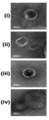
- Cyc (R 4 -EGFP-SpyCatcher) was reacted with TAMRA-Spytag, and the binding of SpyCatcher and spytag was confirmed by SDS-PAGE. Then, Cyc (R 4 -EGFP-SpyCatcher) and TAMRA-Spytag were reacted, and the binding of SpyCatcher and spytag was confirmed using an optical microscope (Cytoviva, USA). In addition, it was confirmed by FACS that the amount of EGFP-Spytag/Spytag-EGFP loaded into LpEV was very small.
Landscapes
- Health & Medical Sciences (AREA)
- Chemical & Material Sciences (AREA)
- Life Sciences & Earth Sciences (AREA)
- Genetics & Genomics (AREA)
- Organic Chemistry (AREA)
- Engineering & Computer Science (AREA)
- General Health & Medical Sciences (AREA)
- Bioinformatics & Cheminformatics (AREA)
- Medicinal Chemistry (AREA)
- Zoology (AREA)
- Biomedical Technology (AREA)
- Molecular Biology (AREA)
- Wood Science & Technology (AREA)
- General Engineering & Computer Science (AREA)
- Biotechnology (AREA)
- Biochemistry (AREA)
- Biophysics (AREA)
- Proteomics, Peptides & Aminoacids (AREA)
- Animal Behavior & Ethology (AREA)
- Pharmacology & Pharmacy (AREA)
- Epidemiology (AREA)
- Public Health (AREA)
- Veterinary Medicine (AREA)
- Gastroenterology & Hepatology (AREA)
- Physics & Mathematics (AREA)
- Microbiology (AREA)
- Plant Pathology (AREA)
- Immunology (AREA)
- Dispersion Chemistry (AREA)
- Peptides Or Proteins (AREA)
Abstract
본 발명은 특정 펩타이드 서열을 도입하여 재조합 단백질을 구성한 이후 N 말단 및 C 말단을 고리화 (Cyclization)한 재조합 단백질 및 이의 제조 방법에 관한 것으로, 본 발명의 재조합 단백질은 펩타이드의 서열에 따라 세포 및 소포체에서의 단백질의 투과성 및 부착성을 조절할 수 있고, 단백질의 안정성을 향상시키는데 효과가 우수하다.
Description
본 발명은 고리형 단백질의 제조방법 및 이의 응용에 관한 것으로, 목적 단백질의 N-말단과 C-말단이 고리화 (Cyclization)된 막 투과성 및 막 부착성이 우수한 단백질에 관한 것이다.
본 발명은 대한민국 과학기술정보통신부의 지원 하에서 과제번호 2022R1A2B5B02002324에 의해 이루어진 것으로서, 상기 과제의 연구관리전문기관은 "한국연구재단", 연구사업명은 "이공분야기초연구사업/중견연구자지원사업/중 견후속연구(연평균연구비 1억원~2억원이내)", 연구과제명은 "고리형 광민감단백질을 이용한 광역학 바이오센서 및 세포 내 전달 플랫폼 개발", 연구기간은 "2022.03.01.-2023.02.28"이다.
본 특허출원은 2022년 11월 15일에 대한민국 특허청에 제출된 대한민국 특허출원 제10-2022-0152335호에 대하여 우선권을 주장하며, 상기 특허출원의 개시 사항은 본 명세서에 참조로서 삽입된다.
약물 전달 및 표적 운반체로는 세포밖 소포체 (Extracellular vesicle, EV) 및 세포 유래 소포체 (Cell-derived vesicle, CDV)이 있으나, 세포 유래 소포체 (CDV)의 낮은 수율과 이질적인 특성, 세포 유래 소포체 (CDV)의 안팎에 이미 존재하는 세포 내재적인 물질 때문에 기능성 물질을 로딩하기가 어렵다는 점 때문에 아직 사용이 승인된 사례가 없다.
세포밖 소포체 (EV) 혹은 세포 유래 소포체 (CDV)에 약물을 로딩하는 방법에는 크게 2가지가 있다. 첫 번째는 세포에 생체 분자 (DNA/RNA/단백질 등)를 직접 발현시킨 뒤 소포체를 정제하는 “사전-로딩 (Pre-loading, Endocellular loading)” 방법이고, 두 번째는 먼저 소포체를 정제한 다음에 약물을 봉입하는 “사후-로딩 (Post-loading, Extracellular loading)” 방법이다. 사전-로딩 방법의 경우 특성에 따라 사용에 제한이 있으며, 세포 유래 소포체 (CDV)를 분리한 후 약물을 로딩하는 방식에 비해 세포 유래 소포체 (CDV)에 로드되는 약물의 양을 조절하기 어려우며, 한 번의 치료에 사용하는 세포 유래 소포체의 양이 많아지면 유래 세포가 갖고 있던 특성에 따른 부작용의 위험이 증가하게 되는 문제점이 있다. 반면 “사후-로딩” 방법에서는 분리된 세포 유래 소포체 (CDV) 내로 물질을 넣는 것이 어렵기 때문에 콜레스테롤 또는 세포막 투과 증폭을 위한 첨가물을 더하거나, 전기천공법 (electroporation)과 같은 물리적인 자극을 주어야 한다. 하지만 이러한 방식은 세포 유래 소포체 (CDV) 막을 손상시키고 약물의 응집을 유발할 수 있다. 이러한 기술적인 문제에 따라 새로운 약물 로딩 기작의 필요성이 요구된다.
세포 및 소포체는 다양한 분리 및 배양 기술이 알려져 왔으나, 세포 및 소포체에 단백질을 전달할 수 있는 기술은 대부분 세포 투과성 펩타이드 (Cell Permeable Peptide)의 부착 기술에 의존하고 있다. 세포 투과성 펩타이드는 일종의 신호 펩타이드로서 약 7 내지 30개의 아미노산 서열로 이루어지며, HIV 바이러스에서 유래된 TAT 단백질에 존재하는 11개 아미노산 서열의 b-galactosidase (120 kDa) 단백질의 세포내 및 조직내 전달이 가능하다는 것을 보여준 1990년대 이후에 본격적으로 관련 연구가 진행되었다. 세포 투과성 펩타이드는 다양한 세포주에 공통적으로 적용할 수 있는 범위가 제한적이며 세포 소기관내에 다양하게 존재할 수 있는 세포내 전달 지향점이 다를 수 있는 문제점이 있다. 또한 최근 일리아스에서 개발한 광 (light) 제어를 통해 소포체에 단백질을 로딩하는 기술이 보고되었으나 여전히 외부자극을 사용해야 하는 번거로움이 있다. 무엇보다 단백질 치료제의 효능을 지속적으로 유지하기 위해서는 단백질의 안정성 확보가 요구되어 세포 및 소포체 내 효과적인 단백질 로딩기술과 동시에 단백질의 높은 안정성을 동시에 구축하는 것이 필요하다.
세포 및 소포체 표면으로의 단백질 부착기술은 단백질의 표지법과 같이 연관되어 왔는데, 주로 화학적인 결합법에 의해 단백질에 다양한 리간드 (펩타이드, 인지질, 콜레스테롤)를 도입함으로써 구현되어 왔다. 그러나 표면에 결합되는 양을 조절하기가 매우 어렵고 세포 및 소포체 종류에 따라 다른 부착 효율이 어려우며 결합과정에서 세포 및 소포체가 손상되거나 단백질의 방향성을 조절하기에 제한적인 문제점이 있다.
따라서, 다양한 펩타이드 서열을 단백질에 도입하여 세포 및 소포체에서의 단백질의 투과성 및 부착성을 조절할 수 있고, 단백질의 안정성을 향상시킬 수 있는 고리형 단백질의 개발이 절실히 요구되고 있는 실정이다.
본 발명자들은 다양한 펩타이드 서열을 도입하여 고리화 함으로써 세포 및 소포체에서의 단백질 로딩 및 부착 기술을 구현하고자 예의 연구 노력하였다.
그 결과, 단백질에 특정 펩타이드 서열을 도입하여 재조합 단백질을 구성하고, N 말단 및 C 말단을 고리화 (Cyclization)함으로써 단백질의 세포 및 소포체 내 투과성 및 안정성을 효과적으로 증가되며, 단백질 말단의 펩타이드 서열에 따라 막 투과성 및 막 부착성을 손쉽게 조절할 수 있음을 확인하여 본 발명을 완성하였다.
이에, 본 발명의 목적은 N-말단 인테인 (intein)을 코딩하는 서열;고리화시키고자 하는 목적 단백질을 코딩하는 서열; C-말단 엑스테인 (extein)을 코딩하는 서열; 및 C-말단 인테인을 코딩하는 서열;을 포함하는 고리형 단백질 발현 플랫폼을 제공하는 것이다.
본 발명의 다른 목적은 a) 고리형 단백질 발현 플랫폼을 숙주세포에 도입하는 단계; 및 b) 상기 숙주세포에서 단백질의 발현을 유도하는 단계를 포함하는 고리형 단백질 제조방법을 제공하는 것이다.
본 발명의 또 다른 목적은 고리형 단백질을 막 투과 또는 막 부착용 단백질로 이용하여 약물을 전달하는 용도를 제공하는 것이다.
세포 및 소포체에 단백질을 전달할 수 있는 기술은 세포에 생체 분자를 직접 발현시키는 방법과 세포 투과성 펩타이드를 부착하는 기술이 있다. 세포에 생체 분자를 직접 발현시키는 방법은 로드되는 약물의 양을 조절하기 어려우며, 유래 세포가 갖고 있던 특성에 따른 부작용의 위험이 있다. 또한 세포 투과성 펩타이드의 부착 기술은 표면에 결합되는 양을 조절하기가 어렵고, 세포 및 소포체의 종류에 따라 다른 부착 효율을 나타내는 문제점이 있다.
이와 같은 문제를 해결하고자 본 발명자들은 세포 및 소포체 내 투과성 및 단백질을 효과적으로 증가시키며 단백질 말단의 펩타이드 서열에 따라 막 투과성 및 막 부착성을 쉽게 조절할 수 있는 방법을 찾고자 예의 연구 노력하였다.
그 결과, 본 발명자들은 하기 실시예를 통해 단백질의 N-말단과 C-말단의 거리가 가까운 경우에는 고리화 이후에도 단백질의 기능을 유지하여 세포 혹은 소포체 내로 높은 효율로 투과된 이후 높은 안정성을 유지할 수 있으며 막 투과성 및 막 부착성을 손쉽게 조절할 수 있음을 확인할 수 있었다.
실시예 2에서는 선형 및 고리형 EGFP의 단백질 분해에 대한 안정성을 평가하였으며, 고리형 EGFP 단백질은 단백질 분해 효소 K, 80 ℃이상의 고온 및 세포 내에서 안정성을 나타내었으며, 세포 내에서 선형 단백질보다 고리형 단백질이 더 느리게 분해되는 것을 확인하였다.
실시예 3에서는 HeLa, HEK293T, SK-BR3 및 U87MG 세포주에 태그를 다양화한 선형 및 고리형 단백질을 침투시켰을 때 세포주별로 세포막에 존재하는 인테그린의 종류와 양에 따라서 세포 투과 능력이 달라짐을 확인하였다. 또한, 고리형 단백질이 선형 단백질로 잘라지자 세포내 투과능이 감소함을 확인하였다.
실시예 4에서는 EGFP 단백질을 처리한 HeLa 세포를 유세포 분석법으로 분석한 결과 선형 단백질보다 고리형 단백질이 세포 내부로 더 많이 침투되었음을 확인하였다.
실시예 5에서는 EV에 로딩된 단백질이 단백질만 처리했을 때보다 세포 내부로 훨씬 더 많이 전달되는 것을 확인할 수 있었다. 또한, Cyc(R4-EGFP-H6)가 로딩된 EV가 다른 조건에 비해 월등히 많은 양의 단백질을 세포 내부로 운반함을 확인할 수 있었다.
실시예 6에서는 PCDA-DC 리포솜 (liposome)에 3종의 재조합 단백질을 로딩 및 정제한 다음, 단백질 분해효소 K (proteinase K)의 처리 유무에 따른 평균 EGFP FL을 비교하였을 때 고리형 단백질이 로딩된 PCDA-DC 리포솜에 가장 많이 존재하는 것을 확인하였다.
실시예 7에서는 고리형 단백질의 소포체 막 부착을 평가하여 고리형 재조합 단백질이 각각의 선형 재조합 단백질보다 더 많이 LpEV에 로딩되는 것을 확인할 수 있었다. 그리고 단백질 분해효소 K를 처리했을 때에는 모두 LpEV에서 떨어지는 것을 보아, 단백질이 LpEV의 바깥에 로딩되는 것임을 확인할 수 있었다. 또한 고리형 재조합 단백질의 로딩 분포가 소포체의 종류에 따라 달라짐을 확인하였다.
실시예 8에서는 고리형 SpyCater 단백질의 막 부착을 평가하여 Spytag 형광 단백질을 부착했을 때 고리형 단백질에서 약 10배 이상 많은 양의 spytag 형광 단백질이 부착되는 것을 FACS와 펠렛 이미지로 확인하였다.
이에 본 발명자들은 단백질의 N-말단 및 C-말단을 고리화 함으로써 세포 및 소포체 내 투과성 및 단백질의 부착성을 조절할 수 있으며, 단백질의 안정성을 향상시킬 수 있음을 찾아내었다. 본 발명의 고리형 단백질은 세포내 단백질 전달기술을 확보하여 재조합 단백질 기반 치료제나 면역원성을 지닌 다양한 단백질 제제를 EV 결합을 통하여 바이러스 백신 또는 항암백신 등으로 활용할 수 있을 것으로 예상한다.
이하, 본 발명을 더욱 자세히 설명하고자 한다.
본 발명의 일 양태는 N-말단 인테인 (intein)을 코딩하는 서열; 고리화시키고자 하는 목적 단백질을 코딩하는 서열; C-말단 엑스테인 (extein)을 코딩하는 서열; 및 C-말단 인테인을 코딩하는 서열;을 포함하는 고리형 단백질 발현 플랫폼이다.
본 명세서 상 용어 “인테인”은 단백질 인트론(intron)이라고도 불리우며, 진핵 세포, 원핵세포 및 고 세균에서 발견되고 있으며, 최근까지 500 여종 이상 발견되고 있다. 인테인은 단백질 분해효소 및 리가아제 활성 둘 모두를 함유하는 자가촉매 효소의 부류이다. "분할형 인테인"은 인테인은 선택적으로, 그리고 매우 단단하게 회합하여, 활성 인테인 효소를 형성하는 N-인테인 및 C-인테인으로 지칭되는 2개의 상보적 절반 인테인을 포함한다.
본 명세서 상 용어 “엑스테인”은 진핵세포 RNA의 엑손 (exon)에 대응하는 단백질로, IntC의 C-말단 부분에 삽입된 짧은 서열인 것일 수 있다.
본 명세서 상 “단백질 발현 플랫폼”은 단백질을 발현시키기 위해 이용되는 플랫폼을 의미하며, “발현 벡터”를 포함한다.
본 발명에 있어서 발현 벡터는 특별히 이에 한정되지 않으나, 박테리아 세포 (예를 들어, 대장균 등), 포유류 세포 (예를 들어, 사람, 원숭이, 토끼, 래트, 햄스터, 마우스 세포 등), 식물 세포, 효모 세포 또는 곤충 세포를 포함하는 원핵 또는 진핵세포에서 상기 폴리뉴클레오티드를 복제 및/또는 발현할 수 있는 벡터가 될 수 있다. 바람직하게는 숙주세포에서 상기 폴리뉴클레오티드가 발현될 수 있도록 적절한 프로모터에 작동 가능하도록 연결되며, 적어도 하나의 선별마커를 포함하는 벡터가 될 수 있다. 그 예로 파지미드, 플라스미드, 코스미드, 미니-염색체, 바이러스 또는 레트로바이러스 벡터 등에 상기 폴리뉴클레오티드가 도입된 형태가 될 수 있다.
또한 발현 벡터는 발현 벡터의 전사에 제공되는 추가 단편에 작동 가능하게 연결된 목적(타겟) 단백질을 암호화하는 단편으로 구성되는 선형 또는 원형의 DNA 분자이다. 그와 같은 추가 단편은 프로모터 및 종료암호 서열을 포함한다. 발현 벡터는 하나 이상의 복제 개시점, 하나 이상의 선택 마커 등을 또한 포함한다. 발현 벡터는 일반적으로 플라스미드 또는 바이러스 DNA로부터 유도되거나 또는 둘 다의 요소를 함유한다.
본 발명에 있어서 발현 벡터의 숙주 세포/형질전환체 내로의 운반은, 당 업계에 널리 알려진 운반 방법을 사용할 수 있다. 상기 운반 방법은 예를 들어, 숙주 세포가 원핵세포이면 CaCl2 방법 또는 전기 천공 방법 등을 사용할 수 있고, 숙주 세포가 진핵세포이면 미세 주입법, 칼슘 포스페이트 침전법, 전기 천공법, 리포솜-매개 형질감염법 및 유전자 밤바드먼트 등을 사용할 수 있으나, 이에 한정되는 것은 아니다.
본 발명에 있어서 고리화시키고자 하는 목적 단백질을 코딩하는 서열은 양 말단에 태그 코딩 서열을 추가로 포함하는 것일 수 있으나, 이에 한정되는 것은 아니다.
본 발명에 있어서 태그는 막 부착성 펩타이드 또는 막 투과성 펩타이드인 것일 수 있으나, 이에 한정되는 것은 아니다.
본 발명에 있어서 막 부착성 펩타이드는 H6-태그, R4-태그, RGD 태그, 비오틴/스트렙타비딘 태그, V5-태그, Myc-태그, HA-태그, NE-태그, His-태그, 플래그 태그 (Flag tag), 할로 태그 (Halo-tag), 스냅 태그 (Snap-tag), Fc-태그, Nus-태그, BCCP, 티오레독신 (thioredoxin), 스눕 태그 (SnooprTag), 스파이 태그 (SpyTag), 이소펩 태그 (Isopeptag), SBP-태그, S-태그, 아비 태그 (AviTag), TAT 태그 (TAT tag), NP2 태그 (NP2 tag), 젠트리 (Xentry tag), 스파이캐처-스파이태그 (spycatcher-spytag), 스눕태그-스눕캐처 (snooptag-snoopcatcher), 알자이드-알킨 (azide-akyne) 및 LPXT-GGGX로 이루어진 군에서 선택된 1종 이상인 것일 수 있으나, 이에 한정되는 것은 아니다.
본 발명에 있어서 막 투과성 펩타이드는 H6-태그, R4-태그, R5-태그, R6-태그, R7-태그, RGD 태그, NP2 태그, 젠트리 (Xentry tag), 스냅 태그 (Snap-tag), 스눕 태그 (SnooprTag), 스파이 태그 (SpyTag), His-태그, TAT 태그, 페네트라틴 (Penetratin), 싸이오레독신 (thioredoxin, Trx) 태그, B6 펩타이드, ABP 태그, 트랜스포탄 (Transportan), VP-22, 양쪽 친매성 펩타이드 (Amphipathic peptides), Pep-1, MAP, SAP, PPTG1, 양이온성 펩타이드 (Cationic peptides), 올리고알기닌 (Oligoarginine), hCT(9-32) (human calcitonin (hCT)-derived cell-penetrating peptide) 및 Pvec (Cadherin-5)로 이루어진 군에서 선택된 1종 이상인 것일 수 있으나, 이에 한정되는 것은 아니다.
본 발명에 있어서 고리화시키고자 하는 목적 단백질은 항체, 세포수용체, 효소, 구조 단백질, 혈청, 항원 단백질, 세포 단백질, 크리스퍼 (Clustered Regularly Interspaced Short Palindromic Repeats, CRISPR), 전사인자, 호르몬 단백질 및 신호 단백질로 이루어진 군에서 선택된 1종 이상인 것일 수 있으나, 이에 한정되는 것은 아니다.
본 발명에 있어서 고리화시키고자 하는 목적 단백질은 아미노산 10개 내지 1500개, 10개 내지 1400개, 10개 내지 1300개, 10개 내지 1200개, 10개 내지 1100개, 10개 내지 1000개, 10개 내지 900개, 10개 내지 800개, 10개 내지 700개, 10개 내지 600개, 10개 내지 500개, 10개 내지 400개, 10개 내지 300개, 10개 내지 200개, 10개 내지 100개, 10개 내지 50개, 100개 내지 1500개, 100개 내지 1400개, 100개 내지 1300개, 100개 내지 1200개, 100개 내지 1100개, 100개 내지 1000개, 100개 내지 900개, 100개 내지 800개, 100개 내지 700개, 100개 내지 600개, 100개 내지 500개, 100개 내지 400개, 100개 내지 300개, 100개 내지 200개, 200개 내지 1500개, 200개 내지 1400개, 200개 내지 1300개, 200개 내지 1200개, 200개 내지 1100개, 200개 내지 1000개, 200개 내지 900개, 200개 내지 800개, 200개 내지 700개, 200개 내지 600개, 200개 내지 500개, 200개 내지 400개, 200개 내지 300개, 300개 내지 1500개, 300개 내지 1400개, 300개 내지 1300개, 300개 내지 1200개, 300개 내지 1100개, 300개 내지 1000개, 300개 내지 900개, 300개 내지 800개, 300개 내지 700개, 300개 내지 600개, 300개 내지 500개, 300개 내지 400개, 400개 내지 1500개, 400개 내지 1400개, 400개 내지 1300개, 400개 내지 1200개, 400개 내지 1100개, 400개 내지 1000개, 400개 내지 900개, 400개 내지 800개, 400개 내지 700개, 400개 내지 600개, 400개 내지 500개, 500개 내지 1500개, 500개 내지 1400개, 500개 내지 1300개, 500개 내지 1200개, 500개 내지 1100개, 500개 내지 1000개, 500개 내지 900개, 500개 내지 800개, 500개 내지 700개, 500개 내지 600개의 크기인 것일 수 있으나, 이에 한정되는 것은 아니다.
본 발명의 다른 일 양태는 고리형 단백질을 코딩하는 폴리뉴클레오티드 (Polynucleotice)이다.
본 발명의 또 다른 일 양태는 단백질 발현 플랫폼이 도입된 숙주세포이다.
본 명세서 상 용어 “숙주세포”는 DNA의 도입효율이 높고, 도입된 DNA의 발현 효율이 높은 숙주세포가 바람직하며, 원핵 및 진핵을 포함한 모든 미생물이 사용될 수 있다.
본 발명에 있어서 숙주세포는 대장균 (E.coli)인 것일 수 있으나, 이에 한정되는 것은 아니다.
본 명세서 상 용어 “목적 단백질”은 고리화 시키고자 하는 단백질로서, 재조합 발현 플랫폼에 상기 단백질을 암호화하는 폴리뉴클레오티드를 삽입하여 숙주세포에서 발현 가능한 단백질을 의미한다.
본 발명의 또 다른 일 양태는 다음의 단계를 포함하는 고리형 단백질 제조방법이다:
a) 제1항에 따른 고리형 단백질 발현 플랫폼을 숙주세포에 도입하는 단계; 및
b) 상기 숙주세포에서 단백질의 발현을 유도하는 단계.
본 발명에 있어서 고리형 단백질 제조방법은 b) 단계 이후 c) 발현된 단백질을 분리하는 단계를 추가로 포함하는 것일 수 있으나, 이에 한정되는 것은 아니다.
본 발명의 또 다른 일 양태는 고리형 단백질 제조방법으로 생산된 고리형 단백질이다.
본 발명의 또 다른 일 양태는 막 부착형 펩타이드 태그를 포함하는 고리형 단백질을 포함하는 EV 태그 (Universal EV-tag) 용 조성물이다.
본 발명에 있어서 막 부착성 펩타이드는 H6-태그, R4-태그, RGD 태그, 비오틴/스트렙타비딘 태그, V5-태그, Myc-태그, HA-태그, NE-태그, His-태그, 플래그 태그 (Flag tag), 할로 태그 (Halo-tag), 스냅 태그 (Snap-tag), Fc-태그, Nus-태그, BCCP, 티오레독신 (thioredoxin), 스눕 태그 (SnooprTag), 스파이 태그 (SpyTag), 이소펩 태그 (Isopeptag), SBP-태그, S-태그, 아비 태그 (AviTag), TAT 태그 (TAT tag), NP2 태그 (NP2 tag), 젠트리 (Xentry tag), 스파이캐처-스파이태그 (spycatcher-spytag), 스눕태그-스눕캐처 (snooptag-snoopcatcher), 알자이드-알킨 (azide-akyne) 및 LPXT-GGGX로 이루어진 군에서 선택된 1종 이상인 것일 수 있으나, 이에 한정되는 것은 아니다.
본 발명의 이점 및 특징, 그리고 그것들을 달성하는 방법은 상세하게 후술되어 있는 실시예들을 참조하면 명확해질 것이다. 그러나 본 발명은 이하에서 개시되는 실시예들에 한정되는 것이 아니라 서로 다른 다양한 형태로 구현될 것이며, 단지 본 실시예들은 본 발명의 개시가 완전하도록 하고, 본 발명이 속하는 기술분야에서 통상의 지식을 가진 자에게 발명의 범주를 완전하게 알려주기 위해 제공되는 것이며, 본 발명은 청구항의 범주에 의해 정의될 뿐이다.
본 발명은 특정 펩타이드 서열을 도입하여 재조합 단백질을 구성한 이후 N 말단 및 C 말단을 고리화 (Cyclization)한 재조합 단백질 및 이의 제조 방법에 관한 것으로, 본 발명의 재조합 단백질은 펩타이드의 서열에 따라 세포 및 소포체에서의 단백질의 막 투과성 및 막 부착성을 조절할 수 있고, 단백질의 안정성을 향상시키는데 효과가 우수하다.
도 1은 본 발명의 일 실시예에 따른 분할 인테인을 사용하여 펩타이드 태그가 있는 고리형 단백질 (IntC 및 IntN)의 구성을 보여주는 모식도이다.
도 2는 본 발명의 일 실시예에 따른 고리형 단백질의 발현 및 정제방법을 보여주는 도이다.
도 3은 본 발명의 일 실시예에 따른 단백질의 고리화 특성을 확인하기 위한 실험 결과를 보여주는 도이다.
도 4는 본 발명의 일 실시예에 따른 단백질 분해 효소 (Protease) K의 단백질 분해, 온도에 의한 단백질 분해 및 세포 내부에서의 단백질 분해에 대한 EGFP 단백질의 안정성을 평가한 결과를 보여주는 도이다.
도 5는 본 발명의 일 실시예에 따른 고리형 단백질의 세포 내 분해 메커니즘, 선형 단백질과 고리형 단백질의 시간에 따른 세포 내 EGFP 형광량 및 라이소트래커 (LysoTracker)와 EGFP 공동국소화(colocalization)를 확인한 결과를 보여주는 그래프이다.
도 6은 본 발명의 일 실시예에 따른 고리형 단백질의 세포 투과 능력을 평가한 결과를 보여주는 도이다.
도 7은 본 발명의 일 실시예에 따른 고리형 단백질의 세포 투과 능력을 평가한 결과를 보여주는 도이다.
도 8은 본 발명의 일 실시예에 따른 고리형 단백질의 세포 투과 능력을 평가한 결과를 보여주는 도이다.
도 9는 본 발명의 일 실시예에 따른 고리형 단백질의 세포 투과성을 평가한 결과를 보여주는 도이다.
도 10은 본 발명의 일 실시예에 따른 3종 EGFP 단백질을 처리한 HeLa 세포를 유세포 분석법으로 분석한 결과를 보여주는 도이다.
도 11은 본 발명의 일 실시예에 따른 고리형 단백질의 세포 투과 매커니즘을 확인한 결과를 보여주는 도이다.
도 12는 본 발명의 일 실시예에 따른 고리형 단백질의 소포체 투과성을 보여주는 도 및 그래프이다.
도 13은 본 발명의 일 실시예에 따른 선형 및 고리형 단백질을 로딩한 PCDA-DC 리포솜의 제조 및 정제과정을 나타낸 도이다.
도 14는 본 발명의 일 실시예에 따른 선형 및 고리형 단백질을 로딩한 PCDA-DC 리포솜의 크기 및 이온 분포를 평가하여 나타낸 그래프이다.
도 15는 본 발명의 일 실시예에 따른 고리형 단백질의 리포솜 투과성을 보여주는 도 및 그래프이다.
도 16은 본 발명의 일 실시예에 따른 PCDA-DC 리포솜에 로딩한 단백질의 기능을 확인한 결과를 보여주는 그래프 및 도이다.
도 17은 본 발명의 일 실시예에 따른 고리형 단백질의 막 부착을 설명한 모식도이다.
도 18은 본 발명의 일 실시예에 따른 고리형 단백질의 소포체 막 부착 평가 결과를 나타낸 도 및 그래프이다.
도 19는 본 발명의 일 실시예에 따른 고리형 단백질의 다양한 소포체에서의 막 투과성 및 막 부착성 평가 결과를 나타낸 그래프이다.
도 20은 본 발명의 일 실시예에 따른 고리형 SpyCater 단백질의 막 부착을 평가한 결과를 나타낸 도 및 그래프이다.
이하 하나 이상의 구체예를 실시예를 통하여 보다 상세하게 설명한다. 그러나 이들 실시예는 하나 이상의 구체예를 예시적으로 설명하기 위한 것으로 본 발명의 범위가 이들 실시예에 한정되는 것은 아니다.
제조예 1. 플라스미드 제조
C-말단에 H6-태그가 포함된 EGFP (enhanced green fluorescent protein) 인코딩 유전자는 pRSETB-EGFP-H6 (선형 단백질의 경우 H6-태그가 N-말단에서 C-말단으로 이동됨) 또는 pBAD-EGFP-H6 (고리형 단백질의 경우) 플라스미드 벡터에 의해 구축되었다.
선형 단백질을 만들기 위해 서열번호 1 및 2의 프라이머로 선형 R4-EGFP를 인코딩하는 유전자를 증폭하였고, Nde I/Hind III를 사용하여 pRSETB-EGFP-H6 플라스미드 벡터에 복제하여 pRSETB-R4-EGFP-H6 플라스미드 벡터를 생성하였다.
서열번호 3 및 4의 프라이머로 선형 RGD(Arginylglycylaspartic acid)-EGFP를 인코딩하는 유전자를 증폭하였고, Nde I/Hind III를 사용하여 pRSETB-RGD-EGFP-H6 플라스미드 벡터를 생성하였다.
서열번호 5 내지 7의 프라이머로 선형 TCS(thrombin cleavage site)-R4-EGFP를 인코딩하는 유전자를 증폭하였고, Nde I/Hind III를 사용하여 pRSETB-TCS-R4-EGFP-H6 플라스미드 벡터를 생성하였다.
서열번호 8 및 9의 프라이머로 선형 RGD-EGFP를 인코딩하는 유전자를 증폭하였고, Nde I/Hind III를 사용하여 pRSETB-RGD-EGFP-H6 플라스미드 벡터를 생성하였다.
서열번호 10 및 11의 프라이머로 선형 R4-MS(monomeric photosensitizing fluorescent protein, miniSOG)를 인코딩하는 유전자를 증폭하였고, Nde I/Hind III를 사용하여 pRSETB-R4-MS-H6 플라스미드 벡터를 생성하였다.
고리형 단백질을 만들기 위해 노스톡 펀크티폼 균주 PCC 73102에서 유래한 Npu dnaE 인테인을 pSKDuet16 (#41684; Addgene, Watertown, USA)에서 증폭하였다. 서열번호 12 및 13의 프라이머로 Npu DnaE 인테인 C-말단 (IntC)을 코딩하는 유전자를 증폭하였고, Nco I/Hind III를 사용하여 pBAD-EGFP-H6 플라스미드 벡터에 복제하여 pBAD-IntC-EGFP-H6 플라스미드 벡터를 생성하였다.
짧은 엑스테인 (extein) 서열은 Intc의 C-말단 사이에 삽입된 네 개의 아미노산인 CFNG를 인코딩하여 자가 스플라이싱의 효율을 높였다.
서열번호 14 및 15의 프라이머로 Npu DnaE 인테인 N-말단 (IntN)을 코딩하는 유전자를 증폭하였고, EcoR I/Sal I을 사용하여 pBAD-IntC-EGFP-H6 플라스미드 벡터에 클론화하여 pBAD-IntC-EGFP-H6-IntN 플라스미드 벡터를 생성하였다.
다양한 프라이머 세트 및 제한효소들을 사용하여 R4-EGFP, TCS-R4-EGFP, RGD-EGFP 또는 R4-MS를 코딩하는 유전자를 pBAD-IntC-EGFP-H6-IntN 플라스미드 벡터에 삽입하여 pBAD-IntC-R4-EGFP-H6-IntN, pBAD-IntC-RGD-EGFP-H6-IntN 또는 pBAD-IntC-R4-MS-H6-IntN을 생성하였다.
플라스미드를 암세포에 감염시키기 위해 선형 또는 분할 인테인 융합 R4-EGFP를 코딩하는 유전자를 참고문헌 (Collagen-Immobilized Extracellular FRET Reporter for Visualizing Protease Activity Secreted by Living Cells, H. Lee, S.J. Kim, H. Shin, Y.P. Kim, ACS Sensors 5 (3) (2020) 655-664)에 기재된 방식으로 pCMV-proMMP2-3×FLAG에서 분리하여 친화성 태그가 없는 pCMV-R4-EGFP 또는 pCMV-IntC-R4-EGFP-IntN을 생성하였다.
| 서열번호 | 유전자 | 방향 | 서열 (5'-->3') |
| 1 | R4-EGFP | 정방향 | GGA ATT CCA TAT GCG ACG ACG ACG AGG AGG AGG AGG AGT GAG |
| 2 | 역방향 | CCC AAG CTT TCA ATG ATG ATG ATG ATG ATG CTT GTA CAG CTC GTC | |
| 3 | RGD-EGFP | 정방향 | GGA ATT CCA TAT GCG AGG TGA TGG AGG AGG AGG AGT GAG C |
| 4 | 역방향 | CCC AAG CTT TCA ATG ATG ATG ATG ATG ATG CTT GTA CAG CTC GTC | |
| 5 | TCS-R4-EGFP | 정방향 | TGC CGC GCG GCA GCC GAC GAC GAC GAG GAG GAG GAG G |
| 6 | 정방향 | GGA ATT CCA TAT GCT GGT GCC GCG CGG CAG CCG ACG ACG ACG AGG AGG A | |
| 7 | 역방향 | CCC AAG CTT TCA ATG ATG ATG ATG ATG ATG CTT GTA CAG CTC GTC | |
| 8 | RGD-EGFP | 정방향 | GGA ATT CCA TAT GCG AGG TGA TGG AGG AGG AGG AGT GAG C |
| 9 | 역방향 | CCC AAG CTT TCA ATG ATG ATG ATG ATG ATG CTT GTA CAG CTC GTC | |
| 10 | R4-MS | 정방향 | GGA ATT CCA TAT G CG ACG ACG ACG AAT GGG GGG AGG AGG |
| 11 | 역방향 | CCC AAG CTT ATG ATG ATG ATG ATG ATG TCC TCC TCC | |
| 12 | Npu DnaE 인테인 C-말단(IntC) | 정방향 | CAT GCC ATG GAT CAA AAT AGC CAC ACG TAA ATA TTT AGG C |
| 13 | 역방향 | CCC AAG CTT ACA ACC ATT GAA ACA ATT AGA AGC TAT GA | |
| 14 | Npu DnaE 인테인 N-말단(IntN) | 정방향 | GGA ATT CTT AAG CTA TGA AAC GGA AAT ATT GAC AGT AGA ATA T |
| 15 | 역방향 | ACG CGT CGA CAT TCG GCA AAT TAT CAA CC |
| 서열번호 | 단백질 | 아미노산 서열 (N ---> C) |
| 16 | EGFP-H6 | MGGGGVSKGEELFTGVVPILVELDGDVNGHKFSVSGEGEGDATYGKLTLKFICTTGKLPVPWPTLVTTLTYGVQCFSRYPDHMKQHDFFKSAMPEGYVQERTIFFKDDGNYKTRAEVKFEGDTLVNRIELKGIDFKEDGNILGHKLEYNYNSHNVYIMADKQKNGIKVNFKIRHNIEDGSVQLADHYQQNTPIGDGPVLLPDNHYLSTQSALSKDPNEKRDHMVLLEFVTAAGITLGMDELYKHHHHHH |
| 17 | Cyc(EGFP-H6) | CFNGCKLGGGGVSKGEELFTGVVPILVELDGDVNGHKFSVSGEGEGDATYGKLTLKFICTTGKLPVPWPTLVTTLTYGVQCFSRYPDHMKQHDFFKSAMPEGYVQERTIFFKDDGNYKTRAEVKFEGDTLVNRIELKGIDFKEDGNILGHKLEYNYNSHNVYIMADKQKNGIKVNFKIRHNIEDGSVQLADHYQQNTPIGDGPVLLPDNHYLSTQSALSKDPNEKRDHMVLLEFVTAAGITLGMDELYKHHHHHH EF |
| 18 | R4-EGFP-H6 | MRRRRGGGGVSKGEELFTGVVPILVELDGDVNGHKFSVSGEGEGDATYGKLTLKFICTTGKLPVPWPTLVTTLTYGVQCFSRYPDHMKQHDFFKSAMPEGYVQERTIFFKDDGNYKTRAEVKFEGDTLVNRIELKGIDFKEDGNILGHKLEYNYNSHNVYIMADKQKNGIKVNFKIRHNIEDGSVQLADHYQQNTPIGDGPVLLPDNHYLSTQSALSKDPNEKRDHMVLLEFVTAAGITLGMDELYKHHHHHH |
| 19 | IntC-R4-EGFP-H6-IntN | MGIKIATRKYLGKQNVYDIGVERDHNFALKNGFIASNCFNGCKLRRRRGGGGVSKGEELFTGVVPILVELDGDVNGHKFSVSGEGEGDATYGKLTLKFICTTGKLPVPWPTLVTTLTYGVQCFSRYPDHMKQHDFFKSAMPEGYVQERTIFFKDDGNYKTRAEVKFEGDTLVNRIELKGIDFKEDGNILGHKLEYNYNSHNVYIMADKQKNGIKVNFKIRHNIEDGSVQLADHYQQNTPIGDGPVLLPDNHYLSTQSALSKDPNEKRDHMVLLEFVTAAGITLGMDELYKHHHHHH EFCLSYETEILTVEYGLLPIGKIVEKRIECTVYSVDNNGNIYTQPVAQWHDRGEQEVFEYCLEDGSLIRATKDHKFMTVDGQMLPIDEIFERELDLMRVDNLPN |
| 20 | Cyc(R4-EGFP-H6) | CFNGCKLRRRRGGGGVSKGEELFTGVVPILVELDGDVNGHKFSVSGEGEGDATYGKLTLKFICTTGKLPVPWPTLVTTLTYGVQCFSRYPDHMKQHDFFKSAMPEGYVQERTIFFKDDGNYKTRAEVKFEGDTLVNRIELKGIDFKEDGNILGHKLEYNYNSHNVYIMADKQKNGIKVNFKIRHNIEDGSVQLADHYQQNTPIGDGPVLLPDNHYLSTQSALSKDPNEKRDHMVLLEFVTAAGITLGMDELYKHHHHHH EF |
| 21 | RGD-EGFP-H6 | MRGDGGGGVSKGEELFTGVVPILVELDGDVNGHKFSVSGEGEGDATYGKLTLKFICTTGKLPVPWPTLVTTLTYGVQCFSRYPDHMKQHDFFKSAMPEGYVQERTIFFKDDGNYKTRAEVKFEGDTLVNRIELKGIDFKEDGNILGHKLEYNYNSHNVYIMADKQKNGIKVNFKIRHNIEDGSVQLADHYQQNTPIGDGPVLLPDNHYLSTQSALSKDPNEKRDHMVLLEFVTAAGITLGMDELYKHHHHHH |
| 22 | Cyc(RGD-EGFP-H6) | CFNGCKLRGDGGGGVSKGEELFTGVVPILVELDGDVNGHKFSVSGEGEGDATYGKLTLKFICTTGKLPVPWPTLVTTLTYGVQCFSRYPDHMKQHDFFKSAMPEGYVQERTIFFKDDGNYKTRAEVKFEGDTLVNRIELKGIDFKEDGNILGHKLEYNYNSHNVYIMADKQKNGIKVNFKIRHNIEDGSVQLADHYQQNTPIGDGPVLLPDNHYLSTQSALSKDPNEKRDHMVLLEFVTAAGITLGMDELYKHHHHHH EF |
| 23 | Cyc(TCS-R4-EGFP-H6) | CFNGCKLLVPRGSRRRRGGGGVSKGEELFTGVVPILVELDGDVNGHKFSVSGEGEGDATYGKLTLKFICTTGKLPVPWPTLVTTLTYGVQCFSRYPDHMKQHDFFKSAMPEGYVQERTIFFKDDGNYKTRAEVKFEGDTLVNRIELKGIDFKEDGNILGHKLEYNYNSHNVYIMADKQKNGIKVNFKIRHNIEDGSVQLADHYQQNTPIGDGPVLLPDNHYLSTQSALSKDPNEKRDHMVLLEFVTAAGITLGMDELYKHHHHHH EF |
| 24 | MS-H6 | MGGGGMEKSFVITDPRLPDNPIIFASDGFLELTEYSREEILGRNGRFLQGPETDQATVQKIRDAIRDQREITVQLINYTKSGKKFWNLLHLQPMRDQKGELQYFIGVQLDGGGGHHHHHH |
| 25 | R4-MS-H6 | MRRRRGGGGMEKSFVITDPRLPDNPIIFASDGFLELTEYSREEILGRNGRFLQGPETDQATVQKIRDAIRDQREITVQLINYTKSGKKFWNLLHLQPMRDQKGELQYFIGVQLDGGGGHHHHHH |
| 26 | Cyc(R4-MS-H6) | CFNGCKLRRRRGGGGMEKSFVITDPRLPDNPIIFASDGFLELTEYSREEILGRNGRFLQGPETDQATVQKIRDAIRDQREITVQLINYTKSGKKFWNLLHLQPMRDQKGELQYFIGVQLDGGGGHHHHHH EF |
실시예 1. 단백질 발현 및 정제
재조합 단백질 (선형 EGFP, 고리형 EGFP, 선형 R4-EGFP, 고리형 R4-EGFP, 선형 RGD-EGFP, 고리형 RGD-EGFP, 선형 MS, 선형 R4-MS, 고리형 R4-MS 및 고리형 TCS-R4-EGFP)의 발현 및 정제를 위해 다양한 플라즈미드 벡터를 보유한 대장균을 50 μg/mL 암피실린이 보충된 670 mL의 Luria 배지에 접종하였다. 배양액을 37 ℃에서 600 nm(OD600)의 광학 밀도가 0.6 내지 0.8에 도달할 때까지 배양하였다. 20 ℃에서 밤새 배양한 후, 1 mM 이소프로필 β-D-티오갈락토피라노사이드(β-D-thiogalactopyranoside, IPTG)(선형 단백질의 경우) 또는 0.4% L-(+)-아라비노즈 (arabinose)(고리형 단백질의 경우)를 배양액에 첨가하여 단백질 발현을 유도하였다.
그 다음, 세포를 1000 ×g에서 10분간 원심 분리하여 수확하고 40 mL의 용해 완충액(20 mM Tris-HCl, 300 mM NaCl, 10 mM 이미다졸, pH 8.0)에 다시 현탁하였다. 재부유된 펠릿을 80 mg 리소자임의 존재 하에 초음파 처리하여 용해시킨 후 4 ℃에서 20분간 7100×g으로 원심분리한 다음 상청액을 0.45 μm 기공 크기의 주사기 필터에 통과시켰다.
완충액 A(buffer A)로 균형을 맞춘 일회용 컬럼에서 HisPur Ni-NTA 수지를 사용하여 상등액으로부터 His6-태그 발현 단백질을 분리하였다. 컬럼을 5 mL 세척 완충액(20mM Tris-HCl, 300 mM NaCl, 25 mM 이미다졸, pH 8.0)으로 세 번 세척하였다.
결합된 단백질을 0.5 mL 용출 완충액(20 mM Tris-HCl, 300 mM NaCl, 500 mM 이미다졸, pH 8.0)으로 스핀 컬럼에 용출시켰다. PD-10 탈염 컬럼 (GE 헬스케어)을 사용하여 버퍼를 인산염 완충 식염수(PBS) 버퍼(2.7 mM 염화칼륨, 10 mM 인산이 나트륨, 137 mM 염화나트륨, 1.8 mM 모노포타슘 포스페이트, pH 7.4)로 교환하였다.
대량의 단백질을 고속 단백질 액체 크로마토그래피(FPLC)(AKTA, Amercham Pharmacia Biotech, 미국 뉴저지주 피스카타웨이)와 HisTrap His-tag 단백질 컬럼(GE Healthcare)을 사용하여 정제하고 Superdex 200 HR 10/30 컬럼(Amercham Pharmacia Biotech)에서 젤 여과를 실시하였다.
단백질 농도는 280 nm에서의 분자 소멸 계수를 기준으로 자외선 가시광선 분광광도계 (Cary 60, Agilent Technologies, Santa Clara, CA, USA)를 사용하여 계산하였다. 고리형 TCS-R4-EGFP에 트롬빈 (1단위/mg)을 25 °C에서 16시간 동안 처리하여 선형 TCS-R4-EGFP를 만들고, 분석하였다. 단백질 생성물을 환원 또는 변성 조건에서 12 % 아크릴아미드 겔에 로드하고 나트륨 도데실 설페이트-폴리아크릴아미드 겔 전기영동 (SDS-PAGE)에서 얻은 밴드의 강도로부터 크기를 추정하였다. 겔 내의 밴드는 청색 LED 램프가 장착된 맞춤형 트랜스일루미네이터를 사용하여 쿠마시 브릴리언트 블루 염색 또는 형광 이미징을 통해 시각화되었다.
Npu DnaE 인테인의 N-말단 (IntN, 잔기 1-102) 및 C-말단 부분 (IntC, 잔기 103-138)은 H6-태그가 부착된 R4-EGFP (즉, IntC-R4-EGFP-H6-IntN)의 각각의 C- 및 N-말단에 융합되어 고리형 융합 단백질을 생성한다. 아미노산 4개 (CFNG)를 IntC의 C-말단과 R4-EGFP의 N-말단 사이에 삽입하여 트랜스 스플라이싱 효율을 높였다 (도 1, 2).
고리화가 일어났는지 확인하기 위해 선형 융합 단백질 (R4-EGFP-H6)과 고리형 단백질 (Cyc(R4-EGFP-H6)를 대장균에서 발현시킨 후 나트륨 도데실 설페이트 폴리 아크릴아마이드 겔 전기영동(SDS-PAGE)과 매트릭스 보조 레이저 탈착/이온화 질량 분석법(MALDI-MS)을 사용하여 분석하였다.
도 3에서 확인할 수 있듯이, EGFP 단백질은 선형일 때보다 고리형일 때 크기나 부피(mass)가 작아졌으며, 이를 통해 고리화가 잘 되었음을 확인할 수 있었다.
실시예 2. 단백질 안정성 테스트
2-1. Proteinase K의 단백질 분해에 대한 EGFP 단백질의 안정성 평가
선형 및 고리형 EGFP의 단백질 분해 저항성 분석을 위해 1× PBS 완충액 (pH 7.4)에서 10μM 정제된 단백질 100 μL를 20 mg/mL 단백질 분해효소 K 10 μL (최종 농도 0.2 mg)와 혼합하였다. 각 혼합물을 37 °C에서 0 내지 48시간 동안 배양하고, 음성 대조군을 같은 부피의 1× PBS 완충액 (pH 7.4)과 혼합하였다.
도 4a에서 확인할 수 있듯이, 선형과 고리형 단백질의 단백질 분해 안정성(proteolytic stability)이 유사하였으며, 고리형 단백질이 시간이 지나도 안정성이 높게 유지되었다.
2-2. 온도에 의한 단백질 분해에 대한 EGFP 단백질의 안정성 평가
선형 및 고리형 EGFP의 단백질의 온도에 따른 분해 안정성을 평가하기 위해 10 μM의 정제된 단백질 100 μL를 30, 70, 75, 80, 85 및 90 ℃에서 10분간 인큐배이션 (incubation)한 뒤 남아있는 형광량을 플레이트 리더 (plate reader)로 측정했다.
도 4b에서 확인할 수 있듯이, 70 ℃ 이상의 고온에서 선형 단백질보다 고리형 단백질의 안정성이 높게 유지되었다.
2-3. 세포 내 EGFP 단백질의 안정성 평가
선형 및 고리형 단백질의 세포 내 안정성을 검사하기 위해 미디 플라스미드 키트 (Geneaid, 대만)를 사용하여 pCMV-R4-EGFP 및 pCMV-IntC-R4-EGFP-IntN 플라스미드 벡터를 증폭하였다. 플라스미드 DNA의 순도와 농도는 마이크로볼륨 분광기 (Colibri, Berthold Technologies, Wildbad, Germany)를 사용하여 측정하였다. 플라스미드 벡터는 제조업체의 지침에 따라 리포펙타민 3000 형질감염 시약 (Thermo Scientific)을 사용하여 인간 자궁경부암 세포 (HeLa 세포)에 감염시켰다. 24시간 동안 배양한 후, 감염된 HeLa 세포를 10 % 태아 소 혈청(FBS)이 포함된 신선한 DMEM으로 옮겼다. 48시간 동안 공초점 현미경 이미징을 사용하여 HeLa 세포의 FL 강도를 모니터링하였다.
도 4c에서 확인할 수 있듯이, 세포 내부에서 EGFP 단백질은 선형일 때보다 고리형일 때 안정성이 높은 것을 확인할 수 있었다.
2-4. 고리형 단백질의 세포 내 분해 매커니즘 평가
고리형 단백질 10 μM을 HeLa 세포에 6시간 처리한 후, DMEM (-FBS, +P/S) 배지로 갈아주고 0, 12, 24 및 48 시간 뒤에 공초점 현미경 (confocal microscopy) 이미징으로 결과를 관찰하였다. 라이소트랙커 (LysoTracker)-RED 시약으로 세포 내 엔도좀/라이소좀 (endosome/lysosome)을 염색했고, DAPI가 들어간 마운팅 용액 (mounting medium)으로 세포 핵을 염색했다.
도 5에서 확인할 수 있듯이, 선형 단백질보다 고리형 단백질이 더 느리게 세포 내에서 분해되는 것을 확인할 수 있었다.
실시예 3. 고리형 단백질의 세포투과성 평가 1
HeLa, HEK293T, SK-BR3 및 U87MG 세포는 24 웰 세포 배양 플레이트 (SPL, 한국)에서 10 % FBS 및 1 % 페니실린-스트렙토마이신을 보충한 DMEM으로 12mm Ø현미경 커버 유리 (Marienfeld Su-perior, 독일)에 배양하였다. 세포는 5 % CO2 대기가 포함된 가습 배양기에서 포화도가 80% 내지 90%에 도달할 때까지 37 °C에서 배양하였다. 세포를 신선한 혈청 결핍 배지로 옮기고 DPBS로 두 번 세척한 후, 선형 또는 고리형 단백질 (100 μM 농도의 50 μL, 최종 농도 10 μM)을 혈청 결핍 배지의 세포와 혼합한 다음 5% CO2 대기가 포함된 가습 배양기에서 37 °C로 6시간 동안 배양하였다. 이미징을 하기 전에 세포를 DPBS로 두 번 세척하여 결합되지 않은 단백질을 제거하였다. 세포소기관 염색을 위해 세포를 5 % CO2 인큐베이터에서 37 °C로 10분간 라이소트랙커 레드 (LysoTracker Red)(리소좀 염색을 위한 최종 50 nM, Thermo Scientific)를 함유한 무혈청 배지로 추가 배양하였다. 그런 다음 DAPI (벡터 래버러토리스, 제품 번호 H-1500)를 포함하는 벡타쉴드 마운팅 용액을 세포에 떨어뜨려 5분 동안 세포 핵을 염색하였다.
리포솜으로의 단백질 전달을 조사하기 위해, 선형 또는 고리형 단백질 (최종 농도 5 μM)을 PCDA 소포체 (Polydiacetylene vesicle) 또는 PCDA-DC 리포솜 (최종 지질 농도 500 uM)과 혼합하여 37 °C에서 1 시간 동안 800 rpm으로 흔들었다. 각 혼합물을 1× PBS 완충액(pH 7.4)으로 4 ℃에서 원심분리 (20,000 ×g, 20분)로 3회 세척하였다.
단백질이 로딩된 리포솜에 단백질 분해효소 K (최종 농도 0.2 mg/mL)를 실온 (RT)에서 첨가하고, 1시간 후 원심분리 (20,000 ×g, 20분)로 2회 세척하였다.
공초점 현미경 이미징을 위해 소포 또는 리포솜을 60 °C로 10분간 가열하였다.
150 μM 선형 또는 고리형 MS와 리포솜을 혼합하여 MS 단백질-로드 PCDA-DC 리포솜을 제조한 다음, 혼합물을 5 μM FMN (flavin mononucleotide; 시그마알드리치, St Louis, MO, USA)과 함께 배양하였다. 그 다음, 살아있는 세포를 이 혼합물에서 여러 시간 동안 배양하였다. 커버글라스에 부착된 세포 또는 리포솜을 공초점 현미경 (Nikon C2si+, 일본)으로 명시야 (bright-field) 이미지 또는 형광 이미지를 확인하였다. DAPI, EGFP, LysoTracker Red 및 EthD-1 이미징의 여기 및 방출 파장은 각각 405/460, 488/511, 561/590 및 561/610 nm였다.
적색 PDA 리포솜의 이미지를 공초점 현미경으로 561/610 nm 파장에서 확인하고, 1024 × 1024 픽셀 크기 해상도로 이미지를 수집하였다. Z-스택 이미지는 15 μm (2D 배양 세포) 또는 49 μm(3D 배양 세포) 범위에서 0.5 μm 또는 1 μm 단계로 촬영하였다. 세포 이미지 분석은 NIS-Elements Advanced Research 소프트웨어(버전 4.4, 일본 니콘) 또는 ImageJ 소프트웨어를 사용하여 수행하였다. 단일 PCDA 소포 또는 리포솜의 이미지 분석은 EVAnalyzer 플러그인(버전 7.2.1)과 결합된 Fiji를 사용하여 수행하였다.
그 결과, HeLa와 SK-BR-3 세포에서, Cyc(R4-EGFP-H6)이 R4-EGFP-H6보다 훨씬 더 많이 세포 안으로 침투한 것을 확인할 수 있었다 (도 6).
이를 통해, 고리형 단백질의 세포 투과 능력이 향상된 것을 확인하였다. 반면에, Cyc(RGD-EGFP-H6)의 경우에는 HeLa, U87MG 세포주에서만 RGD-EGFP-H6보다 더 많은 양이 세포 내부로 침투하였다. 이는 세포주별로 세포막에 존재하는 인테그린 (Integrin)의 종류와 양이 다르기 때문이다 (도 7).
또한, 스페로이드 (Speroid) 형태로 키운 3D 배양 HeLa 세포에 EGFP 단백질 3종을 처리했을 때, Cyc(R4-EGFP-H6)의 향상된 세포 투과 능력을 더 확실하게 확인할 수 있었다 (도 8a).
R4 펩타이드 앞에 TCS (thrombin cleavage site)가 추가된 Cyc(TCS-R4-EGFP-H6)와, 트롬빈을 처리하여 다시 선형으로 만든 선형 TCS-R4-EGFP-H6 간의 세포 투과 능력을 비교하였다. 그 결과, 고리형 단백질이 선형 단백질로 잘리자 향상되었던 세포 투과능이 감소함을 확인할 수 있었다 (도 8b).
도 9는 Z stack으로 촬영한 3D 배양 HeLa 스페로이드의 공초점 이미지를 시리즈 (series) 형태 (A)와 XY-XZ-YZ 형태 (B)로 확인한 결과이다. 스페로이드 내부까지 Cyc(R4-EGFP-H6)가 침투할 수 있음을 확인할 수 있었다 (도 9).
실시예 4. 형광 활성화 세포 분류
3종 EGFP 단백질을 처리한 HeLa 세포를 유세포 분석법으로 분석하였다. FBS가 결핍된 DMEM에서 HeLa 세포를 배양하고, 37 ℃에서 6시간 동안 선형 또는 고리형 단백질(최종 10μM)을 처리하였다. 동일한 조건으로 처리한 단백질 미처리 세포를 음성 대조군으로 사용하였다. 세포를 DPBS로 한 번 세척하고 트립신-트랜스포머를 사용하여 플라스크에서 트립신-EDTA (Thermo Scientific)와 원심분리 (1000 ×g, 5분)를 사용하여 분리하였다. 그런 다음 100 μm 세포 스트레이너를 통해 세포를 여과하고 400 μL의 DPBS에 다시 현탁하였다. 수집된 세포는 레이저가 장착된 유세포 분석기 (FACSCanto II, BD Biosciences, San Jose, SA, USA)를 사용하여 분석하였다.
세포 또는 리포솜의 EGFP 또는 MS 인텐스 분석을 위해 488 nm 레이저와 530/30 nm 대역 통과 필터를 사용하여 데이터를 수집하였다. 그런 다음 샘플을 최종 농도 0.2 mg/mL의 단백질 분해효소 K로 처리하고 RT에서 1시간 동안 배양한 후 분석 전에 두 번 세척하였다. 유세포 분석 데이터는 FlowJo 소프트웨어(버전 10.6.1., BD Biosciences)를 사용하여 분석하였다.
그 결과, 2D 배양 HeLa 세포와 3D 배양 HeLa 세포 모두에서 Cyc(R4-EGFP-H6)를 처리했을 때 EGFP 양성 비율이 가장 높았고, 3D 배양 HeLa 세포에서 그 차이가 더욱 명확한 것을 확인할 수 있었다 (도 10).
실시예 5. 고리형 단백질의 세포 투과성 평가 2
고리형 단백질 10 μM을 HeLa 세포에 처리한 후, 0.5, 1, 2, 4 및 6 시간 뒤에 공초점 현미경으로 세포 투과 정도를 관찰했다. 이때 라이소트랙커 (LysoTracker)-RED 시약으로 세포 내 엔도좀/라이소좀 (endosome/lysosome)을 염색했고, DAPI가 들어간 마운팅 용액으로 세포 핵을 염색했다.
그 결과, 선형 단백질보다 고리형 단백질이 6시간동안 훨씬 더 많이 세포 내부로 침투하였고, 최종적으로 6시간이 지났을 때 라이소트래커와 같은 위치에서 형광이 발견되었다 (도 11).
DLS를 통해 HeLa 세포에서 추출한 포유동물 (mammalian) EV의 크기 및 이온 분포를 확인하고 (도 12a), NTA를 통해 HeLa 세포에서 추출한 포유동물 EV의 크기 분포를 확인하였다 (도 12b). 또한, 사포닌 (saponin) 유무에 따른 선형 및 고리형 재조합 단백질의 포유동물 EV 로딩 효율을 비교하였으며 (도 12c), 포유동물 EV 로딩 전후에 따른 고리형 재조합 단백질의 세포 전달 효율을 공초점 현미경 이미지로 비교하였다 (도 12d). 포유동물 EV와 5 μM의 단백질을 섞어 EV에 단백질을 로딩한 뒤, 세척 과정을 통해 단백질이 로딩된 EV만 정제했다. 정제한 EV를 500 μL의 DMEM (-FBS, +P/S)에 녹여 세포에 6시간 처리하였고, 단백질을 로딩한 포유동물 EV를 HeLa 세포에 처리한 뒤, 유세포 분석법으로 HeLa 세포의 EGFP 양성 비율을 확인하였다.
그 결과, EV에 로딩된 단백질이 단백질만 처리했을 때보다 세포 내부로 훨씬 더 많이 전달되는 것을 확인할 수 있었다. 또한, Cyc(R4-EGFP-H6)가 로딩된 EV가 다른 조건에 비해 월등히 많은 양의 단백질을 세포 내부로 운반함을 확인할 수 있었다.
실시예 6. 합성 리포솜에서의 투과성 평가
PCDA는 로딩된 단백질의 누출을 방지하고 UV 조사 시 색상의 변화를 나타낸다. 선형 및 고리형 단백질의 막 투과성을 확인하기 위해 PCDA-CA 리포솜 안에 로딩하고 UV를 조사하여 재조합 단백질의 투과성을 평가하였다.
선형 및 고리형 단백질과 용해된 PCDA (10,12-pentacosadiynoic acid), DMPC (Dimyristoylphosphatidylcholine) 및 콜레스테롤을 클로로포름에 혼합하여 선형 및 고리형 단백질을 로딩한 PCDA-DC 리포솜을 합성하였다 (몰 비율 = 5:4:1). 용매는 N2 가스 스트림에서 증발시켰다. 그 후, 원하는 리포솜 농도(1 mM)를 얻기 위해 1× PBS (pH 7.4)를 시험관에 첨가하였다. 그 다음, 생성된 현탁액을 80 ℃에서 20 분 동안 Fisher 550 음파 분해기를 사용하여 초음파 처리하였다. 그 다음, 현탁액을 0.8 μM 필터로 여과하여 응집체를 제거한 후 4 °C에서 하룻밤 동안 냉각하여 리포솜을 안정화하였다. 254 nm 광 (1 mW/cm2)을 1분동안 쬐어주어 가교 (crosslinking) 시켰다. 이 과정에서 에네-인 중합을 (ene-yne polymerization)을 촉진하였으며, 이를 통해 단백질 누출을 차단하는 파란색의 폴리디아세틸렌 (polydiacetylene, PDA)-DC 리포솜을 형성하였다. 그 다음, 60 °C에서 10분간 열처리를 하였는데, 이 과정에서 청색 PDA-DC 리포솜은 적색을 방출하는 적색 리포좀으로 전환이 촉진되었다 (도 13).
제타 사이저 프로 시스템 (ZetaSizer Pro system, Malvern Panalytical, Malvern, UK)으로 선형 및 고리형 단백질을 로딩한 PCDA-DC 리포솜의 동적 광산란 (dynamic light scattering, DLS) 크기 및 제타 전위를 측정하였다. 선형 및 고리형 단백질을 로딩한 PCDA-DC 리포솜의 크기 분포는 단백질을 로딩하지 않은 PCDA-DC에 비해 리포솜의 크기가 커졌음을 알 수 있었다 (도 14).
PCDA와 PCDA-DC 리포솜에 3종의 재조합 단백질을 로딩한 다음, 단백질 분해 효소 K의 처리 유무에 따른 평균 EGFP 형광을 비교하였다.
그 결과, Cyc(R4-EGFP-H6)의 형광이 가장 밝았고, 단백질 분해 효소 K를 처리한 뒤에도 형광이 일부 남아있는 것을 보아, 단백질이 리포솜 내외에 로딩되어 있음을 알 수 있었다. 유세포 분석법으로 확인한 결과도 동일한 경향성을 보였다. 공초점 현미경으로 확인했을 때, 열을 주어 빨간 형광을 띠는 PCDA-DC와 EGFP의 형광이 겹쳐 보였고, Cyc(R4-EGFP-H6)의 EGFP 형광이 가장 많았다 (도 15).
PCDA-DC 리포솜에 로딩한 단백질의 기능을 확인하기 위해, MiniSOG (MS)을 모델로 R4 펩타이드가 달린 선형/고리형 단백질을 정제했다. 이를 PCDA-DC에 이전과 같은 방식으로 로딩한 뒤, PCDA-DC의 MS 형광 세기를 유세포 분석법으로 확인하였다.
그 결과, 3가지 종류의 MS 단백질의 ROS 생성 능력을 ADPA bleach assay로 확인한 결과, 모두 LED가 있을 때 일중항 산소 (singlet oxygen)를 만드는 것을 볼 수 있었다. 3가지 종류의 MS 단백질을 로딩한 PCDA-DC를 HeLa 세포에 처리한 다음 LED를 조사하여 세포 사멸 능력을 확인한 결과, Cyc(R4-MS-H6)가 로딩된 PCDA-DC만이 탁월한 세포 사멸 효과를 보였다 (도 16).
실시예 7. 고리형 단백질의 소포체 막 부착 평가
목적 단백질에는 짧은 펩타이드인 spytag만 N- 혹은 C- 말단에 부착하여 고리화 시키고, mPEP-spycatcher를 매개로 하여 막에 부착할 수 있다 (도 17).
고리형 단백질의 소포체 막 부착을 평가하였다.
Lactobacillus plantarum EV (이하, LpEV)를 분리 정제하는 방법은 PEG 분리 (isolation) 방식으로 진행했다. 50 μg의 LpEV에 5 μM의 EGFP 단백질들을 각각 섞어주고 37 ℃, 800rpm 쉐이커에서 1시간 동안 인큐베이션을 했다. 그리고 3× EV 침전 버퍼 (precipitation buffer)를 섞어준 뒤에 4 ℃, 13000g, 10 min 원심 분리하여 펠렛으로 만들었다. 같은 과정을 2번 더 반복하여 세척하고, 마지막에 200 μL의 1×PBS를 넣어 재부유 했다. 단백질 분해 효소 K는 단백질 로딩 이후에 처리했다.
그 결과, FACS로 EGFP의 형광 세기를 측정하였을 때 LpEV + Cyc(R4-EGFP) 조건에서 월등히 많은 양의 단백질이 LpEV에 로딩되었고, 전반적으로 고리형 재조합 단백질이 각각의 선형 재조합 단백질보다 더 많이 LpEV에 로딩되는 것을 확인할 수 있었다. 그리고 단백질 분해 효소 K를 처리했을 때에는 모두 LpEV에서 떨어지는 것을 보아, 단백질이 LpEV의 바깥에 로딩되는 것임을 확인할 수 있었다 (도 18).
다양한 크기의 세균성 (bacterial) EV와 리포솜에 선형 및 고리형 재조합 단백질을 로딩한 뒤, 단백질 분해 효소 K를 처리했을 때 형광 감소치를 확인했다.
그 결과, 고리형 재조합 단백질의 로딩 분포가 소포체의 종류에 따라 달라짐을 확인하였다 (도 19).
실시예 8. 고리형 SpyCater 단백질의 막 부착 평가
Cyc(R4-EGFP-SpyCatcher)와 TAMRA-Spytag를 반응시켜, SpyCatcher와 spytag의 결합을 SDS-PAGE로 확인하였다. 그리고 Cyc(R4-EGFP-SpyCatcher)와 TAMRA-Spytag를 반응시켜, SpyCatcher와 spytag의 결합을 광학 현미경 (Cytoviva, 미국)으로 확인하였다. 또한 LpEV에 EGFP-Spytag/ Spytag-EGFP는 그 상태로 로딩되는 양이 매우 적음을 FACS로 확인하였다. LpEV에 선형 및 고리형 R4-SpyCatcher 단백질을 로딩한 뒤, Spytag 형광 단백질을 부착했을 때 고리형 재조합 단백질에서 약 10배 이상 많은 양의 spytag 형광 단백질이 부착되는 것을 FACS와 펠렛 이미지로 확인하였다 (도 20).
Claims (14)
- N-말단 인테인 (intein)을 코딩하는 서열;고리화시키고자 하는 목적 단백질을 코딩하는 서열;C-말단 엑스테인 (extein)을 코딩하는 서열; 및C-말단 인테인을 코딩하는 서열;을 포함하는 고리형 단백질 발현 플랫폼.
- 제1항에 있어서,상기 고리화시키고자 하는 목적 단백질을 코딩하는 서열은 양 말단에 태그 코딩 서열을 추가로 포함하는 것인, 고리형 단백질 발현 플랫폼.
- 제2항에 있어서,상기 태그는 막 부착성 펩타이드 또는 막 투과성 펩타이드인 것인, 고리형 단백질 발현 플랫폼.
- 제3항에 있어서, 상기 막 부착성 펩타이드는 H6-태그, R4-태그, RGD 태그, 비오틴/스트렙타비딘 태그, V5-태그, Myc-태그, HA-태그, NE-태그, His-태그, 플래그 태그 (Flag tag), 할로 태그 (Halo-tag), 스냅 태그 (Snap-tag), Fc-태그, Nus-태그, BCCP, 티오레독신 (thioredoxin), 스눕 태그 (SnooprTag), 스파이 태그 (SpyTag), 이소펩 태그 (Isopeptag), SBP-태그, S-태그, 아비 태그 (AviTag), TAT 태그 (TAT tag), NP2 태그 (NP2 tag), 젠트리 (Xentry tag), 스파이캐처-스파이태그 (spycatcher-spytag), 스눕태그-스눕캐처 (snooptag-snoopcatcher), 알자이드-알킨 (azide-akyne) 및 LPXT-GGGX로 이루어진 군에서 선택된 1종 이상인 것인, 고리형 단백질 발현 플랫폼.
- 제3항에 있어서, 상기 막 투과성 펩타이드는 H6-태그, R4-태그, R5-태그, R6-태그, R7-태그, RGD 태그, NP2 태그, 젠트리 (Xentry tag), 스냅 태그 (Snap-tag), 스눕 태그 (SnooprTag), 스파이 태그 (SpyTag), His-태그, TAT 태그, 페네트라틴 (Penetratin), 싸이오레독신 (thioredoxin, Trx) 태그, B6 펩타이드, ABP 태그, 트랜스포탄 (Transportan), VP-22, 양쪽 친매성 펩타이드 (Amphipathic peptides), Pep-1, MAP, SAP, PPTG1, 양이온성 펩타이드 (Cationic peptides), 올리고알기닌 (Oligoarginine), hCT(9-32) (human calcitonin (hCT)-derived cell-penetrating peptide) 및 Pvec (Cadherin-5)로 이루어진 군에서 선택된 1종 이상인 것인, 고리형 단백질 발현 플랫폼.
- 제1항에 있어서,상기 고리화시키고자 하는 목적 단백질은 항체, 세포수용체, 효소, 구조 단백질, 혈청, 항원 단백질, 세포 단백질, 크리스퍼 (Clustered Regularly Interspaced Short Palindromic Repeats, CRISPR), 전사인자, 호르몬 단백질 및 신호 단백질로 이루어진 군에서 선택된 1종 이상인 것인, 고리형 단백질 발현 플랫폼.
- 제1항에 있어서,상기 고리화시키고자 하는 목적 단백질은 아미노산 10개 내지 1500개의 크기인, 고리형 단백질 발현 플랫폼.
- 제1항 내지 제7항 중 어느 한 항에 따른 고리형 단백질을 코딩하는 폴리뉴클레오티드 (Polynucleotice).
- 제1항 내지 제7항 중 어느 한 항에 따른 단백질 발현 플랫폼이 도입된 숙주세포.
- 제9항에 있어서,상기 숙주세포는 대장균 (E.coli)인 것인, 숙주세포.
- 다음의 단계를 포함하는 고리형 단백질 제조방법:a) 제1항에 따른 고리형 단백질 발현 플랫폼을 숙주세포에 도입하는 단계; 및b) 상기 숙주세포에서 단백질의 발현을 유도하는 단계.
- 제11항에 있어서, 상기 고리형 단백질 제조방법은 b) 단계 이후 c) 발현된 단백질을 분리하는 단계를 추가로 포함하는 것인, 고리형 단백질 제조방법.
- 제11항의 방법으로 생산된 고리형 단백질.
- 막 부착형 펩타이드 태그를 포함하는 고리형 단백질을 포함하는 EV 태그 (Universal EV-tag) 용 조성물.
Applications Claiming Priority (2)
| Application Number | Priority Date | Filing Date | Title |
|---|---|---|---|
| KR20220152335 | 2022-11-15 | ||
| KR10-2022-0152335 | 2022-11-15 |
Publications (1)
| Publication Number | Publication Date |
|---|---|
| WO2024106955A1 true WO2024106955A1 (ko) | 2024-05-23 |
Family
ID=91084911
Family Applications (1)
| Application Number | Title | Priority Date | Filing Date |
|---|---|---|---|
| PCT/KR2023/018396 Ceased WO2024106955A1 (ko) | 2022-11-15 | 2023-11-15 | 고리형 단백질의 제조방법 및 이의 응용 |
Country Status (2)
| Country | Link |
|---|---|
| KR (1) | KR20240072944A (ko) |
| WO (1) | WO2024106955A1 (ko) |
Citations (2)
| Publication number | Priority date | Publication date | Assignee | Title |
|---|---|---|---|---|
| KR20200048668A (ko) * | 2018-10-30 | 2020-05-08 | 명지대학교 산학협력단 | 신규 고리형 펩티드, 이의 제조방법 및 이의 용도 |
| WO2021233330A1 (zh) * | 2020-05-21 | 2021-11-25 | 北京大学 | 一种蛋白质异质索烃的生物合成方法 |
-
2023
- 2023-11-15 WO PCT/KR2023/018396 patent/WO2024106955A1/ko not_active Ceased
- 2023-11-15 KR KR1020230158140A patent/KR20240072944A/ko active Pending
Patent Citations (2)
| Publication number | Priority date | Publication date | Assignee | Title |
|---|---|---|---|---|
| KR20200048668A (ko) * | 2018-10-30 | 2020-05-08 | 명지대학교 산학협력단 | 신규 고리형 펩티드, 이의 제조방법 및 이의 용도 |
| WO2021233330A1 (zh) * | 2020-05-21 | 2021-11-25 | 北京大学 | 一种蛋白质异质索烃的生物合成方法 |
Non-Patent Citations (3)
| Title |
|---|
| GANG DONGHYEOK, KIM DO WOOK, PARK HEE-SUNG: "Cyclic Peptides: Promising Scaffolds for Biopharmaceuticals", GENES, MDPI, SWITZERLAND, 16 November 2018 (2018-11-16), Switzerland , pages 557, XP055855044, Retrieved from the Internet <URL:https://www.ncbi.nlm.nih.gov/pmc/articles/PMC6267108/pdf/genes-09-00557.pdf> [retrieved on 20211026], DOI: 10.3390/genes9110557 * |
| HAYES HEATHER C, LUK LOUIS Y. P.; TSAI YU-HSUAN: "Approaches for peptide and protein cyclisation", ORGANIC & BIOMOLECULAR CHEMISTRY, ROYAL SOCIETY OF CHEMISTRY, vol. 19, no. 18, 12 May 2021 (2021-05-12), pages 3983 - 4001, XP093170019, ISSN: 1477-0520, DOI: 10.1039/D1OB00411E * |
| YE BINGYU, SHEN WENLONG; SHI MINGLEI; ZHANG YAN; XU CUNSHUAN; ZHAO ZHIHU: "Intein-mediated backbone cyclization of entolimod confers enhanced radioprotective activity in mouse models", PEERJ, vol. 6, 18 June 2018 (2018-06-18), pages e5043, XP093170018, ISSN: 2167-8359, DOI: 10.7717/peerj.5043 * |
Also Published As
| Publication number | Publication date |
|---|---|
| KR20240072944A (ko) | 2024-05-24 |
Similar Documents
| Publication | Publication Date | Title |
|---|---|---|
| JP2020534007A (ja) | 変異型ウイルスタンパク質 | |
| BRPI0113477B1 (pt) | proteína de fusão para distribuição de um peptídeo, proteína, glicoproteína ou oligonucleotídeo em uma célula | |
| KR20050071506A (ko) | 클로스트리디움계 독소에 대한 세포계 형광 공명 에너지전이(fret) 에세이 | |
| CN114957482B (zh) | 一种经修饰的神经毒素的单链多肽及其用途 | |
| WO2015137705A1 (ko) | 세포 투과성 펩티드 및 이를 이용한 생물학적 활성 물질의 전달방법 | |
| Futaki et al. | Arginine carrier peptide bearing Ni (II) chelator to promote cellular uptake of histidine-tagged proteins | |
| WO2016035918A1 (ko) | 신규한 융합체 및 이의 제조 방법 | |
| WO2018221892A1 (ko) | 고용해성 녹색형광 단백질과 보툴리늄 독소 단백질의 융합단백질 구성을 통한 재조합 보툴리늄 독소 단백질의 대량생산 기술 | |
| US10472408B2 (en) | Fusion proteins comprising partial tetraspanin sequences and a system thereof for presenting peptides on the cell surface | |
| Nivaskumar et al. | Pseudopilin residue E5 is essential for recruitment by the type 2 secretion system assembly platform | |
| EP2075336B1 (en) | Purification of recombinant proteins fused to multiple epitopes | |
| CA3087218C (en) | Biological synthesis of amino acid chains for preparation of peptides and proteins | |
| US11517616B2 (en) | Composite polypeptide monomer, aggregate of said composite polypeptide monomer having cell penetration function, and norovirus component vaccine for subcutaneous, intradermal, percutaneous, or intramuscular administration and having said aggregate as effective component thereof | |
| WO2022065726A1 (ko) | 신규의 cd47 바인더와 폴리뉴클레오티드를 포함하는 암 치료를 위한 리포좀 복합체 | |
| JP6910961B2 (ja) | 選択細胞への分子送達に適用する遺伝子操作したボツリヌス菌毒素 | |
| WO2018111051A1 (ko) | 세포막 투과 펩티드 및 이를 포함하는 세포내 전달체 | |
| KR20240072944A (ko) | 고리형 단백질의 제조방법 및 이의 응용 | |
| Zhang et al. | Intracellular protein cyclization catalyzed by exogenously transduced Streptococcus pyogenes sortase A | |
| US20250059578A1 (en) | Sequences and methods for production of recombinant biological molecules in vesicles | |
| FRYKLUND | Production of authentic recombinant somatropin | |
| WO2021125917A1 (ko) | Whep 도메인 융합에 의한 목적 단백질의 수용성 증진 방법 | |
| WO2022255708A1 (ko) | 화물분자 수송 도메인 rmmr1, 이의 변이체, 이를 포함하는 재조합 화물분자 및 이를 이용한 화물분자 수송 방법 | |
| WO2021162375A1 (ko) | 광 절단성 단백질을 포함하는 엑소좀 및 이의 이용 | |
| WO2022139071A1 (ko) | 신규한 세포 투과성 펩타이드 및 이의 용도 | |
| WO2022071771A1 (ko) | 단백질 수송 도메인, 이를 포함하는 융합 화합물 및 이 융합 화합물을 포함하는 약학 조성물 |
Legal Events
| Date | Code | Title | Description |
|---|---|---|---|
| 121 | Ep: the epo has been informed by wipo that ep was designated in this application |
Ref document number: 23892020 Country of ref document: EP Kind code of ref document: A1 |
|
| NENP | Non-entry into the national phase |
Ref country code: DE |
|
| 122 | Ep: pct application non-entry in european phase |
Ref document number: 23892020 Country of ref document: EP Kind code of ref document: A1 |