WO2024029528A1 - アレルギー性疾患の改善用医薬組成物 - Google Patents
アレルギー性疾患の改善用医薬組成物 Download PDFInfo
- Publication number
- WO2024029528A1 WO2024029528A1 PCT/JP2023/028116 JP2023028116W WO2024029528A1 WO 2024029528 A1 WO2024029528 A1 WO 2024029528A1 JP 2023028116 W JP2023028116 W JP 2023028116W WO 2024029528 A1 WO2024029528 A1 WO 2024029528A1
- Authority
- WO
- WIPO (PCT)
- Prior art keywords
- mangiferin
- group
- cells
- administration group
- noratiiol
- Prior art date
- Legal status (The legal status is an assumption and is not a legal conclusion. Google has not performed a legal analysis and makes no representation as to the accuracy of the status listed.)
- Ceased
Links
Images








Classifications
-
- A—HUMAN NECESSITIES
- A61—MEDICAL OR VETERINARY SCIENCE; HYGIENE
- A61P—SPECIFIC THERAPEUTIC ACTIVITY OF CHEMICAL COMPOUNDS OR MEDICINAL PREPARATIONS
- A61P11/00—Drugs for disorders of the respiratory system
-
- A—HUMAN NECESSITIES
- A23—FOODS OR FOODSTUFFS; TREATMENT THEREOF, NOT COVERED BY OTHER CLASSES
- A23L—FOODS, FOODSTUFFS OR NON-ALCOHOLIC BEVERAGES, NOT OTHERWISE PROVIDED FOR; PREPARATION OR TREATMENT THEREOF
- A23L33/00—Modifying nutritive qualities of foods; Dietetic products; Preparation or treatment thereof
- A23L33/10—Modifying nutritive qualities of foods; Dietetic products; Preparation or treatment thereof using additives
-
- A—HUMAN NECESSITIES
- A61—MEDICAL OR VETERINARY SCIENCE; HYGIENE
- A61K—PREPARATIONS FOR MEDICAL, DENTAL OR TOILETRY PURPOSES
- A61K31/00—Medicinal preparations containing organic active ingredients
- A61K31/33—Heterocyclic compounds
- A61K31/335—Heterocyclic compounds having oxygen as the only ring hetero atom, e.g. fungichromin
- A61K31/35—Heterocyclic compounds having oxygen as the only ring hetero atom, e.g. fungichromin having six-membered rings with one oxygen as the only ring hetero atom
- A61K31/352—Heterocyclic compounds having oxygen as the only ring hetero atom, e.g. fungichromin having six-membered rings with one oxygen as the only ring hetero atom condensed with carbocyclic rings, e.g. methantheline
-
- A—HUMAN NECESSITIES
- A61—MEDICAL OR VETERINARY SCIENCE; HYGIENE
- A61K—PREPARATIONS FOR MEDICAL, DENTAL OR TOILETRY PURPOSES
- A61K31/00—Medicinal preparations containing organic active ingredients
- A61K31/70—Carbohydrates; Sugars; Derivatives thereof
- A61K31/7042—Compounds having saccharide radicals and heterocyclic rings
- A61K31/7048—Compounds having saccharide radicals and heterocyclic rings having oxygen as a ring hetero atom, e.g. leucoglucosan, hesperidin, erythromycin, nystatin, digitoxin or digoxin
-
- A—HUMAN NECESSITIES
- A61—MEDICAL OR VETERINARY SCIENCE; HYGIENE
- A61P—SPECIFIC THERAPEUTIC ACTIVITY OF CHEMICAL COMPOUNDS OR MEDICINAL PREPARATIONS
- A61P11/00—Drugs for disorders of the respiratory system
- A61P11/02—Nasal agents, e.g. decongestants
-
- A—HUMAN NECESSITIES
- A61—MEDICAL OR VETERINARY SCIENCE; HYGIENE
- A61P—SPECIFIC THERAPEUTIC ACTIVITY OF CHEMICAL COMPOUNDS OR MEDICINAL PREPARATIONS
- A61P11/00—Drugs for disorders of the respiratory system
- A61P11/06—Antiasthmatics
-
- A—HUMAN NECESSITIES
- A61—MEDICAL OR VETERINARY SCIENCE; HYGIENE
- A61P—SPECIFIC THERAPEUTIC ACTIVITY OF CHEMICAL COMPOUNDS OR MEDICINAL PREPARATIONS
- A61P17/00—Drugs for dermatological disorders
-
- A—HUMAN NECESSITIES
- A61—MEDICAL OR VETERINARY SCIENCE; HYGIENE
- A61P—SPECIFIC THERAPEUTIC ACTIVITY OF CHEMICAL COMPOUNDS OR MEDICINAL PREPARATIONS
- A61P17/00—Drugs for dermatological disorders
- A61P17/04—Antipruritics
-
- A—HUMAN NECESSITIES
- A61—MEDICAL OR VETERINARY SCIENCE; HYGIENE
- A61P—SPECIFIC THERAPEUTIC ACTIVITY OF CHEMICAL COMPOUNDS OR MEDICINAL PREPARATIONS
- A61P37/00—Drugs for immunological or allergic disorders
- A61P37/08—Antiallergic agents
-
- A—HUMAN NECESSITIES
- A61—MEDICAL OR VETERINARY SCIENCE; HYGIENE
- A61P—SPECIFIC THERAPEUTIC ACTIVITY OF CHEMICAL COMPOUNDS OR MEDICINAL PREPARATIONS
- A61P43/00—Drugs for specific purposes, not provided for in groups A61P1/00-A61P41/00
Definitions
- the present invention relates to compounds that inhibit NF- ⁇ B-induced kinase (also known as NIK-MAP3K14). These compounds are useful for the treatment and/or prevention of allergic diseases such as bronchial asthma, atopic dermatitis, allergic rhinitis such as hay fever, allergic conjunctivitis, and food allergy.
- allergic diseases such as bronchial asthma, atopic dermatitis, allergic rhinitis such as hay fever, allergic conjunctivitis, and food allergy.
- the present invention provides activation of immune cells such as B cells, Th1 cells, Th2 cells, Th17 cells, and macrophages, regulation of Treg cells responsible for immunosuppression, and B cell activating factor (BAFF), Interferon ⁇ (IFN ⁇ ), interleukin 3 (IL-3), IL-4, IL-5, IL-6, IL-13, IL-17, tumor necrosis factor ⁇ :
- BAFF B cell activating factor
- IFN ⁇ Interferon ⁇
- IL-3 interleukin 3
- IL-4 interleukin-5
- IL-6 interleukin-13
- IL-17 tumor necrosis factor ⁇
- the present invention relates to compositions, pharmaceutical compositions, and processed foods for the prevention and treatment of allergic diseases accompanied by overexpression of cytokines such as TNF ⁇ ) using the compound.
- NF- ⁇ B Nuclear factor kappa B
- This NF- ⁇ B has five members: (1) NF- ⁇ B p65 (p65), (2) RelB, (3) c-Rel, and (4) NF- ⁇ B1 (which contains both precursor p105 and truncated p50). and (5) NF- ⁇ B2, which is present in both the precursor p100 and truncated p52 forms.
- NF- ⁇ B1 truncated p50; NF- ⁇ Bp50
- NF- ⁇ B2 truncated p52; NF- ⁇ Bp52
- RelB a heterodimer
- activation of these NF- ⁇ B heterodimers is a signal transduction pathway that is strictly controlled by a series of events including phosphorylation and proteolysis, and is classified into two pathways: classical and non-classical.
- NF- ⁇ B inducing kinase is a serine/threonine kinase that plays a role in both classical and non-classical pathways, but is essential in the non-classical signaling pathway, and I ⁇ B kinase ⁇ ( NF- ⁇ Bp100 is partially degraded by phosphorylating IKK ⁇ ), and NF- ⁇ Bp52 is released. NF- ⁇ Bp52 forms a heterodimer with RelB, translocates into the nucleus, and expresses the gene.
- a heterodimer of p65 and NF- ⁇ Bp50 is formed by activating IKK ⁇ , IKK ⁇ , and IKK ⁇ complexes, and this heterodimer is translocated into the nucleus, thereby regulating gene expression.
- Non-classical pathways are activated by ligands such as BAFF, CD40 ligand and lymphotoxin ⁇ receptor ligand, and NIK has been shown to be important in the activation of signal transduction pathways by these ligands. Because of its important role, NIK expression is tightly regulated.
- NIK is degraded by interaction with TNF receptor associated factor (TRAF), which is a ubiquitin ligase, and therefore the amount of NIK protein in cells is small.
- TNF receptor associated factor TRAF
- Non-patent document 1 It is believed that when the non-classical pathway is stimulated by a ligand, the activated receptor dissociates the TRAF-NIK complex, thereby increasing NIK concentration.
- BAFF is produced and secreted by T cells, monocytes/macrophages, dendritic cells, etc., and is known to control B cell differentiation, activation, survival, etc. through three types of receptors on B cells.
- Non-patent Document 2 Non-patent Document 2.
- BAFF receptors include BAFF-R (BAFF-Receptor), TACI (Transmembrane activator and calcium modulator and cyclophilin ligand) Interactor) and BCMA (B cell maturation antigen) are known.
- BAFF-R and BCMA are mainly expressed in B cells, and TACI is expressed in B cells and activated T cells. Furthermore, BAFF-R is also expressed in T cells and macrophages. Interaction between BAFF and BAFF-R activates the NIK-mediated signaling pathway essential for B cell formation and maintenance, which in turn synthesizes immunoglobulins in response to invasion by foreign substances. BAFF also induces T cell differentiation and activation via BAFF-R. Additionally, BAFF promotes macrophage maintenance and activation.
- Appropriate levels of BAFF in a patient help maintain normal levels of immunity, but low expression can lead to immunodeficiency, and overexpression can result in abnormally high antibody production and excessive immune responses. Furthermore, when a patient exhibits allergic disease, activation of B cells, Th1 cells, Th2 cells, Th17 cells, macrophages, suppression of Treg cells, and BAFF, IFN ⁇ , IL-3, IL-4, IL-5, Shows overexpression of cytokines such as IL-6, IL-13, IL-17, and TNF ⁇ .
- allergic diseases include bronchial asthma, atopic dermatitis, hay fever, allergic rhinitis, food allergy, metal allergy, drug allergy, anaphylaxis, urticaria, allergic conjunctivitis, allergic bronchitis, and contact Including dermatitis etc.
- BAFF is overexpressed in the body in allergic diseases, and it has been reported that the expression level of BAFF increases as the disease progresses.
- An increase in BAFF activates NIK within the cell. NIK activation promotes the translocation of NF- ⁇ B into the nucleus, promoting differentiation of B cells into plasma cells, and promoting antibody production from these differentiated plasma cells, which leads to the progression of allergic diseases. do.
- treatment includes not only the recovery of a disease condition but also the effect of suppressing or delaying the progression of a disease condition.
- NF- ⁇ B activates B cells, Th1 cells, Th2 cells, Th17 cells, macrophages, suppresses Treg cells, and inhibits BAFF, IFN ⁇ , IL-3, IL. -4, indicating overexpression of cytokines such as IL-5, IL-6, IL-13, IL-17, and TNF ⁇ . Therefore, inhibiting the activation of NF- ⁇ B by NIK activation etc. suppresses the activation of B cells, Th1 cells, Th2 cells, Th17 cells, and macrophages, as well as inhibiting the activation of BAFF, IFN ⁇ , IL-3, IL-3. It is important in therapy because it inhibits the expression of cytokines such as , IL-5, IL-6, IL-13, IL-17, and TNF ⁇ .
- treatment includes not only the recovery of the disease state but also the effect of suppressing or delaying the progression of the disease state.
- Non-Patent Document 3 It has been shown that the concentration of BAFF in serum is increased in bronchial asthma patients compared to healthy individuals (Non-Patent Document 3). Furthermore, it has been reported that in bronchial asthma model mice, inflammatory cells increase due to an increase in BAFF concentration in serum, and that bronchial asthma symptoms are improved by treatment with BAFF shRNA (Non-Patent Document 4). Furthermore, in bronchial asthma patients and bronchial asthma model mice, activation of the non-classical NF- ⁇ B pathway via NIK in lymphocytes at the lesion site has been shown to be involved in the pathogenesis (Non-patent Document 5). ).
- IL-4 cytokines
- IL-5 promotes proliferation and differentiation of eosinophils in the bone marrow, migration to tissues, activation, and survival
- IL-9 promotes proliferation of mast cells and induces inflammation
- IL-13 promotes proliferation and differentiation of eosinophils in the bone marrow. It has been shown that by inducing the production of nitric oxide synthase in airway epithelial cells, it promotes airway hyperreactivity and fibrosis and induces allergic inflammation.
- Non-Patent Document 8 It has also been shown that activation of NF- ⁇ B in Th2 cells and macrophages regulates the expression of cytokines such as IL-4, IL-5, and TNF ⁇ (Non-Patent Document 8).
- Non-Patent Document 9 BAFF shRNA was observed to reduce Th2 cells, suppress expression of cytokines such as IL-4, and increase Treg cells, indicating that asthma symptoms were improved.
- BAFF activates Th1 cells and Th17 cells
- BAFF also activates NIK through BAFF-R in T cells, activating the non-classical NF- ⁇ B pathway and the classical NF- ⁇ B pathway. It has been shown that (Non-Patent Document 10, Non-Patent Document 11).
- Non-Patent Document 12 It has been reported that in patients with atopic dermatitis, the concentration of BAFF in the serum is increased compared to healthy individuals, and that this is correlated with the progression of the disease state and the amount of serum IgE antibodies. Furthermore, in atopic dermatitis model mice, NF- ⁇ B siRNA treatment has been shown to improve symptoms and suppress the expression of inflammatory cytokines such as TNF ⁇ (Non-Patent Document 13).
- cytokines such as IL-4, IL-5, IL-9, and IL-13 are secreted due to the activation of Th2 cells, and IL-4 converts the immunoglobulin of B cells from IgG to IgE. It has been shown that IL-5 promotes proliferation and differentiation of eosinophils in the bone marrow, promotes their migration into tissues, activation, and survival, and induces allergic inflammation. Furthermore, it has been reported that in patients with atopic dermatitis, an increase in the number of Th17 cells and an increase in the expression of IL-17 are observed at the lesion site (Non-Patent Document 14).
- Non-Patent Document 16 In addition, it has been reported that in inflammatory diseases mediated by Th2 cells such as atopic dermatitis, increased BAFF in serum causes cytokines such as IL-4 and IL-13 to be secreted from Th2 cells, which are involved in disease onset.
- Non-Patent Document 18 In patients with allergic diseases, sensitization to house dust such as pollen and dust mites increases the BAFF concentration in bronchoalveolar lavage fluid, and also increases the number of lymphocytes, neutrophils, eosinophils, IL-6, It has been shown that the production of inflammatory cytokines such as IL-13, and the production of IgE and IgA antibodies increases, and increases in the number of cells, the production of inflammatory cytokines, and the production of IgE and IgA antibodies are associated with BAFF. It has been reported that there is a correlation with concentration (Non-Patent Document 18).
- Non-Patent Document 19 an increase in the number of Th17 cells, an increase in the number of neutrophils, an increase in the number of eosinophils, a decrease in Treg cells, and inflammatory cytokines such as TNF ⁇ and IL-17 in bronchoalveolar lavage fluid. It has been shown that NF- ⁇ B is increased and NF- ⁇ B is activated (Non-Patent Document 19).
- Non-Patent Document 20 It has been shown that in patients with allergic rhinitis, the amount of BAFF produced in the serum and at the lesion site is increased compared to healthy individuals, and is correlated with the progression and severity of the disease condition. Furthermore, it has been reported that in patients with allergic rhinitis, the NF- ⁇ B pathway is activated at the focal site and plays an important role in the onset of pathological conditions (Non-Patent Document 21).
- Th2 cells Furthermore, in patients with hay fever and allergic rhinitis, dendritic cells and the like that have taken up antigen activate Th2 cells through activation of natural killer cells. It has been shown that activated Th2 cells secrete cytokines such as IL-4 and IL-13, causing B cells to produce IgE antibodies and causing rhinitis (Non-Patent Document 22).
- cytokines such as IL-4 and IL-13
- Non-patent Document 23 It has also been shown that BAFF activates Th17 cells, and that BAFF also activates NIK through BAFF-R in T cells, activating the non-classical NF- ⁇ B pathway and the classical NF- ⁇ B pathway.
- Non-Patent Document 10 Non-Patent Document 11).
- Non-Patent Document 24 It has been shown that BAFF concentrations in serum and intestinal lavage fluid are higher in patients with food allergies than in healthy individuals (Non-Patent Document 24). In addition, it has been reported that in food allergy model mice, activation of NF- ⁇ B was observed at the lesion site, and the expression of IgE antibodies and inflammatory cytokines such as IL-13, IL-25, and IL-33 increased. (Non-patent Document 25).
- Th2 cells increase upon antigen presentation from dendritic cells, and the Th2 cells express IL-4 and IL-13, which leads to differentiation into IgE-producing B cells and basophil migration. It has been shown that activation, IL-5, and IL-9 promote eosinophil migration and activation (Non-Patent Document 26). Furthermore, it has been reported that BAFF production at the lesion site induces activation and differentiation of B cells, enhancement of IgE production, and activation of Th2 cells and eosinophils (Non-Patent Document 27).
- Non-Patent Document 28 In animal models that cause anaphylaxis, the amount of BAFF produced in serum increases, and it has been reported that BAFF inhibition inhibits anaphylaxis (Non-Patent Document 28). Furthermore, it has been shown that activation of NF- ⁇ B is important for the expression of inflammatory cytokines and is a factor that causes anaphylaxis (Non-Patent Document 29).
- Th2 cells express cytokines such as IL-4 and IL-5, leading to differentiation into IgE-producing B cells, and IgE expressed from these B cells increases at high levels on mast cell and basophil cell membranes. Binds to affinity IgE receptors and promotes degranulation in mast cells and basophils. This degranulation releases histamine, heparin, tryptase, TNF ⁇ , cytokines, etc., and these factors have been shown to increase vascular permeability (Non-Patent Document 30).
- cytokines such as IL-4 and IL-5
- Non-Patent Document 21 BAFF production at the lesion site induces activation and differentiation of B cells, enhancement of IgE production, and activation of Th2 cells and eosinophils.
- Non-Patent Document 31 It has been reported that patients with urticaria have higher concentrations of BAFF in serum than healthy individuals, and that BAFF concentrations correlate with the progression of the disease state. Furthermore, it has been shown that NF- ⁇ B is activated in lymphocytes of urticaria patients (Non-Patent Document 32).
- Non-patent Document 33 Non-Patent Document 34
- Non-Patent Document 21 BAFF production at the lesion site induces activation and differentiation of B cells, enhancement of IgE production, and activation of Th2 cells and eosinophils. Furthermore, BAFF activates Th1 cells and Th17 cells, and BAFF also activates NIK through BAFF-R in T cells, which activates the non-classical NF- ⁇ B pathway and the classical NF- ⁇ B pathway. It has been shown that (Non-Patent Document 10, Non-Patent Document 11).
- Non-patent Document 35 It has been shown that in vernal catarrh patients, who are severe cases of allergic conjunctivitis, BAFF mRNA expression is increased at the lesion site compared to healthy subjects, and is correlated with the progression of the disease condition. Furthermore, it has been reported that NF- ⁇ B is activated and the amount of IgE antibodies and inflammatory cytokines such as IL-17 increase at the lesion site of allergic conjunctivitis model mice (Non-Patent Document 36).
- Non-Patent Document 37 Non-Patent Document 38.
- Non-Patent Document 21 BAFF production at the lesion site induces activation and differentiation of B cells, enhancement of IgE production, and activation of Th2 cells and eosinophils. Furthermore, BAFF activates Th1 cells and Th17 cells, and BAFF also activates NIK through BAFF-R in T cells, which activates the non-classical NF- ⁇ B pathway and the classical NF- ⁇ B pathway. It has been shown that (Non-Patent Document 10, Non-Patent Document 11).
- Non-patent Reference 39 the amount of BAFF produced in the serum and at the lesion site has been shown to increase and correlate with the amount of IgE antibodies and the amount of inflammatory cytokines such as TNF ⁇ and IL-6 produced. Furthermore, it has been reported that activation of NF- ⁇ B in the bronchial epithelium increases the number of lymphocytes and neutrophils, enhances the production of inflammatory cytokines such as IL-6, and is involved in the development of allergic bronchitis. (Non-patent document 40).
- Non-Patent Document 21 BAFF production at the lesion site induces activation and differentiation of B cells, enhancement of IgE production, and activation of Th2 cells and eosinophils. Furthermore, BAFF activates Th1 cells and Th17 cells, and BAFF also activates NIK through BAFF-R in T cells, which activates the non-classical NF- ⁇ B pathway and the classical NF- ⁇ B pathway. It has been shown that (Non-Patent Document 10, Non-Patent Document 11).
- Non-Patent Document 42 It has been shown that non-classical and classical NF- ⁇ B pathway activation through NIK activation is important for the onset of contact dermatitis. Furthermore, in contact dermatitis model mice, increased expression of IL-4, IL-5, and IL-17 secreted from Th2 cells and Th17 cells at the lesion site has been observed (Non-Patent Document 43).
- Non-Patent Document 21 BAFF production at the lesion site induces activation and differentiation of B cells, enhancement of IgE production, and activation of Th2 cells and eosinophils. Furthermore, BAFF has been shown to activate Th17 cells, and in T cells, BAFF also activates NIK through BAFF-R, activating the non-classical NF- ⁇ B pathway and the classical NF- ⁇ B pathway. (Non-Patent Document 10, Non-Patent Document 11). Enhancement of IgE production is also observed in metal allergy and drug allergy (Non-Patent Document 44, Non-Patent Document 45).
- TNF ⁇ is secreted along with the inflammatory response in inflammatory diseases.
- TNF ⁇ stimulation promotes nuclear translocation of NF- ⁇ Bp52 and RelB heterodimers through activation of the non-classical NF- ⁇ B pathway and induces inflammation.
- TNF ⁇ increases the amount of NIK in the cytoplasm and activates NIK by promoting the decomposition of TRAF (Non-Patent Document 46).
- NF- ⁇ B regulates the expression of many genes involved in inflammation
- NF- ⁇ B signaling has been shown to be chronic in many inflammatory diseases, such as inflammatory bowel disease and sepsis. was found to be activated.
- compounds that can inhibit NIK and thereby attenuate the non-classical NF- ⁇ B signaling pathway may be useful in diseases and disorders in which hyperactivation of non-classical NF- ⁇ B signaling is observed. It has a therapeutic effect on.
- Patent Document 1 discloses an antibody that specifically binds to NF- ⁇ B-inducing kinase (NIK)/MAP3K14 or a specific portion thereof. This antibody is said to act as an immunomodulatory molecule.
- NIK NF- ⁇ B-inducing kinase
- Patent Document 2 describes 3-(1H-pyrazol-4-yl)-1H-pyrrolo[ 2,3-c]pyridine derivatives are disclosed.
- Patent Document 3 discloses a substance of general formula (9) as a therapeutic agent for diseases caused by immune dysfunction.
- R represents hydrogen or a lower alkoxy group
- A represents oxygen or a sulfonyl group.
- Patent Document 4 describes that mangiferin has an anti-inflammatory effect on rheumatism and the like.
- Luo R Liu W, Wang J, Chen Y, Sun C, Zhou L, Li Y, Deng L. :Eur J Pediatr. 2014, 173, 1033-1040.
- Lied GA Lilestol K, Valeur J, Berstad A. :Aliment Pharmacol Ther.
- Bai MT Li Y, Hu ZL. :Int J Ophthalmol. 2021, 14, 955-964.
- Pantano C Ather JL, Alcorn JF, Poynter ME, Brown AL, Guala AS, Beuschel SL, Allen GB, Whittaker LA, Bevelander M, Irvin CG, Janssen-Heininger YM. : Am J Respir Crit Care Med. 2008, 177, 959-969. Cheng FL, An YF, Xue JM, Wang YJ, Ding XW, Zhang YT, Zhao CQ. :Int Forum Allergy Rhinol. 2021, doi: 10.1002/alr. 22914.
- an object of the present invention is to provide a therapeutic agent and a composition for improving allergic diseases by inhibiting NIK by oral administration.
- NIK kinases
- the present inventors searched for compounds that inhibit NIK in order to solve the above problems, and found that certain substances with a xanthone skeleton have NIK inhibitory effects, and have actually been used to treat bronchial asthma, atopic dermatitis, etc. , it was confirmed that mice that had developed allergic rhinitis such as hay fever were completely cured. Furthermore, we found that this substance inhibits BAFF-induced activation of NIK, NF- ⁇ Bp52, and NF- ⁇ Bp65, and also inhibits BAFF-induced differentiation of B cells into plasmablasts and plasma cells. confirmed. Furthermore, the present inventors recognized that this substance induces dedifferentiation of plasma cells into immature B cells, leading to the completion of the present invention.
- the pharmaceutical composition for improving allergic diseases comprises the following mangiferin, mangiferin 8a, noratiiol, 1,3,5,6-tetrahydroxyxanthone, xanthohydrol, ⁇ -mangostin, and ⁇ -mangostin. It is characterized by containing at least one selected compound.
- the pharmaceutical composition for improving allergic diseases can improve allergic diseases, for example, dramatically cure symptoms of allergic rhinitis such as bronchial asthma, atopic dermatitis, and hay fever. be able to. Furthermore, it is effective when administered orally, and when applied to humans, the burden on patients is very light.
- allergic diseases are thought to develop through overexpression of BAFF and activation of the NF- ⁇ B pathway, resulting in activation of B cells and T cells and production of antibodies such as IgE. That is, allergic diseases have the same cause of onset, and it is thought that many allergic diseases other than the allergic diseases listed above also have the same cause. Therefore, the pharmaceutical composition for improving allergic diseases according to the present invention that suppresses NIK activity is useful not only for allergic rhinitis such as bronchial asthma, atopic dermatitis, and hay fever as shown in the examples, but also for BAFF overexpression and It is thought to have an ameliorating effect on all allergic diseases that involve activation of the NF- ⁇ B pathway.
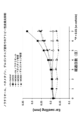
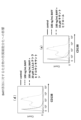
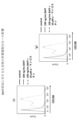
- FIG. 1 It is a graph showing changes over time in ear swelling when MC903 is applied to the ear pinna of a mouse, and noratiiol, betamethasone ointment, and delgocitinib ointment are applied to a mouse that has developed atopic dermatitis.
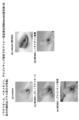
- This is a photograph showing the state of the mouse auricular skin on the 7th day after atopic dermatitis was developed in FIG. 1.
- (a) is the result of control
- (b) is the vehicle (MC903 applied)
- (c) is noratiiol
- (d) is MC903 + betamethasone ointment
- (e) is the result of MC903 + delgocitinib ointment.
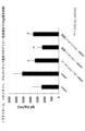
- FIG. 2 is a graph showing the measurement of TSLP secretion in the mouse ear tissue collected 7 days after FIG. 1.
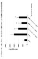
- FIG. 2 is a graph showing the measurement of IL-4 secretion in the mouse ear tissue collected after 7 days in FIG. 1.
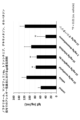
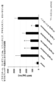
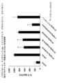
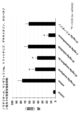
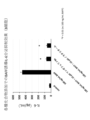
- This is a graph showing the number of nose-scratching behaviors when noratiiol, mangiferin 8a, tofacitinib, dexamethasone, and olopatadine were administered to mice that had been immunized with ovalbumin and developed allergic rhinitis.
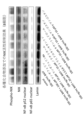
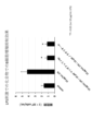
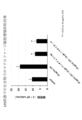
- (b) shows the results of BAFF only, and
- (c) shows the results of BAFF and mangiferin.
- (g) shows the results for BAFF and xanthohydrol
- (h) shows the results for BAFF and ⁇ -mangostin
- (i) shows the results for BAFF and ⁇ -mangostin.
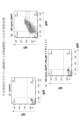
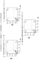
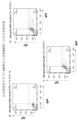
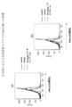
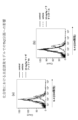
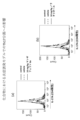
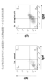
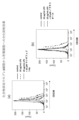
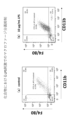
- This is a panel obtained by adding a reagent (compound) to mouse B cells and then measuring CD138 expression in cells subjected to BAFF stimulation using flow cytometry.
- (a) shows the results for mangiferin
- (b) shows the results for mangiferin 8a
- (c) shows the results for noratiol.
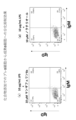
- This is a panel obtained by adding a reagent (compound) to mouse B cells and then measuring CD138 expression in cells subjected to BAFF stimulation using flow cytometry.
- (d) shows the results for 1,3,5,6-tetrahydroxyxanthone
- (e) shows the results for xanthhydrol.
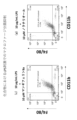
- This is a panel obtained by adding a reagent (compound) to mouse B cells and then measuring CD138 expression in cells subjected to BAFF stimulation using flow cytometry.
- (f) shows the results for ⁇ -mangosteen
- (g) shows the results for ⁇ -mangostin.
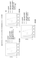
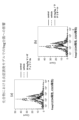
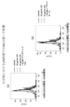
- mangiferin, mangiferin 8a, noratiol, 1,3,5,6-tetrahydroxyxanthone, xanthohydrol, ⁇ -mangostin, and ⁇ -mangostin were added to pre-B cells collected from mice, and then BAFF stimulation was performed.
- (a) shows the results for mangiferin 8a
- (b) shows the results for noratiol.
- FIG. 2 is a graph showing the results of examining the effect of each compound on suppressing the secretion of TNF ⁇ when mouse B cells are stimulated with LPS.
- 1 is a graph showing the results of examining the effect of each compound on suppressing IL-6 secretion when mouse B cells were stimulated with LPS.
- 1 is a graph showing the results of examining the effect of each compound on suppressing the amount of TNF ⁇ secretion when mouse macrophages were stimulated with LPS.
- 2 is a graph showing the results of examining the effect of each compound on suppressing IL-6 secretion when mouse macrophages were stimulated with LPS.
- compositions for improving allergic diseases will be explained below.
- These items are pharmaceutical compositions and are also NIK inhibitors. Note that the following description is an illustration of one embodiment and one example of the present invention, and the present invention is not limited to the following description. The following embodiments can be modified without departing from the spirit of the present invention.
- improvement may include not only treating the target disease or symptom, but also reducing and suppressing the symptom.
- treatment includes not only the recovery of a disease state but also the effect of suppressing or delaying the progression of a disease state.
- the pharmaceutical composition for improving allergic diseases includes mangiferin having a xanthone skeleton (formula (3)), mangiferin 8a (formula (1)), noratiol (formula (2)), and 1,3,5 , 6-tetrahydroxyxanthone (formula (4)), xanthohydrol (formula (5)), ⁇ -mangostin (formula (6)), and ⁇ -mangostin (formula (7)).
- Formula (1) is 1,2',3',4',6-penta-O-propionyl mangiferin, hereinafter referred to as "mangiferin 8a”.
- Mangiferin is shown in formula (3).
- formula (2) is 1,3,6,7-tetrahydroxyxanthone, which is hereinafter referred to as "nolathiol".
- mangiferin 8a is mangiferin (see formula (3)) in which a propionyl group is ether-bonded to some of the hydroxyl groups. As shown in the Examples below, this compound exhibits better inhibition of NIK activity than mangiferin and exhibits a high preventive effect against allergic diseases.
- Noratiiol (CAS number: 3542-72-1) is a xanthone (CAS number: 90-47-1) with a hydroxyl group added to a part thereof, and has a xanthone skeleton like mangiferin 8a. Similarly to mangiferin 8a, it inhibits NIK activity more than mangiferin and exhibits a high preventive effect against allergic diseases.
- 1,3,5,6-tetrahydroxyxanthone (CAS number: 5084-31-1: formula (4)), xanthohydrol (CAS number: 90-46-0: formula (5)), ⁇ - Mangosteen (CAS number: 6147-11-1: formula (6)) and ⁇ -mangosteen (CAS number: 31271-07-5: formula (7)) are also compounds having a xanthone skeleton, and include noratiol and mangiferin 8a. Similarly, it inhibits NIK activity more than mangiferin and exhibits a high preventive effect against allergic diseases.
- the pharmaceutical composition for improving allergic diseases according to the present invention contains at least one of the above-mentioned compounds as an active ingredient. It may also contain other ingredients acceptable as drugs. Furthermore, the pharmaceutical composition for improving allergic diseases according to the present invention may contain a plurality of these compounds.
- the pharmaceutical composition for improving allergic diseases according to the present invention can be provided as a therapeutic agent for allergic diseases (also referred to as a pharmaceutical composition).
- Compounds of the invention can also be provided as NIK inhibitors.
- the composition according to the present invention can exert its effects not only by intravenous, subcutaneous, or intramuscular injection, but also by oral administration. Therefore, it can be provided as an internal preparation.
- a powdered pharmaceutical composition for improving allergic diseases can be formulated into capsules, granules, powders, tablets, etc. and provided.
- additives such as binders, lubricants, disintegrants, coloring agents, flavoring agents, preservatives, antioxidants, and stabilizers are added, and capsules, granules, powders, and tablets are prepared. It can be manufactured by conventional methods.
- the pharmaceutical composition for improving allergic diseases according to the present invention may be formulated into external preparations such as liquids, ointments, creams, gelling agents, patches, and aerosols, and may be administered parenterally.
- external preparations such as liquids, ointments, creams, gelling agents, patches, and aerosols
- water, a lower alcohol, a solubilizing agent, a surfactant, an emulsion stabilizer, a gelling agent, an adhesive, and other necessary base components can be added.
- additives such as vascular dilating agents, adrenal corticosteroids, keratolytic agents, humectants, bactericidal agents, antioxidants, refreshing agents, fragrances, and pigments may be added as appropriate.
- the pharmaceutical composition for improving allergic diseases according to the present invention can also be provided as a processed food. That is, even if the pharmaceutical composition for improving allergic diseases according to the present invention is ingested as a processed food, it has the same effect as the pharmaceutical composition for improving allergic diseases according to the present invention.
- Processed foods include, for example, candies, gum, jelly, biscuits, cookies, rice crackers, bread, noodles, fish and meat paste products, tea, soft drinks, coffee drinks, milk drinks, whey drinks, lactic acid bacteria drinks, yogurt, and ice cream. It includes not only general processed foods including luxury foods such as creams and puddings and health foods, but also foods with health claims such as foods for specified health uses and foods with nutritional claims stipulated in the Ministry of Health, Labor and Welfare's food system with health claims, and nutritional supplements. Food (supplements), feed, food additives, etc. are also included in processed foods.
- the processed food according to the present invention can be prepared by adding a pharmaceutical composition for improving allergic diseases to the raw materials of these processed foods.
- a pharmaceutical composition for improving allergic diseases when adding mangiferin, mangiferin 8a, noratiol, 1,3,5,6-tetrahydroxyxanthone, xanthohydrol, ⁇ -mangostin, and ⁇ -mangostin to these processed foods, these substances must be added to the raw materials in the first place. Exempted from application when using materials containing However, processed foods containing these substances in excess of the content of these substances contained in the material are processed foods according to the present invention.
- the pharmaceutical composition for improving allergic diseases according to the present invention will be explained below based on Examples.
- the diseases targeted by the pharmaceutical composition for improving allergic diseases according to the present invention are as follows: bronchial asthma, atopic dermatitis, hay fever, allergic rhinitis, food allergy, metal allergy, drug allergy, anaphylaxis, and rhinitis. Measles, allergic conjunctivitis, allergic bronchitis, and contact dermatitis.
- diseases other than those mentioned above can be targeted if they have BAFF overexpression or the NF- ⁇ B pathway is activated and can be determined to be allergic diseases.
- Mangiferin 8a was obtained in two steps: synthesis of 1,3,2',3',4',6,6',7-octa-O-propionyl mangiferin as a raw material, and further synthesis based on it. .
- Noratiliol was synthesized according to the method of Non-Patent Document 47.
- the extract was washed with saturated brine, dried over anhydrous sodium sulfate, and the desiccant was filtered off using pleated filter paper.
- the solvent of the filtrate was distilled off under reduced pressure to obtain a crude product.
- Benzophenone Compound V, 8.43 g, 69%) was obtained.
- the extract was washed with saturated brine, dried over anhydrous sodium sulfate, and the desiccant was filtered off using pleated filter paper.
- the solvent of the filtrate was distilled off under reduced pressure to obtain a crude product.
- Example 1 Verification test of anti-inflammatory effect of a compound having a xanthone skeleton on atopic dermatitis> 2 nM MC903 was applied to the right auricle of a 6-week-old male Balb/c (Shimizu Jikken Materials). After 8 hours, a 5% noratiiol solution (containing 0.05 mg of noratiiol in 1 mL of ethanol), betamethasone ointment, and delgocitinib ointment were applied to the auricle for 7 days. Swelling of the auricular skin was measured using calipers. Note that the day when the compound was first applied was defined as day 0. Furthermore, betamethasone and delgocitinib are currently used clinically for atopic dermatitis, and were used as control drugs in this example.
- MC903 and noratiiol were not applied to the control group, MC903 was applied to the vehicle group, 5% noratiiol solution was applied to the 5% noratiiol group, betamethasone ointment was applied to the betamethasone group, and delgocitinib. Those who applied ointment are referred to as the delgocitinib application group.
- Figure 1 shows the results of ear swelling.
- the horizontal axis indicates the number of days that have passed, and the vertical axis indicates swelling of the auricular skin.
- a solid white circle indicates a control group (displayed as "control”).
- the solid black square indicates the vehicle group (indicated as “vehicle")
- the dotted black diamond indicates the 5% noratiiol solution application group (indicates "MC903 + 5% noratiol”)
- the dashed black cross indicates the betamethasone application group (indicates "MC903 + betamethasone ointment").
- Figure 2 shows a photograph of the right auricle of the mouse at day 7 in Figure 1.
- Figure 2(a) is the control group
- Figure 2(b) is the vehicle group
- Figure 2(c) is the 5% noratiiol solution treatment group
- Figure 2(d) is the betamethasone treatment group
- Figure 2(e) is the delgocitinib treatment group.
- a photograph of the right ear of one mouse in the group is shown.
- FIG. 2(a) no swelling of the auricular skin was observed since MC903 was not applied.
- FIG. 2(b) swelling and redness of the auricular skin are clearly observed.
- the group treated with 5% Noratiiol solution suppressed inflammation caused by atopic dermatitis significantly more than the vehicle group.
- a composition containing norachiiol as an active ingredient can suppress inflammation caused by atopic dermatitis.
- Example 2 IgE antibody production suppression effect> Serum was collected from the mouse on day 7 in Example 1, and the amount of IgE antibody produced was measured by enzyme-linked immunosorbent assay (ELISA). This measurement was performed using a mouse IgE antibody ELISA kit (R&D).
- ELISA enzyme-linked immunosorbent assay
- the results are shown in Figure 3.
- the horizontal axis represents the sample group, and the vertical axis represents the amount of IgE antibody.
- the IgE antibody amount increased significantly to 2588 ng/mL, indicating that atopic dermatitis had developed. I know that there is.
- IgE antibody production was significantly lower in the noratiiol, betamethasone, and delgocitinib groups: 1692 ng/mL in the noratiiol treatment group, 1175 ng/mL in the betamethasone treatment group, and 1812 ng/mL in the delgocitinib treatment group. did.
- a composition containing norachiiol as an active ingredient can suppress IgE antibody production caused by atopic dermatitis.
- Example 3 TSLP secretion suppressing effect> Tissues were collected from the mouse on day 7 in Example 1, and the amount of TSLP secreted was measured by enzyme-linked immunosorbent assay (ELISA). This measurement was performed using a mouse TSLP ELISA kit (R&D).
- ELISA enzyme-linked immunosorbent assay
- the results are shown in Figure 4.
- the horizontal axis represents the sample group, and the vertical axis represents the TSLP secretion amount.
- the TSLP secretion amount increased significantly to 1533 pg/mL, indicating that atopic dermatitis developed. I know that there is.
- TSLP secretion was significantly lower in the noratiiol, betamethasone, and delgocitinib groups: 1026 pg/mL in the noratiiol treatment group, 651 pg/mL in the betamethasone treatment group, and 971 pg/mL in the delgocitinib treatment group.
- a composition containing norachiiol as an active ingredient can suppress TSLP secretion caused by atopic dermatitis.
- Example 4 IL-4 secretion suppressing effect> Tissues were collected from the mouse on day 7 in Example 1, and the amount of IL-4 secreted was measured by enzyme-linked immunosorbent assay (ELISA). This measurement was performed using a mouse IL-4 ELISA kit (R&D).
- ELISA enzyme-linked immunosorbent assay
- the results are shown in Figure 5.
- the horizontal axis represents the sample group, and the vertical axis represents the amount of IL-4 secreted.
- the IL-4 secretion level increased significantly to 152 pg/mL, and atopic dermatitis developed. I know what you're doing.
- IL-4 secretion levels were significantly reduced in the noratiiol-treated group and the betamethasone-treated group, with 94 pg/mL in the noratiiol-treated group, 45 pg/mL in the betamethasone-treated group, and 159 pg/mL in the delgocitinib-treated group.
- a composition containing noratiol as an active ingredient can suppress IL-4 secretion.
- Example 5 IL-5 secretion suppressing effect> Tissues were collected from the mouse on day 7 in Example 1, and the amount of IL-5 secreted was measured by enzyme-linked immunosorbent assay (ELISA). This measurement was performed using a mouse IL-5 ELISA kit (R&D).
- ELISA enzyme-linked immunosorbent assay
- the results are shown in Figure 6.
- the horizontal axis represents sample groups, and the vertical axis represents IL-5 secretion amount.
- the IL-5 secretion amount increased significantly to 32 pg/mL, and atopic dermatitis developed. I know what you're doing.
- the IL-5 secretion amount was significantly reduced in the noratiiol-treated group and the betamethasone-treated group, as compared to the vehicle group: 14 pg/mL in the noratiiol-treated group, 8 pg/mL in the betamethasone-treated group, and 33 pg/mL in the delgocitinib-treated group.
- a composition containing noratiol as an active ingredient can suppress IL-5 secretion.
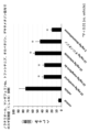
- Example 6 Effect of suppressing rhinitis (nose scratching behavior) in ovalbumin-induced allergic rhinitis model mice> 0.2 mL of a mixture of 50 ⁇ g ovalbumin (OVA) and 2 mg Alum was administered to 6-week-old female Balb/c mice (Shimizu Experimental Materials) (day 0 from the start of the test). Alum was used as an adjuvant. After 7 days and 14 days, Balb/c mice were again injected with 0.2 mL of the above mixture for immunization.
- OVA ovalbumin
- Alum was used as an adjuvant.
- mice were treated with 10 mg/kg of noratiol, 10 mg/kg of mangiferin 8a, 10 mg/kg of tofacitinib (JAK inhibitor), 2 mg/kg of dexamethasone (steroid), and 10 mg/kg of olopatadine (antihistamine). Oral administration was carried out every day for 8 days. Also, from the 21st day, 400 ⁇ g OVA was administered nasally, and the nose scratching behavior was measured on the 27th day. Note that tofacitinib, dexamethasone, and olopatadine are currently used clinically for allergic rhinitis, and were used as control drugs in this example.
- the group that was only immunized with OVA was the control group, the group that received noratiiol was the noratiriol group, the mangiferin 8a group was the mangiferin 8a group, the tofacitinib group was the tofacitinib group, and the dexamethasone group was the dexamethasone group.
- the administration group and the group to which olopatadine was administered are referred to as the olopatadine administration group. Each group consisted of 5 animals.
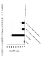
- the results are shown in Figure 7.
- the horizontal axis represents the sample group, and the vertical axis represents the number of nose scratches.
- the OVA-immunized control group (indicated as "vehicle") showed a marked increase in nose-scratching behavior, indicating that they had developed allergic rhinitis.
- Norathiriol administration group (written as "10mg/kg norathyriol"), Mangiferin 8a administration group (written as “10mg/kg Mangiferin 8a”), Tofacitinib administration group (written as "10mg/kg tofacitinib”), Dexamethasone administration group (written as "2 mg
- the number of nose scratches was significantly reduced in the olopatadine administration group (expressed as "10 mg/kg olopatadine”) and the vehicle group (expressed as "10 mg/kg olopatadine”). Furthermore, nose scratching behavior was significantly suppressed in the noratiiol administration group and the mangiferin 8a administration group, compared to the tofacitinib administration group and the olopatadine administration group.
- a composition containing noratiiol or mangiferin 8a as an active ingredient can suppress nose scratching behavior caused by allergic rhinitis.
- OVA ovalbumin
- Alum was used as an adjuvant.
- mice were treated with 10 mg/kg of noratiol, 10 mg/kg of mangiferin 8a, 10 mg/kg of tofacitinib (JAK inhibitor), 2 mg/kg of dexamethasone (steroid), and 10 mg/kg of olopatadine (antihistamine). Oral administration was carried out every day for 8 days. Also, from the 21st day, 400 ⁇ g OVA was administered nasally, and the number of sneezes was measured on the 27th day. Note that tofacitinib, dexamethasone, and olopatadine are currently used clinically for allergic rhinitis, and were used as control drugs in this example.
- control The group not immunized with OVA
- control the group only immunized with OVA
- control group the group only immunized with OVA
- vehicle the control group
- noratiiol the noratiiol administration group (denoted as "10 mg/kg norathyriol").
- Mangiferin 8a administration group (described as “10mg/kg mangiferin 8a")
- Tofacitinib administration group dexamethasone
- the group administered with olopatadine is referred to as the dexamethasone administration group (denoted as "2 mg/kg dexamethasone")
- the group administered with olopatadine is referred to as the olopatadine administration group (denoted as "10 mg/kg olopatadine”).
- Each group consisted of 5 animals.
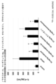
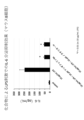
- the results are shown in Figure 8.
- the horizontal axis represents the sample group, and the vertical axis represents the number of sneezes.
- the OVA-immunized control group showed a marked increase in sneezing, indicating that they had developed allergic rhinitis.
- the number of sneezes was significantly reduced in the noratiiol administration group, the mangiferin 8a administration group, the tofacitinib administration group, the dexamethasone administration group, and the olopatadine administration group compared to the vehicle group.
- the number of sneezes was significantly suppressed in the noratiiol administration group and the mangiferin 8a administration group, compared to the tofacitinib administration group and the olopatadine administration group.
- a composition containing noratiol or mangiferin 8a as an active ingredient can suppress sneezing caused by allergic rhinitis.
- Example 8 OVA-IgE antibody production suppressing effect> Serum was collected from the mice on day 28 in Examples 6 and 7, and the amount of OVA-IgE antibody produced was measured by enzyme-linked immunosorbent assay (ELISA). This measurement was performed using a mouse OVA-IgE antibody ELISA kit (FUJIFILMWAKO).
- ELISA enzyme-linked immunosorbent assay
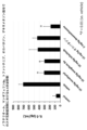
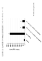
- the results are shown in Figure 9.
- the horizontal axis represents the sample group, and the vertical axis represents the amount of OVA-IgE antibody.
- the OVA-IgE antibody level was 2.7 U/mL in the non-OVA-immunized control group, while it was 69 U/mL in the OVA-immunized control group (indicated as "vehicle"), indicating a marked increase in OVA-IgE antibody level, which was associated with allergic rhinitis. I know it's happening.
- the mangiferin 8a administration group, and the dexamethasone administration group showed a marked decrease in OVA-IgE antibody production. Furthermore, the noratiiol-administered group and the mangiferin 8a-administered group significantly suppressed OVA-IgE antibody production compared to the tofacitinib-administered group and the olopatadine-administered group.
- a composition containing noratiiol or mangiferin 8a as an active ingredient can suppress OVA-IgE antibody production caused by allergic rhinitis.
- Example 9 IgE antibody production suppression effect> Serum was collected from the mice on day 28 in Examples 6 and 7, and the amount of IgE antibody produced was measured by enzyme-linked immunosorbent assay (ELISA). This measurement was performed using a mouse IgE antibody ELISA kit (R&D).
- ELISA enzyme-linked immunosorbent assay
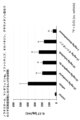
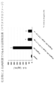
- the results are shown in Figure 10.
- the horizontal axis represents the sample group, and the vertical axis represents the amount of IgE antibody.
- the IgE antibody level increased significantly to 111 ng/mL in the OVA-immunized control group (indicated as "vehicle"), while it was 58 ng/mL in the non-OVA-immunized control group, and they developed allergic rhinitis. I understand that.
- IgE antibody production was significantly suppressed in the noratiiol-administered group and the mangiferin 8a-administered group compared with the tofacitinib-administered group and the olopatadine-administered group.
- a composition containing noratiol or mangiferin 8a as an active ingredient can suppress IgE antibody production caused by allergic rhinitis.
- Example 10 BAFF secretion suppressing effect> Serum was collected from the mice on day 28 in Examples 6 and 7, and BAFF secretion was measured by enzyme-linked immunosorbent assay (ELISA). This measurement was performed using a mouse BAFF ELISA kit (R&D).
- ELISA enzyme-linked immunosorbent assay
- the results are shown in Figure 11.
- the horizontal axis represents the sample group, and the vertical axis represents the BAFF secretion amount.
- BAFF secretion increased significantly to 1450 pg/mL in the OVA-immunized control group (indicated as "vehicle"), while it was 157 pg/mL in non-OVA-immunized controls, and allergic rhinitis developed. I understand that.
- 629 pg/mL in the noratiol administration group (written as "10 mg/kg norathyriol")
- 668 pg/mL in the mangiferin 8a administration group (written as "10 mg/kg mangiferin 8a")
- 668 pg/mL in the tofacitinib administration group (written as "10 mg/kg tofacitin 8a").
- BAFF secretion was significantly decreased in the group treated with mangiferin 8a and the group treated with dexamethasone. Furthermore, BAFF secretion was significantly suppressed in the noratiiol administration group and the mangiferin 8a administration group, compared to the tofacitinib administration group and the olopatadine administration group.
- a composition containing noratiol or mangiferin 8a as an active ingredient can suppress BAFF secretion caused by allergic rhinitis.
- IL-4 secretion suppressing effect Serum was collected from the mice on day 28 in Examples 6 and 7, and the amount of IL-4 secreted was measured by enzyme-linked immunosorbent assay (ELISA). This measurement was performed using a mouse IL-4 ELISA kit (R&D).
- ELISA enzyme-linked immunosorbent assay
- the results are shown in Figure 12.
- the horizontal axis represents the sample group, and the vertical axis represents the amount of IL-4 secretion.
- the IL-4 secretion level was 227 pg/mL in the non-OVA-immunized control group, while it was 872 pg/mL in the OVA-immunized control group (indicated as "vehicle"), which resulted in the development of allergic rhinitis.
- IL-4 secretion level was significantly decreased in the group treated with mangiferin 8a and the group treated with dexamethasone.
- the noratiiol administration group and the mangiferin 8a administration group significantly suppressed IL-4 secretion compared to the tofacitinib administration group and the olopatadine administration group.
- a composition containing noratiol or mangiferin 8a as an active ingredient can suppress IL-4 secretion caused by allergic rhinitis.
- IL-5 secretion suppressing effect Serum was collected from the mice on day 28 in Examples 6 and 7, and the amount of IL-5 secreted was measured by enzyme-linked immunosorbent assay (ELISA). This measurement was performed using a mouse IL-5 ELISA kit (R&D).
- ELISA enzyme-linked immunosorbent assay
- the results are shown in Figure 13.
- the horizontal axis represents sample groups, and the vertical axis represents IL-5 secretion amount.
- the IL-5 secretion amount was 151 pg/mL in the OVA-immunized control group, whereas it was 1010 pg/mL in the OVA-immunized control group (indicated as "vehicle”), which caused allergic rhinitis to develop.
- IL-5 secretion level was significantly decreased in the group treated with mangiferin 8a and the group treated with dexamethasone. Furthermore, the noratiiol administration group and the mangiferin 8a administration group significantly suppressed IL-5 secretion compared to the tofacitinib administration group and the olopatadine administration group.
- a composition containing noratiol or mangiferin 8a as an active ingredient can suppress IL-5 secretion caused by allergic rhinitis.
- IL-17 secretion inhibitory effect Serum was collected from the mice on day 28 in Examples 6 and 7, and the amount of IL-17 secreted was measured by enzyme-linked immunosorbent assay (ELISA). This measurement was performed using a mouse IL-17 ELISA kit (R&D).
- ELISA enzyme-linked immunosorbent assay
- the results are shown in Figure 14.
- the horizontal axis represents sample groups, and the vertical axis represents IL-17 secretion amount.
- the IL-17 secretion level was 204 pg/mL in the non-OVA-immunized control group, while it was 945 pg/mL in the OVA-immunized control group (indicated as "vehicle"), which resulted in the development of allergic rhinitis.
- IL-17 secretion level was significantly decreased in the group treated with mangiferin 8a and the group treated with dexamethasone.
- the noratiiol-administered group and the mangiferin 8a-administered group significantly suppressed IL-17 secretion compared to the tofacitinib-administered group and the olopatadine-administered group.
- a composition containing noratiol or mangiferin 8a as an active ingredient can suppress IL-17 secretion caused by allergic rhinitis.
- Example 14 Cough suppressing effect in ovalbumin-induced bronchial asthma model mice> 0.2 mL of a mixture of 50 ⁇ g ovalbumin (OVA) and 2 mg Alum was administered to 6-week-old female Balb/c mice (Shimizu Experimental Materials) (day 0 from the start of the test). Alum was used as an adjuvant. After 7 days and 14 days, Balb/c mice were again injected with 0.2 mL of the above mixture for immunization.
- OVA ovalbumin
- Alum was used as an adjuvant.
- Balb/c mice were again injected with 0.2 mL of the above mixture for immunization.
- mice were treated with 10 mg/kg of noratiol, 10 mg/kg of mangiferin 8a, 10 mg/kg of tofacitinib (JAK inhibitor), 2 mg/kg of dexamethasone (steroid), and 10 mg/kg of olopatadine (antihistamine). Oral administration was carried out every day for 8 days. Also, from the 21st day, 400 ⁇ g OVA was administered nasally, and the number of coughs was measured on the 27th day. Note that tofacitinib, dexamethasone, and olopatadine are currently used clinically for bronchial asthma, and were used as control drugs in this example.
- the group that was only immunized with OVA was the control group, the group that received noratiiol was the noratiriol group, the mangiferin 8a group was the mangiferin 8a group, the tofacitinib group was the tofacitinib group, and the dexamethasone group was the dexamethasone group.
- the administration group and the group to which olopatadine was administered are referred to as the olopatadine administration group. Each group consisted of 5 animals.
- the results are shown in Figure 15.
- the horizontal axis represents the sample group, and the vertical axis represents the number of coughs.
- coughing was significantly increased in the OVA-immunized control group (indicated as "vehicle"), indicating that they had developed bronchial asthma.
- vehicle group in the noratiol administration group (written as "10 mg/kg norathyriol")
- mangiferin 8a administration group written as "10 mg/kg mangiferin 8a”
- the dexamethasone administration group written as "2 mg/kg dexamethasone
- the number of coughs decreased significantly.
- the noratiiol-administered group and the mangiferin 8a-administered group significantly suppressed cough compared to the tofacitinib-administered group (described as "10 mg/kg tofacitinib") and the olopatadine-administered group (described as "10 mg/kg olopatadine").
- a composition containing noratiol or mangiferin 8a as an active ingredient can suppress coughing caused by bronchial asthma.
- Example 15 OVA-IgE antibody production suppressing effect> Serum was collected from the mouse on day 28 in Example 14, and the amount of OVA-IgE antibody produced was measured by enzyme-linked immunosorbent assay (ELISA). This measurement was performed using a mouse OVA-IgE antibody ELISA kit (FUJIFILMWAKO).
- ELISA enzyme-linked immunosorbent assay
- the results are shown in Figure 16.
- the horizontal axis represents the sample group, and the vertical axis represents the amount of OVA-IgE antibody.
- OVA-IgE antibody levels increased significantly to 66 U/mL in the OVA-immunized control group (indicated as "vehicle"), compared to 2.4 U/mL in non-OVA-immunized controls, resulting in the onset of bronchial asthma. I know what you're doing.
- the noratiiol-administered group and the mangiferin 8a-administered group significantly suppressed OVA-IgE antibody production compared to the tofacitinib-administered group and the olopatadine-administered group.
- a composition containing noratiiol or mangiferin 8a as an active ingredient can suppress OVA-IgE antibody production caused by bronchial asthma.
- Example 16 IgE antibody production suppression effect> Serum was collected from the mouse on day 28 in Example 14, and the amount of IgE antibody produced was measured by enzyme-linked immunosorbent assay (ELISA). This measurement was performed using a mouse IgE antibody ELISA kit (R&D).
- ELISA enzyme-linked immunosorbent assay
- the results are shown in Figure 17.
- the horizontal axis represents the sample group, and the vertical axis represents the amount of IgE antibody.
- the IgE antibody level was 73 ng/mL in the non-OVA-immunized control group, while it was 117 ng/mL in the OVA-immunized control group (indicated as "vehicle"), indicating that they had developed bronchial asthma. I understand.
- the amount of IgE antibody produced was significantly reduced. Furthermore, IgE antibody production was significantly suppressed in the noratiiol-administered group and the mangiferin 8a-administered group compared with the tofacitinib-administered group and the olopatadine-administered group.
- a composition containing noratiol or mangiferin 8a as an active ingredient can suppress IgE antibody production caused by bronchial asthma.
- Example 17 BAFF secretion suppressing effect> Serum was collected from the mouse on day 28 in Example 14, and the amount of BAFF secreted was measured by enzyme-linked immunosorbent assay (ELISA). This measurement was performed using a mouse BAFF ELISA kit (R&D).
- ELISA enzyme-linked immunosorbent assay
- the results are shown in Figure 18.
- the horizontal axis represents the sample group, and the vertical axis represents the BAFF secretion amount.
- BAFF secretion increased significantly to 1319 pg/mL in the OVA-immunized control group (indicated as "vehicle"), whereas it was 97 pg/mL in the non-OVA-immunized control, indicating that they had developed bronchial asthma. I understand.
- BAFF secretion was significantly decreased in the group treated with mangiferin 8a and the group treated with dexamethasone. Furthermore, BAFF secretion was significantly suppressed in the noratiiol administration group and the mangiferin 8a administration group, compared to the tofacitinib administration group and the olopatadine administration group.
- a composition containing noratiol or mangiferin 8a as an active ingredient can suppress BAFF secretion caused by bronchial asthma.
- Example 18 IL-4 secretion suppressing effect> Serum was collected from the mouse on day 28 in Example 14, and the amount of IL-4 secreted was measured by enzyme-linked immunosorbent assay (ELISA). This measurement was performed using a mouse IL-4 ELISA kit (R&D).
- ELISA enzyme-linked immunosorbent assay
- IL-4 secretion was 282 pg/mL in the non-OVA-immunized control group, while it was 863 pg/mL in the OVA-immunized control group (indicated as "vehicle"), indicating that they developed bronchial asthma. I know that there is.
- a composition containing noratiol or mangiferin 8a as an active ingredient can suppress IL-4 secretion caused by bronchial asthma.
- Example 19 IL-5 secretion suppressing effect> Serum was collected from the mouse on day 28 in Example 14, and the amount of IL-5 secreted was measured by enzyme-linked immunosorbent assay (ELISA). This measurement was performed using a mouse IL-5 ELISA kit (R&D).
- ELISA enzyme-linked immunosorbent assay
- the results are shown in Figure 20.
- the horizontal axis represents sample groups, and the vertical axis represents IL-5 secretion amount.
- the IL-5 secretion level was 80 pg/mL in the non-OVA-immunized control group, while it was 992 pg/mL in the OVA-immunized control group (indicated as "vehicle"), indicating that they developed bronchial asthma. I know that there is.
- 601 pg/mL in the noratiol administration group (written as "10 mg/kg norathyriol"), 689 pg/mL in the mangiferin 8a administration group (written as "10 mg/kg mangiferin 8a"), and 689 pg/mL in the tofacitinib administration group (written as "10 mg/kg tofacitin written as “ib” ), 833 pg/mL in the dexamethasone administration group (described as "2 mg/kg dexamethasone"), 286 pg/mL in the olopatadine administration group (described as "10 mg/kg olopatadine”), and 866 pg/mL in the olopatadine administration group (described as "10 mg/kg olopatadine”).
- IL-5 secretion level was significantly decreased in the group treated with mangiferin 8a and the group treated with dexamethasone. Furthermore, the noratiiol administration group and the mangiferin 8a administration group significantly suppressed IL-5 secretion compared to the tofacitinib administration group and the olopatadine administration group.
- a composition containing noratiol or mangiferin 8a as an active ingredient can suppress IL-5 secretion caused by bronchial asthma.
- Example 20 IL-17 secretion suppressing effect> Serum was collected from the mouse on day 28 in Example 14, and the amount of IL-17 secreted was measured by enzyme-linked immunosorbent assay (ELISA). This measurement was performed using a mouse IL-17 ELISA kit (R&D).
- ELISA enzyme-linked immunosorbent assay
- the results are shown in Figure 21.
- the horizontal axis represents sample groups, and the vertical axis represents IL-17 secretion amount.
- the IL-17 secretion amount was 80 pg/mL in the OVA-immunized control group, whereas it was 898 pg/mL in the OVA-immunized control group (indicated as "vehicle"), indicating that they developed bronchial asthma. I know that there is.
- IL-17 secretion level was significantly decreased in the group treated with mangiferin 8a and the group treated with dexamethasone.
- the noratiiol-administered group and the mangiferin 8a-administered group significantly suppressed IL-17 secretion compared to the tofacitinib-administered group and the olopatadine-administered group.
- a composition containing noratiiol or mangiferin 8a as an active ingredient can suppress IL-17 secretion caused by bronchial asthma.
- Example 21 Diarrhea suppressive effect in ovalbumin-induced food allergy model mice> 0.2 mL of a mixture of 10 ⁇ g ovalbumin (OVA) and 1 mg Alum was administered to 6-week-old female Balb/c mice (Shimizu Experimental Materials) (day 0 from the start of the test). Alum was used as an adjuvant. Seven days later, Balb/c mice were again injected with 0.2 mL of the above mixture for immunization.
- OVA ovalbumin
- Alum was used as an adjuvant. Seven days later, Balb/c mice were again injected with 0.2 mL of the above mixture for immunization.
- mice were treated with 10 mg/kg of noratiol, 10 mg/kg of mangiferin 8a, 10 mg/kg of tofacitinib (JAK inhibitor), 2 mg/kg of dexamethasone (steroid), and 10 mg/kg of olopatadine (antihistamine). Oral administration was carried out every day for 14 days. From day 14, 10 mg OVA was orally administered every other day until day 21, and after the final administration on day 28, the frequency of diarrhea was measured. Note that tofacitinib, dexamethasone, and olopatadine are currently used clinically for food allergies, and were used as control drugs in this example.
- the group that was only immunized with OVA was the control group, the group that received noratiiol was the noratiriol group, the mangiferin 8a group was the mangiferin 8a group, the tofacitinib group was the tofacitinib group, and the dexamethasone group was the dexamethasone group.
- the administration group and the group to which olopatadine was administered are referred to as the olopatadine administration group. Each group consisted of 5 animals.
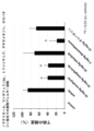
- the results are shown in Figure 22.
- the horizontal axis represents the sample group, and the vertical axis represents the frequency (%) of diarrhea.
- the OVA-immunized control group (indicated as "vehicle") showed a marked increase in the frequency of diarrhea, indicating that they had developed food allergies.
- Comparison with the vehicle group in the noratiol administration group (written as "10 mg/kg norathyriol"), the mangiferin 8a administration group (written as "10 mg/kg mangiferin 8a”), and the dexamethasone administration group (written as "2 mg/kg dexamethasone”)
- the frequency of diarrhea was significantly reduced.
- the noratiiol-administered group and the mangiferin 8a-administered group significantly suppressed the occurrence of diarrhea compared to the tofacitinib-administered group (described as "10mg/kg tofacitinib") and the olopatadine-administered group (described as "10mg/kg olopatadine”).
- a composition containing noratiol or mangiferin 8a as an active ingredient can suppress diarrhea caused by food allergy.
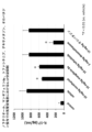
- Example 22 OVA-IgE antibody production suppressing effect> Serum was collected from the mouse on day 28 in Example 21, and the amount of OVA-IgE antibody produced was measured by enzyme-linked immunosorbent assay (ELISA). This measurement was performed using a mouse OVA-IgE antibody ELISA kit (FUJIFILMWAKO).
- ELISA enzyme-linked immunosorbent assay
- the results are shown in Figure 23.
- the horizontal axis represents the sample group, and the vertical axis represents the amount of OVA-IgE antibody.
- the OVA-IgE antibody level increased significantly to 74 U/mL in the OVA-immunized control group (indicated as "vehicle"), compared to 1.5 U/mL in the non-OVA-immunized control group, resulting in food allergy. I know what you're doing.
- the noratiiol-administered group and the mangiferin 8a-administered group significantly suppressed OVA-IgE antibody production compared to the tofacitinib-administered group and the olopatadine-administered group.
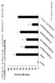
- Example 23 IgE antibody production suppression effect> Serum was collected from the mouse on day 28 in Example 21, and the amount of IgE antibody produced was measured by enzyme-linked immunosorbent assay (ELISA). This measurement was performed using a mouse IgE antibody ELISA kit (R&D).
- ELISA enzyme-linked immunosorbent assay
- the results are shown in Figure 24.
- the horizontal axis represents the sample group, and the vertical axis represents the amount of IgE antibody.
- the IgE antibody level was 56 ng/mL in the OVA non-immunized control group, while it was 124 ng/mL in the OVA immunized control group (indicated as "vehicle"), indicating that they had developed a food allergy. I understand.
- IgE antibody production was significantly suppressed in the noratiiol-administered group and the mangiferin 8a-administered group compared with the tofacitinib-administered group and the olopatadine-administered group.
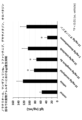
- Example 24 BAFF secretion inhibitory effect> Serum was collected from the mouse on day 28 in Example 21, and the amount of BAFF secreted was measured by enzyme-linked immunosorbent assay (ELISA). This measurement was performed using a mouse BAFF ELISA kit (R&D).
- ELISA enzyme-linked immunosorbent assay
- the results are shown in Figure 25.
- the horizontal axis represents the sample group, and the vertical axis represents the BAFF secretion amount.
- BAFF secretion amount increased significantly to 845 pg/mL in the OVA-immunized control group (indicated as "vehicle"), while it was 134 pg/mL in the OVA non-immunized control, indicating that they had developed food allergy. I understand.
- BAFF secretion was significantly decreased in the group treated with mangiferin 8a and the group treated with dexamethasone. Furthermore, BAFF secretion was significantly suppressed in the noratiiol administration group and the mangiferin 8a administration group, compared to the tofacitinib administration group and the olopatadine administration group.
- a composition containing noratiiol or mangiferin 8a as an active ingredient can suppress BAFF secretion caused by food allergy.
- Example 25 IL-4 secretion suppressing effect> Serum was collected from the mouse on day 28 in Example 21, and the amount of IL-4 secreted was measured by enzyme-linked immunosorbent assay (ELISA). This measurement was performed using a mouse IL-4 ELISA kit (R&D).
- ELISA enzyme-linked immunosorbent assay
- the results are shown in Figure 26.
- the horizontal axis represents the sample group, and the vertical axis represents the amount of IL-4 secretion.
- the IL-4 secretion level was 210 pg/mL in the non-OVA-immunized control group, while it was 1154 pg/mL in the OVA-immunized control group (indicated as "vehicle"), indicating that they developed food allergy. I know that there is.
- IL-4 secretion level was significantly decreased in the group treated with mangiferin 8a and the group treated with dexamethasone. Furthermore, the noratiiol administration group and the mangiferin 8a administration group significantly suppressed IL-4 secretion compared to the tofacitinib administration group and the olopatadine administration group.
- a composition containing noratiiol or mangiferin 8a as an active ingredient can suppress IL-4 secretion caused by food allergy.
- Example 26 IL-5 secretion suppressing effect> Serum was collected from the mouse on day 28 in Example 21, and the amount of IL-5 secreted was measured by enzyme-linked immunosorbent assay (ELISA). This measurement was performed using a mouse IL-5 ELISA kit (R&D).
- ELISA enzyme-linked immunosorbent assay
- the results are shown in Figure 27.
- the horizontal axis represents sample groups, and the vertical axis represents IL-5 secretion amount.
- the IL-5 secretion amount was 135 pg/mL in the non-OVA immunized control group, while it was 916 pg/mL in the OVA immunized control group (indicated as "vehicle"), indicating that the IL-5 secretion level was significantly increased, indicating that they developed food allergy. I know that there is.
- IL-5 secretion level was significantly decreased in the group treated with mangiferin 8a and the group treated with dexamethasone. Furthermore, the noratiiol administration group and the mangiferin 8a administration group significantly suppressed IL-5 secretion compared to the tofacitinib administration group and the olopatadine administration group.
- a composition containing noratiol or mangiferin 8a as an active ingredient can suppress IL-5 secretion caused by food allergy.
- Example 27 IL-17 secretion suppressing effect> Serum was collected from the mouse on day 28 in Example 21, and the amount of IL-17 secreted was measured by enzyme-linked immunosorbent assay (ELISA). This measurement was performed using a mouse IL-17 ELISA kit (R&D).
- ELISA enzyme-linked immunosorbent assay
- IL-17 secretion was 98 pg/mL in the non-OVA immunized control group, while it was 875 pg/mL in the OVA-immunized control group (indicated as "vehicle"), indicating that they developed food allergy. I know that there is.
- IL-17 secretion level was significantly decreased in the group treated with mangiferin 8a and the group treated with dexamethasone.
- the noratiiol-administered group and the mangiferin 8a-administered group significantly suppressed IL-17 secretion compared to the tofacitinib-administered group and the olopatadine-administered group.
- a composition containing noratiol or mangiferin 8a as an active ingredient can suppress IL-17 secretion caused by food allergy.
- ⁇ Example 28 Effect of suppressing rhinitis (nose scratching behavior) in cedar pollinosis model mice> 0.2 mL of a mixture of 100 ⁇ g cedar pollen and 1 mg Alum was administered to 6-week-old female Balb/c mice (Shimizu Experimental Materials) (day 0 from the start of the test). Alum was used as an adjuvant. After 7 days and 14 days, Balb/c mice were again injected with 0.2 mL of the above mixture for immunization.
- mice were treated with 10 mg/kg of noratiol, 10 mg/kg of mangiferin 8a, 10 mg/kg of tofacitinib (JAK inhibitor), 2 mg/kg of dexamethasone (steroid), and 10 mg/kg of olopatadine (antihistamine). Oral administration was carried out every day for 8 days. In addition, from the 21st day, 500 ⁇ g of cedar pollen was administered nasally, and nose scratching behavior was measured on the 27th day. Note that tofacitinib, dexamethasone, and olopatadine are currently used clinically for hay fever, and were used as control drugs in this example.
- the group immunized only with cedar pollen was the control group
- the group immunized with noratiiol was the noratiriol group
- the mangiferin 8a group was the mangiferin 8a group
- the tofacitinib group was the tofacitinib group
- the dexamethasone group was the immunized group.
- the dexamethasone administration group and the olopatadine administration group are referred to as the olopatadine administration group.
- Each group consisted of 5 animals.
- the results are shown in Figure 29.
- the horizontal axis represents the sample group, and the vertical axis represents the number of nose scratches.
- the subject group immunized with cedar pollen (indicated as "vehicle") showed a marked increase in nose scratching behavior, indicating that they had developed cedar pollen allergy.
- Norathiriol administration group (written as "10mg/kg norathyriol"), Mangiferin 8a administration group (written as “10mg/kg Mangiferin 8a”), Tofacitinib administration group (written as "10mg/kg tofacitinib”), Dexamethasone administration group (written as "2 mg
- the number of nose scratches was significantly reduced in the olopatadine administration group (expressed as "10 mg/kg olopatadine”) and the vehicle group (expressed as "10 mg/kg olopatadine”). Furthermore, nose scratching behavior was significantly suppressed in the noratiiol administration group and the mangiferin 8a administration group, compared to the tofacitinib administration group and the olopatadine administration group.
- a composition containing norachiiol or mangiferin 8a as an active ingredient can suppress nose scratching behavior caused by cedar pollen allergy.
- mice were treated with 10 mg/kg of noratiol, 10 mg/kg of mangiferin 8a, 10 mg/kg of tofacitinib (JAK inhibitor), 2 mg/kg of dexamethasone (steroid), and 10 mg/kg of olopatadine (antihistamine). Oral administration was carried out every day for 8 days. Also, from the 21st day, 500 ⁇ g of cedar pollen was administered nasally, and the number of sneezes was measured on the 27th day. Note that tofacitinib, dexamethasone, and olopatadine are currently used clinically for hay fever, and were used as control drugs in this example.
- the group that was not immunized with cedar pollen was the control group (denoted as "control")
- the group that was only immunized with cedar pollen was the control group (denoted as "vehicle")
- the group that was administered with noratiiol was the noratiiol administration group (denoted as "10 mg/kg norathyriol)
- Mangiferin 8a administration group (described as ⁇ 10mg/kg mangiferin 8a'')
- Tofacitinib administration group described as ⁇ 10mg/kg tofacitinib''
- Dexamethasone The group administered with olopatadine is referred to as the dexamethasone administration group (denoted as "2 mg/kg dexamethasone")
- the group administered with olopatadine is referred to as the olopatadine administration group (denoted as "10 mg/kg olopatadine”).
- Each group
- the results are shown in Figure 30.
- the horizontal axis represents the sample group, and the vertical axis represents the number of sneezes.
- sneezing increased significantly in the control group immunized with cedar pollen, indicating that they had developed cedar pollen allergy.
- the number of sneezes was significantly reduced in the noratiiol administration group, the mangiferin 8a administration group, the tofacitinib administration group, the dexamethasone administration group, and the olopatadine administration group compared to the vehicle group.
- the number of sneezes was significantly suppressed in the noratiiol administration group and the mangiferin 8a administration group, compared to the tofacitinib administration group and the olopatadine administration group.
- a composition containing norachiiol or mangiferin 8a as an active ingredient can suppress sneezing caused by cedar pollen allergy.
- Example 30 IgE antibody production suppression effect> Serum was collected from the mice on day 28 in Examples 28 and 29, and the amount of IgE antibody produced was measured by enzyme-linked immunosorbent assay (ELISA). This measurement was performed using a mouse IgE antibody ELISA kit (R&D).
- ELISA enzyme-linked immunosorbent assay
- the results are shown in Figure 31.
- the horizontal axis represents the sample group, and the vertical axis represents the amount of IgE antibody.
- the IgE antibody level was 71 ng/mL in the non-immunized control group with cedar pollen, whereas in the control group immunized with cedar pollen (indicated as "vehicle"), the amount of IgE antibodies increased significantly to 207 ng/mL, indicating that they developed cedar pollen allergy.
- IgE antibody production was significantly suppressed in the noratiiol-administered group and the mangiferin 8a-administered group compared with the tofacitinib-administered group and the olopatadine-administered group.
- a composition containing noratiol or mangiferin 8a as an active ingredient can suppress IgE antibody production caused by cedar pollen allergy.
- IL-4 secretion inhibitory effect Serum was collected from the mice on day 28 in Examples 28 and 29, and the amount of IL-4 secreted was measured by enzyme-linked immunosorbent assay (ELISA). This measurement was performed using a mouse IL-4 ELISA kit (R&D).
- ELISA enzyme-linked immunosorbent assay
- the results are shown in Figure 32.
- the horizontal axis represents the sample group, and the vertical axis represents the amount of IL-4 secreted.
- the IL-4 secretion amount was 55 pg/mL in the control group not immunized with cedar pollen, while it was 374 pg/mL in the control group immunized with cedar pollen (indicated as "vehicle"), indicating that the IL-4 secretion level was significantly increased. I know it's happening.
- IL-4 secretion level was significantly decreased in the group treated with mangiferin 8a and the group treated with dexamethasone. Furthermore, the noratiiol administration group and the mangiferin 8a administration group significantly suppressed IL-4 secretion compared to the tofacitinib administration group and the olopatadine administration group.
- a composition containing noratiol or mangiferin 8a as an active ingredient can suppress IL-4 secretion caused by cedar pollen allergy.
- IL-5 secretion suppressing effect Serum was collected from the mice on day 28 in Examples 28 and 29, and the amount of IL-5 secreted was measured by enzyme-linked immunosorbent assay (ELISA). This measurement was performed using a mouse IL-5 ELISA kit (R&D).
- ELISA enzyme-linked immunosorbent assay
- the results are shown in Figure 33.
- the horizontal axis represents sample groups, and the vertical axis represents IL-5 secretion amount.
- the IL-5 secretion level was 77 pg/mL in the control group not immunized with cedar pollen, while it was 708 pg/mL in the control group immunized with cedar pollen (indicated as "vehicle"), indicating a marked increase in IL-5 secretion. I know it's happening.
- IL-5 secretion level was significantly decreased in the group treated with mangiferin 8a and the group treated with dexamethasone. Furthermore, IL-5 secretion was significantly suppressed in the noratiiol administration group and the mangiferin 8a administration group, compared to the tofacitinib administration group and the olopatadine administration group.
- a composition containing noratiol or mangiferin 8a as an active ingredient can suppress IL-5 secretion caused by cedar pollen allergy.
- IL-17 secretion suppressing effect Serum was collected from the mice on day 28 in Examples 28 and 29, and the amount of IL-17 secreted was measured by enzyme-linked immunosorbent assay (ELISA). This measurement was performed using a mouse IL-17 ELISA kit (R&D).
- ELISA enzyme-linked immunosorbent assay
- the results are shown in Figure 34.
- the horizontal axis represents sample groups, and the vertical axis represents IL-17 secretion amount.
- the IL-17 secretion level was 18 pg/mL in the control group not immunized with cedar pollen, while it was 397 pg/mL in the control group immunized with cedar pollen (indicated as "vehicle"), indicating a marked increase in IL-17 secretion. I know it's happening.
- IL-17 secretion level was significantly decreased in the group treated with mangiferin 8a and the group treated with dexamethasone.
- the noratiiol-administered group and the mangiferin 8a-administered group significantly suppressed IL-17 secretion compared to the tofacitinib-administered group and the olopatadine-administered group.
- a composition containing noratiol or mangiferin 8a as an active ingredient can suppress IL-17 secretion caused by cedar pollen allergy.
- ⁇ Example 34 Effect of suppressing differentiation into mature B cells> B cells collected from mice were cultured in RPMI1640 medium for one day. Then, 100 ⁇ M mangiferin, 10 ⁇ M mangiferin 8a, 10 ⁇ M noratiol, 20 ⁇ M 1,3,5,6-tetrahydroxyxanthone, 10 ⁇ M xanthhydrol, 10 ⁇ M ⁇ -mangostin, and 10 ⁇ M ⁇ -mangostin were added. The cells were added to separate B cells, cultured for 1 day, 100 ng/mL BAFF was added, and cultured for 10 days.
- IgM negative and IgD negative represent pre-B cells
- IgM positive and IgD negative represent immature B cells
- IgM positive and IgD positive represent mature B cells.
- FIGS. 35, 36 and 37 The results are shown in FIGS. 35, 36 and 37.
- the horizontal axis represents the expression level of IgM
- the vertical axis represents the expression level of IgD.
- the IgM-negative, IgD-negative population was 99.5%
- the IgM-positive, IgD-negative population was 0.529%
- the IgM-positive, IgD-positive population was 0%.
- the BAFF-added group Figure 35(b)
- the IgM-negative IgD-negative population was 11.7%
- the IgM-positive, IgD-negative population was 42.3%
- the IgM-positive and IgD-positive population was 45.6%. It can be seen that the cells are differentiated into B cells.
- the IgM-negative, IgD-negative population was 95.8%
- the IgM-positive, IgD-negative population was 3.24%
- the IgM-positive, IgD-positive population was 0.00%.
- the IgM-negative, IgD-negative population was 98.3%, the IgM-positive, IgD-negative population was 1.67%, the IgM-positive, IgD-positive population was 0%, and the noratiiol-administered group ( Figure 36 In (e)), the IgM negative IgD negative population is 98.5%, the IgM positive IgD negative population is 1.46%, the IgM positive IgD positive population is 0.062%, 1,3,5,6- In the tetrahydroxyxanthone administration group ( Figure 36(f)), the IgM-negative, IgD-negative population was 96.5%, the IgM-positive, IgD-negative population was 2.92%, and the IgM-positive, IgD-positive population was 0%.
- the IgM-negative IgD-negative population was 96.3%, the IgM-positive IgD-negative population was 3.64%, the IgM-positive IgD-positive population was 0%, and ⁇ - In the mangosteen administration group ( Figure 37(h)), the IgM-negative, IgD-negative population was 78.1%, the IgM-positive, IgD-negative population was 21.2%, the IgM-positive, IgD-positive population was 0.363%, and ⁇ - In the mangosteen administration group ( Figure 37(i)), the IgM-negative, IgD-negative population was 79.7%, the IgM-positive, IgD-negative population was 19.7%, and the IgM-positive, IgD-positive population was 0.561%. The percentage of mature B cells was also significantly reduced by the drug.
- mangiferin, mangiferin 8a, noratiol, 1,3,5,6-tetrahydroxyxanthone, xanthohydrol, ⁇ -mangostin, and ⁇ -mangostin are involved in the differentiation of pre-B cells into mature B cells during allergic inflammation caused by BAFF. was found to suppress.
- Example 35 Plasma cell differentiation inhibitory effect> B cells collected from mice were cultured in RPMI1640 medium for one day. Then, 100 ⁇ M mangiferin, 10 ⁇ M mangiferin 8a, 10 ⁇ M noratiol, 20 ⁇ M 1,3,5,6-tetrahydroxyxanthone, 10 ⁇ M xanthhydrol, 10 ⁇ M ⁇ -mangostin, and 10 ⁇ M ⁇ -mangostin were added separately. After adding to B cells and culturing for 1 day, 100 ng/mL BAFF was added, and then cultured for 10 days. After culturing for 10 days, the cells were stained with anti-CD138 antibody, which is a plasma cell marker, and the expression of CD138 was measured using BD LSRFortessa.
- the horizontal axis represents the expression level of CD138
- the vertical axis represents the number of cells.
- the solid line in the panel is Control containing neither BAFF nor any compound
- the dotted line is Control with 100 ng/mL BAFF added (100 ng/mL BAFF is simply added to B cells)
- the broken line is 100 ng/mL BAFF and each control. Shows the compound added.
- CD138 expression was significantly increased in the BAFF-added group compared to the control without addition of the compound or BAFF.
- Mangiferin administration group ( Figure 38(a)), Mangiferin 8a administration group ( Figure 38(b)), Noratiiol administration group (Figure 38(c)), 1,3,5,6-tetrahydroxyxanthone administration group ( Figure 39 (d)).
- the CD138 expression level decreased significantly and was almost at the same level as control. That is, mangiferin, mangiferin 8a, noratiriol, 1,3,5,6-tetrahydroxyxanthone, xanthohydrol, ⁇ -mangostin, and ⁇ -mangostin can suppress BAFF-induced differentiation into plasma cells in allergic inflammation. Do you get it.
- Example 36 Effect of inhibiting NIK activity by compounds upon BAFF stimulation in B cells> Spleens were collected from 6-week-old male Balb/c mice, and B cells were sorted. After collection, the cells were seeded in 2 100 mm dishes and cultured for 1 day in RPMI1640 medium, which was used as a control. In addition, B cells were seeded in 2 100 mm dishes, and after culturing for 1 day, 100 ng/mL BAFF was added and cultured for 1 hour, which was designated as "100 ng/mL BAFF".
- B cells were seeded in 100 mm 2 dishes, and after culturing for 1 day, 100 ⁇ M mangiferin, 10 ⁇ M mangiferin 8a, 10 ⁇ M noratiol, 20 ⁇ M 1,3,5,6-tetrahydroxyxanthone, 10 ⁇ M xanthhydrol, 10 ⁇ M of ⁇ -mangostin and 10 ⁇ M of ⁇ -mangostin were added to separate B cells, and after 23 hours, 100 ng/mL BAFF was added to each B cell, and cells were cultured for 1 hour. All were cultured at 37° C. and 5% CO 2 conditions.
- Protein was extracted with the cell lysate and used as a sample. After SDSPAGE, each sample was transferred to a PVDF membrane and assayed using anti-phospho-NIK antibody, anti-NIK antibody, anti-NF- ⁇ Bp52 antibody, anti-NF- ⁇ Bp65 antibody, and anti-Lamin antibody.
- Antibody species are shown vertically. Specifically, in the case of anti-phospho-NIK antibody (described as "Phospho-NIK”), in the case of anti-NIK antibody (described as "NIK”), anti-NF- ⁇ Bp52 antibody (described as “NF- ⁇ Bp52nuclear”), These are an anti-NF- ⁇ Bp65 antibody (described as “NF- ⁇ Bp65nuclear”) and an anti-Lamin antibody (described as “Lamin”).
- NF- ⁇ Bp52nuclear anti-NF- ⁇ Bp52 antibody
- anti-NF- ⁇ Bp65nuclear anti-NF- ⁇ Bp65 antibody
- anti-Lamin antibody anti-Lamin antibody against cellular nuclear substances.
- Lamin is present regardless of all compounds, but NF- ⁇ Bp52 in the nucleus is 100 ng/mL compared to BAFF, compared to mangiferin, mangiferin 8a, noratiol, 1,3,5,6-tetrahydroxyxanthone, and xane. It was confirmed that the skin became thinner after administration of tohydrol, ⁇ -mangostin, and ⁇ -mangostin. In addition, NF- ⁇ Bp65 in the nucleus also decreased (the shadow became lighter) for all compounds compared to 100 ng/mL BAFF.
- Lamin is a fibrous protein that maintains structure and regulates transcription within the cell nucleus, and is detected as an internal standard (loading control) for nuclear proteins. Therefore, when each compound is added while the nuclear protein loading control is detected in all samples, NF- ⁇ Bp65 and NF- ⁇ Bp52 are not present in the nucleus, or even if they are present, very little is present. It is shown that. That is, mangiferin, mangiferin 8a, noratiol, 1,3,5,6-tetrahydroxyxanthone, xanthohydrol, ⁇ -mangostin, and ⁇ -mangostin can be said to be NIK inhibitors.
- TNF ⁇ secretion suppressing effect upon BAFF stimulation Spleens were collected from 6-week-old male DBA/1J mice, and B cells were sorted. After sorting, the cells were cultured in RPMI1640 medium for 1 day. Thereafter, 10 ⁇ M mangiferin 8a and 10 ⁇ M noratiol were added to separate B cells, and 3 hours later, 100 ng/mL BAFF was added to each B cell, followed by culturing for 7 days. After culturing for 7 days, the culture supernatant was collected and the amount of TNF ⁇ secreted was measured by enzyme-linked immunosorbent assay (ELISA). This measurement was performed using a mouse TNF ⁇ ELISA kit (R&D).
- ELISA enzyme-linked immunosorbent assay
- the results are shown in Figure 42.
- the horizontal axis represents the sample group, and the vertical axis represents the amount of TNF ⁇ secretion (pg/mL).
- Control to which neither compound nor BAFF was added
- 100 ng/mL BAFF to which only 100 ng/mL BAFF was added to B cells
- B cells are activated by 64 pg/mL and BAFF stimulation, and TNF ⁇ secretion is enhanced.
- the values were 41.88 pg/mL and 45.07 pg/mL in the mangiferin 8a administration group and the noratiiol administration group, respectively.
- IL-6 secretion suppressing effect upon BAFF stimulation Spleens were collected from 6-week-old male DBA/1J mice, and B cells were sorted. After fractionation, the cells were cultured in RPMI1640 medium for 1 day. Thereafter, 10 ⁇ M mangiferin 8a and 10 ⁇ M noratiol were added to separate B cells, and 3 hours later, 100 ng/mL BAFF was added to each, followed by culturing for 7 days. After culturing for 7 days, the culture supernatant was collected and the amount of IL-6 secreted was measured by enzyme-linked immunosorbent assay (ELISA). This measurement was performed using a mouse IL-6 ELISA kit (R&D).
- ELISA enzyme-linked immunosorbent assay
- the results are shown in Figure 43.
- the horizontal axis represents sample groups, and the vertical axis represents IL-6 secretion amount (pg/mL).
- IL-6 was 26.06 pg/mL, whereas in 100 ng/mL BAFF (only 100 ng/mL BAFF was added to B cells), 454.47 pg/mL, indicating that B cells are activated by BAFF stimulation and enhance IL-6 secretion.
- the values were 71.52 pg/mL and 61.39 pg/mL in the mangiferin 8a administration group and the noratiiol administration group, respectively.
- Example 39 Effect of suppressing Th1 cell activation in inflammation-induced model mice> 0.1 mL of a mixture of equal amounts of Freund's Complete Ajuvant and Bovine type 2 collagen was administered to 6-week-old male DBA/1J mice (Shimizu Experimental Materials) (day 0 from the start of the test). After 21 days, DBA/1J mice were again injected with 0.1 mL of the above mixture for secondary immunization. After the secondary immunization, 10 mg/kg of mangiferin 8a and 10 mg/kg of noratiol were orally administered to the mice for 20 consecutive days. After 20 days, the thymus was collected from the mouse and stained using anti-CD4 antibody and anti-IFN ⁇ antibody, which are Th1 cell markers, and the expression of IFN ⁇ (CD4 positive) was measured using BD LSRFortessa.
- the horizontal axis represents the expression level of IFN ⁇ (CD4 positive), and the vertical axis represents the number of cells.
- the solid line in the panel shows the control in which inflammation was not induced, the broken line shows the vehicle in which inflammation was induced, and the dotted line shows the vehicle in which inflammation was induced to which each compound was administered.
- IFN ⁇ expression significantly increased in the vehicle group, indicating that Th1 cells were activated.
- the IFN ⁇ expression level was significantly reduced compared to the vehicle group, and was almost at the same level as Control. That is, it was found that mangiferin 8a and noratiol suppressed the activation of Th1 cells in inflammation model mice.
- Example 40 Effect of suppressing Th2 cell activation in inflammation-induced model mice> 0.1 mL of a mixture of equal amounts of Freund's Complete Ajuvant and Bovine type 2 collagen was administered to 6-week-old male DBA/1J mice (Shimizu Experimental Materials) (day 0 from the start of the test). After 21 days, DBA/1J mice were again injected with 0.1 mL of the above mixture for secondary immunization. After the secondary immunization, 10 mg/kg of mangiferin 8a and 10 mg/kg of noratiol were orally administered to the mice for 20 consecutive days. After 20 days, the thymus was collected from the mouse and stained with anti-CD4 antibody and anti-IL-4 antibody, which are Th2 cell markers, and the expression of IL-4 (CD4 positive) was measured using BD LSRFortessa.
- the horizontal axis represents the expression level of IL-4 (CD4 positive), and the vertical axis represents the number of cells.
- the solid line in the panel shows the control in which inflammation was not induced, the broken line shows the vehicle in which inflammation was induced, and the dotted line shows the vehicle in which inflammation was induced to which each compound was administered.
- IL-4 expression significantly increased in the vehicle group, indicating that Th2 cells were activated.
- the IL-4 expression level was significantly reduced compared to the vehicle group, and was almost at the same level as Control. That is, it was found that mangiferin 8a and noratiol suppressed the activation of Th2 cells in inflammation model mice.
- Th17 cell activation suppressive effect in inflammation-induced model mice > 0.1 mL of a mixture of equal amounts of Freund's Complete Ajuvant and Bovine type 2 collagen was administered to 6-week-old male DBA/1J mice (Shimizu Experimental Materials) (day 0 from the start of the test). After 21 days, DBA/1J mice were again injected with 0.1 mL of the above mixture for secondary immunization. After the secondary immunization, 10 mg/kg of mangiferin 8a and 10 mg/kg of noratiol were orally administered to the mice for 20 consecutive days. After 20 days, the thymus was collected from the mouse and stained with anti-CD4 antibody and anti-IL-17A antibody, which are Th17 cell markers, and the expression of IL-17A (CD4 positive) was measured using BD LSRFortessa.
- FIG. 46 The results are shown in Figure 46.
- the horizontal axis represents the expression level of IL-17A (CD4 positive), and the vertical axis represents the number of cells.
- the solid line in the panel shows the control in which inflammation was not induced
- the broken line shows the vehicle in which inflammation was induced
- the dotted line shows the vehicle in which inflammation was induced to which each compound was administered.
- IL-17A expression significantly increased in the vehicle group, indicating that Th17 cells were activated.
- FIG. 46(a) the noratiiol administration group
- the IL-17A expression level was significantly reduced compared to the vehicle group, and was almost at the same level as Control. That is, it was found that mangiferin 8a and noratiol suppressed the activation of Th17 cells in inflammation model mice.
- Example 42 Effect of promoting Treg cell activation in inflammation-induced model mice> 0.1 mL of a mixture of equal amounts of Freund's Complete Ajuvant and Bovine type 2 collagen was administered to 6-week-old male DBA/1J mice (Shimizu Experimental Materials) (day 0 from the start of the test). After 21 days, DBA/1J mice were again injected with 0.1 mL of the above mixture for secondary immunization. After the secondary immunization, 10 mg/kg of mangiferin 8a and 10 mg/kg of noratiol were orally administered to the mice for 20 consecutive days.
- the thymus was collected from the mouse, stained with Treg cell markers anti-CD4 antibody, anti-CD25 antibody, and anti-Foxp3 antibody, and the expression of Foxp3 (CD4 positive, CD25 positive) was measured using BD LSRFortessa. did.
- the horizontal axis represents the expression level of Foxp3 (CD4 positive, CD25 positive), and the vertical axis represents the number of cells.
- the solid line in the panel shows the control in which inflammation was not induced, the broken line shows the vehicle in which inflammation was induced, and the dotted line shows the vehicle in which inflammation was induced to which each compound was administered. It can be seen that compared to the control in which inflammation was not induced, Foxp3 expression was significantly reduced in the vehicle group, and Treg cell activity was suppressed.
- the expression level of Foxp3 was slightly increased compared to the vehicle group. That is, mangiferin 8a and noratiol were found to promote activation of Treg cells in inflammation model mice.
- Example 43 Cell proliferation inhibitory effect on B cells> The inhibitory effect of compounds on B cell proliferation was investigated using mouse B cells.
- mice B cells collected from mice were cultured in RPMI1640 medium for one day. Thereafter, 10 ⁇ M mangiferin 8a and 10 ⁇ M noratiol were added to separate B cells, and after culturing for 1 day, 10 ⁇ g/mL LPS (Lipopolysaccharide) was added to each, followed by culturing for 3 days. After culturing for 3 days, the number of viable cells was measured by the trypan blue dye method. The results are shown in FIG. Note that LPS was used as an allergic inflammation inducing agent.
- LPS Lipopolysaccharide
- the horizontal axis shows the treatment of each compound, and the vertical axis shows the number of cells (1 ⁇ 10 4 cells/mL).
- Control was prepared by adding only 0.5% DMSO in PBS used for dissolving the reagent (compound).
- Those administered with 10 ⁇ g/mL LPS will be referred to as the LPS administration group
- those administered with LPS and mangiferin 8a in combination will be referred to as the mangiferin 8a administration group
- those administered with LPS and noratiiol in combination will be referred to as the noratiiol administration group.
- the cell number in the control was 1.66 x 10 4 cells/mL, whereas in the LPS administration group it was 4.25 x 10 4 cells/mL, indicating that B cells were activated by LPS stimulation and cell proliferation was enhanced. I understand. In the mangiferin 8a administration group and the noratiiol administration group, the numbers were 1.16 ⁇ 10 4 cells/mL and 1.16 ⁇ 10 4 cells/mL, respectively.
- Example 44 Cell proliferation inhibitory effect on macrophages> We investigated the inhibitory effect of compounds on macrophage cell proliferation using mouse macrophages.
- Macrophages collected from mice were cultured in RPMI1640 medium for one day. Thereafter, 10 ⁇ M mangiferin 8a and 10 ⁇ M noratiol were added to separate macrophages, and after culturing for 1 day, 10 ⁇ g/mL LPS was added to each macrophage, followed by culturing for 3 days. After culturing for 3 days, the number of viable cells was measured by the trypan blue dye method. The results are shown in FIG. Note that LPS was used as an allergic inflammation inducing agent.
- the horizontal axis shows the treatment of each compound, and the vertical axis shows the number of cells (1 ⁇ 10 4 cells/mL).
- Control was prepared by adding only 0.5% DMSO in PBS used for dissolving the reagent (compound).
- Those administered with 10 ⁇ g/mL LPS will be referred to as the LPS administration group
- those administered with LPS and mangiferin 8a in combination will be referred to as the mangiferin 8a administration group
- those administered with LPS and noratiiol in combination will be referred to as the noratiiol administration group.
- the cell number in the control was 10.65 x 10 4 cells/mL, while in the LPS administration group it was 19 x 10 4 cells/mL, indicating that macrophages were activated by LPS stimulation and cell proliferation was enhanced. .
- the numbers were 8.5 ⁇ 10 4 cells/mL and 7.5 ⁇ 10 4 cells/mL, respectively.
- Example 45 Effect of suppressing differentiation into mature B cells> B cells collected from mice were cultured in RPMI1640 medium for one day. Thereafter, 10 ⁇ M mangiferin 8a and 10 ⁇ M noratiol were added to separate B cells, and after culturing for 1 day, 10 ⁇ g/mL LPS was added to each, followed by culturing for 10 days. After culturing for 10 days, the cells were stained with anti-IgM and anti-IgD antibodies, which are B cell markers, and the expression of IgM and IgD was measured using BD LSRFortessa. Note that IgM negative and IgD negative represent pre-B cells, IgM positive and IgD negative represent immature B cells, and IgM positive and IgD positive represent mature B cells.
- the IgM-negative, IgD-negative population was 97.6%, the IgM-positive, IgD-negative population was 1.82%, and the IgM-positive, IgD-positive population was 0.355%.
- the IgM-negative, IgD-negative population was 97.7%, the IgM-positive, IgD-negative population was 2.14%, and the IgM-positive, IgD-positive population was 0.065%.
- the percentage of mature B cells was also significantly reduced.
- Example 46 Effect of suppressing plasma cell differentiation> B cells collected from mice were cultured in RPMI1640 medium for one day. Thereafter, 10 ⁇ M mangiferin 8a and 10 ⁇ M noratiol were added to separate B cells, and after culturing for 1 day, 10 ⁇ g/mL LPS was added to each, followed by culturing for 10 days. After culturing for 10 days, the cells were stained with anti-CD138 antibody, which is a plasma cell marker, and the expression of CD138 was measured using BD LSRFortessa.
- the horizontal axis represents the expression level of CD138
- the vertical axis represents the number of cells.
- the solid line in the panel shows the Control containing neither LPS nor the compound
- the broken line shows the control with 10 ⁇ g/mL of LPS added
- the dotted line shows the control with 10 ⁇ g/mL of LPS and each compound added.
- CD138 expression was significantly increased in the LPS-added group compared to the compound and the LPS-free control.
- the expression level of CD138 was significantly reduced compared to the LPS addition group, and was almost at the same level as Control. That is, it was found that administration of mangiferin 8a and noratiiol suppressed differentiation into plasma cells in allergic inflammation caused by LPS.
- Macrophage activation inhibitory effect Monocyte cells collected from mice were cultured in RPMI1640 medium for one day. Thereafter, 10 ⁇ M of mangiferin 8a and 10 ⁇ M of noratiol were added to separate monocytic cells, and after culturing for 1 day, 10 ⁇ g/mL of LPS was added to each, and then cultured for 10 days. After culturing for 10 days, the cells were stained with anti-CD11b antibody and anti-F4/80 antibody, which are macrophage markers, and the expression of CD11b and F4/80 was measured using BD LSRFortessa. Note that CD11b positive and F4/80 positive indicate activated macrophages.
- FIGS. 53 and 54 The results are shown in FIGS. 53 and 54.
- the horizontal axis represents the expression level of CD11b
- the vertical axis represents the expression level of F4/80.
- the CD11b positive F4/80 positive population was 6.38%
- the LPS addition group Figure 53(b)
- CD11b It can be seen that the F4/80 positive population was 26.8%, indicating an increase in activated macrophages.
- M1 macrophage activation inhibitory effect M1 macrophage activation inhibitory effect> Monocyte cells collected from mice were cultured in RPMI1640 medium for one day. Thereafter, 10 ⁇ M mangiferin 8a and 10 ⁇ M noratiol were added to separate monocyte cells, and after culturing for 1 day, 10 ⁇ g/mL LPS was added to each, followed by culturing for 10 days. After culturing for 10 days, the cells were stained with anti-CD11b antibody, anti-F4/80 antibody, which is a marker of macrophages, and anti-CD80 antibody, which is a marker of M1 macrophages. 80 positive) was measured.
- the horizontal axis represents the expression level of CD80
- the vertical axis represents the number of cells.
- the solid line in the panel shows the Control containing neither LPS nor the compound
- the broken line shows the control with 10 ⁇ g/mL of LPS added
- the dotted line shows the control with 10 ⁇ g/mL of LPS and each compound added.
- CD80 expression was significantly increased in the LPS-added group compared to the compound and the LPS-free control.
- the CD80 expression level was significantly reduced compared to the LPS addition group, and was almost at the same level as Control. That is, it was found that administration of mangiferin 8a and noratiiol suppressed M1 macrophage activation in allergic inflammation caused by LPS.
- Example 49 Effect of suppressing NIK activity by compounds upon LPS stimulation in B cells> Spleens were collected from 6-week-old male Balb/c mice, and B cells were sorted. After collection, the cells were seeded in 2 100 mm dishes and cultured for 1 day in RPMI1640 medium, which was used as a control.
- B cells were seeded in a 100 mm 2 dish, cultured for 1 day, 10 ⁇ g/mL LPS was added, and cultured for 1 hour, which was named "10 ⁇ g/mL LPS.” Furthermore, B cells were seeded in 2 100 mm dishes, and after culturing for 1 day, 10 ⁇ M mangiferin 8a and 10 ⁇ M noratiol were added to separate B cells, and after 23 hours, 10 ⁇ g/mL LPS was added to each, and then 1 hour after culturing. A cultured product was prepared. Note that the untreated sample was designated as "Control". All were cultured at 37° C. and 5% CO 2 conditions.
- Protein was extracted with the cell lysate and used as a sample. After SDSPAGE, each sample was transferred to a PVDF membrane and assayed using anti-phospho-NIK antibody, anti-NIK antibody, anti-NF- ⁇ Bp52 antibody, anti-NF- ⁇ Bp65 antibody, and anti-Lamin antibody.
- Antibody species are shown vertically. Specifically, in the case of anti-phospho-NIK antibody (described as "Phospho-NIK”), in the case of anti-NIK antibody (described as "NIK”), anti-NF- ⁇ Bp52 antibody (described as “NF- ⁇ Bp52nuclear”), These are an anti-NF- ⁇ Bp65 antibody (described as “NF- ⁇ Bp65nuclear”) and an anti-Lamin antibody (described as “Lamin”).
- NF- ⁇ Bp52nuclear anti-NF- ⁇ Bp52 antibody
- anti-NF- ⁇ Bp65 antibody described as “NF- ⁇ Bp65nuclear”
- anti-Lamin antibody described as “Lamin”
- Lamin is present regardless of the compound, but it was confirmed that NF- ⁇ Bp52 in the nucleus was reduced in the mangiferin 8a administration group and the noratiiol administration group compared to 10 ⁇ g/mL LPS. In addition, NF- ⁇ Bp65 in the nucleus also decreased (the shadow became lighter) with all compounds compared to 10 ⁇ g/mL LPS.
- Lamin is a fibrous protein that maintains structure and regulates transcription within the cell nucleus, and is detected as an internal standard (loading control) for nuclear proteins. Therefore, when each compound is added while the nuclear protein loading control is detected in all samples, NF- ⁇ Bp65 and NF- ⁇ Bp52 are not present in the nucleus, or even if they are present, very little is present. It is shown that. That is, it can be said that mangiferin 8a and noratiol are NIK inhibitors.
- Example 50 Effect of suppressing TNF ⁇ secretion in B cells> The suppression of TNF ⁇ secretion upon LPS stimulation by compounds was investigated using mouse B cells.
- Mouse B cells were cultured in RPMI1640 medium for 1 day. Thereafter, 10 ⁇ M of mangiferin 8a and 10 ⁇ M of noratiol were added to separate B cells, and after culturing for 1 day, 10 ⁇ g/mL of LPS was added to each, and then cultured for 2 days.
- the samples are untreated "Control", "10 ⁇ g/mL LPS + 10 ⁇ M Mangiferin 8a” with 10 ⁇ g/mL LPS and 10 ⁇ M Mangiferin 8a, and "10 ⁇ g/mL LPS + 10 ⁇ M Mangiferin 8a” with 10 ⁇ g/mL LPS and 10 ⁇ M Noratiiol
- 10 ⁇ g/L LPS one in which only 10 ⁇ g/mL LPS was added
- TNF ⁇ secreted was measured by enzyme-linked immunosorbent assay (ELISA). This measurement was performed using a mouse TNF ⁇ ELISA kit (R&D).
- TNF ⁇ was 15.13 pg/mL
- 10 ⁇ g/mL LPS it was 296.31 pg/mL, indicating that LPS stimulation activates B cells and enhances TNF ⁇ secretion.
- the values were 59.31 pg/mL and 64.13 pg/mL in the mangiferin 8a administration group and the noratiiol administration group, respectively.
- Example 51 IL-6 secretion suppressing effect in B cells> The suppression of IL-6 secretion upon LPS stimulation by compounds was investigated using mouse B cells.
- Mouse B cells were cultured in RPMI1640 medium for 1 day. Thereafter, 10 ⁇ M of mangiferin 8a and 10 ⁇ M of noratiol were added to separate B cells and cultured for 1 day, and then 10 ⁇ g/mL of LPS was added to each and cultured for 2 days.
- the samples are untreated "Control", "10 ⁇ g/mL LPS + 10 ⁇ M Mangiferin 8a” with 10 ⁇ g/mL LPS and 10 ⁇ M Mangiferin 8a, and "10 ⁇ g/mL LPS + 10 ⁇ M Mangiferin 8a” with 10 ⁇ g/mL LPS and 10 ⁇ M Noratiiol
- 10 ⁇ g/L LPS one in which only 10 ⁇ g/mL LPS was added
- IL-6 secreted was measured by enzyme-linked immunosorbent assay (ELISA). This measurement was performed using a mouse IL-6 ELISA kit (R&D).
- FIG. 58 The results are shown in Figure 58.
- the horizontal axis represents sample groups, and the vertical axis represents IL-6 secretion amount.
- IL-6 was 9.46 pg/mL, whereas in 10 ⁇ g/mL LPS, it was 246.13 pg/mL, indicating that LPS stimulation activates B cells and enhances IL-6 secretion.
- the values were 54.13 pg/mL and 56.13 pg/mL in the mangiferin 8a administration group and the noratiiol administration group, respectively.
- Example 52 Effect of suppressing TNF ⁇ secretion in macrophages> Using mouse macrophages, we investigated the inhibition of compounds on TNF ⁇ secretion upon LPS stimulation.
- Mouse macrophages were cultured in RPMI1640 medium for 1 day. Thereafter, 10 ⁇ M of mangiferin 8a and 10 ⁇ M of noratiol were added to separate macrophages and cultured for 1 day, and then 10 ⁇ g/mL of LPS was added to each macrophage and cultured for 2 days.
- the samples are untreated "Control", "10 ⁇ g/mL LPS + 10 ⁇ M Mangiferin 8a” with 10 ⁇ g/mL LPS and 10 ⁇ M Mangiferin 8a, and "10 ⁇ g/mL LPS + 10 ⁇ M Mangiferin 8a” with 10 ⁇ g/mL LPS and 10 ⁇ M Noratiiol
- 10 ⁇ g/L LPS one in which only 10 ⁇ g/mL LPS was added
- TNF ⁇ secreted was measured by enzyme-linked immunosorbent assay (ELISA). This measurement was performed using a mouse TNF ⁇ ELISA kit (R&D).
- TNF ⁇ was 24.11 pg/mL, while in 10 ⁇ g/mL LPS, it was 421.13 pg/mL, indicating that LPS stimulation activates macrophages and enhances TNF ⁇ secretion.
- the values were 65.13 pg/mL and 68.13 pg/mL in the mangiferin 8a administration group and the noratiiol administration group, respectively.
- Example 53 IL-6 secretion suppressing effect in mouse macrophages> The suppression of IL-6 secretion upon LPS stimulation by compounds was investigated using mouse macrophages.
- Mouse macrophages were cultured in RPMI1640 medium for 1 day. Thereafter, 10 ⁇ M of mangiferin 8a and 10 ⁇ M of noratiol were added to separate macrophages and cultured for 1 day, and then 10 ⁇ g/mL of LPS (Lipopolysaccharide) was added to each macrophage and cultured for 2 days. After culturing for 2 days, the culture supernatant was collected, and the amount of IL-6 secreted was measured by enzyme-linked immunosorbent assay (ELISA). This measurement was performed using a mouse IL-6 ELISA kit (R&D).
- ELISA enzyme-linked immunosorbent assay
- the results are shown in Figure 60.
- the horizontal axis represents sample groups, and the vertical axis represents IL-6 secretion amount.
- IL-6 was 13.13 pg/mL, whereas in 10 ⁇ g/mL LPS, it was 321.13 pg/mL, indicating that LPS stimulation activates macrophages and enhances IL-6 secretion.
- the values were 69.16 pg/mL and 64.13 pg/mL in the mangiferin 8a administration group and the noratiiol administration group, respectively.
- p52 and p65 are inflammatory signals, and when these substances translocate into the nucleus, they are said to regulate the expression of pro-inflammatory or anti-inflammatory proteins. Therefore, suppressing the nuclear translocation of p52 is thought to make inflammation less likely to occur, which is consistent with the results of the examples shown in FIGS. 1-60. Additionally, activation of the NIK/NF- ⁇ Bp52 pathway and NIK/NF- ⁇ Bp65 pathway by BAFF etc. is believed to be involved in B cell differentiation, B cell activation, antibody production, T cell activation, and macrophage activation associated with allergic diseases. ing. In other words, inhibiting these pathways is thought to promote B cell differentiation, suppression of antibody production, dedifferentiation from plasma cells to immature B cells, and suppress activation of T cells and macrophages. 1- Also corresponds to the embodiment shown in FIG. 60.
- the pharmaceutical composition for improving allergic diseases according to the present invention can be a pharmaceutical composition useful for allergic diseases caused by activation of NF- ⁇ B p52 and NF- ⁇ B p65 as selective NIK inhibitors. .
Landscapes
- Health & Medical Sciences (AREA)
- Life Sciences & Earth Sciences (AREA)
- Chemical & Material Sciences (AREA)
- Veterinary Medicine (AREA)
- Medicinal Chemistry (AREA)
- Pharmacology & Pharmacy (AREA)
- Animal Behavior & Ethology (AREA)
- General Health & Medical Sciences (AREA)
- Public Health (AREA)
- Engineering & Computer Science (AREA)
- Organic Chemistry (AREA)
- Bioinformatics & Cheminformatics (AREA)
- Chemical Kinetics & Catalysis (AREA)
- General Chemical & Material Sciences (AREA)
- Nuclear Medicine, Radiotherapy & Molecular Imaging (AREA)
- Pulmonology (AREA)
- Epidemiology (AREA)
- Dermatology (AREA)
- Immunology (AREA)
- Molecular Biology (AREA)
- Mycology (AREA)
- Nutrition Science (AREA)
- Food Science & Technology (AREA)
- Polymers & Plastics (AREA)
- Otolaryngology (AREA)
- Pharmaceuticals Containing Other Organic And Inorganic Compounds (AREA)
Priority Applications (3)
| Application Number | Priority Date | Filing Date | Title |
|---|---|---|---|
| JP2024539171A JPWO2024029528A1 (cg-RX-API-DMAC7.html) | 2022-08-02 | 2023-08-01 | |
| US19/100,332 US20260007634A1 (en) | 2022-08-02 | 2023-08-01 | Pharmaceutical compositions for the treatment of allergic diseases |
| EP23850082.1A EP4566599A4 (en) | 2022-08-02 | 2023-08-01 | Pharmaceutical composition to improve allergic diseases |
Applications Claiming Priority (4)
| Application Number | Priority Date | Filing Date | Title |
|---|---|---|---|
| JP2022123464 | 2022-08-02 | ||
| JP2022-123464 | 2022-08-02 | ||
| JP2022-198011 | 2022-12-12 | ||
| JP2022198011 | 2022-12-12 |
Publications (1)
| Publication Number | Publication Date |
|---|---|
| WO2024029528A1 true WO2024029528A1 (ja) | 2024-02-08 |
Family
ID=89849384
Family Applications (1)
| Application Number | Title | Priority Date | Filing Date |
|---|---|---|---|
| PCT/JP2023/028116 Ceased WO2024029528A1 (ja) | 2022-08-02 | 2023-08-01 | アレルギー性疾患の改善用医薬組成物 |
Country Status (4)
| Country | Link |
|---|---|
| US (1) | US20260007634A1 (cg-RX-API-DMAC7.html) |
| EP (1) | EP4566599A4 (cg-RX-API-DMAC7.html) |
| JP (1) | JPWO2024029528A1 (cg-RX-API-DMAC7.html) |
| WO (1) | WO2024029528A1 (cg-RX-API-DMAC7.html) |
Citations (7)
| Publication number | Priority date | Publication date | Assignee | Title |
|---|---|---|---|---|
| JPS5716821A (en) | 1980-07-04 | 1982-01-28 | Yoshitomi Pharmaceut Ind Ltd | Remedy for disease due to immunological dysfunction |
| JPH1072357A (ja) * | 1996-08-30 | 1998-03-17 | Lotte Co Ltd | 抗アレルギー剤 |
| JP2007537708A (ja) | 2003-10-07 | 2007-12-27 | イエダ リサーチ アンド ディベロップメント カンパニー リミテッド | Nikに対する抗体、その調製および使用 |
| JP2009023935A (ja) | 2007-07-19 | 2009-02-05 | Univ Kinki | 消炎剤、関節炎における炎症性疾患治療剤 |
| JP2016531858A (ja) | 2013-09-26 | 2016-10-13 | ヤンセン ファーマシューティカ エヌ.ベー. | 新規な、NIK阻害剤としての3−(1H−ピラゾール−4−イル)−1H−ピロロ[2,3−c]ピリジン誘導体 |
| JP2017031146A (ja) * | 2015-07-30 | 2017-02-09 | 学校法人近畿大学 | Nik阻害剤 |
| WO2020130102A1 (ja) * | 2018-12-19 | 2020-06-25 | 学校法人近畿大学 | 自己免疫疾患の改善用組成物 |
Family Cites Families (3)
| Publication number | Priority date | Publication date | Assignee | Title |
|---|---|---|---|---|
| RU2092177C1 (ru) * | 1994-08-19 | 1997-10-10 | Научно-производственное объединение "Всероссийский научно-исследовательский институт лекарственных и ароматических растений" | Средство, обладающее действием против инфекционных агентов |
| JPWO2009142320A1 (ja) * | 2008-05-22 | 2011-09-29 | 株式会社ロッテ | アトピー性皮膚炎予防剤及び/または治療剤 |
| JP7549345B2 (ja) * | 2019-02-08 | 2024-09-11 | 学校法人近畿大学 | 悪性腫瘍疾患の改善用組成物 |
-
2023
- 2023-08-01 JP JP2024539171A patent/JPWO2024029528A1/ja active Pending
- 2023-08-01 US US19/100,332 patent/US20260007634A1/en active Pending
- 2023-08-01 EP EP23850082.1A patent/EP4566599A4/en active Pending
- 2023-08-01 WO PCT/JP2023/028116 patent/WO2024029528A1/ja not_active Ceased
Patent Citations (7)
| Publication number | Priority date | Publication date | Assignee | Title |
|---|---|---|---|---|
| JPS5716821A (en) | 1980-07-04 | 1982-01-28 | Yoshitomi Pharmaceut Ind Ltd | Remedy for disease due to immunological dysfunction |
| JPH1072357A (ja) * | 1996-08-30 | 1998-03-17 | Lotte Co Ltd | 抗アレルギー剤 |
| JP2007537708A (ja) | 2003-10-07 | 2007-12-27 | イエダ リサーチ アンド ディベロップメント カンパニー リミテッド | Nikに対する抗体、その調製および使用 |
| JP2009023935A (ja) | 2007-07-19 | 2009-02-05 | Univ Kinki | 消炎剤、関節炎における炎症性疾患治療剤 |
| JP2016531858A (ja) | 2013-09-26 | 2016-10-13 | ヤンセン ファーマシューティカ エヌ.ベー. | 新規な、NIK阻害剤としての3−(1H−ピラゾール−4−イル)−1H−ピロロ[2,3−c]ピリジン誘導体 |
| JP2017031146A (ja) * | 2015-07-30 | 2017-02-09 | 学校法人近畿大学 | Nik阻害剤 |
| WO2020130102A1 (ja) * | 2018-12-19 | 2020-06-25 | 学校法人近畿大学 | 自己免疫疾患の改善用組成物 |
Non-Patent Citations (52)
| Title |
|---|
| "Pharmaceutical Science/ Physical Pharmacy", 1 January 2012, KYOTO HIROKAWA SHOTEN, JP, ISBN: 978-4-567-48451-0, article MASAKATSU SHIBASAKI, AKINORI AKAIKE, MITSURU HASHIDA: "13.4 Prodrugs // 13.5 Control of biological membrane permeability // 13.6 Use of absorption routes other than the gastrointestinal tract", pages: 259 - 266, XP009553562 * |
| ALFANO DNKLEI LRKLEI HBTROTTA MGOUGH PJFOLEY KPBERTIN JSUMPTER TLLUCAS PCMCALLISTER-LUCAS LM., J IMMUNOL., vol. 204, 2020, pages 2337 - 2348 |
| ALTURAIKI WMUBARAK AMIR SAAFRIDI APREMANATHAN MMICKYMARAY SVIJAYAKUMAR RALSAGABY SAALMALKI SGALGHOFAILI F, SAUDI J BIOL SCI., vol. 28, 2021, pages 7455 - 7459 |
| BAI MTLI YHU ZL., INT J OPHTHALMOL., vol. 14, 2021, pages 955 - 964 |
| BERKER MFRANK LJGEΒNER ALGRASSL NHOLTERMANN AVHÖPPNER SKRAEF CLECLAIRE MDMAIER PMESSERER DA, CLIN IMMUNOL, vol. 174, 2017, pages 73 - 83 |
| BHATTACHARYYA SDUDEJA PKTOBACMAN JK., J BIOL CHEM., vol. 285, 2010, pages 39511 - 39522 |
| BRIAN A., ANTIBODIES, vol. 3, 2014, pages 5 6 - 9 1 |
| CHEN XDENG RCHI WHUA XLU FBIAN FGAO NLI ZPFLUGFELDER SCDE PAIVA CS, ALLERGY, vol. 74, 2019, pages 910 - 921 |
| CHEN YHAN LZHOU YYANG LGUO YS, INT ARCH ALLERGY IMMUNOL, vol. 181, 2020, pages 342 - 352 |
| CHENG FLAN YFXUE JMWANG YJDING XWZHANG YTZHAO CQ., INT FORUM ALLERGY RHINOL., 2021 |
| DAHL A., FRONT IMMUNOL, vol. 9, 2018, pages 2816 |
| DOERFLER PANAYAK SHERZOG RWMOREL LBYRNE BJ., CLIN IMMUNOL., vol. 158, 2015, pages 140 - 147 |
| GARDAM SBRINK R., FRONT IMMUNOL., vol. 4, 2014, pages 509 |
| HU LHU HWU WCHAI XLUO JWU Q., BIOORG MED CHEM LETT., vol. 21, 2011, pages 4013 - 4015 |
| HURRELL BPGALLE-TREGER LJAHANI PSHOWARD EHELOU DGBANIE HSOROOSH PAKBARI O., CELL REP., vol. 29, 2019, pages 4509 - 4524 |
| JEE HMKIM KWHONG JYSOHN MHKIM KE., CLIN EXP DERMATOL., vol. 35, 2010, pages 593 - 598 |
| KANAZAWA THAMASAKI TENDO TTAMANO KSOGABE KSETA YOHGI TOKADA H., INT J PHARM, vol. 489, 2015, pages 261 - 267 |
| KATHERINE AALEKSANDR BJENNY R., J IMMNOTOXICOLOGY, vol. 16, no. 1, 2019, pages 87 - 124 |
| KATO AXIAO HCHUSTZ RTLIU MCSCHLEIMER RP., J ALLERGY CLIN IMMUNOL, vol. 123, 2009, pages 369 - 375 |
| KESSEL ABISHARA RAMITAL ABAMBERGER ESABO EGRUSHKO GTOUBI E., CLIN EXP ALLERGY., vol. 35, 2005, pages 221 - 225 |
| KESSELAYAACOBY-BIANU KVADASZ ZPERI RHALAZS KTOUBI E., HUM IMMUNOL., vol. 73, 2012, pages 620 - 622 |
| KIDO MTANAKA JAOKI NIWAMOTO SNISHIURA HCHIBA TWATANABE N., INFECT IMMUN, vol. 78, 2010, pages 108 - 114 |
| LIANG ZQTU PCJI JJXING QQZHAO X., BIOMED PHARMACOTHER, vol. 132, 2020, pages 110801 |
| LIED GABERSTAD A., SCAND J IMMUNOL., vol. 73, 2011, pages 1 - 7 |
| LIED GALILLESTOL KVALEUR JBERSTAD A., ALIMENT PHARMACOL THER, vol. 32, 2010, pages 66 - 73 |
| LIU WSUN CLUO XHAN MZHOU LLI JWANG JLI YLUO RLI H., INT J PEDIATR OTORHINOLARYNGOL, vol. 78, 2014, pages 2156 - 2160 |
| LUO RLIU WWANG JCHEN YSUN CZHOU LLI YDENG L., EUR J PEDIATR, vol. 173, 2014, pages 1033 - 1040 |
| MOORE PABELVEDERE OORR APIERI KLAFLEUR DWFENG PSOPPET DCHARTERS MGENTZ RPARTNELEE D, SCIENCE, vol. 285, 1999, pages 260 - 263 |
| MOY APMURALI MNAZARIAN RM., J CUTAN PATHOL., vol. 43, 2016, pages 372 - 378 |
| NGUYEN SMTRUPPRECHT CPHAQUE APATTANAIK DYUSIN JKRISHNASWAMY G., INT J MOL SCI., vol. 22, 2021, pages 7785 |
| no. 31271-07 -5 |
| PANDAY AINDA MEBAGAM PSAHOO MKOSORIO DBATRA S., ARCH IMMUNOL THER EXP., vol. 64, 2016, pages 463 - 483 |
| PANTANO CATHER JLALCORN JFPOYNTER MEBROWN ALGUALA ASBEUSCHEL SLALLEN GBWHITTAKER LABEVELANDER M, AM J RESPIR CRIT CARE MED., vol. 177, 2008, pages 959 - 969 |
| PIAO CHFAN YJNGUYEN TVSONG CHJEONG HJCHAI OH., J DERMATOLOG TREAT., 2020, pages 1 - 8 |
| PIAO CHUN HUA, FAN YAN JING, NGUYEN THI VAN, SONG CHANG HO, CHAI OK HEE: "Mangiferin Alleviates Ovalbumin-Induced Allergic Rhinitis via Nrf2/HO-1/NF-κB Signaling Pathways", INTERNATIONAL JOURNAL OF MOLECULAR SCIENCES, MOLECULAR DIVERSITY PRESERVATION INTERNATIONAL (MDPI), BASEL, CH, vol. 21, no. 10, 12 May 2020 (2020-05-12), Basel, CH , pages 3415, XP093134775, ISSN: 1422-0067, DOI: 10.3390/ijms21103415 * |
| PIAO WXIONG YFAMULSKI KBRINKMAN CCLI LTONEY NWAGNER CSAXENA VSIMON TBROMBERG JS., NAT COMMUN., vol. 9, 2018, pages 3020 |
| RODRIGUEZ-COIRA JVILLASEFIOR AIZQUIERDO EHUANG MBARKER-TEJEDA TCRADZIKOWSKA USOKOLOWSKA MBARBER D., FRONT IMMUNOL., vol. 12, 2021, pages 692004 |
| SAIDOVAAHERSHKOP AMPONCE MEIWEGGER T., ARCH IMMUNOL THER EXP., vol. 66, 2018, pages 161 - 170 |
| See also references of EP4566599A4 |
| SHUTO, SATOSHI, ORGANIC MEDICINE MOLECULE THEORY, 25 August 2012 (2012-08-25), pages 169 - 171 * |
| TANG XTONG XAN Y, LIFE SCI, vol. 241, 2020, pages 117172 |
| THU YMRICHMOND A., CYTOKINE GROWTH FACTOR REV, vol. 21, 2010, pages 213 - 226 |
| VIRGENS ARGOES HFOCARVALHO GCPIETROBON AJC C BRANCO ACRAMOS YALPEREIRA NVORFALI RLAOKI VSILVA LFF, EXP DERMATOL, 2021 |
| WANG GLI MZHENG JZHAN JZHENG HLI RWEI X., INT IMMUNOPHARMACOL., vol. 104, 2022, pages 108515 |
| WANG SZMA FMZHAO JD., EUR ARCH OTORHINOLARYNGOL., vol. 270, 2013, pages 1329 - 1334 |
| WANG WSUNG NGILMAN-SACHS AKWAK-KIM J., FRONT IMMUNOL, vol. 11, 2020, pages 2025 |
| YAN ALUO GZHOU ZHANG WQIN D., ALLERGOL IMMUNOPATHOL., vol. 46, 2018, pages 144 - 148 |
| YOSHIDA KSHOJI JISHIMORI ANAKASHIMA MINADA NSAWA M., NIPPON GANKA GAKKAI ZASSHI., vol. 118, 2014, pages 368 - 377 |
| YU QLIN WZHANG JPENG LKONG QZHONG RLAN YXIAO MCHEN M., MEDICINE, vol. 99, 2020, pages 22014 |
| YU YLI JLIU C., BIOCHEM PHARMACOL., vol. 195, 2022, pages 114867 |
| YUN CHENXIA; CHANG MING; HOU GUANGHAN; LAN TAIJIN; YUAN HEBAO; SU ZHIHENG; ZHU DAN; LIANG WEIPING; LI QIAOFENG; ZHU HONGYAN; ZHANG: "Mangiferin suppresses allergic asthma symptoms by decreased Th9 and Th17 responses and increased Treg response", MOLECULAR IMMUNOLOGY, PERGAMON, GB, vol. 114, 3 August 2019 (2019-08-03), GB , pages 233 - 242, XP085844123, ISSN: 0161-5890, DOI: 10.1016/j.molimm.2019.07.025 * |
| ZHOU BKERMANY MHGLICKSTEIN JCAI QCAI CZHOU YNAIR UKIM JWKIM PLIU W, CLIN IMMUNOL., vol. 138, 2011, pages 222 - 230 |
Also Published As
| Publication number | Publication date |
|---|---|
| US20260007634A1 (en) | 2026-01-08 |
| JPWO2024029528A1 (cg-RX-API-DMAC7.html) | 2024-02-08 |
| EP4566599A1 (en) | 2025-06-11 |
| EP4566599A4 (en) | 2026-07-15 |
Similar Documents
| Publication | Publication Date | Title |
|---|---|---|
| Hou et al. | HMGB1 contributes to allergen-induced airway remodeling in a murine model of chronic asthma by modulating airway inflammation and activating lung fibroblasts | |
| Wang et al. | Th17 cells and IL-17A in ischemic stroke | |
| Chen et al. | Gut microbial metabolite urolithin B attenuates intestinal immunity function in vivo in aging mice and in vitro in HT29 cells by regulating oxidative stress and inflammatory signalling | |
| CN110494551A (zh) | 新型乳酸菌及其用途 | |
| CN112601534A (zh) | 包含细菌菌株的组合物 | |
| Liu et al. | Bicyclol attenuates pulmonary fibrosis with silicosis via both canonical and non-canonical TGF-β1 signaling pathways | |
| CN113880766A (zh) | 一种免疫调节剂 | |
| JP2026026185A (ja) | 自己免疫疾患の改善用組成物 | |
| Yoon et al. | Heat-killed Akkermansia muciniphila ameliorates allergic airway inflammation in mice | |
| Li et al. | Lenalidomide attenuates post-inflammation pulmonary fibrosis through blocking NF-κB signaling pathway | |
| Orimo et al. | Innate lymphoid cells in the airways: their functions and regulators | |
| Li et al. | Mollugin ameliorates murine allergic airway inflammation by inhibiting Th2 response and M2 macrophage activation | |
| JPWO2020162638A1 (ja) | 悪性腫瘍改善用組成物 | |
| JP7827306B2 (ja) | 悪性腫瘍疾患の改善用医薬組成物 | |
| Xiao et al. | Inhibition of dendritic cell autophagy alleviates the progression of allergic rhinitis by inhibiting Th1/Th2/Th17 immune imbalance and inflammation | |
| Wu et al. | Limonin alleviates high-fat diet-induced dyslipidemia by regulating the intestinal barrier via the microbiota-related ILC3–IL22–IL22R pathway | |
| Jiang et al. | Epithelium-derived kallistatin promotes CD4+ T-cell chemotaxis to TH2-type inflammation in chronic rhinosinusitis | |
| JP5341526B2 (ja) | 免疫調節化合物、及び炎症性サイトカインの過剰産生に関連する疾患の治療 | |
| Sui et al. | BHBA attenuates endoplasmic reticulum stress‐dependent neuroinflammation via the gut–brain axis in a mouse model of heat stress | |
| Liu et al. | Porphyromonas gingivalis lipopolysaccharide-induced B cell differentiation by toll-like receptors 2 and 4 | |
| US20260007634A1 (en) | Pharmaceutical compositions for the treatment of allergic diseases | |
| CN114732814B (zh) | 尿石素a在防治过敏性鼻炎及过敏性哮喘中的应用 | |
| Ji et al. | Xiao Qing Long Tang ameliorates neutrophil extracellular trap-dendritic cells-T helper 17 cell axis in Neutrophilic Asthma | |
| Zhao et al. | Non-allergic eosinophilic inflammation and airway hyperresponsiveness induced by diesel engine exhaust through activating ILCs | |
| CN118615316B (zh) | 一种抗胰腺癌活性的桦褐孔菌中性多糖iop-w的应用 |
Legal Events
| Date | Code | Title | Description |
|---|---|---|---|
| 121 | Ep: the epo has been informed by wipo that ep was designated in this application |
Ref document number: 23850082 Country of ref document: EP Kind code of ref document: A1 |
|
| WWE | Wipo information: entry into national phase |
Ref document number: 2024539171 Country of ref document: JP |
|
| WWE | Wipo information: entry into national phase |
Ref document number: 202517017597 Country of ref document: IN |
|
| WWE | Wipo information: entry into national phase |
Ref document number: 2023850082 Country of ref document: EP |
|
| NENP | Non-entry into the national phase |
Ref country code: DE |
|
| ENP | Entry into the national phase |
Ref document number: 2023850082 Country of ref document: EP Effective date: 20250303 |
|
| WWP | Wipo information: published in national office |
Ref document number: 202517017597 Country of ref document: IN |
|
| WWP | Wipo information: published in national office |
Ref document number: 2023850082 Country of ref document: EP |