WO2023176986A1 - 免疫寛容誘導用組成物及びその製造方法 - Google Patents
免疫寛容誘導用組成物及びその製造方法 Download PDFInfo
- Publication number
- WO2023176986A1 WO2023176986A1 PCT/JP2023/011018 JP2023011018W WO2023176986A1 WO 2023176986 A1 WO2023176986 A1 WO 2023176986A1 JP 2023011018 W JP2023011018 W JP 2023011018W WO 2023176986 A1 WO2023176986 A1 WO 2023176986A1
- Authority
- WO
- WIPO (PCT)
- Prior art keywords
- cells
- lhx2
- pluripotent stem
- stem cells
- derived
- Prior art date
- Legal status (The legal status is an assumption and is not a legal conclusion. Google has not performed a legal analysis and makes no representation as to the accuracy of the status listed.)
- Ceased
Links
Images






Classifications
-
- A—HUMAN NECESSITIES
- A61—MEDICAL OR VETERINARY SCIENCE; HYGIENE
- A61K—PREPARATIONS FOR MEDICAL, DENTAL OR TOILETRY PURPOSES
- A61K35/00—Medicinal preparations containing materials or reaction products thereof with undetermined constitution
- A61K35/12—Materials from mammals; Compositions comprising non-specified tissues or cells; Compositions comprising non-embryonic stem cells; Genetically modified cells
- A61K35/28—Bone marrow; Haematopoietic stem cells; Mesenchymal stem cells of any origin, e.g. adipose-derived stem cells
-
- A—HUMAN NECESSITIES
- A61—MEDICAL OR VETERINARY SCIENCE; HYGIENE
- A61K—PREPARATIONS FOR MEDICAL, DENTAL OR TOILETRY PURPOSES
- A61K35/00—Medicinal preparations containing materials or reaction products thereof with undetermined constitution
- A61K35/12—Materials from mammals; Compositions comprising non-specified tissues or cells; Compositions comprising non-embryonic stem cells; Genetically modified cells
- A61K35/48—Reproductive organs
- A61K35/54—Ovaries; Ova; Ovules; Embryos; Foetal cells; Germ cells
- A61K35/545—Embryonic stem cells; Pluripotent stem cells; Induced pluripotent stem cells; Uncharacterised stem cells
-
- A—HUMAN NECESSITIES
- A61—MEDICAL OR VETERINARY SCIENCE; HYGIENE
- A61P—SPECIFIC THERAPEUTIC ACTIVITY OF CHEMICAL COMPOUNDS OR MEDICINAL PREPARATIONS
- A61P37/00—Drugs for immunological or allergic disorders
- A61P37/02—Immunomodulators
- A61P37/06—Immunosuppressants, e.g. drugs for graft rejection
-
- C—CHEMISTRY; METALLURGY
- C07—ORGANIC CHEMISTRY
- C07K—PEPTIDES
- C07K14/00—Peptides having more than 20 amino acids; Gastrins; Somatostatins; Melanotropins; Derivatives thereof
- C07K14/435—Peptides having more than 20 amino acids; Gastrins; Somatostatins; Melanotropins; Derivatives thereof from animals; from humans
-
- C—CHEMISTRY; METALLURGY
- C12—BIOCHEMISTRY; BEER; SPIRITS; WINE; VINEGAR; MICROBIOLOGY; ENZYMOLOGY; MUTATION OR GENETIC ENGINEERING
- C12N—MICROORGANISMS OR ENZYMES; COMPOSITIONS THEREOF; PROPAGATING, PRESERVING, OR MAINTAINING MICROORGANISMS; MUTATION OR GENETIC ENGINEERING; CULTURE MEDIA
- C12N5/00—Undifferentiated human, animal or plant cells, e.g. cell lines; Tissues; Cultivation or maintenance thereof; Culture media therefor
- C12N5/06—Animal cells or tissues; Human cells or tissues
-
- C—CHEMISTRY; METALLURGY
- C12—BIOCHEMISTRY; BEER; SPIRITS; WINE; VINEGAR; MICROBIOLOGY; ENZYMOLOGY; MUTATION OR GENETIC ENGINEERING
- C12N—MICROORGANISMS OR ENZYMES; COMPOSITIONS THEREOF; PROPAGATING, PRESERVING, OR MAINTAINING MICROORGANISMS; MUTATION OR GENETIC ENGINEERING; CULTURE MEDIA
- C12N5/00—Undifferentiated human, animal or plant cells, e.g. cell lines; Tissues; Cultivation or maintenance thereof; Culture media therefor
- C12N5/10—Cells modified by introduction of foreign genetic material
Definitions
- the present invention relates to a composition for inducing immune tolerance, a method for producing the same, and its use.
- the main approach to suppress rejection is to suppress the recipient's immune response by administering drugs such as cyclosporine and tacrolimus.
- drugs such as cyclosporine and tacrolimus.
- immunosuppressants can cause infections and malignant tumors due to immunosuppression, and long-term use can cause complications such as nephrotoxicity, hypertension, hyperlipidemia, and diabetes. It is desired to suppress rejection reactions without using suppressants or by reducing the amount of immunosuppressants used.
- Immune tolerance induction has been attracting attention as a means of suppressing recipient rejection in allogeneic transplants, for example, immunomodulatory agents for immune tolerance induction containing regulatory T cells as an active ingredient (Patent Document 1), An immune tolerance inducer containing hematopoietic stem cells, hematopoietic progenitor cells, or a mixture thereof as an active tolerogenic ingredient (Patent Document 2), donor-derived B cells or dendritic cells can be administered to the recipient before transplantation, resulting in allogeneic transplantation. It has been reported that the tissue fixation rate is improved (Non-Patent Documents 1 and 2). Induction of immune tolerance is expected not only to suppress rejection reactions in allogeneic transplants, but also as a means to treat or prevent allergic diseases and autoimmune diseases.
- hematopoietic stem/progenitor cells derived from pluripotent stem cells into which specific genes have been introduced have the ability to induce immune tolerance.
- Item 1 A composition for inducing immune tolerance, containing hematopoietic stem/progenitor cells derived from pluripotent stem cells in which the Lhx2 gene is forcibly expressed.
- Item 2 The composition according to item 1, wherein the HoxB4 gene is further forcibly expressed in the hematopoietic stem/progenitor cells.
- Item 3 The composition according to Item 1 or 2, wherein the pluripotent stem cells are embryonic stem cells or induced pluripotent stem cells.
- Section 4 Used in recipients to induce immune tolerance in major histocompatibility antigen-compatible allogeneic transplants or in major histocompatibility antigen-incompatible allogeneic transplants.
- Item 5 The composition according to item 4, wherein the haplotype of major histocompatibility antigen possessed by the pluripotent stem cells and the haplotype of major histocompatibility antigen possessed by the allogeneic transplant donor completely match.
- Item 6 The composition according to any one of Items 1 to 5, wherein the pluripotent stem cells are derived from a donor.
- Item 7 A method for producing a composition for inducing immune tolerance, which comprises differentiating pluripotent stem cells into hematopoietic stem/progenitor cells under forced expression of Lhx2.
- Item 8 Differentiation of pluripotent stem cells into hematopoietic stem/progenitor cells is performed by forming embryoid bodies from the pluripotent stem cells and then culturing them under conditions that induce differentiation into hematopoietic stem/progenitor cells. The method described in Section 7.
- Item 9 The method according to Item 7 or 8, which comprises culturing the pluripotent stem cells under conditions that allow differentiation into mesodermal cells.
- Item 10 The method according to any one of Items 7 to 9, wherein the pluripotent stem cells are formed into embryoid bodies and then cultured under conditions that induce differentiation into hematopoietic stem/progenitor cells by forcibly expressing Lhx2. the method of.
- Item 11 The method according to item 10, wherein the culturing under conditions for inducing differentiation into hematopoietic stem/progenitor cells includes culturing in the presence of stem cell factors and IL-6.
- Item 12 The method according to any one of Items 7 to 9, wherein the pluripotent stem cells are differentiated into hematopoietic stem/progenitor cells under forced expression of Lhx2 and HoxB4.
- Item 13 The method according to any one of Item 12, wherein the pluripotent stem cells are formed into embryoid bodies and then cultured under conditions that induce differentiation into hematopoietic stem/progenitor cells by forcibly expressing Lhx2 and HoxB4. the method of.
- Item 14 The method according to Item 13, wherein culturing under conditions inducing differentiation into hematopoietic stem/progenitor cells comprises culturing in the presence of stem cell factors and thrombopoietin.
- Item 15 The method according to any one of Items 7 to 14, wherein the pluripotent stem cells are embryonic stem cells or induced pluripotent stem cells.
- Section 16 The composition is used in a recipient to induce immune tolerance in a major histocompatibility antigen compatible allogeneic transplant or a major histocompatibility antigen incompatible allogeneic transplant. , the method according to any one of items 7 to 15.
- Item 17 The method according to item 16, wherein the haplotype of major histocompatibility antigen possessed by the pluripotent stem cells and the haplotype of major histocompatibility antigen possessed by the allogeneic transplant donor completely match.
- Item 18 The method according to any one of Items 7 to 17, wherein the pluripotent stem cells are donor-derived.
- Item 19 Inducing immune tolerance in a mammalian individual in which immune tolerance is desired, comprising administering to the mammalian individual an effective amount of hematopoietic stem/progenitor cells derived from pluripotent stem cells in which the Lhx2 gene is forcibly expressed. Method.
- Item 20 The method according to Item 19, wherein the HoxB4 gene is further forcibly expressed in the hematopoietic stem/progenitor cells.
- Item 21 The mammalian individual is a recipient in a major histocompatibility antigen compatible allograft or in a major histocompatibility antigen incompatible allograft. Method.
- Item 21A The method according to any one of Items 19 to 21, wherein the pluripotent stem cells are embryonic stem cells or induced pluripotent stem cells.
- Paragraph 21B The method according to any one of Paragraphs 19-21 and 21A, wherein the pluripotent stem cells are derived from a donor.
- Item 22 Includes administering to a recipient an effective amount of hematopoietic stem/progenitor cells derived from pluripotent stem cells in which the Lhx2 gene is forcibly expressed, and transplanting donor-derived cells, tissues, or organs to the recipient. , a method for improving the engraftment rate of donor-derived cells, tissues or organs in a recipient.
- Item 23 The method according to Item 22, wherein the HoxB4 gene is further forcibly expressed in the hematopoietic stem/progenitor cells.
- Item 24 The method according to item 22 or 23, wherein the transplant is a major histocompatibility antigen compatible transplant or a major histocompatibility antigen incompatible transplant.
- Item 25A The method according to any one of Items 22 to 25, wherein the pluripotent stem cell is an embryonic stem cell or an induced pluripotent stem cell.
- Paragraph 25B The method according to any one of Paragraphs 22-25 and 25A, wherein the pluripotent stem cells are derived from a donor.
- immune tolerance can be induced in mammalian individuals in which induction of immune tolerance is desired, particularly in recipients undergoing allogeneic transplantation, and it becomes possible to perform allogeneic transplant treatment with fewer rejection reactions.
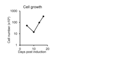
- FIG. 2 is a graph showing changes in cell number during differentiation-induced culture of iPS cells into HSPCs accompanied by Lhx2 gene introduction.
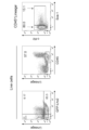
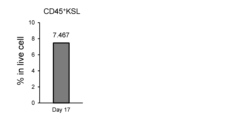
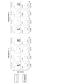
- FIG. This is a dot plot showing the results of flow cytometry analysis of cells (Day 17) obtained by differentiation-induced culture of iPS cells into HSPCs with Lhx2 gene introduction. The dot plot on the right is an analysis of the center dot plot by gating it within a rectangular frame. It is a graph showing the ratio of CD45 + KSL (CD45 + c-Kit + Sca-1 + Lineage ⁇ ) in cells (Day 17) obtained by differentiation-inducing culture of iPS cells into HSPCs accompanied by Lhx2 gene introduction.
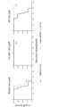
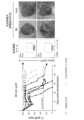
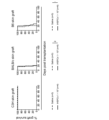
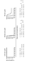
- FIG. 2 is a dot plot showing the results of flow cytometry analysis of peripheral blood mononuclear cells of allogeneic recipient mice at 2 and 6 weeks after transfer of Lhx2 + -iHSPCs. It is a graph showing the engraftment period of skin grafts when skin pieces from BALB/c mice, C3129F1 mice, or B6 mice are transplanted to C3129F1 mice into which Lhx2 + -iHSPCs have been introduced. Graph showing changes in the size of the grafted skin pieces in each individual mouse when skin pieces from B6 mice were transplanted into C3129F1 mice transplanted with Lhx2 + -iHSPCs (Figure 9 left).
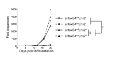
- FIG. 2 is a graph showing changes in cell number during differentiation induction culture from iPS cells to HSPCs accompanied by Lhx2 gene introduction and induction of HoxB4 expression by 4-hydroxytamoxifen (4-HT).
- FIG. The vertical axis of the graph is the relative value when the number of cells on the 5th day of induction (at the time of retrovirus infection) is set to 1.
- the graph shows the average value of three replicates for each condition.
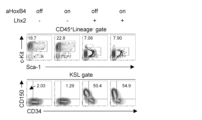
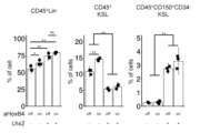
- CD45 + Lineage - cells CD45 + c-Kit + Sca-1 + Lineage in cells (Day 20) obtained by differentiation induction culture from iPS cells to HSPC with Lhx2 gene introduction and induction of HoxB4 expression by 4-HT - (CD45 + KSL) cells
- CD45 + KSL CD45 + KSL
- iPS cell-derived HSPCs into which the Lhx2 gene was introduced (hereinafter referred to as aHoxB4 on -Lhx2 + -iHSPC and aHoxB4 off -Lhx2 + -iHSPC, respectively) were introduced with HoxB4 expression induced or not induced by 4-HT.
- aHoxB4 on -Lhx2 + -iHSPC and aHoxB4 off -Lhx2 + -iHSPC, respectively) were introduced with HoxB4 expression induced or not induced by 4-HT.
- BALB/c mice and C3129F1 mice were transfected with iPS cell-derived HSPCs in which HoxB4 expression was induced by 4-HT (hereinafter referred to as aHoxB4 on -iHSPCs), Lhx2 + -iHSPCs, or aHoxB4 on -Lhx2 + -iHSPCs.
- aHoxB4 on -iHSPCs a HoxB4 on -iHSPCs
- Lhx2 + -iHSPCs Lhx2 + -iHSPCs
- aHoxB4 on -Lhx2 + -iHSPCs aHoxB4 on -Lhx2 + -iHSPCs.
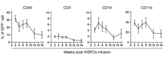
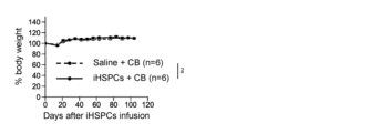
- the survival period of the graft was statistically analyzed using the Log-rank test (corrected by the Bonferroni method). *represents p ⁇ 0.05, **p ⁇ 0.01. It is a graph showing changes in the percentage of GFP-positive cells (donor cell-derived cells) contained in peripheral blood mononuclear cells of C3H mice transfected with aHoxB4 on Lhx2 + -iHSPC and treated with CB. Graphs represent mean ⁇ standard error. It is a graph showing changes in body weight of C3H mice transfected with aHoxB4 on Lhx2 + -iHSPC and treated with CB. Graphs represent average values.
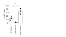
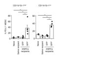
- FIG. 2 is a graph showing the percentage of PD-L1 positive cells in MDSC subsets CD11b + Gr-1 high and CD11b + Gr-1 low in the peripheral blood of C3129F1 mice 8 weeks after aHoxB4 on -Lhx2 + -iHSPC transfer.
- the bar graph represents the average value and standard error, and the dot plot represents the measured value for each mouse.
- Statistical analysis was performed using Tukey's HSD test. *represents p ⁇ 0.05, **p ⁇ 0.01.
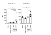
- FIG. 1 Graph showing MFI (mean fluorescence intensity) of PD-L1 in MDSC subsets CD11b + Gr-1 high and CD11b + Gr-1 low in peripheral blood of C3129F1 mice 8 weeks after aHoxB4 on -Lhx2 + -iHSPC transfer It is.
- the bar graph represents the average value and standard error, and the dot plot represents the measured value for each mouse.
- Statistical analysis was performed using Tukey's HSD test. *represents p ⁇ 0.05, **p ⁇ 0.01.
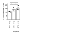
- Figure 2 is a graph showing the percentage of Foxp3 + cells (Treg) in CD4 + T cells in the spleen of C3129F1 mice 16 weeks after HoxB4 on -Lhx2 + -iHSPC transfer.
- FIG. 26A is an image showing luminescence of luciferin at the iPS cell transplantation site of aHoxB4 on -Lhx2 + -iHSPC-transplanted C3129F1 mouse subcutaneously transplanted with B6-derived luciferase-expressing iPS cells.
- FIG. 26B is a graph showing changes in luminescent signal intensity (total flux) of the iPS cell transplanted area of the mouse in FIG. 26A. Graphs represent mean values and standard errors.
- FIG. 27A is an image of a teratoma collected from a C3129F1 mouse transfected with aHoxB4 on -Lhx2 + -iHSPC 21 days after subcutaneous transplantation of B6-derived luciferase-expressing iPS cells. ND in the figure represents not detected.
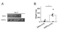
- FIG. 27B is a graph showing the weight of harvested teratomas. Graphs represent mean values and standard errors.
- Statistical analysis was performed using a two-tailed Student's t-test. *Represents p ⁇ 0.05.
- pluripotent stem cells are cells that have self-renewal ability and pluripotency, which is the ability to differentiate into all cell lineages belonging to the three germ layers (ectoderm, mesoderm, endoderm). means.
- pluripotent stem cells include embryonic stem cells (ES cells), induced pluripotent stem cells (iPS cells), and embryonic germ stem cells (EG cells).
- the pluripotent stem cells used in the present disclosure are preferably ES cells or iPS cells, more preferably iPS cells.
- the pluripotent stem cells used in the present disclosure may be mammalian pluripotent stem cells.
- the pluripotent stem cells are pluripotent stem cells of a mammal of the same species as the subject in which immune tolerance is desired to be induced.
- Mammals in the present disclosure include humans and non-human mammals, and examples of non-human mammals include mice, rats, hamsters, rodents including guinea pigs, monkeys, primates including chimpanzees, pigs, cows, Examples include livestock including goats, horses, and sheep, and pet animals including dogs and cats.
- Preferred mammals are mice or humans, more preferably humans.
- ES cells can be produced, for example, by culturing the inner cell mass present in a pre-implantation blastocyst stage embryo on feeder cells or in a medium containing LIF or FGF2.
- examples of human embryonic stem cells include those established from human embryos within 14 days of fertilization.
- iPS cells are cells that have been induced to have pluripotency by reprogramming somatic cells.
- iPS cells can be produced by reprogramming methods known to those skilled in the art.
- An example of a reprogramming method is to inject a gene called a reprogramming factor or a protein encoded therein into a somatic cell, such as a fibroblast, a blood cell (such as a peripheral blood-derived mononuclear cell), etc., or a protein encoded by the gene, such as Oct 3/4. , Sox 2, Klf 4, and Myc (c-Myc, N-Myc, L-Myc) are introduced into cells and cultured under appropriate conditions to induce pluripotent cells. be.
- reprogramming methods include substituting the above genes with drugs such as low-molecular compounds (e.g., Hou P. et al., 2013, Science, 9; 341(6146), 651-4), This method uses RNA (Miyoshi N. et al., Cell Stem Cell, 2011, 8, 633-638).
- drugs such as low-molecular compounds (e.g., Hou P. et al., 2013, Science, 9; 341(6146), 651-4), This method uses RNA (Miyoshi N. et al., Cell Stem Cell, 2011, 8, 633-638).
- pluripotent stem cells prepared from any cells by any method can be used.
- pluripotent stem cells distributed from a cell bank or commercially available pluripotent stem cells can also be used.
- human induced pluripotent stem cell lines such as 201B7 cells, 201B7-Ff cells, 253G1 cells, 253G4 cells, 1201C1 cells, 1205D1 cells, 1210B2 cells, and 1231A3 cells established at Kyoto University are available at Kyoto University and iPS Academia Japan. Available from Co., Ltd.
- Pluripotent stem cells can be produced, maintained, and expanded in the presence of feeder cells.
- Feeder cells are cells other than pluripotent stem cells that are used during undifferentiated maintenance culture of pluripotent stem cells, such as mouse fibroblasts (MEF), human fibroblasts, SNL cells, etc. I can do it. It is preferable that the feeder cells be used after being treated with a proliferation inhibitor such as mitomycin C or by gamma ray irradiation.
- Pluripotent stem cells can also be produced in the absence of feeder cells (feeder-free), maintained and cultured, and expanded.
- Feeder-free is a condition in which no feeder cells are added or substantially no feeder cells are included.
- Examples of the medium for feeder-free culture include Essential 8 medium, Essential 6 medium, TeSR medium, mTeSR medium, mTeSR-E8 medium, Stabilized Essential 8 medium, StemFit (registered trademark), and the like.
- the culture of cells in this disclosure includes BME medium, BGJb medium, CMRL 1066 medium, Glasgow MEM medium, Improved MEM Zinc Option medium, IMDM medium, Medium 199 medium, Eagle MEM medium, ⁇ MEM medium, This can be carried out using DMEM medium, F-12 medium, DMEM/F12 medium, IMDM/F12 medium, Ham's medium, RPMI 1640 medium, Fischer's medium, or a mixed medium thereof.
- any medium other than these may be appropriately selected as long as it is a medium for differentiating pluripotent stem cells into hematopoietic stem cells, or a medium for maintaining after differentiation into hematopoietic stem cells.
- hematopoietic stem cells refer to cells that have self-renewal ability and multipotency that can differentiate into blood cells such as white blood cells, red blood cells, and platelets.
- Hematopoietic stem cells include long-term hematopoietic stem cells (LT-HSC), which have the ability to self-renew over a long period of time, and short-term hematopoietic stem cells (ST-HSC), which originate from LT-HSC and have only short-term self-renewal ability.
- LT-HSC long-term hematopoietic stem cells
- ST-HSC short-term hematopoietic stem cells
- hematopoietic progenitor cells refer to undifferentiated blood cells at an intermediate stage from hematopoietic stem cells to mature blood cells.
- Hematopoietic progenitor cells include multipotent progenitors (MPPs) that do not have the ability to self-renew, which originate from ST-HSCs, common myeloid progenitors (CMPs), which originate from MPPs, and common lymphoid progenitors (common myeloid progenitors; CMPs).
- MPPs multipotent progenitors
- CMPs common myeloid progenitors
- CMPs common lymphoid progenitors
- CLP common lymphoid progenitors
- hematopoietic stem/progenitor cells refer to a cell population containing at least one of hematopoietic stem cells and hematopoietic progenitor cells.
- HSPC may be a cell population containing only hematopoietic stem cells, only hematopoietic progenitor cells, or a cell population containing both hematopoietic stem cells and hematopoietic progenitor cells.
- HSPCs can be distinguished from other cells by using a combination of multiple cell surface markers.
- mouse HSPCs can be detected as a CD45 + c-Kit + Sca-1 + Lineage - cell population, where Lineage - (also referred to as Lin - ) is a differentiated blood cell marker in mice.
- Lineage - also referred to as Lin -
- CD45 + c-Kit + Sca-1 + Lineage ⁇ cells are also called CD45 + KSL cells.
- Human HSPCs can be detected as a Lineage - CD34 + cell population, where Lineage - refers to the human differentiated blood cell markers CD2, CD3, CD14, CD16, CD19, CD24, CD56, CD66b and This means that glycophorin A is negative in both cases.
- Hematopoietic stem cells can also be distinguished from other cells by using a combination of multiple cell surface markers. For example, mouse hematopoietic stem cells can be detected as a CD45 + CD150 + CD34 low/- c-Kit + Sca-1 + Lineage - cell population. Human hematopoietic stem cells can be detected as a Lineage - CD34 + CD38 - CD45RA - CD90 + CD49f + cell population.
- the present disclosure provides a composition for inducing immune tolerance containing HSPCs derived from pluripotent stem cells in which the Lhx2 gene is forcibly expressed.
- the present disclosure also provides a composition for inducing immune tolerance containing HSPC derived from pluripotent stem cells in which the Lhx2 gene and HoxB4 gene are forcibly expressed.
- These pluripotent stem cell-derived HSPCs can be produced by a method that involves differentiating into HSPCs pluripotent stem cells that have been forced to express Lhx2 or both Lhx2 and HoxB4.
- Differentiation of pluripotent stem cells into HSPCs can be performed in vitro by culturing under known HSPC differentiation-inducing conditions; for example, mesoderm cells are induced from pluripotent stem cells, and the resulting mesoderm cells This can be done by differentiating cells into HSPCs.
- the method for differentiation of pluripotent stem cells into HSPCs mediated by mesoderm cells is not particularly limited, and any culture method known to those skilled in the art can be employed. Specific examples include the embryoid body formation method and the adhesive culture method. Differentiation of pluripotent stem cells into HSPCs via mesodermal cells is preferably performed by an embryoid body formation method.
- Embryoid body is a three-dimensional cell aggregate formed by floating culture of pluripotent stem cells, and it contains all cell lineages of ectoderm, mesoderm, and endoderm. include.
- the embryoid body formation method focuses on the fact that the direction of differentiation of pluripotent stem cells into each germ layer is determined from the early stage of differentiation, and contains many cells that have a tendency to differentiate into the germ layer of the target cell lineage. This is a method to efficiently differentiate pluripotent stem cells into target cells by forming EBs. That is, in the present disclosure, EBs formed from pluripotent stem cells may include at least one of cells with a tendency to differentiate into mesoderm and mesodermal cells.
- Examples of EB formation methods include a method in which EBs are formed by adding a cell suspension containing pluripotent stem cells to a non-adhesive culture vessel, and a method in which pluripotent stem cells are detached as colonies or small clumps to form EBs. Examples include a method of floating culture on a bacterial culture dish with low cell adhesion.
- a cell having a tendency to differentiate into mesoderm refers to a cell that has a property of being more likely to differentiate into mesoderm cells than cells of other germ layers.
- EBs are formed by culturing pluripotent stem cells in suspension under conditions that allow differentiation into mesoderm cells (referred to as mesoderm induction conditions), and the mesoderm contained in the EBs
- mesoderm induction conditions referred to as mesoderm induction conditions
- HSPCs are differentiated from cells with a differentiation tendency toward mesoderm, or from mesodermal cells, or from both cells with a mesodermal differentiation tendency and mesodermal cells.
- mesoderm induction conditions include the mesoderm induction medium used in the examples below ( ⁇ -MEM (GIBCO) with 20% fetal bovine serum (Nichirei), 0.1 mM non-essential amino acids, 1 mM sodium pyruvate, 100% Culture medium supplemented with U/ml penicillin, 100 ⁇ g/ml streptomycin, and 55 ⁇ M 2-mercaptoethanol) or Salvagiotto G., et al., PLoS One, 2011, 6(3), e17829, Lange, L Conditions described in literature such as Daniel, M et al., Trends in Cell Biology, 2016, 26 (3): 202-214. etc. can be mentioned.
- the culture time under mesoderm-inducing conditions required to form EBs from pluripotent stem cells is not particularly limited as long as it is 2 days or more. Furthermore, after EB formation is confirmed, EB culture may be continued as is.
- the culture time under mesoderm-inducing conditions for forming EBs from pluripotent stem cells can be, for example, 2 days or more, 3 days or more, 4 days or more, 5 days or more, 6 days or more, 7 days or more. It can also be up to 14 days, up to 13 days, up to 12 days, up to 11 days, up to 10 days, up to 9 days, up to 8 days, up to 7 days, up to 6 days, up to 5 days.
- the culture time under mesoderm-inducing conditions for forming EBs from pluripotent stem cells may be, for example, 2 to 14 days, preferably 3 to 10 days, and more preferably 4 to 8 days.
- the formed EBs are dispersed, preferably unicellularized, and then cultured under HSPC differentiation-inducing conditions.
- Dispersion of EBs means making EBs into finer structures, such as smaller cell clusters or single cells.
- Dispersion of EB can be performed by a known method, for example, a chemical method using an enzyme such as trypsin/EDTA, pronase, dispase, collagenase, etc., or a physical method using operations such as pipetting, scraping, etc.
- the dispersed EBs can be cultured as is or after concentrating mesoderm cells under HSPC differentiation-inducing conditions. Enrichment of mesodermal cells can be carried out using a cell separation means such as a cell sorter using the expression of a known mesodermal marker, such as the VEGF receptor Flk-1 (also called VEGFR-2), as an indicator.
- a cell separation means such as a cell sorter using the expression of a known mesodermal marker, such as the VEGF receptor Flk-1 (also called VEGFR-2), as an indicator.
- Culturing under HSPC differentiation-inducing conditions is preferably carried out under mesoderm-inducing conditions by adding factors required for the survival or proliferation of hematopoietic stem cells to the medium.
- factors include stem cell factor (SCF), thrombopoietin (TPO), FMS-like tyrosine kinase 3 ligand (FMS-like tyrosine kinase 3 Ligand; Flt3L), IL-6, etc.
- SCF stem cell factor
- TPO thrombopoietin
- FMS-like tyrosine kinase 3 Ligand Flt3L
- IL-6 etc.
- the concentration of SCF in the medium used for culturing under HSPC differentiation-inducing conditions is, for example, 1 ng/ml or more, 5 ng/ml or more, 10 ng/ml or more, 15 ng/ml or more, 20 ng/ml. 25 ng/ml or more, 50 ng/ml or more, and 500 ng/ml or less, 400 ng/ml or less, 300 ng/ml or less, 200 ng/ml or less, 150 ng/ml or less, 100 It can be less than ng/ml.
- the concentration of TPO in the medium used for culturing under HSPC differentiation-inducing conditions is, for example, 0.1 ng/ml or more, 1 ng/ml or more, 2 ng/ml or more, 5 ng/ml or more, 6 ng/ml. 7 ng/ml or more, 8 ng/ml or more, 9 ng/ml or more, 10 ng/ml or more, and 50 ng/ml or less, 40 ng/ml or less, 30 ng/ml or less, 20 ng/ml or less, 15 ng/ml or less, 10 ng/ml or less.
- the concentration of IL-6 in the medium used for culturing under HSPC differentiation-inducing conditions is, for example, 0.1 ng/ml or more, 1 ng/ml or more, 2 ng/ml or more, 5 ng/ml or more, 6 ng /ml or more, 7 ng/ml or more, 8 ng/ml or more, 9 ng/ml or more, 10 ng/ml or more, and 50 ng/ml or less, 40 ng/ml or less, 30 ng/ml or less , 20 ng/ml or less, 15 ng/ml or less, 10 ng/ml or less.
- the concentration of SCF in the medium used in culturing under HSPC differentiation-inducing conditions is, for example, 10-500 ng/ml, preferably 20-300 ng/ml, more preferably 50-200 ng/ml. It can be ml. In another embodiment, the concentration of SCF in the medium used in culturing under HSPC differentiation-inducing conditions is, for example, 1-200 ng/ml, preferably 5-100 ng/ml, more preferably 10-50 ng /ml.
- the concentration of TPO in the medium used in culturing under HSPC differentiation-inducing conditions is, for example, 1 to 50 ng/ml, preferably 2 to 30 ng/ml, more preferably 5 to 20 ng/ml. It can be ml.
- the concentration of IL-6 in the medium used in culturing under HSPC differentiation-inducing conditions is, for example, 1 to 50 ng/ml, preferably 2 to 30 ng/ml, more preferably 5 to 20 ng/ml. Can be ng/ml.
- the culture time under HSPC differentiation-inducing conditions is not limited as long as desired HSPCs are obtained, and may be, for example, 7 days or more, 8 days or more, 9 days or more, 10 days or more, 11 days or more, 12 days or more. It can also be less than 28 days, less than 21 days, less than 20 days, less than 19 days, less than 18 days, less than 17 days, less than 16 days, less than 15 days, less than 14 days, less than 13 days, and less than 12 days.
- the culture time under HSPC differentiation-inducing conditions may be, for example, 7 to 28 days, preferably 9 to 21 days, and more preferably 10 to 18 days. In another embodiment, the culture time under HSPC differentiation-inducing conditions may be, for example, 7 to 28 days, preferably 10 to 21 days, and more preferably 12 to 18 days.
- Differentiation of pluripotent stem cells into HSPCs can be performed in the presence of feeder cells such as bone marrow stromal cells, for example OP9 stromal cells, or feeder-free.
- feeder cells such as bone marrow stromal cells, for example OP9 stromal cells, or feeder-free.
- Differentiation of pluripotent stem cells into HSPCs may be performed by inducing mesoderm cells without forming EBs, and culturing the mesoderm cells under the above-mentioned HSPC differentiation-inducing conditions.
- mesodermal cells can be induced by an adhesive culture method, such as directly inoculating pluripotent stem cells onto OP9 stromal cells and co-culturing them.
- enhancing the expression of a specific gene by artificial manipulation includes both expressing an externally introduced gene and enhancing the expression of an endogenous gene.
- Expressing an exogenously introduced gene also includes a method of knocking an exogenous gene into the genome.
- enhancing the expression of the endogenous gene may be a method of enhancing the activity of the endogenous gene by activating an expression control region such as a promoter.
- the expression control region such as the promoter may be activated by any method as long as the expression control region such as the promoter is activated.
- Lhx2 Under forced expression of Lhx2 or under forced expression of Lhx2 and HoxB4, respectively, mean that the gene expression of Lhx2 is in an enhanced state compared to the case without artificial manipulation, or the gene expression of Lhx2 and HoxB4 is in an enhanced state compared to the case without artificial manipulation. This means that it is in an enhanced state compared to when no artificial manipulation is applied.
- Lhx2 forces the differentiation of pluripotent stem cells into HSPCs. This can be carried out under expression or under forced expression of Lhx2 and HoxB4.
- Lhx2 (LIM Homeobox Protein 2) is known as a transcription factor with a cysteine-rich zinc-binding domain (LIM domain), and has been reported to be involved in regulating the induction of differentiation from iPS cells to HSPCs (Kitajima et al. al., BLOOD, 2011, vol. 117, no. 1, pp. 3748-3758).
- the amino acid sequence of human Lhx2 is registered with NCBI as AAD20013, and the nucleotide sequence of human Lhx2 cDNA is registered with NCBI as AF124735.
- the amino acid sequence of mouse Lhx2 is registered with NCBI as AAD20012, and the nucleotide sequence of mouse Lhx2 cDNA is registered with NCBI as AF124734 (both searched on January 13, 2022).
- HoxB4 (Homeobox B4) is known as a transcription factor that is a member of the Antp homeobox family, and has been reported to be involved in regulating the induction of differentiation from iPS cells to HSPC (Izawa et al., Experimental Hematology, 2020, vol. 89, pp. 68-79).
- the amino acid sequence of human HoxB4 is registered with NCBI as NP_076920, and the base sequence of human HoxB4 cDNA is registered with NCBI as NM_024015.
- the amino acid sequence of mouse HoxB4 is registered with NCBI as NP_034589, and the nucleotide sequence of mouse HoxB4 cDNA is registered with NCBI as NM_010459 (both searched on January 13, 2022).
- Lhx2 gene and HoxB4 gene can be performed by a general method of introducing a foreign gene into cells so that it can be expressed, for example, by knocking the foreign gene into the genome using gene modification techniques such as genome editing.
- gene modification techniques such as genome editing.
- Examples of such methods include Manipulating the Mouse Embryo, A Laboratory Manual, Second Edition, Cold Spring Harbor Laboratory Press (1994); Gene Targeting, A Practical Approach, IRL Press at Oxford University Press (1993); Biomanual Series Examples include methods such as gene targeting, generation of mutant mice using ES cells, and methods described in Yodosha (1995).
- the Lhx2 gene and HoxB4 gene can be introduced, for example, by a method of introducing a vector containing a foreign gene into pluripotent stem cells.
- a method of introducing a vector containing a foreign gene into pluripotent stem cells examples include infection methods using viral vectors (e.g., retrovirus vectors, lentivirus vectors, Sendai virus vectors, adenovirus vectors, adeno-associated virus vectors), plasmid vectors (e.g., plasmid vectors, episodic Gene introduction methods using RNA vectors (e.g. calcium phosphate method, lipofection method, retronectin method, electroporation method), gene introduction methods using RNA vectors (e.g. calcium phosphate method, lipofection method, electroporation method), etc. can be mentioned.
- viral vectors e.g., retrovirus vectors, lentivirus vectors, Sendai virus vectors, adenovirus vectors, adeno-associated
- the introduction of the Lhx2 gene is performed using a recombinant retroviral vector carrying the Lhx2 gene in iPS as reported in Kitajima et al. (BLOOD, 2011, vol. 117, no. 1, pp. 3748-3758). This can be done by infecting cells.
- the HoxB4 gene can be introduced into iPS cells using a recombinant lentiviral vector carrying the HoxB4 gene, as reported in Izawa et al. (Experimental Hematology, 2020, vol. 89, pp. 68-79). This can be done by infecting a person.
- the Lhx2 gene and HoxB4 gene may be placed under the control of a promoter whose expression can be regulated by adding a drug or the like to the medium.
- a nucleic acid encoding Lhx2 and a nucleic acid encoding HoxB4 may be contained in a mixed state.
- Cells may be treated with gene transfer reagents to simultaneously introduce both genes.
- both genes may be introduced into cells by the same method or by different methods.
- cells into which the Lhx2 gene and HoxB4 gene have been introduced may be created by introducing the other gene into cells that have been preserved or available as cells into which one gene has been introduced. .
- HSPCs are prepared by forming EBs in pluripotent stem cells, forcing the EBs to express the Lhx2 gene, and culturing them under HSPC differentiation-inducing conditions.
- EB formation is preferably performed by suspension culture under mesoderm-inducing conditions.
- culturing under HSPC differentiation-inducing conditions may include culturing in the presence of SCF and IL-6.
- Culturing in the presence of SCF and IL-6 includes, for example, 10 to 500 ng/ml, preferably 20 to 300 ng/ml, more preferably 50 to 200 ng/ml, and, for example, 1 to 50 ng/ml, This can be carried out using a medium containing IL-6, preferably 2 to 30 ng/ml, more preferably 5 to 20 ng/ml. Further, the culture may be carried out for, for example, 7 to 28 days, preferably 9 to 21 days, and more preferably 10 to 18 days.
- HSPCs are prepared by causing pluripotent stem cells to form EBs, forcing the EBs to express the Lhx2 gene and HoxB4 gene, and culturing them under HSPC differentiation-inducing conditions.
- EB formation is preferably performed by suspension culture under mesoderm-inducing conditions.
- culturing under HSPC differentiation-inducing conditions may include culturing in the presence of SCF and TPO.
- Cultivation in the presence of SCF and TPO may be performed with SCF of, for example, 1 to 200 ng/ml, preferably 5 to 100 ng/ml, more preferably 10 to 50 ng/ml, and 1 to 50 ng/ml, preferably This can be carried out using a medium containing 2 to 30 ng/ml, more preferably 5 to 20 ng/ml of TPO. Further, culturing may be carried out for, for example, 7 to 28 days, preferably 10 to 21 days, and more preferably 12 to 18 days.
- Differentiation of mouse-derived pluripotent stem cells into HSPCs occurs when the proportion of CD45 + c-Kit + Sca-1 + Lin - cells in the entire cell population obtained by culturing under HSPC differentiation-inducing conditions increases This can be confirmed by a greater proportion of CD45 + c-Kit + Sca-1 + Lin ⁇ cells in the total pluripotent stem cell population.
- the proportion of CD45 + c-Kit + Sca-1 + Lin ⁇ cells in the entire cell population obtained by culturing under HSPC differentiation-inducing conditions is, for example, 1% or more, 3% or more, 5% or more, or 7% or more. % or more.
- mouse-derived pluripotent stem cells into HSPCs is due to the increase in the number of CD45 + CD150 + CD34 - c-Kit + Sca-1 + Lin - cells in the entire cell population obtained by culturing under HSPC differentiation-inducing conditions. It can also be confirmed that the proportion is greater than the proportion of CD45 + CD150 + CD34 ⁇ c-Kit + Sca-1 + Lin ⁇ cells in the entire pluripotent stem cell population before culture.
- the percentage of CD45 + CD150 + CD34 - c-Kit + Sca-1 + Lin - cells in the entire cell population obtained by culturing under HSPC differentiation-inducing conditions is, for example, 0.5% or more, 1% or more, or 2%. or more, or 3% or more.
- Detection of cell surface markers can be performed by analyzing cells by flow cytometry.
- the ratio of Lineage - CD34 + is changed to CD45 + CD150 + CD34 - c-Kit + Sca instead of the ratio of CD45 + c - Kit + Sca-1 + Lin - cells.
- Differentiation of pluripotent stem cells into HSPCs can be confirmed using the ratio of Lineage - CD34 + CD38 - CD45RA - CD90 + CD49f + as an indicator instead of -1 + Lin - cells.
- the cell population obtained by culturing pluripotent stem cells under HSPC differentiation-inducing conditions under forced expression of Lhx2 or forced expression of Lhx2 and HoxB4 is usually compared to the bone marrow cell population existing in vivo. It contains many HSPCs and has the ability to induce immune tolerance by forming blood chimeras with cells of different genetic types. Therefore, the present disclosure provides a composition for inducing immune tolerance containing HSPCs derived from pluripotent stem cells, in which the Lhx2 gene is forcibly expressed, or the Lhx2 gene and the HoxB4 gene are forcibly expressed.
- the present disclosure also provides an immune tolerance inducing agent containing HSPC derived from pluripotent stem cells, in which the Lhx2 gene is forcibly expressed, or the Lhx2 gene and the HoxB4 gene are forcibly expressed.
- Immune tolerance refers to a state of unresponsiveness of the immune system to antigens that may cause an immune response.
- Immune tolerance includes self-tolerance, which is immune unresponsiveness to self-antigens, and acquired tolerance (also called induced immune tolerance), which is immune unresponsiveness to non-self antigens.
- Acquired tolerance is known to be induced by administering non-self antigens under predetermined conditions, and is thought to involve suppression of immune responses against non-self antigens by regulatory T cells.
- autoimmune diseases and allergic diseases are thought to be caused by breakdown of immune tolerance to self and non-self, respectively.
- Blood chimera also referred to as blood chimerism refers to a state in which an individual's own blood cells and blood cells derived from a different organism exist simultaneously. It is known that immune tolerance to donor antigens can be induced in a recipient receiving a transplant of non-self cells or tissues by forming a blood chimera in which autologous blood cells and donor-derived blood cells coexist. It is being
- induction of immune tolerance refers to inducing a state of immune unresponsiveness to a specific antigen, and includes, for example, delaying the onset or progression of an immune response, weakening the strength of the immune response, and the like.
- immune tolerance may be directed against either self-antigens or non-self antigens, and the above-mentioned immune tolerance-inducing composition and immune tolerance-inducing agent may be used for inducing acquired tolerance particularly against non-self antigens, e.g. It can be suitably used to induce immune tolerance to donor-derived transplants in allogeneic transplants.
- composition for inducing immune tolerance and the immune tolerance inducing agent of the present disclosure can be used for mammalian individuals in whom induction of immune tolerance is desired (for example, recipients undergoing cell transplantation or organ transplantation).
- HSPCs are derived from pluripotent stem cells derived from the donor or from pluripotent stem cells derived from individuals of the same species as the recipient and donor but different from both. , can be prepared by the above-mentioned manufacturing method.
- the latter pluripotent stem cells preferably have a haplotype of major histocompatibility complex (MHC), also called human leukocyte antigen (HLA) in humans, that completely matches that of the transplant donor. .
- MHC major histocompatibility complex
- HLA human leukocyte antigen
- MHC compatible transplantation refers to a transplant in which the donor's MHC genotype is compatible with the recipient, where the donor or does not have a different allele from the recipient in all cases.
- MHC incompatible transplantation is a transplant in which the donor's MHC genotype is incompatible with the recipient's at all major MHC loci. have different alleles.
- MHC compatibility can be confirmed by conventional methods such as HLA typing, lymphocyte crossmatch, and other histocompatibility tests to determine compatibility between donor and recipient.
- Major MHC loci in mice include H-2K, H-2D, H-2L, IA and IE, and in addition to these, either Qa-2 or Qa-1 It may include one, preferably all.
- mouse MHC haplotypes have names that correspond to specific sets of alleles at major genetic loci.
- MHC haplotype b also called H-2b
- H-2L null the allele set H-2K b
- H-2D b H-2L null
- IA b IE null
- MHC haplotype d also called H-2d
- H-2K d is a haplotype with the allele set H-2K d , H-2D d , H-2L d , IA d , IE d , and MHC haplotype k (also called H-2k) has the genotype H-2K k , It corresponds to a haplotype having the allele set H-2D k , H-2L null , IA k , and IE k .
- Major MHC loci in humans include HLA-A, HLA-B, and HLA-DR, and in addition to these, any one of HLA-C, HLA-DQ, and HLA-DP It may include one, preferably all.
- a transplant that is homozygous for each of the three loci HLA-A, HLA-B, and HLA-DR in the human HLA type is This applies to the case where a transplant having a combination in which at least one allele at a locus matches the allele at the gene locus is transplanted, but is not limited to three loci.
- HLA half-matching transplantation where the donor's HLA-A, HLA-B, HLA-DR are *24:02-*52:01-*15:02, *33:03-*44:03 Contained in peripheral blood or cord blood that is homozygous for -*13:02, *24:02-*07:02-*01:01, or *24:02-*54:01-*04:05 HLA-A, HLA-B, HLA-DR are *24:02-*52:01-*15:02, *33:03-* cells, tissues, or organs created from iPS cells created from cells.
- HLA-A, HLA-B, HLA-DR are *24:02-*52:01-*15:02, *33:03-*44:03-*13:02, *24:02- *07:02-*01:01 or *24:02-*54:01-*04:05 is known as HLA with high frequency in Japan (WO2015/125941, and iPS cell stock cell (HLA homodonor) Kyoto University iPS Cell Research Foundation (see https://www.cira-foundation.or.jp/j/research-institution/ips-stock-project/homozygous.html)).
- the composition for inducing immune tolerance may contain the cell population itself obtained by culturing pluripotent stem cells under HSPC differentiation-inducing conditions under forced expression of Lhx2 or forced expression of Lhx2 and HoxB4; HSPC cell surface markers for cell populations, such as CD45 + c-Kit + Sca-1 + Lin - or CD45 + CD150 + CD34-c-Kit + Sca-1 + Lin - in mice, Lineage - CD34 in humans It may contain a cell population enriched with HSPCs obtained by cell separation or concentration using + or Lineage - CD34 + CD38 - CD45RA - CD90 + CD49 f + as an indicator.
- the cell population enriched with HSPCs is obtained by culturing the cell population before enrichment, that is, pluripotent stem cells, under HSPC differentiation-inducing conditions under forced expression of Lhx2 or forced expression of Lhx2 and HoxB4.
- the proportion of CD45 + c-Kit + Sca-1 + Lin - cells or CD45 + CD150 + CD34 - c-Kit + Sca-1 + Lin - cells in mice is Lineage - refers to a cell population with an increased proportion of CD34 + cells or Lineage - CD34 + CD38 - CD45RA - CD90 + CD49 f + cells.
- compositions for inducing immune tolerance and immune tolerance inducing agents containing HSPCs prepared from mouse-derived pluripotent stem cells CD45 + c-Kit + Sca-1 occupying the entire cell population contained in the compositions and agents.
- the percentage of + Lin ⁇ cells can be, for example, 1% or more, 3% or more, 5% or more, or 7% or more.
- compositions for inducing immune tolerance and immune tolerance inducing agents containing HSPCs prepared from mouse-derived pluripotent stem cells CD45 + CD150 + CD34 - c in the entire cell population contained in the compositions and agents.
- the proportion of -Kit + Sca-1 + Lin - cells can be, for example, 0.5% or more, 1% or more, 2% or more, or 3% or more.
- the proportion of Lineage - CD34 + cells in the entire cell population contained in the compositions and agents is as follows: For example, it can be 1% or more, 3% or more, 5% or more, or 7% or more.
- compositions for inducing immune tolerance and immune tolerance inducing agents containing HSPCs prepared from human-derived pluripotent stem cells Lineage - CD34 + CD38 - CD45RA of the entire cell population contained in the compositions and agents -
- the proportion of CD90 + CD49f + cells can be, for example, 0.5% or more, 1% or more, 2% or more, or 3% or more.
- the HSPC used in the composition for inducing immune tolerance and the immune tolerance inducing agent maintains a state of forced expression of the Lhx2 gene in the body to which it is administered.
- the composition for inducing immune tolerance and the immune tolerance inducing agent contain HSPCs in which a foreign Lhx2 gene has been integrated into the chromosome.
- composition for inducing immune tolerance and the immune tolerance inducing agent are administered to a mammalian individual in which induction of immune tolerance is desired in an amount effective for inducing immune tolerance or an amount effective for blood chimera formation.
- the composition for inducing immune tolerance and the immune tolerance inducing agent administered to mice have a dosage of about 1 ⁇ 10 3 to 1 ⁇ 10 7 HSPCs per mouse, preferably 1 ⁇ 10 4 to 1 ⁇ It is sufficient to contain HSPC so that the number is about 106 .
- the amount of HSPC contained in the composition for inducing immune tolerance and the immune tolerance inducing agent administered to humans can be determined according to the dose for mice described above.
- composition for inducing immune tolerance and the immune tolerance inducing agent administered to humans may be administered at a dose of, for example, 1 ⁇ 10 2 to 1 ⁇ 10 9 HSPCs per kg of human body weight, for example, 1 ⁇ 10 2 to 1 HSPC. Around 8 ⁇ 10 , 3 to 8 1 ⁇ 10 , 4 to 8 1 ⁇ 10, preferably 5 to 7 4 ⁇ 10. It may contain HSPC.
- the composition for inducing immune tolerance is not limited as long as it contains HSPC and a liquid in which the HSPC can survive.
- the liquid in which HSPCs can survive may be any liquid commonly used in cell preparations, such as water, physiological saline, phosphate buffered saline, cell culture medium, and the like.
- the composition for inducing immune tolerance may contain cells other than HSPC, and may also contain pharmaceutically acceptable ingredients such as adjuvants, buffers, antioxidants, preservatives, excipients, carriers, and other pharmaceutically effective ingredients. It may also contain ingredients. Pharmaceutically acceptable ingredients are well known to those skilled in the art, and can be appropriately selected and used within the scope of normal practice by those skilled in the art.
- composition for inducing immune tolerance and the immune tolerance inducing agent are preferably used in the form of parenteral preparations such as injections and drips.
- the method of administering the composition for inducing immune tolerance and the immune tolerance inducing agent is not particularly limited, but in the case of parenteral preparations, examples include intravascular administration (preferably intravenous administration), intraperitoneal administration, and the like.
- the composition for inducing immune tolerance and the immune tolerance inducing agent are administered to a recipient in order to induce immune tolerance in the recipient to a donor-derived transplant in allogeneic transplantation.
- An allogeneic transplant may be an allogeneic transplant.
- an MHC-compatible transplant is preferred, but an MHC-incompatible transplant may also be used.
- Compositions and agents for inducing immune tolerance can be used in transplants that are MHC compatible but have a large degree of mismatch in minor histocompatibility antigens, and even in transplants that are not MHC compatible. Because immune tolerance can be induced, minor histocompatibility antigen matching is not required in allogeneic transplantation, nor is MHC compatibility.
- Implants can also include cells (e.g., but not limited to, retinal pigment epithelium, dopaminergic neural progenitor cells, glial cells, etc.), tissues (e.g., skin, bone, pancreatic islets, heart valves, blood vessels, cornea, etc.), It may be any organ (such as, but not limited to, the heart, kidney, liver, etc.) or an organ (such as, but not limited to, the heart, kidney, liver, etc.).
- cells e.g., but not limited to, retinal pigment epithelium, dopaminergic neural progenitor cells, glial cells, etc.
- tissues e.g., skin, bone, pancreatic islets, heart valves, blood vessels, cornea, etc.
- It may be any organ (such as, but not limited to, the heart, kidney, liver, etc.) or an organ (such as, but not limited to, the heart, kidney, liver, etc.).
- HSPCs produced from iPS cells derived from one human individual are transplanted to the other human individual.
- any cell for example, but not limited to retinal pigment epithelium, dopaminergic neural progenitor cells, glial cells, etc.
- tissue for example, skin, bone, pancreatic islets, heart valves, etc.
- the Lhx2 gene can be used in recipient human individuals to induce immune tolerance in HLA-compatible or HLA-incompatible human allogeneic transplants.
- a composition for inducing immune tolerance containing human pluripotent stem cell-derived HSPCs in which the Lhx2 gene and HoxB4 gene are expressed or forcibly expressed.
- compositions for inducing immune tolerance and agents for inducing immune tolerance can be used in combination with treatments to suppress the immune system, such as administration of immunosuppressive drugs, irradiation, T cell depletion, etc. Examples include treatment.
- immunosuppressants include cyclosporine, tacrolimus, mycophenolate mofetil, steroids (eg, methylprednisolone, etc.), T cell activation inhibitors, and the like.
- T cell activation inhibitors include drugs that inhibit the binding between the co-stimulatory molecule CD28 on T cells and CD80/CD86 on antigen presenting cells, such as the extracellular portion of CTLA-4 and the Fc portion of IgG.
- CTLA-4Ig e.g.
- abatacept a drug that inhibits the binding of the co-stimulatory molecule CD40L8 on T cells to CD40 on antigen-presenting cells, such as anti-CD40L (CD154) antibodies; IL on T cells
- examples include drugs that inhibit the binding between the -2 receptor ⁇ chain (CD25) and IL-2, such as anti-CD25 monoclonal antibodies (eg, basiliximab).
- T-cell activation inhibitors are also called co-stimulatory blockades (CBs).
- CBs co-stimulatory blockades
- the radiation irradiation may be any radiation that is normally given as one of the pretreatments given to the recipient during transplantation treatment, and preferably total body irradiation to the recipient or irradiation to the thymus gland.
- the irradiation dose can be adjusted as appropriate, for example, in the case of whole-body irradiation to humans, it is 1 Gy or more as a single irradiation dose, and in the case of thymus irradiation, it is 3 Gy or more, and the amount does not reach a lethal dose. .
- the T cell removal treatment may be any treatment that is normally performed as one of the pretreatments given to the recipient when performing transplantation treatment, and examples include administration of anti-T cell antibodies and steroids.
- anti-T cell antibodies include anti-CD4 antibodies, anti-CD8 antibodies, anti-CD3 antibodies, anti-TCR antibodies, and anti-thymocyte globulin (ATG), and one or more of these can be administered to the recipient in combination. is preferred.
- the anti-T cell antibody is a combination of an anti-CD4 antibody and an anti-CD8 antibody, which are preferably administered to the recipient intravenously or intraperitoneally, either simultaneously or sequentially. The dose may be sufficient to eliminate T cells in the recipient's body.
- the recipient undergoes radiation irradiation or T cell depletion treatment, and more preferably both radiation irradiation and T cell depletion treatment, before the composition for inducing immune tolerance or the immune tolerance inducing agent is administered.
- the composition for inducing immune tolerance or the immune tolerance inducing agent is administered before the composition for inducing immune tolerance or the immune tolerance inducing agent is administered.
- immune tolerance to the donor-derived transplant can be efficiently induced in the recipient, thereby causing donor-derived
- the survival rate of transplants can be improved. It also becomes possible to avoid or reduce the dose of immunosuppressants to the recipient.
- radiation irradiation and T cell depletion treatment should be performed immediately before transplantation, or after transplantation, i.e. delayed tolerance induction. If induction of tolerance is desired, it can be repeated once or multiple times at the desired timing to induce tolerance. Furthermore, although there is no particular restriction on the order of radiation irradiation and T cell removal treatment, it is preferable to perform T cell removal treatment prior to radiation irradiation.
- HSPCs obtained by culturing pluripotent stem cells under forced expression of Lhx2 or forced expression of Lhx2 and HoxB4 under HSPC differentiation-inducing conditions are capable of controlling immune responses in individuals. In addition to inducing immune tolerance, they can also be used for the formation of blood chimeras and for the treatment or prevention of diseases treatable by blood chimera formation, such as autoimmune or allergic diseases.
- the present disclosure provides an effective amount of HSPC obtained by culturing pluripotent stem cells under forced expression of Lhx2 or forced expression of Lhx2 and HoxB4 under HSPC differentiation-inducing conditions to a mammalian individual in which immune tolerance is desired.
- a method of inducing immune tolerance in a mammalian individual comprising administering.
- the present disclosure also provides a method for administering to a recipient HSPC obtained by culturing pluripotent stem cells under forced expression of Lhx2 or forced expression of Lhx2 and HoxB4 under HSPC differentiation-inducing conditions, and A method of improving the engraftment rate of donor-derived cells, tissues or organs in a recipient, the method comprising transplanting the derived cells, tissues or organs, the pluripotent stem cells are derived from the donor, or the recipient and the donor is derived from an individual mammal that is the same species as but different from both.
- the meaning of each term is as explained above.
- HSPCs are HSPCs obtained by culturing under HSPC differentiation-inducing conditions under forced expression of Lhx2 or forced expression of Lhx2 and HoxB4.
- HSPCs are HSPCs produced by differentiating pluripotent stem cells into hematopoietic stem/progenitor cells under forced expression of Lhx2 or under forced expression of Lhx2 and HoxB4.
- HSPCs are HSPCs produced by differentiating pluripotent stem cells into hematopoietic stem/progenitor cells under forced expression of Lhx2 or under forced expression of Lhx2 and HoxB4.
- immunosuppressive cells include regulatory T cells (Treg), myeloid derived suppressor cells (MDSC), and the like.
- mice were obtained by mating C3H females with 129 males. Male mice were used for all transplantation experiments. In addition, animal experiments in this study were handled based on the ⁇ Hokkaido University Regulations on Animal Experiments'' (approval number: 17-0110).
- b/b is homozygous for MHC haplotype b
- d/d is homozygous for MHC haplotype d
- k/k is homozygous for MHC haplotype k
- k/b is homozygous for MHC haplotype k. Indicates that it is heterozygous for b.
- Cell culture B6 mouse-derived iPS cells are produced by injecting the four factors Oct4, Sox2, Klf4, and c-Myc into mouse fibroblasts isolated from adult B6 mouse ear skin tissue using a retroviral vector. A new system was established by introducing this (Takahashi et al., Cell, 2006, Vol. 126, pp. 663-676. DOI 10.1016/j.cell.2006.07.024).
- activated-HoxB4-Gata2-KI-GFP (AHGG)-iPS cells which are B6-derived iPS cells introduced with DNA encoding mouse HoxB4 (SEQ ID NO: 1) (Izawa et al., Experimental Hematology, 2020, vol. 89) , pp.
- AHGG-iPS cells are capable of reversible expression of HoxB4 by the estrogen analog 4-hydroxytamoxifen (4-HT) by fusion protein of HoxB4 with the estrogen receptor (ER) ligand-binding domain. These are the cells that made induction possible.
- the CSII-EF-MCS used to create the lentiviral vector containing HoxB4-ER was provided by the RIKEN BioResource Research Center.
- iPS cells were cultured in DMEM (WAKO) with 20% StemSure WAKO, 0.1 mM non-essential amino acids (Nacalai Tesque), 100 U/ml penicillin (Nacalai Tesque), 100 ⁇ g/ml streptomycin (Nacalai Tesque), and 55 ⁇ M 2.
- -Culture MEFs as feeder cells at 37°C in a 5% CO 2 incubator using mercaptoethanol (Nacalai Tesque) and recombinant human LIF (in house) hereinafter referred to as iPS cell medium). did.
- iPS cells were passaged as follows. After removing the culture supernatant from the culture medium cultured in iPS cell medium and washing the remaining culture medium with PBS, 1000 ⁇ l of trypsin (0.5 g/l trypsin/0.53 mmol/l-EDTA solution, Nacalai Tesque) was added. , and placed in an incubator. After 5 minutes, 5 ml of iPS cell medium was added to neutralize trypsin. A single cell suspension was obtained by spraying medium on the cells, collected through a 40 ⁇ m filter into a tube, and centrifuged at 1500 rpm for 5 minutes at 4°C. After centrifugation, the supernatant was removed and resuspended in iPS cell medium. The cell concentration of the suspension was measured, and the required number of cells were re-seeded onto new MEFs and cultured again using iPS cell medium.
- trypsin 0.5 g/l trypsin/0.53 mmol/l-EDTA solution
- OP9/N cells were grown in mesoderm induction medium ( ⁇ -MEM (GIBCO) with 20% fetal bovine serum (Nichirei), 0.1 mM nonessential amino acids, 1 mM sodium pyruvate, 100 U/ml penicillin, and 100 ⁇ g/ml streptomycin. , a medium supplemented with 55 ⁇ M 2-mercaptoethanol) at 37°C in a 5% CO 2 incubator in a monolayer culture. OP9/N cells were used as feeder cells for inducing HSPCs from iPS cells.
- PlatE cells (Morita et al., Gene Therapy, 2000, Vol. 7, pp.1063-1066.) were grown in DMEM (WAKO) with 10% fetal bovine serum, 0.1 mM non-essential amino acids, 100 U/ml penicillin, and 100 The cells were cultured at 37° C. in a 5% CO 2 incubator using ⁇ g/ml streptomycin and 55 ⁇ M 2-mercaptoethanol. PlatE cells were used as packaging cells for retroviruses for gene transfer.
- Retrovirus vector pMY-FLAG-Lhx2- which has DNA encoding mouse Lhx2 (SEQ ID NO: 3), was provided by Dr. Kitajima of the Tokyo Metropolitan Institute of Medical Science. IRES-EGFP (Kitajima et al., BLOOD, 2011, vol. 117, no. 1, pp. 3748-3758) and Empty plasmid (pMY-IRES-EGFP) were added to the transfection reagent TransIT-X2 (registered trademark) ( PlatE cell line was transfected according to the protocol of Mirus).
- the retroviral vector (pMY-IRES-EGFP) was kindly provided by Professor Toshio Kitamura of the Institute of Medical Science, the University of Tokyo.
- Example 1 Preparation of iPS cell-derived HSPCs into which the Lhx2 gene has been introduced B6 mouse-derived iPS cells made into a single cell suspension by trypsin treatment are mixed with mesoderm induction medium ( ⁇ -MEM (GIBCO), 20% fetal bovine serum (Nichirei), Medium supplemented with 0.1 mM non-essential amino acids, 1 mM sodium pyruvate, 100 U/ml penicillin, 100 ⁇ g/ml streptomycin, 55 ⁇ M 2-mercaptoethanol) adjusted so that 10 ml contains 6 ⁇ 10 5 cells, Embryoid bodies (EBs) were induced by suspension culture for 5 days on a low-adhesion culture dish (As One).
- ⁇ -MEM mesoderm induction medium
- fetal bovine serum Neichirei
- Medium supplemented with 0.1 mM non-essential amino acids, 1 mM sodium pyruvate, 100 U/ml penicillin
- EBs were collected and centrifuged at 1500 rpm, 4°C, for 5 minutes. After centrifugation, the supernatant was removed, treated with trypsin for 5 minutes, and made into a single cell suspension by pipetting. Neutralization treatment was performed with mesoderm induction medium, and EBs were collected through a 70 ⁇ m filter. The collected EBs were stained with PE-anti-mFlk1 antibody, and Flk1-positive cells were sorted using BD FACSAria II (Becton dickinson) and used for subsequent differentiation induction.
- BD FACSAria II Becton dickinson
- the retrovirus-infected cells were seeded together with the virus-containing culture solution into one well of a 6-well plate that had been seeded with OP9/N cells in advance, and left in an incubator overnight. After incubation, the culture medium was replaced with fresh mesoderm induction medium containing 100 ng/ml mouse SCF and 10 ng/ml mouse IL-6, and suspension culture was performed for 12 days. During this culture, subculture was performed once every 4-5 days as follows. The floating cells were collected into a tube, the culture dish was washed with PBS, and the PBS used for washing was also collected into the tube.
- iPS cell-derived HSPCs into which the Lhx2 gene had been introduced (hereinafter referred to as Lhx2 + -iHSPCs) were prepared by suspension culture for 12 days after retrovirus infection.
- the start of iPS cell culture in mesoderm induction medium was defined as Day 0, and cells were collected over time from retrovirus infection (Day 5) to the end of culture (Day 17), and the total number of cells was counted ( Figure 1). After retrovirus infection (Day 5), the cell number decreased once, but then increased.
- CD45 + Lineage - (inside the rectangular frame of the dot plot in the center of Figure 2). This indicates that more than half of the cells on Day 17 are immature hematopoietic cells.
- CD45 + Lineage - cell population contains 13.1% Sca-1 + c-Kit + cells (inside the rectangular frame on the right side of the dot plot on the right side of Figure 2), which accounts for 13.1% of the total cells on Day 17.
- the percentage of CD45 + c-Kit + Sca-1 + Lin ⁇ cells was approximately 7.5% (Fig. 3).
- Example 2 Analysis of hematopoietic ability of Lhx2 + -iHSPC (1) Transplantation into syngenic recipients
- recipient mice were irradiated with 9 Gy of whole body radiation, and 2.5 ⁇ 10 6 Lhx2 + -iHSPCs and 2.0 ⁇ 10 6 hematopoietic supplementary bone marrow cells were suspended in 200 ⁇ l of physiological saline.
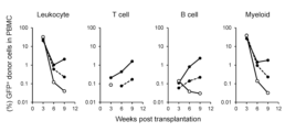
- Peripheral blood mononuclear cells were prepared from peripheral blood at 3, 6, and 9 weeks after Lhx2 + -iHSPC transfer, and the percentage of GFP-positive blood cells (Lhx2 + -iHSPC-derived blood cells) was analyzed by flow cytometry.
- FIG. 4 Blood cells derived from donor cells were detected in 2 out of 3 mice even 9 weeks after transfer of Lhx2 + -iHSPCs (FIG. 4).
- a dot plot of peripheral blood mononuclear cells from two recipient mice is shown in Figure 5.
- donor-derived CD45-positive cells white blood cells
- CD3 ⁇ -positive cells T cells
- CD19-positive cells B cells
- CD11b-positive cells bone marrow cells
- mice On the day of iHSPC administration, recipient mice were irradiated with 3 Gy of whole body radiation, and then 5.0 ⁇ 10 6 Lhx2 + -iHSPCs were suspended in 200 ⁇ l of physiological saline and administered through the tail vein.
- Peripheral blood mononuclear cells were prepared from peripheral blood at 2, 4, and 6 weeks after Lhx2 + -iHSPC transfer, and the percentage of GFP-positive blood cells (Lhx2 + -iHSPC-derived blood cells) was analyzed by flow cytometry.
- FIG. 7 shows dot plots of peripheral blood mononuclear cells of two recipient mice at 2 weeks and 6 weeks after Lhx2 + -iHSPC transfer.
- donor-derived CD45-positive cells white blood cells
- CD3 ⁇ -positive cells T cells
- CD19-positive cells B cells
- CD11b-positive cells bone marrow cells
- Example 3 Transplant immune tolerance induction by Lhx2 + -iHSPC
- C3129F1 mice were treated 7 days after transfer of Lhx2 + -iHSPCs, and as a control, physiological saline was administered instead of Lhx2 + -iHSPCs.
- C3129F1 mice treated otherwise identically were used as recipients of skin grafts.
- the dorsal skin of a sufficiently anesthetized recipient mouse was excised in a circular shape to form a transplant bed, and auricular skin pieces from donor B6 mice, BALB/c mice, and C3129F1 mice were transplanted. After protecting the grafted skin pieces for 7 days after skin grafting, the size of the grafted skin pieces was measured over time.
- the time when the grafted skin piece regressed to less than 30% of the size (longer axis length) of the grafted skin piece on the day of transplantation was defined as the rejection date, and the period from the date of transplantation to the date of rejection was defined as the engraftment period of the grafted skin piece.
- FIG. 8 shows the engraftment period of the grafted skin pieces.
- FIG. 9 shows changes in the size of the grafted skin pieces for each individual C3129F1 mouse to which skin pieces from B6 mice were transplanted.
- the size of the grafted skin pieces was generally maintained until 30 days after transplantation, but regression of the grafted skin pieces was observed in some individuals after 30 days.
- GFP-positive cells contained in peripheral blood mononuclear cells were analyzed for individuals in which the size of the grafted skin did not change after 30 days after transplantation, and in individuals in which regression of the grafted skin was observed after 30 days after transplantation. Analyzed by flow cytometry.
- CD45-positive cells were detected in individuals in which the size of the grafted skin remained unchanged (upper center row of Figure 9), but no CD45-positive cells were detected in individuals in which regression of the skin graft was observed (lower center row of Figure 9). , the blood chimera had disappeared.
- transplants using C3129F1 recipient mice and B6 donor mice result in graft rejection as quickly as non-MHC-compatible transplants due to minor antigen mismatches. It has been reported that (Murata, et al., Scientific Reports, 2020, Vol.10, 13560, https://doi.org/10.1038/s41598-020-69784-4). It has been confirmed that Lhx2 + -iHSPC can induce immune tolerance even in allogeneic transplants that cause rejection due to such minor antigen mismatches, and blood chimera formation is important for the induction of immune tolerance. Something was suggested.
- Example 4 Preparation of iPS cell-derived HSPCs introduced with Lhx2 gene and HoxB4 gene EBs were prepared in the same manner as in Example 1 using AHGG-iPS cells, except that Flk1-positive cells were not sorted, The Lhx2 gene was introduced into EBs using a retroviral vector.
- the mesoderm induction medium used for culture after retrovirus infection contained 10 ng/ml instead of the 100 ng/ml mouse SCF and 10 ng/ml mouse IL-6 used in Example 1.
- Mouse TPO (Biolegend) and 25 ng/ml mouse SCF (Biolegend) were added, and when further inducing HoxB4 expression (aHoxB4 on ), 600 nM (Z)-4-Hydroxytamoxifen (4-HT) (Sigma ), and when HoxB4 expression was not induced (aHoxB4 off ), 99.5% EtOH was added as a solvent.
- HoxB4 expression was induced by suspension culture for 15 days after retrovirus infection, and iPS cell-derived HSPCs into which the Lhx2 gene was introduced (hereinafter referred to as aHoxB4 on -Lhx2 + -iHSPCs, or simply aHoxB4 on -Lhx2 + ) , iPS cell-derived HSPCs in which HoxB4 expression is induced and do not have the Lhx2 gene (hereinafter referred to as aHoxB4 on -Lhx2 - -iHSPCs, or simply aHoxB4 on -Lhx2 - ), HoxB4 expression in not induced , HSPCs derived from iPS cells into which the Lhx2 gene has been introduced (hereinafter referred to as aHoxB4 off -Lhx2 + -iHSPCs, or simply aHoxB4 off -Lhx2 + ), and HSPCs
- HSPC cells derived from iPS cells (hereinafter referred to as aHoxB4 off -Lhx2 - -iHSPCs, or simply aHoxB4 off -Lhx2 - ) were prepared.
- the start of iPS cell culture in mesoderm induction medium was defined as Day 0, and cells were collected over time from retrovirus infection (Day 5) to the end of culture (Day 20), and the total number of cells was counted (Figure 10). Significant enhancement of cell proliferation was observed by introducing the Lhx2 gene.
- mice On the day of iHSPC administration, recipient mice were irradiated with 9 Gy of whole body irradiation to generate 2.5 ⁇ 10 aHoxB4 on -Lhx2 + -iHSPCs or aHoxB4 off -Lhx2 + -iHSPCs, and 1.0 ⁇ 10 hematopoietic support bone marrow.
- the cells were suspended in 200 ⁇ l of physiological saline and administered through the tail vein.
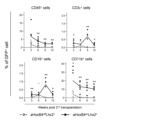
- Peripheral blood mononuclear cells were prepared from peripheral blood at 3, 6, 9, 12, 15, 18, and 20 weeks after iHSPC transfer, and GFP-positive blood cells (aHoxB4 on -Lhx2 + -iHSPC or aHoxB4 off -Lhx2 + - The percentage of iHSPC-derived blood cells was analyzed by flow cytometry.
- GFP-positive blood cells were detected in both aHoxB4 on -Lhx2 + -iHSPC secondary recipients and aHoxB4 off -Lhx2 + -iHSPC secondary recipients (FIG. 14). This result indicates that aHoxB4 on -Lhx2 + -iHSPC and aHoxB4 off -Lhx2 + -iHSPC contained in the primary recipient's bone marrow cells have hematopoietic ability and maintain stemness.
- mice On the day of iHSPC administration, recipient mice were irradiated with 3 Gy or 4 Gy of whole body radiation, and then 5.0 ⁇ 10 Lhx2 + -iHSPCs or aHoxB4 on -Lhx2 + -iHSPCs were suspended in 200 ⁇ l of physiological saline. It was administered through the tail vein. Peripheral blood mononuclear cells were prepared from peripheral blood at 2, 4 , 6, 8, 12, 14 , and 16 weeks after iHSPC transfer. The percentage of cells) was analyzed by flow cytometry.
- Example 6 Transplant immune tolerance induction by aHoxB4 on -Lhx2 + -iHSPC Transfer of aHoxB4 on -Lhx2 + -iHSPC-transferred C3129F1 mice and Lhx2 + -iHSPC-transferred C3129F1 mice 7 days after iHSPC transfer in Example 5 (3) C3129F1 mice transfected with aHoxB4 on -Lhx2 - -iHSPC, which were produced in the same manner using aHoxB4 on -Lhx2 - -iHSPC as cells, and C3129F1 mice that received the same treatment except that physiological saline was administered as a control were used as skin graft recipients.
- the dorsal skin of a sufficiently anesthetized recipient mouse was excised in a circular shape to form a transplant bed, and auricular skin pieces from donor B6 mice, BALB/c mice, and C3129F1 mice were transplanted.
- the size of the grafted skin pieces was measured over time. The time when the grafted skin piece regressed to less than 30% of the size (longer axis length) of the grafted skin piece on the day of transplantation was defined as the rejection date, and the period from the date of transplantation to the date of rejection was defined as the engraftment period of the grafted skin piece.
- FIG. 16 shows the engraftment period of the grafted skin pieces.
- the skin grafts from C3129F1 mice that were autologously transplanted into recipient C3129F1 mice were completely engrafted regardless of the presence or absence of various iHSPC transfers or the amount of radiation exposure.
- Lhx2 + -iHSPC and aHoxB4 on -Lhx2 + -iHSPC are capable of inducing immune tolerance even in allogeneic transplants that cause rejection due to minor antigen mismatch.
- Example 7 Analysis of hematopoietic potential of aHoxB4 on -Lhx2 + -iHSPC subset From aHoxB4 on -Lhx2 + -iHSPC prepared in Example 4, c-Kit + Sca Two types of iHSPC subsets, -1 - Lineage - (KL) cells and c-Kit + Sca-1 + Lineage - (KSL) cells, were sorted and used as transfected cells. Bone marrow cells collected from the femur and tibia of B6 mice were used as bone marrow cells for assisting hematopoiesis.
- Bone marrow cells for assisting hematopoiesis were suspended in 200 ⁇ l of physiological saline and administered through the tail vein.
- Peripheral blood mononuclear cells were prepared from peripheral blood at 3, 6, 9, and 12 weeks after cell transfer, and the percentage of GFP-positive blood cells (aHoxB4 on -Lhx2 + -iHSPC-derived blood cells) was analyzed by flow cytometry. .
- GK1.5 CD4-removing antibody
- CD8 ⁇ -removing antibody 6 days and 1 day before iHSPC administration
- mice On the day of iHSPC administration, recipient mice were irradiated with 3 Gy of whole-body radiation and then treated with 1.0 ⁇ 10 7 aHoxB4 on -Lhx2 + -iHSPC/200 ⁇ l saline or 200 ⁇ l saline as a control. It was administered intravenously.
- the dorsal skin of the fully anesthetized recipient mouse was excised in a circular shape to form a graft bed, and the auricular skin of the donor B6 mouse, BALB/c mouse, and C3H mouse was prepared. The piece was transplanted. After protecting the grafted skin pieces for 7 days after skin grafting, the size of the grafted skin pieces was measured over time.
- the time when the grafted skin piece regressed to less than 30% of the size (longer axis length) of the grafted skin piece on the day of transplantation was defined as the rejection date, and the period from the date of transplantation to the date of rejection was defined as the engraftment period of the grafted skin piece.
- FIG. 18 shows the engraftment period of the grafted skin pieces.
- skin grafts from third-party BALB/c mice were rejected within about 20 days after transplantation in both groups.
- the skin grafts from B6 mice, whose MHC haplotype is mismatched with that of the recipient and whose lineage is the same as the origin of the iPS cells, were rejected by about 30 days after transplantation in the saline-administered group, but aHoxB4 on -Lhx2 + - In the iHSPC transfer group, a slight extension of the engraftment period was observed.
- recipient mice On the day of iHSPC administration, recipient mice received 3 Gy of whole body irradiation and then received 1.0 ⁇ 10 7 or 3.0 ⁇ 10 7 aHoxB4 on -Lhx2 + -iHSPC/200 ⁇ l saline or 200 ⁇ l saline as a control. ⁇ l of physiological saline was administered through the tail vein.
- 500 ⁇ g of anti-CD154 antibody InVivoMAb anti-mouse CD40L (CD154), BioxCell
- diluted in saline was administered intraperitoneally on the same day as iHSPC administration, and then 2 days later. After 9 days, 500 ⁇ g of CTLA4-Ig (Orencia® for intravenous infusion, Bristol-Myers Squibb) diluted with physiological saline was administered intraperitoneally.
- FIG. 19 shows the engraftment period of the grafted skin pieces.
- skin grafts from third-party BALB/c mice were rejected within about 20 days after transplantation in all groups.
- Example 9 Hematopoietic ability analysis of aHoxB4 on -Lhx2 + -iHSPC (MHC-incompatible transplant) C3H mice transfected with 3 ⁇ 10 7 aHoxB4 on -Lhx2 + -iHSPCs were produced in the same manner as in Example 8 (2) in combination with CB.
- Example 8 (2) C3H mice transfected with 3 ⁇ 10 7 aHoxB4 on -Lhx2 + -iHSPCs in combination with CB, and physiological mice as a control instead of aHoxB4 on -Lhx2 + -iHSPCs were used.
- C3H mice were generated with the same treatment except that saline was administered.
- Figure 21 shows changes in body weight using the value before whole body radiation irradiation on the day of iHSPC administration as the standard (100). No significant difference in body weight was observed between the iHSPC administration group and the saline administration group, indicating that iHSPC administration did not cause weight loss.
- Example 10 Analysis of the mechanism of immune tolerance induction by aHoxB4 on -Lhx2 + -iHSPC
- the aHoxB4 on -Lhx2 + -iHSPC prepared in Example 4 was used as the transferred cells, and C3129F1 mice were used as the recipients.
- a pre-treatment for iHSPC administration in order to remove CD4-positive cells and CD8-positive cells from the body of recipient mice, a sufficient amount of CD4-removing antibody (GK1.5) and CD8 ⁇ -removing antibody (6 days and 1 day before iHSPC administration) 53-6.7) was administered intraperitoneally.
- mice On the day of iHSPC administration, recipient mice were irradiated with 4 Gy of whole body radiation, and then 1.0 ⁇ 10 7 aHoxB4 on -Lhx2 + -iHSPCs were suspended in 200 ⁇ l of physiological saline and administered through the tail vein.
- Peripheral blood mononuclear cells were prepared from peripheral blood collected 8 weeks after iHSPC transfer and analyzed by flow cytometry. Peripheral blood mononuclear cells from C3129F1 mice that received the same treatment, except that physiological saline was administered as a control instead of aHoxB4 on -Lhx2 + -iHSPC, and untreated C3129F1 mice (naive mice) were analyzed in the same way. did.
- Figure 22 shows the percentage of GFP-positive cells in -1 high and CD11b + Gr-1 low . It was confirmed that GFP-positive MDSCs were present in the peripheral blood of mice transfected with aHoxB4 on -Lhx2 + -iHSPC, and that a high proportion of GFP-positive cells existed in the CD11b + Gr-1 low subset. Ta.
- the ratio is shown in FIG.
- the proportion of PD-L1-positive cells was observed to be significantly higher in GFP-positive MDSCs than in GFP-negative MDSCs.
- the MFI (mean fluorescence intensity) of PD-L1 was selectively increased in the GFP-positive CD11b + Gr-1 low subset (FIG. 24).
- Figure 25 shows the percentage of Foxp3 + cells (Treg) in CD4 + T cells in the spleen of naive mice, saline-administered mice, and aHoxB4 on -Lhx2 + -iHSPC-transfected mice.
- Teg Foxp3 + cells
- HoxB4 on -Lhx2 + -iHSPC-transfected mice were found to have an increased proportion of Tregs in the spleen compared to naive mice.
- Tregs and MDSCs derived from HoxB4 on -Lhx2 + -iHSPC may have contributed to the immune tolerance induction mechanism using HoxB4 on -Lhx2 + -iHSPC.
- Example 11 Transplantation experiment of luciferase-expressing iPS cells
- the aHoxB4 on -Lhx2 + -iHSPCs prepared in Example 4 were used as the transplanted cells, and C3129F1 mice were used as the recipients.
- a pre-treatment for iHSPC administration in order to remove CD4-positive cells and CD8-positive cells from the body of recipient mice, a sufficient amount of CD4-removing antibody (GK1.5) and CD8 ⁇ -removing antibody (6 days and 1 day before iHSPC administration) 53-6.7) was administered intraperitoneally.
- mice On the day of iHSPC administration, recipient mice were irradiated with 4 Gy of whole-body radiation and then treated with 1.0 ⁇ 10 7 aHoxB4 on -Lhx2 + -iHSPC/200 ⁇ l saline or 200 ⁇ l saline as a control. It was administered intravenously.
- B6-derived luciferase-expressing iPS cells are described in Kamatani, T. et al. (Inflammation and Regeneration, (2022) 42:4. https://doi.org/10.1186/s41232-021-00190-7). These cells were created by introducing the luciferase gene into B6 fetal fibroblast-derived iPS cells using a lentiviral vector (pHIV-Luc-ZsGreen (Addgene)).
- FIG. 26A shows an image showing the luminescence of the iPS cell transplanted area of the mouse after administration of D-luciferin
- FIG. 26B shows the change in luminescence signal intensity (total flux).
- the signal fell below the detection limit in 3 out of 4 animals on the 21st day after transplantation.
- signals were detected in 4 out of 5 animals in the B6-derived aHoxB4 on -Lhx2 + -iHSPC administration group.
- the signal detected 21 days after transplantation of luciferase-expressing iPS cells was significantly higher in the aHoxB4 on -Lhx2 + -iHSPC administration group compared to the physiological saline administration group.
- FIG. 27A An image of the collected teratoma is shown in FIG. 27A, and the weight of the teratoma is shown in FIG. 27B.
- teratoma formation was not observed in 1 out of 4 animals, but in the aHoxB4 on -Lhx2 + -iHSPC administration group, teratoma formation was observed in all animals, and the teratoma weight was lower than that of aHoxB4 on -Lhx2 + -iHSPC. The value was significantly higher in the treated group.
- aHoxB4 on -Lhx2 + -iHSPC promoted the engraftment of donor-derived iPS cells . It was suggested that it may also be effective for cell-derived grafts.
- HSPCs obtained by culturing pluripotent stem cells under HSPC differentiation-inducing conditions under forced expression of Lhx2 or under forced expression of Lhx2 and HoxB4 can be used for minor antigens in mice. Since immune tolerance can be induced in both allogeneic transplants that cause rejection due to mismatching and allogeneic transplants that cause rejection due to MHC mismatch, it is possible to induce pluripotency in other mammals (including humans). HSPCs derived from sexual stem cells are expected to be able to induce immune tolerance.
- HSPCs obtained by culturing pluripotent stem cells under HSPC differentiation-inducing conditions under forced expression of Lhx2 or forced expression of Lhx2 and HoxB4 can reduce the amount of immunosuppressive drugs used by inducing immune tolerance. It is also expected to effectively maintain allogeneic allogeneic organisms (cells, tissues, organs) without the use of immunosuppressants.
Landscapes
- Health & Medical Sciences (AREA)
- Life Sciences & Earth Sciences (AREA)
- Engineering & Computer Science (AREA)
- Chemical & Material Sciences (AREA)
- Cell Biology (AREA)
- Organic Chemistry (AREA)
- Zoology (AREA)
- Biomedical Technology (AREA)
- Immunology (AREA)
- Biotechnology (AREA)
- Bioinformatics & Cheminformatics (AREA)
- Genetics & Genomics (AREA)
- General Health & Medical Sciences (AREA)
- Developmental Biology & Embryology (AREA)
- Wood Science & Technology (AREA)
- Medicinal Chemistry (AREA)
- Pharmacology & Pharmacy (AREA)
- Animal Behavior & Ethology (AREA)
- Public Health (AREA)
- Veterinary Medicine (AREA)
- Biochemistry (AREA)
- Virology (AREA)
- Microbiology (AREA)
- General Engineering & Computer Science (AREA)
- Epidemiology (AREA)
- Reproductive Health (AREA)
- Nuclear Medicine, Radiotherapy & Molecular Imaging (AREA)
- Toxicology (AREA)
- Gastroenterology & Hepatology (AREA)
- Gynecology & Obstetrics (AREA)
- Biophysics (AREA)
- General Chemical & Material Sciences (AREA)
- Molecular Biology (AREA)
- Proteomics, Peptides & Aminoacids (AREA)
- Transplantation (AREA)
- Hematology (AREA)
- Chemical Kinetics & Catalysis (AREA)
- Micro-Organisms Or Cultivation Processes Thereof (AREA)
Priority Applications (1)
| Application Number | Priority Date | Filing Date | Title |
|---|---|---|---|
| JP2024508291A JPWO2023176986A1 (cg-RX-API-DMAC7.html) | 2022-03-18 | 2023-03-20 |
Applications Claiming Priority (2)
| Application Number | Priority Date | Filing Date | Title |
|---|---|---|---|
| JP2022-043703 | 2022-03-18 | ||
| JP2022043703 | 2022-03-18 |
Publications (1)
| Publication Number | Publication Date |
|---|---|
| WO2023176986A1 true WO2023176986A1 (ja) | 2023-09-21 |
Family
ID=88023492
Family Applications (1)
| Application Number | Title | Priority Date | Filing Date |
|---|---|---|---|
| PCT/JP2023/011018 Ceased WO2023176986A1 (ja) | 2022-03-18 | 2023-03-20 | 免疫寛容誘導用組成物及びその製造方法 |
Country Status (2)
| Country | Link |
|---|---|
| JP (1) | JPWO2023176986A1 (cg-RX-API-DMAC7.html) |
| WO (1) | WO2023176986A1 (cg-RX-API-DMAC7.html) |
Cited By (1)
| Publication number | Priority date | Publication date | Assignee | Title |
|---|---|---|---|---|
| WO2024228384A1 (ja) * | 2023-04-30 | 2024-11-07 | 国立大学法人北海道大学 | 免疫寛容誘導用組成物 |
-
2023
- 2023-03-20 JP JP2024508291A patent/JPWO2023176986A1/ja active Pending
- 2023-03-20 WO PCT/JP2023/011018 patent/WO2023176986A1/ja not_active Ceased
Non-Patent Citations (3)
| Title |
|---|
| IZAWA KIYOKO; YAMAZAKI SATOSHI; BECKER HANS J; BHADURY JOYDEEP; KAKEGAWA TOMOYA; SAKAGUCHI MOMOKO; TOJO ARINOBU: "Activated HoxB4-induced hematopoietic stem cells from murine pluripotent stem cells via long-term programming", EXPERIMENTAL HEMATOLOGY, ELSEVIER INC., US, vol. 89, 12 August 2020 (2020-08-12), US , pages 68, XP086265596, ISSN: 0301-472X, DOI: 10.1016/j.exphem.2020.08.003 * |
| K. KITAJIMA, K.-I. MINEHATA, K. SAKIMURA, T. NAKANO, T. HARA: "In vitro generation of HSC-like cells from murine ESCs/iPSCs by enforced expression of LIM-homeobox transcription factor Lhx2", BLOOD, AMERICAN SOCIETY OF HEMATOLOGY, US, vol. 117, no. 14, 7 April 2011 (2011-04-07), US , pages 3748 - 3758, XP055323433, ISSN: 0006-4971, DOI: 10.1182/blood-2010-07-298596 * |
| KENJI KITAJIMA; MANAMI KAWAGUCHI; KAZUYA MIYASHITA; MARINO NAKAJIMA; MAI KANOKODA; TAKAHIKO HARA: "Efficient production of T cells from mouse pluripotent stem cells by controlled expression of Lhx2", GENES TO CELLS, WILEY-BLACKWELL PUBLISHING LTD., GB, vol. 20, no. 9, 7 July 2015 (2015-07-07), GB , pages 720 - 738, XP072055098, ISSN: 1356-9597, DOI: 10.1111/gtc.12266 * |
Cited By (1)
| Publication number | Priority date | Publication date | Assignee | Title |
|---|---|---|---|---|
| WO2024228384A1 (ja) * | 2023-04-30 | 2024-11-07 | 国立大学法人北海道大学 | 免疫寛容誘導用組成物 |
Also Published As
| Publication number | Publication date |
|---|---|
| JPWO2023176986A1 (cg-RX-API-DMAC7.html) | 2023-09-21 |
Similar Documents
| Publication | Publication Date | Title |
|---|---|---|
| US10933124B2 (en) | Methods of transplantation and disease treatment | |
| KR101457742B1 (ko) | 인간 배아 줄기세포 유래의 조혈세포 | |
| DE60026166T2 (de) | Vetozellen die wirksam in der prävention von transplantatabstossung sind und kein graft-versus-host potential aufweisen | |
| JP5572650B2 (ja) | リンパ造血組織を定着させるためのmapcまたはそれらの子孫の使用 | |
| JP2004521877A (ja) | 多能性幹細胞の同種移植片の寛容化 | |
| US8871510B2 (en) | Methods for generating T lymphocytes from hematopoietic stem cells | |
| US12448437B2 (en) | Transplant tolerance induction with carbodiimide treated tolerizing vaccine | |
| WO2012133948A1 (ja) | 生体組織から単離できるssea-3陽性の多能性幹細胞を含む他家移植用細胞治療用組成物 | |
| US20150139994A1 (en) | Compositions and methods for preventing allogeneic immune rejection | |
| US10751368B2 (en) | Methods of transplantation and disease treatment | |
| Gröschel et al. | Efficient killing of murine pluripotent stem cells by natural killer (NK) cells requires activation by cytokines and partly depends on the activating NK receptor NKG2D | |
| Drukker | Immunological considerations for cell therapy using human embryonic stem cell derivatives | |
| WO2021207121A9 (en) | Hypoimmunogenic cells and methods and compositions for their production | |
| WO2024228384A1 (ja) | 免疫寛容誘導用組成物 | |
| JP5089389B2 (ja) | 移植における免疫応答の予防および治療のための肝臓間質細胞 | |
| Pizzato | Metabolic Control and Immune Barriers of Hematopoietic Stem Cells | |
| EP4638709A2 (en) | Nk cell activating receptor ligand knockouts for use in cell therapy | |
| JP2009509911A (ja) | リンパ造血組織を定着させるためのmapcまたはそれらの子孫の使用 | |
| HK40061122A (en) | Transplant tolerance induction with carbodiimide treated tolerizing vaccine | |
| US20060153819A1 (en) | Bone marrow stromal cells for immunoprotection of transplanted neural stem cells | |
| Thompson | Immunogenicity of Embryonic Stem Cell Derived Hematopoietic Progenitors | |
| HK1096996A (en) | Tolerizing allografts of pluripotent stem cells | |
| Milanetti et al. | The induction of mixed chimerism using ES cell-derived hematopoietic stem cells |
Legal Events
| Date | Code | Title | Description |
|---|---|---|---|
| 121 | Ep: the epo has been informed by wipo that ep was designated in this application |
Ref document number: 23770938 Country of ref document: EP Kind code of ref document: A1 |
|
| WWE | Wipo information: entry into national phase |
Ref document number: 2024508291 Country of ref document: JP |
|
| NENP | Non-entry into the national phase |
Ref country code: DE |
|
| 122 | Ep: pct application non-entry in european phase |
Ref document number: 23770938 Country of ref document: EP Kind code of ref document: A1 |