WO2023153471A1 - Fcrl1に結合する抗体又は該抗体断片 - Google Patents
Fcrl1に結合する抗体又は該抗体断片 Download PDFInfo
- Publication number
- WO2023153471A1 WO2023153471A1 PCT/JP2023/004346 JP2023004346W WO2023153471A1 WO 2023153471 A1 WO2023153471 A1 WO 2023153471A1 JP 2023004346 W JP2023004346 W JP 2023004346W WO 2023153471 A1 WO2023153471 A1 WO 2023153471A1
- Authority
- WO
- WIPO (PCT)
- Prior art keywords
- antibody
- amino acid
- seq
- fcrl1
- acid sequence
- Prior art date
- Legal status (The legal status is an assumption and is not a legal conclusion. Google has not performed a legal analysis and makes no representation as to the accuracy of the status listed.)
- Ceased
Links
Images






Classifications
-
- C—CHEMISTRY; METALLURGY
- C07—ORGANIC CHEMISTRY
- C07K—PEPTIDES
- C07K16/00—Immunoglobulins [IG], e.g. monoclonal or polyclonal antibodies
- C07K16/18—Immunoglobulins [IG], e.g. monoclonal or polyclonal antibodies against material from animals or humans
- C07K16/28—Immunoglobulins [IG], e.g. monoclonal or polyclonal antibodies against material from animals or humans against receptors, cell surface antigens or cell surface determinants
- C07K16/2803—Immunoglobulins [IG], e.g. monoclonal or polyclonal antibodies against material from animals or humans against receptors, cell surface antigens or cell surface determinants against the immunoglobulin superfamily
- C07K16/283—Immunoglobulins [IG], e.g. monoclonal or polyclonal antibodies against material from animals or humans against receptors, cell surface antigens or cell surface determinants against the immunoglobulin superfamily against Fc-receptors, e.g. CD16, CD32, CD64
-
- A—HUMAN NECESSITIES
- A61—MEDICAL OR VETERINARY SCIENCE; HYGIENE
- A61K—PREPARATIONS FOR MEDICAL, DENTAL OR TOILETRY PURPOSES
- A61K47/00—Medicinal preparations characterised by the non-active ingredients used, e.g. carriers or inert additives; Targeting or modifying agents chemically bound to the active ingredient
- A61K47/50—Medicinal preparations characterised by the non-active ingredients used, e.g. carriers or inert additives; Targeting or modifying agents chemically bound to the active ingredient the non-active ingredient being chemically bound to the active ingredient, e.g. polymer-drug conjugates
- A61K47/51—Medicinal preparations characterised by the non-active ingredients used, e.g. carriers or inert additives; Targeting or modifying agents chemically bound to the active ingredient the non-active ingredient being chemically bound to the active ingredient, e.g. polymer-drug conjugates the non-active ingredient being a modifying agent
- A61K47/68—Medicinal preparations characterised by the non-active ingredients used, e.g. carriers or inert additives; Targeting or modifying agents chemically bound to the active ingredient the non-active ingredient being chemically bound to the active ingredient, e.g. polymer-drug conjugates the non-active ingredient being a modifying agent the modifying agent being an antibody, an immunoglobulin or a fragment thereof, e.g. an Fc-fragment
-
- A—HUMAN NECESSITIES
- A61—MEDICAL OR VETERINARY SCIENCE; HYGIENE
- A61K—PREPARATIONS FOR MEDICAL, DENTAL OR TOILETRY PURPOSES
- A61K47/00—Medicinal preparations characterised by the non-active ingredients used, e.g. carriers or inert additives; Targeting or modifying agents chemically bound to the active ingredient
- A61K47/50—Medicinal preparations characterised by the non-active ingredients used, e.g. carriers or inert additives; Targeting or modifying agents chemically bound to the active ingredient the non-active ingredient being chemically bound to the active ingredient, e.g. polymer-drug conjugates
- A61K47/51—Medicinal preparations characterised by the non-active ingredients used, e.g. carriers or inert additives; Targeting or modifying agents chemically bound to the active ingredient the non-active ingredient being chemically bound to the active ingredient, e.g. polymer-drug conjugates the non-active ingredient being a modifying agent
- A61K47/68—Medicinal preparations characterised by the non-active ingredients used, e.g. carriers or inert additives; Targeting or modifying agents chemically bound to the active ingredient the non-active ingredient being chemically bound to the active ingredient, e.g. polymer-drug conjugates the non-active ingredient being a modifying agent the modifying agent being an antibody, an immunoglobulin or a fragment thereof, e.g. an Fc-fragment
- A61K47/6801—Drug-antibody or immunoglobulin conjugates defined by the pharmacologically or therapeutically active agent
- A61K47/6803—Drugs conjugated to an antibody or immunoglobulin, e.g. cisplatin-antibody conjugates
- A61K47/68035—Drugs conjugated to an antibody or immunoglobulin, e.g. cisplatin-antibody conjugates the drug being a pyrrolobenzodiazepine
-
- A—HUMAN NECESSITIES
- A61—MEDICAL OR VETERINARY SCIENCE; HYGIENE
- A61K—PREPARATIONS FOR MEDICAL, DENTAL OR TOILETRY PURPOSES
- A61K47/00—Medicinal preparations characterised by the non-active ingredients used, e.g. carriers or inert additives; Targeting or modifying agents chemically bound to the active ingredient
- A61K47/50—Medicinal preparations characterised by the non-active ingredients used, e.g. carriers or inert additives; Targeting or modifying agents chemically bound to the active ingredient the non-active ingredient being chemically bound to the active ingredient, e.g. polymer-drug conjugates
- A61K47/51—Medicinal preparations characterised by the non-active ingredients used, e.g. carriers or inert additives; Targeting or modifying agents chemically bound to the active ingredient the non-active ingredient being chemically bound to the active ingredient, e.g. polymer-drug conjugates the non-active ingredient being a modifying agent
- A61K47/68—Medicinal preparations characterised by the non-active ingredients used, e.g. carriers or inert additives; Targeting or modifying agents chemically bound to the active ingredient the non-active ingredient being chemically bound to the active ingredient, e.g. polymer-drug conjugates the non-active ingredient being a modifying agent the modifying agent being an antibody, an immunoglobulin or a fragment thereof, e.g. an Fc-fragment
- A61K47/6835—Medicinal preparations characterised by the non-active ingredients used, e.g. carriers or inert additives; Targeting or modifying agents chemically bound to the active ingredient the non-active ingredient being chemically bound to the active ingredient, e.g. polymer-drug conjugates the non-active ingredient being a modifying agent the modifying agent being an antibody, an immunoglobulin or a fragment thereof, e.g. an Fc-fragment the modifying agent being an antibody or an immunoglobulin bearing at least one antigen-binding site
- A61K47/6849—Medicinal preparations characterised by the non-active ingredients used, e.g. carriers or inert additives; Targeting or modifying agents chemically bound to the active ingredient the non-active ingredient being chemically bound to the active ingredient, e.g. polymer-drug conjugates the non-active ingredient being a modifying agent the modifying agent being an antibody, an immunoglobulin or a fragment thereof, e.g. an Fc-fragment the modifying agent being an antibody or an immunoglobulin bearing at least one antigen-binding site the antibody targeting a receptor, a cell surface antigen or a cell surface determinant
-
- A—HUMAN NECESSITIES
- A61—MEDICAL OR VETERINARY SCIENCE; HYGIENE
- A61P—SPECIFIC THERAPEUTIC ACTIVITY OF CHEMICAL COMPOUNDS OR MEDICINAL PREPARATIONS
- A61P29/00—Non-central analgesic, antipyretic or antiinflammatory agents, e.g. antirheumatic agents; Non-steroidal antiinflammatory drugs [NSAID]
-
- A—HUMAN NECESSITIES
- A61—MEDICAL OR VETERINARY SCIENCE; HYGIENE
- A61P—SPECIFIC THERAPEUTIC ACTIVITY OF CHEMICAL COMPOUNDS OR MEDICINAL PREPARATIONS
- A61P35/00—Antineoplastic agents
-
- A—HUMAN NECESSITIES
- A61—MEDICAL OR VETERINARY SCIENCE; HYGIENE
- A61P—SPECIFIC THERAPEUTIC ACTIVITY OF CHEMICAL COMPOUNDS OR MEDICINAL PREPARATIONS
- A61P35/00—Antineoplastic agents
- A61P35/02—Antineoplastic agents specific for leukemia
-
- A—HUMAN NECESSITIES
- A61—MEDICAL OR VETERINARY SCIENCE; HYGIENE
- A61P—SPECIFIC THERAPEUTIC ACTIVITY OF CHEMICAL COMPOUNDS OR MEDICINAL PREPARATIONS
- A61P37/00—Drugs for immunological or allergic disorders
-
- A—HUMAN NECESSITIES
- A61—MEDICAL OR VETERINARY SCIENCE; HYGIENE
- A61P—SPECIFIC THERAPEUTIC ACTIVITY OF CHEMICAL COMPOUNDS OR MEDICINAL PREPARATIONS
- A61P37/00—Drugs for immunological or allergic disorders
- A61P37/02—Immunomodulators
-
- C—CHEMISTRY; METALLURGY
- C12—BIOCHEMISTRY; BEER; SPIRITS; WINE; VINEGAR; MICROBIOLOGY; ENZYMOLOGY; MUTATION OR GENETIC ENGINEERING
- C12N—MICROORGANISMS OR ENZYMES; COMPOSITIONS THEREOF; PROPAGATING, PRESERVING, OR MAINTAINING MICROORGANISMS; MUTATION OR GENETIC ENGINEERING; CULTURE MEDIA
- C12N5/00—Undifferentiated human, animal or plant cells, e.g. cell lines; Tissues; Cultivation or maintenance thereof; Culture media therefor
- C12N5/10—Cells modified by introduction of foreign genetic material
- C12N5/12—Fused cells, e.g. hybridomas
-
- G—PHYSICS
- G01—MEASURING; TESTING
- G01N—INVESTIGATING OR ANALYSING MATERIALS BY DETERMINING THEIR CHEMICAL OR PHYSICAL PROPERTIES
- G01N33/00—Investigating or analysing materials by specific methods not covered by groups G01N1/00 - G01N31/00
- G01N33/48—Biological material, e.g. blood, urine; Haemocytometers
- G01N33/50—Chemical analysis of biological material, e.g. blood, urine; Testing involving biospecific ligand binding methods; Immunological testing
- G01N33/53—Immunoassay; Biospecific binding assay; Materials therefor
- G01N33/564—Immunoassay; Biospecific binding assay; Materials therefor for pre-existing immune complex or autoimmune disease, i.e. systemic lupus erythematosus, rheumatoid arthritis, multiple sclerosis, rheumatoid factors or complement components C1-C9
-
- G—PHYSICS
- G01—MEASURING; TESTING
- G01N—INVESTIGATING OR ANALYSING MATERIALS BY DETERMINING THEIR CHEMICAL OR PHYSICAL PROPERTIES
- G01N33/00—Investigating or analysing materials by specific methods not covered by groups G01N1/00 - G01N31/00
- G01N33/48—Biological material, e.g. blood, urine; Haemocytometers
- G01N33/50—Chemical analysis of biological material, e.g. blood, urine; Testing involving biospecific ligand binding methods; Immunological testing
- G01N33/53—Immunoassay; Biospecific binding assay; Materials therefor
- G01N33/575—Immunoassay; Biospecific binding assay; Materials therefor for cancer
- G01N33/5758—Immunoassay; Biospecific binding assay; Materials therefor for cancer involving compounds serving as markers for tumours, cancers or neoplasias, e.g. cellular determinants, receptors, heat shock/stress proteins, A-protein, oligosaccharides or metabolites
- G01N33/5759—Immunoassay; Biospecific binding assay; Materials therefor for cancer involving compounds serving as markers for tumours, cancers or neoplasias, e.g. cellular determinants, receptors, heat shock/stress proteins, A-protein, oligosaccharides or metabolites involving compounds localised on the membrane of tumour or cancer cells
-
- G—PHYSICS
- G01—MEASURING; TESTING
- G01N—INVESTIGATING OR ANALYSING MATERIALS BY DETERMINING THEIR CHEMICAL OR PHYSICAL PROPERTIES
- G01N33/00—Investigating or analysing materials by specific methods not covered by groups G01N1/00 - G01N31/00
- G01N33/48—Biological material, e.g. blood, urine; Haemocytometers
- G01N33/50—Chemical analysis of biological material, e.g. blood, urine; Testing involving biospecific ligand binding methods; Immunological testing
- G01N33/68—Chemical analysis of biological material, e.g. blood, urine; Testing involving biospecific ligand binding methods; Immunological testing involving proteins, peptides or amino acids
-
- G—PHYSICS
- G01—MEASURING; TESTING
- G01N—INVESTIGATING OR ANALYSING MATERIALS BY DETERMINING THEIR CHEMICAL OR PHYSICAL PROPERTIES
- G01N33/00—Investigating or analysing materials by specific methods not covered by groups G01N1/00 - G01N31/00
- G01N33/48—Biological material, e.g. blood, urine; Haemocytometers
- G01N33/50—Chemical analysis of biological material, e.g. blood, urine; Testing involving biospecific ligand binding methods; Immunological testing
- G01N33/68—Chemical analysis of biological material, e.g. blood, urine; Testing involving biospecific ligand binding methods; Immunological testing involving proteins, peptides or amino acids
- G01N33/6872—Intracellular protein regulatory factors and their receptors, e.g. including ion channels
-
- G—PHYSICS
- G01—MEASURING; TESTING
- G01N—INVESTIGATING OR ANALYSING MATERIALS BY DETERMINING THEIR CHEMICAL OR PHYSICAL PROPERTIES
- G01N33/00—Investigating or analysing materials by specific methods not covered by groups G01N1/00 - G01N31/00
- G01N33/48—Biological material, e.g. blood, urine; Haemocytometers
- G01N33/50—Chemical analysis of biological material, e.g. blood, urine; Testing involving biospecific ligand binding methods; Immunological testing
- G01N33/68—Chemical analysis of biological material, e.g. blood, urine; Testing involving biospecific ligand binding methods; Immunological testing involving proteins, peptides or amino acids
- G01N33/6893—Chemical analysis of biological material, e.g. blood, urine; Testing involving biospecific ligand binding methods; Immunological testing involving proteins, peptides or amino acids related to diseases not provided for elsewhere
-
- A—HUMAN NECESSITIES
- A61—MEDICAL OR VETERINARY SCIENCE; HYGIENE
- A61K—PREPARATIONS FOR MEDICAL, DENTAL OR TOILETRY PURPOSES
- A61K39/00—Medicinal preparations containing antigens or antibodies
- A61K2039/505—Medicinal preparations containing antigens or antibodies comprising antibodies
-
- C—CHEMISTRY; METALLURGY
- C07—ORGANIC CHEMISTRY
- C07K—PEPTIDES
- C07K2317/00—Immunoglobulins specific features
- C07K2317/10—Immunoglobulins specific features characterized by their source of isolation or production
- C07K2317/14—Specific host cells or culture conditions, e.g. components, pH or temperature
-
- C—CHEMISTRY; METALLURGY
- C07—ORGANIC CHEMISTRY
- C07K—PEPTIDES
- C07K2317/00—Immunoglobulins specific features
- C07K2317/20—Immunoglobulins specific features characterized by taxonomic origin
- C07K2317/24—Immunoglobulins specific features characterized by taxonomic origin containing regions, domains or residues from different species, e.g. chimeric, humanized or veneered
-
- C—CHEMISTRY; METALLURGY
- C07—ORGANIC CHEMISTRY
- C07K—PEPTIDES
- C07K2317/00—Immunoglobulins specific features
- C07K2317/50—Immunoglobulins specific features characterized by immunoglobulin fragments
- C07K2317/56—Immunoglobulins specific features characterized by immunoglobulin fragments variable (Fv) region, i.e. VH and/or VL
-
- C—CHEMISTRY; METALLURGY
- C07—ORGANIC CHEMISTRY
- C07K—PEPTIDES
- C07K2317/00—Immunoglobulins specific features
- C07K2317/50—Immunoglobulins specific features characterized by immunoglobulin fragments
- C07K2317/56—Immunoglobulins specific features characterized by immunoglobulin fragments variable (Fv) region, i.e. VH and/or VL
- C07K2317/565—Complementarity determining region [CDR]
-
- C—CHEMISTRY; METALLURGY
- C07—ORGANIC CHEMISTRY
- C07K—PEPTIDES
- C07K2317/00—Immunoglobulins specific features
- C07K2317/70—Immunoglobulins specific features characterized by effect upon binding to a cell or to an antigen
- C07K2317/77—Internalization into the cell
-
- C—CHEMISTRY; METALLURGY
- C07—ORGANIC CHEMISTRY
- C07K—PEPTIDES
- C07K2317/00—Immunoglobulins specific features
- C07K2317/90—Immunoglobulins specific features characterized by (pharmaco)kinetic aspects or by stability of the immunoglobulin
- C07K2317/92—Affinity (KD), association rate (Ka), dissociation rate (Kd) or EC50 value
-
- C—CHEMISTRY; METALLURGY
- C12—BIOCHEMISTRY; BEER; SPIRITS; WINE; VINEGAR; MICROBIOLOGY; ENZYMOLOGY; MUTATION OR GENETIC ENGINEERING
- C12N—MICROORGANISMS OR ENZYMES; COMPOSITIONS THEREOF; PROPAGATING, PRESERVING, OR MAINTAINING MICROORGANISMS; MUTATION OR GENETIC ENGINEERING; CULTURE MEDIA
- C12N2510/00—Genetically modified cells
-
- G—PHYSICS
- G01—MEASURING; TESTING
- G01N—INVESTIGATING OR ANALYSING MATERIALS BY DETERMINING THEIR CHEMICAL OR PHYSICAL PROPERTIES
- G01N2333/00—Assays involving biological materials from specific organisms or of a specific nature
- G01N2333/435—Assays involving biological materials from specific organisms or of a specific nature from animals; from humans
- G01N2333/705—Assays involving receptors, cell surface antigens or cell surface determinants
- G01N2333/70503—Immunoglobulin superfamily, e.g. VCAMs, PECAM, LFA-3
- G01N2333/70535—Fc-receptors, e.g. CD16, CD32, CD64 (CD2314/705F)
-
- G—PHYSICS
- G01—MEASURING; TESTING
- G01N—INVESTIGATING OR ANALYSING MATERIALS BY DETERMINING THEIR CHEMICAL OR PHYSICAL PROPERTIES
- G01N2800/00—Detection or diagnosis of diseases
- G01N2800/70—Mechanisms involved in disease identification
- G01N2800/7095—Inflammation
Definitions
- the present invention includes a monoclonal antibody or antibody fragment that binds to the extracellular region of Fc receptor-like protein 1, a hybridoma that produces the antibody, a nucleic acid having a nucleotide sequence that encodes the antibody or the antibody fragment, and the nucleic acid.
- a transformed cell obtained by introducing a vector into a host cell, a method for producing the antibody or the antibody fragment using the hybridoma or the transformed cell, an antibody-drug conjugate containing the antibody or the antibody fragment, the antibody or the antibody
- the present invention relates to therapeutic agents and diagnostic agents comprising antibody fragments, and methods of treating and diagnosing Fc receptor-like protein 1-related diseases using said antibodies or said antibody fragments or antibody-drug conjugates comprising said antibodies or said antibody fragments.
- FCRL1 Fc receptor-like protein 1
- FCRL1 is a membrane protein belonging to the immunoglobulin superfamily, also known as CD307a, FCRH1, IFGP1, IRTA5, and the like.
- the amino acid sequence of human FCRL1 was identified in 2001 (Non-Patent Document 1).
- FCRL1 is a type I transmembrane protein expressed in B cells. It is a protein with three extracellular immunoglobulin-like domains, two intracellular immunoreceptor tyrosine activation motifs and a transmembrane region (Non-Patent Document 1). No endogenous ligand for FCRL1 has been identified to date.
- FCRL1 is reported to be expressed in cancer cells such as chronic lymphocytic leukemia, follicular lymphoma, hairy cell leukemia, and mantle cell lymphoma (Non-Patent Documents 2 and 3). . Furthermore, in recent years, it has been reported that FCRL1 contributes to cancer proliferation (Non-Patent Document 4).
- E3, E9 (Non-Patent Document 2), 2G5, 7G8, 5A2 (Patent Document 1), 1F9, 2A10 (Patent Document 2), and 5A3 (Patent Document 3) are known as monoclonal antibodies against FCRL1. It is also known that binding an immunotoxin to an anti-FCRL1 antibody exhibits cytotoxic activity against cancer cell lines (Non-Patent Document 4).
- the present invention provides a novel monoclonal antibody or antibody fragment that binds to the extracellular region of FCRL1, a hybridoma producing the antibody, a nucleic acid having a nucleotide sequence encoding the antibody or the antibody fragment, and a vector containing the nucleic acid as a host.
- a transformed cell obtained by introducing into a cell, a method for producing the antibody or the antibody fragment using the hybridoma or the transformed cell, an antibody-drug conjugate containing the antibody or the antibody fragment, and the antibody or the antibody fragment. It is an object of the present invention to provide therapeutic and diagnostic agents comprising the antibody or antibody fragment, and methods of treating and diagnosing FCRL1-related diseases using the antibody or antibody fragment, or an antibody-drug conjugate comprising the antibody or antibody fragment.
- the present invention relates to 1 to 26 below.
- FCRL1 Fc receptor-like protein 1
- FCRL1 Fc receptor-like protein 1
- VH heavy chain variable region
- CDR complementarity determining region
- said antibody fragment is selected from Fab, Fab', F(ab') 2 , single chain antibodies (scFv), dimerization V regions (Diabody), disulfide stabilized V regions (dsFv) and peptides containing CDRs 7.
- a hybridoma that produces the antibody according to any one of 1 to 6 above.
- 10. 9 comprising the nucleic acid according to 9 above.
- a transformed cell obtained by introducing the vector according to 10 above into a host cell. 12.
- the antibody or antibody of any one of 1 to 7 above which comprises culturing the hybridoma of 8 above or the transformed cell of 11 above in a medium and collecting the antibody or antibody fragment from the culture.
- a method for producing an antibody fragment 13.
- An antibody-drug conjugate comprising the antibody or antibody fragment of any one of 1 to 7 above.
- the antibody-drug conjugate according to 13 above, wherein the antibody-drug conjugate comprises the antibody or antibody fragment linked to a drug via a linker.
- 15. A composition comprising the antibody or antibody fragment of any one of 1 to 7 above or the antibody-drug conjugate of 13 or 14 above. 16.
- a diagnostic agent for FCRL1-related diseases comprising the antibody or antibody fragment of any one of 1 to 7 above or the antibody-drug conjugate of 13 or 14 above. 18.
- the FCRL1-associated disease is cancer, an autoimmune disease or an inflammatory disease.
- a therapeutic agent for FCRL1-related diseases comprising the antibody or antibody fragment of any one of 1 to 7 above or the antibody-drug conjugate of 13 or 14 above. 20.
- the therapeutic agent according to 19 above, wherein the FCRL1-associated disease is cancer, an autoimmune disease or an inflammatory disease.
- 22. A method for treating FCRL1-related diseases, comprising administering the antibody or antibody fragment of any one of 1 to 7 above or the antibody-drug conjugate of 13 or 14 above.
- 23. 15. Use of the antibody or antibody fragment according to any one of 1 to 7 above or the antibody-drug conjugate according to 13 or 14 above for the manufacture of a diagnostic agent for FCRL1-related diseases.
- the monoclonal antibody or antibody fragment of the present invention selectively binds to the extracellular region of human FCRL1.
- the monoclonal antibody or antibody fragment of the present invention exhibits superior effects when used in an antibody-drug conjugate (hereinafter also referred to as ADC) compared to existing FCRL1 antibodies. Therefore, the monoclonal antibodies or antibody fragments of the present invention can be used as therapeutic agents and diagnostic agents for human FCRL1-related diseases.
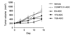
- FIG. 1 shows the results of measuring the antitumor effect of an antibody-drug conjugate in which a payload linker SG3249 was bound to a known anti-human FCRL1 antibody in a mouse model subcutaneously implanted with SU-DHL-6 cells.
- the vertical axis in FIG. 1 indicates tumor size (mm 3 ).
- the horizontal axis in FIG. 1 indicates the number of days after administration of ADC to the SU-DHL-6 cell subcutaneous transplantation mouse model.
- E9, 1F9 and 7G8 were used as known anti-human FCRL1 antibodies.
- Anti-2,4-dinitrophenol (DNP) IgG1 antibody was used as a negative antibody.
- FIG. 1 shows the results of measuring the antitumor effect of an antibody-drug conjugate in which a payload linker SG3249 was bound to a known anti-human FCRL1 antibody in a mouse model subcutaneously implanted with SU-DHL-6 cells.
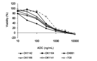
- FIG. 2A shows the results of measuring the effect of the novel anti-human FCRL1 antibody conjugated with the payload linker SG3249 on the survival of SU-DHL-6 cells.
- the vertical axis in FIG. 2A indicates the cell viability (%), and the number of cells in the condition without ADC treatment was defined as 100%.
- the horizontal axis of FIG. 2A indicates the concentration of ADC added to SU-DHL-6 cells.
- DK1142, DK1164, DK681, DK1166 and DK1141 were used as novel anti-human FCRL1 antibodies.
- 7G8 was used as a known anti-human FCRL1 antibody.
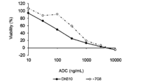
- FIG. 2B shows the results of using DK610 as a novel anti-human FCRL1 antibody in the same assay as in FIG. 2A.
- FIG. 3A shows the results of measuring the effect of the novel anti-human FCRL1 antibody conjugated with SG3249, which is a payload linker, on the survival of Ramos cells.
- the vertical axis in FIG. 3A indicates the cell viability (%), and the number of cells in the condition not treated with ADC was defined as 100%.
- the horizontal axis of FIG. 3A indicates the concentration of ADC added to Ramos cells.
- DK1142, DK1164, DK681, DK1166 and DK1141 were used as novel anti-human FCRL1 antibodies. 7G8 was used as a known anti-human FCRL1 antibody.
- FIG. 3B shows the results of using DK610 as a novel anti-human FCRL1 antibody in the same assay as in FIG. 3A.
- FIG. 4 shows the results of measuring the anti-tumor effect of an ADC comprising a novel anti-human FCRL1 antibody conjugated with a payload linker SG3249 in SU-DHL-6 cell subcutaneous mouse model and Ramos cell subcutaneous mouse model. The results 10 days after drug administration are shown.
- the vertical axis of FIG. 4 shows the relative tumor size when the tumor size of mice administered with 7G8 is set to 1. DK1142, DK1164, DK681, DK1166, DK1141 and DK610 were used as novel anti-human FCRL1 antibodies. 7G8 was used as a known anti-human FCRL1 antibody.
- FIG. 5 shows the results of measuring the antitumor effect of an ADC in which a novel anti-human FCRL1 antibody was conjugated with SG3249, which is a payload linker, in a mouse model of subcutaneous implantation of Ramos cells. The results on day 42 after drug administration are shown.
- the vertical axis in FIG. 5 indicates tumor size (mm 3 ).
- DK1142, DK1164, DK681, DK1166, DK1141 and DK610 were used as novel anti-human FCRL1 antibodies.
- 7G8 was used as a known anti-human FCRL1 antibody.
- FIG. 6 shows the results of measuring internalization of the novel anti-human FCRL1 antibody in Ramos cells. The vertical axis in FIG. 6 indicates fluorescence intensity.
- FIG. 7A shows the results of measuring the effect of the novel anti-human FCRL1 antibody conjugated with the payload linker SG3249 on the survival of SU-DHL-6 cells.
- the vertical axis in FIG. 7A indicates the cell viability (%), and the number of cells in the condition without ADC treatment was defined as 100%.
- the horizontal axis of FIG. 7A indicates the concentration of ADC added to SU-DHL-6 cells.
- DK681 was used as the novel anti-FCRL1 chimeric antibody, and DK681 F11, DK681 F12, DK681 F13 and DK681 F14 were used as the novel anti-FCRL1 humanized antibodies.
- 7G8 was used as a known anti-human FCRL1 antibody.
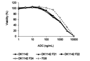
- FIG. 7B shows the results of using DK1142 as the novel anti-FCRL1 chimeric antibody and DK1142 F21, DK1142 F22 and DK1142 F24 as the novel anti-FCRL1 humanized antibody in the same measurements as in FIG. 7A.
- FIG. 7B shows the results of using DK1142 as the novel anti-FCRL1 chimeric antibody and DK1142 F21, DK1142 F22 and DK1142 F24 as the novel anti-FCRL1 humanized antibody in the same measurements as in FIG. 7A.
- FIG. 8A shows the results of measuring the effects on the survival of Ramos cells for ADCs in which the novel anti-human FCRL1 antibody was conjugated with SG3249, which is a payload linker.
- the vertical axis in FIG. 8A indicates the cell viability (%), and the number of cells in the condition without ADC treatment was defined as 100%.
- the horizontal axis of FIG. 8A indicates the concentration of ADC added to Ramos cells.
- DK681 was used as the novel anti-FCRL1 chimeric antibody
- DK681 F11, DK681 F12, DK681 F13 and DK681 F14 were used as the novel anti-FCRL1 humanized antibodies.
- 7G8 was used as a known anti-human FCRL1 antibody.
- FIG. 8B shows the results of using DK1142 as the novel anti-FCRL1 chimeric antibody and DK1142 F21, DK1142 F22 and DK1142 F24 as the novel anti-FCRL1 humanized antibody in the same measurements as in FIG. 8A.
- FIG. 9 shows the results of measuring the antitumor effect of an ADC in which a payload linker SG3249 is bound to a novel anti-human FCRL1 antibody in SU-DHL-6 cell subcutaneous mouse model and Ramos cell subcutaneous mouse model. The results on day 7 after drug administration are shown.
- the vertical axis in FIG. 9 shows the relative tumor size when the tumor size of mice administered with 7G8 is set to 1.
- the present invention relates to monoclonal antibodies or antibody fragments that bind to human FCRL1.
- FCRL1 is also called CD307a, FCRH1, IFGP1 and IRTA5.
- FCRL1 belongs to the immunoglobulin superfamily and is a type 1 membrane protein consisting of 413 amino acids.
- FCRL1 has two intracellular immunoreceptor tyrosine-activated motifs (ITAM). Therefore, it is expected that an activating signal is transmitted into the cell by ligand binding, but at present, an endogenous ligand for FCRL1 has not been identified, and the function of FCRL1 has not been elucidated. Recent experiments using cancer cell lines have reported that FCRL1 is involved in cancer cell proliferation by regulating the expression of apoptosis-related molecules.
- ITAM immunoreceptor tyrosine-activated motifs
- human FCRL1 is a polypeptide comprising the amino acid sequence set forth in SEQ ID NO: 3 or the amino acid sequence of NCBI Accession No. NP_443170, one of the amino acid sequences set forth in SEQ ID NO: 3 or the amino acid sequence of NCBI Accession No. NP_443170
- Examples include polypeptides comprising amino acid sequences having preferably 80% or more, more preferably 90% or more, and most preferably 95% or more similarity, and having the function of human FCRL1.
- a polypeptide having an amino acid sequence in which one or more amino acids are deleted, substituted or added in the amino acid sequence shown in SEQ ID NO: 3 or the amino acid sequence shown in NCBI Accession No. NP_443170 is obtained by site-directed mutagenesis [Molecular Cloning, A Laboratory Manual, Second Edition, Cold Spring Harbor Laboratory Press (1989), Current Protocols in Molecular Biology, John Wiley & Sons (1987-1997), Nucleic acids Research, 10, 6487 (1982), Proc. Natl. A cad USA, 79, 6409 (1982), Gene, 34, 315 (1985), Nucleic Acids Research, 13, 4431 (1985), Proc. Natl. Acad. Sci. USA, 82, 488 (1985)], etc. can be obtained, for example, by introducing site-directed mutation into a DNA encoding a polypeptide containing the amino acid sequence of SEQ ID NO:3.
- the number of amino acids to be deleted, substituted or added is not particularly limited, but preferably 1 to several tens, for example 1 to 20, more preferably 1 to several, for example 1 to 5 amino acids. is.
- the gene encoding human FCRL1 includes the nucleotide sequence set forth in SEQ ID NO: 1 and the nucleotide sequence of NCBI Accession No. NM_052938.
- DNAs that hybridize under stringent conditions include colony hybridization, plaque hybridization, and Southern blotting using DNA containing the nucleotide sequence of SEQ ID NO: 1 or NM — 052938 as a probe. It refers to hybridizable DNA obtained by a hybridization method, a DNA microarray method, or the like.
- the hybridizable DNA is a DNA having at least 60% or more similarity, preferably 80% or more similarity, more preferably 95% similarity to the nucleotide sequence of SEQ ID NO: 1 or NM — 052938. DNA having the above similarity can be mentioned.
- the gene encoding human FCRL1 of the present invention also includes a gene having a small-scale mutation in the base sequence due to such polymorphism in the gene used in the present invention.
- Antibodies of the present invention include antibodies that bind to both human FCRL1 and monkey FCRL1.
- the monkey FCRL1 is a polypeptide comprising the amino acid sequence set forth in SEQ ID NO: 4 or the amino acid sequence of NCBI Accession No. XP_015310712, one of the amino acid sequence set forth in SEQ ID NO: 4 or the amino acid sequence of NCBI Accession No. XP_015310712
- Polypeptides comprising amino acid sequences having preferably 80% or more, more preferably 90% or more, and most preferably 95% or more similarity and having monkey FCRL1 functions are included.
- a polypeptide having an amino acid sequence in which one or more amino acids are deleted, substituted or added in the amino acid sequence represented by SEQ ID NO: 4 or the amino acid sequence represented by NCBI Accession No. XP_015310712 is subjected to site-directed mutagenesis or the like. It can be obtained, for example, by introducing site-directed mutation into a DNA encoding a polypeptide containing the amino acid sequence of SEQ ID NO:4.
- the number of amino acids to be deleted, substituted or added is not particularly limited, but preferably 1 to several tens, for example 1 to 20, more preferably 1 to several, for example 1 to 5 amino acids. is.
- the gene encoding monkey FCRL1 includes the nucleotide sequence set forth in SEQ ID NO: 2 and the nucleotide sequence of NCBI Accession No. XM_005541349.
- a gene consisting of a nucleotide sequence in which one or more nucleotides are deleted, substituted or added in the nucleotide sequence of SEQ ID NO: 2 or the nucleotide sequence of XM_005541349, and comprising a DNA encoding a polypeptide having the function of monkey FCRL1;
- a nucleotide sequence having at least 60% or more similarity, preferably 80% or more similarity, more preferably 95% or more similarity to the nucleotide sequence of SEQ ID NO: 2 or XM_005541349 A DNA that hybridizes under stringent conditions with a gene comprising a nucleotide sequence and encoding a polypeptide having the function of monkey FCRL1 or a DNA comprising the nucleotide sequence
- the similarity of amino acid sequences or base sequences in the present invention refers to a numerical value calculated under specific conditions by comparing two amino acid sequences or base sequences. Specifically, similarity is obtained by obtaining an alignment of two sequences and calculating the percentage of identical or similar residue pairs within the alignment. Algorithms such as the Needleman-Wunsch method, the Smith-Waterman method, the FASTA method and the BLAST method are used to obtain the alignment. Parameters used in each algorithm include similarity evaluation indices in residue pair units (for amino acid sequences, substitution matrices such as BLOSUM62, BLOSUM50 and PAM30 are used, and for base sequences, match reward, mismatch penalty etc.
- a quantitative evaluation index of the gap portion for example, an affine-type gap cost function
- a quantitative evaluation index of the gap portion for example, an affine-type gap cost function
- the binding of the antibody of the present invention to the extracellular region of human FCRL1 is confirmed by measuring the binding ability of the antibody of the present invention to human FCRL1-expressing cells using ELISA, flow cytometry, surface plasmon resonance, and the like. can do.
- known immunological detection methods [Monoclonal Antibodies-Principles and practice, Third edition, Academic Press (1996), Antibodies-A Laboratory Manual, Cold Spring Harbor Laboratory (1988), Monoclonal Antibody Experiment Manual, Kodansha Scientific (1987)] can also be used in combination.
- Antibody molecules are also called immunoglobulins (hereinafter referred to as Ig), and human antibodies are classified into IgA1, IgA2, IgD, IgE, IgG1, IgG2, IgG3, IgG4 and IgM isotypes according to differences in molecular structure. be done.
- IgG1, IgG2, IgG3 and IgG4, which have relatively high amino acid sequence similarity, are also collectively referred to as IgG.
- Antibody molecules are composed of polypeptides called heavy chains (hereafter referred to as H chains) and light chains (hereafter referred to as L chains).
- H chain is the H chain variable region (also referred to as VH) and the H chain constant region (also referred to as CH) from the N-terminus
- L chain is the L chain variable region (also referred to as VL) from the N-terminus. ) and L chain constant region (also denoted as CL), respectively.
- CH is known for ⁇ , ⁇ , ⁇ , ⁇ and ⁇ chains for each Ig isotype.
- CH is further composed of a CH1 domain, a hinge region, a CH2 domain and a CH3 domain from the N-terminal side.
- a domain is a functional structural unit that constitutes each polypeptide of an antibody molecule.
- the CH2 domain and CH3 domain are collectively referred to as the Fc region or simply Fc.
- CL is known as C ⁇ and C ⁇ chains.
- EU index also referred to as EU numbering
- CH1 is the amino acid sequence of EU index 118 to 215
- the hinge is the amino acid sequence of EU index 216 to 230
- CH2 is the amino acid sequence of EU index 231 to 340
- CH3 is the EU index 341 to 447. Each is specified with an amino acid sequence.
- Monoclonal antibodies in the present invention include antibodies produced by hybridomas or genetically recombinant antibodies produced by transformed cells transformed with an expression vector containing an antibody gene.
- Hybridomas are cells that produce monoclonal antibodies with desired antigen specificity, obtained by fusing B cells obtained by immunizing non-human animals with antigens and myeloma cells derived from mice. Say. Therefore, the variable region that constitutes the antibody produced by the hybridoma consists of the amino acid sequence of the non-human animal antibody.
- the antibodies of the present invention include, in particular, genetically engineered recombinant mouse antibodies, recombinant rat antibodies, recombinant rabbit antibodies, human chimeric antibodies (hereinafter also abbreviated simply as chimeric antibodies), humanized antibodies (human Also included are genetically engineered antibodies such as CDR-grafted antibodies (also referred to as CDR-grafted antibodies) and human antibodies.
- a chimeric antibody means an antibody consisting of non-human animal (non-human animal) antibody VH and VL and human antibody CH and CL.
- non-human animals any animals such as mice, rats, hamsters, and rabbits can be used as long as hybridomas can be produced.
- Human chimeric antibodies are produced by obtaining cDNAs encoding VH and VL of monoclonal antibodies from hybridomas derived from non-human animal cells producing monoclonal antibodies, and animal cells having DNAs encoding CH and CL of human antibodies. They can be inserted into an expression vector to construct a human chimeric antibody expression vector, and introduced into animal cells for expression and production.
- a humanized antibody refers to an antibody in which the amino acid sequences of the VH and VL CDRs of a non-human animal antibody have been grafted into the corresponding CDRs of the VH and VL of a human antibody. Regions other than the CDRs of VH and VL are called framework regions (hereinafter referred to as FRs).
- a humanized antibody comprises a cDNA encoding a VH amino acid sequence consisting of a non-human animal antibody VH CDR amino acid sequence and an arbitrary human antibody VH FR amino acid sequence, and a non-human animal antibody VL CDR amino acid sequence.
- a cDNA encoding the VL amino acid sequence consisting of the sequence and the FR amino acid sequence of any human antibody VL is constructed and inserted into an animal cell expression vector having DNA encoding the human antibody CH and CL.
- a modified antibody expression vector can be constructed and introduced into animal cells for expression and production.
- Human antibodies originally refer to antibodies that naturally exist in the human body. Antibodies and the like obtained from genetic animals are also included.
- Human antibodies can be obtained by immunizing mice carrying human immunoglobulin genes (Tomizuka K. et al., Proc Natl Acad Sci U S A. 97, 722-7, 2000) with the desired antigen. can be done.
- human antibodies having desired binding activity can be selected to obtain human antibodies without immunization ( Winter G. et. al., Annu Rev Immunol.12:433-55.1994).
- immortalizing human B cells using EB virus it is possible to prepare cells that produce human antibodies with desired binding activity and obtain human antibodies (Rosen A. et. al., Nature 267, 52-54.1977).
- Antibodies present in the human body can be obtained, for example, by immortalizing lymphocytes isolated from human peripheral blood by infecting them with EB virus or the like and then cloning them to obtain lymphocytes that produce the antibodies.
- the antibody can be purified from the culture in which the lymphocytes are cultured.
- a human antibody phage library is a phage library in which antibody fragments such as Fab and scFv are expressed on the surface by inserting antibody genes prepared from human B cells into the phage genes. From the library, phages expressing antibody fragments having desired antigen-binding activity can be recovered using the binding activity to the antigen-immobilized substrate as an indicator.
- the antibody fragment can also be converted into a human antibody molecule consisting of two complete H chains and two complete L chains by genetic engineering techniques.
- Human antibody-producing transgenic animals refer to animals in which human antibody genes have been integrated into the chromosome of the host animal. Specifically, a human antibody-producing transgenic animal can be produced by introducing a human antibody gene into a mouse ES cell, transplanting the ES cell into an early embryo of another mouse, and allowing the embryo to develop.
- the method for producing human antibodies from human antibody-producing transgenic animals is to obtain human antibody-producing hybridomas by a hybridoma production method commonly used in mammals other than humans, and culture them to produce human antibodies in the culture. It can be produced and accumulated.
- the VH and VL amino acid sequences of the antibody of the present invention may be used in any human antibody frame. Any of the VH and VL amino acid sequences of the humanized antibody grafted onto the work may be used.
- the amino acid sequence of CL in the antibody of the present invention may be either the amino acid sequence of a human antibody or the amino acid sequence of a non-human animal antibody, but is preferably C ⁇ or C ⁇ of the amino acid sequence of a human antibody.
- the CH of the antibody of the present invention may be CH of any molecular species belonging to immunoglobulin, but is preferably a subclass belonging to the IgG class, ⁇ 1 (IgG1; for example, Accession No. AAA02914.1), ⁇ 2 (IgG2; No. AAG00910.2), ⁇ 3 (IgG3; eg Accession No. P01860.2) and ⁇ 4 (IgG4; eg Accession No. P01861.1) can all be used.
- CH may also be CH in which one or more amino acids constituting CH are deleted, substituted, or added.
- the number of amino acids to be deleted, substituted or added is not particularly limited, but preferably 1 to several tens, for example 1 to 20, more preferably 1 to several, for example 1 to 5 amino acids. is.
- Examples of CH in which one or more amino acids constituting CH are deleted, substituted, or added include IgG1 CH variants in which serine at position 239 according to EU numbering is substituted with cysteine in human IgG1 CH. More specifically, for example, an IgG1 CH variant containing an amino acid sequence (SEQ ID NO: 80) in which serine at position 239 according to EU numbering of human IgG1 CH containing the amino acid sequence set forth in SEQ ID NO: 79 is replaced with cysteine. be done.
- Antibodies of the present invention include Fc fusion proteins in which Fc is bound to an antibody fragment, Fc fusion proteins in which Fc is bound to a naturally occurring ligand or receptor (also referred to as an immunoadhesin), and multiple Fc regions fused together.
- the present invention also includes Fc fusion proteins and the like.
- an Fc region with altered amino acid residues can also be used in the antibody of the present invention in order to stabilize the antibody and control half-life in blood.
- the antibodies or antibody fragments of the present invention include antibodies containing any post-translationally modified amino acid residues.
- Post-translational modifications include, for example, deletion of lysine residues at the C-terminus of H chains (lysine clipping), conversion of glutamine residues to pyroglutamine (pyroGlu) at the N-terminus of polypeptides, and the like. [Beck et al, Analytical Chemistry, 85, 715-736 (2013)].
- an antibody fragment is an antibody fragment that binds to the extracellular region of human FCRL1 and has antigen-binding activity.
- antibody fragments include Fab, Fab', F(ab') 2 , scFv, diabodies, dsFv, peptides containing CDRs, and the like.
- Fab is a fragment obtained by treating an IgG antibody with a proteolytic enzyme papain (cleaved at the 224th amino acid residue of the H chain), about half of the N-terminal side of the H chain and the entire L chain are disulfide bonds It is an antibody fragment with a molecular weight of about 50,000 bound by (SS bond) and having antigen-binding activity.
- the antibody fragment of the present invention is preferably an antibody fragment that binds to the extracellular domain of FCRL1 and induces internalization of FCRL1.
- F(ab′) 2 is a fragment obtained by treating IgG with the protease pepsin (cleaved at the 234th amino acid residue of the H chain). It is an antibody fragment having antigen-binding activity with a molecular weight of about 100,000, which is slightly larger than that conjugated with a protein.
- Fab' is an antibody fragment having antigen-binding activity and having a molecular weight of about 50,000, which is obtained by cleaving the S—S bond of the hinge region of F(ab') 2 .
- the scFv uses an appropriate peptide linker (P) such as a linker peptide in which one VH and one VL are connected to any number of linkers (G4S) consisting of 4 Gly and 1 Ser residues.
- P peptide linker
- G4S linkers
- a diabody is an antibody fragment formed by dimerization of scFv with the same or different antigen-binding specificities, and is an antibody fragment having bivalent antigen-binding activity against the same antigen or specific antigen-binding activity against different antigens.
- a dsFv is a polypeptide obtained by substituting one amino acid residue in each of VH and VL with a cysteine residue and binding them via an S—S bond between the cysteine residues.
- a peptide containing CDRs comprises at least one or more regions of CDRs of VH or VL.
- Peptides containing multiple CDRs can have the CDRs linked directly or via suitable peptide linkers.
- DNA encoding the VH and VL CDRs of the modified antibody of the present invention is constructed, the DNA is inserted into a prokaryotic expression vector or a eukaryotic expression vector, and the expression vector is introduced into a prokaryotic or eukaryotic organism. It can be expressed and manufactured by doing.
- Peptides containing CDRs can also be produced by chemical synthesis methods such as the Fmoc method or the tBoc method.
- VH heavy chain variable region
- CDR complementarity determining region
- SEQ ID NOs: 20 to 22 respectively an antibody comprising an amino acid sequence
- CDRs 1 to 3 of a light chain variable region hereinafter abbreviated as VL
- VH CDRs 1-3 comprise the amino acid sequences set forth in SEQ ID NOs: 28-30, respectively
- VL CDRs 1-3 comprise the amino acid sequences set forth in SEQ ID NOs: 32-34, respectively
- VH CDRs 1-3 comprise the amino acid sequences set forth in SEQ ID NOs: 36-38, respectively, and the VL CDRs 1-3 comprise the amino acid sequences set forth in SEQ ID NOs:
- One aspect of the antibody of the present invention includes any one selected from the following (1a) to (1f).
- One aspect of the antibody of the present invention includes the anti-human FCRL1 mouse monoclonal antibodies DK610, DK681, DK1142, DK1141, DK1166 and DK1164 described later in Examples. Moreover, one embodiment of the antibody of the present invention includes an antibody comprising the variable region of any one of DK610, DK681, DK1142, DK1141, DK1166 and DK1164. Further, an embodiment of the antibody of the present invention includes an antibody having the amino acid sequence of VH CDR1-3 and VL CDR1-3 of any one of DK610, DK681, DK1142, DK1141, DK1166 and DK1164. be done.
- One aspect of the antibody of the present invention includes any one selected from the following (2b-1) to (2b-4), (2c-1), (2c-2) and (2g-1).
- (2b-1) An antibody wherein VH comprises the amino acid sequence set forth in SEQ ID NO:72 and VL comprises the amino acid sequence set forth in SEQ ID NO:68.
- (2b-2) An antibody wherein VH comprises the amino acid sequence set forth in SEQ ID NO:73 and VL comprises the amino acid sequence set forth in SEQ ID NO:74.
- An antibody wherein VH comprises the amino acid sequence set forth in SEQ ID NO:72 and VL comprises the amino acid sequence set forth in SEQ ID NO:74.
- One aspect of the antibody of the present invention is a humanized antibody in which the amino acid sequences of VH CDRs 1-3 and VL CDRs 1-3 of the DK681 antibody or DK1142 antibody are grafted to the FRs of a human antibody.
- Such antibodies include, for example, DK681 F11, DK681 F12, DK681 F13, DK681 F14, DK1142 F21 and DK1142 F22, which are described later in Examples.
- DK1142 F24 As a humanized antibody obtained by grafting the amino acid sequences of CDR1 to 3 of VH and CDR1 and 3 of VL of DK1142 antibody and an amino acid sequence obtained by modifying CDR2 of VL of DK1142 antibody into FR of human antibody, DK1142 F24, which will be described later, can be mentioned.
- Antibodies of the present invention include antibodies that selectively bind to FCRL1 expressed on the cell surface and cause internalization of FCRL1. Antibodies of the present invention also include antibodies that exhibit strong efficacy when a drug is bound to the antibody to form an ADC.
- the fact that the antibody of the present invention causes the internalization of FCRL1 can be confirmed by, for example, binding a reagent that emits fluorescence in a low pH environment such as intracellular lysosomes to the antibody, adding it to the cell, and measuring the fluorescence intensity. can be confirmed by
- the antibodies of the present invention also include antibodies into which chemical structures have been introduced that can react with drugs or linkers to form bonds.
- a natural or unnatural amino acid residue having a functional group such as a group, a haloalkyl group, or a carbonyl group is added, inserted, or substituted at the N-terminus, C-terminus, or amino acid sequence of the heavy or light chain of the antibody, and the antibody is ⁇ , ⁇ -unsaturated carbonyl group, ⁇ , ⁇ -unsaturated sulfinyl group, ⁇ , ⁇ -unsaturated sulfonyl group, thiol group, hydroxyl group,
- an antibody in which the amino acid residue at a specific position of the antibody is substituted with cysteine is substituted with cysteine.
- Heavy chain amino acid residues suitable for substitution with cysteine in IgG antibodies include, for example, serine at position 239 according to EU numbering (Dimasi, N. et. al., Molecular Pharmaceutics. 14, 1501-1516, 2017), 442 al., The Journal of Biological Chemistry. 275, 30445-50, 2000), the 290th lysine (Graziani, EI. et. al., Molecular Cancer Therapeutics.
- threonine at position 114 threonine at position 114, alanine at position 140, leucine at position 174, leucine at position 179, threonine at position 187, threonine at position 209, valine at position 262, glycine at position 371, tyrosine at position 373, At least one of glutamic acid at position 382, serine at position 424, asparagine at position 434 and glutamine at position 438 (International Publication No. 2016/040856).
- ⁇ light chain amino acid residues suitable for substitution with cysteine include, for example, lysine at position 183 according to EU numbering (Graziani, EI. et. al., Molecular Cancer Therapeutics.
- an antibody introduced with para-acetylphenylalanine (Skidmore, L. et. al., Molecular Cancer Therapeutics 19(9), 1833-1843, 2020), the thiol group of the cysteine residue was enzymatically converted to a formyl group Antibodies (U.S. Patent Application Publication No. 2012/0183566), antibodies in which a cysteine is inserted between serine 239 and valine 240 in the heavy chain constant region according to EU numbering (U.S. Patent No. 10744204), etc. is mentioned.
- ADCs containing the antibody of the present invention include molecules in which an antibody and a drug are chemically or genetically engineered directly or via a linker. Antibody portions in such ADC molecules are also included in the antibodies of the present invention.
- the drug contained in the ADC of the present invention may be any molecule as long as it is a molecule having physiological activity. Examples include proteins, antibody drugs, nucleic acid drugs, and the like.
- the ADC comprises the N-terminus and C-terminus of the H chain or L chain of the antibody or antibody fragment that binds to human FCRL1 of the present invention, an appropriate functional group or side chain in the antibody molecule, a sugar chain, or the like, and a drug or linker. It can be produced by binding by a chemical method [Introduction to Antibody Engineering, Jijin Shokan (1994)].
- the antibody or antibody fragment of the present invention can be prepared by a known method (e.g., S. J. Walsh et al. Chem. Soc. Rev. 2021, 50, 1305-1353; Tumey, L. Nathan (2020). Antibody-Drug). Conjugates -Methods and Protocols: New York, Springer; and Laurent Ducry (2013). Antibody-Drug Conjugate: New York, Springer, etc.).
- the combination of the functional group contained in the antibody and the functional group contained in the drug or linker can be appropriately selected based on known information.
- a bond can be formed by nucleophilic reaction between a nucleophilic functional group such as a thiol group in the antibody molecule and a Michael acceptor such as an ⁇ , ⁇ unsaturated carboxylic acid contained in the drug or linker.
- a bond can be formed by cyclizing an azide group in an antibody molecule and an alkynyl group in a drug or linker in the presence or absence of a catalyst.
- the DNA encoding the monoclonal antibody or antibody fragment that binds to human FCRL1 of the present invention is ligated with the DNA encoding the protein or antibody drug to be bound, and inserted into an expression vector. It can be produced by a genetic engineering technique in which it is introduced into a host cell and expressed.
- radioactive isotopes examples include 111In, 131I, 125I, 90Y, 64Cu, 99Tc, 77Lu and 211At.
- Radioisotopes can be directly conjugated to antibodies, such as by the chloramine T method. Alternatively, a substance that chelates the radioisotope may be bound to the antibody.
- Chelating agents include, for example, 1-isothiocyanatobenzyl-3-methyldiethylenetriaminepentaacetic acid (MX-DTPA).
- low-molecular drugs examples include alkylating agents, nitrosourea agents, antimetabolites, antibiotics, plant alkaloids, topoisomerase inhibitors, hormone therapy agents, hormone antagonists, aromatase inhibitors, P-glycoprotein inhibitors, and platinum.
- anticancer agents such as M-phase inhibitors or kinase inhibitors [Clinical Oncology, Cancer and Chemotherapy (1996)]
- steroidal agents such as hydrocortisone or prednisone
- non-steroidal agents such as aspirin or indomethacin
- gold thiomalate gold thiomalate
- Immunomodulators such as penicillamine
- immunosuppressants such as cyclophosphamide or azathioprine
- anti-inflammatory agents such as antihistamines such as chlorpheniramine maleate or clemacitin [Inflammation and anti-inflammatory therapy, Ishiyaku Publishing Co., Ltd. (192 )] and the like.
- Anticancer agents include, for example, amifostine (ethol), cisplatin, dacarbazine (DTIC), dactinomycin, mechlorethamine (nitrogen mustard), streptozocin, cyclophosphamide, ifosfamide, carmustine (BCNU), lomustine (CCNU), doxorubicin.
- macromolecular drugs examples include polyethylene glycol (hereinafter referred to as PEG), albumin, dextran, polyoxyethylene, styrene-maleic acid copolymer, polyvinylpyrrolidone, pyran copolymer, or hydroxypropylmethacrylamide.
- methods of binding PEG to antibodies include a method of reacting with a PEG modification reagent [Bioconjugate Pharmaceuticals, Hirokawa Shoten (1993)].
- PEGylation modification reagents include modifiers for the ⁇ -amino group of lysine (Japanese Patent Laid-Open No. 61-178926), modifiers for the carboxyl groups of aspartic acid and glutamic acid (Japanese Patent Laid-Open No. 56-23587 Japanese Patent Application Laid-Open No. 2-117920), or modifiers for the guanidino group of arginine (Japanese Patent Application Laid-Open No. 2-117920).
- the immunopotentiating agent may be a natural product known as an immunoadjuvant, and as a specific example, the agent that enhances immunity is ⁇ (1 ⁇ 3) glucan (e.g., lentinan or schizophyllan) or ⁇ -galactosylceramide (KRN7000 ) and the like.
- glucan e.g., lentinan or schizophyllan
- KRN7000 ⁇ -galactosylceramide
- proteins include cytokines or growth factors that activate immunocompetent cells such as NK cells, macrophages or neutrophils, or toxin proteins.
- cytokines or growth factors examples include interferon (hereinafter referred to as IFN)- ⁇ , IFN- ⁇ , IFN- ⁇ , interleukin (hereinafter referred to as IL)-2, IL-12, IL-15, IL- 18, IL-21, IL-23, granulocyte colony stimulating factor (G-CSF), granulocyte/macrophage colony stimulating factor (GM-CSF) or macrophage colony stimulating factor (M-CSF).
- Toxin proteins include, for example, ricin or diphtheria toxin, and also include protein toxins in which mutations are introduced into the protein to control toxicity.
- Antibody drugs include, for example, antibodies to antigens that induce apoptosis by antibody binding, antigens that are involved in tumor pathogenesis, antigens that regulate immune function, and antigens that are involved in angiogenesis at lesion sites.
- Antigens to which apoptosis is induced by antibody binding include, for example, cluster of differentiation (hereinafter referred to as CD) 19, CD20, CD21, CD22, CD23, CD24, CD37, CD53, CD72, CD73, CD74, CDw75, CDw76, CD77, CDw78, CD79a, CD79b, CD80 (B7.1), CD81, CD82, CD83, CDw84, CD85, CD86 (B7.2), human leukocyte antigen (HLA)-Class II or Epidermal Growth Factor Receptor ( EGFR ) and the like.
- CD cluster of differentiation
- Antigens involved in tumor pathogenesis or antigens of antibodies that regulate immune function include, for example, CD4, CD40, CD40 ligand, B7 family molecules (e.g., CD80, CD86, CD274, B7-DC, B7-H2, B7- H3 or B7-H4), ligands of B7 family molecules (e.g.
- Antigens of antibodies that inhibit angiogenesis at lesion sites include, for example, vascular endothelial growth factor (VEGF), angiopoietin, fibroblast growth factor (FGF), EGF, hepatocyte growth factor (HGF), platelet-derived row factor (PDGF) , insulin-like growth factor (IGF), erythropoietin (EPO), TGF ⁇ , IL-8, ephrin or SDF-1 or their receptors.
- VEGF vascular endothelial growth factor
- FGF fibroblast growth factor
- EGF fibroblast growth factor
- HGF hepatocyte growth factor
- PDGF platelet-derived row factor
- IGF insulin-like growth factor
- EPO erythropoietin
- TGF ⁇ IL-8
- ephrin ephrin or SDF-1 or their receptors.
- a fusion antibody with a protein or an antibody drug is produced by linking a cDNA encoding a monoclonal antibody or an antibody fragment with a cDNA encoding an antibody contained in the protein or antibody drug to construct a DNA encoding the fusion antibody, and converting the DNA into a prokaryote.
- a fusion antibody can be produced by inserting it into an expression vector for organisms or eukaryotes, introducing the expression vector into prokaryotes or eukaryotes, and expressing it.
- Nucleic acid drugs include, for example, drugs containing nucleic acids such as small interference ribonucleic acid (siRNA) or microRNA that act on living organisms by controlling gene functions.
- small interference ribonucleic acid siRNA
- microRNA small interference ribonucleic acid
- conjugates with nucleic acid drugs that suppress the master transcription factor ROR ⁇ t in Th17 cells are contemplated.
- the linker contained in the ADC of the present invention may have any structure as long as it has the function of binding the antibody and the drug.
- it may have a structure having a special function such as being cleaved near or inside a target cell or tissue, or a branched structure capable of binding multiple drugs.
- the ADC of the present invention includes, for example, known linkers (e.g., S. J. Walsh et al. Chem. Soc. Rev. 2021, 50, 1305-1353; Tumey, L. Nathan (2020).
- Linkers e.g., S. J. Walsh et al. Chem. Soc. Rev. 2021, 50, 1305-1353; Tumey, L. Nathan (2020).
- Antibody-Drug Conjugates -Methods and Protocols New York, Springer; and Laurent Ducry (2013). Antibody-Drug Conjugate: New York, Springer, etc.).
- peptide oligosaccharide, -(CH 2 )-, oxygen atom, sulfur atom, -NH-, -(CH 2 CH 2 O)-, -CO-, -PO-, amino acid, para-amino Benzyl (PAB), a cyclic alkyl having 3 to 10 carbon atoms, and a linker consisting of any one selected from the group consisting of structures represented by the following formulas, or connecting two or more units selected from the above group and a linker comprising the structure
- amino acids constituting linkers include valine (Val), citrulline (Cit), phenylalanine (Phe), lysine (Lys), D-valine (D-Val), leucine (Leu), glycine (Gly), and alanine. (Ala), asparagine (Asn) and the like.
- linkers include peptides, oligosaccharides, -(CH 2 ) n -, -(CH 2 CH 2 O) n -, -CO-, Val-Cit-PAB, Val-Ala-PAB, Val- Lys(Ac)-PAB, Phe-Lys-PAB, Phe-Lys(Ac)-PAB, Ala-PAB, PAB, D-Val-Leu-Lys, Gly-Gly-Arg, Ala-Ala-Asn-PAB, Gly-Gly-Phe-Gly-PAB, -Gly-Gly-Phe-Gly-PAB, -Gly-Gly-Phe-Gly-CH 2 -O-CH 2 -CO-, and a linker containing any one selected from the group consisting of structures represented by the following formulas , and a linker comprising a structure connecting two or more units selected from the above group.
- n represents an integer of 1 to 1000, preferably an integer of 1 to 100, more preferably an integer of 1 to 50, still more preferably an integer of 1 to 20, and most preferably an integer of 1 to 15.
- Ac represents an acetyl group.
- Lys(Ac) represents that the side chain amino group of lysine is acetylated.
- linker for example, -(CH 2 ) m -CO-NH-(CH 2 CH 2 O) n -Val-Cit-PAB, -(CH 2 ) m -CO-NH-(CH 2 CH 2 O) n -Val-Ala-PAB, -(CH 2 ) m -CO-NH-(CH 2 CH 2 O) n -Val-Lys(Ac)-PAB, -(CH 2 ) m -CO-NH- (CH 2 CH 2 O) n -Phe-Lys-PAB, -(CH 2 ) m -CO-NH-(CH 2 CH 2 O) n -Phe-Lys(Ac)-PAB, -(CH 2 ) m —CO—NH—(CH 2 CH 2 O) n —Ala-PAB, —(CH 2 ) m —CO—NH—(CH 2 CH 2 O) n —D-Val-PAB,
- n represents an integer of 1-1000, preferably an integer of 1-100, more preferably an integer of 1-50, still more preferably an integer of 1-20, and most preferably an integer of 1-15.
- Ac represents an acetyl group.
- Lys(Ac) represents that the side chain amino group of lysine is acetylated.
- the linker before binding to the antibody preferably has a functional group capable of binding to the antibody and drug.
- functional groups include ⁇ , ⁇ unsaturated carbonyl groups, ⁇ , ⁇ unsaturated sulfinyl groups, ⁇ , ⁇ unsaturated sulfonyl groups, thiol groups, amino groups, hydroxyamino groups, hydrazide groups, hydrazide groups, amide groups. , formyl group, carboxyl group, azide group, alkynyl group, alkenyl group, haloalkyl group and the like.
- the atom adjacent to the carbonyl carbon atom of the ⁇ , ⁇ unsaturated carbonyl group, amido group and carboxyl group and the molecule adjacent to the sulfur atom of the ⁇ , ⁇ unsaturated carbonyl group and ⁇ , ⁇ unsaturated sulfinyl group include carbon, oxygen, Nitrogen, sulfur atoms and the like can be mentioned.
- Examples of the ⁇ , ⁇ unsaturated carbonyl group include maleimide group.
- Examples of the alkenyl groups include vinylpyridyl groups.
- Examples of the alkynyl group include a BCN group (Bicyclo "6.1.0" non-4-yne) and a DBCO group (Dibenzocyclooctyne).
- Linker payloads of the present invention include, for example, PBD dimer payload linkers such as SG3249 represented by the following formula (Med. Chem. Lett. 2016, 7, 983-987).
- the drug that binds to the antibody may be a label used in conventional immunological detection or measurement methods.
- Labels include, for example, enzymes such as alkaline phosphatase, peroxidase, or luciferase, luminescent substances such as acridinium esters or lophine, or fluorescent substances such as fluorescein isothiocyanate (FITC) or tetramethylrhodamine isothiocyanate (RITC). mentioned.
- the present invention also includes a composition containing, as an active ingredient, a monoclonal antibody or antibody fragment that binds to human FCRL1.
- the present invention also relates to therapeutic agents for human FCRL1-related diseases, containing as an active ingredient a monoclonal antibody or antibody fragment that binds to human FCRL1.
- the present invention also relates to a method for treating human FCRL1-related diseases, comprising administering a monoclonal antibody or antibody fragment that binds to human FCRL1.
- a human FCRL1-related disease may be any disease involving human FCRL1 or a ligand of human FCRL1, and includes, for example, cancer, autoimmune disease and inflammatory disease.
- Cancer diseases include, for example, diffuse large B-cell lymphoma, follicular lymphoma, B-cell lymphoma, Hodgkin's lymphoma, chronic lymphocytic leukemia, hairy cell leukemia, mantle cell lymphoma, follicular marginal zone lymphoma, small lymphocytic lymphoma, and the like.
- Autoimmune or inflammatory diseases include, for example, rheumatoid arthritis, multiple sclerosis, chronic obstructive pulmonary disease, systemic lupus erythematosus, lupus nephritis, asthma, atopic dermatological inflammatory colitis, Crohn's disease or Behcet's disease. be done.
- a therapeutic agent containing the antibody or antibody fragment of the present invention may contain only the antibody or antibody fragment as an active ingredient, but usually one or more pharmacologically acceptable carriers and They are preferably mixed together and provided as a pharmaceutical formulation prepared by any method known in the art of pharmacy.
- the route of administration that is most effective for treatment includes oral administration and parenteral administration such as buccal cavity, respiratory tract, rectal, subcutaneous, intramuscular or intravenous administration, preferably intravenous.
- oral administration and parenteral administration such as buccal cavity, respiratory tract, rectal, subcutaneous, intramuscular or intravenous administration, preferably intravenous.
- Internal administration can be mentioned.
- Dosage forms include, for example, sprays, capsules, tablets, powders, granules, syrups, emulsions, suppositories, injections, ointments, and tapes.
- the dosage or frequency of administration varies depending on the desired therapeutic effect, administration method, treatment period, age and body weight, but it is usually 10 ⁇ g/kg to 10 mg/kg per day for adults.
- the present invention relates to reagents for detecting or measuring FCRL1, containing monoclonal antibodies or antibody fragments that bind to human FCRL1.
- the present invention also relates to a method for detecting or measuring FCRL1 using a monoclonal antibody or antibody fragment that binds to human FCRL1.
- Methods for detecting or measuring human FCRL1 in the present invention include any known methods. Examples thereof include immunological detection or measurement methods.
- An immunological detection or measurement method is a method of detecting or measuring the amount of antibody or antigen using a labeled antigen or antibody.
- Immunological detection or measurement methods include, for example, radiolabeled immunoassay (RIA), enzyme immunoassay (EIA or ELISA), fluorescence immunoassay (FIA), luminescence immunoassay, Western A blotting method or a physicochemical method can be used.
- the present invention comprises a diagnostic agent for FCRL1-related diseases comprising a monoclonal antibody or antibody fragment that binds to human FCRL1, or a monoclonal antibody that binds to human FCRL1 or the antibody fragment to detect or measure FCRL1. It relates to a method for diagnosing FCRL1-related diseases. Diseases associated with human FCRL1 can be diagnosed by detecting or measuring cells in which human FCRL1 is expressed according to the methods described above using the monoclonal antibody or antibody fragment of the present invention.
- Biological samples to be detected or measured for human FCRL1 in the present invention include, for example, tissues, cells, blood, plasma, serum, pancreatic juice, urine, feces, tissue fluids, culture fluids, and the like, which express human FCRL1 or human FCRL1. There is no particular limitation as long as it is possible to contain cells that are isolated.
- a diagnostic agent containing the monoclonal antibody or antibody fragment of the present invention may contain a reagent for antigen-antibody reaction and a reagent for detecting the reaction, depending on the diagnostic method of interest.
- Reagents for antigen-antibody reaction include buffers, salts and the like.
- the detection reagent includes a labeled secondary antibody that recognizes the monoclonal antibody or the antibody fragment, or reagents used in conventional immunological detection or measurement methods, such as substrates corresponding to labels.
- the present invention also relates to the use of anti-human FCRL1 monoclonal antibodies or antibody fragments for the production of therapeutic or diagnostic agents for FCRL1-related diseases.
- Antibody production method Antigen preparation Human FCRL1 or human FCRL1-expressing cells to be used as antigens are obtained by transferring an expression vector containing cDNA encoding full-length or partial length of human FCRL1 to Escherichia coli, yeast, insect cells, animal cells, or the like. It can be obtained by installing Human FCRL1 can also be obtained by purifying human FCRL1 from various human cell lines, human cells, human tissues, and the like that express human FCRL1 in large amounts. In addition, these human cell lines, human cells, human tissues, and the like can be used as antigens as they are.
- a synthetic peptide having a partial sequence of human FCRL1 can be prepared by a chemical synthesis method such as the Fmoc method or the tBoc method and used as an antigen.
- a known tag such as FLAG or His may be added to the C-terminus or N-terminus of human FCRL1 or a synthetic peptide having a partial sequence of human FCRL1.
- Human FCRL1 used in the present invention can be obtained by methods described in Molecular Cloning, A Laboratory Manual, Second Edition, Cold Spring Harbor Laboratory Press (1989), Current Protocols In Molecular Biology, John Wiley & Sons (1987-1997), etc. can be produced by expressing the DNA encoding human FCRL1 in host cells, for example, by the following method.
- a recombinant vector is constructed by inserting a full-length cDNA containing a portion encoding human FCRL1 downstream of the promoter of an appropriate expression vector.
- a DNA fragment of appropriate length containing a portion encoding a polypeptide prepared based on full-length cDNA may be used.
- a transformant that produces the polypeptide can be obtained.
- Any expression vector can be used as long as it is capable of autonomous replication in the host cell used or integration into the chromosome and contains an appropriate promoter at a position where the DNA encoding the polypeptide can be transcribed. can be done.
- Any host cell that can express the gene of interest can be used, including microorganisms belonging to the genus Escherichia such as Escherichia coli, yeast, insect cells, and animal cells.
- the recombinant vector When a prokaryote such as E. coli is used as a host cell, the recombinant vector is capable of autonomous replication in the prokaryote and at the same time contains a promoter, a ribosome binding sequence, a DNA containing a portion encoding human FCRL1, and a transcription termination sequence. It is preferably a vector containing Although the recombinant vector does not necessarily have a transcription termination sequence, it is preferable to place a transcription termination sequence immediately below the structural gene. Furthermore, the recombinant vector may contain a promoter-regulating gene.
- the recombinant vector it is preferable to use a plasmid in which the distance between the Shine-Dalgarno sequence (also referred to as the SD sequence), which is a ribosome binding sequence, and the initiation codon is adjusted to an appropriate distance (eg, 6 to 18 bases).
- the SD sequence also referred to as the SD sequence
- the initiation codon is adjusted to an appropriate distance (eg, 6 to 18 bases).
- bases can be substituted so that the codons are optimal for expression in the host, thereby improving the production rate of the desired human FCRL1. can be done.
- Any expression vector can be used as long as it can exhibit its function in the host cell to be used.
- Pharmacia pSE280 (Invitrogen), pGEMEX-1 (Promega), pQE-8 (Qiagen), pKYP10 (Japanese Patent Laid-Open No. 58-110600), pKYP200 [Agricultural Biological Chemistry, 48, 669 (1984)], pLSA1 [Agric. Biol. Chem., 53, 277 (1989)], pGEL1 [Proc. Natl. Acad. Sci. ) (manufactured by Stratagene), pTrs30 [prepared from E.
- coli JM109/pTrS30 (FERM BP-5407)] pTrs32 [prepared from E. coli JM109/pTrS32 (FERM BP-5408)], pGHA2 [E. coli IGHA2 (FERM BP-400) prepared from E. coli IGKA2 (FERM BP-6798), Japanese Patent Application Laid-Open No. 60-221091], pTerm2 (US Pat. No. 4,686,191 No., US Pat. No. 4,939,094, US Pat. No. 160,735), pSupex, pUB110, pTP5, pC194, pEG400 [J. Bacteriol., 172, 2392 (1990)], Examples include pGEX (manufactured by Pharmacia), pET system (manufactured by Novagen), pME18SFL3, and the like.
- Any promoter can be used as long as it can exhibit its function in the host cell used.
- promoters derived from E. coli or phage such as trp promoter (Ptrp), lac promoter, PL promoter, PR promoter or T7 promoter.
- Terp trp promoter
- lac promoter lac promoter
- PL promoter PL promoter
- PR promoter PR promoter
- T7 promoter T7 promoter
- artificially designed and modified promoters such as a tandem promoter in which two Ptrps are arranged in series, a tac promoter, a lacT7 promoter, or a let I promoter, and the like are included.
- E. coli XL1-Blue E. coli XL2-Blue
- E. coli DH1 E. coli MC1000
- E. coli KY3276 E. coli W1485, E. coli JM109, E. coli HB101
- E. coli No. 49 E. coli W3110, E. coli NY49 or E. coli DH5 ⁇ .
- any method that introduces DNA into the host cell to be used can be used.
- a method using calcium ions Proc. Natl. Acad. , 69, 2110 (1972), Gene, 17, 107 (1982), Molecular & General Genetics, 168, 111 (1979)].
- any expression vector can be used as long as it can exhibit its function in animal cells.
- pAS3-3 Japanese Patent Laid-Open No. 2-227075
- pCDM8 Japanese Patent Laid-Open No. 2-227075
- pCDM8 Japanese Patent Laid-Open No. 2-227075
- pCDM8 Japanese Patent Laid-Open No. 2-227075
- pCDM8 Japanese Patent Laid-Open No. 2-227075
- pCDM8 Japanese Patent Laid-Open No. 2-227075
- pCDM8 Japanese Patent Laid-Open No. 2-227075
- pCDM8 Japanese Patent Laid-Open No. 2-227075
- pCDM8 Japanese Patent Laid-Open No. 2-227075
- pCDM8 Japanese Patent Laid-Open No. 2-227075
- pCDM8 Japanese Patent Laid-Open No. 2-227075
- pCDM8 Japanese Patent Laid-Open No.
- Any promoter can be used as long as it can exhibit its function in animal cells.
- CMV cytomegalovirus
- IE immediate early gene promoter
- SV40 early promoter SV40 early promoter
- retrovirus promoter SV40 early promoter
- metallothionein promoter the metallothionein promoter
- the heat shock promoter the SR ⁇ promoter
- Moloney murine leukemia virus promoter or enhancer the enhancer of the IE gene of human CMV may be used together with the promoter.
- host cells examples include human leukemia cell Namalwa cells, monkey cell COS cells, Chinese hamster ovary cell CHO cells [Journal of Experimental Medicine, 108, 945 (1958); Proc. Natl. Acad. Sci. USA, 60, 1275 (1968); Genetics, 55, 513 (1968); Chromosoma, 41, 129 (1973); Methods in Cell Science, 18, 115 (1996); Radiation Research, 148, 260 (1997); USA, 77, 4216 (1980); Proc. Natl. Acad. Sci., 60, 1275 (1968); Cell, 6, 121 (1975); Molecular Cell Genetics, Appendix I, II (pp.
- CHO cells deficient in the dihydrofolate reductase gene (hereinafter referred to as dhfr) (CHO/DG44 cells) [Proc. Natl. Acad. Sci. USA, 77, 4216 (1980)]; CHO-K1 (ATCC CCL-61), DUkXB11 (ATCC CCL-9096), Pro-5 (ATCC CCL-1781), CHO-S (Life Technologies, Cat#11619), Pro-3, rat myeloma cell YB2/3HL. P2. G11.16Ag.
- mouse myeloma cell NSO mouse myeloma cell SP2/0-Ag14
- Syrian hamster cell BHK or HBT5637 Japanese Patent Laid-Open No. 63-000299
- Any method for introducing DNA into animal cells can be used as a method for introducing a recombinant vector into host cells.
- Japanese Patent Application Laid-Open No. 2-227075 Japanese Patent Application Laid-Open No. 2-227075
- the lipofection method Japanese Patent Application Laid-Open No. 2-227075
- the lipofection method Japanese Patent Application Laid-Open No. 2-227075
- the lipofection method Japanese Patent Application Laid-Open No. 2-227075
- the lipofection method [Proc. Natl. Acad. Sci. USA, 84, 7413 (1987)].
- a transformant derived from a microorganism or an animal cell containing a recombinant vector incorporating a DNA encoding human FCRL1 obtained as described above is cultured in a medium to produce and accumulate the human FCRL1 in the culture medium.
- Human FCRL1 can be produced by allowing the cells to grow and collecting from the culture medium.
- a method for culturing the transformant in a medium can be carried out according to a conventional method used for culturing a host.
- an inducer may be added to the medium as necessary.
- an inducer may be added to the medium as necessary.
- a lac promoter isopropyl- ⁇ -D-thiogalactopyranoside or the like is used to culture a microorganism transformed with a recombinant vector using a trp promoter.
- indole acrylic acid or the like may be added to the medium.
- Examples of media for culturing transformants obtained using animal cells as hosts include the commonly used RPMI1640 medium [The Journal of the American Medical Association, 199, 519 (1967)], Eagle's MEM medium [Science , 122, 501 (1952)], Dulbecco's modified MEM medium [Virology, 8, 396 (1959)], 199 medium [Proc. Soc. Exp. Biol. Med., 73, 1 (1950)] or Iscove's Modified Examples thereof include Dulbecco's Medium (IMDM) medium and a medium obtained by adding fetal bovine serum (FBS) or the like to these medium. Cultivation is usually carried out for 1 to 7 days under conditions such as pH 6 to 8, 30 to 40°C in the presence of 5% CO2. Moreover, antibiotics such as kanamycin or penicillin may be added to the medium during the culture, if necessary.
- RPMI1640 medium The Journal of the American Medical Association, 199, 519 (1967)]
- Methods for expressing the gene encoding human FCRL1 include, in addition to direct expression, methods such as secretory production and fusion protein expression [Molecular Cloning, A Laboratory Manual, Second Edition, Cold Spring Harbor Laboratory Press (1989)]. be done.
- Methods for producing human FCRL1 include, for example, a method of producing it in a host cell, a method of secreting it outside the host cell, and a method of producing it on the membrane of the host cell. Appropriate methods can be selected by changing the structure.
- the obtained human FCRL1 can be isolated and purified, for example, as follows.
- human FCRL1 When human FCRL1 is expressed in a dissolved state in cells, the cells are collected by centrifugation after completion of the culture, suspended in an aqueous buffer, and treated with an ultrasonicator, a French press, a Mantongaurin homogenizer, a Dynomill, or the like. to disrupt the cells and obtain a cell-free extract.
- the usual protein isolation and purification methods such as solvent extraction, salting out with ammonium sulfate, desalting, precipitation with an organic solvent, diethylamino Anion exchange chromatography method using resins such as ethyl (DEAE)-Sepharose, DIAION HPA-75 (manufactured by Mitsubishi Chemical), cation exchange chromatography using resins such as S-Sepharose FF (manufactured by Pharmacia) method, hydrophobic chromatography using resins such as butyl sepharose and phenyl sepharose, gel filtration using molecular sieves, affinity chromatography, chromatofocusing, or electrophoresis such as isoelectric focusing. can be used alone or in combination to obtain purified preparations.
- solvent extraction salting out with ammonium sulfate
- desalting precipitation with an organic solvent
- diethylamino Anion exchange chromatography method using resins such as ethyl (DEAE)-S
- human FCRL1 forms an insoluble form in cells and is expressed
- the cells are collected and crushed in the same manner as described above, and centrifuged to collect the insoluble form of human FCRL1 as a precipitate fraction.
- the collected insoluble form of human FCRL1 is solubilized with a protein denaturant.
- a purified preparation of the polypeptide can be obtained by the same isolation and purification method as described above.
- the human FCRL1 or a derivative such as a sugar modification thereof When human FCRL1 or a derivative such as a sugar modification thereof is extracellularly secreted, the human FCRL1 or a derivative such as a sugar modification thereof can be recovered in the culture supernatant.
- a soluble fraction is obtained by treating the culture by a technique such as centrifugation in the same manner as described above, and a purified preparation can be obtained from the soluble fraction by using the same isolation and purification method as described above. can.
- a polypeptide containing a partial amino acid sequence of human FCRL1 used in the present invention can be produced by methods known to those skilled in the art. Specifically, it can be produced by culturing a transformant into which a part of the DNA encoding the amino acid sequence of human FCRL1 has been deleted and an expression vector containing this has been introduced. Also, according to the above method, a polypeptide having an amino acid sequence in which one or more amino acids are deleted, substituted, or added to the amino acid sequence of human FCRL1 can be obtained.
- Human FCRL1 used in the present invention can also be produced by chemical synthesis methods such as the Fmoc method or the tBoc method.
- chemical synthesis may be performed using a peptide synthesizer manufactured by Advanced Chemtech, Perkin-Elmer, Pharmacia, Protein Technology Instrument, Synthecel-Vega, Perceptive, or Shimadzu Corporation. You can also
- mice, rats or hamsters aged 3 to 20 weeks are immunized with the antigen obtained in (1), and the spleen, lymph nodes, Collect antibody-producing cells in peripheral blood.
- a mouse FCRL1 knockout mouse can also be used as an animal to be immunized.
- Immunization is performed by administering the antigen subcutaneously, intravenously, or intraperitoneally to the animal together with an appropriate adjuvant, such as Freund's complete adjuvant, or aluminum hydroxide gel and pertussis vaccine.
- an appropriate adjuvant such as Freund's complete adjuvant, or aluminum hydroxide gel and pertussis vaccine.
- the antigen is a partial peptide
- a conjugate with a carrier protein such as BSA (bovine serum albumin) or KLH (Keyhole Limpet Hemocyanin) is prepared and used as an immunogen.
- BSA bovine serum albumin
- KLH Keyhole Limpet Hemocyanin
- the antigen is administered 5-10 times at intervals of 1-2 weeks after the first administration. Blood is collected 3 to 7 days after each administration, and the serum antibody titer is measured using enzyme immunoassay [Antibodies - A Laboratory Manual, Cold Spring Harbor Laboratory (1988)]. An animal whose serum shows a sufficient antibody titer against the antigen used for immunization is used as a source of antibody-producing cells for fusion.
- tissue containing antibody-producing cells such as spleen is excised from the immunized animal, and antibody-producing cells are collected.
- spleen cells When spleen cells are used, the spleen is minced, loosened, centrifuged, and red blood cells are removed to obtain fusion antibody-producing cells.
- myeloma cells cell lines obtained from mice are used.
- U1 Current Topics in Microbiology and Immunology, 18, 1 (1978)]
- P3-NS1/1-Ag41 NS-1
- SP2/0-Ag14 SP-2
- SP-2 SP2/0-Ag14
- P3-X63-Ag8653(653) J. Immunology, 123, 1548 (1979)] or P3-X63-Ag8(X63) [Nature, 256, 495 (1975)] is used.
- the myeloma cells were passaged in normal medium [RPMI 1640 medium supplemented with glutamine, 2-mercaptoethanol, gentamycin, FBS, and 8-azaguanine] and passaged in normal medium 3-4 days prior to cell fusion. , ensure the number of cells of 2 ⁇ 10 7 or more on the day of fusion.
- the antibody-producing cells for fusion obtained in (2) and the myeloma cells obtained in (3) were mixed in Minimum Essential Medium (MEM) medium or PBS (disodium phosphate 1. 83 g, monopotassium phosphate 0.21 g, salt 7.65 g, distilled water 1 liter, pH 7.2), and the number of cells is 5 to 10:1 for fusion antibody-producing cells: myeloma cells. After mixing and centrifuging, remove the supernatant. After loosening the precipitated cell mass, a mixture of polyethylene glycol-1000 (PEG-1000), MEM medium and dimethylsulfoxide is added at 37° C. with stirring.
- MEM Minimum Essential Medium
- PBS dimethyl phosphate 1.
- HAT medium normal medium supplemented with hypoxanthine, thymidine and aminopterin. This suspension is cultured for 7-14 days at 37° C. in a 5% CO2 incubator.
- a portion of the culture supernatant is removed, and a cell group that reacts to antigens containing human FCRL1 but does not react to antigens that do not contain human FCRL1 is selected by a hybridoma selection method such as the binding assay described later.
- cloning is performed by the limiting dilution method, and those showing stable and strong antibody titers are selected as monoclonal antibody-producing hybridomas.
- the supernatant is removed by centrifugation, suspended in Hybridoma SFM medium, and cultured for 3 to 7 days.
- the obtained cell suspension is centrifuged, and the obtained supernatant is purified with a protein A-column or a protein G-column to collect the IgG fraction to obtain a purified monoclonal antibody.
- 5% Daigo GF21 can also be added to the Hybridoma SFM medium.
- Antibody subclass determination is performed by enzyme immunoassay using a subclass typing kit. Quantification of the amount of protein is calculated by the Lowry method or absorbance at 280 nm.
- Human FCRL1-expressing cells may be any cells that express human FCRL1 on the cell surface, and examples thereof include human cells, human cell lines, and human FCRL1 forced-expressing cell lines obtained in (1).
- a test substance such as serum, hybridoma culture supernatant, or purified monoclonal antibody is dispensed as the first antibody and allowed to react.
- the cells were washed thoroughly with PBS containing 1 to 10% bovine serum albumin (BSA) (hereinafter referred to as BSA-PBS), and then an anti-immunoglobulin antibody labeled with a fluorescent reagent or the like was added as a second antibody. Dispense and react.
- BSA-PBS bovine serum albumin
- a monoclonal antibody that specifically reacts with human FCRL1-expressing cells is selected by measuring the amount of fluorescence of the labeled antibody using a flow cytometer.
- an antibody that competes with the antibody of the present invention and binds to human FCRL1 can be obtained by adding the test antibody to the above-described measurement system using flow cytometry and allowing it to react. That is, by screening antibodies that inhibit the binding of the antibody of the present invention to human FCRL1 when the test antibody is added, the antibody of the present invention competes with the antibody of the present invention for binding to the amino acid sequence or conformation of human FCRL1. It is possible to obtain a monoclonal antibody that
- the antibody that binds to an epitope containing the epitope bound by the monoclonal antibody that binds to human FCRL1 of the present invention is obtained by identifying the epitope of the antibody obtained by the screening method described above by a known method, and synthesizing the epitope containing the identified epitope. It can be obtained by preparing a peptide or a synthetic peptide or the like mimicking the three-dimensional structure of the epitope and immunizing it.
- the epitope bound by the monoclonal antibody that binds to human FCRL1 of the present invention and the antibody that binds to the same epitope are obtained by identifying the epitope of the antibody obtained by the screening method described above, and partially synthetic peptides of the identified epitope. , or by preparing a synthetic peptide or the like mimicking the three-dimensional structure of the epitope and immunizing it.
- Any human antibody CH and CL can be used for the human antibody C region.
- ⁇ 1 subclass CH and ⁇ class CL of human antibodies are used.
- cDNAs are used as DNAs encoding CH and CL of human antibodies, but chromosomal DNAs consisting of exons and introns can also be used.
- an amino acid residue By adding, inserting or substituting a codon encoding an amino acid residue into the DNA encoding CH or CL of a human antibody, an amino acid residue can be added, inserted or substituted at the relevant position.
- Any animal cell expression vector can be used as long as it can integrate and express a gene encoding the C region of a human antibody. For example, pAGE107 [Cytotechnol., 3, 133 (1990)], pAGE103 [J.
- Promoters and enhancers of expression vectors for animal cells include SV40 early promoter [J.Biochem., 101, 1307 (1987)], Moloney murine leukemia virus LTR [Biochem.Biophys.Res.Commun., 149, 960( 1987)] or immunoglobulin heavy chain promoters [Cell, 41, 479 (1985)] and enhancers [Cell, 33, 717 (1983)].
- the recombinant antibody expression vector has a balanced balance between ease of construction of the recombinant antibody expression vector, ease of introduction into animal cells, and expression levels of antibody H and L chains in animal cells. For these reasons, a recombinant antibody expression vector of the type (tandem type) in which the antibody H chain and L chain are present on the same vector [J. Immunol. Methods, 167, 271 (1994)], but a type in which the antibody H chain and L chain are present on separate vectors can also be used.
- pKANTEX93 International Publication No. 97/10354
- pEE18 [Hybridoma, 17, 559 (1998)] and the like are used as tandem-type recombinant antibody expression vectors.
- a cDNA library is constructed by cloning the synthesized cDNA into a vector such as a phage or plasmid. From the library, recombinant phages or recombinant plasmids having cDNAs encoding VH or VL are isolated using DNAs encoding mouse antibody C region or V region as probes. The entire VH or VL nucleotide sequence of the desired mouse antibody on the recombinant phage or recombinant plasmid is determined, and the entire VH or VL amino acid sequence is deduced from the nucleotide sequence.
- mice, rats, hamsters, rabbits, etc. are used as non-human animals for producing hybridoma cells that produce non-human antibodies, but any animal can be used as long as it is possible to produce hybridoma cells.
- RNA easy kit manufactured by Qiagen
- oligo(dT) immobilized cellulose column method [Molecular Cloning, A Laboratory Manual, Second Edition, Cold Spring Harbor Laboratory Press (1989)], or Oligo-dT30 ⁇ Super> mRNA Purification ( A kit such as a registered trademark Kit (manufactured by Takara Bio Inc.) is used.
- mRNA can be prepared from hybridoma cells using a kit such as Fast Track mRNA Isolation (registered trademark) Kit (manufactured by Invitrogen) or QuickPrep mRNA Purification (registered trademark) Kit (manufactured by Pharmacia).
- Synthesis of cDNA and construction of cDNA libraries can be carried out using known methods [Molecular Cloning, A Laboratory Manual, Second Edition, Cold Spring Harbor Laboratory Press (1989), Current Protocols in Molecular Biology, Supplement 1, John Wiley & Sons (1987 -1997)], or a kit such as Superscript Plasmid System for cDNA Synthesis and Plasmid Cloning (manufactured by Invitrogen) or ZAP-cDNA Synthesis (registered trademark) Kit (manufactured by Stratagene).
- Any vector into which cDNA can be incorporated can be used as a vector into which cDNA synthesized using mRNA extracted from hybridoma cells as a template when constructing a cDNA library.
- ZAP Express [Strategies, 5, 58 (1992)], pBluescript II SK (+) [Nucleic Acids Research, 17, 9494 (1989)], ⁇ ZAPII (Stratagene), ⁇ gt10, ⁇ gt11 [DNA Cloning: A Practical Approach, I, 49 (1985)], Lambda BlueMid (manufactured by Clontech), ⁇ ExCell, pT7T3-18U (manufactured by Pharmacia), pCD2 [Mol. Cell. Biol., 3, 280 (1983)] or pUC18 [Gene , 33, 103 (1985)].
- E. coli into which a cDNA library constructed by a phage or plasmid vector can be introduced can be used as long as the cDNA library can be introduced, expressed and maintained.
- XL1-Blue MRF' [Strategies, 5, 81 (1992)], C600 [Genetics, 39, 440 (1954)], Y1088, Y1090 [Science, 222, 778 (1983)], NM522 [J. Mol. Biol., 166, 1 (1983)], K802 [J. Mol. Biol., 16, 118 (1966)] or JM105 [Gene, 38, 275 (1985)].
- primers were prepared and cDNA synthesized from mRNA or a cDNA library was used as a template in the Polymerase Chain Reaction method [hereinafter referred to as the PCR method, Molecular Cloning, A Laboratory Manual, Second Edition, Cold Spring Harbor Laboratory Press (1989 ), Current Protocols in Molecular Biology, Supplement 1, John Wiley & Sons (1987-1997)].
- the selected cDNA is cleaved with an appropriate restriction enzyme or the like, cloned into a plasmid such as pBluescript SK(-) (manufactured by Stratagene), and the base sequence of the cDNA is determined by a commonly used base sequence analysis method.
- Nucleotide sequence analysis methods include, for example, reactions such as the dideoxy method [Proc. Natl. Acad. Sci. L. F.
- a base sequence automatic analyzer such as a DNA sequencer (manufactured by Pharmacia) is used.
- VH and VL amino acid sequences are deduced from the determined nucleotide sequences, respectively, and compared with the known antibody VH and VL amino acid sequences [Sequences of Proteins of Immunological Interest, US Dept. Health and Human Services (1991)]. This confirms whether the obtained cDNA encodes the complete VH and VL amino acid sequences of the antibody, including the secretory signal sequence, respectively. For complete antibody VH and VL amino acid sequences, including the secretory signal sequence, compare with known antibody VH and VL complete amino acid sequences [Sequences of Proteins of Immunological Interest, US Dept. Health and Human Services (1991)].
- the length and N-terminal amino acid sequence of the secretory signal sequence can be estimated, and the subgroup to which they belong can be known.
- the amino acid sequences of each CDR of VH and VL can also be found by comparing with the amino acid sequences of VH and VL of known antibodies [Sequences of Proteins of Immunological Interest, US Dept. Health and Human Services (1991)]. can be done.
- the complete amino acid sequences of the obtained VH and VL can be used to BLAST against any database such as SWISS-PROT or PIR-Protein [J. ] to confirm whether the complete amino acid sequences of VH and VL are novel.
- a human chimeric antibody expression vector VH or VL of a non-human antibody is added upstream of each gene encoding CH or CL of a human antibody in the recombinant antibody expression vector obtained in (1).
- a human chimeric antibody expression vector can be constructed by cloning the respective cDNAs encoding them.
- the nucleotide sequence of the linking portion encodes an appropriate amino acid for linking the 3′ end of the cDNA encoding VH or VL of the non-human antibody and the 5′ end of CH or CL of the human antibody, and VH and VL cDNAs designed with appropriate restriction enzyme recognition sequences are generated.
- the prepared VH and VL cDNAs are placed upstream of the respective genes encoding CH or CL of the human antibody in the recombinant antibody expression vector obtained in (1) so that they are expressed in an appropriate form. Clone and construct a human chimeric antibody expression vector.
- the cDNA encoding the non-human antibody VH or VL is amplified by PCR using synthetic DNA having recognition sequences for appropriate restriction enzymes at both ends, respectively, and the recombinant antibody expression vector obtained in (1). can also be cloned into
- a cDNA encoding VH or VL of a humanized antibody can be constructed as follows.
- the FR amino acid sequences of the human antibody VH or VL are selected, respectively. Any FR amino acid sequence can be used as long as it is derived from a human antibody.
- the amino acid sequences of FRs of human antibodies registered in databases such as the Protein Data Bank, or the common amino acid sequences of each subgroup of FRs of human antibodies [Sequences of Proteins of Immunological Interest, US Dept. Health and Human Services ( 1991)].
- An FR amino acid sequence having as high a similarity (at least 60% or more) as possible to the FR amino acid sequence of VH or VL of the original antibody is selected in order to suppress a decrease in the binding activity of the antibody.
- the amino acid sequences of the CDRs of the original antibody are grafted into the FR amino acid sequences of the selected human antibody VH or VL, respectively, to design the VH or VL amino acid sequences of the humanized antibody, respectively.
- the designed amino acid sequence is converted into a DNA sequence in consideration of the frequency of codon usage [Sequences of Proteins of Immunological Interest, US Dept. Health and Human Services (1991)] found in the nucleotide sequence of the antibody gene, and a humanized antibody A DNA sequence is designed which encodes the VH or VL amino acid sequence of , respectively.
- each amplified product is cloned into a plasmid such as pBluescript SK(-) (manufactured by Stratagene), the base sequence is determined by the same method as described in (2), and the desired humanized antibody is obtained.
- a plasmid having a DNA sequence encoding the amino acid sequence of the VH or VL of is obtained.
- each of the full-length VH and the full-length VL can be synthesized as one long-chain DNA and used instead of the PCR amplification product. Furthermore, by introducing appropriate restriction enzyme recognition sequences at both ends of the synthetic long-chain DNA, the cDNA encoding VH or VL of the humanized antibody can be easily transferred to the recombinant antibody expression vector obtained in (1). can be cloned into
- a humanized antibody retains its antigen-binding activity by simply grafting only the VH and VL CDRs of a non-human antibody to the VH and VL FRs of a human antibody. of non-human antibodies [BIO/TECHNOLOGY, 9, 266 (1991)].
- a humanized antibody among the amino acid sequences of the VH and VL FRs of a human antibody, amino acid residues directly involved in binding to an antigen, amino acid residues interacting with CDR amino acid residues, and amino acid residues of the antibody. Reduced antigen binding by identifying amino acid residues that maintain conformation and are indirectly involved in antigen binding, and substituting those amino acid residues with those of the original non-human antibody activity can be increased.
- X-ray crystallography [J. Mol. Biol., 112, 535 (1977)] or computer modeling [Protein Engineering, 7, 1501 (1994)], etc. to identify amino acid residues of FR involved in antigen-binding activity can be used to construct and analyze the three-dimensional structure of an antibody.
- a humanized antibody having the required antigen-binding activity can be obtained by repeating trial and error by preparing several variants of each antibody and examining their correlation with the antigen-binding activity.
- the FR amino acid residues of the VH and VL of the human antibody can be modified by performing the PCR reaction described in (4) using the synthetic DNA for modification.
- the nucleotide sequence of the amplified product after PCR reaction is determined by the method described in (2) to confirm that the intended modification has been carried out.
- Any host cell that can express a recombinant antibody can be used as the host cell into which the expression vector is introduced.
- COS-7 cells American Type Culture Collection (ATCC) number: CRL1651] can be used. [Methods in Nucleic Acids Res., CRC press, 283 (1991)].
- the expression level and antigen-binding activity of the recombinant antibody in the culture supernatant was determined by an enzyme immunoassay [Monoclonal Antibodies-Principles and practice, Third edition, Academic Press (1996), Antibodies - A Laboratory Manual, Cold Spring Harbor Laboratory (1988), Monoclonal Antibody Experiment Manual, Kodansha Scientific (1987)] and the like.
- a transformant that stably expresses the recombinant antibody can be obtained by the method.
- An electroporation method Japanese Patent Laid-Open No. 2-257891, Cytotechnology, 3, 133 (1990)] or the like is used to introduce an expression vector into a host cell.
- Any cell can be used as the host cell into which the recombinant antibody expression vector is introduced, as long as it is a host cell that can express the recombinant antibody.
- CHO-K1 ATCC CCL-61
- DUKXB11 ATCC CCL-9096
- Pro-5 ATCC CCL-1781
- CHO-S Life Technologies, Cat#11619
- mice 20 (ATCC number: CRL1662, also referred to as YB2/0), mouse myeloma cell NS0, mouse myeloma cell SP2/0-Ag14 (ATCC number: CRL1581), mouse P3X63-Ag8.653 cell (ATCC number: CRL1580), dihydro CHO cells (CHO/DG44 cells) deficient in the folate reductase gene (Dihydroforate Reductase, hereinafter referred to as dhfr) [Proc. Natl. Acad. Sci. USA, 77, 4216 (1980)] are used.
- dhfr dihydro CHO cells deficient in the folate reductase gene
- proteins such as enzymes involved in the synthesis of the intracellular sugar nucleotide GDP-fucose, sugar chain modifications in which the 1-position of fucose is ⁇ -bonded to the 6-position of N-acetylglucosamine at the reducing end of N-glycoside-linked complex-type sugar chains.
- a host cell with reduced or deleted activity such as a protein involved in the transport of the intracellular sugar nucleotide GDP-fucose to the Golgi apparatus, such as a CHO deficient in the ⁇ 1,6-fucosyltransferase gene.
- Cells (WO 2005/035586, WO 02/31140), Lec13 acquired lectin resistance [Somatic Cell and Molecular genetics, 12, 55 (1986)], etc. can also be used.
- a transformant that stably expresses the recombinant antibody is selected by culturing in an animal cell culture medium containing a drug such as G418 sulfate (hereinafter referred to as G418) (Japan Japanese Patent Laid-Open No. 2-257891).
- Animal cell culture medium includes RPMI1640 medium (manufactured by Invitrogen), GIT medium (manufactured by Nihon Pharmaceutical), EX-CELL301 medium (manufactured by JRH), IMDM medium (manufactured by Invitrogen) or Hybridoma SFM medium (manufactured by Invitrogen). company), or a medium obtained by adding various additives such as FBS to these medium.
- the obtained transformant in a medium the recombinant antibody is expressed and accumulated in the culture supernatant.
- the expression level and antigen-binding activity of the recombinant antibody in the culture supernatant can be measured by ELISA or the like.
- the dhfr gene amplification system Japanese Patent Laid-Open No. 2-257891
- Japanese Patent Laid-Open No. 2-257891 Japanese Patent Laid-Open No. 2-257891
- Recombinant antibodies are purified from the culture supernatant of the transformed strain using a protein A-column [Monoclonal Antibodies - Principles and practice, Third edition, Academic Press (1996), Antibodies - A Laboratory Manual, Cold Spring Harbor Laboratory (1988)]. Methods used in protein purification such as gel filtration, ion exchange chromatography and ultrafiltration can also be combined.
- the molecular weight of the purified recombinant antibody H chain, L chain or the entire antibody molecule can be determined by polyacrylamide gel electrophoresis [Nature, 227, 680 (1970)] or Western blotting [Monoclonal Antibodies - Principles and practice, Third edition, Academic Press (1996), Antibodies - A Laboratory Manual, Cold Spring Harbor Laboratory (1988)].
- Activity evaluation of Purified Monoclonal Antibody or Antibody Fragment Activity evaluation of the purified monoclonal antibody or antibody fragment of the present invention can be performed as follows.
- the binding activity of the antibody or antibody fragment of the present invention to human FCRL1 is measured using the flow cytometry described in 1-(6) above. Alternatively, it can be measured using a fluorescent antibody method [Cancer Immunol. Immunother., 36, 373 (1993)].
- CDC activity or ADCC activity against human FCRL1-expressing cells can be measured by known methods [Cancer Immunol. Immunother., 36, 373 (1993); Current protocols in Immunology, Chapter 7. Immunologic studies in humans, Editor, John E, Coligan et al. ., John Wiley & Sons, Inc., (1993)].
- Method for controlling the effector activity of the monoclonal antibody of the present invention the reducing terminal of the N-linked complex-type sugar chain that binds to the 297th asparagine (Asn) in the Fc region of the antibody is Methods for controlling the amount of fucose (also referred to as core fucose) that binds ⁇ 1,6 to N-acetylglucosamine (GlcNAc) (WO 2005/035586, WO 2002/31140, WO 00/61739 JP-A-2003-100033), or a method of controlling by modifying amino acid residues in the Fc region of an antibody, and the like are known. Any method can be used to control effector activity with the monoclonal antibodies of the present invention.
- Effector activity refers to an antibody-dependent activity induced via the Fc region of an antibody, such as ADCC activity, CDC activity, or antibody-dependent phagocytosis by phagocytic cells such as macrophages or dendritic cells. , ADP activity) are known.
- effector activity for example, inflammatory cells as a target, human peripheral blood mononuclear cells (PBMC) as an effector, and an inflammatory cell-specific antibody are mixed and incubated for about 4 hours. Lactate dehydrogenase (LDH) liberated as a can be measured.
- LDH Lactate dehydrogenase
- an antibody that recognizes a blood cell-specific antigen, such as CD20 is added to human whole blood, incubated, and then a reduction in the number of target blood cells can be measured as effector activity.
- human whole blood may be mixed with another target cell, and after incubation with an antibody specific to the target cell, the reduction in the number of target cells can be measured as effector activity.
- effector activity can be measured by the free LDH method, free 51Cr method, flow cytometry method, or the like.
- the effector activity of the antibody can be increased or decreased.
- the antibody is expressed using CHO cells deficient in the ⁇ 1,6-fucosyltransferase gene. Antibodies to which fucose is not bound can be obtained. Antibodies to which fucose is not conjugated have high ADCC activity.
- the antibody is expressed using host cells into which an ⁇ 1,6-fucosyltransferase gene has been introduced. to obtain an antibody to which fucose is bound.
- Antibodies to which fucose is conjugated have lower ADCC activity than antibodies to which fucose is not conjugated.
- ADCC activity or CDC activity can be increased or decreased by modifying amino acid residues in the Fc region of the antibody.
- the Fc region amino acid sequences described in US Patent Application Publication No. 2007/0148165 can be used to increase the CDC activity of an antibody.
- the antibody of the present invention may be modified according to amino acid modification or sugar chain modification in the antibody constant region described above, for example, as described in Japanese Patent Application Publication No. 2013-165716 or Japanese Patent Application Publication No. 2012-021004. Also included are antibodies whose half-life in blood is controlled by controlling reactivity to Fc receptors by modifying the amino acids of .
- a method for producing an antibody-drug conjugate containing the anti-FCRL1 monoclonal antibody or antibody fragment of the present invention is prepared by chemically, enzymatically, or chemically combining a monoclonal antibody and a drug. Alternatively, it can be produced by combining with a genetic engineering technique.
- a method of binding a monoclonal antibody to a drug by a chemical method A reactive substituent is introduced into an antibody by adding, inserting or substituting an amino acid residue having a suitable substituent at any position by the method described in (1).
- a bond at an arbitrary position of an antibody can be cleaved by reduction, hydrolysis, enzymatic degradation, or the like to form a reactive substituent.
- a sugar having a reactive substituent can be introduced into the sugar chain contained in the antibody molecule using an enzyme such as glycosidase or glycosyltransferase.
- reactive substituents include an ⁇ , ⁇ unsaturated carbonyl group, an ⁇ , ⁇ unsaturated sulfinyl group, an ⁇ , ⁇ unsaturated sulfonyl group, a thiol group, an amino group, an amide group, a formyl group, and a carboxyl group. , an azide group, an alkynyl group, an alkenyl group, a haloalkyl group, a carbonyl group, and the like.
- a chemical structure capable of reacting with the reactive functional group introduced into the antibody is introduced into the drug or linker, and reacted under appropriate reaction conditions to bind the antibody and the drug or linker.
- the linker may be bound to the drug before reacting with the antibody, or may be bound to the drug after reacting with the antibody.
- Linkers and drugs can be combined by known methods (e.g., S. J. Walsh et al. Chem. Soc. Rev. 2021, 50, 1305-1353; Tumey, L. Nathan (2020).
- a method of binding a monoclonal antibody to a drug using an enzymatic technique For example, an amino acid sequence recognized by a specific enzyme is added or substituted to the C-terminus of the antibody by the method described in (1).
- amino acid sequences include, for example, CaaX tags (C is cysteine, a is any aliphatic amino acid, and X is the C-terminal amino acid) recognized by farnesyltransferase, geranyltransferase, and the like.
- a functional group that can be transferred by an enzyme that recognizes the amino acid sequence introduced into the antibody is introduced into the drug or linker, and the antibody and the drug or linker are bound by enzymatic reaction with the amino acid sequence under appropriate conditions.
- functional groups corresponding to the CaaX tag include prenyl groups such as geranyl and farnesyl groups.
- the linker may be bound to the drug before reacting with the antibody, or may be bound to the drug after reacting with the antibody.
- a linker and a drug can be bound by a known method.
- a method of binding a monoclonal antibody to a drug by genetic engineering techniques When the drug is a protein or peptide, DNA encoding the protein or peptide is designed and added, inserted, or substituted at any position in the antibody gene.
- the monoclonal antibody can be bound to the drug by expressing it by the same method as in 2 above.
- Methods for treating diseases using the anti-human FCRL1 monoclonal antibody or antibody fragment of the present invention can be used to treat any human FCRL1-related disease as long as it expresses FCRL1. can be done.
- a therapeutic agent containing the monoclonal antibody or antibody fragment of the present invention may contain only the antibody or antibody fragment as an active ingredient, but usually one or more pharmacologically acceptable carriers are mixed together and provided as a pharmaceutical formulation prepared by methods known in the pharmaceutical arts.
- the route of administration includes, for example, oral administration, or parenteral administration such as intraoral administration, intratracheal administration, intrarectal administration, subcutaneous administration, intramuscular administration, and intravenous administration.
- Dosage forms include, for example, sprays, capsules, tablets, powders, granules, syrups, emulsions, suppositories, injections, ointments, and tapes.
- Formulations suitable for oral administration include emulsions, syrups, capsules, tablets, powders or granules.
- Liquid preparations such as emulsions or syrups may contain water, sugars such as sucrose, sorbitol or fructose, glycols such as polyethylene glycol or propylene glycol, oils such as sesame oil, olive oil or soybean oil, p-hydroxybenzoic acid.
- Preservatives such as esters or flavors such as strawberry flavor or peppermint are used as additives for production.
- Capsules, tablets, powders, granules, etc. contain excipients such as lactose, glucose, sucrose or mannitol, disintegrants such as starch or sodium alginate, lubricants such as magnesium stearate or talc, polyvinyl alcohol, hydroxy It is produced using a binder such as propylcellulose or gelatin, a surfactant such as fatty acid ester, or a plasticizer such as glycerin as an additive.
- excipients such as lactose, glucose, sucrose or mannitol, disintegrants such as starch or sodium alginate, lubricants such as magnesium stearate or talc, polyvinyl alcohol, hydroxy It is produced using a binder such as propylcellulose or gelatin, a surfactant such as fatty acid ester, or a plasticizer such as glycerin as an additive.
- Formulations suitable for parenteral administration include injections, suppositories, and sprays. Injections are prepared using a carrier consisting of a salt solution, a glucose solution, or a mixture of both. Suppositories are prepared with carriers such as cocoa butter, hydrogenated fats or carboxylic acids.
- Aerosols are manufactured using carriers that do not irritate the oral cavity and mucous membranes of the respiratory tract of the recipient, disperse the monoclonal antibody or antibody fragment of the present invention as fine particles, and facilitate absorption.
- a carrier for example, lactose or glycerin is used. It can also be manufactured as an aerosol or dry powder.
- the ingredients exemplified as additives in formulations suitable for oral administration can also be added.
- Cancer diseases, autoimmune diseases and inflammatory diseases that are human FCRL1-related diseases can be diagnosed, for example, by detecting or measuring human FCRL1 present in the patient's body by immunological techniques. Diagnosis can also be performed by detecting human FCRL1 expressed in cells in the patient's body using an immunological technique such as flow cytometry.
- An immunological method is a method of detecting or measuring the amount of antibody or antigen using a labeled antigen or antibody.
- radioactive substance-labeled immunoassay enzyme immunoassay, fluorescence immunoassay, luminescence immunoassay, Western blotting, physicochemical technique, or the like is used.
- an antigen or a cell expressing the antigen is reacted with the antibody of the present invention or the antibody fragment thereof, and further reacted with a radiolabeled anti-immunoglobulin antibody or the antibody fragment. Then measure with a scintillation counter or the like.
- Enzyme immunoassay for example, reacts an antigen or cells expressing the antigen with the antibody or antibody fragment of the present invention, and then reacts with an enzyme-labeled anti-immunoglobulin antibody or binding fragment. , the substrate is added and the absorbance of the reaction solution is measured with an absorptiometer. For example, a sandwich ELISA method or the like is used.
- a known enzyme label [enzyme immunoassay method, Igakushoin (1987)] can be used.
- sandwich ELISA is a method in which an antibody is bound to a solid phase, an antigen to be detected or measured is trapped, and a second antibody is allowed to react with the trapped antigen.
- two types of antibodies or antibody fragments that recognize an antigen to be detected or measured and that have different antigen-recognition sites are prepared. well plate), and then the second antibody or antibody fragment is labeled with a fluorescent substance such as FITC, an enzyme such as peroxidase, or biotin.
- Antibodies used in the sandwich ELISA method may be either polyclonal antibodies or monoclonal antibodies, and antibody fragments such as Fab, Fab' or F(ab')2 may be used.
- a combination of two types of antibodies used in the sandwich ELISA method may be a combination of monoclonal antibodies or antibody fragments that recognize different epitopes, or a combination of a polyclonal antibody and a monoclonal antibody or antibody fragment.
- the fluorescence immunoassay method is measured by the method described in the literature [Monoclonal Antibodies-Principles and practice, Third edition, Academic Press (1996), Monoclonal Antibody Experiment Manual, Kodansha Scientific (1987)], etc.
- a known fluorescent label [fluorescent antibody method, Soft Science (1983)] can be used.
- FITC or RITC is used.
- the luminescence immunoassay method is measured by the method described in the literature [Bioluminescence and Chemiluminescence Clinical Test 42, Hirokawa Shoten (1998)]. Labels used in the luminescence immunoassay method include known luminescent labels, such as acridinium ester or lophine.
- antigens or cells expressing antigens are fractionated by SDS (sodium dodecyl sulfate)-PAGE (polyacrylamide gel) [Antibodies-A Laboratory Manual Cold Spring Harbor Laboratory (1988)], and then the gel is analyzed. Blotting is performed on a polyvinylidene fluoride (PVDF) membrane or nitrocellulose membrane, the membrane is reacted with an antibody or antibody fragment that recognizes the antigen, and the anti-antibody is labeled with a fluorescent substance such as FITC, an enzyme label such as peroxidase, or a biotin label. After reaction with a mouse IgG antibody or binding fragment, the label is measured by visualization.
- SDS sodium dodecyl sulfate
- PAGE polyacrylamide gel
- the polypeptide having the amino acid sequence of SEQ ID NO: 3 or 4 is detected by washing with Tween-PBS and detecting the band bound to the monoclonal antibody using ECL Western Blotting Detection Reagents (manufactured by Amersham) or the like.
- ECL Western Blotting Detection Reagents manufactured by Amersham
- An antibody that can bind to a polypeptide that does not retain the native three-dimensional structure is used as the antibody used for Western blotting detection.
- the physicochemical method is performed, for example, by binding human FCRL1, which is an antigen, to the monoclonal antibody or antibody fragment of the present invention to form an aggregate, and then detecting this aggregate.
- a capillary tube method a one-dimensional immunodiffusion method, an immunoturbidimetric method, or a latex immunoturbidimetric method [Clinical Test Method Report, Kanehara Shuppan (1998)] can be used.
- Latex immunoturbidimetry uses a carrier such as polystyrene latex with a particle size of about 0.1 to 1 ⁇ m sensitized with an antibody or antigen, and causes an antigen-antibody reaction with the corresponding antigen or antibody. Scattered light increases and transmitted light decreases. By detecting this change as absorbance or integrating sphere turbidity, the antigen concentration and the like in the test sample are measured.
- Human FCRL1-expressing cells can be detected or measured by known immunological detection methods, among which immunoprecipitation, immunocytostaining, immunohistochemical staining or fluorescent antibody staining. It is preferable to use
- Immunoprecipitation is performed by reacting cells expressing human FCRL1 with the monoclonal antibody or antibody fragment of the present invention, and then adding a carrier having specific binding ability to immunoglobulin such as protein G-Sepharose to obtain an antigen antibody. Allow the complex to settle. Alternatively, it can be carried out by the following method. After immobilizing the above-described monoclonal antibody or antibody fragment of the present invention on a 96-well plate for ELISA, blocking is performed with BSA-PBS. Anti-mouse immunoglobulin, anti-rat immunoglobulin, protein-A or protein-G, etc.
- antigen-expressing cells or tissues are treated with a surfactant, methanol, or the like in some cases to improve passage of the antibody, and then reacted with the monoclonal antibody of the present invention. Furthermore, after reacting with an anti-immunoglobulin antibody or a binding fragment thereof labeled with a fluorescent label such as FITC, an enzyme label such as peroxidase, or a biotin label, the label is visualized and observed under a microscope. .
- the monoclonal antibody or the antibody fragment of the present invention that binds to human FCRL1 can detect cells expressing the native three-dimensional structure while retaining it by fluorescent antibody staining.
- the formed antibody-antigen complex and the free antibody-antigen complex not involved in the formation of the antibody-antigen complex can be measured without separating the antibody or antigen.
- the monoclonal antibody, antibody fragment, or antibody-drug conjugate of the present invention can be applied to any human FCRL1-associated disease, as long as it is a human FCRL1-associated disease. It can be used to treat diseases.
- a therapeutic agent containing the monoclonal antibody or antibody fragment of the present invention may contain only the antibody or antibody fragment as an active ingredient, but usually one or more pharmacologically acceptable carriers are mixed together and provided as a pharmaceutical formulation prepared by methods known in the pharmaceutical arts.
- the route of administration includes, for example, oral administration, or parenteral administration such as intraoral administration, intratracheal administration, intrarectal administration, subcutaneous administration, intramuscular administration, and intravenous administration.
- Dosage forms include, for example, sprays, capsules, tablets, powders, granules, syrups, emulsions, suppositories, injections, ointments, and tapes.
- Formulations suitable for oral administration include emulsions, syrups, capsules, tablets, powders or granules.
- Liquid preparations such as emulsions or syrups may contain water, sugars such as sucrose, sorbitol or fructose, glycols such as polyethylene glycol or propylene glycol, oils such as sesame oil, olive oil or soybean oil, p-hydroxybenzoic acid.
- Preservatives such as esters or flavors such as strawberry flavor or peppermint are used as additives for production.
- Capsules, tablets, powders, granules, etc. contain excipients such as lactose, glucose, sucrose or mannitol, disintegrants such as starch or sodium alginate, lubricants such as magnesium stearate or talc, polyvinyl alcohol, hydroxy It is produced using a binder such as propylcellulose or gelatin, a surfactant such as fatty acid ester, or a plasticizer such as glycerin as an additive.
- excipients such as lactose, glucose, sucrose or mannitol, disintegrants such as starch or sodium alginate, lubricants such as magnesium stearate or talc, polyvinyl alcohol, hydroxy It is produced using a binder such as propylcellulose or gelatin, a surfactant such as fatty acid ester, or a plasticizer such as glycerin as an additive.
- Formulations suitable for parenteral administration include injections, suppositories, and sprays. Injections are prepared using a carrier consisting of a salt solution, a glucose solution, or a mixture of both. Suppositories are prepared with carriers such as cocoa butter, hydrogenated fats or carboxylic acids.
- Aerosols are manufactured using carriers that do not irritate the oral cavity and mucous membranes of the respiratory tract of the recipient, disperse the monoclonal antibody or antibody fragment of the present invention as fine particles, and facilitate absorption.
- a carrier for example, lactose or glycerin is used. It can also be manufactured as an aerosol or dry powder.
- the ingredients exemplified as additives in formulations suitable for oral administration can also be added.
- Example 1 Preparation of known anti-FCRL1 chimeric antibodies Known E3 and E9 (Blood. 2008, 111, 338-43), 1F9 and 2A10 (International A chimeric antibody was prepared based on the amino acid sequence information of the variable region of Publication No. 2005/097185). Table 1 shows the amino acid sequences of the heavy chain variable region (VH) and light chain variable region (VL) of each antibody.
- the chimeric antibody expression vector was constructed by inserting the VH region into the pFUSE-CHIg-hG1 plasmid vector and the VL region into the pFUSE2-CLIg-hk plasmid vector.
- a vector was used in which serine at position 239 (EU numbering) of the heavy chain constant region was converted to cysteine.
- Human chimeric antibodies were produced using these vectors and the Expi293 Expression System (Life Technologies). The procedure was performed as follows according to the attached manual.
- Expi293F cells 7.5 ⁇ 10 8 Expi293F cells (Thermo Fisher Scientific) were added per reaction to 255 mL of Expi293 Expression Medium (Thermo Fisher Scientific). 200 ⁇ g of pFUSE-CHIg-hG1 plasmid vector, 100 ⁇ g of pFUSE2-CLIg-hk plasmid vector and ExpiFectamin 293 Reagent (Thermo Fisher Scientific) were added to Opti-MEM (Thermo Fisher Scientific) and allowed to stand for 20 minutes. After The plasmid solution was added to the above cell-containing solution. After overnight culture, ExpiFectamin 293 Transfection Enhancer was added to the cell-containing solution (total culture volume was 300 mL). After further culturing the cell-containing solution for 2 days, the culture supernatant was collected.
- MabSelect SuRe (GE Healthcare) was used to purify the antibody.
- the collected culture supernatant was centrifuged, and the obtained culture supernatant was filtered with a filter.
- a column was filled with 1 mL of carrier, and the buffer was replaced with DPBS. After adding the culture supernatant to the column and allowing the antibody to adsorb to the carrier, the column was washed twice with 10 mL of DPBS. 2.5 mL of Arg-Antibody Elution Buffer (Nacalai Tesque) was added to the column to elute the antibody.
- the antibody solution was desalted using a NAP column (GE Healthcare) and used for subsequent analysis.
- the obtained antibody is an IgG1 antibody in which the 239th (EU numbering) serine of the heavy chain is substituted with cysteine (hereinafter also referred to as S239C mutation).
- a heavy chain constant region containing the S239C mutation comprises the amino acid sequence set forth in SEQ ID NO:80.
- E3, 2G5 and 5A2 were not subjected to ADC formation because aggregates were detected when they were prepared.
- Example 2 Production of ADC of known anti-FCRL1 chimeric antibody ADC can be produced by the method described in Bioconjug Chem 2013, 24(7), 1256-1263. An ADC was produced by reacting the FCRL1 chimeric antibody having the S239C mutation produced in Example 1 with SG3249 (Med. Chem. Lett. 2016, 7, 983-987), which is a PBD dimer payload linker.
- Drug-to-antibody ratio (DAR) analysis was performed by converting ADC into light chain fragments and heavy chain fragments by pretreatment with a reducing agent, followed by high-performance liquid chromatography and a reversed-phase column ( reverse phase HPLC).
- the DAR is calculated from the peak area ratio of the unreacted light chain, the drug-bound light chain, the unreacted heavy chain, and the drug-bound heavy chain.
- the drug-to-antibody ratios of all generated ADCs were between 1.8 and 1.9.
- 2A10 was excluded from the evaluation because it was difficult to prepare an ADC with controlled DAR.
- Example 3 In vivo efficacy evaluation of ADC of known anti-FCRL1 chimeric antibody SU- SU- suspended in Phosphate Buffered Saline (PBS) containing 50 vol% Matrigel (Corning) subcutaneously on the ventral side of 5-week-old male SCID mice DHL-6 cells were implanted at 1 ⁇ 10 7 cells/0.1 mL/head. Twenty-one days after transplantation, individuals with a tumor volume of 120 mm 3 or more were selected and grouped.
- PBS Phosphate Buffered Saline
- Matrigel Matrigel
- 0.3 mg/kg body weight of diluted known FCRL1 chimeric antibody-ADC or 0.4 mg/kg body weight of anti-DNP antibody-ADC was administered into the tail vein.
- the composition of the vehicle is 10 mmol/L L-sodium glutamate, 262 mmol/L D-sorbitol, 0.05 mg/mL polysorbate 80, pH 5.5. Tumor volumes and body weights of mice were measured twice weekly. The results are shown in FIG.
- the ADC prepared in Example 2 was used as the ADC for the known anti-human FCRL1 chimeric antibody.
- 7G8-ADC showed the strongest antitumor activity among known anti-human FCRL1 antibody ADCs. 7G8-ADC similarly showed the strongest antitumor activity in a Ramos cell subcutaneous transplant mouse model (details are omitted).
- Full-length cynomolgus monkey FCRL1 (XP_015310712.1) or a plasmid vector (15-50 ⁇ g) expressing a mutant thereof, a fusion protein (10-25 ⁇ g) of human FCRL1 extracellular domain and C-terminal His Tag or Rabbit IgG1-Fc, or 293T cells (0.5-2.0 ⁇ 10 7 cells) transiently expressing human FCRL1, cynomolgus monkey FCRL1, or mutants thereof were used.
- any one or more of these immunogens were injected intramuscularly, intradermally, intraperitoneally, or intravenously 3-8 times at intervals of 10-50 days based on various regimens.
- an adjuvant was used, the Sigma adjuvant system (Sigma-Aldrich) was used.
- mice were selected, and 3 days after the final immunization, spleen cells and mouse myeloma cell P3U1 were fused to prepare monoclonal antibody-producing hybridomas.
- FCRL1 binding property of novel anti-human FCRL1 mouse antibody The antigen-specific reactivity of the novel anti-human FCRL1 mouse antibody obtained in Example 4 was evaluated by Flow Cytometry (FCM).
- FCRL1 antigen-expressing cells were prepared by transiently expressing recombinant FCRL1 antigen on 293T cells. A codon-optimized cDNA was synthesized based on the amino acid sequence of human FCRL1 (NCBI accession number: NP_443170.1) or cynomolgus monkey FCRL1 (XP_015310712), and an internal ribosome derived from encephalomyocarditis virus (EMCV) was synthesized downstream thereof.
- EMCV encephalomyocarditis virus
- FCM FCM was performed under the following conditions.
- the recovered cells were suspended in Dulbecco's PBS (FCM buffer) containing 5% FBS, 25% DMEM and 0.1% sodium azide to a concentration of 2 ⁇ 10 6 cells/mL.
- 25 ⁇ L of this cell suspension and 25 ⁇ L of an antibody solution diluted with PBS to 1 ⁇ g/mL of each antibody were mixed in each well of a 96-well V plate (5 ⁇ 10 4 cells/well, 500 ng/mL of each MAb), reacted for 30 minutes at 4°C.
- the secondary antibody when the sample is a mouse antibody, R-Phycoerythrin (PE) F(ab') 2 Fragment Goat Anti-Mouse IgG (H + L) (Jackson ImmunoResearch) is used, and the sample is a human antibody or human chimera.
- R-Phycoerythrin (PE) F(ab') 2 Fragment Goat Anti-Human IgG, Fc ⁇ Fragment Specific Jackson ImmunoResearch
- FCM flow cytometry
- the dissociation constant (K D ) was calculated as the apparent affinity for FCRL1-expressing cells in FCM.
- Example 6 ADC production of novel anti-human FCRL1 chimeric antibody Based on the amino acid sequence information of the variable region of the novel anti-human FCRL1 mouse antibody obtained in Example 4, according to the methods of Examples 1 and 2, Novel anti-FCRL1 antibodies with the S239C mutation and ADCs with SG3249 added specifically to the mutation site of these antibodies were generated. The drug-to-antibody ratios of all ADCs were 1.8-1.9.
- ADC anti-cell test of novel anti-human FCRL1 chimeric antibody SU-DHL-6 cells were seeded on a 384-well plate (Greiner-Bio) at 40 ⁇ L/well so that 5000 cells/well.
- Ramos cells were seeded on a 96-well plate (Thermo fisher scientific) at 80 ⁇ L/well so as to have 4000 cells/well.
- the dilution ratio of ADC was ⁇ 10, and 9 or 10 points were prepared with 10,000 ng/mL as the maximum concentration (final concentration: 10 to 10,000 ng/mL).
- ADC diluted to the desired final concentration was added at 10 ⁇ L/well to the 384-well plate and at 20 ⁇ L/well to the 96-well plate.
- ADC After adding ADC, it was cultured for about 4 days in a carbon dioxide gas incubator set at 37°C. After culturing, CellTiter-Glo Luminescent Cell Viability Assay (Promega) was added at 20 ⁇ L/well to 384-well plate and 100 ⁇ L/well to 96-well plate, reacted for about 15 minutes, and then the luminescence value was measured. Live cells were measured.
- the ADC of the novel anti-human FCRL1 chimeric antibody used was the ADC prepared in Example 6, and the ADC prepared in Example 2 was used as the 7G8-ADC.
- Figures 2A and 2B show the results of the anti-cell test against SU-DHL-6 cells
- Figures 3A and 3B show the results of the anti-cell test against Ramos cells.
- ADCs of any of the novel anti-FCRL1 antibodies on SU-DHL-6 cells and Ramos cells had stronger anti-cellular effects compared to known anti-FCRL1 antibodies. was accepted.
- ADC antitumor test of novel anti-human FCRL1 chimeric antibody A Ramos cell subcutaneous transplantation mouse model was prepared by the following method. Ramos cells were suspended in PBS and implanted subcutaneously on the ventral side of SCID mice at 5 ⁇ 10 6 cells/0.05 mL/head. Individuals with a tumor volume of 85 mm 3 or more on day 7 after transplantation were selected and grouped.
- the Ramos cell subcutaneous mouse model prepared in this manner and the SU-DHL-6 cell subcutaneous mouse model prepared by the method of Example 3 were used in the following antitumor tests.
- 0.3 mg/kg body weight of diluted novel anti-FCRL1 chimeric antibody ADC or 7G8-ADC was administered into the tail vein.
- the composition of the vehicle is 10 mmol/L L-sodium glutamate, 262 mmol/L D-sorbitol, 0.05 mg/mL polysorbate 80, pH 5.5.
- the ADC of the novel anti-human FCRL1 chimeric antibody used was the ADC prepared in Example 6, and the ADC prepared in Example 2 was used as the 7G8-ADC.
- FIG. 5 shows the tumor size on day 42 of the Ramos cell subcutaneous transplantation mouse model. As shown in FIG. 5, tumor growth was observed with 7G8-ADC, whereas all ADCs of the novel anti-human FCRL1 chimeric antibody were found to exhibit sustained and potent efficacy.
- Example 9 Internalization of novel anti-human FCRL1 chimeric antibody
- the novel anti-human FCRL1 chimeric antibody prepared in Example 6 was labeled with IncuCyte Human FabFluor-pH Red Antibody Labeling Reagent (Sartorius) according to the attached instructions.
- Ramos cells were treated with labeled antibody diluted to a final concentration of 200 ng/mL. After culturing for about 4 hours or 24 hours in a carbon dioxide gas incubator set at 37° C., the Mean Fluorescence Intensity (MFI) was measured by FCM. It should be noted that the more the antibody is internalized, the higher the MFI.
- MFI Mean Fluorescence Intensity
- Figure 6 shows the results of internalization. As shown in FIG. 6, all of the novel anti-human FCRL1 chimeric antibodies had higher internalization ability than known anti-human FCRL1 chimeric antibodies.
- Example 10 Preparation of novel anti-human FCRL1 humanized antibody (1) Design of VH and VL amino acid sequences of DK681 humanized antibody and DK1142 humanized antibody DK681 humanized antibody and DK1142 humanized antibody by the method described below Various VH and VL amino acid sequences of the antibody were designed.
- DK681 humanized antibody and DK1142 humanized antibody having various VH and VL amino acid sequences are collectively referred to as hzDK681 antibody and hzDK1142 antibody. Kabat et al. [ Sequences of Proteins of Immunological Interest, US Dept. Health and Human Services (1991)], the human antibody germline sequence, and the BLAST method [J. Mol. Biol.
- hSGHI Subgroup H chain I
- hzDK681 HV0 (SEQ ID NO: 67) was designed by transplanting the amino acid sequences of CDR1-3 of DK681 VH shown in SEQ ID NOS: 28, 29 and 30 into appropriate positions of the FR amino acid sequence of AKU38660.1. did.
- the amino acid sequences of CDR1-3 of DK681 VL represented by SEQ ID NOS: 32, 33 and 34 were added to appropriate positions in the amino acid sequence of FR of AAW69164.1 (FR4 of DK681 chimeric antibody was used as is).
- transplanted and designed hzDK681 LV0 (SEQ ID NO: 68).
- hzDK1142 HV0 (SEQ ID NO: 69) was designed by transplanting the amino acid sequences of CDR1-3 of DK1142 VH represented by SEQ ID NOS: 36, 37 and 38 into appropriate positions of the hSGHI FR amino acid sequence.
- the amino acid sequences of CDR1 to 3 of DK1142 VL represented by SEQ ID NOs: 40, 41 and 42, respectively, were added to appropriate positions in the amino acid sequence of FR of ABG38363.1 (FR4 of DK1142 chimeric antibody was used as is).
- transplanted and designed hzDK1142 LV0 (SEQ ID NO: 70).
- amino acid residues are substituted with amino acid residues present in the same site of the DK1142 antibody, and VH of the humanized antibody with various modifications (SEQ ID NO: 75 and SEQ ID NO:77) and VL (SEQ ID NO:76) were designed.
- VL CDR2 represented by SEQ ID NO: 71 was introduced by replacing the second Val in the VL CDR2 amino acid sequence represented by SEQ ID NO: 41 with Ile.
- a humanized antibody VL (SEQ ID NO: 78) was also designed containing the VL CDR2 set forth in SEQ ID NO: 71.
- variable region genes of humanized antibodies The DK681 humanized antibodies designed in this way are named DK681 F11, DK681 F12, DK681 F13 and DK681 F14, respectively, and the DK1142 humanized antibodies are named DK1142 F21 and DK1142 F22, respectively. and DK1142 F24.
- the variable regions and CDRs of these humanized antibodies are shown in Table 4. Nucleotide sequences encoding the amino acid sequences of the variable regions of these humanized antibodies were designed using codons frequently used in animal cells.
- a necessary plasmid was prepared by introducing a gene fragment corresponding to the base sequence designed in (2) into an expression vector using a seamless cloning method.
- a pCI-OtCMV_hK vector having a signal sequence and a human ⁇ chain constant region sequence was used as the VL expression vector
- a pCI-OtCAG_hG1 (S239C) vector having a signal sequence and a human ⁇ chain constant region sequence was used as the VH expression vector.
- the constant region sequence of the pCI-OtCAG_hG1(S239C) vector is a heavy chain constant region obtained by introducing the S239C mutation into human IgG1.
- vectors are vectors produced by total synthesis using Promega's pCI vector as a common backbone and introducing restriction enzyme sites necessary for expressing human antibody genes.
- the completed plasmid was prepared in bulk using the QIAGEN Plasmid Plus Maxi kit (QIAGEN).
- the humanized antibody of interest was then transiently expressed using the Expi293 Expression System Kit (Thermo Fisher Scientific). The method of plasmid introduction followed the attached document.
- the light chain expression vector and the heavy chain expression vector were mixed at a ratio of 2:1 and introduced.
- the cells after plasmid introduction were cultured for 2 to 4 days under conditions of 37° C., 8% CO 2 and 125 rpm. After that, the cell culture suspension was centrifuged and the culture supernatant was collected through a 0.2 ⁇ m filter.
- a purified antibody was obtained from the culture supernatant by affinity purification using MabSelect SuRe (Cytiva).
- the culture supernatant was added to the column, washed with PBS, and eluted with an elution buffer (20 mM citric acid, 50 mM NaCl, pH 3.4). to elute the antibody.
- elution buffer (20 mM citric acid, 50 mM NaCl, pH 3.4).
- Neutralization buffer (1 M phosphate-NaOH, pH 7.0) was added to the obtained antibody solution to neutralize it, and NAP25 (manufactured by Cytiva) was used to replace the solvent of the antibody solution with PBS. .
- the antibody solution after buffer replacement was concentrated by ultrafiltration using Amicon Ultra-4 Centrifugal Filter Units (Millipore), the absorbance A280 was measured using Nanodrop (Thermo Fisher Scientific), and the concentration of the antibody solution was was measured and prepared.
- the extinction coefficient is according to C.I. N. It was calculated from the amino acid sequence of each humanized antibody according to the method of Pace et al. (1995, Prot. Sci. 4:2411-2423).
- the purified antibody was subjected to quality confirmation by analytical gel filtration chromatography (using column TSKgel SuperSW3000 manufactured by Tosoh Corporation) and SDS-PAGE.
- FCRL1-binding properties of novel anti-human FCRL1 humanized antibodies DK681 chimeric antibodies (chDK681) and DK1142 in which the constant regions of the DK681 and DK1142 mouse antibodies obtained in Example 4 are connected to human IgG1 (S239C) constant regions
- the binding activity to hFCRL1/FcRH1-His was measured using Biacore8K+ (manufactured by Cytiva) and measured by the surface plasmon resonance method (SPR method).
- the binding activity of the anti-FCRL1 antibody was measured as follows.
- the Anti-human IgG antibody was immobilized on a CM5 sensor chip (manufactured by Cytiva) using the Human Antibody Capture Kit (manufactured by Cytiva) according to the attached protocol.
- An anti-FCRL1 antibody adjusted to 5 ⁇ g/mL was added to the flow cell on which the anti-human IgG antibody was immobilized at a flow rate of 10 ⁇ L/min for 30 seconds.
- hFCRL1/FcRH1-His which was diluted from 10000 ng/mL to 5 concentrations by 3-fold dilution, was monitored at a flow rate of 30 ⁇ L/min for 180 seconds of binding reaction and 400 seconds of dissociation reaction.
- the kinetic constant of each antibody was calculated by fitting with a steady state affinity model or a 1:1 binding model using Bia Evaluation Software (manufactured by Cytiva).
- Table 5 shows the calculated binding rate constant (ka), dissociation rate constant (kd) and dissociation constant [KD] of each antibody.
- chDK681 is described as DK681 F01
- chDK1142 is described as DK1142 F02.
- the anti-human FCRL1 humanized antibody prepared in Example 10 has binding activity equivalent to that of the chDK681 antibody and the chDK1142 antibody.
- Example 12 ADC production of novel anti-human FCRL1 humanized antibody According to the method of Examples 1 and 2, novel anti-FCRL1 antibodies having S239C mutation and SG3249 were added specifically to the mutation site of these antibodies. An ADC was fabricated. The variable region sequences of the novel anti-human FCRL1 humanized antibodies shown in Table 4 were used. The drug-to-antibody ratios of all ADCs were 1.7-1.8.
- Example 13 Anti-cell test of ADC of novel anti-human FCRL1 humanized antibody According to the method of Example 7, SU-DHL-6 cells of ADC of novel anti-FCRL1 humanized antibody prepared in Example 12 and Ramos An anti-cellular effect on cells was confirmed.
- Figures 7A and 7B show the results of the anti-cell test against SU-DHL-6 cells
- Figures 8A and 8B show the results of the anti-cell test against Ramos cells.
- ADCs of any of the novel anti-FCRL1 antibodies showed stronger anti-antibodies compared to ADCs of known anti-FCRL1 antibodies against SU-DHL-6 cells and Ramos cells. A cellular effect was observed.
- Example 14 ADC antitumor test of novel anti-human FCRL1 humanized antibody According to the methods of Examples 3 and 8, SU-DHL-6 cell subcutaneous mouse model and Ramos cell subcutaneous mouse model were used. was used in the following antitumor studies.
- 0.3 mg/kg body weight of diluted novel anti-human FCRL1 humanized antibody ADC or 7G8-ADC was administered into the tail vein.
- the composition of the vehicle is 10 mmol/L L-sodium glutamate, 262 mmol/L D-sorbitol, 0.05 mg/mL polysorbate 80, pH 5.5.
- the ADC of the novel anti-human FCRL1 humanized antibody was the ADC prepared in Example 12, and the ADC prepared in Example 2 was used as the 7G8-ADC.
- ADCs of all novel anti-FCRL1 humanized antibodies had stronger antitumor activity than 7G8-ADC. These results indicated that the ADC of the novel anti-FCRL1 humanized antibody had a superior anti-tumor effect to the ADC of the known anti-FCRL1 antibody.
- the present invention provides novel monoclonal antibodies or antibody fragments that bind to the extracellular domain of FCRL1.
- SEQ ID NO: 1 human FCRL1 gene sequence SEQ ID NO: 2: monkey FCRL1 gene sequence SEQ ID NO: 3: human FCRL1 amino acid sequence SEQ ID NO: 4: monkey FCRL1 amino acid sequence SEQ ID NO: 5: E3 VH amino acid sequence SEQ ID NO: 6: E3 VL amino acid sequence SEQ ID NO: 7: E9 VH amino acid sequence SEQ ID NO: 8: E9 VL amino acid sequence SEQ ID NO: 9: 1F9 VH amino acid sequence SEQ ID NO: 10: 1F9 VL amino acid sequence SEQ ID NO: 11: 2A10 VH amino acid sequence SEQ ID NO: 12: 2A10 VL amino acid sequence SEQ ID NO: 13: 7G8 VH amino acid sequence SEQ ID NO: 14: 7G8 VL amino acid sequence SEQ ID NO: 15: 2G5 VH amino acid sequence SEQ ID NO: 16: 2G5 VL amino acid sequence SEQ ID NO: 17: 5A2 VH amino acid sequence SEQ ID NO: 18: 5A2
Landscapes
- Health & Medical Sciences (AREA)
- Life Sciences & Earth Sciences (AREA)
- Chemical & Material Sciences (AREA)
- Immunology (AREA)
- Engineering & Computer Science (AREA)
- General Health & Medical Sciences (AREA)
- Medicinal Chemistry (AREA)
- Organic Chemistry (AREA)
- Molecular Biology (AREA)
- Pharmacology & Pharmacy (AREA)
- Animal Behavior & Ethology (AREA)
- Public Health (AREA)
- Veterinary Medicine (AREA)
- Biomedical Technology (AREA)
- Hematology (AREA)
- Bioinformatics & Cheminformatics (AREA)
- Urology & Nephrology (AREA)
- Biochemistry (AREA)
- Cell Biology (AREA)
- Chemical Kinetics & Catalysis (AREA)
- Nuclear Medicine, Radiotherapy & Molecular Imaging (AREA)
- General Chemical & Material Sciences (AREA)
- Biotechnology (AREA)
- Genetics & Genomics (AREA)
- Proteomics, Peptides & Aminoacids (AREA)
- Microbiology (AREA)
- Food Science & Technology (AREA)
- General Physics & Mathematics (AREA)
- Physics & Mathematics (AREA)
- Pathology (AREA)
- Analytical Chemistry (AREA)
- Epidemiology (AREA)
- Biophysics (AREA)
- Rheumatology (AREA)
- Wood Science & Technology (AREA)
- Zoology (AREA)
- Pain & Pain Management (AREA)
- General Engineering & Computer Science (AREA)
- Rehabilitation Therapy (AREA)
- Oncology (AREA)
Priority Applications (8)
| Application Number | Priority Date | Filing Date | Title |
|---|---|---|---|
| MX2024009817A MX2024009817A (es) | 2022-02-09 | 2023-02-09 | Anticuerpo o fragmento del mismo que se une a fcrl1. |
| CA3252263A CA3252263A1 (en) | 2022-02-09 | 2023-02-09 | ANTIBODY OR FRAGMENT OF IT BINDING TO FCRL1 |
| JP2023580314A JPWO2023153471A1 (https=) | 2022-02-09 | 2023-02-09 | |
| US18/836,859 US20250382371A1 (en) | 2022-02-09 | 2023-02-09 | Antibody or fragment thereof that binds to fcrl1 |
| CN202380020895.2A CN118679260A (zh) | 2022-02-09 | 2023-02-09 | 与fcrl1结合的抗体或该抗体片段 |
| EP23752935.9A EP4477752A4 (en) | 2022-02-09 | 2023-02-09 | ANTIBODY OR FRAGMENT OF IT BINDING TO FCRL1 |
| KR1020247026506A KR20240141760A (ko) | 2022-02-09 | 2023-02-09 | Fcrl1에 결합하는 항체 또는 해당 항체 단편 |
| AU2023218150A AU2023218150A1 (en) | 2022-02-09 | 2023-02-09 | Antibody or fragment thereof that binds to fcrl1 |
Applications Claiming Priority (2)
| Application Number | Priority Date | Filing Date | Title |
|---|---|---|---|
| JP2022019051 | 2022-02-09 | ||
| JP2022-019051 | 2022-02-09 |
Publications (1)
| Publication Number | Publication Date |
|---|---|
| WO2023153471A1 true WO2023153471A1 (ja) | 2023-08-17 |
Family
ID=87564452
Family Applications (1)
| Application Number | Title | Priority Date | Filing Date |
|---|---|---|---|
| PCT/JP2023/004346 Ceased WO2023153471A1 (ja) | 2022-02-09 | 2023-02-09 | Fcrl1に結合する抗体又は該抗体断片 |
Country Status (10)
| Country | Link |
|---|---|
| US (1) | US20250382371A1 (https=) |
| EP (1) | EP4477752A4 (https=) |
| JP (1) | JPWO2023153471A1 (https=) |
| KR (1) | KR20240141760A (https=) |
| CN (1) | CN118679260A (https=) |
| AU (1) | AU2023218150A1 (https=) |
| CA (1) | CA3252263A1 (https=) |
| MX (1) | MX2024009817A (https=) |
| TW (1) | TW202342518A (https=) |
| WO (1) | WO2023153471A1 (https=) |
Citations (32)
| Publication number | Priority date | Publication date | Assignee | Title |
|---|---|---|---|---|
| US160735A (en) | 1875-03-09 | Improvement in sash-holders | ||
| JPS5623587A (en) | 1979-08-03 | 1981-03-05 | Mitsuwa Seiki Co Ltd | Vane type compressor |
| JPS58110600A (ja) | 1981-12-25 | 1983-07-01 | Kyowa Hakko Kogyo Co Ltd | ヒトβ型インタ−フエロン遺伝子を含む組みかえ体プラスミド |
| JPS60221091A (ja) | 1983-12-21 | 1985-11-05 | Kyowa Hakko Kogyo Co Ltd | 新規プロモ−タ− |
| JPS61178926A (ja) | 1984-03-06 | 1986-08-11 | Takeda Chem Ind Ltd | 化学修飾ペプチドホルモンおよびその製造法 |
| JPS63299A (ja) | 1986-04-22 | 1988-01-05 | イミユネツクス・コ−ポレ−シヨン | ヒトg−csfタンパク質の発現 |
| JPH02117920A (ja) | 1988-05-06 | 1990-05-02 | Sumitomo Pharmaceut Co Ltd | ポリエチレングリコール誘導体、修飾ペプチドおよびその製造方法 |
| US4939094A (en) | 1985-08-28 | 1990-07-03 | Kyowa Hakko Kogyo Co., Ltd. | Fused antigen polypeptide |
| JPH02227075A (ja) | 1988-09-29 | 1990-09-10 | Kyowa Hakko Kogyo Co Ltd | 新規ポリペプチド |
| JPH02257891A (ja) | 1989-03-31 | 1990-10-18 | Kyowa Hakko Kogyo Co Ltd | 組換え動物細胞による蛋白質の製造 |
| JPH0322979A (ja) | 1989-06-19 | 1991-01-31 | Kyowa Hakko Kogyo Co Ltd | 新規プラスミノーゲン活性化因子 |
| JPH05336963A (ja) | 1991-12-17 | 1993-12-21 | Kyowa Hakko Kogyo Co Ltd | 新規α2→3シアリルトランスフェラーゼ |
| WO1994023021A1 (fr) | 1993-03-29 | 1994-10-13 | Kyowa Hakko Kogyo Co., Ltd. | α-1,3-FUCOSYLTRANSFERASE |
| WO1997010354A1 (fr) | 1995-09-11 | 1997-03-20 | Kyowa Hakko Kogyo Co., Ltd. | Anticorps de la chaine alpha du recepteur de l'interleukine 5 humaine |
| US6001358A (en) | 1995-11-07 | 1999-12-14 | Idec Pharmaceuticals Corporation | Humanized antibodies to human gp39, compositions containing thereof |
| WO2000061739A1 (fr) | 1999-04-09 | 2000-10-19 | Kyowa Hakko Kogyo Co., Ltd. | Methode de regulation de l'activite d'une molecule immunologiquement fonctionnelle |
| WO2002031140A1 (en) | 2000-10-06 | 2002-04-18 | Kyowa Hakko Kogyo Co., Ltd. | Cells producing antibody compositions |
| US6737056B1 (en) | 1999-01-15 | 2004-05-18 | Genentech, Inc. | Polypeptide variants with altered effector function |
| WO2005035586A1 (ja) | 2003-10-08 | 2005-04-21 | Kyowa Hakko Kogyo Co., Ltd. | 融合蛋白質組成物 |
| WO2005063299A2 (en) | 2003-12-24 | 2005-07-14 | Genentech, Inc. | Compositions and methods for the treatment of tumor of hematopoietic origin |
| WO2005097185A2 (en) | 2004-03-29 | 2005-10-20 | Medarex, Inc. | Irta-5 antibodies and their uses |
| WO2006037048A2 (en) | 2004-09-27 | 2006-04-06 | The Uab Research Foundation | Fc RECEPTOR HOMOLOG ANTIBODIES AND USES THEREOF |
| US20060216232A1 (en) | 2002-09-11 | 2006-09-28 | Genentech, Inc. | Compositions and methods for the treatment of tumor of hematopoietic origin |
| US20070148165A1 (en) | 2005-07-22 | 2007-06-28 | Kyowa Hakko Kogyo Co., Ltd. | Recombinant antibody composition |
| US7297775B2 (en) | 1998-04-02 | 2007-11-20 | Genentech, Inc. | Polypeptide variants |
| US7317091B2 (en) | 2002-03-01 | 2008-01-08 | Xencor, Inc. | Optimized Fc variants |
| WO2010143698A1 (ja) | 2009-06-11 | 2010-12-16 | 大学共同利用機関法人情報・システム研究機構 | タンパク質の生産方法 |
| JP2012021004A (ja) | 2008-04-11 | 2012-02-02 | Chugai Pharmaceut Co Ltd | 複数分子の抗原に繰り返し結合する抗原結合分子 |
| US20120183566A1 (en) | 2011-01-14 | 2012-07-19 | Barfield Robyn M | Aldehyde-Tagged Immunoglobulin Polypeptides and Methods of Use Thereof |
| JP2013165716A (ja) | 2006-03-31 | 2013-08-29 | Chugai Pharmaceut Co Ltd | 抗体の血中動態を制御する方法 |
| WO2016040856A2 (en) | 2014-09-12 | 2016-03-17 | Genentech, Inc. | Cysteine engineered antibodies and conjugates |
| US10744204B2 (en) | 2014-04-11 | 2020-08-18 | Medimmune, Llc | Conjugated compounds comprising cysteine-engineered antibodies |
Family Cites Families (1)
| Publication number | Priority date | Publication date | Assignee | Title |
|---|---|---|---|---|
| WO2008103905A2 (en) * | 2007-02-23 | 2008-08-28 | The Government Of The United States Of America As Represented By The Secretary Of The Department Of Health And Human Services | Antibodies that specifically bind irta and methods of use |
-
2023
- 2023-02-09 US US18/836,859 patent/US20250382371A1/en active Pending
- 2023-02-09 MX MX2024009817A patent/MX2024009817A/es unknown
- 2023-02-09 CN CN202380020895.2A patent/CN118679260A/zh active Pending
- 2023-02-09 AU AU2023218150A patent/AU2023218150A1/en active Pending
- 2023-02-09 JP JP2023580314A patent/JPWO2023153471A1/ja active Pending
- 2023-02-09 CA CA3252263A patent/CA3252263A1/en active Pending
- 2023-02-09 EP EP23752935.9A patent/EP4477752A4/en active Pending
- 2023-02-09 WO PCT/JP2023/004346 patent/WO2023153471A1/ja not_active Ceased
- 2023-02-09 TW TW112104559A patent/TW202342518A/zh unknown
- 2023-02-09 KR KR1020247026506A patent/KR20240141760A/ko active Pending
Patent Citations (34)
| Publication number | Priority date | Publication date | Assignee | Title |
|---|---|---|---|---|
| US160735A (en) | 1875-03-09 | Improvement in sash-holders | ||
| JPS5623587A (en) | 1979-08-03 | 1981-03-05 | Mitsuwa Seiki Co Ltd | Vane type compressor |
| JPS58110600A (ja) | 1981-12-25 | 1983-07-01 | Kyowa Hakko Kogyo Co Ltd | ヒトβ型インタ−フエロン遺伝子を含む組みかえ体プラスミド |
| US4686191A (en) | 1981-12-25 | 1987-08-11 | Hakko Kogyo Co., Ltd. Kyowa | Recombinant plasmid containing human interferon-beta gene |
| JPS60221091A (ja) | 1983-12-21 | 1985-11-05 | Kyowa Hakko Kogyo Co Ltd | 新規プロモ−タ− |
| JPS61178926A (ja) | 1984-03-06 | 1986-08-11 | Takeda Chem Ind Ltd | 化学修飾ペプチドホルモンおよびその製造法 |
| US4939094A (en) | 1985-08-28 | 1990-07-03 | Kyowa Hakko Kogyo Co., Ltd. | Fused antigen polypeptide |
| JPS63299A (ja) | 1986-04-22 | 1988-01-05 | イミユネツクス・コ−ポレ−シヨン | ヒトg−csfタンパク質の発現 |
| JPH02117920A (ja) | 1988-05-06 | 1990-05-02 | Sumitomo Pharmaceut Co Ltd | ポリエチレングリコール誘導体、修飾ペプチドおよびその製造方法 |
| JPH02227075A (ja) | 1988-09-29 | 1990-09-10 | Kyowa Hakko Kogyo Co Ltd | 新規ポリペプチド |
| JPH02257891A (ja) | 1989-03-31 | 1990-10-18 | Kyowa Hakko Kogyo Co Ltd | 組換え動物細胞による蛋白質の製造 |
| JPH0322979A (ja) | 1989-06-19 | 1991-01-31 | Kyowa Hakko Kogyo Co Ltd | 新規プラスミノーゲン活性化因子 |
| JPH05336963A (ja) | 1991-12-17 | 1993-12-21 | Kyowa Hakko Kogyo Co Ltd | 新規α2→3シアリルトランスフェラーゼ |
| WO1994023021A1 (fr) | 1993-03-29 | 1994-10-13 | Kyowa Hakko Kogyo Co., Ltd. | α-1,3-FUCOSYLTRANSFERASE |
| WO1997010354A1 (fr) | 1995-09-11 | 1997-03-20 | Kyowa Hakko Kogyo Co., Ltd. | Anticorps de la chaine alpha du recepteur de l'interleukine 5 humaine |
| US6001358A (en) | 1995-11-07 | 1999-12-14 | Idec Pharmaceuticals Corporation | Humanized antibodies to human gp39, compositions containing thereof |
| US7297775B2 (en) | 1998-04-02 | 2007-11-20 | Genentech, Inc. | Polypeptide variants |
| US6737056B1 (en) | 1999-01-15 | 2004-05-18 | Genentech, Inc. | Polypeptide variants with altered effector function |
| WO2000061739A1 (fr) | 1999-04-09 | 2000-10-19 | Kyowa Hakko Kogyo Co., Ltd. | Methode de regulation de l'activite d'une molecule immunologiquement fonctionnelle |
| WO2002031140A1 (en) | 2000-10-06 | 2002-04-18 | Kyowa Hakko Kogyo Co., Ltd. | Cells producing antibody compositions |
| US7317091B2 (en) | 2002-03-01 | 2008-01-08 | Xencor, Inc. | Optimized Fc variants |
| US20060216232A1 (en) | 2002-09-11 | 2006-09-28 | Genentech, Inc. | Compositions and methods for the treatment of tumor of hematopoietic origin |
| WO2005035586A1 (ja) | 2003-10-08 | 2005-04-21 | Kyowa Hakko Kogyo Co., Ltd. | 融合蛋白質組成物 |
| WO2005063299A2 (en) | 2003-12-24 | 2005-07-14 | Genentech, Inc. | Compositions and methods for the treatment of tumor of hematopoietic origin |
| WO2005097185A2 (en) | 2004-03-29 | 2005-10-20 | Medarex, Inc. | Irta-5 antibodies and their uses |
| JP2007530076A (ja) * | 2004-03-29 | 2007-11-01 | メダレックス インコーポレイテッド | Irta−5抗体およびその使用 |
| WO2006037048A2 (en) | 2004-09-27 | 2006-04-06 | The Uab Research Foundation | Fc RECEPTOR HOMOLOG ANTIBODIES AND USES THEREOF |
| US20070148165A1 (en) | 2005-07-22 | 2007-06-28 | Kyowa Hakko Kogyo Co., Ltd. | Recombinant antibody composition |
| JP2013165716A (ja) | 2006-03-31 | 2013-08-29 | Chugai Pharmaceut Co Ltd | 抗体の血中動態を制御する方法 |
| JP2012021004A (ja) | 2008-04-11 | 2012-02-02 | Chugai Pharmaceut Co Ltd | 複数分子の抗原に繰り返し結合する抗原結合分子 |
| WO2010143698A1 (ja) | 2009-06-11 | 2010-12-16 | 大学共同利用機関法人情報・システム研究機構 | タンパク質の生産方法 |
| US20120183566A1 (en) | 2011-01-14 | 2012-07-19 | Barfield Robyn M | Aldehyde-Tagged Immunoglobulin Polypeptides and Methods of Use Thereof |
| US10744204B2 (en) | 2014-04-11 | 2020-08-18 | Medimmune, Llc | Conjugated compounds comprising cysteine-engineered antibodies |
| WO2016040856A2 (en) | 2014-09-12 | 2016-03-17 | Genentech, Inc. | Cysteine engineered antibodies and conjugates |
Non-Patent Citations (88)
| Title |
|---|
| "Antibodies-A Laboratory Manual", 1988, COLD SPRING HARBOR LABORATORY |
| "Current protocols in Immunology", 1993, JOHN WILEY & SONS, INC. |
| "Fluorescent Antibody Method", 1983, SOFT SCIENCE CO., LTD. |
| "Genbank", Database accession no. ABG38363.1 |
| "Molecular Cloning, A L aboratory Manual", 1989, COLD SPRING HARBOR LABORATORY PRESS |
| "Monoclonal Antibodies-Principles and practice", 1996, ACADEMIC PRESS |
| "NCBI", Database accession no. XM_005541349 |
| AGRIC. BIOL. CHEM., vol. 53, 1989, pages 277 |
| AGRICULTURAL BIOLOGICAL CHEMISTRY, vol. 48, 1984, pages 669 |
| AUAT M. ET AL.: "Evaluation of CD307a expression patterns during normal B-cell maturation and in B-cell malignancies by flow cytometry", CYTOMETRY PART B: CLINICAL CYTOMETRY, vol. 94, no. 4, 2018, pages 588 - 595 |
| BECK ET AL., ANALYTICAL CHEMISTRY, vol. 85, 2013, pages 715 - 736 |
| BIOCHEM. BIOPHYS. RES. COMMUN, vol. 149, 1987, pages 960 |
| BIOCONJUG CHEM, vol. 24, no. 7, 2013, pages 1256 - 1263 |
| BLOOD., vol. 111, 2008, pages 338 - 43 |
| C. N. PACE ET AL., PROT. SCI., vol. 4, 1995, pages 2411 - 2423 |
| CANCER IMMUNOL. IMMUNOTHER., vol. 36, 1993, pages 373 |
| CELL, vol. 33, 1983, pages 717 |
| CELL, vol. 41, 1985, pages 479 |
| CELL, vol. 6, 1975, pages 121 |
| CHROMOSOMA, vol. 41, 1973, pages 129 |
| CLIN CANCER RES, vol. 11, no. 8, 2005, pages 3126 - 3135 |
| CYTOTECHNOLOGY, vol. 3, 1990, pages 133 |
| DAVIS RS. ET AL.: "I", PROCEEDINGS OF THE NATIONAL ACADEMY OF SCIENCES OF THE UNITED STATES OF AMERICA, pages 98 |
| DIMASI, N., MOLECULAR PHARMACEUTICS., vol. 14, 2017, pages 1501 - 1516 |
| DU X. ET AL.: "FCRL 1 on chronic lymphocytic leukemia, hairy cell leukemia, and B-cell non-Hodgkin lymphoma as a target of immunotoxins", BLOOD, vol. 111, no. 1, 2008, pages 338 - 343, XP055955982, DOI: 10.1182/blood-2007- |
| DU XING, NAGATA SATOSHI, ISE TOMOKO, STETLER-STEVENSON MARYALICE, PASTAN IRA: "FCRL1 on chronic lymphocytic leukemia, hairy cell leukemia, and B- cell non-Hodgkin lymphoma as a target of immunotoxins", BLOOD, 1 January 2008 (2008-01-01), pages 338 - 343, XP055955982, Retrieved from the Internet <URL:https://watermark.silverchair.com/zh800108000338.pdf?token=AQECAHi208BE49Ooan9kkhW_Ercy7Dm3ZL_9Cf3qfKAc485ysgAAA-kwggPlBgkqhkiG9w0BBwagggPWMIID0gIBADCCA8sGCSqGSIb3DQEHATAeBglghkgBZQMEAS4wEQQMKaIZd4FJ07d_9d_KAgEQgIIDnE73b2h8GQr1Eun4xOuelW6Ovd7NnI6h_CQBDoRjx4K6yLo7EMRhzK3q0iTL8icKDI9qWVuUvl5XnO8obaOIyg-5RAgDKaLdZU_IgUvE2wLuMA7iv7hvrOARTOMmSXdYLC06Bweukp-OGmrvBsLwBZSmz_ftsEDJZu8QVc8zlQy-avO5P5r19uu0cKl66YvP2yVc2_WYgF-F4DB6N3vTFXmYMEtySKgTQl1WsVDHeqv4HRRD_YQdE-TrsqGBTw0rCwL9-gHIsh93gSx7-KfAQhkaUKlEmyE4JsMeLyo8H1EFnCDXjm-Goz6veqkgtBaaW8zUg2oU5JPWDGk7dQ7mdRZP13vviaIUrIjJu9u8kjuRkewBjeqlVxKXsKD41OlrLCKJknFXQPv3qCe1zd4dXcgIL6fvic3OyChyjZiJtXvSrRUa00A9UvnKz_O2nyelTNSMjhE9L1xq29qr5pKWCWSkhhW5aaBr5gjx * |
| EUROPEAN J. IMMUNOLOGY, vol. 6, 1976, pages 511 |
| GENE, vol. 17, 1982, pages 107 |
| GENE, vol. 27, 1984, pages 223 |
| GENE, vol. 38, 1985, pages 275 |
| GENES DEVELOP., vol. 4, 1990, pages 1288 |
| GENETICS, vol. 39, 1954, pages 440 |
| GENETICS, vol. 55, 1968, pages 513 |
| GRAZIANI, EI., MOLECULAR CANCER THERAPEUTICS., vol. 19, 2020, pages 2068 - 2078 |
| HIROKAWA SHOTEN, BIOLUMINESCENCE, CHEMILUMINESCENCE CLINICAL EXAMINATION, 1998, pages 42 |
| J. BACTERIOL., vol. 172, 1990, pages 2392 |
| J. BIOCHEM., vol. 101, 1987, pages 1307 |
| J. BIOCHEMISTRY, vol. 101, 1987, pages 1307 |
| J. IMMUNOL. METHODS, vol. 167, 1994, pages 271 |
| J. IMMUNOLOGY, vol. 123, 1979, pages 1548 |
| J. MOL. BIOL., vol. 112, 1977, pages 535 |
| J. MOL. BIOL., vol. 16, 1966, pages 118 |
| J. MOL. BIOL., vol. 166, 1983, pages 1 |
| J. MOL. BIOL., vol. 215, 1990, pages 403 |
| JOURNAL OF EXPERIMENTAL MEDICINE, vol. 108, 1958, pages 945 |
| KABAT ET AL.: "Sequences of Proteins of Immunological Interest", 1991, US DEPT. HEALTH AND HUMAN SERVICES, pages: 283 |
| MED. CHEM. LETT., vol. 7, 2016, pages 983 - 987 |
| METHODS IN CELL SCIENCE, vol. 18, 1996, pages 115 |
| METHODS IN ENZYMOL, vol. 154, 1987, pages 3 |
| MOL. CELL. BIOL., vol. 3, 1983, pages 280 |
| MOLECULAR & GENERAL GENETICS, vol. 168, 1979, pages 111 |
| MOLECULAR CELL GENETICS, APPENDIX I, II, pages 883 - 900 |
| MONOCLONAL ANTIBODY EXPERIMENT MANUAL, 1987 |
| MONOCLONAL ANTIBODY LABORATORY MANUAL, 1987 |
| NATURE, vol. 227, 1970, pages 680 |
| NATURE, vol. 256, 1975, pages 495 |
| NATURE, vol. 276, 1978, pages 269 |
| NATURE, vol. 329, 1987, pages 840 |
| NUCLEIC ACIDS RESEARCH, vol. 10, 1982, pages 6487 |
| NUCLEIC ACIDS RESEARCH, vol. 13, 1985, pages 4431 |
| NUCLEIC ACIDS RESEARCH, vol. 17, 1989, pages 9494 |
| PAULSON ET AL., J. BIOL. CHEM., vol. 264, 1989, pages 17619 |
| PROC. NATL. ACAD. SCI. USA, vol. 60, 1968, pages 1275 |
| PROC. NATL. ACAD. SCI. USA, vol. 69, 1972, pages 2110 |
| PROC. NATL. ACAD. SCI. USA, vol. 74, 1977, pages 5463 |
| PROC. NATL. ACAD. SCI. USA, vol. 77, 1980, pages 4216 |
| PROC. NATL. ACAD. SCI. USA, vol. 78, 1981, pages 1527 |
| PROC. NATL. ACAD. SCI. USA, vol. 79, 1982, pages 6409 |
| PROC. NATL. ACAD. SCI. USA, vol. 82, 1985, pages 4306 |
| PROC. NATL. ACAD. SCI. USA, vol. 84, 1987, pages 7413 |
| PROC. NATL. ACAD. SCI., vol. 60, 1968, pages 1275 |
| PROTEIN ENGINEERING, vol. 7, 1994, pages 1501 |
| RADIATION RESEARCH, vol. 148, 1997, pages 260 |
| ROSEN A., NATURE, vol. 267, 1977, pages 52 - 54 |
| ROWE ET AL., PROC. NATL. ACAD. SCI., USA, vol. 86, 1989, pages 8227 |
| S. J. WALSH ET AL., CHEM. SOC. REV., vol. 50, 2021, pages 1305 - 1353 |
| SCIENCE, vol. 122, 1952, pages 501 |
| SCIENCE, vol. 222, 1983, pages 778 |
| SHINMI, D., BIOCONJUGATE CHEMISTRY., vol. 27, 2016, pages 1324 - 31 |
| SKIDMORE, L., MOLECULAR CANCER THERAPEUTICS, vol. 19, no. 9, 2020, pages 1833 - 1843 |
| SOMATIC CELL AND MOLECULAR GENETICS, vol. 12, 1986, pages 55 |
| STIMMEL, JB., THE JOURNAL OF BIOLOGICAL CHEMISTRY, vol. 275, 2000, pages 30445 - 50 |
| STRATEGIES, vol. 5, 1992, pages 81 |
| THE JOURNAL OF THE AMERICAN MEDICAL ASSOCIATION, vol. 199, 1967, pages 519 |
| TOMIZUKA K., PROC NATL ACAD SCI U S A., vol. 97, 2000, pages 722 - 7 |
| TUMEY, L. NATHAN: "Antibody-Drug Conjugates-Methods and Protocols: New York", 2020, SPRINGER |
| WINTER G., ANNU REV IMMUNOL., vol. 12, 1994, pages 433 - 55 |
| YOUSEFI Z. ET AL.: "Fe Receptor-Like 1 as a Promising Target for Immunotherapeutic Interventions of B-Cell-Related Disorders", BIOMARKER INSIGHTS, vol. 14, 2018, pages 1 - 9 |
Also Published As
| Publication number | Publication date |
|---|---|
| CA3252263A1 (en) | 2025-07-07 |
| EP4477752A1 (en) | 2024-12-18 |
| TW202342518A (zh) | 2023-11-01 |
| KR20240141760A (ko) | 2024-09-27 |
| CN118679260A (zh) | 2024-09-20 |
| US20250382371A1 (en) | 2025-12-18 |
| EP4477752A4 (en) | 2026-03-25 |
| JPWO2023153471A1 (https=) | 2023-08-17 |
| AU2023218150A1 (en) | 2024-08-29 |
| MX2024009817A (es) | 2024-08-19 |
Similar Documents
| Publication | Publication Date | Title |
|---|---|---|
| JP6158511B2 (ja) | 抗tim−3抗体 | |
| EP2374883B1 (en) | Anti-cd4 antibody | |
| EP2594589A1 (en) | ANTI-Trop-2 ANTIBODY | |
| JP5812869B2 (ja) | 抗システムascアミノ酸トランスポーター2(asct2)抗体 | |
| WO2012074097A1 (ja) | 抗cd33抗体 | |
| US20240026017A1 (en) | Anti-human ccr1 monoclonal antibody | |
| WO2013005649A1 (ja) | 抗ヒトccr6抗体 | |
| US12606615B2 (en) | Anti-FGF23 antibody or antibody fragment thereof | |
| US20250382371A1 (en) | Antibody or fragment thereof that binds to fcrl1 | |
| EP3971293A1 (en) | Bispecific antibody capable of binding to cd40 and gpc3 | |
| HK40117283A (zh) | 与fcrl1结合的抗体或该抗体片段 | |
| WO2017126587A1 (ja) | 抗ヒトccr1モノクローナル抗体 | |
| HK40032150A (en) | Anti-human ccr1 monoclonal antibody |
Legal Events
| Date | Code | Title | Description |
|---|---|---|---|
| 121 | Ep: the epo has been informed by wipo that ep was designated in this application |
Ref document number: 23752935 Country of ref document: EP Kind code of ref document: A1 |
|
| ENP | Entry into the national phase |
Ref document number: 2023580314 Country of ref document: JP Kind code of ref document: A |
|
| ENP | Entry into the national phase |
Ref document number: 20247026506 Country of ref document: KR Kind code of ref document: A |
|
| WWE | Wipo information: entry into national phase |
Ref document number: 2401005112 Country of ref document: TH |
|
| WWE | Wipo information: entry into national phase |
Ref document number: 202380020895.2 Country of ref document: CN Ref document number: 2024122660 Country of ref document: RU Ref document number: 112405458X Country of ref document: SG |
|
| WWE | Wipo information: entry into national phase |
Ref document number: 202447060303 Country of ref document: IN Ref document number: AU23218150 Country of ref document: AU |
|
| WWE | Wipo information: entry into national phase |
Ref document number: MX/A/2024/009817 Country of ref document: MX |
|
| REG | Reference to national code |
Ref country code: BR Ref legal event code: B01A Ref document number: 112024016275 Country of ref document: BR |
|
| ENP | Entry into the national phase |
Ref document number: 2023218150 Country of ref document: AU Date of ref document: 20230209 Kind code of ref document: A |
|
| WWE | Wipo information: entry into national phase |
Ref document number: 2023752935 Country of ref document: EP |
|
| NENP | Non-entry into the national phase |
Ref country code: DE |
|
| ENP | Entry into the national phase |
Ref document number: 2023752935 Country of ref document: EP Effective date: 20240909 |
|
| WWE | Wipo information: entry into national phase |
Ref document number: 11202405458X Country of ref document: SG |
|
| ENP | Entry into the national phase |
Ref document number: 112024016275 Country of ref document: BR Kind code of ref document: A2 Effective date: 20240808 |