WO2023087482A1 - 一种亮氨酸氨基肽酶和谷胱甘肽双重刺激响应型探针及其制备方法和应用 - Google Patents
一种亮氨酸氨基肽酶和谷胱甘肽双重刺激响应型探针及其制备方法和应用 Download PDFInfo
- Publication number
- WO2023087482A1 WO2023087482A1 PCT/CN2021/140245 CN2021140245W WO2023087482A1 WO 2023087482 A1 WO2023087482 A1 WO 2023087482A1 CN 2021140245 W CN2021140245 W CN 2021140245W WO 2023087482 A1 WO2023087482 A1 WO 2023087482A1
- Authority
- WO
- WIPO (PCT)
- Prior art keywords
- compound
- probe
- glutathione
- leucine aminopeptidase
- tumor
- Prior art date
- Legal status (The legal status is an assumption and is not a legal conclusion. Google has not performed a legal analysis and makes no representation as to the accuracy of the status listed.)
- Ceased
Links
Classifications
-
- A—HUMAN NECESSITIES
- A61—MEDICAL OR VETERINARY SCIENCE; HYGIENE
- A61K—PREPARATIONS FOR MEDICAL, DENTAL OR TOILETRY PURPOSES
- A61K41/00—Medicinal preparations obtained by treating materials with wave energy or particle radiation ; Therapies using these preparations
-
- A—HUMAN NECESSITIES
- A61—MEDICAL OR VETERINARY SCIENCE; HYGIENE
- A61K—PREPARATIONS FOR MEDICAL, DENTAL OR TOILETRY PURPOSES
- A61K47/00—Medicinal preparations characterised by the non-active ingredients used, e.g. carriers or inert additives; Targeting or modifying agents chemically bound to the active ingredient
- A61K47/50—Medicinal preparations characterised by the non-active ingredients used, e.g. carriers or inert additives; Targeting or modifying agents chemically bound to the active ingredient the non-active ingredient being chemically bound to the active ingredient, e.g. polymer-drug conjugates
- A61K47/51—Medicinal preparations characterised by the non-active ingredients used, e.g. carriers or inert additives; Targeting or modifying agents chemically bound to the active ingredient the non-active ingredient being chemically bound to the active ingredient, e.g. polymer-drug conjugates the non-active ingredient being a modifying agent
- A61K47/62—Medicinal preparations characterised by the non-active ingredients used, e.g. carriers or inert additives; Targeting or modifying agents chemically bound to the active ingredient the non-active ingredient being chemically bound to the active ingredient, e.g. polymer-drug conjugates the non-active ingredient being a modifying agent the modifying agent being a protein, peptide or polyamino acid
- A61K47/64—Drug-peptide, drug-protein or drug-polyamino acid conjugates, i.e. the modifying agent being a peptide, protein or polyamino acid which is covalently bonded or complexed to a therapeutically active agent
-
- A—HUMAN NECESSITIES
- A61—MEDICAL OR VETERINARY SCIENCE; HYGIENE
- A61K—PREPARATIONS FOR MEDICAL, DENTAL OR TOILETRY PURPOSES
- A61K47/00—Medicinal preparations characterised by the non-active ingredients used, e.g. carriers or inert additives; Targeting or modifying agents chemically bound to the active ingredient
- A61K47/50—Medicinal preparations characterised by the non-active ingredients used, e.g. carriers or inert additives; Targeting or modifying agents chemically bound to the active ingredient the non-active ingredient being chemically bound to the active ingredient, e.g. polymer-drug conjugates
- A61K47/69—Medicinal preparations characterised by the non-active ingredients used, e.g. carriers or inert additives; Targeting or modifying agents chemically bound to the active ingredient the non-active ingredient being chemically bound to the active ingredient, e.g. polymer-drug conjugates the conjugate being characterised by physical or galenical forms, e.g. emulsion, particle, inclusion complex, stent or kit
-
- A—HUMAN NECESSITIES
- A61—MEDICAL OR VETERINARY SCIENCE; HYGIENE
- A61K—PREPARATIONS FOR MEDICAL, DENTAL OR TOILETRY PURPOSES
- A61K49/00—Preparations for testing in vivo
-
- A—HUMAN NECESSITIES
- A61—MEDICAL OR VETERINARY SCIENCE; HYGIENE
- A61K—PREPARATIONS FOR MEDICAL, DENTAL OR TOILETRY PURPOSES
- A61K49/00—Preparations for testing in vivo
- A61K49/06—Nuclear magnetic resonance [NMR] contrast preparations; Magnetic resonance imaging [MRI] contrast preparations
- A61K49/08—Nuclear magnetic resonance [NMR] contrast preparations; Magnetic resonance imaging [MRI] contrast preparations characterised by the carrier
- A61K49/10—Organic compounds
-
- A—HUMAN NECESSITIES
- A61—MEDICAL OR VETERINARY SCIENCE; HYGIENE
- A61K—PREPARATIONS FOR MEDICAL, DENTAL OR TOILETRY PURPOSES
- A61K49/00—Preparations for testing in vivo
- A61K49/06—Nuclear magnetic resonance [NMR] contrast preparations; Magnetic resonance imaging [MRI] contrast preparations
- A61K49/08—Nuclear magnetic resonance [NMR] contrast preparations; Magnetic resonance imaging [MRI] contrast preparations characterised by the carrier
- A61K49/10—Organic compounds
- A61K49/14—Peptides, e.g. proteins
-
- A—HUMAN NECESSITIES
- A61—MEDICAL OR VETERINARY SCIENCE; HYGIENE
- A61K—PREPARATIONS FOR MEDICAL, DENTAL OR TOILETRY PURPOSES
- A61K49/00—Preparations for testing in vivo
- A61K49/06—Nuclear magnetic resonance [NMR] contrast preparations; Magnetic resonance imaging [MRI] contrast preparations
- A61K49/18—Nuclear magnetic resonance [NMR] contrast preparations; Magnetic resonance imaging [MRI] contrast preparations characterised by a special physical form, e.g. emulsions, microcapsules, liposomes
-
- A—HUMAN NECESSITIES
- A61—MEDICAL OR VETERINARY SCIENCE; HYGIENE
- A61P—SPECIFIC THERAPEUTIC ACTIVITY OF CHEMICAL COMPOUNDS OR MEDICINAL PREPARATIONS
- A61P35/00—Antineoplastic agents
-
- B—PERFORMING OPERATIONS; TRANSPORTING
- B82—NANOTECHNOLOGY
- B82Y—SPECIFIC USES OR APPLICATIONS OF NANOSTRUCTURES; MEASUREMENT OR ANALYSIS OF NANOSTRUCTURES; MANUFACTURE OR TREATMENT OF NANOSTRUCTURES
- B82Y5/00—Nanobiotechnology or nanomedicine, e.g. protein engineering or drug delivery
-
- C—CHEMISTRY; METALLURGY
- C07—ORGANIC CHEMISTRY
- C07K—PEPTIDES
- C07K1/00—General methods for the preparation of peptides, i.e. processes for the organic chemical preparation of peptides or proteins of any length
- C07K1/06—General methods for the preparation of peptides, i.e. processes for the organic chemical preparation of peptides or proteins of any length using protecting groups or activating agents
-
- C—CHEMISTRY; METALLURGY
- C07—ORGANIC CHEMISTRY
- C07K—PEPTIDES
- C07K1/00—General methods for the preparation of peptides, i.e. processes for the organic chemical preparation of peptides or proteins of any length
- C07K1/107—General methods for the preparation of peptides, i.e. processes for the organic chemical preparation of peptides or proteins of any length by chemical modification of precursor peptides
-
- C—CHEMISTRY; METALLURGY
- C07—ORGANIC CHEMISTRY
- C07K—PEPTIDES
- C07K5/00—Peptides containing up to four amino acids in a fully defined sequence; Derivatives thereof
- C07K5/04—Peptides containing up to four amino acids in a fully defined sequence; Derivatives thereof containing only normal peptide links
- C07K5/08—Tripeptides
- C07K5/0802—Tripeptides with the first amino acid being neutral
- C07K5/0804—Tripeptides with the first amino acid being neutral and aliphatic
-
- C—CHEMISTRY; METALLURGY
- C09—DYES; PAINTS; POLISHES; NATURAL RESINS; ADHESIVES; COMPOSITIONS NOT OTHERWISE PROVIDED FOR; APPLICATIONS OF MATERIALS NOT OTHERWISE PROVIDED FOR
- C09K—MATERIALS FOR MISCELLANEOUS APPLICATIONS, NOT PROVIDED FOR ELSEWHERE
- C09K11/00—Luminescent materials, e.g. electroluminescent or chemiluminescent
- C09K11/06—Luminescent materials, e.g. electroluminescent or chemiluminescent containing organic luminescent materials
-
- Y—GENERAL TAGGING OF NEW TECHNOLOGICAL DEVELOPMENTS; GENERAL TAGGING OF CROSS-SECTIONAL TECHNOLOGIES SPANNING OVER SEVERAL SECTIONS OF THE IPC; TECHNICAL SUBJECTS COVERED BY FORMER USPC CROSS-REFERENCE ART COLLECTIONS [XRACs] AND DIGESTS
- Y02—TECHNOLOGIES OR APPLICATIONS FOR MITIGATION OR ADAPTATION AGAINST CLIMATE CHANGE
- Y02P—CLIMATE CHANGE MITIGATION TECHNOLOGIES IN THE PRODUCTION OR PROCESSING OF GOODS
- Y02P20/00—Technologies relating to chemical industry
- Y02P20/50—Improvements relating to the production of bulk chemicals
- Y02P20/55—Design of synthesis routes, e.g. reducing the use of auxiliary or protecting groups
Definitions
- the invention belongs to the technical field of tumor microenvironment-mediated reassembly, and relates to a leucine aminopeptidase and glutathione dual stimulus-responsive probe, a preparation method and application thereof.
- contrast agents including nanometer-sized contrast agents, such as nanoemulsions, liposomes, dendrimers, and inorganic nano-multifunctional contrast agents designed based on heavy metal materials such as gold, tantalum, lanthanides, and bismuth. agent.
- This kind of nanoparticle contrast agent has the advantages of long blood circulation time, low renal clearance rate and capillary leakage rate, and can pass through the high permeability and retention of solid tumors (enhanced permeation and retention effect (EPR) passively accumulates in the tumor site.
- EPR enhanced permeation and retention effect
- the present invention utilizes the stimulation of overexpressed leucine aminopeptidase and glutathione in the tumor microenvironment, designs and develops a kind of leucine aminopeptidase and glutathione
- the dual-stimuli-responsive probe of glutathione realizes the specific response of the probe in the tumor, thereby effectively improving the imaging and treatment effect of the tumor in vivo; this method is to improve the high background noise and diagnostic accuracy of traditional molecular probe imaging.
- Inferior shortcomings provide new strategies and means.
- the present invention adopts the following technical scheme: a double stimulus-responsive probe of leucine aminopeptidase and glutathione, which has the following chemical structural formula: .
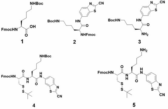
- the preparation method of the above-mentioned leucine aminopeptidase and glutathione dual stimulus-responsive probe comprises the following steps: (1) Compound 1 undergoes amide condensation reaction with NH 2 -CBT to obtain Compound 2; (2) Compound 2 desorbs Compound 3 was obtained by removing the protecting group; (3) compound 3 was subjected to amide condensation reaction with N-fluorenylmethoxycarbonyl-S-tert-butylthio-L-cysteine to obtain compound 4; (4) compound 4 was deprotected (5) Compound 5 reacts with a photosensitizer to obtain Compound 6; (6) Compound 6 removes the protecting group to obtain Compound 7; (7) Compound 7 reacts with N-tert-butoxycarbonyl-L-leucine Acid amide condensation reaction to obtain compound 8; (8) compound 8 deprotected to obtain leucine aminopeptidase and glutathione dual stimulus-responsive probe.
- the leucine aminopeptidase and glutathione dual stimulus-responsive probes disclosed in the present invention self-assemble into spherical nanoparticles in vitro, and the fluorescence is quenched; when the nanoparticles circulate to the tumor site, they are overexpressed in tumor cells Under the stimulation of leucine aminopeptidase and glutathione, the tumor cells are selectively reassembled into nanofibers through the intermolecular CBT condensation reaction, completing the response of the near-infrared molecular probe in the tumor, and the fluorescence recovery.
- the leucine aminopeptidase and glutathione dual stimulus-responsive probe responds to the tumor site, the ability to generate ROS is restored, and the near-infrared small-molecule photosensitizer is used in the light of the tumor after being irradiated by a 660 nm laser.
- dynamic therapy The time point of the strongest fluorescent signal at the tumor site was 3 hours; when the 660 nm laser was irradiated, the irradiation intensity was 0.15 W/cm 2 and the irradiation time was 10 minutes.
- the tail vein was injected into the tumor-bearing mice, and after the probe circulated to the tumor site, under the stimulation of leucine aminopeptidase and glutathione overexpressed in the tumor cells, it selectively condensed in the tumor cells through intermolecular CBT The reaction reassembles into nanofibers, completes the response of the near-infrared molecular probe in the tumor, and restores the fluorescence.
- the probe circulates to the tumor site, under the stimulation of leucine aminopeptidase and glutathione overexpressed in tumor cells, it selectively reassembles into nanofibers in tumor cells through intermolecular CBT condensation reaction, After completing the response of the near-infrared molecular probe in the tumor, the fluorescence gradually recovered, and the ability to generate ROS gradually became stronger. After the fluorescence signal of the probe reaches the strongest at the tumor site, after being irradiated by a 660 nm laser, the near-infrared small molecule photosensitizer is used for tumor treatment.
- step (1) the molar ratio of compound 1 to NH 2 -CBT is 1:1.2; the amide condensation reaction is carried out in the presence of N-methylmorphine and isobutyl chloroformate; the amide condensation reaction is at room temperature React for 15 to 24 hours.
- step (2) the deprotection group of compound 2 is carried out in N,N-dimethylformamide/piperidine mixed solvent; the volume of N,N-dimethylformamide and piperidine The ratio is 4:1.
- step (3) the molar ratio of compound 3 to N-fluorenylmethoxycarbonyl-S-tert-butylthio-L-cysteine is 1:1.2; the amide condensation reaction is carried out in 1-hydroxybenzene Carried out in the presence of triazole, O-benzotriazole-tetramethyluronium hexafluorophosphate and diisopropylethylamine; the amide condensation reaction is carried out at room temperature for 2 to 4 hours.
- step (4) the deprotection of compound 4 is carried out in a mixed solvent of dichloromethane/trifluoroacetic acid; the volume ratio of dichloromethane and trifluoroacetic acid is 4:1.
- step (5) the molar ratio of compound 5 to the photosensitizer is 1.1:1; the photosensitizer is NHS-activated chlorin E6 (Ce6-NHS).
- step (6) the deprotection group of compound 6 is carried out in N,N-dimethylformamide/piperidine mixed solvent; the volume of N,N-dimethylformamide and piperidine The ratio is 4:1.
- step (7) the molar ratio of compound 7 to N-tert-butoxycarbonyl-L-leucine is 1:1.2; Carry out in the presence of 3-ethylcarbodiimide hydrochloride, N-hydroxysuccinimide and diisopropylethylamine; amide condensation reaction takes 8 to 12 hours at room temperature.
- step (8) the deprotection of compound 8 is carried out in a mixed solvent of dichloromethane/trifluoroacetic acid; the volume ratio of dichloromethane and trifluoroacetic acid is 4:1.
- the probe of the present invention reassembles the nanoparticle probes into nanofibers through the dual stimulation of leucine aminopeptidase and glutathione overexpressed in the tumor microenvironment, and realizes the restoration of the fluorescence of the probes and the ability to generate ROS , so as to achieve tumor-specific fluorescence imaging and photodynamic therapy.
- the present invention has the following advantages compared with the prior art: (1)
- 2-cyanobenzothiazole and 1,2-aminothiol are used for rapid and efficient click condensation reaction to form two Affinity dimers, and the reassembly of nanoparticles into nanofibers through the change of intermolecular forces.
- the diagnostic and therapeutic functions of the smart probe that responds to the tumor microenvironment can only be activated when triggered by a special tumor microenvironment. Even if it is trapped by normal tissues, its diagnostic and therapeutic functions will not be activated, so it will not Interference with cancer diagnosis and treatment. Therefore, intelligent diagnosis and treatment reagents that respond to the tumor microenvironment can effectively improve the accuracy of cancer diagnosis and the effect of treatment.
- FIG. 1 is a flow chart of the synthesis of the leucine aminopeptidase and glutathione dual stimulus-responsive probe Ce6-Leu and the control probe Ce6-Ac in Example 1.
- Fig. 2 is the high-resolution mass spectrometry characterization of the leucine aminopeptidase and glutathione dual stimulus-responsive probe Ce6-Leu (a) and the control probe Ce6-Ac (b) in Example 2.
- Fig. 3 is a TEM image of the leucine aminopeptidase and glutathione dual stimulus-responsive probe Ce6-Leu in Example 3 before and after the reaction in the leucine aminopeptidase and glutathione solution.
- Figure 4 is the change of ultraviolet absorption and fluorescence spectrum before and after the reaction of leucine aminopeptidase and glutathione dual stimulus-responsive probe Ce6-Leu in leucine aminopeptidase and glutathione solution in Example 4
- a is the change in UV absorption
- b is the change in fluorescence signal.
- Fig. 5 shows the change of the ROS generating ability of the leucine aminopeptidase and glutathione dual stimulus-responsive probe Ce6-Leu in Example 5 before and after the reaction in the leucine aminopeptidase and glutathione solution.
- Fig. 6 shows the changes in fluorescence intensity of the leucine aminopeptidase and glutathione dual stimulus-responsive probe Ce6-Leu and the control probe Ce6-Ac in the tumor cells in Example 6.
- Example 7 is a comparison of the ability of the leucine aminopeptidase and glutathione dual stimulus-responsive probe Ce6-Leu and the control probe Ce6-Ac to generate ROS in tumor cells in Example 7.
- Fig. 8 is a comparison of the photodynamic killing ability of the leucine aminopeptidase and glutathione dual stimulus-responsive probe Ce6-Leu and the control probe Ce6-Ac in Example 8.
- Figure 9 is a fluorescent imaging photo of mice after tail vein injection of leucine aminopeptidase and glutathione dual stimulus-responsive probe Ce6-Leu and control group probe Ce6-Ac at different times in Example 9 (a ) and the corresponding fluorescence signal values (b).
- FIG. 10 is a study on the effect of photodynamic therapy on mice with the dual stimulus-responsive probe Ce6-Leu of leucine aminopeptidase and glutathione and the control probe Ce6-Ac in Example 10.
- Figure 11 is a comparison of the survival time of mice in each group.
- the present invention has developed a leucine aminopeptidase and glutathione dual-response intelligent molecular probe integrating fluorescence imaging and photodynamic therapy, which has great research and application value.
- the steps of the method provided by the present invention are as follows: (1) Construction and synthesis of dual stimulus-responsive probes: according to the designed synthesis steps: first, compound 1 undergoes amide condensation reaction with NH 2 -CBT, and then uses 20%
- the intermediate obtained by the group Fmoc reacts with N
- nanoparticles After the nanoparticles circulate to the tumor site, they selectively respond to leucine aminopeptidase and glutathione in the tumor cells and reassemble into nanofibers through the intermolecular CBT condensation reaction, completing the near-infrared molecular probe in the tumor. Responsive fluorescence imaging to achieve specific fluorescence imaging of tumors in vivo.
- the probe After the probe circulates to the tumor site for enzyme and glutathione responses, it is selectively reassembled into nanofibers in the tumor cells through an intermolecular CBT condensation reaction, completing the response of the near-infrared molecular probe in the tumor, and the fluorescence gradually recovers , and the ability to generate ROS gradually becomes stronger.
- the fluorescence signal of the probe After the fluorescence signal of the probe reaches the strongest at the tumor site, after 10 minutes of irradiation with a 660 nm laser (0.15 W/cm 2 ), the near-infrared small molecule photosensitizer is used for tumor treatment.
- Example 1 Synthesis and characterization of leucine aminopeptidase and glutathione dual stimulus-responsive probe Ce6-Leu and control probe Ce6-Ac: (1) compound 1 (400 mg, 0.85 mmol) Dissolve in 10 mL tetrahydrofuran, then add N-methylmorphine (130 mg, 1.28 mmol) dropwise, then place the round bottom flask in an ice-salt bath, cool to 0 o C, then add isobutyl chloroformate dropwise (175 mg, 1.28 mmol), after activation for half an hour, add 2-amino-6-cyanobenzothiazole (NH 2 -CBT, 179 mg, 1.00 mmol) dissolved in dry tetrahydrofuran and keep the reaction at 0 o C 1 hour, then stirred overnight at room temperature.
- N-methylmorphine 130 mg, 1.28 mmol
- isobutyl chloroformate dropwise (175 mg, 1.28 mmol)
- the solvent was spin-dried by a rotary evaporator, and then the residual solid was redissolved in ethyl acetate (50 mL), and extracted three times with an aqueous solution of sodium bicarbonate. .
- Example 2 High-resolution mass spectrometry characterization of leucine aminopeptidase and glutathione dual stimulus-responsive probe Ce6-Leu and control probe Ce6-Ac: the leucine amino
- the peptidase and glutathione dual stimulus-responsive probe Ce6-Leu and the control probe Ce6-Ac were diluted with solvent methanol to a concentration of 5 ⁇ M, and the molecular weights of the probes were determined by high-resolution mass spectrometry.
- Example 3 Leucine aminopeptidase and glutathione mediated dual stimulus-responsive probe Ce6-Leu reassembled from nanoparticles into nanofibers: Leucine aminopeptidase and glutathione prepared in Example 1 Glutathione dual stimulus-responsive probe Ce6-Leu was added to PBS buffer solution containing 10 mM glutathione and 50 U/mL leucine aminopeptidase, and reacted at 37°C for 24 hours. The morphology changes before and after the probe reaction were observed by TEM.
- the simple probe Ce6-Leu forms nanoparticles with a particle size of about 79.8 ⁇ 9.3 nm in PBS solution, and reassembles to form nanoparticles under the stimulation of leucine aminopeptidase and glutathione.
- the probe Ce6-Leu undergoes a CBT condensation reaction under the dual stimulation of leucine aminopeptidase and glutathione to reassemble into nanofibers, the distance between the Ce6 structures becomes larger, and the UV absorption from J- Aggregates are transformed into monomers, the UV absorption is elevated and blue-shifted, and the fluorescence signal is enhanced.
- the probe Ce6-Ac in the control group is not enzyme-responsive and cannot undergo condensation reaction, even in PBS (50 U/mL LAP enzyme, 10 mM GSH) solution, still exists in the form of aggregated nanoparticles, so there is no significant change in UV absorption and fluorescence signals.
- PBS 50 U/mL LAP enzyme, 10 mM GSH
- Example 6 Changes in the fluorescence intensity of the dual stimulus-responsive probe Ce6-Leu of leucine aminopeptidase and glutathione and the probe Ce6-Ac of the control group in tumor cell HepG2: it will be in the logarithmic growth phase
- the HepG2 cells were digested and centrifuged, the supernatant was discarded, and the culture medium was added again to form a cell suspension. After the cells were counted, they were seeded in 8-well confocal small dishes, and 5000 HepG2 cells were added to each well. The 8-well confocal small dish was then placed in the incubator for 24 hours.
- Example 7 Comparison of the ability of the leucine aminopeptidase and glutathione dual stimulus-responsive probe Ce6-Leu and the contrast probe Ce6-Ac to generate ROS in tumor cells: take HepG2 cells in the logarithmic growth phase, Digest with trypsin after discarding the original medium, centrifuge, add fresh medium and pipette into a suspension, then count with a counting plate, and inoculate in a confocal small dish, the final number of cells per well is 4 ⁇ 10 5 , Continue to culture in the incubator for 12 hours, discard the original medium after 12 hours, then add 1.5 mL of the same concentration (20 ⁇ M) of the culture medium solution of Ce6-Leu and Ce6-Ac probes, and set a blank control group ( Control group: no probe added), put into the incubator and continue to incubate for 8 hours.
- control group + ⁇ 660 nm no probe added, irradiated with 660 nm laser alone; Ce6-Leu+ ⁇ 660 nm : Ce6-Leu probe incubated for 8 hours, irradiated with 660 nm Laser; Ce6-Ac+ ⁇ 660 nm : After the Ce6-Ac probe was incubated for 8 hours, irradiated with 660 nm laser), the laser power was 0.15 W/cm 2 , and the experimental control group was set at the same time (control group: no probe, no 660 nm laser irradiation; Ce6-Leu: Ce6-Leu probe incubated for 8 hours without 660 nm laser irradiation; Ce6-Ac: Ce6-Ac probe incubated for 8 hours but without 660 nm laser irradiation).
- the Ce6-Leu probe Compared with the cells of the Ce6-Leu+ ⁇ 660 nm group, the Ce6-Leu probe has the ability to reassemble responsively, and the stimulation of overexpressed leucine aminopeptidase and glutathione in tumor cells can detect
- the needles can undergo intermolecular condensation reaction to reassemble into nanofibers, the distance between the Ce6 structures becomes larger, the quenching between each other is weakened, and the ability to generate singlet oxygen is restored. Therefore, after 660 nm laser irradiation, the intracellular A large amount of ROS, so that a distinct green fluorescence (DCF) can be detected.
- DCF green fluorescence
- Example 8 Comparison of the ability of the dual stimulus-responsive probe Ce6-Leu of leucine aminopeptidase and glutathione and the comparison probe Ce6-Ac to kill tumor cells by photodynamic force: HepG2 cells in logarithmic growth phase , Discard the original medium, digest with trypsin, centrifuge, add fresh medium and pipette into a suspension, then count with a counting plate, and inoculate in a 12-well plate, and the final number of cells per well is 4 ⁇ 10 5 , continue to culture in the incubator for 12 hours, discard the original medium after 12 hours, then add 1 mL of the same concentration (20 ⁇ M) of the medium solution of Ce6-Leu and Ce6-Ac probes, and set a blank control group (Control group: no probe added), put into the incubator and continue to incubate for 8 hours.
- control group control group+ ⁇ 660 nm , Ce6-Ac, Ce6-Ac+ ⁇ 660 nm , Ce6-Leu, Ce6-Leu+ ⁇ 660 nm
- control group control group+ ⁇ 660 nm , Ce6-Ac, Ce6-Ac+ ⁇ 660 nm , Ce6-Leu, Ce6-Leu+ ⁇ 660 nm
- PBS PBS
- centrifuge take the pellet, repeat this operation twice, and then blow the cells Form a single cell suspension, add Annexin V-FITC/PI double-stained apoptosis detection solution (see the kit instruction manual for specific operations), and use flow cytometry to detect the apoptosis of tumor cells after culture.
- Annexin V-FITC/PI double-stained apoptosis detection solution see the kit instruction manual for specific operations
- the Ce6-Leu probe Compared with the cells of Ce6-Leu+ ⁇ 660 nm , only a small amount of cell death was seen, indicating that the Ce6-Ac probe still exists in an aggregated state in the cells, and can only produce a small amount of ROS, which cannot kill tumor cells the goal of.
- the Ce6-Leu probe has the ability to reassemble responsively. In the microenvironment of tumor cells, the probe is stimulated by leucine aminopeptidase and glutathione The intermolecular condensation reaction can occur to reassemble into nanofibers, the distance between the Ce6 structures becomes larger, the quenching between each other is weakened, and the ability to generate singlet oxygen is restored. Therefore, after 660 nm laser irradiation, a large amount of ROS, leading to the death of a large number of cells.
- Example 9 Fluorescent imaging photos and corresponding fluorescent signals of mice at different time points after tail vein injection of leucine aminopeptidase and glutathione dual stimulus-responsive probe Ce6-Leu and control group probe Ce6-Ac Value: 2 groups of female nude mice bearing HepG2 tumors (3 mice in each group) were taken respectively. After gas anesthesia, they were placed in the small animal IVIS Lumina XRMS in vivo imaging system for background fluorescence imaging of the mice.
- one group of mice (experimental group) was injected with 200 ⁇ L of 100 ⁇ M Ce6-Leu in PBS buffer solution through the tail vein, and the other group of mice (control group) was injected with 200 ⁇ L of Ce6-Leu with a concentration of 100 ⁇ M through the tail vein. - Ac in PBS buffer solution.
- the two groups of mice were placed in the above-mentioned small animal in vivo imaging system for imaging. After the imaging, the IVIS in vivo imaging analysis software was used to process the image and calculate the fluorescence intensity of the tumor sites of the two groups of mice at each time point.
- the probe Ce6-Leu has a better fluorescence enhancement effect in mouse tumors.
- the fluorescence imaging image (a) shows that the peak of the fluorescence signal is reached at 3 hours
- Figure 9(b) shows that the signal intensity of the experimental group (Ce6-Leu) is significantly greater than that of the control group (Ce6-Ac).
- the probe Ce6-Leu has a good response to the tumor microenvironment, and can undergo a responsive intermolecular condensation reaction to reassemble under the stimulation of overexpressed leucine aminopeptidase and glutathione in tumor cells
- the distance between Ce6 structures becomes larger, and the quenching between each other weakens so that the fluorescence recovers.
- Example 10 Research on the photodynamic therapy of tumors in vivo with the dual stimulus-responsive probe Ce6-Leu of leucine aminopeptidase and glutathione and the probe Ce6-Ac of the control group: 36 mice weighing about 18 g 1.
- the female nude mice bearing HepG2 tumors in the hind legs were divided into 6 groups (6 mice in each group), namely Group1, Group2, Group3, Group4, Group5, Group6.
- Group1 Inject mice with 200 ⁇ L of PBS solution through the tail vein.
- mice were injected with 200 ⁇ L of PBS solution through the tail vein, and 3 hours after the injection, the tumor was irradiated with a 660 nm laser with a power of 0.15 W/cm 2 for 10 minutes.
- Group3 200 ⁇ L of 200 ⁇ M probe Ce6-Ac in PBS buffer solution was injected into mice by tail vein injection.
- Group4 Inject mice with 200 ⁇ L of 200 ⁇ M probe Ce6-Ac in PBS buffer solution by tail vein injection, and irradiate the tumor site with a 660 nm laser with a power of 0.15 W/ cm2 for 10 hours after injection. minute.
- Group5 inject 200 ⁇ L of 200 ⁇ M probe Ce6-Leu in PBS buffer solution to mice by tail vein injection.
- Group6 Inject mice with 200 ⁇ L of 200 ⁇ M probe Ce6-Leu in PBS buffer solution through tail vein injection, and irradiate the tumor site with a 660 nm laser with a power of 0.15 W/ cm2 for 10 hours after injection. minute.
- the invention belongs to the technical field of tumor microenvironment-mediated reassembly, and relates to a leucine aminopeptidase and glutathione dual stimulus-responsive probe, a preparation method and application thereof.
- Mainly use leucine aminopeptidase and glutathione overexpressed in solid tumors to stimulate the reassembly of spherical nanoparticles to form nanofibers, so as to improve the specificity and accuracy of probes in tumor cells and in vivo imaging detection To achieve specific fluorescence imaging of tumors, and to effectively improve the effect of photodynamic therapy on tumors.
- diagnosis and treatment as an emerging tumor diagnosis and treatment strategy that combines disease diagnosis, monitoring and treatment, has brought new hope for human beings to overcome cancer; and the advantages of this strategy are high efficiency and less toxic side effects are expected to promote cancer diagnosis and treatment technology rapid development.
- a tumor microenvironment-responsive near-infrared molecular probe was constructed, and the overexpressed leucine aminopeptidase and glutathione in tumor cells were used to trigger condensation reactions and then reassemble , so that the specific recovery of the ability of the probe to fluoresce and generate ROS at the tumor site, thereby effectively improving the imaging and treatment effect of the tumor.
- the condensation reaction is efficient, mild, fast, and highly selective; second, when the probe enters tumor cells, the overexpressed leucine aminopeptidase and glutathione Under the stimulation of glycine, the original amino group and sulfhydryl group in the cysteine structure are exposed, so that a click condensation reaction occurs, which is not affected by the external environment. Therefore, the near-infrared photosensitizer probe has further application in biological and other fields.
Landscapes
- Health & Medical Sciences (AREA)
- Chemical & Material Sciences (AREA)
- Life Sciences & Earth Sciences (AREA)
- General Health & Medical Sciences (AREA)
- Medicinal Chemistry (AREA)
- Organic Chemistry (AREA)
- Animal Behavior & Ethology (AREA)
- Public Health (AREA)
- Veterinary Medicine (AREA)
- Engineering & Computer Science (AREA)
- Epidemiology (AREA)
- Pharmacology & Pharmacy (AREA)
- Molecular Biology (AREA)
- Proteomics, Peptides & Aminoacids (AREA)
- Bioinformatics & Cheminformatics (AREA)
- Biophysics (AREA)
- Nuclear Medicine, Radiotherapy & Molecular Imaging (AREA)
- Radiology & Medical Imaging (AREA)
- Genetics & Genomics (AREA)
- Biochemistry (AREA)
- General Chemical & Material Sciences (AREA)
- Nanotechnology (AREA)
- Analytical Chemistry (AREA)
- Chemical Kinetics & Catalysis (AREA)
- Crystallography & Structural Chemistry (AREA)
- Medical Informatics (AREA)
- General Engineering & Computer Science (AREA)
- Biotechnology (AREA)
- Materials Engineering (AREA)
- Peptides Or Proteins (AREA)
- Medicines Containing Antibodies Or Antigens For Use As Internal Diagnostic Agents (AREA)
Abstract
提供了一种亮氨酸氨基肽酶和谷胱甘肽双重刺激响应型探针及其制备方法与应用。所述探针Ce6-Leu在肿瘤微环境的刺激下发生点击缩合反应,继而从纳米颗粒重组装成纳米纤维,具体的,探针Ce6-Leu在肿瘤细胞内过表达的亮氨酸氨基肽酶和谷胱甘肽的刺激下,暴露半胱氨酸结构中原有的氨基和巯基,进而发生点击缩合反应形成两亲性的二聚体,并重组装形成纳米纤维,Ce6结构之间的距离变大,彼此之间的淬灭减弱从而荧光信号和产生ROS的能力增强,进而实现肿瘤的特异性荧光成像和光动力治疗,以及肿瘤的诊疗一体化,具有重要的科研及经济价值。
Description
本发明属于肿瘤微环境介导的重组装技术领域,涉及一种亮氨酸氨基肽酶和谷胱甘肽双重刺激响应型探针及其制备方法和应用。
癌症已经成为严重危害人类健康的疾病之一,因此,近些年来,用于癌症诊断和治疗的分子影像技术得到了飞速的发展。应用分子影像诊断技术可以对癌症患者进行早期的诊断,并且为癌症的分类、预后评估及治疗方案的选择提供重要的信息,为人类攻克癌症提供新的机会。
诊疗一体化作为一种将疾病的诊断、监测和治疗结合的新兴肿瘤诊疗策略,为克服癌症带来了新的希望;并且该策略效率高,毒副作用小的优点有望推动癌症诊断和治疗技术的快速发展。目前科学家们已经研发出各类造影剂,其中纳米尺寸的造影剂,如纳米乳剂、脂质体、树状聚合物以及基于金、钽、镧系、铋等重金属材料设计的无机纳米多功能造影剂。这类纳米粒子造影剂具有血液循环时间长,肾清除率和毛细血管渗漏率低的优点,能够通过实体瘤的高通透性和滞留作用(enhanced
permeation and retention effect,EPR)被动积累在肿瘤部位。但是现在大多数这类造影剂仍处于临床前研究阶段,缺乏生物毒性、药代动力学和体内分布的实验评估,距离临床应用仍有一定的距离。
为了克服上述现有造影剂中存在的问题,本发明利用肿瘤微环境中过表达的亮氨酸氨基肽酶和谷胱甘肽的刺激,设计并开发了一种亮氨酸氨基肽酶和谷胱甘肽双重刺激响应型探针,实现探针在肿瘤内的特异性响应,进而有效的改善在体肿瘤的成像及治疗效果;该方法为改善传统分子探针成像背景噪声高、诊断准确性低等缺点提供了新的策略与手段。
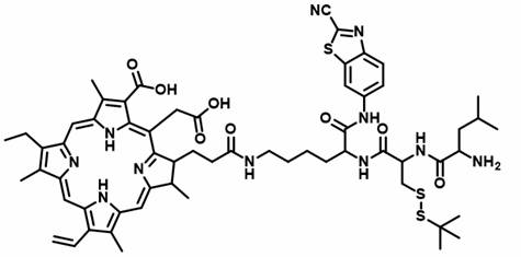
本发明采用以下技术方案:一种亮氨酸氨基肽酶和谷胱甘肽双重刺激响应型探针,具有如下化学结构式:
 。
。
上述亮氨酸氨基肽酶和谷胱甘肽双重刺激响应型探针在制备肿瘤诊断和/或治疗试剂中的应用。
上述亮氨酸氨基肽酶和谷胱甘肽双重刺激响应型探针的制备方法,包括以下步骤:(1)化合物1与NH
2-CBT进行酰胺缩合反应得到化合物2;(2)化合物2脱掉保护基得到化合物3;(3)化合物3与N-芴甲氧羰基-S-叔丁硫基-L-半胱氨酸进行酰胺缩合反应,得到化合物4;(4)化合物4脱去保护基团得到化合物5;(5)化合物5与光敏剂反应,得到化合物6;(6)化合物6脱掉保护基得到化合物7;(7)化合物7与N-叔丁氧羰基-L-亮氨酸进行酰胺缩合反应得到化合物8;(8)化合物8脱掉保护基得到亮氨酸氨基肽酶和谷胱甘肽双重刺激响应型探针。
本发明公开的亮氨酸氨基肽酶和谷胱甘肽双重刺激响应型探针在体外自组装成球形纳米颗粒,并且荧光淬灭;当纳米颗粒循环到肿瘤部位后,在肿瘤细胞中过表达的亮氨酸氨基肽酶和谷胱甘肽的刺激下,选择性在肿瘤细胞内通过分子间CBT缩合反应重新组装成纳米纤维,完成近红外分子探针在肿瘤内的响应,荧光恢复。并且,亮氨酸氨基肽酶和谷胱甘肽双重刺激响应型探针在肿瘤部位发生响应后,产生ROS的能力恢复,经660 nm激光器照射,完成近红外小分子光敏剂用于肿瘤的光动力治疗。肿瘤部位荧光信号最强的时间点为3小时;660 nm激光器照射时,照射强度为0.15 W/cm
2,照射时间为10分钟。
上述技术方案中,将亮氨酸氨基肽酶和谷胱甘肽双重刺激响应型探针溶于PBS(磷酸缓冲盐,pH=7.2~7.4)缓冲液中(浓度为100μM),在37℃的振荡器中反应,使其自组装成球形纳米颗粒。随后尾静脉注入荷瘤鼠体内,探针循环到肿瘤部位后,在肿瘤细胞中过表达的亮氨酸氨基肽酶和谷胱甘肽的刺激下,选择性在肿瘤细胞内通过分子间CBT缩合反应重组装成纳米纤维,完成近红外分子探针在肿瘤内的响应,荧光恢复。并且,探针循环到肿瘤部位后,在肿瘤细胞中过表达的亮氨酸氨基肽酶和谷胱甘肽的刺激下,选择性在肿瘤细胞内通过分子间CBT缩合反应重组装成纳米纤维,完成近红外分子探针在肿瘤内的响应,荧光逐渐恢复,并且产生ROS的能力逐渐变强。待探针在肿瘤部位荧光信号达到最强,经660 nm激光器照射后,完成近红外小分子光敏剂用于肿瘤的治疗。
上述技术方案中,步骤(1)中,化合物1与NH
2-CBT的摩尔比为1∶1.2;酰胺缩合反应在N-甲基吗啡和氯甲酸异丁酯存在下进行;酰胺缩合反应为室温反应15~24小时。
上述技术方案中,步骤(2)中,化合物2脱去保护基团在N,N-二甲基甲酰胺/哌啶混合溶剂中进行;N,N-二甲基甲酰胺、哌啶的体积比为4∶1。
上述技术方案中,步骤(3)中,化合物3与N-芴甲氧羰基-S-叔丁硫基-L-半胱氨酸的摩尔比为1:1.2;酰胺缩合反应在1-羟基苯并三氮唑、O-苯并三氮唑-四甲基脲六氟磷酸盐和二异丙基乙胺存在下进行;酰胺缩合反应为室温反应2~4小时。
上述技术方案中,步骤(4)中,化合物4脱去保护基团在二氯甲烷/三氟乙酸混合溶剂中进行;二氯甲烷、三氟乙酸的体积比为4∶1。
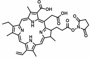
上述技术方案中,步骤(5)中,化合物5与光敏剂的摩尔比为1.1∶1;所述光敏剂为NHS活化的二氢卟吩E6(Ce6-NHS)。
上述技术方案中,步骤(6)中,化合物6脱去保护基团在N,N-二甲基甲酰胺/哌啶混合溶剂中进行;N,N-二甲基甲酰胺、哌啶的体积比为4∶1。
上述技术方案中,步骤(7)中,化合物7与N-叔丁氧羰基-L-亮氨酸的摩尔比为1∶1.2;酰胺缩合反应在1-(3-二甲氨基丙基)-3-乙基碳二亚胺盐酸盐、N-羟基琥珀酰亚胺和二异丙基乙胺存在下进行;酰胺缩合反应为室温反应8~12小时。
上述技术方案中,步骤(8)中,化合物8脱去保护基团在二氯甲烷/三氟乙酸混合溶剂中进行;二氯甲烷、三氟乙酸的体积比为4∶1。
上述技术方案中,化合物1、化合物2、化合物3、化合物4、化合物5、化合物6、化合物7、化合物8的化学结构式分别如下:
 。
。
NHS活化的光敏剂二氢卟吩E6的化学结构式如下:
 。
。
本发明中,化合物8进行脱保护后,使用半制备型高效液相色谱分离提纯,得到亮氨酸氨基肽酶和谷胱甘肽双重刺激响应型探针,产物为墨绿色的固体粉末,为常规技术。优选的,所述的高效液相色谱分离方法为:C18柱,3.5 μm,4.6×100 mm;流动相:A是水;B是乙腈;流速:3 mL/min;线性梯度洗脱程序:0 min,A∶B = 95∶5;13 min,A∶B = 0∶100。
本发明的探针通过肿瘤微环境中过表达的亮氨酸氨基肽酶和谷胱甘肽的双重刺激,使得纳米颗粒探针重新组装成纳米纤维,实现探针的荧光和产生ROS能力的恢复,从而达到肿瘤的特异性荧光成像和光动力治疗。
由于上述技术方案的运用,本发明与现有技术相比具有如下优点:(1)本发明中使用2-氰基苯并噻唑与1,2-氨基硫醇发生快速高效的点击缩合反应形成两亲性的二聚体,并通过分子间作用力的改变使得纳米颗粒重新组装成纳米纤维。
(2)当探针进入肿瘤细胞后,在肿瘤细胞内过表达的亮氨酸氨基肽酶和谷胱甘肽的刺激下,暴露半胱氨酸结构中原有的氨基和巯基,从而与2-氰基苯并噻唑(CBT)的氰基发生点击缩合反应,且不受外界环境影响。
(3)该肿瘤微环境响应的智能型探针的诊断和治疗功能只有在特殊的肿瘤微环境触发下才能被激活,即使被正常组织截留,其诊断和治疗功能不会被激活,因此不会对癌症的诊断和治疗带来干扰。所以,肿瘤微环境响应的智能诊疗试剂能有效的提高癌症诊断的精准度和治疗的效果。
图1为实施例1中亮氨酸氨基肽酶和谷胱甘肽双重刺激响应型探针Ce6-Leu和对照组探针Ce6-Ac的合成流程图。
图2为实施例2中亮氨酸氨基肽酶和谷胱甘肽双重刺激响应型探针Ce6-Leu(a)和对照组探针Ce6-Ac(b)的高分辨质谱表征。
图3为实施例3中亮氨酸氨基肽酶和谷胱甘肽双重刺激响应型探针Ce6-Leu在亮氨酸氨基肽酶和谷胱甘肽溶液中反应前后的TEM图像。
图4为实施例4中亮氨酸氨基肽酶和谷胱甘肽双重刺激响应型探针Ce6-Leu在亮氨酸氨基肽酶和谷胱甘肽溶液中反应前后紫外吸收和荧光光谱的变化情况,a是紫外吸收的变化,b是荧光信号的变化。
图5为实施例5中亮氨酸氨基肽酶和谷胱甘肽双重刺激响应型探针Ce6-Leu在亮氨酸氨基肽酶和谷胱甘肽溶液中反应前后产生ROS能力的变化情况。
图6为实施例6中亮氨酸氨基肽酶和谷胱甘肽双重刺激响应型探针Ce6-Leu和对照组探针Ce6-Ac在肿瘤细胞内荧光强度的变化。
图7为实施例7中亮氨酸氨基肽酶和谷胱甘肽双重刺激响应型探针Ce6-Leu和对照组探针Ce6-Ac在肿瘤细胞内产生ROS能力的比较。
图8为实施例8中亮氨酸氨基肽酶和谷胱甘肽双重刺激响应型探针Ce6-Leu和对照组探针Ce6-Ac光动力杀死肿瘤细胞能力的比较。
图9为实施例9中不同时间内,亮氨酸氨基肽酶和谷胱甘肽双重刺激响应型探针Ce6-Leu和对照组探针Ce6-Ac尾静脉注射后小鼠荧光成像照片(a)和对应的荧光信号值(b)。
图10为实施例10中亮氨酸氨基肽酶和谷胱甘肽双重刺激响应型探针Ce6-Leu和对照组探针Ce6-Ac小鼠光动力治疗效果研究。
图11为各组小鼠存活时间比较。
本发明开发了一种集荧光成像及光动力治疗于一体的亮氨酸氨基肽酶和谷胱甘肽双响应型智能分子探针,具有重大的研究及应用价值,该造影剂可以在肿瘤微环境的刺激下从球形纳米颗粒重新组装为纳米纤维,并且荧光以及产生ROS的能力恢复,从而实现体肿瘤的特异性荧光成像和光动力治疗。
具体而言,本发明提供的方法,其步骤如下:(1)构建、合成双重刺激响应型探针:按照设计的合成步骤:首先化合物1与NH
2-CBT发生酰胺缩合反应,随后用20%的哌啶(N,N-二甲基甲酰胺∶哌啶 = 4∶1,v/v)脱去保护基团Fmoc;接着与N-芴甲氧羰基-S-叔丁硫基-L-半胱氨酸发生酰胺缩合反应,随后用20%的三氟乙酸(二氯甲烷∶三氟乙酸 = 4∶1,v/v)将中间体化合物的Boc保护基团脱掉;接着与已经用NHS活化好的光敏剂二氢卟吩E6反应,所得的中间体化合物再用20%的哌啶(N,N-二甲基甲酰胺∶哌啶 = 4∶1,v/v)脱去保护基团Fmoc所得的中间体与N-叔丁氧羰基-L-亮氨酸反应得到产物再20%的三氟乙酸(二氯甲烷∶三氟乙酸= 4∶1,v/v)将Boc保护基团脱掉得到最终的探针Ce6-Leu,结构式如下:
 。
。
(2)亮氨酸氨基肽酶和谷胱甘肽双重刺激响应型探针Ce6-Leu在肿瘤细胞内的响应情况:将步骤(1)中获得的所述亮氨酸氨基肽酶和谷胱甘肽双重刺激响应型探针溶于细胞培养基中,加入到HepG2细胞培养皿中(浓度:20 μM),放入培养箱培养,分子探针在肿瘤细胞内过表达的亮氨酸氨基肽酶和谷胱甘肽的刺激下,通过分子间CBT缩合反应重新组装成纳米纤维,完成近红外分子探针在肿瘤细胞内的响应,荧光恢复,有利于实现对肿瘤细胞的的特异性成像。
(3)亮氨酸氨基肽酶和谷胱甘肽双重刺激响应型探针Ce6-Leu在实体肿瘤内的响应性荧光成像:将步骤(1)中获得的所述亮氨酸氨基肽酶和谷胱甘肽双重刺激响应型探针Ce6-Leu溶于PBS缓冲液中(浓度:100 μM)使其自组装成纳米颗粒,然后以尾静脉注射的方式将探针注入荷有HepG2肝癌肿瘤的雌性裸鼠体内。纳米颗粒循环到肿瘤部位后,选择性在肿瘤细胞内进行亮氨酸氨基肽酶和谷胱甘肽的响应并通过分子间CBT缩合反应重新组装成纳米纤维,完成近红外分子探针在肿瘤内响应性荧光成像,从而实现对在体肿瘤的特异性荧光成像。
(4)亮氨酸氨基肽酶和谷胱甘肽双重刺激响应型探针Ce6-Leu用于在体肿瘤的光动力治疗:将步骤(1)中获得的所述亮氨酸氨基肽酶和谷胱甘肽双重刺激响应型探针溶于PBS缓冲液中(浓度:200 μM),以尾静脉注射的方式将探针注入荷有HepG2肝癌肿瘤的雌性裸鼠体内。探针循环到肿瘤部位进行酶和谷胱甘肽的响应后,选择性在肿瘤细胞内通过分子间CBT缩合反应重组装成纳米纤维,完成近红外分子探针在肿瘤内的响应,荧光逐渐恢复,并且产生ROS的能力逐渐变强。待探针在肿瘤部位荧光信号达到最强,经660 nm激光(0.15
W/cm
2)照射10分钟后,完成近红外小分子光敏剂用于肿瘤的治疗。
下文将结合附图和具体实施例来进一步阐述本发明。应当理解的是,这些实施例仅用于解释和说明本发明中的技术方案,而并非旨在限制本发明的范围。此外,除非另有说明,下列实施例中所使用的材料、试剂、仪器等均可通过商业手段获得。
实施例1:亮氨酸氨基肽酶和谷胱甘肽双重刺激响应型探针Ce6-Leu和对照组探针Ce6-Ac的合成与表征:(1)将化合物1(400 mg,0.85 mmol)溶于10 mL四氢呋喃中,再滴加入N-甲基吗啡(130 mg,1.28 mmol),然后将圆底烧瓶置于冰盐浴中,冷却至0
oC,随后再滴加入氯甲酸异丁酯(175 mg,1.28
mmol),活化半小时后,再加入用干燥的四氢呋喃溶解的2-氨基-6-氰基苯并噻唑(NH
2-CBT,179 mg,1.00 mmol),保持0
oC反应1小时,然后室温搅拌过夜。反应结束后,通过旋转蒸发仪旋干溶剂,然后将残留固体复溶在乙酸乙酯(50 mL)中,并用碳酸氢钠的水溶液萃取三次,有机相用Na2SO4干燥后进行抽滤,旋干溶剂。以石油醚(PE)和乙酸乙酯(EA)= 2:1的体积比为洗脱液,用硅胶色谱柱纯化粗产物,得到中间体1(其结构如图1中的化合物2所示)(531.88 mg,产率:85%);
1H NMR
(600 MHz, Methanol-d4) δ 8.59 (s, 1H),
8.04 (d,
J = 9.0 Hz, 1H), 7.74 (d,
J = 7.6 Hz, 2H), 7.63 (dt,
J
= 14.8, 5.0 Hz, 3H), 7.33 (t,
J = 7.5 Hz, 2H), 7.26 (t,
J = 7.7
Hz, 2H), 4.36 (d,
J = 6.9 Hz, 2H), 4.24 (dd,
J = 9.0, 5.4 Hz,
1H), 4.17 (t,
J = 7.0 Hz, 1H), 3.02 (dt,
J = 9.8, 4.5 Hz, 2H),
1.83 (dp,
J = 15.1, 5.4 Hz, 1H), 1.73 (tq,
J = 11.7, 7.9, 6.3 Hz,
1H), 1.48 (p,
J = 9.4, 8.3 Hz, 2H), 1.37 (s, 9H), 1.30–1.18 (m, 2H).
13C NMR (151 MHz, Methanol-d4) δ 172.33, 157.18, 148.38, 143.84, 143.70, 141.80, 141.14,
139.09, 136.58, 135.25, 127.33, 126.72, 124.76, 124.49, 120.82, 119.49, 112.63,
111.40, 78.44, 66.51, 55.84, 47.00, 39.57, 31.51, 29.20, 27.36, 22.80;(2)将中间体1(500 mg,0.80 mmol)溶于8 mL
DMF中,然后将反应瓶放置在冰水浴中,随后滴加入2 mL哌啶,保持0
oC反应5分钟,反应结束后,旋蒸除去溶剂和哌啶。以二氯甲烷(DCM)和甲醇(MeOH)=
80:1的体积比为洗脱液,用硅胶色谱柱纯化粗产物,得到中间体2(其结构如图1中的化合物3所示)(290.18 mg,产率:
90%);
1H NMR (600 MHz,
Methanol-d4) δ 8.59–8.45
(m, 1H), 8.07–7.93 (m, 1H), 7.73–7.54 (m, 1H), 3.47 (q,
J = 6.7 Hz, 1H), 3.06–2.97 (m, 2H), 1.85–1.58
(m, 2H), 1.49 (h,
J = 7.1 Hz, 3H), 1.37 (d,
J = 5.9 Hz, 9H), 1.26
(d,
J = 3.5 Hz, 1H).
13C NMR (151 MHz, Methanol-d4) δ 174.81, 161.86, 157.07, 149.04, 141.79, 137.45, 137.07,
123.84, 119.90, 112.02, 78.37, 55.30, 39.62, 34.65, 29.43, 27.32, 22.52;(3)在20 mL的圆底烧瓶中加入中间体2(250 mg,0.62 mmol),然后用干燥的DMF溶清,随后再加入HBTU(282.15 mg,0.74 mmol),HOBT(100.44 mg,0.74 mmol)和DIPEA(213.68 μL),搅拌15分钟后再加入化合物N-芴甲氧羰基-S-叔丁硫基-L-半胱氨酸(321.04 mg,0.74 mmol)。继续搅拌,室温反应2小时,反应结束后通过旋蒸除去溶剂,然后再加入25 mL乙酸乙酯复溶粗产物,随后有机相用25 mL的超纯水,饱和碳酸氢钠,氯化钠水溶液各洗一次。有机相用无水硫酸钠干燥后旋蒸除去溶剂,以石油醚(PE)和乙酸乙酯(EA)= 2:1的体积比为洗脱液,用硅胶色谱柱纯化粗产物,得到中间体3(其结构如图1中的化合物4所示)(405.26 mg,产率:80%);
1H NMR
(600 MHz, Methanol-d4) δ 8.37 (d,
J
= 2.1 Hz, 1H), 7.84 (d,
J = 9.0 Hz, 1H), 7.71 (dd,
J = 9.0, 2.1
Hz, 1H), 7.58 (d,
J = 7.5 Hz, 1H), 7.52 (dd,
J = 11.0, 7.6 Hz,
2H), 7.45 (d,
J = 7.5 Hz, 1H), 7.38–7.27
(m, 1H), 7.22 (q,
J = 7.1 Hz, 2H), 7.16 (t,
J = 7.4 Hz, 1H), 4.52
(dd,
J = 9.9, 4.5 Hz, 1H), 4.42 (t,
J = 7.3 Hz, 1H), 4.28 (qd,
J
= 10.6, 7.5 Hz, 2H), 4.08 (q,
J = 7.1 Hz, 1H), 3.18–3.02 (m, 2H), 3.02 (s, 2H), 1.99 (s, 1H), 1.77–1.68 (m, 1H), 1.53–1.42
(m, 2H), 1.38 (s, 9H), 1.34 (s, 11H).
13C NMR (151 MHz, Methanol-d4)
δ 172.13, 171.21, 157.15, 157.05, 148.27,
143.49, 143.32, 141.02, 140.78, 138.75, 136.27, 135.19, 127.25, 126.78, 124.74,
124.18, 120.92, 119.34, 112.64, 111.50, 78.43, 66.92, 55.08, 54.09, 46.76,
40.65, 39.75, 37.46, 30.87, 29.03, 28.86, 27.39, 22.96. MS (ESI) Calcd for: C
41H
48N
6O
6S
3
([M+H]
+) : 817.2800, found: 817.2865;(4)在20 mL含20%(体积比)三氟乙酸的DMF溶液中加入中间体3,室温反应1小时后,通过旋蒸除去溶剂和三氟乙酸,得到中间体4(其结构如图1中的化合物5所示)。中间体4不做进一步纯化。准确称取40毫克中间体4(0.0558 mmol),加入20 mL的无水DMF溶液溶清,再加入45.71毫克Ce6-NHS(0.05 mmol)和7.76毫克DIPEA(0.06 mmol),室温搅拌2小时后,用HPLC进行分离提纯,收集吸收光谱在400 nm处的组分得到中间体5(其结构如图1中的化合物6所示)(45.9 mg,产率: 71%);MS (MALDI-TOF) Calcd for: C
70H
75N
10O
9S
3
([M+H]
+): 1295.48, found: 1295.736;(5)将中间体5(45 mg,0.035 mmol)溶于8 mL
DMF中,然后将反应瓶放置在冰水浴中,随后逐滴加入2 mL哌啶,保持0
oC反应5分钟,反应结束后,用HPLC进行分离提纯,收集吸收光谱在400 nm处的组分得到中间体6(其结构如图1中的化合物7所示)(26.3 mg,产率: 70%);MS (MALDI-TOF)
Calcd for: C
55H
65N
10O
7S
3
([M+H]
+): 1073.36, found: 1073.687;(6)在10 mL圆底烧瓶中加入中间体6(26 mg,0.025 mmol),用5 mL无水DMF溶清,然后再加入NHS活化的N-叔丁氧羰基-L-亮氨酸(9.85 mg,0.03 mmol),室温反应2小时后旋蒸除去溶剂,使用半制备型高效液相色谱分离提纯得到中间体7(其结构如图1中的化合物8所示)(19.3 mg,产率: 60%);MS (MALDI-TOF)
Calcd for: C
66H
84N
11O
10S
3
([M+H]
+): 1286.64, found: 1286.896;(7)在5 mL含20%三氟乙酸的二氯甲烷溶液中加入中间体7(19 mg,0.015 mmol),室温反应1小时后,通过旋蒸除去溶剂和三氟乙酸,使用半制备型高效液相色谱分离提纯得到实验组探针Ce6-Leu(12.25 mg, 产率: 70%);MS (MALDI-TOF) Calcd for: C
61H
76N
11O
8S
3
([M+H]
+): 1186.520, found: 1186.788;(8)在10 mL圆底烧瓶中加入中间体6(26 mg,0.025 mmol),用5 mL无水DMF溶清,然后再加入乙酸酐(3.06 mg,0.03
mmol),室温搅拌2小时后旋蒸除去溶剂,用HPLC进行纯化分离得到对照组探针Ce6-Ac(22.3
mg,产率: 80%);MS
(MALDI-TOF) Calcd for: C
57H
67N
10O
8S
3
([M+H]
+): 1115.40, found: 1115.525。
实施例2:亮氨酸氨基肽酶和谷胱甘肽双重刺激响应型探针Ce6-Leu和对照组探针Ce6-Ac的高分辨质谱表征:将实施例1中制得的亮氨酸氨基肽酶和谷胱甘肽双重刺激响应型探针Ce6-Leu和对照组探针Ce6-Ac用溶剂甲醇稀释到浓度为5 μM后,通过高分辨质谱对探针进行分子量确定。
如图2a所示,亮氨酸氨基肽酶和谷胱甘肽双重刺激响应型探针Ce6-Leu的理论m/z:1186.520,实际得到高分辨质谱谱图中m/z:1186.788,两者相吻合,即为所要化合物;图2b展示对照组探针Ce6-Ac的理论m/z:1115.40,实际得到高分辨质谱谱图中m/z:1115.525,两者相吻合,即为所要化合物。
实施例3:亮氨酸氨基肽酶和谷胱甘肽介导双重刺激响应型探针Ce6-Leu从纳米颗粒重组装成纳米纤维:将实施例1中制得的亮氨酸氨基肽酶和谷胱甘肽双重刺激响应型探针Ce6-Leu加入到含有10 mM的谷胱甘肽和50 U/mL的亮氨酸氨基肽酶的PBS缓冲溶液中,在37℃下反应24小时。利用TEM观察探针反应前后的形貌变化。
如图3a所示,单纯的探针Ce6-Leu在PBS溶液中形成粒径大概在79.8±9.3 nm的纳米颗粒,而在亮氨酸氨基肽酶和谷胱甘肽的刺激下重组装形成纳米纤维(3b)。
实施例4:亮氨酸氨基肽酶和谷胱甘肽双重刺激响应型探针Ce6-Leu和对照组探针Ce6-Ac在亮氨酸氨基肽酶和谷胱甘肽溶液中反应前后的紫外吸收光谱和荧光光谱的变化情况:将实施例1中制得的亮氨酸氨基肽酶和谷胱甘肽双重刺激响应型探针Ce6-Leu和对照组探针Ce6-Ac分别加入到PBS(pH=7.2)和PBS(50 U/mL LAP酶,10 mM GSH)的体系中,使得探针的终浓度为20 μM,然后放置在37℃的振荡器中反应24小时。分别使用紫外-可见吸收光谱仪和稳态/瞬态荧光光谱仪测量探针的紫外吸收光谱和荧光光谱。
如图4所示,探针Ce6-Leu在亮氨酸氨基肽酶和谷胱甘肽的双重刺激下发生CBT缩合反应重新组装成纳米纤维,Ce6结构之间距离变大,紫外吸收从J-聚体向单体转变,紫外吸收抬高并发生蓝移,并且荧光信号增强。对照组探针Ce6-Ac则不具有酶响应性,不能发生缩合反应,即使在PBS(50
U/mL LAP酶,10 mM GSH)溶液中,仍以聚集的纳米颗粒形式存在,因此紫外吸收和荧光信号无明显变化。
实施例5:亮氨酸氨基肽酶和谷胱甘肽双重刺激响应型探针Ce6-Leu在亮氨酸氨基肽酶和谷胱甘肽溶液中反应前后产生ROS能力的变化情况:将实施例1中制得的亮氨酸氨基肽酶和谷胱甘肽双重刺激响应型探针Ce6-Leu分别加入到PBS(pH=7.2)和PBS(50 U/mL LAP酶,10 mM
GSH)的体系中,使得探针的终浓度为20 μM,然后放置在37
oC的振荡器中反应24小时。反应结束后,在溶液中加入单线态氧指示剂ABDA,然后用660
nm激光(0.15 W/cm
2)照射。ABDA被单线态氧氧化后其在380 nm纳米处的紫外吸收峰会消失,因此可使用紫外-可见分光光度计对其进行检测。
如图5所示,相比于原始探针Ce6-Leu的PBS溶液(5a),发生响应后溶液产生单线态氧的能力明显的变强(5b)。这是由于纳米颗粒在重组装的过程中使Ce6结构之间的距离变大,彼此之间的淬灭减弱从而产生单线态氧的能力恢复。
实施例6:亮氨酸氨基肽酶和谷胱甘肽双重刺激响应型探针Ce6-Leu和对照组探针Ce6-Ac在肿瘤细胞HepG2内荧光强度的变化情况:将处在对数生长期的HepG2细胞进行消化、离心,弃去上清后重新加入培养基吹打成细胞悬液,细胞经计数后接种于8孔共聚焦小皿中,每个孔中加入5000个HepG2细胞。随后将8孔共聚焦小皿放在培养箱中继续培养24小时。24小时后,弃去原培养基,分别在每个孔中加入200 μL用培养基溶解的探针Ce6-Leu和Ce6-Ac(20 μM),放入培养箱中继续培养不同的时间(1 h,2 h,4 h,8 h,12 h)。培养结束后弃去孔中原培养基,用PBS清洗三遍,每次震荡5分钟(洗去未反应的材料)。然后用Hoechst33342溶液染核,室温避光孵育15分钟,之后再用PBS清洗。最后加入新鲜培养基通过激光共聚焦显微镜观察HepG2细胞内的荧光情况。
如图6所示,在不同的孵育时间里,探针Ce6-Leu在HepG2细胞内的荧光信号要明显强于Ce6-Ac在HepG2细胞内的荧光信号,表明在肿瘤细胞内实验组探针的响应性缩合和重组装的能力显著优于对照组探针,因此荧光明显强于对照组。
实施例7:亮氨酸氨基肽酶和谷胱甘肽双重刺激响应型探针Ce6-Leu和对比探针Ce6-Ac在肿瘤细胞内产生ROS能力的比较:取对数生长期的HepG2细胞,弃去原培养基后用胰酶消化,离心,加入新鲜培养基后吹打成悬液,然后用计数板计数,并接种于共聚焦小皿中,最终每孔细胞个数为4×10
5,继续放入培养箱中培养12小时,12小时后弃去原培养基,之后再加入1.5 mL相同浓度(20μM)的Ce6-Leu和Ce6-Ac探针的培养基溶液,并设置空白对照组(对照组:未加入探针),放入培养箱继续孵育8小时。等到孵育时间结束后,弃去原先的培养基,用PBS清洗3遍,每遍5分钟,然后在每个孔中加入1.5 mL含有ROS指示剂DCFH-DA的新鲜培养基,继续培养半小时,等到孵育时间结束后,弃去原先的培养基,用PBS清洗,然后在每个孔中加入1.5 mL的新鲜培养基。随后置于660
nm激光器下照射5分钟(对照组+λ
660 nm:未加探针,单独照射660
nm激光;Ce6-Leu+λ
660
nm:Ce6-Leu探针孵育8小时后,照射660 nm激光;Ce6-Ac+λ
660 nm:Ce6-Ac探针孵育8小时后,照射660 nm激光),激光功率为0.15 W/cm
2,同时设置实验对照组(对照组:未加探针,未进行660
nm激光照射;Ce6-Leu:Ce6-Leu探针孵育8小时,但未进行660 nm激光照射;Ce6-Ac:Ce6-Ac探针孵育8小时后,但未进行660 nm激光照射)。将上述6组经过不同实验操作的细胞(对照组,Ce6-Ac,Ce6-Leu,对照组+λ
660 nm,Ce6-Ac+λ
660 nm,Ce6-Leu+λ
660 nm)用Hoechst33342溶液染核,室温避光孵育15分钟,之后再用PBS清洗三遍,每次震荡5分钟。最后加入新鲜培养基通过激光共聚焦显微镜观察HepG2细胞内绿色荧光(DCF)的情况。DCFH-DA作为一种活性氧检测荧光探针,其本身没有荧光,可以自由穿过细胞膜,进入细胞后会被细胞内的酯酶水解微DCFH。而DCFH不能透过细胞膜,在活性氧存在的条件下,DCFH被氧化为荧光物质DCF,而绿色荧光的强度与细胞内活性氧的水平成正比。
如图7所示,相比于对照组细胞,对照组+λ
660 nm组细胞尽管进行了660
nm激光照射,但是细胞内并没有产生ROS,说明单纯的660
nm激光照射并不能细胞产生ROS。另外,单纯的Ce6-Ac和Ce6-Leu与细胞孵育并不能使细胞产生ROS。再对比Ce6-Ac+λ
660 nm组的细胞,只看到细胞内产生少量的ROS,说明Ce6-Ac探针在细胞内仍然以聚集态存在,所以在细胞内产生ROS的量很少。而相比于Ce6-Leu+λ
660 nm组的细胞,Ce6-Leu探针具有响应性重组装的能力,在肿瘤细胞内过表达的亮氨酸氨基肽酶和谷胱甘肽的刺激下探针可以发生分子间缩合反应而重新组装成纳米纤维,Ce6结构之间的距离变大,彼此之间的淬灭减弱从而产生单线态氧的能力恢复,故经660 nm激光照射后,细胞内产生大量的ROS,从而可以检测到明显的绿色荧光(DCF)。
实施例8:亮氨酸氨基肽酶和谷胱甘肽双重刺激响应型探针Ce6-Leu和对比探针Ce6-Ac依靠光动力杀死肿瘤细胞能力的比较:取对数生长期的HepG2细胞,弃去原培养基后用胰酶消化,离心,加入新鲜培养基后吹打成悬液,然后用计数板计数,并接种于12孔板中,最终每孔细胞个数为4×10
5,继续放入培养箱中培养12小时,12小时后弃去原培养基,之后再加入1 mL相同浓度(20μM)的Ce6-Leu和Ce6-Ac探针的培养基溶液,并设置空白对照组(对照组:未加入探针),放入培养箱继续孵育8小时。等到孵育时间结束后,弃去原先的培养基,用PBS清洗3遍,每遍5分钟,然后在每个孔中加入1 mL新鲜培养基,置于660 nm激光器下照射5分钟(对照组+λ
660 nm:未加探针,单独照射660 nm激光;Ce6-Ac+λ
660 nm:Ce6-Ac探针孵育8小时后,照射660 nm激光;Ce6-Leu+λ
660 nm:Ce6-Leu探针孵育8小时后,照射660 nm激光),激光功率为0.15 W/cm
2,同时设置实验对照组(对照组:未加探针,未进行660 nm激光照射;Ce6-Ac:Ce6-Ac探针孵育8小时,但未进行660 nm激光照射;Ce6-Leu:Ce6-Leu探针孵育8小时后,但未进行660 nm激光照射)。将上述6组经过不同实验操作的细胞(对照组、对照组+λ
660
nm、Ce6-Ac、Ce6-Ac+λ
660 nm、Ce6-Leu、Ce6-Leu+λ
660 nm),放入培养箱继续培养24小时,之后分别收集培养基和细胞,以1000
rpm的转数离心5分钟,弃去上清,用PBS重悬细胞后离心,取沉淀,重复此操作两次,然后将细胞吹打成单细胞悬液,加入Annexin
V-FITC/PI双染凋亡检测液(具体操作见试剂盒说明书),培养结束后用流式细胞仪检测肿瘤细胞的凋亡情况。
如图8所示,相比于对照组的细胞,对照组+λ
660 nm的细胞尽管进行了660
nm激光照射,但是细胞的存活情况几乎无影响,说明单纯的660 nm激光照射对细胞活性的影响很小。另外,Ce6-Ac和Ce6-Leu与细胞孵育8小时,几乎不影响细胞的活性,表明两组探针在实验浓度下对细胞的毒性很小,不会引起细胞的死亡。再对比Ce6-Ac+λ
660 nm的细胞,只看到少量的细胞死亡,说明Ce6-Ac探针在细胞内仍然以聚集态的形势存在,只能产生微量的ROS,不能达到杀死肿瘤细胞的目的。而相比于Ce6-Leu+λ
660 nm的细胞,Ce6-Leu探针具有响应性重组装的能力,在肿瘤细胞的微环境中探针在亮氨酸氨基肽酶和谷胱甘肽的刺激下可以发生分子间缩合反应而重新组装成纳米纤维,Ce6结构之间的距离变大,彼此之间的淬灭减弱从而产生单线态氧的能力恢复,故660 nm激光照射后,细胞内产生大量的ROS,导致大量细胞的死亡。
实施例9:亮氨酸氨基肽酶和谷胱甘肽双重刺激响应型探针Ce6-Leu和对照组探针Ce6-Ac尾静脉注射后不同时间点的小鼠荧光成像照片和对应的荧光信号值:分别取2组雌性荷有HepG2肿瘤的裸鼠(每组3只),进行气体麻醉后,置于小动物IVIS Lumina XRMS活体成像系统中进行小鼠自身背景荧光成像。然后通过尾静脉给其中一组(实验组)老鼠注射200 μL浓度为100 μM的Ce6-Leu的PBS缓冲溶液,另一组(对照组)小鼠经尾静脉注射200 μL浓度为100 μM的Ce6-Ac的PBS缓冲溶液。之后分别于不同的时间点(1、2、3、6、10、12和24 h)将两组老鼠放在上述小动物活体成像系统中进行成像。成像结束后通过IVIS活体成像分析软件进行图像处理并计算各个时间点两组老鼠肿瘤部位的荧光强度。
如图9所示,探针Ce6-Leu在小鼠肿瘤内有较好的荧光增强效果。荧光成像图像(a)显示在3小时处达到荧光信号的高峰,并且图9(b)显示实验组(Ce6-Leu)的信号强度要明显大于对照组(Ce6-Ac)。这说明探针Ce6-Leu具有很好的肿瘤微环境的响应性,在肿瘤细胞中过表达的亮氨酸氨基肽酶和谷胱甘肽的刺激下可以发生响应性分子间缩合反应而重新组装成纳米纤维,Ce6结构之间的距离变大,彼此之间的淬灭减弱从而荧光恢复。
实施例10:亮氨酸氨基肽酶和谷胱甘肽双重刺激响应型探针Ce6-Leu和对照组探针Ce6-Ac在在体肿瘤光动力治疗方面的研究:将36只18 g左右重、后腿处荷有HepG2肿瘤的雌性裸鼠分成6组(每组6只),即Group1、Group2、Group3、Group4、Group5、Group6。
Group1:通过尾静脉给老鼠注射200 μL PBS溶液。
Group2:通过尾静脉给老鼠注射200 μL PBS溶液,注射3小时后,用功率为0.15 W/cm
2的660 nm激光在肿瘤部位照射10分钟。
Group3:通过尾静脉注射的方式给老鼠注射200 μL浓度为200 μM的探针Ce6-Ac的PBS缓冲溶液。
Group4:通过尾静脉注射的方式给老鼠注射200 μL浓度为200 μM的探针Ce6-Ac的PBS缓冲溶液,注射3小时后,用功率为0.15 W/cm
2的660 nm激光在肿瘤部位照射10分钟。
Group5:通过尾静脉注射的方式给老鼠注射200 μL浓度为200 μM的探针Ce6-Leu的PBS缓冲溶液。
Group6:通过尾静脉注射的方式给老鼠注射200 μL浓度为200 μM的探针Ce6-Leu的PBS缓冲溶液,注射3小时后,用功率为0.15 W/cm
2的660 nm激光在肿瘤部位照射10分钟。
在进行光动力治疗前,腹腔注射120μL浓度为4%的水合氯醛水溶液对老鼠进行麻醉。治疗结束后将6组老鼠放置在相同的条件下进行饲养。并在24小时后各取每组老鼠中的一只摘取肿瘤,进行切片、HE染色和TUNEL染色,观察肿瘤细胞的凋亡情况。随后每两天对老鼠进行全身拍照观察,并对老鼠进行称重和测量肿瘤体积大小(肿瘤体积Vm=长×宽
2/2),观察14天后,对老鼠实施安乐死,然后将肿瘤剥离进行拍照。
如图10a的HE和TUNEL染色结果所示,给予相同功率(0.15 W/cm
2)的660 nm激光照射相同的时间(10分钟),注射探针Ce6-Leu的老鼠(Group6)肿瘤部位的细胞凋亡情况要明显高于注射Ce6-Ac探针的老鼠(Group4)。而Group1、Group2、Group3、Group5组的肿瘤细胞没有出现明显的凋亡现象。该实验结果证明探针Ce6-Leu在肿瘤部位有较好的响应性,并且通过其优异的ROS产生能力可以对肿瘤进行有效的治疗。从图10b记录的小鼠体重变化可以看出6组小鼠的体重在观察期间均稍有上升但是没有明显的差异,表明探针毒性小以及光动力治疗的损伤小,对小鼠机体没有造成损伤。并从图10d中可以看出在小鼠治疗14天后的离体肿瘤中,对照组、对照组+λ
660 nm、Ce6-Ac、Ce6-Ac+λ
660 nm、Ce6-Leu的小鼠肿瘤仍然存在,且体积比较大,而Ce6-Leu+λ
660 nm组小鼠的肿瘤多数得到了较好的治愈,即便有复发,与其他5组相比明显较小,这与图10c和10e呈现的在体肿瘤测量和拍摄的小鼠照片的结果一致。图11为各组小鼠存活时间比较。上述实验均验证了探针Ce6-Leu是一个优异的光动力治疗试剂。
本发明属于肿瘤微环境介导的重组装技术领域,涉及一种亮氨酸氨基肽酶和谷胱甘肽双重刺激响应型探针及其制备方法和应用。主要利用实体瘤中过表达的亮氨酸氨基肽酶和谷胱甘肽来刺激球形纳米颗粒重组装形成纳米纤维,以此来提高探针在肿瘤细胞及在体成像检测中的特异性和准确性,实现肿瘤的特异性荧光成像,以及有效提高肿瘤的光动力治疗效果。诊疗一体化作为一种将疾病的诊断、监测和治疗结合的新兴肿瘤诊疗策略,为人类克服癌症带来了新的希望;并且该策略效率高,毒副作用小的优点有望推动癌症诊断和治疗技术的快速发展。为了克服传统诊疗分子探针的缺点,构建一种肿瘤微环境响应型近红外分子探针,利用肿瘤细胞中过表达的亮氨酸氨基肽酶和谷胱甘肽触发缩合反应,进而进行重组装,使得探针在肿瘤部位荧光和产生ROS的能力的特异性恢复,进而有效的改善肿瘤的成像和治疗效果。它具有以下几个优点:首先,该缩合反应高效、温和、速度快、选择性高;第二,当探针进入肿瘤细胞后,在肿瘤细胞中过表达的亮氨酸氨基肽酶和谷胱甘肽的刺激下,暴露半胱氨酸结构中原有的氨基和巯基,从而发生点击缩合反应,不受外界环境影响。因而使近红外光敏剂探针在生物等领域有进一步应用。
Claims (10)
- 一种亮氨酸氨基肽酶和谷胱甘肽双重刺激响应型探针,其特征在于,所述亮氨酸氨基肽酶和谷胱甘肽双重刺激响应型探针具有如下化学结构式:。

- 权利要求1所述亮氨酸氨基肽酶和谷胱甘肽双重刺激响应型探针在制备肿瘤诊断和/或治疗试剂中的应用。
- 根据权利要求2所述的应用,其特征在于,所述亮氨酸氨基肽酶和谷胱甘肽双重刺激响应型探针在肿瘤外为纳米颗粒结构,在肿瘤内为纳米纤维结构。
- 根据权利要求2所述的应用,其特征在于,所述治疗为光动力治疗。
- 权利要求1所述亮氨酸氨基肽酶和谷胱甘肽双重刺激响应型探针的制备方法,其特征在于,包括以下步骤:(1)化合物1与NH 2-CBT反应得到化合物2;(2)化合物2脱掉保护基得到化合物3;(3)化合物3与N-芴甲氧羰基-S-叔丁硫基-L-半胱氨酸反应,得到化合物4;(4)化合物4脱去保护基团得到化合物5;(5)化合物5与光敏剂反应,得到化合物6;(6)化合物6脱掉保护基得到化合物7;(7)化合物7与N-叔丁氧羰基-L-亮氨酸反应得到化合物8;(8)化合物8脱掉保护基得到亮氨酸氨基肽酶和谷胱甘肽双重刺激响应型探针。
- 根据权利要求5所述的制备方法,其特征在于,化合物1与NH 2-CBT的摩尔比为1∶1.2;化合物2脱去保护基团在N,N-二甲基甲酰胺/哌啶混合溶剂中进行;化合物3与N-芴甲氧羰基-S-叔丁硫基-L-半胱氨酸的摩尔比为1:1.2。
- 根据权利要求5所述的制备方法,其特征在于,化合物4脱去保护基团在二氯甲烷/三氟乙酸混合溶剂中进行;化合物5与NHS活化的光敏剂的摩尔比为1.1∶1。
- 根据权利要求5所述的制备方法,其特征在于,所述光敏剂为二氢卟吩E6。
- 根据权利要求5所述的制备方法,其特征在于,化合物6脱去保护基团在N,N-二甲基甲酰胺/哌啶混合溶剂中进行;化合物7与N-叔丁氧羰基-L-亮氨酸的摩尔比为1∶1.2;化合物8脱去保护基团在二氯甲烷/三氟乙酸混合溶剂中进行。
- 根据权利要求5所述的制备方法,其特征在于,反应在室温下进行。
Applications Claiming Priority (2)
| Application Number | Priority Date | Filing Date | Title |
|---|---|---|---|
| CN202111389745.8 | 2021-11-22 | ||
| CN202111389745.8A CN115925785B (zh) | 2021-11-22 | 2021-11-22 | 一种亮氨酸氨基肽酶和谷胱甘肽双重刺激响应型探针及其制备方法和应用 |
Publications (1)
| Publication Number | Publication Date |
|---|---|
| WO2023087482A1 true WO2023087482A1 (zh) | 2023-05-25 |
Family
ID=85822514
Family Applications (1)
| Application Number | Title | Priority Date | Filing Date |
|---|---|---|---|
| PCT/CN2021/140245 Ceased WO2023087482A1 (zh) | 2021-11-22 | 2021-12-21 | 一种亮氨酸氨基肽酶和谷胱甘肽双重刺激响应型探针及其制备方法和应用 |
Country Status (2)
| Country | Link |
|---|---|
| CN (1) | CN115925785B (zh) |
| WO (1) | WO2023087482A1 (zh) |
Cited By (1)
| Publication number | Priority date | Publication date | Assignee | Title |
|---|---|---|---|---|
| CN119285115A (zh) * | 2024-12-10 | 2025-01-10 | 南京大学 | 一种零价铁-碳源-厌氧消化菌缓释微胶囊及制备和应用 |
Citations (1)
| Publication number | Priority date | Publication date | Assignee | Title |
|---|---|---|---|---|
| CN105694851A (zh) * | 2016-03-08 | 2016-06-22 | 武汉大学 | 一种肿瘤靶向诊疗荧光探针 |
Family Cites Families (3)
| Publication number | Priority date | Publication date | Assignee | Title |
|---|---|---|---|---|
| JPS592278B2 (ja) * | 1980-04-07 | 1984-01-18 | 東洋醸造株式会社 | ロイシンアミノペプチダ−ゼ活性の測定法 |
| CN110563650B (zh) * | 2019-08-27 | 2022-03-11 | 湖南大学 | 一种硫酸酯酶的比率型双光子荧光探针及其合成方法和应用 |
| CN111533775B (zh) * | 2020-06-01 | 2022-02-25 | 绍兴文理学院 | 一种比率型亮氨酸氨基肽酶荧光探针及其制备方法和其在肝肿瘤细胞靶向成像中的应用 |
-
2021
- 2021-11-22 CN CN202111389745.8A patent/CN115925785B/zh active Active
- 2021-12-21 WO PCT/CN2021/140245 patent/WO2023087482A1/zh not_active Ceased
Patent Citations (1)
| Publication number | Priority date | Publication date | Assignee | Title |
|---|---|---|---|---|
| CN105694851A (zh) * | 2016-03-08 | 2016-06-22 | 武汉大学 | 一种肿瘤靶向诊疗荧光探针 |
Non-Patent Citations (3)
| Title |
|---|
| "Master's Theses", 1 April 2019, SUZHOU UNIVERSITY, CN, article WANG, ANNA: "Development of Tumor Microenvironment-responsive Probes for Tumor Theranostics Application", pages: 1 - 82, XP009545582, DOI: 10.27351/d.cnki.gszhu * |
| WANG ANNA, FANG JING, YE SHUYUE, MAO QIULIAN, ZHAO YAN, CUI CHAOXIANG, ZHANG YUQI, FENG YALI, LI JIACHEN, HE LEI, QIU LING, SHI HA: "Assembly Transformation Jointly Driven by the LAP Enzyme and GSH Boosting Theranostic Capability for Effective Tumor Therapy", APPLIED MATERIALS & INTERFACES, AMERICAN CHEMICAL SOCIETY, US, vol. 13, no. 50, 22 December 2021 (2021-12-22), US , pages 59787 - 59802, XP093067497, ISSN: 1944-8244, DOI: 10.1021/acsami.1c21062 * |
| WU DAN, SONG GUOFEN, LI ZHI, ZHANG TAO, WEI WEI, CHEN MUZI, HE XUEWEN, MA NAN: "A two-dimensional molecular beacon for mRNA-activated intelligent cancer theranostics", CHEMICAL SCIENCE, ROYAL SOCIETY OF CHEMISTRY, UNITED KINGDOM, vol. 6, no. 7, 1 January 2015 (2015-01-01), United Kingdom , pages 3839 - 3844, XP093067506, ISSN: 2041-6520, DOI: 10.1039/C4SC03894K * |
Cited By (1)
| Publication number | Priority date | Publication date | Assignee | Title |
|---|---|---|---|---|
| CN119285115A (zh) * | 2024-12-10 | 2025-01-10 | 南京大学 | 一种零价铁-碳源-厌氧消化菌缓释微胶囊及制备和应用 |
Also Published As
| Publication number | Publication date |
|---|---|
| CN115925785A (zh) | 2023-04-07 |
| CN115925785B (zh) | 2025-06-17 |
Similar Documents
| Publication | Publication Date | Title |
|---|---|---|
| Zhou et al. | An APN-activated NIR photosensitizer for cancer photodynamic therapy and fluorescence imaging | |
| CN110283583B (zh) | γ-谷氨酰转肽酶响应型分子探针及其应用 | |
| CN115109081B (zh) | 一种辣椒素衍生化光敏剂及其制备方法与应用 | |
| CN109096170B (zh) | 近红外染料、其靶向成像剂、纳米载体和抗癌药物及应用 | |
| CN111298140A (zh) | 还原响应的t1/t2切换型mri造影剂、其制备方法及应用 | |
| CN106929003A (zh) | 一种多功能近红外荧光探针及其制备方法和应用 | |
| CN104650194B (zh) | 一种肽类树状大分子药物及其制备方法和应用 | |
| CN113248408B (zh) | 一种多模态分子影像探针P-FFGd-TCO及其制备方法与应用 | |
| CN114010598B (zh) | 基于切伦科夫效应的酸响应纳米胶束及其制备方法和应用 | |
| CN118930523B (zh) | 一种蛋白锚定型铜响应光声探针及其制备方法与应用 | |
| CN104861039B (zh) | 一种酞菁基化合物、制备方法及作为单、双光子荧光探针在癌症靶向及线粒体标记中的应用 | |
| CN110256313A (zh) | 一种光敏剂前药化合物及其制备方法和应用 | |
| CN112592386B (zh) | 红光介导的核酸锚定型荧光探针及其制备方法和应用 | |
| CN111253464A (zh) | 一种γ-谷氨酰转肽酶靶向的分子探针及其制备方法和用途 | |
| CN114149482B (zh) | 一种螯合金属离子的智能转换双重刺激响应型探针及其制备方法和应用 | |
| CN116217515B (zh) | 一种亚甲基蓝型探针及其制备方法和在检测半胱氨酸中的应用 | |
| WO2023087482A1 (zh) | 一种亮氨酸氨基肽酶和谷胱甘肽双重刺激响应型探针及其制备方法和应用 | |
| CN113512022B (zh) | 一种基于pH响应的多功能荧光链接体及制备方法和应用 | |
| CN110407873A (zh) | 一种肿瘤微环境h2o2响应交联型近红外分子探针及其应用 | |
| CN115998908B (zh) | 一种诊疗一体化核酸锚定型荧光探针及其制备方法和应用 | |
| CN116102521B (zh) | 一种基于体内活性氧响应的多功能前药递送化合物及制备方法和应用 | |
| CN118005573A (zh) | 一种基于亚甲基蓝的激活型近红外荧光探针及其制备方法和应用 | |
| CN119176851A (zh) | 靶向fap的化合物、基于其的放射性核素标记配合物以及其制备方法和应用 | |
| WO2024066540A1 (zh) | 一种天冬氨酸蛋白酶靶向识别pet分子探针及应用 | |
| CN114632079A (zh) | 一种基于青蒿素的铁池靶向分子影像探针制备及其应用 |
Legal Events
| Date | Code | Title | Description |
|---|---|---|---|
| 121 | Ep: the epo has been informed by wipo that ep was designated in this application |
Ref document number: 21964627 Country of ref document: EP Kind code of ref document: A1 |
|
| NENP | Non-entry into the national phase |
Ref country code: DE |
|
| 122 | Ep: pct application non-entry in european phase |
Ref document number: 21964627 Country of ref document: EP Kind code of ref document: A1 |
|
| 122 | Ep: pct application non-entry in european phase |
Ref document number: 21964627 Country of ref document: EP Kind code of ref document: A1 |