WO2023085320A1 - 抗EphA4抗体 - Google Patents
抗EphA4抗体 Download PDFInfo
- Publication number
- WO2023085320A1 WO2023085320A1 PCT/JP2022/041723 JP2022041723W WO2023085320A1 WO 2023085320 A1 WO2023085320 A1 WO 2023085320A1 JP 2022041723 W JP2022041723 W JP 2022041723W WO 2023085320 A1 WO2023085320 A1 WO 2023085320A1
- Authority
- WO
- WIPO (PCT)
- Prior art keywords
- acid sequence
- amino acid
- epha4
- seq
- antibody
- Prior art date
- Legal status (The legal status is an assumption and is not a legal conclusion. Google has not performed a legal analysis and makes no representation as to the accuracy of the status listed.)
- Ceased
Links
Images











Classifications
-
- C—CHEMISTRY; METALLURGY
- C12—BIOCHEMISTRY; BEER; SPIRITS; WINE; VINEGAR; MICROBIOLOGY; ENZYMOLOGY; MUTATION OR GENETIC ENGINEERING
- C12N—MICROORGANISMS OR ENZYMES; COMPOSITIONS THEREOF; PROPAGATING, PRESERVING, OR MAINTAINING MICROORGANISMS; MUTATION OR GENETIC ENGINEERING; CULTURE MEDIA
- C12N5/00—Undifferentiated human, animal or plant cells, e.g. cell lines; Tissues; Cultivation or maintenance thereof; Culture media therefor
- C12N5/10—Cells modified by introduction of foreign genetic material
-
- C—CHEMISTRY; METALLURGY
- C07—ORGANIC CHEMISTRY
- C07K—PEPTIDES
- C07K16/00—Immunoglobulins [IG], e.g. monoclonal or polyclonal antibodies
- C07K16/005—Immunoglobulins [IG], e.g. monoclonal or polyclonal antibodies constructed by phage libraries
-
- C—CHEMISTRY; METALLURGY
- C07—ORGANIC CHEMISTRY
- C07K—PEPTIDES
- C07K16/00—Immunoglobulins [IG], e.g. monoclonal or polyclonal antibodies
- C07K16/18—Immunoglobulins [IG], e.g. monoclonal or polyclonal antibodies against material from animals or humans
- C07K16/28—Immunoglobulins [IG], e.g. monoclonal or polyclonal antibodies against material from animals or humans against receptors, cell surface antigens or cell surface determinants
- C07K16/2866—Immunoglobulins [IG], e.g. monoclonal or polyclonal antibodies against material from animals or humans against receptors, cell surface antigens or cell surface determinants against receptors for cytokines, lymphokines, interferons
-
- G—PHYSICS
- G01—MEASURING; TESTING
- G01N—INVESTIGATING OR ANALYSING MATERIALS BY DETERMINING THEIR CHEMICAL OR PHYSICAL PROPERTIES
- G01N33/00—Investigating or analysing materials by specific methods not covered by groups G01N1/00 - G01N31/00
- G01N33/48—Biological material, e.g. blood, urine; Haemocytometers
- G01N33/50—Chemical analysis of biological material, e.g. blood, urine; Testing involving biospecific ligand binding methods; Immunological testing
- G01N33/53—Immunoassay; Biospecific binding assay; Materials therefor
- G01N33/573—Immunoassay; Biospecific binding assay; Materials therefor for enzymes or isoenzymes
-
- G—PHYSICS
- G01—MEASURING; TESTING
- G01N—INVESTIGATING OR ANALYSING MATERIALS BY DETERMINING THEIR CHEMICAL OR PHYSICAL PROPERTIES
- G01N33/00—Investigating or analysing materials by specific methods not covered by groups G01N1/00 - G01N31/00
- G01N33/48—Biological material, e.g. blood, urine; Haemocytometers
- G01N33/50—Chemical analysis of biological material, e.g. blood, urine; Testing involving biospecific ligand binding methods; Immunological testing
- G01N33/68—Chemical analysis of biological material, e.g. blood, urine; Testing involving biospecific ligand binding methods; Immunological testing involving proteins, peptides or amino acids
- G01N33/6803—General methods of protein analysis not limited to specific proteins or families of proteins
- G01N33/6848—Methods of protein analysis involving mass spectrometry
-
- G—PHYSICS
- G01—MEASURING; TESTING
- G01N—INVESTIGATING OR ANALYSING MATERIALS BY DETERMINING THEIR CHEMICAL OR PHYSICAL PROPERTIES
- G01N33/00—Investigating or analysing materials by specific methods not covered by groups G01N1/00 - G01N31/00
- G01N33/48—Biological material, e.g. blood, urine; Haemocytometers
- G01N33/50—Chemical analysis of biological material, e.g. blood, urine; Testing involving biospecific ligand binding methods; Immunological testing
- G01N33/68—Chemical analysis of biological material, e.g. blood, urine; Testing involving biospecific ligand binding methods; Immunological testing involving proteins, peptides or amino acids
- G01N33/6863—Cytokines, i.e. immune system proteins modifying a biological response such as cell growth proliferation or differentiation, e.g. TNF, CNF, GM-CSF, lymphotoxin, MIF or their receptors
-
- C—CHEMISTRY; METALLURGY
- C07—ORGANIC CHEMISTRY
- C07K—PEPTIDES
- C07K2317/00—Immunoglobulins specific features
- C07K2317/20—Immunoglobulins specific features characterized by taxonomic origin
- C07K2317/24—Immunoglobulins specific features characterized by taxonomic origin containing regions, domains or residues from different species, e.g. chimeric, humanized or veneered
-
- C—CHEMISTRY; METALLURGY
- C07—ORGANIC CHEMISTRY
- C07K—PEPTIDES
- C07K2317/00—Immunoglobulins specific features
- C07K2317/30—Immunoglobulins specific features characterized by aspects of specificity or valency
- C07K2317/33—Crossreactivity, e.g. for species or epitope, or lack of said crossreactivity
-
- C—CHEMISTRY; METALLURGY
- C07—ORGANIC CHEMISTRY
- C07K—PEPTIDES
- C07K2317/00—Immunoglobulins specific features
- C07K2317/30—Immunoglobulins specific features characterized by aspects of specificity or valency
- C07K2317/34—Identification of a linear epitope shorter than 20 amino acid residues or of a conformational epitope defined by amino acid residues
-
- C—CHEMISTRY; METALLURGY
- C07—ORGANIC CHEMISTRY
- C07K—PEPTIDES
- C07K2317/00—Immunoglobulins specific features
- C07K2317/50—Immunoglobulins specific features characterized by immunoglobulin fragments
- C07K2317/56—Immunoglobulins specific features characterized by immunoglobulin fragments variable (Fv) region, i.e. VH and/or VL
- C07K2317/565—Complementarity determining region [CDR]
-
- C—CHEMISTRY; METALLURGY
- C07—ORGANIC CHEMISTRY
- C07K—PEPTIDES
- C07K2317/00—Immunoglobulins specific features
- C07K2317/60—Immunoglobulins specific features characterized by non-natural combinations of immunoglobulin fragments
- C07K2317/62—Immunoglobulins specific features characterized by non-natural combinations of immunoglobulin fragments comprising only variable region components
- C07K2317/622—Single chain antibody (scFv)
-
- C—CHEMISTRY; METALLURGY
- C07—ORGANIC CHEMISTRY
- C07K—PEPTIDES
- C07K2317/00—Immunoglobulins specific features
- C07K2317/90—Immunoglobulins specific features characterized by (pharmaco)kinetic aspects or by stability of the immunoglobulin
- C07K2317/92—Affinity (KD), association rate (Ka), dissociation rate (Kd) or EC50 value
-
- G—PHYSICS
- G01—MEASURING; TESTING
- G01N—INVESTIGATING OR ANALYSING MATERIALS BY DETERMINING THEIR CHEMICAL OR PHYSICAL PROPERTIES
- G01N2333/00—Assays involving biological materials from specific organisms or of a specific nature
- G01N2333/90—Enzymes; Proenzymes
- G01N2333/91—Transferases (2.)
- G01N2333/912—Transferases (2.) transferring phosphorus containing groups, e.g. kinases (2.7)
Definitions
- the present disclosure provides an antibody that binds to EphA4, a nucleic acid encoding the antibody, a vector containing the nucleic acid, a cell containing the vector, a method for producing the antibody, a method for detecting or quantifying EphA4 using the antibody, and A kit for detecting or quantifying EphA4.
- EphA4 is a member of the receptor-type tyrosine kinase family, and is a molecule that controls spines, which are small spiny structures present on dendrites.
- Ephrin type A and type B are known as ligands of EphA4, and when EphA4 and its ligand, ephrin, bind, a de-adhesion signal is induced, causing retraction of spines.
- EphA4 is highly expressed in the hippocampus and cerebral cortex, and its extracellular domain is cleaved by matrix metalloproteinase (MMP) and ADAM (a disintegrin and metalloproteinase). This cleaved EphA4 fragment is released extracellularly and is also present in plasma (Patent Document 1).
- MMP matrix metalloproteinase
- ADAM a disintegrin and metalloproteinase
- EphA4 has been suggested to be involved in the pathology of Alzheimer's disease (hereinafter also referred to as "AD") (Non-Patent Documents 1 to 4).
- AD Alzheimer's disease
- Non-Patent Documents 1 to 4 EphA4 is known to be activated in AD patients and AD model mice (Non-Patent Documents 2 to 4).
- Non-Patent Document 5 abnormal EphA4 activation is considered to be one of the causes of the onset and progression of AD (Non-Patent Document 6).
- EphA4 in vivo has been suggested as a marker capable of detecting a given nervous system disease such as AD, and EphA4 or its extracellular fragment in a biological sample can be detected with higher detection sensitivity than conventional antibodies. Antibodies that can be detected are desired.
- antibodies capable of specifically binding to EphA4 with particularly high detection sensitivity can be composed from a large number of scFvs obtained from screening of a rabbit antibody phage library. This discovery led to the completion of an anti-EphA4 antibody.
- An anti-EphA4 antibody or an antigen-binding fragment thereof The antibody is (a) heavy chain CDR1 consisting of the amino acid sequence shown in SEQ ID NO: 52; heavy chain CDR2 consisting of the amino acid sequence shown in SEQ ID NO:53; and heavy chain containing heavy chain CDR3 consisting of the amino acid sequence shown in SEQ ID NO:54; and light chain CDR1 consisting of the amino acid sequence shown in SEQ ID NO:55; a light chain comprising a light chain CDR2 consisting of the amino acid sequence shown in SEQ ID NO:56; and a light chain CDR3 consisting of the amino acid sequence shown in SEQ ID NO:57; (b) a heavy chain CDR1 consisting of the amino acid sequence shown in SEQ ID NO: 64; a heavy chain CDR2 consisting of the amino acid sequence shown in SEQ ID NO: 65; and a heavy chain comprising a heavy chain CDR3 consisting of the amino acid sequence shown in SEQ ID NO:
- the anti-EphA4 antibody or antigen-binding fragment thereof of [1] is heavy chain CDR1 consisting of the amino acid sequence shown in SEQ ID NO: 52; heavy chain CDR2 consisting of the amino acid sequence shown in SEQ ID NO:53; and heavy chain containing heavy chain CDR3 consisting of the amino acid sequence shown in SEQ ID NO:54; and light chain CDR1 consisting of the amino acid sequence shown in SEQ ID NO:55; a light chain comprising a light chain CDR2 consisting of the amino acid sequence shown in SEQ ID NO:56; and a light chain CDR3 consisting of the amino acid sequence shown in SEQ ID NO:57; including, An anti-EphA4 antibody or antigen-binding fragment thereof.
- the anti-EphA4 antibody or antigen-binding fragment thereof of [2] is a heavy chain variable region consisting of the amino acid sequence shown in SEQ ID NO: 10, and a light chain variable region consisting of the amino acid sequence shown in SEQ ID NO: 11, including, An anti-EphA4 antibody or antigen-binding fragment thereof.
- the anti-EphA4 antibody or antigen-binding fragment thereof of [2] is a heavy chain variable region consisting of the amino acid sequence shown in SEQ ID NO: 14, and a light chain variable region consisting of the amino acid sequence shown in SEQ ID NO: 15, including, An anti-EphA4 antibody or antigen-binding fragment thereof.
- the anti-EphA4 antibody or antigen-binding fragment thereof of [1] is heavy chain CDR1 consisting of the amino acid sequence shown in SEQ ID NO: 64; a heavy chain CDR2 consisting of the amino acid sequence shown in SEQ ID NO: 65; and a heavy chain comprising a heavy chain CDR3 consisting of the amino acid sequence shown in SEQ ID NO: 66; A light chain comprising a light chain CDR2 consisting of the amino acid sequence shown in SEQ ID NO: 68; and a light chain CDR3 consisting of the amino acid sequence shown in SEQ ID NO: 69; including, An anti-EphA4 antibody or antigen-binding fragment thereof.
- the anti-EphA4 antibody or antigen-binding fragment thereof of [5] is a heavy chain variable region consisting of the amino acid sequence shown in SEQ ID NO: 18, and a light chain variable region consisting of the amino acid sequence shown in SEQ ID NO: 19, including, An anti-EphA4 antibody or antigen-binding fragment thereof.
- the anti-EphA4 antibody or antigen-binding fragment thereof of [1] is heavy chain CDR1 consisting of the amino acid sequence shown in SEQ ID NO: 40; heavy chain CDR2 consisting of the amino acid sequence shown in SEQ ID NO: 41; heavy chain containing heavy chain CDR3 consisting of the amino acid sequence shown in SEQ ID NO: 42; and light chain CDR1 consisting of the amino acid sequence shown in SEQ ID NO: 43; a light chain comprising a light chain CDR2 consisting of the amino acid sequence shown in SEQ ID NO: 44; and a light chain CDR3 consisting of the amino acid sequence shown in SEQ ID NO: 45; including, An anti-EphA4 antibody or antigen-binding fragment thereof.
- the anti-EphA4 antibody or antigen-binding fragment thereof of [7] is a heavy chain variable region consisting of the amino acid sequence shown in SEQ ID NO: 6, and a light chain variable region consisting of the amino acid sequence shown in SEQ ID NO: 7, including, An anti-EphA4 antibody or antigen-binding fragment thereof.
- the anti-EphA4 antibody or antigen-binding fragment thereof of [9], the rabbit IgG constant region comprises the amino acid sequence shown in SEQ ID NO: 22; An anti-EphA4 antibody or antigen-binding fragment thereof.
- the anti-EphA4 antibody or antigen-binding fragment thereof of [11], the rabbit Ig ⁇ constant region comprises the amino acid sequence shown in SEQ ID NO: 23; An anti-EphA4 antibody or antigen-binding fragment thereof.
- An anti-EphA4 antibody comprising: The antibody is a heavy chain comprising the amino acid sequence set forth in SEQ ID NO:24; and a light chain comprising the amino acid sequence set forth in SEQ ID NO:25.
- Anti-EphA4 antibody comprising: The antibody is a heavy chain comprising the amino acid sequence set forth in SEQ ID NO:24; and a light chain comprising the amino acid sequence set forth in SEQ ID NO:25.
- An anti-EphA4 antibody comprising: The antibody is a heavy chain comprising the amino acid sequence set forth in SEQ ID NO:28; and a light chain comprising the amino acid sequence set forth in SEQ ID NO:29. Anti-EphA4 antibody.
- An anti-EphA4 antibody The antibody is a heavy chain comprising the amino acid sequence set forth in SEQ ID NO:32; and a light chain comprising the amino acid sequence set forth in SEQ ID NO:33.
- Anti-EphA4 antibody The antibody is a heavy chain comprising the amino acid sequence set forth in SEQ ID NO:32; and a light chain comprising the amino acid sequence set forth in SEQ ID NO:33.
- An anti-EphA4 antibody comprising: The antibody is a heavy chain comprising the amino acid sequence set forth in SEQ ID NO:36; and a light chain comprising the amino acid sequence set forth in SEQ ID NO:37. Anti-EphA4 antibody.
- a method for producing an anti-EphA4 antibody or an antigen-binding fragment thereof comprising the step of culturing the host cell of [20].
- An anti-EphA4 antibody or antigen thereof comprising the step of culturing a host cell containing an isolated nucleic acid encoding the anti-EphA4 antibody or antigen-binding fragment thereof according to any one of [1] to [16] Methods for producing binding fragments.
- a method for detecting or quantifying human EphA4 in a biological sample comprising: contacting the biological sample with the anti-EphA4 antibody or antigen-binding fragment thereof according to any one of [1] to [16]; Method.
- kits of [29] are kits, comprising at least two anti-EphA4 antibodies or antigen-binding fragments thereof; kit.
- kit according to any one of [29] to [34], said kit comprising an N-terminal fragment of human EphA4; kit.
- kits of [36] The kit, wherein the biological sample is blood, serum, plasma, or cerebrospinal fluid.
- antibodies are provided that specifically bind to EphA4 and are capable of detecting EphA4 with high detection sensitivity.
- the present disclosure also provides kits containing anti-EphA4 antibodies capable of specifically detecting EphA4 with high detection sensitivity.
- FIG. 1 shows evaluation results of the binding activity of each anti-human EphA4 monoclonal antibody prepared in Example 1 to each human Eph receptor family.
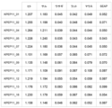
- FIG. 2 shows evaluation results of the binding activity of each anti-human EphA4 monoclonal antibody prepared in Example 1 to various EphA4s.
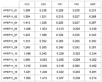
- FIG. 3 shows evaluation results of the binding activity of each anti-human EphA4 monoclonal antibody prepared in Example 1 to each region of human EphA4.
- FIG. 4 shows the evaluation results SN ((10 ng/mL EphA4 cells of 10 ng/mL EphA4 cells The signal obtained when the ectodomain was seeded)-(the signal obtained when 0 ng/mL EphA4 ectodomain was seeded)) is shown.
- Figure 5 shows the evaluation results S/N for the human EphA4 extracellular region in sandwich ELISA using a combination of the anti-human EphA4 monoclonal antibodies prepared in Example 1 (10 ng/mL EphA4 cells The signal obtained when the ectodomain was seeded)/(the signal obtained when 0 ng/mL EphA4 ectodomain was seeded)) is shown.
- FIG. 6 shows the evaluation results SN ((1 ng/mL EphA4 cells The signal obtained when the ectodomain was seeded)-(the signal obtained when 0 ng/mL EphA4 ectodomain was seeded)) is shown.
- FIG. 1 ((1 ng/mL EphA4 cells The signal obtained when the ectodomain was seeded)-(the signal obtained when 0 ng/mL EphA4 ectodomain was seeded)) is shown.
- FIG. 1 ((1 ng/mL EphA4
- FIG. 7 shows the evaluation results S/N for the human EphA4 extracellular region in sandwich ELISA using a combination of the anti-human EphA4 monoclonal antibodies prepared in Example 1 ((1 ng/mL EphA4 cells The signal obtained when the ectodomain was seeded)/(the signal obtained when 0 ng/mL EphA4 ectodomain was seeded)) is shown.
- FIG. 8 shows representative binding response curves of the KPEP11_04, KPEP11_08, KPEP11_10 and KPEP11_18 antibodies to human EphA4.
- FIG. 9 shows the binding specificities of the KPEP11 — 04, KPEP11 — 08, KPEP11 — 10 and KPEP11 — 18 antibodies to each human Eph receptor family.
- FIG. 10 shows the results of the reactivity of the antibodies KPEP11_04, KPEP11_08, KPEP11_10 and KPEP11_18 to mouse, rat, rabbit, monkey and human EphA4.
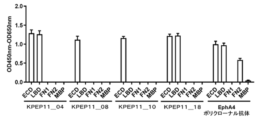
- FIG. 11 shows KPEP11_04 against the extracellular domain (ECD) of human EphA4, the ligand binding domain (LBD), fibronectin type III domain 1 (FN1), fibronectin type III domain 2 (FN2), and maltose binding protein (MBP).
- ECD extracellular domain
- LBD ligand binding domain
- FN1 fibronectin type III domain 1
- FN2 fibronectin type III domain 2
- MBP maltose binding protein
- FIG. 12 shows the results of reactivity to human EphA4 extracellular domain in sandwich ELISA using antibody KPEP11_10 and HRP-labeled antibody KPEP11_18.
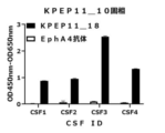
- FIG. 13 shows the results of reactivity to the EphA4 N-terminal fragment in cerebrospinal fluid in sandwich ELISA using antibody KPEP11_10 and HRP-labeled antibody KPEP11_18.
- FIG. 14 shows the results of reactivity to human EphA4 extracellular domain in sandwich ELISA constructed using antibody KPEP11_18 and HRP-labeled antibody KPEP11_10.
- FIG. 12 shows the results of reactivity to human EphA4 extracellular domain in sandwich ELISA using antibody KPEP11_10 and HRP-labeled antibody KPEP11_18.
- FIG. 15 shows the results of reactivity to EphA4 N-terminal fragment in cerebrospinal fluid in sandwich ELISA constructed using antibody KPEP11_18 and HRP-labeled antibody KPEP11_10.
- FIG. 16 shows the results of reactivity to human EphA4 extracellular domain in sandwich ELISA constructed using antibody KPEP11_10 and HRP-labeled antibody KPEP11_04.
- FIG. 17 shows the results of reactivity to EphA4 N-terminal fragment in cerebrospinal fluid in sandwich ELISA constructed using antibody KPEP11_10 and HRP-labeled antibody KPEP11_04.
- FIG. 18 shows the results of reactivity to human EphA4 extracellular domain in sandwich ELISA constructed using antibody KPEP11_18 and HRP-labeled antibody KPEP11_08.
- FIG. 19 shows the results of reactivity to EphA4 N-terminal fragment in cerebrospinal fluid in sandwich ELISA constructed using antibody KPEP11_18 and HRP-labeled antibody KPEP11_08.
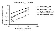
- Figure 20 shows the results of evaluating the reactivity to the extracellular region of human EphA4 in a sandwich ELISA constructed using antibody KPEP11_10, HRP-labeled antibody KPEP11_04, HRP-labeled antibody KPEP11_18, and HRP-labeled EphA4 antibody (Sino or R&D). .
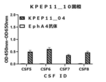
- Figure 21 shows a sandwich ELISA constructed using the antibody KPEP11_10, the HRP-labeled antibody KPEP11_04, the HRP-labeled antibody KPEP11_18, and the HRP-labeled EphA4 antibody (Sino or R&D) to evaluate the reactivity to the EphA4 N-terminal fragment in human plasma. Show the results.
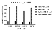
- Figure 22 shows the reactivity to the EphA4 N-terminal fragment in human cerebrospinal fluid in a sandwich ELISA constructed using antibody KPEP11_10, HRP-labeled antibody KPEP11_04, HRP-labeled antibody KPEP11_18, and HRP-labeled EphA4 antibody (Sino or R&D).
- Figure 23 shows the results of evaluating the reactivity to the human EphA4 extracellular region in a sandwich ELISA constructed using the antibody KPEP11_18, the HRP-labeled antibody KPEP11_08, the HRP-labeled antibody KPEP11_10, and the HRP-labeled EphA4 antibody (Sino or R&D). .
- Figure 24 shows a sandwich ELISA constructed using the antibody KPEP11_18, the HRP-labeled antibody KPEP11_08, the HRP-labeled antibody KPEP11_10, and the HRP-labeled EphA4 antibody (Sino or R&D) to evaluate the reactivity to the EphA4 N-terminal fragment in human plasma. Show the results.
- Figure 25 shows the reactivity to the EphA4 N-terminal fragment in human cerebrospinal fluid in a sandwich ELISA constructed using antibody KPEP11_18, HRP-labeled antibody KPEP11_08, HRP-labeled antibody KPEP11_10, and HRP-labeled EphA4 antibody (Sino or R&D).
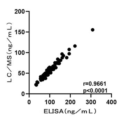
- Figure 26 also shows the quantification results of EphA4 N-terminal fragments in human cerebrospinal fluid analyzed by LC-MS and the quantification results of EphA4 N-terminal fragments in human cerebrospinal fluid analyzed in quantitative analysis 1 by ELISA analysis. The results of the correlation analysis performed on and are shown.
- Figure 27 also shows the quantification results of EphA4 N-terminal fragments in human cerebrospinal fluid analyzed by LC-MS and the quantification results of EphA4 N-terminal fragments in human cerebrospinal fluid analyzed in quantitative analysis 2 by ELISA analysis. The results of the correlation analysis performed on and are shown.
- the present disclosure relates to anti-EphA4 antibodies that bind EphA4.
- An anti-EphA4 antibody according to the present disclosure is an antibody that can specifically recognize and bind to EphA4.
- Anti-EphA4 antibodies may be intact antibodies or synthetic antibodies (eg, recombinant antibodies, chimeric antibodies, humanized antibodies, etc.) as long as they have binding affinity for EphA4.
- EphA4 may be understood to refer to EphA4 from human, mouse, rat, rabbit or monkey. Human-, mouse-, rat-, rabbit- and monkey-derived EphA4 are available from public databases with sequence information such as Genbank provided by the National Center for Biotechnology Information.
- Sequence information of the EphA4 gene can be obtained by designing primers based on the sequence information and cloning from RNA extracted from a desired animal species.
- the nucleotide sequence information for human, mouse, rat, rabbit and monkey EphA4 is available at Genbank Accession No. 1, respectively. It is registered on the database as NM_004438.5, NM_007936.3, NM_001162411.1, XM_002712496.3, and NM_001260870.1.
- EphA4 comprises the amino acid sequence shown in SEQ ID NO: 1 or an amino acid sequence in which one or more amino acids are substituted, added, or deleted from the amino acid sequence.
- “plurality” is not limited as long as it retains the same functional properties as the original sequence, but 2 to 100, for example 2 to 90, 2 to 80, 2 to 70 2 to 60, 2 to 50, 2 to 40, 2 to 30, 2 to 20, 2 to 10, 9, 8, 7, 6, 5, 4, 3 or 2, or within 10% of the number of amino acids in the amino acid sequence, such as within 9%, within 8%, within 7%, within 6%, within 5%, 4 %, 3%, 2% or 1%.
- the term "specific binding” is a term well known to those skilled in the art, and methods for determining specific binding of an antibody or antigen-binding fragment thereof to an antigen or epitope is also well known.
- “specific binding” means that the anti-EphA4 antibody or antigen-binding fragment thereof binds to other target molecules with greater binding affinity, avidity, more rapidly, and/or Alternatively, it is understood that EphA4 can be bound by an immunological response over a longer period of time.
- “specific binding” is at least about 10-7 M, or at least about 10-8 M, or at least about 10-9 M, or at least 10-10 M or less to EphA4.
- EphA4 includes immunologically binding to EphA4, but not other Eph receptor family molecules (e.g., EphA1, EphA2, EphA3, EphA5, EphA6, EphA7, EphA8). , EphA10, EphB1, EphB2, EphB3, EphB4, EphB6).
- the "antigen-binding fragment” is not particularly limited as long as it is a fragment of an anti-EphA4 antibody that maintains specific binding to EphA4. mentioned.
- binding affinity can be measured by Biacore® biosensor, KinExA biosensor, scintillation proximity assay, ELISA, ORIGEN immunoassay (IGEN), flow cytometry, fluorescence quenching, fluorescence transfer, yeast display, and/or , may be measured using, but not limited to, immunostaining.
- An anti-EphA4 antibody or antigen-binding fragment thereof may be of any class such as IgG, IgA or IgM (or subclass thereof) and is not limited to a particular class.
- immunoglobulins are classified into different classes. There are five main immunoglobulin classes: IgA, IgD, IgE, IgG and IgM, some of which are, for example, IgG 1 , IgG 2 , IgG 3 , IgG 4 , and subclasses IgA 1 and IgA 2 ( can be further subdivided into isotypes).
- the corresponding heavy chain constant regions of the different classes of immunoglobulins are called ⁇ , ⁇ , ⁇ , ⁇ and ⁇ , respectively.
- Antibody light chains also called L chains
- Anti-EphA4 antibodies or antigen-binding fragments thereof according to the present disclosure may be IgG antibodies, and anti-EphA4 antibodies according to the present disclosure may optionally be in the form of monomers, dimers or multimers. you can
- variable region of the antibody or antigen-binding fragment thereof may mean the variable region of the antibody light chain and/or the variable region of the antibody heavy chain, and the constant region of the antibody is the constant region of the antibody light chain and /or it may refer to the constant region of the antibody heavy chain.
- the heavy and light chain variable regions each consist of four framework regions (FR) joined by three CDRs, also known as complementarity determining regions. The CDRs on each chain are held in close proximity by the FRs and, together with the CDRs on the other chain, contribute to the formation of the antigen-binding site of the antibody.
- Techniques for determining CDRs include, but are not limited to, (1) approaches based on interspecies sequence variability (e.g., Kabat et al., Sequences of Proteins of Immunological Interest, 5th ed., 1991, National Institutes and (2) approaches based on crystallographic studies of antigen-antibody complexes (Al-lazikani et al., 1997 J. Molec. Biol. 273:927-948). These approaches and other approaches may be used in combination.
- Heavy and/or light chain sequences from, for example, but not limited to, human, mouse, rat, rabbit or monkey can be used in the anti-EphA4 antibodies or antigen-binding fragments thereof of the present disclosure.
- an anti-EphA4 antibody or antigen-binding fragment thereof of the present disclosure has heavy and light chain sequences derived from rabbit.
- the anti-EphA4 antibody or antigen-binding fragment thereof according to the present disclosure may be modified as desired.
- Modifications of an anti-EphA4 antibody or antigen-binding fragment thereof may be modified by (a) the three-dimensional conformation of the amino acid sequence in the modified region, such as, for example, sheet or helical conformations; (b) the charge or hydrophobicity of the molecule at the target site; or (c) the effect of the modification on the maintenance of side chain volume, or may be modifications such that these changes are not overtly observed.
- Modification of the anti-EphA4 antibody or antigen-binding fragment thereof according to the present disclosure may be achieved, for example, by substitution, deletion, addition, etc. of constituent amino acid residues.
- amino acid is used in its broadest sense and refers to naturally occurring amino acids such as serine (Ser), asparagine (Asn), valine (Val), leucine (Leu), isoleucine (Ile), alanine (Ala ), Tyrosine (Tyr), Glycine (Gly), Lysine (Lys), Arginine (Arg), Histidine (His), Aspartic acid (Asp), Glutamic acid (Glu), Glutamine (Gln), Threonine (Thr), Cysteine ( Cys), methionine (Met), phenylalanine (Phe), tryptophan (Trp), proline (Pro), as well as unnatural amino acids such as amino acid variants and derivatives.
- amino acids herein include, for example, L-amino acids; D-amino acids; chemically modified amino acids such as amino acid variants and amino acid derivatives; norleucine, ⁇ -alanine, It is of course understood that amino acids such as ornithine that do not become protein constituents in vivo; and chemically synthesized compounds having the properties of amino acids known to those skilled in the art.
- unnatural amino acids examples include ⁇ -methylamino acids ( ⁇ -methylalanine, etc.), D-amino acids (D-aspartic acid, D-glutamic acid, etc.), histidine-like amino acids (2-amino-histidine, ⁇ -hydroxy-histidine , homohistidine, ⁇ -fluoromethyl-histidine, ⁇ -methyl-histidine, etc.), amino acids with an extra methylene in the side chain (“homo” amino acids) and carboxylic acid functional amino acids in the side chain replaced with sulfonic acid groups. amino acids (cysteic acid, etc.)
- Naturally occurring amino acid residues can be classified into the following groups, for example, based on common side chain properties: (1) Hydrophobicity: Met, Ala, Val, Leu, Ile; (2) Neutral hydrophilicity: Asn, Gln, Cys, Ser, Thr; (3) acidic: Asp, Glu; (4) basic: His, Lys, Arg; (5) residues that influence chain orientation: Gly, Pro; and (6) aromatics: Trp, Tyr, Phe.
- Non-conservative substitutions of amino acid sequences that constitute antibodies may be made by exchanging amino acids belonging to one of these groups with amino acids belonging to another group. More conservative substitutions may be made by exchanging an amino acid belonging to one of these groups with another amino acid of the same group. Similarly, deletions or substitutions of amino acid sequences may be made as appropriate.
- Modifications of amino acids that constitute antibodies may be, for example, post-translational modifications such as glycosylation with sugars, acetylation or phosphorylation.
- Antibodies can be glycosylated at conserved positions in their constant regions.
- Glycosylation of antibodies is typically either N-linked or O-linked.
- N-linked refers to the attachment of carbohydrate moieties to the side chains of asparagine residues.
- O-linked glycosylation may be the attachment of either N-acetylgalactosamine, galactose, or xylose to a hydroxyamino acid (eg, serine or threonine), optionally 5-hydroxyproline or 5- It may be a bond to hydroxylysine.
- Glycosylation conditions when glycosylation is performed using a biological method, for example, the type of host cell or cell medium, pH, etc. can be appropriately selected by those skilled in the art according to the purpose. .
- the anti-EphA4 antibody or antigen-binding fragment thereof according to the present disclosure may be further modified, alone or in combination, by other modification methods based on the common technical knowledge known to those skilled in the art.
- the anti-EphA4 antibody or antigen-binding fragment thereof according to the present disclosure can be produced by methods well known to those skilled in the art.
- a nucleic acid encoding an anti-EphA4 antibody or an antigen-binding fragment thereof according to the present disclosure is incorporated into an expression vector, the expression vector is introduced into a host cell, and the host cell is cultured to obtain an antibody or an antigen-binding fragment thereof. may be produced.
- the present disclosure provides nucleic acids encoding anti-EphA4 antibodies or antigen-binding fragments thereof, vectors comprising such nucleic acids, host cells comprising such vectors, and anti-EphA4 antibodies or antigen-binding fragments thereof comprising culturing said host cells. It also includes methods of making sex fragments.
- a nucleic acid encoding an anti-EphA4 antibody or an antigen-binding fragment thereof according to the present disclosure may have a DNA encoding a signal sequence, a DNA encoding a heavy chain variable region, and a DNA encoding a light chain variable region.
- a DNA encoding a signal sequence may be present at the 5' end of the DNA.
- a signal sequence is an amino acid residue present at the N-terminus of a protein that is necessary for secretory proteins and integral membrane proteins to pass through the lipid bilayer after being synthesized on the ribosome. It is not particularly limited as long as it has a functional sequence.
- Signal sequences that may be included in the anti-EphA4 antibodies or antigen-binding fragments thereof of the present disclosure include signal sequences derived from humans, mice, rats, rabbits, donkeys, goats, horses, birds, dogs, cats, yeast, and the like. be done.
- the anti-EphA4 antibody or antigen-binding fragment thereof according to the present disclosure may be isolated or purified according to methods known to those skilled in the art.
- isolated or purified means artificially isolated or purified from the natural state. If a molecule or composition is naturally occurring, it is “isolated” or “purified” if it has been altered or removed from its original environment, or both. be.
- isolation or purification methods include electrophoretic, molecular biological, immunological or chromatographic techniques, specifically ion exchange chromatography, hydrophobic chromatography, reversed phase Examples include, but are not limited to, HPLC chromatography, isoelectric focusing, or alkaline extraction.
- an anti-EphA4 antibody or antigen-binding fragment thereof of the present disclosure comprises the following CDRs: heavy chain CDR1 consisting of the amino acid sequence shown in SEQ ID NO: 40; heavy chain CDR2 consisting of the amino acid sequence shown in SEQ ID NO: 41; heavy chain containing heavy chain CDR3 consisting of the amino acid sequence shown in SEQ ID NO: 42; and light chain CDR1 consisting of the amino acid sequence shown in SEQ ID NO: 43; A light chain comprising a light chain CDR2 consisting of the amino acid sequence shown in SEQ ID NO:44; and a light chain CDR3 consisting of the amino acid sequence shown in SEQ ID NO:45.
- an anti-EphA4 antibody or antigen-binding fragment thereof of the present disclosure comprises the following CDRs: heavy chain CDR1 consisting of the amino acid sequence shown in SEQ ID NO: 52; heavy chain CDR2 consisting of the amino acid sequence shown in SEQ ID NO:53; and heavy chain containing heavy chain CDR3 consisting of the amino acid sequence shown in SEQ ID NO:54; and light chain CDR1 consisting of the amino acid sequence shown in SEQ ID NO:55; a light chain comprising a light chain CDR2 consisting of the amino acid sequence shown in SEQ ID NO:56; and a light chain CDR3 consisting of the amino acid sequence shown in SEQ ID NO:57;
- an anti-EphA4 antibody or antigen-binding fragment thereof of the present disclosure comprises the following CDRs: heavy chain CDR1 consisting of the amino acid sequence shown in SEQ ID NO: 64; a heavy chain CDR2 consisting of the amino acid sequence shown in SEQ ID NO: 65; and a heavy chain comprising a heavy chain CDR3 consisting of the amino acid sequence shown in SEQ ID NO: 66; A light chain comprising a light chain CDR2 consisting of the amino acid sequence shown in SEQ ID NO:68; and a light chain CDR3 consisting of the amino acid sequence shown in SEQ ID NO:69.
- the anti-EphA4 antibody or antigen-binding fragment thereof comprises a heavy chain variable region consisting of the amino acid sequence shown in SEQ ID NO:6 and a light chain variable region consisting of the amino acid sequence shown in SEQ ID NO:7.
- the anti-EphA4 antibody or antigen-binding fragment thereof comprises a heavy chain variable region consisting of the amino acid sequence shown in SEQ ID NO:10 and a light chain variable region consisting of the amino acid sequence shown in SEQ ID NO:11.
- the anti-EphA4 antibody or antigen-binding fragment thereof comprises a heavy chain variable region consisting of the amino acid sequence shown in SEQ ID NO:14 and a light chain variable region consisting of the amino acid sequence shown in SEQ ID NO:15.
- the anti-EphA4 antibody or antigen-binding fragment thereof comprises a heavy chain variable region consisting of the amino acid sequence shown in SEQ ID NO:18 and a light chain variable region consisting of the amino acid sequence shown in SEQ ID NO:19.
- the heavy chain variable region and/or the light chain variable region may have an amino acid sequence in which one or more amino acids are substituted, added and/or deleted from the original sequence.
- “plurality” is not limited as long as it retains binding affinity for EphA4 and promotes cleavage of EphA4, but is not limited to 2 to 15, or 2 to 10, such as 9, 8, 7, 6, 5, 4, 3 or 2, or within 10% of the number of amino acids in the amino acid sequence, such as within 9%, within 8%, within 7%, within 6%, 5 %, 4%, 3%, 2%, or 1%.
- the heavy chain of the anti-EphA4 antibody or antigen-binding fragment thereof according to the present disclosure comprises a rabbit IgG constant region.
- the rabbit IgG constant region comprises the amino acid sequence of SEQ ID NO:22.
- the light chain of the anti-EphA4 antibody or antigen-binding fragment thereof according to the present disclosure comprises a rabbit Ig ⁇ constant region.
- the rabbit Ig ⁇ constant region comprises the amino acid sequence of SEQ ID NO:23.
- the heavy chain of the anti-EphA4 antibody of the present disclosure comprises the amino acid sequence shown in SEQ ID NO:24, and the light chain of the anti-EphA4 antibody comprises the amino acid sequence shown in SEQ ID NO:25.
- the heavy chain of the anti-EphA4 antibody of the present disclosure comprises the amino acid sequence set forth in SEQ ID NO:28, and the light chain of the anti-EphA4 antibody comprises the amino acid sequence set forth in SEQ ID NO:29.
- the heavy chain of the anti-EphA4 antibody of the present disclosure comprises the amino acid sequence shown in SEQ ID NO:32
- the light chain of the anti-EphA4 antibody comprises the amino acid sequence shown in SEQ ID NO:33.
- the heavy chain of the anti-EphA4 antibody of the present disclosure comprises the amino acid sequence set forth in SEQ ID NO:36
- the light chain of the anti-EphA4 antibody comprises the amino acid sequence set forth in SEQ ID NO:37.
- an anti-EphA4 antibody or antigen-binding fragment thereof according to the present disclosure is labeled.
- label means a detectable compound or composition that is directly or indirectly attached to an antibody or antigen-binding fragment thereof.
- a label may be detectable by itself or in combination with another specific binding pair, e.g. The reaction may produce a detectable signal.
- anti-EphA4 antibodies may lack the lysine located at the C-terminus (carboxy terminus) of the heavy chain.
- the anti-EphA4 antibody in which the C-terminal lysine of the heavy chain has been deleted includes an anti-EphA4 antibody in which the C-terminal lysine of the heavy chain has been deleted by genetic modification, and post-translational C of the heavy chain by carboxypeptidase or the like.
- Anti-EphA4 antibodies with truncated terminal lysines and the like are also included.
- anti-EphA4 antibodies in which the C-terminal lysines of the heavy chains have been deleted include anti-EphA4 antibodies in which the C-terminal lysines have been deleted in both heavy chains, as well as anti-EphA4 antibodies in which only one heavy chain has Also included are anti-EphA4 antibodies in which the C-terminal lysine has been deleted.
- the disclosure relates to an isolated nucleic acid encoding an anti-EphA4 antibody or antigen-binding fragment thereof.
- An isolated nucleic acid encoding an anti-EphA4 antibody or antigen-binding fragment thereof refers to one or more nucleic acid molecules encoding the heavy and/or light chains of an anti-EphA4 antibody or antigen-binding fragment thereof.
- a nucleic acid of the disclosure encodes the heavy chain of an anti-EphA4 antibody or antigen-binding fragment thereof.
- a nucleic acid of this disclosure encodes the light chain of an anti-EphA4 antibody or antigen-binding fragment thereof.
- nucleic acids of this disclosure encode the heavy and light chains of an anti-EphA4 antibody or antigen-binding fragment thereof.
- Nucleic acids of the present disclosure include a first nucleic acid molecule encoding a heavy chain of an anti-EphA4 antibody or antigen-binding fragment thereof, and a second nucleic acid molecule encoding a light chain of an anti-EphA4 antibody or antigen-binding fragment thereof is also included.
- the disclosure relates to a vector comprising an isolated nucleic acid encoding an anti-EphA4 antibody or antigen-binding fragment thereof.
- a vector of the present disclosure refers to one or more vectors comprising an isolated nucleic acid encoding an anti-EphA4 antibody or antigen-binding fragment thereof.
- a vector according to the present disclosure is a vector comprising a nucleic acid encoding a heavy chain of an anti-EphA4 antibody or antigen-binding fragment thereof and a nucleic acid encoding a light chain of an anti-EphA4 antibody or antigen-binding fragment thereof. .
- the vector of the present disclosure is a vector comprising nucleic acids encoding heavy and light chains of an anti-EphA4 antibody or antigen-binding fragment thereof.
- the vector of the present disclosure is a first vector comprising a nucleic acid encoding a heavy chain of an anti-EphA4 antibody or antigen-binding fragment thereof and a light chain of an anti-EphA4 antibody or antigen-binding fragment thereof
- a second vector comprising a nucleic acid encoding Vectors according to the present disclosure may be, but are not limited to, plasmids, cosmids, viruses, phages, and the like.
- viral vectors such as retrovirus, lentivirus, adenovirus, adeno-associated virus, or herpes simplex virus vectors are also included in the vectors of the present disclosure.
- the present disclosure also includes a method of producing an anti-EphA4 antibody or antigen-binding fragment thereof comprising a host cell comprising a vector of the present disclosure, and culturing the host cell.
- Host cells may be, but are not limited to, E. coli cells, monkey COS cells, Chinese Hamster Ovary (CHO) cells, NS0 cells, and the like.
- the method for producing an anti-EphA4 antibody or antigen-binding fragment thereof comprises the steps of culturing a host cell and extracting an anti-EphA4 antibody or antigen-binding fragment thereof secreted from the host cell (or the culture medium of the host cell). A step of recovering the fragment is included.
- the anti-EphA4 antibody or antigen-binding fragment thereof binds to any N-terminal region of EphA4.
- the "N-terminal region" of EphA4 refers to the extracellular domain (ECD) of EphA4, or the N-terminal side when EphA4 is cleaved by matrix metalloprotease (MMP) or ADAM (a disintegrin and metalloproteinase) refers to the area of
- MMP matrix metalloprotease
- ADAM a disintegrin and metalloproteinase
- the ECD in human EphA4 is defined as having the amino acid sequence shown in SEQ ID NO: 2, or an amino acid sequence in which one or more amino acids are substituted, added and/or deleted in the amino acid sequence.
- plality is 2 to 15, or 2 to 10, such as 9, 8, 7, 6, 5, 4, 3 or 2, or within 10% of the number of amino acids in the amino acid sequence, such as within 9%, within 8%, within 7%, within 6%, within 5%, within 4%, within 3%, within 2%, or within 1% .
- the anti-EphA4 antibody or antigen-binding fragment thereof according to the present disclosure can specifically bind to EphA4 and detect EphA4 with high detection sensitivity. Accordingly, in another aspect, the present disclosure provides a method for detecting or quantifying EphA4 in a biological sample using the anti-EphA4 antibody or antigen-binding fragment thereof of the present disclosure (hereinafter also referred to as the method of the present disclosure) Regarding.
- the "EphA4" to be measured includes full-length EphA4 as well as a fragment consisting of the N-terminal region of EphA4 (herein referred to simply as "EphA4 N-terminal fragment” or “EphA4 N-terminal fragment ”) is included.
- the biological sample is not particularly limited as long as it can contain full-length EphA4 or an EphA4 N-terminal fragment.
- examples include blood, serum, plasma, cerebrospinal fluid (CSF), urine, saliva, tears Liquid components (also referred to as body fluids) derived from living organisms such as liquids and perspiration can be used.
- the biological sample is blood, serum, plasma, or cerebrospinal fluid.
- biological samples are not limited to human-derived biological samples, and include biological samples of animals other than humans. Examples of such animals include, but are not limited to, mice, rats, rabbits, monkeys, and the like.
- a method according to the present disclosure includes contacting a biological sample with an anti-EphA4 antibody or antigen-binding fragment thereof according to the present disclosure. Detection or quantification of EphA4 can be performed using immunoassays well known in the art.
- the biological sample and the labeled anti-EphA4 antibody or an antigen-binding fragment thereof are different antibodies.
- the immunoassay uses a detectably labeled anti-EphA4 antibody or antigen-binding fragment thereof, or a detectably labeled anti-EphA4 antibody or antigen-binding fragment thereof (secondary antibody).
- ELISA uses enzymes such as peroxidase and alkaline phosphatase; RIA uses radioactive substances such as 125 I, 131 I, 35 S and 3 H; FPIA uses fluorescein isothiocyanate, rhodamine, dansyl chloride, phycoerythrin, tetra Antibodies labeled with fluorescent substances such as methylrhodamine isothiocyanate and near-infrared fluorescent materials, enzymes such as luciferase and ⁇ -galactosidase, luminescent substrates that change into luminescent substances with each enzyme, and luminescent substances such as luciferin and aequorin in the CLIA method. can be used. In addition, antibodies labeled with nanoparticles such as colloidal gold and quantum dots can also be detected.
- EphA4 can also be detected and measured by labeling an anti-EphA4 antibody or an antigen-binding fragment thereof with biotin and binding with enzyme-labeled avidin or streptavidin.
- the sandwich method can be used.
- An anti-EphA4 antibody or an antigen-binding fragment thereof is immobilized on a solid phase carrier, an appropriately treated biological sample is added and allowed to react, and then another enzyme-labeled anti-EphA4 antibody or antigen-binding fragment thereof is added. to react. After washing, EphA4 or the N-terminal fragment of EphA4 can be quantified by reacting with an enzyme substrate, developing color, and measuring absorbance.
- the enzyme substrate is 3,3′-diaminobenzidine (DAB), 3,3′,5,5′-tetramethylbenzidine (3,3′,5,5 '-tetramethylbenzidine; TMB or TMBZ), o-phenylenediamine (OPD) and the like can be used, and in the case of alkaline phosphatase, p-nitrophenyl phosphate (NPP) and the like can be used. can.
- DAB 3,3′-diaminobenzidine
- TMB or TMBZ 3,3′,5,5′-tetramethylbenzidine
- OPD o-phenylenediamine
- NPP p-nitrophenyl phosphate
- the agglutination method can be mentioned as a method that can easily detect a minute amount of protein.
- the agglutination method includes, for example, a latex agglutination method in which latex particles are bound to an antibody.
- the antigen concentration can be determined by irradiating a sample with near-infrared light and quantifying aggregates by measuring absorbance (turbidimetric method) or scattered light (nephelometric method).
- the method of the present disclosure is a method of detecting or quantifying human EphA4 in a biological sample.
- the method according to the present disclosure is a method for detecting or quantifying an N-terminal fragment of human EphA4 in a biological sample.
- the method of the present disclosure uses a sandwich ELISA using a combination of antibodies or antigen-binding fragments thereof of the present disclosure to detect or quantify human EphA4.
- kits of the present disclosure also relates to a kit for detecting or quantifying EphA4, which includes the anti-EphA4 antibody or antigen-binding fragment thereof of the present disclosure (hereinafter also referred to as the kit of the present disclosure).
- Kits of the present disclosure can include any reagents or instruments that can be used in detecting or quantifying EphA4 or instructions for using the kit.
- EphA4 to be measured includes full-length EphA4 as well as EphA4 N-terminal fragments.
- the kit according to the present disclosure relates to kits for detecting or quantifying human EphA4.
- the kit according to the present disclosure relates to a kit for detecting or quantifying an N-terminal fragment of human EphA4.
- the kit according to the present disclosure contains full-length human EphA4 or a serial dilution of full-length EphA4 that can be used as a positive control when creating a standard curve for human EphA4.
- the kit according to the present disclosure contains an N-terminal fragment of human EphA4 or a serial dilution of the fragment that can be used as a positive control when creating a standard curve for human EphA4.
- the kit according to the present disclosure comprises an anti-EphA4 antibody or an antigen-binding antibody thereof comprising a heavy chain variable region consisting of the amino acid sequence shown in SEQ ID NO: 14 and a light chain variable region consisting of the amino acid sequence shown in SEQ ID NO: 15. and an anti-EphA4 antibody or antigen-binding fragment thereof comprising a heavy chain variable region consisting of the amino acid sequence shown in SEQ ID NO:18 and a light chain variable region consisting of the amino acid sequence shown in SEQ ID NO:19.
- the kit according to the present disclosure comprises an anti-EphA4 antibody or antigen thereof, comprising a heavy chain variable region consisting of the amino acid sequence shown in SEQ ID NO:6 and a light chain variable region consisting of the amino acid sequence shown in SEQ ID NO:7. and an anti-EphA4 antibody or antigen-binding fragment thereof comprising a heavy chain variable region consisting of the amino acid sequence shown in SEQ ID NO:14 and a light chain variable region consisting of the amino acid sequence shown in SEQ ID NO:15.
- the kit according to the present disclosure comprises an anti-EphA4 antibody or antigen thereof, comprising a heavy chain variable region consisting of the amino acid sequence shown in SEQ ID NO: 10 and a light chain variable region consisting of the amino acid sequence shown in SEQ ID NO: 11 and an anti-EphA4 antibody or antigen-binding fragment thereof comprising a heavy chain variable region consisting of the amino acid sequence shown in SEQ ID NO:18 and a light chain variable region consisting of the amino acid sequence shown in SEQ ID NO:19.
- the kit according to the present disclosure comprises an anti-EphA4 antibody or its and an anti-EphA4 antibody or antigen-binding fragment thereof comprising a heavy chain variable region consisting of the amino acid sequence shown in SEQ ID NO:10 and a light chain variable region consisting of the amino acid sequence shown in SEQ ID NO:11.
- kits of the present disclosure include the following: (solid-phase antibody) An antibody or an antigen-binding fragment thereof, comprising a heavy chain variable region consisting of the amino acid sequence shown in SEQ ID NO:10 and a light chain variable region consisting of the amino acid sequence shown in SEQ ID NO:11. (labeled antibody) An antibody or an antigen-binding fragment thereof, comprising a heavy chain variable region consisting of the amino acid sequence shown in SEQ ID NO:6 and a light chain variable region consisting of the amino acid sequence shown in SEQ ID NO:7.
- solid-phase antibody An antibody or an antigen-binding fragment thereof, comprising a heavy chain variable region consisting of the amino acid sequence shown in SEQ ID NO:14 and a light chain variable region consisting of the amino acid sequence shown in SEQ ID NO:15.
- labeled antibody An antibody or an antigen-binding fragment thereof, comprising a heavy chain variable region consisting of the amino acid sequence shown in SEQ ID NO:6 and a light chain variable region consisting of the amino acid sequence shown in SEQ ID NO:7.
- solid-phase antibody An antibody or an antigen-binding fragment thereof, comprising a heavy chain variable region consisting of the amino acid sequence shown in SEQ ID NO:6 and a light chain variable region consisting of the amino acid sequence shown in SEQ ID NO:7.
- labeled antibody An antibody or an antigen-binding fragment thereof, comprising a heavy chain variable region consisting of the amino acid sequence shown in SEQ ID NO:10 and a light chain variable region consisting of the amino acid sequence shown in SEQ ID NO:11.
- solid-phase antibody An antibody or an antigen-binding fragment thereof, comprising a heavy chain variable region consisting of the amino acid sequence shown in SEQ ID NO:18 and a light chain variable region consisting of the amino acid sequence shown in SEQ ID NO:19.
- labeled antibody An antibody or an antigen-binding fragment thereof, comprising a heavy chain variable region consisting of the amino acid sequence shown in SEQ ID NO:10 and a light chain variable region consisting of the amino acid sequence shown in SEQ ID NO:11.
- solid-phase antibody An antibody or an antigen-binding fragment thereof, comprising a heavy chain variable region consisting of the amino acid sequence shown in SEQ ID NO:6 and a light chain variable region consisting of the amino acid sequence shown in SEQ ID NO:7.
- labeled antibody An antibody or an antigen-binding fragment thereof, comprising a heavy chain variable region consisting of the amino acid sequence shown in SEQ ID NO:14 and a light chain variable region consisting of the amino acid sequence shown in SEQ ID NO:15.
- solid-phase antibody An antibody or an antigen-binding fragment thereof, comprising a heavy chain variable region consisting of the amino acid sequence shown in SEQ ID NO:18 and a light chain variable region consisting of the amino acid sequence shown in SEQ ID NO:19.
- labeled antibody An antibody or an antigen-binding fragment thereof, comprising a heavy chain variable region consisting of the amino acid sequence shown in SEQ ID NO:14 and a light chain variable region consisting of the amino acid sequence shown in SEQ ID NO:15.
- solid-phase antibody An antibody or an antigen-binding fragment thereof, comprising a heavy chain variable region consisting of the amino acid sequence shown in SEQ ID NO:10 and a light chain variable region consisting of the amino acid sequence shown in SEQ ID NO:11.
- labeled antibody An antibody or an antigen-binding fragment thereof, comprising a heavy chain variable region consisting of the amino acid sequence shown in SEQ ID NO:18 and a light chain variable region consisting of the amino acid sequence shown in SEQ ID NO:19.
- solid-phase antibody An antibody or an antigen-binding fragment thereof, comprising a heavy chain variable region consisting of the amino acid sequence shown in SEQ ID NO:14 and a light chain variable region consisting of the amino acid sequence shown in SEQ ID NO:15.
- labeled antibody An antibody or an antigen-binding fragment thereof, comprising a heavy chain variable region consisting of the amino acid sequence shown in SEQ ID NO:18 and a light chain variable region consisting of the amino acid sequence shown in SEQ ID NO:19.
- first, second, etc. are used to describe various elements, it is understood that these elements should not be limited by these terms themselves. These terms are only used to distinguish one element from another, e.g., a first element referred to as a second element; similarly, a second element referred to as a first element. can be written without departing from the scope of the present disclosure.
- Example 1 Generation of Anti-Human EphA4 Rabbit Monoclonal Antibody
- SEAP secretory alkaline phosphatase
- SEQ ID NO: 2 the extracellular region of human EphA (20- 547 position)
- SEQ ID NO: 4 the extracellular region of human EphA (20- 547 position)
- MBP maltose binding protein
- SEQ ID NO: 5 A histidine tag-fused protein
- the synthesized human EphA4 extracellular domain gene fragment was cloned into the constructed pcDNA3.4-SEAP-His expression vector to construct a human EphA4 extracellular domain-SEAP-His expression vector.
- the pcDNA3.4-human EphA4 extracellular domain-His expression vector was prepared by cloning the synthesized human EphA4 extracellular domain gene fragment into a pcDNA3.4 vector (Invitrogen/Life Technologies) having a DNA sequence encoding a histidine tag. It was constructed.
- the DNA sequence encoding the signal sequence and extracellular region of human EphA4 was amplified by PCR, and pcDNA3 with the DNA sequence encoding MBP and histidine tag was generated. .4 vector (Invitrogen/Life Technologies) and constructed. Using the Expi293 expression system (Thermo SCIENTIFIC), each of the above expression vectors was transfected into Expi293F cells (Thermo SCIENTIFIC). The medium was harvested and clarified to remove cells. Purification was performed using TALON resin (TaKaRa), and the buffer was replaced with PBS (FUJI FILM Wako) by dialysis or a desalting column (Thermo SCIENTIFIC).
- Rabbits were immunized with the prepared human EphA4 extracellular region-SEAP-His protein together with an adjuvant (IBL) according to a standard method. After immunization, total RNA was prepared from the collected lymph node cells using RNeasy (QIAGEN) and treated with DNase (QIAGEN, RNase free DNase set). A reverse transcription product was prepared from the total RNA using an RNA PCR kit (TAKARA). A rabbit antibody phage library was constructed using the resulting reverse transcription product as a template. Screening was performed using human EphA4 protein, human EphA4 antibody and rabbit antibody phage libraries to obtain rabbit antibody fragments (scFv) that specifically bind to human EphA4.
- IBL adjuvant
- Human EphA4 extracellular domain-MBP-His protein and human EphA4 antibody were captured using Dynabeads magnetic beads (Thermo SCIENTIFIC), rabbit antibody phage library was added, and after 1 or 2 hours, unbound phages were detected. Removed by a series of washing cycles using PBS-Tween (0.1% v/v) or PBS. After elution of bound phage particles, they were amplified via infection into E. coli TG1 host cells. Infected TG1 cells were harvested, plated and incubated at 30°C. This panning process was performed one more time with the amplified phage.
- Reactivity to human EphA4 was evaluated by ELISA using human EphA4 extracellular region-MBP-His protein according to the following steps.
- An anti-FLAG antibody SIGMA was coated onto the wells of a 96-well plate (Thermo SCIENTIFIC). After overnight incubation at 4° C., wells were blocked with 2% skimmed milk (BD) for 2 hours at room temperature. After washing three times with 0.02% Tween20/PBS, human EphA4 extracellular domain-MBP-His protein (final concentration 20 nM) and scFv-containing Escherichia coli culture supernatant were added to each well and allowed to cool to room temperature. and incubated for 2 hours.
- SIGMA anti-FLAG antibody
- Clones were converted from scFv to IgG format by subcloning DNA sequences encoding the variable regions of the obtained rabbit antibody fragment (scFv) into vectors expressing antibody heavy and light chain constant regions, respectively.
- An expression vector (pcDNA3.4) containing a gene sequence encoding an anti-human EphA4 rabbit monoclonal antibody was constructed.
- the amino acid sequence of the heavy chain variable region of KPEP11_04 is the amino acid sequence shown in SEQ ID NO:6, and the amino acid sequence of the light chain variable region is the amino acid sequence shown in SEQ ID NO:7.
- the nucleic acid sequence shown in SEQ ID NO: 8 was used for the heavy chain variable region, and the nucleic acid sequence shown in SEQ ID NO: 9 was used for the light chain variable region.
- the amino acid sequence of the heavy chain variable region of KPEP11_08 is the amino acid sequence shown in SEQ ID NO:10, and the amino acid sequence of the light chain variable region is the amino acid sequence shown in SEQ ID NO:11.
- the nucleic acid sequence shown in SEQ ID NO: 12 was used for the heavy chain variable region
- the nucleic acid sequence shown in SEQ ID NO: 13 was used for the light chain variable region.
- the amino acid sequence of the heavy chain variable region of KPEP11_10 is the amino acid sequence shown in SEQ ID NO:14, and the amino acid sequence of the light chain variable region is the amino acid sequence shown in SEQ ID NO:15.
- the gene sequence encoding the amino acid sequence of KPEP11_10 the nucleic acid sequence shown in SEQ ID NO: 16 was used for the heavy chain variable region, and the nucleic acid sequence shown in SEQ ID NO: 17 was used for the light chain variable region.
- the amino acid sequence of the heavy chain variable region of KPEP11_18 is the amino acid sequence shown in SEQ ID NO:18, and the amino acid sequence of the light chain variable region is the amino acid sequence shown in SEQ ID NO:19.
- the nucleic acid sequence shown in SEQ ID NO: 20 was used for the heavy chain variable region, and the nucleic acid sequence shown in SEQ ID NO: 21 was used for the light chain variable region.
- the constant region of rabbit IgG (SEQ ID NO: 22) was used as the heavy chain constant region of KPEP11_04, KPEP11_08, KPEP11_10 and KPEP11_18.
- Rabbit Ig ⁇ (SEQ ID NO: 23) was used as the light chain constant region of KPEP11_04, KPEP11_08, KPEP11_10 and KPEP11_18.
- the amino acid sequence of the full-length heavy chain (not including the signal sequence) of KPEP11_04 is the amino acid sequence shown in SEQ ID NO:24, and the amino acid sequence of the full-length light chain (not including the signal sequence) is the amino acid sequence shown in SEQ ID NO:25.
- the nucleic acid sequence encoding the full length heavy chain of KPEP11_04 is the nucleic acid sequence shown in SEQ ID NO:26, and the nucleic acid sequence encoding the full length light chain is the nucleic acid sequence shown in SEQ ID NO:27.
- the amino acid sequence of the full-length heavy chain (not including the signal sequence) of KPEP11_08 is the amino acid sequence shown in SEQ ID NO: 28, and the amino acid sequence of the full-length light chain (not including the signal sequence) is the amino acid sequence shown in SEQ ID NO: 29.
- the nucleic acid sequence encoding the full-length heavy chain of KPEP11_08 is the nucleic acid sequence shown in SEQ ID NO:30, and the nucleic acid sequence encoding the full-length light chain is the nucleic acid sequence shown in SEQ ID NO:31.
- the amino acid sequence of the full-length heavy chain (not including the signal sequence) of KPEP11_10 is the amino acid sequence shown in SEQ ID NO: 32, and the amino acid sequence of the full-length light chain (not including the signal sequence) is the amino acid sequence shown in SEQ ID NO: 33.
- the nucleic acid sequence encoding the full-length heavy chain of KPEP11_10 is the nucleic acid sequence shown in SEQ ID NO:34, and the nucleic acid sequence encoding the full-length light chain is the nucleic acid sequence shown in SEQ ID NO:35.
- the amino acid sequence of the full-length heavy chain (not including the signal sequence) of KPEP11_18 is the amino acid sequence shown in SEQ ID NO: 36, and the amino acid sequence of the full-length light chain (not including the signal sequence) is the amino acid sequence shown in SEQ ID NO: 37. be.
- the nucleic acid sequence encoding the full-length heavy chain of KPEP11_18 is the nucleic acid sequence shown in SEQ ID NO:38, and the nucleic acid sequence encoding the full-length light chain is the nucleic acid sequence shown in SEQ ID NO:39.
- the CDRs of KPEP11_04, KPEP11_08, KPEP11_10 and KPEP11_18 were determined according to the Kabat definition for identification of CDRs.
- the amino acid and nucleic acid sequences of the CDRs of KPEP11_04 are shown in Tables 1 and 2, respectively.
- the amino acid and nucleic acid sequences of the CDRs of KPEP11_08 and KPEP11_10 are shown in Tables 3 and 4, respectively.
- the amino acid and nucleic acid sequences of the CDRs of KPEP11_18 are shown in Tables 5 and 6, respectively.
- the anti-human EphA4 rabbit monoclonal antibodies KPEP11_01, KPEP11_02, KPEP11_05, KPEP11_07, KPEP11_09, KPEP11_12, KPEP11_13 and KPEP11_20 were prepared by the same method as the above preparation method.
- the amino acid sequence of the heavy chain variable region of KPEP11_01 is the amino acid sequence shown in SEQ ID NO:76
- the amino acid sequence of the light chain variable region is the amino acid sequence shown in SEQ ID NO:77.
- the amino acid sequence of the heavy chain variable region of KPEP11_02 is the amino acid sequence shown in SEQ ID NO:78, and the amino acid sequence of the light chain variable region is the amino acid sequence shown in SEQ ID NO:79.
- the amino acid sequence of the heavy chain variable region of KPEP11_05 is the amino acid sequence shown in SEQ ID NO:80, and the amino acid sequence of the light chain variable region is the amino acid sequence shown in SEQ ID NO:81.
- the amino acid sequence of the heavy chain variable region of KPEP11_07 is the amino acid sequence shown in SEQ ID NO:82, and the amino acid sequence of the light chain variable region is the amino acid sequence shown in SEQ ID NO:83.
- the amino acid sequence of the heavy chain variable region of KPEP11_09 is the amino acid sequence shown in SEQ ID NO:84, and the amino acid sequence of the light chain variable region is the amino acid sequence shown in SEQ ID NO:85.
- the amino acid sequence of the heavy chain variable region of KPEP11_12 is the amino acid sequence shown in SEQ ID NO:86, and the amino acid sequence of the light chain variable region is the amino acid sequence shown in SEQ ID NO:87.
- the amino acid sequence of the heavy chain variable region of KPEP11_13 is the amino acid sequence shown in SEQ ID NO:88, and the amino acid sequence of the light chain variable region is the amino acid sequence shown in SEQ ID NO:89.
- the amino acid sequence of the heavy chain variable region of KPEP11_20 is the amino acid sequence shown in SEQ ID NO:90, and the amino acid sequence of the light chain variable region is the amino acid sequence shown in SEQ ID NO:91.
- the constant region of rabbit IgG (SEQ ID NO: 22) was used as the heavy chain constant region of these antibodies.
- Rabbit Ig ⁇ (SEQ ID NO: 23) was used as the light chain constant region of these antibodies.
- the CDRs of each antibody produced were determined according to the Kabat definition for identification of CDRs.
- the CDR amino acid sequences of each antibody are shown in Tables 7 to 11, respectively.
- Example 2 Selectivity of anti-human EphA4 monoclonal antibody to human Eph receptor
- the anti-human EphA4 monoclonal antibody prepared in Example 1 was evaluated for human Eph receptor-binding activity according to the following steps.
- a mouse anti-6-His antibody (R&D) was coated onto the wells of a 96-well plate (Thermo SCIENTIFIC). After incubation for 1 hour at room temperature, the wells were blocked with 1% Block Ace (KAC) for 1 hour at room temperature. After washing three times with 0.02% Tween20/PBS, each well was seeded with each human Eph receptor extracellular domain-His protein (Creative biomart, final concentration 1 nM) and incubated at room temperature for 1 hour.
- KAC Block Ace
- anti-human EphA4 rabbit monoclonal antibody (10 ⁇ g/mL) was added and incubated for 1 hour at room temperature.
- horseradish peroxidase-labeled goat anti-rabbit IgG polyclonal antibody (abcam) was added and incubated for 1 hour at room temperature.
- TMBZ (3,3',5,5'-tetramethylbenzidine, KPL) solution was added to the wells and incubated for 4.5 minutes at room temperature.
- reaction stop solution (2N H 2 SO 4 , FUJIFILM Wako) was added to the wells, and the absorbance at 450 nm and 650 nm was read with a microplate reader (Thermo SCIENTIFIC).
- KPEP11_01, KPEP11_02, KPEP11_04, KPEP11_05, KPEP11_07, KPEP11_08, KPEP11_09, KPEP11_10, KPEP11_12, KPEP11_13, KPEP11_18 and KPEP11_20 are human Ep It was found to specifically bind to hA4 (Fig. 1).
- Example 3 Reactivity of anti-human EphA4 monoclonal antibodies to mouse, rat, rabbit, monkey and human EphA4
- the anti-human EphA4 monoclonal antibodies prepared in Example 1 were evaluated for binding activity to various EphA4s according to the following steps. gone.
- a mouse anti-6-His antibody (R&D) was coated onto the wells of a 96-well plate (Thermo SCIENTIFIC). After incubating for 1 hour at room temperature, the wells were blocked overnight at 4°C with 1% Block Ace (KAC).
- KPEP11_01, KPEP11_02, KPEP11_04, KPEP11_05, KPEP11_07, KPEP11_08, KPEP11_09, KPEP11_10, KPEP11_12, KPEP11_13, KPEP11_18 and KPEP11_20 have binding activity in monkey and human EphA4 (Fig. 2).
- Example 4 Reactivity of anti-human EphA4 monoclonal antibody to human EphA4 extracellular region, ligand-binding domain, fibronectin type III domain 1, fibronectin type III domain 2 Regarding the anti-human EphA4 monoclonal antibody prepared in Example 1, various EphA4 The binding activity of was evaluated according to the following steps. A mouse anti-6-His antibody (R&D) was coated onto the wells of a 96-well plate (Thermo SCIENTIFIC). After incubation for 1 hour at room temperature, the wells were blocked with 1% Block Ace (KAC) for 1 hour at room temperature.
- R&D mouse anti-6-His antibody
- KAC Block Ace
- KPEP11_02, KPEP11_04, KPEP11_05, KPEP11_07, KPEP11_18 and KPEP11_20 had binding activity to the human EphA4 extracellular domain (ECD) and ligand binding domain (LBD).
- KPEP11_01, KPEP11_08, KPEP11_09, KPEP11_10, KPEP11_12, and KPEP11_13 had binding activity to the human EphA4 extracellular domain (ECD) (Fig. 3).
- Example 5 Reactivity to Human EphA4 Extracellular Domain by Sandwich ELISA
- the anti-human EphA4 monoclonal antibodies prepared in Example 1 were evaluated for their binding activity to human EphA4 extracellular domain according to the following steps.
- Each anti-human EphA4 monoclonal antibody was adjusted to a final concentration of 1 ⁇ g/mL using 50 mM Tris-HCl (pH 7.5)/0.1% sodium azide, and 100 ⁇ L of each was coated on wells of a 96-well plate (Nunc). .
- each anti-human EphA4 monoclonal antibody was labeled with HRP (Horseradish Peroxidase) according to the attached manual using Peroxidase Labeling Kit-NH2 (Dojindo Laboratories) as a detection antibody.
- the blocked plate was washed 3 times with a washing solution (50 mM Tris-HCl (pH 7.5)/150 mM NaCl/0.01% Tween 20), and then a sample diluent (50 mM Tris-HCl (pH 7.5)/150 mM NaCl) was added to the wells. / 0.2% EDTA-3Na / 4% PEG6000 / 0.01% Tween20 / 0.2% Proclin 150 (Sigma-Aldrich) / 5% skim milk (FUJIFILM Wako)) serially diluted human EphA4 extracellular domain (0, 1 , 10 ng/mL) and incubated for 2 hours at room temperature.
- a washing solution 50 mM Tris-HCl (pH 7.5)/150 mM NaCl/0.01% Tween 20
- a sample diluent 50 mM Tris-HCl (pH 7.5)/150 mM NaCl
- Example 6 Binding Affinities of Anti-Human EphA4 Monoclonal Antibodies to Human EphA4
- the binding affinities of KPEP11_04, KPEP11_08, KPEP11_10 and KPEP11_18 to human EphA4 were determined by surface plasmon resonance (SPR) using a Biacore T200 (Cytiva).
- SPR surface plasmon resonance
- Biacore T200 Biacore T200
- an anti-His antibody (Cytiva, 28-9950-56) was diluted to 10 ⁇ g/mL using an immobilization buffer (10 mM sodium acetate, pH 4.5), and applied on the sensor chip CM5 according to the protocol attached to the Biacore T200.
- KPEP11_04, KPEP11_08 or KPEP11_10 with HBS-EP+ in the range of 50, 25, 12.5, 6.25, 3.125, 1.5625, 0 nM, 25, 12.5, 6.
- Serially diluted KPEP11_18 in the range of 25, 3.125, 1.5625, 0.78125, 0 nM was added to the sensor chip for 120 seconds, and during addition (binding phase, 120 seconds) and after addition (dissociation phase, 300 seconds) binding response curves were observed sequentially. After each observation, 10 mM glycine hydrochloride pH 1.5 (60 seconds) and 3M MgCl 2 (30 seconds) were added to regenerate the sensor chip.
- the above experiment was performed three times and the average value was calculated for each parameter.
- binding affinities (KD values) of KPEP11_04, KPEP11_08, KPEP11_10 and KPEP11_18 to human EphA4 are shown in Tables 12, 13, 14 and 15, respectively. Also, a typical binding reaction curve is shown as FIG.
- Example 7 Selectivity of Anti-Human EphA4 Monoclonal Antibodies for Human Eph Receptors KPEP11_04, KPEP11_08, KPEP11_10, KPEP11_18 and anti-EphA4 polyclonal antibodies (Sino Biological) were evaluated for human Eph receptor-binding activity according to the following steps. . A mouse anti-6-His antibody (R&D) was coated onto the wells of a 96-well plate (Thermo SCIENTIFIC). After incubation for 1 hour at room temperature or overnight at 4°C, the wells were blocked with 1% Block Ace (KAC) for 1 hour at room temperature or overnight at 4°C.
- KAC Block Ace
- each well was seeded with each human Eph receptor extracellular domain-His protein (Creative biomart, final concentration 1 nM) and incubated at room temperature for 1 hour.
- KPEP11_04, KPEP11_08, KPEP11_10, KPEP11_18 or anti-EphA4 polyclonal antibody (1 ⁇ g/mL) was added and incubated for 1 hour at room temperature.
- horseradish peroxidase-labeled goat anti-rabbit IgG polyclonal antibody (abcam) was added and incubated for 1 hour at room temperature.
- TMBZ (3,3′,5,5′-tetramethylbenzidine, KPL) solution was added to the wells and incubated for 3-5 minutes at room temperature.
- KPEP11_04, KPEP11_08, KPEP11_10 and KPEP11_18 were found to specifically bind to human EphA4 among the human Eph receptor families (Fig. 9).
- Example 8 Reactivity of Anti-Human EphA4 Monoclonal Antibodies to Mouse, Rat, Rabbit, Monkey and Human EphA4 Mouse EphA4 Extracellular Domain-SEAP-His Protein, Rat EphA4 Extracellular Domain-SEAP-His Protein, Rabbit EphA4 Extracellular Domain -SEAP-His protein, monkey EphA4 extracellular domain-SEAP-His protein and human EphA4 extracellular domain-SEAP-His protein were produced according to the following steps. Genes encoding SEAP-His and mouse EphA4 extracellular domain, rat EphA4 extracellular domain, rabbit EphA4 extracellular domain, monkey EphA4 extracellular domain and human EphA4 extracellular domain were synthesized in Genscript.
- the synthesized SEAP-His-encoding gene fragment was cloned into the pcDNA3.4 vector (Invitrogen/Life Technologies). Gene fragments of the synthesized mouse EphA4 extracellular region, rat EphA4 extracellular region, rabbit EphA4 extracellular region, monkey EphA4 extracellular region and human EphA4 extracellular region were added to the constructed pcDNA3.4-SEAP-His expression vector.
- mouse EphA4 extracellular domain-SEAP-His expression vector mouse EphA4 extracellular domain-SEAP-His expression vector, rat EphA4 extracellular domain-SEAP-His expression vector, rabbit EphA4 extracellular domain-SEAP-His expression vector, monkey EphA4 extracellular domain-SEAP-His expression vector and human EphA4 extracellular region-SEAP-His expression vectors were constructed.
- the amino acid sequence of human EphA4 used in vector construction is SEQ ID NO: 1, the extracellular region is SEQ ID NO: 2, the amino acid sequence of monkey EphA4 is SEQ ID NO: 113, the extracellular region is SEQ ID NO: 114, and the amino acid sequence of rabbit EphA4 is sequenced. No.
- EphA4 extracellular domain-SEAP-His protein expression vector monkey EphA4 extracellular domain-SEAP-His protein expression vector, rabbit EphA4 extracellular domain-SEAP-His protein expression vector, rat EphA4 extracellular domain-SEAP-His protein expression
- Various EphA4 ectodomain-SEAP-His proteins were prepared using vectors and mouse EphA4 ectodomain-SEAP-His protein expression vectors.
- the above expression vector was transfected into Expi293F cells (Thermo SCIENTIFIC) using the Expi293 expression system (Thermo SCIENTIFIC). Cultures were harvested after 4 days and clarified to remove cells. Purification was performed using TALON resin (TaKaRa), and the buffer was replaced with PBS (FUJIFILM Wako) by dialysis.
- KPEP11_04, KPEP11_08, KPEP11_10, KPEP11_18, and EphA4 polyclonal antibodies (Sino Biological)
- a mouse anti-6-His antibody (R&D) was coated onto the wells of a 96-well plate (Thermo SCIENTIFIC). After incubation for 1 hour at room temperature or overnight at 4°C, the wells were blocked with 1% Block Ace (KAC) for 1 hour at room temperature or overnight at 4°C.
- KAC Block Ace
- KPEP11_04, KPEP11_08, KPEP11_10, KPEP11_18 or EphA4 polyclonal antibodies (0, 1.024e-6, 0.00000512, 0.0000256, 0.000128, 0.00064, 0.0032, 0.016, 0.08, 0.4, 2, 10 ⁇ g/mL) and incubated at room temperature for about 1 hour.
- horseradish peroxidase-labeled goat anti-rabbit IgG polyclonal antibody (abcam) was added and incubated for 1 hour at room temperature.
- TMBZ (3,3′,5,5′-tetramethylbenzidine, KPL) solution was added to the wells and incubated for 3-5 minutes at room temperature.
- KPEP11_04, KPEP11_08, KPEP11_10 and KPEP11_18 had comparable binding activity in monkey and human EphA4 (Fig. 10).
- Example 9 Human EphA4 Extracellular Domain, Ligand Binding Domain, Fibronectin Type III Domain 1, Fibronectin Type III Domain 2 Reactive Human EphA4 Extracellular Domain (ECD), Ligand Binding Domain (LBD) of Anti-Human EphA4 Monoclonal Antibodies , fibronectin type III domain 1 (FN1) or fibronectin type III domain 2 (FN2), maltose binding protein (MBP) and a histidine tag fusion protein (hereinafter, "human EphA4 extracellular region-MBP-His protein", “Human EphA4 Ligand Binding Domain-MBP-His Protein", “Human EphA4 Fibronectin Type III Domain 1-MBP-His Protein", and "Human EphA4 Fibronectin Type III Domain 2-MBP-His Protein”) were prepared as follows: It was carried out according to the process of First, pcDNA3.4-human EphA4 extracellular region, ligand binding domain, fibronectin type III domain 1, or
- the amino acid sequence of human EphA4 used in vector construction is SEQ ID NO: 1, its extracellular region is SEQ ID NO: 2, the ligand binding domain is SEQ ID NO: 123, fibronectin type III domain 1 is SEQ ID NO: 124, and fibronectin type III domain 2 is SEQ ID NO: 125, MBP and histidine tag (MBP-His protein) are shown as SEQ ID NO:126.
- the above expression vector was transfected into Expi293F cells (Thermo SCIENTIFIC) using the Expi293 expression system (Thermo SCIENTIFIC). Cultures were harvested after 4 days and clarified to remove cells.
- Human EphA4 extracellular region-MBP-His protein or human EphA4 ligand-binding domain-MBP-His protein was purified using TALON resin (TaKaRa), and buffer-substituted with PBS (FUJIFILM Wako) by dialysis.
- Human EphA4 fibronectin type III domain 1-MBP-His protein and human EphA4 fibronectin type III domain 2-MBP-His protein were purified using Amylose resin (NEB) and purified using AKTA Explore 10s/Superdex200 10/300 GL (Cytiva ) to fractionate and purify the monomer fraction.
- KPEP11_04, KPEP11_08, KPEP11_10, KPEP11_18, and EphA4 polyclonal antibodies (Sino Biological)
- a mouse anti-6-His antibody (R&D) was coated onto the wells of a 96-well plate (Thermo SCIENTIFIC). After incubation for 1 hour at room temperature or overnight at 4°C, the wells were blocked with 1% Block Ace (KAC) for 1 hour at room temperature or overnight at 4°C.
- KAC Block Ace
- KPEP11_04 and KPEP11_18 had binding activity to the human EphA4 extracellular domain (ECD) and ligand binding domain (LBD).
- KPEP11_08 and KPEP11_10 had binding activity to the human EphA4 extracellular domain (ECD) (Fig. 11).
- KPEP11_10 was adjusted to a final concentration of 1 ⁇ g/mL using 50 mM Tris-HCl (pH 7.5)/0.1% sodium azide, and 100 ⁇ L each was coated on wells of a 96-well plate (Nunc).
- the wells were treated with a blocking solution (50 mM Tris-HCl (pH 7.5)/150 mM NaCl/0.01% Tween 20/5% skimmed milk (FUJIFILM Wako)) for at least 1 hour at room temperature. Alternatively, the cells were blocked overnight at 4°C.
- KPEP11_18 and EphA4 antibody were HRP-labeled using Peroxidase Labeling Kit-NH2 (Dojindo Laboratories) according to the attached manual.
- the blocked plate was washed 3 times with a washing solution (50 mM Tris-HCl (pH 7.5)/150 mM NaCl/0.01% Tween 20), and then a sample diluent (50 mM Tris-HCl (pH 7.5)/150 mM NaCl) was added to the wells.
- a washing solution 50 mM Tris-HCl (pH 7.5)/150 mM NaCl/0.01% Tween 20
- a sample diluent 50 mM Tris-HCl (pH 7.5)/150 mM NaCl
- FIG. 12 shows the results of evaluating the reactivity to the human EphA4 extracellular domain.
- Sandwich ELISA constructed using KPEP11_10 and HRP-labeled KPEP11_18 has extremely high reactivity to the extracellular region of human EphA4 compared to a measurement system constructed using KPEP11_10 and HRP-labeled EphA4 antibody (R&D).
- R&D HRP-labeled EphA4 antibody
- Example 11 Reactivity to Human EphA4 Extracellular Domain and EphA4 N-Terminal Fragment in Human Cerebrospinal Fluid by Sandwich ELISA 2
- KPEP11_10, KPEP11_18 and EphA4 antibodies were evaluated for binding activity to the human EphA4 extracellular domain and EphA4 N-terminal fragment in human cerebrospinal fluid (CSF) according to the following steps.
- KPEP11_18 was adjusted to a final concentration of 1 ⁇ g/mL using 50 mM Tris-HCl (pH 7.5)/0.1% sodium azide, and 100 ⁇ L each was coated on wells of a 96-well plate (Nunc).
- the wells were treated with a blocking solution (50 mM Tris-HCl (pH 7.5)/150 mM NaCl/0.01% Tween 20/5% skimmed milk (FUJIFILM Wako)) for at least 1 hour at room temperature. Alternatively, the cells were blocked overnight at 4°C.
- KPEP11_10 and EphA4 antibody (R&D) were HRP-labeled using Peroxidase Labeling Kit-NH2 (Dojindo Laboratories) according to the attached manual.
- the blocked plate was washed 3 times with a washing solution (50 mM Tris-HCl (pH 7.5)/150 mM NaCl/0.01% Tween 20), and then a sample diluent (50 mM Tris-HCl (pH 7.5)/150 mM NaCl) was added to the wells.
- a washing solution 50 mM Tris-HCl (pH 7.5)/150 mM NaCl/0.01% Tween 20
- a sample diluent 50 mM Tris-HCl (pH 7.5)/150 mM NaCl
- FIG. 14 shows the results of evaluating the reactivity to the human EphA4 extracellular domain.
- Sandwich ELISA constructed using KPEP11_18 and HRP-labeled KPEP11_10 has extremely high reactivity to the extracellular region of human EphA4 compared to the assay system constructed using KPEP11_18 and HRP-labeled EphA4 antibody (R&D).
- R&D HRP-labeled EphA4 antibody
- KPEP11_10 was adjusted to a final concentration of 1 ⁇ g/mL using 50 mM Tris-HCl (pH 7.5)/0.1% sodium azide, and 100 ⁇ L each was coated on wells of a 96-well plate (Nunc).
- the wells were treated with a blocking solution (50 mM Tris-HCl (pH 7.5)/150 mM NaCl/0.01% Tween 20/5% skimmed milk (FUJIFILM Wako)) for at least 1 hour at room temperature. Alternatively, the cells were blocked overnight at 4°C.
- KPEP11_04 and EphA4 antibody (R&D) were HRP-labeled using Peroxidase Labeling Kit-NH2 (Dojindo Laboratories) according to the attached manual.
- the blocked plate was washed 3 times with a washing solution (50 mM Tris-HCl (pH 7.5)/150 mM NaCl/0.01% Tween 20), and then a sample diluent (50 mM Tris-HCl (pH 7.5)/150 mM NaCl) was added to the wells.
- a washing solution 50 mM Tris-HCl (pH 7.5)/150 mM NaCl/0.01% Tween 20
- a sample diluent 50 mM Tris-HCl (pH 7.5)/150 mM NaCl
- FIG. 16 shows the results of evaluating the reactivity to the human EphA4 extracellular domain.
- Sandwich ELISA constructed using KPEP11_10 and HRP-labeled KPEP11_04 has significantly higher reactivity to the extracellular region of human EphA4 than the assay system constructed using KPEP11_10 and HRP-labeled EphA4 antibody (R&D).
- FIG. 17 shows the results of evaluating the reactivity to the EphA4 N-terminal fragment in cerebrospinal fluid.
- Example 13 Reactivity to Human EphA4 Extracellular Domain and EphA4 N-Terminal Fragments in Human Cerebrospinal Fluid by Sandwich ELISA 4
- KPEP11_08, KPEP11_18 and EphA4 antibodies were evaluated for binding activity to the human EphA4 extracellular domain and EphA4 N-terminal fragment in human cerebrospinal fluid (CSF) according to the following steps.
- KPEP11_18 was adjusted to a final concentration of 1 ⁇ g/mL using 50 mM Tris-HCl (pH 7.5)/0.1% sodium azide, and 100 ⁇ L each was coated on wells of a 96-well plate (Nunc).
- the wells were treated with a blocking solution (50 mM Tris-HCl (pH 7.5)/150 mM NaCl/0.01% Tween 20/5% skimmed milk (FUJIFILM Wako)) for at least 1 hour at room temperature. Alternatively, the cells were blocked overnight at 4°C.
- KPEP11_08 and EphA4 antibody (R&D) were HRP-labeled using Peroxidase Labeling Kit-NH2 (Dojindo Laboratories) according to the attached manual.
- the blocked plate was washed 3 times with a washing solution (50 mM Tris-HCl (pH 7.5)/150 mM NaCl/0.01% Tween 20), and then a sample diluent (50 mM Tris-HCl (pH 7.5)/150 mM NaCl) was added to the wells.
- a washing solution 50 mM Tris-HCl (pH 7.5)/150 mM NaCl/0.01% Tween 20
- a sample diluent 50 mM Tris-HCl (pH 7.5)/150 mM NaCl
- FIG. 18 shows the results of evaluating the reactivity to the human EphA4 extracellular domain.
- Sandwich ELISA constructed using KPEP11_18 and HRP-labeled KPEP11_08 has significantly higher reactivity with the extracellular region of human EphA4 than the measurement system constructed using KPEP11_18 and HRP-labeled EphA4 antibody (R&D).
- FIG. 19 shows the results of evaluating the reactivity to the EphA4 N-terminal fragment in cerebrospinal fluid.
- Example 14 Reactivity to Human EphA4 Extracellular Domain, EphA4 N-Terminal Fragment in Human Plasma and EphA4 N-Terminal Fragment in Human Cerebrospinal Fluid by Sandwich ELISA 5
- KPEP11_04, KPEP11_10, KPEP11_18, EphA4 antibody (Sino Biological, hereinafter referred to as "EphA4 antibody (Sino)") and EphA4 antibody (R & D) human EphA4 extracellular region, EphA4 N-terminal fragment in human plasma and human brain and spinal cord Evaluation of the binding activity to the EphA4 N-terminal fragment in fluid (CSF) was performed according to the following steps.
- KPEP11_10 was adjusted to a final concentration of 1 ⁇ g/mL using 50 mM Tris-HCl (pH 7.5)/0.1% sodium azide, and 100 ⁇ L each was coated on wells of a 96-well plate (Nunc). After overnight incubation at 4° C., the wells were treated with a blocking solution (50 mM Tris-HCl (pH 7.5)/150 mM NaCl/0.01% Tween 20/5% skimmed milk (FUJIFILM Wako)) for at least 1 hour at room temperature. Alternatively, the cells were blocked overnight at 4°C.
- a blocking solution 50 mM Tris-HCl (pH 7.5)/150 mM NaCl/0.01% Tween 20/5% skimmed milk (FUJIFILM Wako)
- KPEP11_04, KPEP11_18, EphA4 antibody (Sino) and EphA4 antibody (R&D) were labeled with HRP using Peroxidase Labeling Kit-NH2 (Dojindo Laboratories) according to the attached manual.
- the blocked plate was washed 3 times with a washing solution (50 mM Tris-HCl (pH 7.5)/150 mM NaCl/0.01% Tween 20), and then a sample diluent (50 mM Tris-HCl (pH 7.5)/150 mM NaCl) was added to the wells.
- FIG. 21 A sandwich ELISA constructed using KPEP11_10 and HRP-labeled KPEP11_18 was highly reactive to both the human EphA4 extracellular region, the EphA4 N-terminal fragment in human plasma, and the EphA4 N-terminal fragment in human cerebrospinal fluid.
- Example 15 Reactivity to Human EphA4 Extracellular Domain, EphA4 N-Terminal Fragment in Human Plasma and EphA4 N-Terminal Fragment in Human Cerebrospinal Fluid by Sandwich ELISA 6 Binding activity to human EphA4 extracellular region, EphA4 N-terminal fragment in human plasma and EphA4 N-terminal fragment in human cerebrospinal fluid (CSF) for KPEP11_08, KPEP11_10, KPEP11_18, EphA4 antibody (Sino) and EphA4 antibody (R&D) was evaluated according to the following steps.
- KPEP11_18 was adjusted to a final concentration of 1 ⁇ g/mL using 50 mM Tris-HCl (pH 7.5)/0.1% sodium azide, and 100 ⁇ L each was coated on wells of a 96-well plate (Nunc). After overnight incubation at 4° C., the wells were treated with a blocking solution (50 mM Tris-HCl (pH 7.5)/150 mM NaCl/0.01% Tween 20/5% skimmed milk (FUJIFILM Wako)) for at least 1 hour at room temperature. Alternatively, the cells were blocked overnight at 4°C.
- a blocking solution 50 mM Tris-HCl (pH 7.5)/150 mM NaCl/0.01% Tween 20/5% skimmed milk (FUJIFILM Wako)
- KPEP11_08, KPEP11_10, EphA4 antibody (Sino) and EphA4 antibody (R&D) were labeled with HRP using Peroxidase Labeling Kit-NH2 (Dojindo Laboratories) according to the attached manual.
- the blocked plate was washed 3 times with a washing solution (50 mM Tris-HCl (pH 7.5)/150 mM NaCl/0.01% Tween 20), and then a sample diluent (50 mM Tris-HCl (pH 7.5)/150 mM NaCl) was added to the wells.
- FIG. 23 The results of evaluating the reactivity to the extracellular region of human EphA4, the results of evaluating the reactivity to the EphA4 N-terminal fragment in human plasma, and the results of evaluating the reactivity to the EphA4 N-terminal fragment in human cerebrospinal fluid are shown in the figure. 23, FIGS. 24 and 25.
- FIG. Sandwich ELISAs constructed using KPEP11_18 and HRP-tagged KPEP11_08 and sandwich ELISAs constructed using KPEP11_18 and HRP-tagged KPEP11_10 were able to detect human EphA4 extracellular domain, EphA4 N-terminal fragment in human plasma and EphA4 N in human cerebrospinal fluid. It had very high reactivity to any of the terminal fragments.
- Example 16 Quantitative and Correlative Analysis of EphA4 N-Terminal Fragments in Human Cerebrospinal Fluid Using ELISA and LC-MS Sample preparation for quantitative analysis by LC-MS analysis was carried out as follows. As a sample for the standard curve, human EphA4 extracellular region (0, 3.125, 6.25, 12.5, 25, 50, 100, 200 ng/mL) was used at 100 ⁇ L each. A 50 ⁇ L cerebrospinal fluid (CSF) sample was mixed with 50 ⁇ L of a BSA (SIGMA) solution diluted to a final concentration of 500 ⁇ g/mL using aCSF (Harvard Apparatus) and subjected to the following operations.
- CSF cerebrospinal fluid
- Sample purification was performed using Oasis HLB 96-Well Plate (Waters) as follows. After washing the plate with 500 ⁇ L of methanol (FUJIFILM Wako), it was equilibrated with 500 ⁇ L of 0.1% TFA (Thermo Fisher). After the sample prepared above was applied and adsorbed to the column, it was washed with 500 ⁇ L of 0.1% TFA. Subsequently, 200 ⁇ L of eluate (0.1% TFA-80% acetonitrile in water (FUJIFILM Wako)) was added and the eluate was collected.
- eluate 0.1% TFA-80% acetonitrile in water
- the collected eluate was dried by a SpeedVac system (Thermo Fisher) followed by 30 ⁇ L of 5% acetonitrile in 0.1% TFA-water to obtain the final reconstituted solution, which was analyzed by LC-MS. submitted for analysis.
- KPEP11_10 was adjusted to a final concentration of 1 ⁇ g/mL using 50 mM Tris-HCl (pH 7.5)/0.1% sodium azide, and 100 ⁇ L each was coated on wells of a 96-well plate (Nunc). After overnight incubation at 4° C., the wells were treated with a blocking solution (50 mM Tris-HCl (pH 7.5)/150 mM NaCl/0.01% Tween 20/5% skimmed milk (FUJIFILM Wako)) for at least 1 hour at room temperature.
- a blocking solution 50 mM Tris-HCl (pH 7.5)/150 mM NaCl/0.01% Tween 20/5% skimmed milk (FUJIFILM Wako)
- the cells were blocked overnight at 4°C.
- KPEP11_18 was labeled with HRP using Peroxidase Labeling Kit-NH2 (Dojindo Laboratories) according to the attached manual.
- the blocked plate was washed 3 times with a washing solution (50 mM Tris-HCl (pH 7.5)/150 mM NaCl/0.01% Tween 20), and then a sample diluent (50 mM Tris-HCl (pH 7.5)/150 mM NaCl) was added to the wells.
- TMBZ (3,3′,5,5′-tetramethylbenzidine, KPL) solution was added to the wells and incubated for 30 minutes at room temperature.
- KPEP11_18 was adjusted to a final concentration of 1 ⁇ g/mL using 50 mM Tris-HCl (pH 7.5)/0.1% sodium azide, and 100 ⁇ L each was coated on wells of a 96-well plate (Nunc). After overnight incubation at 4° C., the wells were treated with a blocking solution (50 mM Tris-HCl (pH 7.5)/150 mM NaCl/0.01% Tween 20/5% skimmed milk (FUJIFILM Wako)) for at least 1 hour at room temperature.
- a blocking solution 50 mM Tris-HCl (pH 7.5)/150 mM NaCl/0.01% Tween 20/5% skimmed milk (FUJIFILM Wako)
- the cells were blocked overnight at 4°C.
- KPEP11_10 was labeled with HRP using Peroxidase Labeling Kit-NH2 (Dojindo Laboratories) according to the attached manual.
- the blocked plate was washed 3 times with a washing solution (50 mM Tris-HCl (pH 7.5)/150 mM NaCl/0.01% Tween 20), and then a sample diluent (50 mM Tris-HCl (pH 7.5)/150 mM NaCl) was added to the wells.
- TMBZ (3,3′,5,5′-tetramethylbenzidine, KPL) solution was added to the wells and incubated for 30 minutes at room temperature.
Landscapes
- Health & Medical Sciences (AREA)
- Life Sciences & Earth Sciences (AREA)
- Chemical & Material Sciences (AREA)
- Immunology (AREA)
- Engineering & Computer Science (AREA)
- Molecular Biology (AREA)
- Biomedical Technology (AREA)
- Hematology (AREA)
- Urology & Nephrology (AREA)
- Biochemistry (AREA)
- General Health & Medical Sciences (AREA)
- Organic Chemistry (AREA)
- Medicinal Chemistry (AREA)
- Cell Biology (AREA)
- Physics & Mathematics (AREA)
- Proteomics, Peptides & Aminoacids (AREA)
- Biotechnology (AREA)
- Genetics & Genomics (AREA)
- Microbiology (AREA)
- Food Science & Technology (AREA)
- Analytical Chemistry (AREA)
- General Physics & Mathematics (AREA)
- Pathology (AREA)
- Biophysics (AREA)
- Bioinformatics & Cheminformatics (AREA)
- Bioinformatics & Computational Biology (AREA)
- Wood Science & Technology (AREA)
- Zoology (AREA)
- Spectroscopy & Molecular Physics (AREA)
- Virology (AREA)
- General Engineering & Computer Science (AREA)
- Peptides Or Proteins (AREA)
Priority Applications (8)
| Application Number | Priority Date | Filing Date | Title |
|---|---|---|---|
| CN202280071642.3A CN118234864A (zh) | 2021-11-11 | 2022-11-09 | 抗EphA4抗体 |
| JP2023559866A JPWO2023085320A1 (https=) | 2021-11-11 | 2022-11-09 | |
| MX2024005057A MX2024005057A (es) | 2021-11-11 | 2022-11-09 | Anticuerpo de receptor 4 de efrina tipo a (anti-epha4). |
| AU2022388189A AU2022388189A1 (en) | 2021-11-11 | 2022-11-09 | Anti-epha4 antibody |
| CA3235297A CA3235297A1 (en) | 2021-11-11 | 2022-11-09 | Anti-epha4 antibody |
| KR1020247013303A KR20240099197A (ko) | 2021-11-11 | 2022-11-09 | 항-EphA4 항체 |
| EP22892818.0A EP4431606A4 (en) | 2021-11-11 | 2022-11-09 | ANTI-EPHA4 ANTIBODIES |
| US18/704,106 US20240385190A1 (en) | 2021-11-11 | 2022-11-09 | Anti-epha4 antibody |
Applications Claiming Priority (2)
| Application Number | Priority Date | Filing Date | Title |
|---|---|---|---|
| JP2021184126 | 2021-11-11 | ||
| JP2021-184126 | 2021-11-11 |
Publications (1)
| Publication Number | Publication Date |
|---|---|
| WO2023085320A1 true WO2023085320A1 (ja) | 2023-05-19 |
Family
ID=86335794
Family Applications (1)
| Application Number | Title | Priority Date | Filing Date |
|---|---|---|---|
| PCT/JP2022/041723 Ceased WO2023085320A1 (ja) | 2021-11-11 | 2022-11-09 | 抗EphA4抗体 |
Country Status (10)
| Country | Link |
|---|---|
| US (1) | US20240385190A1 (https=) |
| EP (1) | EP4431606A4 (https=) |
| JP (1) | JPWO2023085320A1 (https=) |
| KR (1) | KR20240099197A (https=) |
| CN (1) | CN118234864A (https=) |
| AU (1) | AU2022388189A1 (https=) |
| CA (1) | CA3235297A1 (https=) |
| MX (1) | MX2024005057A (https=) |
| TW (1) | TW202323286A (https=) |
| WO (1) | WO2023085320A1 (https=) |
Families Citing this family (1)
| Publication number | Priority date | Publication date | Assignee | Title |
|---|---|---|---|---|
| CN120988114A (zh) * | 2025-08-08 | 2025-11-21 | 上海领检科技有限公司 | 一种人源化IgE嵌合抗体及其用途 |
Citations (5)
| Publication number | Priority date | Publication date | Assignee | Title |
|---|---|---|---|---|
| US20100297697A1 (en) | 2007-08-29 | 2010-11-25 | Boehringer Ingelheim Pharma Gmbh & Co Kg | Methods for increasing protein titers |
| WO2012147798A1 (ja) | 2011-04-25 | 2012-11-01 | エーザイ・アール・アンド・ディー・マネジメント株式会社 | EphA4細胞外ドメインの測定による認知機能障害を伴う神経系疾患の検出方法 |
| WO2017043466A1 (ja) * | 2015-09-08 | 2017-03-16 | エーザイ・アール・アンド・ディー・マネジメント株式会社 | 抗EphA4抗体 |
| JP2019501112A (ja) * | 2015-10-27 | 2019-01-17 | ザ ユニバーシティー オブ クイーンズランド | 治療の方法およびそのために有用な剤 |
| WO2021002312A1 (ja) * | 2019-07-01 | 2021-01-07 | エーザイ・アール・アンド・ディー・マネジメント株式会社 | 抗EphA4抗体 |
-
2022
- 2022-11-09 MX MX2024005057A patent/MX2024005057A/es unknown
- 2022-11-09 EP EP22892818.0A patent/EP4431606A4/en active Pending
- 2022-11-09 CA CA3235297A patent/CA3235297A1/en active Pending
- 2022-11-09 KR KR1020247013303A patent/KR20240099197A/ko active Pending
- 2022-11-09 AU AU2022388189A patent/AU2022388189A1/en active Pending
- 2022-11-09 WO PCT/JP2022/041723 patent/WO2023085320A1/ja not_active Ceased
- 2022-11-09 CN CN202280071642.3A patent/CN118234864A/zh active Pending
- 2022-11-09 TW TW111142711A patent/TW202323286A/zh unknown
- 2022-11-09 US US18/704,106 patent/US20240385190A1/en active Pending
- 2022-11-09 JP JP2023559866A patent/JPWO2023085320A1/ja active Pending
Patent Citations (5)
| Publication number | Priority date | Publication date | Assignee | Title |
|---|---|---|---|---|
| US20100297697A1 (en) | 2007-08-29 | 2010-11-25 | Boehringer Ingelheim Pharma Gmbh & Co Kg | Methods for increasing protein titers |
| WO2012147798A1 (ja) | 2011-04-25 | 2012-11-01 | エーザイ・アール・アンド・ディー・マネジメント株式会社 | EphA4細胞外ドメインの測定による認知機能障害を伴う神経系疾患の検出方法 |
| WO2017043466A1 (ja) * | 2015-09-08 | 2017-03-16 | エーザイ・アール・アンド・ディー・マネジメント株式会社 | 抗EphA4抗体 |
| JP2019501112A (ja) * | 2015-10-27 | 2019-01-17 | ザ ユニバーシティー オブ クイーンズランド | 治療の方法およびそのために有用な剤 |
| WO2021002312A1 (ja) * | 2019-07-01 | 2021-01-07 | エーザイ・アール・アンド・ディー・マネジメント株式会社 | 抗EphA4抗体 |
Non-Patent Citations (10)
| Title |
|---|
| AL-LAZIKANI ET AL., J. MOLEC. BIOL., vol. 273, 1997, pages 927 - 948 |
| BOROS ET AL., ANN NEUROL, vol. 82, no. 4, October 2017 (2017-10-01), pages 602 - 614 |
| FU AK ET AL., PROC NATL ACAD SCI USA., vol. 111, no. 27, 8 July 2014 (2014-07-08), pages 9959 - 64 |
| KABAT ET AL.: "Sequences of Proteins of Immunological Interest", 1991, NATIONAL INSTITUTES OF HEALTH |
| LIU H ET AL., MABS, vol. 6, no. 5, October 2014 (2014-10-01), pages 1145 - 1154 |
| ROSENBERGER AF ET AL., ACTA NEUROPATHOL COMMUN., vol. 2, 16 July 2014 (2014-07-16), pages 79 |
| See also references of EP4431606A4 |
| TY ET AL., J EXP MED., vol. 214, no. 12, 4 December 2017 (2017-12-04), pages 3669 - 3685 |
| VARGAS LM ET AL., BIOCHIM BIOPHYS ACTA MOL BASIS DIS., vol. 1864, April 2018 (2018-04-01), pages 1148 - 1159 |
| VARGAS LM ET AL., PLOS ONE., vol. 3, 21 March 2014 (2014-03-21), pages 9 |
Also Published As
| Publication number | Publication date |
|---|---|
| JPWO2023085320A1 (https=) | 2023-05-19 |
| US20240385190A1 (en) | 2024-11-21 |
| MX2024005057A (es) | 2024-05-10 |
| CA3235297A1 (en) | 2023-05-19 |
| TW202323286A (zh) | 2023-06-16 |
| KR20240099197A (ko) | 2024-06-28 |
| AU2022388189A1 (en) | 2024-05-02 |
| CN118234864A (zh) | 2024-06-21 |
| EP4431606A4 (en) | 2025-11-26 |
| EP4431606A1 (en) | 2024-09-18 |
Similar Documents
| Publication | Publication Date | Title |
|---|---|---|
| CN112250763B (zh) | 靶向SARS-CoV-2冠状病毒的抗体及其诊断和检测用途 | |
| KR101682257B1 (ko) | 인간 cxcl1 단백질의 면역학적 측정 방법 | |
| US9453073B2 (en) | Anti-glucagon antibodies and uses thereof | |
| KR20180053751A (ko) | 소르타아제 접합 루프를 포함하는 재조합 면역글로불린 중쇄 및 이의 접합체 | |
| US10676532B2 (en) | Anti-presepsin antibody | |
| CN113214393A (zh) | Il-6抗体或其抗原结合片段及包含其的检测试剂盒 | |
| US20140243265A1 (en) | Antibodies to modified human igf-1/e peptides | |
| US11505614B2 (en) | Antibodies binding to soluble BCMA | |
| CN113939314B (zh) | 新的抗可溶性cd14亚型抗体及其应用 | |
| WO2023085320A1 (ja) | 抗EphA4抗体 | |
| JP2016510870A (ja) | 補体因子h関連タンパク質1検出のための剤、キットおよび方法 | |
| CN113366021A (zh) | 糖基化Apo J特异性抗体及其用途 | |
| CN115947857A (zh) | 与imp酶结合的抗体或其抗原结合片段及其应用 | |
| CN118255883B (zh) | 针对人视锥蛋白样蛋白1的单克隆抗体、抗体对和试剂盒及其应用 | |
| CN120349419B (zh) | 可溶性生长刺激表达基因2蛋白抗体、检测试剂盒及其应用 | |
| JPWO2018079393A1 (ja) | ジスルフィド型hmgb1特異的抗体、ジスルフィド型hmgb1の測定方法および測定用キット、ならびに、還元型hmgb1、ジスルフィド型hmgb1、トロンビン分解hmgb1等の全てのhmgb1を定量することが可能な測定方法および測定用キット | |
| WO2022138708A1 (ja) | 抗EphA4抗体 | |
| CN121285662A (zh) | 高通量Fab抗体片段生成和排序 | |
| CN116003592A (zh) | 针对人血小板生成素的抗体 | |
| JP2011160696A (ja) | 修飾ヒトigf−1/eペプチドに対する抗体 |
Legal Events
| Date | Code | Title | Description |
|---|---|---|---|
| 121 | Ep: the epo has been informed by wipo that ep was designated in this application |
Ref document number: 22892818 Country of ref document: EP Kind code of ref document: A1 |
|
| WWE | Wipo information: entry into national phase |
Ref document number: 2023559866 Country of ref document: JP |
|
| WWE | Wipo information: entry into national phase |
Ref document number: AU2022388189 Country of ref document: AU |
|
| WWE | Wipo information: entry into national phase |
Ref document number: 3235297 Country of ref document: CA |
|
| WWE | Wipo information: entry into national phase |
Ref document number: 18704106 Country of ref document: US Ref document number: 202280071642.3 Country of ref document: CN |
|
| WWE | Wipo information: entry into national phase |
Ref document number: 202447032729 Country of ref document: IN Ref document number: MX/A/2024/005057 Country of ref document: MX |
|
| ENP | Entry into the national phase |
Ref document number: 2022388189 Country of ref document: AU Date of ref document: 20221109 Kind code of ref document: A |
|
| REG | Reference to national code |
Ref country code: BR Ref legal event code: B01A Ref document number: 112024007970 Country of ref document: BR |
|
| WWE | Wipo information: entry into national phase |
Ref document number: 2022892818 Country of ref document: EP |
|
| NENP | Non-entry into the national phase |
Ref country code: DE |
|
| ENP | Entry into the national phase |
Ref document number: 2022892818 Country of ref document: EP Effective date: 20240611 |
|
| ENP | Entry into the national phase |
Ref document number: 112024007970 Country of ref document: BR Kind code of ref document: A2 Effective date: 20240424 |