WO2023008337A1 - 宿主因子lipgをターゲットとした抗b型肝炎ウイルス剤 - Google Patents
宿主因子lipgをターゲットとした抗b型肝炎ウイルス剤 Download PDFInfo
- Publication number
- WO2023008337A1 WO2023008337A1 PCT/JP2022/028503 JP2022028503W WO2023008337A1 WO 2023008337 A1 WO2023008337 A1 WO 2023008337A1 JP 2022028503 W JP2022028503 W JP 2022028503W WO 2023008337 A1 WO2023008337 A1 WO 2023008337A1
- Authority
- WO
- WIPO (PCT)
- Prior art keywords
- polypeptide
- seq
- sequence
- lipg
- amino acid
- Prior art date
- Legal status (The legal status is an assumption and is not a legal conclusion. Google has not performed a legal analysis and makes no representation as to the accuracy of the status listed.)
- Ceased
Links
Images





















Classifications
-
- A—HUMAN NECESSITIES
- A61—MEDICAL OR VETERINARY SCIENCE; HYGIENE
- A61K—PREPARATIONS FOR MEDICAL, DENTAL OR TOILETRY PURPOSES
- A61K47/00—Medicinal preparations characterised by the non-active ingredients used, e.g. carriers or inert additives; Targeting or modifying agents chemically bound to the active ingredient
- A61K47/50—Medicinal preparations characterised by the non-active ingredients used, e.g. carriers or inert additives; Targeting or modifying agents chemically bound to the active ingredient the non-active ingredient being chemically bound to the active ingredient, e.g. polymer-drug conjugates
- A61K47/51—Medicinal preparations characterised by the non-active ingredients used, e.g. carriers or inert additives; Targeting or modifying agents chemically bound to the active ingredient the non-active ingredient being chemically bound to the active ingredient, e.g. polymer-drug conjugates the non-active ingredient being a modifying agent
- A61K47/68—Medicinal preparations characterised by the non-active ingredients used, e.g. carriers or inert additives; Targeting or modifying agents chemically bound to the active ingredient the non-active ingredient being chemically bound to the active ingredient, e.g. polymer-drug conjugates the non-active ingredient being a modifying agent the modifying agent being an antibody, an immunoglobulin or a fragment thereof, e.g. an Fc-fragment
- A61K47/6801—Drug-antibody or immunoglobulin conjugates defined by the pharmacologically or therapeutically active agent
- A61K47/6803—Drugs conjugated to an antibody or immunoglobulin, e.g. cisplatin-antibody conjugates
- A61K47/6811—Drugs conjugated to an antibody or immunoglobulin, e.g. cisplatin-antibody conjugates the drug being a protein or peptide, e.g. transferrin or bleomycin
-
- A—HUMAN NECESSITIES
- A61—MEDICAL OR VETERINARY SCIENCE; HYGIENE
- A61K—PREPARATIONS FOR MEDICAL, DENTAL OR TOILETRY PURPOSES
- A61K38/00—Medicinal preparations containing peptides
- A61K38/04—Peptides having up to 20 amino acids in a fully defined sequence; Derivatives thereof
- A61K38/08—Peptides having 5 to 11 amino acids
-
- A—HUMAN NECESSITIES
- A61—MEDICAL OR VETERINARY SCIENCE; HYGIENE
- A61K—PREPARATIONS FOR MEDICAL, DENTAL OR TOILETRY PURPOSES
- A61K38/00—Medicinal preparations containing peptides
- A61K38/04—Peptides having up to 20 amino acids in a fully defined sequence; Derivatives thereof
- A61K38/10—Peptides having 12 to 20 amino acids
-
- A—HUMAN NECESSITIES
- A61—MEDICAL OR VETERINARY SCIENCE; HYGIENE
- A61K—PREPARATIONS FOR MEDICAL, DENTAL OR TOILETRY PURPOSES
- A61K47/00—Medicinal preparations characterised by the non-active ingredients used, e.g. carriers or inert additives; Targeting or modifying agents chemically bound to the active ingredient
- A61K47/50—Medicinal preparations characterised by the non-active ingredients used, e.g. carriers or inert additives; Targeting or modifying agents chemically bound to the active ingredient the non-active ingredient being chemically bound to the active ingredient, e.g. polymer-drug conjugates
- A61K47/51—Medicinal preparations characterised by the non-active ingredients used, e.g. carriers or inert additives; Targeting or modifying agents chemically bound to the active ingredient the non-active ingredient being chemically bound to the active ingredient, e.g. polymer-drug conjugates the non-active ingredient being a modifying agent
- A61K47/68—Medicinal preparations characterised by the non-active ingredients used, e.g. carriers or inert additives; Targeting or modifying agents chemically bound to the active ingredient the non-active ingredient being chemically bound to the active ingredient, e.g. polymer-drug conjugates the non-active ingredient being a modifying agent the modifying agent being an antibody, an immunoglobulin or a fragment thereof, e.g. an Fc-fragment
- A61K47/6835—Medicinal preparations characterised by the non-active ingredients used, e.g. carriers or inert additives; Targeting or modifying agents chemically bound to the active ingredient the non-active ingredient being chemically bound to the active ingredient, e.g. polymer-drug conjugates the non-active ingredient being a modifying agent the modifying agent being an antibody, an immunoglobulin or a fragment thereof, e.g. an Fc-fragment the modifying agent being an antibody or an immunoglobulin bearing at least one antigen-binding site
- A61K47/6849—Medicinal preparations characterised by the non-active ingredients used, e.g. carriers or inert additives; Targeting or modifying agents chemically bound to the active ingredient the non-active ingredient being chemically bound to the active ingredient, e.g. polymer-drug conjugates the non-active ingredient being a modifying agent the modifying agent being an antibody, an immunoglobulin or a fragment thereof, e.g. an Fc-fragment the modifying agent being an antibody or an immunoglobulin bearing at least one antigen-binding site the antibody targeting a receptor, a cell surface antigen or a cell surface determinant
-
- A—HUMAN NECESSITIES
- A61—MEDICAL OR VETERINARY SCIENCE; HYGIENE
- A61P—SPECIFIC THERAPEUTIC ACTIVITY OF CHEMICAL COMPOUNDS OR MEDICINAL PREPARATIONS
- A61P31/00—Antiinfectives, i.e. antibiotics, antiseptics, chemotherapeutics
- A61P31/12—Antivirals
- A61P31/20—Antivirals for DNA viruses
-
- C—CHEMISTRY; METALLURGY
- C07—ORGANIC CHEMISTRY
- C07K—PEPTIDES
- C07K14/00—Peptides having more than 20 amino acids; Gastrins; Somatostatins; Melanotropins; Derivatives thereof
- C07K14/435—Peptides having more than 20 amino acids; Gastrins; Somatostatins; Melanotropins; Derivatives thereof from animals; from humans
- C07K14/46—Peptides having more than 20 amino acids; Gastrins; Somatostatins; Melanotropins; Derivatives thereof from animals; from humans from vertebrates
- C07K14/47—Peptides having more than 20 amino acids; Gastrins; Somatostatins; Melanotropins; Derivatives thereof from animals; from humans from vertebrates from mammals
- C07K14/4701—Peptides having more than 20 amino acids; Gastrins; Somatostatins; Melanotropins; Derivatives thereof from animals; from humans from vertebrates from mammals not used
- C07K14/4702—Regulators; Modulating activity
-
- C—CHEMISTRY; METALLURGY
- C07—ORGANIC CHEMISTRY
- C07K—PEPTIDES
- C07K16/00—Immunoglobulins [IG], e.g. monoclonal or polyclonal antibodies
- C07K16/18—Immunoglobulins [IG], e.g. monoclonal or polyclonal antibodies against material from animals or humans
- C07K16/28—Immunoglobulins [IG], e.g. monoclonal or polyclonal antibodies against material from animals or humans against receptors, cell surface antigens or cell surface determinants
-
- C—CHEMISTRY; METALLURGY
- C07—ORGANIC CHEMISTRY
- C07K—PEPTIDES
- C07K16/00—Immunoglobulins [IG], e.g. monoclonal or polyclonal antibodies
- C07K16/18—Immunoglobulins [IG], e.g. monoclonal or polyclonal antibodies against material from animals or humans
- C07K16/28—Immunoglobulins [IG], e.g. monoclonal or polyclonal antibodies against material from animals or humans against receptors, cell surface antigens or cell surface determinants
- C07K16/2851—Immunoglobulins [IG], e.g. monoclonal or polyclonal antibodies against material from animals or humans against receptors, cell surface antigens or cell surface determinants against the lectin superfamily, e.g. CD23, CD72
-
- C—CHEMISTRY; METALLURGY
- C07—ORGANIC CHEMISTRY
- C07K—PEPTIDES
- C07K7/00—Peptides having 5 to 20 amino acids in a fully defined sequence; Derivatives thereof
- C07K7/04—Linear peptides containing only normal peptide links
- C07K7/06—Linear peptides containing only normal peptide links having 5 to 11 amino acids
-
- C—CHEMISTRY; METALLURGY
- C07—ORGANIC CHEMISTRY
- C07K—PEPTIDES
- C07K7/00—Peptides having 5 to 20 amino acids in a fully defined sequence; Derivatives thereof
- C07K7/04—Linear peptides containing only normal peptide links
- C07K7/08—Linear peptides containing only normal peptide links having 12 to 20 amino acids
-
- C—CHEMISTRY; METALLURGY
- C12—BIOCHEMISTRY; BEER; SPIRITS; WINE; VINEGAR; MICROBIOLOGY; ENZYMOLOGY; MUTATION OR GENETIC ENGINEERING
- C12N—MICROORGANISMS OR ENZYMES; COMPOSITIONS THEREOF; PROPAGATING, PRESERVING, OR MAINTAINING MICROORGANISMS; MUTATION OR GENETIC ENGINEERING; CULTURE MEDIA
- C12N15/00—Mutation or genetic engineering; DNA or RNA concerning genetic engineering, vectors, e.g. plasmids, or their isolation, preparation or purification; Use of hosts therefor
- C12N15/09—Recombinant DNA-technology
- C12N15/11—DNA or RNA fragments; Modified forms thereof; Non-coding nucleic acids having a biological activity
- C12N15/113—Non-coding nucleic acids modulating the expression of genes, e.g. antisense oligonucleotides; Antisense DNA or RNA; Triplex- forming oligonucleotides; Catalytic nucleic acids, e.g. ribozymes; Nucleic acids used in co-suppression or gene silencing
- C12N15/1137—Non-coding nucleic acids modulating the expression of genes, e.g. antisense oligonucleotides; Antisense DNA or RNA; Triplex- forming oligonucleotides; Catalytic nucleic acids, e.g. ribozymes; Nucleic acids used in co-suppression or gene silencing against enzymes
-
- A—HUMAN NECESSITIES
- A61—MEDICAL OR VETERINARY SCIENCE; HYGIENE
- A61K—PREPARATIONS FOR MEDICAL, DENTAL OR TOILETRY PURPOSES
- A61K38/00—Medicinal preparations containing peptides
-
- C—CHEMISTRY; METALLURGY
- C07—ORGANIC CHEMISTRY
- C07K—PEPTIDES
- C07K2317/00—Immunoglobulins specific features
- C07K2317/20—Immunoglobulins specific features characterized by taxonomic origin
- C07K2317/21—Immunoglobulins specific features characterized by taxonomic origin from primates, e.g. man
-
- C—CHEMISTRY; METALLURGY
- C07—ORGANIC CHEMISTRY
- C07K—PEPTIDES
- C07K2317/00—Immunoglobulins specific features
- C07K2317/60—Immunoglobulins specific features characterized by non-natural combinations of immunoglobulin fragments
- C07K2317/62—Immunoglobulins specific features characterized by non-natural combinations of immunoglobulin fragments comprising only variable region components
- C07K2317/622—Single chain antibody (scFv)
-
- C—CHEMISTRY; METALLURGY
- C07—ORGANIC CHEMISTRY
- C07K—PEPTIDES
- C07K2317/00—Immunoglobulins specific features
- C07K2317/70—Immunoglobulins specific features characterized by effect upon binding to a cell or to an antigen
- C07K2317/77—Internalization into the cell
-
- C—CHEMISTRY; METALLURGY
- C07—ORGANIC CHEMISTRY
- C07K—PEPTIDES
- C07K2319/00—Fusion polypeptide
-
- C—CHEMISTRY; METALLURGY
- C07—ORGANIC CHEMISTRY
- C07K—PEPTIDES
- C07K2319/00—Fusion polypeptide
- C07K2319/01—Fusion polypeptide containing a localisation/targetting motif
- C07K2319/09—Fusion polypeptide containing a localisation/targetting motif containing a nuclear localisation signal
-
- C—CHEMISTRY; METALLURGY
- C07—ORGANIC CHEMISTRY
- C07K—PEPTIDES
- C07K2319/00—Fusion polypeptide
- C07K2319/01—Fusion polypeptide containing a localisation/targetting motif
- C07K2319/10—Fusion polypeptide containing a localisation/targetting motif containing a tag for extracellular membrane crossing, e.g. TAT or VP22
Definitions
- the present invention relates to an anti-hepatitis B virus agent that targets the host factor LIPG.
- HBV infection is the main cause of hepatocellular carcinoma (HCC), and 250 million people are said to be chronic carriers (Non-Patent Document 1).
- HBV is a DNA virus and has a 3.2 kb-long incomplete circular double-stranded DNA (relaxed circular genome) with overlapping open reading frames (Non-Patent Document 2).
- Hepatitis B virus control agents such as entecavir, adefovir, and tenofovir, have been developed mainly as nucleic acid analogues to inhibit the reverse transcription step.
- rice field That is, most of them are classified as reverse transcription step inhibitors of pregenome HBV information, and are responsible for suppressing viral proliferation after infection has already been completed.
- drugs aimed at inhibiting nucleocapsid formation has also shown results.
- vaccine development is being carried out with the aim of controlling the spread of infection through the route of mother-to-child transmission as part of HBV eradication, and certain results are being achieved. It was necessary to identify target molecules during viral infection in order to actively inhibit infection from the perspective of controlling the spread of infection. However, until now, the identification of a specific infectious receptor for HBV has not been successful.
- Non-Patent Document 3 NTCP (Na + /taurocholate cotransporting polypeptide; bile acid transporter) was identified as a specific and functional receptor for HBV (Non-Patent Document 3).
- NTCP Na + /taurocholate cotransporting polypeptide; bile acid transporter
- Non-Patent Document 7 HBV infection in in vitro cell lines is also attenuated by administration of gefitinib, a cancer therapeutic agent targeting EGFR.
- Non-Patent Document 8 Several receptors and cofactors necessary for the establishment of infection with hepatitis C virus (HCV) have been identified (Non-Patent Document 11), but the factors required for HBV adhesion and entry have not yet been fully elucidated.
- Non-Patent Document 12 endothelial lipase (LIPG; Lipase G, Endothlial Type; NCBI Gene ID: 9388) is one of the triglyceride lipase family and is mainly expressed in vascular endothelial cells (Non-Patent Document 13). It is also expressed in tissues such as kidney, lung, testis and placenta (Non-Patent Document 14).
- LIPG endothelial lipase
- Non-Patent Document 13 is one of the triglyceride lipase family and is mainly expressed in vascular endothelial cells. It is also expressed in tissues such as kidney, lung, testis and placenta (Non-Patent Document 14).
- LIPG contributes to various functions in vivo, such as high-density lipoprotein (HDL) metabolism, cytokine expression, lipid formation, and cancer onset and progression (non-patent literature). 15-18). It has also been reported to contribute to intracellular uptake of lipoproteins as a bridging molecule between lipoproteins and HSPGs (Non-Patent Document 19).
- HBV does not infect using only NTCP.
- cell entry inhibitors targeting NTCP have become available, but other pathways are unknown, and each corresponding inhibitor has not yet been researched and developed, so it is still free. It is possible to be infected. Development of an inhibitor is desired along with the rapid elucidation of the infection route. In other words, it is presumed that infection occurs even in cells that do not express NTCP, and an unknown infection receptor exists.
- many infectious receptors such as HSPG, hCD81, DC-SIGN, LDLR, SR-BI, CLDN-1, OCLN, and NPC1L1 have been elucidated, and this information has contributed to drug development as well as progress in virology. .
- HSPG Heparan sulfate proteoglycan
- Non-Patent Document 20 It is possible that the HSPG-associated molecule functions as a receptor entity.
- Several candidates have been proposed as the core protein of HSPG, but only Glypican-5 has been identified so far (Non-Patent Document 21), and its true status is still unclear.
- the mechanism of HBV cell entry is gradually being clarified. Since NTCP expression can be suppressed by the addition of bile acids (CDCA), which fluctuate within a day, it is important to elucidate infection routes other than NTCP.
- CDA bile acids
- LIPG is among the molecules identified as functioning to maintain the HBV gene.
- Kaneko et al. one of the inventors of the present application, discovered four host genes involved in HBV maintenance in infected cells and filed a patent application (Patent Document 1).
- the LIPG gene is also described among the 4 genes.
- Patent Document 1 confirms the HBV-cccDNA reduction effect of GSK264220A, which is known as a LIPG inhibitor, together with a gene expression suppression system using shRNA.
- no LIPG inhibitor applicable to HBV-infected patients has been developed yet.
- An object of the present invention is to provide a novel anti-HBV drug that targets LIPG.
- Non-Patent Document 19 There is also a report that EL/LIPG and HSPG interact (Non-Patent Document 19), and it was thought that they may function as cofactors involved in infection.
- LIPG gene was identified as a gene necessary for HBV maintenance, a decrease in HBV-DNA/HBV-cccDNA was confirmed in a system in which LIPG expression was suppressed by the shRNA method (Patent Document 1).
- forced expression of LIPG increased the amount of HBV-DNA. From this, we suspected its involvement as a receptor and found that it can be discussed as a new receptor candidate.
- we constructed a screening system for infection inhibitors targeting LIPG and found that HBV infection was established even in cells with extremely low NTCP expression if LIPG was expressed.
- LIPG endothelial lipase-KO mouse was reported in 2011 and reported to raise HDL, known as good cholesterol, but other phenotypes were not described (Hara, T. et al. ., J. Lipid Res., 52: 57-67 (2011)). Recently, a mAb against LIPG (endothelial lipase) has also been created and has already entered the clinical trial stage, but no particular side effects have been described (LeLay, JE. Et al., Sci. Transl. Med., 13:eabb0602 (2021)). Taking these into account, it suggests the validity that targeting LIPG has few serious side effects.
- LIPG increases NTCP-independent adhesion and entry of HBV into cells
- LIPG, HSPGs on the cell membrane surface, and HBV particles bind to caveolin-dependent endocytosis.
- LIPG which is taken up by hepatocytes by cytosis, increases the expression of Caveolin-1, thereby increasing HBV entry in a Caveolin-1-dependent manner.
- An anti-hepatitis B virus agent containing, as an active ingredient, a substance that binds to LIPG and inhibits the function of LIPG.
- the anti-hepatitis B virus agent of [1] wherein the inhibition of LIPG function is at least one selected from inhibition of binding of LIPG to N4BP1 and inhibition of N4BP1 RNase activity inhibition by LIPG. .
- the substance binds to the heparin-binding region of LIPG and inhibits the binding of LIPG and heparan sulfate proteoglycan to hepatitis B virus, or the binding of LIPG-heparan sulfate proteoglycan complex to hepatitis B virus.
- the anti-hepatitis B virus agent according to [3].
- a polypeptide of the sequence NARHNTAG SEQ ID NO:26).
- a polypeptide of the sequence LNCRDNTR (SEQ ID NO:23).
- a polypeptide of the sequence LNARDNTR (SEQ ID NO:24).
- a polypeptide of the sequence LNCRDNTRPVMSAMTC (SEQ ID NO: 1).
- a polypeptide of the sequence IRNVNHSDH (SEQ ID NO:9).
- a polypeptide of the sequence LNVGYVFYP (SEQ ID NO: 18).
- a polypeptide of the sequence SLYTGFRAH (SEQ ID NO: 15).
- a polypeptide of the sequence RCRQSWNTM (SEQ ID NO: 14).
- a polypeptide of the sequence GADLRRGCC (SEQ ID NO: 11).
- a polypeptide of the sequence RKSGGVCLNCRHNTAG (SEQ ID NO:21).
- a polypeptide of the sequence LNCRHNTAGRHCHYCK (SEQ ID NO:22).
- SEQ ID NO: 28 represented by an amino acid sequence in which one or more amino acids are substituted, deleted, inserted or added in a region other than the region of amino acids 370 to 377 in the amino acid sequence shown in SEQ ID NO: 28; A polypeptide with 95% or more identity.
- a polypeptide of the sequence LCECRDVLSCYYITDT (SEQ ID NO:4).
- a polypeptide of the sequence CPCVNGATRHRPTSLC (SEQ ID NO:8).
- a polypeptide of the sequence LVPWLRYAY (SEQ ID NO: 17).
- a polypeptide of the sequence LNXRDNTR SEQ ID NO: 76
- X is an amino acid other than cysteine and alanine.
- a polypeptide of the sequence LNCRHNTAG (SEQ ID NO:77).
- a polypeptide of the sequence SNCRINTFRTVPIEQK (SEQ ID NO:78).
- a polypeptide of the sequence SNCRINTFR (SEQ ID NO:79).
- a polypeptide of the sequence TSNCRINTFR (SEQ ID NO:80).
- SEQ ID NO: 82 represented by an amino acid sequence in which one or more amino acids are substituted, deleted, inserted or added in a region other than the region of amino acids 456 to 464 in the amino acid sequence shown in SEQ ID NO: 82; A polypeptide with 95% or more identity.
- (30) A polypeptide having 80% or more and less than 100% identity with any of (1) to (14) and (17) to (27). [6]
- the polypeptide of (15) has 98% or more identity with SEQ ID NO: 28, and the polypeptide of (28) has 98% or more identity with SEQ ID NO: 82.
- the anti-hepatitis B virus agent described above [7] The anti-hepatitis B virus agent according to [5], which contains at least one polypeptide selected from the above (1) to (13) and (22) to (26) as an active ingredient. [8] The anti-hepatitis B virus agent according to [5], which contains at least one polypeptide selected from the above (1) to (12) and (17) to (25) as an active ingredient. [9] The anti-hepatitis B virus agent according to any one of [5] to [8], wherein the polypeptide is in a form linked to a carrier molecule for delivery into hepatocytes.
- the antibody, antibody fragment or single-chain antibody is A heavy chain CDR1 comprising an amino acid sequence shown in SEQ ID NO: 83, 89, 95 or 101, or an amino acid sequence in which some residues are substituted in the amino acid sequence and has an identity of 80% or more with the amino acid sequence;
- a heavy chain CDR2 comprising an amino acid sequence shown in SEQ ID NO: 84, 90, 96 or 102, or an amino acid sequence in which some residues are substituted in the amino acid sequence and has 80% or more identity with the amino acid sequence;
- a heavy chain CDR3 comprising an amino acid sequence shown in SEQ ID NO: 85, 91, 97 or 103, or an amino acid sequence in which some residues are substituted in the amino acid sequence and has 80% or more identity with the amino acid sequence;
- the anti-hepatitis B virus agent of [13], wherein the cell membrane permeation promoting molecule is a polypeptide having the amino acid sequence shown in SEQ ID NO: 107 or 108.
- polypeptides selected from the following polypeptides (1) to (30) as a LIPG-binding peptide.
- a polypeptide of the sequence NCRHNTAG SEQ ID NO:25.
- a polypeptide of the sequence NARHNTAG SEQ ID NO:26.
- a polypeptide of the sequence LNCRDNTR SEQ ID NO:23.
- a polypeptide of the sequence LNARDNTR SEQ ID NO:24.
- a polypeptide of the sequence LNCRDNTRPVMSAMTC SEQ ID NO: 1.
- a polypeptide of the sequence IRNVNHSDH (SEQ ID NO:9).
- a polypeptide of the sequence LNVGYVFYP (SEQ ID NO: 18).
- a polypeptide of the sequence SLYTGFRAH (SEQ ID NO: 15).
- a polypeptide of the sequence RCRQSWNTM (SEQ ID NO: 14).
- a polypeptide of the sequence GADLRRGCC (SEQ ID NO: 11).
- (11) A polypeptide of the sequence RKSGGVCLNCRHNTAG (SEQ ID NO:21).
- a polypeptide of the sequence LNCRHNTAGRHCHYCK (SEQ ID NO:22).
- SEQ ID NO: 28 represented by an amino acid sequence in which one or more amino acids are substituted, deleted, inserted or added in a region other than the region of amino acids 370 to 377 in the amino acid sequence shown in SEQ ID NO: 28; A polypeptide with 95% or more identity.
- a polypeptide of the sequence LCECRDVLSCYYITDT (SEQ ID NO:4).
- a polypeptide of the sequence CPCVNGATRHRPTSLC (SEQ ID NO:8).
- a polypeptide of the sequence LVPWLRYAY (SEQ ID NO: 17).
- a polypeptide of the sequence NXRHNTAG (SEQ ID NO:75), where X is an amino acid other than cysteine and alanine.
- a polypeptide of the sequence LNXRDNTR (SEQ ID NO: 76), where X is an amino acid other than cysteine and alanine.
- a polypeptide of the sequence LNCRHNTAG (SEQ ID NO:77).
- a polypeptide of the sequence SNCRINTFRTVPIEQK (SEQ ID NO:78).
- a polypeptide of the sequence SNCRINTFR (SEQ ID NO:79).
- a polypeptide of the sequence TSNCRINTFR (SEQ ID NO:80).
- a LIPG-binding peptide comprising at least one polypeptide selected from the following polypeptides (1) to (30).
- a polypeptide of the sequence NARHNTAG SEQ ID NO:26
- a polypeptide of the sequence LNCRDNTR SEQ ID NO:23.
- (4) A polypeptide of the sequence LNARDNTR SEQ ID NO:24).
- a polypeptide of the sequence LNCRDNTRPVMSAMTC (SEQ ID NO: 1). (6) A polypeptide of the sequence IRNVNHSDH (SEQ ID NO:9). (7) A polypeptide of the sequence LNVGYVFYP (SEQ ID NO: 18). (8) A polypeptide of the sequence SLYTGFRAH (SEQ ID NO: 15). (9) A polypeptide of the sequence RCRQSWNTM (SEQ ID NO: 14). (10) A polypeptide of the sequence GADLRRGCC (SEQ ID NO: 11). (11) A polypeptide of the sequence RKSGGVCLNCRHNTAG (SEQ ID NO:21).
- a polypeptide of the sequence LNCRHNTAGRHCHYCK (SEQ ID NO:22).
- a polypeptide of the sequence LCECRDVLSCYYITDT (SEQ ID NO:4).
- a polypeptide of the sequence CPCVNGATRHRPTSLC (SEQ ID NO:8).
- a polypeptide of the sequence LVPWLRYAY (SEQ ID NO: 17).
- a polypeptide of the sequence NXRHNTAG (SEQ ID NO:75), where X is an amino acid other than cysteine and alanine.
- a polypeptide of the sequence LNXRDNTR SEQ ID NO: 76
- a polypeptide of the sequence LNCRHNTAG (SEQ ID NO:77).
- a polypeptide of the sequence SNCRINTFRTVPIEQK (SEQ ID NO:78).
- a polypeptide of the sequence SNCRINTFR (SEQ ID NO:79).
- a polypeptide of the sequence TSNCRINTFR (SEQ ID NO:80).
- SEQ ID NO: 82 represented by an amino acid sequence in which one or more amino acids are substituted, deleted, inserted or added in a region other than the region of amino acids 456 to 464 in the amino acid sequence shown in SEQ ID NO: 82; A polypeptide with 95% or more identity.
- a polypeptide comprising a partial sequence of the polypeptide of (28), which comprises the region of amino acids 456-464.
- the present invention provides the first anti-HBV agent targeting LIPG. Since the anti-HBV agent of the present invention can reduce intracellular HBV DNA and cccDNA, it is expected to cure hepatitis B completely. A substance that binds to LIPG and inhibits its function exhibits anti-HBV activity by inhibiting HBV entry into cells via LIPG even when used without binding a carrier molecule for intrahepatocyte delivery. However, a stronger anti-HBV effect can be obtained by binding to a carrier molecule for intrahepatic delivery. Among the above polypeptides (1) to (12), (17) to (25) and polypeptides (14), (16), (29), and (30), shorter ones are chemically synthesized. Since it can be easily prepared by the method, there is no risk of contamination with components derived from host cells, unlike production by genetic recombination technology, which is advantageous as a pharmaceutical.
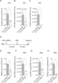
- E Results of qRT-PCR analysis of LIPG mRNA in PHH cells transfected with Cont shRNA and PHH cells transfected with LIPG shRNA. Results were normalized with ACTB results.
- F Results of qPCR analysis of HBV-DNA in PHH cells transfected with Cont shRNA and PHH cells transfected with LIPG shRNA (2 weeks after HBV infection).
- G Results of qPCR analysis of cccDNA in PHH cells transfected with Cont shRNA and PHH cells transfected with LIPG shRNA two weeks after HBV infection.
- H Results of qRT-PCR analysis of pgRNA transcripts in Cont shRNA-introduced PHH cells and LIPG shRNA-introduced PHH cells 2 weeks after HBV infection.
- Results were normalized with ACTB results. These data represent the mean ⁇ SEM of three independent experiments. Statistical testing is by two-tailed unpaired t-test. ****p ⁇ 0.0001, ***p ⁇ 0.001, **p ⁇ 0.01. LIPG positively regulates the HBV life cycle in HepG2-NTCP cells.
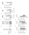
- C Schematic of the experimental design.
- D HepG2-NTCP-C4 cells (Cont) and HepG2-NTCP-C4-LIPG cells (LIPG) in the presence or absence of heparin and heparanase, 3 hours after HBV inoculation at 4°C. qPCR analysis of HBV-DNA.
- E qPCR analysis of HBV-DNA in HepG2-NTCP-C4 and HepG2-NTCP-C4-LIPG cells 12 hours after re-incubation at 37°C.
- F qPCR analysis of HBV-DNA 3 days after re-incubation of HepG2-NTCP-C4 and HepG2-NTCP-C4-LIPG cells at 37°C.
- G qPCR analysis of cccDNA 3 days after re-incubation of HepG2-NTCP-C4 and HepG2-NTCP-C4-LIPG cells at 37°C.
- HBV-DNA was analyzed by qPCR in HepG2-NTCP-C4 and LIPG-overexpressing HepG2-NTCP-C4 cells 3 hours after HBV inoculation at 4°C in the presence of a LIPG inhibitor.
- I qPCR analysis of HBV-DNA 12 h after re-incubation of HepG2-NTCP-C4 and LIPG-overexpressing HepG2-NTCP-C4 cells at 37°C.
- C qPCR analysis of HBV-DNA in HepG2-NTCP-C4 cells transfected with Cont shRNA and HepG2-NTCP-C4 cells transduced with LIPG shRNA after re-incubation at 37°C.
- D qPCR analysis of HBV-DNA in HepG2-NTCP-C4 cells transfected with Cont shRNA and HepG2-NTCP-C4 cells transfected with LIPG shRNA was performed 3 days after re-incubation at 37°C.
- E qPCR analysis of cccDNA after 3 days of reculturing HepG2-NTCP-C4 cells transfected with Cont shRNA and HepG2-NTCP-C4 cells transfected with LIPG shRNA at 37°C.
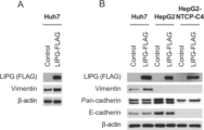
- HBV-infected Huh7 cells Huh7-Control + HBV
- HBV-infected Huh7-LIPG cells Huh7-LIPG-FLAG + HBV
- HBV-non-inoculated RD cells RD
- HBV-inoculated RD cells RD + HBV
- C HBV-DNA of Huh7 cells (Cont) and Huh7-LIPG cells (LIPG) analyzed by qPCR.
- D qPCR analysis of HBV-DNA in Huh7 cells (Cont) and Huh7-LIPG cells (LIPG) 12 h after re-culture at 37°C under Cont siRNA transfection or LIPG siRNA transfection.
- A GLuc activity (multiplicity of infection (MOI) 1.0) in Huh7 cells (Cont) and Huh7-LIPG cells (LIPG) 3 days after HCV infection.
- C Immunoblotting analysis of LIPG, Influenza A H1N1, Influenza A nucleoprotein (NP) and ⁇ -actin in Huh7 cells (Cont) and Huh7-LIPG cells (LIPG) 12 hours after Flu infection (MOI 1.0). These data represent the mean ⁇ SEM of three independent experiments. Statistical tests were performed with unpaired two-tailed t-tests. ****p ⁇ 0.0001, not significantly different (ns). DNA library design for IVV screening.
- the green fluorescent protein GFP was added, following the RNA polymerase promoter SP6 and the 5′ untranslated (UTR) region containing a portion of the tobacco mosaic virus omega sequence as a translation enhancer.
- the peptide portion was a random peptide sequence corresponding to 16 or 9 amino acids.
- Flag-tag, His-tag and Atail were added.
- LIPG and HSPG bind to each other, and that LIPG binds to heparin as a homodimer (Gastroenterology 2014; 147: 48 and PLoS ONE, 2013, 8(3): e55716). It is thought that some of the peptides that bind to the LIPG-HSPG binding region of this portion have activity to inhibit HBV invasion.
- D Effect of LIPH4-NTNBs on HBV-DNA copy number.
- E Effect of LIPH4-NTNBs on cccDNA copy number. Copy number ratios of two LIPG-binding peptides, LIPH4-NTNBsA and LIPH4-NTNBs, at each concentration of HBV-DNA (A) and cccDNA (B) in PXB cells. In the amino acid sequence of LIPH4-NTNBsA, the second cysteine in the amino acid sequence of LIPH4-NTNBs is replaced with alanine.
- A Effect of Netrin-1 on HBV-DNA copy number.
- B Effect of Netrin-1 on cccDNA copy number.
- C Effect of Netrin-1 gene knockdown using shRNA on HBV-DNA copy number.
- D Effect on cccDNA copy number by Netrin-1 gene knockdown using shRNA.
- B After treating PXB cells with 100 nM LIPG-binding peptide LIPH4-23S or LIPH4-NTNBS for 15 hours, purified FLAG-LIPG was added and cultured for 3 hours. After that, preS1 peptide fluorescently labeled with TMR was added, cultured for an additional 30 minutes, and observed using a confocal microscope. Fluorescence of the preS1 peptide was detected and normalized using the fluorescence intensity of DAPI. *p ⁇ 0.05.
- C PXB cells were added with Netrin-1 and cultured for 3 hours, then preS1 peptide fluorescently labeled with TMR was added, cultured for an additional 30 minutes, and observed using a confocal microscope.
- Fluorescence of the preS1 peptide was detected and normalized using the fluorescence intensity of DAPI. *p ⁇ 0.05.
- D PXB cells were added with Netrin-1 and cultured for 3 hours, then FLAG-LIPG was added and cultured for another 3 hours. After that, preS1 peptide fluorescently labeled with TMR was added, cultured for an additional 30 minutes, and observed using a confocal microscope. Fluorescence of the preS1 peptide was detected and normalized using the fluorescence intensity of DAPI. HBV cell entry promoting action by LIPG and HBV cell entry inhibitory action of LIPG-binding peptide and Netrin-1.
- Biotin-labeled heparan sulfate was added to a streptavidin-coated 96-well plate and allowed to stand overnight at 4°C for binding. After washing the plate with TBS, FLAG-LIPG was added to 10, 25, and 50 fmol per well and allowed to react at room temperature for 2 hours. After washing, LIPG-binding peptides LIPH4-NTNBS, LIPH4-23S, LIPH4-NTNBSA, and LIPH4-23SA were added to 10 or 100 nmol per well. TMR was added and further treated at room temperature for 1 hour. After washing these plates, TMR fluorescence was measured with a plate reader.
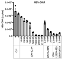
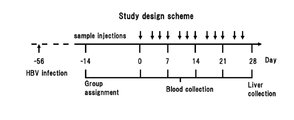
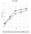
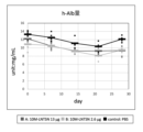
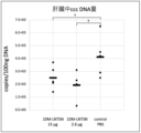
- HBV-DNA copy number in the liver of HBV-infected PXB mice administered with test article (LIPH4-NTNBs).
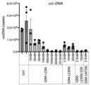
- cccDNA copy number in the liver of HBV-infected PXB mice treated with the test article (LIPH4-NTNBs).
- Histopathological examination (HBsAg immunostaining) of liver tissue from HBV-infected PXB mice administered with the test substance (LIPH4-NTNBs).
- B control (PBS). Histopathological examination (HBcAg immunostaining) of liver tissue from HBV-infected PXB mice administered with the test substance (LIPH4-NTNBs).
- A LIPH4-NTNBs 0.1 mg
- B control (PBS).
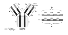
- Antibody structure (Left) Basic structure of IgG antibody. Consists of two H chains and two L chains bound by SS bonds, divided into variable regions (V H , V L ), which are antigen-binding sites, and other constant regions (C H , C L ). ing. The Fc region of the constant region is the Fc receptor binding site. (Right) Complementarity determining regions (CDRs) in the variable region. Other areas are framework areas.
- Single-chain antibody scFv
- the C-terminus of VH chain and the N-terminus of VL are linked via a peptide linker.
- a scheme for creating a mouse single-chain antibody cDNA library Scheme for construction of human single-chain antibody cDNA library. Selection experiments for single-chain antibodies that bind to ASGR by the IVV method. Pull-down assay with ASGR-binding single-chain antibody (1) Pull-down assay with ASGR-binding single-chain antibody (2) Inferred intracellular mechanism of fusion between LIPG-binding peptide and anti-asialoglycoprotein receptor antibody.
- a binding partner of LIPG in cells can be, for example, N4BP1.
- anti-hepatitis B virus includes treatment of HBV infection, prevention of HBV infection, inhibition of HBV proliferation, treatment of hepatitis B, and prevention of hepatitis B. be.
- hepatitis B can be treated by suppressing the proliferation of HBV in the patient's body (inside the liver).
- an anti-HBV agent to an HBV carrier before the onset of hepatitis B, the proliferation of HBV in the carrier can be prevented and the onset of hepatitis B can be prevented (prevention of hepatitis B).
- the anti-HBV agent of the present invention inhibits re-invasion (re-infection) of HBV particles proliferated in the liver cells of patients or HBV carriers after they are extracellularly secreted.
- the proliferation of HBV DNA in patients or carriers is suppressed, and therapeutic effects and preventive effects for hepatitis B can be obtained.
- the anti-HBV agent of the present invention is a substance that binds to LIPG and inhibits the function of LIPG (hereinafter sometimes referred to as LIPG-binding substance.
- LIPG-binding substance When the substance is a polypeptide, it is sometimes referred to as LIPG-binding peptide.) as an active ingredient.
- LIPG-binding substance binds to LIPG on the surface of hepatocytes to inhibit cell adhesion and uptake of HBV, and when delivered into hepatocytes, binds to LIPG present in the cells and inhibits intracellular proliferation of HBV.
- a LIPG-binding substance can be expressed as a substance that inhibits LIPG-mediated intracellular entry of HBV by binding to LIPG.
- the substance is, for example, a substance that binds to the heparin-binding domain of LIPG and inhibits the binding of LIPG, HSPG, or the LIPG-HSPG complex to HBV.
- LIPG has two isoforms, the amino acid sequence of LIPG isoform 1 shown in SEQ ID NO: 30 (NCBI Accession No. NP_006024.1) shows the region of amino acids 312 to 340, and LIPG isoform 2 shown in SEQ ID NO: 138. (NCBI Accession No.
- NP_001294935.1 the region of 238th to 266th amino acids is the heparin-binding region. It has been known that LIPG has the effect of maintaining HBV genomic DNA and cccDNA in host cells (Patent Document 1), but how LIPG functions at which step in the HBV infection process remains unclear. was completely unknown. LIPG, HSPG and HBV particles bind (Fig. 10), and substances that bind to the heparin-binding domain of LIPG can inhibit the binding of LIPG, HSPG, or LIPG-HSPG complexes to HBV, thereby inhibiting the binding of HBV to host cells.
- the present inventors have clarified for the first time that HBV proliferation in host cells (in the body of an HBV-infected patient, particularly in the liver) is suppressed by inhibiting HBV entry.
- Embodiments for inhibiting the binding of LIPG, HSPG, and HBV on the cell surface include inhibition of binding between LIPG and HBV, inhibition of binding between HSPG and HBV, and LIPG- Inhibition of binding between HSPG complex and HBV may be mentioned, and the substance used as an active ingredient in the present application may be any of these aspects.
- Substances that bind to the heparin-binding region of LIPG and inhibit the binding of LIPG, HSPG, or the LIPG-HSPG complex to HBV include, for example, antibodies that recognize and bind to the heparin-binding region of LIPG, or antigen-binding properties thereof. Fragments or aptamers, polypeptides that selectively bind to the heparin-binding region of LIPG, and the like can be mentioned.
- Inhibition of LIPG function by a LIPG-binding substance in hepatocytes may be, for example, at least one selected from inhibition of binding of LIPG to N4BP1 and inhibition of N4BP1 RNase activity suppression by LIPG (described below. See Example 20).
- the present inventors also revealed for the first time that HBV proliferation is suppressed by LIPG inhibition in hepatocytes.
- the anti-HBV agent of the present invention may contain, as an active ingredient, at least one polypeptide (LIPG-binding peptide) selected from the following polypeptides (1) to (30), for example.
- LPG-binding peptide selected from the following polypeptides (1) to (30), for example.
- [] is the name of each polypeptide used in the following examples.
- (1) A polypeptide of the sequence NCRHNTAG (SEQ ID NO:25).
- [LIPH4-NTNBs] (2) A polypeptide of the sequence NARHNTAG (SEQ ID NO:26).
- [LIPH4-NTNBsA] (3) A polypeptide of the sequence LNCRDNTR (SEQ ID NO:23).
- [LIPH4-23s] A polypeptide of the sequence LNARDNTR (SEQ ID NO:24).
- [LIPH4-23SA] (5) A polypeptide of the sequence LNCRDNTRPVMSAMTC (SEQ ID NO: 1).
- [LIPH4-23] (6) A polypeptide of the sequence IRNVNHSDH (SEQ ID NO:9).
- [LIPH4-74] (7) A polypeptide of the sequence LNVGYVFYP (SEQ ID NO: 18).
- [LIPH4-15] (8) A polypeptide of the sequence SLYTGFRAH (SEQ ID NO: 15).
- [LIPH4-2] (9) A polypeptide of the sequence RCRQSWNTM (SEQ ID NO: 14).
- [LIPH4-14] 10 A polypeptide of the sequence GADLRRGCC (SEQ ID NO: 11).
- [LIPH4-87] (11) A polypeptide of the sequence RKSGGVCLNCRHNTAG (SEQ ID NO:21).
- [LIPH4-NTNA] (12) A polypeptide of the sequence LNCRHNTAGRHCHYCK (SEQ ID NO:22).
- [LIPH4-NTNB] (13) A polypeptide having the amino acid sequence shown in SEQ ID NO:28.
- (14) A polypeptide comprising a partial sequence of the polypeptide of (13), which comprises the region of amino acids 370-377 in SEQ ID NO:28.
- SEQ ID NO: 28 represented by an amino acid sequence in which one or more amino acids are substituted, deleted, inserted or added in a region other than the region of amino acids 370 to 377 in the amino acid sequence shown in SEQ ID NO: 28; A polypeptide with 95% or more identity.
- [LIPH4-90] (18) A polypeptide of the sequence CPCVNGATRHRPTSLC (SEQ ID NO:8).
- [LIPH4-16] (19) A polypeptide of the sequence LVPWLRYAY (SEQ ID NO: 17).
- [LIPH4-12] (20) A polypeptide of the sequence NXRHNTAG (SEQ ID NO:75), where X is an amino acid other than cysteine and alanine. (21) A polypeptide of the sequence LNXRDNTR (SEQ ID NO: 76), where X is an amino acid other than cysteine and alanine. (22) A polypeptide of the sequence LNCRHNTAG (SEQ ID NO:77). [LIPH4-LNTNBs] (23) A polypeptide of the sequence SNCRINTFRTVPIEQK (SEQ ID NO:78). [LIPH4-N4BP1a] (24) A polypeptide of the sequence SNCRINTFR (SEQ ID NO:79).
- [LIPH4-N4BP1b] (25) A polypeptide of the sequence TSNCRINTFR (SEQ ID NO:80).
- [LIPH4-N4BP1c] (26) A polypeptide having the amino acid sequence shown in SEQ ID NO:82.
- SEQ ID NO: 82 represented by an amino acid sequence in which one or more amino acids are substituted, deleted, inserted or added in a region other than the region of amino acids 456 to 464 in the amino acid sequence shown in SEQ ID NO: 82; A polypeptide with 95% or more identity.
- (30) A polypeptide having 80% or more and less than 100% identity with any of (1) to (14) and (17) to (27).
- polypeptides (5) to (10) and (17) to (19) were identified as peptides that bind to LIPG by the IVV method in the following examples, and were identified by a pull-down assay using full-length LIPG as a bait. It is a polypeptide that positively confirmed binding to (Fig. 16). All of these polypeptides have the effect of reducing the copy number of HBV DNA and HBV cccDNA in HepG2-NTCP-C4 cells or PXB cells (Fig. 17B-C, Fig. 18A-B). Among these, the polypeptides (5) to (10) are particularly effective in reducing the number of cccDNA copies in HBV-infected hepatic cell lines (Fig. 17C, Fig. 18B).
- (11) and (12) are composed of partial sequences of Netrin-1, and are polypeptides whose anti-HBV activity was confirmed by experiments using HBV-infected PXB cells in Example 12 below (Fig. 18). . Both have an effect of reducing HBV DNA and cccDNA as strongly as the polypeptides (5) to (10), and can be preferably used as active ingredients of anti-HBV agents.
- Polypeptides (1) to (4) are polypeptides designed based on polypeptides (5) and (12), which were particularly effective in reducing HBV DNA and cccDNA copy numbers in HBV-infected hepatic cell lines.
- (1) is a polypeptide composed of the 2nd to 9th residues of the polypeptide of (12), and (3) is the 1st to 8th residues of the polypeptide of (5) , both of which have a very strong effect of reducing the copy number of HBV DNA and cccDNA in HBV-infected hepatic cell lines (Fig. 19).
- (2) is a polypeptide obtained by substituting alanine for the 2nd cysteine in the polypeptide of (1)
- (4) is a polypeptide obtained by substituting alanine for the 3rd cysteine in the polypeptide of (3). Both of these peptides have the same level of strong HBV DNA and cccDNA copy number reducing effects as the original polypeptides (2) and (4) (Fig. 20).
- (22) to (25) are LIPG-binding peptides additionally synthesized for intracellular delivery experiments in Example 14.
- (22) is composed of a partial sequence of Netrin-1, and is a polypeptide having a sequence obtained by extending the polypeptide LIPH4-NTNBs of (1) by one residue to the N-terminal side.
- (23) to (25) are polypeptides composed of a partial sequence of N4BP1 synthesized based on the homology between LIPH4-23 and N4BP1 (NEDD4-binding protein 1). All of them have the effect of reducing the copy numbers of HBV DNA and cccDNA (see Examples 18 and 19), and can be used as anti-HBV agents.
- the polypeptides (20) and (21) are, respectively, a polypeptide obtained by substituting an amino acid other than alanine for cysteine 2 in the polypeptide of (1), and a polypeptide obtained by substituting cysteine 3 in the polypeptide of (3). It is a polypeptide substituted with an amino acid other than alanine. From the data in FIG.
- NCRHNTAG of (1) and LNCRDNTR of (3) do not adversely affect anti-HBV activity even if the cysteine residue is substituted, so the polypeptide of the sequence of NXRHNTAG (SEQ ID NO: 75) and It can be said that the polypeptide of the sequence LNXRDNTR (SEQ ID NO: 76) (where X is an amino acid other than cysteine and alanine) also has the same level of anti-HBV activity as the polypeptides (1)-(4).
- Preferred examples of X include, but are not limited to, glycine, isoleucine, leucine, valine, and the like.
- the polypeptide of (13) is a polypeptide composed of the full-length sequence of Netrin-1.
- the sequences shown in SEQ ID NOs: 27 and 28 are the nucleotide sequence of the coding region in the Netrin-1 gene sequence of NCBI Accession No. NM_004822.3 and the amino acid sequence of Netrin-1 encoded by this (NP_004813.2). . Since full-length Netrin-1 can also reduce the copy numbers of HBV DNA and cccDNA in HBV-infected hepatocytes (FIGS. 21A, B), the polypeptide of (13) is also useful as an anti-HBV agent.
- the polypeptide of (14) is a polypeptide (Netrin- 1 partial polypeptide).
- This region of 370th to 377th amino acids has the same amino acid sequence as the amino acid sequence of (1) (SEQ ID NO: 25).
- SEQ ID NO: 25 The fact that the polypeptide consisting of the amino acid sequence of (1) has anti-HBV activity is fully demonstrated in the following Examples (see FIG. 19 and Example 13). Therefore, as long as the region of 370th to 377th amino acids (amino acid sequence of SEQ ID NO: 25) is included, a polypeptide composed of a fragment (partial sequence) of Netrin-1 can also exhibit anti-HBV activity, and thus anti-HBV agents can be used.
- the polypeptide of (14) includes a polypeptide composed of a fragment of (13) containing the region of amino acids 362 to 377 in the amino acid sequence of SEQ ID NO: 28 (amino acid sequence of SEQ ID NO: 21), and a polypeptide of SEQ ID NO: Polypeptides composed of fragments of (13) containing the region of amino acids 369-384 in the 28 amino acid sequence (amino acid sequence of SEQ ID NO:22) are included.
- the polypeptide of (15) is a polypeptide composed of a mutant Netrin-1 sequence. Specifically, in the amino acid sequence of Netrin-1 shown in SEQ ID NO: 28, one or more amino acids are substituted, deleted, inserted or added in a region other than the region of amino acids 370 to 377. A polypeptide having 95% or more identity to SEQ ID NO:28. As long as it contains this 370th to 377th amino acid region (amino acid sequence of SEQ ID NO: 25), it can exhibit anti-HBV activity even if a small number of amino acid mutations are contained in the region other than this region, so it is useful as an anti-HBV agent. is.
- the polypeptide of (15) contains one or more amino acid substitutions, deletions, insertions or additions in a region other than the 362nd to 377th amino acid region of SEQ ID NO: 28, or 369th to Those contained in regions other than the 384th amino acid region are included.
- the "identity" of amino acid sequences means that the two amino acid sequences to be compared are aligned so that the amino acid residues of the two amino acid sequences are matched as much as possible, and the number of matched amino acid residues is calculated as the total number of amino acid residues. It is expressed as a percentage after being divided by .
- gaps are appropriately inserted in one or both of the two sequences to be compared, if necessary.
- sequence alignment can be performed using well-known programs such as BLAST, FASTA, CLUSTAL W, and the like.
- the above total number of amino acid residues is the number of residues obtained by counting one gap as one amino acid residue. If the total number of amino acid residues counted in this way differs between the two sequences being compared, then the % identity is the total number of amino acid residues in the longer sequence and the number of matching amino acid residues. calculated by dividing
- amino acids with similar side chains have similar chemical properties.
- Grouping amino acids by side chain similarity includes, for example, the group of amino acids with aliphatic side chains (glycine, alanine, valine, leucine, isoleucine), the group of amino acids with aliphatic hydroxyl side chains (serine, threonine).
- the group of amino acids with amide-containing side chains (asparagine, glutamine), the group of amino acids with aromatic side chains (phenylalanine, tyrosine, tryptophan), the group of amino acids with basic side chains (arginine, lysine, histidine), They can be classified into groups of amino acids with acidic side chains (aspartic acid, glutamic acid), groups of amino acids with sulfur-containing side chains (cysteine, methionine), and the like.
- a substitution for another amino acid belonging to the same group is a conservative substitution.
- a typical example of the amino acid sequence of (15) includes, but is not limited to, an amino acid sequence in which a conservative substitution has been introduced in a region other than the 370th to 377th amino acid region (NCRHNTAG) of SEQ ID NO:28. .
- the polypeptide of (16) is a fragment of the polypeptide of (15) composed of a mutant Netrin-1 sequence, wherein the region of amino acids 370 to 377 (amino acid sequence of SEQ ID NO: 25) is A polypeptide (mutant Netrin-1 partial polypeptide) composed of a fragment containing A polypeptide fragment composed of a mutant Netrin-1 sequence can also exhibit anti-HBV activity as long as it includes the region of amino acids 370 to 377 (amino acid sequence of SEQ ID NO: 25), and is therefore useful as an anti-HBV agent. is.
- the polypeptide of (26) is a polypeptide composed of the full-length sequence of N4BP1.
- the sequences shown in SEQ ID NOs: 81 and 82 are the nucleotide sequence of the coding region in the N4BP1 gene sequence of NCBI Accession No. NM_153029.4 and the amino acid sequence of N4BP1 encoded by this (NP_694574.3).
- full-length N4BP1 binds to LIPG, and since the polypeptides (23) to (25), which are N4BP1 partial polypeptides containing a given region, have anti-HBV activity, full-length N4BP1 can also be used as an anti-HBV agent.
- the polypeptide of (27) is a polypeptide (N4BP1 partial polypeptide) composed of a partial sequence of N4BP1, including the region of amino acids 456-464 in the amino acid sequence of N4BP1 shown in SEQ ID NO:82.
- This region of 456th to 464th amino acids is a region common to the polypeptides (23) to (25) composed of the partial sequence of N4BP1, and the polypeptide LIPH4-N4BP1b (SEQ ID NO: 79) of (24) is also the total length of
- the fact that the polypeptide of (24) has anti-HBV activity is as shown in the examples below (Fig. 17), and therefore it is composed of a partial sequence of N4BP1 as long as it includes this region of amino acids 456-464.
- Polypeptides can also exert anti-HBV activity and are useful as anti-HBV agents.
- the polypeptide of (27) includes a polypeptide composed of an N4BP1 partial sequence containing the region of amino acids 455 to 464 in the amino acid sequence of SEQ ID NO: 82 (SEQ ID NO: 80), Polypeptides composed of N4BP partial sequences containing the region from amino acid number 1 to 471 (SEQ ID NO:78) are included.
- the polypeptides (23) to (25) correspond to specific examples of the polypeptide (27).
- the polypeptide of (28) is a polypeptide composed of a mutant N4BP1 sequence. Specifically, in the N4BP1 amino acid sequence shown in SEQ ID NO: 82, one or more amino acids are substituted, deleted, inserted or added in a region other than the region of amino acids 456 to 464. Represented by an amino acid sequence, A polypeptide having 95% or more identity with SEQ ID NO:82. As long as it contains this 456th to 464th amino acid region (amino acid sequence of SEQ ID NO: 79), it can exhibit anti-HBV activity even if a small number of amino acid mutations are contained in other regions, so it is useful as an anti-HBV agent. is.
- the polypeptide of (28) contains one or more amino acid substitutions, deletions, insertions or additions in a region other than the region of amino acids 455 to 464 of SEQ ID NO: 82, or 456 to Those contained in regions other than the 471st amino acid region are included.
- the polypeptide of (29) is a fragment of the polypeptide of (28) composed of the mutant N4BP1 sequence, and is a mutant containing the region of amino acids 456 to 464 (amino acid sequence of SEQ ID NO: 79). It is a polypeptide composed of a partial sequence of the N4BP1 sequence (mutant N4BP1 partial polypeptide).
- a polypeptide fragment composed of the mutant N4BP1 sequence can also exhibit anti-HBV activity as long as it includes the region of amino acids 456 to 464 (amino acid sequence of SEQ ID NO: 79), and is therefore useful as an anti-HBV agent. .
- the polypeptide of (30) has a sequence of 80% or more of the original polypeptide in which some of the residues are modified in any of the polypeptides (1) to (14) and (17) to (27) A polypeptide with identity. It is well known in the art that a very small number of residues can be altered while maintaining equivalent or better activity of the polypeptide.
- residues of polypeptides (1)-(14), (16)-(27) and (29) (20 residues such as (1)-(12), (17)-(25)
- residues such as (1)-(12), (17)-(25)
- 1 to 20 residues, 1 to 15 residues, 1 to 10 residues, or 1 to several residues that have 80% or more identity with the original sequence, such as 85% or more, 90% or more, 95% or more, or 98% or more, are also anti- It can exhibit HBV activity and is useful as an anti-HBV agent.
- Modifications of residues herein are substitutions, deletions, insertions or additions, typically substitutions, particularly conservative substitutions.
- the chain length of the Netrin-1 partial polypeptide of (14), the mutant Netrin-1 partial polypeptide of (16), the N4BP1 partial polypeptide of (27), and the mutant N4BP1 partial polypeptide of (29) is particularly limited. However, for convenience such as synthesis and delivery into hepatocytes, for example, within 100 residues, within 80 residues, within 70 residues, within 60 residues, within 50 residues, within 40 residues, 30 residues The size may be within 25 residues, or within 20 residues.
- polypeptides (1) to (12) and (17) to (25) have short chain lengths, they can be easily prepared by chemical synthesis.
- chemical synthesis methods include the Fmoc method (fluorenylmethyloxycarbonyl method) and the tBoc method (t-butyloxycarbonyl method). Moreover, it can also synthesize
- the (13) polypeptide (Netrin-1) and (26) polypeptide (N4BP1) can be prepared by well-known genetic engineering techniques. Specifically, RNA is extracted from cells expressing Netrin-1 or N4BP1 (e.g., human-derived cell lines), and cDNA encoding Netrin-1 or N4BP1 is prepared by RT-PCR using this RNA as a template.
- the polypeptide of (13) or (26) is prepared by inserting the cDNA into an appropriate expression vector, introducing the polypeptide into an appropriate host cell, producing the polypeptide in the host cell, and extracting and purifying the polypeptide. be able to.
- the primer used for RT-PCR is the nucleotide sequence shown in SEQ ID NO: 27 if it is a primer for Netrin-1 (the sequence of the coding region of NCBI Accession No. NM_004822.3, and the amino acid sequence shown in SEQ ID NO: 28).
- SEQ. can be designed based on Various expression vectors and host cell strains are known, and many are commercially available, so they are easily available.
- the polypeptides (15) and (27) can be prepared by introducing mutations into the cDNA encoding Netrin-1 and the cDNA encoding N4BP1 prepared as described above, respectively. Various kits for mutagenesis are commercially available.
- Polypeptides (14), (16), (27), (29) and (30) with short chain lengths can be easily prepared by chemical synthesis.
- RNA extracted from Netrin-1-expressing cells or N4BP1-expressing cells for chain lengths that are difficult to prepare by chemical synthesis When RT-PCR is used as a template, use primers set inside the coding region (introduce mutations if desired).
- Netrin-1 cDNA or N4BP1 cDNA has been synthesized or cloned, Netrin -1 cDNA or N4BP1 cDNA as a template to amplify fragments by PCR (introduce mutations if desired), etc., to synthesize DNA encoding the desired polypeptide, insert it into an appropriate expression vector, and host It can be prepared by expressing the polypeptide in cells and then extracting and purifying it.
- polypeptides prepared by chemical synthesis are advantageous as pharmaceuticals because they do not contain components derived from host cells. Therefore, among the polypeptides (1) to (30), the polypeptides (1) to (12) and (17) to (25), and the polypeptides (14), (16), (27), (29) ) and (30) with a short size (within several tens of residues) can be preferably used as an active ingredient of an anti-HBV agent.
- polypeptides of (14) and (16) contain the region of 370th to 377th amino acids (amino acid sequence of SEQ ID NO: 25), the size is 9 residues or more, and (27) and (29) ) contains a region of 456th to 464th amino acids (amino acid sequence of SEQ ID NO: 79), and thus has a size of 9 residues or more.
- polypeptides of (14) and (16) those with a size of 8 residues are the same as the polypeptide of (1). Since (11), (12), and (22) are partial polypeptides of Netrin-1 containing the predetermined region, they correspond to specific examples of (14). Since (23) to (25) are partial polypeptides of N4BP1 containing the predetermined region, they correspond to specific examples of (27).
- polyethylene glycol (PEG) chains are added for the purpose of improving the stability of peptides in vivo and increasing the half-life in blood (Clin Nephrol. 2006 Mar;65(3): 180-90. and Proc Natl Acad Sci USA. 2005 Sep 6;102(36):12962-7.), mainly adding sugar chains to the N-terminus or C-terminus (J Am Chem Soc. 2004 Nov 3;126 (43): 14013-22 and Angew Chem Int Ed Engl. 2004 Mar 12;43(12): 1516-20), at least part of the amino acid residues are in the D form (J Pharmacol Exp Ther.
- PEG polyethylene glycol
- the Fc region of the antibody is appropriately modified and added (e.g., J.Immunol., 154( 10), 5590-5600 (1995), Nature, 332, 563-564 (1998), Nature, 332, 738-740 (1998), BioDrugs. 2008;22:11-26, etc.), C-terminal amidation , N-terminal acetylation, etc. are used.
- the polypeptide used as an active ingredient in the anti-HBV agent of the present invention may be one to which such techniques are applied. It is also possible to use polypeptides modified with molecules such as other polypeptides that have the effect of enhancing delivery to the liver as active ingredients.
- polypeptide having the sequence of SEQ ID NO: X refers to a polypeptide having the amino acid sequence shown in SEQ ID NO: X and having a total length of N residues (such a polypeptide is referred to as polypeptide X for convenience), and the Fc region and It includes polypeptides having structures with other functional polypeptides attached, such as polypeptides that have the effect of enhancing delivery to the liver.
- the term "comprising a ⁇ polypeptide having the sequence of SEQ ID NO: X'', ⁇ a polypeptide represented by SEQ ID NO: X'', or a ⁇ polypeptide consisting of SEQ ID NO: X'' as an active ingredient” has a total length of N residues
- an embodiment in which a polypeptide having a structure in which another functional polypeptide is added to the polypeptide X is included as an active ingredient. Any functional polypeptide may be added as long as it does not impair the anti-HBV activity of the polypeptide.
- a polypeptide having a structure to which another functional polypeptide is added can be produced by chemical synthesis if the size of the functional polypeptide is short, or by genetic engineering if the size of the functional polypeptide is long. can.
- the LIPG-binding peptide may have a tag sequence such as a Flag tag or His tag added for convenience of purification and detection.
- a tag sequence can also be regarded as one example of a functional molecule.
- a Flag tag and a His tag are added to the C-terminus of each LIPG-binding peptide.
- addition of such a tag sequence is optional, and it is also possible to prepare the anti-HBV agent of the present invention with a LIPG-binding peptide without a tag sequence added.
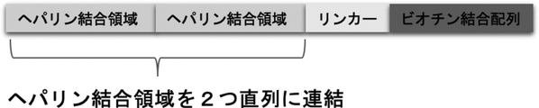
- a functional molecule is a carrier molecule for delivery into hepatocytes.
- the LIPG-binding peptide may be in a form linked to a carrier molecule for delivery into hepatocytes.
- delivery of LIPG-binding peptides into hepatocytes can greatly enhance anti-HBV effects.
- DDS using asialoglycoprotein receptor (ASGR) and ApoE receptor which are abundant in hepatocytes, is known (Yuko Ito, STI Horizon, 2019, Vol.5, No.4, pp.21-25).
- ASGR asialoglycoprotein receptor
- ApoE receptor which are abundant in hepatocytes
- carrier molecules that deliver drugs into hepatocytes using ASGR or ApoE receptors can be preferably used.
- ASGR asialoglycoprotein receptor
- ASGR is a type II single-pass transmembrane protein with the N-terminus directed intracellularly and the C-terminal carbohydrate recognition sites (CRDs) directed extracellularly, as shown in FIG.
- CCDs carbohydrate recognition sites
- ASGR-specific binding molecule used as a drug delivery carrier may bind to the extracellular domain of ASGR.
- a specific binding molecule that can bind to both ASGR1 and ASGR2 can be particularly preferably used, but even a specific binding molecule that binds to only one of them can be used as a drug delivery carrier.
- a specific binding molecule that binds only to either one it may be used alone as a drug delivery carrier, or a combination of a specific binding molecule that binds to one and a specific binding molecule that binds to the other may be used.
- a LIPG-binding peptide linked to an ASGR1-specific binding molecule and a LIPG-binding peptide linked to an ASGR2-specific binding molecule may be prepared, mixed and used as an anti-HBV agent.
- the base sequence of the coding region in the human ASGR1 gene sequence registered under NM_001671.5 in GenBank and the amino acid sequence of ASGR1 encoded by this are shown in SEQ ID NOs: 113 and 114, and the human ASGR2 gene registered under NM_001181.4.
- the nucleotide sequence of the coding region in the sequence and the amino acid sequence of ASGR2 encoded by this are shown in SEQ ID NOs: 115 and 116, respectively.
- the 41st to 61st amino acids of SEQ ID NO: 114 (ASGR1) and the 59th to 79th amino acids of SEQ ID NO: 116 (ASGR2) are transmembrane domains, the N-terminal side of which is the intracellular domain, and the C-terminal side of which is the cell. Foreign domain.
- ASGR extracellular domain includes hetero-oligomerized extracellular domains.
- hetero-oligomer is meant a hetero-oligomerized ASGR extracellular domain.
- Anti-ASGR polyclonal antibodies can be obtained by immunizing a non-human animal using the ASGR extracellular domain as an immunogen, collecting blood, separating the serum, and collecting and purifying the antibody that binds to the ASGR extracellular domain from the serum. can be done.
- Anti-ASGR monoclonal antibodies are produced by collecting antibody-producing cells such as splenocytes and lymphocytes from non-human animals immunized with the ASGR extracellular domain, fusing them with myeloma cells to prepare hybridomas, and binding to the ASGR extracellular domain.
- Anti-ASGR monoclonal antibodies can be obtained from culture supernatants by selecting hybridomas that produce antibodies against the anti-ASGR and proliferating them.
- An anti-ASGR antibody fragment can be obtained by treating an anti-ASGR antibody with a proteolytic enzyme such as papain or pepsin.
- a proteolytic enzyme such as papain or pepsin.
- the definition of the antibody fragment is as described above, and includes antibody fragments such as Fab, Fab', F(ab') 2 that maintain the binding ability to the antigen (ASGR extracellular domain).
- cDNA is prepared by extracting the mRNA of the hybridoma produced as described above, and PCR is performed using primers specific for the immunoglobulin H chain and L chain to obtain the immunoglobulin H chain gene and L chain.
- PCR is performed using primers specific for the immunoglobulin H chain and L chain to obtain the immunoglobulin H chain gene and L chain.
- anti-ASGR scFv can be obtained from scFv libraries by techniques such as the phage display method and IVV method.
- a phage library with scFv displayed on the phage surface is prepared as an scFv library.
- a naive scFv phage library may be prepared by the following procedure. mRNA is extracted from antibody-producing cells such as splenocytes and lymphocytes collected from healthy humans or non-human animals, cDNA is synthesized by reverse transcription reaction, and cDNA encoding the region containing the heavy chain variable region (VH) (VH cDNA) and cDNA encoding a region containing the light chain variable region (VL) (VL cDNA) are comprehensively amplified by PCR.
- VH heavy chain variable region
- VL light chain variable region
- the amplified VH cDNA and VL cDNA are randomly ligated via a suitable linker (for example, a linker consisting of three repeated GGGGS units, etc.) by standard assembly PCR or Fusion PCR to obtain scFv-encoding cDNA.
- a suitable linker for example, a linker consisting of three repeated GGGGS units, etc.
- Fusion PCR Fusion PCR
- Plasmid vectors for phage include all phage genes necessary for forming phage particles and capable of forming phage particles independently, and phage vectors containing the g3p gene but no other phage protein genes and containing phage Although there are two types of phagemid vectors that require helper phage for particle formation, phagemid vectors are preferably used.
- a scFv cDNA library prepared using a phagemid vector is introduced into Escherichia coli, and the E. coli is superinfected with a helper phage to package each vector of the scFv cDNA library, and the scFv expressed by the vector is placed on the surface. Libraries of displaying phage can be generated.
- a phage library of mutant scFv may be prepared by randomly introducing mutations into the prepared VH cDNA and VL cDNA or scFv cDNA.
- phages displaying scFv that bind to the ASGR extracellular domain are selected (panning).
- This panning step can be performed using a solid-phase carrier (chip, plate, magnetic bead, etc.) on which the ASGR extracellular domain is immobilized, and a carrier on which a hetero-oligomer is immobilized is particularly preferably used.
- the extracellular domain of ASGR1 (ASGR1ex) and the extracellular domain of ASGR2 (ASGR2ex) are biotinylated, and by contacting the biotinylated ASGR1ex and ASGR2ex with a carrier coated with avidins, ASGR1ex and ASGR2ex are hetero-oligomerized on the carrier surface.
- the phage library is brought into contact with the ASGR extracellular domain-immobilized carrier, and after washing, the phages bound on the carrier are recovered.
- the recovered phages are lysed, the packaged vector is recovered, introduced again into E. coli, and superinfected with helper phages to form phage particles again.
- These phage particles are again brought into contact with the ASGR extracellular domain-immobilized carriers.
- Phages enriched through multiple rounds of panning can be obtained as anti-ASGR scFv candidate clones, but clone selection may be performed to further narrow down the candidates.
- the scFv expression vector is collected from the phage after concentration and introduced into an appropriate host cell such as E. coli to prepare host cell clones expressing scFv. and select clones with high specific binding to the ASGR extracellular domain. Reactivity can be confirmed by an immunoassay such as ELISA using the ASGR extracellular domain, preferably a hetero-oligomer, as an antigen.
- clones may be grouped by confirming duplication of clones based on the base sequences of VH and VL. Through these clone selection activities, it is possible to select clones with high specific binding to the ASGR extracellular domain and further narrow down the candidates.
- the scFv expression vector is recovered from the candidate clone, the scFv cDNA is amplified from the scFv expression vector, incorporated into an appropriate plasmid expression vector to express the scFv in an appropriate host cell, recovered and purified.
- the reactivity with ASGR1ex and ASGR2ex or the reactivity with heterooligomers is finally confirmed for the scFv after purification, and scFv that specifically binds to the ASGR extracellular domain can be obtained.
- IVV method (Nemoto, N. et al., FEBS Lett., 414:405-408, 1997; Miyamoto-Sato, E. et al., Nucleic Acids Res., 28:1176-1182, 2000; WO 03/106675 A1) is a technique developed by Yanagawa, one of the inventors of the present application, and his collaborators.
- puromycin a type of antibiotic
- PEG polyethylene glycol
- An mRNA-protein junction molecule (in vitro virus; IVV) is formed covalently linked to the mRNA molecule via puromycin. From the IVV library constructed in this way, IVVs containing proteins that bind to baits (proteins, peptides, antigens, etc.) are picked in vitro, and the genes (mRNA) linked to them are amplified by reverse transcription PCR. Then, by deciphering the base sequence with a DNA sequencer, interacting proteins, peptides and antibodies can be easily identified. Competitive elution with free bait is the common method for eluting and recovering IVV bound to bait.
- IVV in vitro virus
- the spacer can be cleaved by UV irradiation at 365 nm, and the mRNA portion can be eluted and recovered (Doi, N. et al., J. Biotechnol., 131:231-239, 2007).
- mRNA is synthesized from the cDNA library by reverse transcription reaction, puromycin is bound to the 3' end of the mRNA via a PEG spacer, and scFv mRNA-puromycin library.
- a cell-free translation reaction is carried out to construct a library of mRNA-scFv linking molecules (IVV) in which scFv molecules and mRNA molecules encoding them are covalently bound via puromycin.
- a scFv that binds to the ASGR extracellular domain is selected from the scFv IVV library.
- this selection step can also be performed using a solid-phase carrier (chip, plate, magnetic beads, etc.) on which ASGR extracellular domains, preferably hetero-oligomers, are immobilized as bait.
- the scFv IVV library is brought into contact with the ASGR extracellular domain-immobilized carrier, and after washing, the IVV bound to the carrier is eluted and collected. IVV can be eluted and recovered by competitive elution using ASGR1ex or ASGR2ex.
- Reverse transcription PCR is performed using the recovered IVV or mRNA part as a template to prepare a scFv cDNA library after the 1st round of selection.
- An IVV library can be prepared again from this cDNA library and a second round of selection can be performed. It is preferable to perform multiple rounds of selection while sequentially increasing the selection pressure (contact time between IVV library and bait, amount of bait immobilized on carrier, etc.).
- This vector is introduced into a suitable host cell such as E. coli to obtain a library of scFv clones after selection.
- the obtained clone library is subjected to evaluation of reactivity to the ASGR extracellular domain, analysis of the variable region sequence, and grouping. and final confirmation of reactivity with ASGR1ex and ASGR2ex or reactivity with hetero-oligomers, scFv with high specific binding to the extracellular domain of ASGR can be obtained.
- anti-ASGR antibody, antibody fragment or scFv are a heavy chain CDR1 comprising the amino acid sequence shown in SEQ ID NO: 83, 89, 95 or 101; a heavy chain CDR2 comprising the amino acid sequence shown in SEQ ID NO: 84, 90, 96 or 102; a heavy chain CDR3 comprising the amino acid sequence shown in SEQ ID NO: 85, 91, 97 or 103; a light chain CDR1 comprising the amino acid sequence shown in SEQ ID NO: 86, 92, 98 or 104; a light chain CDR2 comprising the amino acid sequence shown in SEQ ID NO: 87, 93, 99 or 105; An antibody, antibody fragment or scFv having a light chain CDR3 comprising the amino acid sequence shown in SEQ ID NO: 88, 94, 100 or 106 can be mentioned.
- CDR sequences are based on the CDR sequences of the anti-ASGR scFv obtained in the Examples below.
- anti-ASGR scFv having heavy chain CDRs 1-3 of the amino acid sequences shown in SEQ ID NOS: 83-85 and light chain CDRs 1-3 of the amino acid sequences shown in SEQ ID NOS: 86-88, amino acids shown in SEQ ID NOS: 89-91 an anti-ASGR scFv having the heavy chain CDRs 1-3 of the sequences and the light chain CDRs 1-3 of the amino acid sequences shown in SEQ ID NOs: 92-94, the heavy chain CDRs 1-3 of the amino acid sequences shown in SEQ ID NOs: 95-97 and SEQ ID NOs: 98-100 and anti-ASGR scFv with heavy chain CDRs 1-3 shown in SEQ ID NOs: 101-103 and light chain CDRs 1-3 shown in SEQ ID NOs: 104-106.
- the CDR sequence is not limited to the specific amino acid sequence described above, and a CDR containing an amino acid sequence having 80% or more identity with the original amino acid sequence by substituting a part of the bases in the above amino acid sequence. It may be an antibody or the like having For example, heavy and light chain CDR1 allows substitution of 1 residue, heavy and light chain CDR2 allows substitution of 1-3, 1-2 or 1 residue, Heavy and light chain CDR3s tolerate substitutions of 1-2 or 1 residue.
- An antibody, antibody fragment or scFv having such modified CDRs can also be used as an antibody, antibody fragment or scFv that specifically binds to ASGR.
- Antibodies and the like having CDRs of a given sequence encode the amino acid sequences described above by, for example, introducing mutations into the CDR regions of the VH and VL genes of any cloned antibody or the scFv gene encoding any scFv. It can be easily prepared by modifying as follows. Any antibody and any scFv as a base for introducing the above CDR sequence may be an anti-ASGR antibody and anti-ASGR scFv having a CDR sequence different from the above, or an antibody and scFv against other antigens There may be.
- the anti-ASGR antibody, antibody fragment or scFv having CDRs or modified CDRs containing the specific amino acid sequence described above can be used not only as a carrier for delivery of the anti-HVB agent of the present invention, but also as various drugs to be delivered into hepatocytes. It is an excellent delivery carrier for This anti-ASGR antibody, antibody fragment, or scFv can be used as a carrier for drug delivery into hepatocytes, and a drug to be delivered into hepatocytes can be complexed with the carrier by techniques known in the pharmaceutical field and used as a pharmaceutical composition. .
- the anti-ASGR antibody may be a human antibody, a humanized antibody, a human-non-human animal chimeric antibody, or a non-human animal antibody.
- anti-ASGR scFv antibodies may be derived from human antibodies, humanized antibodies, chimeric antibodies between human and non-human animals, or non-human animal antibodies.
- those derived from human antibodies, humanized antibodies or chimeric antibodies, particularly those derived from human antibodies or humanized antibodies, particularly those derived from human antibodies are preferred.
- carrier molecule for intrahepatocyte delivery such as an anti-ASGR antibody to a LIPG-binding peptide
- it may be linked to either end of the LIPG-binding peptide. If one or more other functional molecules are also used, they may be linked to the LIPG-binding peptide so as to function properly in conjunction with those other functional molecules.
- the carrier molecule for intrahepatic delivery is preferably linked to the LIPG-binding peptide via a cleavage sequence that is cleaved by endogenous enzymes present in the cells of patients receiving the anti-HBV agent of the present invention.
- An example of a cleavage sequence is the sequence RVRR (SEQ ID NO: 111) that Furin recognizes and cleaves, but the cleavage sequence is not limited to this.
- a functional molecule is a cell membrane permeation promoting molecule. That is, the LIPG-binding peptide may be in a form linked to a cell membrane permeabilization molecule.
- a cell membrane permeation-enhancing molecule is a molecule that has the effect of promoting the LIPG-binding peptide taken up into cells by endocytosis to permeate the membrane from within the endosome and be released into the cytoplasm.
- S19 Sudo, K. et al., J. Control. Release, 255 : 1-11, 2017, Sudo, K. et al., J. Control.
- 28-residue syntisin 1 partial peptide S28 (PFVIGAGVLGALGTGIGGITTSTQFYYK, SEQ ID NO: 107) and 39-residue syntisin 1 partial peptide S39 (PFVIGAGVLGALGTGIGGITTSTQFYYKLSQELNGDMER, SEQ ID NO: 108), which were developed as peptides that can function more efficiently.
- S28 and S39 are cell membrane permeation promoting molecules that can be preferably used in the present invention, but usable cell membrane permeation promoting molecules are not limited to these.
- the cell membrane permeation promoting molecule may be linked to either end of the LIPG-binding peptide. or to the end opposite to the carrier molecule.
- a nuclear localization signal can be mentioned as a further example of a functional molecule. That is, the LIPG-binding peptide may be in a form linked to a nuclear localization signal.
- Various nuclear localization signals are known, and any of them may be used.
- PAAKRVKLD SEQ ID NO: 110
- the nuclear localization signal may be ligated to either end of the LIPG-binding peptide. , or at the opposite end to the carrier molecule. When the cell membrane permeabilization molecule and the nuclear localization signal are linked to the LIPG-binding peptide, both ends may be linked individually, or both may be linked to one end.
- the delivery carrier + cleavage sequence, cell membrane permeabilization molecule and nuclear localization signal are linked to the LIPG-binding peptide, the delivery carrier + cleavage sequence is at one end and the cell membrane permeabilization molecule and nuclear localization signal are at the other end. Link.
- HBV-infected patients including hepatitis B patients and HBV-infected patients who have not developed hepatitis B (HBV carriers).
- Patients are typically, but not limited to, mammals, particularly humans.
- the dosage of the anti-HBV agent of the present invention may be any amount that provides an anti-HBV effect in the patient to whom it is administered.
- An effective dose can be appropriately selected according to the patient's symptoms, viral load, age, body weight and the like.
- the dosage of the anti-HBV agent of the present invention may be about 1 ⁇ g to 10000 mg, for example about 100 ⁇ g to 1000 mg per 1 kg body weight of the active ingredient per day for the subject.
- the amount of active ingredient here means the amount of the polypeptide portion consisting of the amino acid sequence of (1) to (30) when the polypeptide of (1) to (30) is used as an active ingredient.
- the amount does not include the amount of the other functional polypeptide.
- the amount of active ingredient is the total amount.
- the daily dose may be administered once or divided into several doses. Administration may be daily or every few days.
- the administration route of the anti-HBV agent of the present invention may be either oral administration or parenteral administration, but parenteral administration such as intramuscular administration, subcutaneous administration, intravenous administration, and intraarterial administration is generally preferred.
- the active ingredient of the anti-HBV agent of the present invention contains pharmaceutically acceptable carriers, diluents, excipients, binders, lubricants, disintegrants, sweeteners, suspending agents suitable for each administration route. , an emulsifier, a coloring agent, a corrigent, a stabilizer, and the like.
- Formulations include oral agents such as tablets, capsules, granules, powders and syrups, and parenteral agents such as inhalants, injections, suppositories and liquid agents.
- Formulation methods and usable excipients are well known in the field of pharmaceutical formulations, and any method and excipients can be used.
- the anti-HBV agent containing two or more of the polypeptides (1) to (30) as active ingredients may be a combination drug containing all of the two or more polypeptides in the same formulation, It may also include single agent combinations containing each polypeptide alone. In embodiments involving a combination of single agents, the single agents are usually administered simultaneously or sequentially, although each single agent may be administered at appropriate intervals.
- Example 1 Knockdown of LIPG expression negatively regulates HBV infection.
- the effect of knockdown of LIPG expression on HBV infection was investigated.
- Cells, viruses, reagents, etc. used in the experiments of Examples 1-4 are as follows.
- HC1 cells derived from human hepatocellular carcinoma
- HepG2 cells derived from human hepatocellular carcinoma
- HepG2-NTCP-C4 cells HepG2 cells overexpressing the HBV receptor NTCP, Iwamoto, M. et al. Biochem. Biophys. Res. Commun.
- HepG2.2.15 cells HepG with HBV genome integrated
- Huh1 cells derived from human hepatocellular carcinoma
- Huh6 cells human hepatoblasts.
- Huh7 cells derived from human hepatocellular carcinoma
- Huh7.5 cells derived from Huh7
- T5B cells derived from normal human hepatocytes
- RD cells derived from human rhabdomyosarcoma
- bovine DMEM Thermo Fisher Scientific
- fetal serum Thermo Fisher Scientific, Waltham, MA
- 1% L-glutamine Thermo Fisher Scientific
- penicillin/streptomycin Thermo Fisher Scientific
- PHH cells primary human hepatocytes
- PhoenixBio Hiroshima, Japan
- Heparin and heparanase were purchased from R&D systems (Minneapolis, Minn.).
- LIPG inhibitor (GSK-264220A) was purchased from Tocris Bioscience (Minneapolis).
- Chlorpromazine (CPZ) and 5-(N-ethyl-N-isopropyl) amiloride (EIPA) were purchased from Sigma-Aldrich (St. Louis, Mo.).
- DYKDDDDK FLAG (#14793) (SEQ ID NO:74), Caveolin-1 (#3238), Vimentin (#5741), Pan-cadherin (#4068), E-cadherin (#3195) and ⁇ -actin (#4970) ) were from Cell Signaling Technology (Beverly, Mass.), NTCP (ab131084) and LIPG (ab24447) were from abcam (Cambridge, UK).
- HBV used in PHH (primary human hepatocyte) infection experiments was purchased from PhoenixBio. HepG2.2.15 cell-derived HBV was used for infection experiments on cultured cells other than PHH cells.
- HCV Myocarditis virus
- Dr. Mitsutoshi Yoneyama Medical Mycology Research Center, Chiba University, Japan
- the influenza virus A/WSN/33 strain was donated by Dr. Masayoshi Enami (Department of Molecular Genetics, Kanazawa University).
- LIPG-targeting shRNA target sequence: 5′-TTACACGGATGCG-GTCAATAA-3′, SEQ ID NO: 73
- cloned into the lentiviral-based pLKO.1-puro expression vector used for knockdown of LIPG was purchased from Sigma. .
- An empty vector was used for control shRNA (Cont shRNA).
- Lentiviral particles were generated by co-transfecting packaging plasmids into packaging cells (293FT cells). Constitutively LIPG knockdown cells were selected using the puromycin selectable marker.
- HBV-DNA and cccDNA by qPCR were performed as follows. Total DNA was extracted from cells using the DNeasy Blood & Tissue Kit (Qiagen, Hilden, Germany). HBV-DNA was quantified by qPCR analysis as previously reported (Honda, M. et al. J. Infect. Dis., 213, 1096-1106, 2016). Extracted DNA (50 ng) was treated with 10 U Plasmid-safe DNase I (Epicentre, Madison, WI) for 60 min at 37°C followed by 30 min at 70°C for DNase inactivation. cccDNA was quantified by qPCR analysis as previously reported (Honda, M. et al. J. Infect. Dis., 213, 1096-1106, 2016).
- Quantitative RT-PCR was performed as follows. Total RNA was extracted using the GenEluteTM Mammalian Total RNA Miniprep Kit (Sigma-Aldrich), and cDNA was synthesized using the High Capacity cDNA reverse transcription kit (Applied Biosystems, Carlsbad, Calif.). 7500 Real Time PCR System (Applied Biosystems) was used for RT-PCR. 5'-GCTCTGTATCGGGAGGCCTTA-3' (SEQ ID NO: 31) and 5'-TGAGTGCTGTATGGTGAGGAGAA-3' (SEQ ID NO: 32) as primers for pregenomic RNA, and 5'-FAM-AGTCTCCGGAACATT-MGB-3' (SEQ ID NO: 33) as a probe. Using. Primer pairs and probes for LIPG, NTCP, 18S rRNA, and ACTB were obtained from the TaqMan assay reagents library.
- SDS-PAGE and immunoblotting were performed as follows. Cells were washed with phosphate-buffered saline (PBS) and lysed with RIPA Lysis Buffer (EMD Millipore, Burlington, Mass.) containing complete Protease Inhibitor Cocktail (Roche Applied Science). The membrane was blocked with Blocking One solution (Nacalai Tesque, Kyoto). Western blotting was performed by standard methods. ChemiDoc Imaging Systems (Bio-Rad, Hercules, CA) was used for detection.
- HC1 cells established from human hepatocellular carcinoma were infected with a lentivirus expressing shRNA against the LIPG gene (LIPG shRNA) to generate LIPG constitutive knockdown cells.
- LIPG shRNA lentivirus expressing shRNA against the LIPG gene
- LIPG positively regulates HBV infection in HepG2-NTCP cells.
- HBV infection we investigated the relationship between the expression level of LIPG and HBV infection.
- PCR was performed using forward primer 5'-TTAGAATTCATGAGCAACTCCGTTC-3' (with EcoRI recognition site, SEQ ID NO: 34) and reverse primer 5'-AATTCTAGATCATCTGCTCATCTGTTGTTGTGGGAAGCTCCACAGTG-3' (with XbaI recognition site, SEQ ID NO: 35).
- Amplification generated a lentiviral transfer plasmid encoding the LIPG gene (SEQ ID NO:29).
- the PCR product was digested with EcoRI and XbaI and ligated into similarly digested pLVSIN-CMV Pur Vector (Takara Bio, Otsu, Japan) to obtain pLVSIN-CMV-Flag tagged LIPG Vector.
- pLVSIN-CMV Pur Vector or pLVSIN-CMV-Flag-tagged LIPG Vector was transfected into Lenti-X 293T cells (Takara Bio) using FuGENE-HD transfection reagent (Promega, Madison, WI) with Lentiviral High Titer Packaging Mix ( Takara Bio) to generate virus particles.
- the supernatant collected after 72 hours was filtered with a 0.22 ⁇ m syringe filter and introduced into the cells. Seventy-two hours post-transduction, these cells were treated with puromycin (5 mg/mL) for antibiotic selection. To avoid clonal bias, experiments used antibiotic-resistant bulk cell populations.
- HC1 cells KM
- PHH cells PHH cells
- HepG2-NTCP-C4 cells showed similar levels of LIPG expression (Fig. 2A).
- HepG2-NTCP-C4 cells overexpressing FLAG-LIPG HepG2-NTCP-C4-LIPG cells
- Fig. 2B As control compounds, heparin, which has been reported to inhibit HBV adhesion (Schulze, A. et al.
- Hepatology 46:1759-1768, 2007 cleaves heparan sulfate side chains of heparan sulfate proteoglycans (HSPGs) Heparanase, an endo- ⁇ -D-glucuronidase, was used (Bame, KJ. Glycobiology, 11:91R-98R, 2001).
- HBV was treated at 4°C for 3 hours to adhere to the cells and washed off. Uptake was detected after further incubation at 37°C for 12 hours (Fig. 2C).
- HBV adhesion was detected by qPCR of HBV-DNA 3 hours after HBV inoculation at 4°C in the presence or absence of compounds (heparin, heparanase).
- HBV adhesion Fig. 2D
- uptake Fig. 2E
- HBV DNA and cccDNA replication Figs. 2F and G
- Huh7 cells, HepG2 cells, and HepG2-NTCP-C4 cells that constitutively overexpress FLAG-LIPG were established using a lentiviral expression system (Fig. 5A). When these cells were infected with HBV and cccDNA levels were measured 24 or 72 hours later, Huh7 cells maintained HBV replication by expressing FLAG-LIPG (Fig. 5B), whereas HepG2 cells overexpressed LIPG. had no effect on HBV replication (Fig. 5C). HepG2-NTCP-C4 cells maintained HBV replication in controls, which was further enhanced by LIPG overexpression (Fig. 5D). At this time, when the NTCP expression level of each cell was examined, Huh7 cells and Huh7-LIPG cells were much lower than HepG2-NTCP-C4 cells (Fig. 5E).
- NTCP in Huh7 cells was knocked out using CRISPR-Cas9 gene editing technology.
- the NTCP KO cell line was established as follows. Production of sgRNA CRISPR/Cas9 lentiviruses was performed using FuGENE-HD transfection reagents with sgRNA expression vectors targeting NTCP, as well as non-targeting controls (abm, Richmond, Canada) and a third-generation packaging system mix (abm ) into Lenti-X 293T cells. The supernatant collected after 72 hours was filtered with a 0.22 ⁇ m syringe filter and then introduced into Huh7 cells. Seventy-two hours after transduction, cells were treated with puromycin (5 mg/mL) for antibiotic selection. To avoid clonal bias, experiments used antibiotic-resistant bulk cell populations.
- NTCP was detected in Huh7 cells transfected with non-targeting single guide RNA (sgRNA) with a scrambled sequence, but not in NTCP-KO cells transfected with NTCP-specific sgRNA (Fig. 5F).
- LIPG was forced to express in these cells, and adhesion and uptake assays were performed as described above.
- overexpression of LIPG reduced HBV cell adhesion (Fig. 5G), uptake (Fig. 5H), and replication (HBV-DNA, cccDNA, pgRNA transcripts) (Fig. 5H).
- Figure 5I, J, K increased to the same level.
- LIPG contributes to caveolae-dependent HBV uptake.
- LIPG contributes to caveolae-dependent HBV uptake.
- Fig. 6A Overexpression of LIPG in Huh7 cells promoted the expression of Caveolin-1 protein (Fig. 6A). Therefore, we knocked down Caveolin-1 using Caveolin-1-specific siRNA and examined how LIPG-mediated increases in HBV adhesion, uptake and replication were affected.
- Caveolin-1-specific siRNA and Negative Control siRNA were obtained from Sigma-Aldrich. Lipofectamine RNAiMAX Transfection Reagent (Invitrogen) was used for siRNA transfection.
- Caveolin-1 protein expression was detected in Huh7-Cont and Huh7-LIPG cells transfected with control siRNA, but was significantly reduced in these cells knocked down Caveolin-1 (Fig. 6B). Knockdown of Caveolin-1 suppressed the increase in HBV adhesion by LIPG overexpression (Fig. 6C) and blocked the increase in uptake and replication (cccDNA) to the same level as controls (Fig. 6D and E). Furthermore, both the clathrin endocytosis inhibitor chlorpromazine (CPZ) and the macropinocytosis inhibitor 5-(N-ethyl-N-isopropyl)amyloride (EIPA) inhibited LIPG in Huh7-LIPG cells. The LIPG inhibitor decreased HBV replication in Huh7-LIPG cells (Fig. 6H), whereas it had no effect on HBV-mediated increased replication of HBV (Fig. 6F and G).
- CPZ chlorpromazine
- EIPA macropinocytosis inhibitor 5-(N-ethyl-
- LIPG does not affect clathrin-dependent viral endocytosis.
- HCV hepatitis C virus
- EMCV myocarditis virus
- Flu influenza virus
- LIPG is a factor that plays an important role in HBV adhesion and uptake, and functions as a cofactor that supports HBV intracellular entry independently of NTCP. That is, it is suggested that HBV binds to the LIPG-HSPG complex on the cell membrane surface and is taken up by hepatocytes by caveolin-dependent endocytosis. This pathway is consistent with the action of LIPG in HDL uptake into cells (Gerold, G. Cold Spring Harb. Perspect Med. 10, a036830, 2020), and HBV uses this pathway to It is thought that it penetrates into the cell at
- FIG. 9 shows the design of a DNA library for IVV screening.
- puromycin a type of antibiotic
- PEG polyethylene glycol
- IVVs containing proteins that bind to baits are picked in vitro, and the linked genes (mRNA) are analyzed by reverse transcription and PCR.
- mRNA linked genes
- PCR was carried out by reacting at 94°C for 5 minutes, followed by 16 cycles of 94°C for 30 seconds, 58°C for 30 seconds, and 68°C for 2 minutes, followed by reaction at 68°C for 5 minutes.
- the cDNA library was purified with Wizard SV Gel PCR Clean-Up System (Promega) and recovered as 50 ⁇ l of GSP6-GFP-DNA solution.
- PCR was carried out by reacting at 94°C for 5 minutes, followed by 16 cycles of 94°C for 30 seconds, 58°C for 30 seconds, and 68°C for 2 minutes, followed by reaction at 68°C for 5 minutes.
- the cDNA library was purified with Wizard SV Gel PCR Clean-Up System (Promega) and recovered as 50 ⁇ l of Flag-His Atail-DNA solution.
- Atail-DNA solution 1 ⁇ l, 10 ⁇ KOD plus buffer (TOYOBO) 10 ⁇ l, 2 mM dNTPs (TOYOBO) 10 ⁇ l, 25 mM MgSO4 4 ⁇ l, forward primer: 16NNS-F or 9NNS-F (10 pmol/ ⁇ l) 3 ⁇ l, Reverse primer: Atail (R) (10 pmol/ ⁇ l) 3 ⁇ l and KOD plus polymerase (TOYOBO) 2 ⁇ l, add RNase-free water to make the total volume 100 ⁇ l, put this in 1 tube, total 300 ⁇ l (Tube 3 book) was subjected to a PCR reaction.
- PCR was carried out by reacting at 94°C for 5 minutes, followed by 12 cycles of 94°C for 30 seconds, 58°C for 30 seconds, and 68°C for 2 minutes, followed by reaction at 68°C for 5 minutes.
- the cDNA library was purified with Wizard SV Gel PCR Clean-Up System (Promega) and recovered as 50 ⁇ l of 16NNS Atail-DNA solution or 9NNS Atail-DNA solution.
- cDNA library was purified with Wizard SV Gel PCR Clean-Up System (Promega) and recovered as 50 ⁇ l of cDNA library 16NNSLib and 9NNSLib.
- Example 6 Preparation of IVV library (6-1) Transcription of library 2 pmol each of cDNA libraries 16NNSLib and 9NNSLib, 8 ⁇ l of 5 ⁇ SP6 buffer, 2 ⁇ l of ATP (100 mM), 2 ⁇ l of CTP (100 mM), UTP (100 mM) 2 ⁇ l, GTP (10 mM) 4 ⁇ l, cap analog (m7G(5')PPP(5')G) (Thermo Fisher Scientific) (40 mM) 5 ⁇ l, Enzyme Mix SP6 RNA polymerase (Promega) 4 ⁇ l, RNase-Free water.
- RPE buffer Qiagen
- Example 7 Preparation of biotinylated LIPG
- Fig. 10 shows a schematic diagram of the binding of endothelial lipase LIPG, heparan sulfate proteoglycan, and HBV virus.
- FIG. 11 shows the design of biotinylated LIPG as a bait.
- HBV binds to the LIPG-HSPG complex on the cell membrane surface and is taken up by hepatocytes by caveolin-dependent endocytosis, whereas LIPG binds to heparin as a homodimer.
- biotinylated LIPG has two heparin-binding regions (RKNRCNSIGYNAKKMRNKRNSKMYLKTRA, SEQ ID NO: 42) of LIPG (SEQ ID NO: 30). It has a structure in which two are connected in series.
- LIPG-BioFLAGHis-pcDNA vector was constructed by adding a biotinylation sequence, a Flag-tag and a His-tag sequence to the full-length LIPG gene.
- LIPG expression vector pCMV6-LIPG (1 ng/ ⁇ l) 0.5 ⁇ l, KAPA HiFi HS RM 12.5 ⁇ l, 10 ⁇ M LIPG-ifpc-F 0.75 ⁇ l, and 10 ⁇ M LIPG-ifbio-R 0.75 ⁇ l
- the total volume was adjusted to 25 ⁇ l, and PCR reaction was performed. PCR was carried out at 95°C for 5 minutes, followed by 25 cycles of 98°C for 20 seconds, 60°C for 15 seconds, and 72°C for 1 minute, followed by reaction at 72°C for 1 minute.
- the PCR product was purified with Wizard SV Gel PCR Clean-Up System (Promega) and collected as 30 ⁇ l of DNA solution to obtain LIPG-ifpcB.
- Bio-tag (Nucleic Acids Research, 2009 , Vol. 37, No. 8, page e64) was added with Flag-tag and His-tag.
- Bio-tag 2.37 ⁇ l, 10 ⁇ KOD plus buffer (TOYOBO) 40 ⁇ l, 2 mM dNTPs (TOYOBO) 40 ⁇ l, 25 mM MgSO 4 16 ⁇ l, F-Bio (10 pmol/ ⁇ l) 12 ⁇ l, Bio-Flag-Histag A stop (10 pmol/ ⁇ l) and 8 ⁇ l of KOD plus polymerase (TOYOBO), RNase-free water was added to make the total volume 200 ⁇ l, and PCR reaction was performed.
- PCR was carried out by reacting at 94°C for 5 minutes, followed by 20 or 25 cycles of 94°C for 30 seconds, 58°C for 30 seconds, and 68°C for 2 minutes, followed by reaction at 68°C for 5 minutes. After confirming a DNA band (264 bp) by agarose gel electrophoresis, the PCR product was purified by Wizard SV Gel PCR Clean-Up System (Promega) and recovered as 50 ⁇ l of DNA solution to obtain Bio-Flag-His.
- PCR was carried out at 94°C for 5 minutes, followed by 12 cycles of 94°C for 30 seconds, 58°C for 30 seconds, and 68°C for 2 minutes, followed by reaction at 68°C for 5 minutes.
- the cDNA library was purified with Wizard SV Gel PCR Clean-Up System (Promega) and recovered as 50 ⁇ l of DNA solution to obtain LIPG-BioFlagHis.
- LIPG-BioFlagHis was introduced into the pcDNA 3.3 vector using a TOPO cloning kit (Invitrogen) according to the procedure. After confirming that the resulting clone had the correct sequence, a colony was inoculated and cultured at 37°C for 16 hours. Plasmid LIPG-BioFlagHis-pcDNA was purified from the cell pellet with PureYield TM Plasmid Maxiprep System (Promega).
- Fragment2 was created from the vector LIPG-BioFlagHis-pcDNA containing the LIPG gene (Fig. 12).
- LIPG-BioFlagHis-pcDNA (1 ng/ ⁇ l) 0.5 ⁇ l, KAPA HiFi HS RM 12.5 ⁇ l, 10 ⁇ M LIPG-hb-F2 0.75 ⁇ l, and 10 ⁇ M LIPG-hb-if-R2 0.75 ⁇ l were added with RNase-free water.
- a total volume of 25 ⁇ l was used for PCR reaction. PCR was carried out by reacting at 95°C for 5 minutes, followed by 25 cycles of 98°C for 20 seconds, 60°C for 15 seconds, and 72°C for 1 minute, followed by reaction at 72°C for 1 minute.
- the PCR product was purified with the Wizard SV Gel PCR Clean-Up System (Promega) and collected as 30 ⁇ l of DNA solution to obtain LIPGhb-2 (Fragment2). .
- the PCR product was purified with Wizard SV Gel PCR Clean-Up System (Promega) and collected as 30 ⁇ l of DNA solution to obtain LIPGhb-1 (Fragment1).
- RNase-free water was added to 0.5 ⁇ l of Linker, 12.5 ⁇ l of KAPA HiFi HS RM, 0.75 ⁇ l of 10 ⁇ M GSlinker-F1, and 0.75 ⁇ l of 10 ⁇ M GSlinker-ifbio-R1 to make the total volume 25 ⁇ l, and a PCR reaction was performed. PCR was carried out by reacting at 95°C for 5 minutes, followed by 25 cycles of 98°C for 20 seconds, 60°C for 15 seconds, and 72°C for 1 minute, followed by reaction at 72°C for 1 minute.
- the PCR product was purified with Wizard SV Gel PCR Clean-Up System (Promega) and recovered as 30 ⁇ l of DNA solution to obtain GSlink-ifBio (Fragment3).
- RNase-free water was added to LIPG-BioFLAGHis-pcDNA 0.5 ⁇ l, KAPA HiFi HS RM 10 ⁇ l, 10 ⁇ M F-Bio 0.6 ⁇ l, and 10 ⁇ M pcDNA-KATG-inv 0.6 ⁇ l to make the total volume 20 ⁇ l, and PCR reaction was performed. PCR was carried out by reacting at 95°C for 3 minutes, followed by 25 cycles of 98°C for 20 seconds, 60°C for 15 seconds, and 72°C for 3 minutes, followed by reaction at 72°C for 1 minute.
- the PCR product was purified with the Wizard SV Gel PCR Clean-Up System (Promega) and collected as 30 ⁇ l of DNA solution to obtain KATG-BFH-inv (Fragment4). .
- LIPGhb-1 (Fragment1) 1.0 ⁇ l, LIPGhb-2 (Fragment2) 1.0 ⁇ l, GSlink-ifBio (Fragment3) 1.0 ⁇ l, KATG-BFH-inv (Fragment4) 1.0 ⁇ l, 5x infusion HD Enzyme premix 1.0 ⁇ l (Takara)
- the mixture was mixed and reacted at 50°C for 15 minutes.
- 2.5 ⁇ l was transformed into One Shot TOP10 competent cells and cultured overnight at 37° C. to obtain clones. Sequence analysis of the clone confirmed the plasmid LIPG-HBSx2-GS-BioFLAGHis-pcDNA shown in FIG.
- RIPA buffer 25 mM Tris ⁇ HCl, 150 mM NaCl, 1% NP-40, 1% sodium deoxycholate, 0.1% SDS, pH 7.6
- 800 ⁇ l protease inhibitor
- animal cell extract Cat#25955-11
- AEBSF benzenesulfonyl hydrochloride
- aprotinin E-64
- leupeptin hemisulfate monohydrate bestatin
- pepstatin A 8 ⁇ l of 0.1M PMSF
- electrophoresis was performed with 4-12% Bis-Tris NuPAGE gel and MES electrophoresis buffer (Invitrogen) at 200V, 400mA, 35 minutes. After electrophoresis, the gel was stained with SimplyBlue TM SafeStain (Invitrogen), and the biotinylated LIPG heparin-binding site could be purified as a band with a molecular weight of 17,508 Da.
- Example 8 Selection of peptides that bind to LIPG was performed according to the procedure shown in Fig. 13 .
- Biotinylated LIPG, HBSx2-GS-BioFLAGHis (0.7 nM) was used to immobilize to flow cells 1-4.
- Flow was in buffer HBS-P, 20 ⁇ l/min. Manual injection of 100 ⁇ l resulted in a total of 807 RU binding to flow cells 1-4.
- an extra wash was performed with a buffer solution HBS-P at 10 ⁇ l/min using 50% Isopropanol, 50 mM NaOH and 1M NaCl.
- Biacore selection was performed with buffer HBS-P at 40 ⁇ l/min, and after 240 seconds of binding and 5000 seconds of dissociation, elution was performed with 7 ⁇ l of 10 ⁇ M Heparin Sodium Salt (17513-41, Nacalai) using the Biacore recovery method. As shown in Table 3, the selection pressure in the selection experiment was stepwise increased in each round.
- reverse transcription reaction solution 100 ⁇ l of reverse transcription reaction solution, 100 ⁇ l of 10 ⁇ KOD plus buffer (TOYOBO), 100 ⁇ l of 2 mM dNTPs (TOYOBO), 40 ⁇ l of 25 mM MgSO 4 , forward primer: 30 ⁇ l of GSP6omega F (10 pmol/ ⁇ l), reverse primer: Atail RNase-free water was added to 30 ⁇ l of (10 pmol/ ⁇ l) and 20 ⁇ l of KOD plus polymerase (TOYOBO) to make the total volume 1000 ⁇ l, and PCR reaction was performed.
- FIG. 15 shows the results of electrophoresis after PCR reaction.
- Example 9 Cloning and base sequence determination (9-1) Cloning and base sequence Insert creation of in-fusion cloning library from peptide library (LIPG) Library LIPH-4 subjected to 4-round selection experiment 1 ⁇ l , 10 ⁇ KOD plus buffer (TOYOBO) 100 ⁇ l, 2 mM dNTPs (TOYOBO) 100 ⁇ l, 25 mM MgSO 4 40 ⁇ l, GFP-F in (10 pmol/ ⁇ l) 30 ⁇ l, His-Atail-R in (10 pmol/ ⁇ l) 30 ⁇ l , and 20 ⁇ l of KOD plus polymerase (TOYOBO), RNase-free water was added to make the total volume 1000 ⁇ l, and PCR reaction was performed.
- PCR was carried out by reacting at 94°C for 5 minutes, followed by 8 cycles of 94°C for 30 seconds, 58°C for 30 seconds, and 68°C for 2 minutes, followed by reaction at 68°C for 5 minutes.
- the cDNA library was purified with Wizard SV Gel PCR Clean-Up System (Promega) and recovered as 50 ⁇ l of DNA solution to obtain GFP-LIPH4in.
- Example 10 Activity evaluation of clones (10-1) Preparation of protein Colonies of in-frame clones were inoculated from a master plate onto an LB medium containing 20 ⁇ g/ml of carbenicillin and cultured at 37°C for 16 hours. Plasmids were purified from cell pellets with the PureYield TM Plasmid Miniprep System (Promega).
- a pull-down assay (Fig. 16) was performed for full-length LIPG (500 amino acids) using the LIPH4 peptide.
- 9 clones (KIgk-LIPH4-23 (SEQ ID NO: 1), KIgk-LIPH4-90 (SEQ ID NO: 4), KIgk-LIPH4-16 (SEQ ID NO: 4) number 8), KIgk-LIPH4-74 (SEQ ID NO: 9), KIgk-LIPH4-87 (SEQ ID NO: 11), KIgk-LIPH4-14 (SEQ ID NO: 14), KIgk-LIPH4-2 (SEQ ID NO: 15), KIgk- LIPH4-12 (SEQ ID NO: 17), KIgk-LIPH4-15 (SEQ ID NO: 18)) were positive.
- the infection source medium was HBV-infected chimeric mouse serum (PhoenixBio) at a final concentration of 5 genome equivalents/cell, DMSO at a final concentration of 2%, and PEG 8000 solution at a final concentration of 4%.
- PXB-cells culture medium (PhoenixBio) was prepared as follows. Various synthesized peptides were adjusted to different concentrations in this medium, and the medium was added to PXB cells (PhoenixBio). After standing in a 37°C, 5% CO 2 incubator for 20 to 28 hours, the medium was removed and the wells were washed once with PBS(-).
- the PXB-cells culture medium was adjusted with DMSO at a final concentration of 2% and various synthesized peptides at different concentrations, added to the PXB cells, and the medium was exchanged. This medium exchange was performed 1, 5, and 9 days after the day of infection, and samples were collected after 14 days.
- Example 11 Anti-HBV activity of screened LIPG-binding peptide (11-1) Anti-HBV activity of LIPG-binding peptide in HepG2-NTCP-C4 cells We evaluated the anti-HBV activity of the LIPG peptide in -C4 cells (Fig. 17). A secretion signal was added to the plasmid containing the peptide. Therefore, it is conceivable that transfection of a cell with a plasmid causes extracellular secretion of the peptide and binding to extracellular LIPG. Addition of KIgk-LIPH4-23 reduced the HBV-DNA copy number to near zero. All other clones that were positive in the pull-down experiment also reduced HBV DNA copy number compared to controls. In addition, the copy number of cccDNA was decreased in most of the clones.
- Example 12 Anti-HBV activity of LIPG-binding peptide in PXB cells Liver cells, persistently infected with hepatitis B virus (HBV). We therefore evaluated the anti-HBV activity of LIPG-binding peptides using PXB cells.
- the LIPG-binding peptide was chemically synthesized (Eurofins Genomics, Inc.) for extracellular addition. Clones with good results in Examples 10 and 11 (KIgk-LIPH4-23, KIgk-LIPH4-74, KIgk-LIPH4-15, KIgk-LIPH4-2, KIgk-LIPH4-14, KIgk-LIPH4-87 ) was selected and the peptide was chemically synthesized.
- Figure 18 shows the anti-HBV activity of LIPG-binding peptides in PXB cells.
- LIPH4-23 which showed remarkable anti-HBV activity by HepG2-NTCP-C4 cells
- LIPH4-NTNB which has a netrin sequence
- Figure 19 shows the anti-HBV activity of these two LIPG-binding peptides in PXB cells.
- LIPH4-23s and LIPH4-NTNBs having a Netrin-1 sequence markedly decreased the copy numbers of HBV-DNA and cccDNA, and exhibited very high anti-HBV activity.
- FIG. 20 shows the copy number ratios of the two LIPG-binding peptides LIPH4-NTNBsA and LIPH4-NTNBs in PXB cells at each concentration of HBV-DNA and cccDNA (FIGS. 20A and 20B).
- LIPH4-NTNBsA showed activity equal to or higher than that of LIPH4-NTNBs. As a result, it was found that the cysteine residue did not affect the anti-HBV activity.
- preS1 region SEQ ID NO: 65
- TAMRA TAMRA
- myristoyl group at the N-terminus
- preS1 peptide SEQ ID NO: 66
- PXB cells primary human hepatocytes
- LIPG-binding peptide binds to LIPG and blocks HBV adhesion and invasion via LIPG.
- the LIPG-binding peptide binds to both LIPG and HSPG, suggesting that inhibition of LIPG-HSPG binding inhibits HBV adhesion.
- LIPG contributes to caveolae-dependent endocytosis of HBV (see Example 3), but HBV is first enriched by binding to polysaccharides, including HSPGs, on the cell surface, and then NTCP, etc. (Watashi, K., Wakita, T. Cold Spring Harb. Perspect. Med., 5:a021378, 2015). Therefore, it is suggested that LIPG-binding peptides may inhibit the binding of HSPGs to HBV, thereby also reducing clathrin-dependent endocytosis.
- LIPG-binding peptide LIPH4-23S is homologous to the Netrin-1 sequence and LIPH4-NTNBS mimics the Netrin-1 sequence, we next verified the effect of Netrin-1 on HBV entry. bottom.
- PXB cells were treated with Netrin-1 and intracellular uptake of preS1 peptide was examined, entry of preS1 peptide was inhibited in a Netrin-1 concentration-dependent manner (Fig. 22C).
- Example 13 Efficacy test using HBV-infected PXB mice PXB mice were infected with HBV, and the following experiment was performed for the purpose of confirming the efficacy of administration of the test substance (LIPH4-NTNBs, SEQ ID NO: 25). .
- Drug B LIPH4-NTNBs 2mg Form: Powder Quantity: 144mg (12 mg x 12 bottles) Storage: -80°C Preparation method: Dissolve 12 mg in 1800 ⁇ l PBS and administer at 300 ⁇ L/animal (2 mg/animal)
- Drug C LIPH4-NTNBs 0.1mg Form: Powder Quantity: 7.2mg (0.6 mg x 12 bottles) Storage: -80°C Preparation method: Dissolve 0.6 mg in 1800 ⁇ l PBS and administer at 300 ⁇ L/animal (0.1 mg/animal)
- Virus used was HBV provided by Phoenix Bio Co., Ltd.
- Virus name Hepatitis B virus Strain name: PBB004 (Genotype C)
- BSL 2 Viral titer: 1.1E+09 copies/mL (2 tubes of 10 ⁇ L/tube)
- Storage Store in an ultra-low temperature freezer
- Preparation method Thaw one bottle of frozen virus solution and adjust to 1.0E+06 copies/mL using physiological saline (Otsuka Pharmaceutical Factory Co., Ltd.).
- mice are sorted into groups at eight weeks after virus inoculation (day of first administration of the test substance).
- Virus Inoculation HBV is inoculated into the mouse tail vein at 1.0E+05 copies/100 ⁇ L/body.
- Test Substance 300 ⁇ L/body is intraperitoneally administered to mice once every 2 to 3 days (Mon, Wed, Fri) from 8 weeks after virus inoculation (Day 0) to the day before sample collection (Day 27).
- livers are collected and weighed. Livers are partially cryopreserved and the rest formalin-fixed for histopathological examination for HBV DNA and cccDNA. Lungs, spleens and kidneys are also fixed in formalin.
- HBV DNA in Liver DNA was extracted from RNAlater-immersed liver samples using DNeasy (registered trademark) Blood & Tissue Kits (Qiagen Co., Ltd., Tokyo), and the DNA was dissolved in Nuclease-free water. After DNA concentration was measured with BioPhotometer (registered trademark) 6131 (Eppendorf Co., Ltd.), the final concentration was adjusted to 20 ng/ ⁇ L using Nuclease-free water.
- a PCR reaction solution was prepared using 5 ⁇ L of dissolved DNA stock solution or diluted DNA and TaqMan (registered trademark) Fast Advanced Master Mix.
- the CFX96 Touch TM Real-Time PCR Detection System was used for PCR reaction and analysis.
- the PCR reaction was carried out as follows: 50°C 2 minutes ⁇ 95°C 20 seconds ⁇ (95°C 3 seconds ⁇ 60°C 32 seconds) ⁇ 53 cycles.
- HBV DNA concentration in liver was calculated by averaging 2 wells.
- the sequences of the primers and probes used are listed in Table 9 below.
- the lower limit of detection by the quantitation method is 50 copies/100 ng DNA.
- the HBV DNA standard used serum obtained from HBV-infected PXB mice. The HBV DNA concentration contained in this serum was quantified by digital PCR.
- cccDNA When measuring HBV DNA in liver, 5 ⁇ L of purified DNA stock solution or diluted DNA was prepared using TaqMan® Fast Advanced Master Mix. Using the PCR Detection System, the PCR reaction was performed as follows: 50°C for 2 minutes ⁇ 95°C for 20 seconds ⁇ (95°C for 3 seconds ⁇ 60°C for 32 seconds) ⁇ 55 cycles. The HBV cccDNA concentration in the liver was the average of 2 wells. The sequences of the primers (Takara Bio Inc., Shiga) and the probes (Takara Bio Inc.) used are shown in Table 10. The lower limit of detection by the quantification method is 1.0 ⁇ 10 2 . copies/100 ng DNA, and the HBV cccDNA standard utilized a plasmid containing the entire HBV genome sequence.
- HBsAg immunostaining Liver tissue was fixed in 10% neutral buffered formalin solution and then replaced with 70% ethanol. These samples were requested to Nara Pathological Research Institute (Nara) to prepare paraffin-embedded blocks by standard methods, and then sliced. got
- HBsAg anti-HBsAg antibody, Code: OBT0990, Bio-Rad AbD Serotec Limited, Oxford, UK
- HBcAg anti-HBcAg antibody, Code: PAB14506, Abnova, Taipei City, Taiwa
- the primary antibody was reacted with a biotin-avidin-peroxidase complex and then developed with DAB. After staining cell nuclei with hematoxylin, these sections were dehydrated, cleared and mounted. After that, a microscopic examination was performed using an optical microscope at Hamley Co., Ltd.
- Fig. 25 shows micrographs of histopathological examination HBsAg immunostaining.
- the staining image of the antigen was suppressed in one eye compared to the control (PBS) administration group.
- micrographs of histopathological examination HBcAg immunostaining are shown in FIG. Regarding this antigen, it was found that the LIPH4-NTNBs 0.1 mg administration group showed a suppressed antigen staining image compared to the control (PBS) administration group.
- LIPG-binding peptides reduce HBV-DNA and cccDNA in the liver in animal experiments using HBV-infected human hepatocyte chimeric mice, and have a clear anti-HBV effect. It was proved.
- LIPG-binding peptides LIPH4-NTNBs
- LIPG-binding peptides LIPH4-NTNBs
- LIPG-binding peptides LIPH4-NTNBs
- LIPG-binding peptides were administered after sufficient HBV infection, so the LIPG-binding peptides (LIPH4-NTNBs) were able to release HBV particles outside the cells. After being secreted once, it is highly likely that it prevents the process of reinfection, in which it reenters cells.
- Example 14 Production of additional LIPG-binding peptides LIPH4-23 (SEQ ID NO: 1), which had a remarkable anti-HBV activity in HepG2-NTCP-C4 cells in Example 11, was found to be common to Netrin as a result of further data analysis. It was found to have homology with N4BP1 (NEDD4 Binding Protein 1, SEQ ID NO: 82) at the sequence portion. Therefore, three types of N4BP1 partial peptides LIPH4-N4BP1a, LIPH4-N4BP1b, and LIPH4-N4BP1c were chemically synthesized centering on the common sequence with N4BP1.
- N4BP1 NEDD4 Binding Protein 1
- LIPG-binding peptides 7 kinds of LIPG-binding peptides including LIPH4-23s were used in addition to these peptides.
- Example 15 Preparation of single-chain antibody that binds to ASGR Most proteins in the blood are glycoproteins to which sialic acid has been added. be. Therefore, the liver has an asialoglycoprotein receptor (ASGR) that specifically recognizes and takes up asialoglycoprotein in the blood. Anti-ASGR antibodies are also thought to be taken up into liver cells after binding to receptors. In order to selectively deliver LIPG-binding peptides into hepatocytes, we prepared anti-ASGR single-chain antibodies as carriers for intrahepatocyte delivery using the IVV method.
- ASGR asialoglycoprotein receptor
- biotinylated ASGR was prepared as an antigen.
- Asialoglycoprotein receptor As shown in Figure 27, is a type II single transmembrane protein with the N-terminus directed intracellularly and the carbohydrate recognition sites (CRDs) directed extracellularly.
- CCDs carbohydrate recognition sites
- a construct was prepared by removing the transmembrane domain and adding a biotinylated sequence and the like.
- Human hepatocytes have receptors ASGR1 (SEQ ID NO: 114) and ASGR2 (SEQ ID NO: 116), which form hetero-oligomers. Biotinylated ASGR2ex was generated.
- FIG. cDNA Library Human Liver (1 ng/ ⁇ l) (Takara Bio) 1 ⁇ l, KAPA HiFi HS RM 10 ⁇ l, 10 ⁇ M ASGR1-ex-if-F1 0.6 ⁇ l or ASGR2-ex-if-F1 0.6 ⁇ l, and 10 ⁇ M ASGR1-if- RNase-free water was added to 0.6 ⁇ l of R2 or 0.6 ⁇ l of ASGR2-if-R2 to make the total volume 20 ⁇ l, and PCR reaction was performed.
- PCR was carried out at 95°C for 5 minutes, followed by 25 cycles of 98°C for 20 seconds, 60°C for 15 seconds, and 72°C for 1 minute, followed by reaction at 72°C for 1 minute. After confirming the DNA band by agarose gel electrophoresis, the PCR product was purified by Wizard SV Gel PCR Clean-Up System (Promega) and collected as a 30 ⁇ l DNA solution to obtain ASGR1-if or ASGR2-if.
- TBS-washed M2 agarose beads (ANTI-FLAG (registered trademark) M2 Affinity Gel, Sigma, A2220) were added to the resulting cell extract and mixed at 4°C for 16 hours. After washing three times with TBST (10x TBS-t 1% Tween-20, Nakarai, 12749-21), competitive elution was performed with 150 ⁇ g/mL 3x FLAG peptide (Sigma, F4799) diluted with TBS.
- EKMax buffer 500 mM Tris-HCl, pH 8.0, 10 mM CaCl 2 , 1% Tween-20.
- EKMax TM Enterokinase (Thermo Fisher Scientific) was added, EKMax digestion was performed by rotating at 37°C O/N (16.5h), SA beads were adsorbed on a magnetic stand, and the supernatant was collected.
- Pretreated EK Away resin was added to the collected supernatant, and EKMax was removed by rotating at rt for 15 min. After centrifugation, the supernatant was recovered (5000 rcf 2 min x 2), and detected by Western blotting using SDS-PAGE and Flag antibody. ASGR1ex molecular weight 26394 Da band could be confirmed.
- mice-derived single-chain antibody cDNA library was constructed using mouse spleen Poly A+ RNA as a starting material. Preparation of H chain DNA solution, preparation of L chain DNA solution, and unification PCR of H chain and L chain were carried out. (Tabata, N., et al., Nucleic Acids Res., 37, e64, 2009).
- H-chain DNA solution was first prepared. 11 ⁇ l of mouse spleen Poly A+ RNA (5 ⁇ g/ ⁇ l) (DEPC-treated water) (CLONTECH) diluted 100-fold with RNase-free water, 22 ⁇ l of 5 ⁇ RT buffer (TOYOBO), (10 mM) Mix 11 ⁇ l of dNTPs (TOYOBO), 27.5 ⁇ l of forward primer MulgG1/2 (1 pmol/ ⁇ l), and 27.5 ⁇ l of forward primer MulgG3 (1 pmol/ ⁇ l), react at 65°C for 9 minutes, then immediately cool to 4°C and leave at 4°C for 2 minutes.
- PCR reaction was carried out. PCR was performed at 96°C for 5 minutes, followed by 25 cycles of 96°C for 30 seconds, 50°C for 30 seconds, and 72°C for 1 minute, followed by reaction at 72°C for 5 minutes.
- each DNA (19 types) was dissolved in 20 ⁇ l of RNase-free water.
- 1 ⁇ l of each synthesized DNA solution (19 types) 2 ⁇ l of each corresponding HB primer (10 pmol/ ⁇ l) shown in HB primer, 10 ⁇ l of 10 ⁇ PCR buffer (TOYOBO), 10 ⁇ l of (2 mM) dNTPs (TOYOBO) , VH forward primer HF1:HF2:HF3:HF4 (1:1:1:1) mixture (10pmol/ ⁇ l) 2 ⁇ l, KOD DASH polymerase (TOYOBO) 0.5 ⁇ l, and RNase-free water were added to HF primer.
- the total volume was 100 ⁇ l, and each was subjected to PCR reaction. PCR was performed at 96°C for 5 minutes, followed by 20 cycles of 96°C for 30 seconds, 50°C for 30 seconds, and 72°C for 1 minute, followed by reaction at 72°C for 5 minutes.
- the amplified gene was subjected to 2% agarose gel electrophoresis to confirm bands of 500-900 bp, followed by phenol/chloroform treatment and ethanol precipitation. After centrifugation for about 15 minutes, each DNA (19 types) was dissolved in 10 ⁇ l of RNase-free water. The obtained 19 DNAs were subjected to 2% low melting point agarose gel (Sigma) electrophoresis, and respective bands were excised.
- PCR was performed at 96°C for 5 minutes, followed by 25 cycles of 96°C for 30 seconds, 48°C for 30 seconds, and 72°C for 1 minute, followed by reaction at 72°C for 5 minutes. Bands of 500-900 bp were confirmed for each amplified gene by 2% agarose gel electrophoresis, and treated with phenol/chloroform. Ethanol precipitation was performed on the resulting solution. After centrifugation for about 15 minutes, each DNA (18 types) was dissolved in 20 ⁇ l of RNase-free water.
- PCR was performed at 96°C for 5 minutes, followed by 20 cycles of 96°C for 30 seconds, 48°C for 30 seconds, and 72°C for 1 minute, followed by reaction at 72°C for 5 minutes.
- the amplified gene was subjected to 2% agarose gel electrophoresis to confirm bands of 500-900 bp, followed by phenol/chloroform treatment and ethanol precipitation. After centrifugation for about 15 minutes, each DNA (18 types) was dissolved in 10 ⁇ l of RNase-free water. The obtained 18 DNAs were subjected to 2% low melting point agarose gel (Sigma) electrophoresis, and respective bands were excised. Each DNA (18 species) was dissolved in 10 ⁇ l of RNase-free water.
- PCR was performed at 96°C for 5 minutes, followed by 15 cycles of 96°C for 30 seconds, 58°C for 30 seconds, and 72°C for 1 minute, followed by reaction at 72°C for 5 minutes.
- the resulting DNA was electrophoresed on a 1% low melting point agarose gel (Sigma) and each band was excised.
- the DNA was dissolved in 10 ⁇ l of RNase-free water to obtain mouse-derived single-chain antibody library DNA.
- mice mutated H chain DNA and mouse mutated L chain DNA unification PCR of H chain and L chain was performed to create a cDNA library of mouse mutated single chain antibody.
- human mutant H chain DNA and human mutant L chain DNA unification PCR of H chain and L chain was performed to construct a cDNA library of human mutant single chain antibody.
- the fixed amount of bait is shown in Table 13.
- the biotinylated extracellular domain ASGR2ex was then used to immobilize to flow cells 3-4.
- the fixed amount of bait is shown in Table 7.
- an extra wash was performed with a buffer solution HBS-P at 10 ⁇ l/min using 50% Isopropanol, 50 mM NaOH and 1M NaCl.
- Biacore was used to increase the selection pressure in the selection experiment step by step as shown in Table 13.
- PCR was carried out by reacting at 94°C for 5 minutes, followed by 8 cycles of 94°C for 30 seconds, 58°C for 30 seconds, and 68°C for 2 minutes, followed by reaction at 68°C for 5 minutes.
- the cDNA library was purified with Wizard SV Gel PCR Clean-Up System (Promega) and recovered as 50 ⁇ l of DNA solution to obtain T7-ASGR3.
- PCR was carried out by reacting at 94°C for 2 minutes, followed by 25 cycles of 98°C for 10 seconds and 68°C for 5 minutes and 11 seconds.
- the cDNA library was purified with Wizard SV Gel PCR Clean-Up System (Promega) and recovered as 40 ⁇ l of DNA solution to obtain KIgk-stop 3.3 vector.
- T7-ASGR3 0.08 ⁇ l, KIgk-stop 3.3vector 0.14 ⁇ l, 5x infusion HD Enzyme premix 1.0 ⁇ l (Takara) were mixed, RNase-Free water was added to bring the total volume to 5 ⁇ l, and the mixture was reacted at 50°C for 15 minutes.
- 2.5 ⁇ l of the reaction solution was transformed into One Shot TOP10 competent cells and cultured overnight at 37° C. to obtain a KIgk-ASGR3 clone. Sequence analysis of the obtained clones was performed by Eurofin DNA sequence contract service, ValueRead Premix.
- a pull-down assay (Figs. 35 and 36) was performed using an ASGR-binding single-chain antibody.
- ASGR3-10M recognized both antigens, and ASGR3-39D recognized the ASGR1 antigen particularly strongly.
- Clones ASGR4-70D, ASGR5-24M subjected to 4 and 5 rounds of selection experiments recognized the antigen compared to controls.
- ASGR4-70D and ASGR5-24M particularly strongly recognized the ASGR1 antigen.
- FIG. 37 shows a schematic diagram of a method for selectively delivering a LIPG-binding peptide to hepatocytes using an anti-ASGR single-chain antibody.
- LIPG-binding peptides linked with anti-ASGR antibodies are packaged into early endosomes by endocytosis after binding to receptors.
- a cleavage sequence that is cleaved by an enzyme in the endosome upstream of the LIPG-binding peptide, it is cleaved and the peptide is dissociated.
- a fusion-promoting peptide is added downstream of the LIPG-binding peptide, thereby releasing the peptide into the cytoplasm.
- a nuclear localization signal is added when the LIPG-binding peptide is to be delivered to the nucleus.
- ASGR3-10M as an anti-ASGR single-chain antibody LIPH4-23 (SEQ ID NO: 1), LIPH4-23s (SEQ ID NO: 23) as LIPG-binding peptides, and the partial polypeptide LIPH4- of Netrin-1 NTNBs (SEQ ID NO: 25), Netrin-1 partial polypeptide LIPH4-LNTNBs (SEQ ID NO: 77), N4BP1 partial polypeptide LIPH4-N4BP1a (SEQ ID NO: 78), N4BP1 partial polypeptide LIPH4-N4BP1b (SEQ ID NO: 79) , or the partial polypeptide LIPH4-N4BP1c of N4BP1 (SEQ ID NO: 80) was used to create fusions.
- Constructs were created by infusion cloning of functional peptides required for the genes incorporated into the respective expression vectors.
- the sequence recognized by the endogenous protease Furin (RVRR, SEQ ID NO: 111) was used as the cleavage sequence, and the membrane fusion promoting peptide S28 (PFVIGAGVLGALGTGIGGITTSTQFYYK, SEQ ID NO: 107) or S39 (PFVIGAGVLGALGTGIGGITTSTQFYYKLSQELNGDMER, SEQ ID NO: 108) was used as the membrane permeation promoting peptide.
- PAAKRVKLD (SEQ ID NO: 110) was utilized as the nuclear localization signal.
- Example 17 N4BP1 pull-down assay for full-length LIPG Since the LIPG-binding peptide was homologous to N4BP1, a pull-down assay was performed to examine whether LIPG and N4BP1 bind.
- the resin (Streptavidin MagneSphere Paramagnetic Particles) (Promega) was washed using the MagneSphere Technology Magnetic Separation Stand (twelve-position). 40 ⁇ l of the resin was placed in a 1.5 ml tube, and after removing the solution, the resin was washed with 1 ml of PBS three times. The resin was transferred to a new tube, an equal volume of biotinylated full-length LIPG or PBS without bait was added, and the mixture was mixed with a mini disc rotor (Biocraft) and allowed to bind at 4°C for 1 hour. After removing the solution, washing was performed three times with 1 ml of TBST (Tween20 0.05%).
- N4BP1 A pull-down assay (Fig. 40) of N4BP1, which has homology with the LIPG-binding peptide, against LIPG revealed that N4BP1 binds to LIPG.
- Example 18 Anti-HBV activity-1 of LIPG-binding peptide delivered into PXB cells Using PXB cells and anti-ASGR single-chain antibody-LIPG-binding peptide fusions, the anti-HBV activity of intracellularly delivered LIPG-binding peptides was assessed. Among the fusions prepared in Example 16, 10M-L23N, 10M-L23SN, 10M-L23SN-S39, and 10M-LNTSN were used. Anti-HBV activity of the fusion was determined by the method described in "(10-5) Infection experiment using PXB cells" and "(10-6) Quantification of HBV-DNA and cccDNA" according to the time schedule shown in Fig. 41A. evaluated.
- Figures 41B and C show the anti-HBV activity of the LIPG-binding peptide delivered into PXB cells. All of the LIPG-binding peptides 10M-L23N, 10M-L23SN, 10M-L23SN-S39, and 10M-LNTSN markedly reduced the HBV-DNA and cccDNA copy numbers at the indicated peptide concentrations (nM). It was found to exhibit high anti-HBV activity.
- 10M-L23SN and 10M-L23SN-S39 are fusions prepared using the LIPG-binding peptide LIPH4-23s
- 10M-LNTSN is a fusion prepared using the LIPG-binding peptide LIPH4-NTNBs.
- Figures 19B-E show the anti-HBV activity when LIPH4-23s and LIPH4-NTNBs were added alone to the PXB cell culture medium without being bound to intrahepatocyte delivery carriers, and the addition concentration was 1-100 ⁇ M.
- the 10M-L23SN-S39 and 10M-LNTSN fusions markedly decreased the copy numbers of HBV-DNA and cccDNA at concentrations of about 2.5 to 25 nM (Fig. 41B, C). Comparison of these data revealed that delivery of LIPG-binding peptides into hepatocytes enhanced anti-HBV activity by 100- to 1000-fold.
- Example 19 Anti-HBV activity-2 of LIPG-binding peptide delivered into PXB cells Using PXB cells and anti-ASGR single-chain antibody-LIPG-binding peptide fusions, the anti-HBV activity of intracellularly delivered LIPG-binding peptides was evaluated. Anti-HBV activity was evaluated in the presence of 4% PEG. Since the copy numbers of HBV-DNA and cccDNA are increased in the presence of PEG, the anti-HBV evaluation is more severe than under the conditions of Example 18 without PEG. Among the fusions prepared in Example 16, 10M-LN4SN, 10M-LN4N, 10M-LLNTSN, and 10M-LLNTSN-S39 were used.
- the anti-HBV activity of the fusion was determined by the method described in "(10-5) Infection experiment using PXB cells” and "(10-6) Quantification of HBV-DNA and cccDNA” according to the time schedule shown in Fig. 42A. evaluated.
- Figures 42B and C show the anti-HBV activity of the LIPG-binding peptide delivered into PXB cells. Even under harsh conditions in the presence of PEG, all of the LIPG-binding peptides 10M-LN4SN, 10M-LN4N, 10M-LLNTSN, and 10M-LLNTSN-S39 markedly reduced HBV-DNA and cccDNA copy numbers, with very high It was found to exhibit anti-HBV activity.
- LIPG-binding peptides obtained as LIPG-binding peptides by IVV screening, LIPH4-23 are not only netrin but also N4BP1 (NEDD4 Binding Protein There was also homology in the sequence of 1).
- N4BP1 has RNase activity (residues 617-769 of SEQ ID NO: 82 are the RNase domain, and D623, D704, and D723 are catalytic sites). It may work to maintain HBV by suppressing the degradation of viral mRNA.
- LIPG 500aa, MW: 56.8kDa
- N4BP1 N4BP1
- HBV pregenomic RNA degradation HBV pregenomic RNA degradation
- HBV proliferation HBV proliferation was inhibited.
- HBV L protein RNA of 1203 bases was used as RNA.
- the LIPG-binding peptide used was LIPH4-NTNBs (SEQ ID NO: 25), which is highly homologous to N4BP1. First, N4BP1, LIPG, and LIPH4-NTNBs were pre-incubated at 37°C for 30 minutes, then RNA was added and incubated at 37°C for 15 minutes for reaction.
- Each concentration was 20nM RNA, 50pM N4BP1, 50pM LIPG, 50pM LIPH4-NTNBS, and the buffer composition was 25mM HEPES, 50mM potassium acetate, 5mM DTT, 5mM magnesium acetate, 0.2U/ml RNase inhibitor (TOYOBO). The volume was 10 ⁇ l.
- Fig. 43 shows 2% TAE agarose gel electrophoresis (buffer: TAE) after the reaction and stained with ethidium bromide.
- RNA was degraded by N4BP1.
- LIPG in the presence of LIPG, LIPG bound to N4BP1 and RNA degradation was suppressed.
- Addition of the LIPG-binding peptide LIPH4-NTNBs rescued RNA degradation by N4BP1, as LIPH4-NTNBs inhibited the binding of LIPG to N4BP1, as shown in lane 3.
- RNA is degraded by N4BP1 in the absence of LIPG.
- Lanes 5-8 show that materials other than N4BP1 do not affect RNA degradation.
- Example 21 Efficacy test of anti-ASGR antibody-LIPG-binding peptide fusion using HBV-infected PXB mice For the purpose of confirming the efficacy of the LIPG-binding peptide fused with an intrahepatocyte delivery carrier, HBV was infected. Anti-ASGR single-chain antibody-LIPG-binding peptide fusions were administered to PXB mice that had been exposed to anti-HBV, and their anti-HBV effects were evaluated. The fusion used was 10M-LNTSN in which LIPH4-NTNBs (SEQ ID NO: 25) were fused to an anti-ASGR single chain antibody.
- Drug B 10M-LNTSN Properties: dissolved in PBS Amount: 936 ⁇ g/21.6mL (78 ⁇ g/1800 ⁇ L x 12 bottles) Storage: refrigerated Preparation method: Agent A diluted 5-fold with PBS was administered to 300 ⁇ L/mouse (2.6 ⁇ g/mouse).
- HBV PBB004 strain (Genotype C) purchased from Phoenix Bio Co., Ltd. (virus titer: 1.1E+09 copies/mL) was diluted with physiological saline to 1.0E+06. It was used after preparing copies/mL.
- Blood collection Approximately 70 ⁇ L of blood was collected from the subclavian vein once a week from 6 weeks after virus inoculation (2 weeks before the first administration) and before the first administration (Day 0: 8 weeks after virus inoculation) to Day 21. At the time of necropsy (Day 28), under isoflurane inhalation anesthesia, the total amount of blood (400 ⁇ L or more) that can be collected was collected from the abdominal vena cava or heart. After the blood collection is completed, 2 ⁇ L of blood is mixed with 200 ⁇ L of physiological saline to measure the h-Alb concentration in the blood. 0150) and cryopreserved (below -60°C) until it was sent to an analysis subcontractor.
- the rest of the blood was centrifuged at 6000 rpm for 15 minutes at 4°C. More than ⁇ L (5 ⁇ L ⁇ 2 bottles, 20 ⁇ L ⁇ 1 bottle, remaining 1 bottle)) was separated and stored frozen (below -60°C) until sent to the analysis subcontractor.
- HBsAg, HBeAg and HBcrAg Serum HBsAg concentration measurement was performed by SRL Co., Ltd. (Tokyo).
- Lumipulse registered trademark
- PrestoII PrestoII
- CLIA ChemiLuminescence Enzyme ImmunoAssay
- Serum HBsAg concentration was measured by SRL Co., Ltd. (Tokyo). Lumipulse (registered trademark) PrestoII using CLEIA was used for the measurement. The measurement range was 0.1 to 1590 C.O.I. In this test, the dilution ratio of the sample to be measured was 30 times, and the measurement range at the same dilution ratio was 3 to 47700 C.O.I.
- Serum HBc-rAg concentration was measured by SRL Co., Ltd. (Tokyo).
- LUMIPULSE HBcrAg, LUMIPULSE F (Fujirebio Co., Ltd., Tokyo) using CLEIA was used.
- the lower limit of measurement was 3.0 log U/mL.
- the sample to be measured was diluted 300 times, and the lower limit of measurement at the same dilution rate was 5.5 log U/mL.
- h-Alb concentration was measured using a latex agglutination immunoturbidimetry (LZ test 'Eiken' U-ALB, Eiken Chemical Co., Ltd., Tokyo) and an automatic analyzer BioMajestyTM (JCA-BM6050, JEOL, Tokyo). Measured in
- ALT ALT was measured by PhoenixBio using serum at autopsy. Plasma ALT activity was measured using 10 ⁇ L of collected plasma.
- the object to be measured is a diarylimidazole leuco dye (diarylimidazole leuco dye is colored blue by hydrogen peroxide generated by pyruvate oxidase and peroxidase), and DRYCHEM 7000/NX500sV was used for the measurement.
- HBV-DNA in blood It was carried out at Qubix Co., Ltd. via Phoenix Bio Co., Ltd.
- the serum was mixed with HBV Pre-treatment Solution for HBV-DNA in Serum and heat-treated at 98°C for 5 minutes to inactivate HBV and expose HBV-DNA.
- KUBIX HBV qPCR Kit Kubix Inc.
- CFX96 Touch TM Real-Time PCR Detection System Bio-Rad Laboratories, Inc., Hercules, Calif., USA
- 10 ⁇ L of HBV 2 ⁇ PCR Solution and 10 ⁇ L of heat-treated standard or sample to be measured were mixed.
- the PCR reaction was performed at 95°C for 2 minutes ⁇ (95°C for 5 seconds ⁇ 54°C for 30 seconds) ⁇ 45 cycles.
- the blood HBV-DNA concentration was calculated by averaging 2 wells. The lower limit of detection by the quantitative determination method is 4.0E+04 copies/mL.
- serum obtained from HBV-infected PXB mice was used as the HBV-DNA standard.
- HBV-DNA concentration in serum was quantified by digital PCR.
- the same primers and probes as those used for measuring HBV-DNA in liver in Example 13 were used as the primers and probes.
- the HBV DNA standard used serum obtained from HBV-infected PXB mice.
- the 10M-LNTSN used in Example 21 is a fusion using the LIPG-binding peptide LIPH4-NTNBs.
- Example 13 an experiment was conducted in which LIPH4-NTNBs were administered to PXB mice without being bound to intrahepatocyte delivery carriers, and the results are shown in FIGS. Comparing the measurement results of HBV DNA and cccDNA in the liver in Example 13 (Figs. 23 and 24) with the measurement results of HBV DNA and cccDNA in the liver in Example 21 (Figs. 52 and 53), the fusion was It exhibits an anti-HBV effect equivalent to that of the unfused LIPG-binding peptide at approximately 1/1000 the dose (in grams). A comparison of these data revealed that delivery of the LIPG-binding peptide into hepatocytes enhanced the in vivo anti-HBV activity by more than 1000-fold.
Landscapes
- Health & Medical Sciences (AREA)
- Chemical & Material Sciences (AREA)
- Life Sciences & Earth Sciences (AREA)
- Organic Chemistry (AREA)
- General Health & Medical Sciences (AREA)
- Medicinal Chemistry (AREA)
- Engineering & Computer Science (AREA)
- Molecular Biology (AREA)
- Genetics & Genomics (AREA)
- Bioinformatics & Cheminformatics (AREA)
- Proteomics, Peptides & Aminoacids (AREA)
- Immunology (AREA)
- Biochemistry (AREA)
- Biophysics (AREA)
- Animal Behavior & Ethology (AREA)
- Public Health (AREA)
- Veterinary Medicine (AREA)
- Pharmacology & Pharmacy (AREA)
- Virology (AREA)
- Epidemiology (AREA)
- Biotechnology (AREA)
- Gastroenterology & Hepatology (AREA)
- Zoology (AREA)
- Biomedical Technology (AREA)
- General Chemical & Material Sciences (AREA)
- Chemical Kinetics & Catalysis (AREA)
- Communicable Diseases (AREA)
- Nuclear Medicine, Radiotherapy & Molecular Imaging (AREA)
- Oncology (AREA)
- Wood Science & Technology (AREA)
- General Engineering & Computer Science (AREA)
- Cell Biology (AREA)
- Toxicology (AREA)
- Physics & Mathematics (AREA)
- Microbiology (AREA)
- Plant Pathology (AREA)
- Peptides Or Proteins (AREA)
- Medicines That Contain Protein Lipid Enzymes And Other Medicines (AREA)
Priority Applications (3)
| Application Number | Priority Date | Filing Date | Title |
|---|---|---|---|
| JP2023538494A JP7851625B2 (ja) | 2021-07-26 | 2022-07-22 | 宿主因子lipgをターゲットとした抗b型肝炎ウイルス剤 |
| EP22849399.5A EP4378480A1 (en) | 2021-07-26 | 2022-07-22 | Anti-hepatitis b virus agent targeting host factor lipg |
| US18/292,064 US20240277861A1 (en) | 2021-07-26 | 2022-07-22 | Anti-hepatitis b virus agent targeting host factor lipg |
Applications Claiming Priority (2)
| Application Number | Priority Date | Filing Date | Title |
|---|---|---|---|
| JP2021-121446 | 2021-07-26 | ||
| JP2021121446 | 2021-07-26 |
Publications (1)
| Publication Number | Publication Date |
|---|---|
| WO2023008337A1 true WO2023008337A1 (ja) | 2023-02-02 |
Family
ID=85086920
Family Applications (1)
| Application Number | Title | Priority Date | Filing Date |
|---|---|---|---|
| PCT/JP2022/028503 Ceased WO2023008337A1 (ja) | 2021-07-26 | 2022-07-22 | 宿主因子lipgをターゲットとした抗b型肝炎ウイルス剤 |
Country Status (4)
| Country | Link |
|---|---|
| US (1) | US20240277861A1 (https=) |
| EP (1) | EP4378480A1 (https=) |
| JP (1) | JP7851625B2 (https=) |
| WO (1) | WO2023008337A1 (https=) |
Cited By (1)
| Publication number | Priority date | Publication date | Assignee | Title |
|---|---|---|---|---|
| WO2023176861A1 (ja) * | 2022-03-15 | 2023-09-21 | ピューロテックバイオ株式会社 | 宿主因子dennd2aをターゲットとした抗b型肝炎ウイルス剤 |
Citations (6)
| Publication number | Priority date | Publication date | Assignee | Title |
|---|---|---|---|---|
| JP2002540127A (ja) * | 1999-03-26 | 2002-11-26 | アベンティス ファーマスーティカルズ プロダクツ インコーポレイテッド | コレステロールのレベルをもたらすための組成物および方法 |
| WO2003106675A1 (ja) | 2002-06-12 | 2003-12-24 | 学校法人 慶應義塾 | 翻訳テンプレートおよびそのライブラリー、それらから合成される蛋白質および蛋白質のライブラリー、ならびにそれらを構成する要素、ならびにそれらの製造法および利用方法 |
| JP2010539123A (ja) * | 2007-09-14 | 2010-12-16 | エスシーアイエル テクノロジー ゲゼルシャフト ミット ベシュレンクテル ハフツング | 変性疾患の処置のための神経内分泌因子 |
| WO2016199674A1 (ja) | 2015-06-11 | 2016-12-15 | 学校法人慶應義塾 | 融合タンパク質又は複合タンパク質、細胞内送達用担体、部分ペプチド、細胞膜透過促進剤、dna、及びベクター |
| WO2017082202A1 (ja) | 2015-11-09 | 2017-05-18 | Idacセラノスティクス株式会社 | 抗ウイルス薬 |
| WO2017132420A1 (en) * | 2016-01-26 | 2017-08-03 | The Methodist Hospital | Compositions and methods for suppression and diagnosis of cancer metastasis |
Family Cites Families (2)
| Publication number | Priority date | Publication date | Assignee | Title |
|---|---|---|---|---|
| WO2023090381A1 (ja) | 2021-11-18 | 2023-05-25 | ピューロテックバイオ株式会社 | 宿主因子dock11をターゲットとした抗b型肝炎ウイルス剤 |
| WO2023176861A1 (ja) | 2022-03-15 | 2023-09-21 | ピューロテックバイオ株式会社 | 宿主因子dennd2aをターゲットとした抗b型肝炎ウイルス剤 |
-
2022
- 2022-07-22 JP JP2023538494A patent/JP7851625B2/ja active Active
- 2022-07-22 EP EP22849399.5A patent/EP4378480A1/en not_active Withdrawn
- 2022-07-22 US US18/292,064 patent/US20240277861A1/en active Pending
- 2022-07-22 WO PCT/JP2022/028503 patent/WO2023008337A1/ja not_active Ceased
Patent Citations (6)
| Publication number | Priority date | Publication date | Assignee | Title |
|---|---|---|---|---|
| JP2002540127A (ja) * | 1999-03-26 | 2002-11-26 | アベンティス ファーマスーティカルズ プロダクツ インコーポレイテッド | コレステロールのレベルをもたらすための組成物および方法 |
| WO2003106675A1 (ja) | 2002-06-12 | 2003-12-24 | 学校法人 慶應義塾 | 翻訳テンプレートおよびそのライブラリー、それらから合成される蛋白質および蛋白質のライブラリー、ならびにそれらを構成する要素、ならびにそれらの製造法および利用方法 |
| JP2010539123A (ja) * | 2007-09-14 | 2010-12-16 | エスシーアイエル テクノロジー ゲゼルシャフト ミット ベシュレンクテル ハフツング | 変性疾患の処置のための神経内分泌因子 |
| WO2016199674A1 (ja) | 2015-06-11 | 2016-12-15 | 学校法人慶應義塾 | 融合タンパク質又は複合タンパク質、細胞内送達用担体、部分ペプチド、細胞膜透過促進剤、dna、及びベクター |
| WO2017082202A1 (ja) | 2015-11-09 | 2017-05-18 | Idacセラノスティクス株式会社 | 抗ウイルス薬 |
| WO2017132420A1 (en) * | 2016-01-26 | 2017-08-03 | The Methodist Hospital | Compositions and methods for suppression and diagnosis of cancer metastasis |
Non-Patent Citations (50)
| Title |
|---|
| "Antibody Engineering, Springer Lab. Manual", 2001, pages: 93 - 108 |
| "NCBI", Database accession no. NM_153029.4 |
| ANGEW CHEM INT ED ENGL., vol. 43, no. 12, 12 March 2004 (2004-03-12), pages 1516 - 20 |
| BIODRUGS, vol. 22, 2008, pages 11 - 26 |
| CLIN NEPHROL, vol. 65, no. 3, March 2006 (2006-03-01), pages 180 - 90 |
| DOI, N. ET AL., J. BIOTECHNOL., vol. 131, 2007, pages 231 - 239 |
| FUKI, IV. ET AL., J. BIOL. CHEM., vol. 278, 2003, pages 34331 - 34338 |
| HARA. T, J. LIPID RES., vol. 52, 2011, pages 57 - 67 |
| HASHIMOTO, S. ET AL., PLOS ONE, vol. 16, 2021, pages e0246313 |
| HIRATA. K. ET AL., J. BIOL. CHEM., vol. 274, 1999, pages 14170 - 14175 |
| HONDA, M ET AL., J. INFECT. DIS., vol. 213, 2016, pages 1096 - 1106 |
| HU, Q ET AL., FRONT. CELL INFECT. MICROBIOL, vol. 10, 2020, pages 74 |
| HUANG, HC., J. VIROL., vol. 86, 2012, pages 9443 - 9453 |
| IWAMOTO, M ET AL., BIOCHEM. BIOPHYS. RES. COMMUN, vol. 443, no. 3, 2014, pages 808 - 81 |
| IWAMOTO, M, PRO. NATL. ACAD. SCI. USA, vol. 116, 2019, pages 8487 - 8492 |
| IWAMOTO, M. ET AL., PROC. NATL. ACAD. SCI. USA., vol. 116, 2019, pages 8482 - 8492 |
| J AM CHEM SOC., vol. 126, no. 43, 3 November 2004 (2004-11-03), pages 14013 - 22 |
| J PHARMACOL EXP THER, vol. 309, no. 3, June 2004 (2004-06-01), pages 190 - 7 |
| J PHARMACOL EXP THER., vol. 309, no. 3, June 2004 (2004-06-01), pages 1183 - 9 |
| J. IMMUNOL., vol. 154, no. 10, 1995, pages 5590 - 5600 |
| J. MOL. BIOL., vol. 222, 1991, pages 581 - 597 |
| JAYE, M ET AL., NAT. GENET., vol. 21, 1999, pages 424 - 428 |
| LELAY, JE. ET AL., SCI. TRANSL. MED., vol. 13, 2021, pages eabb0602 |
| LO, PK ET AL., ELIFE, vol. 7, 2018 |
| MACOVEI, A, J. VIROL., vol. 84, 2010, pages 243 - 253 |
| MIYAMOTO-SATO, E. ET AL., NUCLEIC ACIDS RES., vol. 28, 2000, pages 1176 - 1182 |
| MIYAMOTO-SATO, E. ET AL., NUCLEIC ACIDS RES., vol. 31, 2003, pages e78 |
| NAT. BIOTECHNOL., vol. 23, 2005, pages 344 - 348 |
| NATURE, vol. 332, 1998, pages 738 - 740 |
| NEMOTO, N ET AL., FEBS LETT., vol. 414, 1997, pages 405 - 408 |
| NIELSEN, JE. ET AL., INT. J. ANDROL., vol. 33, 2010, pages e207 - 215 |
| NUCLEIC ACIDS RESEARCH, vol. 37, no. 8, 2009, pages e64 |
| OGURA, N. ET AL., BIOCHEM. BIOPHYS. RES. COMMUN., vol. 498, 2018, pages 64 - 71 |
| PASSIOURA, T ET AL., CELL CHEM. BIOL., vol. 25, 2018, pages 1 - 10 |
| PROC NATL ACAD SCI USA., vol. 102, no. 36, 6 September 2005 (2005-09-06), pages 12962 - 7 |
| RAMOS, I ET AL., INT. J. MOL. SCI, vol. 21, 2020, pages a036830 |
| RAZZAGHI ET AL., PLOS ONE, vol. 8, no. 3, 2013, pages e55716 |
| SCHULZE. A ET AL., HEPATOLOGY, vol. 46, 2007, pages 1759 - 1768 |
| SLEBE. F. ET AL., NAT. COMMUN, vol. 7, 2016, pages 11199 |
| SUDO. K. ET AL., J. CONTROL. RELEASE, vol. 255, 2017, pages 1 - 11 |
| SUKUDA, SWATASHI K., ANTIVIRAL RES, vol. 104925, 2020 |
| TABATA, N., NUCLEIC ACIDS RES., vol. 37, 2009, pages e64 |
| THOMAS, DL., N. ENGL. J. MED., vol. 380, 2019, pages 2041 - 2050 |
| UMETSU. T. ET AL., BIOCHEM. BIOPHYS. REP., vol. 14, 2018, pages 20 - 25 |
| URBAN, S. ET AL., GASTROENTEROL, vol. 147, 2014, pages 48 - 64 |
| VERRIER, ER. ET AL., HEPATOL, vol. 36, 2016, pages 35 - 48 |
| YAN, H. ET AL., ΕLIFE, vol. 1, 2012, pages e00049 |
| YU, J. E. ET AL., HISTOL. HISTOPATHOL, vol. 33, 2018, pages 1 - 10 |
| YUKO ITO, STI HORIZON, vol. 5, no. 4, 2019, pages 21 - 25 |
| ZHAO, K, EMERG. MICROBES INFECT., vol. 8, 2018, pages 186 |
Cited By (1)
| Publication number | Priority date | Publication date | Assignee | Title |
|---|---|---|---|---|
| WO2023176861A1 (ja) * | 2022-03-15 | 2023-09-21 | ピューロテックバイオ株式会社 | 宿主因子dennd2aをターゲットとした抗b型肝炎ウイルス剤 |
Also Published As
| Publication number | Publication date |
|---|---|
| JP7851625B2 (ja) | 2026-04-27 |
| US20240277861A1 (en) | 2024-08-22 |
| EP4378480A1 (en) | 2024-06-05 |
| JPWO2023008337A1 (https=) | 2023-02-02 |
Similar Documents
| Publication | Publication Date | Title |
|---|---|---|
| US12331290B2 (en) | Recombinant nucleic acid molecule and application thereof in preparation of circular RNA | |
| US12466884B2 (en) | Tissue-specific WNT signal enhancing molecules and uses | |
| ES2567634T3 (es) | Proteínas de fusión de albúmina | |
| US20240182538A1 (en) | Modulation of ifi16 and sting activity | |
| JP2013535462A (ja) | 細胞内免疫 | |
| JP2014527801A (ja) | ハイスループットペプチドミメティックを調製するための方法、経口バイオアベイラブルな薬物およびそれを含有する組成物 | |
| WO2023090381A1 (ja) | 宿主因子dock11をターゲットとした抗b型肝炎ウイルス剤 | |
| JP7851625B2 (ja) | 宿主因子lipgをターゲットとした抗b型肝炎ウイルス剤 | |
| US10195166B2 (en) | Methods for treating hepatitis C virus infectious disease | |
| WO2023176861A1 (ja) | 宿主因子dennd2aをターゲットとした抗b型肝炎ウイルス剤 | |
| JP6986263B2 (ja) | 抗ウイルス薬 | |
| AU2022318416A1 (en) | Engineered immune cell that specifically targets mesothelin and uses thereof | |
| US20160200804A1 (en) | Compositions and methods for treating anaplastic thyroid cancer | |
| CN110790838B (zh) | 阻断ev71感染的单克隆抗体及其应用 | |
| WO2004087762A1 (ja) | ペプチドトランスポーターの輸送活性を阻害する抗体 | |
| US12006502B2 (en) | Therapeutic compounds for red blood cell-mediated delivery of an active pharmaceutical ingredient to a target cell | |
| US20260125688A1 (en) | Therapeutic Compounds for Red Blood Cell-Mediated Delivery of an Active Pharmaceutical Ingredient to a Target Cell | |
| CN121293370A (zh) | 基于靶向结直肠癌标志物CEA的p53融合蛋白及其在制备抑制结直肠癌药物中的应用 | |
| HK1185353A (zh) | 细胞内免疫 |
Legal Events
| Date | Code | Title | Description |
|---|---|---|---|
| 121 | Ep: the epo has been informed by wipo that ep was designated in this application |
Ref document number: 22849399 Country of ref document: EP Kind code of ref document: A1 |
|
| WWE | Wipo information: entry into national phase |
Ref document number: 2023538494 Country of ref document: JP |
|
| WWE | Wipo information: entry into national phase |
Ref document number: 2022849399 Country of ref document: EP |
|
| NENP | Non-entry into the national phase |
Ref country code: DE |
|
| ENP | Entry into the national phase |
Ref document number: 2022849399 Country of ref document: EP Effective date: 20240226 |
|
| WWE | Wipo information: entry into national phase |
Ref document number: 18292064 Country of ref document: US |
|
| WWW | Wipo information: withdrawn in national office |
Ref document number: 2022849399 Country of ref document: EP |