WO2022260166A1 - 癌診断用キット及びその使用 - Google Patents
癌診断用キット及びその使用 Download PDFInfo
- Publication number
- WO2022260166A1 WO2022260166A1 PCT/JP2022/023474 JP2022023474W WO2022260166A1 WO 2022260166 A1 WO2022260166 A1 WO 2022260166A1 JP 2022023474 W JP2022023474 W JP 2022023474W WO 2022260166 A1 WO2022260166 A1 WO 2022260166A1
- Authority
- WO
- WIPO (PCT)
- Prior art keywords
- cancer
- spsb2
- protein
- gene
- expression level
- Prior art date
Links
- 206010028980 Neoplasm Diseases 0.000 title claims abstract description 116
- 201000011510 cancer Diseases 0.000 title claims abstract description 110
- 238000003745 diagnosis Methods 0.000 title abstract description 3
- 101150045406 SPSB2 gene Proteins 0.000 claims abstract description 62
- 238000000034 method Methods 0.000 claims abstract description 57
- 239000012472 biological sample Substances 0.000 claims abstract description 38
- 108090000623 proteins and genes Proteins 0.000 claims abstract description 30
- 239000000523 sample Substances 0.000 claims abstract description 28
- 239000000126 substance Substances 0.000 claims abstract description 23
- 238000004393 prognosis Methods 0.000 claims abstract description 20
- 102000004169 proteins and genes Human genes 0.000 claims abstract description 19
- 108020004999 messenger RNA Proteins 0.000 claims abstract description 10
- 239000002299 complementary DNA Substances 0.000 claims abstract description 9
- 102100022330 SPRY domain-containing SOCS box protein 2 Human genes 0.000 claims description 109
- 101710178385 SPRY domain-containing SOCS box protein 2 Proteins 0.000 claims description 109
- 230000014509 gene expression Effects 0.000 claims description 103
- 206010005003 Bladder cancer Diseases 0.000 claims description 75
- 208000007097 Urinary Bladder Neoplasms Diseases 0.000 claims description 63
- 201000005112 urinary bladder cancer Diseases 0.000 claims description 62
- 206010073071 hepatocellular carcinoma Diseases 0.000 claims description 31
- 231100000844 hepatocellular carcinoma Toxicity 0.000 claims description 31
- 206010061902 Pancreatic neoplasm Diseases 0.000 claims description 24
- 208000015486 malignant pancreatic neoplasm Diseases 0.000 claims description 24
- 201000002528 pancreatic cancer Diseases 0.000 claims description 24
- 208000008443 pancreatic carcinoma Diseases 0.000 claims description 24
- 238000012360 testing method Methods 0.000 claims description 23
- 239000002246 antineoplastic agent Substances 0.000 claims description 14
- 230000009870 specific binding Effects 0.000 claims description 11
- 238000009007 Diagnostic Kit Methods 0.000 claims description 9
- 238000012216 screening Methods 0.000 claims description 7
- 238000010837 poor prognosis Methods 0.000 claims description 6
- 229940041181 antineoplastic drug Drugs 0.000 claims description 5
- 239000011230 binding agent Substances 0.000 claims 2
- 102000000886 SPRY domains Human genes 0.000 abstract 1
- 108050007917 SPRY domains Proteins 0.000 abstract 1
- 210000001519 tissue Anatomy 0.000 description 32
- 210000002700 urine Anatomy 0.000 description 30
- 210000001808 exosome Anatomy 0.000 description 21
- 230000004083 survival effect Effects 0.000 description 15
- 208000019206 urinary tract infection Diseases 0.000 description 13
- 238000004458 analytical method Methods 0.000 description 11
- 210000004027 cell Anatomy 0.000 description 11
- 238000012744 immunostaining Methods 0.000 description 10
- 210000002966 serum Anatomy 0.000 description 10
- 238000001262 western blot Methods 0.000 description 10
- DQLATGHUWYMOKM-UHFFFAOYSA-L cisplatin Chemical compound N[Pt](N)(Cl)Cl DQLATGHUWYMOKM-UHFFFAOYSA-L 0.000 description 9
- 229960004316 cisplatin Drugs 0.000 description 9
- 230000002485 urinary effect Effects 0.000 description 9
- 238000011282 treatment Methods 0.000 description 5
- WVYWICLMDOOCFB-UHFFFAOYSA-N 4-methyl-2-pentanol Chemical compound CC(C)CC(C)O WVYWICLMDOOCFB-UHFFFAOYSA-N 0.000 description 4
- 238000002965 ELISA Methods 0.000 description 4
- 238000000585 Mann–Whitney U test Methods 0.000 description 4
- 238000011156 evaluation Methods 0.000 description 4
- 230000001575 pathological effect Effects 0.000 description 4
- 108010021625 Immunoglobulin Fragments Proteins 0.000 description 3
- 102000008394 Immunoglobulin Fragments Human genes 0.000 description 3
- 206010033128 Ovarian cancer Diseases 0.000 description 3
- 206010061535 Ovarian neoplasm Diseases 0.000 description 3
- 238000012631 diagnostic technique Methods 0.000 description 3
- 238000002493 microarray Methods 0.000 description 3
- 238000000491 multivariate analysis Methods 0.000 description 3
- 210000002381 plasma Anatomy 0.000 description 3
- 239000000439 tumor marker Substances 0.000 description 3
- 238000007473 univariate analysis Methods 0.000 description 3
- 108091032973 (ribonucleotides)n+m Proteins 0.000 description 2
- 102000007469 Actins Human genes 0.000 description 2
- 108010085238 Actins Proteins 0.000 description 2
- 108091023037 Aptamer Proteins 0.000 description 2
- 206010007027 Calculus urinary Diseases 0.000 description 2
- 102000018803 Calgranulin A Human genes 0.000 description 2
- 108010052500 Calgranulin A Proteins 0.000 description 2
- 102000018755 Calgranulin B Human genes 0.000 description 2
- 108010052495 Calgranulin B Proteins 0.000 description 2
- AOJJSUZBOXZQNB-TZSSRYMLSA-N Doxorubicin Chemical compound O([C@H]1C[C@@](O)(CC=2C(O)=C3C(=O)C=4C=CC=C(C=4C(=O)C3=C(O)C=21)OC)C(=O)CO)[C@H]1C[C@H](N)[C@H](O)[C@H](C)O1 AOJJSUZBOXZQNB-TZSSRYMLSA-N 0.000 description 2
- 238000000729 Fisher's exact test Methods 0.000 description 2
- 102100035669 Heterogeneous nuclear ribonucleoprotein A3 Human genes 0.000 description 2
- 101000854041 Homo sapiens Heterogeneous nuclear ribonucleoprotein A3 Proteins 0.000 description 2
- 101000825291 Homo sapiens SPRY domain-containing SOCS box protein 2 Proteins 0.000 description 2
- 108091093037 Peptide nucleic acid Proteins 0.000 description 2
- 102000009764 Uroplakin III Human genes 0.000 description 2
- 108010009737 Uroplakin III Proteins 0.000 description 2
- 229940049595 antibody-drug conjugate Drugs 0.000 description 2
- 230000002596 correlated effect Effects 0.000 description 2
- 238000009799 cystectomy Methods 0.000 description 2
- 230000036210 malignancy Effects 0.000 description 2
- 230000003211 malignant effect Effects 0.000 description 2
- 239000003550 marker Substances 0.000 description 2
- 238000012758 nuclear staining Methods 0.000 description 2
- 150000007523 nucleic acids Chemical group 0.000 description 2
- 239000002773 nucleotide Substances 0.000 description 2
- 125000003729 nucleotide group Chemical group 0.000 description 2
- 125000002467 phosphate group Chemical group [H]OP(=O)(O[H])O[*] 0.000 description 2
- 239000007790 solid phase Substances 0.000 description 2
- 239000000758 substrate Substances 0.000 description 2
- RYYWUUFWQRZTIU-UHFFFAOYSA-K thiophosphate Chemical compound [O-]P([O-])([O-])=S RYYWUUFWQRZTIU-UHFFFAOYSA-K 0.000 description 2
- 208000008281 urolithiasis Diseases 0.000 description 2
- 206010006187 Breast cancer Diseases 0.000 description 1
- 208000026310 Breast neoplasm Diseases 0.000 description 1
- 206010009944 Colon cancer Diseases 0.000 description 1
- 208000000461 Esophageal Neoplasms Diseases 0.000 description 1
- 102000004157 Hydrolases Human genes 0.000 description 1
- 108090000604 Hydrolases Proteins 0.000 description 1
- 208000008839 Kidney Neoplasms Diseases 0.000 description 1
- FBOZXECLQNJBKD-ZDUSSCGKSA-N L-methotrexate Chemical compound C=1N=C2N=C(N)N=C(N)C2=NC=1CN(C)C1=CC=C(C(=O)N[C@@H](CCC(O)=O)C(O)=O)C=C1 FBOZXECLQNJBKD-ZDUSSCGKSA-N 0.000 description 1
- 206010058467 Lung neoplasm malignant Diseases 0.000 description 1
- 208000007433 Lymphatic Metastasis Diseases 0.000 description 1
- 102100035486 Nectin-4 Human genes 0.000 description 1
- 101710043865 Nectin-4 Proteins 0.000 description 1
- 101710163270 Nuclease Proteins 0.000 description 1
- 206010030155 Oesophageal carcinoma Diseases 0.000 description 1
- 108010079855 Peptide Aptamers Proteins 0.000 description 1
- 206010060862 Prostate cancer Diseases 0.000 description 1
- 208000000236 Prostatic Neoplasms Diseases 0.000 description 1
- 206010038389 Renal cancer Diseases 0.000 description 1
- 208000005718 Stomach Neoplasms Diseases 0.000 description 1
- 208000009911 Urinary Calculi Diseases 0.000 description 1
- JXLYSJRDGCGARV-WWYNWVTFSA-N Vinblastine Natural products O=C(O[C@H]1[C@](O)(C(=O)OC)[C@@H]2N(C)c3c(cc(c(OC)c3)[C@]3(C(=O)OC)c4[nH]c5c(c4CCN4C[C@](O)(CC)C[C@H](C3)C4)cccc5)[C@@]32[C@H]2[C@@]1(CC)C=CCN2CC3)C JXLYSJRDGCGARV-WWYNWVTFSA-N 0.000 description 1
- 229940009456 adriamycin Drugs 0.000 description 1
- 239000000611 antibody drug conjugate Substances 0.000 description 1
- 239000011324 bead Substances 0.000 description 1
- 230000037396 body weight Effects 0.000 description 1
- 230000009400 cancer invasion Effects 0.000 description 1
- 230000015556 catabolic process Effects 0.000 description 1
- 238000007385 chemical modification Methods 0.000 description 1
- 239000003153 chemical reaction reagent Substances 0.000 description 1
- 238000002512 chemotherapy Methods 0.000 description 1
- 208000029742 colonic neoplasm Diseases 0.000 description 1
- 230000000295 complement effect Effects 0.000 description 1
- 150000001875 compounds Chemical class 0.000 description 1
- 238000012258 culturing Methods 0.000 description 1
- 238000006731 degradation reaction Methods 0.000 description 1
- 201000010099 disease Diseases 0.000 description 1
- 208000037265 diseases, disorders, signs and symptoms Diseases 0.000 description 1
- 229940079593 drug Drugs 0.000 description 1
- 239000003814 drug Substances 0.000 description 1
- 229950004930 enfortumab vedotin Drugs 0.000 description 1
- 201000004101 esophageal cancer Diseases 0.000 description 1
- 206010017758 gastric cancer Diseases 0.000 description 1
- SDUQYLNIPVEERB-QPPQHZFASA-N gemcitabine Chemical compound O=C1N=C(N)C=CN1[C@H]1C(F)(F)[C@H](O)[C@@H](CO)O1 SDUQYLNIPVEERB-QPPQHZFASA-N 0.000 description 1
- 229940020967 gemzar Drugs 0.000 description 1
- 230000005484 gravity Effects 0.000 description 1
- 102000047129 human SPSB2 Human genes 0.000 description 1
- 238000009396 hybridization Methods 0.000 description 1
- 230000009545 invasion Effects 0.000 description 1
- 238000002955 isolation Methods 0.000 description 1
- 201000010982 kidney cancer Diseases 0.000 description 1
- 238000002372 labelling Methods 0.000 description 1
- 201000005202 lung cancer Diseases 0.000 description 1
- 208000020816 lung neoplasm Diseases 0.000 description 1
- 238000005259 measurement Methods 0.000 description 1
- 239000012528 membrane Substances 0.000 description 1
- 239000002207 metabolite Substances 0.000 description 1
- 229960000485 methotrexate Drugs 0.000 description 1
- YACKEPLHDIMKIO-UHFFFAOYSA-N methylphosphonic acid Chemical compound CP(O)(O)=O YACKEPLHDIMKIO-UHFFFAOYSA-N 0.000 description 1
- 229930014626 natural product Natural products 0.000 description 1
- 108091008104 nucleic acid aptamers Proteins 0.000 description 1
- 108020004707 nucleic acids Proteins 0.000 description 1
- 102000039446 nucleic acids Human genes 0.000 description 1
- 210000000496 pancreas Anatomy 0.000 description 1
- 229960002621 pembrolizumab Drugs 0.000 description 1
- 238000009801 radical cystectomy Methods 0.000 description 1
- 238000001959 radiotherapy Methods 0.000 description 1
- 230000009257 reactivity Effects 0.000 description 1
- 238000002271 resection Methods 0.000 description 1
- 230000004044 response Effects 0.000 description 1
- 230000035945 sensitivity Effects 0.000 description 1
- 201000011549 stomach cancer Diseases 0.000 description 1
- 239000004575 stone Substances 0.000 description 1
- 238000001356 surgical procedure Methods 0.000 description 1
- 208000024891 symptom Diseases 0.000 description 1
- 230000009897 systematic effect Effects 0.000 description 1
- 230000008685 targeting Effects 0.000 description 1
- 210000004881 tumor cell Anatomy 0.000 description 1
- 230000009790 vascular invasion Effects 0.000 description 1
- 229960003048 vinblastine Drugs 0.000 description 1
- JXLYSJRDGCGARV-XQKSVPLYSA-N vincaleukoblastine Chemical compound C([C@@H](C[C@]1(C(=O)OC)C=2C(=CC3=C([C@]45[C@H]([C@@]([C@H](OC(C)=O)[C@]6(CC)C=CCN([C@H]56)CC4)(O)C(=O)OC)N3C)C=2)OC)C[C@@](C2)(O)CC)N2CCC2=C1NC1=CC=CC=C21 JXLYSJRDGCGARV-XQKSVPLYSA-N 0.000 description 1
Images


Classifications
-
- C—CHEMISTRY; METALLURGY
- C07—ORGANIC CHEMISTRY
- C07K—PEPTIDES
- C07K16/00—Immunoglobulins [IGs], e.g. monoclonal or polyclonal antibodies
- C07K16/18—Immunoglobulins [IGs], e.g. monoclonal or polyclonal antibodies against material from animals or humans
-
- C—CHEMISTRY; METALLURGY
- C12—BIOCHEMISTRY; BEER; SPIRITS; WINE; VINEGAR; MICROBIOLOGY; ENZYMOLOGY; MUTATION OR GENETIC ENGINEERING
- C12Q—MEASURING OR TESTING PROCESSES INVOLVING ENZYMES, NUCLEIC ACIDS OR MICROORGANISMS; COMPOSITIONS OR TEST PAPERS THEREFOR; PROCESSES OF PREPARING SUCH COMPOSITIONS; CONDITION-RESPONSIVE CONTROL IN MICROBIOLOGICAL OR ENZYMOLOGICAL PROCESSES
- C12Q1/00—Measuring or testing processes involving enzymes, nucleic acids or microorganisms; Compositions therefor; Processes of preparing such compositions
- C12Q1/68—Measuring or testing processes involving enzymes, nucleic acids or microorganisms; Compositions therefor; Processes of preparing such compositions involving nucleic acids
- C12Q1/6844—Nucleic acid amplification reactions
-
- C—CHEMISTRY; METALLURGY
- C12—BIOCHEMISTRY; BEER; SPIRITS; WINE; VINEGAR; MICROBIOLOGY; ENZYMOLOGY; MUTATION OR GENETIC ENGINEERING
- C12Q—MEASURING OR TESTING PROCESSES INVOLVING ENZYMES, NUCLEIC ACIDS OR MICROORGANISMS; COMPOSITIONS OR TEST PAPERS THEREFOR; PROCESSES OF PREPARING SUCH COMPOSITIONS; CONDITION-RESPONSIVE CONTROL IN MICROBIOLOGICAL OR ENZYMOLOGICAL PROCESSES
- C12Q1/00—Measuring or testing processes involving enzymes, nucleic acids or microorganisms; Compositions therefor; Processes of preparing such compositions
- C12Q1/68—Measuring or testing processes involving enzymes, nucleic acids or microorganisms; Compositions therefor; Processes of preparing such compositions involving nucleic acids
- C12Q1/6876—Nucleic acid products used in the analysis of nucleic acids, e.g. primers or probes
- C12Q1/6883—Nucleic acid products used in the analysis of nucleic acids, e.g. primers or probes for diseases caused by alterations of genetic material
- C12Q1/6886—Nucleic acid products used in the analysis of nucleic acids, e.g. primers or probes for diseases caused by alterations of genetic material for cancer
-
- G—PHYSICS
- G01—MEASURING; TESTING
- G01N—INVESTIGATING OR ANALYSING MATERIALS BY DETERMINING THEIR CHEMICAL OR PHYSICAL PROPERTIES
- G01N33/00—Investigating or analysing materials by specific methods not covered by groups G01N1/00 - G01N31/00
- G01N33/48—Biological material, e.g. blood, urine; Haemocytometers
- G01N33/50—Chemical analysis of biological material, e.g. blood, urine; Testing involving biospecific ligand binding methods; Immunological testing
- G01N33/53—Immunoassay; Biospecific binding assay; Materials therefor
- G01N33/574—Immunoassay; Biospecific binding assay; Materials therefor for cancer
Definitions
- the present invention relates to a cancer diagnostic kit and its use. More specifically, the present invention provides a cancer diagnosis kit, a prognosis determination kit for cancer patients, a method for determining a biological sample, a method for collecting data for determining whether a subject is suffering from cancer, a cancer
- the present invention relates to a method for predicting patient prognosis and a method for screening anticancer agents.
- Non-Patent Document 1 Treatments for bladder cancer include surgery, chemotherapy, and radiation therapy. However, response rates to treatment of bladder cancer have been disappointing. One reason for this is the lack of useful cancer markers and follow-up markers.
- the purpose of the present invention is to provide a new cancer diagnostic technique.
- the present invention includes the following aspects.
- a specific binding substance for SPRY domain-containing SOCS box protein 2 (SPSB2) protein, a primer set capable of amplifying SPSB2 gene cDNA, or a probe that specifically hybridizes to SPSB2 gene mRNA Cancer diagnostic kit.
- a kit for determining the prognosis of cancer patients comprising a specific binding substance for SPSB2 protein, a primer set capable of amplifying SPSB2 gene cDNA, or a probe that specifically hybridizes to SPSB2 gene mRNA.
- [5] comprising the step of measuring the expression level of an SPSB2 protein or SPSB2 gene in a biological sample, wherein the measured expression level of the protein or gene is greater than that of a control, wherein the biological sample is derived from a cancer patient; A method for determining a biological sample, which indicates that the biological sample is of [6] The determination method according to [5], wherein the cancer is bladder cancer, pancreatic cancer or hepatocellular carcinoma.
- a method for collecting data for determining whether a subject is suffering from cancer comprising the step of measuring the expression level of SPSB2 protein or SPSB2 gene in a biological sample derived from the subject, A method (excluding medical practice by a doctor), wherein the measured expression level data of the protein or gene is data for determining whether or not the subject is suffering from cancer.
- the cancer is bladder cancer, pancreatic cancer or hepatocellular carcinoma.
- a method for predicting the prognosis of a cancer patient comprising the step of measuring the expression level of the SPSB2 protein or SPSB2 gene in a biological sample derived from the cancer patient, wherein the measured expression level of the protein or gene is , greater than a control indicates a poor prognosis for said cancer patient.
- the cancer is bladder cancer, pancreatic cancer or hepatocellular carcinoma.
- a step of measuring the expression level of SPSB2 protein or SPSB2 gene in cancer cells cultured in the presence of a test substance, wherein the expression level is the expression of SPSB2 protein or SPSB2 gene in the absence of the test substance A method for screening an anticancer agent, wherein a significant decrease compared to the amount indicates that the test substance is an anticancer agent.
- the cancer cells are derived from bladder cancer, pancreatic cancer or hepatocellular carcinoma.
- a new cancer diagnostic technique can be provided by the present invention.
- FIG. 1 is a graph showing the results of Experimental Example 2.
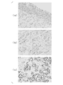
- FIG. 2(a) to (c) are photographs showing typical results of immunostaining in Experimental Example 3.
- FIG. 3 is a graph showing the results of Experimental Example 5.
- FIG. 4 is a graph showing the results of Experimental Example 5.
- FIG. 5(a) to (d) are photographs showing typical results of immunostaining in Experimental Example 6.
- FIG. 6 is a graph showing the results of Experimental Example 7.
- FIG. 7 is a graph showing the results of Experimental Example 7.
- FIG. 8 is a photograph showing the results of Western blotting in Experimental Example 8.
- FIG. 9 is a photograph showing the results of Western blotting in Experimental Example 8.
- FIG. 10 is a graph showing quantitative values of SPSB2 protein in urine samples measured in Experimental Example 9.
- FIG. 11 is a graph showing quantitative values of SPSB2 protein in urine samples measured in Experimental Example 9.
- FIG. 12 is a graph showing quantitative values of SPSB2 protein in urine samples measured in Experimental Example 9.
- FIG. 13 is a graph showing quantitative values of SPSB2 protein in urine samples measured in Experimental Example 9.
- FIG. 14 is an ROC curve created in Experimental Example 10.
- FIG. 15 is an ROC curve created in Experimental Example 10.
- FIG. 16 is a graph showing the results of cancer-specific survival analysis in Experimental Example 10.
- FIG. 17 is a graph showing the results of analyzing the progression-free survival rate in Experimental Example 10.
- FIG. FIG. 18(a) is a photograph showing the results of Western blotting in Experimental Example 11.
- FIG.18(b) is a graph which shows the result of Fig.18 (a).
- the present invention provides a cancer diagnostic kit comprising a specific binding substance to the SPSB2 protein, a primer set capable of amplifying the cDNA of the SPSB2 gene, or a probe that specifically hybridizes to the mRNA of the SPSB2 gene. I will provide a.
- the inventors have clarified that the SPSB2 protein or SPSB2 gene can be used as a cancer marker.
- the inventors also revealed that the SPSB2 protein or SPSB2 gene is useful as a marker not only for bladder cancer but also for pancreatic cancer and hepatocellular carcinoma. Therefore, in the cancer diagnostic kit of the present embodiment, examples of cancer include bladder cancer, pancreatic cancer, and hepatocellular carcinoma.
- kits of the present embodiment it is possible to measure the expression level of the SPSB2 protein or SPSB2 gene in a biological sample derived from a subject, and determine whether or not the subject is suffering from cancer.
- Biological samples include serum, plasma, urine, tissue, and the like.
- examples of the biological sample include urine, tissue, and the like.
- urinary exosomes may be extracted and used as a biological sample.
- the cancer diagnostic kit of this embodiment can also be said to be a prognostic kit.
- poor prognosis may include a low survival rate, a short progression-free survival period, and the like.
- the present invention provides a prognostic kit for cancer patients, comprising a specific binding substance for the SPSB2 protein, a primer set capable of amplifying the cDNA of the SPSB2 gene, or a probe that specifically hybridizes to the mRNA of the SPSB2 gene. It can also be said that it provides In the cancer patient prognosis determination kit (cancer patient prognosis prediction kit), examples of cancer include bladder cancer, pancreatic cancer, and hepatocellular carcinoma.
- NCBI accession numbers for human SPSB2 proteins are NP_001139788.1, NP_001306599.1, NP_116030.1, and so on.
- the NCBI accession numbers of the human SPSB2 gene mRNA are NM_001146316.2, NM_001146317.1, NM_001319670.2, NM_032641.4, and the like.
- the kit of this embodiment may contain a specific binding substance for SPSB2 protein.
- specific binding substances include antibodies, antibody fragments, aptamers, and the like.
- Antibody fragments include F(ab') 2 , Fab', Fab, Fv, scFv and the like.
- the above antibodies or antibody fragments may be polyclonal or monoclonal. Aptamers are not particularly limited as long as they are substances having specific binding ability to SPSB2 protein, and include nucleic acid aptamers, peptide aptamers, and the like.
- the expression level of SPSB2 protein can be measured by immunostaining a fixed tissue section using the above specific binding substance. Measurement of the expression level of the SPSB2 protein is not limited to immunostaining, and may be performed by extracting the protein from the test sample and performing Western blotting or ELISA.
- a higher SPSB2 protein expression level in the test sample compared to the control indicates that the test sample is derived from a cancer patient.
- "more than the control” is preferably a statistically significant more than the control.
- the control includes, for example, the expression level of SPSB2 protein measured using a normal tissue-derived sample.
- the cancer diagnostic kit of this embodiment may contain a primer set for amplifying the cDNA of the SPSB2 gene.
- the sequence of the primer set is not particularly limited as long as it can amplify at least part of the cDNA of the SPSB2 gene.
- a higher expression level of the SPSB2 gene in the test sample compared to the control indicates that the test sample is derived from a cancer patient.
- the control includes, for example, the expression level of the SPSB2 gene measured using a normal tissue-derived sample.
- the kit of this embodiment may contain a probe that specifically hybridizes to the mRNA of the SPSB2 gene.
- the probe may be, for example, a nucleic acid fragment having a nucleotide sequence complementary to the nucleotide sequence of at least part of the mRNA of the SPSB2 gene.
- the probe may have various chemical modifications for the purpose of improving stability, specificity during hybridization, and the like.
- phosphate residues may be substituted with chemically modified phosphate residues such as phosphorothioate (PS), methylphosphonate, phosphorodithionate, etc., in order to inhibit degradation by hydrolases such as nucleases.
- at least a part thereof may be composed of a nucleic acid analogue such as peptide nucleic acid (PNA).
- PNA peptide nucleic acid
- the probe may be immobilized on a solid phase.
- Solid phases include, for example, beads, plate-like substrates, membranes, and the like.
- the probes may be immobilized on the surface of a plate-like substrate to form a microarray.
- the expression of the SPSB2 gene in the test sample is detected by extracting RNA from the test sample, labeling it with a fluorescent substance, hybridizing with the microarray, and detecting the RNA bound to the probes on the microarray. be able to.
- a higher expression level of the SPSB2 gene in the test sample compared to the control indicates that the test sample is derived from a cancer patient.
- the control includes, for example, the expression level of the SPSB2 gene measured using a normal tissue-derived sample.
- the present invention comprises the step of measuring the expression level of the SPSB2 protein or gene in a biological sample, wherein the measured expression level of the protein or gene is greater than that of a control, Provided is a method for determining a biological sample that indicates that the sample is from a cancer patient.
- the inventors have clarified that the SPSB2 protein or SPSB2 gene can be used as a cancer marker.
- the inventors also revealed that the SPSB2 protein or SPSB2 gene is useful as a marker not only for bladder cancer but also for pancreatic cancer and hepatocellular carcinoma. Therefore, according to the method of this embodiment, it can be determined whether or not a biological sample is derived from a cancer patient.
- examples of cancer include bladder cancer, pancreatic cancer, and hepatocellular carcinoma.
- serum, plasma, urine, tissue, etc. derived from subjects can be used as biological samples.
- the cancer is bladder cancer
- examples of the biological sample include urine, tissue, and the like.
- urinary exosomes may be extracted and used as a biological sample.
- the term "higher than the control" is statistically significantly higher than the control, as described above.
- Controls include, for example, the expression level of SPSB2 protein or SPSB2 gene measured using a normal tissue-derived sample.
- the determination method of this embodiment can also be said to be a method of collecting data for determining whether a biological sample is derived from a cancer patient.
- the method of collecting data does not involve medical intervention by a physician.
- the present invention provides a method for collecting data for determining whether a biological sample is derived from a cancer patient, comprising the step of measuring the expression level of the SPSB2 protein or gene in the biological sample. It can also be said that the present invention provides a method in which the measured expression level of the protein or gene is data for determining whether or not the biological sample is derived from a cancer patient. A high SPSB2 protein or gene expression level in a biological sample compared to a control indicates that the biological sample is derived from a cancer patient.
- the present invention provides a method for collecting data for determining whether a subject is suffering from cancer, comprising the step of measuring the expression level of the SPSB2 protein or gene in a biological sample derived from the subject. It can also be said that the present invention provides a method in which the measured expression level of the protein or gene is data for determining whether or not the subject is suffering from cancer. A higher SPSB2 protein or gene expression level in a subject-derived biological sample compared to a control indicates that the subject is suffering from cancer.
- cancers include bladder cancer, pancreatic cancer, hepatocellular carcinoma, and the like.
- the determination method of this embodiment can also be said to be a prognosis prediction method.
- the present invention provides a method for predicting the prognosis of a cancer patient, comprising the step of measuring the expression level of the SPSB2 protein or SPSB2 gene in a biological sample derived from the cancer patient, It can also be said that the fact that the expression level is high compared to controls provides a method of indicating that the prognosis of the cancer patient is poor.
- examples of cancer include bladder cancer, pancreatic cancer, and hepatocellular carcinoma.
- the present invention comprises a step of measuring the expression level of SPSB2 protein or SPSB2 gene in cancer cells cultured in the presence of a test substance, wherein the expression level is SPSB2 in the absence of the test substance
- a screening method for an anticancer agent wherein a significant decrease in protein or SPSB2 gene expression level indicates that the test substance is an anticancer agent.
- test substance that reduces the expression level of the SPSB2 protein or SPSB2 gene is a candidate substance for an anticancer drug.
- test substance is not particularly limited, and examples include natural compound libraries, synthetic compound libraries, existing drug libraries, metabolite libraries, and the like.
- cancer cells may be cancer cells derived from bladder cancer, pancreatic cancer, hepatocellular carcinoma, and the like.
- the cancer cells may also be cisplatin-resistant strains obtained by culturing in the presence of cisplatin at increasing concentrations. Cisplatin-resistant strains tend to be resistant to anticancer agents other than cisplatin, and tend to be highly malignant.
- the present invention is a step of measuring the expression level of the SPSB2 protein or SPSB2 gene in a subject-derived biological sample, wherein the expression level is higher than a control, the subject has cancer Including the step of indicating that the subject is afflicted, and the step of surgically removing cancer tissue from the subject if the subject has cancer, or administering anticancer drug treatment to the subject. , provides a method of treating cancer.
- examples of cancer include bladder cancer, pancreatic cancer, and hepatocellular carcinoma.
- Biological samples include serum, plasma, urine, tissue, and the like.
- the cancer is bladder cancer, the biological sample includes urine, tissue, and the like.
- urinary exosomes may be extracted and used as a biological sample.
- the expression level of SPSB2 protein or SPSB2 gene may be high not only in cancer patients but also in urinary tract infection patients.
- a urine culture test or the like can be used to diagnose whether or not the subject has a urinary tract infection. That is, a subject with a high SPSB2 protein or SPSB2 gene expression level and no urinary tract infection can be diagnosed as having cancer.
- the anticancer agents include cisplatin, M-VAC (combination of methotrexate, vinblastine, adriamycin, and cisplatin), GC (combination of cisplatin and Gemzar), and an antibody-drug conjugate targeting Nectin-4.
- ADC enfortumab vedotin, padoceb, keytruda (penprolizumab) and the like.
- anticancer drugs are commonly administered intravenously.
- the dosage of these anticancer agents varies depending on the patient's symptoms, body weight, age, sex, etc., but those skilled in the art can appropriately select an appropriate dosage.
- Example 1 The inventors have already produced a large number of bladder cancer-specific antibodies in previous studies. In addition, we have already established a technology for identifying target proteins using autoantibodies in serum by the dot blot method, and have successfully identified a large number of autoantibodies against bladder cancer. The reactivity of these produced bladder cancer-specific antibodies and autoantibodies against bladder cancer with serum and tumor tissues collected from a large number of bladder cancer patients was examined. As a result, SPSB2 protein or SPSB2 gene was identified as a new cancer marker candidate.
- FIG. 1 is a graph showing the study results.
- the vertical axis indicates the expression level of the SPSB2 gene.
- “Normal” indicates the expression level of the SPSB2 gene in normal tissues
- "Primary tumor” indicates the expression level of the SPSB2 gene in bladder cancer tissues.
- Evaluation criteria 0 to 1 were classified into the SPSB2 protein low expression group, evaluation criteria 2 was classified into the SPSB2 protein high expression group, and the following analysis was performed.
- FIG. 2(a) to (c) are photographs showing typical results of immunostaining.
- FIG. 2(a) is a photograph showing the results of immunostaining of normal urothelial tissue.
- FIG. 2(b) is a photograph showing the results of immunostaining of bladder cancer tissue classified as the SPSB2 protein low expression group.
- FIG. 2(c) is a photograph showing the results of immunostaining of bladder cancer tissue classified as the SPSB2 protein high expression group.
- Table 2 shows the results of examining the correlation between the expression of S100A8 protein, S100A9 protein, Uroplakin III protein, and HNRNPA3 protein, which are reported to be bladder cancer markers, and the expression of SPSB2 protein.
- p-value indicates the p-value calculated by Fisher's exact test. A p ⁇ 0.05 was considered significant. Bold indicates significant difference.
- the expression of the SPSB2 protein correlated with the expression of the S100A8 protein, S100A9 protein, Uroplakin III protein, and HNRNPA3 protein.
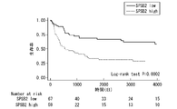
- FIG. 3 is a graph showing the results of cancer-specific survival analysis by the Kaplan-Meier method.
- SPSB2 low indicates the result of the SPSB2 protein low expression group
- SPSB2 high indicates the result of the SPSB2 protein high expression group
- Numberer at risk indicates the number of survivors at each time point. As a result, it was revealed that the SPSB2 protein high expression group had a significantly higher risk of death from bladder cancer.
- FIG. 4 is a graph showing the results of analyzing the progression-free survival rate by the Kaplan-Meier method based on the results of Experimental Example 3.
- SPSB2 low indicates the result of the SPSB2 protein low expression group
- SPSB2 high indicates the result of the SPSB2 protein high expression group.
- Numberer at risk indicates the number of progression-free survivors at each time point. As a result, it was revealed that the SPSB2 protein high expression group had a significantly shorter period of time until recurrence of bladder cancer.
- Table 3 shows the results of univariate analysis and multivariate analysis based on the Cox proportional hazards model for cancer-specific survival rates.
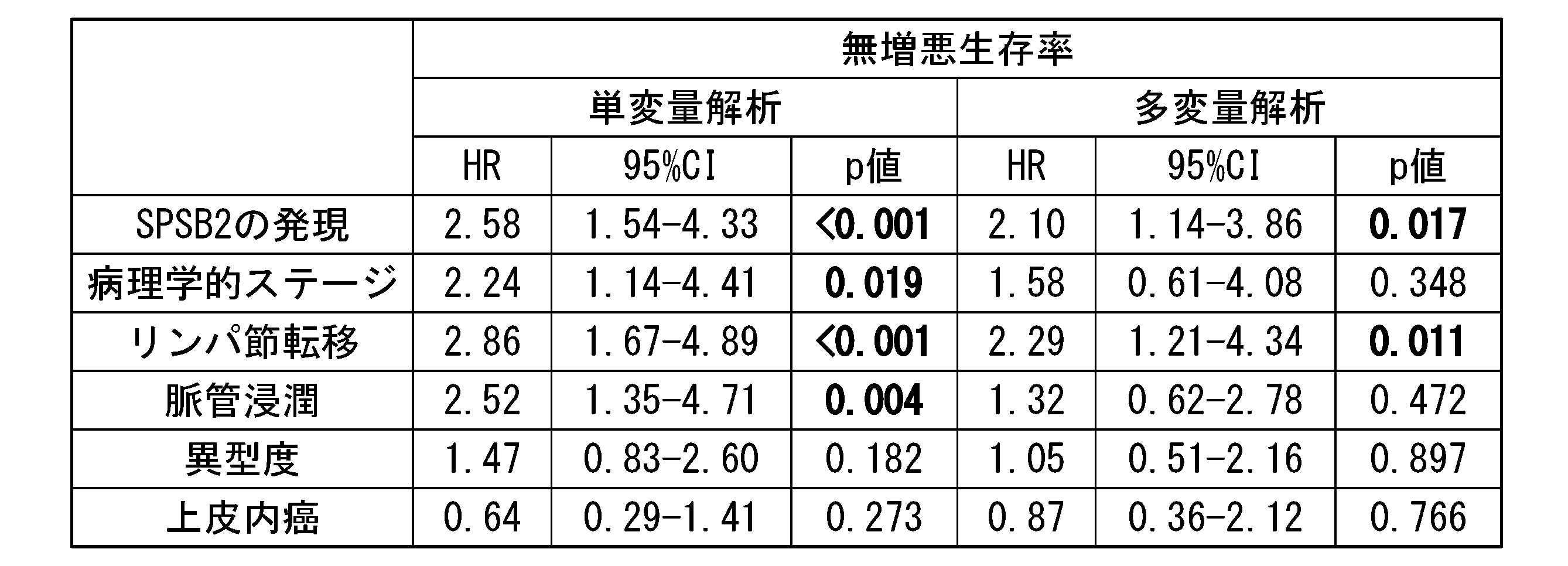
- Table 4 shows the results of univariate analysis and multivariate analysis based on the Cox proportional hazards model for progression-free survival.
- "HR” indicates hazard ratio and "95% CI” indicates 95% confidence interval.
- a p ⁇ 0.05 was considered significant.
- Bold indicates significant difference.
- SPSB2 protein is an independent factor of cancer-specific survival rate and progression-free survival rate along with lymph node metastasis.
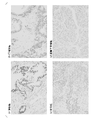
- FIGS. 5(a) to 5(c) are photographs showing representative results of immunostaining tissue sections of pancreatic cancer, hepatocellular carcinoma, and ovarian cancer with anti-SPSB2 antibody.
- FIG. 5(a) shows the results of pancreatic cancer tissue sections
- FIG. 5(b) shows the results of hepatocellular carcinoma tissue sections
- FIG. 5(c) shows the results of ovarian cancer tissue sections.
- 5(d) is the result of a tissue section of pancreas normal tissue.
- the SPSB2 protein was strongly expressed in pancreatic cancer, and moderately expressed in hepatocellular carcinoma and ovarian cancer.
- the SPSB2 protein was weakly expressed or not expressed in renal cancer, prostate cancer, esophageal cancer, gastric cancer, colon cancer, breast cancer, and lung cancer.
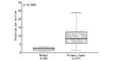
- FIG. 6 is a graph showing the study results.
- the vertical axis indicates the expression level of the SPSB2 gene.
- Normal indicates the expression level of the SPSB2 gene in normal tissues
- Primary tumor indicates the expression level of the SPSB2 gene in hepatocellular carcinoma tissues.
- FIG. 7 is a graph showing the relationship between the expression level of the SPSB2 gene and prognosis in hepatocellular carcinoma in TCGA samples.
- “High expression” indicates the result of the SPSB2 protein high expression group
- “Low/Medium-expression” indicates the result of the SPSB2 protein low to medium expression group.
- exosomes were extracted from serum and urine samples using Total Exosome Isolation Reagent (Thermo Fisher Scientific).
- the SPSB2 protein was detected using the extracted exosomes.
- exosomes could be extracted by Western blotting of CD9, which is one of the exosome markers.
- the molecular weight of SPSB2 protein is about 26 kDa, and the molecular weight of CD9 is about 24 kDa.
- Figure 8 is a photograph showing the results of Western blotting for exosomes in serum.
- C indicates that exosomes derived from healthy subjects
- T indicates that exosomes derived from bladder cancer patients.
- Figure 9 is a photograph showing the results of Western blotting for urinary exosomes.
- C indicates that exosomes derived from healthy individuals
- T indicates that exosomes derived from bladder cancer patients.
- bladder cancer patients As for bladder cancer patients, 91 cases of urine collection immediately before transurethral bladder tumor resection at Kitasato University Hospital from 2009 to 2015 were targeted. Table 5 below shows the background of bladder cancer patients. In Table 5, “NMIBC” indicates non-muscle-invasive bladder cancer, and “MIBC” indicates muscle-invasive bladder cancer. All urine samples were corrected for urine specific gravity to 1.002 and SPSB2 protein abundance was determined by ELISA.
- FIG. 10 is a graph showing quantitative values of SPSB2 protein in urine samples of each group.
- FIG. 10 also shows the analysis results of the Mann-Whitney U test.
- BC indicates results for bladder cancer patients
- Healthy indicates results for healthy subjects
- Stone indicates results for urinary stone patients
- UTI indicates results from patients with urinary tract infections.
- FIG. 11 is a graph showing quantitative values of SPSB2 protein in urine samples from non-muscle-invasive bladder cancer patients and muscle-invasive bladder cancer patients.
- FIG. 11 also shows the analysis results of the Mann-Whitney U test.
- “NMIBC” indicates non-muscle-invasive bladder cancer
- “MIBC” indicates muscle-invasive bladder cancer.
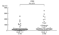
- FIG. 12 is a graph showing quantitative values of SPSB2 protein in urine samples of pathological grade 1 and 2 bladder cancer patients and pathological grade 3 bladder cancer patients.
- FIG. 12 also shows the analysis results of the Mann-Whitney U test.
- FIG. 13 is a graph showing quantitative values of SPSB2 protein in urine samples from muscle-invasive bladder cancer patients and urinary tract infection patients.
- FIG. 13 also shows the analysis results of the Mann-Whitney U test.
- MIBC indicates muscle-invasive bladder cancer
- UTI indicates results from urinary tract infection patients.
- Urinary tract infections can be diagnosed by a urine culture test or the like.
- FIG. 15 is an ROC curve created based on the quantitative values of SPSB2 protein in urine samples of muscle-invasive bladder cancer patients and healthy subjects measured in Experimental Example 9. As a result, the area under the ROC curve (AUC) was found to be 0.8699.
- the cutoff value was set at 162.8 ng/mL (sensitivity 58.2%, specificity 80.0%), and urinary SPSB2 protein abundance of less than 162.8 ng/mL was Survival analysis was performed for the SPSB2 protein low expression group and the SPSB2 protein high expression group for 162.8 ng/mL or more.
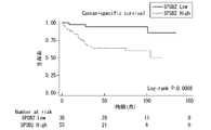
- FIG. 16 is a graph showing the results of cancer-specific survival analysis by the Kaplan-Meier method.
- SPSB2 Low indicates the result of the SPSB2 protein low expression group
- SPSB2 High indicates the result of the SPSB2 protein high expression group
- Numberer at risk indicates the number of survivors at each time point. As a result, it was revealed that the SPSB2 protein high expression group had a significantly higher risk of death from bladder cancer.
- FIG. 17 is a graph showing the results of analyzing the progression-free survival rate by the Kaplan-Meier method.
- SPSB2 Low indicates the result of the SPSB2 protein low expression group
- SPSB2 High indicates the result of the SPSB2 protein high expression group
- Numberer at risk indicates the number of progression-free survivors at each time point. As a result, no significant difference was observed between the SPSB2 protein low expression group and the SPSB2 protein high expression group in the period until recurrence of bladder cancer.
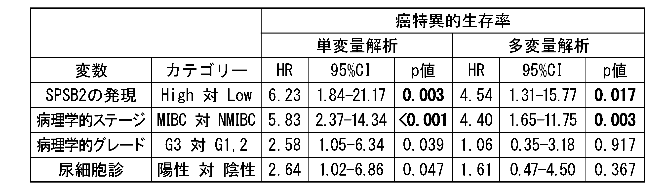
- Table 6 shows the results of univariate analysis and multivariate analysis based on the Cox proportional hazards model for cancer-specific survival rates.
- NMIBC non-muscle-invasive bladder cancer
- MIBC muscle-invasive bladder cancer
- G3 indicates grade 3
- G1,2 indicates grade 1 or 2.
- HR indicates hazard ratio and "95% CI” indicates 95% confidence interval. A p ⁇ 0.05 was considered significant. Bold indicates significant difference.
- the expression of the SPSB2 protein is a prognostic factor for cancer-specific survival along with the depth of cancer invasion.
- T24CDDPR and 5637CDDPR Human bladder cancer cell lines T24 and 5637 were cultured in the presence of increasing concentrations of cisplatin to obtain cisplatin-resistant lines, T24CDDPR and 5637CDDPR, respectively.
- T24CDDPR and 5637CDDPR are considered to be more malignant cancers than T24 and 5637.
- the expression level of SPSB2 protein was measured by Western blotting of each of the T24, T24CDDPR, 5637, and 5637CDDPR cell lines. Also, as a control, the expression level of ⁇ -actin protein was measured.
- FIG. 18(a) is a photograph showing the results of Western blotting. Moreover, FIG.18(b) makes the result of Fig.18 (a) the graph.
- the vertical axis in FIG. 18(b) shows the expression level of SPSB2 with respect to the expression level of ⁇ -actin, "*" indicates a significant difference at p ⁇ 0.05, and "**" indicates p ⁇ 0. 0.01 indicates a significant difference.
- T24CDDPR and 5637CDDPR have significantly higher expression levels of SPSB2 than T24 and 5637, respectively.
- a new cancer diagnostic technique can be provided by the present invention.
Abstract
Description
[1]SPRY domain-containing SOCS box protein 2(SPSB2)タンパク質に対する特異的結合物質、SPSB2遺伝子のcDNAを増幅可能なプライマーセット、又は、SPSB2遺伝子のmRNAに特異的にハイブリダイズするプローブ、を含む、癌診断用キット。
[2]前記癌が、膀胱癌、膵臓癌又は肝細胞癌である、[1]に記載の癌診断用キット。
[3]SPSB2タンパク質に対する特異的結合物質、SPSB2遺伝子のcDNAを増幅可能なプライマーセット、又は、SPSB2遺伝子のmRNAに特異的にハイブリダイズするプローブ、を含む、癌患者の予後判定用キット。
[4]前記癌が、膀胱癌、膵臓癌又は肝細胞癌である、[3]に記載の予後判定用キット。
[5]生体試料中のSPSB2タンパク質又はSPSB2遺伝子の発現量を測定する工程を含み、測定された前記タンパク質又は遺伝子の発現量が、対照と比較して多いことが、前記生体試料が癌患者由来のものであることを示す、生体試料の判定方法。
[6]前記癌が、膀胱癌、膵臓癌又は肝細胞癌である、[5]に記載の判定方法。
[7]被験者が癌に罹患しているか否かを判定するためのデータを収集する方法であって、前記被験者由来の生体試料中のSPSB2タンパク質又はSPSB2遺伝子の発現量を測定する工程を含み、測定された前記タンパク質又は遺伝子の発現量のデータが、前記被験者が癌に罹患しているか否かを判定するためのデータである、方法(医師による医療行為を除く。)。
[8]前記癌が、膀胱癌、膵臓癌又は肝細胞癌である、[7]に記載の方法。
[9]癌患者の予後を予測する方法であって、前記癌患者由来の生体試料中のSPSB2タンパク質又はSPSB2遺伝子の発現量を測定する工程を含み、測定された前記タンパク質又は遺伝子の発現量が、対照と比較して多いことが、前記癌患者の予後が不良であることを示す、方法。
[10]前記癌が、膀胱癌、膵臓癌又は肝細胞癌である、[9]に記載の方法。
[11]被験物質の存在下で培養した癌細胞における、SPSB2タンパク質又はSPSB2遺伝子の発現量を測定する工程を含み、前記発現量が、前記被験物質の非存在下におけるSPSB2タンパク質又はSPSB2遺伝子の発現量と比較して有意に低下したことが、前記被験物質が抗癌剤であることを示す、抗癌剤のスクリーニング方法。
[12]前記癌細胞が、膀胱癌、膵臓癌又は肝細胞癌に由来する癌細胞である、[11]に記載の方法。
1実施形態において、本発明は、SPSB2タンパク質に対する特異的結合物質、SPSB2遺伝子のcDNAを増幅可能なプライマーセット、又は、SPSB2遺伝子のmRNAに特異的にハイブリダイズするプローブ、を含む、癌診断用キットを提供する。
本実施形態のキットは、SPSB2タンパク質に対する特異的結合物質を含んでいてもよい。特異的結合物質としては、例えば、抗体、抗体断片、アプタマー等が挙げられる。抗体断片としては、F(ab’)2、Fab’、Fab、Fv、scFv等が挙げられる。上記の抗体又は抗体断片は、ポリクローナルであってもよく、モノクローナルであってもよい。アプタマーとしては、SPSB2タンパク質に対する特異的結合能を有する物質であれば特に限定されず、核酸アプタマー、ペプチドアプタマー等が挙げられる。
本実施形態の癌診断用キットは、SPSB2遺伝子のcDNAを増幅するためのプライマーセットを含んでいてもよい。プライマーセットは、SPSB2遺伝子のcDNAの少なくとも1部を増幅することができれば、その配列は特に限定されない。
本実施形態のキットは、SPSB2遺伝子のmRNAに特異的にハイブリダイズするプローブを含んでいてもよい。
1実施形態において、本発明は、生体試料中のSPSB2タンパク質又は遺伝子の発現量を測定する工程を含み、測定された前記タンパク質又は遺伝子の発現量が、対照と比較して多いことが、前記生体試料が癌患者由来のものであることを示す、生体試料の判定方法を提供する。
1実施形態において、本発明は、被験物質の存在下で培養した癌細胞における、SPSB2タンパク質又はSPSB2遺伝子の発現量を測定する工程を含み、前記発現量が、前記被験物質の非存在下におけるSPSB2タンパク質又はSPSB2遺伝子の発現量と比較して有意に低下したことが、前記被験物質が抗癌剤であることを示す、抗癌剤のスクリーニング方法を提供する。
一実施形態において、本発明は、被験者由来の生体試料中のSPSB2タンパク質又はSPSB2遺伝子の発現量を測定する工程であって、前記発現量が対照と比較して高いことが、前記被験者が癌に罹患していることを示す工程と、前記被験者が癌に罹患していた場合に、外科的手術により被験者から癌組織を摘出するか、又は、被験者に対して抗癌剤治療を行う工程と、を含む、癌の治療方法を提供する。
発明者らは、これまでの研究で、多数の膀胱癌特異的抗体をすでに作製済みである。また、ドットブロット法による血清中の自己抗体を用いた対象タンパク質の同定技術を確立済みであり、膀胱癌に対する多数の自己抗体の同定に成功している。これらの作製済みの膀胱癌特異的抗体及び膀胱癌に対する自己抗体に対して、収集済みの多数例の膀胱癌患者血清・腫瘍組織との反応性を検討した。その結果、SPSB2タンパク質又はSPSB2遺伝子を新たな癌マーカーの候補として特定した。
公開データベースであるThe Cancer Genome Atlas(TCGA)を用いて、膀胱癌組織におけるSPSB2遺伝子の発現量を検討した。図1は、検討結果を示すグラフである。図1中、縦軸はSPSB2遺伝子の発現量を示す。また、「Normal」は正常組織におけるSPSB2遺伝子の発現量であることを示し、「Primary tumor」は膀胱癌組織におけるSPSB2遺伝子の発現量であることを示す。その結果、膀胱癌組織において、SPSB2遺伝子の発現量が有意に増加していることが明らかとなった。
膀胱全摘標本から作製した組織切片を、抗SPSB2抗体で免疫染色した。膀胱全摘標本としては、1990年から2015年までに北里大学病院で膀胱全摘術を施行された126例の標本を対象とした。免疫染色には、Bond-MAX自動免疫染色装置(ライカ社)を使用した。
0:低発現
1:同等
2:高発現
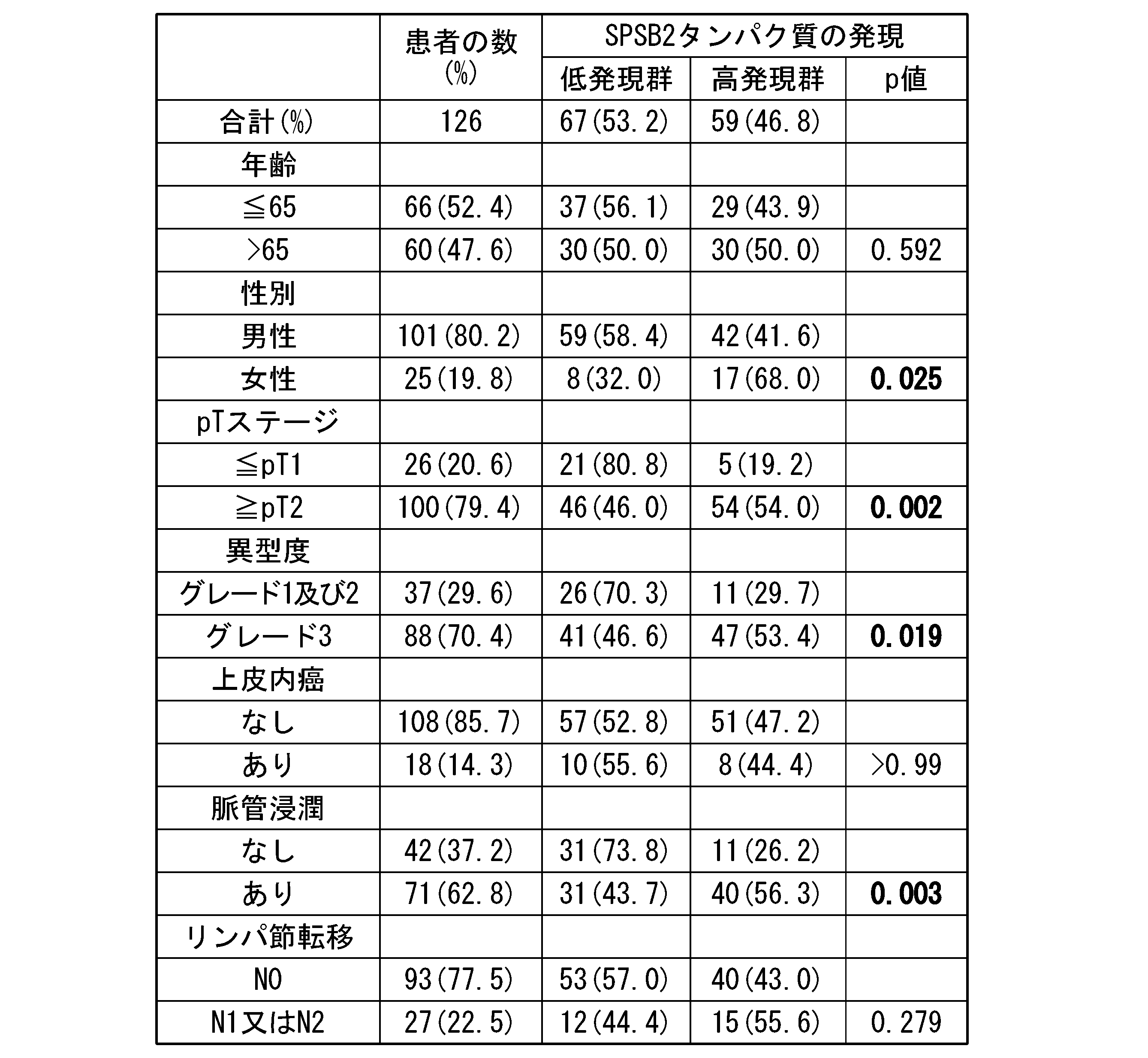
実験例3の結果に基づいて、SPSB2タンパク質の発現と臨床病理学的因子の関連性を解析した。下記表1に解析結果を示す。表1中、「p値」はフィッシャーの正確確率検定により算出されたp値を示す。p<0.05を有意差ありと判断した。太字は有意差があることを示す。その結果、SPSB2タンパク質の発現は、性別、深達度(pTステージ)、異型度、脈管浸潤と相関があることが明らかとなった。

実験例3の結果に基づいて、SPSB2タンパク質の発現と予後との関連を解析した。図3は、カプラン・マイヤー法により、癌特異的生存率を解析した結果を示すグラフである。図3中、「SPSB2 low」はSPSB2タンパク質低発現群の結果であることを示し、「SPSB2 high」はSPSB2タンパク質高発現群の結果であることを示す。また、「Number at risk」は各時点における生存者の数を示す。その結果、SPSB2タンパク質高発現群は、膀胱癌による死亡のリスクが有意に高いことが明らかとなった。

膀胱癌以外の癌種におけるSPSB2タンパク質の発現を検討した。図5(a)~(c)は、膵臓癌、肝細胞癌、卵巣癌の各組織切片を抗SPSB2抗体で免疫染色した代表的な結果を示す写真である。図5(a)は膵臓癌の組織切片の結果であり、図5(b)は肝細胞癌の組織切片の結果であり、図5(c)は卵巣癌の組織切片の結果であり、図5(d)は膵臓の正常組織の組織切片の結果である。
公開データベースであるThe Cancer Genome Atlas(TCGA)を用いて、肝細胞癌組織におけるSPSB2遺伝子の発現量を検討した。図6は、検討結果を示すグラフである。図6中、縦軸はSPSB2遺伝子の発現量を示す。また、「Normal」は正常組織におけるSPSB2遺伝子の発現量であることを示し、「Primary tumor」は肝細胞癌組織におけるSPSB2遺伝子の発現量であることを示す。
健常人及び膀胱癌患者由来の血清中エクソソーム及び尿中エクソソームにおける、SPSB2タンパク質の存在量を、ウエスタンブロッティングにより検討した。
健常人、尿路結石患者、尿路感染症患者及び膀胱癌患者由来の尿における、SPSB2タンパク質の存在量を、ELISA(酵素結合免疫吸着測定法)により定量し、検討した。

実験例9の結果に基づいて、SPSB2タンパク質の発現と予後との関連を解析した。図14は、実験例9で測定した、膀胱癌患者及び健常人の尿試料中のSPSB2タンパク質の定量値に基づいて作成したROC曲線である。その結果、ROC曲線下面積(AUC:area under the curve)は0.7791であることが明らかとなった。
ヒト膀胱癌細胞株であるT24及び5637を、濃度を漸増しながらシスプラチンの存在下で培養し、それぞれシスプラチン耐性株である、T24CDDPR及び5637CDDPRを取得した。T24CDDPR及び5637CDDPRは、T24及び5637と比較して、癌の悪性度が高いと考えられる。
Claims (12)
- SPRY domain-containing SOCS box protein 2(SPSB2)タンパク質に対する特異的結合物質、
SPSB2遺伝子のcDNAを増幅可能なプライマーセット、又は
SPSB2遺伝子のmRNAに特異的にハイブリダイズするプローブ、
を含む、癌診断用キット。 - 前記癌が、膀胱癌、膵臓癌又は肝細胞癌である、請求項1に記載の癌診断用キット。
- SPSB2タンパク質に対する特異的結合物質、
SPSB2遺伝子のcDNAを増幅可能なプライマーセット、又は、
SPSB2遺伝子のmRNAに特異的にハイブリダイズするプローブ、
を含む、癌患者の予後判定用キット。 - 前記癌が、膀胱癌、膵臓癌又は肝細胞癌である、請求項3に記載の予後判定用キット。
- 生体試料中のSPSB2タンパク質又はSPSB2遺伝子の発現量を測定する工程を含み、
測定された前記タンパク質又は遺伝子の発現量が、対照と比較して多いことが、前記生体試料が癌患者由来のものであることを示す、生体試料の判定方法。 - 前記癌が、膀胱癌、膵臓癌又は肝細胞癌である、請求項5に記載の判定方法。
- 被験者が癌に罹患しているか否かを判定するためのデータを収集する方法であって、前記被験者由来の生体試料中のSPSB2タンパク質又はSPSB2遺伝子の発現量を測定する工程を含み、測定された前記タンパク質又は遺伝子の発現量のデータが、前記被験者が癌に罹患しているか否かを判定するためのデータである、方法(医師による医療行為を除く。)。
- 前記癌が、膀胱癌、膵臓癌又は肝細胞癌である、請求項7に記載の方法。
- 癌患者の予後を予測する方法であって、前記癌患者由来の生体試料中のSPSB2タンパク質又はSPSB2遺伝子の発現量を測定する工程を含み、
測定された前記タンパク質又は遺伝子の発現量が、対照と比較して多いことが、前記癌患者の予後が不良であることを示す、方法。 - 前記癌が、膀胱癌、膵臓癌又は肝細胞癌である、請求項9に記載の方法。
- 被験物質の存在下で培養した癌細胞における、SPSB2タンパク質又はSPSB2遺伝子の発現量を測定する工程を含み、
前記発現量が、前記被験物質の非存在下におけるSPSB2タンパク質又はSPSB2遺伝子の発現量と比較して有意に低下したことが、前記被験物質が抗癌剤であることを示す、抗癌剤のスクリーニング方法。 - 前記癌細胞が、膀胱癌、膵臓癌又は肝細胞癌に由来する癌細胞である、請求項11に記載の方法。
Priority Applications (2)
| Application Number | Priority Date | Filing Date | Title |
|---|---|---|---|
| CN202280040408.4A CN117425827A (zh) | 2021-06-10 | 2022-06-10 | 用于诊断癌症的试剂盒及其用途 |
| EP22820344.4A EP4354143A1 (en) | 2021-06-10 | 2022-06-10 | Kit for diagnosis of cancer and use thereof |
Applications Claiming Priority (4)
| Application Number | Priority Date | Filing Date | Title |
|---|---|---|---|
| JP2021-097456 | 2021-06-10 | ||
| JP2021097456 | 2021-06-10 | ||
| JP2021201094 | 2021-12-10 | ||
| JP2021-201094 | 2021-12-10 |
Publications (1)
| Publication Number | Publication Date |
|---|---|
| WO2022260166A1 true WO2022260166A1 (ja) | 2022-12-15 |
Family
ID=84424615
Family Applications (1)
| Application Number | Title | Priority Date | Filing Date |
|---|---|---|---|
| PCT/JP2022/023474 WO2022260166A1 (ja) | 2021-06-10 | 2022-06-10 | 癌診断用キット及びその使用 |
Country Status (2)
| Country | Link |
|---|---|
| EP (1) | EP4354143A1 (ja) |
| WO (1) | WO2022260166A1 (ja) |
Citations (6)
| Publication number | Priority date | Publication date | Assignee | Title |
|---|---|---|---|---|
| WO2009113674A1 (ja) * | 2008-03-14 | 2009-09-17 | 学校法人北里研究所 | 膀胱癌の診断 |
| US20130196876A1 (en) * | 2010-10-01 | 2013-08-01 | Universitatsklinikum Schleswig-Holstein | Differential diagnosis of pancreatic adenomas |
| JP2016526009A (ja) * | 2013-04-12 | 2016-09-01 | ヴィヴェンティア バイオ インコーポレイテッド | 肝細胞癌の検出および治療のための組成物および方法 |
| JP2016211913A (ja) * | 2015-05-01 | 2016-12-15 | 学校法人北里研究所 | 膀胱癌細胞のシスプラチン耐性マーカー及びその使用 |
| JP2018533724A (ja) * | 2015-09-28 | 2018-11-15 | アボットジャパン株式会社 | 肝細胞癌および膵臓がんを診断するためのラミニン2の使用 |
| JP2021097456A (ja) | 2019-12-13 | 2021-06-24 | トヨタ自動車株式会社 | 非接触充電システム |
-
2022
- 2022-06-10 EP EP22820344.4A patent/EP4354143A1/en active Pending
- 2022-06-10 WO PCT/JP2022/023474 patent/WO2022260166A1/ja active Application Filing
Patent Citations (6)
| Publication number | Priority date | Publication date | Assignee | Title |
|---|---|---|---|---|
| WO2009113674A1 (ja) * | 2008-03-14 | 2009-09-17 | 学校法人北里研究所 | 膀胱癌の診断 |
| US20130196876A1 (en) * | 2010-10-01 | 2013-08-01 | Universitatsklinikum Schleswig-Holstein | Differential diagnosis of pancreatic adenomas |
| JP2016526009A (ja) * | 2013-04-12 | 2016-09-01 | ヴィヴェンティア バイオ インコーポレイテッド | 肝細胞癌の検出および治療のための組成物および方法 |
| JP2016211913A (ja) * | 2015-05-01 | 2016-12-15 | 学校法人北里研究所 | 膀胱癌細胞のシスプラチン耐性マーカー及びその使用 |
| JP2018533724A (ja) * | 2015-09-28 | 2018-11-15 | アボットジャパン株式会社 | 肝細胞癌および膵臓がんを診断するためのラミニン2の使用 |
| JP2021097456A (ja) | 2019-12-13 | 2021-06-24 | トヨタ自動車株式会社 | 非接触充電システム |
Non-Patent Citations (3)
| Title |
|---|
| "NCBI", Database accession no. NM_001319670.2 |
| BEOW KEAT YAP, ELEANOR W. W. LEUNG, HIROMASA YAGI, CHARLES A. GALEA, SANDEEP CHHABRA, DAVID K. CHALMERS, SANDRA E. NICHOLSON, PHIL: "A Potent Cyclic Peptide Targeting SPSB2 Protein as a Potential Anti-infective Agent", JOURNAL OF MEDICINAL CHEMISTRY, AMERICAN CHEMICAL SOCIETY, US, vol. 57, no. 16, 28 August 2014 (2014-08-28), US , pages 7006 - 7015, XP055252156, ISSN: 0022-2623, DOI: 10.1021/jm500596j * |
| FITZMAURICE C. ET AL.: "Global, Regional, and National Cancer Incidence, Mortality, Years of Life Lost, Years Lived With Disability, and Disability-Adjusted Life-Years for 29 Cancer Groups, 1990 to 2017: A Systematic Analysis for the Global Burden of Disease Study", JAMA ONCOL., vol. 5, no. 12, 2019, pages 1749 - 1768 |
Also Published As
| Publication number | Publication date |
|---|---|
| EP4354143A1 (en) | 2024-04-17 |
Similar Documents
| Publication | Publication Date | Title |
|---|---|---|
| JP6630766B2 (ja) | 膵臓癌診断用組成物およびこれを用いた膵臓癌診断方法 | |
| De Salins et al. | Discordance between immunochemistry of mismatch repair proteins and molecular testing of microsatellite instability in colorectal cancer | |
| Kanda et al. | The expression of melanoma-associated antigen D2 both in surgically resected and serum samples serves as clinically relevant biomarker of gastric cancer progression | |
| Fujioka et al. | Expression of minichromosome maintenance 7 (MCM7) in small lung adenocarcinomas (pT1): Prognostic implication | |
| US20150275307A1 (en) | Compositions and methods for detecting sessile serrated adenomas/polyps | |
| WO2022053065A1 (zh) | 用于预测或评估肺癌患者的生物标志物、检测方法及应用 | |
| Isono et al. | ADP-ribosylation factor-like 4C is a predictive biomarker of poor prognosis in patients with renal cell carcinoma | |
| WO2022260166A1 (ja) | 癌診断用キット及びその使用 | |
| KR101334123B1 (ko) | 소세포폐암 진단용 조성물 및 소세포폐암 진단키트 | |
| JP2010526996A (ja) | チオレドキシン発現に基づいた化学療法後の非小細胞肺癌の無進行期間の決定方法 | |
| Sanganeria et al. | Molecular Diagnostics in Renal Cancer | |
| Shinozuka et al. | Identification of stromal cell-derived factor 4 as a liquid biopsy-based diagnostic marker in solid cancers | |
| CN117604111B (zh) | 用于小细胞肺癌诊断和预后判断的生物标志物及其应用 | |
| CN117425827A (zh) | 用于诊断癌症的试剂盒及其用途 | |
| CN117604112B (zh) | 用于胰腺癌诊断和预后判断的生物标志物及其应用 | |
| CN117625792B (zh) | 用于胃癌诊断和预后判断的生物标志物及其应用 | |
| KR102416614B1 (ko) | 방사선 저항성 지표 단백질 및 이의 검출방법 | |
| CN117604106B (zh) | 用于非小细胞肺癌诊断和预后判断的生物标志物及其应用 | |
| WO2013010140A9 (en) | Methods of diagnosing cancer | |
| KR102382674B1 (ko) | 직장 신경내분비종양의 예후 예측 방법 | |
| KR102416607B1 (ko) | 방사선 저항성 지표 단백질 및 이의 검출방법 | |
| AU2020332349A1 (en) | Protein panels for the early diagnosis/prognosis and treatment of aggressive prostate cancer | |
| EP3948291A1 (en) | Biomarker with therapeutic implications for peritoneal carcinomatosis | |
| Mirus | Antibody Microarray Interrogation of Tissue and Plasma for the Improved Early Detection of Pancreas Cancer | |
| WO2016060382A1 (ko) | 췌장암 진단용 조성물 및 이를 이용한 췌장암 진단방법 |
Legal Events
| Date | Code | Title | Description |
|---|---|---|---|
| 121 | Ep: the epo has been informed by wipo that ep was designated in this application |
Ref document number: 22820344 Country of ref document: EP Kind code of ref document: A1 |
|
| DPE2 | Request for preliminary examination filed before expiration of 19th month from priority date (pct application filed from 20040101) | ||
| WWE | Wipo information: entry into national phase |
Ref document number: 2023527944 Country of ref document: JP |
|
| WWE | Wipo information: entry into national phase |
Ref document number: 2022820344 Country of ref document: EP |
|
| NENP | Non-entry into the national phase |
Ref country code: DE |
|
| ENP | Entry into the national phase |
Ref document number: 2022820344 Country of ref document: EP Effective date: 20240110 |