WO2022234856A1 - メモリーt細胞の製造方法 - Google Patents
メモリーt細胞の製造方法 Download PDFInfo
- Publication number
- WO2022234856A1 WO2022234856A1 PCT/JP2022/019563 JP2022019563W WO2022234856A1 WO 2022234856 A1 WO2022234856 A1 WO 2022234856A1 JP 2022019563 W JP2022019563 W JP 2022019563W WO 2022234856 A1 WO2022234856 A1 WO 2022234856A1
- Authority
- WO
- WIPO (PCT)
- Prior art keywords
- cells
- memory
- naive
- differentiation
- cultured
- Prior art date
- Legal status (The legal status is an assumption and is not a legal conclusion. Google has not performed a legal analysis and makes no representation as to the accuracy of the status listed.)
- Ceased
Links
Images


Classifications
-
- A—HUMAN NECESSITIES
- A61—MEDICAL OR VETERINARY SCIENCE; HYGIENE
- A61K—PREPARATIONS FOR MEDICAL, DENTAL OR TOILETRY PURPOSES
- A61K35/00—Medicinal preparations containing materials or reaction products thereof with undetermined constitution
- A61K35/12—Materials from mammals; Compositions comprising non-specified tissues or cells; Compositions comprising non-embryonic stem cells; Genetically modified cells
- A61K35/14—Blood; Artificial blood
- A61K35/17—Lymphocytes; B-cells; T-cells; Natural killer cells; Interferon-activated or cytokine-activated lymphocytes
-
- C—CHEMISTRY; METALLURGY
- C12—BIOCHEMISTRY; BEER; SPIRITS; WINE; VINEGAR; MICROBIOLOGY; ENZYMOLOGY; MUTATION OR GENETIC ENGINEERING
- C12N—MICROORGANISMS OR ENZYMES; COMPOSITIONS THEREOF; PROPAGATING, PRESERVING, OR MAINTAINING MICROORGANISMS; MUTATION OR GENETIC ENGINEERING; CULTURE MEDIA
- C12N5/00—Undifferentiated human, animal or plant cells, e.g. cell lines; Tissues; Cultivation or maintenance thereof; Culture media therefor
- C12N5/06—Animal cells or tissues; Human cells or tissues
- C12N5/0602—Vertebrate cells
- C12N5/0634—Cells from the blood or the immune system
- C12N5/0635—B lymphocytes
-
- C—CHEMISTRY; METALLURGY
- C12—BIOCHEMISTRY; BEER; SPIRITS; WINE; VINEGAR; MICROBIOLOGY; ENZYMOLOGY; MUTATION OR GENETIC ENGINEERING
- C12N—MICROORGANISMS OR ENZYMES; COMPOSITIONS THEREOF; PROPAGATING, PRESERVING, OR MAINTAINING MICROORGANISMS; MUTATION OR GENETIC ENGINEERING; CULTURE MEDIA
- C12N5/00—Undifferentiated human, animal or plant cells, e.g. cell lines; Tissues; Cultivation or maintenance thereof; Culture media therefor
- C12N5/06—Animal cells or tissues; Human cells or tissues
- C12N5/0602—Vertebrate cells
- C12N5/0634—Cells from the blood or the immune system
- C12N5/0636—T lymphocytes
-
- C—CHEMISTRY; METALLURGY
- C12—BIOCHEMISTRY; BEER; SPIRITS; WINE; VINEGAR; MICROBIOLOGY; ENZYMOLOGY; MUTATION OR GENETIC ENGINEERING
- C12N—MICROORGANISMS OR ENZYMES; COMPOSITIONS THEREOF; PROPAGATING, PRESERVING, OR MAINTAINING MICROORGANISMS; MUTATION OR GENETIC ENGINEERING; CULTURE MEDIA
- C12N2500/00—Specific components of cell culture medium
- C12N2500/02—Atmosphere, e.g. low oxygen conditions
-
- C—CHEMISTRY; METALLURGY
- C12—BIOCHEMISTRY; BEER; SPIRITS; WINE; VINEGAR; MICROBIOLOGY; ENZYMOLOGY; MUTATION OR GENETIC ENGINEERING
- C12N—MICROORGANISMS OR ENZYMES; COMPOSITIONS THEREOF; PROPAGATING, PRESERVING, OR MAINTAINING MICROORGANISMS; MUTATION OR GENETIC ENGINEERING; CULTURE MEDIA
- C12N2500/00—Specific components of cell culture medium
- C12N2500/90—Serum-free medium, which may still contain naturally-sourced components
-
- C—CHEMISTRY; METALLURGY
- C12—BIOCHEMISTRY; BEER; SPIRITS; WINE; VINEGAR; MICROBIOLOGY; ENZYMOLOGY; MUTATION OR GENETIC ENGINEERING
- C12N—MICROORGANISMS OR ENZYMES; COMPOSITIONS THEREOF; PROPAGATING, PRESERVING, OR MAINTAINING MICROORGANISMS; MUTATION OR GENETIC ENGINEERING; CULTURE MEDIA
- C12N2501/00—Active agents used in cell culture processes, e.g. differentation
- C12N2501/20—Cytokines; Chemokines
- C12N2501/23—Interleukins [IL]
- C12N2501/2302—Interleukin-2 (IL-2)
-
- C—CHEMISTRY; METALLURGY
- C12—BIOCHEMISTRY; BEER; SPIRITS; WINE; VINEGAR; MICROBIOLOGY; ENZYMOLOGY; MUTATION OR GENETIC ENGINEERING
- C12N—MICROORGANISMS OR ENZYMES; COMPOSITIONS THEREOF; PROPAGATING, PRESERVING, OR MAINTAINING MICROORGANISMS; MUTATION OR GENETIC ENGINEERING; CULTURE MEDIA
- C12N2501/00—Active agents used in cell culture processes, e.g. differentation
- C12N2501/20—Cytokines; Chemokines
- C12N2501/23—Interleukins [IL]
- C12N2501/2304—Interleukin-4 (IL-4)
-
- C—CHEMISTRY; METALLURGY
- C12—BIOCHEMISTRY; BEER; SPIRITS; WINE; VINEGAR; MICROBIOLOGY; ENZYMOLOGY; MUTATION OR GENETIC ENGINEERING
- C12N—MICROORGANISMS OR ENZYMES; COMPOSITIONS THEREOF; PROPAGATING, PRESERVING, OR MAINTAINING MICROORGANISMS; MUTATION OR GENETIC ENGINEERING; CULTURE MEDIA
- C12N2501/00—Active agents used in cell culture processes, e.g. differentation
- C12N2501/20—Cytokines; Chemokines
- C12N2501/23—Interleukins [IL]
- C12N2501/2306—Interleukin-6 (IL-6)
-
- C—CHEMISTRY; METALLURGY
- C12—BIOCHEMISTRY; BEER; SPIRITS; WINE; VINEGAR; MICROBIOLOGY; ENZYMOLOGY; MUTATION OR GENETIC ENGINEERING
- C12N—MICROORGANISMS OR ENZYMES; COMPOSITIONS THEREOF; PROPAGATING, PRESERVING, OR MAINTAINING MICROORGANISMS; MUTATION OR GENETIC ENGINEERING; CULTURE MEDIA
- C12N2501/00—Active agents used in cell culture processes, e.g. differentation
- C12N2501/999—Small molecules not provided for elsewhere
-
- C—CHEMISTRY; METALLURGY
- C12—BIOCHEMISTRY; BEER; SPIRITS; WINE; VINEGAR; MICROBIOLOGY; ENZYMOLOGY; MUTATION OR GENETIC ENGINEERING
- C12N—MICROORGANISMS OR ENZYMES; COMPOSITIONS THEREOF; PROPAGATING, PRESERVING, OR MAINTAINING MICROORGANISMS; MUTATION OR GENETIC ENGINEERING; CULTURE MEDIA
- C12N2506/00—Differentiation of animal cells from one lineage to another; Differentiation of pluripotent cells
- C12N2506/11—Differentiation of animal cells from one lineage to another; Differentiation of pluripotent cells from blood or immune system cells
- C12N2506/115—Differentiation of animal cells from one lineage to another; Differentiation of pluripotent cells from blood or immune system cells from monocytes, from macrophages
Definitions
- the present invention relates to a method for conveniently producing memory T cells from naive T cells.
- T cells and B cells are cells that play an important role in immunity.
- T cells are lymphocytes that have differentiated and matured from progenitor cells produced in the bone marrow through selection in the thymus, and T cells present in the periphery are mainly classified into CD4 + T cells and CD8 + T cells.
- CD4 + T cells are cells that express CD4 as a cell surface antigen, regulate the functions of other T cells and B cells in response to antigen stimulation, and are called helper T cells.
- CD4 + T cells are classified into subpopulations such as Th1 cells, Th2 cells, and Th17 cells, depending on the types of cytokines they secrete.
- CD8 + T cells are cells that express CD8 as a cell surface antigen and are responsible for cell-mediated immunity that kills virus-infected cells and the like, and effector cells after activation are also called killer T cells.
- B cells are lymphocytes differentiated from hematopoietic cells in the bone marrow, are stimulated by helper T cells, differentiate into plasma cells, and play a role in humoral immunity to produce antibodies.
- Naive T cells are activated upon recognition of antigens presented by antigen-presenting cells and differentiate into effector cells. When naive CD4 + T cells are activated, they differentiate into helper CD4 + Th1 cells, helper CD4 + Th2 cells, etc., and when naive CD8 + T cells are activated, they differentiate into killer T cells. These effector cells work together to eliminate pathogens and the like. These effector cells have a short lifespan and die within a few days to a few weeks unless they are continuously exposed to the antigen, but some of them become memory cells and survive for a long period of time. These memory cells enable a rapid immune response when reinfected with the same pathogen.
- cancer vaccine therapy is a therapy in which antigen-presenting cells (dendritic cells) are made to present cancer cell surface antigens, and T cells are stimulated to activate the immune system and eliminate cancer.
- antigen-presenting cells that present cancer cell surface antigens are produced by causing antigen-presenting cells collected from patients to phagocytize cancer cells or cancer cell surface antigens, which are proliferated and activated.
- DC vaccine therapy that is returned to the patient's body after conversion.
- the various activated lymphocytes returned to the patient's body together with the activated cancer cell surface antigen-presenting cells to be returned to the patient's body contain memoryized cells.
- CAR-T cell therapy T cells collected from patients are modified by gene recombination technology to express chimeric antigen receptors (CAR) that recognize cancer cell surface antigens and proliferate. After that, it is a therapy that puts it back into the patient's body.
- the modified T cells (CAR-T cells) returned to the body recognize and attack cancer cells, eliminating them.
- the CAR-T cell population to be returned to the body contains cells that have undergone memoryization. In these therapies, if memoryized cells exist in the lymphocytes that are returned to the patient's body, the long-term survival of these memory cells makes it possible to eliminate cancer for a long period of time. It is also effective in preventing cancer recurrence.
- T cells it is extremely difficult to convert T cells into memory ex vivo.
- activated cancer cell surface antigen-presenting cells or CAR-T cells are cultured, and cells that do not decline in proliferative capacity over a long period of time are treated as memory cells.
- this method allows long-term growth, growth cannot be maintained unless stimulation with antigens or cytokines occurs once every few days or once every 1 to 2 weeks, resulting in death.
- natural memory T cells can survive for a long time in a state of quiescence without cytokine or antigen stimulation.
- natural memory T cells are small spherical cells, whereas memory T cells obtained ex vivo are blast cells with irregular shapes (Non-Patent Document 1).
- the main purpose of the present invention is to provide a method for producing memory T cells from naive T cells, a composition containing memory T cells produced by the method, and the like.
- memory T cells can be produced by culturing naive T cells in a differentiation-inducing factor-free medium in a hypoxic environment after activating them with a differentiation-inducing factor. and by treating naive B cells in the same manner, cells that can survive in a differentiation-inducing factor-independent manner can be obtained, thereby completing the present invention.
- the method for producing memory T cells, the composition containing memory T cells, the pharmaceutical composition, and the method for producing differentiation-inducing factor-independent viable cells according to the present invention are described in [1] to [12] below. be.
- an activation step of activating naive T cells with a differentiation-inducing factor After the activation step, the activated T cells are cultured in the absence of a differentiation-inducing factor to produce memory T cells; has A method for producing memory T cells, wherein in the memory conversion step, the activated T cells are cultured in a hypoxic environment.
- an activation step of activating naive T cells with a differentiation-inducing factor After the activation step, the activated T cells are cultured in the absence of a differentiation-inducing factor to produce memory T cells; has A method for producing memory T cells, wherein the naive T cells are stress-treated cells, or the activated T cells are subjected to stress treatment before the memory conversion step.
- the naive T cells are cells collected from a person aged 50 or over.
- [6] The method for producing memory T cells according to [3] above, wherein the activated T cells are freeze-thawed and then subjected to the memory conversion step.
- [7] The method for producing memory T cells according to any one of [1] to [6], wherein the naive T cells are naive CD4 + T cells or naive CD8 + T cells.
- the differentiation-inducing factor is differentiating naive CD4 + T cells into CD4 + Th1 cells;
- a composition containing memory T cells wherein the total number of living cells of stem cell memory T cells, central memory T cells, and effector memory T cells is 20% or more of the total number of living cells contained in the composition. thing.
- a pharmaceutical composition comprising the memory T cell-containing composition of [9] as an active ingredient.
- an activation step of activating naive B cells with a differentiation-inducing factor After the activation step, the activated B cells are cultured in the absence of a differentiation inducer to produce cells that can survive in a differentiation inducer-independent manner; has A method for producing cells capable of long-term viability independent of differentiation-inducing factors, wherein in the long-term viability acquisition step, the activated B cells are cultured in a hypoxic environment.
- the induction of differentiation according to [11], wherein the cells capable of long-term viability independent of the differentiation-inducing factor are one or more selected from the group consisting of plasma cells, germinal center B cells, and transitional B cells.
- a method for producing long-term viable cells in a factor-independent manner A method for producing long-term viable cells in a factor-independent manner.
- memory T cells can be produced very efficiently and simply from activated T cells.
- B cells that can survive independently of differentiation-inducing factors can be produced very efficiently and simply from activated B cells.
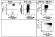
- FIG. 1 shows the results of FACS analysis of naive CD4 + T cells cultured in a Th1 medium and then hypoxic cultured in a differentiation-inducing factor starvation medium in Example 1.
- FIG. FIG. 1(A) is a dot plot of naive CD4 + T cells before induction of differentiation (cells before stimulation), and FIG. ),
- FIG. 1 (C) and (D) are cells cultured in Th1 medium for 13 days and then hypoxic cultured in differentiation inducer starvation medium for 9 days (stimulation 13 days + non-stimulation 9 days cells) are dot plots.
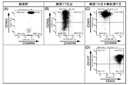
- FIG. 1 is a diagram showing the results of FACS analysis of naive CD4 + T cells cultured in Th2 medium and then hypoxic cultured in a differentiation-inducing factor starvation medium in Example 1.
- FIG. FIG. 2(A) is a dot plot of naive CD4 + T cells before induction of differentiation (cells before stimulation), and FIG. ),
- FIG. 2 (C) and (D) are cells cultured in Th2 medium for 10 days and then hypoxic cultured in differentiation inducer starvation medium for 7 days (stimulation 10 days + non-stimulation 7 days cells) are dot plots.
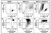
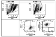
- FIG. Figures 3(A) and (D) are dot plots of naive CD8 + T cells before induction of differentiation (cells before stimulation); Dot plots of cultured cells (cells on day 8 of stimulation), FIG. 3 (C) and (F) are cultured in CD8-TA medium for 7 days, followed by hypoxic culture for 8 days in differentiation inducer starvation medium. Dot plots of stimulated cells (7 days stimulated + 8 days unstimulated cells).
- FIG. 3 is a dot plot showing the results of FACS of naive CD4 + T cells cultured in a Th2 medium, cryopreserved, and then cultured normally in a differentiation-inducing factor starvation medium in Example 3.
- FIG. 4 is a dot plot showing the results of FACS of naive CD8 + T cells cultured in CD8-TA medium, cryopreserved, and then normally cultured in differentiation-inducing factor starvation medium in Example 4.
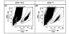
- Example 6 naive CD4 + T cells or naive CD8 + T cells collected from a 59-year-old human were subjected to hypoxic culture or normal culture in a differentiation-inducing factor starvation medium after induction of differentiation, and the results of FACS were shown. It is a diagram.
- Example 7 naive B cells were activated by inducing differentiation (activated B cells), and then hypoxic cultured cells in a differentiation-inducing factor starvation medium (cells on day 6 of non-stimulation) FACS It is the figure which showed the result of.
- Example 8 naive B cells were activated by inducing differentiation (activated B cells), and then hypoxic cultured cells in a differentiation-inducing factor starvation medium (cells on day 7 of non-stimulation) FACS It is the figure which showed the result of.
- Example 9 naive B cells were activated by inducing differentiation (activated B cells), and then hypoxic cultured cells in a differentiation-inducing factor starvation medium (cells on day 8 of non-stimulation) FACS It is the figure which showed the result of.
- normal oxygen environment refers to an environment in which the oxygen concentration is approximately the same as the atmospheric pressure, that is, approximately 19 to 21% by volume.
- a “low-oxygen environment” refers to an environment with an oxygen concentration of 0.1 to 10% by volume.
- the term “memory conversion” of T cells and B cells refers to stopping proliferation and retaining the ability to differentiate into effector cells in an environment without stimulation by antigens or differentiation-inducing factors. It means that the cells acquire a trait that enables long-term survival (undifferentiated growth arrest long-term viability) and that the expression pattern of cell surface antigens becomes the same as that of natural memory cells.
- the undifferentiated growth arrest long-term viability specifically means a trait capable of surviving for 7 days or longer, preferably 1 month or longer, in the absence of an antigen or differentiation-inducing factor.
- the method for producing memory T cells comprises an activation step of activating naive T cells with a differentiation inducer, and after the activation step, activating the activated T cells in the absence of a differentiation inducer. and a memoryization step of producing memory T cells by culturing in a memory, wherein the activated T cells are cultured in a hypoxic environment in the memoryization step.
- Cell dormancy (Quiescent) is induced by culturing T cells activated by a differentiation-inducing factor in the absence of the differentiation-inducing factor, but only in a hypoxic environment instead of a normal oxygen environment. It is a finding first discovered by the present inventors.
- naive T cells provided in the present invention may be naive T cells collected from animals, or may be cells proliferated by primary culture or subculture of naive T cells collected from animals. good.
- Naive T cells differentiated from ES (embryonic stem) cells or iPS (induced pluripotent stem) cells can also be used in the present invention.
- naive T cells from living bodies can be performed by standard methods. For example, naive T cells are present in peripheral blood like other lymphocytes. Therefore, naive T cells can be collected by fractionating peripheral blood mononuclear cells (PBMC) from peripheral blood collected from an animal and purifying naive T cells from the PBMC.
- PBMC peripheral blood mononuclear cells
- the fractionation of PBMC fractions from peripheral blood can be performed by conventional methods such as density gradient centrifugation using Ficoll (registered trademark). Alternatively, a commercially available PBMC preparation kit can be used.
- Naive T cells differ from effector cells in cell surface antigens.
- naive T cells have CD45RA, CD62L and CD127 among cell surface antigens. Therefore, only the naive T cell population with CD45RA, CD62L and CD127 can be purified by labeling PBMC with fluorescently labeled antibodies against these cell surface antigens and then using a fluorescence activated cell sorter (FACS).
- FACS fluorescence activated cell sorter
- the naive CD4 + T cell population can be purified, and only the cell population with CD45RA, CD62L, CD127 and CD8 can be purified.
- a naive CD8 + T cell population can be purified by .
- the FACS apparatus and fluorescence-labeled antibodies against various surface antigens can be appropriately selected from commonly used ones, and commercially available ones can also be used.
- naive T cells can be purified from PBMC by a magnet using immunomagnetic beads, in which antibodies against the above-mentioned cell surface antigens are bound to magnetic beads.
- naive T cells are first activated with a differentiation-inducing factor as the activation step.
- naive T cells are cultured in a culture medium containing a differentiation inducer.
- a differentiation-inducing factor an anti-human CD3 antibody or an anti-human CD3 antibody/anti-human CD28 antibody for giving an activation signal to the T cell receptor (TCR) is used as a basic factor, and in addition naive T cells are used as effector cells.
- Cytokines required for induction of differentiation can be appropriately selected and used.
- Such cytokines include various growth factors such as interleukin (IL), interferon (IFN) and transforming growth factor (TGF).
- naive CD4 + T cells can be activated to differentiate into CD4 + Th1 cells by stimulation with IL-12, IFN ⁇ .
- IL-12 IL-12
- IL-4 IL-4
- IL-7 TSLP (thymic stromal lymphopoietin)
- they can be activated to differentiate into CD4 + Th2 cells
- naive CD4 + T cells can be stimulated by IL-2.
- IL-6 can be activated to differentiate into CD4 + T FH cells
- naive CD4 + T cells can be stimulated with TGF ⁇ , IL-6 to differentiate into CD4 + Th17 cells.
- naive CD8 + T cells can be activated to differentiate into CD8 + killer T cells by stimulation with IL-2.
- These differentiation-inducing factors may be recombinant proteins, natural products secreted from animal cells, or chemically synthesized products.
- the culture medium for culturing naive T cells containing a differentiation-inducing factor is not particularly limited as long as it is a medium capable of culturing lymphocytes.
- media such as RPMI (Roswell Park Memorial Institute) 1640 medium and IMDM (Iscove's Modified Dulbecco's Medium) medium can be used.
- These media may be media containing inactivated serum or serum-free media.
- serum concentration is not particularly limited, and can be, for example, 5 to 15% by volume.
- any of fetal bovine serum, bovine serum, horse serum, goat serum, human serum and the like can be used.
- these culture media contain additives such as 2-mercaptoethanol, insulin, transferrin, sodium selenite, ethanolamine, and albumin that are commonly used as additives to culture media. may
- the culturing in the activation process can be carried out in the same way as the culturing of ordinary cultured cell lines, except for culturing in a medium capable of culturing lymphocytes containing a differentiation-inducing factor.
- the culture temperature is preferably 30-41°C, more preferably 36-38°C. Cultivation can also be performed in the air, but it is preferable to culture in a normal oxygen concentration environment in which the carbon dioxide concentration is controlled to 4 to 10% by volume.
- the culture time is not particularly limited as long as it is a sufficient time to activate naive T cells. More preferably, naive T cells can be activated by culturing for 6 to 10 days.
- the activated T cells are cultured in a hypoxic environment in a culture medium that does not contain differentiation-inducing factors as a memory formation step.
- the culture medium containing no differentiation-inducing factor is not particularly limited, and a medium capable of culturing lymphocytes can be used without containing a differentiation-inducing factor.
- RPMI 1640 medium, IMDM medium, etc. can be used as a medium capable of culturing lymphocytes, and serum and various additives may be added to these basal mediums.
- the same additives as listed above can be used.
- the culture medium used in the memoryization step may be the culture medium used in the activation step from which the differentiation inducer has been removed, or a basal medium different from the culture medium used in the activation step.
- the oxygen concentration during culture in the memory-forming step is 0.1 to 10% by volume, preferably 0.5 to 5.0% by volume, more preferably 0.5 to 1.5% by volume. preferable.
- activated T cells are treated at 30 to 41° C., preferably 36 to 38° C., in a hypoxic environment in which the carbon dioxide concentration is controlled to 4 to 10% by volume, for 5 to 21 days, preferably is cultured for 6-14 days, more preferably 7-10 days.
- Activated T cells can be easily memorized by lowering the oxygen concentration and applying stress.
- activated T cells differentiate into stem cell memory T cells, central memory T cells, or effector memory T cells.
- Stemness memory T cells have self-renewal ability and stemness in addition to long-term viability, and differentiate into central memory T cells and effector memory T cells upon antigen stimulation.
- Central memory T cells have long-term viability and self-renewal capacity and are commonly localized in lymph nodes and peripheral circulation.
- Effector memory T cells have long-term viability and are generally localized in the peripheral circulation and tissues.
- Stem cell memory T cells, central memory T cells, and effector memory T cells can be distinguished by differences in the expression patterns of cell surface antigens.
- Conventional memory T cells formed ex vivo are blast cells with a heterogeneous shape and continue self-renewal, whereas memory T cells obtained by the memory formation process of the present invention are , similar to natural memory T cells, are small, spherical, relatively constant-shaped, growth-arrested cells. From the characteristics of this morphology and cell growth arrest/diapause, the method for producing memory T cells according to the present invention can be said to be a method for easily obtaining memory T cells having characteristics similar to those of natural memory T cells.
- a cell population in which the ratio of memory cells to all living cells is 20% or more, preferably 50% or more, more preferably 70% or more, and still more preferably 80% or more is obtained by the memory conversion step. That is, the culture after culturing in the memory formation step is such that the total number of living cells of stem cell memory T cells, central memory T cells, and effector memory T cells is 20 of the total number of living cells contained in the composition. % or more, preferably 50% or more, more preferably 70% or more, and still more preferably 80% or more.
- pre-stressed naive cells may be used, or the activated T cells may be subjected to stress treatment before the memory conversion step. It can induce cell dormancy and can make naive T cells memoryized. Stress treatments include temperature stress and aging.
- naive T cells can also be memoryized by subjecting them to temperature stress.
- Temperature stress includes low temperature stress of 0 to 10° C., preferably 1 to 5° C., and freezing stress.
- naive T cells are kept at 0-10° C. for 1 hour or more, preferably 1-7 days, more preferably 2-4 days, before being subjected to the activation step of activating with a differentiation-inducing factor. Treat and apply cold stress.
- Memory T cells can be produced by activating cold-stressed naive T cells with a differentiation inducer and culturing the activated T cells in the absence of a differentiation inducer.
- the activated T cells may be cultured in a culture medium containing no differentiation-inducing factor, and may be performed in a normal oxygen environment or in a hypoxic environment.
- naive T cells before being subjected to the activation step of activating with a differentiation-inducing factor are freeze-thawed, then activated with a differentiation-inducing factor, and then the activated T cells are subjected to differentiation induction.
- Memory T cells can also be produced by culturing in the absence of factors. Freezing and thawing can be performed in the same manner as freezing and thawing of general cells.
- naive T cells can be frozen by suspending cells in a cryopreservation medium and leaving the resulting suspension in a freezer at -70 to -150°C.
- the cryopreservation solution may be the medium itself used for cell culture, a solution obtained by adding serum to a buffer such as phosphate saline (PBS), or a solution obtained by adding a cytoprotective agent such as DMSO or sericin. Addition of some cytoprotective agent is essential, but serum may or may not be added. Frozen cells can be thawed using a water bath or the like.
- a buffer such as phosphate saline (PBS)
- a cytoprotective agent such as DMSO or sericin. Addition of some cytoprotective agent is essential, but serum may or may not be added.
- Frozen cells can be thawed using a water bath or the like.
- the temperature stress may be applied to the activated T cells before memoryization.
- memory T cells can be produced by freezing and thawing the activated T cells and then culturing them in the absence of a differentiation-inducing factor as a memory conversion step. Cultivation of T cells after freezing and thawing may be performed in a culture medium containing no differentiation inducer, and may be performed in a normal oxygen environment or in a hypoxic environment.
- memory T cells can be produced without applying hypoxic stress or temperature stress.
- naive T cells collected from a person aged 50 years or older are activated with a differentiation-inducing factor and then cultured in a culture medium containing no differentiation-inducing factor in a normal oxygen environment to make them memory. can be done.
- Memory T cells obtained by memory conversion and compositions containing them can be used as active ingredients of pharmaceutical compositions in the same manner as other memory T cells.
- memory T cells themselves can be used as raw materials for cells to be administered to patients as pharmaceuticals in cell therapy and the like.
- memory T cells obtained by memory conversion can be genetically modified to produce CAR-T cells used in CAR-T therapy.
- a higher therapeutic effect can be expected by returning memory T cells obtained by memory conversion to the patient's body together with activated cancer cell surface antigen-presenting cells.
- memory T cells obtained by memory conversion For example, a portion of the fraction containing activated lymphocytes collected at the same time as DC vaccine production is frozen, thawed, and then cultured in a differentiation-inducing factor-free medium under a hypoxic environment.
- a cancer antigen-specific memoryized lymphocyte population obtained by mixing cells that survived the memoryization with activated cancer cell surface antigen-presenting cells is returned to the body of the patient.
- T cells other than naive T cells can be made to become memories by culturing them in a culture medium that does not contain differentiation-inducing factors under a hypoxic environment. For example, by culturing CAR-T cells prepared for use in CAR-T therapy in a culture medium that does not contain a differentiation-inducing factor in a hypoxic environment, memoryized CAR-T cells, that is, CAR-T cells with undifferentiated growth-arrested long-term viability are obtained.
- the method for producing memory T cells according to the present invention can also be used to eliminate exhaustion of T cells, which is a problem in cancer immunotherapy.
- T cell exhaustion is a phenomenon in which the ability of T cells to attack cancer cells to attack cancer cells is weakened due to continued exposure to cancer antigens for too long. Exhaustion is known to involve multiple transcription factors such as TOX, TOX2, and NR4A, genome modification, metabolic changes, and the like. Culturing T cells exhausted by excessive activation stimulation due to long-term exposure to cancer antigens in a culture medium that does not contain differentiation-inducing factors under a hypoxic environment caused T cell exhaustion. It is possible that factors can be downregulated to reset the state inside the T cell. By returning the memoryized T cells thus obtained to the patient's body, a longer-term therapeutic effect can be obtained in cancer immunotherapy.
- naive B cells under hypoxic stress after activation, it is possible to acquire the trait of undifferentiated growth arrest and long-term viability in a differentiation-inducing factor-independent manner.
- an activation step of activating naive B cells with a differentiation-inducing factor and after the activation step, culturing the activated B cells in the absence of a differentiation-inducing factor causes differentiation.
- naive B cells provided in the present invention may be naive B cells collected from animals, or may be cells proliferated by primary culture or subculture of naive B cells collected from animals. good.
- Naive B cells differentiated from ES (embryonic stem) cells or iPS (induced pluripotent stem) cells can also be used in the present invention.
- Naive B cells can be collected from a living body and purified from PBMC using FACS in the same way as naive T cells.
- naive B cells have cell surface antigens CD20 and CD27, but not CD38. Therefore, it is possible to purify only the naive B cell population expressing CD20 and CD27 but not CD38 by labeling PBMC with fluorescently labeled antibodies against these cell surface antigens and then using FACS. can.
- the FACS apparatus and fluorescence-labeled antibodies against various surface antigens can be appropriately selected from commonly used ones, and commercially available ones can also be used.
- naive T cells can be purified from PBMC by a magnet using immunomagnetic beads, in which antibodies against the above-mentioned cell surface antigens are bound to magnetic beads.
- the differentiation-inducing factor can be appropriately selected and used from cytokines required for inducing differentiation of naive B cells into effector cells.
- naive B cells can be activated to differentiate into plasma cells by using IL-2, IL-4, IL-6, IL-10, IL-15, IL-21.
- a culture medium containing a differentiation-inducing factor in a medium capable of culturing lymphocytes is used.
- RPMI 1640 medium, IMDM medium, etc. can be used as a medium capable of culturing lymphocytes, and serum and various additives may be added to these basal mediums.
- the additive the same additives as listed above can be used.
- the culturing in the activation process can be carried out in the same way as the culturing of ordinary cultured cell lines, except for culturing in a medium capable of culturing lymphocytes containing a differentiation-inducing factor.
- the culture temperature is preferably 30-41°C, more preferably 36-38°C. Cultivation can also be performed in the air, but it is preferable to culture in a normal oxygen concentration environment in which the carbon dioxide concentration is controlled to 4 to 10% by volume.
- the culture time is not particularly limited as long as it is a sufficient time to activate naive B cells. More preferably, naive B cells can be activated by culturing for 3 to 10 days.
- the activated B cells are cultured in a hypoxic environment in a culture medium containing no differentiation-inducing factor as a long-term viability acquisition step.
- the culture medium containing no differentiation-inducing factor is not particularly limited, and a medium capable of culturing lymphocytes can be used without containing a differentiation-inducing factor.
- RPMI 1640 medium, IMDM medium, etc. can be used as a medium capable of culturing lymphocytes, and serum and various additives may be added to these basal mediums.
- the same additives as listed above can be used.
- the culture medium used in the long-term viability acquisition step may be the culture medium used in the activation step from which differentiation-inducing factors have been removed, or may be a basal medium different from the culture medium used in the activation step. good.
- the oxygen concentration during culture in the long-term viability acquisition step is 0.1 to 10% by volume, preferably 0.5 to 5% by volume, more preferably 0.5 to 1.5% by volume. preferable.
- activated B cells are treated at 30 to 41° C., preferably 36 to 38° C., in a hypoxic environment in which the carbon dioxide concentration is controlled to 4 to 10% by volume, for 5 to 21 days, preferably is cultured for 6-14 days, more preferably 6-10 days.
- activated B cells acquire long-term viability independent of differentiation-inducing factors while remaining plasma cells, or differentiate into germinal center B cells or transitional B cells. Both germinal center B cells and transitional B cells have long-term viability independent of differentiation inducers.
- B cells that have acquired differentiation-inducing factor-independent long-term viability and compositions containing them can be used as active ingredients of pharmaceutical compositions in the same manner as other B cells.
- B cells themselves that have acquired long-term viability independent of differentiation-inducing factors can be used as raw materials for cells to be administered to patients as pharmaceuticals in cell therapy and the like.
- composition of the medium used in subsequent experiments is as follows.
- PBS/2% FBS A solution of 49 mL PBS (Phosphate Saline) mixed with 1 mL FBS (Fetal Bovine Serum).
- PBS/2% FBS/1 mM EDTA A solution of 1 mL FBS and 0.1 mL 0.5 M EDTA in 49 mL PBS.
- RPMI 1640/10% FBS/2-ME To 45 mL of RPMI 1640 (#12633-012, Invitrogen), 5 mL of FBS and 0.5 mL of 100 ⁇ PSG (“100 ⁇ penicillin-streptomycin-glutamine”, # 10378-016, manufactured by Invitrogen) and 50 ⁇ L of 50 mM 2-mercaptoethanol (2-ME).
- IMDM/10% FBS/2-ME In 45 mL of IMDM (“IMDM ( ⁇ 1) + GlutaMAX-1 medium”, #31980-030, manufactured by Invitrogen), 5 mL of FBS and 0.5 mL of 100 ⁇ PSG Medium mixed with 50 ⁇ L of 50 mM 2-ME.
- Th1 medium 10 mL of RPMI 1640/10% FBS/2-ME, 50 ⁇ L of "200 ⁇ Human Th1 reagent 1 (IL-12)", 50 ⁇ L of "200 ⁇ Human Th1 reagent 2 (IFN- ⁇ )", A medium mixed with 200x Human Th1 reagent 1 (IL-12) and 200x Human Th1 reagent 2 (IFN- ⁇ ) are included in the Th1 differentiation induction kit ("CellXVivo human Th1 cell differentiation kit", #CDK001, manufactured by R&D Systems). Using.
- Th2 medium 10 mL of RPMI 1640/10% FBS/2-ME, 10 ⁇ L of "1000 ⁇ Human Th2 reagent 1 (IL-2)" and 10 ⁇ L of "1000 ⁇ Human Th2 reagent 2 (IL-4)" , 10 ⁇ L of “1000 ⁇ Human Th2 reagent 3 (IL-7)” and 10 ⁇ L of “1000 ⁇ Human Th2 reagent 4 (TSLP)” mixed medium.
- 1000x Human Th2 reagent 1 (IL-2), 1000x Human Th2 reagent 2 (IL-4), 1000x Human Th2 reagent 3 (IL-7), and 1000x Human Th2 reagent 4 (TSLP) induce Th2 differentiation.
- CD8-TA medium In 10 mL of RPMI 1640/10% FBS/2-ME, 10 ⁇ L of T cell activation magnetic beads (“Dynabeads human T-activator CD3/CD28”, #11131D, manufactured by Gibco) and 10 ⁇ L of A medium mixed with IL-2 (human recombinant IL-2, #202-IL, manufactured by R&D Systems) solution (10 ⁇ g/mL).
- IL-2 human recombinant IL-2, #202-IL, manufactured by R&D Systems
- Step 1 medium 10 mL of RPMI 1640/10% FBS/2-ME, 20 ⁇ L of "500 ⁇ B cell Expander 1 (CD40L)", 20 ⁇ L of "500 ⁇ B cell Expander 2 (IL-4)", 20 ⁇ L of “500 ⁇ B cell Expander 3 (anti-CD40L)”, 10 ⁇ L of IL-2 (human recombinant IL-2, #202-IL, manufactured by R & D Systems) solution (10 ⁇ g/mL) and 50 ⁇ L of IL-10 (human recombinant IL-10, #217-IL-005, manufactured by R & D Systems) solution (10 ⁇ g / mL) and 50 ⁇ L of IL-21 (human recombinant IL-21, #200-21-10UG , manufactured by PeproTech) solution (10 ⁇ g/mL) and a medium mixed with.
- IL-2 human recombinant IL-2, #202-IL, manufactured by R & D Systems
- IL-10 human recombinant
- Step 2 Medium In 10 mL IMDM/10% FBS/2-ME, 10 ⁇ L IL-2 solution (10 ⁇ g/mL) and 5 ⁇ L IL-6 (human recombinant IL-6, #200-06-20UG, PeproTech) solution (20 ⁇ g/mL) and 50 ⁇ L of IL-21 solution (10 ⁇ g/mL) mixed together.
- B27 medium 48.5 mL of RPMI 1640 was mixed with 1 mL of "50 x B27 supplement" (serum free, #17504044, manufactured by Gibco), 0.5 mL of 100 x PSG, and 50 ⁇ L of 50 mM 2-ME. solution.
- N2 medium A solution obtained by mixing 49 mL of RPMI 1640 with 0.5 mL of “100 ⁇ N2 supplement” (#17502001, manufactured by Gibco), 0.5 mL of 100 ⁇ PSG, and 50 ⁇ L of 50 mM 2-ME.
- BSA medium 49 mL of RPMI 1640 was mixed with 0.5 mL of 100 ⁇ BSA (200 mg/mL, #A4161, Sigma-Aldrich), 0.5 mL of 100 ⁇ PSG, and 50 ⁇ L of 50 mM 2-ME solution.
- LPS/IL-4 medium 50 ⁇ L of lipopolysaccharide (LPS) (E. coli OIII:B4, L4391-1MG, manufactured by Sigma) solution (1 mg/mL) in 10 mL of RPMI 1640/10% FBS/2-ME ) and 10 ⁇ L of IL-4 (human recombinant IL-4, AF-200-04, manufactured by PeproTech) solution (10 ⁇ g/mL).
- LPS lipopolysaccharide
- IL-4 human recombinant IL-4, AF-200-04, manufactured by PeproTech
- ODN2006 medium 10 mL of RPMI 1640/10% FBS/2-ME with 10 ⁇ L of IL-2 solution (10 ⁇ g/mL) and 5 ⁇ L of IL-6 (human recombinant IL-6, #200-06-20UG, PeproTech) solution (20 ⁇ g/mL) and 20 ⁇ L of oligonucleotide 2006 (ODN2006) (tlrl-2006, Invivogen) solution (2.5 mg/mL).
- Antibodies and the like used in FACS are as follows.
- Anti-CD45RA antibody FITC-labeled mouse anti-human CD45RA monoclonal antibody (HI100, #11-0458-42, manufactured by Invitrogen)
- Anti-CD62L antibody PE-labeled mouse anti-human CD62L monoclonal antibody (DREG-56, #12-0629-42, manufactured by Invitrogen)
- Anti-CD127 antibody PE-Cyanine7-labeled mouse anti-human CD127 monoclonal antibody (eBioRDR5, #25-1278-42, manufactured by Invitrogen)
- Anti-CD20 antibody FITC-labeled mouse anti-human CD20 monoclonal antibody (2H7, #11-0209-42, manufactured by Invitrogen)
- Anti-CD27 antibody PE-labeled mouse anti-human CD27 monoclonal antibody (O323, #12-0279-42, manufactured by nvitrogen)
- Anti-CD38 antibody PE-Cyan
- Example 1 Naive CD4 + T cells collected from peripheral blood collected from a 21-year-old human were differentiated and then cultured in a differentiation-inducing factor-starved medium in a hypoxic environment to produce memory lymphocytes.
- PBMC peripheral blood mononuclear cells
- the PBMC collection tube was then centrifuged (2,600 rpm, 10 minutes, room temperature), and the contents of the PBMC collection tube were transferred to a 50 mL volume containing 25 mL of PBS/2% FBS in advance. After being transferred to a plastic tube by decantation, it was centrifuged (1,300 rpm, 8 minutes, room temperature). After removing the supernatant of the plastic tube and loosening the precipitated cell pellet by tapping, add 50 mL of PBS/2% FBS by decantation and mix by inverting 3 times, then centrifuge again (1,300 rpm , 8 min, room temperature) and the supernatant was removed. 10 mL of PBS/2% FBS/1 mM EDTA was added to the cell pellet that had settled in the plastic tube, and the cells were suspended by pipetting about 10 times.
- Naive CD4 + T cells were recovered from the cell suspension prepared in (1) above.
- Naive CD4 + T cells were collected using a commercially available naive CD4 + T cell isolation kit (“EasySep human naive CD4 + T cell isolation kit II”, #17555, manufactured by STEMCELL Technologies). I followed the manual. 10 ⁇ L of purified naive CD4 + T cell suspension was aliquoted into a separate tube for cell counting. An equal volume of trypan blue solution (#29853-34, manufactured by Nacalai Tesque) was added to 10 ⁇ L of this cell suspension, and the number of viable cells was calculated.
- a Th1 medium was added to the cell pellets that had settled in the plastic tube so that the concentration was 2 ⁇ 10 5 cells/mL, and the cells were suspended.
- the resulting cell suspension was dispensed into 24-well plates coated with mouse anti-human CD3 antibody (accessory of the kit) at 1 mL per well.
- the plate was cultured at 37° C., 5% CO 2 , 20% O 2 for 7 days or more. During the culture period, if the pH of the medium drops due to lactic acid produced by an increase in the number of cells and the medium turns yellow, transfer half of the medium to another mouse anti-human CD3 antibody-coated well and dilute twice with fresh Th1 medium. and continued culturing.
- naive CD4 + T cells into CD4 + Th2 cells is performed using a commercially available Th2 differentiation induction kit (“CellXVivo human Th2 cell differentiation kit”, # CDK002, manufactured by R&D Systems) was used according to the manual attached to the kit.
- the purified naive CD4 + T cell suspension prepared in (2) above was transferred to a 15 mL plastic tube, and then centrifuged (1,000 rpm, 5 minutes, room temperature) to remove the supernatant. .
- a Th2 medium was added to the cell pellets that had settled in the plastic tube to suspend the cells at 2 ⁇ 10 5 cells/mL.
- the resulting cell suspension was dispensed into 24-well plates coated with mouse anti-human CD3 antibody (accessory of the kit) at 1 mL per well.
- the plate was cultured at 37° C., 5% CO 2 , 20% O 2 for 7 days or more. During the culture period, if the pH of the medium drops due to the lactic acid produced by the increase in the number of cells and turns yellow, transfer half of the culture medium to another mouse anti-human CD3 antibody-coated well and dilute 2-fold with fresh Th2 medium. and continued culturing.
- the prepared cell suspension was dispensed into T25 flasks in 5 mL aliquots. These T25 flasks were cultured (hypoxic culture) for 7 days or more in an environment of 37° C., 5% CO 2 , 1% O 2 . Control cells were cultured at 37° C., 5% CO 2 , 20% O 2 for 7 days or more (normal culture). Cultured cells were subjected to FACS analysis.
- the cells after the culture in (4) above were also subjected to hypoxic culture in the same way, and the cultured cells were subjected to FACS analysis.
- T cell antibody cocktail (5 ⁇ L of anti-CD45RA antibody, 5 ⁇ L of anti-CD62L antibody, 5 ⁇ L of anti-CD127 antibody, and 7 ⁇ L of 7-AAD) was added and mixed by pipetting. After that, it was allowed to stand in ice for 20 minutes in a dark place. Then, 5 mL of PBS/2% FBS was added to the cell suspension to suspend the cells, and then the plastic tube was centrifuged (1,000 rpm, 5 minutes, room temperature) to remove the supernatant. did. 0.5 mL of PBS/2% FBS was added to the precipitated cell pellet to suspend the cells, and the resulting cell suspension was transferred to a 5 mL FACS tube. The FACS tube was measured using a FACS apparatus ("FACSCanto II", manufactured by Becton Dickinson), and the obtained data was analyzed using analysis software ("FACSDiva ver. 6.1", manufactured by Becton Dickinson).
- Naive T cells naive CD4 + T cells, naive CD8 + T cells
- effector T cells CD4 + Th1 cells, CD4 + Th2 cells, CD8 + killer T cells
- stem cell memory T cells Ste Cell Memory T cells
- central memory T cells central memory T cells
- effector memory T cells each have cell surface antigens shown in Table 1.
- FIG. 1(A) is a dot plot showing the results of FACS of naive CD4 + T cells before induction of differentiation (cells before stimulation), and FIG. 1(B) shows cells cultured in Th1 medium for 15 days ( Fig. 1 (C) and (D) are dot plots showing FACS results of cells on day 15 of stimulation), Figs. Fig. 3 is a dot plot showing the results of FACS of cells hypoxic cultured for 9 days in a differentiation-inducing factor starvation medium (cells on day 13 of stimulation + cells on day 9 of non-stimulation). It was confirmed by FACS analysis that when naive CD4 + T cells were cultured in a Th1 medium for 13 days and then cultured normally in a differentiation-inducing factor starvation medium for 9 days, all cells died.
- naive CD4 + T cells When naive CD4 + T cells were cultured in Th1 medium for 15 days, most of them were CD45RA- cells, forming a mixed population of mature effector cells (CD4 + Th1 cells) and immature effector cells. was confirmed (FIG. 1(B)). In contrast, when naive CD4 + T cells were cultured in Th1 medium for 13 days and then hypoxic cultured in differentiation-inducing factor starvation medium for 9 days, 52.0% of the cells survived, unlike the normal culture. Was. In addition, it was confirmed that the cell population consisted of stem cell memory T cells, central memory T cells, and effector memory T cells (FIGS. 1(C) and (D)).
- FIG. 2(A) is a dot plot showing the results of FACS of naive CD4 + T cells before induction of differentiation (cells before stimulation), and FIG. 2(B) shows cells cultured in Th2 medium for 17 days ( Fig. 2(C) and (D) are dot plots showing FACS results of cells on day 17 of stimulation), Figs. 2(C) and (D) are cultured in Th2 medium for 10 days, then RPMI 1640/10% FBS/2-ME ( FIG. 10 is a dot plot showing the results of FACS of cells hypoxically cultured for 7 days in a differentiation-inducing factor starvation medium (cells on day 10 of stimulation + cells on day 7 of non-stimulation). It was confirmed by FACS analysis that when naive CD4 + T cells were cultured in a Th2 medium for 10 days and then cultured normally in a differentiation-inducing factor starvation medium for 7 days, all cells died.
- naive CD4 + T cells were activated by induction of differentiation and then cultured in a hypoxic environment to acquire long-term viability and become memory cells. It was confirmed that not only central memory T cells and effector memory T cells but also stem cell memory T cells having self-renewal ability and stemness can be generated.
- Example 2 Naive CD8 + T cells collected from peripheral blood collected from a 20-year-old human were differentiated and then cultured in a differentiation-inducing factor-starved medium in a hypoxic environment to produce memory lymphocytes.
- naive CD8 + T cells were recovered from the cell suspension prepared in the same manner as in Example 1 (1).
- Naive CD8 + T cells were collected using a commercially available naive CD8 + T cell isolation kit (“EasySep human naive CD8 + T cell isolation kit II”, #17928, manufactured by STEMCELL Technologies). I followed the manual. 10 ⁇ L of purified naive CD8 + T cell suspension was aliquoted to a separate tube for cell counting. An equal volume of trypan blue solution was added to 10 ⁇ L of this cell suspension, and the number of viable cells was calculated.
- naive CD8 + T cells Differentiation induction from naive CD8 + T cells to CD8 + killer T cells
- the purified naive CD8 + T cell suspension prepared in (1) above was transferred to a 15 mL plastic tube, and then centrifuged. Treat (1,000 rpm, 5 minutes, room temperature) and remove the supernatant.
- a CD8-TA medium was added to the cell pellets that had settled in the plastic tube so that the cells were suspended at 2 ⁇ 10 5 cells/mL.
- the resulting cell suspension was dispensed into uncoated 24-well plates at 1 mL per well. The plate was cultured at 37° C., 5% CO 2 , 20% O 2 for 7 days or more.
- the cell suspension in the FACS tube was transferred to a new 5 mL FACS tube by decantation while still set on the magnetic stand.
- This new FACS tube was again set on the magnetic stand and allowed to stand at room temperature for 3 minutes, after which the cell suspension in the FACS tube was transferred to a new 5 mL FACS tube by decantation.
- the prepared cell suspension was dispensed into T25 flasks in 5 mL aliquots. These T25 flasks were cultured (hypoxic culture) for 7 days in an environment of 37° C., 5% CO 2 , 1% O 2 . Control cells were cultured at 37° C., 5% CO 2 , 20% O 2 for 7 days (normal culture). Cultured cells were subjected to FACS analysis.
- FIG. 3 shows the results of FACS analysis of naive CD8 + T cells cultured in CD8-TA medium and then hypoxic cultured in differentiation-inducing factor starvation medium.
- (A) and (D) are dot plots showing the results of FACS of naive CD8 + T cells before induction of differentiation (cells before stimulation)
- (B) and (E) are CD8 - Dot plots showing FACS results for cells cultured for 8 days in CD8-TA medium (cells on day 8 of stimulation)
- FIG. 3 is a dot plot showing the results of FACS of cells hypoxic cultured in 1640/10% FBS/2-ME (differentiation-inducing factor starvation medium) for 8 days (7 days of stimulation + 8 days of non-stimulation).
- the upper (A) to (C) are plotted with the vertical axis as the side scattered light signal (SSC) and the horizontal axis as the forward scattered light signal (FSC).
- SSC side scattered light signal
- FSC forward scattered light signal
- FIG. 3 the upper (A) to (C) are plotted with the vertical axis as the side scattered light signal (SSC) and the horizontal axis as the forward scattered light signal (FSC).
- SSC side scattered light signal
- FSC forward scattered light signal
- FIG. 3 the upper (A) to (C) are plotted with the vertical axis as the side scattered light signal (SSC) and the horizontal axis as the forward scattered light signal (FSC).
- F is a diagram plotting the fluorescence intensity
- naive CD8 + T cells are relatively uniform in shape and small in size (FIG. 3(A)), but CD8 + cells that are activated by differentiation induction. T cells are highly variable in shape and most cells are larger than naive CD8 + T cells (Fig. 3(B)).
- the cell population hypoxic cultured in the differentiation-inducing factor starvation medium after activation had a relatively constant shape and was smaller in size than the activated cells (Fig. 3(C)).
- the cells after hypoxic culture in the differentiation-inducing factor starvation medium were closer to natural memory T cells than effector T cells not only in the expression pattern of cell surface antigens but also in shape and size.
- naive CD8 + T cells were activated by differentiation induction and then cultured in a differentiation-inducing factor-starved medium in a hypoxic environment to acquire long-term viability and become memory cells. Moreover, it was confirmed that stem cell memory T cells having self-renewal ability and stem cell properties can be produced very efficiently.
- Example 3 Naive CD4 + T cells collected from peripheral blood collected from a 21-year-old human were induced to differentiate into CD4 + Th2 cells, and then freeze-thawed to produce memory lymphocytes. Recovery of naive CD4 + T cells from human peripheral blood and induction of differentiation from naive CD4 + T cells to CD4 + Th2 cells were carried out in the same manner as in Example 1.
- cryopreservation solution A (90% FBS/10% DMSO) containing serum and DMSO
- cryopreservation solution B (“BAMBANKER” containing DMSO but not serum) was used. , #CS-04-001, Nippon Genetics), or cryopreservation solution C containing neither serum nor DMSO (“Cell Reservoir One”, #07579-24, Nacalai Tesque).
- cryopreservation containers (“BICELL”, manufactured by Nippon Freezer) and stored at -80°C.
- the cryotube was immersed in a water bath at room temperature to thaw the cell suspension inside the tube. The entire amount of the cell suspension in the tube is transferred to a 15 mL plastic tube, and the plastic tube is centrifuged (1,000 rpm, 5 minutes, room temperature) to remove the supernatant.
- a cell suspension was prepared by adding RPMI 1640/10% FBS/2-ME to 4 ⁇ 10 5 cells/mL and suspending the cells.
- the prepared cell suspension was dispensed into T25 flasks in 5 mL aliquots. These T25 flasks were cultured (normal culture) for 8 days in an environment of 37° C., 5% CO 2 , 20% O 2 .
- FIG. 4 shows the results of FACS of cells that were cultured in Th2 medium and then cryopreserved in cryopreservation medium A and then cultured normally in RPMI 1640/10% FBS/2-ME (differentiation-inducing factor starvation medium).
- Dot plot. (A) is a plot with SSC on the vertical axis and FSC on the horizontal axis.
- (B) is a plot with the vertical axis representing the fluorescence intensity emitted from the anti-CD62L antibody and the horizontal axis representing the fluorescence intensity emitted from the anti-CD45RA antibody.
- C is a plot with the vertical axis representing the fluorescence intensity emitted from the anti-CD127 antibody and the horizontal axis representing the fluorescence intensity emitted from the anti-CD62L antibody.
- cells within the gated area are viable cells.
- the cell viability after hypoxic culture was 27.1%, which was comparable to Example 2.
- the cell survival rate after normal culture was 2.3%, and while almost all cells were dead in Example 2, some cells survived.
- This surviving cell population was confirmed to be a cell population composed of stem cell memory T cells, central memory T cells, and effector memory T cells, similar to the hypoxic cultured cell population (Fig. 4 (B) and (C)). Even when cryopreservation was performed using cryopreservation medium B and cryopreservation medium C, cell populations composed of stem cell memory T cells, central memory T cells, and effector memory T cells were similarly obtained.
- naive CD4 + T cells were activated by induction of differentiation, then frozen, thawed, and cultured normally in a differentiation-inducing factor starvation medium. Regardless of the presence or absence of DMSO, it was confirmed that the cells acquire long-term viability and become memory T cells, as in the case of hypoxic culture.
- Example 4 Memory lymphocytes were produced by inducing differentiation of naive CD8 + T cells collected from peripheral blood collected from a 21-year-old human and then cryopreserving them. Recovery of naive CD8 + T cells from human peripheral blood, subsequent induction of differentiation, and removal of magnetic beads were performed in the same manner as in Example 2 (1) to (3).
- Example 4 (2) Thawing and culturing After storage at -80°C, the cells in the cryotube were thawed and then cultured (normal culture) for 8 days in an environment of 37°C, 5% CO 2 , 20% O 2 . Cells were fermented and cultured in the same manner as in Example 4 (2).
- FIG. 5 shows the FACS results of cells that were cultured in CD8-TA medium and then cryopreserved in cryopreservation medium A and then cultured normally in RPMI 1640/10% FBS/2-ME (differentiation-inducing factor starvation medium). Dot plot shown. Cells within the gated region in FIG. 5(A) are viable cells. The cell survival rate after hypoxic culture was 36.0%, which was comparable to Example 3. On the other hand, the cell survival rate after normal culture was 20.4%, and while almost all cells were dead in Example 3, a very large number of cells survived. Similar to the hypoxic cultured cell population, most of the surviving cell populations were stem cell memory T cells and central memory T cells, and effector memory T cells were also included (Fig.
- cryopreservation medium B and cryopreservation medium C Even when cryopreserved using cryopreservation medium B and cryopreservation medium C, similarly, most of them are stem cell memory T cells and central memory T cells, and there is a cell population containing effector memory T cells. Got.
- naive CD8 + T cells were activated by induction of differentiation, then frozen, thawed, and cultured normally in a differentiation-inducing factor starvation medium. Regardless of the presence or absence of DMSO, it was confirmed that the cells acquire long-term viability and become memory T cells, as in the case of hypoxic culture.
- Example 5 Naive CD4 + T cells and naive CD8 + T cells collected from human peripheral blood after refrigerated storage are induced to differentiate, respectively, and then cultured in a differentiation-inducing factor starvation medium in an oxygen environment of normal concentration to obtain memory T cells. Lymphocytes were produced.
- FIG. 6(A) shows FACS of naive CD4 + T cells after refrigerated storage, cultured in Th2 medium, and then normally cultured in RPMI 1640/10% FBS/2-ME (differentiation-inducing factor starvation medium) for 8 days.
- FIG. 6(B) shows naive CD8 + T cells after refrigerated storage, cultured in CD8-TA medium, and then normal cultured in RPMI 1640/10% FBS/2-ME (differentiation-inducing factor starvation medium) for 8 days.
- FIG. 6(B) shows naive CD8 + T cells after refrigerated storage, cultured in CD8-TA medium, and then normal cultured in RPMI 1640/10% FBS/2-ME (differentiation-inducing factor starvation medium) for 8 days.
- FIG. 6(B) shows naive CD8 + T cells after refrigerated storage, cultured in CD8-TA medium, and then normal cultured in RPMI 1640
- naive CD4 + T cells When non-refrigerated naive CD4 + T cells were induced to differentiate and then cultured normally, almost all of the cells died (Example 2) . In the case of cells, the cell viability was as high as 12.1% when cultured normally after induction of differentiation (Fig. 6(A)). It was confirmed from the surface antigens that the surviving cell population consisted of stem cell memory T cells, central memory T cells, and effector memory T cells, as in the case of hypoxic culture after induction of differentiation. .
- CD8 + T cells when non-refrigerated naive CD8 + T cells were differentiated and then cultured normally, almost all of the cells died (Example 3). In the case of CD8 + T cells, the cell viability was 1.7% in normal culture after induction of differentiation, and some cells survived (FIG. 6(B)). The surviving cell population is mostly stem cell memory T cells and central memory T cells, and effector memory T cells are also included, as in the case of hypoxic culture after differentiation induction, based on the surface antigens. was a group.
- Example 6 Naive CD4 + T cells and naive CD8 + T cells recovered from peripheral blood collected from a 59-year-old human were each induced to differentiate and then cultured in a differentiation-inducing factor starvation medium to produce memory lymphocytes. .
- FIGS. 7(A) and (C) show FACS results of cells cultured in RPMI 1640/10% FBS/2-ME (differentiation-inducing factor starvation medium) after culturing naive CD4 + T cells in Th2 medium. Dot plot shown.
- FIGS. 7(B) and (D) are dot plots showing FACS results of cells cultured in RPMI 1640/10% FBS/2-ME after naive CD4 + T cells were cultured in Th2 medium.
- naive CD4 + T cells collected from a 20-year-old human were induced to differentiate and then cultured normally, almost all of the cells died (Example 2).
- the cell viability in normal culture after induction of differentiation was 51.4%, which is lower than the cell viability of 60.9% in hypoxic culture. , was very high (FIGS. 7(A) and (C)). It was confirmed from the surface antigens that the surviving cell population consisted of stem cell memory T cells, central memory T cells, and effector memory T cells, as in the case of hypoxic culture after induction of differentiation. .
- naive CD8 + T cells collected from a 21-year-old human were induced to differentiate and then cultured normally, almost all of the cells died (Example 3).
- the cell viability was 19.0% when normal culture was performed after differentiation induction, and the cell viability was 24.9% when hypoxic culture was performed. Although not as high as it was, it was sufficiently high (FIGS. 7(B) and (D)).
- the surviving cell population is mostly stem cell memory T cells and central memory T cells, and effector memory T cells are also included, as in the case of hypoxic culture after differentiation induction, based on the surface antigens. was a group.
- Example 7 Naive B cells collected from human peripheral blood were induced to differentiate and then cultured in a differentiation-inducing factor-starved medium in a hypoxic environment to induce cell dormancy and stop proliferation without stimulation by a differentiation-inducing factor. Cells were produced that were capable of long-term survival under conditions.
- Naive B cells Collection of Naive B Cells from PBMC Naive B cells were collected from the cell suspension prepared in the same manner as in Example 1 (1). Naive B cells were collected using a commercially available naive B cell isolation kit (“EasySep human naive B cell isolation kit”, #17254, manufactured by STEMCELL Technologies) according to the manual attached to the kit. 10 ⁇ L of the purified naive B cell suspension was aliquoted into a separate tube for cell counting. An equal volume of trypan blue solution was added to 10 ⁇ L of this cell suspension, and the number of viable cells was calculated.
- naive B cell isolation kit (“EasySep human naive B cell isolation kit”, #17254, manufactured by STEMCELL Technologies) according to the manual attached to the kit. 10 ⁇ L of the purified naive B cell suspension was aliquoted into a separate tube for cell counting. An equal volume of trypan blue solution was added to 10 ⁇ L of this cell suspension, and the number of viable
- Step 1 medium was added to the cell pellets that had settled in the plastic tube so that the concentration was 2 ⁇ 10 5 cells/mL, and the cells were suspended.
- the resulting cell suspension was dispensed into 24-well plates coated with a goat anti-human Ig mixed secondary antibody (#H17000, manufactured by Invitrogen) at 1 mL per well. The plate was cultured for 5 days at 37° C., 5% CO 2 , 20% O 2 .
- the culture medium in each well was collected in a 15 mL plastic tube, centrifuged (1,000 rpm, 5 minutes, room temperature), and the supernatant was removed.
- Step 2 medium was added to the cell pellets that had settled in the plastic tube so that the concentration was 4 ⁇ 10 5 cells/mL, and the cells were suspended.
- the resulting cell suspension was dispensed into uncoated 24-well plates at 1 mL per well. The plate was cultured (hypoxic culture) for 4 days in an environment of 37° C., 5% CO 2 , 1% O 2 .
- the prepared cell suspension was dispensed into uncoated 24-well plates at 1 mL per well.
- the plate was cultured for 6 days in an environment of 37° C., 5% CO 2 , 1% O 2 .
- Control cells were cultured at 37° C., 5% CO 2 , 20% O 2 for 6 days (normal culture).
- Naive B cells, effector B cells (plasma cells), memory B cells, germinal center B cells, and transitional B cells each have cell surface antigens shown in Table 2.
- FIG. Figures 8 (A) and (B) are plotted with SSC on the vertical axis and FSC on the horizontal axis. It is a diagram plotted as the fluorescence intensity emitted from the anti-CD20 antibody, and FIG. 8 (D) plots the vertical axis as the fluorescence intensity emitted from the anti-CD38 antibody and the horizontal axis as the fluorescence intensity emitted from the anti-CD27 antibody. It is a diagram of Also, in the plots of FIGS. 8(A) and (B), the cells within the gated regions are viable cells.
- the cell survival rate after hypoxic culture in the differentiation-inducing factor starvation medium was 0.3%, and the majority (92.4%) was confirmed to be plasma cells from the surface antigen results.
- Example 8 Differentiation of naive B cells is performed with LPS and IL-4, which are outer membrane components of Gram-negative bacteria, and B cells that have acquired long-term viability of differentiation-inducing factor-independent undifferentiated growth arrest from naive B cells are obtained. manufactured.
- LPS and IL-4 which are outer membrane components of Gram-negative bacteria
- naive B cells Differentiation induction of naive B cells
- the purified naive B cell suspension prepared in (1) above was transferred to a 15 mL plastic tube, and then centrifuged (1,000 rpm, 5 minutes, room temperature). ) to remove the supernatant.
- An LPS/IL-4 medium was added to the cell pellet that had settled in the plastic tube to make the cell concentration 1 ⁇ 10 5 cells/mL, and the cells were suspended.
- the resulting cell suspension was dispensed into uncoated 24-well plates at 1 mL per well. The plate was cultured for 7 days in an environment of 37° C., 5% CO 2 , 1% O 2 (hypoxic culture).
- FIG. Figures 9 (A) and (B) are plotted with SSC on the vertical axis and FSC on the horizontal axis. It is a diagram plotted as the fluorescence intensity emitted from the anti-CD20 antibody, and FIG. 9 (D) plots the vertical axis as the fluorescence intensity emitted from the anti-CD38 antibody and the horizontal axis as the fluorescence intensity emitted from the anti-CD27 antibody. It is a diagram of Also, in the plots of FIGS. 9A and 9B, the cells within the gated regions are viable cells.
- the cell survival rate after hypoxic culture in the differentiation-inducing factor starvation medium was 12.4%, and most of them were confirmed to be long-lived transitional B cells from the surface antigen results. rice field.
- Example 9 Differentiation of naive B cells was induced with IL-2, IL-6, and ODN2006 to produce B cells from naive B cells that had acquired undifferentiated proliferation-arrested long-term viability independent of differentiation-inducing factors.
- naive B cells Differentiation induction of naive B cells
- the purified naive B cell suspension prepared in (1) above was transferred to a 15 mL plastic tube, and then centrifuged (1,000 rpm, 5 minutes, room temperature). ) to remove the supernatant.
- ODN2006 medium was added to the cell pellets that had settled in the plastic tube to suspend the cells to 1 ⁇ 10 5 cells/mL.
- the resulting cell suspension was dispensed into 24-well plates coated with a goat anti-human IgD polyclonal antibody (NBP2-50086, manufactured by Novus biologicals) at 1 mL per well.
- the plate was cultured (hypoxic culture) for 6 days in an environment of 37° C., 5% CO 2 and 1% O 2 .
- FIG. Figures 10 (A) and (B) are plotted with SSC on the vertical axis and FSC on the horizontal axis. It is a diagram plotted as fluorescence intensity emitted from an anti-CD20 antibody, and FIG. 10 (D) plots the vertical axis as the fluorescence intensity emitted from the anti-CD38 antibody and the horizontal axis as the fluorescence intensity emitted from the anti-CD27 antibody. It is a diagram of Also, in the plots of FIGS. 10(A) and (B), the cells within the gated regions are viable cells.
- the cell survival rate after hypoxic culture in the differentiation-inducing factor starvation medium was 0.2%, of which 55.1% were long-term surviving germinal center B cells and 34.4% They were confirmed to be long-lived transitional B cells. Since the cell survival rate after hypoxic culture in the differentiation-inducing factor starvation medium was about the same as in Example 7, among T cells, na ⁇ ve cells collected from humans aged 50 years or older are more likely to be in the twenties. Although they were more likely to develop memory than naive cells collected from humans, B cells did not exhibit an age-related memory-enhanced effect.
Landscapes
- Health & Medical Sciences (AREA)
- Engineering & Computer Science (AREA)
- Life Sciences & Earth Sciences (AREA)
- Biomedical Technology (AREA)
- Immunology (AREA)
- Biotechnology (AREA)
- Zoology (AREA)
- Chemical & Material Sciences (AREA)
- Bioinformatics & Cheminformatics (AREA)
- Genetics & Genomics (AREA)
- Organic Chemistry (AREA)
- Wood Science & Technology (AREA)
- Cell Biology (AREA)
- General Health & Medical Sciences (AREA)
- Hematology (AREA)
- Biochemistry (AREA)
- General Engineering & Computer Science (AREA)
- Microbiology (AREA)
- Developmental Biology & Embryology (AREA)
- Virology (AREA)
- Medicinal Chemistry (AREA)
- Pharmacology & Pharmacy (AREA)
- Epidemiology (AREA)
- Animal Behavior & Ethology (AREA)
- Public Health (AREA)
- Veterinary Medicine (AREA)
- Micro-Organisms Or Cultivation Processes Thereof (AREA)
- Medicines Containing Material From Animals Or Micro-Organisms (AREA)
Priority Applications (3)
| Application Number | Priority Date | Filing Date | Title |
|---|---|---|---|
| US18/289,361 US20240368545A1 (en) | 2021-05-07 | 2022-05-06 | Manufacturing method of memory t cells |
| EP22798960.5A EP4321613A4 (en) | 2021-05-07 | 2022-05-06 | Method for producing memory t cells |
| JP2023518703A JP7692633B2 (ja) | 2021-05-07 | 2022-05-06 | メモリーt細胞の製造方法 |
Applications Claiming Priority (2)
| Application Number | Priority Date | Filing Date | Title |
|---|---|---|---|
| JP2021079296 | 2021-05-07 | ||
| JP2021-079296 | 2021-05-07 |
Publications (1)
| Publication Number | Publication Date |
|---|---|
| WO2022234856A1 true WO2022234856A1 (ja) | 2022-11-10 |
Family
ID=83932780
Family Applications (1)
| Application Number | Title | Priority Date | Filing Date |
|---|---|---|---|
| PCT/JP2022/019563 Ceased WO2022234856A1 (ja) | 2021-05-07 | 2022-05-06 | メモリーt細胞の製造方法 |
Country Status (4)
| Country | Link |
|---|---|
| US (1) | US20240368545A1 (https=) |
| EP (1) | EP4321613A4 (https=) |
| JP (1) | JP7692633B2 (https=) |
| WO (1) | WO2022234856A1 (https=) |
Citations (5)
| Publication number | Priority date | Publication date | Assignee | Title |
|---|---|---|---|---|
| US20160250248A1 (en) * | 2013-03-18 | 2016-09-01 | Northeastern University | Method for generation of broadly neutralizing anti-pathogen antibodies |
| WO2020081987A1 (en) * | 2018-10-18 | 2020-04-23 | Board Of Regents The University Of Texas System | Methods for production of tissue resident memory-like t cells and use thereof |
| US20200188435A1 (en) * | 2018-11-08 | 2020-06-18 | Nexlmmune, Inc. | T cell compositions with improved phenotypic properties |
| JP2020534313A (ja) * | 2017-09-20 | 2020-11-26 | ネクシミューン インコーポレイテッド | 養子療法のための抗原特異的t細胞を含む細胞組成物 |
| JP2021079296A (ja) | 2021-03-08 | 2021-05-27 | 株式会社三洋物産 | 遊技機 |
-
2022
- 2022-05-06 JP JP2023518703A patent/JP7692633B2/ja active Active
- 2022-05-06 WO PCT/JP2022/019563 patent/WO2022234856A1/ja not_active Ceased
- 2022-05-06 US US18/289,361 patent/US20240368545A1/en active Pending
- 2022-05-06 EP EP22798960.5A patent/EP4321613A4/en active Pending
Patent Citations (5)
| Publication number | Priority date | Publication date | Assignee | Title |
|---|---|---|---|---|
| US20160250248A1 (en) * | 2013-03-18 | 2016-09-01 | Northeastern University | Method for generation of broadly neutralizing anti-pathogen antibodies |
| JP2020534313A (ja) * | 2017-09-20 | 2020-11-26 | ネクシミューン インコーポレイテッド | 養子療法のための抗原特異的t細胞を含む細胞組成物 |
| WO2020081987A1 (en) * | 2018-10-18 | 2020-04-23 | Board Of Regents The University Of Texas System | Methods for production of tissue resident memory-like t cells and use thereof |
| US20200188435A1 (en) * | 2018-11-08 | 2020-06-18 | Nexlmmune, Inc. | T cell compositions with improved phenotypic properties |
| JP2021079296A (ja) | 2021-03-08 | 2021-05-27 | 株式会社三洋物産 | 遊技機 |
Non-Patent Citations (5)
| Title |
|---|
| AIZORA IKEJIRI, SHIGENORI NAGAI, SHIGEO KOYASU: "DI-10 About differentiation of Th17 cells under hypoxic conditions reflecting the oxygen concentration of the living body", NIPPON JUI GAKKAI GAKUJUTSU SHUKAI KOEN YOSHISHU = PROCEEDINGS OF THE JAPANESE SOCIETY OF VETERINARY, JAPANESE SOCIETY OF VETERINARY, JP, vol. 152, 31 August 2011 (2011-08-31), JP , pages 225, XP009541024, ISSN: 1347-8621 * |
| KAARTINEN ET AL., CYTOTHERAPY, vol. 19, 2017, pages 689 - 702 |
| NGUYEN DOAN C., GARIMALLA SWETHA, XIAO HAOPENG, KYU SHUYA, ALBIZUA IGOR, GALIPEAU JACQUES, CHIANG KUANG-YUEH, WALLER EDMUND K., WU: "Factors of the bone marrow microniche that support human plasma cell survival and immunoglobulin secretion", NATURE COMMUNICATIONS, vol. 9, no. 1, 1 December 2018 (2018-12-01), XP055859433, DOI: 10.1038/s41467-018-05853-7 * |
| See also references of EP4321613A4 |
| TOKUMOTO YASUHITO, ARAKI YASUTO, NARIZUKA YUSUKE, MIZUNO YOSUKE, OHSHIMA SUSUMU, MIMURA TOSHIHIDE: "Induction of memory-like CD8+Tcells and CD4+Tcells from human naive T cells in culture", CLINICAL AND EXPERIMENTAL IMMUNOLOGY, WILEY-BLACKWELL PUBLISHING LTD., GB, vol. 207, no. 1, 1 January 2022 (2022-01-01), GB , pages 95 - 103, XP009540936, ISSN: 0009-9104, DOI: 10.1093/cei/uxab012 * |
Also Published As
| Publication number | Publication date |
|---|---|
| US20240368545A1 (en) | 2024-11-07 |
| EP4321613A1 (en) | 2024-02-14 |
| EP4321613A4 (en) | 2025-04-23 |
| JP7692633B2 (ja) | 2025-06-16 |
| JPWO2022234856A1 (https=) | 2022-11-10 |
Similar Documents
| Publication | Publication Date | Title |
|---|---|---|
| CN104321425B (zh) | 用于诱导和扩增源自外周血单个核细胞的自然杀伤细胞的方法 | |
| JP6073417B2 (ja) | 自然殺害細胞増殖方法、及び自然殺害細胞増殖用の組成物 | |
| CN107475192B (zh) | 一种以记忆干t细胞为主要成分的淋巴细胞群及其体外高效扩增方法 | |
| CA3048831C (en) | Method for obtaining monocytes or nk cells | |
| US8871510B2 (en) | Methods for generating T lymphocytes from hematopoietic stem cells | |
| CN117050941B (zh) | 一种制备自然杀伤细胞的方法 | |
| JP7144872B2 (ja) | ヒトリンパ球細胞培養用無血清培地 | |
| WO2017194924A1 (en) | Methods of sorting and culturing t cells | |
| KR101894428B1 (ko) | 제대혈 cd34 양성 세포에서 골수유래억제세포로의 분화 유도 및 증식 방법, 및 상기 골수유래억제세포의 용도 | |
| WO2017193009A1 (en) | Maintenance, enrichment, enhancement and expansion of human hematopoietic stem cells | |
| US10626371B2 (en) | Co-differentiation of monocytes from allogeneic donors | |
| CN114402065A (zh) | 低密度细胞培养 | |
| CN116574679A (zh) | 特异性nk细胞的制备方法及其应用 | |
| Kalinski et al. | Generation of stable Th1/CTL-, Th2-, and Th17-inducing human dendritic cells | |
| JP7692633B2 (ja) | メモリーt細胞の製造方法 | |
| HK40104686A (en) | Method for producing memory t cells | |
| JP6088633B2 (ja) | 樹状細胞の培養方法 | |
| JP2011239701A (ja) | 樹状細胞の培養方法 | |
| WO2010122945A1 (ja) | 細胞障害活性及び免疫抑制活性を併せ持つヒトt細胞集団の製造方法 | |
| JP6426767B2 (ja) | 樹状細胞の培養方法 | |
| CN117487749A (zh) | 一种nk细胞培养基及nk细胞的培养方法 | |
| JP2014111631A (ja) | 樹状細胞の培養方法 | |
| KR20210067776A (ko) | 냉동 및 해동 과정을 포함하는 자연살해세포의 대량생산방법 | |
| JP2018007599A (ja) | 樹状細胞集団の調製方法 |
Legal Events
| Date | Code | Title | Description |
|---|---|---|---|
| 121 | Ep: the epo has been informed by wipo that ep was designated in this application |
Ref document number: 22798960 Country of ref document: EP Kind code of ref document: A1 |
|
| WWE | Wipo information: entry into national phase |
Ref document number: 2022798960 Country of ref document: EP |
|
| WWE | Wipo information: entry into national phase |
Ref document number: 2023518703 Country of ref document: JP |
|
| ENP | Entry into the national phase |
Ref document number: 2022798960 Country of ref document: EP Effective date: 20231106 |
|
| NENP | Non-entry into the national phase |
Ref country code: DE |