WO2022181797A1 - 糖尿病および合併症の新規治療、診断および検出のための方法および剤 - Google Patents
糖尿病および合併症の新規治療、診断および検出のための方法および剤 Download PDFInfo
- Publication number
- WO2022181797A1 WO2022181797A1 PCT/JP2022/008036 JP2022008036W WO2022181797A1 WO 2022181797 A1 WO2022181797 A1 WO 2022181797A1 JP 2022008036 W JP2022008036 W JP 2022008036W WO 2022181797 A1 WO2022181797 A1 WO 2022181797A1
- Authority
- WO
- WIPO (PCT)
- Prior art keywords
- diabetes
- cells
- abnormal
- stem cells
- diabetic
- Prior art date
Links
- 206010012601 diabetes mellitus Diseases 0.000 title claims abstract description 433
- 238000000034 method Methods 0.000 title claims description 106
- 230000002159 abnormal effect Effects 0.000 claims abstract description 236
- 210000000130 stem cell Anatomy 0.000 claims abstract description 230
- 208000037265 diseases, disorders, signs and symptoms Diseases 0.000 claims abstract description 209
- 201000010099 disease Diseases 0.000 claims abstract description 111
- 208000035475 disorder Diseases 0.000 claims abstract description 98
- 230000012292 cell migration Effects 0.000 claims abstract description 54
- 239000003276 histone deacetylase inhibitor Substances 0.000 claims abstract description 43
- 208000024891 symptom Diseases 0.000 claims abstract description 27
- 229940121372 histone deacetylase inhibitor Drugs 0.000 claims abstract description 24
- 210000003958 hematopoietic stem cell Anatomy 0.000 claims description 174
- 239000003795 chemical substances by application Substances 0.000 claims description 171
- 238000011282 treatment Methods 0.000 claims description 153
- 230000014509 gene expression Effects 0.000 claims description 122
- 108090000623 proteins and genes Proteins 0.000 claims description 120
- 108060008682 Tumor Necrosis Factor Proteins 0.000 claims description 118
- 102100023543 Vascular cell adhesion protein 1 Human genes 0.000 claims description 110
- 210000001185 bone marrow Anatomy 0.000 claims description 110
- 101000622304 Homo sapiens Vascular cell adhesion protein 1 Proteins 0.000 claims description 106
- MZOFCQQQCNRIBI-VMXHOPILSA-N (3s)-4-[[(2s)-1-[[(2s)-1-[[(1s)-1-carboxy-2-hydroxyethyl]amino]-4-methyl-1-oxopentan-2-yl]amino]-5-(diaminomethylideneamino)-1-oxopentan-2-yl]amino]-3-[[2-[[(2s)-2,6-diaminohexanoyl]amino]acetyl]amino]-4-oxobutanoic acid Chemical compound OC[C@@H](C(O)=O)NC(=O)[C@H](CC(C)C)NC(=O)[C@H](CCCN=C(N)N)NC(=O)[C@H](CC(O)=O)NC(=O)CNC(=O)[C@@H](N)CCCCN MZOFCQQQCNRIBI-VMXHOPILSA-N 0.000 claims description 99
- 239000000203 mixture Substances 0.000 claims description 92
- 108090000353 Histone deacetylase Proteins 0.000 claims description 91
- 102000003964 Histone deacetylase Human genes 0.000 claims description 77
- 108010076181 Proinsulin Proteins 0.000 claims description 73
- 102000004169 proteins and genes Human genes 0.000 claims description 64
- 238000001514 detection method Methods 0.000 claims description 58
- 230000005012 migration Effects 0.000 claims description 58
- 238000013508 migration Methods 0.000 claims description 58
- RTKIYFITIVXBLE-QEQCGCAPSA-N trichostatin A Chemical compound ONC(=O)/C=C/C(/C)=C/[C@@H](C)C(=O)C1=CC=C(N(C)C)C=C1 RTKIYFITIVXBLE-QEQCGCAPSA-N 0.000 claims description 58
- 239000003814 drug Substances 0.000 claims description 52
- 239000003112 inhibitor Substances 0.000 claims description 44
- 230000002265 prevention Effects 0.000 claims description 41
- 102100039619 Granulocyte colony-stimulating factor Human genes 0.000 claims description 38
- 230000002688 persistence Effects 0.000 claims description 33
- 230000001617 migratory effect Effects 0.000 claims description 31
- OHRURASPPZQGQM-GCCNXGTGSA-N romidepsin Chemical compound O1C(=O)[C@H](C(C)C)NC(=O)C(=C/C)/NC(=O)[C@H]2CSSCC\C=C\[C@@H]1CC(=O)N[C@H](C(C)C)C(=O)N2 OHRURASPPZQGQM-GCCNXGTGSA-N 0.000 claims description 27
- RTKIYFITIVXBLE-UHFFFAOYSA-N Trichostatin A Natural products ONC(=O)C=CC(C)=CC(C)C(=O)C1=CC=C(N(C)C)C=C1 RTKIYFITIVXBLE-UHFFFAOYSA-N 0.000 claims description 23
- 229940079593 drug Drugs 0.000 claims description 21
- 102000001301 EGF receptor Human genes 0.000 claims description 20
- 108060006698 EGF receptor Proteins 0.000 claims description 20
- 108010017080 Granulocyte Colony-Stimulating Factor Proteins 0.000 claims description 20
- NIJJYAXOARWZEE-UHFFFAOYSA-N Valproic acid Chemical compound CCCC(C(O)=O)CCC NIJJYAXOARWZEE-UHFFFAOYSA-N 0.000 claims description 20
- 108010027178 cyclic hydroxamic acid-containing peptide 1 Proteins 0.000 claims description 18
- 229960000556 fingolimod Drugs 0.000 claims description 18
- 229960003452 romidepsin Drugs 0.000 claims description 18
- 108010091666 romidepsin Proteins 0.000 claims description 18
- 229960000604 valproic acid Drugs 0.000 claims description 18
- 229960000237 vorinostat Drugs 0.000 claims description 18
- WAEXFXRVDQXREF-UHFFFAOYSA-N vorinostat Chemical compound ONC(=O)CCCCCCC(=O)NC1=CC=CC=C1 WAEXFXRVDQXREF-UHFFFAOYSA-N 0.000 claims description 18
- 229930189037 Trapoxin Natural products 0.000 claims description 17
- 108010060597 trapoxin A Proteins 0.000 claims description 17
- 208000004930 Fatty Liver Diseases 0.000 claims description 16
- 206010019708 Hepatic steatosis Diseases 0.000 claims description 16
- 208000010706 fatty liver disease Diseases 0.000 claims description 16
- 231100000240 steatosis hepatitis Toxicity 0.000 claims description 16
- 208000017442 Retinal disease Diseases 0.000 claims description 15
- 206010038923 Retinopathy Diseases 0.000 claims description 15
- -1 divinostat Chemical compound 0.000 claims description 15
- 208000017169 kidney disease Diseases 0.000 claims description 15
- 208000002249 Diabetes Complications Diseases 0.000 claims description 13
- 102100028989 C-X-C chemokine receptor type 2 Human genes 0.000 claims description 12
- 206010012655 Diabetic complications Diseases 0.000 claims description 12
- 108010018951 Interleukin-8B Receptors Proteins 0.000 claims description 12
- 102000000852 Tumor Necrosis Factor-alpha Human genes 0.000 claims description 12
- 201000001119 neuropathy Diseases 0.000 claims description 12
- 230000007823 neuropathy Effects 0.000 claims description 12
- 208000033808 peripheral neuropathy Diseases 0.000 claims description 12
- 101710085496 C-X-C motif chemokine 2 Proteins 0.000 claims description 10
- 208000030814 Eating disease Diseases 0.000 claims description 10
- 208000019454 Feeding and Eating disease Diseases 0.000 claims description 10
- 208000018522 Gastrointestinal disease Diseases 0.000 claims description 10
- 102100038138 WD repeat-containing protein 26 Human genes 0.000 claims description 10
- 235000014632 disordered eating Nutrition 0.000 claims description 10
- 230000035876 healing Effects 0.000 claims description 10
- 208000017520 skin disease Diseases 0.000 claims description 10
- 108010002156 Depsipeptides Proteins 0.000 claims description 9
- 108010029961 Filgrastim Proteins 0.000 claims description 9
- 206010017076 Fracture Diseases 0.000 claims description 9
- 239000005411 L01XE02 - Gefitinib Substances 0.000 claims description 9
- 239000005551 L01XE03 - Erlotinib Substances 0.000 claims description 9
- 108010062867 Lenograstim Proteins 0.000 claims description 9
- 229960001686 afatinib Drugs 0.000 claims description 9
- ULXXDDBFHOBEHA-CWDCEQMOSA-N afatinib Chemical compound N1=CN=C2C=C(O[C@@H]3COCC3)C(NC(=O)/C=C/CN(C)C)=CC2=C1NC1=CC=C(F)C(Cl)=C1 ULXXDDBFHOBEHA-CWDCEQMOSA-N 0.000 claims description 9
- NCNRHFGMJRPRSK-MDZDMXLPSA-N belinostat Chemical compound ONC(=O)\C=C\C1=CC=CC(S(=O)(=O)NC=2C=CC=CC=2)=C1 NCNRHFGMJRPRSK-MDZDMXLPSA-N 0.000 claims description 9
- 229960003094 belinostat Drugs 0.000 claims description 9
- 229940121384 cxc chemokine receptor type 4 (cxcr4) antagonist Drugs 0.000 claims description 9
- 229960001433 erlotinib Drugs 0.000 claims description 9
- AAKJLRGGTJKAMG-UHFFFAOYSA-N erlotinib Chemical compound C=12C=C(OCCOC)C(OCCOC)=CC2=NC=NC=1NC1=CC=CC(C#C)=C1 AAKJLRGGTJKAMG-UHFFFAOYSA-N 0.000 claims description 9
- 229960004177 filgrastim Drugs 0.000 claims description 9
- 229960002584 gefitinib Drugs 0.000 claims description 9
- XGALLCVXEZPNRQ-UHFFFAOYSA-N gefitinib Chemical compound C=12C=C(OCCCN3CCOCC3)C(OC)=CC2=NC=NC=1NC1=CC=C(F)C(Cl)=C1 XGALLCVXEZPNRQ-UHFFFAOYSA-N 0.000 claims description 9
- 229960002618 lenograstim Drugs 0.000 claims description 9
- 229960003278 osimertinib Drugs 0.000 claims description 9
- DUYJMQONPNNFPI-UHFFFAOYSA-N osimertinib Chemical compound COC1=CC(N(C)CCN(C)C)=C(NC(=O)C=C)C=C1NC1=NC=CC(C=2C3=CC=CC=C3N(C)C=2)=N1 DUYJMQONPNNFPI-UHFFFAOYSA-N 0.000 claims description 9
- 230000002018 overexpression Effects 0.000 claims description 9
- 229960005184 panobinostat Drugs 0.000 claims description 9
- FWZRWHZDXBDTFK-ZHACJKMWSA-N panobinostat Chemical compound CC1=NC2=CC=C[CH]C2=C1CCNCC1=CC=C(\C=C\C(=O)NO)C=C1 FWZRWHZDXBDTFK-ZHACJKMWSA-N 0.000 claims description 9
- HQQSBEDKMRHYME-UHFFFAOYSA-N pefloxacin mesylate Chemical compound [H+].CS([O-])(=O)=O.C1=C2N(CC)C=C(C(O)=O)C(=O)C2=CC(F)=C1N1CCN(C)CC1 HQQSBEDKMRHYME-UHFFFAOYSA-N 0.000 claims description 9
- 108010044644 pegfilgrastim Proteins 0.000 claims description 9
- 229960001373 pegfilgrastim Drugs 0.000 claims description 9
- MFBOGIVSZKQAPD-UHFFFAOYSA-M sodium butyrate Chemical compound [Na+].CCCC([O-])=O MFBOGIVSZKQAPD-UHFFFAOYSA-M 0.000 claims description 9
- 229960002232 sodium phenylbutyrate Drugs 0.000 claims description 9
- VPZRWNZGLKXFOE-UHFFFAOYSA-M sodium phenylbutyrate Chemical compound [Na+].[O-]C(=O)CCCC1=CC=CC=C1 VPZRWNZGLKXFOE-UHFFFAOYSA-M 0.000 claims description 9
- 208000019423 liver disease Diseases 0.000 claims description 8
- 230000003111 delayed effect Effects 0.000 claims description 3
- KKGQTZUTZRNORY-UHFFFAOYSA-N fingolimod Chemical compound CCCCCCCCC1=CC=C(CCC(N)(CO)CO)C=C1 KKGQTZUTZRNORY-UHFFFAOYSA-N 0.000 claims 2
- 210000003995 blood forming stem cell Anatomy 0.000 claims 1
- 238000002560 therapeutic procedure Methods 0.000 abstract description 13
- 230000008685 targeting Effects 0.000 abstract description 11
- NOESYZHRGYRDHS-UHFFFAOYSA-N insulin Chemical compound N1C(=O)C(NC(=O)C(CCC(N)=O)NC(=O)C(CCC(O)=O)NC(=O)C(C(C)C)NC(=O)C(NC(=O)CN)C(C)CC)CSSCC(C(NC(CO)C(=O)NC(CC(C)C)C(=O)NC(CC=2C=CC(O)=CC=2)C(=O)NC(CCC(N)=O)C(=O)NC(CC(C)C)C(=O)NC(CCC(O)=O)C(=O)NC(CC(N)=O)C(=O)NC(CC=2C=CC(O)=CC=2)C(=O)NC(CSSCC(NC(=O)C(C(C)C)NC(=O)C(CC(C)C)NC(=O)C(CC=2C=CC(O)=CC=2)NC(=O)C(CC(C)C)NC(=O)C(C)NC(=O)C(CCC(O)=O)NC(=O)C(C(C)C)NC(=O)C(CC(C)C)NC(=O)C(CC=2NC=NC=2)NC(=O)C(CO)NC(=O)CNC2=O)C(=O)NCC(=O)NC(CCC(O)=O)C(=O)NC(CCCNC(N)=N)C(=O)NCC(=O)NC(CC=3C=CC=CC=3)C(=O)NC(CC=3C=CC=CC=3)C(=O)NC(CC=3C=CC(O)=CC=3)C(=O)NC(C(C)O)C(=O)N3C(CCC3)C(=O)NC(CCCCN)C(=O)NC(C)C(O)=O)C(=O)NC(CC(N)=O)C(O)=O)=O)NC(=O)C(C(C)CC)NC(=O)C(CO)NC(=O)C(C(C)O)NC(=O)C1CSSCC2NC(=O)C(CC(C)C)NC(=O)C(NC(=O)C(CCC(N)=O)NC(=O)C(CC(N)=O)NC(=O)C(NC(=O)C(N)CC=1C=CC=CC=1)C(C)C)CC1=CN=CN1 NOESYZHRGYRDHS-UHFFFAOYSA-N 0.000 description 273
- 210000004027 cell Anatomy 0.000 description 244
- 241000699670 Mus sp. Species 0.000 description 188
- 229940125396 insulin Drugs 0.000 description 138
- 102000004877 Insulin Human genes 0.000 description 135
- 108090001061 Insulin Proteins 0.000 description 135
- 239000008188 pellet Substances 0.000 description 117
- 102100040247 Tumor necrosis factor Human genes 0.000 description 109
- 210000004369 blood Anatomy 0.000 description 77
- 239000008280 blood Substances 0.000 description 77
- 102100031573 Hematopoietic progenitor cell antigen CD34 Human genes 0.000 description 55
- 101000777663 Homo sapiens Hematopoietic progenitor cell antigen CD34 Proteins 0.000 description 55
- 150000007523 nucleic acids Chemical group 0.000 description 52
- 235000018102 proteins Nutrition 0.000 description 52
- ZSJLQEPLLKMAKR-GKHCUFPYSA-N streptozocin Chemical compound O=NN(C)C(=O)N[C@H]1[C@@H](O)O[C@H](CO)[C@@H](O)[C@@H]1O ZSJLQEPLLKMAKR-GKHCUFPYSA-N 0.000 description 45
- 210000001541 thymus gland Anatomy 0.000 description 45
- 108020004999 messenger RNA Proteins 0.000 description 44
- 229960001052 streptozocin Drugs 0.000 description 44
- ZSJLQEPLLKMAKR-UHFFFAOYSA-N Streptozotocin Natural products O=NN(C)C(=O)NC1C(O)OC(CO)C(O)C1O ZSJLQEPLLKMAKR-UHFFFAOYSA-N 0.000 description 43
- 210000002754 ksl cell Anatomy 0.000 description 42
- 241000699666 Mus <mouse, genus> Species 0.000 description 41
- 238000003745 diagnosis Methods 0.000 description 41
- WQZGKKKJIJFFOK-GASJEMHNSA-N Glucose Natural products OC[C@H]1OC(O)[C@H](O)[C@@H](O)[C@@H]1O WQZGKKKJIJFFOK-GASJEMHNSA-N 0.000 description 39
- 239000008103 glucose Substances 0.000 description 37
- 210000002798 bone marrow cell Anatomy 0.000 description 34
- 229930185603 trichostatin Natural products 0.000 description 34
- 208000001072 type 2 diabetes mellitus Diseases 0.000 description 33
- 108090000765 processed proteins & peptides Proteins 0.000 description 30
- 230000006870 function Effects 0.000 description 29
- 210000004153 islets of langerhan Anatomy 0.000 description 29
- 102000004196 processed proteins & peptides Human genes 0.000 description 28
- 108091028043 Nucleic acid sequence Proteins 0.000 description 27
- 206010067584 Type 1 diabetes mellitus Diseases 0.000 description 27
- 125000003275 alpha amino acid group Chemical group 0.000 description 27
- 238000010322 bone marrow transplantation Methods 0.000 description 27
- 229920001184 polypeptide Polymers 0.000 description 26
- 239000000523 sample Substances 0.000 description 26
- 241000282414 Homo sapiens Species 0.000 description 24
- 108010014608 Proto-Oncogene Proteins c-kit Proteins 0.000 description 24
- 102000016971 Proto-Oncogene Proteins c-kit Human genes 0.000 description 24
- 230000000694 effects Effects 0.000 description 24
- 229960004666 glucagon Drugs 0.000 description 24
- 108020004707 nucleic acids Proteins 0.000 description 24
- 102000039446 nucleic acids Human genes 0.000 description 24
- 108091033319 polynucleotide Proteins 0.000 description 24
- 102000040430 polynucleotide Human genes 0.000 description 24
- 239000002157 polynucleotide Substances 0.000 description 24
- 239000000126 substance Substances 0.000 description 24
- 235000000346 sugar Nutrition 0.000 description 24
- 102000051325 Glucagon Human genes 0.000 description 23
- 108060003199 Glucagon Proteins 0.000 description 23
- 108091034117 Oligonucleotide Proteins 0.000 description 23
- MASNOZXLGMXCHN-ZLPAWPGGSA-N glucagon Chemical compound C([C@@H](C(=O)N[C@H](C(=O)N[C@@H](CCC(N)=O)C(=O)N[C@@H](CC=1C2=CC=CC=C2NC=1)C(=O)N[C@@H](CC(C)C)C(=O)N[C@@H](CCSC)C(=O)N[C@@H](CC(N)=O)C(=O)N[C@@H]([C@@H](C)O)C(O)=O)C(C)C)NC(=O)[C@H](CC(O)=O)NC(=O)[C@H](CCC(N)=O)NC(=O)[C@H](C)NC(=O)[C@H](CCCNC(N)=N)NC(=O)[C@H](CCCNC(N)=N)NC(=O)[C@H](CO)NC(=O)[C@H](CC(O)=O)NC(=O)[C@H](CC(C)C)NC(=O)[C@H](CC=1C=CC(O)=CC=1)NC(=O)[C@H](CCCCN)NC(=O)[C@H](CO)NC(=O)[C@H](CC=1C=CC(O)=CC=1)NC(=O)[C@H](CC(O)=O)NC(=O)[C@H](CO)NC(=O)[C@@H](NC(=O)[C@H](CC=1C=CC=CC=1)NC(=O)[C@@H](NC(=O)CNC(=O)[C@H](CCC(N)=O)NC(=O)[C@H](CO)NC(=O)[C@@H](N)CC=1NC=NC=1)[C@@H](C)O)[C@@H](C)O)C1=CC=CC=C1 MASNOZXLGMXCHN-ZLPAWPGGSA-N 0.000 description 23
- 201000001421 hyperglycemia Diseases 0.000 description 23
- 125000003729 nucleotide group Chemical group 0.000 description 23
- 150000001413 amino acids Chemical class 0.000 description 22
- 239000002773 nucleotide Substances 0.000 description 22
- 238000012360 testing method Methods 0.000 description 21
- 230000001594 aberrant effect Effects 0.000 description 20
- 210000005087 mononuclear cell Anatomy 0.000 description 19
- 241000282412 Homo Species 0.000 description 17
- SWZTYAVBMYWFGS-UHFFFAOYSA-N fingolimod hydrochloride Chemical compound Cl.CCCCCCCCC1=CC=C(CCC(N)(CO)CO)C=C1 SWZTYAVBMYWFGS-UHFFFAOYSA-N 0.000 description 17
- 230000003399 chemotactic effect Effects 0.000 description 16
- 238000004519 manufacturing process Methods 0.000 description 16
- MSRILKIQRXUYCT-UHFFFAOYSA-M valproate semisodium Chemical compound [Na+].CCCC(C(O)=O)CCC.CCCC(C([O-])=O)CCC MSRILKIQRXUYCT-UHFFFAOYSA-M 0.000 description 16
- 238000001943 fluorescence-activated cell sorting Methods 0.000 description 15
- 239000003550 marker Substances 0.000 description 15
- 108020004414 DNA Proteins 0.000 description 14
- 102000053602 DNA Human genes 0.000 description 14
- 238000012413 Fluorescence activated cell sorting analysis Methods 0.000 description 14
- 230000001225 therapeutic effect Effects 0.000 description 14
- 238000010186 staining Methods 0.000 description 13
- FAPWRFPIFSIZLT-UHFFFAOYSA-M Sodium chloride Chemical compound [Na+].[Cl-] FAPWRFPIFSIZLT-UHFFFAOYSA-M 0.000 description 12
- 230000005764 inhibitory process Effects 0.000 description 12
- 239000000047 product Substances 0.000 description 12
- 150000003839 salts Chemical class 0.000 description 12
- SXEHKFHPFVVDIR-UHFFFAOYSA-N [4-(4-hydrazinylphenyl)phenyl]hydrazine Chemical compound C1=CC(NN)=CC=C1C1=CC=C(NN)C=C1 SXEHKFHPFVVDIR-UHFFFAOYSA-N 0.000 description 11
- 230000005856 abnormality Effects 0.000 description 11
- 239000012491 analyte Substances 0.000 description 11
- 230000027455 binding Effects 0.000 description 11
- 238000009826 distribution Methods 0.000 description 11
- 229920002477 rna polymer Polymers 0.000 description 11
- 210000000227 basophil cell of anterior lobe of hypophysis Anatomy 0.000 description 10
- 230000006378 damage Effects 0.000 description 10
- 230000001747 exhibiting effect Effects 0.000 description 10
- 238000002347 injection Methods 0.000 description 10
- 239000007924 injection Substances 0.000 description 10
- 239000000243 solution Substances 0.000 description 10
- 238000002054 transplantation Methods 0.000 description 10
- 230000004071 biological effect Effects 0.000 description 9
- 239000002975 chemoattractant Substances 0.000 description 9
- 239000012634 fragment Substances 0.000 description 9
- 238000012758 nuclear staining Methods 0.000 description 9
- 239000011780 sodium chloride Substances 0.000 description 9
- 230000001629 suppression Effects 0.000 description 9
- YBJHBAHKTGYVGT-ZKWXMUAHSA-N (+)-Biotin Chemical compound N1C(=O)N[C@@H]2[C@H](CCCCC(=O)O)SC[C@@H]21 YBJHBAHKTGYVGT-ZKWXMUAHSA-N 0.000 description 8
- 208000010392 Bone Fractures Diseases 0.000 description 8
- 102100031650 C-X-C chemokine receptor type 4 Human genes 0.000 description 8
- 208000032131 Diabetic Neuropathies Diseases 0.000 description 8
- 241001465754 Metazoa Species 0.000 description 8
- 230000037396 body weight Effects 0.000 description 8
- BFMYDTVEBKDAKJ-UHFFFAOYSA-L disodium;(2',7'-dibromo-3',6'-dioxido-3-oxospiro[2-benzofuran-1,9'-xanthene]-4'-yl)mercury;hydrate Chemical compound O.[Na+].[Na+].O1C(=O)C2=CC=CC=C2C21C1=CC(Br)=C([O-])C([Hg])=C1OC1=C2C=C(Br)C([O-])=C1 BFMYDTVEBKDAKJ-UHFFFAOYSA-L 0.000 description 8
- 239000005090 green fluorescent protein Substances 0.000 description 8
- 230000001976 improved effect Effects 0.000 description 8
- 230000004048 modification Effects 0.000 description 8
- 238000012986 modification Methods 0.000 description 8
- 229940124597 therapeutic agent Drugs 0.000 description 8
- XLYOFNOQVPJJNP-UHFFFAOYSA-N water Substances O XLYOFNOQVPJJNP-UHFFFAOYSA-N 0.000 description 8
- 102100031585 ADP-ribosyl cyclase/cyclic ADP-ribose hydrolase 1 Human genes 0.000 description 7
- 102100039398 C-X-C motif chemokine 2 Human genes 0.000 description 7
- 101000777636 Homo sapiens ADP-ribosyl cyclase/cyclic ADP-ribose hydrolase 1 Proteins 0.000 description 7
- 101000922348 Homo sapiens C-X-C chemokine receptor type 4 Proteins 0.000 description 7
- 101000889128 Homo sapiens C-X-C motif chemokine 2 Proteins 0.000 description 7
- 210000001744 T-lymphocyte Anatomy 0.000 description 7
- 101150052863 THY1 gene Proteins 0.000 description 7
- 239000000427 antigen Substances 0.000 description 7
- 230000008827 biological function Effects 0.000 description 7
- 239000003153 chemical reaction reagent Substances 0.000 description 7
- 230000003247 decreasing effect Effects 0.000 description 7
- 238000010586 diagram Methods 0.000 description 7
- 208000010643 digestive system disease Diseases 0.000 description 7
- 239000000975 dye Substances 0.000 description 7
- 239000007850 fluorescent dye Substances 0.000 description 7
- 208000018685 gastrointestinal system disease Diseases 0.000 description 7
- 238000009396 hybridization Methods 0.000 description 7
- 238000000691 measurement method Methods 0.000 description 7
- 230000007246 mechanism Effects 0.000 description 7
- 210000000496 pancreas Anatomy 0.000 description 7
- 210000005259 peripheral blood Anatomy 0.000 description 7
- 239000011886 peripheral blood Substances 0.000 description 7
- 239000002243 precursor Substances 0.000 description 7
- 238000011160 research Methods 0.000 description 7
- 238000013518 transcription Methods 0.000 description 7
- 230000035897 transcription Effects 0.000 description 7
- 102100022005 B-lymphocyte antigen CD20 Human genes 0.000 description 6
- 102000004190 Enzymes Human genes 0.000 description 6
- 108090000790 Enzymes Proteins 0.000 description 6
- PEDCQBHIVMGVHV-UHFFFAOYSA-N Glycerine Chemical compound OCC(O)CO PEDCQBHIVMGVHV-UHFFFAOYSA-N 0.000 description 6
- 101000897405 Homo sapiens B-lymphocyte antigen CD20 Proteins 0.000 description 6
- 238000007792 addition Methods 0.000 description 6
- 108091007433 antigens Proteins 0.000 description 6
- 102000036639 antigens Human genes 0.000 description 6
- 239000003124 biologic agent Substances 0.000 description 6
- 239000000872 buffer Substances 0.000 description 6
- 239000000969 carrier Substances 0.000 description 6
- 238000002372 labelling Methods 0.000 description 6
- 239000003446 ligand Substances 0.000 description 6
- 230000007774 longterm Effects 0.000 description 6
- 230000007830 nerve conduction Effects 0.000 description 6
- 210000000056 organ Anatomy 0.000 description 6
- 238000011321 prophylaxis Methods 0.000 description 6
- 230000001105 regulatory effect Effects 0.000 description 6
- 230000003248 secreting effect Effects 0.000 description 6
- 241000894007 species Species 0.000 description 6
- 238000006467 substitution reaction Methods 0.000 description 6
- 210000002237 B-cell of pancreatic islet Anatomy 0.000 description 5
- 208000032928 Dyslipidaemia Diseases 0.000 description 5
- 206010022489 Insulin Resistance Diseases 0.000 description 5
- 208000017170 Lipid metabolism disease Diseases 0.000 description 5
- 108010004729 Phycoerythrin Proteins 0.000 description 5
- 108010029485 Protein Isoforms Proteins 0.000 description 5
- 102000001708 Protein Isoforms Human genes 0.000 description 5
- 108010000134 Vascular Cell Adhesion Molecule-1 Proteins 0.000 description 5
- JLCPHMBAVCMARE-UHFFFAOYSA-N [3-[[3-[[3-[[3-[[3-[[3-[[3-[[3-[[3-[[3-[[3-[[5-(2-amino-6-oxo-1H-purin-9-yl)-3-[[3-[[3-[[3-[[3-[[3-[[5-(2-amino-6-oxo-1H-purin-9-yl)-3-[[5-(2-amino-6-oxo-1H-purin-9-yl)-3-hydroxyoxolan-2-yl]methoxy-hydroxyphosphoryl]oxyoxolan-2-yl]methoxy-hydroxyphosphoryl]oxy-5-(5-methyl-2,4-dioxopyrimidin-1-yl)oxolan-2-yl]methoxy-hydroxyphosphoryl]oxy-5-(6-aminopurin-9-yl)oxolan-2-yl]methoxy-hydroxyphosphoryl]oxy-5-(6-aminopurin-9-yl)oxolan-2-yl]methoxy-hydroxyphosphoryl]oxy-5-(6-aminopurin-9-yl)oxolan-2-yl]methoxy-hydroxyphosphoryl]oxy-5-(6-aminopurin-9-yl)oxolan-2-yl]methoxy-hydroxyphosphoryl]oxyoxolan-2-yl]methoxy-hydroxyphosphoryl]oxy-5-(5-methyl-2,4-dioxopyrimidin-1-yl)oxolan-2-yl]methoxy-hydroxyphosphoryl]oxy-5-(4-amino-2-oxopyrimidin-1-yl)oxolan-2-yl]methoxy-hydroxyphosphoryl]oxy-5-(5-methyl-2,4-dioxopyrimidin-1-yl)oxolan-2-yl]methoxy-hydroxyphosphoryl]oxy-5-(5-methyl-2,4-dioxopyrimidin-1-yl)oxolan-2-yl]methoxy-hydroxyphosphoryl]oxy-5-(6-aminopurin-9-yl)oxolan-2-yl]methoxy-hydroxyphosphoryl]oxy-5-(6-aminopurin-9-yl)oxolan-2-yl]methoxy-hydroxyphosphoryl]oxy-5-(4-amino-2-oxopyrimidin-1-yl)oxolan-2-yl]methoxy-hydroxyphosphoryl]oxy-5-(4-amino-2-oxopyrimidin-1-yl)oxolan-2-yl]methoxy-hydroxyphosphoryl]oxy-5-(4-amino-2-oxopyrimidin-1-yl)oxolan-2-yl]methoxy-hydroxyphosphoryl]oxy-5-(6-aminopurin-9-yl)oxolan-2-yl]methoxy-hydroxyphosphoryl]oxy-5-(4-amino-2-oxopyrimidin-1-yl)oxolan-2-yl]methyl [5-(6-aminopurin-9-yl)-2-(hydroxymethyl)oxolan-3-yl] hydrogen phosphate Polymers Cc1cn(C2CC(OP(O)(=O)OCC3OC(CC3OP(O)(=O)OCC3OC(CC3O)n3cnc4c3nc(N)[nH]c4=O)n3cnc4c3nc(N)[nH]c4=O)C(COP(O)(=O)OC3CC(OC3COP(O)(=O)OC3CC(OC3COP(O)(=O)OC3CC(OC3COP(O)(=O)OC3CC(OC3COP(O)(=O)OC3CC(OC3COP(O)(=O)OC3CC(OC3COP(O)(=O)OC3CC(OC3COP(O)(=O)OC3CC(OC3COP(O)(=O)OC3CC(OC3COP(O)(=O)OC3CC(OC3COP(O)(=O)OC3CC(OC3COP(O)(=O)OC3CC(OC3COP(O)(=O)OC3CC(OC3COP(O)(=O)OC3CC(OC3COP(O)(=O)OC3CC(OC3COP(O)(=O)OC3CC(OC3COP(O)(=O)OC3CC(OC3CO)n3cnc4c(N)ncnc34)n3ccc(N)nc3=O)n3cnc4c(N)ncnc34)n3ccc(N)nc3=O)n3ccc(N)nc3=O)n3ccc(N)nc3=O)n3cnc4c(N)ncnc34)n3cnc4c(N)ncnc34)n3cc(C)c(=O)[nH]c3=O)n3cc(C)c(=O)[nH]c3=O)n3ccc(N)nc3=O)n3cc(C)c(=O)[nH]c3=O)n3cnc4c3nc(N)[nH]c4=O)n3cnc4c(N)ncnc34)n3cnc4c(N)ncnc34)n3cnc4c(N)ncnc34)n3cnc4c(N)ncnc34)O2)c(=O)[nH]c1=O JLCPHMBAVCMARE-UHFFFAOYSA-N 0.000 description 5
- 210000003719 b-lymphocyte Anatomy 0.000 description 5
- 229960002685 biotin Drugs 0.000 description 5
- 239000011616 biotin Substances 0.000 description 5
- 239000002299 complementary DNA Substances 0.000 description 5
- OPTASPLRGRRNAP-UHFFFAOYSA-N cytosine Chemical compound NC=1C=CNC(=O)N=1 OPTASPLRGRRNAP-UHFFFAOYSA-N 0.000 description 5
- 229940039227 diagnostic agent Drugs 0.000 description 5
- 239000000032 diagnostic agent Substances 0.000 description 5
- 238000009472 formulation Methods 0.000 description 5
- 230000002641 glycemic effect Effects 0.000 description 5
- 230000001900 immune effect Effects 0.000 description 5
- 230000006872 improvement Effects 0.000 description 5
- 238000001727 in vivo Methods 0.000 description 5
- 230000006698 induction Effects 0.000 description 5
- 230000003914 insulin secretion Effects 0.000 description 5
- 150000002632 lipids Chemical class 0.000 description 5
- 230000001019 normoglycemic effect Effects 0.000 description 5
- 239000000546 pharmaceutical excipient Substances 0.000 description 5
- 229920000642 polymer Polymers 0.000 description 5
- 238000003753 real-time PCR Methods 0.000 description 5
- 230000001953 sensory effect Effects 0.000 description 5
- 239000002904 solvent Substances 0.000 description 5
- 230000000638 stimulation Effects 0.000 description 5
- 230000002992 thymic effect Effects 0.000 description 5
- 210000001519 tissue Anatomy 0.000 description 5
- 210000003462 vein Anatomy 0.000 description 5
- 102100024222 B-lymphocyte antigen CD19 Human genes 0.000 description 4
- 241000725101 Clea Species 0.000 description 4
- LFQSCWFLJHTTHZ-UHFFFAOYSA-N Ethanol Chemical compound CCO LFQSCWFLJHTTHZ-UHFFFAOYSA-N 0.000 description 4
- 101000980825 Homo sapiens B-lymphocyte antigen CD19 Proteins 0.000 description 4
- 101001046686 Homo sapiens Integrin alpha-M Proteins 0.000 description 4
- 101000979001 Homo sapiens Methionine aminopeptidase 2 Proteins 0.000 description 4
- 101000969087 Homo sapiens Microtubule-associated protein 2 Proteins 0.000 description 4
- 108010073816 IgE Receptors Proteins 0.000 description 4
- 102000009438 IgE Receptors Human genes 0.000 description 4
- 102100022338 Integrin alpha-M Human genes 0.000 description 4
- 102100022297 Integrin alpha-X Human genes 0.000 description 4
- 102100023174 Methionine aminopeptidase 2 Human genes 0.000 description 4
- ISAKRJDGNUQOIC-UHFFFAOYSA-N Uracil Chemical compound O=C1C=CNC(=O)N1 ISAKRJDGNUQOIC-UHFFFAOYSA-N 0.000 description 4
- 230000009471 action Effects 0.000 description 4
- 239000000556 agonist Substances 0.000 description 4
- 238000004458 analytical method Methods 0.000 description 4
- 235000020958 biotin Nutrition 0.000 description 4
- 208000037887 cell injury Diseases 0.000 description 4
- 150000001875 compounds Chemical class 0.000 description 4
- 210000001151 cytotoxic T lymphocyte Anatomy 0.000 description 4
- 238000011161 development Methods 0.000 description 4
- 230000004069 differentiation Effects 0.000 description 4
- 238000005516 engineering process Methods 0.000 description 4
- 230000002255 enzymatic effect Effects 0.000 description 4
- 230000002401 inhibitory effect Effects 0.000 description 4
- 238000001990 intravenous administration Methods 0.000 description 4
- 239000011259 mixed solution Substances 0.000 description 4
- 230000003287 optical effect Effects 0.000 description 4
- 210000004923 pancreatic tissue Anatomy 0.000 description 4
- 239000008194 pharmaceutical composition Substances 0.000 description 4
- 230000005855 radiation Effects 0.000 description 4
- 210000003594 spinal ganglia Anatomy 0.000 description 4
- QKNYBSVHEMOAJP-UHFFFAOYSA-N 2-amino-2-(hydroxymethyl)propane-1,3-diol;hydron;chloride Chemical compound Cl.OCC(N)(CO)CO QKNYBSVHEMOAJP-UHFFFAOYSA-N 0.000 description 3
- 206010003694 Atrophy Diseases 0.000 description 3
- 238000011746 C57BL/6J (JAX™ mouse strain) Methods 0.000 description 3
- 108020004705 Codon Proteins 0.000 description 3
- 206010009944 Colon cancer Diseases 0.000 description 3
- KCXVZYZYPLLWCC-UHFFFAOYSA-N EDTA Chemical compound OC(=O)CN(CC(O)=O)CCN(CC(O)=O)CC(O)=O KCXVZYZYPLLWCC-UHFFFAOYSA-N 0.000 description 3
- 238000002965 ELISA Methods 0.000 description 3
- 101150041489 HDAC3 gene Proteins 0.000 description 3
- 101150024367 HDAC8 gene Proteins 0.000 description 3
- 101150104125 Hdac4 gene Proteins 0.000 description 3
- 101000800116 Homo sapiens Thy-1 membrane glycoprotein Proteins 0.000 description 3
- 101000611183 Homo sapiens Tumor necrosis factor Proteins 0.000 description 3
- 206010054805 Macroangiopathy Diseases 0.000 description 3
- 241000124008 Mammalia Species 0.000 description 3
- 102100027754 Mast/stem cell growth factor receptor Kit Human genes 0.000 description 3
- 102000018697 Membrane Proteins Human genes 0.000 description 3
- 108010052285 Membrane Proteins Proteins 0.000 description 3
- PYMYPHUHKUWMLA-LMVFSUKVSA-N Ribose Natural products OC[C@@H](O)[C@@H](O)[C@@H](O)C=O PYMYPHUHKUWMLA-LMVFSUKVSA-N 0.000 description 3
- HEMHJVSKTPXQMS-UHFFFAOYSA-M Sodium hydroxide Chemical compound [OH-].[Na+] HEMHJVSKTPXQMS-UHFFFAOYSA-M 0.000 description 3
- 108010090804 Streptavidin Proteins 0.000 description 3
- 229930006000 Sucrose Natural products 0.000 description 3
- CZMRCDWAGMRECN-UGDNZRGBSA-N Sucrose Chemical compound O[C@H]1[C@H](O)[C@@H](CO)O[C@@]1(CO)O[C@@H]1[C@H](O)[C@@H](O)[C@H](O)[C@@H](CO)O1 CZMRCDWAGMRECN-UGDNZRGBSA-N 0.000 description 3
- 102100033523 Thy-1 membrane glycoprotein Human genes 0.000 description 3
- ZMANZCXQSJIPKH-UHFFFAOYSA-N Triethylamine Chemical compound CCN(CC)CC ZMANZCXQSJIPKH-UHFFFAOYSA-N 0.000 description 3
- HMFHBZSHGGEWLO-UHFFFAOYSA-N alpha-D-Furanose-Ribose Natural products OCC1OC(O)C(O)C1O HMFHBZSHGGEWLO-UHFFFAOYSA-N 0.000 description 3
- 230000037444 atrophy Effects 0.000 description 3
- 230000008901 benefit Effects 0.000 description 3
- 230000007910 cell fusion Effects 0.000 description 3
- 230000008859 change Effects 0.000 description 3
- 238000012512 characterization method Methods 0.000 description 3
- VYXSBFYARXAAKO-WTKGSRSZSA-N chembl402140 Chemical compound Cl.C1=2C=C(C)C(NCC)=CC=2OC2=C\C(=N/CC)C(C)=CC2=C1C1=CC=CC=C1C(=O)OCC VYXSBFYARXAAKO-WTKGSRSZSA-N 0.000 description 3
- 239000007979 citrate buffer Substances 0.000 description 3
- 238000012217 deletion Methods 0.000 description 3
- 230000037430 deletion Effects 0.000 description 3
- 239000002552 dosage form Substances 0.000 description 3
- 210000002889 endothelial cell Anatomy 0.000 description 3
- 238000002474 experimental method Methods 0.000 description 3
- GNBHRKFJIUUOQI-UHFFFAOYSA-N fluorescein Chemical compound O1C(=O)C2=CC=CC=C2C21C1=CC=C(O)C=C1OC1=CC(O)=CC=C21 GNBHRKFJIUUOQI-UHFFFAOYSA-N 0.000 description 3
- MHMNJMPURVTYEJ-UHFFFAOYSA-N fluorescein-5-isothiocyanate Chemical compound O1C(=O)C2=CC(N=C=S)=CC=C2C21C1=CC=C(O)C=C1OC1=CC(O)=CC=C21 MHMNJMPURVTYEJ-UHFFFAOYSA-N 0.000 description 3
- 108091008053 gene clusters Proteins 0.000 description 3
- 230000002068 genetic effect Effects 0.000 description 3
- 230000025628 hematopoietic stem cell migration Effects 0.000 description 3
- 238000001802 infusion Methods 0.000 description 3
- 239000004615 ingredient Substances 0.000 description 3
- 230000003993 interaction Effects 0.000 description 3
- 239000000463 material Substances 0.000 description 3
- 238000005259 measurement Methods 0.000 description 3
- 210000001616 monocyte Anatomy 0.000 description 3
- 210000003643 myeloid progenitor cell Anatomy 0.000 description 3
- 210000004940 nucleus Anatomy 0.000 description 3
- 230000010412 perfusion Effects 0.000 description 3
- 239000002504 physiological saline solution Substances 0.000 description 3
- 239000001267 polyvinylpyrrolidone Substances 0.000 description 3
- 229920000036 polyvinylpyrrolidone Polymers 0.000 description 3
- 235000013855 polyvinylpyrrolidone Nutrition 0.000 description 3
- 230000001737 promoting effect Effects 0.000 description 3
- 230000000069 prophylactic effect Effects 0.000 description 3
- 238000011002 quantification Methods 0.000 description 3
- 230000002829 reductive effect Effects 0.000 description 3
- 210000003497 sciatic nerve Anatomy 0.000 description 3
- 239000012453 solvate Substances 0.000 description 3
- 238000010254 subcutaneous injection Methods 0.000 description 3
- 239000007929 subcutaneous injection Substances 0.000 description 3
- 239000005720 sucrose Substances 0.000 description 3
- 230000002459 sustained effect Effects 0.000 description 3
- 230000002485 urinary effect Effects 0.000 description 3
- 239000013598 vector Substances 0.000 description 3
- VBICKXHEKHSIBG-UHFFFAOYSA-N 1-monostearoylglycerol Chemical compound CCCCCCCCCCCCCCCCCC(=O)OCC(O)CO VBICKXHEKHSIBG-UHFFFAOYSA-N 0.000 description 2
- CZIHNRWJTSTCEX-UHFFFAOYSA-N 2 Acetylaminofluorene Chemical compound C1=CC=C2C3=CC=C(NC(=O)C)C=C3CC2=C1 CZIHNRWJTSTCEX-UHFFFAOYSA-N 0.000 description 2
- VOUAQYXWVJDEQY-QENPJCQMSA-N 33017-11-7 Chemical group OC(=O)CC[C@H](N)C(=O)N[C@@H](C)C(=O)N[C@@H](CCC(O)=O)C(=O)N[C@@H](CC(O)=O)C(=O)N[C@@H](CC(C)C)C(=O)N[C@@H](CCC(N)=O)C(=O)N[C@@H](C(C)C)C(=O)NCC(=O)N[C@@H](CCC(N)=O)C(=O)N[C@@H](C(C)C)C(=O)N[C@@H](CCC(O)=O)C(=O)N[C@@H](CC(C)C)C(=O)NCC(=O)NCC(=O)NCC(=O)N1CCC[C@H]1C(=O)NCC(=O)N[C@@H](C)C(=O)NCC(=O)N[C@@H](CO)C(=O)N[C@@H](CC(C)C)C(=O)N[C@@H](CCC(N)=O)C(=O)N1[C@H](C(=O)N[C@@H](CC(C)C)C(=O)N[C@@H](C)C(=O)N[C@@H](CC(C)C)C(=O)N[C@@H](CCC(O)=O)C(=O)NCC(=O)N[C@@H](CO)C(=O)N[C@@H](CC(C)C)C(=O)N[C@@H](CCC(N)=O)C(O)=O)CCC1 VOUAQYXWVJDEQY-QENPJCQMSA-N 0.000 description 2
- 108010088751 Albumins Proteins 0.000 description 2
- 102000009027 Albumins Human genes 0.000 description 2
- GUBGYTABKSRVRQ-XLOQQCSPSA-N Alpha-Lactose Chemical compound O[C@@H]1[C@@H](O)[C@@H](O)[C@@H](CO)O[C@H]1O[C@@H]1[C@@H](CO)O[C@H](O)[C@H](O)[C@H]1O GUBGYTABKSRVRQ-XLOQQCSPSA-N 0.000 description 2
- 108010075254 C-Peptide Proteins 0.000 description 2
- OYPRJOBELJOOCE-UHFFFAOYSA-N Calcium Chemical compound [Ca] OYPRJOBELJOOCE-UHFFFAOYSA-N 0.000 description 2
- 241000283707 Capra Species 0.000 description 2
- 229920002134 Carboxymethyl cellulose Polymers 0.000 description 2
- 108010008951 Chemokine CXCL12 Proteins 0.000 description 2
- 208000017667 Chronic Disease Diseases 0.000 description 2
- 108020004635 Complementary DNA Proteins 0.000 description 2
- 102000004127 Cytokines Human genes 0.000 description 2
- 108090000695 Cytokines Proteins 0.000 description 2
- FBPFZTCFMRRESA-KVTDHHQDSA-N D-Mannitol Chemical compound OC[C@@H](O)[C@@H](O)[C@H](O)[C@H](O)CO FBPFZTCFMRRESA-KVTDHHQDSA-N 0.000 description 2
- HMFHBZSHGGEWLO-SOOFDHNKSA-N D-ribofuranose Chemical compound OC[C@H]1OC(O)[C@H](O)[C@@H]1O HMFHBZSHGGEWLO-SOOFDHNKSA-N 0.000 description 2
- LYCAIKOWRPUZTN-UHFFFAOYSA-N Ethylene glycol Chemical compound OCCO LYCAIKOWRPUZTN-UHFFFAOYSA-N 0.000 description 2
- ZHNUHDYFZUAESO-UHFFFAOYSA-N Formamide Chemical compound NC=O ZHNUHDYFZUAESO-UHFFFAOYSA-N 0.000 description 2
- 102000003688 G-Protein-Coupled Receptors Human genes 0.000 description 2
- 108090000045 G-Protein-Coupled Receptors Proteins 0.000 description 2
- 108010010803 Gelatin Proteins 0.000 description 2
- 208000002705 Glucose Intolerance Diseases 0.000 description 2
- 206010018429 Glucose tolerance impaired Diseases 0.000 description 2
- WZUVPPKBWHMQCE-UHFFFAOYSA-N Haematoxylin Chemical compound C12=CC(O)=C(O)C=C2CC2(O)C1C1=CC=C(O)C(O)=C1OC2 WZUVPPKBWHMQCE-UHFFFAOYSA-N 0.000 description 2
- 102100021455 Histone deacetylase 3 Human genes 0.000 description 2
- 108010033040 Histones Proteins 0.000 description 2
- 101000899282 Homo sapiens Histone deacetylase 3 Proteins 0.000 description 2
- 101000801232 Homo sapiens Tumor necrosis factor receptor superfamily member 1B Proteins 0.000 description 2
- DGAQECJNVWCQMB-PUAWFVPOSA-M Ilexoside XXIX Chemical compound C[C@@H]1CC[C@@]2(CC[C@@]3(C(=CC[C@H]4[C@]3(CC[C@@H]5[C@@]4(CC[C@@H](C5(C)C)OS(=O)(=O)[O-])C)C)[C@@H]2[C@]1(C)O)C)C(=O)O[C@H]6[C@@H]([C@H]([C@@H]([C@H](O6)CO)O)O)O.[Na+] DGAQECJNVWCQMB-PUAWFVPOSA-M 0.000 description 2
- 206010052341 Impaired insulin secretion Diseases 0.000 description 2
- 102100020880 Kit ligand Human genes 0.000 description 2
- GUBGYTABKSRVRQ-QKKXKWKRSA-N Lactose Natural products OC[C@H]1O[C@@H](O[C@H]2[C@H](O)[C@@H](O)C(O)O[C@@H]2CO)[C@H](O)[C@@H](O)[C@H]1O GUBGYTABKSRVRQ-QKKXKWKRSA-N 0.000 description 2
- 206010067125 Liver injury Diseases 0.000 description 2
- 229930195725 Mannitol Natural products 0.000 description 2
- 238000000636 Northern blotting Methods 0.000 description 2
- 102000015636 Oligopeptides Human genes 0.000 description 2
- 108010038807 Oligopeptides Proteins 0.000 description 2
- 241000283973 Oryctolagus cuniculus Species 0.000 description 2
- 240000007594 Oryza sativa Species 0.000 description 2
- 235000007164 Oryza sativa Nutrition 0.000 description 2
- VYPSYNLAJGMNEJ-UHFFFAOYSA-N Silicium dioxide Chemical compound O=[Si]=O VYPSYNLAJGMNEJ-UHFFFAOYSA-N 0.000 description 2
- 229920002472 Starch Polymers 0.000 description 2
- 102100021669 Stromal cell-derived factor 1 Human genes 0.000 description 2
- 102100033732 Tumor necrosis factor receptor superfamily member 1A Human genes 0.000 description 2
- 102100033733 Tumor necrosis factor receptor superfamily member 1B Human genes 0.000 description 2
- 230000003187 abdominal effect Effects 0.000 description 2
- 230000007488 abnormal function Effects 0.000 description 2
- 230000021736 acetylation Effects 0.000 description 2
- 238000006640 acetylation reaction Methods 0.000 description 2
- 239000004480 active ingredient Substances 0.000 description 2
- 239000013543 active substance Substances 0.000 description 2
- 229960002964 adalimumab Drugs 0.000 description 2
- 239000003708 ampul Substances 0.000 description 2
- 230000000692 anti-sense effect Effects 0.000 description 2
- 230000006907 apoptotic process Effects 0.000 description 2
- 239000007864 aqueous solution Substances 0.000 description 2
- 210000001367 artery Anatomy 0.000 description 2
- WQZGKKKJIJFFOK-VFUOTHLCSA-N beta-D-glucose Chemical compound OC[C@H]1O[C@@H](O)[C@H](O)[C@@H](O)[C@@H]1O WQZGKKKJIJFFOK-VFUOTHLCSA-N 0.000 description 2
- 239000011230 binding agent Substances 0.000 description 2
- 238000009534 blood test Methods 0.000 description 2
- 238000006664 bond formation reaction Methods 0.000 description 2
- 239000011575 calcium Substances 0.000 description 2
- 229910052791 calcium Inorganic materials 0.000 description 2
- 239000002775 capsule Substances 0.000 description 2
- 125000003178 carboxy group Chemical group [H]OC(*)=O 0.000 description 2
- 235000010948 carboxy methyl cellulose Nutrition 0.000 description 2
- 210000001715 carotid artery Anatomy 0.000 description 2
- 239000002771 cell marker Substances 0.000 description 2
- 230000001413 cellular effect Effects 0.000 description 2
- 230000005754 cellular signaling Effects 0.000 description 2
- 208000026106 cerebrovascular disease Diseases 0.000 description 2
- 238000006243 chemical reaction Methods 0.000 description 2
- 238000009535 clinical urine test Methods 0.000 description 2
- 208000029742 colonic neoplasm Diseases 0.000 description 2
- 230000000295 complement effect Effects 0.000 description 2
- 229940104302 cytosine Drugs 0.000 description 2
- 230000007423 decrease Effects 0.000 description 2
- 210000004443 dendritic cell Anatomy 0.000 description 2
- 239000008121 dextrose Substances 0.000 description 2
- 230000004064 dysfunction Effects 0.000 description 2
- 238000002592 echocardiography Methods 0.000 description 2
- 230000008030 elimination Effects 0.000 description 2
- 238000003379 elimination reaction Methods 0.000 description 2
- 238000005538 encapsulation Methods 0.000 description 2
- 238000011156 evaluation Methods 0.000 description 2
- 230000029142 excretion Effects 0.000 description 2
- 239000012997 ficoll-paque Substances 0.000 description 2
- 239000000796 flavoring agent Substances 0.000 description 2
- 238000002795 fluorescence method Methods 0.000 description 2
- 235000013355 food flavoring agent Nutrition 0.000 description 2
- 239000008273 gelatin Substances 0.000 description 2
- 229920000159 gelatin Polymers 0.000 description 2
- 235000019322 gelatine Nutrition 0.000 description 2
- 235000011852 gelatine desserts Nutrition 0.000 description 2
- 208000018914 glucose metabolism disease Diseases 0.000 description 2
- 231100000234 hepatic damage Toxicity 0.000 description 2
- 210000003630 histaminocyte Anatomy 0.000 description 2
- 229910052739 hydrogen Inorganic materials 0.000 description 2
- 210000002865 immune cell Anatomy 0.000 description 2
- 238000003125 immunofluorescent labeling Methods 0.000 description 2
- 238000011532 immunohistochemical staining Methods 0.000 description 2
- 238000003364 immunohistochemistry Methods 0.000 description 2
- 238000001114 immunoprecipitation Methods 0.000 description 2
- 238000012744 immunostaining Methods 0.000 description 2
- 238000000338 in vitro Methods 0.000 description 2
- 238000000099 in vitro assay Methods 0.000 description 2
- 229960000598 infliximab Drugs 0.000 description 2
- 238000003780 insertion Methods 0.000 description 2
- 230000037431 insertion Effects 0.000 description 2
- 210000001596 intra-abdominal fat Anatomy 0.000 description 2
- 238000007918 intramuscular administration Methods 0.000 description 2
- 238000007912 intraperitoneal administration Methods 0.000 description 2
- 239000008101 lactose Substances 0.000 description 2
- 210000000265 leukocyte Anatomy 0.000 description 2
- 230000000670 limiting effect Effects 0.000 description 2
- 239000007788 liquid Substances 0.000 description 2
- 201000007270 liver cancer Diseases 0.000 description 2
- 230000008818 liver damage Effects 0.000 description 2
- 208000014018 liver neoplasm Diseases 0.000 description 2
- 210000003141 lower extremity Anatomy 0.000 description 2
- 210000004698 lymphocyte Anatomy 0.000 description 2
- 210000002540 macrophage Anatomy 0.000 description 2
- HQKMJHAJHXVSDF-UHFFFAOYSA-L magnesium stearate Chemical compound [Mg+2].CCCCCCCCCCCCCCCCCC([O-])=O.CCCCCCCCCCCCCCCCCC([O-])=O HQKMJHAJHXVSDF-UHFFFAOYSA-L 0.000 description 2
- 239000000594 mannitol Substances 0.000 description 2
- 235000010355 mannitol Nutrition 0.000 description 2
- 230000015654 memory Effects 0.000 description 2
- 238000010172 mouse model Methods 0.000 description 2
- 208000010125 myocardial infarction Diseases 0.000 description 2
- 229910052759 nickel Inorganic materials 0.000 description 2
- PXHVJJICTQNCMI-UHFFFAOYSA-N nickel Substances [Ni] PXHVJJICTQNCMI-UHFFFAOYSA-N 0.000 description 2
- 239000003921 oil Substances 0.000 description 2
- 235000019198 oils Nutrition 0.000 description 2
- 238000007410 oral glucose tolerance test Methods 0.000 description 2
- 230000001575 pathological effect Effects 0.000 description 2
- 230000002093 peripheral effect Effects 0.000 description 2
- 102000013415 peroxidase activity proteins Human genes 0.000 description 2
- 108040007629 peroxidase activity proteins Proteins 0.000 description 2
- YIQPUIGJQJDJOS-UHFFFAOYSA-N plerixafor Chemical compound C=1C=C(CN2CCNCCCNCCNCCC2)C=CC=1CN1CCCNCCNCCCNCC1 YIQPUIGJQJDJOS-UHFFFAOYSA-N 0.000 description 2
- 229960002169 plerixafor Drugs 0.000 description 2
- 239000000843 powder Substances 0.000 description 2
- 201000009104 prediabetes syndrome Diseases 0.000 description 2
- 230000003449 preventive effect Effects 0.000 description 2
- 230000035755 proliferation Effects 0.000 description 2
- 238000003498 protein array Methods 0.000 description 2
- 238000011084 recovery Methods 0.000 description 2
- 230000004044 response Effects 0.000 description 2
- 210000001525 retina Anatomy 0.000 description 2
- 238000003757 reverse transcription PCR Methods 0.000 description 2
- 235000009566 rice Nutrition 0.000 description 2
- 238000009097 single-agent therapy Methods 0.000 description 2
- 210000003491 skin Anatomy 0.000 description 2
- 150000003384 small molecules Chemical class 0.000 description 2
- 239000011734 sodium Substances 0.000 description 2
- 229910052708 sodium Inorganic materials 0.000 description 2
- 239000001509 sodium citrate Substances 0.000 description 2
- 239000003381 stabilizer Substances 0.000 description 2
- 239000008107 starch Substances 0.000 description 2
- 235000019698 starch Nutrition 0.000 description 2
- 238000007920 subcutaneous administration Methods 0.000 description 2
- 239000003826 tablet Substances 0.000 description 2
- ANRHNWWPFJCPAZ-UHFFFAOYSA-M thionine Chemical compound [Cl-].C1=CC(N)=CC2=[S+]C3=CC(N)=CC=C3N=C21 ANRHNWWPFJCPAZ-UHFFFAOYSA-M 0.000 description 2
- 238000011269 treatment regimen Methods 0.000 description 2
- 238000007817 turbidimetric assay Methods 0.000 description 2
- 229940035893 uracil Drugs 0.000 description 2
- 210000003556 vascular endothelial cell Anatomy 0.000 description 2
- 238000005406 washing Methods 0.000 description 2
- 238000001262 western blot Methods 0.000 description 2
- HDTRYLNUVZCQOY-UHFFFAOYSA-N α-D-glucopyranosyl-α-D-glucopyranoside Natural products OC1C(O)C(O)C(CO)OC1OC1C(O)C(O)C(O)C(CO)O1 HDTRYLNUVZCQOY-UHFFFAOYSA-N 0.000 description 1
- FVPXJPVSJNSJBL-UHFFFAOYSA-N 1h-pyrimidine-2,4-dione;1,3-thiazole Chemical class C1=CSC=N1.O=C1C=CNC(=O)N1 FVPXJPVSJNSJBL-UHFFFAOYSA-N 0.000 description 1
- VGONTNSXDCQUGY-RRKCRQDMSA-N 2'-deoxyinosine Chemical group C1[C@H](O)[C@@H](CO)O[C@H]1N1C(N=CNC2=O)=C2N=C1 VGONTNSXDCQUGY-RRKCRQDMSA-N 0.000 description 1
- MIJDSYMOBYNHOT-UHFFFAOYSA-N 2-(ethylamino)ethanol Chemical compound CCNCCO MIJDSYMOBYNHOT-UHFFFAOYSA-N 0.000 description 1
- 125000000134 2-(methylsulfanyl)ethyl group Chemical group [H]C([H])([H])SC([H])([H])C([H])([H])[*] 0.000 description 1
- GOLORTLGFDVFDW-UHFFFAOYSA-N 3-(1h-benzimidazol-2-yl)-7-(diethylamino)chromen-2-one Chemical compound C1=CC=C2NC(C3=CC4=CC=C(C=C4OC3=O)N(CC)CC)=NC2=C1 GOLORTLGFDVFDW-UHFFFAOYSA-N 0.000 description 1
- GOZMBJCYMQQACI-UHFFFAOYSA-N 6,7-dimethyl-3-[[methyl-[2-[methyl-[[1-[3-(trifluoromethyl)phenyl]indol-3-yl]methyl]amino]ethyl]amino]methyl]chromen-4-one;dihydrochloride Chemical compound Cl.Cl.C=1OC2=CC(C)=C(C)C=C2C(=O)C=1CN(C)CCN(C)CC(C1=CC=CC=C11)=CN1C1=CC=CC(C(F)(F)F)=C1 GOZMBJCYMQQACI-UHFFFAOYSA-N 0.000 description 1
- 208000004998 Abdominal Pain Diseases 0.000 description 1
- 206010001580 Albuminuria Diseases 0.000 description 1
- 102000002260 Alkaline Phosphatase Human genes 0.000 description 1
- 108020004774 Alkaline Phosphatase Proteins 0.000 description 1
- QGZKDVFQNNGYKY-UHFFFAOYSA-O Ammonium Chemical compound [NH4+] QGZKDVFQNNGYKY-UHFFFAOYSA-O 0.000 description 1
- 206010002091 Anaesthesia Diseases 0.000 description 1
- 206010003011 Appendicitis Diseases 0.000 description 1
- 208000000575 Arteriosclerosis Obliterans Diseases 0.000 description 1
- 241000972773 Aulopiformes Species 0.000 description 1
- 201000004569 Blindness Diseases 0.000 description 1
- 241000283690 Bos taurus Species 0.000 description 1
- 108050006947 CXC Chemokine Proteins 0.000 description 1
- 102000019388 CXC chemokine Human genes 0.000 description 1
- 108010061299 CXCR4 Receptors Proteins 0.000 description 1
- 241000282836 Camelus dromedarius Species 0.000 description 1
- 241000700199 Cavia porcellus Species 0.000 description 1
- 241000282693 Cercopithecidae Species 0.000 description 1
- 206010008479 Chest Pain Diseases 0.000 description 1
- VEXZGXHMUGYJMC-UHFFFAOYSA-M Chloride anion Chemical compound [Cl-] VEXZGXHMUGYJMC-UHFFFAOYSA-M 0.000 description 1
- 208000001333 Colorectal Neoplasms Diseases 0.000 description 1
- 206010010774 Constipation Diseases 0.000 description 1
- 229920002261 Corn starch Polymers 0.000 description 1
- 241000699800 Cricetinae Species 0.000 description 1
- 238000000018 DNA microarray Methods 0.000 description 1
- 208000001380 Diabetic Ketoacidosis Diseases 0.000 description 1
- 208000007342 Diabetic Nephropathies Diseases 0.000 description 1
- 208000002230 Diabetic coma Diseases 0.000 description 1
- 206010012689 Diabetic retinopathy Diseases 0.000 description 1
- 101100476779 Dictyostelium discoideum scaA gene Proteins 0.000 description 1
- 206010048554 Endothelial dysfunction Diseases 0.000 description 1
- 241000283073 Equus caballus Species 0.000 description 1
- 208000010228 Erectile Dysfunction Diseases 0.000 description 1
- 206010015548 Euthanasia Diseases 0.000 description 1
- 108010037362 Extracellular Matrix Proteins Proteins 0.000 description 1
- 102000010834 Extracellular Matrix Proteins Human genes 0.000 description 1
- 241000282326 Felis catus Species 0.000 description 1
- 102100037060 Forkhead box protein D3 Human genes 0.000 description 1
- 108010043121 Green Fluorescent Proteins Proteins 0.000 description 1
- 102000004144 Green Fluorescent Proteins Human genes 0.000 description 1
- 238000013218 HFD mouse model Methods 0.000 description 1
- 102100038009 High affinity immunoglobulin epsilon receptor subunit beta Human genes 0.000 description 1
- 102100039996 Histone deacetylase 1 Human genes 0.000 description 1
- 102100039999 Histone deacetylase 2 Human genes 0.000 description 1
- 102100021454 Histone deacetylase 4 Human genes 0.000 description 1
- 102100022537 Histone deacetylase 6 Human genes 0.000 description 1
- 102100038715 Histone deacetylase 8 Human genes 0.000 description 1
- 102100038720 Histone deacetylase 9 Human genes 0.000 description 1
- 102000006947 Histones Human genes 0.000 description 1
- 101001029308 Homo sapiens Forkhead box protein D3 Proteins 0.000 description 1
- 101000878594 Homo sapiens High affinity immunoglobulin epsilon receptor subunit beta Proteins 0.000 description 1
- 101001035024 Homo sapiens Histone deacetylase 1 Proteins 0.000 description 1
- 101001035011 Homo sapiens Histone deacetylase 2 Proteins 0.000 description 1
- 101000899259 Homo sapiens Histone deacetylase 4 Proteins 0.000 description 1
- 101000899330 Homo sapiens Histone deacetylase 6 Proteins 0.000 description 1
- 101001032118 Homo sapiens Histone deacetylase 8 Proteins 0.000 description 1
- 101001032092 Homo sapiens Histone deacetylase 9 Proteins 0.000 description 1
- 101000801228 Homo sapiens Tumor necrosis factor receptor superfamily member 1A Proteins 0.000 description 1
- 108010001336 Horseradish Peroxidase Proteins 0.000 description 1
- MHAJPDPJQMAIIY-UHFFFAOYSA-N Hydrogen peroxide Chemical compound OO MHAJPDPJQMAIIY-UHFFFAOYSA-N 0.000 description 1
- 229920002153 Hydroxypropyl cellulose Polymers 0.000 description 1
- 101000829171 Hypocrea virens (strain Gv29-8 / FGSC 10586) Effector TSP1 Proteins 0.000 description 1
- 102000001706 Immunoglobulin Fab Fragments Human genes 0.000 description 1
- 108010054477 Immunoglobulin Fab Fragments Proteins 0.000 description 1
- 102000017727 Immunoglobulin Variable Region Human genes 0.000 description 1
- 108010067060 Immunoglobulin Variable Region Proteins 0.000 description 1
- 108010002386 Interleukin-3 Proteins 0.000 description 1
- 208000008839 Kidney Neoplasms Diseases 0.000 description 1
- HNDVDQJCIGZPNO-YFKPBYRVSA-N L-histidine Chemical compound OC(=O)[C@@H](N)CC1=CN=CN1 HNDVDQJCIGZPNO-YFKPBYRVSA-N 0.000 description 1
- 238000007397 LAMP assay Methods 0.000 description 1
- NNJVILVZKWQKPM-UHFFFAOYSA-N Lidocaine Chemical compound CCN(CC)CC(=O)NC1=C(C)C=CC=C1C NNJVILVZKWQKPM-UHFFFAOYSA-N 0.000 description 1
- KDXKERNSBIXSRK-UHFFFAOYSA-N Lysine Natural products NCCCCC(N)C(O)=O KDXKERNSBIXSRK-UHFFFAOYSA-N 0.000 description 1
- 239000004472 Lysine Substances 0.000 description 1
- 101710087603 Mast/stem cell growth factor receptor Kit Proteins 0.000 description 1
- 229920000168 Microcrystalline cellulose Polymers 0.000 description 1
- 208000001089 Multiple system atrophy Diseases 0.000 description 1
- 108010085220 Multiprotein Complexes Proteins 0.000 description 1
- 102000007474 Multiprotein Complexes Human genes 0.000 description 1
- 241000699660 Mus musculus Species 0.000 description 1
- 208000007101 Muscle Cramp Diseases 0.000 description 1
- GXCLVBGFBYZDAG-UHFFFAOYSA-N N-[2-(1H-indol-3-yl)ethyl]-N-methylprop-2-en-1-amine Chemical compound CN(CCC1=CNC2=C1C=CC=C2)CC=C GXCLVBGFBYZDAG-UHFFFAOYSA-N 0.000 description 1
- WWGBHDIHIVGYLZ-UHFFFAOYSA-N N-[4-[3-[[[7-(hydroxyamino)-7-oxoheptyl]amino]-oxomethyl]-5-isoxazolyl]phenyl]carbamic acid tert-butyl ester Chemical compound C1=CC(NC(=O)OC(C)(C)C)=CC=C1C1=CC(C(=O)NCCCCCCC(=O)NO)=NO1 WWGBHDIHIVGYLZ-UHFFFAOYSA-N 0.000 description 1
- 206010028851 Necrosis Diseases 0.000 description 1
- 206010028980 Neoplasm Diseases 0.000 description 1
- 206010029719 Nonspecific reaction Diseases 0.000 description 1
- 108091005461 Nucleic proteins Proteins 0.000 description 1
- CTQNGGLPUBDAKN-UHFFFAOYSA-N O-Xylene Chemical compound CC1=CC=CC=C1C CTQNGGLPUBDAKN-UHFFFAOYSA-N 0.000 description 1
- 208000008589 Obesity Diseases 0.000 description 1
- 206010030113 Oedema Diseases 0.000 description 1
- 206010031127 Orthostatic hypotension Diseases 0.000 description 1
- 206010033128 Ovarian cancer Diseases 0.000 description 1
- 206010061535 Ovarian neoplasm Diseases 0.000 description 1
- 208000002193 Pain Diseases 0.000 description 1
- 241000282577 Pan troglodytes Species 0.000 description 1
- 206010061902 Pancreatic neoplasm Diseases 0.000 description 1
- 235000019483 Peanut oil Nutrition 0.000 description 1
- 241001494479 Pecora Species 0.000 description 1
- 108010043958 Peptoids Proteins 0.000 description 1
- 208000030831 Peripheral arterial occlusive disease Diseases 0.000 description 1
- 241000009328 Perro Species 0.000 description 1
- 229920003171 Poly (ethylene oxide) Polymers 0.000 description 1
- 208000004880 Polyuria Diseases 0.000 description 1
- ZLMJMSJWJFRBEC-UHFFFAOYSA-N Potassium Chemical compound [K] ZLMJMSJWJFRBEC-UHFFFAOYSA-N 0.000 description 1
- 241000288906 Primates Species 0.000 description 1
- 102000003923 Protein Kinase C Human genes 0.000 description 1
- 108090000315 Protein Kinase C Proteins 0.000 description 1
- 108020004518 RNA Probes Proteins 0.000 description 1
- 239000003391 RNA probe Substances 0.000 description 1
- 241000700159 Rattus Species 0.000 description 1
- 108700005075 Regulator Genes Proteins 0.000 description 1
- 208000001647 Renal Insufficiency Diseases 0.000 description 1
- 206010038389 Renal cancer Diseases 0.000 description 1
- 238000010818 SYBR green PCR Master Mix Methods 0.000 description 1
- 206010040047 Sepsis Diseases 0.000 description 1
- 238000012300 Sequence Analysis Methods 0.000 description 1
- 108010032838 Sialoglycoproteins Proteins 0.000 description 1
- 102000007365 Sialoglycoproteins Human genes 0.000 description 1
- 238000002105 Southern blotting Methods 0.000 description 1
- 108010039445 Stem Cell Factor Proteins 0.000 description 1
- 208000005718 Stomach Neoplasms Diseases 0.000 description 1
- 238000000692 Student's t-test Methods 0.000 description 1
- 241000282898 Sus scrofa Species 0.000 description 1
- 108091008874 T cell receptors Proteins 0.000 description 1
- 102000016266 T-Cell Antigen Receptors Human genes 0.000 description 1
- 241000473945 Theria <moth genus> Species 0.000 description 1
- HDTRYLNUVZCQOY-WSWWMNSNSA-N Trehalose Natural products O[C@@H]1[C@@H](O)[C@@H](O)[C@@H](CO)O[C@@H]1O[C@@H]1[C@H](O)[C@@H](O)[C@@H](O)[C@@H](CO)O1 HDTRYLNUVZCQOY-WSWWMNSNSA-N 0.000 description 1
- 229920004890 Triton X-100 Polymers 0.000 description 1
- 239000013504 Triton X-100 Substances 0.000 description 1
- 101710187743 Tumor necrosis factor receptor superfamily member 1A Proteins 0.000 description 1
- 238000005411 Van der Waals force Methods 0.000 description 1
- 208000036142 Viral infection Diseases 0.000 description 1
- 208000027418 Wounds and injury Diseases 0.000 description 1
- DOQPXTMNIUCOSY-UHFFFAOYSA-N [4-cyano-4-(3,4-dimethoxyphenyl)-5-methylhexyl]-[2-(3,4-dimethoxyphenyl)ethyl]-methylazanium;chloride Chemical compound [H+].[Cl-].C1=C(OC)C(OC)=CC=C1CCN(C)CCCC(C#N)(C(C)C)C1=CC=C(OC)C(OC)=C1 DOQPXTMNIUCOSY-UHFFFAOYSA-N 0.000 description 1
- 238000010521 absorption reaction Methods 0.000 description 1
- VJHCJDRQFCCTHL-UHFFFAOYSA-N acetic acid 2,3,4,5,6-pentahydroxyhexanal Chemical compound CC(O)=O.OCC(O)C(O)C(O)C(O)C=O VJHCJDRQFCCTHL-UHFFFAOYSA-N 0.000 description 1
- DPXJVFZANSGRMM-UHFFFAOYSA-N acetic acid;2,3,4,5,6-pentahydroxyhexanal;sodium Chemical compound [Na].CC(O)=O.OCC(O)C(O)C(O)C(O)C=O DPXJVFZANSGRMM-UHFFFAOYSA-N 0.000 description 1
- 230000002378 acidificating effect Effects 0.000 description 1
- 208000017733 acquired polycythemia vera Diseases 0.000 description 1
- 230000003213 activating effect Effects 0.000 description 1
- 230000004913 activation Effects 0.000 description 1
- 208000038016 acute inflammation Diseases 0.000 description 1
- 230000006022 acute inflammation Effects 0.000 description 1
- 210000000577 adipose tissue Anatomy 0.000 description 1
- 239000002671 adjuvant Substances 0.000 description 1
- HDTRYLNUVZCQOY-LIZSDCNHSA-N alpha,alpha-trehalose Chemical compound O[C@@H]1[C@@H](O)[C@H](O)[C@@H](CO)O[C@@H]1O[C@@H]1[C@H](O)[C@@H](O)[C@H](O)[C@@H](CO)O1 HDTRYLNUVZCQOY-LIZSDCNHSA-N 0.000 description 1
- 102000005840 alpha-Galactosidase Human genes 0.000 description 1
- 108010030291 alpha-Galactosidase Proteins 0.000 description 1
- 230000004075 alteration Effects 0.000 description 1
- 150000003863 ammonium salts Chemical class 0.000 description 1
- 230000037005 anaesthesia Effects 0.000 description 1
- 238000010171 animal model Methods 0.000 description 1
- 125000000129 anionic group Chemical group 0.000 description 1
- 210000000612 antigen-presenting cell Anatomy 0.000 description 1
- 230000000890 antigenic effect Effects 0.000 description 1
- 238000013459 approach Methods 0.000 description 1
- 239000012062 aqueous buffer Substances 0.000 description 1
- 238000003491 array Methods 0.000 description 1
- 230000002238 attenuated effect Effects 0.000 description 1
- 238000013475 authorization Methods 0.000 description 1
- 230000005784 autoimmunity Effects 0.000 description 1
- 238000011888 autopsy Methods 0.000 description 1
- 210000003651 basophil Anatomy 0.000 description 1
- 230000031018 biological processes and functions Effects 0.000 description 1
- 230000033228 biological regulation Effects 0.000 description 1
- 230000015572 biosynthetic process Effects 0.000 description 1
- 230000000903 blocking effect Effects 0.000 description 1
- 210000001772 blood platelet Anatomy 0.000 description 1
- 210000004204 blood vessel Anatomy 0.000 description 1
- 210000000988 bone and bone Anatomy 0.000 description 1
- 235000019577 caloric intake Nutrition 0.000 description 1
- 201000011510 cancer Diseases 0.000 description 1
- 150000001720 carbohydrates Chemical class 0.000 description 1
- 235000014633 carbohydrates Nutrition 0.000 description 1
- 239000001768 carboxy methyl cellulose Substances 0.000 description 1
- 239000008112 carboxymethyl-cellulose Substances 0.000 description 1
- 229950008138 carmellose Drugs 0.000 description 1
- 239000004359 castor oil Substances 0.000 description 1
- 235000019438 castor oil Nutrition 0.000 description 1
- 230000003197 catalytic effect Effects 0.000 description 1
- 125000002091 cationic group Chemical group 0.000 description 1
- 230000021164 cell adhesion Effects 0.000 description 1
- 230000005779 cell damage Effects 0.000 description 1
- 230000030833 cell death Effects 0.000 description 1
- 230000004663 cell proliferation Effects 0.000 description 1
- 239000001913 cellulose Substances 0.000 description 1
- 229920002678 cellulose Polymers 0.000 description 1
- 235000010980 cellulose Nutrition 0.000 description 1
- 206010008118 cerebral infarction Diseases 0.000 description 1
- 210000000349 chromosome Anatomy 0.000 description 1
- 238000003759 clinical diagnosis Methods 0.000 description 1
- 238000010367 cloning Methods 0.000 description 1
- 238000000975 co-precipitation Methods 0.000 description 1
- 230000015271 coagulation Effects 0.000 description 1
- 238000005345 coagulation Methods 0.000 description 1
- 208000010877 cognitive disease Diseases 0.000 description 1
- 239000003086 colorant Substances 0.000 description 1
- 238000010835 comparative analysis Methods 0.000 description 1
- 239000002131 composite material Substances 0.000 description 1
- 239000012141 concentrate Substances 0.000 description 1
- 230000021615 conjugation Effects 0.000 description 1
- 238000010276 construction Methods 0.000 description 1
- 238000007796 conventional method Methods 0.000 description 1
- 239000008120 corn starch Substances 0.000 description 1
- 125000004122 cyclic group Chemical group 0.000 description 1
- 238000004163 cytometry Methods 0.000 description 1
- 230000003013 cytotoxicity Effects 0.000 description 1
- 231100000135 cytotoxicity Toxicity 0.000 description 1
- 230000002950 deficient Effects 0.000 description 1
- 230000018044 dehydration Effects 0.000 description 1
- 238000006297 dehydration reaction Methods 0.000 description 1
- 230000001419 dependent effect Effects 0.000 description 1
- 230000000368 destabilizing effect Effects 0.000 description 1
- 229960000633 dextran sulfate Drugs 0.000 description 1
- 208000033679 diabetic kidney disease Diseases 0.000 description 1
- 238000013118 diabetic mouse model Methods 0.000 description 1
- 238000002405 diagnostic procedure Methods 0.000 description 1
- 235000005911 diet Nutrition 0.000 description 1
- 230000000378 dietary effect Effects 0.000 description 1
- CRVGKGJPQYZRPT-UHFFFAOYSA-N diethylamino acetate Chemical compound CCN(CC)OC(C)=O CRVGKGJPQYZRPT-UHFFFAOYSA-N 0.000 description 1
- 239000003085 diluting agent Substances 0.000 description 1
- 238000010790 dilution Methods 0.000 description 1
- 239000012895 dilution Substances 0.000 description 1
- 238000006471 dimerization reaction Methods 0.000 description 1
- 230000003292 diminished effect Effects 0.000 description 1
- 230000008034 disappearance Effects 0.000 description 1
- 239000007884 disintegrant Substances 0.000 description 1
- 208000002173 dizziness Diseases 0.000 description 1
- 231100000673 dose–response relationship Toxicity 0.000 description 1
- 238000001647 drug administration Methods 0.000 description 1
- 239000003937 drug carrier Substances 0.000 description 1
- 238000013399 early diagnosis Methods 0.000 description 1
- 230000005611 electricity Effects 0.000 description 1
- 239000003995 emulsifying agent Substances 0.000 description 1
- 239000000839 emulsion Substances 0.000 description 1
- 210000002472 endoplasmic reticulum Anatomy 0.000 description 1
- 230000008694 endothelial dysfunction Effects 0.000 description 1
- 230000003511 endothelial effect Effects 0.000 description 1
- 210000003038 endothelium Anatomy 0.000 description 1
- 210000003989 endothelium vascular Anatomy 0.000 description 1
- 210000003979 eosinophil Anatomy 0.000 description 1
- 210000003743 erythrocyte Anatomy 0.000 description 1
- 150000002148 esters Chemical class 0.000 description 1
- 239000002360 explosive Substances 0.000 description 1
- 238000010195 expression analysis Methods 0.000 description 1
- 210000002744 extracellular matrix Anatomy 0.000 description 1
- 229960004887 ferric hydroxide Drugs 0.000 description 1
- 235000013312 flour Nutrition 0.000 description 1
- 230000004927 fusion Effects 0.000 description 1
- 206010017758 gastric cancer Diseases 0.000 description 1
- 238000012252 genetic analysis Methods 0.000 description 1
- 208000004104 gestational diabetes Diseases 0.000 description 1
- 235000001727 glucose Nutrition 0.000 description 1
- 238000007446 glucose tolerance test Methods 0.000 description 1
- 230000004190 glucose uptake Effects 0.000 description 1
- YQEMORVAKMFKLG-UHFFFAOYSA-N glycerine monostearate Natural products CCCCCCCCCCCCCCCCCC(=O)OC(CO)CO YQEMORVAKMFKLG-UHFFFAOYSA-N 0.000 description 1
- SVUQHVRAGMNPLW-UHFFFAOYSA-N glycerol monostearate Natural products CCCCCCCCCCCCCCCCC(=O)OCC(O)CO SVUQHVRAGMNPLW-UHFFFAOYSA-N 0.000 description 1
- ZEMPKEQAKRGZGQ-XOQCFJPHSA-N glycerol triricinoleate Natural products CCCCCC[C@@H](O)CC=CCCCCCCCC(=O)OC[C@@H](COC(=O)CCCCCCCC=CC[C@@H](O)CCCCCC)OC(=O)CCCCCCCC=CC[C@H](O)CCCCCC ZEMPKEQAKRGZGQ-XOQCFJPHSA-N 0.000 description 1
- 230000013595 glycosylation Effects 0.000 description 1
- 238000006206 glycosylation reaction Methods 0.000 description 1
- 239000008187 granular material Substances 0.000 description 1
- 210000003714 granulocyte Anatomy 0.000 description 1
- 230000012010 growth Effects 0.000 description 1
- 230000036541 health Effects 0.000 description 1
- 208000019622 heart disease Diseases 0.000 description 1
- HNDVDQJCIGZPNO-UHFFFAOYSA-N histidine Natural products OC(=O)C(N)CC1=CN=CN1 HNDVDQJCIGZPNO-UHFFFAOYSA-N 0.000 description 1
- 230000006197 histone deacetylation Effects 0.000 description 1
- 230000003054 hormonal effect Effects 0.000 description 1
- 150000004677 hydrates Chemical class 0.000 description 1
- 239000001257 hydrogen Substances 0.000 description 1
- 230000002209 hydrophobic effect Effects 0.000 description 1
- WGCNASOHLSPBMP-UHFFFAOYSA-N hydroxyacetaldehyde Natural products OCC=O WGCNASOHLSPBMP-UHFFFAOYSA-N 0.000 description 1
- 239000001863 hydroxypropyl cellulose Substances 0.000 description 1
- 235000010977 hydroxypropyl cellulose Nutrition 0.000 description 1
- 239000001866 hydroxypropyl methyl cellulose Substances 0.000 description 1
- 235000010979 hydroxypropyl methyl cellulose Nutrition 0.000 description 1
- 229920003088 hydroxypropyl methyl cellulose Polymers 0.000 description 1
- UFVKGYZPFZQRLF-UHFFFAOYSA-N hydroxypropyl methyl cellulose Chemical compound OC1C(O)C(OC)OC(CO)C1OC1C(O)C(O)C(OC2C(C(O)C(OC3C(C(O)C(O)C(CO)O3)O)C(CO)O2)O)C(CO)O1 UFVKGYZPFZQRLF-UHFFFAOYSA-N 0.000 description 1
- 210000000987 immune system Anatomy 0.000 description 1
- 208000026278 immune system disease Diseases 0.000 description 1
- 230000006058 immune tolerance Effects 0.000 description 1
- 238000003018 immunoassay Methods 0.000 description 1
- 230000000951 immunodiffusion Effects 0.000 description 1
- 201000001881 impotence Diseases 0.000 description 1
- 238000007850 in situ PCR Methods 0.000 description 1
- 238000007901 in situ hybridization Methods 0.000 description 1
- 238000010874 in vitro model Methods 0.000 description 1
- 238000012750 in vivo screening Methods 0.000 description 1
- 230000006882 induction of apoptosis Effects 0.000 description 1
- 230000001939 inductive effect Effects 0.000 description 1
- 208000015181 infectious disease Diseases 0.000 description 1
- 230000002757 inflammatory effect Effects 0.000 description 1
- 208000014674 injury Diseases 0.000 description 1
- 229910052500 inorganic mineral Inorganic materials 0.000 description 1
- 210000002660 insulin-secreting cell Anatomy 0.000 description 1
- 210000004347 intestinal mucosa Anatomy 0.000 description 1
- 238000010253 intravenous injection Methods 0.000 description 1
- 150000002496 iodine Chemical class 0.000 description 1
- IEECXTSVVFWGSE-UHFFFAOYSA-M iron(3+);oxygen(2-);hydroxide Chemical compound [OH-].[O-2].[Fe+3] IEECXTSVVFWGSE-UHFFFAOYSA-M 0.000 description 1
- 230000007794 irritation Effects 0.000 description 1
- JJWLVOIRVHMVIS-UHFFFAOYSA-N isopropylamine Chemical compound CC(C)N JJWLVOIRVHMVIS-UHFFFAOYSA-N 0.000 description 1
- 201000010982 kidney cancer Diseases 0.000 description 1
- 201000006370 kidney failure Diseases 0.000 description 1
- 230000002147 killing effect Effects 0.000 description 1
- 229960004194 lidocaine Drugs 0.000 description 1
- 208000013433 lightheadedness Diseases 0.000 description 1
- 230000037356 lipid metabolism Effects 0.000 description 1
- 230000029226 lipidation Effects 0.000 description 1
- 239000002502 liposome Substances 0.000 description 1
- 239000003589 local anesthetic agent Substances 0.000 description 1
- 239000000314 lubricant Substances 0.000 description 1
- 238000004020 luminiscence type Methods 0.000 description 1
- 210000000207 lymphocyte subset Anatomy 0.000 description 1
- 239000008176 lyophilized powder Substances 0.000 description 1
- ZLNQQNXFFQJAID-UHFFFAOYSA-L magnesium carbonate Chemical compound [Mg+2].[O-]C([O-])=O ZLNQQNXFFQJAID-UHFFFAOYSA-L 0.000 description 1
- 239000001095 magnesium carbonate Substances 0.000 description 1
- 229910000021 magnesium carbonate Inorganic materials 0.000 description 1
- 235000014380 magnesium carbonate Nutrition 0.000 description 1
- 235000019359 magnesium stearate Nutrition 0.000 description 1
- 238000012423 maintenance Methods 0.000 description 1
- 208000015486 malignant pancreatic neoplasm Diseases 0.000 description 1
- 241001515942 marmosets Species 0.000 description 1
- 230000001404 mediated effect Effects 0.000 description 1
- 150000004667 medium chain fatty acids Chemical class 0.000 description 1
- 239000002207 metabolite Substances 0.000 description 1
- 229910052751 metal Inorganic materials 0.000 description 1
- 239000002184 metal Substances 0.000 description 1
- 239000003094 microcapsule Substances 0.000 description 1
- 239000008108 microcrystalline cellulose Substances 0.000 description 1
- 235000019813 microcrystalline cellulose Nutrition 0.000 description 1
- 229940016286 microcrystalline cellulose Drugs 0.000 description 1
- 239000011859 microparticle Substances 0.000 description 1
- 239000011707 mineral Substances 0.000 description 1
- 235000010755 mineral Nutrition 0.000 description 1
- 150000007522 mineralic acids Chemical class 0.000 description 1
- 238000010369 molecular cloning Methods 0.000 description 1
- 238000012544 monitoring process Methods 0.000 description 1
- 230000004660 morphological change Effects 0.000 description 1
- 210000003205 muscle Anatomy 0.000 description 1
- 201000006938 muscular dystrophy Diseases 0.000 description 1
- 230000035772 mutation Effects 0.000 description 1
- 230000003525 myelopoietic effect Effects 0.000 description 1
- 210000000822 natural killer cell Anatomy 0.000 description 1
- 239000006199 nebulizer Substances 0.000 description 1
- 230000017074 necrotic cell death Effects 0.000 description 1
- 230000008035 nerve activity Effects 0.000 description 1
- 210000000944 nerve tissue Anatomy 0.000 description 1
- 230000007935 neutral effect Effects 0.000 description 1
- 210000000440 neutrophil Anatomy 0.000 description 1
- 238000010606 normalization Methods 0.000 description 1
- 238000003199 nucleic acid amplification method Methods 0.000 description 1
- 235000020824 obesity Nutrition 0.000 description 1
- 229920002113 octoxynol Polymers 0.000 description 1
- 150000007524 organic acids Chemical class 0.000 description 1
- 235000005985 organic acids Nutrition 0.000 description 1
- 150000007530 organic bases Chemical class 0.000 description 1
- 239000006179 pH buffering agent Substances 0.000 description 1
- 230000036407 pain Effects 0.000 description 1
- 201000002528 pancreatic cancer Diseases 0.000 description 1
- 208000008443 pancreatic carcinoma Diseases 0.000 description 1
- 239000012188 paraffin wax Substances 0.000 description 1
- 230000007170 pathology Effects 0.000 description 1
- 239000000312 peanut oil Substances 0.000 description 1
- 229960001412 pentobarbital Drugs 0.000 description 1
- WEXRUCMBJFQVBZ-UHFFFAOYSA-N pentobarbital Chemical compound CCCC(C)C1(CC)C(=O)NC(=O)NC1=O WEXRUCMBJFQVBZ-UHFFFAOYSA-N 0.000 description 1
- 239000003208 petroleum Substances 0.000 description 1
- 239000008177 pharmaceutical agent Substances 0.000 description 1
- NTGBUUXKGAZMSE-UHFFFAOYSA-N phenyl n-[4-[4-(4-methoxyphenyl)piperazin-1-yl]phenyl]carbamate Chemical compound C1=CC(OC)=CC=C1N1CCN(C=2C=CC(NC(=O)OC=3C=CC=CC=3)=CC=2)CC1 NTGBUUXKGAZMSE-UHFFFAOYSA-N 0.000 description 1
- 239000008363 phosphate buffer Substances 0.000 description 1
- 230000026731 phosphorylation Effects 0.000 description 1
- 238000006366 phosphorylation reaction Methods 0.000 description 1
- 239000006187 pill Substances 0.000 description 1
- 210000004180 plasmocyte Anatomy 0.000 description 1
- 208000037244 polycythemia vera Diseases 0.000 description 1
- 206010036067 polydipsia Diseases 0.000 description 1
- 230000000291 postprandial effect Effects 0.000 description 1
- 239000011591 potassium Substances 0.000 description 1
- 229910052700 potassium Inorganic materials 0.000 description 1
- 239000002244 precipitate Substances 0.000 description 1
- 230000035935 pregnancy Effects 0.000 description 1
- 239000003755 preservative agent Substances 0.000 description 1
- 125000002924 primary amino group Chemical group [H]N([H])* 0.000 description 1
- MFDFERRIHVXMIY-UHFFFAOYSA-N procaine Chemical compound CCN(CC)CCOC(=O)C1=CC=C(N)C=C1 MFDFERRIHVXMIY-UHFFFAOYSA-N 0.000 description 1
- 229960004919 procaine Drugs 0.000 description 1
- 239000000651 prodrug Substances 0.000 description 1
- 229940002612 prodrug Drugs 0.000 description 1
- 238000004393 prognosis Methods 0.000 description 1
- 230000002250 progressing effect Effects 0.000 description 1
- 230000000750 progressive effect Effects 0.000 description 1
- 230000002062 proliferating effect Effects 0.000 description 1
- XJMOSONTPMZWPB-UHFFFAOYSA-M propidium iodide Chemical compound [I-].[I-].C12=CC(N)=CC=C2C2=CC=C(N)C=C2[N+](CCC[N+](C)(CC)CC)=C1C1=CC=CC=C1 XJMOSONTPMZWPB-UHFFFAOYSA-M 0.000 description 1
- QQONPFPTGQHPMA-UHFFFAOYSA-N propylene Natural products CC=C QQONPFPTGQHPMA-UHFFFAOYSA-N 0.000 description 1
- 125000004805 propylene group Chemical group [H]C([H])([H])C([H])([*:1])C([H])([H])[*:2] 0.000 description 1
- 235000004252 protein component Nutrition 0.000 description 1
- 230000004853 protein function Effects 0.000 description 1
- 150000003254 radicals Chemical class 0.000 description 1
- 230000010837 receptor-mediated endocytosis Effects 0.000 description 1
- 102000005962 receptors Human genes 0.000 description 1
- 108020003175 receptors Proteins 0.000 description 1
- 230000009467 reduction Effects 0.000 description 1
- 230000008439 repair process Effects 0.000 description 1
- 230000000754 repressing effect Effects 0.000 description 1
- 230000001177 retroviral effect Effects 0.000 description 1
- PYWVYCXTNDRMGF-UHFFFAOYSA-N rhodamine B Chemical compound [Cl-].C=12C=CC(=[N+](CC)CC)C=C2OC2=CC(N(CC)CC)=CC=C2C=1C1=CC=CC=C1C(O)=O PYWVYCXTNDRMGF-UHFFFAOYSA-N 0.000 description 1
- 239000002336 ribonucleotide Substances 0.000 description 1
- 150000003291 riboses Chemical class 0.000 description 1
- 235000019515 salmon Nutrition 0.000 description 1
- 229910052594 sapphire Inorganic materials 0.000 description 1
- 239000010980 sapphire Substances 0.000 description 1
- 101150053010 sca1 gene Proteins 0.000 description 1
- 210000004739 secretory vesicle Anatomy 0.000 description 1
- 208000013223 septicemia Diseases 0.000 description 1
- 210000002966 serum Anatomy 0.000 description 1
- 239000008159 sesame oil Substances 0.000 description 1
- 235000011803 sesame oil Nutrition 0.000 description 1
- 230000019491 signal transduction Effects 0.000 description 1
- 239000000741 silica gel Substances 0.000 description 1
- 229910002027 silica gel Inorganic materials 0.000 description 1
- 238000002741 site-directed mutagenesis Methods 0.000 description 1
- 235000020183 skimmed milk Nutrition 0.000 description 1
- NLJMYIDDQXHKNR-UHFFFAOYSA-K sodium citrate Chemical compound O.O.[Na+].[Na+].[Na+].[O-]C(=O)CC(O)(CC([O-])=O)C([O-])=O NLJMYIDDQXHKNR-UHFFFAOYSA-K 0.000 description 1
- RYYKJJJTJZKILX-UHFFFAOYSA-M sodium octadecanoate Chemical compound [Na+].CCCCCCCCCCCCCCCCCC([O-])=O RYYKJJJTJZKILX-UHFFFAOYSA-M 0.000 description 1
- ILJOYZVVZZFIKA-UHFFFAOYSA-M sodium;1,1-dioxo-1,2-benzothiazol-3-olate;hydrate Chemical compound O.[Na+].C1=CC=C2C(=O)[N-]S(=O)(=O)C2=C1 ILJOYZVVZZFIKA-UHFFFAOYSA-M 0.000 description 1
- 239000003549 soybean oil Substances 0.000 description 1
- 235000012424 soybean oil Nutrition 0.000 description 1
- 230000009870 specific binding Effects 0.000 description 1
- 230000000087 stabilizing effect Effects 0.000 description 1
- 229910001220 stainless steel Inorganic materials 0.000 description 1
- 239000010935 stainless steel Substances 0.000 description 1
- 239000008227 sterile water for injection Substances 0.000 description 1
- 150000003431 steroids Chemical class 0.000 description 1
- 201000011549 stomach cancer Diseases 0.000 description 1
- 210000002536 stromal cell Anatomy 0.000 description 1
- 239000000758 substrate Substances 0.000 description 1
- 150000008163 sugars Chemical class 0.000 description 1
- 239000013589 supplement Substances 0.000 description 1
- 239000000829 suppository Substances 0.000 description 1
- 239000004094 surface-active agent Substances 0.000 description 1
- 238000001356 surgical procedure Methods 0.000 description 1
- 239000000375 suspending agent Substances 0.000 description 1
- 239000000725 suspension Substances 0.000 description 1
- 238000013268 sustained release Methods 0.000 description 1
- 239000012730 sustained-release form Substances 0.000 description 1
- 208000011580 syndromic disease Diseases 0.000 description 1
- 238000003786 synthesis reaction Methods 0.000 description 1
- 230000009885 systemic effect Effects 0.000 description 1
- 238000012353 t test Methods 0.000 description 1
- 239000000454 talc Substances 0.000 description 1
- 229910052623 talc Inorganic materials 0.000 description 1
- 238000011287 therapeutic dose Methods 0.000 description 1
- 230000035922 thirst Effects 0.000 description 1
- 238000004448 titration Methods 0.000 description 1
- 238000012549 training Methods 0.000 description 1
- 230000002103 transcriptional effect Effects 0.000 description 1
- 230000009466 transformation Effects 0.000 description 1
- 230000009261 transgenic effect Effects 0.000 description 1
- 238000011830 transgenic mouse model Methods 0.000 description 1
- 230000001052 transient effect Effects 0.000 description 1
- 238000013519 translation Methods 0.000 description 1
- 102000027257 transmembrane receptors Human genes 0.000 description 1
- 108091008578 transmembrane receptors Proteins 0.000 description 1
- 150000003626 triacylglycerols Chemical class 0.000 description 1
- UFTFJSFQGQCHQW-UHFFFAOYSA-N triformin Chemical compound O=COCC(OC=O)COC=O UFTFJSFQGQCHQW-UHFFFAOYSA-N 0.000 description 1
- 230000006433 tumor necrosis factor production Effects 0.000 description 1
- 238000010396 two-hybrid screening Methods 0.000 description 1
- 210000000689 upper leg Anatomy 0.000 description 1
- 235000013311 vegetables Nutrition 0.000 description 1
- 239000003981 vehicle Substances 0.000 description 1
- 229960000881 verapamil hydrochloride Drugs 0.000 description 1
- 230000009385 viral infection Effects 0.000 description 1
- 238000009736 wetting Methods 0.000 description 1
- 239000000080 wetting agent Substances 0.000 description 1
- 230000029663 wound healing Effects 0.000 description 1
- 239000008096 xylene Substances 0.000 description 1
Images

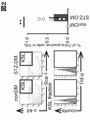


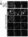


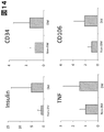
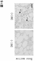


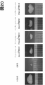


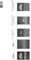
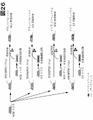

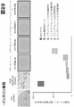


Classifications
-
- A—HUMAN NECESSITIES
- A61—MEDICAL OR VETERINARY SCIENCE; HYGIENE
- A61K—PREPARATIONS FOR MEDICAL, DENTAL OR TOILETRY PURPOSES
- A61K31/00—Medicinal preparations containing organic active ingredients
- A61K31/16—Amides, e.g. hydroxamic acids
- A61K31/165—Amides, e.g. hydroxamic acids having aromatic rings, e.g. colchicine, atenolol, progabide
- A61K31/167—Amides, e.g. hydroxamic acids having aromatic rings, e.g. colchicine, atenolol, progabide having the nitrogen of a carboxamide group directly attached to the aromatic ring, e.g. lidocaine, paracetamol
-
- G—PHYSICS
- G01—MEASURING; TESTING
- G01N—INVESTIGATING OR ANALYSING MATERIALS BY DETERMINING THEIR CHEMICAL OR PHYSICAL PROPERTIES
- G01N33/00—Investigating or analysing materials by specific methods not covered by groups G01N1/00 - G01N31/00
- G01N33/48—Biological material, e.g. blood, urine; Haemocytometers
- G01N33/50—Chemical analysis of biological material, e.g. blood, urine; Testing involving biospecific ligand binding methods; Immunological testing
- G01N33/68—Chemical analysis of biological material, e.g. blood, urine; Testing involving biospecific ligand binding methods; Immunological testing involving proteins, peptides or amino acids
- G01N33/6893—Chemical analysis of biological material, e.g. blood, urine; Testing involving biospecific ligand binding methods; Immunological testing involving proteins, peptides or amino acids related to diseases not provided for elsewhere
-
- A—HUMAN NECESSITIES
- A61—MEDICAL OR VETERINARY SCIENCE; HYGIENE
- A61K—PREPARATIONS FOR MEDICAL, DENTAL OR TOILETRY PURPOSES
- A61K31/00—Medicinal preparations containing organic active ingredients
- A61K31/33—Heterocyclic compounds
- A61K31/395—Heterocyclic compounds having nitrogen as a ring hetero atom, e.g. guanethidine or rifamycins
-
- A—HUMAN NECESSITIES
- A61—MEDICAL OR VETERINARY SCIENCE; HYGIENE
- A61K—PREPARATIONS FOR MEDICAL, DENTAL OR TOILETRY PURPOSES
- A61K31/00—Medicinal preparations containing organic active ingredients
- A61K31/13—Amines
- A61K31/135—Amines having aromatic rings, e.g. ketamine, nortriptyline
- A61K31/137—Arylalkylamines, e.g. amphetamine, epinephrine, salbutamol, ephedrine or methadone
-
- A—HUMAN NECESSITIES
- A61—MEDICAL OR VETERINARY SCIENCE; HYGIENE
- A61K—PREPARATIONS FOR MEDICAL, DENTAL OR TOILETRY PURPOSES
- A61K31/00—Medicinal preparations containing organic active ingredients
- A61K31/16—Amides, e.g. hydroxamic acids
- A61K31/165—Amides, e.g. hydroxamic acids having aromatic rings, e.g. colchicine, atenolol, progabide
-
- A—HUMAN NECESSITIES
- A61—MEDICAL OR VETERINARY SCIENCE; HYGIENE
- A61K—PREPARATIONS FOR MEDICAL, DENTAL OR TOILETRY PURPOSES
- A61K31/00—Medicinal preparations containing organic active ingredients
- A61K31/21—Esters, e.g. nitroglycerine, selenocyanates
- A61K31/27—Esters, e.g. nitroglycerine, selenocyanates of carbamic or thiocarbamic acids, meprobamate, carbachol, neostigmine
-
- A—HUMAN NECESSITIES
- A61—MEDICAL OR VETERINARY SCIENCE; HYGIENE
- A61K—PREPARATIONS FOR MEDICAL, DENTAL OR TOILETRY PURPOSES
- A61K35/00—Medicinal preparations containing materials or reaction products thereof with undetermined constitution
-
- A—HUMAN NECESSITIES
- A61—MEDICAL OR VETERINARY SCIENCE; HYGIENE
- A61K—PREPARATIONS FOR MEDICAL, DENTAL OR TOILETRY PURPOSES
- A61K38/00—Medicinal preparations containing peptides
- A61K38/04—Peptides having up to 20 amino acids in a fully defined sequence; Derivatives thereof
- A61K38/07—Tetrapeptides
-
- A—HUMAN NECESSITIES
- A61—MEDICAL OR VETERINARY SCIENCE; HYGIENE
- A61K—PREPARATIONS FOR MEDICAL, DENTAL OR TOILETRY PURPOSES
- A61K38/00—Medicinal preparations containing peptides
- A61K38/04—Peptides having up to 20 amino acids in a fully defined sequence; Derivatives thereof
- A61K38/15—Depsipeptides; Derivatives thereof
-
- A—HUMAN NECESSITIES
- A61—MEDICAL OR VETERINARY SCIENCE; HYGIENE
- A61K—PREPARATIONS FOR MEDICAL, DENTAL OR TOILETRY PURPOSES
- A61K39/00—Medicinal preparations containing antigens or antibodies
- A61K39/395—Antibodies; Immunoglobulins; Immune serum, e.g. antilymphocytic serum
- A61K39/39533—Antibodies; Immunoglobulins; Immune serum, e.g. antilymphocytic serum against materials from animals
- A61K39/3955—Antibodies; Immunoglobulins; Immune serum, e.g. antilymphocytic serum against materials from animals against proteinaceous materials, e.g. enzymes, hormones, lymphokines
-
- A—HUMAN NECESSITIES
- A61—MEDICAL OR VETERINARY SCIENCE; HYGIENE
- A61K—PREPARATIONS FOR MEDICAL, DENTAL OR TOILETRY PURPOSES
- A61K45/00—Medicinal preparations containing active ingredients not provided for in groups A61K31/00 - A61K41/00
- A61K45/06—Mixtures of active ingredients without chemical characterisation, e.g. antiphlogistics and cardiaca
-
- A—HUMAN NECESSITIES
- A61—MEDICAL OR VETERINARY SCIENCE; HYGIENE
- A61P—SPECIFIC THERAPEUTIC ACTIVITY OF CHEMICAL COMPOUNDS OR MEDICINAL PREPARATIONS
- A61P3/00—Drugs for disorders of the metabolism
- A61P3/08—Drugs for disorders of the metabolism for glucose homeostasis
-
- A—HUMAN NECESSITIES
- A61—MEDICAL OR VETERINARY SCIENCE; HYGIENE
- A61P—SPECIFIC THERAPEUTIC ACTIVITY OF CHEMICAL COMPOUNDS OR MEDICINAL PREPARATIONS
- A61P3/00—Drugs for disorders of the metabolism
- A61P3/08—Drugs for disorders of the metabolism for glucose homeostasis
- A61P3/10—Drugs for disorders of the metabolism for glucose homeostasis for hyperglycaemia, e.g. antidiabetics
-
- C—CHEMISTRY; METALLURGY
- C07—ORGANIC CHEMISTRY
- C07K—PEPTIDES
- C07K16/00—Immunoglobulins [IGs], e.g. monoclonal or polyclonal antibodies
- C07K16/18—Immunoglobulins [IGs], e.g. monoclonal or polyclonal antibodies against material from animals or humans
- C07K16/24—Immunoglobulins [IGs], e.g. monoclonal or polyclonal antibodies against material from animals or humans against cytokines, lymphokines or interferons
- C07K16/241—Tumor Necrosis Factors
-
- C—CHEMISTRY; METALLURGY
- C12—BIOCHEMISTRY; BEER; SPIRITS; WINE; VINEGAR; MICROBIOLOGY; ENZYMOLOGY; MUTATION OR GENETIC ENGINEERING
- C12Q—MEASURING OR TESTING PROCESSES INVOLVING ENZYMES, NUCLEIC ACIDS OR MICROORGANISMS; COMPOSITIONS OR TEST PAPERS THEREFOR; PROCESSES OF PREPARING SUCH COMPOSITIONS; CONDITION-RESPONSIVE CONTROL IN MICROBIOLOGICAL OR ENZYMOLOGICAL PROCESSES
- C12Q1/00—Measuring or testing processes involving enzymes, nucleic acids or microorganisms; Compositions therefor; Processes of preparing such compositions
- C12Q1/34—Measuring or testing processes involving enzymes, nucleic acids or microorganisms; Compositions therefor; Processes of preparing such compositions involving hydrolase
-
- G—PHYSICS
- G01—MEASURING; TESTING
- G01N—INVESTIGATING OR ANALYSING MATERIALS BY DETERMINING THEIR CHEMICAL OR PHYSICAL PROPERTIES
- G01N33/00—Investigating or analysing materials by specific methods not covered by groups G01N1/00 - G01N31/00
- G01N33/48—Biological material, e.g. blood, urine; Haemocytometers
- G01N33/50—Chemical analysis of biological material, e.g. blood, urine; Testing involving biospecific ligand binding methods; Immunological testing
- G01N33/5005—Chemical analysis of biological material, e.g. blood, urine; Testing involving biospecific ligand binding methods; Immunological testing involving human or animal cells
- G01N33/5091—Chemical analysis of biological material, e.g. blood, urine; Testing involving biospecific ligand binding methods; Immunological testing involving human or animal cells for testing the pathological state of an organism
-
- G—PHYSICS
- G01—MEASURING; TESTING
- G01N—INVESTIGATING OR ANALYSING MATERIALS BY DETERMINING THEIR CHEMICAL OR PHYSICAL PROPERTIES
- G01N33/00—Investigating or analysing materials by specific methods not covered by groups G01N1/00 - G01N31/00
- G01N33/48—Biological material, e.g. blood, urine; Haemocytometers
- G01N33/50—Chemical analysis of biological material, e.g. blood, urine; Testing involving biospecific ligand binding methods; Immunological testing
- G01N33/68—Chemical analysis of biological material, e.g. blood, urine; Testing involving biospecific ligand binding methods; Immunological testing involving proteins, peptides or amino acids
-
- A—HUMAN NECESSITIES
- A61—MEDICAL OR VETERINARY SCIENCE; HYGIENE
- A61K—PREPARATIONS FOR MEDICAL, DENTAL OR TOILETRY PURPOSES
- A61K39/00—Medicinal preparations containing antigens or antibodies
- A61K2039/505—Medicinal preparations containing antigens or antibodies comprising antibodies
-
- G—PHYSICS
- G01—MEASURING; TESTING
- G01N—INVESTIGATING OR ANALYSING MATERIALS BY DETERMINING THEIR CHEMICAL OR PHYSICAL PROPERTIES
- G01N2333/00—Assays involving biological materials from specific organisms or of a specific nature
- G01N2333/90—Enzymes; Proenzymes
- G01N2333/914—Hydrolases (3)
- G01N2333/978—Hydrolases (3) acting on carbon to nitrogen bonds other than peptide bonds (3.5)
- G01N2333/98—Hydrolases (3) acting on carbon to nitrogen bonds other than peptide bonds (3.5) acting on amide bonds in linear amides (3.5.1)
-
- G—PHYSICS
- G01—MEASURING; TESTING
- G01N—INVESTIGATING OR ANALYSING MATERIALS BY DETERMINING THEIR CHEMICAL OR PHYSICAL PROPERTIES
- G01N2800/00—Detection or diagnosis of diseases
- G01N2800/04—Endocrine or metabolic disorders
- G01N2800/042—Disorders of carbohydrate metabolism, e.g. diabetes, glucose metabolism
-
- G—PHYSICS
- G01—MEASURING; TESTING
- G01N—INVESTIGATING OR ANALYSING MATERIALS BY DETERMINING THEIR CHEMICAL OR PHYSICAL PROPERTIES
- G01N2800/00—Detection or diagnosis of diseases
- G01N2800/50—Determining the risk of developing a disease
-
- G—PHYSICS
- G01—MEASURING; TESTING
- G01N—INVESTIGATING OR ANALYSING MATERIALS BY DETERMINING THEIR CHEMICAL OR PHYSICAL PROPERTIES
- G01N2800/00—Detection or diagnosis of diseases
- G01N2800/52—Predicting or monitoring the response to treatment, e.g. for selection of therapy based on assay results in personalised medicine; Prognosis
Definitions
- the present disclosure provides treatment of diabetes and/or its related diseases by combining suppression of abnormal stem cells based on HDAC inhibition and migration of stem cells, and treatment of diabetes and/or its related diseases using the presence, migration and/or persistence of abnormal stem cells as an index.
- the diagnosis of related diseases More particularly, the present disclosure treats diabetes and/or its related diseases by migrating and suppressing abnormal hematopoietic stem cells, and detecting the presence, migration out of and/or persistence in specific niches of abnormal hematopoietic stem cells. Diagnosis of possible diabetes and/or related diseases.
- Non-Patent Document 1 Diabetes is generally classified into type 1 and type 2 (Non-Patent Document 1). However, since the details of the onset of both are unknown, it must be said that the classification of type 1 and type 2 itself lacks validity as based on etiology.
- various drugs are currently being developed for the treatment of diabetes, but most of them are aimed at blood sugar control and may be effective in preventing the progression of diabetes, but are insufficient for curing diabetes. can be In addition, diabetic complications such as neuropathy, nephropathy, liver damage, retinopathy, fatty liver, gastrointestinal disorders, delayed healing of fractures, eating disorders, and skin disorders are common to diabetes classified as type 1 and type 2. However, once it develops, it can be difficult to cure.
- the present inventors have found that a therapeutic strategy for diabetes and/or its related diseases by suppressing abnormal stem cells based on HDAC inhibition combined with stem cell migration is highly effective. Based on this new finding, the present disclosure provides tools for the treatment and diagnosis of diabetes and/or its related diseases.
- the present disclosure provides: (Item 1) A composition for treating and/or preventing diabetes and/or diabetes-related diseases, disorders and/or conditions comprising an inhibitor of abnormal hematopoietic stem cells (HSC), wherein the inhibitor is a histone deacetylase (HDAC) inhibitor, wherein the composition is administered in combination with a stem cell migration agent. (Item 2) The composition of any of the preceding items, wherein the abnormal HSCs are those in which the HDAC gene or protein is not expressed at normal levels and/or is non-functional. (Item 3) The composition of any of the preceding items, wherein said expression other than normal levels is overexpression.
- HSC hematopoietic stem cells
- HDAC histone deacetylase
- composition A composition of any of the preceding items for treating diabetes and/or diabetes-related diseases, disorders and/or conditions.
- the inhibitor further comprises at least one selected from the group consisting of an anti-CD106 antibody, an anti-TNF- ⁇ antibody, or functional variants thereof.
- the abnormal HSC further does not express at normal levels a gene or protein selected from the group consisting of tumor necrosis factor alpha (TNF- ⁇ ), CD106, and proinsulin.
- TNF- ⁇ tumor necrosis factor alpha
- CD106 tumor necrosis factor alpha
- the disease, disorder and/or symptom is selected from the group consisting of neuropathy, nephropathy, liver disorder, retinopathy, fatty liver, gastrointestinal disorder, fracture healing delay, eating disorder, and skin disorder. any composition.
- Said stem cell migration agent comprises at least one drug selected from the group consisting of CXCR4 antagonists, CXCR2 stimulators, epidermal growth factor receptor (EGFR) inhibitors, granulocyte colony stimulating factor (G-CSF) agents. , the composition of any of the preceding items.
- the stem cell migration agent is at least one selected from the group consisting of plexisafor, GRO ⁇ 2 (MIP2), gefitinib, erlotinib, afatinib, osimertinib, filgrastim, naltograstim, lenograstim, and pegfilgrastim.
- composition of any of the preceding items comprising: (Item 12) Said HDAC inhibitors are TSA (trichostatin A), VPA (valproic acid), sodium butyrate (NaBu), SAHA (suberoylanilide hydroxamic acid or vorinostat), sodium phenylbutyrate, depsipeptide (FR901228, FK228), trapoxin ( TPX), cyclic hydroxamic acid-containing peptide 1 (CHAP1), MS-275, LBH589, divinostat, fingolimod (FTY720) and at least one selected from the group consisting of PXD101.
- TSA trichostatin A
- VPA valproic acid
- NaBu sodium butyrate
- SAHA suberoylanilide hydroxamic acid or vorinostat
- TPX trapoxin
- cyclic hydroxamic acid-containing peptide 1 CH1
- MS-275 MS-275
- LBH589 divinostat
- a medicament for treating and/or preventing diabetes and/or diabetes-related diseases, disorders and/or symptoms comprising a combination of an inhibitor of abnormal hematopoietic stem cells (HSC), including an HDAC inhibitor, and a stem cell migration agent.
- HSC abnormal hematopoietic stem cells
- stem cell migration agent a composition for treating and/or preventing diabetes and/or diabetes-related diseases, disorders and/or conditions comprising a stem cell migratory agent, wherein the stem cell migratory agent comprises abnormal hematopoietic stem cells.
- HSC hematopoietic stem cell
- (Item 18) A composition according to any of the preceding items, wherein said detection of migration is achieved by detection of CD106 or a functional equivalent.
- (Item 19) The composition of any of the preceding items, wherein a subject diagnosed with diabetes or at risk thereof by use of said composition is indicated to be administered an HDAC inhibitor.
- (Item 20) A method of treating and/or preventing diabetes or a disease, disorder and/or condition associated with diabetes in a subject, comprising reducing or eliminating abnormal hematopoietic stem cells (HSC) comprising HDAC inhibitors in the subject.
- HSC hematopoietic stem cells
- (Item 21) A method of diagnosing diabetes or diabetes and/or diabetes-related diseases, disorders and/or conditions or risk thereof in a subject, comprising: A method comprising the step of detecting based on.
- (Item 22) A method in which abnormal hematopoietic stem cell (HSC) migration and/or persistence is indicative of treatment for the treatment and/or prevention of diabetes or diabetes and/or diabetes-related diseases, disorders and/or conditions in a subject. detecting abnormal hematopoietic stem cell (HSC) migration and/or persistence in said subject based on HDAC gene or protein expression.
- HSC hematopoietic stem cell
- (Item 23) A method of treating diabetes and/or diabetes-related diseases, disorders and/or conditions in a subject, comprising: a step of detecting abnormal hematopoietic stem cells (HSC) in said subject based on the expression of HDAC gene or protein; administering an amount.
- HSC abnormal hematopoietic stem cells
- (Item 24) The composition of any of the preceding items, wherein the composition is a pharmaceutical composition.
- (Item 25) The composition of any of the preceding items, further comprising a pharmaceutically acceptable excipient.
- (Item 26) A method of treating and/or preventing diabetes and/or diabetes-related diseases, disorders and/or conditions in a subject, comprising administering to the subject an effective amount of an inhibitor of abnormal hematopoietic stem cells (HSC) and a stem cell migration agent.
- HSC abnormal hematopoietic stem cells
- the inhibitor comprises a histone deacetylase (HDAC) inhibitor.
- HDAC histone deacetylase
- the abnormal HSC is one in which the HDAC gene or protein is not expressed at normal levels and/or is non-functional.
- Item 28 The method of any of the preceding items, wherein said expression other than normal levels is overexpression.
- Item 29 The method of any of the above items for treating diabetes and/or diabetes-related diseases, disorders and/or conditions.
- the inhibitor further comprises at least one selected from the group consisting of an anti-CD106 antibody, an anti-TNF- ⁇ antibody, or functional variants thereof.
- the abnormal HSC further does not express at normal levels a gene or protein selected from the group consisting of tumor necrosis factor alpha (TNF- ⁇ ), CD106, and proinsulin. the method of.
- TNF- ⁇ tumor necrosis factor alpha
- CD106 tumor necrosis factor alpha
- proinsulin proinsulin.
- the method of. (Item 32) The method of any of the preceding items, wherein said disease, disorder and/or condition comprises diabetic complications.
- the disease, disorder and/or symptom is selected from the group consisting of neuropathy, nephropathy, liver disorder, retinopathy, fatty liver, gastrointestinal disorder, fracture healing delay, eating disorder, and skin disorder. either way.
- stem cell migration agent has the ability to cause said abnormal HSCs to migrate out of their niche.
- Said stem cell migration agent comprises at least one drug selected from the group consisting of CXCR4 antagonists, CXCR2 stimulators, epidermal growth factor receptor (EGFR) inhibitors, granulocyte colony stimulating factor (G-CSF) agents. , by any of the above items.
- the stem cell migration agent is at least one selected from the group consisting of plexisafor, GRO ⁇ 2 (MIP2), gefitinib, erlotinib, afatinib, osimertinib, filgrastim, naltograstim, lenograstim, and pegfilgrastim.
- Said HDAC inhibitors are TSA (trichostatin A), VPA (valproic acid), sodium butyrate (NaBu), SAHA (suberoylanilide hydroxamic acid or vorinostat), sodium phenylbutyrate, depsipeptide (FR901228, FK228), trapoxin ( TPX), cyclic hydroxamic acid-containing peptide 1 (CHAP1), MS-275, LBH589, divinostat, fingolimod (FTY720) and PXD101.
- (Item 38) A method of diagnosing diabetes and/or diabetes-related diseases, disorders and/or conditions or risk thereof comprising detecting abnormal hematopoietic stem cells (HSC) comprising detecting HDACs.
- HSC hematopoietic stem cells
- (Item 40) The method of any of the preceding items, wherein said migration and/or persistence is migration out of the bone marrow niche and/or persistence in the bone marrow niche.
- Item 41 A method of any of the preceding items comprising detecting CD106 or a functional equivalent.
- HDAC histone deacetylase
- (Item 45) Use of any of the items above, wherein said expression other than normal levels is overexpression.
- (Item 46) Use of any of the above items for treating diabetes and/or diabetes-related diseases, disorders and/or conditions.
- the inhibitor further comprises at least one selected from the group consisting of an anti-CD106 antibody, an anti-TNF- ⁇ antibody, or functional variants thereof.
- the abnormal HSC further does not express at normal levels a gene or protein selected from the group consisting of tumor necrosis factor alpha (TNF- ⁇ ), CD106, and proinsulin. Use of.
- (Item 49) Use of any of the items above, wherein said disease, disorder and/or condition comprises diabetic complications.
- the disease, disorder and/or symptom is selected from the group consisting of neuropathy, nephropathy, liver disorder, retinopathy, fatty liver, gastrointestinal disorder, fracture healing delay, eating disorder, and skin disorder. any use.
- (Item 51) Use of any of the preceding items, wherein said stem cell migration agent has the ability to cause said abnormal HSCs to migrate out of their niche.
- Said stem cell migration agent comprises at least one drug selected from the group consisting of CXCR4 antagonists, CXCR2 stimulators, epidermal growth factor receptor (EGFR) inhibitors, granulocyte colony stimulating factor (G-CSF) agents. , the use of any of the above items.
- the stem cell migration agent is at least one selected from the group consisting of plexisafor, GRO ⁇ 2 (MIP2), gefitinib, erlotinib, afatinib, osimertinib, filgrastim, naltograstim, lenograstim, and pegfilgrastim.
- Said HDAC inhibitors are TSA (trichostatin A), VPA (valproic acid), sodium butyrate (NaBu), SAHA (suberoylanilide hydroxamic acid or vorinostat), sodium phenylbutyrate, depsipeptide (FR901228, FK228), trapoxin ( TPX), cyclic hydroxamic acid-containing peptide 1 (CHAP1), MS-275, LBH589, divinostat, fingolimod (FTY720) and at least one selected from the group consisting of PXD101.
- TSA trichostatin A
- VPA valproic acid
- NaBu sodium butyrate
- SAHA suberoylanilide hydroxamic acid or vorinostat
- TPX trapoxin
- CHAP1 cyclic hydroxamic acid-containing peptide 1
- MS-275 MS-275
- LBH589 divinostat
- fingolimod fingolimod
- HSC abnormal hematopoietic stem cell
- HDAC hematopoietic stem cell
- (Item 58) Use according to any of the preceding items, wherein the migration detection agent comprises a CD106 or functional equivalent detection agent.
- (Item 59) Use of any of the preceding items, wherein a subject diagnosed with diabetes or at risk thereof by use of said composition is indicated to be administered an HDAC inhibitor.
- (Item 1A) A composition for treating and/or preventing diabetes and/or diabetes-related diseases, disorders and/or conditions comprising a histone deacetylase (HDAC) inhibitor, said composition comprising a stem cell migration agent and A composition characterized in that it is administered in combination.
- HDAC histone deacetylase
- (Item 3A) The composition of any of the preceding items, wherein said expression other than normal levels is overexpression.
- (Item 4A) A composition of any of the preceding items for treating diabetes and/or diabetes-related diseases, disorders and/or conditions.
- (Item 5A) The composition according to any one of the above items, which is further administered in combination with at least one selected from the group consisting of anti-CD106 antibody, anti-TNF- ⁇ antibody and functional variants thereof.
- (Item 6A) Any of the above items, wherein the subject to be administered further does not express at normal levels a gene or protein selected from the group consisting of tumor necrosis factor alpha (TNF- ⁇ ), CD106, and proinsulin. composition.
- (Item 7A) The composition of any of the preceding items, wherein said disease, disorder and/or condition comprises diabetic complications.
- the disease, disorder and/or symptom is selected from the group consisting of neuropathy, nephropathy, liver disorder, retinopathy, fatty liver, gastrointestinal disorder, fracture healing delay, eating disorder, and skin disorder. any composition.
- (Item 9A) The composition of any of the preceding items, wherein said stem cell migration agent has the ability to cause said abnormal HSCs to migrate out of their niche.
- Said stem cell migration agent comprises at least one drug selected from the group consisting of CXCR4 antagonists, CXCR2 stimulators, epidermal growth factor receptor (EGFR) inhibitors, granulocyte colony stimulating factor (G-CSF) agents. , the composition of any of the preceding items.
- the stem cell migration agent is at least one selected from the group consisting of plexisafor, GRO ⁇ 2 (MIP2), gefitinib, erlotinib, afatinib, osimertinib, filgrastim, naltograstim, lenograstim, and pegfilgrastim.
- composition of any of the preceding items comprising: (Item 12A) Said HDAC inhibitors are TSA (trichostatin A), VPA (valproic acid), sodium butyrate (NaBu), SAHA (suberoylanilide hydroxamic acid or vorinostat), sodium phenylbutyrate, depsipeptide (FR901228, FK228), trapoxin ( TPX), cyclic hydroxamic acid-containing peptide 1 (CHAP1), MS-275, LBH589, divinostat, fingolimod (FTY720) and at least one selected from the group consisting of PXD101.
- TSA trichostatin A
- VPA valproic acid
- NaBu sodium butyrate
- SAHA suberoylanilide hydroxamic acid or vorinostat
- TPX trapoxin
- CH1 cyclic hydroxamic acid-containing peptide 1
- MS-275 MS-275
- LBH589 divinostat
- (Item 13A) A medicament for treating and/or preventing diabetes and/or diabetes-related diseases, disorders and/or symptoms comprising a combination of an HDAC inhibitor and a stem cell migratory agent.
- (Item 14A) A composition for treating and/or preventing diabetes and/or diabetes-related diseases, disorders and/or conditions comprising a stem cell migratory agent, wherein the stem cell migratory agent is administered in combination with an HDAC inhibitor
- (Item 16A) Compositions for treatment options for the treatment and/or prevention of diabetes and/or diabetes-related diseases, disorders and/or conditions comprising a detection agent for HDACs.
- (Item 17A) The composition of any of the preceding items, wherein said selection is made by detecting migration and/or persistence of abnormal hematopoietic stem cells (HSC).
- (Item 18A) The composition of any of the preceding items, wherein said migration and/or persistence is migration out of the bone marrow niche and/or persistence in the bone marrow niche.
- (Item 19A) The composition of any of the preceding items, wherein said detection of migration is by detection of CD106 or a functional equivalent.
- (Item 20A) The composition of any of the preceding items, wherein a subject diagnosed with diabetes or at risk thereof by use of said composition is indicated to be administered an HDAC inhibitor.
- (Item 21A) A method of treating and/or preventing diabetes and/or diabetes-related diseases, disorders and/or conditions in a subject, comprising treating the subject with a histone deacetylase (HDAC) inhibitor and a stem cell migratory agent. A method comprising the step of dosing.
- HDAC histone deacetylase
- stem cell migratory agent A method comprising the step of dosing.
- (Item 22A) The method of any of the preceding items, wherein the subject is a subject in which the HDAC gene or protein is not expressed at normal levels and/or is not functioning.
- (Item 23A) The method of any of the preceding items, wherein said expression other than normal levels is overexpression.
- (Item 24A) The method of any of the above items for treating diabetes and/or diabetes-related diseases, disorders and/or conditions.
- (Item 25A) The method of any of the preceding items, wherein the subject is further administered at least one selected from the group consisting of an anti-CD106 antibody, an anti-TNF- ⁇ antibody, or a functional variant thereof.
- (Item 26A) Any of the preceding items, wherein the subject further does not express at normal levels a gene or protein selected from the group consisting of tumor necrosis factor alpha (TNF- ⁇ ), CD106, and proinsulin.
- Method. The method of any of the preceding items, wherein said disease, disorder and/or condition comprises diabetic complications.
- the disease, disorder and/or symptom is selected from the group consisting of neuropathy, nephropathy, liver disorder, retinopathy, fatty liver, gastrointestinal disorder, fracture healing delay, eating disorder, and skin disorder. either way.
- Said stem cell migration agent comprises at least one drug selected from the group consisting of CXCR4 antagonists, CXCR2 stimulators, epidermal growth factor receptor (EGFR) inhibitors, granulocyte colony stimulating factor (G-CSF) agents. , by any of the above items.
- the stem cell migration agent is at least one selected from the group consisting of plexisafor, GRO ⁇ 2 (MIP2), gefitinib, erlotinib, afatinib, osimertinib, filgrastim, naltograstim, lenograstim, and pegfilgrastim.
- HDAC inhibitors are TSA (trichostatin A), VPA (valproic acid), sodium butyrate (NaBu), SAHA (suberoylanilide hydroxamic acid or vorinostat), sodium phenylbutyrate, depsipeptide (FR901228, FK228), trapoxin ( TPX), cyclic hydroxamic acid-containing peptide 1 (CHAP1), MS-275, LBH589, divinostat, fingolimod (FTY720) and PXD101.
- TSA trichostatin A
- VPA valproic acid
- NaBu sodium butyrate
- SAHA suberoylanilide hydroxamic acid or vorinostat
- TPX trapoxin
- CHAP1 cyclic hydroxamic acid-containing peptide 1
- MS-275 MS-275
- LBH589 divinostat
- fingolimod fingolimod
- PXD101 PXD101.
- (Item 34A) A method of selecting a treatment for the treatment and/or prevention of diabetes and/or diabetes-related diseases, disorders and/or conditions comprising detecting HDACs.
- (Item 35A) The method of any of the preceding items, wherein said selecting is by detecting migration and/or persistence of abnormal hematopoietic stem cells (HSC).
- (Item 36A) The method of any of the preceding items, wherein said migration and/or persistence is migration out of the bone marrow niche and/or persistence in the bone marrow niche.
- (Item 37A) The method of any of the preceding items, wherein said detection of migration is by detection of CD106 or a functional equivalent.
- (Item 38A) The method of any of the preceding items, further comprising administering an HDAC inhibitor to a subject diagnosed with diabetes or at risk thereof.
- (Item 39A) Use of a histone deacetylase (HDAC) inhibitor and a stem cell migratory agent in the manufacture of a medicament for treating and/or preventing diabetes and/or diabetes-related diseases, disorders and/or conditions.
- (Item 40A) Use of any of the preceding items, wherein the medicament is administered to a subject in which the HDAC gene or protein is not expressed and/or functioning at normal levels.
- (Item 41A) Use of any of the items above, wherein said expression other than normal levels is overexpression.
- (Item 42A) Use of any of the above items for treating diabetes and/or diabetes-related diseases, disorders and/or conditions.
- (Item 43A) Use of any of the above items, wherein the medicament is further administered in combination with at least one selected from the group consisting of an anti-CD106 antibody, an anti-TNF- ⁇ antibody, or a functional variant thereof.
- (Item 44A) Any of the above items, wherein the subject to be administered further does not express at normal levels a gene or protein selected from the group consisting of tumor necrosis factor alpha (TNF- ⁇ ), CD106, and proinsulin. Use of.
- (Item 45A) Use of any of the items above, wherein said disease, disorder and/or condition comprises diabetic complications.
- the disease, disorder and/or symptom is selected from the group consisting of neuropathy, nephropathy, liver disorder, retinopathy, fatty liver, gastrointestinal disorder, fracture healing delay, eating disorder, and skin disorder. any use.
- the stem cell migration agent has the ability to cause said abnormal HSCs to migrate out of their niche.
- Said stem cell migration agent comprises at least one drug selected from the group consisting of CXCR4 antagonists, CXCR2 stimulators, epidermal growth factor receptor (EGFR) inhibitors, granulocyte colony stimulating factor (G-CSF) agents. , the use of any of the above items.
- the stem cell migration agent is at least one selected from the group consisting of plexisafor, GRO ⁇ 2 (MIP2), gefitinib, erlotinib, afatinib, osimertinib, filgrastim, naltograstim, lenograstim, and pegfilgrastim.
- Said HDAC inhibitors are TSA (trichostatin A), VPA (valproic acid), sodium butyrate (NaBu), SAHA (suberoylanilide hydroxamic acid or vorinostat), sodium phenylbutyrate, depsipeptide (FR901228, FK228), trapoxin ( TPX), cyclic hydroxamic acid-containing peptide 1 (CHAP1), MS-275, LBH589, divinostat, fingolimod (FTY720) and at least one selected from the group consisting of PXD101.
- TSA trichostatin A
- VPA valproic acid
- NaBu sodium butyrate
- SAHA suberoylanilide hydroxamic acid or vorinostat
- TPX trapoxin
- CHAP1 cyclic hydroxamic acid-containing peptide 1
- MS-275 MS-275
- LBH589 divinostat
- fingolimod fingolimod
- (Item 51A) Use of a detection agent for HDACs in the manufacture of a medicament for diagnosing diabetes and/or diabetes-related diseases, disorders and/or conditions or risk thereof.
- (Item 52A) Use of a detection agent for HDACs in the manufacture of a medicament for the selection of treatments for the treatment and/or prevention of diabetes and/or diabetes-related diseases, disorders and/or conditions.
- (Item 53A) Use of any of the above items, wherein said selecting is made by detecting migration and/or persistence of abnormal hematopoietic stem cells (HSC).
- HSC abnormal hematopoietic stem cells
- (Item 54A) Use of any of the above items, wherein said migration and/or persistence is migration out of the bone marrow niche and/or persistence in the bone marrow niche.
- (Item 55A) Use of any of the above items, wherein said detection of migration is by detection of CD106 or a functional equivalent.
- (Item 56A) Use of any of the above items, wherein a subject diagnosed with diabetes or at risk thereof by use of said medicament is indicated to be administered an HDAC inhibitor.
- HDAC histone deacetylase
- HDAC inhibitor or stem cell migratory agent according to any one of the above items, which is further administered in combination with at least one selected from the group consisting of an anti-CD106 antibody, an anti-TNF- ⁇ antibody, or a functional variant thereof. .
- Item 62A Any of the above items, wherein the subject to be administered further does not express at normal levels a gene or protein selected from the group consisting of tumor necrosis factor alpha (TNF- ⁇ ), CD106, and proinsulin.
- TNF- ⁇ tumor necrosis factor alpha
- CD106 tumor necrosis factor alpha
- proinsulin proinsulin.
- HDAC inhibitors or stem cell migratory agents of. (Item 63A) The HDAC inhibitor or stem cell migratory agent of any of the preceding items, wherein said disease, disorder and/or condition comprises diabetic complications.
- the disease, disorder and/or symptom is selected from the group consisting of neuropathy, nephropathy, liver disorder, retinopathy, fatty liver, gastrointestinal disorder, fracture healing delay, eating disorder, and skin disorder.
- Any HDAC inhibitor or stem cell migration agent is selected from the group consisting of neuropathy, nephropathy, liver disorder, retinopathy, fatty liver, gastrointestinal disorder, fracture healing delay, eating disorder, and skin disorder.
- Any HDAC inhibitor or stem cell migration agent is selected from the group consisting of CXCR4 antagonists, CXCR2 stimulators, epidermal growth factor receptor (EGFR) inhibitors, granulocyte colony stimulating factor (G-CSF) agents.
- the stem cell migration agent is at least one selected from the group consisting of plexisafor, GRO ⁇ 2 (MIP2), gefitinib, erlotinib, afatinib, osimertinib, filgrastim, naltograstim, lenograstim, and pegfilgrastim.
- MIP2 GRO ⁇ 2
- HDAC inhibitors or stem cell migratory agents of any of the above items are examples of the stem cell migratory agents of any of the above items.
- HDAC inhibitors are TSA (trichostatin A), VPA (valproic acid), sodium butyrate (NaBu), SAHA (suberoylanilide hydroxamic acid or vorinostat), sodium phenylbutyrate, depsipeptide (FR901228, FK228), trapoxin ( TPX), cyclic hydroxamic acid-containing peptide 1 (CHAP1), MS-275, LBH589, divinostat, fingolimod (FTY720) and PXD101. Or a stem cell chemoattractant.
- a detection agent for HDACs for diagnosing diabetes and/or diabetes-related diseases, disorders and/or conditions or risk thereof.
- pancreatic transplantation and pancreatic islet transplantation which are methods of restoring insulin secretion in diabetic patients, do not require donors, and can overcome the limitation of supply capacity with respect to the number of recipients. Also, patient burden such as surgery that the recipient has undergone can be avoided.
- the treatment of the present disclosure can be accomplished by drug administration and can therefore be much more convenient and less burdensome.
- radical treatment is possible, the prognosis is expected to be good.
- the upper left two panels show KSL cells (enclosed in squares) among the lineage-negative cells, the vertical axis is c-kit fluorescence intensity, and the horizontal axis is Sca- A fluorescence intensity of 1 is shown.
- the two lower left panels show the distribution of proinsulin-positive cells (enclosed in squares), with the vertical axis representing the number of KSL cells and the horizontal axis representing the fluorescence intensity of proinsulin. show.
- the upper left two panels show KSL cells (enclosed in squares) among the lineage-negative cells, the vertical axis is c-kit fluorescence intensity, and the horizontal axis is Sca- A fluorescence intensity of 1 is shown.
- the two lower left panels show the distribution of TNF- ⁇ -positive cells (enclosed in squares), the vertical axis is the number of KSL cells, and the horizontal axis is the fluorescence of TNF- ⁇ . Show strength.
- the left two panels show KSL cells (enclosed in squares) among the lineage-negative cells, the vertical axis is c-kit fluorescence intensity, and the horizontal axis is Sca- A fluorescence intensity of 1 is shown.
- the two right panels show the distribution of CD106-positive cells (boxed), the vertical axis is the intensity of side scatter (SSC), and the horizontal axis is CD106.
- the upper two panels show the distribution of TNF- ⁇ -positive cells (enclosed in squares), the vertical axis is the intensity of forward scattered light (FSC), and the horizontal axis is TNF- ⁇ . shows the fluorescence intensity of
- the lower two panels show the distribution of CD106-positive cells (enclosed in squares), with the vertical axis representing the intensity of forward scattered light (FSC) and the horizontal axis representing the fluorescence intensity of CD106. show. FACS analysis of side population (SP) and non-SP fractions in non-DM and STZ-DM KSL cells.
- the two left panels show the SP fraction (lower left circle) and non-SP fraction (upper right circle) of KSL cells, with the vertical axis The fluorescence intensity of Hoechst Blue is shown, and the horizontal axis shows the fluorescence intensity of Hoechst Red.
- Two panels show the distribution of CD106-positive cells (enclosed in squares), where the vertical axis is side scatter (SSC) intensity and the horizontal axis is CD106 fluorescence. Show strength.
- FACS analysis of CD106-positive cells in non-DM and STZ-DM KSL-SP cells show the distribution of CD106-positive cells (enclosed in squares), where the vertical axis is side scatter (SSC) intensity and the horizontal axis is CD106 fluorescence. Show strength.
- the two panels show the distribution of CD106-positive cells (boxed, was 0%), the vertical axis is the side scatter (SSC) intensity, The horizontal axis indicates the fluorescence intensity of CD106.
- Two panels show the distribution of TNF- ⁇ -positive cells (enclosed in squares), the vertical axis is the intensity of side scattered light (SSC), and the horizontal axis is TNF- Fluorescence intensity of ⁇ is shown.
- Two panels show the distribution of TNF- ⁇ positive cells (boxed, was 0%), the vertical axis is the intensity of side scatter (SSC).
- the horizontal axis indicates the fluorescence intensity of TNF- ⁇ .
- HDACs histone deacetylase gene clusters
- the vertical axis indicates the relative amount of mRNA expression levels in both mice when the mRNA expression level in non-DM mice is set to 1. *: P ⁇ 0.05.
- a schematic of experiments for transplantation of KSL cells from non-DM or STZ-DM mice into normoglycemic mice is shown.
- Transgenic mice transgenic for green fluorescent protein (GFP-Tg mice) were treated with STZ to induce diabetes (STZ-DM GFP) and control treated non-DM mice with intravenous citrate buffer injections. A mouse was prepared (non-DM GFP).
- non-DM-derived KSL-T 9 Gy lethally irradiated normoglycemic wild-type mice
- blood glucose concentration mg/gL
- STZ-DM-derived KSL-T 10
- measured value in non-DM-derived KSL-T was set to 1 Relative ratios of sensory nerve conduction velocities (SNCV) in the sciatic nerve in different cases (right).
- SNCV sensory nerve conduction velocities
- DRG dorsal root ganglion
- Four columns from the left are the results of STZ-DM-derived KSL-T. From the left, nuclei (blue), GFP (green), target molecules (top: MAP2, middle: proinsulin, bottom: TNF- ⁇ ) (red) ), which is the image of the merge.
- the right column is the result of non-DM-derived KSL-T, with merged images of nuclei (blue), GFP (green), and target molecules (top: MAP2, middle: proinsulin, bottom: TNF- ⁇ ) (red).
- the upper left two panels show KSL cells (boxed) among the lineage-negative cells, with the vertical axis indicating c-kit. Fluorescence intensity, the horizontal axis indicates the fluorescence intensity of Sca-1.
- the two lower left panels show the distribution of proinsulin-positive cells (enclosed in squares). The axis indicates the fluorescence intensity of proinsulin.
- the upper left two panels show KSL cells (boxed) among the lineage-negative cells, with the vertical axis indicating c-kit. Fluorescence intensity, the horizontal axis indicates the fluorescence intensity of Sca-1.
- the two lower left panels show the distribution of TNF- ⁇ -positive cells (enclosed in squares), with the vertical axis representing the number of KSL cells; The horizontal axis indicates the fluorescence intensity of TNF- ⁇ .
- FIG. 2 is a schematic diagram showing an overview of bone marrow transplantation therapy for STZ diabetic mice.
- FIG. 16 shows changes in blood glucose levels when the treatments shown in FIG. 16 are performed.
- the vertical axis shows the average blood glucose level (mg/dL) at each time point with standard error.
- the horizontal axis indicates the time course of treatment, with the time of bone marrow transplantation set as week 0.
- the shelf life of insulin pellets is also illustrated. ** indicates P ⁇ 0.01.
- FIG. 17 shows pancreatic frozen section results when the treatment shown in FIG. 16 was performed. (Upper panel) Insulin and nuclear staining of frozen sections. The mouse condition under which each section was acquired is displayed above each image. Scale bar indicates 10 ⁇ m.
- Pancreatic islets were randomly selected in each mouse, and the ratio (%) of insulin-positive cells to the islet size was calculated using Image J software. From left, non-diabetic mice, STZ-diabetic mice, STZ-diabetic mice treated with blank pellets and diabetic bone marrow transplants, STZ-diabetic mice treated with blank pellets and non-diabetic bone marrow transplants, STZ-diabetic mice treated with insulin pellets and diabetic bone marrow transplants. , and STZ diabetic mice treated with insulin pellets and non-diabetic bone marrow transplantation. ** indicates P ⁇ 0.01.
- FIG. 17 shows pancreatic frozen section results when the treatment shown in FIG.
- FIG. 16 shows a comparison of sizes of representative excised thymuses when the treatments shown in FIG. 16 are performed.
- FIG. 2 is a schematic diagram showing an outline of drug treatment of STZ diabetic mice.
- STZ diabetic mice are outlined to be treated with trichostatin (stem cell chemoattractant + trichostatin A) under insulin pellet or blank pellet treatment.
- FIG. 21 shows changes in blood glucose levels when the treatments shown in FIG. 21 are performed.
- the vertical axis shows the average blood glucose level (mg/dL) at each time point with standard error.
- the horizontal axis shows the time course of the treatment with the diabetes treatment starting point set at week 0.
- the shelf life of insulin pellets is also illustrated. * indicates P ⁇ 0.05.
- FIG. 21 shows pancreatic frozen section results when the treatment shown in Fig. 21 was performed.
- (Upper panel) Insulin and nuclear staining of frozen sections. The mouse condition under which each section was acquired is displayed above each image. Scale bar indicates 10 ⁇ m.
- Pancreatic islets were randomly selected in each mouse, and the ratio (%) of insulin-positive cells to the size of the islets was calculated using Image J software. From left, results are shown for nondiabetic mice, STZ diabetic mice, STZ diabetic mice treated with insulin pellets only, STZ diabetic mice treated with blank pellets and trichostatin, and STZ diabetic mice treated with insulin pellets and trichostatin. ** indicates P ⁇ 0.01.
- FIG. 21 shows pancreatic frozen section results when the treatment shown in Fig. 21 was performed.
- (Upper panel) Glucagon and nuclear staining of frozen sections. The mouse condition under which each section was acquired is displayed above each image. Scale bar indicates 10 ⁇ m.
- Pancreatic islets were randomly selected in each mouse, and the ratio (%) of glucagon-positive cells to the islet size was calculated using Image J software. From left, results are shown for nondiabetic mice, STZ diabetic mice, STZ diabetic mice treated with insulin pellets only, STZ diabetic mice treated with blank pellets and trichostatin, and STZ diabetic mice treated with insulin pellets and trichostatin. ** indicates P ⁇ 0.01.
- FIG. 21 shows a comparison of sizes of representative excised thymuses when the treatments shown in FIG. 21 are performed. From left, excised thymus from nondiabetic mice, STZ diabetic mice, STZ diabetic mice treated with insulin pellets and trichostatin, STZ diabetic mice treated with insulin pellets only, and STZ diabetic mice treated with blank pellets and trichostatin. show. The images of the thymus excised from the nondiabetic mouse and the STZ diabetic mouse are the same as those in FIG.
- FIG. 2 is a schematic diagram showing an overview of bone marrow transplantation therapy for NOD mice. Schematic transplantation of non-diabetic or diabetic-derived bone marrow into NOD mice under insulin pellet or blank pellet treatment.
- FIG. 27 shows predictions of changes in blood glucose levels when the treatments shown in FIG. 26 are performed.
- the vertical axis indicates the blood sugar level (mg/dL) at each time point.
- the horizontal axis indicates the time course of treatment, with the time of bone marrow transplantation set as week 0.
- the shelf life of insulin pellets is also illustrated.
- FIG. 26 shows observation results of pancreas frozen sections when the treatment shown in FIG. 26 was performed. (Upper panel) Insulin and nuclear staining of frozen sections. The mouse condition under which each section was acquired is displayed above each image. Images of ICR mice and diabetic NOD mice are actual images. Scale bar indicates 10 ⁇ m.
- Pancreatic islets were randomly selected in each mouse, and the ratio (%) of insulin-positive cells to the size of the islets was calculated using Image J software. From left, ICR mice, diabetic NOD mice, NOD diabetic mice treated with blank pellet and diabetic bone marrow transplant, NOD diabetic mice treated with insulin pellet and diabetic bone marrow transplant, NOD diabetic mice treated with blank pellet and non-diabetic bone marrow transplant. , and NOD diabetic mice treated with insulin pellets and non-diabetic bone marrow transplantation.
- FIG. 26 shows observation results of pancreas frozen sections when the treatment shown in FIG. 26 was performed. (Upper panel) Glucagon and nuclear staining of frozen sections.
- Pancreatic islets were randomly selected in each mouse, and the ratio (%) of glucagon-positive cells to the size of the pancreatic islets was calculated using Image J software. From left, ICR mice, diabetic NOD mice, NOD diabetic mice treated with blank pellets and diabetic bone marrow transplants, NOD diabetic mice treated with insulin pellets and diabetic bone marrow transplants, NOD diabetic mice treated with blank pellets and non-diabetic bone marrow transplants. , and NOD diabetic mice treated with insulin pellets and non-diabetic bone marrow transplantation.
- FIG. 26 shows a size comparison of representative excised thymuses when the treatments shown in FIG. 26 are performed. Images of thymus excised from ICR mice (left), non-diabetic NOD mice (middle), and diabetic NOD mice (right) in the upper row are actual and to scale. Scale bar indicates 200 ⁇ m. Bottom, from left, NOD diabetic mice treated with blank pellet and diabetic bone marrow transplant, NOD diabetic mice treated with blank pellet and non-diabetic bone marrow transplant, NOD diabetic mice treated with insulin pellet and diabetic bone marrow transplant, and insulin pellet and FIG. 13 shows a prospective view of excised thymus from NOD diabetic mice treated with non-diabetic bone marrow transplantation.
- FIG. 13 shows a prospective view of excised thymus from NOD diabetic mice treated with non-diabetic bone marrow transplantation.
- FIG. 31 shows a predicted change in blood glucose level when the treatment shown in FIG. 31 is performed.
- the vertical axis indicates the blood sugar level (mg/dL) at each time point.
- the horizontal axis shows the time course of the treatment with the diabetes treatment starting point set at week 0.
- the shelf life of insulin pellets is also illustrated.
- FIG. 31 shows observation results of pancreas frozen sections when the treatment shown in FIG. 31 was performed. (Upper panel) Insulin and nuclear staining of frozen sections. The mouse condition under which each section was acquired is displayed above each image.
- FIG. 31 shows observation results of pancreas frozen sections when the treatment shown in FIG. 31 was performed.
- Upper panel Glucagon and nuclear staining of frozen sections. The mouse condition under which each section was acquired is displayed above each image.
- FIG. 26 shows a size comparison of representative excised thymuses when the treatments shown in FIG. 26 are performed. Images of thymus excised from ICR mice (left), non-diabetic NOD mice (middle), and diabetic NOD mice (right) in the upper row are actual and to scale.
- FIG. 2 is a schematic diagram showing the mechanism by which bone marrow-derived cells cause abnormalities in type 2 diabetes.
- FIG. 1 is a schematic diagram showing the mechanism by which bone marrow-derived cells cause abnormalities in type 1 diabetes.
- diabetes is used in the usual sense used in the art, except for diabetes associated with pregnancy or due to specific genetic abnormalities, and usually insulin depletion due to destruction of pancreatic ⁇ cells.
- type 1 diabetes mellitus caused by obesity and type 2 diabetes mellitus caused by decreased insulin secretion from ⁇ -cells in the islets of Langerhans (pancreatic islets) in the pancreas, resulting in decreased glucose uptake into muscle and adipose tissue. are categorized. The results of this study suggest that both type 1 diabetes and type 2 diabetes are caused by common cellular genesis mediated by immune disorders, which is completely different from what was previously thought to be the cause.
- Diabetes can be diagnosed, for example, by two or more tests, fasting blood glucose ⁇ 126 mg/dL, HbA1c ⁇ 6.5%, oral glucose tolerance test (75 g OGTT) 2 hour value ⁇ 200 mg/dL, and the like.
- Diabetes as used herein also includes juvenile-onset maturity diabetes and borderline diabetes.
- diabetes-related disease or “diabetes-related disease, disorder and/or condition” can include any disease, disorder and condition associated with diabetes, examples of which include diabetic neuropathy.
- diabetes can include diabetic neuropathy.
- Disorder diabetic retinopathy, diabetic nephropathy, secondary diabetes, diabetic coma, disturbance of consciousness, abdominal pain, cramps, neuropathy, hyperosmotic hyperglycemia syndrome, albuminuria, edema, renal failure, blindness, Alzheimer's cognition disease, myocardial infarction, arteriosclerosis obliterans, cerebral infarction, fatty liver, skin symptoms (diabetic lipoid necrosis, etc.), decreased wound healing ability, susceptibility to infection (septicemia, etc.), cancer (liver cancer, kidney cancer, pancreatic cancer, colon cancer, stomach cancer, liver cancer, ovarian cancer, colorectal cancer, colon cancer, etc.), diabetic ketoacidosis, heart disease, cerebrovascular disease, steroid diabetes, constip
- non-diabetic subject means a subject who does not have diabetes or its related diseases.
- treatment refers to prevention, preferably maintenance, more preferably alleviation of aggravation of a disease or disorder when such a condition occurs. Preferably, it refers to curing, and includes the ability to exert symptom ameliorating effects or preventive effects on the patient's disease or one or more symptoms associated with the disease.
- Alleviating refers to reducing the severity of a disease or disorder when such a condition occurs
- cure refers to To be undiagnosed as having such a disease or disorder.
- Preliminary diagnosis and appropriate treatment are called “companion therapy”, and the diagnostic agent for that purpose is sometimes called “companion diagnostic agent”.
- prevention refers to preventing a disease or disorder from becoming such a condition before it becomes such a condition.
- the agents of the present disclosure can be used for diagnosis, and if necessary, the agents of the present disclosure can be used to prevent, for example, diabetes, or preventive measures can be taken.
- diagnosis refers to identifying various parameters related to a condition (eg, disease, disorder) in a subject and determining the current status or future of such condition.
- conditions within the body can be determined, and such information can be used to determine conditions in a subject, treatment or prophylaxis formulations or methods to be administered, etc. can be selected.
- diagnosis in a narrow sense means diagnosing the current situation, but in a broader sense it includes “early diagnosis”, “predictive diagnosis”, “pre-diagnosis” and the like.
- the diagnostic method of the present disclosure is industrially useful because, in principle, it can use what comes out of the body and can be performed without the hands of medical professionals such as doctors.
- "predictive diagnosis, preliminary diagnosis or diagnosis” is sometimes referred to as "assisting".
- the technology of the present disclosure is applicable to such diagnostic technology.
- suppression of target cells means reducing the proliferation rate of target cells, damaging target cells, reducing target cells, and/or killing target cells.
- Suppression of target cells can be achieved by direct and/or indirect action.
- a direct effect of inhibiting a target cell is by targeting any characteristic (e.g., an expressed molecule) of the cell to cause target cell injury, target cell reduction and/or target cell death. This is accomplished by mechanisms such as, but not limited to, irradiation, induction of apoptosis, induction of attack by immune cells, and the like.
- Indirect effects of suppressing target cells include, but are not limited to, methods for training the immune system to recognize and suppress these target cells.
- a "suppressor" of target cells means an agent that suppresses target cells by any means.
- the means of inhibiting the target cell can be any, e.g., promoting apoptosis by intervening the molecular machinery in the target cell, targeting cells and targeting molecules (e.g., antibodies) including, but not limited to, attack by immune cells caused by binding to, the use of radiogenic molecules, and the like.
- the term "migration" of cells means that cells move from one place to another in vivo, but typically cells move into peripheral blood and/or circulating blood. means to In a more specific embodiment, migration means that cells move from the bone marrow (or a particular site therein) into the peripheral and/or circulating blood. Such a specific site in the bone marrow may be called a niche or a specific niche, and the specific niche refers to a microenvironment necessary for stem cells to maintain their properties in vivo. Thus, in certain embodiments, the specification does not refer to, but is not limited to, migrating stem cells out of a niche.
- a cell “migratory agent” means an agent that causes target cells to migrate by any means.
- hematopoietic stem cells or “HSCs” refer to stem cells capable of differentiating into blood lineage cells. In the human adult, it is mainly present in the bone marrow and produces leukocytes (neutrophils, eosinophils, basophils, lymphocytes, monocytes, macrophages), erythrocytes, platelets, mast cells, and dendritic cells.
- Human hematopoietic stem cells can be characterized, for example, by being CD34 positive Thy-1 positive and also characterized by Lineage negative CD34 positive CD38 negative CD90 positive CD45RA negative (i.e. Lin ⁇ CD34 + CD38 ⁇ CD90 + CD45RA ⁇ ).
- Mouse hematopoietic stem cells can be characterized as c-kit positive Sca-1 positive lineage marker negative (KSL) cells. Hematopoietic stem cells were also characterized by the fact that bone marrow cells stained with Hoechest 33342 dye were both negative when excited with ultraviolet light (350 nm) and developed with two types of optical filters, Hoechst blue and Hoechst red. can be attached. Hematopoietic stem cells can be subdivided into long-term hematopoietic stem cells (LT-HSC), short-term hematopoietic stem cells (ST-HSC), and the like. For example, characterization of hematopoietic stem cells can be found in Cytometry Research 19(2):25-32, 2009.
- LT-HSC long-term hematopoietic stem cells
- LT-HSC are usually identified by surface antigen markers and are CD34-negative, CD150-positive, CD48-negative, Lin-negative, sca1-positive, c-kit-positive (or Lineage ⁇ c-Kit + Sca ⁇ 1 + CD34 ⁇ /low CD150 + cells).
- LT-HSCs can also be described as CD34-negative, CD38-negative (or CD34 ⁇ CD38 ⁇ cells) (eg, in humans) (eg, Nat Immunol. 2010 Jul;11(7):585-93; Blood 108, 2446-2454, 2006).
- ST-HSC short-term hematopoietic stem cells
- ST-HSC are usually identified by surface antigenic markers and can be described as CD34-positive, CD150-positive, CD48-negative, Lin-negative, sca-1-positive, c-kit-positive (eg in mice).
- ST-HSCs can also be described (eg, in humans) as CD34-positive, CD38-negative (or CD34 + CD38 ⁇ cells) (eg, Nat Immunol. 2010 Jul;11(7):585-93; Blood 108, 2446-2454, 2006).
- abnormal hematopoietic stem cells or “abnormal HSCs” refer to hematopoietic stem cells that express abnormal functions and/or lack at least some of their normal functions. HSCs refer to cells that do not express and/or function at normal levels of HDAC, TNF- ⁇ , CD106 or functional equivalent genes or proteins thereof.
- the term "not expressed at a normal level" of a gene or protein means that the gene or protein is expressed at an amount or level that is not the expression level seen in cells with normal function.
- it can be determined to be abnormal when CD106 is normally expressed (normal value seen in normal cells), preferably when CD106 is expressed more than normal can be determined to be abnormal.
- KSL cells were sorted from non-diabetic and diabetic mice by FACS, RNA was extracted, and comparative analysis of gene expression levels was performed using QT-PCR to determine unusual levels of gene expression (e.g., overexpression) can be tested.
- FACS analysis can be performed to detect aberrant hematopoietic stem cell surface antigens and to examine abnormal levels of protein expression by comparing diabetic and non-diabetic subjects.
- a gene or protein is not functioning at a normal level
- it may be determined to be abnormal if it deviates from the level at which CD106 normally functions (the normal level or value found in normal cells), preferably at a level above normal that CD106 functions. If observed, it can be determined as abnormal. For example, by performing FACS analysis to detect aberrant hematopoietic stem cell surface antigens and observing protein activation in comparison to diabetic and non-diabetic subjects, the protein functions at normal levels. You can test that it is not.
- CXCR4 is a seven-transmembrane G protein-coupled receptor (GPCR), also known as CD184 or Fusin.
- GPCR G protein-coupled receptor
- the physiological ligand of CXCR4 is one of the CXC chemokines, stromal cell-derived factor-1 (SDF-1), which strongly induces the migration of monocytes and lymphocytes. Inhibition of CXCR4 can result in migration of hematopoietic stem cells (Future Oncol. 2007 Feb;3(1):19-27).
- CD106 is a type I membrane protein, a member of the Ig superfamily, which is a type of surface antigen and is also known as Vascular cell adhesion molecule-1 (VCAM-1) or INCAM-100. is a cell surface sialoglycoprotein expressed by cytokine-activated endothelium and is known to mediate leukocyte-endothelial cell adhesion and signal transduction.
- VCAM-1 Vascular cell adhesion molecule-1
- INCAM-100 INCAM-100
- VCAM-1 isoform a precursor (nucleic acid sequence: NM_001078.4, amino acid sequence: NP_001069.1)
- VCAM-1 isoform b precursor (nucleic acid sequence: NM_080682.2, amino acid sequence: NP_542413.1)
- VCAM -1 isoform c precursor (nucleic acid sequence: NM_001199834.1, amino acid sequence: NP_001186763.1) is representative.
- CD34 is a surface protein believed to be involved in the attachment of stem cells to bone marrow extracellular matrix or stromal cells and is highly glycosylated and phosphorylated by protein kinase C. is known.
- CD34 transcription variant 1 nucleic acid sequence: NM_001025109.2, amino acid sequence: NP_001020280.1
- CD34 transcription variant 2 nucleic acid sequence: NM_001773.3, amino acid sequence: NP_001764.1
- tumor necrosis factor alpha or its abbreviation “TNF- ⁇ ” refers to a multifunctional inflammatory factor belonging to the tumor necrosis factor (TNF) superfamily, also known as TNF, DIF, TNFA, TNFSF2, TNLG1F.
- TNF- ⁇ can function through the receptors TNFRSF1A/TNFR1 and TNFRSF1B/TNFBR and is involved in the regulation of a wide range of biological processes such as cell proliferation, differentiation, apoptosis, lipid metabolism and coagulation.
- nucleic acid sequence: NM_000594.4 and amino acid sequence: NP_000585.2 are typical.
- proinsulin refers to a precursor protein of insulin. Proinsulin is processed in the endoplasmic reticulum of pancreatic ⁇ -cells, and the C-peptide region is removed in immature secretory granules to produce insulin composed of A and B chains.
- proinsulin transcript variant 1 (nucleic acid sequence: NM_000207.3, amino acid sequence: NP_000198.1)
- proinsulin transcript variant 2 (nucleic acid sequence: NM_001185097.2, amino acid sequence: NP_001172026.1)
- proinsulin transcript variant 3 (nucleic acid sequence: NM_001185098.1, amino acid sequence: NP_001172027.1)
- proinsulin transcriptional variant 4 nucleic acid sequence: NM_001291897.2, amino acid sequence: NP_001278826.
- Kit isoform 1 precursor (nucleic acid sequence: NM_000222.2, amino acid sequence: NP_000213.1)
- Kit isoform 2 precursor (nucleic acid sequence: NM_001093772.1, amino acid sequence: NP_001087241.1) are representative.
- CD20 is a member of the transmembrane 4A gene family, also known as MS4A1, B1, S7, Bp35, CD20, CVID5, MS4A2 or LEU-16, which is responsible for the development of B cells into plasma cells. and refers to a B-lymphocyte surface molecule that plays a role in differentiation.
- CD20 transcription variant 1 nucleic acid sequence: NM_152866.2, amino acid sequence: NP_690605.1
- CD20 transcription variant 3 are representative.
- histone deacetylase refers to a family of proteins that are said to have the enzymatic activity of releasing lysine acetylation modifications on proteins such as histones. 3, 6, 8, 9, etc. are known.
- HDAC1 nucleic acid sequence: NM_004964.2, amino acid sequence: NP_004955.2
- HDAC2 nucleic acid sequence: NM_001527.3, amino acid sequence: NP_001518.3
- HDAC3 nucleic acid sequence: NM_003883.3, amino acid sequence: NP_003874 .2
- HDAC4 nucleic acid sequence: NM_006037.3, amino acid sequence: NP_006028.2
- HDAC6 nucleic acid sequence: NM_001321226.1, amino acid sequence: NP_001308155.1
- HDAC8 nucleic acid sequence: NM_001166418.1, amino acid sequence: NP_001159890.1
- HDAC9 nucleic acid sequence: NM_001204144.2, amino acid sequence: NP_001191073.1
- each protein contemplates not only the protein having the amino acid sequence set forth in the specific accession number (or the nucleic acid encoding it), but also functional equivalents thereof. It is understood that
- a "functional equivalent" of a molecule includes a mutant or variant (e.g., amino acid sequence variant, etc.) of that molecule that has the characteristics described herein (e.g., a marker ), perform a function similar to (but not necessarily to the same extent) the biological function of the molecule, and are capable of altering the molecule itself at the time of action is understood to include
- a "functional variant" of a molecule includes a substance that has been altered while maintaining (or changing the degree of) the function of the molecule.
- functional variants of binding molecules such as antibodies include those conjugated to other moieties (labels, other functional molecules (such as proteins), etc.) such that they retain their binding function with the target molecule. , fragmented, etc.
- functional variants as used herein are those that maintain the underlying function prior to the alteration.
- one or more amino acid insertions, substitutions or deletions, or additions to one or both ends thereof can be used in the amino acid sequence.
- insertion, substitution or deletion of one or more amino acids in an amino acid sequence, or addition to one or both ends thereof refers to well-known techniques such as site-directed mutagenesis. It means that it has been modified by a method or by natural mutation, such as substitution of a plurality of amino acids to the extent that it can occur naturally.
- biological function refers to a specific function that the gene, nucleic acid molecule or polypeptide can have in vivo when referring to a gene or a nucleic acid molecule or polypeptide related thereto. Examples include, but are not limited to, generation of specific antibodies, enzymatic activity, imparting resistance, and the like. Such biological activities can be referred to documents such as those cited in the accession numbers, Entrez numbers, etc. referred to in the table above, and such documents are also referred to herein.
- a biological function can be exerted by "biological activity.”
- biological activity refers to an activity that a certain factor (e.g., polynucleotide, protein, etc.) can have in vivo, and exhibits various functions (e.g., transcription promoting activity).
- activities in which interaction with one molecule activates or inactivates another molecule are also encompassed.
- two agents interact, their biological activity is determined by the binding between the two molecules and the resulting biological changes, e.g., when one molecule is precipitated with an antibody, the other Two molecules are considered bound when the molecules also co-precipitate. Therefore, observing such coprecipitation is one method of determination.
- an agent is an enzyme
- its biological activity includes its enzymatic activity.
- Another example includes binding to the receptor to which the ligand corresponds when the agent is a ligand.
- Such biological activity can be measured by techniques well known in the art.
- a “derivative” or “analog” or “variant” of a protein or nucleic acid as used herein is not intended to be limiting, but includes molecules containing regions of substantial homology to the protein or nucleic acid. , such molecules, in various embodiments, have at least 30% , 40%, 50%, 60%, 70%, 80%, 90%, 95%, 98% or 99% identical, or "derivatives" or “analogs” or “variants” of such nucleic acids ' can hybridize to the original nucleic acid under stringent, moderately stringent, or non-stringent conditions.
- a “derivative” or “analog” or “variant” of a protein is the product of modifications, such as amino acid substitutions, deletions and additions, to the naturally occurring protein, which still exist in the organism of the naturally occurring protein.
- a protein that exhibits a biological function although not necessarily to the same degree.
- the biological function of such proteins can be determined by suitable and available in vitro assays described herein or known in the art. While this disclosure will primarily discuss humans, it is understood that it applies to other species, such as other species within the primates or species of other genera of animals, and these mammals are also included in the present disclosure. It is understood to fall within the scope of disclosure.
- activity refers to the function of a molecule in the broadest sense. Activity generally includes, but is not limited to, any biological, biochemical, physical or chemical function of the molecule. Activities include, for example, activating, promoting, stabilizing, inhibiting, repressing, or destabilizing enzymatic activity, ability to interact with other molecules, and functions of other molecules. stability, ability to localize to specific subcellular locations. Where applicable, the term also relates to the function of protein complexes in the broadest sense.
- a “functionally active" protein, polypeptide, fragment or derivative possesses a protein's structural, regulatory, or biochemical function, such as biological activity.
- the term “gene” refers to factors that define genetic traits. They are usually arranged in a certain order on the chromosome. A gene that defines the primary structure of a protein is called a structural gene, and a gene that controls its expression is called a regulatory gene. As used herein, “gene” may refer to "polynucleotide,” “oligonucleotide,” and “nucleic acid,” including DNA, RNA, and the like. A “gene product” refers to a substance produced based on a gene, such as protein, mRNA, and the like. Therefore, mRNA can also be included in the concept of gene and can also be applied to gene products. In addition, the term “gene expression” often refers to the transcription level of mRNA and the like.
- protein protein
- polypeptide oligopeptide
- peptide refers to polymers of amino acids of any length.
- the polymer may be linear, branched, or cyclic.
- Amino acids may be naturally occurring or non-naturally occurring, and may be modified amino acids.
- the term can also include multiple polypeptide chains assembled into a complex.
- the term also includes natural or artificially modified amino acid polymers. Such modifications include, for example, disulfide bond formation, glycosylation, lipidation, acetylation, phosphorylation or any other manipulation or modification (eg, conjugation with a labeling component).
- This definition also includes, for example, polypeptides containing one or more analogs of amino acids (including, for example, unnatural amino acids), peptide-like compounds (eg, peptoids), and other modifications known in the art. be done.
- polynucleotide As used herein, “polynucleotide”, “oligonucleotide” and “nucleic acid” are used interchangeably herein and refer to polymers of nucleotides of any length. The term also includes “oligonucleotide derivatives” or “polynucleotide derivatives.” “Oligonucleotide derivative” or “polynucleotide derivative”, used interchangeably, refer to oligonucleotides or polynucleotides that contain derivatives of nucleotides or that have unusual linkages between nucleotides.
- oligonucleotides include, for example, 2′-O-methyl-ribonucleotides, oligonucleotide derivatives in which phosphodiester bonds in oligonucleotides are converted to phosphorothioate bonds, and phosphodiester bonds in oligonucleotides.
- oligonucleotide derivatives in which ribose and phosphodiester bonds in the oligonucleotide are converted to peptide nucleic acid bonds uracil in the oligonucleotide is converted to C- 5-propynyluracil-substituted oligonucleotide derivative, oligonucleotide derivative wherein uracil in oligonucleotide is substituted with C-5 thiazole uracil, oligonucleotide derivative wherein cytosine in oligonucleotide is substituted with C-5 propynylcytosine, oligo Oligonucleotide derivatives in which cytosine in nucleotides are replaced with phenoxazine-modified cytosine, oligonucleotide derivatives in which ribose in DNA is replaced with 2'-O-prop
- nucleic acid sequence also includes conservatively modified variants (e.g., degenerate codon substitutions) and complementary sequences thereof, as well as sequences explicitly indicated. is intended to include Specifically, degenerate codon substitutions create sequences in which the third position of one or more selected (or all) codons is replaced with mixed bases and/or deoxyinosine residues.
- degenerate codon substitutions create sequences in which the third position of one or more selected (or all) codons is replaced with mixed bases and/or deoxyinosine residues.
- the term "homology" of a gene refers to the degree of identity of two or more gene sequences to each other. Generally, having “homology” means having a high degree of identity or similarity. Say. Therefore, the higher the homology between two genes, the higher the identity or similarity of their sequences. Whether two types of genes have homology can be determined by direct sequence comparison or, in the case of nucleic acids, hybridization under stringent conditions. When two gene sequences are directly compared, the DNA sequences are typically at least 50% identical, preferably at least 70% identical, more preferably at least 80%, 90% identical between the gene sequences. , 95%, 96%, 97%, 98% or 99% identical, the genes are homologous.
- a “homologue” or “homologous gene product” as used herein therefore refers to a protein in another species, preferably a mammal, that performs the same biological function as the protein component of the complex further described herein. means Such homologues may also be referred to as “orthologous gene products.”
- Amino acids may be referred to herein by either their commonly known three-letter symbols or by the one-letter symbols recommended by the IUPAC-IUB Biochemical Nomenclature Commission. Nucleotides may likewise be referred to by their commonly recognized one-letter code.
- comparisons of similarity, identity and homology of amino acid and nucleotide sequences are calculated using the sequence analysis tool BLAST with default parameters.
- the identity search can be performed using, for example, NCBI BLAST 2.2.9 (published May 12, 2004).
- the value of identity in the present specification generally refers to the value when aligned under the default conditions using the above BLAST. However, if a higher value is obtained by changing the parameters, the highest value is taken as the identity value. When identity is evaluated in multiple regions, the highest value among them is taken as the identity value. Similarity is a number that takes into account similar amino acids in addition to identity.
- polynucleotide that hybridizes under stringent conditions refers to well-known conditions commonly used in the art.
- Such polynucleotides can be obtained by colony hybridization, plaque hybridization, Southern blot hybridization, or the like using a polynucleotide selected from among the polynucleotides of the present disclosure as a probe. Specifically, using a filter on which colony- or plaque-derived DNA is immobilized, hybridization is performed at 65° C. in the presence of 0.7-1.0 M NaCl, followed by 0.1- to 2-fold concentration.
- a polynucleotide that can be identified by washing the filter under conditions of 65°C using an SSC (saline-sodium citrate) solution (the composition of a 1-fold SSC solution is 150 mM sodium chloride and 15 mM sodium citrate).
- SSC saline-sodium citrate
- do Hybridization is described in laboratory books such as Molecular Cloning 2nd ed., Current Protocols in Molecular Biology, Supplement 1-38, DNA Cloning 1: Core Techniques, A Practical Approach, Second Edition, Oxford University Press (1995) It can be performed according to the method.
- Low stringency conditions were 35% formamide, 5 ⁇ SSC, 50 mM Tris-HCl (pH 7.5), 5 mM EDTA, 0.02% polyvinylpyrrolidone (PVP), 0.02% BSA, 100 ⁇ g/ml denatured salmon sperm DNA, and Hybridization at 40° C. for 18-20 hours in a buffer containing 10% (weight/volume) dextran sulfate, followed by a buffer consisting of 2 ⁇ SSC, 25 mM Tris-HCl (pH 7.4), 5 mM EDTA, and 0.1% SDS. 55° C. for 1-5 hours, and 1.5 hours at 60° C. in a buffer consisting of 2 ⁇ SSC, 25 mM Tris-HCl (pH 7.4), 5 mM EDTA, and 0.1% SDS. Including.
- a “purified” substance or biological agent refers to one from which at least part of the factors naturally associated with the biological agent have been removed. Therefore, the purity of the biological agent in a purified biological agent is generally higher (ie, more concentrated) than the state in which the biological agent is normally present.
- the term “purified” preferably refers to at least 75%, more preferably at least 85%, even more preferably at least 95%, and most preferably at least 98% by weight of It means that the same type of biological agent is present. Materials used in this disclosure are preferably "purified" materials.
- a "corresponding" amino acid or nucleic acid has or has the same effect in a given polypeptide or polynucleotide molecule as a given amino acid or nucleotide in a polypeptide or polynucleotide to which it is compared.
- Amino acids or nucleotides that are predicted to be similar, particularly in enzyme molecules, to amino acids that are in similar positions in the active site and contribute similarly to catalytic activity.
- an antisense molecule can be the same portion in an ortholog that corresponds to a particular portion of the antisense molecule.
- the corresponding amino acid can be, for example, cysteinylated, glutathionylated, S—S bond formation, oxidized (eg of the methionine side chain), formylated, acetylated, phosphorylated, glycosylated, myristylated, etc. can be an amino acid of Alternatively, the corresponding amino acid can be the amino acid responsible for dimerization.
- Such "corresponding" amino acids or nucleic acids may be regions or domains spanning a range. Accordingly, such cases are referred to herein as "corresponding" regions or domains.
- a "corresponding" gene e.g., polynucleotide sequence or molecule
- a gene e.g., a polynucleotide sequence or molecule
- a gene corresponding to a gene can be an ortholog of that gene.
- Such corresponding genes can be identified using techniques well known in the art. Thus, for example, a corresponding gene in an animal (e.g., mouse, rat) is searched in the animal's sequence database using the reference gene sequence of the corresponding gene (e.g., human) as a query sequence. can be found by
- fragment refers to a polypeptide or polynucleotide having a sequence length of 1 to n-1 relative to a full-length polypeptide or polynucleotide (length of n).
- the length of the fragment can be changed as appropriate according to its purpose.
- the lower limit of the length of the polypeptide is 15, 20, 25, 30, 40, 50 and more amino acids are included, and lengths represented by integers not specifically recited herein (eg, 11, etc.) are also suitable as lower limits. obtain.
- polynucleotides 5, 6, 7, 8, 9, 10, 15, 20, 25, 30, 40, 50, 75, 100 and more nucleotides are included, specifically recited herein.
- a length expressed as an integer that does not contain a number may also be suitable as a lower bound.
- such fragments are understood to fall within the scope of the present disclosure as long as the fragment itself also functions as a marker, eg, if the full-length version functions as a marker.
- a fragment of a molecule is a substance (typically a polypeptide) containing any region of this molecule, as long as it can be used for the purposes of the present disclosure (e.g., treatment, detection, diagnosis, etc.). , may not have the biological function of the naturally occurring molecule.
- expression of a gene, polynucleotide, polypeptide, etc. refers to the transformation of the gene into a different form after undergoing a certain action in vivo.
- genes, polynucleotides, etc. are transcribed and translated into a form of polypeptide, but transcription to produce mRNA can also be an aspect of expression. More preferably, such polypeptide forms may be post-translationally processed (derivatives as referred to herein).
- the expression level of a molecule can be determined by any method. Specifically, the expression level can be known by evaluating the amount of mRNA of this molecule, the amount of protein, or the biological activity of the protein.
- the term "expression level” refers to the amount of expression of a polypeptide, mRNA, or the like in a target cell, tissue, or the like. Such expression levels are evaluated by any appropriate method including immunological measurement methods such as ELISA, RIA, fluorescent antibody, Western blotting, and immunohistochemical staining using the antibodies of the present disclosure.
- the expression level of the polypeptide of the present disclosure at the protein level, or the polypeptide used in the present disclosure evaluated by any suitable method, including molecular biological measurement methods such as Northern blotting, dot blotting, and PCR Examples include the amount of peptide expression at the mRNA level.
- “Change in expression level” means expression at the protein level or mRNA level of the polypeptide used in the present disclosure evaluated by any appropriate method including the immunological measurement method or molecular biological measurement method Means to increase or decrease in quantity.
- Various marker-based detections or diagnoses can be performed by measuring the expression level of a certain marker.
- the term "marker (substance, protein or gene (nucleic acid))” refers to a state (e.g., cell type, normal cell state, disease state, disorder state, proliferative ability, level of differentiation state, etc.) A substance that serves as an indicator to track whether a substance is present or at risk of it.
- the detection, diagnosis, preliminary detection, prediction or pre-diagnosis of a condition includes agents, agents, factors or means specific for markers associated with that condition, or compositions containing them. , a kit or system, or the like.
- antibody is broadly defined as polyclonal antibodies, monoclonal antibodies, multispecific antibodies, chimeric antibodies, and anti-idiotypic antibodies, as well as fragments thereof, such as Fv fragments, Fab' fragments, F(ab') 2 and Fab fragments, and other recombinantly produced conjugates or functional equivalents (e.g., chimeric antibodies, humanized antibodies, multifunctional antibodies, bispecific or oligospecific antibodies, single chain Antibodies, scFVs, diabodies, sc(Fv) 2 (single chain (Fv) 2 ), scFv-Fc).
- antibodies may be covalently linked or recombinantly fused to enzymes such as alkaline phosphatase, horseradish peroxidase, ⁇ -galactosidase, and the like.
- the antibody used in the present disclosure can be of any origin, type, shape, or the like. Specifically, known antibodies such as non-human animal antibodies (eg, mouse antibodies, rat antibodies, camel antibodies), human antibodies, chimeric antibodies, and humanized antibodies can be used. Monoclonal or polyclonal antibodies can be used in the present disclosure, but monoclonal antibodies are preferred. Binding of the antibody to the target protein is preferably specific binding.
- ⁇ refers to any tool that can achieve a certain purpose (e.g., detection, diagnosis, treatment, prevention, migration).
- “Means for detecting” refers to means capable of recognizing (detecting) an object differently from others.
- detection or “quantitation” of polynucleotide or polypeptide expression includes, for example, binding to or interaction with a marker detection agent, using appropriate methods including mRNA measurement and immunological measurement methods.
- molecular biological measurement methods include Northern blotting, dot blotting and PCR.
- immunological measurement methods include ELISA method using a microtiter plate, RIA method, fluorescent antibody method, luminescence immunoassay (LIA), immunoprecipitation method (IP), immunodiffusion method (SRID), immunological Examples include turbidimetric assay (TIA), Western blotting, and immunohistochemical staining.
- the detection agent or detection means of the present disclosure may be a complex or complex molecule in which a detectable moiety (eg, antibody, etc.) is bound to another substance (eg, label, etc.).
- complex or “composite molecule” means any construct comprising two or more moieties.
- one part is a polypeptide
- the other part may be a polypeptide or other substance (e.g., base material, sugar, lipid, nucleic acid, other carbohydrate, etc.).
- two or more moieties that constitute a complex may be covalently bonded or otherwise bonded (e.g., hydrogen bond, ionic bond, hydrophobic interaction, van der Waals force, etc.).
- complex includes molecules formed by linking multiple types of molecules such as polypeptides, polynucleotides, lipids, sugars, and small molecules.
- the detection agents or other medicaments of the present disclosure can take the form of probes and primers.
- the probes and primers of the present disclosure are capable of specifically hybridizing to target nucleic acid molecules.
- Probes and primers of the present disclosure need only be capable of detecting expression of a target nucleic acid molecule, and can be polymers composed of multiple bases or base pairs such as deoxyribonucleic acid (DNA) or ribonucleic acid (RNA).
- Double-stranded cDNAs are also known to be available for tissue in situ hybridization, and probes and primers of the present disclosure include such double-stranded cDNAs.
- Particularly preferred probes and primers for detecting RNA in tissue include RNA probes (riboprobes).
- the primers and primer sets according to the present disclosure are used in known methods for detecting target genes using nucleic acid amplification methods such as the PCR method, RT-PCR method, real-time PCR method, in situ PCR method, and LAMP method, according to conventional methods. Can be used as primers and primer sets.
- probe refers to a substance that serves as a means of searching for use in biological experiments such as in vitro and/or in vivo screening. Examples include, but are not limited to, peptides containing amino acid sequences, specific antibodies or fragments thereof, and the like. Probes are used herein as marker detection means.
- Nucleic acid molecules that are usually used as probes include those that have a nucleic acid sequence of at least 8 consecutive nucleotides that is homologous or complementary to the nucleic acid sequence of the target gene. Such nucleic acid sequences are preferably at least 9 consecutive nucleotides long, more preferably at least 10 consecutive nucleotides long, even more preferably at least 11 consecutive nucleotides long, at least 12 consecutive nucleotides long.
- nucleic acid sequences used as probes should have at least about 70% homology, more preferably at least about 80% homology, even more preferably at least about 90% homology, at least about 95% Homologous nucleic acid sequences are included.
- the detection agents of the present disclosure can be labeled.
- the detection agents of the present disclosure may have attached tags.
- label refers to an entity (eg, substance, energy, electromagnetic waves, etc.) that distinguishes a target molecule or substance from others.
- labeling methods include RI (radioisotope) method, fluorescence method, biotin method, chemiluminescence method and the like.
- RI radioisotope
- fluorescence method fluorescence method
- biotin method biotin method
- chemiluminescence method chemiluminescence method and the like.
- the labeling is performed with fluorescent substances having mutually different fluorescence emission maximum wavelengths. The difference in fluorescence emission maximum wavelength is preferably 10 nm or more.
- Alexa TM Fluor is preferable as the fluorescent substance.
- Alexa TM Fluor is a water-soluble fluorescent dye obtained by modifying coumarin, rhodamine, fluorescein, cyanine, etc., and is a series that supports a wide range of fluorescent wavelengths. stable, bright, and insensitive to pH.
- Combinations of fluorescent dyes having a fluorescence maximum wavelength of 10 nm or more include a combination of Alexa TM 555 and Alexa TM 633, a combination of Alexa TM 488 and Alexa TM 555, and the like.
- any substance that can bind to its base portion can be used, and cyanine dyes (eg, CyDye TM series Cy3, Cy5, etc.), rhodamine 6G reagent, N-acetoxy-N2- It is preferred to use acetylaminofluorene (AAF), AAIF (iodine derivative of AAF) and the like.
- fluorescent substances having a difference in fluorescence emission maximum wavelength of 10 nm or more include a combination of Cy5 and rhodamine 6G reagent, a combination of Cy3 and fluorescein, and a combination of rhodamine 6G reagent and fluorescein.
- such labels can be utilized to modify the subject of interest so that it can be detected by the detection means used. Such modifications are known in the art, and those skilled in the art can carry out such methods as appropriate depending on the label and the intended subject.
- a "tag” is a substance for selecting molecules by a specific recognition mechanism such as a receptor-ligand, more specifically a binding for binding a specific substance. It refers to a substance that acts as a partner (for example, having a relationship such as biotin-avidin or biotin-streptavidin) and can be included in the category of “label”. Thus, for example, a specific substance to which a tag is bound can be selected by contacting a substrate to which a binding partner of the tag sequence is bound.
- tags or labels are well known in the art. Representative tag sequences include, but are not limited to, myc tags, His tags, HA, Avi tags, and the like. Such tags may be attached to markers or marker-detecting agents of the disclosure.
- drug As used herein, “drug”, “agent” or “agent” (both equivalent to agents in English) are broadly used interchangeably and are used to describe any agent capable of achieving its intended purpose. It may be matter or other elements (eg, energy such as light, radiation, heat, electricity, etc.). Such substances include, for example, cells (e.g., T cells), proteins, polypeptides, oligopeptides, peptides, polynucleotides, oligonucleotides, nucleotides, nucleic acids (e.g., DNA such as cDNA, genomic DNA, mRNA (including RNA such as , small molecule ligands, etc.), including but not limited to these complex molecules.
- cells e.g., T cells
- proteins polypeptides, oligopeptides, peptides, polynucleotides, oligonucleotides, nucleotides, nucleic acids (e.g., DNA such as cDNA, genomic
- kits refers to a unit that provides parts (e.g., test agents, diagnostic agents, therapeutic agents, antibodies, labels, instructions, etc.) to be provided, usually divided into two or more compartments.
- parts e.g., test agents, diagnostic agents, therapeutic agents, antibodies, labels, instructions, etc.
- This kit form is preferred when the purpose is to provide a composition that should not be provided in a mixed form for reasons such as stability, and is preferably used in a mixed form immediately before use.
- kits preferably include the parts provided (e.g., instructions or instructions describing how to use the test, diagnostic, therapeutic agent, or how to handle the reagents).
- the kit When the kit is used as a reagent kit in the present specification, the kit usually includes an instruction manual describing how to use the test agent, diagnostic agent, therapeutic agent, antibody, etc. is included.
- the term "instructions” describes instructions for a physician or other user on how to use the present disclosure.
- the instructions describe the detection method of the present disclosure, how to use the diagnostic agent, or instructions for administering medicine or the like.
- the instructions may include a statement instructing oral or esophageal administration (for example, by injection) as the site of administration.
- This instruction is prepared in accordance with the format prescribed by the regulatory authority of the country in which this disclosure is implemented (for example, the Ministry of Health, Labor and Welfare in Japan, the Food and Drug Administration (FDA) in the United States, etc.) and is approved by the regulatory authority It is stated that it has received Instructions are so-called package inserts, which are usually provided in paper form, but are not limited to that, for example, in forms such as electronic media (for example, homepages provided on the Internet, e-mail) can also be provided.
- the "amount" of an analyte, etc. in a sample generally refers to an absolute value that reflects the mass of the analyte that can be detected in the volume of the sample. However, an amount also contemplates a relative amount compared to another analyte amount. For example, the amount of analyte in the sample may be an amount greater than a control or normal value for the analyte normally present in the sample.
- the "level" of an analyte or the like in a sample generally reflects the value of the analyte's activity or the like when the analyte is a target that exhibits a function such as an enzyme. refers to the absolute value of However, a level also contemplates a relative level compared to another analyte level. For example, the level of an analyte in a sample may be a level greater than a control or normal value for the analyte normally present in the sample.
- the present inventors have discovered a novel aspect that the efficacy of diabetes treatments targeting abnormal stem cells characterized by abnormal expression of CD106 and others can be improved in combination with stem cell migration.
- the present disclosure provides new treatment strategies and diagnoses for diabetes and/or its related diseases based on this new diabetes treatment strategy.
- the abnormal stem cells targeted by the present disclosure may be hematopoietic stem cells.
- the abnormal stem cells of the present disclosure do not express normal levels of at least one of CD106, histone deacetylases (HDACs), CD34, TNF- ⁇ , and proinsulin, and / or characterized by non-functioning, optionally other characteristics of the abnormal stem cells described herein (e.g., c-Kit positive, Sca-1 positive, hematopoietic stem cell lineage marker negative, above) It can be further characterized by having short-term hematopoietic stem cell characteristics (such as CD38 negative).
- the abnormal stem cells of the present disclosure can be characterized by not expressing CD106 at normal levels.
- the abnormal stem cells of the present disclosure (a) do not express CD106 at normal levels, and (b) have CD34 abnormal expression, TNF- ⁇ abnormal expression, histone deacetylase (HDACs) abnormal and aberrant expression of proinsulin, optionally other characteristics of the abnormal stem cells described herein (e.g., c- Kit-positive, Sca-1-positive, hematopoietic stem cell lineage marker-negative, characteristics of short-term hematopoietic stem cells described above (such as CD38-negative)).
- HDACs histone deacetylase
- abnormal stem cells eg, hematopoietic stem cells targeted by the present disclosure can be characterized by abnormal expression of CD106.
- the abnormal stem cells of the present disclosure can be characterized by CD106 expression levels that are higher than CD106 expression levels in total bone marrow cells or hematopoietic stem cells (eg, CD34-positive Thy-1-positive cells) of a non-diabetic subject population. .
- the abnormal stem cells of the present disclosure are 1 ⁇ 10 2 or more, 2 ⁇ 10 2 or more, 5 ⁇ 10 2 or more, 1 ⁇ 10 3 or more, 2 ⁇ 10 3 or more, 5 ⁇ 10 3 or more, 1 ⁇ 10 4 or more, 2 ⁇ 10 4 or more, 5 ⁇ 10 4 or more, 1 ⁇ 10 5 or more, 2 ⁇ 10 5 or more, 5 ⁇ 10 5 or more, 1 ⁇ 10 6 or more, 2 ⁇ 10 6 or more , 5 ⁇ 10 6 or more, or 1 ⁇ 10 7 or more CD106 molecules on the cell surface.
- the abnormal stem cells of the present disclosure can be characterized by expressing about 1 ⁇ 10 4 or more CD106 molecules on the cell surface per cell.
- the abnormal stem cells of the present disclosure are normal subject's (eg, human) whole bone marrow cells or hematopoietic stem cells (eg, CD34-positive Thy-1-positive cells) with a fluorochrome-conjugated antibody against CD106.
- a fluorochrome-conjugated antibody against CD106 1-fold or more, 2-fold or more, 5-fold or more, 10-fold or more, 20-fold or more the median fluorescence intensity from this fluorochrome observed for whole bone marrow cells or hematopoietic stem cells when labeled and analyzed by FACS , 50-fold or more, 100-fold or more, 200-fold or more, 500-fold or more, or 1000-fold or more fluorescence intensity.
- the abnormal stem cells of the present disclosure are 10% or more, 20% or more, 30% or more, 40% or more, 50% or more than hematopoietic stem cells (e.g., CD34 positive cells) from a non-diabetic subject population. , 60% or more, 70% or more, 80% or more, 90% or more, 100% or more, 150% or more or 200% or more higher CD106 mRNA expression.
- hematopoietic stem cells e.g., CD34 positive cells
- the abnormal stem cells targeted by the present disclosure can be characterized by the expression of histone deacetylase genes (HDACs).
- HDACs histone deacetylase genes
- the abnormal stem cells of the present disclosure are 10% or more, 20% or more, 30% or more, 40% more than hematopoietic stem cells (e.g., c-Kit positive lineage marker negative cells) from a population of non-diabetic subjects.
- HDACs enzyme gene clusters
- the abnormal stem cells targeted by the present disclosure can be characterized by the expression of TNF- ⁇ .
- the abnormal stem cells of the present disclosure can be characterized by being positive for TNF- ⁇ expression.
- being positive for the expression of TNF- ⁇ means testing a normal subject's (eg, human) whole bone marrow cells or hematopoietic stem cells (eg, CD34-positive Thy-1-positive cells) against TNF- ⁇ .
- the abnormal stem cells of the present disclosure are 10% or more, 20% or more, 30% or more, 40% or more, 50% or more than hematopoietic stem cells (e.g., CD34 positive cells) from a non-diabetic subject population. , 60% or more, 70% or more, 80% or more, 90% or more, 100% or more, 150% or more or 200% or more higher mRNA expression of TNF- ⁇ .
- the abnormal stem cells targeted by the present disclosure can be characterized by abnormal expression of proinsulin.
- the abnormal stem cells of the present disclosure can be characterized by being positive for expression of proinsulin.
- being positive for the expression of proinsulin comprises treating whole bone marrow cells or hematopoietic stem cells (eg, CD34-positive Thy-1-positive cells) of a non-diabetic subject (eg, human) with a fluorescent dye for proinsulin.
- the abnormal stem cells of the present disclosure are 10% or more, 20% or more, 30% or more, 40% or more, 50% or more than hematopoietic stem cells (e.g., CD34 positive cells) from a non-diabetic subject population.
- the abnormal stem cells targeted by the present disclosure can be characterized by the expression of c-Kit.
- the abnormal stem cells of the present disclosure can be characterized by being positive for c-Kit expression.
- being positive for expression of c-Kit includes testing whole bone marrow cells or hematopoietic stem cells (eg, CD34-positive Thy-1-positive cells) of a non-diabetic subject (eg, human) for c-Kit.
- the abnormal stem cells targeted by the present disclosure can be characterized by the expression of Sca-1.
- the abnormal stem cells of the present disclosure can be characterized by being positive for Sca-1 expression.
- positive for expression of Sca-1 is determined by labeling whole bone marrow cells of a non-diabetic subject (e.g., human) with a fluorochrome-conjugated antibody against Sca-1 and analyzing by FACS.
- the abnormal stem cells targeted by the present disclosure can be characterized by the expression of hematopoietic stem cell lineage markers.
- Lineage markers of hematopoietic stem cells include CD3 (T cells), CD19 (B cells), NK1.1 (NK cells), CD11c (dendritic cells), CD11b (monocytes), Fc ⁇ RI (mast cells), Gr-1 (granulocytes) and the like.
- the abnormal stem cells of the present disclosure are negative for expression of one or more (eg, all) of CD3, CD19, NK1.1, CD11c, CD11b, Fc ⁇ RI, and Gr-1 can be characterized by In one embodiment, being negative for the expression of each of CD3, CD19, NK1.1, CD11c, CD11b, Fc ⁇ RI, and Gr-1 determines whole bone marrow cells of a non-diabetic subject (eg, human) as CD3 , CD19, NK1.1, CD11c, CD11b, Fc ⁇ RI or Gr-1.
- a non-diabetic subject eg, human
- the abnormal stem cells of the present disclosure are 10% or more, 20% or more, 30% or more, 40% or more, 50% or more, 60% or more, 70% more than hematopoietic stem cells from a population of non-diabetic subjects. It may be characterized by an 80% or more, 90% or more, 100% or more, 150% or more, or 200% or more higher CD34 mRNA expression level.
- the abnormal stem cells targeted by the present disclosure can be characterized by being hematopoietic stem cells.
- being a human hematopoietic stem cell can also be characterized by being CD34 positive Thy-1 positive or Lineage negative CD34 positive CD38 negative CD90 positive CD45RA negative.
- mouse hematopoietic stem cells can be characterized by being c-kit positive Sca-1 positive lineage marker negative (KSL).
- being a hematopoietic stem cell is defined by both Hoechest 33342 dye-stained bone marrow cells when excited with ultraviolet light (350 nm) and developed with two types of optical filters, Hoechst blue and Hoechstred. It can also be characterized by being negative.
- the abnormal stem cells targeted by the present disclosure can be characterized by the cell stage of hematopoietic stem cells.
- the abnormal stem cells of the present disclosure can be short-term hematopoietic stem cells.
- proinsulin and/or TNF- ⁇ can be markers that indicate that the abnormal stem cells targeted by the present disclosure are localized in the bone marrow.
- FIG. 36 schematically shows the types of bone marrow cells identified by the inventors as being involved in the onset of diabetes, their representative characteristics, and their involvement in diabetes.
- TNF- ⁇ -producing cells are blood-derived cells, and it is unlikely that TNF- ⁇ is directly produced by the chief cells of an organ.
- HSC hematopoietic stem cells
- TNF- ⁇ is expressed in the most undifferentiated long term (LT)-HSC. It was first expressed in short term (ST)-HSCs, and expression was restricted to the CD106-positive fraction of ST-HSCs.
- the CD106- and TNF- ⁇ -expressing ST-HSCs also had enhanced expression of HDACs (HDAC3, 4, 8, 9, etc.). Inhibition of HDACs can be advantageous because CD106 and TNF- ⁇ can also be expressed in normal cells with important functions. Abnormal cells may also reside in specific niches within the bone marrow to which drugs are normally difficult to deliver, so migration of the abnormal cells out of the bone marrow may be effective in eradicating the abnormal cells.
- FIG. 37 schematically shows the mechanism by which abnormal cells develop type 2 diabetes.
- the CD106/TNF- ⁇ -positive abnormal hematopoietic stem cells seen in STZ mice were also seen in high-fat diet mice, a different type 2 diabetes model. Impaired insulin secretion results from decreased insulin sensitivity in both STZ and high-fat-fed mice (1), which results in hyperglycemia and promotes the production of CD106/TNF- ⁇ positive abnormal stem cells in the bone marrow.
- Abnormal expression of histone deacetylases occurs in abnormal stem cells, which is thought to cause various gene expression abnormalities.
- Abnormal stem cells in the bone marrow are thought to reside in a niche as a memory that maintains memories of type 2 diabetes, creating an intractable pathology (2). These cascades of abnormalities lead to (A) elimination of abnormal stem cells in the bone marrow and (B) restoration of insulin secretory function to the point that blood glucose levels can be normalized to prevent the emergence of new abnormal stem cells. It is considered possible to cure type 2 diabetes by administering insulin externally for a certain period of time to normalize the blood sugar level.
- type 1 diabetes mouse model (NOD mouse)
- CD106/TNF- ⁇ positive cells that appeared in type 2 also appear.
- cells that produce autoantibodies that damage ⁇ -cells emerge from B-lineage progenitor cells (1).
- Autoantibodies released from these cells lead to progressive depletion of ⁇ -cells (2).
- Some of these abnormal B cells also migrate to the thymus, where they also produce autoantibodies and attack and eliminate pancreatic islet autoantigen-presenting cells such as proinsulin and GAD (3). This renders the thymus incapable of negative selection of islet-attacking T cells, allowing them to migrate to the periphery (4).
- endothelial proliferation Induces endothelial proliferation and catastrophic tissue destruction of ⁇ -cell progenitors (11).
- endothelial dysfunction (12) occurs in other peripheral organs as well as type 2, and various complications specific to diabetes progress due to cell fusion (13) and the like.
- type 1 diabetes and type 2 diabetes The decisive difference between type 1 diabetes and type 2 diabetes is the complete destruction of cells expressing insulin or proinsulin protein in the thymus and pancreatic islets.
- the present disclosure provides treatment and/or prevention of diabetes and/or diabetes-related diseases, disorders and/or conditions by migrating and suppressing abnormal stem cells having the characteristics described herein. offer.
- This treatment and/or prevention may be accomplished by any means, including practicing methods, providing compositions, combinations, kits, or systems for this purpose.
- inhibitors such as antibodies
- the inhibitory effect on cells residing in the bone marrow may be limited (see, e.g., Examples).
- the suppressive effect of abnormal stem cells may be improved.
- treatment and/or prophylaxis of the present disclosure can be performed by migrating and suppressing all cells that have at least one characteristic of abnormal stem cells described herein. That is, in this embodiment, the cells that migrate and are suppressed are not limited to the abnormal stem cells described herein. It may be technically difficult to migrate/suppress only the abnormal stem cells described in this specification, and the benefits obtained by migrating/suppressing abnormal stem cells and the effects of migrating/suppressing other cells should be taken into consideration. can be selected for treatment and/or prevention. On the other hand, migration/suppression of some of the abnormal stem cells described herein may result in insufficient treatment and/or prevention of diabetes and/or diabetes-related diseases, disorders and/or symptoms.
- the characteristics of abnormal stem cells utilized for migration and the characteristics of abnormal stem cells utilized for suppression may be the same or different.
- the treatment and/or prophylaxis of the present disclosure takes advantage of the characteristic that the abnormal stem cells are hematopoietic stem cells for migration (e.g., using CXCR4 antagonists and/or CXCR2 stimulators). ), the characteristic that abnormal stem cells aberrantly express a particular protein (such as HDAC) may be exploited for suppression.
- migration in the treatment and/or prevention of the present disclosure can be performed by migrating abnormal stem cells having any of the characteristics described herein.
- migration of abnormal stem cells as described herein can be performed by a treatment that causes hematopoietic stem cells to migrate.
- treatments that induce migration of hematopoietic stem cells include inhibition of CXCR4 (Future Oncol. 2007 Feb;3(1):19-27), inhibition of epidermal growth factor receptor (EGFR) (Nature Medicine volume 16). , pages 1141-1146 (2010)), induction of granulocyte colony-stimulating factor (G-CSF) stimulation (Blood. 1995 Dec 15;86(12):4437-45), and induction of CXCR2 stimulation (Cell. 2018 Jan 11;172(1-2):191-204.e10), but are not limited to these.
- migration in the treatment and/or prevention of the present disclosure can be performed using an abnormal stem cell migration agent having any of the characteristics described herein.
- a person skilled in the art can appropriately select a migration agent that can be used in migration in the treatment and/or prevention of the present disclosure. By measuring the proportion of (e.g., abnormal stem cells with the characteristics described herein) and examining their migratory effects, migratory agents that can be used for the treatment and/or prevention of the present disclosure can be selected.
- migration in the treatment and/or prevention of the present disclosure may be performed using a hematopoietic stem cell migration agent.
- the hematopoietic stem cell migration agent inhibits CXCR4 (e.g., plelixafor (AMD3100)), inhibits epidermal growth factor receptor (EGFR) (e.g., gefitinib, erlotinib, afatinib, osimertinib, etc.); Induction of granulocyte colony-stimulating factor (G-CSF) stimulation (e.g., filgrastim, naltograstim, lenograstim, pegfilgrastim, etc.), and induction of CXCR2 stimulation (e.g., GRO ⁇ (MIP2)).
- CXCR4 e.g., plelixafor (AMD3100)
- EGFR epidermal growth factor receptor
- G-CSF granulocyte colony-stimulating factor
- MIP2 stimulation e.g., GRO ⁇ (MIP2)
- GRO ⁇ GRO ⁇
- hematopoietic stem cell migration agents can be used in any combination, for example, inhibitors of CXCR4 (e.g., plelixafor (AMD3100)) and CXCR2 stimulators (e.g., GRO ⁇ (MIP2 )).
- CXCR4 e.g., plelixafor (AMD3100)
- CXCR2 stimulators e.g., GRO ⁇ (MIP2 )
- inhibition of abnormal stem cells described herein is accomplished by targeting any characteristic of abnormal stem cells described herein, causing the targeted cells to migrate, and suppress be able to.
- the abnormal stem cells described herein target a molecule expressed on the cell surface (CD106, CD34, TNF- ⁇ , proinsulin, c-Kit, Sca-1, etc.)
- Biologically binding molecules antibodies, T-cell receptors, small compounds, etc.
- a molecule that binds to multiple types of molecules such as a multispecific antibody, can be used to target cells of interest.
- suppression in the treatment and/or prevention of the present disclosure is CD106 aberrant expression, histone deacetylase (HDACs) aberrant CD34 aberrant expression, TNF- ⁇ aberrant expression, and proinsulin aberrant expression. It can be carried out by migrating and suppressing abnormal stem cells characterized by at least one. In one embodiment, suppression in the treatment and/or prevention of the present disclosure can be performed by migrating and suppressing abnormal stem cells characterized by CD106 aberrant expression. In one embodiment, the inhibition in the treatment and/or prevention of the present disclosure is (a) CD106 aberrant expression, (b) histone deacetylase (HDACs) aberrant expression, CD34 aberrant expression, TNF- ⁇ and proinsulin aberrant expression.
- HDACs histone deacetylase
- inhibition in the treatment and/or prevention of the present disclosure targets one or more of at least one of histone deacetylases (HDACs), CD106, CD34, TNF- ⁇ , and proinsulin.
- HDACs histone deacetylases
- CD106 CD34
- TNF- ⁇ TNF- ⁇
- proinsulin can be carried out using an inhibitor of
- inhibition in the treatment and/or prevention of the present disclosure can be performed using inhibitors that target histone deacetylases (HDACs).
- inhibition in the treatment and/or prevention of the present disclosure is (a) histone deacetylases (HDACs) and (b) at least one of CD106, CD34, TNF- ⁇ , and proinsulin. It can be practiced using one or more inhibitors that target two.
- inhibitors targeting histone deacetylases can be HDAC inhibitors, such as TSA (trichostatin A), VPA (valproic acid), sodium butyrate (NaBu), SAHA (suberoylanilide hydroxamic acid or vorinostat), sodium phenylbutyrate, depsipeptide (FR901228, FK228), trapoxin (TPX), cyclic hydroxamic acid-containing peptide 1 (CHAP1), MS-275, LBH589, divinostat, fingolimod (FTY720) ) and PXD101, and the like.
- HDAC inhibitors such as TSA (trichostatin A), VPA (valproic acid), sodium butyrate (NaBu), SAHA (suberoylanilide hydroxamic acid or vorinostat), sodium phenylbutyrate, depsipeptide (FR901228, FK228), trapoxin (TPX), cyclic hydroxamic acid-containing peptide 1
- the treatment and/or prevention of the present disclosure can be treatment and/or prevention of diabetes.
- the treatment and/or prevention of the present disclosure is a complication of diabetes such as neuropathy, nephropathy, liver damage, retinopathy, fatty liver, gastrointestinal disorders, delayed healing of bone fractures, eating disorders or skin disorders. It can be treatment and/or prevention of disorders.
- the treatment and/or prevention of the present disclosure includes any known therapeutic and/or prophylactic treatment or means (e.g., treatment and/or prophylactic treatment or means of diabetes and/or diabetes-related diseases, disorders and/or symptoms). may be combined with any known therapeutic and/or prophylactic treatment or means (e.g., treatment and/or prophylactic treatment or means of diabetes and/or diabetes-related diseases, disorders and/or symptoms). may be combined with any known therapeutic and/or prophylactic treatment or means (e.g., treatment and/or prophylactic treatment or means of diabetes and/or diabetes-related diseases, disorders and/or symptoms). may be combined with any known therapeutic and/or prophylactic treatment or means (e.g., treatment and/or prophylactic treatment or means of diabetes and/or diabetes-related diseases, disorders and/or symptoms). may be combined with any known therapeutic and/or prophylactic treatment or means (e.g., treatment and/or prophylactic treatment or means of diabetes and/or diabetes-related diseases, disorders and/or symptoms). may be combined with any known therapeutic
- removal of abnormal hematopoietic stem cells as described herein can be combined with treatment to suppress hyperglycemia.
- glycemic control may be poor because pancreatic ⁇ -cells (or even the thymus, which prevents ⁇ -cell damage) may not yet have recovered.
- hyperglycemia can occur.
- hyperglycemia can promote the reappearance of abnormal hematopoietic stem cells, hyperglycemia is inhibited until pancreatic ⁇ -cells (or even the thymus, which prevents ⁇ -cell damage) are restored. can be advantageous.
- Treatments to suppress hyperglycemia include, but are not limited to, insulin administration, dietary control, and the like. Treatment to suppress hyperglycemia may be performed in parallel with the removal of abnormal hematopoietic stem cells described herein, or may be performed after removal of abnormal hematopoietic stem cells, and is capable of self-regulation of blood glucose. To some extent pancreatic ⁇ -cells (or even the thymus to prevent ⁇ -cell damage) may continue until recovery.
- the present disclosure provides for curing diabetes and/or diabetes-related diseases, disorders and/or symptoms.
- the therapeutic methods described herein may not only slow diabetes but also cure it.
- cure of diabetes and/or diabetes-related diseases, disorders and/or conditions is at least 1 month, 3 months, 6 months, 1 year, 1 year after treatment of the disease, disorder and/or condition has ended. Resulting in an undiagnosed condition for 2 or 5 years.
- the cure for diabetes and/or diabetes-related diseases, disorders and/or conditions is the response of blood glucose levels by the glucose tolerance test, which provides diagnostic criteria for diabetes, and insulin secretory capacity, glucagon load, which has ancillary diagnostic value. A determination may be made based on the test resulting in normalization in the C-peptide secretory response.
- removal of abnormal hematopoietic stem cells as described herein can restore atrophied thymus.
- Morphological changes in the thymus can be observed by MRI examination of the chest, and by measuring lymphocyte subsets and CD5 antigen-positive leukocytes, it is possible to estimate the function of the thymus.
- these thymus tests may be performed for assessment of diabetes-related diseases, disorders and/or conditions, or risk thereof.
- these thymic tests may be performed as a prognostic item following treatment of diabetes and/or diabetes-related diseases, disorders and/or conditions of the present disclosure.
- the present disclosure provides diagnosis of diabetes and/or diabetes-related diseases, disorders and/or conditions, or risk thereof, based on the presence, migration and/or persistence of abnormal stem cells described herein. do. This diagnosis may be accomplished by any means, including practicing methods, providing compositions, combinations, kits, or systems for this purpose. Subjects demonstrating the presence, migration and/or persistence of abnormal stem cells may be suitable subjects for treatment methods of the present disclosure.
- the diagnostics of the present disclosure are performed by detecting the presence and/or abundance at a particular site of at least some cells having at least one characteristic of an abnormal stem cell described herein. obtain.
- the “abundance” of cells refers not only to the number of cells (or any index indicating the number of cells), but also to a specific proportion of cells in a cell population (or a specific Any indicator of percentage of cells) is also meant.
- the diagnostics of the present disclosure can be performed by detecting the presence and/or abundance in the bone marrow and/or bone marrow niche of the abnormal stem cells described herein.
- the diagnosis of the present disclosure can be performed by detecting the presence and/or abundance in the circulation of abnormal stem cells described herein. In one embodiment, the diagnosis of the present disclosure can be performed by detecting the presence and/or abundance of abnormal stem cells at a particular site in a subject administered a chemotactic agent described herein, For example, it can be performed based on the presence and/or change in abundance of abnormal stem cells at a particular site (eg, bone marrow) before and after administration of a chemotactic agent. In this embodiment, the subject is diagnosed with diabetes and/or diabetes-related diseases, disorders and/or conditions, or at risk thereof, for example, if abnormal stem cell abundance is reduced in the subject's bone marrow after administration of the chemotactic agent. and in one embodiment, the subject may be subjected to therapeutic and/or prophylactic treatment of the present disclosure with the migratory agent.
- the diagnosis of the present disclosure can be performed based on a quantitative index (e.g., cell surface protein expression level, mRNA expression level) of at least one characteristic of abnormal stem cells described herein.
- a quantitative index e.g., cell surface protein expression level, mRNA expression level
- the subject-derived sample e.g., blood sample, bone marrow sample
- hematopoietic stem cells e.g., CD34-positive cells
- hematopoietic stem cells e.g., CD34 positive cells
- CD106, HDAC, TNF- ⁇ , etc. high abnormal stem cell marker
- the diagnosis of the present disclosure can be performed based on the percentage of cells exhibiting at least one characteristic (including quantitative indicators) of abnormal stem cells described herein. In one embodiment, the diagnosis of the present disclosure can be performed based on the proportion of hematopoietic stem cells of a subject that exhibit abnormal levels of abnormal stem cell marker (CD106, HDAC, TNF- ⁇ , etc.) expression. It can optionally be performed by comparing the percentage of hematopoietic stem cells of a non-diabetic or diabetic subject population that exhibit abnormal levels of CD106 expression.
- abnormal stem cell marker CD106, HDAC, TNF- ⁇ , etc.
- the proportion of cells exhibiting abnormal levels of expression of abnormal stem cell markers (CD106, HDAC, TNF- ⁇ , etc.) among the hematopoietic stem cells of the subject is non- 1.1-fold or more, 1.2-fold or more, 1.3-fold the proportion of hematopoietic stem cells in a diabetic subject population that express abnormal levels of abnormal stem cell markers (CD106, HDAC, TNF- ⁇ , etc.) 1.4 times or more, 1.5 times or more, 1.6 times or more, 1.7 times or more, 1.8 times or more, 1.9 times or more, 2 times or more, 2.1 times or more, 2.
- the subject has diabetes and/or a diabetes-related disease, disorder and/or condition, or risk thereof , or may be determined to be suitable for the abnormal cell migration treatment of the present disclosure.
- the diagnosis of the present disclosure can be performed by detecting any characteristic of abnormal stem cells described herein.
- the diagnosis of the present disclosure is characterized by at least one of CD106, CD34, TNF- ⁇ , proinsulin, and histone deacetylase (HDACs) aberrant expression. and optionally other characteristics of abnormal stem cells described herein (e.g., c-Kit positive, Sca-1 positive, hematopoietic stem cell lineage marker negative, characteristics of short-term hematopoietic stem cells described above (CD38 negative, etc.) )).
- HDACs histone deacetylase
- the diagnosis of the present disclosure can be performed by detecting cells characterized by abnormal CD106 expression.
- the diagnosis of the present disclosure includes (a) CD106 aberrant expression and (b) histone deacetylase (HDACs) aberrant expression, CD34 aberrant expression, TNF- ⁇ aberrant expression, and proinsulin aberrant expression. and optionally other characteristics of the abnormal stem cells described herein (e.g., c-Kit positive, Sca-1 positive, lineage marker negative for hematopoietic stem cells, short-term hematopoietic stem cells described above) (such as CD38-negative)).
- HDACs histone deacetylase
- the diagnostics of the present disclosure use one or more detection agents that target at least one of CD106, histone deacetylases (HDACs), CD34, TNF- ⁇ , and proinsulin.
- HDACs histone deacetylases
- the diagnosis of the present disclosure can be performed using a detection agent that targets CD106.
- the diagnostics of the present disclosure target (a) CD106 and (b) at least one of histone deacetylases (HDACs), CD34, TNF-alpha, and proinsulin. It can be performed using one or more detection agents.
- the detection agent can be any detection agent described herein, including, for example, antibodies that bind to the molecules described above.
- the diagnosis of the present disclosure may be performed by administering a detection agent to a subject or by testing a sample derived from the subject.
- a diagnosis of the disclosure can be performed by administering a detection agent of the disclosure to a subject to detect the presence and/or abundance of abnormal stem cells of the disclosure in the bone marrow (or a particular niche thereof).
- diagnostics of the present disclosure involve obtaining a sample (e.g., blood sample, bone marrow sample) containing cells of a subject and performing at least one characteristic of abnormal stem cells and/or bone marrow (or a particular niche thereof) of the present disclosure. This can be done by checking if there are cells with markers that indicate their presence in .
- the diagnosis of the present disclosure can be diagnosis of diabetes. In one embodiment, the diagnosis of the present disclosure is a complication of diabetes, e.g. It can be a diagnosis of risk.
- the diagnosis of the present disclosure may be combined with any known diagnosis (eg, diagnosis of diabetes and/or diabetes-related diseases, disorders and/or conditions).
- the treatment and/or prevention of the present disclosure is based on the diagnosis of the present disclosure (e.g., for a subject predicted to have abnormal stem cells in the bone marrow as a result of the diagnosis of the present disclosure ) can be implemented.
- abnormal hematopoietic stem cells can be responsible for diabetes and/or diabetes-related diseases, disorders and/or conditions, and therefore, based on the detection of abnormal hematopoietic stem cells.
- a companion therapy for treating diabetes and/or diabetes-related diseases, disorders and/or conditions is one of the preferred embodiments contemplated in this disclosure.
- the disclosure also provides companion diagnostics, including agents that detect abnormal hematopoietic stem cells, to lead to the treatment or prevention of diabetes and/or diabetes-related diseases, disorders, and/or conditions.
- any treatment or prevention of diabetes and/or diabetes-related diseases, disorders and/or symptoms can be appropriately adjusted according to the level of detection of abnormal hematopoietic stem cells, the site of detection, and other conditions of the subject (level of progression of diabetes, age, sex, etc.). As such, detection of abnormal hematopoietic stem cells can be an important factor in guiding treatment or prevention strategies for diabetes and/or diabetes-related diseases, disorders and/or conditions.
- the treatment, prevention and/or diagnosis of the present disclosure can be performed on any subject.
- the subject is a mammal, such as a human, mouse, guinea pig, hamster, rat, mouse, rabbit, pig, sheep, goat, cow, horse, cat, dog, marmoset, monkey, or chimpanzee. is.
- the subject is human.
- agents for suppressing abnormal stem cells, agents for migration of abnormal stem cells, and agents for detecting abnormal stem cells described herein can be provided as compositions or pharmaceuticals in various forms.
- the routes of administration of the agents for suppressing, migrating, and detecting abnormal stem cells described herein are effective in treating, preventing, or detecting diabetes and/or diabetes-related diseases, disorders, and/or symptoms.
- intravenous, subcutaneous, intramuscular, intraperitoneal or oral administration may be used.
- the dosage form may be, for example, injections, capsules, tablets, granules and the like.
- Aqueous solutions for injection may be stored, for example, in vials or stainless steel containers.
- Aqueous solutions for injection may also contain physiological saline, sugar (eg, trehalose), NaCl, NaOH, or the like.
- the therapeutic agent may also contain, for example, buffers (eg, phosphate buffers), stabilizers, and the like.
- compositions, medicaments, inhibitors, chemotactic agents, detection agents, etc. of the present disclosure comprise a therapeutically effective amount of active ingredient or a detectable amount of detection means, and a pharmaceutically acceptable carrier or excipient.
- pharmaceutically acceptable means a compound that has been approved by a governmental regulatory agency or approved by a pharmacopoeia or other generally recognized pharmacopoeia for use in animals, and more particularly in humans. Means it is enumerated.
- carrier refers to a diluent, adjuvant, excipient, or vehicle with which the therapeutic or detectable agent is administered.
- Such carriers can be sterile liquids such as water and oils, including those of petroleum, animal, vegetable or synthetic origin, including but not limited to peanut oil, soybean oil, minerals. Oil, sesame oil, etc. are included. Water is a preferred carrier when the drug is administered orally. Saline and aqueous dextrose are preferred carriers when the pharmaceutical composition is administered intravenously. Saline solutions and aqueous dextrose and glycerol solutions are preferably employed as liquid carriers for injectable solutions.
- Suitable excipients include light silicic anhydride, microcrystalline cellulose, mannitol, starch, glucose, lactose, sucrose, gelatin, malt, rice, flour, chalk, silica gel, sodium stearate, glycerol monostearate, talc, chloride.
- composition if desired, can also contain minor amounts of wetting or emulsifying agents, or pH buffering agents. These compositions can take the form of solutions, suspensions, emulsions, tablets, pills, capsules, powders, sustained release formulations, and the like.
- compositions can also be formulated as a suppository, with traditional binders and carriers such as triglycerides.
- Oral formulations can also include standard carriers such as pharmaceutical grades of mannitol, lactose, starch, magnesium stearate, sodium saccharin, cellulose, magnesium carbonate, and the like. Examples of suitable carriers include E. W. Described in Martin, Remington's Pharmaceutical Sciences (Mark Publishing Company, Easton, U.S.A).
- Such compositions will contain a therapeutically effective amount of the therapeutic agent, preferably in purified form, together with a suitable amount of carrier so as to provide the form for proper administration to the patient.
- the formulation should suit the mode of administration.
- surfactants for example, surfactants, excipients, coloring agents, flavoring agents, preservatives, stabilizers, buffers, suspending agents, tonicity agents, binders, disintegrants, lubricants, fluidity Accelerators, flavoring agents and the like may also be included.
- a "salt” includes, for example, an anionic salt formed with any acidic (eg, carboxyl) group, or a cationic salt formed with any basic (eg, amino) group.
- Salts include inorganic salts or organic salts, for example, salts described in Berge et al., J. Pharm. Sci., 1977, 66, 1-19. Further examples include metal salts, ammonium salts, salts with organic bases, salts with inorganic acids, salts with organic acids and the like.
- a "solvate” in one embodiment of the disclosure is a compound formed by a solute and a solvent. For solvates, see, for example, J.
- the solvates formed when the solvent is water are hydrates.
- the solvent does not interfere with the biological activity of the solute. Examples of such preferred solvents include, but are not limited to, water or various buffers.
- various delivery systems can be used together, and such systems can be used to administer the inhibitors and/or migratory agents of the present disclosure to the appropriate site.
- Such systems include, for example, encapsulation in liposomes, microparticles, and microcapsules; use of receptor-mediated endocytosis; construction of therapeutic nucleic acids as part of retroviral vectors or other vectors; be.
- Methods of introduction include, but are not limited to, intradermal, intramuscular, intraperitoneal, intravenous, subcutaneous, intranasal, epidural, and oral routes.
- the medicament by any suitable route, such as by infusion, by bolus injection, by absorption through epithelial or mucocutaneous linings (such as oral, rectal and intestinal mucosa).
- An inhaler or nebulizer may be used, with an aerosolizing agent as appropriate, and may be co-administered with other biologically active agents. Administration can be systemic or local.
- the composition can be formulated as a pharmaceutical composition adapted for administration to humans according to known methods. Such compositions can be administered by injection. Typically, compositions for injectable administration are solutions in sterile isotonic aqueous buffer. If desired, the composition can also include a solubilizing agent and a local anesthetic such as lidocaine to relieve pain at the injection site. Generally, the ingredients are supplied separately or mixed together in a unit dosage form, for example a lyophilized powder or water-free concentrate, in a hermetically sealed container such as an ampoule or sachet indicating the amount of active agent. can be supplied as goods.
- a unit dosage form for example a lyophilized powder or water-free concentrate
- a hermetically sealed container such as an ampoule or sachet indicating the amount of active agent. can be supplied as goods.
- composition is to be administered by infusion
- it can be dispensed with an infusion bottle containing sterile pharmaceutical grade water or saline.
- an ampoule of sterile water for injection or saline can be provided so that the ingredients can be mixed prior to administration.
- compositions, medicaments, chemotactic agents, and inhibitors of the present disclosure in neutral or salt form, or in other prodrugs (eg, esters, etc.).
- Pharmaceutically acceptable salts include those formed with free carboxyl groups derived from hydrochloric, phosphoric, acetic, oxalic, tartaric, etc., isopropylamine, triethylamine, 2-ethylaminoethanol, histidine, procaine and those derived from sodium, potassium, ammonium, calcium, ferric hydroxide, and the like.
- Amounts of inhibitors and/or chemotactic agents of the present disclosure may vary depending on the nature of the disorder or condition, but can be determined by one of skill in the art by standard clinical techniques based on the description herein.
- in vitro assays may optionally be employed to help identify optimal dosage ranges.
- the precise dose to be employed in the formulations can also vary with route of administration, and the seriousness of the disease or disorder, and should be decided according to the judgment of the attending physician and each patient's circumstances.
- the dosage is not particularly limited, and may be, for example, 0.001, 1, 5, 10, 15, 100, or 1000 mg/kg body weight per dose, within the range of any two of these values. There may be.
- the administration interval is not particularly limited. good too.
- the dose, administration interval and administration method may be appropriately selected depending on the patient's age, body weight, symptoms, target organ and the like.
- the therapeutic agent preferably contains a therapeutically effective amount, or an amount effective to exert the desired effect, of the active ingredient. Effective doses may be extrapolated from dose-response curves derived from in vitro or animal model test systems.
- the pharmaceutical composition or therapeutic agent or prophylactic agent of the present disclosure can be provided as a kit.
- the disclosure provides pharmaceutical packs or kits comprising one or more containers filled with one or more ingredients of the compositions or medicaments of the disclosure.
- DM diabetes STZ-DM streptozotocin-induced diabetes KSL cells c-Kit positive Sca-1 positive Lineage (lineage marker) negative cells SNCV sensory nerve conduction velocity
- Example 1 Identification of abnormal stem cells involved in diabetes
- FACS Mononuclear cells were isolated from whole bone marrow using Ficoll-Paque Plus (GE Healthcare Bio-Sciences AB, Uppsala, Sweden).
- TNF- ⁇ and CD106 expression mononuclear cells were stained with PE-Cy7 conjugated streptavidin antibody (BD Biosciences, San Jose, Calif.), biotin mouse strain panel staining (BD Biosciences), APC conjugated conjugated anti-c-kit antibody (BD Biosciences), APC-Cy7 conjugated anti-Ly6A/E antibody (Sca-1) (BD Biosciences), fluorescein isothiocyanate (FITC) conjugated anti-CD106 antibody (BD Biosciences) and phycoerythrin (PE) immunoreacted with a conjugated anti-TNF- ⁇ antibody (eBiosciences).
- PE-Cy7 conjugated streptavidin antibody BD Biosciences, San Jose, Calif.
- biotin mouse strain panel staining BD Biosciences
- APC conjugated conjugated anti-c-kit antibody BD Biosciences
- APC-Cy7 conjugated anti-Ly6A/E antibody Sca-1 (BD Biosciences)
- TNF- ⁇ -positive cells and proinsulin-positive cells are present in KSL cells, and CD106-expressing cells are increased, and hematopoietic stem cells with these characteristics emerge as a chronic disease of diabetes. It is thought that this is the reason why
- Example 2 Identification of abnormal stem cells involved in type 1 diabetes
- stem cells obtained from type 1 diabetes model mice were also characterized as abnormal cells involved in diabetes.
- Example 2 mononuclear cells were stained for TNF- ⁇ , CD106, c-kit, Sca-1, and mouse strain (Lineage) and subjected to FACS analysis.
- TNF- ⁇ -positive cells and CD106-expressing cells are also increased in KSL cells of type 1 diabetic mice, and hematopoietic stem cells with these characteristics are responsible for the emergence of diabetes as a chronic disease. it is conceivable that.
- Example 3 Cellular stages of abnormal stem cells involved in diabetes
- SP Side populations of KSL cells were analyzed by FACS according to the method reported by Goodell (J Exp Med. 1996 Apr 1;183(4):1797-806; Nat Med. 1997 Dec;3(12). ):1337-45).
- Hoechest 33342 dye is cell permeable and binds to DNA, but in hematopoietic stem cells the dye can be efficiently exported out of the cells.
- Using this property when bone marrow cells stained with this dye are excited with ultraviolet light (350 nm) and developed with two types of optical filters, Hoechst blue and Hoechst red, the hematopoietic stem cell fraction is unstained. It has been reported to be contained in a cell population (SP cells).
- Hoechest 33342 Sigma-Aldrich Japan KK, Tokyo, Japan
- Hoechest 33342 positive cells were incubated with verapamil hydrochloride (Tocris bioscience, Bristol, UK) for gating and mononuclear cells were separated by Ficoll-Paque Plus. After biotin mouse strain panel staining, these cells were then treated with PE-Cy7 conjugated streptavidin, APC conjugated anti-c-kit, APC-Cy7 conjugated anti-Ly6A/E (Sca-1), and incubated with PE-conjugated anti-TNF- ⁇ or FITC-conjugated anti-CD106.
- CD106-positive cells and TNF- ⁇ -positive cells which characterize hematopoietic stem cell abnormalities, were contained in non-SP cells (short-term hematopoietic stem cells: ST-HSC) in KSL cells, but in KSL cells at an earlier stage of differentiation. of SP cells (long-term hematopoietic stem cells: LT-HSC).
- ST-HSC short-term hematopoietic stem cells
- LT-HSC long-term hematopoietic stem cells
- Example 4 Further characterization of abnormal stem cells
- Hdacs epigenome-associated genes
- stem cells In diabetic KSL cells, the expression of epigenome-associated genes (Hdacs), which are important for regulating gene expression through histone modifications, was increased, suggesting that hyperglycemia conferred abnormal properties on stem cells at the genetic level. It was thought that It has been reported that cells exposed to transient hyperglycemia maintain their abnormalities caused by hyperglycemia even when returned to a normoglycemic environment (El-Osta et al. J Exp Med. 2008 Sep 29;205(10):2409-17), also from this, the increased expression of the histone deacetylase gene in KSL cells is associated with the growth of CD106, TNF- ⁇ , and proinsulin-positive cells. It is thought that it may be involved in emergence. To clarify this, it is conceivable to examine whether treatment with Hdac inhibitors (eg, trichostatin A) is possible.
- Hdac inhibitors eg, trichostatin A
- FIG. GFP-Tg mice The Jackson Laboratory, Bar Harbor, Me.
- STZ-DM GFP STZ-DM GFP
- non-DM GFP intravenous citrate buffer injection
- KSL cells obtained from non-DM or STZ-DM mice, respectively were transplanted into lethally irradiated normoglycemic wild-type C57BL/6J mice (Crea, Osaka, Japan) at 9 Gy (respectively, non-DM-derived KSL-T or STZ-DM-derived KSL-T) (Fig. 10, left).
- the KSL cell fraction of STZ-DM-derived KSL-T mice was found to be characterized by abnormal stem cells such as TNF- ⁇ and proinsulin, but the KSL cell fraction of non-DM-derived KSL-T mice was found to be abnormal stem cells. characteristics were not found.
- abnormal stem cells did not disappear even when the blood glucose level of diabetic mice was normalized. This indicates that the abnormal cells have settled in the bone marrow while remaining abnormal, that is, they have settled as pathological stem cells.
- cells derived from the abnormal stem cells migrate to nerve tissue and cause neuropathy, making it clear that as long as these stem cells remain, diabetes cannot be cured even if blood sugar is controlled. became.
- the bone marrow containing the abnormal stem cells from this diabetic mouse was transplanted into a normal mouse, glucose intolerance was observed, and the glucose metabolism disorder that is the main cause of diabetes was reproduced by the bone marrow transplantation.
- diabetes and diabetic complications cannot be cured even if hyperglycemia is improved, and suggests that the ultimate therapeutic target for diabetes is abnormal hematopoietic stem cells.
- diabetic hematopoietic stem cells by transplanting diabetic hematopoietic stem cells into normal mice, glucose metabolism disorders and diabetic neuropathy could be reproduced in normal mice.
- Example 6 Characteristics of abnormal stem cells in human diabetic patients.
- the subjects from whom the samples were obtained were 3 patients diagnosed with diabetes (3 males/0 females, mean age 72.0 ⁇ 14.2) and 4 healthy subjects (3 males/females). 1, mean age 76.5 ⁇ 8.5).
- Example 7 Abnormal cells in human diabetic bone marrow
- DM type 2 diabetes
- Example 8 Bone marrow transplantation treatment of STZ-DM mice
- Diabetes was treated in STZ-DM mice (type 2 diabetes model) by bone marrow transplantation.
- Streptozotocin 150 mg/kg was administered to C57BL/6J mice (wild-type, Clea Japan, Osaka, Japan) to develop diabetes.
- Cat No: Pr-1-B administration to normalize the blood glucose level, followed by 9 Gy irradiation.
- Whole bone marrow cells (4 ⁇ 10 6 cells/100 ul) collected from non-diabetic mice or whole bone marrow cells (4 ⁇ 10 6 cells/100 ul) collected from diabetic mice were transplanted into these mice to prepare bone marrow transplanted mice.
- 9 Gy radiation was applied, and bone marrow transplantation was performed on the same day.
- mice that had been administered blank pellets that maintained hyperglycemia even after administration, and whole bone marrow cells (4 x 10 cells/100ul) collected from non-diabetic mice, or diabetic mice were treated with 9Gy radiation.
- Control bone marrow transplanted mice were generated in which whole bone marrow cells (4 ⁇ 10 6 /100 ul) harvested from mice were transplanted. Insulin pellets and blank pellets were administered only before bone marrow transplantation, and were not additionally administered.
- mice 8 weeks after bone marrow transplantation were perfusion-fixed with a solution of 4% PFA in 0.1 M PB, and the collected pancreatic tissue was immersed overnight in a solution of 15% sucrose in 0.1 M PB, and then treated with a cryostat. 8um cryosections were made. After washing the frozen sections with PBS(-) for 10 minutes three times, they were treated with 0.3% hydrogen peroxide solution (0.3% H 2 O 2 in PBS(-) to inactivate endogenous peroxidase in the sections).
- mice of each experimental group were dissected after perfusion fixation, and the thymus was collected.
- mice receiving insulin pellets and receiving nondiabetic bone marrow continued to maintain euglycemia levels after the effects of insulin pellets wore off, whereas mice receiving insulin pellets and receiving diabetic bone marrow were less likely to undergo transplantation. 3/5 of the individuals died later, and even if they survived, an increase in blood glucose level was observed after the effect of insulin pellets was attenuated (Fig. 17). In mice to which insulin pellets were administered and diabetic bone marrow was transplanted, euthanasia was selected in cases where the blood sugar level was too high to be measured or in cases where the mouse was severely debilitated.
- mice Two mice were euthanized in the insulin pellet and diabetic bone marrow transplant groups and two mice were euthanized in the blank pellet and diabetic bone marrow transplant groups during the experimental period.
- blood glucose levels did not normalize even when non-diabetic bone marrow cells were transplanted, and hyperglycemia continued to be maintained (Fig. 17).
- pancreatic glucagon DAB staining (Fig. 19)
- the ratio of glucagon-positive cells decreased compared to the STZ diabetes group, and glucagon production improved to a non-diabetic state.
- the ratio of glucagon-positive cells in STZ diabetes was the same, and glucagon production was not improved. rice field.
- Fig. 20 When the size of the collected thymus was compared (Fig. 20), the thymus was significantly atrophied in diabetic mice compared to non-diabetic mice. On the other hand, administration of insulin pellets and transplantation of bone marrow from non-diabetic mice restored the size of the thymus to the same level as that of non-diabetic mice, and no improvement was observed in the other mice.
- Example 9 Drug treatment of STZ-DM mice
- Stem cell migratory agents and HDAC inhibitors were used to treat diabetes in STZ-DM mice.
- Fig. 21 shows an overview of the treatment.
- streptozotocin 150 mg/kg was administered to wild-type mice via the tail vein to develop diabetes, and insulin pellets were administered to the mice to normalize blood glucose levels.
- 100 ⁇ l of a mixed solution of Plelixafer (abcam, product number: ab120718) (5 mg/kg) and CXCL2 agonist (PEPROTECH, product number: 250-15) (0.1 mg/kg) as a stem cell migration agent was added to physiological saline.
- 200 ul of Trichostatin A TCI, product number: T2477
- mice As a control, 100 ul of a mixed solution of plelixafer (5 mg/kg) and CXCL2 agonist (0.1 mg/kg) was subcutaneously injected with saline in the state of sustained hyperglycemia with blank pellet administration. (1 mg/kg) was administered to the tail vein, and a group was prepared in which the blood glucose level was normalized only by administration of insulin pellets. Administration to mice was carried out every 3 days, and blood glucose level and body weight were measured each time. Eight weeks after starting treatment, perfusion fixation was performed.
- mice treated with insulin pellets and treated with stem cell migratory agents and trichostatin continued to maintain euglycemia after the effects of insulin pellets diminished, whereas insulin pellets alone
- blood glucose levels began to rise as the effect waned, and at week 8, blood glucose levels in mice treated with insulin pellets and treated with stem cell chemoattractants and trichostatin increased significantly to those in the insulin pellet-only group.
- significantly lower than the blood glucose level of Mice treated with trichostatin after administration of blank pellets were euthanized from the viewpoint of animal welfare when the blood glucose level was too high to be measured or when the mice were severely debilitated.
- there was 1 mouse in the blank pellet and trichostatin-treated groups there was 1 mouse in the blank pellet and trichostatin-treated groups.
- pancreatic glucagon DAB staining (FIG. 24) in the group treated with insulin pellets and stem cell chemoattractant and trichostatin, the STZ diabetes group, the insulin pellet only group, and the blank pellet were administered, and the stem cell chemotactic agent was administered. and trichostatin-treated group, the percentage of glucagon-positive cells was significantly decreased, suggesting improved glucagon production. In the insulin pellet only administration group, the blank pellet administration group, and the stem cell migration agent and trichostatin treatment group, the ratio of glucagon-positive cells was almost the same as in the STZ diabetes group, and no improvement was observed.
- the thymus was significantly atrophied in the insulin pellet only administration group and the blank pellet administration group and the stem cell migration agent and trichostatin-treated group. and thymic size was restored in the group treated with stem cell migratory agents and trichostatin.
- Example 10 NOD mouse bone marrow transplantation treatment
- Diabetes is treated in NOD mice (type 1 diabetes model) by bone marrow transplantation.
- FIG. After normalizing blood glucose levels by administering insulin pellets to NOD mice exhibiting hyperglycemia, 9 Gy irradiation was performed, and whole bone marrow cells (4 ⁇ 10 6 cells/100 ul) collected from control ICR mice, Alternatively, a bone marrow-transplanted mouse is prepared by transplanting whole bone marrow cells (4 ⁇ 10 6 cells/100 ul) collected from another NOD mouse exhibiting hyperglycemia.
- NOD mice to which blank pellets were administered were irradiated with 9 Gy radiation, and whole bone marrow cells (4 ⁇ 10 cells/100 ul) collected from ICR mice, or whole bone marrow cells collected from NOD mice exhibiting hyperglycemia ( Control bone marrow engrafted mice receiving 4 ⁇ 10 6 cells/100 ul) are generated.
- insulin pellets or blank pellets are administered only before bone marrow transplantation, and are not additionally administered.
- pancreatic tissue is stained with antibodies, and thymus is collected.
- Fig. 27 shows changes in blood sugar levels. Only in the non-diabetic bone marrow transplant group receiving insulin pellets would blood glucose levels remain low even after the insulin pellets had stopped working. Results of insulin DAB staining of pancreatic frozen sections (partially actual images) are shown in FIG. 28, and results of glucagon DAB staining are shown in FIG. Only in the insulin pellet + non-diabetic bone marrow transplantation treatment group would one expect to observe a trend toward restoration of insulin- or glucagon-positive cell percentages similar to non-diabetics. A size comparison of the excised thymus is shown in FIG.
- the atrophy of the thymus remained only slightly, but severe atrophy was observed at the moment diabetes was developed. Only the insulin pellet + non-diabetic bone marrow transplant treatment group is expected to observe thymus size similar to non-diabetic.
- Diabetes is expected to be treated with the treatment of insulin pellets + non-diabetic bone marrow transplantation.
- Example 11 Diabetes treatment in NOD mice
- Stem cell migratory agents and HDAC inhibitors are used to treat diabetes in NOD mice.
- Fig. 31 shows an overview of the treatment.
- a mixed solution of plelixafer (5 mg/kg) and a CXCL2 agonist (0.1 mg/kg) was administered as a stem cell migration agent.
- 200 ul of Trichostatin A (1 mg/kg) is administered in the tail vein immediately after the 100 ul subcutaneous injection with saline.
- mice 100 ul of a mixed solution of plelixafer (5 mg/kg) and CXCL2 agonist (0.1 mg/kg) was subcutaneously injected with physiological saline in a state of sustained hyperglycemia with blank pellet administration, and 200 ul of trichostatin was administered immediately after subcutaneous injection.
- a group in which A (1 mg/kg) was administered through the tail vein and a group in which blood glucose levels were normalized only by administration of insulin pellets were prepared.
- Administration to mice is performed every 3 days, and blood glucose level and body weight are measured each time. Eight weeks after the start of treatment, perfusion fixation is performed.
- pancreatic tissue is stained with antibodies, and thymus is collected.
- Fig. 31 shows a diagram of changes in blood sugar levels. Only in the group that received insulin pellets and was treated with a stem cell migratory agent and trichostatin, blood glucose levels would remain low after the effects of insulin pellets ceased.
- the results of insulin DAB staining of pancreatic cryosections are shown in FIG. 32, and the results of glucagon DAB staining are shown in FIG. Only in the group receiving insulin pellets and treated with a stem cell migratory agent and trichostatin would it be expected to observe a trend toward recovery of insulin- or glucagon-positive cell proportions similar to non-diabetics.
- a size comparison of the excised thymus is shown in FIG. Thymus size similar to non-diabetics is expected to be observed only in the group receiving insulin pellets and treated with stem cell migratory agents and trichostatin.
- Treatment with insulin pellets + stem cell migratory agents and HDAC inhibitors is expected to treat diabetes.
- Example 12 Application to humans
- the diabetic group was defined as having HbA1c of 6.5% or more or being treated for diabetes, and a list of non-diabetic groups and gender/age matched patients from the Shiga University of Medical Science Hospital Diabetes Endocrinology and Endocrinology Outpatient Department was selected from electronic medical records. 80 people (40 type 1 diabetes, 40 type 2 diabetes) are randomly enrolled.
- Nerve conduction velocity test electrocardiogram R-R interval test
- fundus retina test to know the presence or absence of typical complications such as diabetic neuropathy, retinopathy, nephropathy, fatty liver, dyslipidemia, and macroangiopathy , urinary albumin excretion rate, quantification of intraabdominal fat mass using abdominal CT, blood lipid test, electrocardiogram, carotid artery echocardiography, lower extremity artery echocardiography.
- TNF- ⁇ mRNA and insulin mRNA are observed in peripheral blood CD34/CD106 bone marrow progenitor cells in the non-diabetic group and the type 2 diabetes group, but the expression levels are increased in the type 2 diabetes group.
- TNF- ⁇ mRNA expression is increased compared to non-diabetic cells, but insulin mRNA is not expressed at all.
- type 2 diabetes increases the number of cells expressing both.
- type 1 diabetes the number of cells expressing TNF- ⁇ protein increases, but there are no cells expressing proinsulin.
- the onset of typical complications such as diabetic neuropathy, retinopathy, nephropathy, fatty liver, and dyslipidemia is caused by TNF- ⁇ in CD34/CD106 bone marrow progenitor cells in peripheral blood. Associated with an increase in protein-positive cells.
- the development of diabetic neuropathy, retinopathy, nephropathy, fatty liver, and dyslipidemia is associated with an increase in TNF- ⁇ protein-positive cells in CD3/CD106 bone marrow progenitor cells in peripheral blood and proinsulin. Associated with an increase in positive cells.
- CD34/CD106 myeloid progenitor cells expressing insulin mRNA and TNF- ⁇ mRNA are present in the blood in non-diabetics, albeit in small numbers. It is hypothesized that these cells function as endothelial cells that present self-antigens when homing to pancreatic islets and thymus. 2) In type 2 diabetes, hyperglycemia causes this cell (CD34/CD106 myeloid progenitor cells that express insulin mRNA and TNF- ⁇ mRNA) to already express proinsulin and TNF- ⁇ protein while in the bone marrow and blood.
- vascular endothelium with abnormal functions and having abnormal cell fusion ability. Since these cells are normally abnormal cells, they are destined to be recognized as abnormal cells by thymus-derived cytotoxic T cells and destroyed. However, due to the action of thymic endothelial cells that simultaneously express proinsulin and TNF- ⁇ proteins homed to the thymus, the cytotoxic T cells that are supposed to attack them are negatively selected. All abnormal cells expressing proinsulin and TNF- ⁇ proteins that migrate to pancreatic islets and various organs will be recognized as autologous cells and escape destruction by T cells. Therefore, TNF- ⁇ expressed by abnormal cells causes impaired insulin secretion, insulin resistance, and complications.
- CD34/CD106/TNF- ⁇ or CD34/CD106/proinsulin-positive cells continue to remain as stem cells in the bone marrow, diabetes does not spontaneously remit.
- autoantibodies anti-GAD antibody, anti-IA-2 antibody, anti-insulin antibody, etc.
- pancreatic islet-associated autoantigens are generated due to dysfunction of B cells originating from the bone marrow.
- This autoantibody not only attacks pancreatic islets, but also autoantigen-presenting cells in the thymus.
- the autoantigen-presenting cells include the pancreatic islet and thymic vascular endothelial cells mentioned above.
- the attack of self cells by autoantibodies persists chronically, and injury and repair are repeated.
- pancreatic islets are rapidly destroyed by the attack of cytotoxic T cells that react with pancreatic islet-associated antigens, which are autoantigens, and insulin secretion is reduced. This raises blood sugar levels.
- This increase in blood sugar produces CD34/CD106-positive proinsulin and TNF- ⁇ -positive cells in the bone marrow, similar to type 2.
- all abnormal cells that produce proinsulin are also destroyed by cytotoxic T cells, and cells that produce insulin and proinsulin disappear completely from the body.
- CD34/CD106/TNF- ⁇ abnormal hematopoietic stem cells continue to remain in the bone marrow, resulting in permanent destruction of the thymus and pancreatic islets.
- the duration varies between reports, but generally ranges from 1 month to 13 years (Wallensteen M, Dahlquist G, Persson B, Landin-Olsson M, Lernmark A, Sundkvist G, Thalme B (1988) Factors influencing the magnitude, duration , and rate off all of ⁇ -cell function in type 1 (insulin-dependent) diabetic children followed for two years from their clinical diagnosis. Diabetologia 31: 664-669). Therefore, it may be difficult to distinguish whether the honeymoon period has occurred or the disease itself has been cured by implementing this treatment plan.
- HE Lebovitz (2001) Insulin resistance: definition and consequences.
- Nerve conduction velocity test electrocardiogram R-R interval test
- fundus retina test to know the presence or absence of typical complications such as diabetic neuropathy, retinopathy, nephropathy, fatty liver, dyslipidemia, and macroangiopathy , urinary albumin excretion rate, quantification of intraabdominal fat mass using abdominal CT, blood lipid examination, carotid artery echo examination, lower extremity artery echo examination.
- Type 1 cases are randomly classified into 7 groups of 20 cases each, and type 2 cases are classified into 7 groups of 20 cases each.
- 20 cases each were controlled with insulin treatment alone for 3 months (control group), and 60 cases out of the remaining 120 cases started insulin treatment and 20 cases each of the following two treatments.
- treatment arm without migratory agent 1) anti-TNF- ⁇ antibody (adalimumab 40 mg or infliximab 3 mg/kg) and 2) trichostatin (0.5 mg/kg) are administered intravenously once a week for 12 weeks.
- insulin therapy is started, and at the same time, the following two types of treatment are started for 20 cases each (treatment group with chemotactic agent).
- (Judgment item) Effect of eliminating abnormal hematopoietic stem cells Expressed proteins (CD34, proinsulin, TNF- ⁇ , CD106, etc.) and expressed genes (CD34 mRNA, insulin mRNA, TNF- ⁇ mRNA, CD106 mRNA, etc.) are quantified.
- (B) Effect of Curing Diabetes Blood glucose level, HbA1c, urinary CPR, lipid, and insulin secretion ability using glucagon tolerance test are measured before and after treatment. Measurement of pancreatic islet-related antibody and its disappearance will be determined.
- C Therapeutic Effect on Complications Presence and absence of typical complications such as diabetic neuropathy, retinopathy, nephropathy, fatty liver, dyslipidemia, and macroangiopathy are compared before and after treatment.
- Example 13 Other therapies
- Orally administrable divinostat is administered twice a day as an HDAC inhibitor for the suppression of abnormal hematopoietic stem cells.
- the duration of treatment follows the clinical trials for polycythemia vera and Duchamp muscular dystrophy.
- Example 14 Use of different HDAC inhibitors
- Orally administrable fingolimod hydrochloride is administered once a day as an HDAC inhibitor for the suppression of the abnormal hematopoietic stem cells.
- the duration of treatment follows the clinical trial for multiple sclerosis.
- As an HDAC inhibitor fingolimod hydrochloride is indicated for multiple sclerosis as an immunosuppressant. Therefore, it may be necessary to change the dosage and treatment period as basic research progresses.
- the present disclosure provides improved stem cell-targeted diabetes treatment and, based on this finding, the treatment and prevention of diabetes and/or diabetes-related diseases, disorders and/or conditions, and diabetes and/or diabetes-related diseases, disorders and/or conditions.
- New techniques are provided for diagnosing diseases, disorders and/or conditions that cause disease or risk thereof.
Landscapes
- Health & Medical Sciences (AREA)
- Life Sciences & Earth Sciences (AREA)
- Chemical & Material Sciences (AREA)
- Engineering & Computer Science (AREA)
- General Health & Medical Sciences (AREA)
- Medicinal Chemistry (AREA)
- Immunology (AREA)
- Public Health (AREA)
- Veterinary Medicine (AREA)
- Animal Behavior & Ethology (AREA)
- Pharmacology & Pharmacy (AREA)
- Hematology (AREA)
- Molecular Biology (AREA)
- Epidemiology (AREA)
- Urology & Nephrology (AREA)
- Biomedical Technology (AREA)
- Proteomics, Peptides & Aminoacids (AREA)
- Microbiology (AREA)
- Biochemistry (AREA)
- Organic Chemistry (AREA)
- Analytical Chemistry (AREA)
- Physics & Mathematics (AREA)
- Biotechnology (AREA)
- Diabetes (AREA)
- Bioinformatics & Cheminformatics (AREA)
- Emergency Medicine (AREA)
- Food Science & Technology (AREA)
- Pathology (AREA)
- General Physics & Mathematics (AREA)
- Cell Biology (AREA)
- Endocrinology (AREA)
- Nuclear Medicine, Radiotherapy & Molecular Imaging (AREA)
- General Chemical & Material Sciences (AREA)
- Chemical Kinetics & Catalysis (AREA)
- Obesity (AREA)
- Tropical Medicine & Parasitology (AREA)
- Zoology (AREA)
- Genetics & Genomics (AREA)
- Wood Science & Technology (AREA)
- Biophysics (AREA)
Priority Applications (7)
| Application Number | Priority Date | Filing Date | Title |
|---|---|---|---|
| EP22759831.5A EP4299076A1 (en) | 2021-02-26 | 2022-02-25 | Novel method and agent for treating, diagnosing and detecting diabetes and complications |
| AU2022225981A AU2022225981A1 (en) | 2021-02-26 | 2022-02-25 | Novel method and agent for treating, diagnosing and detecting diabetes and complications |
| IL305464A IL305464A (en) | 2021-02-26 | 2022-02-25 | Method and cause of treatment, diagnosis and detection of diabetes and complications |
| KR1020237032953A KR20230152099A (ko) | 2021-02-26 | 2022-02-25 | 당뇨병 및 합병증의 신규 치료, 진단 및 검출을 위한 방법 및 제제 |
| JP2023502560A JPWO2022181797A1 (ko) | 2021-02-26 | 2022-02-25 | |
| CN202280030689.5A CN117750976A (zh) | 2021-02-26 | 2022-02-25 | 用于糖尿病以及并发病的新型治疗、诊断和检测的方法以及制剂 |
| US18/548,023 US20240139129A1 (en) | 2021-02-26 | 2022-02-25 | Novel method and agent for treating, diagnosing and detecting diabetes and complications |
Applications Claiming Priority (2)
| Application Number | Priority Date | Filing Date | Title |
|---|---|---|---|
| JP2021-030812 | 2021-02-26 | ||
| JP2021030812 | 2021-02-26 |
Publications (1)
| Publication Number | Publication Date |
|---|---|
| WO2022181797A1 true WO2022181797A1 (ja) | 2022-09-01 |
Family
ID=83049251
Family Applications (1)
| Application Number | Title | Priority Date | Filing Date |
|---|---|---|---|
| PCT/JP2022/008036 WO2022181797A1 (ja) | 2021-02-26 | 2022-02-25 | 糖尿病および合併症の新規治療、診断および検出のための方法および剤 |
Country Status (8)
| Country | Link |
|---|---|
| US (1) | US20240139129A1 (ko) |
| EP (1) | EP4299076A1 (ko) |
| JP (1) | JPWO2022181797A1 (ko) |
| KR (1) | KR20230152099A (ko) |
| CN (1) | CN117750976A (ko) |
| AU (1) | AU2022225981A1 (ko) |
| IL (1) | IL305464A (ko) |
| WO (1) | WO2022181797A1 (ko) |
Cited By (1)
| Publication number | Priority date | Publication date | Assignee | Title |
|---|---|---|---|---|
| WO2023074794A1 (ja) * | 2021-10-29 | 2023-05-04 | 国立大学法人滋賀医科大学 | Hdac調節剤による糖尿病ならびに合併症の治療方法または治療剤 |
Citations (8)
| Publication number | Priority date | Publication date | Assignee | Title |
|---|---|---|---|---|
| JPH08509203A (ja) * | 1992-10-15 | 1996-10-01 | ダナ−ファーバー キャンサー インステテュート インコーポレイテッド | TNF−α作用の拮抗剤を用いる肥満関連糖尿病▲II▼型のインスリン耐性の治療 |
| JP2009542197A (ja) * | 2006-07-06 | 2009-12-03 | メルク セローノ ソシエテ アノニム | Csf3rポリペプチド及びそれらの使用 |
| JP2010508265A (ja) * | 2006-10-30 | 2010-03-18 | ドン・ア・ファーム・カンパニー・リミテッド | G−csfを含有する、糖尿性末梢神経病予防および治療剤 |
| JP2010538275A (ja) * | 2007-08-31 | 2010-12-09 | マサチューセッツ インスティテュート オブ テクノロジー | 自己免疫疾患の処置 |
| WO2013073698A1 (ja) * | 2011-11-17 | 2013-05-23 | 公益財団法人東京都医学総合研究所 | Cxcr4活性阻害ペプチド及びその用途 |
| JP2019509988A (ja) * | 2016-02-05 | 2019-04-11 | ヤンセン バイオテツク,インコーポレーテツド | 1型糖尿病の処置又は予防のための抗tnf抗体、組成物、方法及び使用 |
| JP2021030812A (ja) | 2019-08-21 | 2021-03-01 | 株式会社 商船三井 | 船舶 |
| WO2021075536A1 (ja) * | 2019-10-18 | 2021-04-22 | 国立大学法人滋賀医科大学 | 幹細胞遊走剤を使用した糖尿病治療 |
-
2022
- 2022-02-25 EP EP22759831.5A patent/EP4299076A1/en active Pending
- 2022-02-25 AU AU2022225981A patent/AU2022225981A1/en active Pending
- 2022-02-25 US US18/548,023 patent/US20240139129A1/en active Pending
- 2022-02-25 WO PCT/JP2022/008036 patent/WO2022181797A1/ja active Application Filing
- 2022-02-25 KR KR1020237032953A patent/KR20230152099A/ko unknown
- 2022-02-25 CN CN202280030689.5A patent/CN117750976A/zh active Pending
- 2022-02-25 JP JP2023502560A patent/JPWO2022181797A1/ja active Pending
- 2022-02-25 IL IL305464A patent/IL305464A/en unknown
Patent Citations (8)
| Publication number | Priority date | Publication date | Assignee | Title |
|---|---|---|---|---|
| JPH08509203A (ja) * | 1992-10-15 | 1996-10-01 | ダナ−ファーバー キャンサー インステテュート インコーポレイテッド | TNF−α作用の拮抗剤を用いる肥満関連糖尿病▲II▼型のインスリン耐性の治療 |
| JP2009542197A (ja) * | 2006-07-06 | 2009-12-03 | メルク セローノ ソシエテ アノニム | Csf3rポリペプチド及びそれらの使用 |
| JP2010508265A (ja) * | 2006-10-30 | 2010-03-18 | ドン・ア・ファーム・カンパニー・リミテッド | G−csfを含有する、糖尿性末梢神経病予防および治療剤 |
| JP2010538275A (ja) * | 2007-08-31 | 2010-12-09 | マサチューセッツ インスティテュート オブ テクノロジー | 自己免疫疾患の処置 |
| WO2013073698A1 (ja) * | 2011-11-17 | 2013-05-23 | 公益財団法人東京都医学総合研究所 | Cxcr4活性阻害ペプチド及びその用途 |
| JP2019509988A (ja) * | 2016-02-05 | 2019-04-11 | ヤンセン バイオテツク,インコーポレーテツド | 1型糖尿病の処置又は予防のための抗tnf抗体、組成物、方法及び使用 |
| JP2021030812A (ja) | 2019-08-21 | 2021-03-01 | 株式会社 商船三井 | 船舶 |
| WO2021075536A1 (ja) * | 2019-10-18 | 2021-04-22 | 国立大学法人滋賀医科大学 | 幹細胞遊走剤を使用した糖尿病治療 |
Non-Patent Citations (33)
| Title |
|---|
| "Cellular engineering, Extra issue", article "DNA Microarrays and Latest PCR Methods" |
| "Genome analysis experimental method Yusuke Nakamura Lab Manual", 2002, article "Genomu Kaiseki Jikkenho Nakamura Yusuke Labo Manyuaru" |
| "Molecular Cloning", 1995, OXFORD UNIVERSITY PRESS, article "Current Protocols in Molecular Biology" |
| AN JIAHUI; ZHANG XIANG; JIA KEQI; ZHANG CUIHONG; ZHU LIN; CHENG MEIJUAN; LI FAN; ZHAO SONG; HAO JUN: "Trichostatin A increases BDNF protein expression by improving XBP-1s/ATF6/GRP78 axis in Schwann cells of diabetic peripheral neuropathy", BIOMEDICINE & PHARMACOTHERAPY, ELSEVIER, FR, vol. 133, 2 December 2020 (2020-12-02), FR , XP086416289, ISSN: 0753-3322, DOI: 10.1016/j.biopha.2020.111062 * |
| BATZER ET AL., NUCLEIC ACID RES., vol. 19, 1991, pages 5081 |
| BERGE ET AL., J. PHARM. SCI., vol. 66, 1977, pages 1 - 19 |
| BLOOD, vol. 108, 2006, pages 2446 - 2454 |
| BLOOD., vol. 86, no. 12, 15 December 1995 (1995-12-15), pages 4437 - 45 |
| BROOKS MARVIN B.: "Erlotinib and Gefitinib, Epidermal Growth Factor Receptor Kinase Inhibitors, May Treat Non‐Cancer‐Related Tumor Necrosis Factor‐α Mediated Inflammatory Diseases", THE ONCOLOGIST, vol. 18, no. 1, 1 January 2013 (2013-01-01), XP055819076, ISSN: 1083-7159, DOI: 10.1634/theoncologist.2012-0219 * |
| CELL., vol. 172, no. 1-2, 11 January 2018 (2018-01-11), pages 191 - 204 |
| CYTOMETRY RESEARCH, vol. 19, no. 2, 2009, pages 25 - 32 |
| DIABETOLOGIA, vol. 31, pages 664 - 669 |
| ELGAMAL DALIA A.; ABOU-ELGHAIT AMAL T.; ALI ASMAA Y.; ALI MAHA; BAKR MARWA H.: "Ultrastructure characterization of pancreatic β-cells is accompanied by modulatory effects of the HDAC inhibitor sodium butyrate on the PI3/AKT insulin signaling pathway in juvenile diabetic rats", MOLECULAR AND CELLULAR ENDOCRINOLOGY, ELSEVIER IRELAND LTD, IE, vol. 503, 2 January 2020 (2020-01-02), IE , XP086008781, ISSN: 0303-7207, DOI: 10.1016/j.mce.2019.110700 * |
| EL-OSTA, J EXP MED., vol. 205, no. 10, 29 September 2008 (2008-09-29), pages 2409 - 17 |
| EL-SERAFI, A. & EL-SERAFI, E.: " Chapter 14. The potentiality of histone deacetylase inhibitors for diabetes and obesity.", HISTONE MODIFICATION IN THERAPY. 1ST EDITION., 1 January 2020 (2020-01-01), pages 361 - 371, XP009539279, ISBN: 9780128164228 * |
| FUTURE ONCOL., vol. 3, no. 1, February 2007 (2007-02-01), pages 19 - 27 |
| GOKSEN DAMLA, DARCAN SUKRAN, BUYUKINAN MUAMMER, KURT EMINE, NP: "Improvement of Type 2 Diabetes in a Lung Cancer Patient Treated With Erlotinib", DIABETES CARE, vol. 29, no. 7, 7 July 2006 (2006-07-07), pages 1711, XP055819072 * |
| GOODELL, J EXP MED., vol. 183, no. 4, 1 April 1996 (1996-04-01), pages 1797 - 806 |
| HIDETO KOJIMA ET AL: "O-34-2 Hyperphagia in diabetes involves abnormal differentiation of bone marrow-derived brain microglia", REGENERATIVE MEDICINE, JAPANESE SOCIETY FOR REGENERATIVE MEDICINE, JP, vol. 15, no. Suppl., 1 February 2016 (2016-02-01), JP , pages 248, XP009539275, ISSN: 1347-7919 * |
| J.HONIG ET AL., THE VAN NOSTRAND CHEMIST'S DICTIONARY, 1953 |
| KATAGI, MIWAKO: "O-06-6 Identification of new therapeutic targets for the complete recovery of diabetic neuropathy", REGENERATIVE MEDICINE, JAPANESE SOCIETY FOR REGENERATIVE MEDICINE, JP, vol. 18, no. Suppl., 22 February 2019 (2019-02-22) - 23 March 2019 (2019-03-23), JP , pages 109, XP009536259, ISSN: 1347-7919 * |
| MIWAKO KASHI, ET AL: "1P-067 Abnormal differentiation of bone marrow stem cells in diabetes -abnormal expression of histone deacetylase gene-,", REGENERATIVE MEDICINE, JAPANESE SOCIETY FOR REGENERATIVE MEDICINE, JP, vol. 10, no. Suppl. 2011, 1 January 2011 (2011-01-01), JP , pages 208, XP009539281, ISSN: 1347-7919 * |
| MIWAKO KASHI, ET AL: "O-11-4 Development of therapeutic method aiming at complete cure of diabetes by targeting bone marrow cells", REGENERATIVE MEDICINE, JAPANESE SOCIETY FOR REGENERATIVE MEDICINE, JP, vol. 20, no. Suppl., 26 February 2021 (2021-02-26) - 16 March 2021 (2021-03-16), JP , pages 220, XP009539553, ISSN: 1347-7919 * |
| MIWAKO KASHI: "O-6-3 Abnormal differentiation of bone marrow stem cells and glucose toxicity memory in diabetes,", REGENERATIVE MEDICINE, JAPANESE SOCIETY FOR REGENERATIVE MEDICINE, JP, vol. 12, no. Suppl. 2013, 28 February 2013 (2013-02-28), JP , pages 155, XP009539280, ISSN: 1347-7919 * |
| NAT GENET, vol. 32, December 2002 (2002-12-01), pages 526 - 32 |
| NAT IMMUNOL., vol. 11, no. 7, July 2010 (2010-07-01), pages 585 - 93 |
| NAT MED., vol. 3, no. 12, December 1997 (1997-12-01), pages 1337 - 45 |
| NATURE MEDICINE, vol. 16, 2010, pages 1141 - 1146 |
| OHTSUKA ET AL., J. BIOL. CHEM., vol. 260, 1985, pages 2605 - 2608 |
| PROC NATL ACAD SCI USA, vol. 108, no. 50, 13 December 2011 (2011-12-13), pages 20012 - 20017 |
| ROSSOLINI ET AL., MOL. CELL. PROBES, vol. 8, 1994, pages 91 - 98 |
| SHARMA SORABH; TALIYAN RAJEEV: "Histone deacetylase inhibitors: Future therapeutics for insulin resistance and type 2 diabetes", PHARMACOLOGICAL RESEARCH, ELSEVIER, AMSTERDAM, NL, vol. 113, 1 January 1900 (1900-01-01), AMSTERDAM, NL, pages 320 - 326, XP029804511, ISSN: 1043-6618, DOI: 10.1016/j.phrs.2016.09.009 * |
| TSUKAMOTO K, ET AL.: "ADMINISTRATION OF MONOCLONAL ANTIBODIES AGAINST VASCULAR CELL ADHESION MOLECULE-1/VERY LATE ANTIGEN-4 ABROGATES PREDISPOSING AUTOIMMUNE DIABETES IN NOD MICE", CELLULAR IMMUNOLOGY, ACADEMIC PRESS, SAN DIEGO, CA., US, vol. 165, no. 02, 15 October 1995 (1995-10-15), US , pages 193 - 201, XP001097592, ISSN: 0008-8749, DOI: 10.1006/cimm.1995.1205 * |
Cited By (1)
| Publication number | Priority date | Publication date | Assignee | Title |
|---|---|---|---|---|
| WO2023074794A1 (ja) * | 2021-10-29 | 2023-05-04 | 国立大学法人滋賀医科大学 | Hdac調節剤による糖尿病ならびに合併症の治療方法または治療剤 |
Also Published As
| Publication number | Publication date |
|---|---|
| IL305464A (en) | 2023-10-01 |
| AU2022225981A1 (en) | 2023-10-12 |
| KR20230152099A (ko) | 2023-11-02 |
| CN117750976A (zh) | 2024-03-22 |
| US20240139129A1 (en) | 2024-05-02 |
| EP4299076A1 (en) | 2024-01-03 |
| JPWO2022181797A1 (ko) | 2022-09-01 |
Similar Documents
| Publication | Publication Date | Title |
|---|---|---|
| Wang et al. | Netrin-1 and kidney injury. I. Netrin-1 protects against ischemia-reperfusion injury of the kidney | |
| WO2021075536A1 (ja) | 幹細胞遊走剤を使用した糖尿病治療 | |
| JP5798037B2 (ja) | 蛋白尿腎疾患の病因における可溶性uPARの役割 | |
| Dewachter et al. | Inflammation in right ventricular failure: does it matter? | |
| US11752193B2 (en) | Administering methylation-controlled J protein (MCJ) SIRNA to a kidney cell | |
| Cai et al. | Deficiency of telomere-associated repressor activator protein 1 precipitates cardiac aging in mice via p53/PPARα signaling | |
| WO2022181797A1 (ja) | 糖尿病および合併症の新規治療、診断および検出のための方法および剤 | |
| Xue et al. | A novel peptide relieves endothelial cell dysfunction in preeclampsia by regulating the PI3K/mTOR/HIF1α pathway | |
| Sourris et al. | Glucagon-like peptide-1 receptor signaling modifies the extent of diabetic kidney disease through dampening the receptor for advanced glycation end products–induced inflammation | |
| Freeburg et al. | Divergent expression patterns for hypoxia-inducible factor-1β and aryl hydrocarbon receptor nuclear transporter-2 in developing kidney | |
| Li et al. | Phosphodiesterase-5a knock-out suppresses inflammation by down-regulating adhesion molecules in cardiac rupture following myocardial infarction | |
| US20190145961A1 (en) | Ppar agonist or lxr agonist for use in the treatment of systemic lupus erythematosus by modulation of lap activity | |
| Lin et al. | Human resistin induces cardiac dysfunction in pulmonary hypertension | |
| WO2021075535A1 (ja) | 異常幹細胞を標的とする糖尿病治療 | |
| WO2023074794A1 (ja) | Hdac調節剤による糖尿病ならびに合併症の治療方法または治療剤 | |
| Lynch et al. | Solving the conundrum of eosinophils in alloimmunity | |
| Rao et al. | Dipeptidyl Peptidase 4/Midline‐1 Axis Promotes T Lymphocyte Motility in Atherosclerosis | |
| Correa Pinto Jr et al. | Osteocalcin of maternal and embryonic origins synergize to establish homeostasis in offspring | |
| Lei et al. | Acetyl-CoA production by specific metabolites promotes cardiac repair after myocardial infarction via mediating histone acetylation | |
| CA2388659A1 (en) | Phospholipase a2 expression and activity and use thereof for diagnosis, prognostication, prevention and treatment of neural inflammatory and demyelinating disease | |
| JP2019131523A (ja) | くすぶり型又は慢性型成人t細胞性白血病(インドレントatl)治療薬 |
Legal Events
| Date | Code | Title | Description |
|---|---|---|---|
| 121 | Ep: the epo has been informed by wipo that ep was designated in this application |
Ref document number: 22759831 Country of ref document: EP Kind code of ref document: A1 |
|
| DPE1 | Request for preliminary examination filed after expiration of 19th month from priority date (pct application filed from 20040101) | ||
| WWE | Wipo information: entry into national phase |
Ref document number: 2023502560 Country of ref document: JP |
|
| WWE | Wipo information: entry into national phase |
Ref document number: 305464 Country of ref document: IL |
|
| WWE | Wipo information: entry into national phase |
Ref document number: 2022225981 Country of ref document: AU Ref document number: AU2022225981 Country of ref document: AU |
|
| ENP | Entry into the national phase |
Ref document number: 20237032953 Country of ref document: KR Kind code of ref document: A |
|
| WWE | Wipo information: entry into national phase |
Ref document number: 2022759831 Country of ref document: EP |
|
| NENP | Non-entry into the national phase |
Ref country code: DE |
|
| ENP | Entry into the national phase |
Ref document number: 2022225981 Country of ref document: AU Date of ref document: 20220225 Kind code of ref document: A |
|
| ENP | Entry into the national phase |
Ref document number: 2022759831 Country of ref document: EP Effective date: 20230926 |
|
| WWE | Wipo information: entry into national phase |
Ref document number: 523450416 Country of ref document: SA |