WO2022168984A1 - 膵臓の切断面を覆うためのシート状細胞培養物 - Google Patents
膵臓の切断面を覆うためのシート状細胞培養物 Download PDFInfo
- Publication number
- WO2022168984A1 WO2022168984A1 PCT/JP2022/004813 JP2022004813W WO2022168984A1 WO 2022168984 A1 WO2022168984 A1 WO 2022168984A1 JP 2022004813 W JP2022004813 W JP 2022004813W WO 2022168984 A1 WO2022168984 A1 WO 2022168984A1
- Authority
- WO
- WIPO (PCT)
- Prior art keywords
- sheet
- cell culture
- pancreatic
- pancreas
- small intestine
- Prior art date
- Legal status (The legal status is an assumption and is not a legal conclusion. Google has not performed a legal analysis and makes no representation as to the accuracy of the status listed.)
- Ceased
Links
Images


Classifications
-
- A—HUMAN NECESSITIES
- A61—MEDICAL OR VETERINARY SCIENCE; HYGIENE
- A61L—METHODS OR APPARATUS FOR STERILISING MATERIALS OR OBJECTS IN GENERAL; DISINFECTION, STERILISATION OR DEODORISATION OF AIR; CHEMICAL ASPECTS OF BANDAGES, DRESSINGS, ABSORBENT PADS OR SURGICAL ARTICLES; MATERIALS FOR BANDAGES, DRESSINGS, ABSORBENT PADS OR SURGICAL ARTICLES
- A61L27/00—Materials for grafts or prostheses or for coating grafts or prostheses
- A61L27/36—Materials for grafts or prostheses or for coating grafts or prostheses containing ingredients of undetermined constitution or reaction products thereof, e.g. transplant tissue, natural bone, extracellular matrix
- A61L27/38—Materials for grafts or prostheses or for coating grafts or prostheses containing ingredients of undetermined constitution or reaction products thereof, e.g. transplant tissue, natural bone, extracellular matrix containing added animal cells
-
- A—HUMAN NECESSITIES
- A61—MEDICAL OR VETERINARY SCIENCE; HYGIENE
- A61P—SPECIFIC THERAPEUTIC ACTIVITY OF CHEMICAL COMPOUNDS OR MEDICINAL PREPARATIONS
- A61P1/00—Drugs for disorders of the alimentary tract or the digestive system
- A61P1/18—Drugs for disorders of the alimentary tract or the digestive system for pancreatic disorders, e.g. pancreatic enzymes
-
- C—CHEMISTRY; METALLURGY
- C12—BIOCHEMISTRY; BEER; SPIRITS; WINE; VINEGAR; MICROBIOLOGY; ENZYMOLOGY; MUTATION OR GENETIC ENGINEERING
- C12N—MICROORGANISMS OR ENZYMES; COMPOSITIONS THEREOF; PROPAGATING, PRESERVING, OR MAINTAINING MICROORGANISMS; MUTATION OR GENETIC ENGINEERING; CULTURE MEDIA
- C12N5/00—Undifferentiated human, animal or plant cells, e.g. cell lines; Tissues; Cultivation or maintenance thereof; Culture media therefor
- C12N5/06—Animal cells or tissues; Human cells or tissues
Definitions
- the present invention relates to a sheet-like cell culture and the like for covering the cut surface of the pancreas.
- Non-Patent Document 1 fetal myocardial cells, skeletal myoblasts, mesenchymal stem cells, cardiac stem cells, ES cells, iPS cells, etc. for the repair of myocardial tissue damaged by ischemic heart diseases such as angina pectoris and myocardial infarction. has been attempted (Non-Patent Document 1).
- Patent Document 1 a cell structure formed using a scaffold and a sheet-like cell culture in which cells are formed into a sheet have been developed (Patent Document 1, Non-Patent Document 2).
- sheet cell culture use of cultured epidermal sheet for skin damage due to burns, etc., use of corneal epithelial sheet cell culture for corneal damage, oral mucosa sheet for endoscopic resection of esophageal cancer
- Studies on the use of cell cultures, etc. are underway, and some of them have entered the stage of clinical application.
- Patent Document 2 describes the use of a sheet-like cell culture containing mesenchymal stem cells to heal or prevent leakage from damaged parts of the gastrointestinal tract caused by suture failure or the like.
- Non-Patent Documents 3 and 4 describe that a skeletal myoblast sheet can be used to cure pancreatic fistula and gastric perforation in model animals.
- a pancreatic fistula refers to the leakage of pancreatic juice from the pancreas, and can be caused, for example, by pancreatitis or trauma, or by a perforation at the sutured site after pancreatic surgery. Leaked pancreatic juice can be activated by mixing with digestive juices and bile and can cause inflammation by lysing tissue around the leak site. Pancreatic fistulas may also have a disastrous prognosis by lysing the blood vessels around the leak site and causing bleeding.
- Patent Document 3 describes that an adipose tissue-derived mesenchymal stem cell sheet treated with mannose exhibited an excellent therapeutic effect in a rat pancreatic fistula model. Specifically, when the mesenchymal stem cell sheet was applied to the pancreatic stump of the rat pancreas with an incision, the amylase value of the ascites, which is an indicator of pancreatic fistula, decreased, and the lipase value of the ascites also showed a similar trend.
- the purpose of the present invention is to provide means for preventing or treating pancreatic fistula.
- pancreatic fistula In the course of researching methods for preventing or treating pancreatic fistula, the present inventor found that the liquid-tightness of the sutured site deteriorated after surgical treatment of the pancreas due to the formation of through holes at the sutured site. It was thought that pancreatic juice could leak from the site.
- the cut surface of the pancreas including the cut surfaces of the pancreatic parenchyma and the pancreatic duct was covered with a sheet-like cell culture, and the cut surface of the pancreas was covered with a sheet-like cut surface. It was found that liquid-tight binding to the intestinal wall of the small intestine via cell culture can maintain the liquid-tightness of the suture site after surgical treatment of the pancreas. was completed.
- the present invention relates to the following.
- a sheet-like cell culture for covering a cut surface of the pancreas wherein the cut surface of the pancreas includes cut surfaces of the pancreatic parenchyma and the pancreatic duct.
- the sheet-like cell culture which is tightly bound.
- each lumen of the pancreatic duct and the small intestine passes through each through-hole of the sheet-like cell culture and the intestinal wall of the small intestine.
- the sheet-like cell culture of [2] which communicates with each other.
- [6] The sheet-like cell culture of [1]-[5], wherein the cut surface of the pancreas, the through-hole in the intestinal wall of the small intestine, and/or the through-hole of the sheet-like cell culture are formed by surgical treatment.
- the sheet-like cell culture of [1] to [9] comprising a reinforcing layer containing gel and/or polymer.
- a method for preventing or treating pancreatic fistula comprising the step of covering cut surfaces of the pancreas including cut surfaces of pancreatic parenchyma and pancreatic ducts with a sheet-like cell culture; The above method, comprising the step of liquid-tight binding to the intestinal wall of the small intestine via the culture.
- the method of [11] further comprising the step of fluid-tightly connecting the pancreatic duct to the through-hole of the intestinal wall of the small intestine via the sheet-like cell culture.
- the present invention it is possible to maintain the liquid-tightness of the suture site after surgical treatment of the pancreas, and to prevent or treat pancreatic fistula.
- pancreatic fistula not only is pancreatic fistula prevented or treated, but adhesions and inflammation do not occur at sites other than sutured sites in the abdominal cavity, thus improving the prognosis of surgical treatment of the pancreas. be able to.
- the sheet-like cell culture can be used to promote the fusion of living tissue at the suture site after pancreatic surgery.
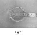
- FIG. 1 shows the prepared sheet-like cell culture.
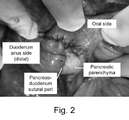
- FIG. 2 shows the operation sites of the pancreaticoduodenectomy in the control group.
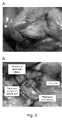
- FIG. 3 shows the operation sites of the pancreaticoduodenectomy in the sheet treatment group.
- the pancreatic head and the anal side of the duodenal papilla were jointly resected, the duodenal stump was anastomosed end to end, and a sheet-like cell culture was transplanted to the cut surface of the pancreatic head (the area surrounded by the dotted line in Fig. 3A).
- the duodenum and pancreatic parenchyma were anastomosed (Fig. 3B).
- FIG. 4A shows an HE-stained image (x200) of the anastomotic site between the duodenum and pancreatic parenchyma excised 3 days after pancreaticoduodenectomy in the control group. Arrows indicate hemorrhage and concentrated proteins.
- FIGS. 4B and C show HE-stained images ( ⁇ 200) of the implanted site of the sheet-like cell culture excised 3 days after the sheet-like cell culture was implanted in the sheet-treated group. Areas between dotted lines indicate sheet-like cell cultures.
- FIG. 4D shows an anti-DESMIN antibody immunostaining image (x200) of the implanted site of the sheet-like cell culture excised 3 days after the sheet-like cell culture was implanted in the sheet-treated group.
- FIG. 5 shows (ascites amylase level)/(serum amylase level) (FIG. 5A) and (ascites lipase level)/(serum lipase level) (FIG. 5B) three days after pancreatoduodenectomy surgery.
- the quantile, the second quartile (median) and the first quartile are shown, the bottom bar indicates the minimum value, and the value in the figure indicates the p-value between the two groups (Mann-Whitney U test).
- One aspect of the present invention is a sheet-like cell culture for covering a cut surface of the pancreas, wherein the cut surface of the pancreas includes cut surfaces of the pancreatic parenchyma and the pancreatic duct.
- the present invention relates to the sheet-like cell culture (sometimes referred to as "the sheet-like cell culture of the present invention") liquid-tightly bound to the intestinal wall.
- the pancreatic duct is fluid-tightly connected to the perforation of the intestinal wall of the small intestine via a sheet-like cell culture.
- the lumen of the pancreatic duct and the small intestine passes through the sheet-like cell culture and each through-hole of the intestinal wall of the small intestine. communicate through.
- the pancreatic duct is the main pancreatic duct and/or the accessory pancreatic duct.
- the small intestine is the duodenum or jejunum.
- the cut surface of the pancreas, the perforation of the intestinal wall of the small intestine, and/or the perforation of the sheet-like cell culture are formed by a surgical procedure.
- the surgical procedure is a pancreaticoduodenectomy.
- the sheet-like cell culture of the present invention is for preventing or treating pancreatic fistula.
- the sheet-like cell culture of the present invention comprises skeletal myoblasts.
- the sheet-like cell culture of the present invention comprises a reinforcing layer comprising gel and/or polymer.
- the pancreas is an exocrine gland that secretes pancreatic juice and pumps pancreatic juice into the duodenum.
- the portion of the pancreas on the duodenum side is called the pancreatic head, the portion on the splenic side is called the pancreatic tail, and the portion between the pancreatic head and the pancreatic tail is called the pancreatic body.
- the pancreas has pancreatic ducts inside it that carry pancreatic juice to the duodenum. Many branched thin pancreatic ducts merge from the pancreatic tail to the pancreatic head to form the main pancreatic duct (and accessory pancreatic duct), which leads to the duodenum.
- the main pancreatic duct joins the common bile duct, which carries bile from the gallbladder, before joining the duodenum.
- the pancreatic duct connects to the intestinal wall of the duodenum and drains pancreatic juice into the lumen of the duodenum.
- the opening of the pancreatic duct rises into the lumen of the duodenum and is called the duodenal papilla.
- the pancreas may be the pancreas of any organism, including but not limited to, humans, non-human primates, rodents (mouse, rat, hamster, guinea pig, etc.), rabbits, dogs, cats. , pigs, horses, cows, goats, sheep and other mammalian pancreases.
- the pancreas can be said to be an organ that has lumens such as the pancreatic duct, the main pancreatic duct, and the accessory pancreatic duct.
- Biological fluids such as digestive fluids flow through such lumens.
- the cut surface of the pancreas is liquid-tightly bound to the intestinal wall of the small intestine via the sheet-like cell culture
- the pancreatic duct is liquid-tightly connected to the through-hole of the intestinal wall of the small intestine via the sheet-like cell culture. The bonding can maintain the liquid-tightness of such lumen suture sites after pancreatic surgery.
- a "cut plane of the pancreas” refers to a cross section and/or a cross section of a fragment resulting from cutting the pancreas at an arbitrary site and/or cutting the pancreas at an arbitrary site.
- a cut plane of the pancreas is, for example, a section of the resulting fragment that cuts the pancreas between the head and body of the pancreas.
- the cut plane of the pancreas includes cut planes of each of the pancreatic parenchyma and the pancreatic duct, wherein the cut plane of the pancreatic duct is, for example, the cut plane of the main pancreatic duct and/or the accessory pancreatic duct.
- covering the cut surface of the pancreas refers to covering the entire cut surface of the pancreas with the sheet-like cell culture.
- the size of the sheet-like cell culture is equal to or larger than the cut surface of the pancreas, and just covers the entire cut surface of the pancreas, or covers the entire cut surface of the pancreas and has an extra portion. may have an edge of Such margins may additionally wrap around the cut surface of the pancreas.
- the sheet-like cell culture may have through holes, and "covering the cut surface of the pancreas” means that the entire cut surface of the pancreas is covered by the sheet-like cell culture having through holes at arbitrary positions. includes covering the At this time, the sheet-like cell culture is arranged so that the through-holes thereof are aligned with the lumen portion of the cut surface of the pancreatic duct.
- a "sheet-like cell culture” refers to a sheet-like cell formed by connecting cells to each other.
- Cells may be connected to each other directly (including through cellular elements such as adhesion molecules) and/or through intermediaries.
- the intervening substance is not particularly limited as long as it is a substance capable of at least physically (mechanically) connecting cells to each other, and examples thereof include extracellular matrix and the like.
- the mediator is preferably of cell origin, in particular of cells that make up the cell culture.
- the cells are at least physically (mechanically) linked, but may also be functionally linked, eg chemically, electrically.
- the sheet-like cell culture may be composed of one cell layer (single layer) or composed of two or more cell layers (laminated (multilayered) body, e.g., two layers, three layers, 4 layers, 5 layers, 6 layers, etc.).
- the sheet-like cell culture may have a three-dimensional structure in which the cells do not exhibit a clear layered structure and have a thickness exceeding the thickness of a single cell.
- the cells may be non-uniformly arranged (for example, in a mosaic pattern) without being evenly aligned in the horizontal direction.
- the sheet-like cell culture preferably does not contain a scaffold (support). Scaffolds may be used in the art to maintain the physical integrity of sheet-like cell cultures by attaching cells on and/or within them, such as polyvinylidene difluoride ( PVDF) membranes and the like are known, but the sheet-like cell culture of the present invention can maintain its physical integrity without such a scaffold.
- the sheet-like cell culture of the present invention preferably consists only of substances derived from cells constituting the sheet-like cell culture, and does not contain other substances.
- the cells that make up the sheet-like cell culture are not particularly limited as long as they can form a sheet-like cell culture, and include, for example, adherent cells (adherent cells).
- Adherent cells include, for example, adherent somatic cells (e.g., cardiomyocytes, fibroblasts, epithelial cells, endothelial cells, hepatocytes, pancreatic cells, kidney cells, adrenal cells, periodontal ligament cells, gingival cells, periosteal cells, skin cells, synovial cells, chondrocytes, etc.) and stem cells (e.g., myoblasts, tissue stem cells such as cardiac stem cells, embryonic stem cells, pluripotent stem cells such as iPS (induced pluripotent stem) cells, mesenchymal stem cells, etc.) etc.
- adherent somatic cells e.g., cardiomyocytes, fibroblasts, epithelial cells, endothelial cells, hepatocytes, pancreatic cells
- Somatic cells may be stem cells, particularly those differentiated from iPS cells (iPS cell-derived adherent cells).
- Non-limiting examples of cells constituting sheet-like cell cultures include, for example, myoblasts (e.g., skeletal myoblasts), mesenchymal stem cells (e.g., bone marrow, adipose tissue, peripheral blood, skin, hair roots, muscle tissue, endometrium, placenta, those derived from umbilical cord blood, etc.), cardiomyocytes, fibroblasts, cardiac stem cells, embryonic stem cells, iPS cells, synovial cells, chondrocytes, epithelial cells (e.g., oral mucosal epithelial cells , retinal pigment epithelial cells, nasal mucosal epithelial cells, etc.), endothelial cells (e.g., vascular endothelial cells, etc.), hepatocytes (e.g., hepatocytes, etc.), pancreatic
- Non-limiting examples of iPS cell-derived adhesive cells include iPS cell-derived cardiomyocytes, fibroblasts, epithelial cells, endothelial cells, hepatocytes, pancreatic cells, renal cells, adrenal cells, periodontal ligament cells, gingival cells, and periosteal cells. , skin cells, synovial cells, chondrocytes, and the like.
- skeletal myoblasts refer to myoblasts present in skeletal muscle. Skeletal myoblasts are well known in the art, and can be prepared from skeletal muscle by any known method (e.g., the method described in Japanese Patent Application Laid-Open No. 2007-89442), or commercially available. Also available (eg Lonza, Cat# CC-2580). Skeletal myoblasts have markers such as, but not limited to, CD56, ⁇ 7 integrin, myosin heavy chain IIa, myosin heavy chain IIb, myosin heavy chain IId (IIx), MyoD, Myf5, Myf6, myogenin, desmin, PAX3.
- markers such as, but not limited to, CD56, ⁇ 7 integrin, myosin heavy chain IIa, myosin heavy chain IIb, myosin heavy chain IId (IIx), MyoD, Myf5, Myf6, myogenin, desmin, PAX3.
- the skeletal myoblasts are CD56 positive. In a more particular embodiment, the skeletal myoblasts are CD56 positive and desmin positive. Skeletal myoblasts can be derived from any organism with skeletal muscle, including but not limited to humans, non-human primates, rodents (mouse, rat, hamster, guinea pig, etc.), rabbits, dogs, cats, pigs, It may be derived from mammals such as horses, cows, goats, and sheep. In one aspect, the skeletal myoblast is a mammalian skeletal myoblast. In certain embodiments, the skeletal myoblasts are human skeletal myoblasts. Also, skeletal myoblasts can be collected from any skeletal muscle. In one aspect, the skeletal myoblasts of the invention are skeletal myoblasts from the thigh, neck, or abdomen.
- the cells that make up the sheet-like cell culture can be derived from any organism that can be treated with the sheet-like cell culture.
- organisms include, but are not limited to, humans, non-human primates, dogs, cats, pigs, horses, goats, sheep, rodents (eg, mice, rats, hamsters, guinea pigs, etc.), rabbits, and the like. is included.
- rodents eg, mice, rats, hamsters, guinea pigs, etc.
- rabbits and the like.
- the number of types of cells constituting the sheet-like cell culture is not particularly limited, and it may be composed of only one type of cell, or may be composed of two or more types of cells.
- the content ratio (purity) of the largest number of cells is 50% or more, preferably 60% or more, more preferably 60% or more at the end of formation of the sheet-like cell culture. is 70% or more, more preferably 75% or more.
- the cells may be heterologous or allogeneic.
- heterologous cells refer to cells derived from organisms of a species different from that of the recipient when sheet-like cell cultures are used for transplantation. For example, when the recipient is a human, cells derived from monkeys, pigs, etc. correspond to heterologous cells.
- allogeneic cells refer to cells derived from the same species of organism as the recipient. For example, if the recipient is human, human cells are allogeneic cells. Allogeneic cells include autologous cells (also referred to as autologous or autologous cells), ie cells derived from the recipient, and allogeneic non-autologous cells (also referred to as allogeneic cells).
- Autologous cells are preferred in the present invention because they do not cause rejection when transplanted. However, it is also possible to use xenogenic or allogeneic non-autologous cells. Immunosuppressive treatment may be required to prevent rejection when xenogeneic or allogeneic non-autologous cells are used.
- cells other than autologous cells that is, heterologous cells and allogeneic non-autologous cells are sometimes referred to as non-autologous cells.
- the cells are autologous or allogeneic.
- the cells are autologous cells.
- the cells are allogeneic cells.
- a sheet-like cell culture can be produced by any known method (see, for example, Patent Document 1, JP-A-2010-081829, JP-A-2011-110368, etc.).
- a method for producing a sheet-like cell culture typically includes the steps of seeding cells on a culture substrate, forming a sheet from the seeded cells, and peeling the formed sheet-like cell culture from the culture substrate. Including but not limited to steps.
- the step of seeding the cells onto the culture substrate may be preceded by steps of freezing the cells and thawing the cells. Additionally, a step of washing the cells may be performed after the step of thawing the cells.
- Each of these steps can be performed by any known method suitable for producing a sheet-like cell culture.
- the method for producing a sheet-like cell culture may include the step of producing a sheet-like cell culture, in which case the step of producing a sheet-like cell culture includes, as a substep, the method for producing a sheet-like cell culture may include one or more of the steps of In one aspect, there is no step of growing the cells after the step of thawing the cells and before seeding the cells onto the culture substrate.
- the culture substrate is not particularly limited as long as cells can form a cell culture on it, and includes, for example, vessels made of various materials, solid or semi-solid surfaces in vessels, and the like.
- the container preferably has a structure and material impermeable to a liquid such as a culture solution.
- materials include, without limitation, polyethylene, polypropylene, Teflon (registered trademark), polyethylene terephthalate, polymethyl methacrylate, nylon 6,6, polyvinyl alcohol, cellulose, silicon, polystyrene, glass, polyacrylamide, polydimethyl acrylamide, metals (eg, iron, stainless steel, aluminum, copper, brass) and the like. It is also preferred that the container has at least one flat surface.
- Such vessels include, but are not limited to, culture vessels having a bottom surface composed of a culture substrate capable of forming a cell culture and liquid-impermeable sides.
- Particular examples of such culture vessels include, without limitation, cell culture dishes, cell culture bottles, and the like.
- the bottom of the container may be transparent or opaque. When the bottom surface of the container is transparent, cells can be observed, counted, etc. from the back side of the container.
- the container may also have a solid or semi-solid surface inside it. Solid surfaces include plates and containers of various materials as described above, and semi-solid surfaces include gels, soft polymer matrices, and the like.
- the culture substrate may be produced using the materials described above, or commercially available ones may be used.
- Preferred culture substrates include, without limitation, substrates having an adherent surface suitable for forming, for example, sheet-like cell cultures.
- substrates having a hydrophilic surface for example, corona discharge-treated polystyrene, substrates coated with hydrophilic compounds such as collagen gel and hydrophilic polymers on the surface, collagen, fibronectin, and laminin. , vitronectin, proteoglycans, glycosaminoglycans and the like, and substrates coated with cell adhesion factors such as cadherin family, selectin family and integrin family on the surface.
- substrates are commercially available (eg, Corning® TC-Treated Culture Dish, Corning, etc.).
- the culture substrate may be wholly or partially transparent or opaque.
- the surface of the culture substrate may be coated with a material whose physical properties change in response to stimuli such as temperature and light.
- materials include, but are not limited to, (meth)acrylamide compounds, N-alkyl-substituted (meth)acrylamide derivatives (e.g., N-ethylacrylamide, Nn-propylacrylamide, Nn-propylmethacrylamide, N-isopropylacrylamide, N-isopropylmethacrylamide, N-cyclopropylacrylamide, N-cyclopropylmethacrylamide, N-ethoxyethylacrylamide, N-ethoxyethylmethacrylamide, N-tetrahydrofurfurylacrylamide, N-tetrahydrofurfurylmethacrylamide amide, etc.), N,N-dialkyl-substituted (meth)acrylamide derivatives (e.g., N,N-dimethyl(meth)acrylamide, N,N-eth
- the culture substrate may be of various shapes, but is preferably flat.
- the area is not particularly limited, but may be, for example, about 1 cm 2 to about 200 cm 2 , about 2 cm 2 to about 100 cm 2 , about 3 cm 2 to about 50 cm 2 .
- the culture substrate may be a circular culture dish with a diameter of 10 cm. In this case, the area will be 56.7 cm 2 .
- the culture substrate may be coated (coated or coated) with serum.
- a sheet-like cell culture with a higher density can be formed by using a serum-coated culture substrate.
- “Serum-coated” refers to a state in which serum components adhere to the surface of the culture substrate. Such a state can be obtained, without limitation, for example, by treating the culture substrate with serum. Treatment with serum includes contacting the serum with the culture substrate and optionally incubating for a period of time.
- Heterologous serum and/or allogeneic serum can be used as the serum.
- Heterologous serum means serum derived from an organism of a different species than the recipient when sheet cell cultures are used for transplantation.
- serum derived from cows or horses such as fetal bovine serum (FBS, FCS), calf serum (CS), horse serum (HS), etc.
- FBS, FCS fetal bovine serum
- CS calf serum
- HS horse serum
- allogeneic serum refers to serum derived from organisms of the same species as the recipient. For example, if the recipient is human, human serum is an allogeneic serum.
- Allogeneic serum includes autologous serum (also referred to as autologous serum), ie, serum derived from the recipient, and allogeneic serum derived from allogeneic individuals other than the recipient.
- autologous serum also referred to as autologous serum
- serum derived from the recipient serum derived from allogeneic individuals other than the recipient.
- sera other than self sera that is, heterologous sera and allogeneic sera are sometimes referred to as non-self sera.
- Serum for coating the culture substrate is commercially available or can be prepared by conventional methods from blood drawn from the desired organism. Specifically, for example, the collected blood is left at room temperature for about 20 to about 60 minutes to coagulate, centrifuged at about 1000 ⁇ g to about 1200 ⁇ g, and the supernatant is collected. etc.
- the serum When incubating on a culture substrate, the serum may be used in its original form or diluted. Dilutions can be made in any medium, including, but not limited to, water, saline, various buffers (eg, PBS, HBSS, etc.), various liquid media (eg, DMEM, MEM, F12, DMEM/F12, DME, RPMI1640, MCDB (MCDB102, 104, 107, 120, 131, 153, 199, etc.), L15, SkBM, RITC80-7, etc.) can be used.
- the dilution concentration is not particularly limited as long as the serum components can adhere to the culture substrate. /v), more preferably from about 5% to about 40% (v/v).
- the incubation time is also not particularly limited as long as the serum components can adhere to the culture substrate. 24 hours, more preferably about 2 hours to about 12 hours.
- the incubation temperature is also not particularly limited as long as serum components can adhere to the culture substrate. is.
- the serum may be discarded after incubation.
- a serum disposal method a conventional liquid disposal method such as aspiration with a pipette or the like or decantation can be used.
- the culture substrate may be washed with a serum-free washing solution after discarding the serum.
- the serum-free washing solution is not particularly limited as long as it is a liquid medium that does not contain serum and does not adversely affect the serum components attached to the culture substrate.
- Liquid e.g., PBS, HBSS, etc.
- various liquid media e.g., DMEM, MEM, F12, DMEM/F12, DME, RPMI1640, MCDB (MCDB102, 104, 107, 120, 131, 153, 199, etc.), L15 , SkBM, RITC80-7, etc.
- the washing method may be a conventional washing method of the culture substrate, for example, without limitation, a method of adding a serum-free washing solution onto the culture substrate, stirring for a predetermined time (for example, about 5 seconds to about 60 seconds), and then discarding it. etc. can be used.
- the culture substrate may be coated with growth factors.
- growth factor refers to any substance that promotes cell proliferation compared to its absence, e.g., epidermal growth factor (EGF), vascular endothelial growth factor (VEGF), fibroblast Including cell growth factor (FGF) and the like.
- the dilution concentration at the time of incubation is, for example, about 0.0001 ⁇ g/mL to about 1 ⁇ g/mL, preferably about 0.0005 ⁇ g/mL to about 0.5 ⁇ g/mL. 05 ⁇ g/mL, more preferably from about 0.001 ⁇ g/mL to about 0.01 ⁇ g/mL, but essentially the same as serum.
- the culture substrate may be coated with a steroid agent.
- a steroid agent refers to compounds having a steroidal nucleus, which can have adverse effects on living organisms, such as adrenal cortex dysfunction and Cushing's syndrome. Such compounds include, but are not limited to, cortisol, prednisolone, triamcinolone, dexamethasone, betamethasone, and the like.
- the dilution concentration during incubation is, for example, about 0.1 ⁇ g/mL to about 100 ⁇ g/mL, preferably about 0.4 ⁇ g/mL to about 0.4 ⁇ g/mL, as dexamethasone. It is essentially the same as serum except that it is about 40 ⁇ g/mL, more preferably about 1 ⁇ g/mL to about 10 ⁇ g/mL.
- the culture medium may be coated with any one of serum, a growth factor and a steroid agent, or any combination thereof, i.e. serum and growth factor, serum and steroid agent, serum and growth factor and steroid agent, or , may be coated with a combination of growth factors and steroids.
- serum and growth factor serum and steroid agent
- serum and growth factor and steroid agent serum and growth factor and steroid agent
- these components may be mixed and coated at the same time or in separate steps.
- the culture substrate may be seeded with cells immediately after being coated with serum or the like, or may be stored after being coated and then seeded with cells.
- the coated substrate can be stored for a long period of time, for example, by keeping it at about 4°C or lower, preferably about -20°C or lower, more preferably about -80°C or lower.
- Cells can be seeded onto the culture substrate by any known technique and conditions. Cells may be sown on the culture substrate, for example, by injecting a cell suspension obtained by suspending cells in a culture solution into the culture substrate (culture vessel).
- an instrument suitable for injecting the cell suspension such as a dropper or a pipette, can be used.
- Seeding is from about 7.1 ⁇ 10 5 /cm 2 to about 3.0 ⁇ 10 6 /cm 2 , from about 7.3 ⁇ 10 5 /cm 2 to about 2.8 ⁇ 10 6 /cm 2 . , about 7.5 ⁇ 10 5 pieces/cm 2 to about 2.5 ⁇ 10 6 pieces/cm 2 , about 7.8 ⁇ 10 5 pieces/cm 2 to about 2.3 ⁇ 10 6 pieces/cm 2 , about 8.0 ⁇ 10 5 pieces/cm 2 to about 2.0 ⁇ 10 6 pieces/cm 2 , about 8.5 ⁇ 10 5 pieces/cm 2 to about 1.8 ⁇ 10 6 pieces/cm 2 , about 9.5 ⁇ 10 5 pieces/cm 2 to about 1.8 ⁇ 10 6 pieces/cm 2 . 0 ⁇ 10 5 pieces/cm 2 to about 1.6 ⁇ 10 6 pieces/cm 2 , about 1.0 ⁇ 10 6 pieces/cm 2 to about 1.6 ⁇ 10 6 pieces/cm 2 , etc. can be done.
- the cells constituting the sheet-like cell culture of the present invention are as detailed above.
- the sheet-like cell culture of the present invention does not need to contain cells of the target tissue, but rather may contain cells that do not exist in the tissue.
- the sheet-like cell culture of the present invention comprises cells that are ectopic, ie, cells that are not naturally present in the applied tissue.
- the sheet-like cell culture of the present invention is applied to tissue damage in the pancreas and small intestine, and the ectopic cells in this case include skeletal myoblasts and the like. Examples include cells derived from striated muscle, mesenchymal stem cells, and the like.
- the sheet-like cell culture of the present invention contains skeletal myoblasts. Skeletal myoblasts may be derived from striated muscles of the thigh, neck, abdomen, and the like.
- the prepared cell population contains fibroblasts.
- a cell population containing skeletal myoblasts prepared from striated muscle tissue is used to produce the sheet-like cell culture of the present invention, the cell population contains a certain amount of fibroblasts.
- Fibroblasts are well-known in the art and are TE-7 (e.g. Rosendaal et al., J Cell Sci. 1994; 107 (Pt 1):29-37, Goodpaster et al., J Histochem Cytochem. 2008;56(4):347-58, etc.).
- the cells forming the sheet-like cell culture of the present invention comprise skeletal myoblasts prepared from striated muscle tissue. Therefore, the cell population used in the production of the sheet-like cell culture of the present invention can contain skeletal myoblasts and fibroblasts. In one aspect, the cell population used in the production of the sheet-like cell culture of the present invention has a CD56 positive rate of 50% or more, 60% or more, 65% or more, 70% or more, 75% or more, 80% or more, 85% or more % or more, 90% or more, preferably 60% or more.
- the cell population used in the production of the sheet-shaped cell culture of the present invention may contain fibroblasts, but if the fibroblast content is too high, the skeletal myoblast content will decrease, which is undesirable. . Therefore, in one aspect, the cell population used in the production of the sheet-shaped cell culture of the present invention has a TE7 positive rate of 50% or less, 40% or less, 35% or less, 30% or less, 25% or less, 20% or less, It can be 15% or less, 10% or less, preferably 40% or less.
- the cell population used in the production of the sheet-like cell culture of the present invention may contain cells other than skeletal myoblasts and fibroblasts, but the number of such cells is preferably as small as possible. Therefore, the total value of CD56 positive rate and TE7 positive rate is preferably higher, for example, 80% or more, 85% or more, 90% or more, 95% or more, 96% or more, 97% or more, 98% or more, 99% or more etc., preferably 90% or more.
- the thickness of the sheet-like cell culture of the present invention is not particularly limited. When a monolayer sheet is used as a sheet-like cell culture, the thickness is usually equal to or greater than one cell, and the thickness varies depending on the type of sheet-forming cells.
- the sheet-like cell culture has a thickness of 30 ⁇ m or more, and in a preferred embodiment, a thickness of 50 ⁇ m or more.
- the thickness range of the sheet-shaped cell culture of the present invention is, for example, 30 ⁇ m to 200 ⁇ m, preferably 50 ⁇ m to 150 ⁇ m, more preferably 60 ⁇ m to 100 ⁇ m.
- the thickness does not exceed the thickness of the monolayer sheet ⁇ the number of laminated sheets.
- the thickness is 150 ⁇ m or more, preferably 250 ⁇ m or more, and the thickness range is, for example, 150 ⁇ m to 1000 ⁇ m, preferably 250 ⁇ m to 750 ⁇ m, and more. 300 ⁇ m to 500 ⁇ m are preferred. Therefore, the thickness of the sheet-like cell culture of the present invention is, for example, 30 ⁇ m to 1000 ⁇ m, preferably 50 ⁇ m to 750 ⁇ m, 50 ⁇ m to 500 ⁇ m, 60 ⁇ m to 500 ⁇ m.
- the sheet-like cell culture of the present invention is very fragile and may be difficult to handle. Therefore, the sheet-like cell culture of the present invention may further have a reinforcing layer for the purpose of facilitating handling and reducing the risk of breakage. Any reinforcing layer may be used as long as it does not impair the function of the sheet-shaped cell culture of the present invention and can reinforce the structure.
- it may be a reinforcing layer containing gel and/or polymer. Since it is to be implanted in vivo, it is preferably a biocompatible reinforcing layer containing, for example, a biocompatible gel or polymer.
- the gel that can be used for the reinforcing layer of the present invention may be any gel that does not adversely affect the body when introduced into the body, and is not limited to this.
- examples include fibrin gel, fibrinogen gel, gelatin gel, collagen gel and the like.
- the polymer that can be used in the reinforcing layer of the present invention, preferably a biocompatible polymer may be any polymer that does not adversely affect the body when introduced into the body, and is not limited to this. Examples include polylactic acid, polydioxano, polyglycapro, collagen, and the like.
- a method known in the art can be used to form the reinforcing layer containing the biocompatible gel.
- examples of such methods include, but are not limited to, a method of spraying a biocompatible gel or polymer or a component thereof onto a sheet-shaped cell culture, a method of spraying a biosol on a sheet-shaped cell culture.
- the method of solidifying the gel after immersing it in a liquid gel, and the like the method described in JP-A-2016-52271 and the like can be mentioned.
- the reinforcing layer is intended to facilitate handling of the sheet-like cell culture of the present invention and reduce the risk of breakage, it preferably has a certain level of strength, and furthermore has elasticity. preferably.
- Known evaluation units for the strength of structures containing gels and polymers include, for example, jelly strength
- known evaluation units for the strength of sheet-like structures include, for example, tensile breaking load.
- a method for measuring jelly strength is described in, for example, JIS K 6503.
- Tensile breaking load means the maximum load until both ends of a sheet-like cell culture are pulled horizontally and broken, and its measuring method is described, for example, in Japanese Patent Application Laid-Open No. 2016-52272.
- the reinforcing layer of the sheet-shaped cell culture of the present invention is not limited to this, but for example, a tensile breaking load of about 0.010 N or more, about 0.015 N or more, about 0.020 N or more, about 0.025 N or more, about 0.030 N or more, about 0.035 N or more, about 0.040 N or more, about 0.045 N or more, etc., and about 0.010 N to about 0.200 N, about 0.015 N to about 0 .100 N, about 0.020 N to about 0.50 N, and the like.
- a sheet-like cell culture with a reinforcing layer has a strength of about 1.5 times or more, about 2 times or more, about 3 times or more, about 4 times or more, as compared with a sheet-like cell culture without a reinforcing layer. about 5 times or more, about 6 times or more, about 7 times or more, about 8 times or more, about 9 times or more, about 10 times or more, and about 1.5 times to about 20 times, about 2 times to about 15 times It can range from about 2.5 times to about 10 times, and so on.
- the reinforcing layer When applying a sheet-like cell culture having a reinforcing layer, it is preferable to apply the reinforcing layer so as not to directly contact the application site. That is, it is preferable to apply the sheet-like cell culture so that it is positioned between the application site and the reinforcing layer.
- Application of the sheet-like cell culture to the application site can be performed using any device and/or technique known in the art.
- the sheet-like cell culture of the present invention when applied to tissue, it may be applied in combination with other compositions and/or grafts that promote healing and adhesion.
- Other compositions and/or grafts that promote healing and adhesion include, but are not limited to, grafts containing pedicled blood vessels such as pedicled omentum, fibrin gels, ad sprays ( registered trademark).
- the sheet-like cell culture of the present invention is applied together with a graft containing pedicled blood vessels.
- a typical example of a graft containing a pedicled blood vessel is, for example, a pedicled omentum.
- Other compositions and/or grafts that promote such healing and adhesion may be compositions or grafts independent of the sheet-like cell culture of the present invention. Alternatively, it may be incorporated in a reinforcing layer or the like.
- compositions and/or grafts can be may be applied before or after. If other compositions and/or grafts are applied prior to application of the sheet-like cell culture, they are applied so that they are located between the application site and the sheet-like cell culture. That is, first, another composition and/or graft is applied to the application site, and then the sheet-like cell culture (optionally including a reinforcing layer) is applied thereover. When applied after application of a sheet-like cell culture, it is applied to the application site from above the sheet-like cell culture (optionally including a reinforcing layer). That is, first, a sheet-like cell culture is applied to the application site, and then another composition and/or graft is applied thereon.
- the sheet-like cell culture continuously releases cytokines such as VEGF, HGF, etc., which are responsible for angiogenesis, cell protection, and repair at the site of injury, collagen, etc., and is due to the paracrine effect.
- cytokines such as VEGF, HGF, etc.
- progenitor cells and stem cells in the applied surrounding tissue are activated to obtain an autocrine effect that promotes collagen production and the like.
- the size of the sheet-like cell culture of the present invention is not particularly limited as long as it can cover a predetermined portion of a living tissue. is 10 to 55 mm, 15 to 50 mm, 20 to 45 mm, 25 to 40 mm, or 30 to 35 mm, preferably 30 to 35 mm from the viewpoint of covering the cut surface of the pancreas.
- the small intestine is the digestive tract that follows the stomach and connects to the large intestine.
- the part on the stomach side is called the duodenum
- the part on the large intestine side is called the ileum
- the part between the duodenum and the ileum is called the jejunum.
- the duodenum surrounds the head of the pancreas while curving in a C shape.
- the opening of the pancreatic duct of the pancreas rises into the lumen of the duodenum and is called the duodenal papilla. Pancreatic juice and bile are supplied through such openings.
- the term “small intestine wall” refers to the wall of any part of the small intestine that separates the inside and outside of the small intestine.
- the intestinal wall of the small intestine is composed of serosa, outer longitudinal muscle layer, inner circular muscle layer, submucosa, muscularis mucosa, lamina intestinal, mucosal epithelium, and the like.
- the small intestine wall is a duodenal wall, a jejunal wall, or an ileal wall.
- liquid-tightly bonded means that different parts of the biological tissue are closely bonded together so that biological fluids such as digestive juices in the biological tissue do not leak out.
- liquid-tightness refers to the property of liquid-tight bonding between different parts of living tissue. Such bonding can be performed using any medical device or device such as sutures, medical staplers, suture adhesives, etc., and is preferably performed using sutures from the viewpoint of liquid tightness. For example, different parts of the body tissue are sutured with suture thread at the parts where they are joined or the peripheral part of the surface, etc., and are tightly joined together so that the biological fluid such as digestive juice in the body tissue does not leak out. .
- the term “liquid-tightly bonded via a sheet-like cell culture” means that different parts of the living tissue sandwich the sheet-like cell culture, and live digestive juices and the like in the living tissue. It refers to bonding tightly to each other so that body fluids cannot leak out. In other words, such bonding results in a structure in which "part of biological tissue--sheet-like cell culture--part of biological tissue" is tightly bonded to each other. At this time, all of the parts or surfaces where one part of the living tissue is connected to the other part are covered with the sheet-like cell culture. In addition, such a connection can be made using any medical device or device such as a suture thread, a medical stapler, a suture adhesive, etc.
- a suture thread For example, different parts of the biological tissue are sutured together with the sheet-shaped cell culture at the part where they are joined or the edge of the surface, etc., and the sheet-shaped cell culture is sandwiched in the biological tissue. They are tightly bonded to each other so that biological fluids such as digestive juices do not leak out.
- the cut surface of the pancreas is liquid-tightly bound to the intestinal wall of the small intestine via the sheet-like cell culture
- the cut surface of the pancreas and the intestinal wall of the small intestine sandwich the sheet-like cell culture. It means that the body fluids such as digestive juices in the pancreas and small intestine are tightly bound to each other so as not to leak out. In other words, such bonding results in a structure of "cut surface of pancreas-sheet-like cell culture-small intestine intestinal wall" that is tightly bonded to each other. At this time, the entire cut surface of the pancreas is covered with the sheet-like cell culture.
- connection can be made using any medical device or device such as a suture thread, a medical stapler, a suture adhesive, etc.
- a suture thread For example, the cut surface of the pancreas and the intestinal wall of the small intestine, along with the sheet-like cell culture, are sutured with sutures at the parts where they are joined or at the edge of the surface, etc., and the sheet-like cell culture is sandwiched between the pancreas. and closely adhere to each other so that biological fluids such as digestive juices in the small intestine do not leak out.
- the small intestine can be the duodenum or the jejunum.
- the pancreatic duct is liquid-tightly bound to the through-hole of the intestinal wall of the small intestine via the sheet-like cell culture
- the sheet-like cell culture is sandwiched between the pancreatic duct and the through-hole of the intestinal wall of the small intestine. It means that the body fluids such as digestive juices in the pancreas and small intestine are tightly bound to each other so as not to leak out. In other words, such binding results in a structure of "pancreatic duct-sheet-like cell culture-through-hole of the intestinal wall of the small intestine" that is tightly bound to each other.
- the pancreatic duct is exposed as a cross section of the pancreatic duct at the cut surface of the pancreas, and the cross section is composed of the pancreatic duct wall and the lumen, and the pancreatic duct wall and the lumen are all covered with the sheet-like cell culture.
- a connection can be made using any medical device or device such as a suture thread, a medical stapler, a suture adhesive, etc. From the viewpoint of liquid tightness, it is preferably made using a suture thread.
- the through-holes of the pancreatic duct and the intestinal wall of the small intestine, together with the sheet-like cell culture are sutured with sutures at the parts where they are joined or at the edge of the surface, etc., and the sheet-like cell culture is sandwiched between the pancreas. and closely adhere to each other so that biological fluids such as digestive juices in the small intestine do not leak out.
- the through-holes of the pancreatic duct and the wall of the small intestine are tightly joined together so that the cut surface of the pancreatic duct and the through-hole of the wall of the small intestine are of the same size.
- the pancreatic duct can be the main pancreatic duct or the accessory pancreatic duct.
- the small intestine can be the duodenum or the jejunum.
- the cut surface of the pancreas is fluid-tightly bound to the intestinal wall of the small intestine via the sheet-like cell culture
- the pancreatic duct is fluidly connected to the through-hole of the intestinal wall of the small intestine via the sheet-like cell culture.
- the cut surface of the pancreas is entirely covered with the sheet-shaped cell culture of the present invention, and the cut surface of the pancreas and the intestinal wall of the small intestine are separated from each other at the portion where they are joined or the peripheral portion of the surface. They are sewn together with sutures and tightly joined together to prevent leakage of biological fluids such as digestive juices in the pancreas and small intestine.
- the cut surface of the pancreas, the sheet-like cell culture of the present invention and the intestinal wall of the small intestine are arranged in this order and fixed while being tightly bound to each other.
- the cut surface of the pancreas and the intestinal wall of the small intestine are the sheet-like cell culture of the present invention present between the cut surface of the pancreas and the intestinal wall of the small intestine, and the portion where they are bonded or the peripheral portion of the surface. are sewn together with sutures, and are tightly joined together so that biological fluids such as digestive juices in the pancreas and small intestine do not leak out.
- the pancreatic duct can be the main pancreatic duct or the accessory pancreatic duct.
- the small intestine can be the duodenum or the jejunum.
- the position of the lumen portion of the cut surface of the pancreatic duct is aligned with the through-hole of the intestinal wall of the small intestine.
- the pancreatic duct is exposed as a cross section of the pancreatic duct at the cut surface of the pancreas, and the cross section is composed of the pancreatic duct wall and the lumen, and the pancreatic duct wall and the lumen are all covered with the sheet-like cell culture. ing.
- the through-holes of the pancreatic duct and the intestinal wall of the small intestine, together with the sheet-shaped cell culture, are sutured with sutures at the portions where they are joined or at the margins of the planes, and the pancreas and the small intestine with the sheet-shaped cell culture sandwiched therebetween. They are tightly bonded to each other so that biological fluids such as digestive juices inside do not leak out.
- the through-holes of the pancreatic duct and the wall of the small intestine are tightly joined together so that the cut surface of the pancreatic duct and the through-hole of the wall of the small intestine are of the same size.
- each lumen of the pancreatic duct and the small intestine becomes a sheet. Communicate through the perforations of the morphogenetic cell culture and the intestinal wall of the small intestine.
- the pancreatic duct can be the main pancreatic duct or the accessory pancreatic duct.
- the small intestine can be the duodenum or the jejunum.
- a through hole refers to a perforation penetrating from a site on the surface of a living tissue to another site on the surface of the living tissue.
- a through hole is, for example, a perforation penetrating from any portion of the outer surface of the small intestine wall to any portion of the inner surface of the small intestine wall.
- a through-hole is a perforation through the wall of the small intestine perpendicular to the wall of the small intestine from any location on the outer surface of the wall of the small intestine.
- a through hole is a perforation through the duodenal wall perpendicular to the duodenal wall from any site on the outer surface of the duodenal wall.
- a through-hole is a perforation through the jejunal wall perpendicular to the jejunal wall from any site on the outer surface of the jejunal wall.
- a plurality of through-holes may exist in the biological tissue.
- a through-hole may be a perforation penetrating from one site on the surface of the sheet-like cell culture to another site on the surface of the sheet-like cell culture.
- the size of the through-hole is not particularly limited as long as the through-hole connects to a predetermined site of the living tissue. 7-5 mm, 0.8-4 mm, 0.9-3 mm, or 1-2 mm.
- the diameter of the through-hole is preferably 1 to 2 mm from the viewpoint of liquid-tight connection of the pancreatic duct to the through-hole of the intestinal wall of the small intestine.
- forming through-holes in the sheet-like cell culture in the lumen of the cut surface of the pancreatic duct means that the pancreatic duct and the small intestine are formed by any effect such as physical action or biochemical reaction. It refers to forming through-holes in the sheet-like cell culture at the lumen portion of the cut surface of the pancreatic duct so that each lumen communicates through the through-holes in the sheet-like cell culture and the intestinal wall of the small intestine.
- Physical action refers to piercing, perforation, penetration, etc. of the sheet-like cell culture at the lumen portion of the cut surface of the pancreatic duct with any medical instrument such as forceps or medical instrument.
- a biochemical reaction refers to the dissolution, decomposition, digestion, etc. of the sheet-like cell culture in the lumen of the cut surface of the pancreatic duct by a biological fluid such as pancreatic juice.
- a biological fluid such as pancreatic juice.
- the sheet-like cell culture since the sheet-like cell culture does not come into contact with biological fluids such as pancreatic juice except in the lumen of the cut surface of the pancreatic duct, the sheet-like cell culture is dissolved, degraded, and digested in areas other than the lumen of the cut surface of the pancreatic duct. etc. do not occur, and the sheet-like cell culture remains.
- Dissolution, decomposition, digestion, etc. of such sheet-like cell culture can be performed within several hours to several days after transplantation of the sheet-like cell culture.
- the sheet-like cell culture of the present invention when the sheet-like cell culture of the present invention does not contain a reinforcing layer, the sheet-like cell culture can be dissolved, decomposed, digested, etc. within several hours after the sheet-like cell culture is transplanted.
- the sheet-like cell culture of the present invention contains a reinforcing layer containing a gel and/or a polymer, dissolution, decomposition, digestion, etc. of the sheet-like cell culture takes several hours to several hours after transplantation of the sheet-like cell culture. It can be done in a matter of days.
- the pancreatic duct can be the main pancreatic duct or the accessory pancreatic duct.
- the small intestine can be the duodenum or the jejunum.
- the through-holes of the pancreatic duct and the intestinal wall of the small intestine sandwich the sheet-like cell culture, and are tightly connected to each other so that biological fluids such as digestive juice in the pancreas and small intestine do not leak out.
- biological fluids such as digestive juice in the pancreas and small intestine do not leak out.
- such binding results in a structure of "pancreatic duct-sheet-like cell culture-through-hole of the intestinal wall of the small intestine" that is tightly bound to each other.
- the pancreatic duct is exposed as a cross section of the pancreatic duct at the cut surface of the pancreas, and the cross section is composed of the pancreatic duct wall and the lumen, and the pancreatic duct wall and the lumen are all covered with the sheet-like cell culture.
- the pancreatic duct can be the main pancreatic duct or the accessory pancreatic duct.
- the small intestine can be the duodenum or the jejunum.
- the sheet-like cell culture of the present invention may have through-holes at arbitrary positions before covering the cut surface of the pancreas.
- the sheet-like cell culture is placed so that the through hole of the sheet-like cell culture is aligned with the position of the lumen of the cut surface of the pancreatic duct, so that each lumen of the pancreatic duct and the small intestine is filled with the sheet-like cell culture and the small intestine.
- Communication can be provided through each through-hole in the intestinal wall.
- the pancreatic duct can be the main pancreatic duct or the accessory pancreatic duct.
- the small intestine can be the duodenum or the jejunum.
- the through-holes of the pancreatic duct and the intestinal wall of the small intestine sandwich the sheet-like cell culture, and are tightly connected to each other so that biological fluids such as digestive juice in the pancreas and small intestine do not leak out.
- biological fluids such as digestive juice in the pancreas and small intestine do not leak out.
- such binding results in a structure of "pancreatic duct-sheet-like cell culture-through-hole of the intestinal wall of the small intestine" that is tightly bound to each other.
- the pancreatic duct is exposed as a cross section of the pancreatic duct at the cut surface of the pancreas, and the cross section is composed of the pancreatic duct wall and the lumen, and the pancreatic duct wall and the lumen are all covered with the sheet-like cell culture.
- the sheet-like cell culture has through-holes at arbitrary positions before covering the cut surface of the pancreas.
- the sheet-like cell culture is placed so that the through hole of the sheet-like cell culture is aligned with the position of the lumen of the cut surface of the pancreatic duct, so that each lumen of the pancreatic duct and the small intestine is filled with the sheet-like cell culture and the small intestine.
- Communication can be provided through each through-hole in the intestinal wall.
- the through-holes of the pancreatic duct and the wall of the small intestine are tightly joined together so that the cut surface of the pancreatic duct and the through-hole of the wall of the small intestine are of the same size.
- the pancreatic duct can be the main pancreatic duct or the accessory pancreatic duct.
- the small intestine can be the duodenum or the jejunum.
- the lumens of the pancreatic duct and the small intestine communicate with each other means that the lumens of the pancreatic duct and the small intestine are physically connected, and the biological fluid such as pancreatic juice in the lumen of the pancreatic duct flows into the lumen of the small intestine.
- the pancreatic duct can be the main pancreatic duct or the accessory pancreatic duct.
- the small intestine can be the duodenum or the jejunum.
- the term "surgical treatment” refers to incising a predetermined site in a living tissue using a surgical instrument, scalpel, or the like to perform therapeutic treatment.
- pancreatoduodenectomy refers to a surgical procedure for resecting tumors and the like occurring in the bile duct, pancreatic head, duodenum, and the like.
- stomach, duodenum, bile duct, gallbladder, pancreatic head, etc. are resected, and the resected stomach, bile duct, pancreas, etc. are anastomosed to the resected duodenum and jejunum.
- pancreatic fistula can occur due to a perforation at an anastomotic site after surgery.
- pancreatic fistula refers to leakage of pancreatic juice from the pancreas, which can be caused, for example, by pancreatitis or trauma, or can be caused by a perforation at the sutured site after surgical treatment of the pancreas. .
- Leaked pancreatic juice can be activated by mixing with digestive juices and bile and can cause inflammation by lysing tissue around the leak site.
- Pancreatic fistulas may also have a disastrous prognosis by lysing the blood vessels around the leak site and causing bleeding.
- the term "union" refers to healing by binding damaged organs or tissues to each other by suturing or the like.
- the fusion can be the fusion of tissue at the suture site following pancreatic surgery.
- the fusion can be the fusion of the pancreas and small intestine at suture sites following pancreatic surgery.
- the cut surface of the pancreas and the intestinal wall of the small intestine, together with the sheet-like cell culture are sutured with a suture at the portion where they are joined or the peripheral portion of the surface, etc., and the sheet-like cell culture is sandwiched between the pancreas.
- the fusion is the fusion of the cut surface of the pancreas and the intestinal wall of the small intestine at the sutured site after surgical treatment of the pancreas. obtain.
- the pancreatic duct and the through-hole of the intestinal wall of the small intestine are sutured together with the sheet-like cell culture at the part where they are joined or the peripheral part of the surface, etc., and the sheet-like cell culture is sandwiched, and the pancreas and when they are tightly bound to each other so that biological fluids such as digestive juices in the small intestine do not leak out, the fusion is the fusion of the pancreatic duct at the sutured site after the surgical procedure of the pancreas and the perforation of the intestinal wall of the small intestine.
- the pancreatic duct can be the main pancreatic duct or the accessory pancreatic duct.
- the small intestine can be the duodenum or the jejunum.
- the cut surface of the pancreas is liquid-tightly bound and fused to the intestinal wall of the small intestine via the sheet-like cell culture, and the pancreatic duct penetrates the intestinal wall of the small intestine via the sheet-like cell culture.
- the fluid-tightness of the sutured sites of such lumens can be maintained after pancreatic surgery.
- it is possible to prevent the occurrence of through-holes at the sutured site, so that it is possible to prevent or treat the leakage of biological fluids such as digestive juices flowing through the lumen from the sutured site to the outside of the pancreas or small intestine. can.
- the present invention it is possible to maintain the liquid-tightness of the sutured portion of the lumen after pancreaticoduodenectomy. As a result, it is possible to prevent the occurrence of through-holes at the sutured site, so that it is possible to prevent or treat the leakage of biological fluids such as digestive juices flowing through the lumen from the sutured site to the outside of the pancreas or small intestine. can. Moreover, according to the present invention, by using a sheet-like cell culture, it is possible to promote the fusion of living tissue at the sutured site after surgical treatment of the pancreas.
- the term "adhesion” refers to the binding of organs or tissues that are originally separate from each other due to injury, inflammation, or the like.
- complications such as intestinal obstruction, infertility, and chronic pelvic pain may occur when adhesions are caused by laparotomy, appendicitis, inflammation such as endometriosis.
- site other than the sutured site in the abdominal cavity refers to any site in the abdominal cavity other than the sutured site between the pancreas and the small intestine, which is originally a separate organ or tissue. .
- the cut surface of the pancreas is liquid-tightly bound to the wall of the small intestine via the sheet-like cell culture and fused, the cut surface of the pancreas and the wall of the small intestine are sutured together.
- Any site in the abdominal cavity other than the site for example, any site such as the abdominal wall, the small intestine, the large intestine, etc.).
- the suture between the pancreatic duct and the through-hole of the small intestine wall is Any site in the abdominal cavity other than the site (for example, any site such as the abdominal wall, the small intestine, the large intestine, etc.).
- adheresion at a site other than the sutured site in the abdominal cavity means any site in the abdominal cavity other than the sutured site between the pancreas and the small intestine, and any organ or tissue that is originally separated. Refers to the suture site attachment of the pancreas and small intestine to the site.
- the cut surface of the pancreas when the cut surface of the pancreas is liquid-tightly bound to the intestinal wall of the small intestine via the sheet-like cell culture and fused at sites other than the suture site in the abdominal cavity, the cut surface of the pancreas and the intestinal wall of the small intestine It is a connection of the suture site of the cut surface of the pancreas and the intestinal wall of the small intestine to any site in the abdominal cavity (for example, any site such as the abdominal wall, the small intestine, the large intestine, etc.) other than the sutured site with.
- any site in the abdominal cavity for example, any site such as the abdominal wall, the small intestine, the large intestine, etc.
- pancreatic fistula prevented or treated, but adhesions and inflammation do not occur at sites other than sutured sites in the abdominal cavity, so that the prognosis of surgical treatment of the pancreas can be improved. can.
- Another aspect of the present invention is a method for preventing or treating pancreatic fistula, comprising the steps of covering cut surfaces of the pancreas, including cut surfaces of pancreatic parenchyma and pancreatic ducts, with a sheet-like cell culture, and cutting the pancreas.
- the method (sometimes referred to as the "method of treatment of the present invention") comprising the step of fluid-tightly bonding the surface to the intestinal wall of the small intestine via the sheet-like cell culture.
- the treatment method of the present invention further comprises the step of fluid-tightly connecting the pancreatic duct to the through-hole of the intestinal wall of the small intestine via the sheet-like cell culture.
- the treatment method of the present invention forms a through-hole in the sheet-like cell culture at the cut surface of the pancreatic duct, thereby forming a through-hole in each lumen of the pancreatic duct and the small intestine. Further comprising communicating through each through-hole in the intestinal wall.
- the step of covering cut surfaces of the pancreas, including cut surfaces of pancreatic parenchyma and pancreatic ducts, with the sheet-like cell culture is accomplished by an implantation device.
- the term "implantation device” refers to any medical device capable of covering cut surfaces of the pancreas, including cut surfaces of pancreatic parenchyma and pancreatic ducts, with a sheet-like cell culture.
- An implantation device is, for example, a medical device having a planar structure capable of supporting a sheet-like cell culture and exfoliating the sheet-like cell culture. It is preferable to have a form that can be inserted into a cylindrical body that is transperitoneally inserted into the body.
- Another aspect of the present invention is a method for producing a sheet-like cell culture of the present invention, comprising the steps of (i) seeding a cell population containing sheet-forming cells onto a culture substrate, (ii) (i) sheeting the seeded cell population to form a sheet-like cell culture; and (iii) peeling the sheet-like cell culture formed in step (ii) from the culture substrate.
- the manufacturing method (sometimes referred to as “the manufacturing method of the present invention”).
- compositions e.g., pharmaceutical compositions, etc.
- implants e.g., medical products, etc.
- compositions, etc. of the present invention containing the sheet-like cell culture of the present invention.
- the composition and the like of the present invention can contain various additional components in addition to the sheet-like cell culture of the present invention.
- additional components include, for example, pharmaceutically acceptable carriers, components that enhance the viability, engraftment and/or functionality of sheet-like cell cultures, regeneration, healing and/or fusion of biological tissues or Components useful for promotion, etc., and/or implants, and the like.
- compositions, etc. of the present invention can also use the above-described additional components in combination.
- the composition, etc. of the present invention is for covering a cut surface of the pancreas.
- the composition, etc. of the present invention is for preventing or treating pancreatic fistula.
- Another aspect of the present invention is the use of the sheet-like cell culture of the present invention, the composition of the present invention, or the like in the manufacture of a medicament for preventing or treating pancreatic fistula (“use in the manufacture of the medicament of the present invention”). ”).
- Another aspect of the present invention relates to the use of the sheet-like cell culture of the present invention or the composition of the present invention for preventing or treating pancreatic fistula (sometimes referred to as "use of the present invention"). .
- Example 1 Preparation of sheet-like cell culture (1) Preparation of cells Striated muscles of swine lower limbs were collected under general anesthesia, and the collected tissue was treated with an enzymatic digestion solution containing collagenase and trypsin to disperse into single cells. rice field. Such cells were cultured in MCDB131 medium containing 20% FBS at 37° C., 5% CO 2 until confluent. (2) Preparation of sheet-like porcine skeletal myoblast culture On the day of the pancreaticoduodenectomy shown in Example 2, the cells cultured in (1) were collected and placed in a 60 mm temperature-responsive culture dish (UpCell (registered trademark).
- UpCell registered trademark
- Example 2 Prevention of pancreatic fistula by sheet-like cell culture
- Sections of the pancreas in the control group included sections of pancreatic parenchyma and pancreatic ducts.
- the cut surface of the pancreas of the control group was sutured directly to the duodenal intestinal wall without passing through the sheet-like cell culture.
- the pancreatic head and duodenal papilla on the anal side were resected together, the duodenal stump was anastomosed end-to-end, and the distal duodenum and pancreatic parenchyma were anastomosed (Fig. 2).
- the head of the pancreas and the head of the pancreas were excised 2 cm oral to 2 cm anal from the duodenal papilla, and the cut duodenal stump was anastomosed end-to-end with a 4-0 synthetic absorbable monofilament suture with a needle by the Gambee method.
- the pancreas and duodenum were subjected to crimping anastomosis (excluding the pancreatic duct anastomosis) using a 4-0 synthetic absorbable monofilament suture with double-ended needle according to the modified Blumgart method.
- a CV catheter and an ascites drain were placed in the pig body for clinical chemical examination.
- the cut surfaces of the pancreas in the sheet-treated group included cut surfaces of pancreatic parenchyma and pancreatic duct.
- the sheet-like cell culture prepared in Example 1 was transplanted so as to cover all the cut surfaces of the pancreas of the sheet-treated group. A few minutes after such implantation, the cut surface of the pancreas was sutured to the duodenal wall via the sheet-like cell culture.
- the head of the pancreas and the head of the pancreas were excised 2 cm oral to 2 cm anal from the duodenal papilla, and the cut duodenal stump was anastomosed end-to-end with a 4-0 synthetic absorbable monofilament suture with a needle by the Gambee method. . Then, the sheet-like cell culture was transplanted to the cut surface of the pancreas (the pancreatic duct is cut and exposed).
- pancreas and duodenum were subjected to crimping anastomosis (excluding the pancreatic duct anastomosis) using a 4-0 synthetic absorbable monofilament double-ended needle suture by the modified Blumgart method.
- a CV catheter and an ascites drain were placed in the pig body for clinical chemical examination.
- FIG. 4A shows an HE-stained image (x200) of the anastomotic site between the duodenum and pancreatic parenchyma excised 3 days after pancreaticoduodenectomy in the control group. Arrows indicate hemorrhage and concentrated proteins.
- FIGS. 4A shows an HE-stained image (x200) of the anastomotic site between the duodenum and pancreatic parenchyma excised 3 days after pancreaticoduodenectomy in the control group. Arrows indicate hemorrhage and concentrated proteins.
- FIG. 4B and C show HE-stained images ( ⁇ 200) of the implanted site of the sheet-like cell culture excised 3 days after the sheet-like cell culture was implanted in the sheet-treated group. Areas between dotted lines indicate sheet-like cell cultures.
- FIG. 4D shows an anti-DESMIN antibody immunostaining image (x200) of the implanted site of the sheet-like cell culture excised 3 days after the sheet-like cell culture was implanted in the sheet-treated group. Areas between dotted lines indicate sheet-like cell cultures.
- the sheet-like cell culture remained in close contact with the pancreatic parenchyma (a part of the cut surface of the pancreas) and the duodenum (a part of the duodenum intestinal wall) at the transplantation site of the excised sheet-like cell culture. (regions between dotted lines in FIGS. 4B-D).
- the uniform layer adjacent to the sheet-like cell culture is the reinforcing layer of fibrin gel.
- FIG. 5 is based on the clinical evaluation method of human pancreatic fistula, 3 days after pancreatoduodenectomy (ascites amylase value)/(serum amylase value) (FIG. 5A) and (ascites lipase value)/ (serum lipase level) (Fig. 5B).
- the present invention it is possible to maintain the liquid-tightness of the suture site after the surgical treatment of the pancreas, and to prevent or treat pancreatic fistula.
- pancreatic fistula not only is pancreatic fistula prevented or treated, but adhesions and inflammation do not occur at sites other than sutured sites in the abdominal cavity, thus improving the prognosis of surgical treatment of the pancreas. be able to.
- the sheet-like cell culture can be used to promote the fusion of living tissue at the suture site after pancreatic surgery.
Landscapes
- Health & Medical Sciences (AREA)
- Life Sciences & Earth Sciences (AREA)
- Engineering & Computer Science (AREA)
- Chemical & Material Sciences (AREA)
- Biomedical Technology (AREA)
- General Health & Medical Sciences (AREA)
- Chemical Kinetics & Catalysis (AREA)
- Medicinal Chemistry (AREA)
- Veterinary Medicine (AREA)
- Public Health (AREA)
- Animal Behavior & Ethology (AREA)
- Zoology (AREA)
- Organic Chemistry (AREA)
- Bioinformatics & Cheminformatics (AREA)
- Cell Biology (AREA)
- Botany (AREA)
- Dermatology (AREA)
- Transplantation (AREA)
- Pharmacology & Pharmacy (AREA)
- Oral & Maxillofacial Surgery (AREA)
- Nuclear Medicine, Radiotherapy & Molecular Imaging (AREA)
- General Chemical & Material Sciences (AREA)
- Epidemiology (AREA)
- Wood Science & Technology (AREA)
- Genetics & Genomics (AREA)
- Biotechnology (AREA)
- Biochemistry (AREA)
- General Engineering & Computer Science (AREA)
- Microbiology (AREA)
- Micro-Organisms Or Cultivation Processes Thereof (AREA)
Priority Applications (4)
| Application Number | Priority Date | Filing Date | Title |
|---|---|---|---|
| EP22749855.7A EP4289937A4 (en) | 2021-02-08 | 2022-02-08 | SHEET-SHAPED CELL CULTURE TO COVER THE CUT SURFACE OF THE PANCREAS |
| CN202280013493.5A CN116848231A (zh) | 2021-02-08 | 2022-02-08 | 用于覆盖胰脏的切面的片状细胞培养物 |
| JP2022579646A JPWO2022168984A1 (https=) | 2021-02-08 | 2022-02-08 | |
| US18/216,380 US20230347017A1 (en) | 2021-02-08 | 2023-06-29 | Sheet-shaped cell culture for covering cut surface of pancreas |
Applications Claiming Priority (2)
| Application Number | Priority Date | Filing Date | Title |
|---|---|---|---|
| JP2021-018430 | 2021-02-08 | ||
| JP2021018430 | 2021-02-08 |
Related Child Applications (1)
| Application Number | Title | Priority Date | Filing Date |
|---|---|---|---|
| US18/216,380 Continuation US20230347017A1 (en) | 2021-02-08 | 2023-06-29 | Sheet-shaped cell culture for covering cut surface of pancreas |
Publications (1)
| Publication Number | Publication Date |
|---|---|
| WO2022168984A1 true WO2022168984A1 (ja) | 2022-08-11 |
Family
ID=82741187
Family Applications (1)
| Application Number | Title | Priority Date | Filing Date |
|---|---|---|---|
| PCT/JP2022/004813 Ceased WO2022168984A1 (ja) | 2021-02-08 | 2022-02-08 | 膵臓の切断面を覆うためのシート状細胞培養物 |
Country Status (5)
| Country | Link |
|---|---|
| US (1) | US20230347017A1 (https=) |
| EP (1) | EP4289937A4 (https=) |
| JP (1) | JPWO2022168984A1 (https=) |
| CN (1) | CN116848231A (https=) |
| WO (1) | WO2022168984A1 (https=) |
Citations (10)
| Publication number | Priority date | Publication date | Assignee | Title |
|---|---|---|---|---|
| JP2003033177A (ja) | 2001-07-24 | 2003-02-04 | Mitsuo Okano | 高密度細胞アレイ用基板、製造法、及びその利用方法 |
| JP2007089442A (ja) | 2005-09-28 | 2007-04-12 | Terumo Corp | 骨格筋芽細胞の分離方法 |
| JP2010081829A (ja) | 2008-09-30 | 2010-04-15 | Terumo Corp | 医療用細胞シートの製造方法 |
| JP2011110368A (ja) | 2009-11-30 | 2011-06-09 | Terumo Corp | シート状細胞培養物の製造方法 |
| JP2016022079A (ja) * | 2014-07-17 | 2016-02-08 | 国立大学法人名古屋大学 | 膵液瘻の予防及び治療 |
| JP2016052271A (ja) | 2014-09-03 | 2016-04-14 | テルモ株式会社 | シート状細胞培養物とフィブリンゲルとの積層体の製造方法 |
| JP2016052272A (ja) | 2014-09-03 | 2016-04-14 | テルモ株式会社 | 補強部を有するシート状細胞培養物とフィブリンゲルとの積層体 |
| WO2020022494A1 (ja) * | 2018-07-27 | 2020-01-30 | 国立大学法人 長崎大学 | 消化管再生のためのシート状細胞培養物 |
| WO2020196885A1 (ja) * | 2019-03-28 | 2020-10-01 | テルモ株式会社 | 医療デバイス |
| WO2020196857A1 (ja) * | 2019-03-28 | 2020-10-01 | テルモ株式会社 | 癒合促進デバイス |
-
2022
- 2022-02-08 CN CN202280013493.5A patent/CN116848231A/zh active Pending
- 2022-02-08 EP EP22749855.7A patent/EP4289937A4/en not_active Withdrawn
- 2022-02-08 JP JP2022579646A patent/JPWO2022168984A1/ja active Pending
- 2022-02-08 WO PCT/JP2022/004813 patent/WO2022168984A1/ja not_active Ceased
-
2023
- 2023-06-29 US US18/216,380 patent/US20230347017A1/en active Pending
Patent Citations (10)
| Publication number | Priority date | Publication date | Assignee | Title |
|---|---|---|---|---|
| JP2003033177A (ja) | 2001-07-24 | 2003-02-04 | Mitsuo Okano | 高密度細胞アレイ用基板、製造法、及びその利用方法 |
| JP2007089442A (ja) | 2005-09-28 | 2007-04-12 | Terumo Corp | 骨格筋芽細胞の分離方法 |
| JP2010081829A (ja) | 2008-09-30 | 2010-04-15 | Terumo Corp | 医療用細胞シートの製造方法 |
| JP2011110368A (ja) | 2009-11-30 | 2011-06-09 | Terumo Corp | シート状細胞培養物の製造方法 |
| JP2016022079A (ja) * | 2014-07-17 | 2016-02-08 | 国立大学法人名古屋大学 | 膵液瘻の予防及び治療 |
| JP2016052271A (ja) | 2014-09-03 | 2016-04-14 | テルモ株式会社 | シート状細胞培養物とフィブリンゲルとの積層体の製造方法 |
| JP2016052272A (ja) | 2014-09-03 | 2016-04-14 | テルモ株式会社 | 補強部を有するシート状細胞培養物とフィブリンゲルとの積層体 |
| WO2020022494A1 (ja) * | 2018-07-27 | 2020-01-30 | 国立大学法人 長崎大学 | 消化管再生のためのシート状細胞培養物 |
| WO2020196885A1 (ja) * | 2019-03-28 | 2020-10-01 | テルモ株式会社 | 医療デバイス |
| WO2020196857A1 (ja) * | 2019-03-28 | 2020-10-01 | テルモ株式会社 | 癒合促進デバイス |
Non-Patent Citations (9)
| Title |
|---|
| GOODPASTER ET AL., J HISTOCHEM CYTOCHEM., vol. 56, no. 4, 2008, pages 347 - 58 |
| HARAGUCHI Y. ET AL., STEM CELLS TRANSL. MED., vol. 1, no. 2, 2012, pages 136 - 141 |
| KIM SEONG-RYONG, YI HYE-JIN, LEE YU NA, PARK JI YOON, HOFFMAN ROBERT M., OKANO TERUO, SHIM IN KYONG, KIM SONG CHEOL: "Engineered mesenchymal stem-cell-sheets patches prevents postoperative pancreatic leakage in a rat model", SCIENTIFIC REPORTS, vol. 8, no. 1, 1 December 2018 (2018-12-01), pages 360, XP055955251, DOI: 10.1038/s41598-017-18490-9 * |
| ROSENDAAL ET AL., J CELL SCI., vol. 107, no. 1, 1994, pages 29 - 37 |
| SAWA Y. ET AL., SURG. TODAY, vol. 42, no. 2, 2012, pages 181 - 184 |
| See also references of EP4289937A4 |
| TAKAYUKI TANAKA, TAMOTSU KUROKI, TOMOHIKO ADACHI, SHINICHIRO ONO, AMANE KITASATO, MASATAKA HIRABARU, MITSUHISA TAKATSUKI, SUSUMU E: "Development of a novel rat model with pancreatic fistula and the prevention of this complication using tissue-engineered myoblast sheets", JOURNAL OF GASTROENTERLOGY, SPRINGER JAPAN KK, JP, vol. 48, no. 9, 30 September 2013 (2013-09-30), JP , pages 1081 - 1089, XP055680016, ISSN: 0944-1174, DOI: 10.1007/s00535-012-0706-9 * |
| TANAKA S. ET AL., SURG. TODAY, vol. 47, no. 1, 2017, pages 114 - 121 |
| TANAKA T. ET AL., J. GASTROENTEROL., vol. 48, no. 9, 2013, pages 1081 - 1089 |
Also Published As
| Publication number | Publication date |
|---|---|
| CN116848231A (zh) | 2023-10-03 |
| US20230347017A1 (en) | 2023-11-02 |
| EP4289937A1 (en) | 2023-12-13 |
| JPWO2022168984A1 (https=) | 2022-08-11 |
| EP4289937A4 (en) | 2024-08-21 |
Similar Documents
| Publication | Publication Date | Title |
|---|---|---|
| Devalliere et al. | Improving functional re-endothelialization of acellular liver scaffold using REDV cell-binding domain | |
| JP5599722B2 (ja) | 脱細胞化網マトリックス及びその使用 | |
| JP2019048094A (ja) | 細胞足場構造体 | |
| CN103180437B (zh) | 细胞片叠层化物的制造方法、由该方法得到的具有血管网的细胞片叠层化物及其利用方法 | |
| CN102772824A (zh) | 增强血管通路的方法和组合物 | |
| WO2012036225A1 (ja) | 細胞シート積層化物の製造方法、それより得られる血管網を有する細胞シート積層化物及びその利用方法 | |
| JP2009011322A (ja) | 細胞シートを作製するための支持体をコーティングするための組成物、細胞シート作製用支持体及び細胞シートの製造方法 | |
| JP6884394B2 (ja) | 間葉系幹細胞を含む細胞シート組成物、及び、それを用いた管腔臓器の治癒方法 | |
| Kim et al. | Engineered mesenchymal stem-cell-sheets patches prevents postoperative pancreatic leakage in a rat model | |
| KR20190093199A (ko) | 망막 색소 상피 이식을 위해 피브린 지지체를 사용하는 방법 및 물질 | |
| US10251916B2 (en) | Cell composition for treatment of uterine tissue and method for producing same | |
| US20220347345A1 (en) | Bioadhesive sheet-shaped material for attaching onto surface of organ | |
| Leiby et al. | Engineered lung tissues prepared from decellularized lung slices | |
| WO2022168984A1 (ja) | 膵臓の切断面を覆うためのシート状細胞培養物 | |
| US20200390937A1 (en) | Sheet-shaped cell culture for regeneration of digestive tract | |
| CN101141988B (zh) | 增强血管通路的方法和组合物 | |
| US20250082690A1 (en) | Sheet-shaped cell culture for treating liver dysfunction | |
| TWI875793B (zh) | 創傷被覆用片及創傷被覆方法 | |
| JP7637356B2 (ja) | 創傷被覆用シート、及び創傷被覆方法 | |
| Magalhaes | BIOENGINEERING FUNCTIONAL AUTOLOGOUS UTERINE TISSUE IN A RABBIT MODEL | |
| WO2024090312A1 (ja) | 人工臓器及びその製造方法 | |
| WO2019075101A1 (en) | EX VIVO DERIVED FABRICS COMPRISING STEM CELL MULTILAYERS DERIVED FROM ADIPOSE TISSUE AND USES THEREOF | |
| Tian | Creation of Thick Prevascularized Implantable Tissues | |
| Abdullah et al. | Comparative Study of Auto and Xenograft for Reconstruction of Experimentally induced Cerebral Meningeal Defect in Dogs | |
| CN103251979A (zh) | 一种人工尿液流出道及其制备方法 |
Legal Events
| Date | Code | Title | Description |
|---|---|---|---|
| 121 | Ep: the epo has been informed by wipo that ep was designated in this application |
Ref document number: 22749855 Country of ref document: EP Kind code of ref document: A1 |
|
| ENP | Entry into the national phase |
Ref document number: 2022579646 Country of ref document: JP Kind code of ref document: A |
|
| WWE | Wipo information: entry into national phase |
Ref document number: 202280013493.5 Country of ref document: CN |
|
| NENP | Non-entry into the national phase |
Ref country code: DE |
|
| ENP | Entry into the national phase |
Ref document number: 2022749855 Country of ref document: EP Effective date: 20230904 |
|
| WWW | Wipo information: withdrawn in national office |
Ref document number: 2022749855 Country of ref document: EP |