WO2022138550A1 - 血管のイメージング試薬 - Google Patents
血管のイメージング試薬 Download PDFInfo
- Publication number
- WO2022138550A1 WO2022138550A1 PCT/JP2021/046991 JP2021046991W WO2022138550A1 WO 2022138550 A1 WO2022138550 A1 WO 2022138550A1 JP 2021046991 W JP2021046991 W JP 2021046991W WO 2022138550 A1 WO2022138550 A1 WO 2022138550A1
- Authority
- WO
- WIPO (PCT)
- Prior art keywords
- mmol
- compound
- group
- oligoarginine
- blood vessel
- Prior art date
Links
- 238000003384 imaging method Methods 0.000 title claims abstract description 84
- 210000004204 blood vessel Anatomy 0.000 title claims abstract description 74
- 239000003153 chemical reaction reagent Substances 0.000 title claims abstract description 61
- 150000001875 compounds Chemical class 0.000 claims abstract description 82
- 108010011110 polyarginine Proteins 0.000 claims abstract description 50
- 210000004899 c-terminal region Anatomy 0.000 claims abstract description 7
- 229910052741 iridium Inorganic materials 0.000 claims description 22
- GKOZUEZYRPOHIO-UHFFFAOYSA-N iridium atom Chemical compound [Ir] GKOZUEZYRPOHIO-UHFFFAOYSA-N 0.000 claims description 22
- 239000003446 ligand Substances 0.000 claims description 16
- 125000002950 monocyclic group Chemical group 0.000 claims description 12
- 125000003118 aryl group Chemical group 0.000 claims description 10
- 229910052717 sulfur Inorganic materials 0.000 claims description 9
- NINIDFKCEFEMDL-UHFFFAOYSA-N Sulfur Chemical compound [S] NINIDFKCEFEMDL-UHFFFAOYSA-N 0.000 claims description 8
- 239000011593 sulfur Substances 0.000 claims description 8
- 125000003367 polycyclic group Chemical group 0.000 claims description 6
- DGEZNRSVGBDHLK-UHFFFAOYSA-N [1,10]phenanthroline Chemical group C1=CN=C2C3=NC=CC=C3C=CC2=C1 DGEZNRSVGBDHLK-UHFFFAOYSA-N 0.000 claims description 5
- OAICVXFJPJFONN-UHFFFAOYSA-N Phosphorus Chemical compound [P] OAICVXFJPJFONN-UHFFFAOYSA-N 0.000 abstract 1
- RTZKZFJDLAIYFH-UHFFFAOYSA-N Diethyl ether Chemical compound CCOCC RTZKZFJDLAIYFH-UHFFFAOYSA-N 0.000 description 84
- 239000007787 solid Substances 0.000 description 67
- 239000000243 solution Substances 0.000 description 53
- XLYOFNOQVPJJNP-UHFFFAOYSA-N water Substances O XLYOFNOQVPJJNP-UHFFFAOYSA-N 0.000 description 53
- 239000000203 mixture Substances 0.000 description 39
- 229920001223 polyethylene glycol Polymers 0.000 description 33
- 108090000765 processed proteins & peptides Proteins 0.000 description 28
- 210000001519 tissue Anatomy 0.000 description 26
- 239000012043 crude product Substances 0.000 description 22
- 235000001014 amino acid Nutrition 0.000 description 21
- 150000001413 amino acids Chemical class 0.000 description 21
- 239000002523 lectin Substances 0.000 description 21
- 108090001090 Lectins Proteins 0.000 description 20
- 102000004856 Lectins Human genes 0.000 description 20
- 210000003734 kidney Anatomy 0.000 description 20
- 210000003989 endothelium vascular Anatomy 0.000 description 18
- 241000699670 Mus sp. Species 0.000 description 17
- 239000011347 resin Substances 0.000 description 16
- 229920005989 resin Polymers 0.000 description 16
- 230000002792 vascular Effects 0.000 description 16
- WEVYAHXRMPXWCK-UHFFFAOYSA-N Acetonitrile Chemical compound CC#N WEVYAHXRMPXWCK-UHFFFAOYSA-N 0.000 description 15
- JGFZNNIVVJXRND-UHFFFAOYSA-N N,N-Diisopropylethylamine (DIPEA) Chemical compound CCN(C(C)C)C(C)C JGFZNNIVVJXRND-UHFFFAOYSA-N 0.000 description 15
- 210000004185 liver Anatomy 0.000 description 15
- 238000000034 method Methods 0.000 description 15
- 210000000056 organ Anatomy 0.000 description 15
- -1 phosphorus compound Chemical class 0.000 description 15
- 239000002504 physiological saline solution Substances 0.000 description 14
- YMWUJEATGCHHMB-UHFFFAOYSA-N Dichloromethane Chemical compound ClCCl YMWUJEATGCHHMB-UHFFFAOYSA-N 0.000 description 12
- 125000004432 carbon atom Chemical group C* 0.000 description 12
- 206010028980 Neoplasm Diseases 0.000 description 11
- 238000004108 freeze drying Methods 0.000 description 11
- 239000007821 HATU Substances 0.000 description 10
- 210000004369 blood Anatomy 0.000 description 10
- 239000008280 blood Substances 0.000 description 10
- 238000006243 chemical reaction Methods 0.000 description 10
- QJGQUHMNIGDVPM-UHFFFAOYSA-N nitrogen group Chemical group [N] QJGQUHMNIGDVPM-UHFFFAOYSA-N 0.000 description 10
- OKKJLVBELUTLKV-UHFFFAOYSA-N Methanol Chemical compound OC OKKJLVBELUTLKV-UHFFFAOYSA-N 0.000 description 9
- 125000003277 amino group Chemical group 0.000 description 9
- 125000003178 carboxy group Chemical group [H]OC(*)=O 0.000 description 9
- 210000004027 cell Anatomy 0.000 description 9
- 229910052757 nitrogen Inorganic materials 0.000 description 9
- 125000003088 (fluoren-9-ylmethoxy)carbonyl group Chemical group 0.000 description 8
- HEDRZPFGACZZDS-UHFFFAOYSA-N Chloroform Chemical compound ClC(Cl)Cl HEDRZPFGACZZDS-UHFFFAOYSA-N 0.000 description 8
- IAZDPXIOMUYVGZ-UHFFFAOYSA-N Dimethylsulphoxide Chemical compound CS(C)=O IAZDPXIOMUYVGZ-UHFFFAOYSA-N 0.000 description 8
- 239000007850 fluorescent dye Substances 0.000 description 8
- IJGRMHOSHXDMSA-UHFFFAOYSA-N nitrogen Substances N#N IJGRMHOSHXDMSA-UHFFFAOYSA-N 0.000 description 8
- 238000006467 substitution reaction Methods 0.000 description 8
- 230000015572 biosynthetic process Effects 0.000 description 7
- 125000002091 cationic group Chemical group 0.000 description 7
- 238000004440 column chromatography Methods 0.000 description 7
- 229910052739 hydrogen Inorganic materials 0.000 description 7
- 239000001257 hydrogen Substances 0.000 description 7
- 150000002500 ions Chemical class 0.000 description 7
- 210000000496 pancreas Anatomy 0.000 description 7
- 239000012071 phase Substances 0.000 description 7
- 210000000952 spleen Anatomy 0.000 description 7
- 238000003786 synthesis reaction Methods 0.000 description 7
- ZCVAGTPWBAZXAL-UHFFFAOYSA-N 4-nitro-2,1,3-benzoxadiazole Chemical compound [O-][N+](=O)C1=CC=CC2=NON=C12 ZCVAGTPWBAZXAL-UHFFFAOYSA-N 0.000 description 6
- 239000004475 Arginine Substances 0.000 description 6
- WYURNTSHIVDZCO-UHFFFAOYSA-N Tetrahydrofuran Chemical compound C1CCOC1 WYURNTSHIVDZCO-UHFFFAOYSA-N 0.000 description 6
- 125000000539 amino acid group Chemical group 0.000 description 6
- ODKSFYDXXFIFQN-UHFFFAOYSA-N arginine Natural products OC(=O)C(N)CCCNC(N)=N ODKSFYDXXFIFQN-UHFFFAOYSA-N 0.000 description 6
- 230000027455 binding Effects 0.000 description 6
- 201000011510 cancer Diseases 0.000 description 6
- ZYGHJZDHTFUPRJ-UHFFFAOYSA-N coumarin Chemical compound C1=CC=C2OC(=O)C=CC2=C1 ZYGHJZDHTFUPRJ-UHFFFAOYSA-N 0.000 description 6
- 210000003494 hepatocyte Anatomy 0.000 description 6
- 125000002887 hydroxy group Chemical group [H]O* 0.000 description 6
- 150000002632 lipids Chemical class 0.000 description 6
- VLKZOEOYAKHREP-UHFFFAOYSA-N n-Hexane Chemical compound CCCCCC VLKZOEOYAKHREP-UHFFFAOYSA-N 0.000 description 6
- 206010033675 panniculitis Diseases 0.000 description 6
- 239000002904 solvent Substances 0.000 description 6
- 210000004304 subcutaneous tissue Anatomy 0.000 description 6
- 239000002202 Polyethylene glycol Substances 0.000 description 5
- 239000000654 additive Substances 0.000 description 5
- 230000000996 additive effect Effects 0.000 description 5
- 229910052736 halogen Inorganic materials 0.000 description 5
- 150000002367 halogens Chemical class 0.000 description 5
- 150000002430 hydrocarbons Chemical group 0.000 description 5
- 238000004020 luminiscence type Methods 0.000 description 5
- 210000003205 muscle Anatomy 0.000 description 5
- 125000001424 substituent group Chemical group 0.000 description 5
- 210000003462 vein Anatomy 0.000 description 5
- WORJRXHJTUTINR-UHFFFAOYSA-N 1,4-dioxane;hydron;chloride Chemical compound Cl.C1COCCO1 WORJRXHJTUTINR-UHFFFAOYSA-N 0.000 description 4
- 229920002307 Dextran Polymers 0.000 description 4
- 208000004930 Fatty Liver Diseases 0.000 description 4
- 206010019708 Hepatic steatosis Diseases 0.000 description 4
- UFHFLCQGNIYNRP-UHFFFAOYSA-N Hydrogen Chemical compound [H][H] UFHFLCQGNIYNRP-UHFFFAOYSA-N 0.000 description 4
- CKLJMWTZIZZHCS-REOHCLBHSA-N L-aspartic acid Chemical compound OC(=O)[C@@H](N)CC(O)=O CKLJMWTZIZZHCS-REOHCLBHSA-N 0.000 description 4
- 125000000217 alkyl group Chemical group 0.000 description 4
- 150000001450 anions Chemical class 0.000 description 4
- 229910052799 carbon Inorganic materials 0.000 description 4
- 239000003795 chemical substances by application Substances 0.000 description 4
- 238000006482 condensation reaction Methods 0.000 description 4
- 239000013078 crystal Substances 0.000 description 4
- 230000000694 effects Effects 0.000 description 4
- 230000003511 endothelial effect Effects 0.000 description 4
- 230000005284 excitation Effects 0.000 description 4
- 208000010706 fatty liver disease Diseases 0.000 description 4
- NPZTUJOABDZTLV-UHFFFAOYSA-N hydroxybenzotriazole Substances O=C1C=CC=C2NNN=C12 NPZTUJOABDZTLV-UHFFFAOYSA-N 0.000 description 4
- 239000003999 initiator Substances 0.000 description 4
- 238000001840 matrix-assisted laser desorption--ionisation time-of-flight mass spectrometry Methods 0.000 description 4
- 239000012046 mixed solvent Substances 0.000 description 4
- 125000002924 primary amino group Chemical group [H]N([H])* 0.000 description 4
- 239000000047 product Substances 0.000 description 4
- 125000006239 protecting group Chemical group 0.000 description 4
- 239000002994 raw material Substances 0.000 description 4
- PYWVYCXTNDRMGF-UHFFFAOYSA-N rhodamine B Chemical compound [Cl-].C=12C=CC(=[N+](CC)CC)C=C2OC2=CC(N(CC)CC)=CC=C2C=1C1=CC=CC=C1C(O)=O PYWVYCXTNDRMGF-UHFFFAOYSA-N 0.000 description 4
- 229940043267 rhodamine b Drugs 0.000 description 4
- 238000010532 solid phase synthesis reaction Methods 0.000 description 4
- 231100000240 steatosis hepatitis Toxicity 0.000 description 4
- 210000001550 testis Anatomy 0.000 description 4
- 125000003396 thiol group Chemical group [H]S* 0.000 description 4
- 241000196324 Embryophyta Species 0.000 description 3
- 241000282412 Homo Species 0.000 description 3
- 241000699666 Mus <mouse, genus> Species 0.000 description 3
- 210000000577 adipose tissue Anatomy 0.000 description 3
- 235000003704 aspartic acid Nutrition 0.000 description 3
- QVGXLLKOCUKJST-UHFFFAOYSA-N atomic oxygen Chemical compound [O] QVGXLLKOCUKJST-UHFFFAOYSA-N 0.000 description 3
- OQFSQFPPLPISGP-UHFFFAOYSA-N beta-carboxyaspartic acid Natural products OC(=O)C(N)C(C(O)=O)C(O)=O OQFSQFPPLPISGP-UHFFFAOYSA-N 0.000 description 3
- 229960000956 coumarin Drugs 0.000 description 3
- 235000001671 coumarin Nutrition 0.000 description 3
- VBVAVBCYMYWNOU-UHFFFAOYSA-N coumarin 6 Chemical compound C1=CC=C2SC(C3=CC4=CC=C(C=C4OC3=O)N(CC)CC)=NC2=C1 VBVAVBCYMYWNOU-UHFFFAOYSA-N 0.000 description 3
- 239000000975 dye Substances 0.000 description 3
- 238000002330 electrospray ionisation mass spectrometry Methods 0.000 description 3
- 210000002216 heart Anatomy 0.000 description 3
- 150000002431 hydrogen Chemical class 0.000 description 3
- 239000007788 liquid Substances 0.000 description 3
- 238000005259 measurement Methods 0.000 description 3
- 230000007935 neutral effect Effects 0.000 description 3
- 239000012299 nitrogen atmosphere Substances 0.000 description 3
- 229910052760 oxygen Inorganic materials 0.000 description 3
- 239000001301 oxygen Substances 0.000 description 3
- 230000001575 pathological effect Effects 0.000 description 3
- 238000006116 polymerization reaction Methods 0.000 description 3
- 102000004196 processed proteins & peptides Human genes 0.000 description 3
- 150000003839 salts Chemical class 0.000 description 3
- 239000000523 sample Substances 0.000 description 3
- 230000035945 sensitivity Effects 0.000 description 3
- 238000001179 sorption measurement Methods 0.000 description 3
- MCSXGCZMEPXKIW-UHFFFAOYSA-N 3-hydroxy-4-[(4-methyl-2-nitrophenyl)diazenyl]-N-(3-nitrophenyl)naphthalene-2-carboxamide Chemical compound Cc1ccc(N=Nc2c(O)c(cc3ccccc23)C(=O)Nc2cccc(c2)[N+]([O-])=O)c(c1)[N+]([O-])=O MCSXGCZMEPXKIW-UHFFFAOYSA-N 0.000 description 2
- CPELXLSAUQHCOX-UHFFFAOYSA-M Bromide Chemical compound [Br-] CPELXLSAUQHCOX-UHFFFAOYSA-M 0.000 description 2
- CURLTUGMZLYLDI-UHFFFAOYSA-N Carbon dioxide Chemical compound O=C=O CURLTUGMZLYLDI-UHFFFAOYSA-N 0.000 description 2
- VEXZGXHMUGYJMC-UHFFFAOYSA-M Chloride anion Chemical compound [Cl-] VEXZGXHMUGYJMC-UHFFFAOYSA-M 0.000 description 2
- 108010043121 Green Fluorescent Proteins Proteins 0.000 description 2
- 206010021143 Hypoxia Diseases 0.000 description 2
- OUYCCCASQSFEME-QMMMGPOBSA-N L-tyrosine Chemical compound OC(=O)[C@@H](N)CC1=CC=C(O)C=C1 OUYCCCASQSFEME-QMMMGPOBSA-N 0.000 description 2
- KDXKERNSBIXSRK-UHFFFAOYSA-N Lysine Natural products NCCCCC(N)C(O)=O KDXKERNSBIXSRK-UHFFFAOYSA-N 0.000 description 2
- 239000004472 Lysine Substances 0.000 description 2
- 241000124008 Mammalia Species 0.000 description 2
- 241001465754 Metazoa Species 0.000 description 2
- 125000000637 arginyl group Chemical group N[C@@H](CCCNC(N)=N)C(=O)* 0.000 description 2
- 210000001367 artery Anatomy 0.000 description 2
- 230000037396 body weight Effects 0.000 description 2
- 210000004556 brain Anatomy 0.000 description 2
- 229940006460 bromide ion Drugs 0.000 description 2
- 230000002490 cerebral effect Effects 0.000 description 2
- HVYWMOMLDIMFJA-DPAQBDIFSA-N cholesterol Chemical compound C1C=C2C[C@@H](O)CC[C@]2(C)[C@@H]2[C@@H]1[C@@H]1CC[C@H]([C@H](C)CCCC(C)C)[C@@]1(C)CC2 HVYWMOMLDIMFJA-DPAQBDIFSA-N 0.000 description 2
- 208000019425 cirrhosis of liver Diseases 0.000 description 2
- 125000004122 cyclic group Chemical group 0.000 description 2
- 230000018044 dehydration Effects 0.000 description 2
- 238000006297 dehydration reaction Methods 0.000 description 2
- 238000001514 detection method Methods 0.000 description 2
- 238000003745 diagnosis Methods 0.000 description 2
- MHMNJMPURVTYEJ-UHFFFAOYSA-N fluorescein-5-isothiocyanate Chemical compound O1C(=O)C2=CC(N=C=S)=CC=C2C21C1=CC=C(O)C=C1OC1=CC(O)=CC=C21 MHMNJMPURVTYEJ-UHFFFAOYSA-N 0.000 description 2
- 125000000524 functional group Chemical group 0.000 description 2
- 125000004435 hydrogen atom Chemical group [H]* 0.000 description 2
- 230000001771 impaired effect Effects 0.000 description 2
- 238000010172 mouse model Methods 0.000 description 2
- 125000004433 nitrogen atom Chemical group N* 0.000 description 2
- 125000004430 oxygen atom Chemical group O* 0.000 description 2
- 229920001184 polypeptide Polymers 0.000 description 2
- 229920006395 saturated elastomer Polymers 0.000 description 2
- 210000000813 small intestine Anatomy 0.000 description 2
- WCNWLERBLMBSOT-UHFFFAOYSA-N tert-butyl n-[2-[2-[2-[2-(2-aminoethoxy)ethoxy]ethoxy]ethoxy]ethyl]carbamate Chemical compound CC(C)(C)OC(=O)NCCOCCOCCOCCOCCN WCNWLERBLMBSOT-UHFFFAOYSA-N 0.000 description 2
- OUYCCCASQSFEME-UHFFFAOYSA-N tyrosine Natural products OC(=O)C(N)CC1=CC=C(O)C=C1 OUYCCCASQSFEME-UHFFFAOYSA-N 0.000 description 2
- 210000003556 vascular endothelial cell Anatomy 0.000 description 2
- 238000012800 visualization Methods 0.000 description 2
- 238000005406 washing Methods 0.000 description 2
- MTCFGRXMJLQNBG-REOHCLBHSA-N (2S)-2-Amino-3-hydroxypropansäure Chemical compound OC[C@H](N)C(O)=O MTCFGRXMJLQNBG-REOHCLBHSA-N 0.000 description 1
- POILWHVDKZOXJZ-ARJAWSKDSA-M (z)-4-oxopent-2-en-2-olate Chemical compound C\C([O-])=C\C(C)=O POILWHVDKZOXJZ-ARJAWSKDSA-M 0.000 description 1
- PXQQYSWCJNQZFP-UHFFFAOYSA-N 3-(1,3-benzothiazol-2-yl)-8-(diethylamino)benzo[g]chromen-2-one Chemical compound C1=CC=C2SC(C3=CC4=CC5=CC=C(C=C5C=C4OC3=O)N(CC)CC)=NC2=C1 PXQQYSWCJNQZFP-UHFFFAOYSA-N 0.000 description 1
- PGZIDERTDJHJFY-UHFFFAOYSA-N 4-fluoro-7-nitro-2,1,3-benzoxadiazole Chemical compound [O-][N+](=O)C1=CC=C(F)C2=NON=C12 PGZIDERTDJHJFY-UHFFFAOYSA-N 0.000 description 1
- CEKMIGXNOXMNQG-UHFFFAOYSA-N 5-piperazin-1-yl-1,10-phenanthroline Chemical compound N1(CCNCC1)C1=C2C=CC=NC2=C2N=CC=CC2=C1 CEKMIGXNOXMNQG-UHFFFAOYSA-N 0.000 description 1
- 206010003210 Arteriosclerosis Diseases 0.000 description 1
- 201000001320 Atherosclerosis Diseases 0.000 description 1
- WNQKFHSPCZOELY-UHFFFAOYSA-N CN(S(=O)(=O)C1=CC=CC2=C1N=NS2)C Chemical compound CN(S(=O)(=O)C1=CC=CC2=C1N=NS2)C WNQKFHSPCZOELY-UHFFFAOYSA-N 0.000 description 1
- 241000282472 Canis lupus familiaris Species 0.000 description 1
- 108010016626 Dipeptides Proteins 0.000 description 1
- 206010016654 Fibrosis Diseases 0.000 description 1
- 241000233866 Fungi Species 0.000 description 1
- WHUUTDBJXJRKMK-UHFFFAOYSA-N Glutamic acid Natural products OC(=O)C(N)CCC(O)=O WHUUTDBJXJRKMK-UHFFFAOYSA-N 0.000 description 1
- 102000003886 Glycoproteins Human genes 0.000 description 1
- 108090000288 Glycoproteins Proteins 0.000 description 1
- WHUUTDBJXJRKMK-VKHMYHEASA-N L-glutamic acid Chemical compound OC(=O)[C@@H](N)CCC(O)=O WHUUTDBJXJRKMK-VKHMYHEASA-N 0.000 description 1
- AYFVYJQAPQTCCC-GBXIJSLDSA-N L-threonine Chemical compound C[C@@H](O)[C@H](N)C(O)=O AYFVYJQAPQTCCC-GBXIJSLDSA-N 0.000 description 1
- 241000227653 Lycopersicon Species 0.000 description 1
- 235000007688 Lycopersicon esculentum Nutrition 0.000 description 1
- 241000699660 Mus musculus Species 0.000 description 1
- 208000008589 Obesity Diseases 0.000 description 1
- 241000283973 Oryctolagus cuniculus Species 0.000 description 1
- ONIBWKKTOPOVIA-UHFFFAOYSA-N Proline Natural products OC(=O)C1CCCN1 ONIBWKKTOPOVIA-UHFFFAOYSA-N 0.000 description 1
- MTCFGRXMJLQNBG-UHFFFAOYSA-N Serine Natural products OCC(N)C(O)=O MTCFGRXMJLQNBG-UHFFFAOYSA-N 0.000 description 1
- 244000061458 Solanum melongena Species 0.000 description 1
- 235000002597 Solanum melongena Nutrition 0.000 description 1
- 241000282887 Suidae Species 0.000 description 1
- AYFVYJQAPQTCCC-UHFFFAOYSA-N Threonine Natural products CC(O)C(N)C(O)=O AYFVYJQAPQTCCC-UHFFFAOYSA-N 0.000 description 1
- 239000004473 Threonine Substances 0.000 description 1
- 241000251539 Vertebrata <Metazoa> Species 0.000 description 1
- 230000002159 abnormal effect Effects 0.000 description 1
- 230000005856 abnormality Effects 0.000 description 1
- 238000010521 absorption reaction Methods 0.000 description 1
- 238000000862 absorption spectrum Methods 0.000 description 1
- 125000002777 acetyl group Chemical group [H]C([H])([H])C(*)=O 0.000 description 1
- CUJRVFIICFDLGR-UHFFFAOYSA-N acetylacetonate Chemical compound CC(=O)[CH-]C(C)=O CUJRVFIICFDLGR-UHFFFAOYSA-N 0.000 description 1
- 239000002253 acid Substances 0.000 description 1
- 125000002252 acyl group Chemical group 0.000 description 1
- 125000003282 alkyl amino group Chemical group 0.000 description 1
- 125000003368 amide group Chemical group 0.000 description 1
- 230000033115 angiogenesis Effects 0.000 description 1
- 239000002246 antineoplastic agent Substances 0.000 description 1
- 229940041181 antineoplastic drug Drugs 0.000 description 1
- 238000013459 approach Methods 0.000 description 1
- 239000003125 aqueous solvent Substances 0.000 description 1
- 208000011775 arteriosclerosis disease Diseases 0.000 description 1
- 230000003143 atherosclerotic effect Effects 0.000 description 1
- 229940049706 benzodiazepine Drugs 0.000 description 1
- 229910002092 carbon dioxide Inorganic materials 0.000 description 1
- 239000001569 carbon dioxide Substances 0.000 description 1
- 235000012000 cholesterol Nutrition 0.000 description 1
- 201000010897 colon adenocarcinoma Diseases 0.000 description 1
- 208000029742 colonic neoplasm Diseases 0.000 description 1
- 239000003086 colorant Substances 0.000 description 1
- 239000002872 contrast media Substances 0.000 description 1
- 125000004093 cyano group Chemical group *C#N 0.000 description 1
- 238000010586 diagram Methods 0.000 description 1
- 125000001664 diethylamino group Chemical group [H]C([H])([H])C([H])([H])N(*)C([H])([H])C([H])([H])[H] 0.000 description 1
- 125000000118 dimethyl group Chemical group [H]C([H])([H])* 0.000 description 1
- 125000002147 dimethylamino group Chemical group [H]C([H])([H])N(*)C([H])([H])[H] 0.000 description 1
- 201000010099 disease Diseases 0.000 description 1
- 208000037265 diseases, disorders, signs and symptoms Diseases 0.000 description 1
- 239000003814 drug Substances 0.000 description 1
- 210000003038 endothelium Anatomy 0.000 description 1
- 210000003743 erythrocyte Anatomy 0.000 description 1
- 238000002474 experimental method Methods 0.000 description 1
- 238000000605 extraction Methods 0.000 description 1
- 230000004761 fibrosis Effects 0.000 description 1
- GNBHRKFJIUUOQI-UHFFFAOYSA-N fluorescein Chemical group O1C(=O)C2=CC=CC=C2C21C1=CC=C(O)C=C1OC1=CC(O)=CC=C21 GNBHRKFJIUUOQI-UHFFFAOYSA-N 0.000 description 1
- 238000000799 fluorescence microscopy Methods 0.000 description 1
- 210000004392 genitalia Anatomy 0.000 description 1
- 235000013922 glutamic acid Nutrition 0.000 description 1
- 239000004220 glutamic acid Substances 0.000 description 1
- 230000002440 hepatic effect Effects 0.000 description 1
- 125000005842 heteroatom Chemical group 0.000 description 1
- BHEPBYXIRTUNPN-UHFFFAOYSA-N hydridophosphorus(.) (triplet) Chemical group [PH] BHEPBYXIRTUNPN-UHFFFAOYSA-N 0.000 description 1
- 230000007954 hypoxia Effects 0.000 description 1
- 230000001146 hypoxic effect Effects 0.000 description 1
- 238000010348 incorporation Methods 0.000 description 1
- 210000000936 intestine Anatomy 0.000 description 1
- 238000007918 intramuscular administration Methods 0.000 description 1
- 238000001990 intravenous administration Methods 0.000 description 1
- MILUBEOXRNEUHS-UHFFFAOYSA-N iridium(3+) Chemical compound [Ir+3] MILUBEOXRNEUHS-UHFFFAOYSA-N 0.000 description 1
- 230000001678 irradiating effect Effects 0.000 description 1
- 238000002372 labelling Methods 0.000 description 1
- 230000006372 lipid accumulation Effects 0.000 description 1
- 201000007270 liver cancer Diseases 0.000 description 1
- 208000014018 liver neoplasm Diseases 0.000 description 1
- 230000014759 maintenance of location Effects 0.000 description 1
- 238000004519 manufacturing process Methods 0.000 description 1
- 239000000463 material Substances 0.000 description 1
- 239000002207 metabolite Substances 0.000 description 1
- 125000002496 methyl group Chemical group [H]C([H])([H])* 0.000 description 1
- LJSNVIGLRJNJEB-UHFFFAOYSA-N n,n-dimethyl-1,2,3-benzoxadiazole-4-sulfonamide Chemical compound CN(C)S(=O)(=O)C1=CC=CC2=C1N=NO2 LJSNVIGLRJNJEB-UHFFFAOYSA-N 0.000 description 1
- 239000002105 nanoparticle Substances 0.000 description 1
- 229930014626 natural product Natural products 0.000 description 1
- 208000008338 non-alcoholic fatty liver disease Diseases 0.000 description 1
- 238000011580 nude mouse model Methods 0.000 description 1
- 235000015097 nutrients Nutrition 0.000 description 1
- 235000020824 obesity Nutrition 0.000 description 1
- 108010038765 octaarginine Proteins 0.000 description 1
- 239000003960 organic solvent Substances 0.000 description 1
- 230000035699 permeability Effects 0.000 description 1
- 238000011170 pharmaceutical development Methods 0.000 description 1
- 238000001296 phosphorescence spectrum Methods 0.000 description 1
- 229910052698 phosphorus Inorganic materials 0.000 description 1
- 239000011574 phosphorus Substances 0.000 description 1
- 125000005936 piperidyl group Chemical group 0.000 description 1
- 210000003240 portal vein Anatomy 0.000 description 1
- 230000035755 proliferation Effects 0.000 description 1
- 235000018102 proteins Nutrition 0.000 description 1
- 102000004169 proteins and genes Human genes 0.000 description 1
- 108090000623 proteins and genes Proteins 0.000 description 1
- 238000006862 quantum yield reaction Methods 0.000 description 1
- UQDJGEHQDNVPGU-UHFFFAOYSA-N serine phosphoethanolamine Chemical compound [NH3+]CCOP([O-])(=O)OCC([NH3+])C([O-])=O UQDJGEHQDNVPGU-UHFFFAOYSA-N 0.000 description 1
- 230000009870 specific binding Effects 0.000 description 1
- 230000003595 spectral effect Effects 0.000 description 1
- 239000012128 staining reagent Substances 0.000 description 1
- 150000003431 steroids Chemical class 0.000 description 1
- 238000007920 subcutaneous administration Methods 0.000 description 1
- 125000004434 sulfur atom Chemical group 0.000 description 1
- CWXPZXBSDSIRCS-UHFFFAOYSA-N tert-butyl piperazine-1-carboxylate Chemical compound CC(C)(C)OC(=O)N1CCNCC1 CWXPZXBSDSIRCS-UHFFFAOYSA-N 0.000 description 1
- YLQBMQCUIZJEEH-UHFFFAOYSA-N tetrahydrofuran Natural products C=1C=COC=1 YLQBMQCUIZJEEH-UHFFFAOYSA-N 0.000 description 1
- MPLHNVLQVRSVEE-UHFFFAOYSA-N texas red Chemical group [O-]S(=O)(=O)C1=CC(S(Cl)(=O)=O)=CC=C1C(C1=CC=2CCCN3CCCC(C=23)=C1O1)=C2C1=C(CCC1)C3=[N+]1CCCC3=C2 MPLHNVLQVRSVEE-UHFFFAOYSA-N 0.000 description 1
- ANRHNWWPFJCPAZ-UHFFFAOYSA-M thionine Chemical compound [Cl-].C1=CC(N)=CC2=[S+]C3=CC(N)=CC=C3N=C21 ANRHNWWPFJCPAZ-UHFFFAOYSA-M 0.000 description 1
- 108010014765 tomato lectin Proteins 0.000 description 1
- 210000002700 urine Anatomy 0.000 description 1
- 239000002699 waste material Substances 0.000 description 1
Images


Classifications
-
- A—HUMAN NECESSITIES
- A61—MEDICAL OR VETERINARY SCIENCE; HYGIENE
- A61K—PREPARATIONS FOR MEDICAL, DENTAL OR TOILETRY PURPOSES
- A61K49/00—Preparations for testing in vivo
-
- C—CHEMISTRY; METALLURGY
- C07—ORGANIC CHEMISTRY
- C07F—ACYCLIC, CARBOCYCLIC OR HETEROCYCLIC COMPOUNDS CONTAINING ELEMENTS OTHER THAN CARBON, HYDROGEN, HALOGEN, OXYGEN, NITROGEN, SULFUR, SELENIUM OR TELLURIUM
- C07F15/00—Compounds containing elements of Groups 8, 9, 10 or 18 of the Periodic System
-
- C—CHEMISTRY; METALLURGY
- C07—ORGANIC CHEMISTRY
- C07K—PEPTIDES
- C07K7/00—Peptides having 5 to 20 amino acids in a fully defined sequence; Derivatives thereof
- C07K7/04—Linear peptides containing only normal peptide links
- C07K7/06—Linear peptides containing only normal peptide links having 5 to 11 amino acids
-
- C—CHEMISTRY; METALLURGY
- C07—ORGANIC CHEMISTRY
- C07K—PEPTIDES
- C07K7/00—Peptides having 5 to 20 amino acids in a fully defined sequence; Derivatives thereof
- C07K7/04—Linear peptides containing only normal peptide links
- C07K7/08—Linear peptides containing only normal peptide links having 12 to 20 amino acids
-
- G—PHYSICS
- G01—MEASURING; TESTING
- G01N—INVESTIGATING OR ANALYSING MATERIALS BY DETERMINING THEIR CHEMICAL OR PHYSICAL PROPERTIES
- G01N21/00—Investigating or analysing materials by the use of optical means, i.e. using sub-millimetre waves, infrared, visible or ultraviolet light
- G01N21/62—Systems in which the material investigated is excited whereby it emits light or causes a change in wavelength of the incident light
- G01N21/63—Systems in which the material investigated is excited whereby it emits light or causes a change in wavelength of the incident light optically excited
- G01N21/64—Fluorescence; Phosphorescence
Definitions
- the present invention relates to a blood vessel imaging reagent, a compound that can be used in the reagent, and the like.
- Living tissue (organ) cells acquire necessary nutrients and oxygen from the blood and release synthesized metabolites and carbon dioxide into the blood. For this reason, the vascular system plays an important role in life support. If for some reason an abnormality occurs in the vascular system, the surrounding tissue cells will not be able to perform their full function.
- arteriosclerosis in which lipid components such as cholesterol accumulate in the intima of the arteries and form atherosclerotic foci called atherosclerosis, and liver cirrhosis, which causes fibrosis between blood vessels and hepatocytes, are deeply disrupted in the vascular system. I am involved.
- the formation of angiogenesis cannot follow the abnormal proliferation of cells, so many immature and abnormally shaped blood vessels can be seen. Therefore, it is important to visualize and image the shape and running of blood vessels in a living individual in the above-mentioned diagnosis and treatment of pathological conditions.
- a method of administering a contrast medium in blood and imaging with MRI and a method using photoacoustic imaging technology are known. Although these provide insights into the human vascular network, they are large-scale and unsuitable for small animals. Moreover, it does not have the spatial resolution at the single cell level.
- the imaging technique using light can easily realize the spatial resolution at the single cell level by combining with a microscope in addition to high sensitivity, convenience, and real-time measurement.
- an important element is a probe molecule that exists stably in a contaminated environment such as blood and exhibits high-luminance emission.
- Blood vessel imaging reagents are classified into blood retention type and vascular endothelial adsorption type.
- the former is a reagent that stays in the blood, and fluorescent dextran and fluorescent nanoparticles have been developed.
- fluorescent dextran is a reagent in which a fluorescent molecule is covalently bonded to dextran, and various reagents having different molecular weights of dextran are commercially available.
- fluorescent lectins are commercially available as vascular endothelial adsorption type imaging reagents.
- Lectin is a general term for proteins that exhibit specific binding to a specific sugar chain, and many of them are derived from animals, plants, and fungi. Of these, plant-derived lectins recognize and bind to sugar chains present on the surface of vascular endothelial cells of mammals including humans. So far, a fluorescent lectin in which fluorescein or Texas red is covalently bonded to tomato lectin has been commercially available (Vector Laboratories). The synthesis of fluorescent lectins requires complicated work to extract tomato lectins from nature, and the number of fluorescent molecules bound to the lectins cannot be controlled.
- the fluorescent lectin has the following problems. Since it recognizes sugar chains present on the surface of vascular endothelial cells, only plant-derived lectins can be used; complicated work is required to extract lectins from nature; since lectins are glycoproteins, luminescent groups can be used. Labeling becomes non-uniform and differences occur between lots; it may be indistinguishable from tissue autofluorescence due to the use of fluorescence.
- Patent Documents 1 and 2 have not studied the use of this method for visualization of blood vessels.
- the present invention has been made in view of the above problems, and an object of the present invention is to develop a new blood vessel imaging reagent that is easy to manufacture, has high sensitivity, and is highly accurate.
- the present inventors have clearly imaged blood vessels in an individual using a compound containing oligoarginine and a phosphorescent group or a chromophore which is a fluorescent group. I found that I could do it. We have also developed a compound containing an oligoarginine and an iridium complex that can be used as such a chromophore. Based on such findings, the present invention has been completed. That is, the gist of the present invention relates to the following.
- Oligoarginine represented by the following formula and Phosphorescent or fluorescent groups bound to the C-terminal side or N-terminal side of the oligoarginine, Blood vessel imaging reagents, including compounds containing:
- n is an integer of 4 to 20.
- the reagent according to [1] or [2], wherein the phosphorescent group is a compound containing an iridium complex.
- the reagent according to [3], wherein the iridium complex is a compound represented by the following formula (I) or (II):
- Ring R 1 represents a monocyclic or polycyclic nitrogen-containing aromatic ring.
- Ring R 2 represents a monocyclic or polycyclic sulfur-containing aromatic ring.
- L 1 indicates a bidentate ligand having a ⁇ -diketonate structure;
- Ring R 1 represents a monocyclic or polycyclic nitrogen-containing aromatic ring.
- Ring R 2 represents a monocyclic or polycyclic sulfur-containing aromatic ring.
- L 2 represents a bidentate ligand having a phenanthroline skeleton.
- n is an integer of 4 to 20.
- the present invention provides a blood vessel imaging reagent containing a compound containing oligoarginine and a chromophore. Since the reagent can chemically synthesize a compound for an imaging reagent, it does not require an extraction operation from a natural product and can be easily produced. In addition, the label of the chromophore can be made uniform, and a uniform reagent with a small lot difference can be made, which enables highly accurate imaging. When a phosphorescent compound is used as the chromophore, autofluorescence can be eliminated by time-resolved measurement, and high-sensitivity and high-precision imaging becomes possible.
- a compound to which 8 or more arginine is bound has excellent binding to the vascular endothelium, and can image the vascular endothelium with higher sensitivity and accuracy. Even when a fluorescent compound is used, it is possible to distinguish it from autofluorescence by using a compound that exhibits fluorescence in the near-infrared light region, and it is also possible to image the structure of deep blood vessels.
- the present invention also provides a compound containing an oligoarginine and an iridium complex that can be used as a chromophore of a blood vessel imaging reagent. The compound has excellent phosphorescence emission performance and can clearly image blood vessels.
- the present invention also provides a compound containing oligoarginine and a fluorescent compound that can be used as a chromophore of a blood vessel imaging reagent. The compound has excellent fluorescence emission performance and can clearly image blood vessels.
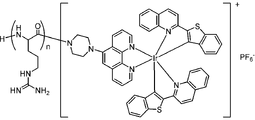
- FIG. 1 shows a conceptual diagram of the compound (blood vessel imaging reagent) of the present invention.
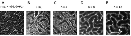
- FIG. 3 shows an imaging image (picture substitute) of the capillaries of the kidney.
- A shows the results using FITC-tomato lectin
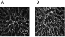
- FIG. 4 shows a capillary imaging image (photograph of a drawing) of the kidney.
- FIG. 5 shows an image (drawing substitute photograph) of a section (glomerulus) of a kidney.
- A shows the result of the objective lens ⁇ 20, and B shows the result of the objective lens ⁇ 10.
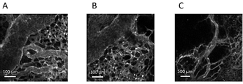
- FIG. 6 shows an imaging image (photograph of a drawing substitute) of a blood vessel of the vas sinusoide of the liver.
- a and B are images of different parts of the liver.
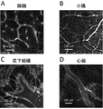
- FIG. 7 shows blood vessel imaging images (drawing substitute photographs) of various organs.
- FIG. 8 shows a tumor blood vessel imaging image (drawing substitute photograph) of a cancer-bearing mouse.
- a to C are images of different parts of the tumor.
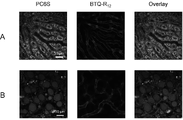
- FIG. 9 shows a multicolor imaging image (drawing substitute photograph) of PC6S and BTQ-R 12 in the liver.
- Column A shows the results of healthy mice
- column B shows the results of fatty liver model mice.
- the left side of the column shows the PC 6S
- the center shows the BTQ-R 12
- the right side shows the result of their superposition.
- FIG. 10 shows blood vessel imaging images (drawing substitute photographs) of various organs using RhB-pipe-PEG 12 -R 12 .
- A is the liver
- B and C are the kidneys (C is a magnified view of part of B)
- D is the spleen
- E is the pancreas
- F is the testis
- G is the muscle tissue
- H is the subcutaneous tissue.
- FIG. 11 shows blood vessel imaging images (drawing substitute photographs) of various organs using NBD-PEG 4 -R 12 .
- A is the liver
- B is the kidney
- C is the spleen
- D is the subcutaneous tissue
- E is the adipose tissue.
- FIG. 12 shows blood vessel imaging images (drawing substitute photographs) of various organs using C6-PEG 4 -R 12 .
- A is the kidney
- B is the spleen
- C is the pancreas
- D is the testis
- E is the adipose tissue
- F is the muscle tissue.
- FIG. 13 shows an imaging image (drawing substitute photograph) of a cerebral blood vessel using C6-PEG 4 -R 12 .
- A, B and C are images of different parts of the brain.
- D is a magnified image of a part of C.
- oligoarginine represented by the following formula and Phosphorescent or fluorescent groups bound to the C-terminal side or N-terminal side of the oligoarginine, Regarding a blood vessel imaging reagent (hereinafter, may be referred to as "the blood vessel imaging reagent of the present invention") containing a compound containing and:
- n is an integer of 4 to 20.
- the blood vessel imaging reagent developed in the present invention contains a compound having a structure in which a luminescent group is bound to the C-terminal side or the N-terminal side of the oligoarginine peptide (Fig. 1).
- the chromophore may be bound to either the C-terminal side or the N-terminal side of the oligoarginine peptide, but is preferably the C-terminal side.
- a fluorescent group fluorescent compound
- a phosphorescent compound for example, an iridium complex
- autofluorescence can be eliminated by time-resolved measurement. Even when a fluorescent compound is used, it is possible to distinguish it from autofluorescence by using a compound that exhibits fluorescence in the near-infrared light region, and it is also possible to image the structure of deep blood vessels.
- the compound used in the blood vessel imaging reagent of the present invention contains oligoarginine represented by the following formula.
- n is an integer of 4 to 20, preferably n is an integer of 6 to 16, and more preferably an integer of 8 to 12.
- the compound used in the blood vessel imaging reagent of the present invention contains oligoarginine to achieve improved adsorption to the vascular endothelium.
- oligoarginine has n ⁇ 8 or more, it exhibits particularly excellent adsorptivity to the vascular endothelium. Therefore, such an embodiment can be preferably used for the purpose of imaging the vascular endothelium.
- the compound used for the blood vessel imaging reagent of the present invention may contain a group other than oligoarginine as long as the effect of the present invention is not impaired.
- the group other than oligoarginine may contain one kind or two or more kinds.
- Groups other than oligoarginine may be attached to the end of oligoarginine. That is, a group other than oligoarginine may be present between oligoarginine and the chromophore (this embodiment may be referred to as a "linker") or on the opposite side of the oligoarginine's binding to the chromophore. May exist.
- a group containing steroids, peptides, polyethylene glycol and the like can be relatively easily bonded to a chromophore, and thus can be suitably used as a group other than oligoarginine.
- the polypeptide contains a group other than oligoarginine and oligoarginine consisting of a peptide
- the polypeptide as a whole preferably has 4 to 20 amino acid residues, more preferably 6 to 16 amino acid residues, and more preferably amino acid residues. The number is 8-12.
- the peptide other than oligoarginine is not limited, but for example, proline is preferable from the viewpoint of easiness of synthesis.
- aspartic acid and lysine are also preferable because the compound can be made water-soluble.
- the degree of polymerization of polyethylene glycol can be adjusted depending on the type of luminescent group and the like, but is preferably 2 to 20, for example. It is 3 to 16, and more preferably 4 to 12.
- any aspect containing or not containing a group other than oligoarginine can be preferably used.
- an embodiment containing a group other than oligoarginine as a linker can be more preferably used.
- fluorescent group contained in the compound used for the imaging reagent for blood vessels of the present invention a fluorescent compound conventionally used for imaging blood vessels and the like can be used without particular limitation. Further, the compound of the present invention described later can be used as a compound used as an imaging reagent for blood vessels having a fluorescent group.
- the fluorescent compound may be used alone or in combination of two or more. Fluorescent compounds include, but are not limited to, 4-nitrobenzo-2-oxa-1,3-diazole (NBD), dimethylaminosulfonylbenzoxadiazole (DBD), dimethylaminosulfonylbenzothiadiazole (DBThD), dimethyl.
- Examples thereof include aminosulfonylbenzoselenaziazole (DBSeD), fluorescein isothiocyanate (FITC), coumarin dyes, rhodamines, borondipyrromethene (BODIPY), cyanine dyes and the like.
- Examples of the fluorescent compound include, but are not limited to, coumarin-based dyes such as NBD and coumarin 6, and rhodamines such as rhodamine B.
- a phosphorescent compound conventionally used for imaging blood vessels and the like can be used without particular limitation.
- the compound of the present invention described later can be used as a compound used as an imaging reagent for blood vessels having a phosphorescent group.
- the phosphorus compound can be used alone or in combination of two or more.
- the phosphorescent compound include, but are not limited to, compounds containing an iridium complex.
- the iridium complex include, but are not limited to, compounds represented by the following formulas (I) or (II).
- Ring R 1 represents a monocyclic or polycyclic nitrogen-containing aromatic ring.
- Ring R 2 represents a monocyclic or polycyclic sulfur-containing aromatic ring.
- L 1 indicates a bidentate ligand having a ⁇ -diketonate structure;
- Ring R 1 represents a monocyclic or polycyclic nitrogen-containing aromatic ring.
- Ring R 2 represents a monocyclic or polycyclic sulfur-containing aromatic ring.
- L 2 represents a bidentate ligand having a phenanthroline skeleton.
- complex (I) the compound represented by the formula (I) (hereinafter, may be referred to as “complex (I)”) will be described.
- the complex (I) contained in the blood vessel imaging reagent of the present invention may be one kind or two or more kinds.
- the ring R 1 is not limited, and examples thereof include a nitrogen-containing aromatic ring having a structure represented by the following formula (1-1), (1-2), (1-3), or (1-4). Be done.
- X 1 indicates hydrogen.
- the bond extending from the carbon atom next to N is bonded to the ring R2 .
- N is coordinated to Ir.
- a polycyclic nitrogen-containing aromatic ring is preferable because the light emission approaches near infrared rays in combination with the ring R 2 and the permeability in the living body is good, and the above formula (1-2). ), (1-3), or (1-4), a nitrogen-containing aromatic ring having the structure shown in (1-3) or (1-4) is more preferable.
- Examples of the ring R 2 include a sulfur-containing aromatic ring having a structure represented by the following formula (2-1), (2-2), or (2-3).
- X 2 represents hydrogen.
- the bond extending from the carbon atom next to S is bonded to ring R1 , and the carbon atom next to this carbon atom is coordinated to Ir.
- a sulfur-containing aromatic ring having the structure represented by the above formula (2-1) is preferable.
- the ligand composed of ring R 1 and ring R 2 is a cyclometallated ligand and contributes to the light emission performance of the complex (I).
- L 1 represents a bidentate ligand having a ⁇ -diketonate structure. Although L 1 does not affect the luminescence performance, having L 1 enhances the biocompatibility and facilitates the incorporation of the complex (I) into the living tissue. Examples of L 1 include bidentate ligands having a structure represented by the following formula (L-1). In this case, the complex (I) is represented by the following formula (I-1).
- R 3 represents a substituted or unsubstituted alkyl group.
- the two oxygen atoms in the structure are each coordinated to Ir.
- Examples of the alkyl group of R 3 include an alkyl group having 1 to 5 carbon atoms.
- the substituent may be, for example, a halogen, a hydroxy group, a mercapto group, a carboxy group, a substituted or unsubstituted amino group, a substituted or unsubstituted amide group, a substituted or unsubstituted acyl group, etc.
- Examples include amino acid residues and peptide residues.
- R 3 is -CH 2 CH 2 COCH 2 NH (CH 2 ) m CH 2 NR 4 R 5 and -CH 2 CH 2 CONHCH 2 CH 2 N (CH 3 ), respectively.
- 2 , -CH 2 CH 2 COOH,-(CH 2 ) n COR 6 , or -CH 3 is a structure.
- m represents an integer of 1 to 5
- R 4 represents a hydrogen, a halogen, a hydroxy group, an amino group, a mercapto group, or a hydrocarbon group having 1 to 20 carbon atoms
- R 5 represents hydrogen or a hydrocarbon group. It represents a hydrocarbon group of 1 to 6
- n represents an integer of 1 to 5
- R 6 represents an amino acid residue or a peptide residue.
- m is preferably an integer of 1 to 3, and 2 is particularly preferable.
- the halogen of R4 is preferably Cl, Br, or F.
- the amino group may be -NH 2 or an alkylamino group.
- the hydrocarbon group having 1 to 20 carbon atoms may be a straight chain, a branched chain, or a cyclic chain. Further, it may be saturated or may contain an unsaturated bond.
- One or more hydrogen atoms may be substituted with a substituent such as a halogen, a hydroxy group, an amino group or a mercapto group.
- the number of carbon atoms is preferably 1 to 10, more preferably 1 to 5, and even more preferably 1 to 3.
- the hydrocarbon group having 1 to 6 carbon atoms of R5 may be a straight chain, a branched chain, or a cyclic group. Further, it may be saturated or may contain an unsaturated bond.
- One or more hydrogen atoms may be substituted with a substituent such as a halogen, a hydroxy group, an amino group or a mercapto group.
- the number of carbon atoms is preferably 1 to 3. In the above formula (L-11), it is preferable that both R 4 and R 5 are methyl groups and n is 2.
- n is preferably an integer of 1 to 3, and 2 is particularly preferable.
- Amino acid residue and peptide residue means a residue when an amino acid or peptide is amide-bonded via its amino group.
- R6 is preferably a residue of an amino acid having a hydroxyl group, a carboxy group, or an amino group in the side chain, or a residue of a peptide composed of the amino acid.
- the amino acid having a hydroxyl group in the side chain include tyrosine, serine, threonine and the like, and tyrosine is more preferable.
- amino acids having a carboxy group in the side chain include aspartic acid and glutamic acid.
- amino acids having an amino group in the side chain include lysine and arginine.
- the amino acid may be L-form or D-form, or may be an unnatural amino acid.
- the peptide residue include a peptide residue composed of one or more of the above amino acids, the length of which is preferably 2 to 10, and more preferably 2 to 5.
- R6 -NH - CH (COOH) 2 which is an aspartic acid residue or -NH-CH (COOH) -CO-NH-CH (COOH) 2 which is an aspartic acid dipeptide residue is preferable.
- the structure represented by the formula (L-1) is the above formula (L-11), (L-12), (L-13), or (L-14) in terms of biocompatibility.
- the structure shown by is preferred.
- These structures are structures in which a functional group is introduced into the structure represented by the formula (L-15) (acetylacetonate (acac)). By introducing a functional group, the biocompatibility is more excellent.
- complex (I) a complex having a structure represented by the following formula (Ia), (Ib), (Ic), or (Id) is preferable.
- L 11 to L 14 are the above formulas (L-11), (L-12), (L-13), (L-14), or (L-), respectively.
- a bidentate ligand having the structure shown in 15) is shown.
- L 11 to L 14 are preferably bidentate ligands having the structures represented by the above formulas (L-11), (L-12), (L-13), or (L-14).
- complex (II) the compound represented by the formula (II) (hereinafter, may be referred to as “complex (II)”) will be described.
- the complex (II) contained in the blood vessel imaging reagent of the present invention may be one kind or two or more kinds.
- the complex (II) is the same as the complex (I) except that L 1 is L 2 . That is, the ring R 1 and the ring R 2 in the complex (II) are the same as the ring R 1 and the ring R 2 in the complex (I), and contribute to the light emission performance of the complex (II). Preferred embodiments of ring R 1 and ring R 2 in complex (II) are similar to ring R 1 and ring R 2 in complex (I).
- L 2 represents a bidentate ligand having a phenanthroline skeleton. Although L 2 does not affect the luminescence performance, having L 2 further enhances the biocompatibility as compared with the case of having L 1 , and makes it easier for the complex (II) to be incorporated into the living tissue.
- L 2 include bidentate ligands having a structure represented by the following formula (L-2). In this case, the complex (II) is represented by the following formula (II-1).
- R 7 represents a substituent having a hetero atom such as a nitrogen atom, an oxygen atom, and a sulfur atom
- X 3 represents hydrogen.
- the two nitrogen atoms (N) that make up the phenanthroline skeleton are each coordinated to Ir.
- R 7 examples include an amino group, a dimethylamino group, a diethylamino group, a cyano group, an acetyl group, a carboxyl group, a piperidyl group, and a piperazyl group.
- complex (II) a complex having a structure represented by the following formulas (IIa), (IIb), (IIc), or (IId) is preferable.
- X 1 and X 2 represent hydrogen
- L 2 represents a bidentate ligand having the structure represented by the above formula (L-2).
- the complexes (I) and (II) may be a cationic iridium complex or a neutral iridium complex, and are preferably a cationic iridium complex.
- oligoarginine for example, has a charge of +8 for octaarginine, remains +9 with a cationic iridium complex, and remains +8 with neutrality.
- the cationic iridium complex may form a salt with an anion.
- the anion include, but are not limited to, hexafluorophosphate ion (PF 6- ), chloride ion ( Cl- ) , bromide ion (Br-), trifluoroacetate ion (CF 3 COO- ) and the like. It is preferably PF 6 ⁇ .
- the iridium complex is preferably a compound represented by the above formula (IIc), and is a compound in which R 7 is a piperazyl group in the formula (L-2).
- oligoarginine, complexes (I) and (II), oligoarginine and a chromophore can be synthesized by using a conventional organic synthesis method. For example, it can be synthesized according to the method described in the examples below.
- the raw material a commercially available product may be used, or a raw material synthesized by a known method may be used.
- the blood vessel imaging reagent of the present invention may further contain a solvent, an additive, a compound used as a conventional blood vessel imaging reagent, and the like, as long as the effects of the present invention are not impaired.
- the blood vessel imaging reagent can be added to the tissue as it is to perform blood vessel imaging.
- the solvent may be any solvent as long as it can dissolve a compound containing oligoarginine and a luminescent group, and for example, an organic solvent such as tetrahydrofuran, acetonitrile or dimethyl sulfoxide, water or physiological saline (for example, 0.9% (w / v)). It can be appropriately selected from an aqueous solvent such as (physiological saline) and a mixed solvent thereof.
- a mixed solvent of dimethyl sulfoxide and water is preferable.
- the blood vessel imaging reagent of the present invention is a liquid composition containing a compound containing oligoarginine and a chromophore and a solvent
- concentration of the compound containing oligoarginine and the chromophore depends on the type of the compound and the like. However, it may be 0.01 to 500 mM, 0.1 to 100 mM, or the like.
- the blood vessel imaging reagent of the present invention is used for visualization of blood vessels in living tissues.
- the blood vessel imaging reagent of the present invention accumulates in the vascular endothelium of a living tissue. Therefore, the blood vessel imaging reagent of the present invention is useful for blood vessel imaging, particularly vascular endothelium imaging.
- the type of biological tissue to be measured is not particularly limited, and examples thereof include organs such as skin, muscle, fat, liver, heart, pancreas, kidney, spleen, intestine, genital organ, and brain. Further, the tissue may be either a normal tissue or a pathological tissue.
- the individual organism to be administered is not particularly limited, and examples thereof include vertebrates and invertebrates including mammals (mice, humans, pigs, dogs, rabbits, humans, etc.).
- Imaging of blood vessels in tissue can be performed, for example, as follows.
- the blood vessel imaging reagent of the present invention is added to an individual to be measured, and then a compound containing oligoarginine and a luminescent group in the blood vessel imaging reagent taken into the sample is excited and luminescence is observed. Excitation of the compound can be performed by irradiating the sample with visible light. The luminescence can be observed by using a known device such as a fluorescence microscope, a fluorescence measuring device, or a fluorescence imaging device.
- the amount of the blood vessel imaging reagent added to an individual can be appropriately changed depending on the individual used, the blood vessel density, and the like, and for example, 0.01 to 1,000 ⁇ mol / kg body weight, preferably 0.1 to 100 ⁇ mol. It can be administered to an individual in the range of / kg body weight.
- Examples of the administration form of the blood vessel imaging reagent of the present invention include intravenous administration, subcutaneous administration, and intramuscular administration.
- n is an integer of 4 to 20.
- the compound of the present invention is a compound containing oligoarginine and an iridium complex.
- n is an integer of 4 to 20, preferably n is an integer of 6 to 16, and more preferably n is an integer of 8 to 12.
- the iridium complex may form a salt with an anion.
- the anion include, but are not limited to, hexafluorophosphate ion (PF 6- ), chloride ion ( Cl- ) , bromide ion (Br-), trifluoroacetate ion (CF 3 COO- ) and the like. It is preferably PF 6 ⁇ .
- the compound of the present invention has excellent phosphorescence emission performance and is excellent in binding to the vascular endothelium, so that it can be used as an imaging reagent for blood vessels.
- Yet another aspect of the invention relates to a compound in which oligoarginine and NBD, coumarin 6 or rhodamine B are bound via a linker.
- a linker is not limited, but is preferably a group containing polyethylene glycol.
- the degree of polymerization p of polyethylene glycol (PEG p ) is not limited, but is, for example, 2 to 20, preferably 3 to 16, from the viewpoint of easy staying on the vascular endothelium and easy availability of materials. Yes, more preferably 4-12.
- the degree of polymerization n of oligoarginine (R n ) is not limited, but is, for example, 4 to 20, preferably 6 to 16, and more preferably 8 to 12.
- R n degree of polymerization n of oligoarginine
- Such a compound of the present invention has excellent fluorescence emission performance and is excellent in binding to the vascular endothelium, and therefore can be used as an imaging reagent for blood vessels.
- the compound of the present invention can be synthesized by using a conventional organic synthesis method. For example, it can be synthesized according to the method described in the examples below.
- As the raw material a commercially available product may be used, or a raw material synthesized by a known method may be used.
- the condensation reaction consisted of 0.5 M Fmoc-R (Pbf) -OH and Boc-R (Pbf) -OH DMF solutions as amino acids, 0.6 M HBTU DMF solution as a condensing agent, and 0.5 M HOBt DMF as an additive.
- a 2.0 M DIEA NMP solution as a solution and a base
- 3 equivalents, 3 equivalents, 3 equivalents, and 6 equivalents were added to the number of amino acid-introduced moles, respectively, and the mixture was reacted at room temperature for 1 hour.
- the obtained solid was centrifuged (3500 rpm, 5 minutes), water was removed, water was added again, and the mixture was centrifuged. This operation was repeated twice. The water in the vial was removed by lyophilization to give a red solid (390 mg, 0.14 mmol, crude product yield: 270%).
- BTQ-R 4 Weigh BTQ- [R (Pbf)] 4 -Boc (279 mg, 0.10 mmol) into a 50 mL centrifuge tube, add 1 mL of TFA: water: TIPS (95: 2.5: 2.5), and react at room temperature for 2 hours. I let you. Cold diethyl ether was added to the solution to precipitate a solid. The obtained solid was centrifuged (3500 rpm, 5 minutes), diethyl ether was removed, diethyl ether was added again, and the mixture was centrifuged. This operation was repeated twice.
- Boc- [R (Pbf)] 8 -OH was synthesized by the Fmoc solid-phase synthesis method using a fully automatic microwave peptide synthesizer (Initiator + Alstra, Biotage).
- As the resin chlorotrityl resin (1.67 g, number of moles of amino acid introduced: 0.30 mmol / g) into which R (Pbf) was introduced was used.
- the condensation reaction consisted of 0.6 M Fmoc-R (Pbf) -OH and Boc-R (Pbf) -OH DMF solutions as amino acids, 0.5 M HBTU DMF solution as a condensing agent, and 0.5 M HOBt DMF as an additive.
- the obtained solid was centrifuged (3500 rpm, 5 minutes), water was removed, water was added again, and the mixture was centrifuged. This operation was repeated twice. The water in the vial was removed by lyophilization to give a red solid (235 mg, 0.055 mmol, crude product yield: 110%).
- BTQ-R 8 Weigh BTQ- [R (Pbf)] 8 -Boc (110 mg, 0.026 mmol) into a 50 mL centrifuge tube, add 1 mL of TFA: water: TIPS (95: 2.5: 2.5), and react at room temperature for 2 hours. I let you. Cold diethyl ether was added to the solution to precipitate a solid. The obtained solid was centrifuged (3500 rpm, 5 minutes), diethyl ether was removed, diethyl ether was added again, and the mixture was centrifuged. This operation was repeated twice.
- the condensation reaction consisted of 0.5 M Fmoc-R (Pbf) -OH and Boc-R (Pbf) -OH DMF solutions as amino acids, 0.6 M HBTU DMF solution as a condensing agent, and 0.5 M HOBt DMF as an additive.
- a 2.0 M DIEA NMP solution as a solution and a base
- 3 equivalents, 3 equivalents, 3 equivalents, and 6 equivalents were added to the number of amino acid-introduced moles, respectively, and the mixture was reacted at room temperature for 1 hour.
- the obtained solid was centrifuged (3500 rpm, 5 minutes), water was removed, water was added again, and the mixture was centrifuged. This operation was repeated twice. The water in the vial was removed by lyophilization to give a red solid (320 mg, 0.055 mmol, crude product yield: 110%).
- BTQ-R 12 Weigh BTQ- [R (Pbf)] 12 -Boc (151 mg, 0.026 mmol) into a 50 mL centrifuge tube, add 1 mL of TFA: water: TIPS (95: 2.5: 2.5), and react at room temperature for 2 hours. I let you. Cold diethyl ether was added to the solution to precipitate a solid. The obtained solid was centrifuged (3500 rpm, 5 minutes), diethyl ether was removed, diethyl ether was added again, and the mixture was centrifuged. This operation was repeated twice.
- Boc- [R (Pbf)] 16 -OH was synthesized by the Fmoc solid-phase synthesis method using a fully automatic microwave peptide synthesizer (Initiator + Alstra, Biotage).
- As the resin chlorotrityl resin (0.34 g, number of moles of amino acid introduced: 0.30 mmol / g) into which R (Pbf) was introduced was used.
- the condensation reaction consisted of 0.3 M Fmoc-R (Pbf) -OH and Boc-R (Pbf) -OH DMF solutions as amino acids, 0.6 M HBTU DMF solution as a condensing agent, and 0.5 M HOBt DMF as an additive.
- a 2.0 M DIEA NMP solution as a solution and a base
- 3 equivalents, 3 equivalents, 3 equivalents, and 6 equivalents were added to the number of amino acid-introduced moles, respectively, and the mixture was reacted at room temperature for 1 hour.
- BTQ-R 16 Weigh BTQ- [R (Pbf)] 16 -Boc (113 mg, 0.015 mmol) into a 50 mL centrifuge tube, add 1 mL of TFA: water: TIPS (95: 2.5: 2.5), and react at room temperature for 2 hours. I let you. Cold diethyl ether was added to the solution to precipitate a solid. The obtained solid was centrifuged (3500 rpm, 5 minutes), diethyl ether was removed, diethyl ether was added again, and the mixture was centrifuged. This operation was repeated twice.
- Figure 2 shows the structural formulas (BTQ-R 4 , BTQ-R 8 , BTQ-R 12 , BTQ-R 16 ) of the compound of the present invention synthesized in the examples.
- the number of arginine residues is 4, 8, 12, and 16.
- a compound to which arginine is not bound (BTQ) was used as a reference compound.
- the absorption and phosphorescence spectra of these compounds were measured, the absorption maximum wavelength was shown near 500 nm, and the phosphorescence maximum wavelength was shown near 660 nm. Since phosphorescence is observed at 600 nm and above, multicolor imaging using molecules that emit green light is possible.
- the phosphorescent quantum yield and phosphorescent lifetime were measured and found to be 0.32 (under 0.015 air saturation) and 5.7 ⁇ s (under 0.28 ⁇ s air saturation), respectively. Even compounds with different numbers of arginine residues have almost the same spectral and photophysical properties because the same phosphorous group is bound to the oligoarginine peptide.
- FIG. 3 shows a mixed solvent of FITC-tomatolectin (1 mg / mL (physiological saline) administered in 50 ⁇ L) and BTQ (100 nmol: 1 mmol / L (physiological saline: dimethylsulfoxide (9: 1, v / v)).
- BTQ-R n (n: 4, 8, 12, 100 nmol: 1 mmol / L (physiological saline) was administered at 100 ⁇ L) from the tail vein of anesthetized mice.
- the ventral part is incised to expose the kidney, and the image of the kidney surface is shown by imaging the kidney surface with a confocal laser microscope.
- BTQ and BTQ-R 4 are observed to emit light from a region (tubular cells) different from that of FITC-tomato lectin, whereas BTQ-R 8 and BTQ -R 12 shows that the capillaries of the kidney are imaged in the same way as FITC-tomato lectin.
- BTQ-R 12 has cavities in the blood vessels, so BTQ-R 12 is distributed in the vascular endothelium, not in the blood. From the above, it was clarified that the number of residues of arginine of 8 or more is preferable for imaging the vascular endothelium.
- FITC-tomato lectin (1 mg / mL (physiological saline) administered in 50 ⁇ L) and BTQ-R 12 (100 nmol: 1 mmol / L (physiological saline) administered in 100 ⁇ L) were administered to mice. The same spot was imaged. As shown in A and C of FIG. 4, when imaging with the fluorescence of FITC-tomato lectin, fluorescence was observed from a place other than the vascular endothelium.
- FIG. 6 shows a phosphorescent microscopic image obtained by administering BTQ-R 12 (100 nmol: 1 mmol / L (physiological saline) 100 ⁇ L) to mice.
- BTQ-R 12 100 nmol: 1 mmol / L (physiological saline) 100 ⁇ L
- the vas sinusoide vessels are clearly imaged.
- Similar experiments were performed on other organs (pancreas, small intestine, subcutaneous tissue, heart) and it became clear that the capillaries of many organs could be imaged (Fig. 7).
- FIG. 8 shows a phosphorescent microscopic image obtained by administering BTQ-R 12 (100 nmol: 1 mmol / L (physiological saline) 100 ⁇ L) to a cancer-bearing mouse. From the image, it was found that many small blood vessels were branched from the thick blood vessel, and the imaging of the vascular network in the tumor was successful.
- lipids are abnormally accumulated in the hepatocytes, large lipid droplets are formed, and the hepatocytes are enlarged. Therefore, the narrowing of the vas sinusoideal blood vessels progresses, and the entire liver becomes hypoxic.
- the green fluorescent lipid droplet reagent (3- (benzo [d] thiazol-2-yl) -8- (diethylamino) -2H-benzo [g] chromen-2-one (PC6S)) and BTQ-R 12 Simultaneously administered to healthy mice and adipose liver model mice (PC6S: 50 nmol: 0.5 mmol / L (physiological saline: dimethyl sulfoxide (9: 1, v / v), in a mixed solvent containing 10 wt% BSA).
- Multicolor imaging was performed by administering 100 ⁇ L, BTQ-R 12 : 100 nmol: 1 mmol / L (physiological saline) 100 ⁇ L), and measuring green fluorescence and deep red phosphorescence.
- PC6S fluorescence was observed from lipid droplets in hepatocytes
- phosphorescence of BTQ-R 12 was observed from the sinusoideal endothelium (A in Fig. 9). From the image, it can be seen that the sinusoide vessels run linearly between the hepatocytes.
- hepatocytes are enlarged due to the formation of large lipid droplets, and the sinusoideal blood vessels are greatly tortured (Fig. 9, B). As a result, the flow of red blood cells is obstructed, the oxygen supply to the liver is insufficient, and hypoxia may occur.
- the obtained solid was centrifuged (3500 rpm, 5 minutes), water was removed, water was added again, and the mixture was centrifuged. This operation was repeated twice. The water in the vial was removed by lyophilization to give a magenta solid (157 mg, 0.026 mmol, crude product yield: 45%).
- RhB-pipe-PEG 12 -R 12 Weigh RhB-pipe-PEG 12- [R (Pbf)] 12 -Boc (21.5 mg, 0.0035 mmol) into a 50 mL centrifuge tube, add 1 mL of TFA: water: TIPS (95: 2.5: 2.5), and room temperature. Was reacted for 2 hours. Cold diethyl ether was added to the solution to precipitate a solid. The obtained solid was centrifuged (3500 rpm, 5 minutes), diethyl ether was removed, diethyl ether was added again, and the mixture was centrifuged. This operation was repeated twice.
- NBD-PEG 4- [R (Pbf)] 12 -Boc) Boc- [R (Pbf)] 12 -OH (429 mg, 0.085 mmol), NBD-PEG 4 -NH 2 (0.10 mmol), HATU (65.9 mg, 0.17 mmol) were added to 100 mL eggplant frass, and dehydrated DMF 8 was added. Dissolved in mL. To this solution, 0.17 mL of DIEA was added, and the mixture was stirred at room temperature for 24 hours under nitrogen substitution. The solution was transferred to a 50 mL centrifuge tube and water was added to precipitate a solid.
- NBD-PEG 4 -R 12 Weigh NBD-PEG 4- [R (Pbf)] 12 -Boc (30.2 mg, 0.0060 mmol) into a 50 mL centrifuge tube, add 1 mL of TFA: water: TIPS (95: 2.5: 2.5), and at room temperature. It was allowed to react for 2 hours. Cold diethyl ether was added to the solution to precipitate a solid. The obtained solid was centrifuged (3500 rpm, 5 minutes), diethyl ether was removed, diethyl ether was added again, and the mixture was centrifuged. This operation was repeated twice.
- C6-PEG 4 -R 12 Weigh C6-PEG 4- [R (Pbf)] 12 -Boc (46.0 mg, 0.0082 mmol) into a 50 mL centrifuge tube, add 1 mL of TFA: water: TIPS (95: 2.5: 2.5), and at room temperature. It was allowed to react for 2 hours. Cold diethyl ether was added to the solution to precipitate a solid. The obtained solid was centrifuged (3500 rpm, 5 minutes), diethyl ether was removed, diethyl ether was added again, and the mixture was centrifuged. This operation was repeated twice.
- RhB-pipe-PEG 12 -R 12 was administered to mice to perform vascular imaging of various tissues.
- RhB-pipe-PEG 12 -R 12 (100 nmol: 2 mmmol / L (in physiological saline) 50 ⁇ L) was administered from the tail vein of anesthetized mice, and the ventral part was incised in various ways.
- An image of exposed organs (liver, kidney, spleen, pancreas, testis) and tissues (muscle tissue, subcutaneous tissue) and imaging their surfaces with a confocal laser microscope is shown.
- the excitation wavelength is 550 nm and the observation wavelength is> 590 nm.
- Capillaries can be imaged in all the observed organs and tissues.
- NBD-PEG-R 12 was administered to mice to perform vascular imaging of various tissues.
- NBD-PEG-R 12 100 nmol: 2 mmmol / L (in physiological saline) administered at 50 ⁇ L
- the liver, kidney, spleen) and tissues (subcutaneous tissue, adipose tissue) are exposed, and images of their surfaces imaged with a confocal laser microscope are shown.
- the excitation wavelength is 488 nm and the observation wavelength is 510-550 nm. Capillaries can be imaged in all the observed organs and tissues.
- C6-PEG-R 12 was administered to mice to perform vascular imaging of various tissues.
- C 6 -PEG-R 12 (100 nmol: 2 mm mol / L (in physiological saline) 50 ⁇ L) was administered from the tail vein of anesthetized mice, and the ventral part was incised to various organs. (Kidney, spleen, pancreas, testis) and tissues (fat tissue, muscle tissue) are exposed, and images of their surfaces imaged with a confocal laser microscope are shown.
- the excitation wavelength is 488 nm and the observation wavelength is 510-550 nm.
- Capillaries can be imaged in all the observed organs and tissues. After euthanizing the mice, cerebrovascular imaging was performed. As shown in Fig. 13, it was shown that cerebral blood vessel imaging is also possible.
- a compound containing oligoarginine and a chromophore can be used to clearly image blood vessels in an individual and can be used as an imaging reagent for blood vessels.
- BTQ-R n developed by the present invention can be used as an imaging reagent for blood vessels.
- BTQ-R 12 is a new reagent capable of imaging the vascularization of normal and pathological tissues in an individual.
- the distribution to the vascular endothelium is the action of the oligoarginine peptide, and by changing the luminescence group (BTQ), it is possible to develop vascular endothelium imaging reagents with various luminescent colors.
- RhB-pipe-PEG p -R n , NBD-PEG p -R n , and C6-PEG p -R n developed by the present invention can also be preferably used as blood vessel imaging reagents.
- the present invention can be used in fields such as medical diagnosis, pharmaceutical development, and basic medicine.
Abstract
製造が簡便であり、高感度、高精度な新たな血管のイメージング試薬を開発することを課題とする。本発明は、下記式で示されるオリゴアルギニンと、前記オリゴアルギニンのC末端側またはN末端側に結合しているりん光団または蛍光団、とを含む化合物を含む、血管のイメージング試薬を提供する:
Description
本発明は、血管のイメージング試薬、および同試薬に用いることができる化合物等に関する。
生体組織(臓器)細胞は、必要な栄養素や酸素を血中から獲得し、合成した代謝産物や二酸化炭素を血中に放出している。このため、血管系は生命維持において重要な役割を果たす。何らかの理由により、血管系に異常が生じると周辺組織細胞は十分な機能を果たすことが出来なくなる。特に動脈内膜にコレステロール等の脂質成分が蓄積し、アテロームと呼ばれる粥状硬化巣が形成される動脈硬化症や、血管と肝細胞の間が線維化を起こす肝硬変は、血管系の破綻と深く関与している。また、がんでは細胞の異常増殖に血管新生の形成が追従できないため、未成熟で異常な形状をした血管が多数見られる。よって、生きた個体において、血管の形状や走行を可視化・イメージングすることは、上記、病態の診断・治療において重要である。
臨床における血管イメージングでは、造影剤を血中投与してMRIで画像化する方法や、光音響イメージング技術を用いた方法が知られている。これらは人体の血管網に関する知見は与えるが、装置が大がかりであり小動物レベルに不向きである。また、単一細胞レベルの空間分解能は有していない。一方、光を用いたイメージング技術は、高感度・簡便性・リアルタイム計測に加えて、顕微鏡と組み合わせることにより、単一細胞レベルの空間分解能を容易に実現できる。ここで、重要な要素は、血中のような夾雑環境下で安定に存在し、高輝度な発光を示すプローブ分子である。
血管のイメージング試薬は、血中滞留型と血管内皮吸着型に分類される。前者は、血中内を滞留する試薬であり、蛍光性デキストランや蛍光性ナノ粒子が開発されている。特に蛍光性デキストランは、デキストランに蛍光性分子を共有結合させた試薬であり、デキストランの分子量を変えた様々な試薬が市販されている。
一方、血管内皮吸着型イメージング試薬には、蛍光性レクチンが市販されている。レクチンは、特定の糖鎖に特異的な結合を示すタンパク質の総称であり、動物・植物・菌類に由来するものが多数存在する。このうち、植物由来レクチンは、ヒトをはじめとする哺乳動物の血管内皮細胞表面に存在する糖鎖を認識して結合する。これまで、トマトレクチンにフルオレセインやテキサスレッドを共有結合させた蛍光性レクチンが市販(Vector Laboratories社)されている。蛍光性レクチンの合成では、トマトレクチンを天然から抽出する煩雑な作業が必要であり、また、レクチンに結合させる蛍光性分子の数は制御できない。
すなわち、蛍光性レクチンには、以下のような課題がある。血管内皮細胞の表面に存在する糖鎖を認識しているため、植物由来のレクチンしか使用できない;レクチンを天然から抽出する煩雑な作業が必要である;レクチンは糖タンパク質であるため、発光団の標識が不均一になり、ロット間に差が生じる;蛍光を利用しているため、組織の自家蛍光と区別できないことがある。
他方、生体組織や細胞内の酸素濃度を定量するための方法として、btq(2-(2’-ベンゾチエニル)-キノリナート-N,C3’)等のシクロメタル化配位子を有するイリジウム(III)錯体、あるいは同イリジウム錯体と蛍光性化合物とを含む化合物を用いる方法が提案されている(例えば、特許文献1および2)。しかしながら、特許文献1および2では、同方法を、血管を可視化に用いることについては検討されていない。
本発明は、上記課題に鑑みなされたものであり、本発明は、製造が簡便であり、高感度、高精度な新たな血管のイメージング試薬を開発することを課題とする。
本発明者らは、上記課題を解決すべく鋭意検討を行った結果、オリゴアルギニンとりん光団または蛍光団である発光団とを含む化合物を用いて、個体内の血管を明瞭にイメージングすることができることを知見した。また、このような発光団として用いることができる、オリゴアルギニンとイリジウム錯体を含む化合物を開発した。このような知見に基づいて、本発明を完成した。
すなわち、本発明の要旨は以下に関する。
すなわち、本発明の要旨は以下に関する。
[1] 下記式で示されるオリゴアルギニンと、
前記オリゴアルギニンのC末端側またはN末端側に結合しているりん光団または蛍光団、
とを含む化合物を含む、血管のイメージング試薬:
前記オリゴアルギニンのC末端側またはN末端側に結合しているりん光団または蛍光団、
とを含む化合物を含む、血管のイメージング試薬:

式中、
nは、4~20の整数である。
[2] 前記オリゴアルギニンは、nが8以上である、[1]に記載の試薬。
[3] 前記りん光団は、イリジウム錯体を含む化合物である、[1]または[2]に記載の試薬。
[4] 前記イリジウム錯体は、下記式(I)または(II)で示される化合物である、[3]に記載の試薬:
nは、4~20の整数である。
[2] 前記オリゴアルギニンは、nが8以上である、[1]に記載の試薬。
[3] 前記りん光団は、イリジウム錯体を含む化合物である、[1]または[2]に記載の試薬。
[4] 前記イリジウム錯体は、下記式(I)または(II)で示される化合物である、[3]に記載の試薬:

式(I)中、
環R1は、単環または多環式の含窒素芳香族環を示し、
環R2は、単環または多環式の含硫黄芳香族環を示し、
L1は、β-ジケトネート構造を有する二座配位子を示す;
環R1は、単環または多環式の含窒素芳香族環を示し、
環R2は、単環または多環式の含硫黄芳香族環を示し、
L1は、β-ジケトネート構造を有する二座配位子を示す;

式(II)中、
環R1は、単環または多環式の含窒素芳香族環を示し、
環R2は、単環または多環式の含硫黄芳香族環を示し、
L2はフェナントロリン骨格を有する二座配位子を示す。
環R1は、単環または多環式の含窒素芳香族環を示し、
環R2は、単環または多環式の含硫黄芳香族環を示し、
L2はフェナントロリン骨格を有する二座配位子を示す。
[5] 前記イリジウム錯体は、下記式で示される化合物である、[3]または[4]に記載の試薬:

式中、
nは、4~20の整数である。
[6] 下記式で示される化合物:
nは、4~20の整数である。
[6] 下記式で示される化合物:

式中、
nは4~20の整数である。
nは4~20の整数である。
本発明により、オリゴアルギニンと発光団とを含む化合物を含む、血管のイメージング試薬が提供される。同試薬は、イメージング試薬用の化合物を化学合成できるため、天然物からの抽出操作等が不要であり、簡便に製造することができる。また、発光団の標識を均一とすることができ、ロット差の少ない均一な試薬とすることができ、高精度なイメージングが可能となる。
発光団として、りん光性化合物を用いた場合、時間分解計測により自家蛍光を排除することができ、高感度、高精度なイメージングが可能となる。特に、アルギニンが8個以上結合した化合物は、血管内皮の結合に優れ、より高感度、高精度に血管内皮をイメージングすることができる。
蛍光性化合物を用いた場合においても、近赤外光領域に蛍光を示す化合物を用いれば自家蛍光と区別するこが可能となり、さらに深部血管の構造をイメージングすることもできる。
また、本発明により、血管のイメージング試薬の発光団として用いることができる、オリゴアルギニンとイリジウム錯体を含む化合物が提供される。同化合物は、優れたりん光発光性能を有し、血管を明瞭にイメージングすることができる。また、本発明により、血管のイメージング試薬の発光団として用いることができる、オリゴアルギニンと蛍光性化合物を含む化合物が提供される。同化合物は、優れた蛍光発光性能を有し、血管を明瞭にイメージングすることができる。
以下、本発明について説明する。
<血管のイメージング試薬>
本発明の一態様は、下記式で示されるオリゴアルギニンと、
前記オリゴアルギニンのC末端側またはN末端側に結合しているりん光団または蛍光団、
とを含む化合物を含む、血管のイメージング試薬(以下、「本発明の血管のイメージング試薬」ということがある。)に関する:
本発明の一態様は、下記式で示されるオリゴアルギニンと、
前記オリゴアルギニンのC末端側またはN末端側に結合しているりん光団または蛍光団、
とを含む化合物を含む、血管のイメージング試薬(以下、「本発明の血管のイメージング試薬」ということがある。)に関する:

式中、
nは、4~20の整数である。
nは、4~20の整数である。
本発明で開発した血管のイメージング試薬は、オリゴアルギニンペプチドのC末端側またはN末端側に、発光団が結合した構造の化合物を含むものである(図1)。発光団は、オリゴアルギニンペプチドのC末端側またはN末端側のいずれに結合していてもよいが、好ましくはC末端側である。発光団としては、蛍光団(蛍光性化合物)、またはりん光団(りん光性化合物)の両方が可能である。りん光性化合物(例えば、イリジウム錯体)を用いた場合、時間分解計測により自家蛍光を排除することが可能となる。蛍光性化合物を用いた場合においても、近赤外光領域に蛍光を示す化合物を用いれば自家蛍光と区別するこが可能となり、さらに深部血管の構造をイメージングすることもできる。
≪オリゴアルギニン≫
本発明の血管のイメージング試薬に用いられる化合物は、下記式で示されるオリゴアルギニンを含有する。
本発明の血管のイメージング試薬に用いられる化合物は、下記式で示されるオリゴアルギニンを含有する。

ここで、nは4~20の整数であり、好ましくは、nは6~16の整数であり、より好ましくは8~12の整数である。
本発明の血管のイメージング試薬に用いられる化合物は、オリゴアルギニンを含むことにより、血管内皮への吸着性向上を達成するものである。オリゴアルギニンが、n≧8以上の場合には、特に優れた血管内皮への吸着性を示すため、血管内皮のイメージングを目的とする場合は、このような態様を好ましく用いることができる。
本発明の血管のイメージング試薬に用いられる化合物においては、本発明の効果を損なわない範囲で、オリゴアルギニン以外の基を含んでいてもよい。
オリゴアルギニン以外の基は1種を含んでもよいし、2種以上を含んでもよい。オリゴアルギニン以外の基は、オリゴアルギニンの末端に結合してよい。すなわち、オリゴアルギニン以外の基は、オリゴアルギニンと発光団との間に存在(この態様を「リンカー」と称することがある)してもよいし、オリゴアルギニンの発光団との結合の反対側に存在してもよい。
一般に、ステロイド、ペプチド、ポリエチレングリコールを含む基等は比較的容易に発光団と結合させることができるため、オリゴアルギニン以外の基として好適に使用しうる。
オリゴアルギニン、およびペプチドからなるオリゴアルギニン以外の基を含む場合、ポリペプチド全体としては、好ましくは、アミノ酸残基数4~20、より好ましくはアミノ酸残基数6~16、さらに好ましくはアミノ酸残基数8~12である。
オリゴアルギニン以外のペプチドとしては、限定されないが、例えば、合成容易性の観点からプロリンが好ましい。また、化合物に水溶性を持たせられるため、アスパラギン酸、リシンも好ましい。
オリゴアルギニン、およびポリエチレングリコールを含む基からなるオリゴアルギニン以外の基を含む場合、ポリエチレングリコールの重合度は、発光団の種類等に応じて調整し得るが、例えば、2~20であり、好ましくは、3~16であり、より好ましくは4~12である。
本発明の血管のイメージング試薬に用いられる化合物においては、オリゴアルギニン以外の基を含む態様および含まない態様のいずれも好適に用いることができる。蛍光団を有する化合物の場合、オリゴアルギニン以外の基をリンカーとして含む態様をより好ましく用い得る。
≪蛍光団≫
本発明の血管のイメージング試薬に用いられる化合物に含まれる蛍光団としては、従来血管等のイメージングに用いられる蛍光性化合物を、特に制限されず用いることができる。また、後記本発明の化合物を、蛍光団を有する血管のイメージング試薬に用いられる化合物として用いることができる。蛍光性化合物は、1種または2種以上を組み合わせて使用できる。
蛍光性化合物としては、限定されないが、例えば、4-ニトロベンゾ-2-オキサ-1,3-ジアゾール(NBD)、ジメチルアミノスルホニルベンゾオキサジアゾール(DBD)、ジメチルアミノスルホニルベンゾチアジアゾール(DBThD)、ジメチルアミノスルホニルベンゾセレナジアゾール(DBSeD)、フルオレセインイソチオシアネート(FITC)、またはクマリン系色素、ローダミン類、ボロンジピロメテン(BODIPY)、シアニン系色素等が挙げられる。蛍光性化合物として、限定されないが、好ましくは、NBD、クマリン6等のクマリン系色素、ローダミンB等のローダミン類が挙げられる。
≪りん光団≫
本発明の血管のイメージング試薬に用いられる化合物に含まれるりん光団としては、従来血管等のイメージングに用いられるりん光性化合物を、特に制限されず用いることができる。また、後記本発明の化合物を、りん光団を有する血管のイメージング試薬に用いられる化合物として用いることができる。りん性化合物は、1種または2種以上を組み合わせて使用できる。
りん光性化合物としては、限定されないが、例えば、イリジウム錯体を含む化合物等が挙げられる。イリジウム錯体としては、限定されないが、例えば、下記式(I)または(II)で示される化合物等が挙げられる。
本発明の血管のイメージング試薬に用いられる化合物に含まれるりん光団としては、従来血管等のイメージングに用いられるりん光性化合物を、特に制限されず用いることができる。また、後記本発明の化合物を、りん光団を有する血管のイメージング試薬に用いられる化合物として用いることができる。りん性化合物は、1種または2種以上を組み合わせて使用できる。
りん光性化合物としては、限定されないが、例えば、イリジウム錯体を含む化合物等が挙げられる。イリジウム錯体としては、限定されないが、例えば、下記式(I)または(II)で示される化合物等が挙げられる。

式(I)中、
環R1は、単環または多環式の含窒素芳香族環を示し、
環R2は、単環または多環式の含硫黄芳香族環を示し、
L1は、β-ジケトネート構造を有する二座配位子を示す;
環R1は、単環または多環式の含窒素芳香族環を示し、
環R2は、単環または多環式の含硫黄芳香族環を示し、
L1は、β-ジケトネート構造を有する二座配位子を示す;

式(II)中、
環R1は、単環または多環式の含窒素芳香族環を示し、
環R2は、単環または多環式の含硫黄芳香族環を示し、
L2はフェナントロリン骨格を有する二座配位子を示す。
環R1は、単環または多環式の含窒素芳香族環を示し、
環R2は、単環または多環式の含硫黄芳香族環を示し、
L2はフェナントロリン骨格を有する二座配位子を示す。
≪式(I)で示される化合物≫
以下、式(I)で示される化合物(以下、「錯体(I)」ということがある。)について説明する。なお、本発明の血管のイメージング試薬に含まれる錯体(I)は、1種でも2種以上でもよい。
以下、式(I)で示される化合物(以下、「錯体(I)」ということがある。)について説明する。なお、本発明の血管のイメージング試薬に含まれる錯体(I)は、1種でも2種以上でもよい。
環R1としては、限定されないが、例えば、下記式(1-1)、(1-2)、(1-3)、または(1-4)で示される構造の含窒素芳香族環が挙げられる。

ここで、X1は水素を示す。
環骨格を構成する炭素原子のうち、Nの隣の炭素原子から伸びる結合手は環R2に結合している。NはIrに配位している。
環骨格を構成する炭素原子のうち、Nの隣の炭素原子から伸びる結合手は環R2に結合している。NはIrに配位している。
環R1としては、環R2との組み合わせにおいて発光が近赤外に近づき、生体内での透過性がよい点で、多環式の含窒素芳香族環が好ましく、前記式(1-2)、(1-3)、または(1-4)で示される構造の含窒素芳香族環がより好ましい。
環R2としては、例えば、以下の式(2-1)、(2-2)、または(2-3)で示される構造の含硫黄芳香族環が挙げられる。

ここで、X2は水素を示す。
環骨格を構成する炭素原子のうち、Sの隣の炭素原子から伸びる結合手は環R1に結合し、この炭素原子の隣の炭素原子はIrに配位している。
環骨格を構成する炭素原子のうち、Sの隣の炭素原子から伸びる結合手は環R1に結合し、この炭素原子の隣の炭素原子はIrに配位している。
環R2としては、前記式(2-1)で示される構造の含硫黄芳香族環が好ましい。
環R1および環R2で構成される配位子は、シクロメタル化配位子であり、錯体(I)の発光性能に寄与する。
L1は、β-ジケトネート構造を有する二座配位子を示す。L1は発光性能には影響しないが、L1を有することで、生体親和性が高まり、錯体(I)が生体組織内に取り込まれやすくなる。
L1としては、例えば、下記式(L-1)で示される構造の二座配位子が挙げられる。この場合、錯体(I)は、下記式(I-1)で示される。
L1としては、例えば、下記式(L-1)で示される構造の二座配位子が挙げられる。この場合、錯体(I)は、下記式(I-1)で示される。

ここで、R3は、置換または無置換のアルキル基を示す。
構造中の2つの酸素原子はそれぞれIrに配位している。
構造中の2つの酸素原子はそれぞれIrに配位している。

R3のアルキル基としては、例えば炭素数1~5のアルキル基が挙げられる。
アルキル基が置換基を有する場合、置換基としては、例えばハロゲン、ヒドロキシ基、メルカプト基、カルボキシ基、置換または無置換のアミノ基、置換または無置換のアミド基、置換または無置換のアシル基、アミノ酸残基、ペプチド残基等が挙げられる。
アルキル基が置換基を有する場合、置換基としては、例えばハロゲン、ヒドロキシ基、メルカプト基、カルボキシ基、置換または無置換のアミノ基、置換または無置換のアミド基、置換または無置換のアシル基、アミノ酸残基、ペプチド残基等が挙げられる。
式(L-1)で示される構造の例として、下記式(L-11)、(L-12)、(L-13)、(L-14)、または(L-15)で示される構造が挙げられる。これらはそれぞれ、前記式(L-1)において、R3が-CH2CH2COCH2NH(CH2)mCH2NR4R5、-CH2CH2CONHCH2CH2N(CH3)2、-CH2CH2COOH、-(CH2)nCOR6、、または-CH3である構造である。

ここで、mは1~5の整数を示し、R4は水素、ハロゲン、ヒドロキシ基、アミノ基、メルカプト基、または炭素数1~20の炭化水素基を示し、R5は水素、または炭素数1~6の炭化水素基を示し、nは1~5の整数を示し、R6はアミノ酸残基、またはペプチド残基を示す。
前記式(L-11)中、mは1~3の整数が好ましく、2が特に好ましい。
R4のハロゲンとしては、Cl、Br、またはFが好ましい。
アミノ基は、-NH2でもよいし、アルキルアミノ基でもよい。
炭素数1~20の炭化水素基は、直鎖でもよいし分岐鎖でもよいし環状でもよい。また、飽和でもよく不飽和結合を含んでいてもよい。1以上の水素原子がハロゲン、ヒドロキシ基、アミノ基、メルカプト基等の置換基で置換されていてもよい。炭素数は好ましくは1~10であり、より好ましくは1~5であり、さらに好ましくは1~3である。
R5の炭素数1~6の炭化水素基は、直鎖でもよいし分岐鎖でもよいし環状でもよい。また、飽和でもよく不飽和結合を含んでいてもよい。1以上の水素原子がハロゲン、ヒドロキシ基、アミノ基、メルカプト基等の置換基で置換されていてもよい。炭素数は好ましくは1~3である。
前記式(L-11)においては、R4およびR5がいずれもメチル基であり、nが2であることが好ましい。
前記式(L-14)中、nは1~3の整数が好ましく、2が特に好ましい。
「アミノ酸残基、およびペプチド残基」とは、アミノ酸、またはペプチドがそのアミノ基を介してアミド結合したときの残基をいう。
R6としては、側鎖に水酸基、カルボキシ基、またはアミノ基を有するアミノ酸の残基、または該アミノ酸からなるペプチドの残基であることが好ましい。 側鎖に水酸基を有するアミノ酸としては、チロシン、セリン、スレオニン等が挙げられ、チロシンがより好ましい。
側鎖にカルボキシ基を有するアミノ酸としては、アスパラギン酸、グルタミン酸等が挙げられる。側鎖にアミノ基を有するアミノ酸としては、リシン、アルギニン等が挙げられる。なお、アミノ酸はL体でもD体でもよく、非天然のアミノ酸でもよい。
ペプチド残基としては、上記アミノ酸の1種、または複数種からなるペプチド残基が挙げられ、その長さは好ましくは2~10、より好ましくは2~5である。
R6としては、アスパラギン酸残基である-NH-CH(COOH)2、またはアスパラギン酸ジペプチド残基である-NH-CH(COOH)-CO-NH-CH(COOH)2が好ましい。
式(L-1)で示される構造としては、上記の中でも、生体親和性の点で、前記式(L-11)、(L-12)、(L-13)、または(L-14)で示される構造が好ましい。これらの構造は、式(L-15)で示される構造(アセチルアセトナート(acac))に官能基が導入された構造である。官能基が導入されていることで、生体親和性がより優れる。
錯体(I)としては、下記式(Ia)、(Ib)、(Ic)、または(Id)で示される構造の錯体が好ましい。

ここで、X1およびX2は水素を示し、L11~L14はそれぞれ前記式(L-11)、(L-12)、(L-13)、(L-14)、または(L-15)で示される構造の二座配位子を示す。
L11~L14は、前記式(L-11)、(L-12)、(L-13)、または(L-14)で示される構造の二座配位子であることが好ましい。
L11~L14は、前記式(L-11)、(L-12)、(L-13)、または(L-14)で示される構造の二座配位子であることが好ましい。
≪式(II)で示される化合物≫
以下、式(II)で示される化合物(以下、「錯体(II)」ということがある。)について説明する。本発明の血管のイメージング試薬に含まれる錯体(II)は、1種でも2種以上でもよい。
以下、式(II)で示される化合物(以下、「錯体(II)」ということがある。)について説明する。本発明の血管のイメージング試薬に含まれる錯体(II)は、1種でも2種以上でもよい。

錯体(II)は、L1がL2である点以外は、前記錯体(I)と同様である。すなわち、錯体(II)における環R1および環R2は、錯体(I)における環R1および環R2と同様であり、錯体(II)の発光性能に寄与する。錯体(II)における環R1および環R2の好ましい態様は、錯体(I)における環R1、および環R2と同様である。
L2は、フェナントロリン骨格を有する二座配位子を示す。L2は発光性能には影響しないが、L2を有することで、L1を有する場合よりさらに生体親和性がさらに高まり、錯体(II)が生体組織内にさらに取り込まれやすくなる。
L2としては、例えば、下記式(L-2)で示される構造の二座配位子が挙げられる。この場合、錯体(II)は、下記式(II-1)で示される。
L2としては、例えば、下記式(L-2)で示される構造の二座配位子が挙げられる。この場合、錯体(II)は、下記式(II-1)で示される。

ここで、R7は、窒素原子、酸素原子、硫黄原子等のヘテロ原子を有する置換基を示し、X3は水素を示す。
フェナントロリン骨格を構成する2つの窒素原子(N)は、それぞれIrに配位している。
フェナントロリン骨格を構成する2つの窒素原子(N)は、それぞれIrに配位している。

R7の具体例としては、アミノ基、ジメチルアミノ基、ジエチルアミノ基、シアノ基、アセチル基、カルボキシル基、ピペリジル基、ピペラジル基が挙げられる。
錯体(II)としては、下記式(IIa)、(IIb)、(IIc)、または(IId)で示される構造の錯体が好ましい。
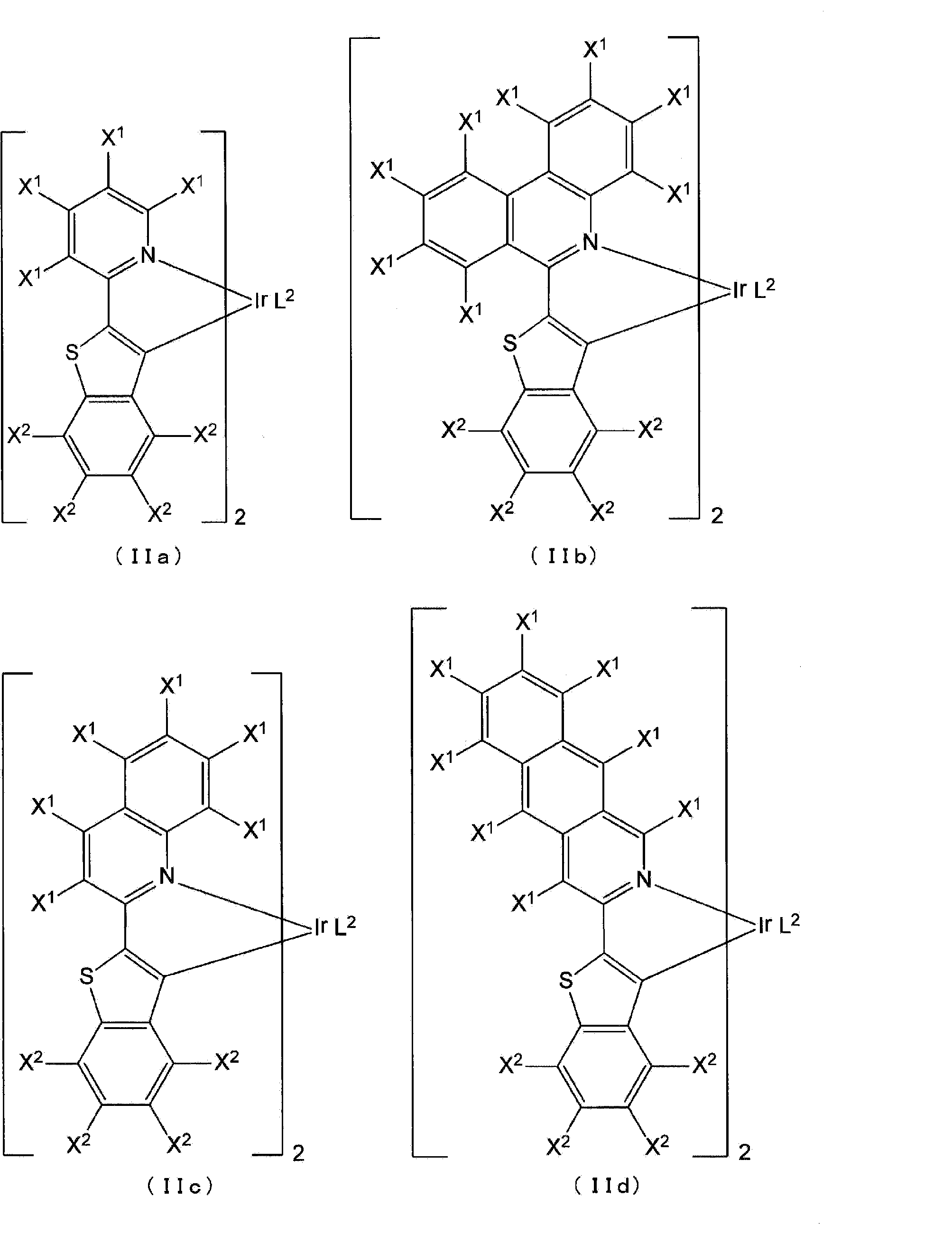
ここで、X1およびX2は水素を示し、L2は前記式(L-2)で示される構造の二座配位子を示す。
なお、錯体(I)および(II)としては、カチオン性イリジウム錯体、または中性イリジウム錯体であってよく、好ましくは、カチオン性イリジウム錯体である。本発明では、錯体(I)または(II)にオリゴアルギニンが結合することで、カチオン性部位が増加し、血管内皮結合性が大幅に増加すると考えられる。ここで、オリゴアルギニンは、例えば、オクタアルギニンでは電荷は+8になり、カチオン性イリジウム錯体が付くと+9、中性では+8のままである。つまり、アルギニンの数が多いときは、カチオン性でも中性でもさほど電荷の数は変化しないため、カチオン性イリジウム錯体だけでなく、中性イリジウム錯体を用いることができる。
なお、カチオン性イリジウム錯体は、陰イオンとの間で塩を形成していてもよい。陰イオンとしては、限定されないが、例えば、ヘキサフルオロリン酸イオン(PF6 -)、塩化物イオン(Cl-)、臭化物イオン(Br-)、トリフルオロ酢酸イオン(CF3COO-)等が挙げられ、好ましくはPF6 -である。
なお、カチオン性イリジウム錯体は、陰イオンとの間で塩を形成していてもよい。陰イオンとしては、限定されないが、例えば、ヘキサフルオロリン酸イオン(PF6 -)、塩化物イオン(Cl-)、臭化物イオン(Br-)、トリフルオロ酢酸イオン(CF3COO-)等が挙げられ、好ましくはPF6 -である。
イリジウム錯体として、好ましくは、前記式(IIc)で示される化合物であり、式(L-2)中、R7がピペラジル基である化合物である。
オリゴアルギニン、錯体(I)および(II)、オリゴアルギニンと発光団とを含む化合物は、常法の有機合成方法を用いて合成できる。例えば、後記実施例に記載の方法に従って合成できる。原料は、市販品を用いてもよく、公知の方法により合成したものを用いてもよい。
≪使用方法≫
本発明の血管のイメージング試薬は、本発明の効果を妨げない限り、溶媒、添加物、および従来血管のイメージング試薬として用いられる化合物等をさらに含んでもよい。
溶媒を含むと、血管のイメージング試薬をそのまま組織に添加して、血管のイメージングを実施できる。
溶媒としては、オリゴアルギニンと発光団とを含む化合物を溶解可能であればよく、例えばテトラヒドロフラン、アセトニトリル、ジメチルスルホキシド等の有機溶媒、水、生理食塩水(例えば、0.9%(w/v)生理食塩水)等の水系溶媒、これらの混合溶媒等の中から適宜選択できる。生体へ添加するときの溶媒としては、ジメチルスルホキシドと水の混合溶媒が好ましい。
本発明の血管のイメージング試薬は、本発明の効果を妨げない限り、溶媒、添加物、および従来血管のイメージング試薬として用いられる化合物等をさらに含んでもよい。
溶媒を含むと、血管のイメージング試薬をそのまま組織に添加して、血管のイメージングを実施できる。
溶媒としては、オリゴアルギニンと発光団とを含む化合物を溶解可能であればよく、例えばテトラヒドロフラン、アセトニトリル、ジメチルスルホキシド等の有機溶媒、水、生理食塩水(例えば、0.9%(w/v)生理食塩水)等の水系溶媒、これらの混合溶媒等の中から適宜選択できる。生体へ添加するときの溶媒としては、ジメチルスルホキシドと水の混合溶媒が好ましい。
本発明の血管のイメージング試薬が、オリゴアルギニンと発光団とを含む化合物と溶媒とを含む液状組成物である場合、オリゴアルギニンと発光団とを含む化合物の濃度は、化合物の種類等にもよるが、0.01~500mM、または0.1~100mM等であってよい。
本発明の血管のイメージング試薬は、生体組織の血管の可視化に用いられる。
本発明の血管のイメージング試薬は、生体組織の血管内皮に集積する。したがって、本発明の血管のイメージング試薬は、血管のイメージング、特に血管内皮のイメージングに有用である。
測定対象としての生体組織の種類は特に制限されないが、例えば、皮膚、筋肉、脂肪、肝臓、心臓、膵臓、腎臓、脾臓、腸、生殖器、脳等の臓器等が挙げられる。また、組織は、正常組織、および病態組織のいずれであってもよい。
投与対象となる生物個体としては、特に限定されず、例えば、哺乳動物(マウス、ヒト、ブタ、イヌ、ウサギ、ヒト等)を含む脊椎動物や無脊椎動物が挙げられる。
本発明の血管のイメージング試薬は、生体組織の血管内皮に集積する。したがって、本発明の血管のイメージング試薬は、血管のイメージング、特に血管内皮のイメージングに有用である。
測定対象としての生体組織の種類は特に制限されないが、例えば、皮膚、筋肉、脂肪、肝臓、心臓、膵臓、腎臓、脾臓、腸、生殖器、脳等の臓器等が挙げられる。また、組織は、正常組織、および病態組織のいずれであってもよい。
投与対象となる生物個体としては、特に限定されず、例えば、哺乳動物(マウス、ヒト、ブタ、イヌ、ウサギ、ヒト等)を含む脊椎動物や無脊椎動物が挙げられる。
組織内血管のイメージングは、例えば、以下のようにして行うことができる。
本発明の血管のイメージング試薬を、測定対象の個体に添加し、その後、検体に取り込まれた血管のイメージング試薬中のオリゴアルギニンと発光団とを含む化合物を励起して発光を観察する。
化合物の励起は、検体に可視光を照射することにより行うことができる。
発光の観察は、例えば、蛍光顕微鏡、蛍光測定装置、蛍光イメージング装置等の公知の装置を用いて行うことができる。
個体への本発明の血管のイメージング試薬の添加量は、使用する個体や血管密度等によって適宜変更可能であるが、例えば、0.01~1,000μmol/kg体重、好ましくは0.1~100μmol/kg体重の範囲で個体に投与することができる。
本発明の血管のイメージング試薬の投与形態としては、例えば、静脈内投与、皮下投与、筋肉内投与が挙げられる。
≪本発明の化合物≫
本発明の別の一態様は、下記式で示される化合物(以下、「本発明の化合物」ということがある)に関する。
本発明の別の一態様は、下記式で示される化合物(以下、「本発明の化合物」ということがある)に関する。

式中、
nは、4~20の整数である。
nは、4~20の整数である。
前記式に示されるように、本発明の化合物は、オリゴアルギニンとイリジウム錯体を含む化合物である。
ここで、nは4~20の整数であり、好ましくは、nは6~16の整数であり、より好ましくは、nは8~12の整数である。
なお、イリジウム錯体は、陰イオンとの間で塩を形成していてもよい。陰イオンとしては、限定されないが、例えば、ヘキサフルオロリン酸イオン(PF6 -)、塩化物イオン(Cl-)、臭化物イオン(Br-)、トリフルオロ酢酸イオン(CF3COO-)等が挙げられ、好ましくはPF6 -である。
なお、イリジウム錯体は、陰イオンとの間で塩を形成していてもよい。陰イオンとしては、限定されないが、例えば、ヘキサフルオロリン酸イオン(PF6 -)、塩化物イオン(Cl-)、臭化物イオン(Br-)、トリフルオロ酢酸イオン(CF3COO-)等が挙げられ、好ましくはPF6 -である。
本発明の化合物は、優れたりん光発光性能を有し、また、血管内皮への結合に優れるため、血管のイメージング試薬として用いることができる。
本発明のさらに別の一態様は、オリゴアルギニンと、NBD、クマリン6またはローダミンBとが、リンカーを介して結合された化合物に関する。このような化合物も、「本発明の化合物」である。リンカーは、限定されないが、好ましくは、ポリエチレングリコールを含む基である。ポリエチレングリコール(PEGp)の重合度pは、限定されないが、血管内皮へのとどまりやすさや、材料の入手しやすさの観点等から、例えば、2~20であり、好ましくは、3~16であり、より好ましくは4~12である。また、オリゴアルギニン(Rn)の重合度nは、限定されないが、例えば、4~20であり、好ましくは、6~16であり、より好ましくは8~12である。
このような本発明の化合物は、優れた蛍光発光性能を有し、また、血管内皮への結合に優れるため、血管のイメージング試薬として用いることができる。
このような本発明の化合物は、優れた蛍光発光性能を有し、また、血管内皮への結合に優れるため、血管のイメージング試薬として用いることができる。
本発明の化合物は、常法の有機合成方法を用いて合成できる。例えば、後記実施例に記載の方法に従って合成できる。原料は、市販品を用いてもよく、公知の方法により合成したものを用いてもよい。
以下、実施例によって本発明を具体的に説明するが、これらは本発明の例示であり、本発明の範囲はこれらに限定されるものではない。
なお、各略称は以下の意味である。
btq:[2-(2’-ベンゾチエニル)-キノリナート-N,C3’]
phen-pipe:5-piperazinyl-1,10-phenanthroline
BTQ:(btq)2Ir(phen-pipe)
RhB: Rhodamine B
NBD: Nitrobenzofurazan
C6: Coumarin 6
btq:[2-(2’-ベンゾチエニル)-キノリナート-N,C3’]
phen-pipe:5-piperazinyl-1,10-phenanthroline
BTQ:(btq)2Ir(phen-pipe)
RhB: Rhodamine B
NBD: Nitrobenzofurazan
C6: Coumarin 6
<合成例>
本発明の化合物を以下の合成スキームのとおり、合成した。
本発明の化合物を以下の合成スキームのとおり、合成した。

(Boc-[R(Pbf)]4-OH)
Boc-[R(Pbf)]4-OHは全自動マイクロウェーブペプチド合成装置(Initiator + Alstra, Biotage)を用いてFmoc固相合成法にて合成した。樹脂はR(Pbf)が導入されているクロロトリチルレジン(1.7 g,アミノ酸導入モル数:0.30 mmol/g)を用いた。縮合反応は、アミノ酸として0.5 MのFmoc-R(Pbf)-OHおよびBoc-R(Pbf)-OHのDMF溶液、縮合剤として0.6 MのHBTUのDMF溶液、添加剤として0.5 MのHOBtのDMF溶液、塩基として2.0 MのDIEAのNMP溶液を用い、それぞれアミノ酸導入モル数に対して3等量、3等量、3等量、6等量加え、室温にて1時間反応させた。Fmoc保護基の脱保護は、2%DBUのDMF溶液を用いて、アミノ酸導入モル数に対して3.5等量加え、室温にて5分反応させた。脱保護を完全に行うため、再度、同条件で10分反応させた。ペプチドを樹脂から切り出すために、ペプチドの樹脂が入ったバイアルに、1%TFAのジクロロメタン溶液10 mLを加え、室温にて90分反応させた。切り出したペプチドの溶液を減圧乾固させ、少量のアセトニトリルに再度溶解させた。その溶液を50 mL遠沈管に移し、水を加えて固体を析出させた。得られた固体を遠心し(3500 rpm,5分間)、水を除去し、再度水を加えて遠心した。この操作を2回繰り返した。バイアル内の水を凍結乾燥より除去し、白色固体を得た(734 mg,0.42 mmol,粗生成物収率:84%)。
Boc-[R(Pbf)]4-OHは全自動マイクロウェーブペプチド合成装置(Initiator + Alstra, Biotage)を用いてFmoc固相合成法にて合成した。樹脂はR(Pbf)が導入されているクロロトリチルレジン(1.7 g,アミノ酸導入モル数:0.30 mmol/g)を用いた。縮合反応は、アミノ酸として0.5 MのFmoc-R(Pbf)-OHおよびBoc-R(Pbf)-OHのDMF溶液、縮合剤として0.6 MのHBTUのDMF溶液、添加剤として0.5 MのHOBtのDMF溶液、塩基として2.0 MのDIEAのNMP溶液を用い、それぞれアミノ酸導入モル数に対して3等量、3等量、3等量、6等量加え、室温にて1時間反応させた。Fmoc保護基の脱保護は、2%DBUのDMF溶液を用いて、アミノ酸導入モル数に対して3.5等量加え、室温にて5分反応させた。脱保護を完全に行うため、再度、同条件で10分反応させた。ペプチドを樹脂から切り出すために、ペプチドの樹脂が入ったバイアルに、1%TFAのジクロロメタン溶液10 mLを加え、室温にて90分反応させた。切り出したペプチドの溶液を減圧乾固させ、少量のアセトニトリルに再度溶解させた。その溶液を50 mL遠沈管に移し、水を加えて固体を析出させた。得られた固体を遠心し(3500 rpm,5分間)、水を除去し、再度水を加えて遠心した。この操作を2回繰り返した。バイアル内の水を凍結乾燥より除去し、白色固体を得た(734 mg,0.42 mmol,粗生成物収率:84%)。
(BTQ-[R(Pbf)]4-Boc)
50 mLナス型フラスコにBoc-[R(Pbf)]4-OH(116 mg,0.065 mmol)、(btq)2Ir(phen-pipe)・PF6・CF3COO(63 mg,0.051 mmol)、HATU(41 mg,0.10 mmol)を加え、脱水DMF 1 mLに溶解させた。この溶液にDIEAを0.17 mL加え、窒素置換下、室温にて24時間撹拌した。溶液を50 mL遠沈管に移し、水を加えて固体を析出させた。得られた固体を遠心し(3500 rpm、5分間)、水を除去し、再度水を加えて遠心した。この操作を2回繰り返した。バイアル内の水を凍結乾燥より除去し、赤色固体を得た(390 mg,0.14 mmol,粗生成物収率:270%)。
50 mLナス型フラスコにBoc-[R(Pbf)]4-OH(116 mg,0.065 mmol)、(btq)2Ir(phen-pipe)・PF6・CF3COO(63 mg,0.051 mmol)、HATU(41 mg,0.10 mmol)を加え、脱水DMF 1 mLに溶解させた。この溶液にDIEAを0.17 mL加え、窒素置換下、室温にて24時間撹拌した。溶液を50 mL遠沈管に移し、水を加えて固体を析出させた。得られた固体を遠心し(3500 rpm、5分間)、水を除去し、再度水を加えて遠心した。この操作を2回繰り返した。バイアル内の水を凍結乾燥より除去し、赤色固体を得た(390 mg,0.14 mmol,粗生成物収率:270%)。
(BTQ-R4)
50 mL遠沈管にBTQ-[R(Pbf)]4-Boc(279 mg,0.10 mmol)を量りとり、TFA:水:TIPS(95:2.5:2.5)を1 mL加え、室温にて2時間反応させた。溶液に冷ジエチルエーテルを加えて固体を析出させた。得られた固体を遠心し(3500 rpm,5分間)、ジエチルエーテルを除去し、再度ジエチルエーテルを加えて遠心した。この操作を2回繰り返した。バイアル内のジエチルエーテルを除去し、デシケータ内で乾燥させ赤色固体を得た(82 mg,0.04 mmol,粗生成物収率:40%)。赤色固体を逆相カラムクロマトグラフィーで精製してBTQ-R4を得た。同定はMALDI-TOF-MSを用いて行い、1価イオンピーク(m/z)を検出した。calcd.:1601.9 [M-PF6 -]+,found:1601.2
50 mL遠沈管にBTQ-[R(Pbf)]4-Boc(279 mg,0.10 mmol)を量りとり、TFA:水:TIPS(95:2.5:2.5)を1 mL加え、室温にて2時間反応させた。溶液に冷ジエチルエーテルを加えて固体を析出させた。得られた固体を遠心し(3500 rpm,5分間)、ジエチルエーテルを除去し、再度ジエチルエーテルを加えて遠心した。この操作を2回繰り返した。バイアル内のジエチルエーテルを除去し、デシケータ内で乾燥させ赤色固体を得た(82 mg,0.04 mmol,粗生成物収率:40%)。赤色固体を逆相カラムクロマトグラフィーで精製してBTQ-R4を得た。同定はMALDI-TOF-MSを用いて行い、1価イオンピーク(m/z)を検出した。calcd.:1601.9 [M-PF6 -]+,found:1601.2
(Boc-[R(Pbf)]8-OH)
Boc-[R(Pbf)]8-OHは全自動マイクロウェーブペプチド合成装置(Initiator + Alstra, Biotage)を用いてFmoc固相合成法にて合成した。樹脂はR(Pbf)が導入されているクロロトリチルレジン(1.67 g,アミノ酸導入モル数:0.30 mmol/g)を用いた。縮合反応は、アミノ酸として0.6 MのFmoc-R(Pbf)-OHおよびBoc-R(Pbf)-OHのDMF溶液、縮合剤として0.5 MのHBTUのDMF溶液、添加剤として0.5 MのHOBtのDMF溶液、塩基として2.0 MのDIEAのNMP溶液を用い、それぞれアミノ酸導入モル数に対して3等量、3等量、3等量、6等量加え、室温にて1時間反応させた。Fmoc保護基の脱保護は、2%DBUのDMF溶液を用いて、アミノ酸導入モル数に対して3.5等量加え、室温にて5分反応させた。脱保護を完全に行うため、再度、同条件で10分反応させた。ペプチドを樹脂から切り出すために、ペプチドの樹脂が入ったバイアルに、1%TFAのジクロロメタン溶液10 mLを加え、室温にて90分反応させた。切り出したペプチドの溶液を減圧乾固させ、少量のアセトニトリルに再度溶解させた。その溶液を50 mL遠沈管に移し、水を加えて固体を析出させた。得られた固体を遠心し(3500 rpm,5分間)、水を除去し、再度水を加えて遠心した。この操作を2回繰り返した。バイアル内の水を凍結乾燥より除去し、白色固体を得た(1.26 g,0.37 mmol,粗生成物収率:74%)。
Boc-[R(Pbf)]8-OHは全自動マイクロウェーブペプチド合成装置(Initiator + Alstra, Biotage)を用いてFmoc固相合成法にて合成した。樹脂はR(Pbf)が導入されているクロロトリチルレジン(1.67 g,アミノ酸導入モル数:0.30 mmol/g)を用いた。縮合反応は、アミノ酸として0.6 MのFmoc-R(Pbf)-OHおよびBoc-R(Pbf)-OHのDMF溶液、縮合剤として0.5 MのHBTUのDMF溶液、添加剤として0.5 MのHOBtのDMF溶液、塩基として2.0 MのDIEAのNMP溶液を用い、それぞれアミノ酸導入モル数に対して3等量、3等量、3等量、6等量加え、室温にて1時間反応させた。Fmoc保護基の脱保護は、2%DBUのDMF溶液を用いて、アミノ酸導入モル数に対して3.5等量加え、室温にて5分反応させた。脱保護を完全に行うため、再度、同条件で10分反応させた。ペプチドを樹脂から切り出すために、ペプチドの樹脂が入ったバイアルに、1%TFAのジクロロメタン溶液10 mLを加え、室温にて90分反応させた。切り出したペプチドの溶液を減圧乾固させ、少量のアセトニトリルに再度溶解させた。その溶液を50 mL遠沈管に移し、水を加えて固体を析出させた。得られた固体を遠心し(3500 rpm,5分間)、水を除去し、再度水を加えて遠心した。この操作を2回繰り返した。バイアル内の水を凍結乾燥より除去し、白色固体を得た(1.26 g,0.37 mmol,粗生成物収率:74%)。
(BTQ-[R(Pbf)]8-Boc)
50 mLナス型フラスコにBoc-[R(Pbf)]8-OH(209 mg,0.062 mmol)、(btq)2Ir(phen-pipe)・PF6・CF3COO(62 mg,0.050 mmol)、HATU(40 mg,0.11 mmol)を加え、脱水DMF 1 mLに溶解させた。この溶液にDIEAを0.17 mL加え、窒素置換下、室温にて24時間撹拌した。溶液を50 mL遠沈管に移し、水を加えて固体を析出させた。得られた固体を遠心し(3500 rpm,5分間)、水を除去し、再度水を加えて遠心した。この操作を2回繰り返した。バイアル内の水を凍結乾燥より除去し、赤色固体を得た(235 mg,0.055 mmol,粗生成物収率:110%)。
50 mLナス型フラスコにBoc-[R(Pbf)]8-OH(209 mg,0.062 mmol)、(btq)2Ir(phen-pipe)・PF6・CF3COO(62 mg,0.050 mmol)、HATU(40 mg,0.11 mmol)を加え、脱水DMF 1 mLに溶解させた。この溶液にDIEAを0.17 mL加え、窒素置換下、室温にて24時間撹拌した。溶液を50 mL遠沈管に移し、水を加えて固体を析出させた。得られた固体を遠心し(3500 rpm,5分間)、水を除去し、再度水を加えて遠心した。この操作を2回繰り返した。バイアル内の水を凍結乾燥より除去し、赤色固体を得た(235 mg,0.055 mmol,粗生成物収率:110%)。
(BTQ-R8)
50 mL遠沈管にBTQ-[R(Pbf)]8-Boc(110 mg,0.026 mmol)を量りとり、TFA:水:TIPS(95:2.5:2.5)を1 mL加え、室温にて2時間反応させた。溶液に冷ジエチルエーテルを加えて固体を析出させた。得られた固体を遠心し(3500 rpm,5分間)、ジエチルエーテルを除去し、再度ジエチルエーテルを加えて遠心した。この操作を2回繰り返した。バイアル内のジエチルエーテルを除去し、デシケータ内で乾燥させ赤色固体を得た(72 mg,0.022 mmol,粗生成物収率:85%)。赤色固体を逆相カラムクロマトグラフィーで精製してBTQ-R8を得た。同定はMALDI-TOF-MSを用いて行い、1価イオンピーク(m/z)を検出した。calcd.:2226.6 [M-PF6 -]+,found:2224.8
50 mL遠沈管にBTQ-[R(Pbf)]8-Boc(110 mg,0.026 mmol)を量りとり、TFA:水:TIPS(95:2.5:2.5)を1 mL加え、室温にて2時間反応させた。溶液に冷ジエチルエーテルを加えて固体を析出させた。得られた固体を遠心し(3500 rpm,5分間)、ジエチルエーテルを除去し、再度ジエチルエーテルを加えて遠心した。この操作を2回繰り返した。バイアル内のジエチルエーテルを除去し、デシケータ内で乾燥させ赤色固体を得た(72 mg,0.022 mmol,粗生成物収率:85%)。赤色固体を逆相カラムクロマトグラフィーで精製してBTQ-R8を得た。同定はMALDI-TOF-MSを用いて行い、1価イオンピーク(m/z)を検出した。calcd.:2226.6 [M-PF6 -]+,found:2224.8
(Boc-[R(Pbf)]12-OH)
Boc-[R(Pbf)]12-OHは全自動マイクロウェーブペプチド合成装置(Initiator + Alstra,Biotage)を用いてFmoc固相合成法にて合成した。樹脂はR(Pbf)が導入されているクロロトリチルレジン(1.0 g,アミノ酸導入モル数:0.30 mmol/g)を用いた。縮合反応は、アミノ酸として0.5 MのFmoc-R(Pbf)-OHおよびBoc-R(Pbf)-OHのDMF溶液、縮合剤として0.6 MのHBTUのDMF溶液、添加剤として0.5 MのHOBtのDMF溶液、塩基として2.0 MのDIEAのNMP溶液を用い、それぞれアミノ酸導入モル数に対して3等量、3等量、3等量、6等量加え、室温にて1時間反応させた。Fmoc保護基の脱保護は、2%DBUのDMF溶液を用いて、アミノ酸導入モル数に対して3.5等量加え、室温にて5分反応させた。脱保護を完全に行うため、再度、同条件で10分反応させた。ペプチドを樹脂から切り出すために、ペプチドの樹脂が入ったバイアルに、1%TFAのジクロロメタン溶液10 mLを加え、室温にて90分反応させた。切り出したペプチドの溶液を減圧乾固させ、少量のアセトニトリルに再度溶解させた。その溶液を50 mL遠沈管に移し、水を加えて固体を析出させた。得られた固体を遠心し(3500 rpm,5分間)、水を除去し、再度水を加えて遠心した。この操作を2回繰り返した。バイアル内の水を凍結乾燥より除去し、白色固体を得た(999 mg,0.20 mmol,粗生成物収率:67%)。
Boc-[R(Pbf)]12-OHは全自動マイクロウェーブペプチド合成装置(Initiator + Alstra,Biotage)を用いてFmoc固相合成法にて合成した。樹脂はR(Pbf)が導入されているクロロトリチルレジン(1.0 g,アミノ酸導入モル数:0.30 mmol/g)を用いた。縮合反応は、アミノ酸として0.5 MのFmoc-R(Pbf)-OHおよびBoc-R(Pbf)-OHのDMF溶液、縮合剤として0.6 MのHBTUのDMF溶液、添加剤として0.5 MのHOBtのDMF溶液、塩基として2.0 MのDIEAのNMP溶液を用い、それぞれアミノ酸導入モル数に対して3等量、3等量、3等量、6等量加え、室温にて1時間反応させた。Fmoc保護基の脱保護は、2%DBUのDMF溶液を用いて、アミノ酸導入モル数に対して3.5等量加え、室温にて5分反応させた。脱保護を完全に行うため、再度、同条件で10分反応させた。ペプチドを樹脂から切り出すために、ペプチドの樹脂が入ったバイアルに、1%TFAのジクロロメタン溶液10 mLを加え、室温にて90分反応させた。切り出したペプチドの溶液を減圧乾固させ、少量のアセトニトリルに再度溶解させた。その溶液を50 mL遠沈管に移し、水を加えて固体を析出させた。得られた固体を遠心し(3500 rpm,5分間)、水を除去し、再度水を加えて遠心した。この操作を2回繰り返した。バイアル内の水を凍結乾燥より除去し、白色固体を得た(999 mg,0.20 mmol,粗生成物収率:67%)。
(BTQ-[R(Pbf)]12-Boc)
50 mLナス型フラスコにBoc-[R(Pbf)]12-OH(321 mg,0.064 mmol)、(btq)2Ir(phen-pipe)・PF6・CF3COO(62 mg,0.050 mmol)、HATU(0.040 mg,0.10 mmol)を加え、脱水DMF 1 mLに溶解させた。この溶液にDIEAを0.17 mL加え、窒素置換下、室温にて24時間撹拌した。溶液を50 mL遠沈管に移し、水を加えて固体を析出させた。得られた固体を遠心し(3500 rpm,5分間)、水を除去し、再度水を加えて遠心した。この操作を2回繰り返した。バイアル内の水を凍結乾燥より除去し、赤色固体を得た(320 mg,0.055 mmol,粗生成物収率:110%)。
50 mLナス型フラスコにBoc-[R(Pbf)]12-OH(321 mg,0.064 mmol)、(btq)2Ir(phen-pipe)・PF6・CF3COO(62 mg,0.050 mmol)、HATU(0.040 mg,0.10 mmol)を加え、脱水DMF 1 mLに溶解させた。この溶液にDIEAを0.17 mL加え、窒素置換下、室温にて24時間撹拌した。溶液を50 mL遠沈管に移し、水を加えて固体を析出させた。得られた固体を遠心し(3500 rpm,5分間)、水を除去し、再度水を加えて遠心した。この操作を2回繰り返した。バイアル内の水を凍結乾燥より除去し、赤色固体を得た(320 mg,0.055 mmol,粗生成物収率:110%)。
(BTQ-R12)
50 mL遠沈管にBTQ-[R(Pbf)]12-Boc(151 mg,0.026 mmol)を量りとり、TFA:水:TIPS(95:2.5:2.5)を1 mL加え、室温にて2時間反応させた。溶液に冷ジエチルエーテルを加えて固体を析出させた。得られた固体を遠心し(3500 rpm,5分間)、ジエチルエーテルを除去し、再度ジエチルエーテルを加えて遠心した。この操作を2回繰り返した。バイアル内のジエチルエーテルを除去し、デシケータ内で乾燥させ赤色固体を得た(93 mg,0.021 mmol,粗生成物収率:81%)。赤色固体を逆相カラムクロマトグラフィーで精製してBTQ-R12を得た。同定はMALDI-TOF-MSを用いて行い、1価イオンピーク(m/z)を検出した。calcd.:2851.4 [M-PF6 -]+,found:2846.8
50 mL遠沈管にBTQ-[R(Pbf)]12-Boc(151 mg,0.026 mmol)を量りとり、TFA:水:TIPS(95:2.5:2.5)を1 mL加え、室温にて2時間反応させた。溶液に冷ジエチルエーテルを加えて固体を析出させた。得られた固体を遠心し(3500 rpm,5分間)、ジエチルエーテルを除去し、再度ジエチルエーテルを加えて遠心した。この操作を2回繰り返した。バイアル内のジエチルエーテルを除去し、デシケータ内で乾燥させ赤色固体を得た(93 mg,0.021 mmol,粗生成物収率:81%)。赤色固体を逆相カラムクロマトグラフィーで精製してBTQ-R12を得た。同定はMALDI-TOF-MSを用いて行い、1価イオンピーク(m/z)を検出した。calcd.:2851.4 [M-PF6 -]+,found:2846.8
(Boc-[R(Pbf)]16-OH)
Boc-[R(Pbf)]16-OHは全自動マイクロウェーブペプチド合成装置(Initiator + Alstra,Biotage)を用いてFmoc固相合成法にて合成した。樹脂はR(Pbf)が導入されているクロロトリチルレジン(0.34 g,アミノ酸導入モル数:0.30 mmol/g)を用いた。縮合反応は、アミノ酸として0.3 MのFmoc-R(Pbf)-OHおよびBoc-R(Pbf)-OHのDMF溶液、縮合剤として0.6 MのHBTUのDMF溶液、添加剤として0.5 MのHOBtのDMF溶液、塩基として2.0 MのDIEAのNMP溶液を用い、それぞれアミノ酸導入モル数に対して3等量、3等量、3等量、6等量加え、室温にて1時間反応させた。Fmoc保護基の脱保護は、2%DBUのDMF溶液を用いて、アミノ酸導入モル数に対して3.5等量加え、室温にて5分反応させた。脱保護を完全に行うため、再度、同条件で10分反応させた。ペプチドを樹脂から切り出すために、ペプチドの樹脂が入ったバイアルに、1%TFAのジクロロメタン溶液10 mLを加え、室温にて90分反応させた。切り出したペプチドの溶液を減圧乾固させ、少量のアセトニトリルに再度溶解させた。その溶液を50 mL遠沈管に移し、水を加えて固体を析出させた。得られた固体を遠心し(3500 rpm,5分間)、水を除去し、再度水を加えて遠心した。この操作を2回繰り返した。バイアル内の水を凍結乾燥より除去し、白色固体を得た(418 mg,0.063 mmol,粗生成物収率:63%)。
Boc-[R(Pbf)]16-OHは全自動マイクロウェーブペプチド合成装置(Initiator + Alstra,Biotage)を用いてFmoc固相合成法にて合成した。樹脂はR(Pbf)が導入されているクロロトリチルレジン(0.34 g,アミノ酸導入モル数:0.30 mmol/g)を用いた。縮合反応は、アミノ酸として0.3 MのFmoc-R(Pbf)-OHおよびBoc-R(Pbf)-OHのDMF溶液、縮合剤として0.6 MのHBTUのDMF溶液、添加剤として0.5 MのHOBtのDMF溶液、塩基として2.0 MのDIEAのNMP溶液を用い、それぞれアミノ酸導入モル数に対して3等量、3等量、3等量、6等量加え、室温にて1時間反応させた。Fmoc保護基の脱保護は、2%DBUのDMF溶液を用いて、アミノ酸導入モル数に対して3.5等量加え、室温にて5分反応させた。脱保護を完全に行うため、再度、同条件で10分反応させた。ペプチドを樹脂から切り出すために、ペプチドの樹脂が入ったバイアルに、1%TFAのジクロロメタン溶液10 mLを加え、室温にて90分反応させた。切り出したペプチドの溶液を減圧乾固させ、少量のアセトニトリルに再度溶解させた。その溶液を50 mL遠沈管に移し、水を加えて固体を析出させた。得られた固体を遠心し(3500 rpm,5分間)、水を除去し、再度水を加えて遠心した。この操作を2回繰り返した。バイアル内の水を凍結乾燥より除去し、白色固体を得た(418 mg,0.063 mmol,粗生成物収率:63%)。
(BTQ-[R(Pbf)]16-Boc)
50 mLナス型フラスコにBoc-[R(Pbf)]16-OH(183 mg,0.028 mmol)、(btq)2Ir(phen-pipe)・PF6・CF3COO(37 mg,0.030 mmol)、HATU(35 mg,0.09 mmol)を加え、脱水DMF 1 mLに溶解させた。この溶液にDIEAを0.17 mL加え、窒素置換下、室温にて24時間撹拌した。溶液を50 mL遠沈管に移し、水を加えて固体を析出させた。得られた固体を遠心し(3500 rpm,5分間)、水を除去し、再度水を加えて遠心した。この操作を2回繰り返した。バイアル内の水を凍結乾燥より除去し、赤色固体を得た(186 mg,0.024 mmol,粗生成物収率:87%)。
50 mLナス型フラスコにBoc-[R(Pbf)]16-OH(183 mg,0.028 mmol)、(btq)2Ir(phen-pipe)・PF6・CF3COO(37 mg,0.030 mmol)、HATU(35 mg,0.09 mmol)を加え、脱水DMF 1 mLに溶解させた。この溶液にDIEAを0.17 mL加え、窒素置換下、室温にて24時間撹拌した。溶液を50 mL遠沈管に移し、水を加えて固体を析出させた。得られた固体を遠心し(3500 rpm,5分間)、水を除去し、再度水を加えて遠心した。この操作を2回繰り返した。バイアル内の水を凍結乾燥より除去し、赤色固体を得た(186 mg,0.024 mmol,粗生成物収率:87%)。
(BTQ-R16)
50 mL遠沈管にBTQ-[R(Pbf)]16-Boc(113 mg,0.015 mmol)を量りとり、TFA:水:TIPS(95:2.5:2.5)を1 mL加え、室温にて2時間反応させた。溶液に冷ジエチルエーテルを加えて固体を析出させた。得られた固体を遠心し(3500 rpm,5分間)、ジエチルエーテルを除去し、再度ジエチルエーテルを加えて遠心した。この操作を2回繰り返した。バイアル内のジエチルエーテルを除去し、デシケータ内で乾燥させ赤色固体を得た(73 mg,0.013 mmol,粗生成物収率:87%)。赤色固体を逆相カラムクロマトグラフィーで精製してBTQ-R16を得た。同定はMALDI-TOF-MSを用いて行い、1価イオンピーク(m/z)を検出した。calcd.:3474.8 [M-PF6 -]+,found:3473.7
50 mL遠沈管にBTQ-[R(Pbf)]16-Boc(113 mg,0.015 mmol)を量りとり、TFA:水:TIPS(95:2.5:2.5)を1 mL加え、室温にて2時間反応させた。溶液に冷ジエチルエーテルを加えて固体を析出させた。得られた固体を遠心し(3500 rpm,5分間)、ジエチルエーテルを除去し、再度ジエチルエーテルを加えて遠心した。この操作を2回繰り返した。バイアル内のジエチルエーテルを除去し、デシケータ内で乾燥させ赤色固体を得た(73 mg,0.013 mmol,粗生成物収率:87%)。赤色固体を逆相カラムクロマトグラフィーで精製してBTQ-R16を得た。同定はMALDI-TOF-MSを用いて行い、1価イオンピーク(m/z)を検出した。calcd.:3474.8 [M-PF6 -]+,found:3473.7
<実施例1>
図2に、実施例で合成した本発明の化合物の構造式(BTQ-R4,BTQ-R8,BTQ-R12,BTQ-R16)を示す。アルギニン残基数は、4,8,12,16である。また、アルギニンの効果を示すために、アルギニンが結合していない化合物(BTQ)を参照化合物とした。これら化合物の吸収・りん光スペクトルを測定したところ、500 nm付近に吸収極大波長、660 nm付近にりん光極大波長を示した。りん光は600nm以上で観測されているため、緑色発光を示す分子を用いた多色イメージングが可能である。また、りん光量子収率、りん光寿命を測定したところ、それぞれ0.32(0.015空気飽和下)、5.7 μs(0.28 μs空気飽和下)であった。アルギニン残基数の異なる化合物であっても、オリゴアルギニンペプチドに同じりん光団が結合しているため、スペクトル、および光物理特性はほぼ同じである。
図2に、実施例で合成した本発明の化合物の構造式(BTQ-R4,BTQ-R8,BTQ-R12,BTQ-R16)を示す。アルギニン残基数は、4,8,12,16である。また、アルギニンの効果を示すために、アルギニンが結合していない化合物(BTQ)を参照化合物とした。これら化合物の吸収・りん光スペクトルを測定したところ、500 nm付近に吸収極大波長、660 nm付近にりん光極大波長を示した。りん光は600nm以上で観測されているため、緑色発光を示す分子を用いた多色イメージングが可能である。また、りん光量子収率、りん光寿命を測定したところ、それぞれ0.32(0.015空気飽和下)、5.7 μs(0.28 μs空気飽和下)であった。アルギニン残基数の異なる化合物であっても、オリゴアルギニンペプチドに同じりん光団が結合しているため、スペクトル、および光物理特性はほぼ同じである。
合成した化合物、および市販の緑色蛍光性血管内皮染色試薬であるFITC-トマトレクチンをマウスに投与して、腎臓の毛細血管のイメージングを行った。図3にFITC-トマトレクチン(1 mg/mL(生理食塩水中)を50 μL投与)、BTQ(100 nmol:1 mmol/L(生理食塩水:ジメチルスルホキシド(9:1,v/v)混合溶媒中)を100 μL投与)、BTQ-Rn (n:4, 8, 12,100 nmol:1 mmol/L(生理食塩水中)を100 μL投与)を麻酔下にあるマウスの尾静脈から投与して、腹側部を切開し腎臓を露出させ、共焦点レーザー顕微鏡で腎臓表面をイメージングした画像を示す。FITC-トマトレクチンの画像と比較すると、BTQ、およびBTQ-R4では、FITC-トマトレクチンとは異なる領域(尿細管細胞)から発光が観測されているのに対して、BTQ-R8、BTQ-R12は、FITC-トマトレクチンと同様に腎臓の毛細血管がイメージングされていることがわかる。特にBTQ-R12では血管内に空洞が見られることから、BTQ-R12が血中ではなく、血管内皮に分布している。以上より、血管内皮をイメージングするためには、8以上のアルギニンの残基数が好ましいことが明らかとなった。
腎臓は、自家蛍光の強い臓器であるため、FITC-トマトレクチンを用いた場合、血管内皮に加えて腎臓の自家蛍光を検出されることが危惧される。そこで、FITC-トマトレクチン(1 mg/mL(生理食塩水中)を50 μL投与)、およびBTQ-R12(100 nmol:1 mmol/L(生理食塩水中)を100 μL投与)をマウスに投与して、同一箇所のイメージングを行った。図4のA、Cに示すようにFITC-トマトレクチンの蛍光でイメージングした場合、血管内皮以外の箇所から蛍光が観測されている。この蛍光がFITC-トマトレクチン由来あるいは自家蛍光由来であるかは区別がつかない。一方、BTQ-R12のりん光でイメージングした場合(図4のB、D)、蛍光成分を排除することにより、血管内皮からのみりん光が観測され、より鮮明に血管内皮を選択的にイメージングするこができる。腎臓は、血液内の老廃物や塩分を尿として体外に排泄するため、糸球体で血液をろ過する。多くの糸球体は、腎臓表面から約200 μmよりも深い場所にあるため、1光子顕微鏡でイメージングすることは困難である。ここでは、マウスを安楽死させた後、腎臓を摘出し2つにカットし、その断面の観察を行った。図5のAに示すように丸く光っている箇所が見られる。拡大すると線状の構造体が集合していることがわかり、糸球体に集まる血管網がイメージング出来ている(図5のB)。
次に肝臓の血管イメージングを行った。肝臓は、肝小葉と呼ばれる6角形の機能単位の集合体であり、頂点付近に動脈や門脈、中心部に中心静脈があり、中心部から放射状に類洞血管が伸びている。図6にBTQ-R12(100 nmol:1 mmol/L(生理食塩水中)を100 μL投与)をマウスに投与して得られたりん光顕微画像を示す。類洞血管が鮮明にイメージングされている。同様な実験を、他の臓器(膵臓、小腸、皮下組織、心臓)においても行ったところ、多くの臓器の毛細血管のイメージングが可能であることが明らかとなった(図7)。
正常組織では、血管構造が堅固であるのに対して、がん腫瘍では未発達で脆弱な新生血管が多数あることが指摘されている。よって、血中投与された抗がん剤がすべてのがん細胞まで到達しないため、投与量を増加させる必要がある。これは副作用の増加にもつながり患者に対して大きな負荷となる。ここでは、BTQ-R12を用いた腫瘍血管イメージングを試みた。ヌードマウスにヒト結腸腺癌由来HCT116細胞を移植し、担がんマウスを作製した。図8にBTQ-R12(100 nmol:1 mmol/L(生理食塩水中)を100 μL投与)を担がんマウスに投与して得られたりん光顕微画像を示す。画像より、太い血管から多数の細い血管が枝分かれしていることが分かり、腫瘍内の血管網のイメージングに成功した。
近年、脂肪肝は肝硬変や肝臓がんへ進行するため、肝臓内の脂質蓄積を早期に発見することが注目されている。特に非アルコール性脂肪肝は、肥満とはことなり、外見からは分からないため病気の発見が遅れる危険性がある。脂肪肝の肝臓では、肝細胞内に脂質が異常蓄積し、大きな脂質滴が形成され、肝細胞が肥大化する。そのため、類洞血管の狭小化が進み肝臓全体が低酸素状態になる。ここでは、緑色蛍光性脂質滴試薬(3-(benzo[d]thiazol-2-yl)-8-(diethylamino)-2H-benzo[g]chromen-2-one (PC6S))とBTQ-R12を健常マウス、および脂肪肝モデルマウスに同時投与(PC6S:50 nmol:0.5 mmol/L(生理食塩水:ジメチルスルホキシド(9:1,v/v),10 wt% BSAを含む混合溶媒中)を100 μL投与,BTQ-R12:100 nmol:1 mmol/L(生理食塩水中)を100 μL投与)し、緑色蛍光と深赤色りん光を測定することで、多色イメージングを実施した。健常マウスでは、肝細胞内の脂質滴からPC6Sの蛍光、類洞血管内皮からBTQ-R12のりん光が観測されている(図9のA)。画像より類洞血管が肝細胞の間を直線的に走行していることがわかる。一方、脂肪肝モデルマウスでは、大きな脂質滴が形成されることで肝細胞が肥大化し、類洞血管が大きく蛇行している(図9のB)。これにより赤血球の流れが阻害され、肝臓のへの酸素供給が不足し、低酸素状態になることが考えられる。
<合成例2>
本発明の化合物を以下のとおり、合成した。
本発明の化合物を以下のとおり、合成した。
(RhB-pipe-PEG12-NH2)
100 mLナス型フラスコに、Boc-piperazine(35.6 mg, 0.19 mmol)、Rhodamine B(73.4 mg, 0.15 mmol)、HATU(58.3 mg, 0.15 mmol)を加え、脱水CH2Cl2(5 mL)とDIEA(170 μL)で溶解させた後、室温の窒素雰囲気下で 24 時間反応させた。クロロホルムで洗浄し、析出した結晶を乾燥させた。その後、4N-HCl/dioxane(10 mL)を用いて、室温で2時間反応させBoc基の脱保護を行った。脱水THFとヘキサンを用いて洗浄し乾燥させ、赤紫色固体を得た。(0.224 g, 0.41 mmol, 粗生成物収率:268%)
100 mLナス型フラスコに、Boc-piperazine(35.6 mg, 0.19 mmol)、Rhodamine B(73.4 mg, 0.15 mmol)、HATU(58.3 mg, 0.15 mmol)を加え、脱水CH2Cl2(5 mL)とDIEA(170 μL)で溶解させた後、室温の窒素雰囲気下で 24 時間反応させた。クロロホルムで洗浄し、析出した結晶を乾燥させた。その後、4N-HCl/dioxane(10 mL)を用いて、室温で2時間反応させBoc基の脱保護を行った。脱水THFとヘキサンを用いて洗浄し乾燥させ、赤紫色固体を得た。(0.224 g, 0.41 mmol, 粗生成物収率:268%)
次に、100 mLナス型フラスコにt-Boc-N-amido-dPEG12-acid(100 mg, 0.14 mmol)、RhB-pipe(79.5 mg, 0.15 mmol)、HATU(105 mg, 0.28 mmol)を加え、脱水CH2Cl2(5 mL)とDIEA(170 μL)で溶解させた後、室温の窒素雰囲気下で 24 時間反応させた。クロロホルムで洗浄し、析出した結晶を乾燥させ、赤紫色固体を得た。(0.399 g, 0.320 mmol, 粗生成物収率:166%)その後、4N-HCl/dioxane(10 mL)を用いて、室温で3時間反応させることでBoc基の脱保護を行った。脱水THFとメタノールを用いて洗浄し乾燥させ赤紫色固体を得た(99.0 mg, 0.086 mmol, 生成物収率:62%)。
(RhB-pipe-PEG12-[R(Pbf)]12-Boc)
100 mLナスフラスに、Boc-[R(Pbf)]12-OH(286 mg, 0.057 mmol)、RhB-pipe-PEG12-NH2(78 mg, 0.068 mmol)、HATU(44.5 mg, 0.117 mmol)を加え、脱水DMF 10 mLに溶解させた。この溶液にDIEAを0.17 mL加え、窒素置換下、室温にて24時間撹拌した。溶液を50 mL遠沈管に移し,水を加えて固体を析出させた。得られた固体を遠心し(3500 rpm,5分間)、水を除去し、再度水を加えて遠心した。この操作を2回繰り返した。バイアル内の水を凍結乾燥より除去し、赤紫色固体を得た(157 mg,0.026 mmol,粗生成物収率:45%)。
100 mLナスフラスに、Boc-[R(Pbf)]12-OH(286 mg, 0.057 mmol)、RhB-pipe-PEG12-NH2(78 mg, 0.068 mmol)、HATU(44.5 mg, 0.117 mmol)を加え、脱水DMF 10 mLに溶解させた。この溶液にDIEAを0.17 mL加え、窒素置換下、室温にて24時間撹拌した。溶液を50 mL遠沈管に移し,水を加えて固体を析出させた。得られた固体を遠心し(3500 rpm,5分間)、水を除去し、再度水を加えて遠心した。この操作を2回繰り返した。バイアル内の水を凍結乾燥より除去し、赤紫色固体を得た(157 mg,0.026 mmol,粗生成物収率:45%)。
(RhB-pipe-PEG12-R12)
50 mL遠沈管にRhB-pipe-PEG12-[R(Pbf)]12-Boc(21.5 mg,0.0035 mmol)を量りとり、TFA:水:TIPS(95:2.5:2.5)を1 mL加え、室温にて2時間反応させた。溶液に冷ジエチルエーテルを加えて固体を析出させた。得られた固体を遠心し(3500 rpm,5分間)、ジエチルエーテルを除去し、再度ジエチルエーテルを加えて遠心した。この操作を2回繰り返した。バイアル内のジエチルエーテルを除去し、デシケータ内で乾燥させ赤色固体を得た(21 mg,0.0042 mmol,粗生成物収率:119%)。赤紫色固体を逆相カラムクロマトグラフィーで精製してRhB-pipe-PEG12-R12を得た。同定はESI-MSを用いて行い、3価イオンピーク(m/z)を検出した。calcd.:995.48 [M;3H]3+, found:996.6
50 mL遠沈管にRhB-pipe-PEG12-[R(Pbf)]12-Boc(21.5 mg,0.0035 mmol)を量りとり、TFA:水:TIPS(95:2.5:2.5)を1 mL加え、室温にて2時間反応させた。溶液に冷ジエチルエーテルを加えて固体を析出させた。得られた固体を遠心し(3500 rpm,5分間)、ジエチルエーテルを除去し、再度ジエチルエーテルを加えて遠心した。この操作を2回繰り返した。バイアル内のジエチルエーテルを除去し、デシケータ内で乾燥させ赤色固体を得た(21 mg,0.0042 mmol,粗生成物収率:119%)。赤紫色固体を逆相カラムクロマトグラフィーで精製してRhB-pipe-PEG12-R12を得た。同定はESI-MSを用いて行い、3価イオンピーク(m/z)を検出した。calcd.:995.48 [M;3H]3+, found:996.6

(NBD-PEG4-NH2)
100 mLナス型フラスコに、Boc-NH-PEG4-amine(521 mg, 1.54 mmol)、4-Fluoro-7-nitrobenzofurazan(210 mg, 1.14 mmol)、脱水CH2Cl2(5 mL)とDIEA(0.17 mL)で溶解させた後、窒素置換下、室温にて24時間撹拌した。クロロホルムとヘキサンで洗浄を行い、黄色液体を得た。その後、4N-HCl/dioxane(10 mL)を用いて、室温で3時間反応させBoc基の脱保護を行った。脱水THFとメタノールを用いて洗浄し乾燥させ、粘性のある黄色液体を得た(0.614 g, 1.53mmol, 粗生成物収率:134%)。
100 mLナス型フラスコに、Boc-NH-PEG4-amine(521 mg, 1.54 mmol)、4-Fluoro-7-nitrobenzofurazan(210 mg, 1.14 mmol)、脱水CH2Cl2(5 mL)とDIEA(0.17 mL)で溶解させた後、窒素置換下、室温にて24時間撹拌した。クロロホルムとヘキサンで洗浄を行い、黄色液体を得た。その後、4N-HCl/dioxane(10 mL)を用いて、室温で3時間反応させBoc基の脱保護を行った。脱水THFとメタノールを用いて洗浄し乾燥させ、粘性のある黄色液体を得た(0.614 g, 1.53mmol, 粗生成物収率:134%)。
(NBD-PEG4-[R(Pbf)]12-Boc)
100 mLナスフラスに、Boc-[R(Pbf)]12-OH(429 mg, 0.085 mmol)、NBD-PEG4-NH2(0.10 mmol)、HATU(65.9 mg, 0.17 mmol)を加え、脱水DMF 8 mLに溶解させた。この溶液にDIEAを0.17 mL加え、窒素置換下、室温にて24時間撹拌した。溶液を50 mL遠沈管に移し,水を加えて固体を析出させた。得られた固体を遠心し(3500 rpm,5分間)、水を除去し、再度水を加えて遠心した。この操作を2回繰り返した。バイアル内の水を凍結乾燥より除去し、黄色固体を得た(313 mg,0.058 mmol,粗生成物収率:68%)。
100 mLナスフラスに、Boc-[R(Pbf)]12-OH(429 mg, 0.085 mmol)、NBD-PEG4-NH2(0.10 mmol)、HATU(65.9 mg, 0.17 mmol)を加え、脱水DMF 8 mLに溶解させた。この溶液にDIEAを0.17 mL加え、窒素置換下、室温にて24時間撹拌した。溶液を50 mL遠沈管に移し,水を加えて固体を析出させた。得られた固体を遠心し(3500 rpm,5分間)、水を除去し、再度水を加えて遠心した。この操作を2回繰り返した。バイアル内の水を凍結乾燥より除去し、黄色固体を得た(313 mg,0.058 mmol,粗生成物収率:68%)。
(NBD-PEG4-R12)
50 mL遠沈管にNBD-PEG4-[R(Pbf)]12-Boc(30.2 mg,0.0060 mmol)を量りとり、TFA:水:TIPS(95:2.5:2.5)を1 mL加え、室温にて2時間反応させた。溶液に冷ジエチルエーテルを加えて固体を析出させた。得られた固体を遠心し(3500 rpm,5分間)、ジエチルエーテルを除去し、再度ジエチルエーテルを加えて遠心した。この操作を2回繰り返した。バイアル内のジエチルエーテルを除去し、デシケータ内で乾燥させ黄色固体を得た(13 mg,0.0057 mmol,粗生成物収率:95%)。黄色固体を逆相カラムクロマトグラフィーで精製してNBD-PEG4-R12を得た。同定はESI-MSを用いて行い、4価イオンピーク(m/z)を検出した。calcd.:569.1 [M;4H]4+, found:596.4
50 mL遠沈管にNBD-PEG4-[R(Pbf)]12-Boc(30.2 mg,0.0060 mmol)を量りとり、TFA:水:TIPS(95:2.5:2.5)を1 mL加え、室温にて2時間反応させた。溶液に冷ジエチルエーテルを加えて固体を析出させた。得られた固体を遠心し(3500 rpm,5分間)、ジエチルエーテルを除去し、再度ジエチルエーテルを加えて遠心した。この操作を2回繰り返した。バイアル内のジエチルエーテルを除去し、デシケータ内で乾燥させ黄色固体を得た(13 mg,0.0057 mmol,粗生成物収率:95%)。黄色固体を逆相カラムクロマトグラフィーで精製してNBD-PEG4-R12を得た。同定はESI-MSを用いて行い、4価イオンピーク(m/z)を検出した。calcd.:569.1 [M;4H]4+, found:596.4

(C6-PEG4-NH2)
100 mLナス型フラスコに、Boc-NH-PEG4-amine(79.0 mg, 0.24 mmol)、Coumarin 6-COOH(79.3 mg, 0.20 mmol)、HATU(154.8 mg, 0.41 mmol)を加え、脱水CH2Cl2(11 mL)とDIEA(170 μL)で溶解させた後、室温の窒素雰囲気下で 24 時間反応させた。クロロホルムで洗浄を行い、黄色固体を得た。その後、4N-HCl/dioxane(10 mL)を用いて、室温で2時間反応させBoc基の脱保護を行った。脱水THFとメタノールを用いて洗浄し乾燥させ、粘性のある深赤色固体を得た(0.184 g, 0.300mmol, 粗生成物収率:150%)。
100 mLナス型フラスコに、Boc-NH-PEG4-amine(79.0 mg, 0.24 mmol)、Coumarin 6-COOH(79.3 mg, 0.20 mmol)、HATU(154.8 mg, 0.41 mmol)を加え、脱水CH2Cl2(11 mL)とDIEA(170 μL)で溶解させた後、室温の窒素雰囲気下で 24 時間反応させた。クロロホルムで洗浄を行い、黄色固体を得た。その後、4N-HCl/dioxane(10 mL)を用いて、室温で2時間反応させBoc基の脱保護を行った。脱水THFとメタノールを用いて洗浄し乾燥させ、粘性のある深赤色固体を得た(0.184 g, 0.300mmol, 粗生成物収率:150%)。
(C6-PEG4-[R(Pbf)]12-Boc)
100 mLナスフラスに、Boc-[R(Pbf)]12-OH(413 mg, 0.082 mmol)、C6-PEG4-NH2(63.0 mg, 0.10 mmol)、HATU(62.3 mg, 0.17 mmol)を加え、脱水DMF 10 mLに溶解させた。この溶液にDIEAを0.17 mL加え、窒素置換下、室温にて24時間撹拌した。溶液を50 mL遠沈管に移し、水を加えて固体を析出させた。得られた固体を遠心し(3500 rpm,5分間)、水を除去し、再度水を加えて遠心した。この操作を2回繰り返した。バイアル内の水を凍結乾燥より除去し、黄色固体を得た(326 mg,0.058 mmol,粗生成物収率:71%)。
100 mLナスフラスに、Boc-[R(Pbf)]12-OH(413 mg, 0.082 mmol)、C6-PEG4-NH2(63.0 mg, 0.10 mmol)、HATU(62.3 mg, 0.17 mmol)を加え、脱水DMF 10 mLに溶解させた。この溶液にDIEAを0.17 mL加え、窒素置換下、室温にて24時間撹拌した。溶液を50 mL遠沈管に移し、水を加えて固体を析出させた。得られた固体を遠心し(3500 rpm,5分間)、水を除去し、再度水を加えて遠心した。この操作を2回繰り返した。バイアル内の水を凍結乾燥より除去し、黄色固体を得た(326 mg,0.058 mmol,粗生成物収率:71%)。
(C6-PEG4-R12)
50 mL遠沈管にC6-PEG4-[R(Pbf)]12-Boc(46.0 mg,0.0082 mmol)を量りとり、TFA:水:TIPS(95:2.5:2.5)を1 mL加え、室温にて2時間反応させた。溶液に冷ジエチルエーテルを加えて固体を析出させた。得られた固体を遠心し(3500 rpm,5分間)、ジエチルエーテルを除去し、再度ジエチルエーテルを加えて遠心した。この操作を2回繰り返した。バイアル内のジエチルエーテルを除去し、デシケータ内で乾燥させ黄色固体を得た(34 mg,0.0014 mmol,粗生成物収率:166%)。黄色固体を逆相カラムクロマトグラフィーで精製してC6-PEG4-R12を得た。同定はESI-MSを用いて行い、4価イオンピーク(m/z)を検出した。calcd.:622.4 [M;4H]4+, found:622.8
50 mL遠沈管にC6-PEG4-[R(Pbf)]12-Boc(46.0 mg,0.0082 mmol)を量りとり、TFA:水:TIPS(95:2.5:2.5)を1 mL加え、室温にて2時間反応させた。溶液に冷ジエチルエーテルを加えて固体を析出させた。得られた固体を遠心し(3500 rpm,5分間)、ジエチルエーテルを除去し、再度ジエチルエーテルを加えて遠心した。この操作を2回繰り返した。バイアル内のジエチルエーテルを除去し、デシケータ内で乾燥させ黄色固体を得た(34 mg,0.0014 mmol,粗生成物収率:166%)。黄色固体を逆相カラムクロマトグラフィーで精製してC6-PEG4-R12を得た。同定はESI-MSを用いて行い、4価イオンピーク(m/z)を検出した。calcd.:622.4 [M;4H]4+, found:622.8

<実施例2>
RhB-pipe-PEG12-R12をマウスに投与して,様々な組織の血管イメージングを行った。図10にRhB-pipe-PEG12-R12(100 nmol:2 mmmol/L(生理食塩中)を50μL投与)を麻酔下にあるマウスの尾静脈から投与して、腹側部を切開し様々な臓器(肝臓,腎臓,脾臓,膵臓,精巣)や組織(筋肉組織,皮下組織)を露出させ、共焦点レーザー顕微鏡でそれらの表面をイメージングした画像を示す。励起波長は550 nm,観測波長は>590 nmである。観測したすべての臓器や組織において毛細血管がイメージングできている。
RhB-pipe-PEG12-R12をマウスに投与して,様々な組織の血管イメージングを行った。図10にRhB-pipe-PEG12-R12(100 nmol:2 mmmol/L(生理食塩中)を50μL投与)を麻酔下にあるマウスの尾静脈から投与して、腹側部を切開し様々な臓器(肝臓,腎臓,脾臓,膵臓,精巣)や組織(筋肉組織,皮下組織)を露出させ、共焦点レーザー顕微鏡でそれらの表面をイメージングした画像を示す。励起波長は550 nm,観測波長は>590 nmである。観測したすべての臓器や組織において毛細血管がイメージングできている。
NBD-PEG-R12をマウスに投与して,様々な組織の血管イメージングを行った。図11にNBD-PEG-R12(100 nmol:2 mmmol/L(生理食塩中)を50μL投与)を麻酔下にあるマウスの尾静脈から投与して、腹側部を切開し様々な臓器(肝臓,腎臓,脾臓)や組織(皮下組織,脂肪組織)を露出させ、共焦点レーザー顕微鏡でそれらの表面をイメージングした画像を示す。励起波長は488 nm,観測波長は510-550 nmである。観測したすべての臓器や組織において毛細血管がイメージングできている。
C6-PEG-R12をマウスに投与して,様々な組織の血管イメージングを行った。図12にC6-PEG-R12(100 nmol:2 mmmol/L(生理食塩中)を50μL投与)を麻酔下にあるマウスの尾静脈から投与して、腹側部を切開し様々な臓器(腎臓,脾臓,膵臓,精巣)や組織(脂肪組織,筋肉組織)を露出させ、共焦点レーザー顕微鏡でそれらの表面をイメージングした画像を示す。励起波長は488 nm,観測波長は510-550 nmである。観測したすべての臓器や組織において毛細血管がイメージングできている。また,マウスを安楽死させた後,脳血管のイメージングを実施した。図13に示すように,脳血管のイメージングも可能であることが示された。
以上の結果より、オリゴアルギニンと発光団とを含む化合物を用いて、個体内の血管を明瞭にイメージングすることが可能であり、血管のイメージング試薬として用いることができることが分かった。
また、本発明によって開発されたBTQ-Rnは、血管のイメージング試薬として用いることができる。特に、BTQ-R12は、個体内の正常組織、および病態組織の血管走行をイメージング可能な新しい試薬である。また、血管内皮への分布はオリゴアルギニンペプチドの作用であり、発光団(BTQ)を変えることで、様々な発光色の血管内皮イメージング試薬の開発が可能である。また、本発明によって開発されたRhB-pipe-PEGp-Rn、NBD-PEGp-Rn、C6-PEGp-Rnも、血管のイメージング試薬として好ましく用いることができる。
また、本発明によって開発されたBTQ-Rnは、血管のイメージング試薬として用いることができる。特に、BTQ-R12は、個体内の正常組織、および病態組織の血管走行をイメージング可能な新しい試薬である。また、血管内皮への分布はオリゴアルギニンペプチドの作用であり、発光団(BTQ)を変えることで、様々な発光色の血管内皮イメージング試薬の開発が可能である。また、本発明によって開発されたRhB-pipe-PEGp-Rn、NBD-PEGp-Rn、C6-PEGp-Rnも、血管のイメージング試薬として好ましく用いることができる。
本発明は、医療診断、製薬開発、基礎医学等の分野で利用できる。
Claims (6)
- 下記式で示されるオリゴアルギニンと、
前記オリゴアルギニンのC末端側またはN末端側に結合しているりん光団または蛍光団、
とを含む化合物を含む、血管のイメージング試薬:

nは、4~20の整数である。 - 前記オリゴアルギニンは、nが8以上である、請求項1に記載の試薬。
- 前記りん光団は、イリジウム錯体を含む化合物である、請求項1または2に記載の試薬。
- 前記イリジウム錯体は、下記式(I)または(II)で示される化合物である、請求項3に記載の試薬:

環R1は、単環または多環式の含窒素芳香族環を示し、
環R2は、単環または多環式の含硫黄芳香族環を示し、
L1は、β-ジケトネート構造を有する二座配位子を示す;

環R1は、単環または多環式の含窒素芳香族環を示し、
環R2は、単環または多環式の含硫黄芳香族環を示し、
L2はフェナントロリン骨格を有する二座配位子を示す。 - 前記イリジウム錯体は、下記式で示される化合物である、請求項3または4に記載の試薬:

nは、4~20の整数である。 - 下記式で示される化合物:

nは4~20の整数である。
Priority Applications (1)
| Application Number | Priority Date | Filing Date | Title |
|---|---|---|---|
| JP2022571439A JPWO2022138550A1 (ja) | 2020-12-21 | 2021-12-20 |
Applications Claiming Priority (2)
| Application Number | Priority Date | Filing Date | Title |
|---|---|---|---|
| JP2020-211560 | 2020-12-21 | ||
| JP2020211560 | 2020-12-21 |
Publications (1)
| Publication Number | Publication Date |
|---|---|
| WO2022138550A1 true WO2022138550A1 (ja) | 2022-06-30 |
Family
ID=82157922
Family Applications (1)
| Application Number | Title | Priority Date | Filing Date |
|---|---|---|---|
| PCT/JP2021/046991 WO2022138550A1 (ja) | 2020-12-21 | 2021-12-20 | 血管のイメージング試薬 |
Country Status (2)
| Country | Link |
|---|---|
| JP (1) | JPWO2022138550A1 (ja) |
| WO (1) | WO2022138550A1 (ja) |
-
2021
- 2021-12-20 JP JP2022571439A patent/JPWO2022138550A1/ja active Pending
- 2021-12-20 WO PCT/JP2021/046991 patent/WO2022138550A1/ja active Application Filing
Non-Patent Citations (2)
| Title |
|---|
| YASUKAGAWA MAMI; YAMADA KEIICH; TOBITA SEIJI; YOSHIHARA TOSHITADA: "Ratiometric oxygen probes with a cell-penetrating peptide for imaging oxygen levels in living cells", JOURNAL OF PHOTOCHEMISTRY, ELSEVIER, AMSTERDAM, NL, vol. 383, 25 July 2019 (2019-07-25), AMSTERDAM, NL, XP085785139, ISSN: 1010-6030, DOI: 10.1016/j.jphotochem.2019.111983 * |
| YOSHIHARA TOSHITADA; HIRAKAWA YOSUKE; HOSAKA MASAHIRO; NANGAKU MASAOMI; TOBITA SEIJI: "Oxygen imaging of living cells and tissues using luminescent molecular probes", JOURNAL OF PHOTOCHEMISTRY, ELSEVIER, AMSTERDAM, NL, vol. 30, 20 January 2017 (2017-01-20), AMSTERDAM, NL, pages 71 - 95, XP029940267, ISSN: 1389-5567, DOI: 10.1016/j.jphotochemrev.2017.01.001 * |
Also Published As
| Publication number | Publication date |
|---|---|
| JPWO2022138550A1 (ja) | 2022-06-30 |
Similar Documents
| Publication | Publication Date | Title |
|---|---|---|
| JP6270981B2 (ja) | 腫瘍の標的型画像化のために用いられる化合物にコンジュゲートされたアミノ酸連結基の製造および合成の方法 | |
| CA2749108C (en) | Intramolecularly-quenched fluorescent imaging agents | |
| RU2484111C2 (ru) | Агенты для оптической визуализации | |
| CN103402547B (zh) | 开关型荧光纳米颗粒探针以及使用其的荧光分子成像法 | |
| JP7308366B2 (ja) | アクティブターゲティング型葉酸受容体近赤外蛍光分子及びその調製方法 | |
| CN115006553B (zh) | 用于制备肿瘤诊断显像剂的多肽及其应用 | |
| EP1281405B1 (de) | Kurzkettige Peptid-Farbstoffkonjugate als Kontrastmittel für die optische Diagnostik | |
| JP2022532628A (ja) | 修飾シアニン色素およびそのコンジュゲート | |
| CN106568757A (zh) | 一种检测结肠癌肿瘤的量子点靶向探针试剂盒 | |
| JP5425181B2 (ja) | 蛍光mriプローブ | |
| JP2023530575A (ja) | 近赤外シアニン色素およびそのコンジュゲート | |
| WO2022138550A1 (ja) | 血管のイメージング試薬 | |
| Park et al. | Rapid and selective targeting of heterogeneous pancreatic neuroendocrine tumors | |
| CN110201189A (zh) | 白蛋白结合型近红外荧光染料-马来酰亚胺共轭物 | |
| JP2011173859A (ja) | 新規インドシアニン化合物を用いた診断用組成物及び分析方法 | |
| CN108250125B (zh) | 一种肿瘤靶向探针化合物及其合成和应用 | |
| EP4360659A1 (en) | Fluorescent water-soluble polycationic chitosan polymers as markers for biological 3d imaging | |
| EP3768780A1 (en) | Azacyanine dyes and use thereof | |
| Zhai | Self-Threaded Fluorescent Probes for Biological Imaging and Self-Assembly | |
| US20220306665A1 (en) | Reagent for measuring oxygen concentration in cell and tissue | |
| JP6834112B2 (ja) | レシオ法を用いた細胞内酸素濃度測定試薬 | |
| YASUKAGAWA | Synthesis and Biological Applications of Phosphorescent Ir (Ⅲ) Complex-Based Probes with Arginine Peptide | |
| CA3177944A1 (en) | Methods of manufacture and synthesis of fluorescent dye compounds and uses thereof | |
| Williams | Design, Synthesis, and Evaluation of Innovative BODIPY-Peptidic Conjugates for Biological Application | |
| CN116655516A (zh) | 用于前列腺癌特异性荧光探针的化合物、制备方法及其应用 |
Legal Events
| Date | Code | Title | Description |
|---|---|---|---|
| 121 | Ep: the epo has been informed by wipo that ep was designated in this application |
Ref document number: 21908105 Country of ref document: EP Kind code of ref document: A1 |
|
| ENP | Entry into the national phase |
Ref document number: 2022571439 Country of ref document: JP Kind code of ref document: A |
|
| NENP | Non-entry into the national phase |
Ref country code: DE |
|
| 122 | Ep: pct application non-entry in european phase |
Ref document number: 21908105 Country of ref document: EP Kind code of ref document: A1 |