WO2022138550A1 - 血管のイメージング試薬 - Google Patents
血管のイメージング試薬 Download PDFInfo
- Publication number
- WO2022138550A1 WO2022138550A1 PCT/JP2021/046991 JP2021046991W WO2022138550A1 WO 2022138550 A1 WO2022138550 A1 WO 2022138550A1 JP 2021046991 W JP2021046991 W JP 2021046991W WO 2022138550 A1 WO2022138550 A1 WO 2022138550A1
- Authority
- WO
- WIPO (PCT)
- Prior art keywords
- mmol
- compound
- group
- oligoarginine
- blood vessel
- Prior art date
- Legal status (The legal status is an assumption and is not a legal conclusion. Google has not performed a legal analysis and makes no representation as to the accuracy of the status listed.)
- Ceased
Links
Images





Classifications
-
- A—HUMAN NECESSITIES
- A61—MEDICAL OR VETERINARY SCIENCE; HYGIENE
- A61K—PREPARATIONS FOR MEDICAL, DENTAL OR TOILETRY PURPOSES
- A61K49/00—Preparations for testing in vivo
-
- C—CHEMISTRY; METALLURGY
- C07—ORGANIC CHEMISTRY
- C07F—ACYCLIC, CARBOCYCLIC OR HETEROCYCLIC COMPOUNDS CONTAINING ELEMENTS OTHER THAN CARBON, HYDROGEN, HALOGEN, OXYGEN, NITROGEN, SULFUR, SELENIUM OR TELLURIUM
- C07F15/00—Compounds containing elements of Groups 8, 9, 10 or 18 of the Periodic Table
-
- C—CHEMISTRY; METALLURGY
- C07—ORGANIC CHEMISTRY
- C07K—PEPTIDES
- C07K7/00—Peptides having 5 to 20 amino acids in a fully defined sequence; Derivatives thereof
- C07K7/04—Linear peptides containing only normal peptide links
- C07K7/06—Linear peptides containing only normal peptide links having 5 to 11 amino acids
-
- C—CHEMISTRY; METALLURGY
- C07—ORGANIC CHEMISTRY
- C07K—PEPTIDES
- C07K7/00—Peptides having 5 to 20 amino acids in a fully defined sequence; Derivatives thereof
- C07K7/04—Linear peptides containing only normal peptide links
- C07K7/08—Linear peptides containing only normal peptide links having 12 to 20 amino acids
-
- G—PHYSICS
- G01—MEASURING; TESTING
- G01N—INVESTIGATING OR ANALYSING MATERIALS BY DETERMINING THEIR CHEMICAL OR PHYSICAL PROPERTIES
- G01N21/00—Investigating or analysing materials by the use of optical means, i.e. using sub-millimetre waves, infrared, visible or ultraviolet light
- G01N21/62—Systems in which the material investigated is excited whereby it emits light or causes a change in wavelength of the incident light
- G01N21/63—Systems in which the material investigated is excited whereby it emits light or causes a change in wavelength of the incident light optically excited
- G01N21/64—Fluorescence; Phosphorescence
Definitions
- the present invention relates to a blood vessel imaging reagent, a compound that can be used in the reagent, and the like.
- Living tissue (organ) cells acquire necessary nutrients and oxygen from the blood and release synthesized metabolites and carbon dioxide into the blood. For this reason, the vascular system plays an important role in life support. If for some reason an abnormality occurs in the vascular system, the surrounding tissue cells will not be able to perform their full function.
- arteriosclerosis in which lipid components such as cholesterol accumulate in the intima of the arteries and form atherosclerotic foci called atherosclerosis, and liver cirrhosis, which causes fibrosis between blood vessels and hepatocytes, are deeply disrupted in the vascular system. I am involved.
- the formation of angiogenesis cannot follow the abnormal proliferation of cells, so many immature and abnormally shaped blood vessels can be seen. Therefore, it is important to visualize and image the shape and running of blood vessels in a living individual in the above-mentioned diagnosis and treatment of pathological conditions.
- a method of administering a contrast medium in blood and imaging with MRI and a method using photoacoustic imaging technology are known. Although these provide insights into the human vascular network, they are large-scale and unsuitable for small animals. Moreover, it does not have the spatial resolution at the single cell level.
- the imaging technique using light can easily realize the spatial resolution at the single cell level by combining with a microscope in addition to high sensitivity, convenience, and real-time measurement.
- an important element is a probe molecule that exists stably in a contaminated environment such as blood and exhibits high-luminance emission.
- Blood vessel imaging reagents are classified into blood retention type and vascular endothelial adsorption type.
- the former is a reagent that stays in the blood, and fluorescent dextran and fluorescent nanoparticles have been developed.
- fluorescent dextran is a reagent in which a fluorescent molecule is covalently bonded to dextran, and various reagents having different molecular weights of dextran are commercially available.
- fluorescent lectins are commercially available as vascular endothelial adsorption type imaging reagents.
- Lectin is a general term for proteins that exhibit specific binding to a specific sugar chain, and many of them are derived from animals, plants, and fungi. Of these, plant-derived lectins recognize and bind to sugar chains present on the surface of vascular endothelial cells of mammals including humans. So far, a fluorescent lectin in which fluorescein or Texas red is covalently bonded to tomato lectin has been commercially available (Vector Laboratories). The synthesis of fluorescent lectins requires complicated work to extract tomato lectins from nature, and the number of fluorescent molecules bound to the lectins cannot be controlled.
- the fluorescent lectin has the following problems. Since it recognizes sugar chains present on the surface of vascular endothelial cells, only plant-derived lectins can be used; complicated work is required to extract lectins from nature; since lectins are glycoproteins, luminescent groups can be used. Labeling becomes non-uniform and differences occur between lots; it may be indistinguishable from tissue autofluorescence due to the use of fluorescence.
- Patent Documents 1 and 2 have not studied the use of this method for visualization of blood vessels.
- the present invention has been made in view of the above problems, and an object of the present invention is to develop a new blood vessel imaging reagent that is easy to manufacture, has high sensitivity, and is highly accurate.
- the present inventors have clearly imaged blood vessels in an individual using a compound containing oligoarginine and a phosphorescent group or a chromophore which is a fluorescent group. I found that I could do it. We have also developed a compound containing an oligoarginine and an iridium complex that can be used as such a chromophore. Based on such findings, the present invention has been completed. That is, the gist of the present invention relates to the following.
- Oligoarginine represented by the following formula and Phosphorescent or fluorescent groups bound to the C-terminal side or N-terminal side of the oligoarginine, Blood vessel imaging reagents, including compounds containing:
- n is an integer of 4 to 20.
- the reagent according to [1] or [2], wherein the phosphorescent group is a compound containing an iridium complex.
- the reagent according to [3], wherein the iridium complex is a compound represented by the following formula (I) or (II):
- Ring R 1 represents a monocyclic or polycyclic nitrogen-containing aromatic ring.
- Ring R 2 represents a monocyclic or polycyclic sulfur-containing aromatic ring.
- L 1 indicates a bidentate ligand having a ⁇ -diketonate structure;
- Ring R 1 represents a monocyclic or polycyclic nitrogen-containing aromatic ring.
- Ring R 2 represents a monocyclic or polycyclic sulfur-containing aromatic ring.
- L 2 represents a bidentate ligand having a phenanthroline skeleton.
- n is an integer of 4 to 20.
- the present invention provides a blood vessel imaging reagent containing a compound containing oligoarginine and a chromophore. Since the reagent can chemically synthesize a compound for an imaging reagent, it does not require an extraction operation from a natural product and can be easily produced. In addition, the label of the chromophore can be made uniform, and a uniform reagent with a small lot difference can be made, which enables highly accurate imaging. When a phosphorescent compound is used as the chromophore, autofluorescence can be eliminated by time-resolved measurement, and high-sensitivity and high-precision imaging becomes possible.
- a compound to which 8 or more arginine is bound has excellent binding to the vascular endothelium, and can image the vascular endothelium with higher sensitivity and accuracy. Even when a fluorescent compound is used, it is possible to distinguish it from autofluorescence by using a compound that exhibits fluorescence in the near-infrared light region, and it is also possible to image the structure of deep blood vessels.
- the present invention also provides a compound containing an oligoarginine and an iridium complex that can be used as a chromophore of a blood vessel imaging reagent. The compound has excellent phosphorescence emission performance and can clearly image blood vessels.
- the present invention also provides a compound containing oligoarginine and a fluorescent compound that can be used as a chromophore of a blood vessel imaging reagent. The compound has excellent fluorescence emission performance and can clearly image blood vessels.
- FIG. 1 shows a conceptual diagram of the compound (blood vessel imaging reagent) of the present invention.
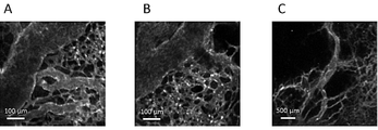
- FIG. 3 shows an imaging image (picture substitute) of the capillaries of the kidney.
- A shows the results using FITC-tomato lectin
- FIG. 4 shows a capillary imaging image (photograph of a drawing) of the kidney.
- FIG. 5 shows an image (drawing substitute photograph) of a section (glomerulus) of a kidney.
- A shows the result of the objective lens ⁇ 20, and B shows the result of the objective lens ⁇ 10.
- FIG. 6 shows an imaging image (photograph of a drawing substitute) of a blood vessel of the vas sinusoide of the liver.
- a and B are images of different parts of the liver.
- FIG. 7 shows blood vessel imaging images (drawing substitute photographs) of various organs.
- FIG. 8 shows a tumor blood vessel imaging image (drawing substitute photograph) of a cancer-bearing mouse.
- a to C are images of different parts of the tumor.
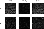
- FIG. 9 shows a multicolor imaging image (drawing substitute photograph) of PC6S and BTQ-R 12 in the liver.
- Column A shows the results of healthy mice
- column B shows the results of fatty liver model mice.
- the left side of the column shows the PC 6S
- the center shows the BTQ-R 12
- the right side shows the result of their superposition.
- FIG. 10 shows blood vessel imaging images (drawing substitute photographs) of various organs using RhB-pipe-PEG 12 -R 12 .
- A is the liver
- B and C are the kidneys (C is a magnified view of part of B)
- D is the spleen
- E is the pancreas
- F is the testis
- G is the muscle tissue
- H is the subcutaneous tissue.
- FIG. 11 shows blood vessel imaging images (drawing substitute photographs) of various organs using NBD-PEG 4 -R 12 .
- A is the liver
- B is the kidney
- C is the spleen
- D is the subcutaneous tissue
- E is the adipose tissue.
- FIG. 12 shows blood vessel imaging images (drawing substitute photographs) of various organs using C6-PEG 4 -R 12 .
- A is the kidney
- B is the spleen
- C is the pancreas
- D is the testis
- E is the adipose tissue
- F is the muscle tissue.
- FIG. 13 shows an imaging image (drawing substitute photograph) of a cerebral blood vessel using C6-PEG 4 -R 12 .
- A, B and C are images of different parts of the brain.
- D is a magnified image of a part of C.
- oligoarginine represented by the following formula and Phosphorescent or fluorescent groups bound to the C-terminal side or N-terminal side of the oligoarginine, Regarding a blood vessel imaging reagent (hereinafter, may be referred to as "the blood vessel imaging reagent of the present invention") containing a compound containing and:
- n is an integer of 4 to 20.
- the blood vessel imaging reagent developed in the present invention contains a compound having a structure in which a luminescent group is bound to the C-terminal side or the N-terminal side of the oligoarginine peptide (Fig. 1).
- the chromophore may be bound to either the C-terminal side or the N-terminal side of the oligoarginine peptide, but is preferably the C-terminal side.
- a fluorescent group fluorescent compound
- a phosphorescent compound for example, an iridium complex
- autofluorescence can be eliminated by time-resolved measurement. Even when a fluorescent compound is used, it is possible to distinguish it from autofluorescence by using a compound that exhibits fluorescence in the near-infrared light region, and it is also possible to image the structure of deep blood vessels.
- the compound used in the blood vessel imaging reagent of the present invention contains oligoarginine represented by the following formula.
- n is an integer of 4 to 20, preferably n is an integer of 6 to 16, and more preferably an integer of 8 to 12.
- the compound used in the blood vessel imaging reagent of the present invention contains oligoarginine to achieve improved adsorption to the vascular endothelium.
- oligoarginine has n ⁇ 8 or more, it exhibits particularly excellent adsorptivity to the vascular endothelium. Therefore, such an embodiment can be preferably used for the purpose of imaging the vascular endothelium.
- the compound used for the blood vessel imaging reagent of the present invention may contain a group other than oligoarginine as long as the effect of the present invention is not impaired.
- the group other than oligoarginine may contain one kind or two or more kinds.
- Groups other than oligoarginine may be attached to the end of oligoarginine. That is, a group other than oligoarginine may be present between oligoarginine and the chromophore (this embodiment may be referred to as a "linker") or on the opposite side of the oligoarginine's binding to the chromophore. May exist.
- a group containing steroids, peptides, polyethylene glycol and the like can be relatively easily bonded to a chromophore, and thus can be suitably used as a group other than oligoarginine.
- the polypeptide contains a group other than oligoarginine and oligoarginine consisting of a peptide
- the polypeptide as a whole preferably has 4 to 20 amino acid residues, more preferably 6 to 16 amino acid residues, and more preferably amino acid residues. The number is 8-12.
- the peptide other than oligoarginine is not limited, but for example, proline is preferable from the viewpoint of easiness of synthesis.
- aspartic acid and lysine are also preferable because the compound can be made water-soluble.
- the degree of polymerization of polyethylene glycol can be adjusted depending on the type of luminescent group and the like, but is preferably 2 to 20, for example. It is 3 to 16, and more preferably 4 to 12.
- any aspect containing or not containing a group other than oligoarginine can be preferably used.
- an embodiment containing a group other than oligoarginine as a linker can be more preferably used.
- fluorescent group contained in the compound used for the imaging reagent for blood vessels of the present invention a fluorescent compound conventionally used for imaging blood vessels and the like can be used without particular limitation. Further, the compound of the present invention described later can be used as a compound used as an imaging reagent for blood vessels having a fluorescent group.
- the fluorescent compound may be used alone or in combination of two or more. Fluorescent compounds include, but are not limited to, 4-nitrobenzo-2-oxa-1,3-diazole (NBD), dimethylaminosulfonylbenzoxadiazole (DBD), dimethylaminosulfonylbenzothiadiazole (DBThD), dimethyl.
- Examples thereof include aminosulfonylbenzoselenaziazole (DBSeD), fluorescein isothiocyanate (FITC), coumarin dyes, rhodamines, borondipyrromethene (BODIPY), cyanine dyes and the like.
- Examples of the fluorescent compound include, but are not limited to, coumarin-based dyes such as NBD and coumarin 6, and rhodamines such as rhodamine B.
- a phosphorescent compound conventionally used for imaging blood vessels and the like can be used without particular limitation.
- the compound of the present invention described later can be used as a compound used as an imaging reagent for blood vessels having a phosphorescent group.
- the phosphorus compound can be used alone or in combination of two or more.
- the phosphorescent compound include, but are not limited to, compounds containing an iridium complex.
- the iridium complex include, but are not limited to, compounds represented by the following formulas (I) or (II).
- Ring R 1 represents a monocyclic or polycyclic nitrogen-containing aromatic ring.
- Ring R 2 represents a monocyclic or polycyclic sulfur-containing aromatic ring.
- L 1 indicates a bidentate ligand having a ⁇ -diketonate structure;
- Ring R 1 represents a monocyclic or polycyclic nitrogen-containing aromatic ring.
- Ring R 2 represents a monocyclic or polycyclic sulfur-containing aromatic ring.
- L 2 represents a bidentate ligand having a phenanthroline skeleton.
- complex (I) the compound represented by the formula (I) (hereinafter, may be referred to as “complex (I)”) will be described.
- the complex (I) contained in the blood vessel imaging reagent of the present invention may be one kind or two or more kinds.
- the ring R 1 is not limited, and examples thereof include a nitrogen-containing aromatic ring having a structure represented by the following formula (1-1), (1-2), (1-3), or (1-4). Be done.
- X 1 indicates hydrogen.
- the bond extending from the carbon atom next to N is bonded to the ring R2 .
- N is coordinated to Ir.
- a polycyclic nitrogen-containing aromatic ring is preferable because the light emission approaches near infrared rays in combination with the ring R 2 and the permeability in the living body is good, and the above formula (1-2). ), (1-3), or (1-4), a nitrogen-containing aromatic ring having the structure shown in (1-3) or (1-4) is more preferable.
- Examples of the ring R 2 include a sulfur-containing aromatic ring having a structure represented by the following formula (2-1), (2-2), or (2-3).
- X 2 represents hydrogen.
- the bond extending from the carbon atom next to S is bonded to ring R1 , and the carbon atom next to this carbon atom is coordinated to Ir.
- a sulfur-containing aromatic ring having the structure represented by the above formula (2-1) is preferable.
- the ligand composed of ring R 1 and ring R 2 is a cyclometallated ligand and contributes to the light emission performance of the complex (I).
- L 1 represents a bidentate ligand having a ⁇ -diketonate structure. Although L 1 does not affect the luminescence performance, having L 1 enhances the biocompatibility and facilitates the incorporation of the complex (I) into the living tissue. Examples of L 1 include bidentate ligands having a structure represented by the following formula (L-1). In this case, the complex (I) is represented by the following formula (I-1).
- R 3 represents a substituted or unsubstituted alkyl group.
- the two oxygen atoms in the structure are each coordinated to Ir.
- Examples of the alkyl group of R 3 include an alkyl group having 1 to 5 carbon atoms.
- the substituent may be, for example, a halogen, a hydroxy group, a mercapto group, a carboxy group, a substituted or unsubstituted amino group, a substituted or unsubstituted amide group, a substituted or unsubstituted acyl group, etc.
- Examples include amino acid residues and peptide residues.
- R 3 is -CH 2 CH 2 COCH 2 NH (CH 2 ) m CH 2 NR 4 R 5 and -CH 2 CH 2 CONHCH 2 CH 2 N (CH 3 ), respectively.
- 2 , -CH 2 CH 2 COOH,-(CH 2 ) n COR 6 , or -CH 3 is a structure.
- m represents an integer of 1 to 5
- R 4 represents a hydrogen, a halogen, a hydroxy group, an amino group, a mercapto group, or a hydrocarbon group having 1 to 20 carbon atoms
- R 5 represents hydrogen or a hydrocarbon group. It represents a hydrocarbon group of 1 to 6
- n represents an integer of 1 to 5
- R 6 represents an amino acid residue or a peptide residue.
- m is preferably an integer of 1 to 3, and 2 is particularly preferable.
- the halogen of R4 is preferably Cl, Br, or F.
- the amino group may be -NH 2 or an alkylamino group.
- the hydrocarbon group having 1 to 20 carbon atoms may be a straight chain, a branched chain, or a cyclic chain. Further, it may be saturated or may contain an unsaturated bond.
- One or more hydrogen atoms may be substituted with a substituent such as a halogen, a hydroxy group, an amino group or a mercapto group.
- the number of carbon atoms is preferably 1 to 10, more preferably 1 to 5, and even more preferably 1 to 3.
- the hydrocarbon group having 1 to 6 carbon atoms of R5 may be a straight chain, a branched chain, or a cyclic group. Further, it may be saturated or may contain an unsaturated bond.
- One or more hydrogen atoms may be substituted with a substituent such as a halogen, a hydroxy group, an amino group or a mercapto group.
- the number of carbon atoms is preferably 1 to 3. In the above formula (L-11), it is preferable that both R 4 and R 5 are methyl groups and n is 2.
- n is preferably an integer of 1 to 3, and 2 is particularly preferable.
- Amino acid residue and peptide residue means a residue when an amino acid or peptide is amide-bonded via its amino group.
- R6 is preferably a residue of an amino acid having a hydroxyl group, a carboxy group, or an amino group in the side chain, or a residue of a peptide composed of the amino acid.
- the amino acid having a hydroxyl group in the side chain include tyrosine, serine, threonine and the like, and tyrosine is more preferable.
- amino acids having a carboxy group in the side chain include aspartic acid and glutamic acid.
- amino acids having an amino group in the side chain include lysine and arginine.
- the amino acid may be L-form or D-form, or may be an unnatural amino acid.
- the peptide residue include a peptide residue composed of one or more of the above amino acids, the length of which is preferably 2 to 10, and more preferably 2 to 5.
- R6 -NH - CH (COOH) 2 which is an aspartic acid residue or -NH-CH (COOH) -CO-NH-CH (COOH) 2 which is an aspartic acid dipeptide residue is preferable.
- the structure represented by the formula (L-1) is the above formula (L-11), (L-12), (L-13), or (L-14) in terms of biocompatibility.
- the structure shown by is preferred.
- These structures are structures in which a functional group is introduced into the structure represented by the formula (L-15) (acetylacetonate (acac)). By introducing a functional group, the biocompatibility is more excellent.
- complex (I) a complex having a structure represented by the following formula (Ia), (Ib), (Ic), or (Id) is preferable.
- L 11 to L 14 are the above formulas (L-11), (L-12), (L-13), (L-14), or (L-), respectively.
- a bidentate ligand having the structure shown in 15) is shown.
- L 11 to L 14 are preferably bidentate ligands having the structures represented by the above formulas (L-11), (L-12), (L-13), or (L-14).
- complex (II) the compound represented by the formula (II) (hereinafter, may be referred to as “complex (II)”) will be described.
- the complex (II) contained in the blood vessel imaging reagent of the present invention may be one kind or two or more kinds.
- the complex (II) is the same as the complex (I) except that L 1 is L 2 . That is, the ring R 1 and the ring R 2 in the complex (II) are the same as the ring R 1 and the ring R 2 in the complex (I), and contribute to the light emission performance of the complex (II). Preferred embodiments of ring R 1 and ring R 2 in complex (II) are similar to ring R 1 and ring R 2 in complex (I).
- L 2 represents a bidentate ligand having a phenanthroline skeleton. Although L 2 does not affect the luminescence performance, having L 2 further enhances the biocompatibility as compared with the case of having L 1 , and makes it easier for the complex (II) to be incorporated into the living tissue.
- L 2 include bidentate ligands having a structure represented by the following formula (L-2). In this case, the complex (II) is represented by the following formula (II-1).
- R 7 represents a substituent having a hetero atom such as a nitrogen atom, an oxygen atom, and a sulfur atom
- X 3 represents hydrogen.
- the two nitrogen atoms (N) that make up the phenanthroline skeleton are each coordinated to Ir.
- R 7 examples include an amino group, a dimethylamino group, a diethylamino group, a cyano group, an acetyl group, a carboxyl group, a piperidyl group, and a piperazyl group.
- complex (II) a complex having a structure represented by the following formulas (IIa), (IIb), (IIc), or (IId) is preferable.
- X 1 and X 2 represent hydrogen
- L 2 represents a bidentate ligand having the structure represented by the above formula (L-2).
- the complexes (I) and (II) may be a cationic iridium complex or a neutral iridium complex, and are preferably a cationic iridium complex.
- oligoarginine for example, has a charge of +8 for octaarginine, remains +9 with a cationic iridium complex, and remains +8 with neutrality.
- the cationic iridium complex may form a salt with an anion.
- the anion include, but are not limited to, hexafluorophosphate ion (PF 6- ), chloride ion ( Cl- ) , bromide ion (Br-), trifluoroacetate ion (CF 3 COO- ) and the like. It is preferably PF 6 ⁇ .
- the iridium complex is preferably a compound represented by the above formula (IIc), and is a compound in which R 7 is a piperazyl group in the formula (L-2).
- oligoarginine, complexes (I) and (II), oligoarginine and a chromophore can be synthesized by using a conventional organic synthesis method. For example, it can be synthesized according to the method described in the examples below.
- the raw material a commercially available product may be used, or a raw material synthesized by a known method may be used.
- the blood vessel imaging reagent of the present invention may further contain a solvent, an additive, a compound used as a conventional blood vessel imaging reagent, and the like, as long as the effects of the present invention are not impaired.
- the blood vessel imaging reagent can be added to the tissue as it is to perform blood vessel imaging.
- the solvent may be any solvent as long as it can dissolve a compound containing oligoarginine and a luminescent group, and for example, an organic solvent such as tetrahydrofuran, acetonitrile or dimethyl sulfoxide, water or physiological saline (for example, 0.9% (w / v)). It can be appropriately selected from an aqueous solvent such as (physiological saline) and a mixed solvent thereof.
- a mixed solvent of dimethyl sulfoxide and water is preferable.
- the blood vessel imaging reagent of the present invention is a liquid composition containing a compound containing oligoarginine and a chromophore and a solvent
- concentration of the compound containing oligoarginine and the chromophore depends on the type of the compound and the like. However, it may be 0.01 to 500 mM, 0.1 to 100 mM, or the like.
- the blood vessel imaging reagent of the present invention is used for visualization of blood vessels in living tissues.
- the blood vessel imaging reagent of the present invention accumulates in the vascular endothelium of a living tissue. Therefore, the blood vessel imaging reagent of the present invention is useful for blood vessel imaging, particularly vascular endothelium imaging.
- the type of biological tissue to be measured is not particularly limited, and examples thereof include organs such as skin, muscle, fat, liver, heart, pancreas, kidney, spleen, intestine, genital organ, and brain. Further, the tissue may be either a normal tissue or a pathological tissue.
- the individual organism to be administered is not particularly limited, and examples thereof include vertebrates and invertebrates including mammals (mice, humans, pigs, dogs, rabbits, humans, etc.).
- Imaging of blood vessels in tissue can be performed, for example, as follows.
- the blood vessel imaging reagent of the present invention is added to an individual to be measured, and then a compound containing oligoarginine and a luminescent group in the blood vessel imaging reagent taken into the sample is excited and luminescence is observed. Excitation of the compound can be performed by irradiating the sample with visible light. The luminescence can be observed by using a known device such as a fluorescence microscope, a fluorescence measuring device, or a fluorescence imaging device.
- the amount of the blood vessel imaging reagent added to an individual can be appropriately changed depending on the individual used, the blood vessel density, and the like, and for example, 0.01 to 1,000 ⁇ mol / kg body weight, preferably 0.1 to 100 ⁇ mol. It can be administered to an individual in the range of / kg body weight.
- Examples of the administration form of the blood vessel imaging reagent of the present invention include intravenous administration, subcutaneous administration, and intramuscular administration.
- n is an integer of 4 to 20.
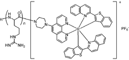
- the compound of the present invention is a compound containing oligoarginine and an iridium complex.
- n is an integer of 4 to 20, preferably n is an integer of 6 to 16, and more preferably n is an integer of 8 to 12.
- the iridium complex may form a salt with an anion.
- the anion include, but are not limited to, hexafluorophosphate ion (PF 6- ), chloride ion ( Cl- ) , bromide ion (Br-), trifluoroacetate ion (CF 3 COO- ) and the like. It is preferably PF 6 ⁇ .
- the compound of the present invention has excellent phosphorescence emission performance and is excellent in binding to the vascular endothelium, so that it can be used as an imaging reagent for blood vessels.
- Yet another aspect of the invention relates to a compound in which oligoarginine and NBD, coumarin 6 or rhodamine B are bound via a linker.
- a linker is not limited, but is preferably a group containing polyethylene glycol.
- the degree of polymerization p of polyethylene glycol (PEG p ) is not limited, but is, for example, 2 to 20, preferably 3 to 16, from the viewpoint of easy staying on the vascular endothelium and easy availability of materials. Yes, more preferably 4-12.
- the degree of polymerization n of oligoarginine (R n ) is not limited, but is, for example, 4 to 20, preferably 6 to 16, and more preferably 8 to 12.
- R n degree of polymerization n of oligoarginine
- Such a compound of the present invention has excellent fluorescence emission performance and is excellent in binding to the vascular endothelium, and therefore can be used as an imaging reagent for blood vessels.
- the compound of the present invention can be synthesized by using a conventional organic synthesis method. For example, it can be synthesized according to the method described in the examples below.
- As the raw material a commercially available product may be used, or a raw material synthesized by a known method may be used.
- the condensation reaction consisted of 0.5 M Fmoc-R (Pbf) -OH and Boc-R (Pbf) -OH DMF solutions as amino acids, 0.6 M HBTU DMF solution as a condensing agent, and 0.5 M HOBt DMF as an additive.
- a 2.0 M DIEA NMP solution as a solution and a base
- 3 equivalents, 3 equivalents, 3 equivalents, and 6 equivalents were added to the number of amino acid-introduced moles, respectively, and the mixture was reacted at room temperature for 1 hour.
- the obtained solid was centrifuged (3500 rpm, 5 minutes), water was removed, water was added again, and the mixture was centrifuged. This operation was repeated twice. The water in the vial was removed by lyophilization to give a red solid (390 mg, 0.14 mmol, crude product yield: 270%).
- BTQ-R 4 Weigh BTQ- [R (Pbf)] 4 -Boc (279 mg, 0.10 mmol) into a 50 mL centrifuge tube, add 1 mL of TFA: water: TIPS (95: 2.5: 2.5), and react at room temperature for 2 hours. I let you. Cold diethyl ether was added to the solution to precipitate a solid. The obtained solid was centrifuged (3500 rpm, 5 minutes), diethyl ether was removed, diethyl ether was added again, and the mixture was centrifuged. This operation was repeated twice.
- Boc- [R (Pbf)] 8 -OH was synthesized by the Fmoc solid-phase synthesis method using a fully automatic microwave peptide synthesizer (Initiator + Alstra, Biotage).
- As the resin chlorotrityl resin (1.67 g, number of moles of amino acid introduced: 0.30 mmol / g) into which R (Pbf) was introduced was used.
- the condensation reaction consisted of 0.6 M Fmoc-R (Pbf) -OH and Boc-R (Pbf) -OH DMF solutions as amino acids, 0.5 M HBTU DMF solution as a condensing agent, and 0.5 M HOBt DMF as an additive.
- the obtained solid was centrifuged (3500 rpm, 5 minutes), water was removed, water was added again, and the mixture was centrifuged. This operation was repeated twice. The water in the vial was removed by lyophilization to give a red solid (235 mg, 0.055 mmol, crude product yield: 110%).
- BTQ-R 8 Weigh BTQ- [R (Pbf)] 8 -Boc (110 mg, 0.026 mmol) into a 50 mL centrifuge tube, add 1 mL of TFA: water: TIPS (95: 2.5: 2.5), and react at room temperature for 2 hours. I let you. Cold diethyl ether was added to the solution to precipitate a solid. The obtained solid was centrifuged (3500 rpm, 5 minutes), diethyl ether was removed, diethyl ether was added again, and the mixture was centrifuged. This operation was repeated twice.
- the condensation reaction consisted of 0.5 M Fmoc-R (Pbf) -OH and Boc-R (Pbf) -OH DMF solutions as amino acids, 0.6 M HBTU DMF solution as a condensing agent, and 0.5 M HOBt DMF as an additive.
- a 2.0 M DIEA NMP solution as a solution and a base
- 3 equivalents, 3 equivalents, 3 equivalents, and 6 equivalents were added to the number of amino acid-introduced moles, respectively, and the mixture was reacted at room temperature for 1 hour.
- the obtained solid was centrifuged (3500 rpm, 5 minutes), water was removed, water was added again, and the mixture was centrifuged. This operation was repeated twice. The water in the vial was removed by lyophilization to give a red solid (320 mg, 0.055 mmol, crude product yield: 110%).
- BTQ-R 12 Weigh BTQ- [R (Pbf)] 12 -Boc (151 mg, 0.026 mmol) into a 50 mL centrifuge tube, add 1 mL of TFA: water: TIPS (95: 2.5: 2.5), and react at room temperature for 2 hours. I let you. Cold diethyl ether was added to the solution to precipitate a solid. The obtained solid was centrifuged (3500 rpm, 5 minutes), diethyl ether was removed, diethyl ether was added again, and the mixture was centrifuged. This operation was repeated twice.
- Boc- [R (Pbf)] 16 -OH was synthesized by the Fmoc solid-phase synthesis method using a fully automatic microwave peptide synthesizer (Initiator + Alstra, Biotage).
- As the resin chlorotrityl resin (0.34 g, number of moles of amino acid introduced: 0.30 mmol / g) into which R (Pbf) was introduced was used.
- the condensation reaction consisted of 0.3 M Fmoc-R (Pbf) -OH and Boc-R (Pbf) -OH DMF solutions as amino acids, 0.6 M HBTU DMF solution as a condensing agent, and 0.5 M HOBt DMF as an additive.
- a 2.0 M DIEA NMP solution as a solution and a base
- 3 equivalents, 3 equivalents, 3 equivalents, and 6 equivalents were added to the number of amino acid-introduced moles, respectively, and the mixture was reacted at room temperature for 1 hour.
- BTQ-R 16 Weigh BTQ- [R (Pbf)] 16 -Boc (113 mg, 0.015 mmol) into a 50 mL centrifuge tube, add 1 mL of TFA: water: TIPS (95: 2.5: 2.5), and react at room temperature for 2 hours. I let you. Cold diethyl ether was added to the solution to precipitate a solid. The obtained solid was centrifuged (3500 rpm, 5 minutes), diethyl ether was removed, diethyl ether was added again, and the mixture was centrifuged. This operation was repeated twice.
- Figure 2 shows the structural formulas (BTQ-R 4 , BTQ-R 8 , BTQ-R 12 , BTQ-R 16 ) of the compound of the present invention synthesized in the examples.
- the number of arginine residues is 4, 8, 12, and 16.
- a compound to which arginine is not bound (BTQ) was used as a reference compound.
- the absorption and phosphorescence spectra of these compounds were measured, the absorption maximum wavelength was shown near 500 nm, and the phosphorescence maximum wavelength was shown near 660 nm. Since phosphorescence is observed at 600 nm and above, multicolor imaging using molecules that emit green light is possible.
- the phosphorescent quantum yield and phosphorescent lifetime were measured and found to be 0.32 (under 0.015 air saturation) and 5.7 ⁇ s (under 0.28 ⁇ s air saturation), respectively. Even compounds with different numbers of arginine residues have almost the same spectral and photophysical properties because the same phosphorous group is bound to the oligoarginine peptide.
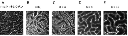
- FIG. 3 shows a mixed solvent of FITC-tomatolectin (1 mg / mL (physiological saline) administered in 50 ⁇ L) and BTQ (100 nmol: 1 mmol / L (physiological saline: dimethylsulfoxide (9: 1, v / v)).
- BTQ-R n (n: 4, 8, 12, 100 nmol: 1 mmol / L (physiological saline) was administered at 100 ⁇ L) from the tail vein of anesthetized mice.
- the ventral part is incised to expose the kidney, and the image of the kidney surface is shown by imaging the kidney surface with a confocal laser microscope.
- BTQ and BTQ-R 4 are observed to emit light from a region (tubular cells) different from that of FITC-tomato lectin, whereas BTQ-R 8 and BTQ -R 12 shows that the capillaries of the kidney are imaged in the same way as FITC-tomato lectin.
- BTQ-R 12 has cavities in the blood vessels, so BTQ-R 12 is distributed in the vascular endothelium, not in the blood. From the above, it was clarified that the number of residues of arginine of 8 or more is preferable for imaging the vascular endothelium.
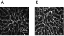
- FITC-tomato lectin (1 mg / mL (physiological saline) administered in 50 ⁇ L) and BTQ-R 12 (100 nmol: 1 mmol / L (physiological saline) administered in 100 ⁇ L) were administered to mice. The same spot was imaged. As shown in A and C of FIG. 4, when imaging with the fluorescence of FITC-tomato lectin, fluorescence was observed from a place other than the vascular endothelium.
- FIG. 6 shows a phosphorescent microscopic image obtained by administering BTQ-R 12 (100 nmol: 1 mmol / L (physiological saline) 100 ⁇ L) to mice.
- BTQ-R 12 100 nmol: 1 mmol / L (physiological saline) 100 ⁇ L
- the vas sinusoide vessels are clearly imaged.
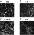
- Similar experiments were performed on other organs (pancreas, small intestine, subcutaneous tissue, heart) and it became clear that the capillaries of many organs could be imaged (Fig. 7).
- FIG. 8 shows a phosphorescent microscopic image obtained by administering BTQ-R 12 (100 nmol: 1 mmol / L (physiological saline) 100 ⁇ L) to a cancer-bearing mouse. From the image, it was found that many small blood vessels were branched from the thick blood vessel, and the imaging of the vascular network in the tumor was successful.
- lipids are abnormally accumulated in the hepatocytes, large lipid droplets are formed, and the hepatocytes are enlarged. Therefore, the narrowing of the vas sinusoideal blood vessels progresses, and the entire liver becomes hypoxic.
- the green fluorescent lipid droplet reagent (3- (benzo [d] thiazol-2-yl) -8- (diethylamino) -2H-benzo [g] chromen-2-one (PC6S)) and BTQ-R 12 Simultaneously administered to healthy mice and adipose liver model mice (PC6S: 50 nmol: 0.5 mmol / L (physiological saline: dimethyl sulfoxide (9: 1, v / v), in a mixed solvent containing 10 wt% BSA).
- Multicolor imaging was performed by administering 100 ⁇ L, BTQ-R 12 : 100 nmol: 1 mmol / L (physiological saline) 100 ⁇ L), and measuring green fluorescence and deep red phosphorescence.
- PC6S fluorescence was observed from lipid droplets in hepatocytes
- phosphorescence of BTQ-R 12 was observed from the sinusoideal endothelium (A in Fig. 9). From the image, it can be seen that the sinusoide vessels run linearly between the hepatocytes.
- hepatocytes are enlarged due to the formation of large lipid droplets, and the sinusoideal blood vessels are greatly tortured (Fig. 9, B). As a result, the flow of red blood cells is obstructed, the oxygen supply to the liver is insufficient, and hypoxia may occur.
- the obtained solid was centrifuged (3500 rpm, 5 minutes), water was removed, water was added again, and the mixture was centrifuged. This operation was repeated twice. The water in the vial was removed by lyophilization to give a magenta solid (157 mg, 0.026 mmol, crude product yield: 45%).
- RhB-pipe-PEG 12 -R 12 Weigh RhB-pipe-PEG 12- [R (Pbf)] 12 -Boc (21.5 mg, 0.0035 mmol) into a 50 mL centrifuge tube, add 1 mL of TFA: water: TIPS (95: 2.5: 2.5), and room temperature. Was reacted for 2 hours. Cold diethyl ether was added to the solution to precipitate a solid. The obtained solid was centrifuged (3500 rpm, 5 minutes), diethyl ether was removed, diethyl ether was added again, and the mixture was centrifuged. This operation was repeated twice.
- NBD-PEG 4- [R (Pbf)] 12 -Boc) Boc- [R (Pbf)] 12 -OH (429 mg, 0.085 mmol), NBD-PEG 4 -NH 2 (0.10 mmol), HATU (65.9 mg, 0.17 mmol) were added to 100 mL eggplant frass, and dehydrated DMF 8 was added. Dissolved in mL. To this solution, 0.17 mL of DIEA was added, and the mixture was stirred at room temperature for 24 hours under nitrogen substitution. The solution was transferred to a 50 mL centrifuge tube and water was added to precipitate a solid.
- NBD-PEG 4 -R 12 Weigh NBD-PEG 4- [R (Pbf)] 12 -Boc (30.2 mg, 0.0060 mmol) into a 50 mL centrifuge tube, add 1 mL of TFA: water: TIPS (95: 2.5: 2.5), and at room temperature. It was allowed to react for 2 hours. Cold diethyl ether was added to the solution to precipitate a solid. The obtained solid was centrifuged (3500 rpm, 5 minutes), diethyl ether was removed, diethyl ether was added again, and the mixture was centrifuged. This operation was repeated twice.
- C6-PEG 4 -R 12 Weigh C6-PEG 4- [R (Pbf)] 12 -Boc (46.0 mg, 0.0082 mmol) into a 50 mL centrifuge tube, add 1 mL of TFA: water: TIPS (95: 2.5: 2.5), and at room temperature. It was allowed to react for 2 hours. Cold diethyl ether was added to the solution to precipitate a solid. The obtained solid was centrifuged (3500 rpm, 5 minutes), diethyl ether was removed, diethyl ether was added again, and the mixture was centrifuged. This operation was repeated twice.
- RhB-pipe-PEG 12 -R 12 was administered to mice to perform vascular imaging of various tissues.
- RhB-pipe-PEG 12 -R 12 (100 nmol: 2 mmmol / L (in physiological saline) 50 ⁇ L) was administered from the tail vein of anesthetized mice, and the ventral part was incised in various ways.
- An image of exposed organs (liver, kidney, spleen, pancreas, testis) and tissues (muscle tissue, subcutaneous tissue) and imaging their surfaces with a confocal laser microscope is shown.
- the excitation wavelength is 550 nm and the observation wavelength is> 590 nm.
- Capillaries can be imaged in all the observed organs and tissues.
- NBD-PEG-R 12 was administered to mice to perform vascular imaging of various tissues.
- NBD-PEG-R 12 100 nmol: 2 mmmol / L (in physiological saline) administered at 50 ⁇ L
- the liver, kidney, spleen) and tissues (subcutaneous tissue, adipose tissue) are exposed, and images of their surfaces imaged with a confocal laser microscope are shown.
- the excitation wavelength is 488 nm and the observation wavelength is 510-550 nm. Capillaries can be imaged in all the observed organs and tissues.
- C6-PEG-R 12 was administered to mice to perform vascular imaging of various tissues.
- C 6 -PEG-R 12 (100 nmol: 2 mm mol / L (in physiological saline) 50 ⁇ L) was administered from the tail vein of anesthetized mice, and the ventral part was incised to various organs. (Kidney, spleen, pancreas, testis) and tissues (fat tissue, muscle tissue) are exposed, and images of their surfaces imaged with a confocal laser microscope are shown.
- the excitation wavelength is 488 nm and the observation wavelength is 510-550 nm.
- Capillaries can be imaged in all the observed organs and tissues. After euthanizing the mice, cerebrovascular imaging was performed. As shown in Fig. 13, it was shown that cerebral blood vessel imaging is also possible.
- a compound containing oligoarginine and a chromophore can be used to clearly image blood vessels in an individual and can be used as an imaging reagent for blood vessels.
- BTQ-R n developed by the present invention can be used as an imaging reagent for blood vessels.
- BTQ-R 12 is a new reagent capable of imaging the vascularization of normal and pathological tissues in an individual.
- the distribution to the vascular endothelium is the action of the oligoarginine peptide, and by changing the luminescence group (BTQ), it is possible to develop vascular endothelium imaging reagents with various luminescent colors.
- RhB-pipe-PEG p -R n , NBD-PEG p -R n , and C6-PEG p -R n developed by the present invention can also be preferably used as blood vessel imaging reagents.
- the present invention can be used in fields such as medical diagnosis, pharmaceutical development, and basic medicine.
Landscapes
- Chemical & Material Sciences (AREA)
- Health & Medical Sciences (AREA)
- Organic Chemistry (AREA)
- General Health & Medical Sciences (AREA)
- Life Sciences & Earth Sciences (AREA)
- Biochemistry (AREA)
- Biophysics (AREA)
- Genetics & Genomics (AREA)
- Medicinal Chemistry (AREA)
- Molecular Biology (AREA)
- Proteomics, Peptides & Aminoacids (AREA)
- Physics & Mathematics (AREA)
- Nuclear Medicine, Radiotherapy & Molecular Imaging (AREA)
- Analytical Chemistry (AREA)
- General Physics & Mathematics (AREA)
- Immunology (AREA)
- Pathology (AREA)
- Epidemiology (AREA)
- Animal Behavior & Ethology (AREA)
- Public Health (AREA)
- Veterinary Medicine (AREA)
- Medicines Containing Antibodies Or Antigens For Use As Internal Diagnostic Agents (AREA)
Priority Applications (1)
| Application Number | Priority Date | Filing Date | Title |
|---|---|---|---|
| JP2022571439A JPWO2022138550A1 (https=) | 2020-12-21 | 2021-12-20 |
Applications Claiming Priority (2)
| Application Number | Priority Date | Filing Date | Title |
|---|---|---|---|
| JP2020211560 | 2020-12-21 | ||
| JP2020-211560 | 2020-12-21 |
Publications (1)
| Publication Number | Publication Date |
|---|---|
| WO2022138550A1 true WO2022138550A1 (ja) | 2022-06-30 |
Family
ID=82157922
Family Applications (1)
| Application Number | Title | Priority Date | Filing Date |
|---|---|---|---|
| PCT/JP2021/046991 Ceased WO2022138550A1 (ja) | 2020-12-21 | 2021-12-20 | 血管のイメージング試薬 |
Country Status (2)
| Country | Link |
|---|---|
| JP (1) | JPWO2022138550A1 (https=) |
| WO (1) | WO2022138550A1 (https=) |
-
2021
- 2021-12-20 WO PCT/JP2021/046991 patent/WO2022138550A1/ja not_active Ceased
- 2021-12-20 JP JP2022571439A patent/JPWO2022138550A1/ja active Pending
Non-Patent Citations (2)
| Title |
|---|
| YASUKAGAWA MAMI; YAMADA KEIICH; TOBITA SEIJI; YOSHIHARA TOSHITADA: "Ratiometric oxygen probes with a cell-penetrating peptide for imaging oxygen levels in living cells", JOURNAL OF PHOTOCHEMISTRY, ELSEVIER, AMSTERDAM, NL, vol. 383, 25 July 2019 (2019-07-25), AMSTERDAM, NL, XP085785139, ISSN: 1010-6030, DOI: 10.1016/j.jphotochem.2019.111983 * |
| YOSHIHARA TOSHITADA; HIRAKAWA YOSUKE; HOSAKA MASAHIRO; NANGAKU MASAOMI; TOBITA SEIJI: "Oxygen imaging of living cells and tissues using luminescent molecular probes", JOURNAL OF PHOTOCHEMISTRY, ELSEVIER, AMSTERDAM, NL, vol. 30, 20 January 2017 (2017-01-20), AMSTERDAM, NL, pages 71 - 95, XP029940267, ISSN: 1389-5567, DOI: 10.1016/j.jphotochemrev.2017.01.001 * |
Also Published As
| Publication number | Publication date |
|---|---|
| JPWO2022138550A1 (https=) | 2022-06-30 |
Similar Documents
| Publication | Publication Date | Title |
|---|---|---|
| CA2903727C (en) | Methods of manufacture and synthesis of amino acid linking groups conjugated to compounds used for targeted imaging of tumors | |
| JP7308366B2 (ja) | アクティブターゲティング型葉酸受容体近赤外蛍光分子及びその調製方法 | |
| EP1281405B1 (de) | Kurzkettige Peptid-Farbstoffkonjugate als Kontrastmittel für die optische Diagnostik | |
| CN115006553B (zh) | 用于制备肿瘤诊断显像剂的多肽及其应用 | |
| JP2010534712A (ja) | 光学イメージング剤 | |
| JP2026012682A (ja) | 近赤外シアニン色素およびそのコンジュゲート | |
| JP2022532628A (ja) | 修飾シアニン色素およびそのコンジュゲート | |
| CN110201189B (zh) | 白蛋白结合型近红外荧光染料-马来酰亚胺共轭物 | |
| CA3177944A1 (en) | Methods of manufacture and synthesis of fluorescent dye compounds and uses thereof | |
| JP5425181B2 (ja) | 蛍光mriプローブ | |
| WO2022138550A1 (ja) | 血管のイメージング試薬 | |
| CN108250125B (zh) | 一种肿瘤靶向探针化合物及其合成和应用 | |
| EP4360659B1 (en) | Fluorescent water-soluble polycationic chitosan polymers as markers for biological 3d imaging | |
| 安カ川真美 | Synthesis and Biological Applications of Phosphorescent Ir (III) Complex-based Probes with Arginine Peptide | |
| CN121378406B (zh) | 靶向Nectin-4的寡肽及其应用 | |
| JP7621655B2 (ja) | 細胞および組織内酸素濃度測定試薬 | |
| KR101774345B1 (ko) | 석회화 표적 근적외선 형광 탐지자 | |
| Zhai | Self-Threaded Fluorescent Probes for Biological Imaging and Self-Assembly | |
| WO2019180475A1 (en) | Azacyanine dyes and use thereof | |
| CN118159831A (zh) | 细胞和组织内脂滴的荧光成像试剂 | |
| JP6834112B2 (ja) | レシオ法を用いた細胞内酸素濃度測定試薬 | |
| JP2016160194A (ja) | pH応答性シアニン系近赤外色素の開発 | |
| Williams | Design, Synthesis, and Evaluation of Innovative BODIPY-Peptidic Conjugates for Biological Application | |
| CN107739528A (zh) | 一种五肽改性菁染料化合物及其制备方法和应用 | |
| NZ522135A (en) | Somatostatin dye conjugates used as contrast agents for optical diagnostics |
Legal Events
| Date | Code | Title | Description |
|---|---|---|---|
| 121 | Ep: the epo has been informed by wipo that ep was designated in this application |
Ref document number: 21908105 Country of ref document: EP Kind code of ref document: A1 |
|
| ENP | Entry into the national phase |
Ref document number: 2022571439 Country of ref document: JP Kind code of ref document: A |
|
| NENP | Non-entry into the national phase |
Ref country code: DE |
|
| 122 | Ep: pct application non-entry in european phase |
Ref document number: 21908105 Country of ref document: EP Kind code of ref document: A1 |